Handbook of Peripheral Neuropathy - Taylor & Francis eBooks
803
-
Upload
khangminh22 -
Category
Documents
-
view
2 -
download
0
Transcript of Handbook of Peripheral Neuropathy - Taylor & Francis eBooks

Handbook ofPeripheral
Neuropathy
DK1596_half-series-title 7/6/05 1:55 PM Page A
NEUROLOGICAL DISEASE AND THERAPY
Advisory Board
Louis R. Caplan, M.D.Professor of Neurology
Harvard University School of MedicineBeth Israel Deaconess Medical Center
Boston, Massachusetts
William C. Koller, M.D.Mount Sinai School of Medicine
New York, New York
John C. Morris, M.D.Friedman Professor of Neurology
Co-Director, Alzheimer’s Disease Research CenterWashington University School of Medicine
St. Louis, Missouri
Bruce Ransom, M.D., Ph.D.Warren Magnuson Professor
Chair, Department of NeurologyUniversity of Washington School of Medicine
Seattle, Washington
Kapil Sethi, M.D.Professor of Neurology
Director, Movement Disorders ProgramMedical College of Georgia
Augusta, Georgia
Mark Tuszynski, M.D., Ph.D.Associate Professor of Neurosciences
Director, Center for Neural RepairUniversity of California–San Diego
La Jolla, California
DK1596_half-series-title 7/6/05 1:55 PM Page B

1. Handbook of Parkinson’s Disease, edited by William C. Koller
2. Medical Therapy of Acute Stroke, edited by Mark Fisher3. Familial Alzheimer’s Disease: Molecular Genetics
and Clinical Perspectives, edited by Gary D. Miner, Ralph W. Richter, John P. Blass, Jimmie L. Valentine, and Linda A. Winters-Miner
4. Alzheimer’s Disease: Treatment and Long-TermManagement, edited by Jeffrey L. Cummings and Bruce L. Miller
5. Therapy of Parkinson’s Disease, edited by William C. Koller and George Paulson
6. Handbook of Sleep Disorders, edited by Michael J. Thorpy7. Epilepsy and Sudden Death, edited by Claire M. Lathers
and Paul L. Schraeder8. Handbook of Multiple Sclerosis, edited by Stuart D. Cook9. Memory Disorders: Research and Clinical Practice,
edited by Takehiko Yanagihara and Ronald C. Petersen10. The Medical Treatment of Epilepsy, edited by
Stanley R. Resor, Jr., and Henn Kutt11. Cognitive Disorders: Pathophysiology and Treatment,
edited by Leon J. Thal, Walter H. Moos, and Elkan R. Gamzu
12. Handbook of Amyotrophic Lateral Sclerosis, edited byRichard Alan Smith
13. Handbook of Parkinson’s Disease: Second Edition,Revised and Expanded, edited by William C. Koller
14. Handbook of Pediatric Epilepsy, edited by Jerome V. Murphy and Fereydoun Dehkharghani
15. Handbook of Tourette’s Syndrome and Related Tic and Behavioral Disorders, edited by Roger Kurlan
16. Handbook of Cerebellar Diseases, edited by Richard Lechtenberg
17. Handbook of Cerebrovascular Diseases, edited by Harold P. Adams, Jr.
18. Parkinsonian Syndromes, edited by Matthew B. Stern and William C. Koller
19. Handbook of Head and Spine Trauma, edited by Jonathan Greenberg
20. Brain Tumors: A Comprehensive Text, edited by Robert A. Morantz and John W. Walsh
21. Monoamine Oxidase Inhibitors in Neurological Diseases,edited by Abraham Lieberman, C. Warren Olanow,Moussa B. H. Youdim, and Keith Tipton
DK1596_half-series-title 7/6/05 1:55 PM Page C

22. Handbook of Dementing Illnesses, edited by John C. Morris
23. Handbook of Myasthenia Gravis and MyasthenicSyndromes, edited by Robert P. Lisak
24. Handbook of Neurorehabilitation, edited by David C. Good and James R. Couch, Jr.
25. Therapy with Botulinum Toxin, edited by Joseph Jankovicand Mark Hallett
26. Principles of Neurotoxicology, edited by Louis W. Chang27. Handbook of Neurovirology, edited by
Robert R. McKendall and William G. Stroop28. Handbook of Neuro-Urology, edited by David N. Rushton29. Handbook of Neuroepidemiology, edited by
Philip B. Gorelick and Milton Alter30. Handbook of Tremor Disorders, edited by Leslie J. Findley
and William C. Koller31. Neuro-Ophthalmological Disorders: Diagnostic Work-Up
and Management, edited by Ronald J. Tusa and Steven A. Newman
32. Handbook of Olfaction and Gustation, edited by Richard L. Doty
33. Handbook of Neurological Speech and LanguageDisorders, edited by Howard S. Kirshner
34. Therapy of Parkinson’s Disease: Second Edition, Revised and Expanded, edited by William C. Koller and George Paulson
35. Evaluation and Management of Gait Disorders, edited by Barney S. Spivack
36. Handbook of Neurotoxicology, edited by Louis W. Changand Robert S. Dyer
37. Neurological Complications of Cancer, edited by Ronald G. Wiley
38. Handbook of Autonomic Nervous System Dysfunction,edited by Amos D. Korczyn
39. Handbook of Dystonia, edited by Joseph King Ching Tsuiand Donald B. Calne
40. Etiology of Parkinson’s Disease, edited by Jonas H. Ellenberg, William C. Koller, and J. William Langston
41. Practical Neurology of the Elderly, edited by Jacob I. Sageand Margery H. Mark
42. Handbook of Muscle Disease, edited by Russell J. M. Lane43. Handbook of Multiple Sclerosis: Second Edition,
Revised and Expanded, edited by Stuart D. Cook
DK1596_half-series-title 7/6/05 1:55 PM Page D

44. Central Nervous System Infectious Diseases and Therapy,edited by Karen L. Roos
45. Subarachnoid Hemorrhage: Clinical Management, edited by Takehiko Yanagihara, David G. Piepgras, and John L. D. Atkinson
46. Neurology Practice Guidelines, edited by Richard Lechtenberg and Henry S. Schutta
47. Spinal Cord Diseases: Diagnosis and Treatment, edited byGordon L. Engler, Jonathan Cole, and W. Louis Merton
48. Management of Acute Stroke, edited by Ashfaq Shuaiband Larry B. Goldstein
49. Sleep Disorders and Neurological Disease, edited byAntonio Culebras
50. Handbook of Ataxia Disorders, edited by Thomas Klockgether
51. The Autonomic Nervous System in Health and Disease,David S. Goldstein
52. Axonal Regeneration in the Central Nervous System, edited by Nicholas A. Ingoglia and Marion Murray
53. Handbook of Multiple Sclerosis: Third Edition,edited by Stuart D. Cook
54. Long-Term Effects of Stroke, edited by Julien Bogousslavsky
55. Handbook of the Autonomic Nervous System in Healthand Disease, edited by C. Liana Bolis, Julio Licinio, and Stefano Govoni
56. Dopamine Receptors and Transporters: Function, Imaging, and Clinical Implication, Second Edition, edited by Anita Sidhu, Marc Laruelle, and Philippe Vernier
57. Handbook of Olfaction and Gustation: Second Edition,Revised and Expanded, edited by Richard L. Doty
58. Handbook of Stereotactic and Functional Neurosurgery,edited by Michael Schulder
59. Handbook of Parkinson’s Disease: Third Edition, edited byRajesh Pahwa, Kelly E. Lyons, and William C. Koller
60. Clinical Neurovirology, edited by Avindra Nath and Joseph R. Berger
61. Neuromuscular Junction Disorders: Diagnosis andTreatment, Matthew N. Meriggioli, James F. Howard, Jr.,and C. Michel Harper
62. Drug-Induced Movement Disorders, edited by Kapil D. Sethi
DK1596_half-series-title 7/6/05 1:55 PM Page E

63. Therapy of Parkinson’s Disease: Third Edition, Revisedand Expanded, edited by Rajesh Pahwa, Kelly E. Lyons,and William C. Koller
64. Epilepsy: Scientific Foundations of Clinical Practice, edited by Jong M. Rho, Raman Sankar, and José E. Cavazos
65. Handbook of Tourette’s Syndrome and Related Tic and Behavioral Disorders: Second Edition, edited by Roger Kurlan
66. Handbook of Cerebrovascular Diseases: Second Edition,Revised and Expanded, edited by Harold P. Adams, Jr.
67. Emerging Neurological Infections, edited by ChristopherPower and Richard T. Johnson
68. Treatment of Pediatric Neurologic Disorders, edited byHarvey S. Singer, Eric H. Kossoff, Adam L. Hartman, and Thomas O. Crawford
69. Synaptic Plasticity : Basic Mechanisms to ClinicalApplications, edited by Michel Baudry, Xiaoning Bi, and Steven S. Schreiber
70. Handbook of Essential Tremor and Other TremorDisorders, edited by Kelly E. Lyons and Rajesh Pahwa
71. Handbook of Peripheral Neuropathy, edited by Mark B. Bromberg and A. Gordon Smith
72. Carotid Artery Stenosis: Current and EmergingTreatments, edited by Seemant Chaturvedi and Peter M. Rothwell
73. Gait Disorders: Evaluation and Management, edited byJeffrey M. Hausdorff and Neil B. Alexander
DK1596_half-series-title 7/6/05 1:55 PM Page F

Handbook ofPeripheral
Neuropathy
edited by
Mark B. BrombergUniversity of Utah
Salt Lake City, Utah, U.S.A.
A. Gordon SmithUniversity of Utah
Salt Lake City, Utah, U.S.A.
Boca Raton London New York Singapore
DK1596_half-series-title 7/6/05 1:55 PM Page i

CRC PressTaylor & Francis Group6000 Broken Sound Parkway NW, Suite 300Boca Raton, FL 33487-2742
© 2005 by Taylor & Francis Group, LLCCRC Press is an imprint of Taylor & Francis Group, an Informa business
No claim to original U.S. Government worksVersion Date: 20130726
International Standard Book Number-13: 978-0-8493-5486-1 (eBook - PDF)
This book contains information obtained from authentic and highly regarded sources. While all reasonable efforts have been made to publish reliable data and information, neither the author[s] nor the publisher can accept any legal responsibility or liability for any errors or omissions that may be made. The publishers wish to make clear that any views or opinions expressed in this book by individual editors, authors or contributors are personal to them and do not necessarily reflect the views/opinions of the publishers. The information or guidance contained in this book is intended for use by medical, scientific or health-care professionals and is provided strictly as a supplement to the medical or other professional’s own judgement, their knowledge of the patient’s medical history, relevant manufacturer’s instructions and the appropriate best practice guidelines. Because of the rapid advances in medical science, any information or advice on dosages, procedures or diagnoses should be independently verified. The reader is strongly urged to consult the drug companies’ printed instructions, and their websites, before adminis-tering any of the drugs recommended in this book. This book does not indicate whether a particular treatment is appropriate or suitable for a particular individual. Ultimately it is the sole responsibility of the medical professional to make his or her own professional judgements, so as to advise and treat patients appropriately. The authors and publishers have also attempted to trace the copyright holders of all material reproduced in this publication and apologize to copyright holders if permission to publish in this form has not been obtained. If any copyright material has not been acknowledged please write and let us know so we may rectify in any future reprint.
Except as permitted under U.S. Copyright Law, no part of this book may be reprinted, reproduced, transmitted, or utilized in any form by any electronic, mechanical, or other means, now known or hereafter invented, including photocopying, microfilming, and recording, or in any information storage or retrieval system, without written permission from the publishers.
For permission to photocopy or use material electronically from this work, please access www.copyright.com (http://www.copy-right.com/) or contact the Copyright Clearance Center, Inc. (CCC), 222 Rosewood Drive, Danvers, MA 01923, 978-750-8400. CCC is a not-for-profit organization that provides licenses and registration for a variety of users. For organizations that have been granted a photocopy license by the CCC, a separate system of payment has been arranged.
Trademark Notice: Product or corporate names may be trademarks or registered trademarks, and are used only for identifica-tion and explanation without intent to infringe.
Visit the Taylor & Francis Web site athttp://www.taylorandfrancis.com
and the CRC Press Web site athttp://www.crcpress.com

To my wife, Diane, and my daughter, Katherine.
Mark
To Sam and Emily.
Gordon


Foreword
Writing a foreword to a new textbook is an act of faith. I do, however, have a considerable
degree of faith since I know the editors and many of the authors. They are well trained,
hard working, and expert.
Their intent has been to write a clinically useful handbook on the subject of periph-
eral neuropathy. Having edited (with others) four editions of another book on the same
subject, I can understand the desire to produce a short and light book. The eminent neur-
ologist Raymond Adams who reviewed our textbook on several occasions, complained
about its weight, arguing that it was not a handbook and could not be read in bed.
The Handbook of Peripheral Neuropathy is to be a practical book that provides easy
to understand information and which is light enough to hold in the hand, allowing it to be
read in bed as well as at the bedside of a patient. The editors acknowledge the clinical
expertise and mentorship of James Albers, MD, PhD, and his influence is perhaps reflected
in the well ordered table of contents. The editors and contributors are experienced neur-
ologists and electromyographers whose writings are worthy to be read. Now we await
what they say about the subjects which they review—the proof of the pudding will be
in the eating or in seeing the play (“to catch the conscience of the king”—Hamlet.). I
hope this foreword whets your appetite so that you will read it.
Peter James Dyck, M.D.
Roy E. and Merle Meyer Professor of Neuroscience
The Peripheral Neuropathy Research Laboratory
Mayo Clinic College of Medicine, Rochester, Minnesota, U.S.A.
v


Preface
Peripheral neuropathies are common clinical problems. Foot numbness and pain are
frequent complaints in the primary care setting. The complexity of nerve anatomy and
pathology and the long list of causes of peripheral neuropathy may seem daunting
and often lead primary care providers to refer patients to a neurologist. Even among
neurologists, the evaluation can be challenging. All of these factors contribute to the
general perception that the diagnosis and management of peripheral neuropathies is
complex and mysterious. The goal of this text is to de-mystify the evaluation and treatment
of peripheral neuropathies and to provide a practical guide to the management of common
symptoms.
The diagnosis of peripheral neuropathy can be simplified by using a structured
approach. One purpose of medical monographs is to bring order to apparent complexity.
Accordingly, this book is designed around a diagnostic approach based on an under-
standing of the anatomy and pathophysiology of the peripheral nervous system. This
approach involves asking a series of simple clinical and electrodiagnostic questions
whose answers lead to a full characterization of the neuropathy. When knowledge of
basic classes of neuropathies is added to this characterization, it can yield a reasonable
differential diagnosis. At this point, a focused set of laboratory tests can be rationally
selected that will maximize the diagnostic yield. Treatment and management of peripheral
neuropathy also benefit from an approach that emphasizes an understanding of underlying
pathophysiology of the neuropathy.
This book is divided into three broad sections. The first broad section provides an
evaluation algorithm. This algorithm is based on the anatomy, physiology and pathology
of peripheral nerves in the context of symptoms and signs. A similar algorithm is offered
for designing and interpreting electrodiagnostic tests. The role of special diagnostic tests,
including imaging, quantitative sensory testing, and nerve and skin biopsy is also reviewed
in the first section. The second broad section presents classes of peripheral neuropathies.
For each class, the clinical characteristics, electrodiagnostic features, examination find-
ings, treatment options and outcome are reviewed. The third broad section discusses
general treatment modalities and management issues which will improve the patient’s
well being.
We thank our many contributors who have provided their experience and expertise
to make this a clinically useful book. The mechanics of editing a book on peripheral
neuropathy are as daunting as the subject, and we could not have succeeded without the
vii

expertise of Ms. Becky Guertler. We also thank our publisher, especially Jinnie Kim the
acquiring editor, for their patience, in preparing the book.
We want to acknowledge our mentor in neuromuscular diseases, James Albers M.D.,
Ph.D., who has provided an approach to the diagnosis and management of peripheral
neuropathies that is reflected in the general outline of this book.
Mark B. Bromberg
A. Gordon Smith
viii Preface

Contents
Foreword Peter James Dyck . . . . . . . . . . . . . . . . . . . . . . . . . . . . . . . . . . . . v
Preface . . . . . . . . . . . . . . . . . . . . . . . . . . . . . . . . . . . . . . . . . . . . . . . . . . . . . . . vii
Contributors . . . . . . . . . . . . . . . . . . . . . . . . . . . . . . . . . . . . . . . . . . . . . . . . . . . xiii
1. An Approach to the Evaluation of Peripheral Nerve Diseases . . . . . . . . . . . . 1
Mark B. Bromberg
Laboratory Evaluation of Peripheral Neuropathy
2. Electrodiagnostic Evaluation . . . . . . . . . . . . . . . . . . . . . . . . . . . . . . . . . . . . 17
Mark B. Bromberg
3. Quantitative Sensory Testing . . . . . . . . . . . . . . . . . . . . . . . . . . . . . . . . . . . . 45
James W. Russell
4. Evaluation of the Autonomic Nervous System . . . . . . . . . . . . . . . . . . . . . . . 53
Safwan S. Jaradeh and Thomas E. Prieto
5. Peripheral Nerve Histology and Pathology . . . . . . . . . . . . . . . . . . . . . . . . . . 65
Mark B. Bromberg
6. Skin Biopsy . . . . . . . . . . . . . . . . . . . . . . . . . . . . . . . . . . . . . . . . . . . . . . . . 83
A. Gordon Smith
7. MR Imaging of the Peripheral Nerve . . . . . . . . . . . . . . . . . . . . . . . . . . . . . . 91
Kevin R. Moore
Inflammatory Demyelinating Neuropathies
8. Acute Inflammatory Demyelinating Polyradiculoneuropathy
(Guillain–Barre Syndrome) . . . . . . . . . . . . . . . . . . . . . . . . . . . . . . . . . . . . . 111
Amanda C. Peltier and James W. Russell
9. Chronic Inflammatory Demyelinating Polyradiculoneuropathy . . . . . . . . . . . 127
Zachary Simmons
ix

10. Multifocal Demyelinating Neuropathies . . . . . . . . . . . . . . . . . . . . . . . . . . . . 161
Richard A. Lewis
Neuropathies Associated with Systemic Disease
11. Diabetic Neuropathy . . . . . . . . . . . . . . . . . . . . . . . . . . . . . . . . . . . . . . . . . . 179
A. Gordon Smith and J. Robinson Singleton
12. Endocrine Neuropathies . . . . . . . . . . . . . . . . . . . . . . . . . . . . . . . . . . . . . . . 205
Patrick M. Grogan and Jonathan S. Katz
13. Neuropathy and Rheumatologic Disease . . . . . . . . . . . . . . . . . . . . . . . . . . . . 215
Rachel A. Nardin and Seward B. Rutkove
14. Neuropathy Associated with Cancer . . . . . . . . . . . . . . . . . . . . . . . . . . . . . . . 239
David R. Renner and Deborah T. Blumenthal
15. Paraproteinemias and Acquired Amyloidosis . . . . . . . . . . . . . . . . . . . . . . . . 259
Mark E. Landau and William W. Campbell
16. Peripheral Neuropathy in HIV Infection . . . . . . . . . . . . . . . . . . . . . . . . . . . . 275
Lydia Estanislao and David Simpson
17. Infectious and Granulomatous Neuropathies . . . . . . . . . . . . . . . . . . . . . . . . . 293
Sharon P. Nations, Jaya R. Trivedi, and Gil I. Wolfe
18. Critical Illness Polyneuropathy and Myopathy . . . . . . . . . . . . . . . . . . . . . . . 315
J. Robinson Singleton and Estelle S. Harris
19. Nutritional Neuropathies . . . . . . . . . . . . . . . . . . . . . . . . . . . . . . . . . . . . . . . 325
Patrick C. Nolan and James W. Albers
20. Toxic Neuropathy . . . . . . . . . . . . . . . . . . . . . . . . . . . . . . . . . . . . . . . . . . . . 351
Eric J. Sorenson and A. Gordon Smith
Hereditary Neuropathies
21. Inherited Peripheral Neuropathies: Charcot–Marie–Tooth Disease . . . . . . . . 379
Michael E. Shy, Richard A. Lewis, and Jun Li
22. Hereditary Sensory Autonomic Neuropathies . . . . . . . . . . . . . . . . . . . . . . . . 395
Victoria H. Lawson
23. Other Hereditary Neuropathies . . . . . . . . . . . . . . . . . . . . . . . . . . . . . . . . . . 411
Jun Li and A. Gordon Smith
Focal Peripheral Neuropathy
24. Radiculopathy . . . . . . . . . . . . . . . . . . . . . . . . . . . . . . . . . . . . . . . . . . . . . . . 439
Firas G. Saleh and Rahman Pourmand
x Contents

25. Plexopathies . . . . . . . . . . . . . . . . . . . . . . . . . . . . . . . . . . . . . . . . . . . . . . . . 453
John C. Kincaid
26. Median Mononeuropathies . . . . . . . . . . . . . . . . . . . . . . . . . . . . . . . . . . . . . 467
A. Gordon Smith
27. Radial Neuropathy . . . . . . . . . . . . . . . . . . . . . . . . . . . . . . . . . . . . . . . . . . . 481
Michael Stanton
28. Ulnar Mononeuropathies . . . . . . . . . . . . . . . . . . . . . . . . . . . . . . . . . . . . . . . 487
John D. Steffens
29. Other Upper Extremity Neuropathies . . . . . . . . . . . . . . . . . . . . . . . . . . . . . . 497
J. Steven Schultz
30. Sciatic Neuropathy . . . . . . . . . . . . . . . . . . . . . . . . . . . . . . . . . . . . . . . . . . . 527
A. Mouaz Sbei
31. Focal Neuropathies of the Femoral, Obturator, Lateral Femoral
Cutaneous Nerve, and Other Nerves of the Thigh and Pelvis . . . . . . . . . . . . . 541
Kevin J. Felice
32. Management of Traumatic Mononeuropathies . . . . . . . . . . . . . . . . . . . . . . . 559
Lisa DiPonio, James A. Leonard Jr, and John E. McGillicuddy
33. Cranial Mononeuropathies . . . . . . . . . . . . . . . . . . . . . . . . . . . . . . . . . . . . . . 583
Mark B. Bromberg
Treatment of Peripheral Neuropathy
34. A Diagnostic and Treatment Approach to Painful Neuropathy . . . . . . . . . . . . 599
Hiroyuki Nodera and David N. Herrmann
35. Intravenous Immunoglobulin in the Treatment of Peripheral Neuropathy . . . . 621
Reem F. Bunyan
36. Treatment of Immune-Mediated Peripheral Neuropathy . . . . . . . . . . . . . . . . 639
Orly Vardeny and A. Gordon Smith
37. Managing Joint and Skin Complications from Peripheral Neuropathy . . . . . . 657
M. Catherine Spires
38. West Nile Virus . . . . . . . . . . . . . . . . . . . . . . . . . . . . . . . . . . . . . . . . . . . . . 667
Patrick Luedtke and John E. Greenlee
39. Diagnostic Yield for Peripheral Neuropathy . . . . . . . . . . . . . . . . . . . . . . . . . 677
A. Gordon Smith
Index . . . . . . . . . . . . . . . . . . . . . . . . . . . . . . . . . . . . . . . . . . . . . . . . . . . . . . . . 687
Contents xi


Contributors
James W. Albers, MD, PhD Department of Neurology, University of Michigan,
Ann Arbor, Michigan, USA
Deborah T. Blumenthal, MD Department of Neurology, University of Utah, Salt Lake
City, Utah, USA
Mark B. Bromberg, MD, PhD Department of Neurology, University of Utah, Salt Lake
City, Utah, USA
Reem F. Bunyan, MD Department of Neurology, King Faisal University, Al-Khobar,
Saudi Arabia
William W. Campbell, MD, MSHA Department of Neurology, Walter Reed Army
Medical Center, Washington DC, USA
Lisa DiPonio, MD Department of Physical Medicine & Rehabilitation, University of
Michigan Medical Center, Ann Arbor, Michigan, USA
Lydia Estanislao, MD Department of Neurology and Clinical Neurophysiology,
Mount Sinai Medical Center, New York, New York, USA
Kevin J. Felice, DO Department of Neurology, University of Connecticut Health
Center, Farmington, Connecticut, USA
John E. Greenlee, MD Department of Neurology, University of Utah, Salt Lake City,
Utah, USA
Patrick M. Grogan, MD Department of Neurology, Stanford University School of
Medicine, Stanford, California, USA
Estelle S. Harris, MD Department of Pulmonary and Critical Care Medicine,
University of Utah, Salt Lake City, Utah, USA
David N. Herrmann, MB, BCh Department of Neurology, University of Rochester
Medical Center, Rochester, New York, USA
Safwan S. Jaradeh, MD Department of Neurology, Medical College of Wisconsin,
Milwaukee, Wisconsin, USA
Jonathan S. Katz, MD Department of Neurology, Stanford University School of
Medicine, Stanford, California, USA
xiii

John C. Kincaid, MD Department of Neurology, Indiana University School of
Medicine, Indianapolis, Indiana, USA
Mark E. Landau, MD Department of Neurology, Walter Reed Army Medical Center,
Washington DC, USA
Victoria H. Lawson, MD Department of Neurology, University of Utah, Salt Lake City,
Utah, USA
James A. Leonard Jr, MD Department of Physical Medicine & Rehabilitation,
University of Michigan Medical Center, Ann Arbor, Michigan, USA
Richard A. Lewis, MD Department of Neurology, Wayne State University School of
Medicine, Detroit, Michigan, USA
Jun Li, MD, PhD Department of Neurology, Wayne State University School of
Medicine, Detroit, Michigan, USA
Patrick Luedtke, MD, MPH Department of Family and Preventive Medicine,
University of Utah, Salt Lake City, Utah, USA
John E. McGillicuddy, MD Department of Neurosurgery, University of Michigan
Medical Center, Ann Arbor, Michigan, USA
Kevin R. Moore, MD Department of Neuroradiology, University of Utah, Salt Lake
City, Utah, USA
Rachel A. Nardin, MD Department of Neurology, Beth Israel Hospital, Boston,
Massachusetts, USA
Sharon P. Nations, MD Department of Neurology, University of Texas Southwestern
Medical Center, Dallas, Texas, USA
Hiroyuki Nodera, MD Department of Neurology, University of Rochester Medical
Center, Rochester, New York, USA
Patrick C. Nolan, MD, PhD Department of Neurology, University of Michigan,
Ann Arbor, Michigan, USA
Amanda C. Peltier, MD Department of Neurology, University of Michigan and
Veterans Affairs Medical Center, Ann Arbor, Michigan, USA
Rahman Pourmand, MD Department of Neurology, Stony Brook University Hospital,
Stony Brook, New York, USA
Thomas E. Prieto, PhD Department of Neurology, Medical College of Wisconsin,
Milwaukee, Wisconsin, USA
David R. Renner, MD Department of Neurology, University of Utah, Salt Lake City,
Utah, USA
James W. Russell, MD, MS, FRCP Veterans Affairs Medical Center and Department
of Neurology, University of Michigan, Ann Arbor, Michigan, USA
Seward B. Rutkove, MD Department of Neurology, Beth Israel Hospital, Boston,
Massachusetts, USA
Firas G. Saleh, MD Department of Neurology, Stony Brook University Hospital, Stony
Brook, New York, USA
xiv Contributors
A. Mouaz Sbei, MD Department of Neurology, University of Utah, Salt Lake City,
Utah, USA
J. Steven Schultz, MD Department of Physical Medicine and Rehabilitation, University
of Michigan Health System, Ann Arbor, Michigan, USA
Michael E. Shy, MD Department of Neurology, Wayne State University School of
Medicine, Detroit, Michigan, USA
Zachary Simmons, MD, FAAN Department of Neurology, Penn State College of
Medicine, Hershey, Pennsylvania, USA
David Simpson, MD Department of Neurology and Clinical Neurophysiology,
Mount Sinai Medical Center, New York, New York, USA
J. Robinson Singleton, MD Department of Neurology, University of Utah, Salt Lake
City, Utah, USA
A. Gordon Smith, MD Department of Neurology, University of Utah, Salt Lake City,
Utah, USA
Eric J. Sorenson, MD Department of Neurology, University of Utah, Salt Lake City,
Utah, USA
M. Catherine Spires, MS, MD Department of Physical Medicine and Rehabilitation,
University of Michigan, Ann Arbor, Michigan, USA
Michael Stanton, MD Department of Neurology, University of Rochester Medical
Center, Rochester, New York, USA
John D. Steffens, MD Department of Neurology, University of Utah, Salt Lake City,
Utah, USA
Jaya R. Trivedi, MD Department of Neurology, University of Texas Southwestern
Medical Center, Dallas, Texas, USA
Orly Vardeny, Pharm.D. Department of Pharmacy, University of Utah, Salt Lake City,
Utah, USA
Gil I. Wolfe, MD Department of Neurology, University of Texas Southwestern Medical
Center, Dallas, Texas, USA
Contributors xv


1An Approach to the Evaluation ofPeripheral Nerve Diseases
Mark B. BrombergUniversity of Utah, Salt Lake City, Utah, USA
Abstract 2
1. Introduction 2
2. Layer 1: Localization to the Peripheral Nervous System 3
3. Layer 2: Localization Within the Peripheral Nervous System 4
3.1. Radicular Pattern 4
3.2. Plexus Pattern 4
3.3. Mononeuropathy Pattern 5
3.4. Peripheral Neuropathy Pattern 5
4. Layer 3: What is the Time Course? 6
5. Layer 4: Which Nerve Fibers are Involved? 6
5.1. Symptoms 7
5.2. Signs 7
5.2.1. Touch Stimuli 8
5.2.2. Vibration Stimuli 8
5.2.3. Sharp Stimuli 8
5.2.4. Position Sense 8
5.2.5. Deep Tendon Reflexes 8
5.2.6. Motor Signs 9
5.2.7. Orthopedic Signs 9
5.2.8. Other Signs 10
5.2.9. Autonomic System Signs 10
6. Layer 5: What is the Primary Pathology? 10
7. Localization Summary 10
8. Layer 6: What are the Other Pertinent Features? 11
8.1. Medical History 11
8.2. Medications and Other Compounds 11
8.3. Family History 11
1

9. Layer 7: What are Pertinent Epidemiologic Factors? 12
10. Summary 12
References 12
ABSTRACT
Neurologic diagnosis can be challenging, particularly for disorders of peripheral nerve.
For many clinicians, basic nerve anatomy and physiology and the various patterns of path-
ology can be daunting. In general neurology, diagnostic efficiency is enhanced by first
localizing the lesion within the nervous system followed by determination of the pathol-
ogy, and then a consideration of cause. The same approach can be applied to peripheral
nerve disorders. This chapter presents a structured approach to the evaluation of peripheral
nerve diseases based on asking a series of questions during the history and examination,
whose answers will lead to localization and characterization of the peripheral nerve lesion.
Following this, a meaningful list of laboratory tests can be ordered.
1. INTRODUCTION
Numbness, tingling, and pain and weakness in the limbs are among the most common com-
plaints voiced to clinicians. In the general population, the prevalence of peripheral neuropa-
thy approaches 10% (1), that of median mononeuropathies 4% (2), and radiculopathies likely
exceed median nerve mononeuropathies (carpal tunnel syndrome) (3). It can be challenging
to separate, among these common symptoms, those that reflect peripheral nerve disorders
from those that arise from the central nervous system and to identify underlying causes.
Most experienced clinicians would likely agree that a structured approach to patient evalu-
ation based on localization is more efficient than an unstructured or shotgun approach. Dis-
advantages of an unstructured approach are many. For the patient, there may be unnecessary
false leads, hopes, and anxieties. Computed tomography (CT) and magnetic resonance
imaging (MRI) scans are frequently relied upon to exclude lesion sites, but most lesions
of the peripheral nervous system are not diagnosable by imaging studies. Further, unnecess-
ary imaging studies are costly, and they may reveal coincident or irrelevant findings that can
lead to unnecessary surgery. A shotgun approach to laboratory testing is expensive and
unproductive (4). Batteries of blood tests are frequently ordered, but rarely informative
because the majority of common tests are unrelated to disorders of peripheral nerves.
This chapter will outline a structured and efficient evaluation that leads to a full
localization and characterization of the neuropathy. At this point, the scope of the differ-
ential diagnosis is reduced, and a rational list of laboratory tests can be ordered. Localiz-
ation and characterization are discussed in terms of a series of layers (Table 1.1). The first
layer is localization to the peripheral nervous system. Subsequent layers refine the local-
ization within the peripheral nervous system and define underlying peripheral nerve path-
ology. The final layer includes a consideration of probability and epidemiology of
peripheral nerve disorders that leads to an initial and reasonable differential diagnosis.
Assigning layers and a sequence is somewhat artificial. The evaluation process is
dynamic and layers will merge and the sequence will vary depending upon the clinician’s
experience and clinical situation.
The approach in this chapter can not only serve as a framework for evaluating
patients, but also to be used to determine localizing features when reviewing the literature
on specific disorders of peripheral nerve (including material presented in this book).
2 Bromberg

2. LAYER 1: LOCALIZATION TO THE PERIPHERALNERVOUS SYSTEM
This level is usually assumed, but it deserves mention that not all symptoms of numbness,
tingling, and pain and weakness are referable to the peripheral nervous system. Failure to
localize can lead to diagnostic errors. Classic examples are strokes, tumors, central
nervous system demyelinating disorders, and compressive myelopathies. Cortical
origins of numbness and weakness frequently involve one side of the body, whereas
spinal origins have bilateral leg symptoms with a truncal level of demarcation (5).
Central demyelinating disorders can be challenging because they may include asymmetric
and unusual symptom patterns, but associated features and examination findings such as
pathologic tendon reflexes help localize the disorder to the central nervous system.
Some patterns of symptoms and signs may not be attributable to definable lesions
within either the central or peripheral nervous systems (6). Under these circumstances,
a somatoform disorder should be considered. Somatoform disorders represent distressful
physical symptoms causing impaired social or occupational dysfunction with no diagno-
sable condition to account for them (7). Numbness, paresthesias, pain, weakness, and
fatigue are among common peripheral nerve symptoms that are considered to be “pseudo-
neurological” when no neurological basis can be found. Whether a patient fulfills complete
DSM-IV criteria for somatization or undifferentiated somatoform disorders is less
important than recognition that further evaluation is unlikely to be diagnostic (7). Every
patient deserves a considered evaluation, no matter how high the initial index of suspicion
is that symptoms are not physiologic, because somatization can accompany true diseases
(6). However, when reasonable localization cannot be achieved, the neurologic and lab-
oratory examinations are normal, and the temporal pattern does not fit known pathologic
processes, it is highly unlikely that the symptoms represent a definable pathology.
Under these circumstances, a discussion seeking other factors is appropriate.
Leading such patients to understand that internal factors are causative or contributory
should be taken slowly. Replacing long-standing symptoms with the “good news” of
“good health” is rarely successful. Other psychiatric conditions such as depression,
Table 1.1 Layers of Diagnostic Evaluation Leading to a Full Characterization of the Neuropathy
and an Accurate Localization within the Peripheral Nervous System
First Layer
Within the peripheral nervous system? Exclude cortical, spinal, psychogenic loci
Second Layer
What part of the peripheral nervous system? Root, plexus, single nerve, multiple nerves,
peripheral neuropathy
Third Layer
What is the time course? Acute, subacute, chronic, progressive,
relapsing-remitting, event-linked
Fourth Layer
What nerve fibers are involved? Sensory, motor, autonomic, combinations
Fifth Layer
What is the primary pathology? Demyelination, axonal, mixed
Sixth Layer
What are the other pertinent features? Family, medical, social histories
Seventh Layer
What are the epidemiologic features? Probability, age of onset, rarity, gender
Evaluation of Peripheral Nerve Diseases 3

anxiety, and panic disorders may coexist (8). Many times, with long-standing symptoms,
the family milieu has incorporated the patient’s symptoms, and giving the patient a thera-
peutic way out of their dilemma, such as physical therapy, may be more successful for the
patient and family.
3. LAYER 2: LOCALIZATION WITHIN THE PERIPHERALNERVOUS SYSTEM
This layer is based on the anatomy of the peripheral nervous system. Symptoms expected
from lesions at various sites along nerves, from root to plexus and peripheral nerve, can be
queried during the history. The history should be taken as an active process, with the goal
of understanding what the patient is experiencing to answer the question of localization.
Neurologic findings during the examination should therefore be predicted from the
history and represent a confirmatory step. Examples of symptoms that help localization
to sites along peripheral nerves are considered below. At each site, issues addressed in
layers 3 through 7 will be emphasized.
3.1. Radicular Pattern
Radicular patterns of shooting pain and paresthesias down one limb following a root dis-
tribution are rare compared with diffuse low back pain, and occur more frequently with
lumbosacral than cervical spine or truncal movements (5,9). Diffuse back pain is not
usually caused by focal radiculopathies. Degenerative disk and vertebral findings on
MRI scanning are common, and unless marked in degree, are rarely the cause of focal radi-
culopathies (10). The frequency of root involvement is one-thirds cervical spine in the dis-
tribution of C7 (�70%), C6 (�20%), and C8 (�10%) and two-thirds lumbosacral spine in
the distribution of L4–5 (�40%), S1 (�40%) and other roots (�10%). Sensory symptoms
including pain are more common than weakness, and altered sensations and reduced
tendon reflexes in the appropriate root distribution are supportive. Muscle atrophy and
true weakness are rare examination findings (9). Diagnostically, acute disk herniation is
less challenging than chronic disease due to degenerative foraminal narrowing. Spinal
stenosis produces involvement of multiple roots and follows a chronic course with low
back pain, leg pain, and paresthesias triggered or increased by walking (9).
3.2. Plexus Pattern
Plexopathies are rare. Trauma to the brachial plexus is the most common cause. Acute
plexopathies associated with marked pain are frequently idiopathic when involving the
brachial plexus (Parsonage–Turner syndrome) (11) and associated with diabetes when
involving the lumbosacral plexus (12). Thoracic outlet syndrome is frequently queried,
but the incidence of true neurogenic thoracic outlet syndrome is exceedingly rare, and
is characterized by the pattern of axonal loss of ulnar sensory and motor and median
motor nerve fibers caused by chronic pressure on the lower trunk (13). Plexopathies due
to cancer are chronic in nature and may or may not include pain (14,15).
Differentiating radiculopathies from plexopathies may be difficult when symptoms
are primarily vague pain. Radiculopathies more commonly include focal symptoms,
whereas plexopathies produce more diffuse symptoms. Both conditions involve axonal
pathology, and differentiating between them can be achieved by sensory nerve conduction
studies and needle electromyography (EMG).
4 Bromberg

3.3. Mononeuropathy Pattern
Lesions involving individual nerves are characterized by loss of sensation and strength in
the distribution of the affected nerve. There are multiple causes of mononeuropathies, but
chronic pressure (entrapment neuropathies) with focal demyelinating pathology is most
common. Mononeuropathy multiplex is a rare condition frequently caused by vasculitis
associated with collagen vascular diseases. Another class of mononeuropathy multiplex
pattern is multifocal motor neuropathy with conduction block caused by focal immune
mediated demyelination.
3.4. Peripheral Neuropathy Pattern
Several patterns of peripheral nerve involvement can be recognized Table 1.2. The proto-
typic and most common pattern is length-dependent, with sensory loss and pain preceding
distal weakness. As progressively shorter nerves are affected, symptoms and signs unroll
as a stocking up the leg. The nerve length at the knee level approximately equals the length
innervating the hand, and with further progression, symptoms and signs unroll as a long
glove up the arm. The distribution is usually symmetric. In the extreme, a shield loss
over the chest and abdomen can be observed when nerve length involvement reaches
the circumference of the thorax. Most length-dependent peripheral neuropathies are
chronic and involve axonal pathology (16). As a corollary, it is rare in polyneuropathy
for there to be sensory involvement to the waist level, especially without marked
sensory loss also to the elbows. Accordingly, isolated sensory loss to the upper thigh
and waist levels suggests central nervous system localization (myelopathy). Causes of
peripheral neuropathy are many, and are felt to reflect metabolic abnormalities. Diabetes
mellitus is the most common underlying cause, followed by hereditary (genetic) neuro-
pathies. The cause for many neuropathies is unknown, and idiopathic or cryptogenic
neuropathies represent up to 25% of peripheral neuropathies.
When the pattern of symptoms and signs includes both proximal and distal limb
involvement, the pathologic process is usually demyelination at multifocal sites along
roots and nerves (inflammatory polyradiculoneuropathy). Acute and chronic forms
occur (AIDP and CIDP).
Table 1.2 Patterns of Peripheral Neuropathy and Examples of Disorders and Causes
Sensory-motor symmetric
(length-dependent pattern)
Diabetes, medications, toxins, metabolic disorders, hereditary
Sensory-motor symmetric
(proximal and distal
pattern)
Acute inflammatory demyelinating polyradiculopathy, chronic
inflammatory demyelinating polyradiculopathy
Sensory-motor asymmetric
(nerve or plexus pattern)
Diabetic amyotrophy, idiopathic plexopathy, vasculitic
mononeuritis multiplex
Sensory-motor asymmetric Porphyria, leprosy
Sensory symmetric or
asymmetric
Paraneoplastic neuronopathy, Sjogren syndrome, idiopathic
ganglionitis, vitamin B6 toxicity, leprosy
Motor symmetric or
asymmetric
Amyotrophic lateral sclerosis, multifocal motor conduction block
neuropathy, lower motor neuron syndrome, poliomyelitis,
West Nile syndrome
Autonomic symmetric or
asymmetric
With other neuropathies (diabetes, acute inflammatory
demyelinating polyradiculopathy), isolated involvement
(amyloidosis)
Evaluation of Peripheral Nerve Diseases 5

Asymmetric patterns are less common and include a number of different lesion
sites and pathologic processes. It is useful to determine whether sensory or motor nerves
are predominantly involved. Unilateral and focal symptoms and signs in a limb must
be distinguished from radiculopathy, plexopathy, or mononeuropathy (mononeuritis
multiplex). Atrophic weakness without sensory loss that does not follow radicular,
plexopathy, or mononeuropathy pattern suggests motor neuron disease (amyotrophic
lateral sclerosis).
4. LAYER 3: WHAT IS THE TIME COURSE?
The usual definitions of time course in neurology are somewhat blurred in diseases of the
peripheral nerve, and Table 1.3 lists time courses with examples. True acute or apoplectic
events are rare in atraumatic nerve injuries, and occur in the setting of vasculitis. More
commonly, an acute onset is defined as days to several weeks, and suggests the
Guillain–Barre syndrome or AIDP, a metabolic event, or a toxic exposure. Most
chronic neuropathies are steadily progressive. A history of clear remissions and exacer-
bations suggests CIDP or other form of immune-mediated neuropathy. It can be challen-
ging to accurately determine the start of a slowly progressive chronic neuropathy because
patients may not appreciate an insidious onset. Querying patients about their functional
performance during historic or calendar events can be helpful to identify the onset.
When the time course clearly starts in adult life, an acquired neuropathy is more likely
than a hereditary disorder. When the time course cannot be dated, a hereditary neuropathy
should be considered. However, our understanding of hereditary neuropathies is expand-
ing and a number of “idiopathic” neuropathies beginning in adulthood may represent mild
forms of hereditary neuropathies.
5. LAYER 4: WHICH NERVE FIBERS ARE INVOLVED?
The peripheral nervous system can be divided into somatic and autonomic components,
and somatic peripheral nerves can be further divided into sensory and motor functions.
Within the somatic nervous system, sensory and motor fiber involvement can be accu-
rately assessed and there are neuropathies affecting sensory, motor, or both types of
fibers. In the autonomic nervous system, separating sensory (afferent) from motor (effer-
ent) involvement is difficult and both are commonly affected. Neuropathies with isolated
autonomic nervous system involvement are rare. Accordingly, it is practical to determine
whether there is any involvement of autonomic nerve involvement.
Table 1.3 Peripheral Neuropathy Time Courses and Examples of Associated Neuropathies
Acute
Apoplectic Vasculitic mononeuritis multiplex, idiopathic plexopathy
Days to weeks Acute inflammatory demyelinating polyradiculopathy, porphyria,
acute toxic exposure, proximal diabetic neuropathy,
paraneoplastic sensory neuronopathy
Chronic
Years Diabetic polyneuropathy, chronic inflammatory demyelinating
polyradiculopathy, idiopathic
Insidious Hereditary
6 Bromberg

5.1. Symptoms
From the chief complaint it may not be apparent which types of nerves are involved. Nerve
dysfunction can be expressed as negative and positive symptoms (Table 1.4). Positive
symptoms are felt to reflect inappropriate spontaneous nerve activity detected by the
patient as uncomfortable and painful sensations, or other spontaneous phenomena. Nega-
tive symptoms reflect loss of nerve signaling. An important clinical difference between
sensory and motor somatic nerves involves compensatory mechanisms. Following
motor nerve loss, surviving motor nerves undergo collateral reinnervation to reinnervate
orphaned muscle fibers. This compensatory process has the effect of blunting weakness
due to mild motor nerve loss, and clinical weakness may not be apparent to the patient
or on physical examination until 50% of motor nerve fibers are lost (80% in slowly pro-
gressive denervating disorders) (17). However, positive symptoms of cramps and fascicu-
lations may be present early on as the only clinical indication of motor nerve involvement.
The needle EMG is sensitive in detecting early motor fiber loss and will confirm motor
nerve involvement.
5.2. Signs
The clinical neurologic examination is sensitive for peripheral nerve loss and dysfunction,
and informative for localization. An appreciation of nerve physiology and pathology, and
the limitations of clinical testing, are necessary to accurately interpret the clinical
examination. It is important to emphasize that the sensory examination can be challenging
and confusing because responses are indirect and represent a patient’s interpretation of
the testing questions. Accordingly, it is important that the patient fully understands the
object of the test. Attention and co-operation are imperative. It is worthwhile having
specific questions derived from the history to address on the neurologic examination.
For example, does the sensory loss follow a stocking-glove (distal predominate), derma-
tomal, or radicular pattern?
The sensory examination frequently focuses on determining whether there is “large
fiber” or “small fiber” involvement, based on a battery of simple clinical tests. However,
psychophysical sensory perception testing suggests these distinctions are more apparent
than real because of overlap between nociception, touch, and pressure stimulus properties.
Although nociceptive information is conveyed by small diameter nerve fibers, some
nociceptive receptors are innervated by myelinated fibers, and subjects can distinguish
sharp from dull stimuli without feeling pain. Formal psychophysical testing of nociception
is performed using hot stimuli, cold stimuli, and special equipment, which contrasts to
clinical sensory testing performed using cool instruments (tuning fork and reflex
Table 1.4 Positive and Negative Symptoms Associated with Nerve Damage
Positive symptoms Negative symptoms
Somatic nerves
Sensory Pain, tingling Numbness, lack of feeling
Motor Cramps, fasciculations Weakness, atrophy
Autonomic nerves
Hyperhydrosis, diarrhea Orthostatic hypotension,
impotence, anhydrosis,
constipation
Evaluation of Peripheral Nerve Diseases 7

hammer) and sharp objects of varying shape (safety pin, broken wooden stick, and
commercial pin probe).
Cutaneous mechanoreceptors are mainly innervated by large-diameter nerve
fibers and are activated by a variety of moving stimuli. Touch stimulus threshold
changes modestly with age. A comparison of quantitative sensory testing in neuropathy
patients indicates that vibratory thresholds are well correlated with touch-pressure
thresholds and vibratory thresholds are suitable indicators of large-diameter sensory
nerve dysfunction (18).
With these principles and specific questions in mind, an informative battery of
sensory tests can be performed. It is noteworthy that patients are generally able to deter-
mine the level of involvement on a limb by asking them to make a line of demarcation,
below which sensations are abnormal and above which they are normal.
5.2.1. Touch Stimuli
Application of the lightest touch to the dorsum of the hand and foot represents a measure
of low threshold mechanoreception. A series of monofilaments can be applied to grade the
severity of touch loss. Ten-gram filaments are useful because lack of touch perception at
this level of pressure is associated with risk for unappreciated trauma.
5.2.2. Vibration Stimuli
Tuning forks of 128 Hz assess larger diameter nerve fiber function. Various comparisons
can be made, and it is very important that patients are fully attentive and understand the
need to indicate complete disappearance of the vibration. Comparisons between patient
and examiner for the disappearance of the vibration can be measured in seconds. Alterna-
tively, the time for the vibration to disappear for the patient after the tuning is forcefully
struck can be measured in seconds. Empiric data from the great toe indicate that young
adults lose vibration perception after 15 s, with a loss of 1 s per decade of age, and a
loss of vibratory perception in ,10 s is abnormal at any age (4).
5.2.3. Sharp Stimuli
The goal is to apply a sharp stimulus without also applying undo pressure on the skin. A
distinction between noxious and light pressure stimuli can be made by gently applying the
two ends of a safety pin in association with a three-part question: “which is sharper, the
first application, the second application, or are both the same?”. Inability to distinguish
between sharp and dull supports loss of nociceptive fibers relative to low-threshold
mechanoreceptor fibers.
5.2.4. Position Sense
The ability to detect changes in digital joint position is normally exquisite (two degrees). It
is important that patients understand the degree of sensitivity requested, and that they are
blinded to the testing. Accordingly, misperception of joint movements (including falsely
perceived position changes), and insensitivity to movements are significant for loss of
large-diameter fibers. Profound joint position loss is unusual in peripheral nerve disorders,
and often reflects central nervous system involvement.
5.2.5. Deep Tendon Reflexes
Tendon reflexes represent an objective measure of sensory nerve function. The myotatic
reflex consists of a monosynaptic arc with large-diameter afferent (sensory) nerve fiber
input from muscle spindle fibers and large-diameter efferent (motor) nerve fiber output
8 Bromberg
from alpha motor neuron fibers. The reflex is much more vulnerable to sensory nerve fiber
than to motor nerve fiber damage. Accordingly, an absent reflex is an objective indication
of significant dysfunction of large-diameter sensory fibers. However, assurance that the
reflex is truly absent is essential, and reinforcing maneuvers, such as clinching the jaw
or fists and the Jendrassic maneuver, should be used before the reflex is considered
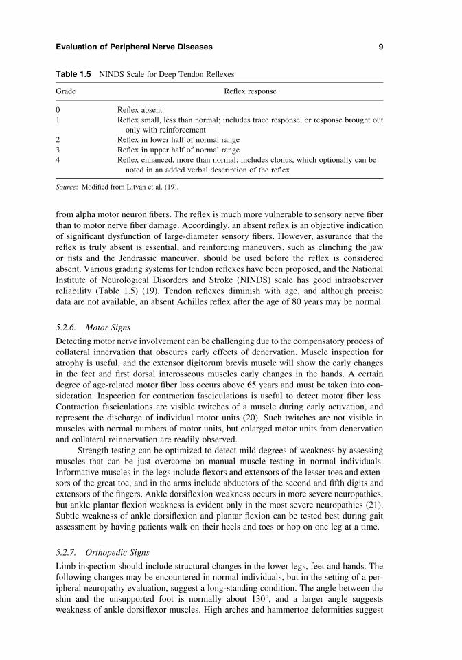
absent. Various grading systems for tendon reflexes have been proposed, and the National
Institute of Neurological Disorders and Stroke (NINDS) scale has good intraobserver
reliability (Table 1.5) (19). Tendon reflexes diminish with age, and although precise
data are not available, an absent Achilles reflex after the age of 80 years may be normal.
5.2.6. Motor Signs
Detecting motor nerve involvement can be challenging due to the compensatory process of
collateral innervation that obscures early effects of denervation. Muscle inspection for
atrophy is useful, and the extensor digitorum brevis muscle will show the early changes
in the feet and first dorsal interosseous muscles early changes in the hands. A certain
degree of age-related motor fiber loss occurs above 65 years and must be taken into con-
sideration. Inspection for contraction fasciculations is useful to detect motor fiber loss.
Contraction fasciculations are visible twitches of a muscle during early activation, and
represent the discharge of individual motor units (20). Such twitches are not visible in
muscles with normal numbers of motor units, but enlarged motor units from denervation
and collateral reinnervation are readily observed.
Strength testing can be optimized to detect mild degrees of weakness by assessing
muscles that can be just overcome on manual muscle testing in normal individuals.
Informative muscles in the legs include flexors and extensors of the lesser toes and exten-
sors of the great toe, and in the arms include abductors of the second and fifth digits and
extensors of the fingers. Ankle dorsiflexion weakness occurs in more severe neuropathies,
but ankle plantar flexion weakness is evident only in the most severe neuropathies (21).
Subtle weakness of ankle dorsiflexion and plantar flexion can be tested best during gait
assessment by having patients walk on their heels and toes or hop on one leg at a time.
5.2.7. Orthopedic Signs
Limb inspection should include structural changes in the lower legs, feet and hands. The
following changes may be encountered in normal individuals, but in the setting of a per-
ipheral neuropathy evaluation, suggest a long-standing condition. The angle between the
shin and the unsupported foot is normally about 1308, and a larger angle suggests
weakness of ankle dorsiflexor muscles. High arches and hammertoe deformities suggest
Table 1.5 NINDS Scale for Deep Tendon Reflexes
Grade Reflex response
0 Reflex absent
1 Reflex small, less than normal; includes trace response, or response brought out
only with reinforcement
2 Reflex in lower half of normal range
3 Reflex in upper half of normal range
4 Reflex enhanced, more than normal; includes clonus, which optionally can be
noted in an added verbal description of the reflex
Source: Modified from Litvan et al. (19).
Evaluation of Peripheral Nerve Diseases 9

long-standing differences in the muscular forces exerted on the bones of the foot leading to
foreshorten feet. Fallen arches can also be observed in severe neuropathies. Toe and foot
injuries unnoticed by the patient suggest a marked degree of sensory loss. In the hands,
flexion contractions of the fingers suggest weakness of finger extensor muscles. Inability
to adduct the fifth digits suggests weakness of lumbrical muscles.
5.2.8. Other Signs
Mild dependent pedal edema, rubor, coolness and shininess of the lower leg and foot
despite good distal arterial pulses, suggests decreased movements of distal leg muscles
caused by mild muscle weakness, reducing the vascular return of blood and lymph.
5.2.9. Autonomic System Signs
The autonomic nervous system is involved in many peripheral neuropathies, but symp-
toms and signs of dysautonomia are uncommonly voiced by the patient and must
queried. Orthostatic dizziness and changes in blood pressure (a drop of .30 mmHg
systolic pressure and .15 mmHg diastolic pressure recorded 60–90 s after standing
following 5 min of supine rest) support autonomic involvement. Impotence has many
causes, but is frequently associated with autonomic neuropathy. The sicca symptoms
(dry eyes and mouth) are associated with the Sjogren syndrome and represent end
organ failure of salivary and tear glands. Sjogren syndrome is associated with sensory
neuropathies.
6. LAYER 5: WHAT IS THE PRIMARY PATHOLOGY?
Determining the primary pathologic process is important for diagnosis, treatment, and
prognosis. The two basic pathologic processes are demyelination and axonal loss. They
may occur together, especially when the primary process is demyelination because demye-
lination frequently involves immune attack and axons can be damaged as innocent
bystanders.
Electrodiagnostic testing is most able to distinguish axonal from demyelinating
primary pathology (discussed in Chapter 2). Nerve biopsy is less practical and informative
in this regard for several reasons. Biopsies evaluate only a small segment of sensory
nerves, and the relevant pathologic process may be missed. Biopsies are rarely repeated,
and the time course of changes cannot be followed. A nerve biopsy leaves permanent
dysfunction, and most biopsies are of sensory nerves because a localized area of numbness
is tolerable whereas permanent weakness is not. Nerve biopsy is important when vasculitis
is a consideration, and a biopsy can detect rare causes of neuropathy due to deposition of
protein or other substance, such as amyloid, and abnormal cells such as sarcoid (granulo-
mas) and malignant cells. The role of nerve biopsy is further discussed in other chapters.
7. LOCALIZATION SUMMARY
At this point in the evaluation, the neuropathy should be fully characterized. Table 1.6
summarizes the clinical, electrodiagnostic, and pathologic characteristics. The next two
layers focus on factors unique to the patient under evaluation, and help refine diagnostic
considerations and the order of laboratory testing.
10 Bromberg

8. LAYER 6: WHAT ARE THE OTHER PERTINENT FEATURES?
Determining the underlying causes of peripheral neuropathies and other disorders of per-
ipheral nerve is challenging, and a thorough review of the patient’s medical and family
history is valuable.
8.1. Medical History
Past and current medical histories are obviously important, but the number of medical con-
ditions associated with clinically significant peripheral nerve disorders is limited (4). The
clear exceptions are diabetes mellitus, certain collagen vascular disorders, chronic renal
failure, and HIV infection. Despite this, many laboratory tests are frequently ordered in
the evaluation of a neuropathy that represent general medical tests that are not truly infor-
mative or pertinent to the evaluation of peripheral nerve disorders. Medical causes of per-
ipheral neuropathy will be covered in other chapters.
8.2. Medications and Other Compounds
Inquiring about medication use is important, and should include vitamins and other over
the counter compounds. Although the list of drugs, compounds, and vitamins associated
with peripheral neuropathies is limited, drug-induced neuropathies represent readily
treatable causes. These will be discussed in other chapters.
8.3. Family History
An important line of inquiry is the family history, seeking evidence to support a hereditary
neuropathy. Although it may seem that a hereditary condition should be known within a
family, the slow progression and variable expression masks detection. Interestingly, in
large families with known Charcot–Marie–Tooth neuropathy, ,30% of affected individ-
uals seek medical attention for their symptoms (22). Therefore, a careful line of questions
can be very informative when there are clinical features suggesting a very long-standing
condition, such as insidious onset, high arches, and hammertoes. Table 1.7 lists useful
questions that can be addressed to parents, siblings, and children.
Table 1.6 Clinical, Electrodiagnostic, and
Pathologic Characteristics of Peripheral
Neuropathies
Clinical characteristics
Acute or chronic
Symmetric or asymmetric
Distal length-dependent or distal and proximal
Electrodiagnostic and pathologic characteristics
Uniform demyelinating; sensoryþmotor
Segmental demyelinating; motor . sensory
Axonal; motor . sensory
Axonal; sensory neuropathy or neuronopathy
Axonal; motor neuropathy or neuronopathy
Axonal and demyelinating; sensoryþmotor
Evaluation of Peripheral Nerve Diseases 11

9. LAYER 7: WHAT ARE PERTINENT EPIDEMIOLOGIC FACTORS?
Review of epidemiology and establishing disease probability in the context of the specific
patient is the final step. The often recited maximum of hoof beats being more likely to be
caused by horses than by zebras applies to peripheral neuropathies. Clinicians frequently
express premature concern about zebras in the form of rare and unlikely diseases on the
initial laboratory evaluation. The fear of “not missing something” overrides good epide-
miologic sense. Knowledge about epidemiology patterns and disease probability, and
comfort with these issues, comes from the literature on specific types of neuropathies. It
is hoped that subsequent chapters in this book will be informative in this regard.
10. SUMMARY
The diagnostic process, in clinical practice, takes many forms. However, there is a core
amount of information that is necessary to make an efficient and accurate differential
diagnosis. This chapter identifies the major points to be covered during the history and
examination. The clinical goal is the correct diagnosis, the art of achieving this goal is
in making the process direct and efficient.
REFERENCES
1. The Italian General Practitioner Study Group. Chronic symmetric symptomatic polyneuro-
pathy in the elderly: A field screening investigation in two Italian regions. I. Prevalence and
general characteristics of the sample. Neurology 1995; 45:1832–1836.
2. Astroshi I, Gummersson C, Johnsson R, Ornstein E, Ranstam J, Rosen I. Prevalence of carpal
tunnel syndrome in a general population. J Am Med Assoc 1999; 282:153–158.
3. Wilbourn A, Aminoff M. AAEM Minimonograph 32: The electrodiagnostic examination in
patients with radiculopathies. Muscle Nerve 1998; 21:1612–1631.
4. Barohn R. Approach to peripheral neuropathy and neuronopathy. Semin Neurol 1998;
18:7–18.
5. Brazis P, Masdeu J, Biller J. Localization in Clinical Neurology. Boston: Little, Brown and
Company, 1990.
6. Schiffer R. Psychiatric aspects of clinical neurology. Am J Psychiatry 1983; 140:205–207.
Table 1.7 Questions Pertinent to Chronic Neuropathies
Difficulty with running, sports, or military activities?
High-arched feet?
Hammer or curled-up toes?
Claw hands?
Wasting of distal muscles?
Foot troubles, foot ulcers?
Use of braces?
Foot troubles attributed to “arthritis” or “poliomyelitis (incorrect diagnosis)?”
Difficulty with walking on heels?
Difficulty rising from a kneeled position?
12 Bromberg

7. Task Force on DSM-IV. Diagnostic and Statistical Manual of Mental Disorders. Washington,
DC: American Psychiartic Association, 1994.
8. Halloway K, Zerbe K. Simplified approach to somatization disorder. Postgrad Med 2000;
108:89–95.
9. Quebec Task Force on Spinal Disorders. Scientific approach to the assessment and manage-
ment of activity-related spinal disorders. Spine 1987; 12 (suppl):S1–S54.
10. Jensen M, Brant-Zawadzki M, Obuchowski N, Modic M, Malkasian D, Ross J. Magnetic
resonance imaging of the lumbar spine in people without back pain. N Engl J Med 1994;
331:69–71.
11. Tsairis P, Dyck P, Mulder D. Natural history of brachial plexus neuropathy. Arch Neurol 1972;
27:109–117.
12. Barohn R, Sahenk Z, Warmolts J, Mendell J. The Burns-Garland syndrome (diabetic amyotro-
phy). Revisited 100 years later. Arch Neurol 1991; 48:1130–1135.
13. Le Forestier N, Moulonguet A, Maisonobe T, Leger J-M, Bouche P. True neurogenic
thoracic outlet syndrome: Electrophysiological diagnosis in six cases. Muscle Nerve 1998;
21:1129–1134.
14. Lederman R, Wilbourn A. Brachial plexopathy: recurrent cancer or radiation? Neurology
1984; 34:1331–1335.
15. Thomas J, Cascino T, Earle J. Differential diagnosis between radiation and tumor plexopathy
of the pelvis. Neurology 1985; 35:1–7.
16. Sabin T. Classification of peripheral neuropathy: the long and the short of it. Muscle Nerve
1986; 9:711–719.
17. McComas A, Sica R, Campbell M, Upton A. Functional compensation in partially denervated
muscles. J Neurol Neurosurg Psychiat 1971; 34:453–460.
18. Dyck P, Karnes J, O’Brien P, Zimmerman I. Detection thresholds of cutaneous sensation in
humans. In: Dyck P, Thomas P, Lambert E, Bunge R, eds. Peripheral Neuropathy. Vol. 1.
Philadelphia: WB Saunders Company, 1984:1103–1136.
19. Litvan I, Mangone C, Werden W et al. Reliability of the NINDS myotatic reflex scale.
Neurology 1996; 47:969–972.
20. Denny-Brown D, Pennybacker J. Fibrillation and fasciculation in voluntary muscle. Brain
1938; 61:311–334.
21. Bourque P, Dyck P. Selective calf weakness suggests intraspinal pathology, not peripheral
neuropathy. Arch Neurol 1990; 47:79–80.
22. Dyck P, Oviart K, Lambert E. Intensive evaluation of referral unclassified neuropathies yields
improved diagnosis. Ann Neurol 1981; 10:222–226.
Evaluation of Peripheral Nerve Diseases 13


References
1 An Approach to the Evaluation ofPeripheral Nerve Diseases
7. Task Force on DSM-IV. Diagnostic and Statistical Manualof Mental Disorders. Washington, DC: American PsychiarticAssociation, 1994.
8. Halloway K, Zerbe K. Simplified approach to somatizationdisorder. Postgrad Med 2000; 108:89–95.
9. Quebec Task Force on Spinal Disorders. Scientificapproach to the assessment and management ofactivity-related spinal disorders. Spine 1987; 12(suppl):S1–S54.
10. Jensen M, Brant-Zawadzki M, Obuchowski N, Modic M,Malkasian D, Ross J. Magnetic resonance imaging of thelumbar spine in people without back pain. N Engl J Med1994; 331:69–71.
11. Tsairis P, Dyck P, Mulder D. Natural history ofbrachial plexus neuropathy. Arch Neurol 1972; 27:109–117.
12. Barohn R, Sahenk Z, Warmolts J, Mendell J. TheBurns-Garland syndrome (diabetic amyotrophy). Revisited 100years later. Arch Neurol 1991; 48:1130–1135.
13. Le Forestier N, Moulonguet A, Maisonobe T, Le´ger J-M,Bouche P. True neurogenic thoracic outlet syndrome:Electrophysiological diagnosis in six cases. Muscle Nerve1998; 21:1129–1134.
14. Lederman R, Wilbourn A. Brachial plexopathy: recurrentcancer or radiation? Neurology 1984; 34:1331–1335.
15. Thomas J, Cascino T, Earle J. Differential diagnosisbetween radiation and tumor plexopathy of the pelvis.Neurology 1985; 35:1–7.
16. Sabin T. Classification of peripheral neuropathy: thelong and the short of it. Muscle Nerve 1986; 9:711–719.
17. McComas A, Sica R, Campbell M, Upton A. Functionalcompensation in partially denervated muscles. J NeurolNeurosurg Psychiat 1971; 34:453–460.
18. Dyck P, Karnes J, O’Brien P, Zimmerman I. Detectionthresholds of cutaneous sensation in humans. In: Dyck P,

Thomas P, Lambert E, Bunge R, eds. Peripheral Neuropathy.Vol. 1. Philadelphia: WB Saunders Company, 1984:1103–1136.
19. Litvan I, Mangone C, Werden W et al. Reliability of theNINDS myotatic reflex scale. Neurology 1996; 47:969–972.
20. Denny-Brown D, Pennybacker J. Fibrillation andfasciculation in voluntary muscle. Brain 1938; 61:311–334.
21. Bourque P, Dyck P. Selective calf weakness suggestsintraspinal pathology, not peripheral neuropathy. ArchNeurol 1990; 47:79–80.
22. Dyck P, Oviart K, Lambert E. Intensive evaluation ofreferral unclassified neuropathies yields improveddiagnosis. Ann Neurol 1981; 10:222–226.

Laboratory Evaluation of PeripheralNeuropathy
1. Dumitru D, Amato A, Zwarts M. ElectrodiagnosticMedicine. Philadelphia: Hanley and Belfus, Inc, 2002.
2. Kimura J. Electrodiagnosis in Diseases of Nerve andMuscle: Principles and Practice. Philadelphia: FA DavisCompany, 2001.
3. Preston D, Shapiro B. Electromyography and NeuromuscularDisorders. Boston: ButterworthHeinemann, 1998.
4. Oh S. Clinical Electromyography. Nerve ConductionStudies. Philadelphia: Lippincott Williams & Wilkins, 2003.
5. Bromberg M, Spiegelberg T. The influence of activeelectrode placement on CMAP amplitude. ElectroencephalogrClin Neurophysiol 1997; 105:385–389.
6. Raynor E, Preston D, Logigian E. Influence of surfacerecording electrode placement on nerve action potentials.Muscle Nerve 1997; 20:361–363.
7. Robinson L, Temkin N, Fujimoto W, Stolov W. Effects ofstatistical methodology on normal limits in nerveconduction studies. Muscle Nerve 1991; 14:1084–1090.
8. Campbell W, Robinson L. Deriving reference values inelectrodiagnostic medicine. Muscle Nerve 1993; 16:424–428.
9. Rivner M, Swift T, Crout B, Rhodes K. Towards morerational nerve conduction interpretations: the effect ofheight. Muscle Nerve 1990; 13:232–239.
10. Bromberg M, Jaros L. Symmetry of normal motor andsensory nerve conduction measurement. Muscle Nerve 1998;21:498–503.
11. Thiele B, Bo¨hle A. Anzahl der Spike-Komponenten imMotor-Unit Potential. Zeit EEG-EMG 1978; 9:125–130. 12.Feinstein B, Lindega˚rd B, Nyman E, Wohlfart G. Morphologicstudies of motor units in normal human muscle. Acta Anat1955; 23:127–143. 13. Engstrom J, Olney R. Quantitativemotor unit analysis: The effect of sample size. MuscleNerve 1992; 15:277–281. 14. Dorfman L. The distribution ofconduction velocities (DCV) in peripheral nerves: a review.Muscle Nerve 1984; 7:2–11. 15. Olney R, Budingen H, MillerR. The effect of temporal dispersionon compound actionpotential area in human peripheral nerve. Muscle Nerve

1987; 10:728–733. 16. Rosenfalck A. Early recognition ofnerve disorders by near-nerve recording of sensory actionpotentials. Muscle Nerve 1978; 1:360–367. 17. Carleton S,Brown W. Changes in motor unit populations in motor neuronedisease. J Neurol Neurosurg Psychiat 1979; 42:42–51. 18.Cornblath D, Kuncl R, Mellits E et al. Nerve conductionstudies in amyotrophic lateral sclerosis. Muscle Nerve1992; 15:1111–1115. 19. van der Meche´ F, Meulstee J.Guillain–Barre´ syndrome: A model of random conductionblock. J Neurol Neurosurg Psychiat 1988; 51:1158–1163. 20.Pestronk A, Cornblath D, Ilyas A et al. A treatablemultifocal motor neuropathy with antibodies to GM1ganglioside. Neurology 1988; 24:73–78. 21. Rhee E, EnglandJ, Sumner A. A computer simulation of conduction block:effects produced by actual block versus interphasecancellation. Ann Neurol 1990; 28:146–156. 22. Uncini A, DiMuzio A, Sabatelli M, Magi S, Tonali P, Gambi D.Sensitivity and specificity of diagnostic criteria forconduction block in chronic inflammatory demyelinatingpolyneuropathy. Electroencephalogr Clin Neurophysiol 1993;89:161–169. 23. Lange D, Trojaborg W, Latov N et al.Multifocal motor neuropathy with conduction block: is it adistinct clinical entity? Neurology 1992; 42:497–505. 24.Kraft G. Fibrillation potential amplitude and muscleatrophy following peripheral nerve injury. Muscle Nerve1990; 13:814–821. 25. Borenstein S, Desmedt J. Range ofvariations in motor unit potentials during reinnervationafter traumatic nerve lesions in humans. Ann Neurol 1980;8:460–467. 26. Zalewska E, Rowinska-Marcinska K,Hausmanowa-Petrusewicz I. Shape irregularity of motor unitpotentials in some neuromuscular disorders. Muscle Nerve1998; 21:1181–1187. 27. Barohn R, Kissel J, Warmolts J,Mendell J. Chronic inflammatory demyelinatingpolyradiculoneuropathy. Arch Neurol 1989; 46:878–884. 28.Parry G. Are multifocal motor neuropathy and Lewis–Sumnersyndrome distinct nosologic entities? Muscle Nerve 1999;22:557–559. 29. Lewis R. Multifocal motor neuropathy andLewis–Sumner syndrome. Two distinct entities. Muscle Nerve1999; 22:1738–1739. 30. Latov N. Diagnosis of CIDP.Neurology 2002; 59(suppl 6):S2–S6. 31. Dyck P, Lais A, OhtaM, Bastron J, Okazaki H, Groover R. Chronic inflammatorypolyradiculoneuropathy. Mayo Clin Proc 1975; 50:621–636.32. Katz J, Saperstein D, Gronseth G, Amato A, Barohn R.Distal acquired demyelinating symmetric neuropathy.Neurology 2000; 54:615–620. 33. Thomas P, Claus D, JaspertA et al. Focal upper limb demyelinating neuropahty. Brain1996; 119:765–774. 34. Oh S, Joy J, Kuruoglu R. Chronicsensory demyelinating neuropthy: chronic inflammatorydemyelinating polyneuropathy presenting as a pure sensoryneuropathy. J Neurol Neurosurg Psychiatry 1992; 55:677–680.

35. Rotta F, Sussman A, Bradley W, Ayyar D, Sharma K,Shebert R. The spectrum of chronic inflammatorydemyelinating polyneuropathy. J Neurol Sci 2000;173:129–139. 36. Saperstein D, Amato A, Wolfe G et al.Multifocal acquired demyelinating sensory and motorneuropathy: the Lewis–Sumner syndrome. Muscle Nerve 1999;22:560–566. 37. Albers J, Donofrio P, McGonagle T.Sequential electrodiagnostic abnormalities in acuteinflammatory demyelinating polyradiculoneuropathy. MuscleNerve 1985; 8:528–539. 38. Albers J, Kelly J. Acquiredinflammatory demyelinating polyneuropathies: clinical andelectrodiagnostic features. Muscle Nerve 1989; 12:435–451.
39. Cornblath D. Electrophysiology in Guillain–Barre´syndrome. Ann Neurol 1990; 27(suppl):S17–S20.
40. Ho R, Mishu B, Li C et al. Guillain–Barre´ syndrome innorthern China. Relationship to Campylobacter jejuniinfection and anti-glycolipid antibodies. Brain 1995;118:597–605.
41. Meulstee J, Meche´ vd, Group DG-BS. Electrodiagnosticcriteria for polyneuropathy and demyelination: applicationin 135 patients with Guillain–Barre´ syndrome. J NeurolNeurosurg Psychiatry 1995; 59:482–486.
42. The Italian Guillain–Barre´ Study Group. The prognosisand main prognostic indicators of Guillain–Barre´ syndrome.A multicentre prospective study of 297 patients. Brain1996; 119:2053–2061.
43. Ad Hoc Subcommittee of the American Academy ofNeurology ATF. Research criteria for diagnosis of chronicinflammatory demyelinating polyneuropathy (CIDP). Neurology1991; 41:617–618.
44. Thaisetthawatkuo P, Logigian E, Herrmann D. Dispersionof the distal compound muscle action potential as adiagnostic criterion for chronic inflammatory demyelinatingpolyneuropathy. Neurology 2002; 59:1526–1532.
45. Nicolas G, Maisonobe T, Le Forestier N, Le´ger J-M,Bouche P. Proposed revised electrophysiological criteriafor chronic inflammatory demyelinatingpolyradiculoneuropathy. Muscle Nerve 2002; 25:26–30.
46. Alam T, Chaudhry V, Cornblath D. Electrophysiologicalstudies in the Guillain–Barre´ syndrome: distinguishingsubtypes by published criteria. Muscle Nerve 1998;21:1275–1279.

47. Bromberg M. Comparison of electrodiagnostic criteriafor primary demyelination in chronic polyneuropathy. MuscleNerve 1991; 14:968–976.
48. Bromberg M, Albers J. Patterns of sensory nerveconduction abnormalities in demyelinating and axonalperipheral nerve disorders. Muscle Nerve 1993; 16:262–266.
49. Sabin T. Classification of peripheral neuropathy: thelong and the short of it. Muscle Nerve 1986; 9:711–719.
50. Donofrio P, Albers J. AAEM minimonograph no.34:polyneuropathy: classification by nerve conduction studiesand electromyography. Muscle Nerve 1990; 13:889–903.
51. Herrmann D, Ferguson M, Logigian E. Conduction slowingin diabetic distal polyneuropathy. Muscle Nerve 2002;26:232–237.
52. Wilbourn A. Diabetic neuropathies. In: Brown W, BoltonC, eds. Clinical Electromyography. Boston:Butterworth-Heinemann, 1993:477–515.
53. Said G, Goulon-Goeau C, Slama G, Tchobroutsky G. Severeearly-onset polyneuropathy in insulin-dependent diabetesmellitus: a clinical and pathological study. N Engl J Med1992; 326:1257–1263.
54. Said G, Bigo A, Ame´ri A et al. Uncommon early-onsetneuropathy in diabetic patients. J Neurol 1998; 245:61–68.
55. Albers J, Brown M, Sima A, Greene D, Trial TTSGatEDI.Nerve conduction measures in mild diabetic neuropathy inthe Early Diabetes Intervention Trial: the effects of age,sex, type of diabetes, disease duration, and anthropometricfactors. Neurology 1996; 46:85–91.
56. Andersen H, Sta˚lberg E, Falck B. F-wave latency, themost sensitive nerve conduction parameter in patients withdiabetes mellitus. Muscle Nerve 1997; 20:1296–1302.
57. Krendel D, Costigan D, Hopkins L. Successful treatmentof neuropathies in patients with diabetes mellitus. ArchNeurol 1995; 52:1053–1061.
58. Stewart J, McKelvey R, Durcan L, Carpenter S, KarpatiG. Chronic inflammatory demyelinating polyneuropathy (CIDP)in diabetes. J Neurol Sci 1996; 142:59–64.
59. Sharma K, Cross J, Farronay O, Ayyar D, Shebert R,Bradley W. Demyelinating neuropathy in diabetes mellitus.Arch Neurol 2002; 59:758–765.
60. Sharma K, Cross J, Ayyar D, Martinez-Arizala A, BradleyW. Diabetic demyelinating polyneuropathy responsive tointravenous immunoglobulin therapy. Arch Neurol 2002;59:751–757.
61. Lacomis D. Small-fiber neuropathy. Muscle Nerve 2002;26:173–188.
62. Mendell J, Sahenk Z. Clinical practice. Painful sensoryneuropathy. N Engl J Med 2003; 348:1243–1255. 63. PeriquetM, Novak V, Collins M et al. Painful sensory neuropathy.Prospective evaluation using skin biopsy. Neurology 1999;53:1641–1647. 64. Chaudhry V, Corse A, Cornblath D, KunckR, Freimer M, Griffin J. Multifocal motor neuropathy:electrodiagnostic features. Muscle Nerve 1994; 17:198–205.65. Katz J, Wolfe G, Bryan W, Jackson C, Amato A, Barohn R.Electrophysiologic findings in multifocal motor neuropathy.Neurology 1997; 48:700–707. 66. Pakiam A, Parry G.Multifocal motor neuropathy without overt conduction block.Muscle Nerve 1998; 21:243–245. 67. Valls-Sole´ J, CruzMartinez A, Graus F, Saiz A, Arpa J, Grau J. Abnormalsensory nerve conduction in multifocal demyelinatingneuropathy with persistent conduction block. Neurology1995; 45:2024–2028. 68. Lewis R, Sumner A, Shy M.Electrophysiological features of inherited demyelinatingneuropathies: a reappraisal in the era of moleculardiagnosis. Muscle Nerve 2000; 23:1472–1487. 69. Boerkoel C,Takashima H, Lupski J. The genetic convergence ofCharcot–Marie–Tooth disease types 1 and 2 and the role ofgenetics in sporadic neuropathy. Curr Neurol Neurosci Rep2002; 2:70–77. 70. Dyck P, Lambert E. Lower motor andprimary sensory neuron diseases with peroneal muscularatrophy. I. Neurologic, genetic, and electrophysiologicfindings in hereditary polyneuropathies. Arch Neurol 1968;18:603–681. 71. Dyck P, Lambert E. Lower motor and primarysensory neuron diseases with peroneal muscular atrophy. II.Neurologic, genetic, and electrophysiologic findings invarious neuronal degenerations. Arch Neurol 1968;18:619–621. 72. Krajewski K, Lewis R, Fuerst D et al.Neurological dysfunction and axonal degeneration inCharcot-Marie-Tooth disease type 1A. Brain 2000;123:1516–1527. 73. Lewis R, Sumner A. The electrodiagnosticdistinctions between chronic familial and acquireddemyelinative neuropathies. Neurology 1982; 32:592–596. 74.Birouk N, LeGuern E, Maisonobe T et al. X-linkedCharcot–Marie–Tooth disease with connexin 32 mutations:

clinical and electrophysiologic study. Neurology 1998;50:1074–1082. 75. Tabaraud F, Lagrange E, Sindou P,Vandenberghe A, Levy N, Vallat J. Demyelinating X-linkedCharcot–Marie–Tooth disease: unusual electrophysiologicalfindings. Muscle Nerve 1999; 22:1442–1447. 76. GutierrezEJD, Sumner A, Ferer S, Warner L, Supski J, Garcia C.Unusual electrophysiological findings in X-linked dominantCharcot–Marie–Tooth disease. Muscle Nerve 2000; 23:182–188. 77. Mouton P, Tardieu S, Gouider R et al. Spectrum ofclinical and electrophysiologic features in HNPP patientswith the 17p11.2 deletion. Neurology 1999; 52:1440–1446.78. Li J, Krajewski K, Shy M, Lewis R. Hereditaryneuropathy with liability to pressure palsy: theelectrophysiology fits the name. Neurology 2002;58:1769–1773. 79. Dyck P, Oviart K, Lambert E. Intensiveevaluation of referral unclassified neuropathies yieldsimproved diagnosis. Ann Neurol 1981; 10:222–226. 80.Stewart J. Focal Peripheral Neuropathies. New York: RavenPress, 1993. 81. Wilbourn A. Ischemic neuropathies. In:Brown W, Bolton C, eds. Clinical Electromyography. Boston:Butterworth-Heinemann, 1993:369–390. 82. Brown W.Pathophysiology of conduction in peripheral nerves. In:Brown W, Bolton C, Aminoff M, eds. Neuromuscular Functionand Disease. Philadelphia: WB Saunders Company, 2002:56–95.83. Cornblath D, Sumner A, Daube J et al. Conduction blockin clinical practice. Muscle Nerve 1991; 14:869–871. 84.Wilbourn A. Electrodiagnosis of plexopathies. Neurol Clin1985; 3:511–529. 85. Ferrante M, Wilbourn A. The utility ofvarious sensory nerve conduction responses in assessingbrachial plexopathies. Muscle Nerve 1995; 18:879–889. 86.Lederman R, Wilbourn A. Brachial plexopathy: recurrentcancer or radiation? Neurology 1984; 34:1331–1335.
87. Thomas J, Cascino T, Earle J. Differential diagnosisbetween radiation and tumor plexopathy of the pelvis.Neurology 1985; 35:1–7.
88. Wilbourn A, Aminoff M. AAEM Minimonograph 32: theelectrodiagnostic examination in patients withradiculopathies. Muscle Nerve 1998; 21:1612–1631.
89. Levin K, Maggiano H, Wilbourn A. Cervicalradiculopathies: comparison of surgical and EMGlocalization of single-root lesions. Neurology 1996;46:1022–1025.
90. Kerr A. The brachial plexus of nerves in man, thevariations in its formation and branches. The Am J Anat1918; 23:285–395.

91. Phillips L, Park T. Electrophysiologic mapping of thesegmental anatomy of the muscles of the lower extremity.Muscle Nerve 1991; 14:1213–1218.
92. Johnson E, Melvin J. Value of electromyography inlumbar radiculopaty. Arch Phys Med Rehabil 1971;52:239–243.
93. Jonsson B. Morphology, innervation, andelectromyographic study of erector spinae. Arch Phys MedRehabil 1869; 50:638–641.
94. Haig A, Talley C, Grobler L, LeBreck D. Paraspinalmapping: quantified needle electromyography in lumbarradiculopathy. Muscle Nerve 1993; 16:477–484.
95. Weddell G, Feinstein B, Pattle R. The electricalactivity of voluntary muscle in man under normal andpthological condictions. Brain 1967; 67:178–257.
96. Kimura J, Machida M, Ishida T et al. Relation betweensize of compound sensory or motor action potentials andlength of nerve segment. Neurology 1986; 36:647–652.
3
Quantitative Sensory Testing
James W. Russell
Veterans Affairs Medical Center and University of Michigan,Ann Arbor, Michigan, USA
Abstract 45
1. Introduction 46
2. Testing Strategies 46
3. Methods 47 3.1. VDT 48 3.2. CDT and WDT 49 3.3. HPDT 49
4. Clinical Uses of QST 51
5. Conclusions 51
Acknowledgments 52
ABSTRACT
Quantitative sensory testing (QST) supports the clinical
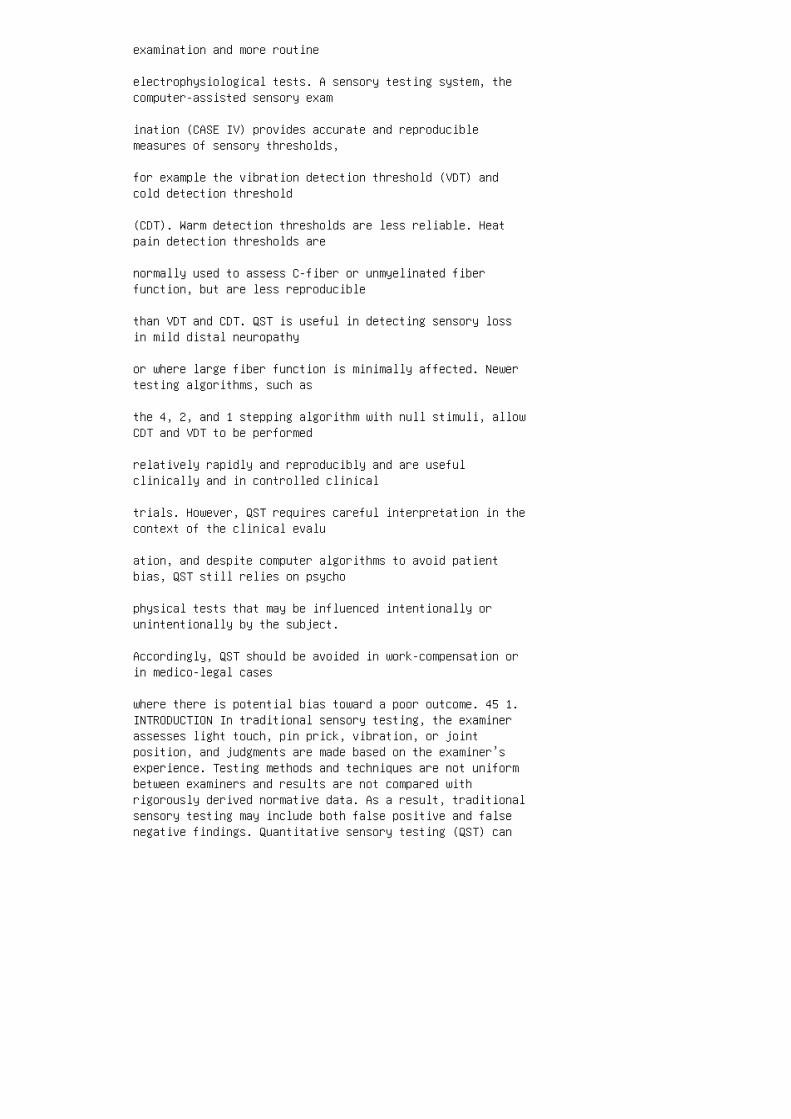
examination and more routine
electrophysiological tests. A sensory testing system, thecomputer-assisted sensory exam
ination (CASE IV) provides accurate and reproduciblemeasures of sensory thresholds,
for example the vibration detection threshold (VDT) andcold detection threshold
(CDT). Warm detection thresholds are less reliable. Heatpain detection thresholds are
normally used to assess C-fiber or unmyelinated fiberfunction, but are less reproducible
than VDT and CDT. QST is useful in detecting sensory lossin mild distal neuropathy
or where large fiber function is minimally affected. Newertesting algorithms, such as
the 4, 2, and 1 stepping algorithm with null stimuli, allowCDT and VDT to be performed
relatively rapidly and reproducibly and are usefulclinically and in controlled clinical
trials. However, QST requires careful interpretation in thecontext of the clinical evalu
ation, and despite computer algorithms to avoid patientbias, QST still relies on psycho
physical tests that may be influenced intentionally orunintentionally by the subject.
Accordingly, QST should be avoided in work-compensation orin medico-legal cases
where there is potential bias toward a poor outcome. 45 1.INTRODUCTION In traditional sensory testing, the examinerassesses light touch, pin prick, vibration, or jointposition, and judgments are made based on the examiner’sexperience. Testing methods and techniques are not uniformbetween examiners and results are not compared withrigorously derived normative data. As a result, traditionalsensory testing may include both false positive and falsenegative findings. Quantitative sensory testing (QST) can

provide information about pathology that may not beavailable from routine nerve conduction studies. QST canprovide reliable and reproducible information about bothlarge and small sensory fiber functions (1). QST is basedon well-established psychophysical methods (2) that allowaccurate characterization of responses from peripheralsensory receptors. Computer-based systems facilitate testand retest of sensory levels leading to normative databased on percentiles. This allows early detection ofperipheral neuropathy in diseases where there may beminimal involvement of distal fibers or where small fiberloss predominates. QST also lends itself to epidemiologicaland clinical treatment trials. Despite good sensitivity andreliability of QST, there are drawbacks. Unlike nerveconduction studies, QST is subjective. Response variabilityis reduced by repeatedly presenting the same sensorystimulus using a computer-based algorithm and determiningwhether the sensory response accurately represents thepatient’s sensory level. Patients with poor attention orcognitive deficits may struggle to complete the testing.Use of null stimuli coupled with repeated testing atthreshold improves the accuracy in these subjects; however,if the deficit is severe it may not be possible to completesensory testing. Methods of measuring sensory thresholdsvary depending on the technical method and data may not betransferable from one testing device to another, incontrast to nerve conduction data. In QST, a subject’sresponse to quantified natural occurring stimuli isspecific to a particular modality, for example, colddetection threshold (CDT), warm detection threshold (WDT),heat pain detection threshold (HPDT), and vibrationdetection threshold (VDT). These stimuli test specificsensory organ modalities and do not necessarily reflectchanges in afferent fiber function, although in general theVDT is a surrogate measure of large fiber sensory function,the CDT of small myelinated fibers (A d), and the HPDT ofC-nociceptor fiber function. 2. TESTING STRATEGIES QSTtechniques significantly affect testing outcome (1). Steep,linear, or exponential increases in temperature, where thepatient experiences a change in temperature and signals bydepressing a response key, overestimate the true sensorythreshold. Furthermore, alternating the direction ofthermal change and having subjects respond during heatingor cooling also overestimates thresholds and provides poordiscrimination for specific thresholds such as the CDT,WDT, and HPDT. A better method is a flat-topped pyramidalshaped stimulus ramp that provides a better range ofdefined levels of intensity (1,3,4). In thecomputer-assisted sensory examination (CASE IV) system,trapezoid stimuli are applied and range from baseline in a

series of 25 steps called just noticeable differences(JND). From JND 1 through 21, heating pulses are pyramidalshaped. Temperature changes from baseline (348C) to peakand returning to baseline are at 48C/s. The highesttemperature achieved is 488C at step 21. At steps 22–25,488C is held for progressively longer periods of time. Asimilar technique is used for VDT. The thresholds obtainedusing these JND values can then be converted into normativecurves based on age and sex, and ultimately converted tostandard normal deviates.
3. METHODS
The CASE IV system consists of a personal computer thatdrives an electronic controller
to shape and control stimulus characteristics (Fig. 3.1).There is a visual cueing device and
response key. Prior to performing the QST, the patient isread specific instructions on the
test. The same instructions are given each time the test isperformed, and the technician
avoids interpreting the test for the patient. This isimportant to ensure reproducibility of
the test. The patient is presented with a visual cueindicating that a stimulus is being
given. The technician must be convinced that the patientunderstood and was cooperating
during the test. Furthermore, the patient should not bedrowsy or under the influence of
sedatives, pain medications, or other medications thatwould affect their concentration.
At the end of the stimulus the patient responds to indicatewhether or not they felt the
stimulus. Among testing algorithms the two most commonmethods used in the CASE
IV system are a forced choice method and a one-period 4, 2,and 1 stepping algorithm.
In the forced choice method, the patient is given a

stimulus during either period one or
period two, and before proceeding to the next level oftesting has to indicate which
period they felt the stimulus in. The forced choice methodhas the advantage that it
Figure 3.1 The computer-assisted sensory examination (CaseIV) system (A), temperature
thermode (B), and vibrometer (C). Figure (A) was kindlyprovided by WR Electronics (Stillwater,
MN). Similar equipment is available from several differentmanufacturers. requires a subject to provide a positiveresponse comparing one stimulus to another. However, theforced choice method has the disadvantage that it is oftenvery lengthy, particularly when the patient has minimalsensation and there is difficulty obtaining a sensorythreshold. The one-period 4, 2, and 1 stepping algorithmwith null stimuli can be used for detection of both the VDTand the CDT (5). The one-period algorithm uses the methodof limits to detect temperature or vibration thresholds.The system begins at an intermediate level, level 13, andthe stimulus is increased if not felt or decreased if felt,by four steps to the point of turn around. After the firstcomputer generated reversal in the stimulus (turn),stepping occurs in steps of two, and after the second turn,stepping occurs in steps of one until the sensory thresholdis determined. A total of 20 stimulus events are used, withfive of them being randomly distributed null stimuli. Ifthree consecutive failures are observed at level 25, thetesting is terminated, and the subject is classified asbeing insensitive. If there is a positive response to morethan one null stimulus, the program is aborted. The patientis then re-instructed and the test is re-run. Theone-period 4, 2 and 1 stepping algorithm has proved to beas sensitive as the forced choice system, while permittingmore rapid assessment of sensory levels. The specificmethodology outlined in this section is that used by theCASE IV system. Similar methods are used in othercommercially available quantitative sensory equipment. Ingeneral, it is important that reliable normative data forthe specific sensory device are obtained from a sizablecohort of normal subjects using the specific sensorydevice. Furthermore, the equipment must allow forreproducible testing. Thus hand-held sensory devicesgenerally produce less reproducible data than fixedpositioned equipment. 3.1. VDT The VDT is obtained using a
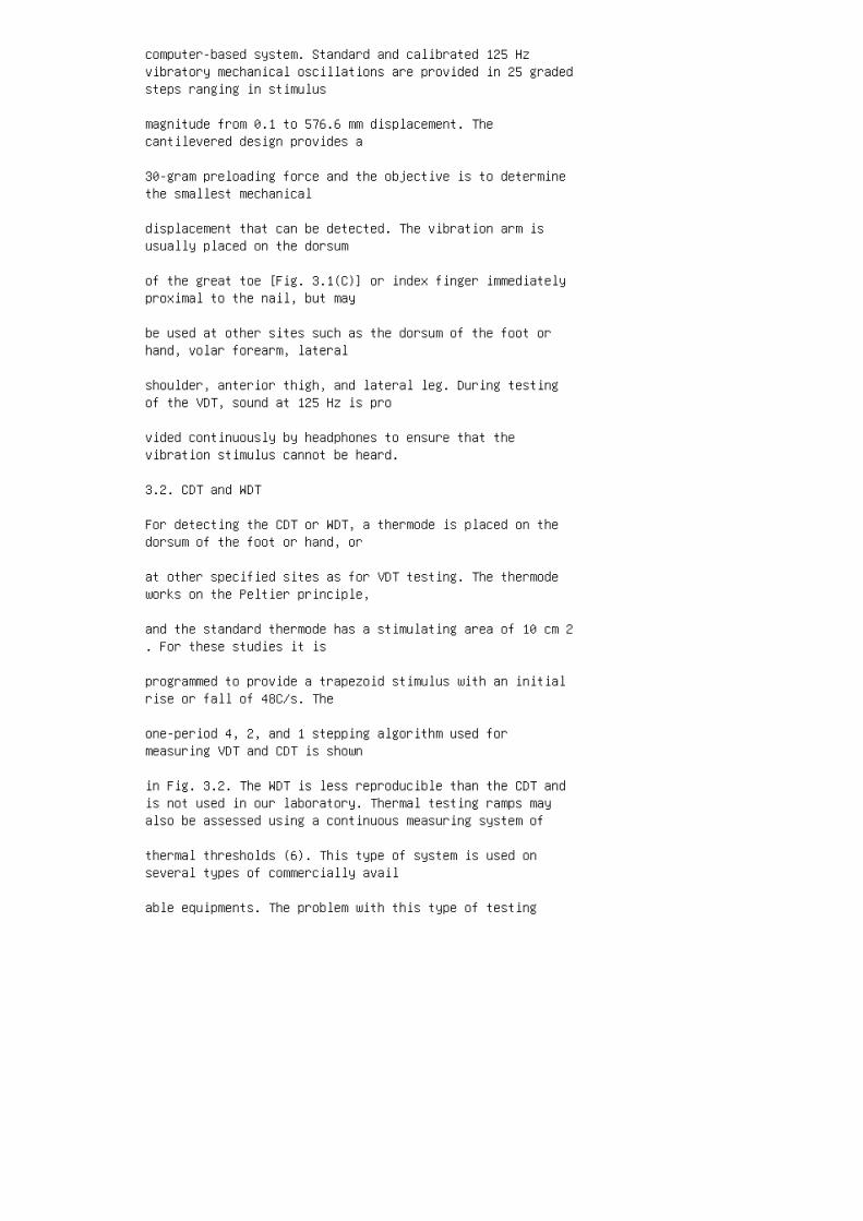
computer-based system. Standard and calibrated 125 Hzvibratory mechanical oscillations are provided in 25 gradedsteps ranging in stimulus
magnitude from 0.1 to 576.6 mm displacement. Thecantilevered design provides a
30-gram preloading force and the objective is to determinethe smallest mechanical
displacement that can be detected. The vibration arm isusually placed on the dorsum
of the great toe [Fig. 3.1(C)] or index finger immediatelyproximal to the nail, but may
be used at other sites such as the dorsum of the foot orhand, volar forearm, lateral
shoulder, anterior thigh, and lateral leg. During testingof the VDT, sound at 125 Hz is pro
vided continuously by headphones to ensure that thevibration stimulus cannot be heard.
3.2. CDT and WDT
For detecting the CDT or WDT, a thermode is placed on thedorsum of the foot or hand, or
at other specified sites as for VDT testing. The thermodeworks on the Peltier principle,
and the standard thermode has a stimulating area of 10 cm 2. For these studies it is
programmed to provide a trapezoid stimulus with an initialrise or fall of 48C/s. The
one-period 4, 2, and 1 stepping algorithm used formeasuring VDT and CDT is shown
in Fig. 3.2. The WDT is less reproducible than the CDT andis not used in our laboratory. Thermal testing ramps mayalso be assessed using a continuous measuring system of
thermal thresholds (6). This type of system is used onseveral types of commercially avail
able equipments. The problem with this type of testing

algorithm is that the heating or
cooling ramp continues while the patient is deciding topress the response button. This
system will thus overestimate the degree of heating orcooling. In addition, patients will
improve the rate of response to the test with repeatedtesting. The net result of these
defects in the testing method are that there will be anoverestimate of heating or
cooling, and lack of reproducibility with subsequent trials.
3.3. HPDT
Testing algorithms for heat pain are even more complicated.The rate of rise in tempera
ture, frequency of testing, and the specific testing deviceall affect the perceived pain
threshold. If a continuous linear ramp is used, the painthreshold will be inaccurate.
Thus, a further algorithm, the nonrepeating ascending withnull stimuli (NRA–NS) is
used for heat pain responses and for measuring the HPDT(Fig. 3.3). The HPDT provides
indirect information about C-nociceptor function. Theinitial skin temperature is taken
with an infrared thermometer. If the temperature of theskin is ,328C, the limb is
warmed in water. The thermode is calibrated for a rangebetween 348C and 508C. Once
the thermode is applied to the skin, it is set at 348C andallowed to accommodate to the
skin temperature. A series of software and hardwarefeatures in the system recognize
excessive temperatures, or faulty operation and shut downthe system if a fault is

sensed. Although the test may induce erythema at the sightof testing, injury to the skin
is extremely rare. During heat pain testing, specificinstructions are read and presented to the patient as
for other types of QST. The patient is asked if they feelsome degree of discomfort or pain,
and if they do then the severity is graded by the patientfrom 1 to 10. Number 1 would
correspond to the lowest level of discomfort or pain,number 10 would correspond to
the most severe level of pain. Typically at a low stimulusmagnitude nothing is felt.
With a stronger stimulus, a pulse of warmth or heat isexperienced. Occasionally, patients
may not report warmth, but may report pain as the initialstimulus. This likely occurs at
sites where warm receptors are not present. In the CASE IVsystem, the heating ramp
uses a trapezoid stimulus to 498C in order to try to avoidpain and skin temperature
damage associated with higher temperatures. The measuredHPDT is the thermal step level at which the stimulus isfelt as painful 50% of the time. In order to limit actualtesting within the nociceptive range, an estimate is madeof the 0.5 (HP:0.5) and 5.0 (HP:5.0) pain response level bytesting all steps between the two response levels (7). TheNRA–NS algorithm is superior to the 4, 2, and 1 algorithmand the forced choice algorithm for testing pain (7)because it avoids repeating pain stimuli at specific levelsthat may subsequently alter pain thresholds. The test canbe completed in 8–15 min, which is important with limitedtesting time, or when patients have a poor attention span.Testing may be Figure 3.2 One-period 4, 2, and 1 steppingalgorithm to determine VDT or CDT. Normal VDT response (A),subject of similar age with alcohol associated axonalneuropathy and an impaired VDT (B), normal CDT (C), samesubject with an impaired CDT (D).
repeated in, as little as, 45 min without any significant
difference in the second recorded
response level (7).
4. CLINICAL USES OF QST
Potential uses for QST include (1) determination of thetype of sensory modality affected
in a peripheral neuropathy; (2) characterize the type ofnerve fiber degeneration in a “small
fiber neuropathy” where standard nerve conduction studiesare normal (8); (3) characterize
heat pain insensitivity or hypersensitivity in peripheralneuropathies, as well as in focal
disorders such as trigeminal neuralgia; and (4)characterize response of sensory, or
“small fiber neuropathies” to pharmacological agents. Thereare concerns about using QST for workman’s compensation ormedico-legal
claims. In psychophysical testing, a subject may have abias toward a worse outcome, and
therefore may consciously or unconsciously influenceresults of the test making QST unre
liable (9,10). Furthermore, there is a concern about thereliability of QST in characterizing
an entrapment neuropathy such as carpal tunnel syndrome.The diagnosis should not be
made on the basis of a single test, but should rathercorrelate clinical findings, electrodiag
nostic studies, QST findings, and the physician’s judgmentto determine the final assess
ment of the disorder.
5. CONCLUSIONS
QST using sensitive psychophysical methods attempts toevaluate subjective alterations in

sensation such as hypoor hyper-sensitivity. There areseveral indications for QST: (1) to
Figure 3.3 Measurement of the HPDT showing hypersensitivityto heat pain (A) in a patient with
a cryptogenic “small fiber neuropathy,” andhypersensitivity (B) heat pain in a patient with impaired
4
Evaluation of the Autonomic
Nervous System
Safwan S. Jaradeh and Thomas E. Prieto
Medical College of Wisconsin, Milwaukee, Wisconsin, USA
Abstract 54
1. Overview of the Anatomy, Physiology, and Chemistry 54
2. Laboratory Evaluation of Autonomic Disorders 54 2.1.Tests of Sudomotor Function 55 2.1.1. QuantitativeSudomotor Axon Reflex Test 56 2.1.2. Thermoregulatory SweatTest 56 2.1.3. Sympathetic Skin Responses 57 2.2. Tests ofCardiovagal Function 57 2.2.1. HR Response to DeepBreathing 57 2.2.2. Valsalva Ratio 58 2.2.3. HR Response toStanding Up 58 2.3. Tests of Adrenergic Function 59 2.3.1.BP Responses to the Valsalva Maneuver 59 2.3.2. BPResponses to HUT 59
3. Clinical Applications 60 3.1. Acute AutonomicNeuropathies 60 3.1.1. Botulism 60 3.1.2. Porphyria 603.1.3. Drug or Toxin-Induced Autonomic Neuropathies 60 3.2.Chronic Autonomic Neuropathies 61 3.2.1. Diabetic AutonomicNeuropathy 61 3.2.2. Amyloid Neuropathy 61 3.2.3. FamilialDysautonomia (Riley-Day Syndrome) 61 3.2.4. Pure AutonomicFailure 61 3.2.5. Multiple System Atrophy 61 3.2.6. PureAdrenergic Neuropathy 62 3.2.7. Chronic IdiopathicAnhidrosis 62 3.2.8. Small-Fiber Sensory Neuropathies 623.3. Orthostatic Hypotension, Syncope, and OrthostaticIntolerance 62
Autonomic testing is also used to evaluate the progressionand therapeutic responses in
dysautonomia. Very specialized dysautonomias, such aspupillary, genitourinary, or

gastrointestinal disorders, are beyond the scope of thischapter. Given the influence of many substances onautonomic function, it is important to
withhold these substances, if medically possible, forvariable periods before testing.
Caffeine and nicotine should be held at least 4 h beforetesting. Alcohol should be held
8 h. Over-the-counter drugs (decongestants) andanticholinergics (including tricyclic anti
depressants) should be held 48 h. Sympathomimetics shouldbe held 24 h. The patient
should be quietly seated indoors for 30 min before the testto allow for adequate rest
and thermal equilibration.
2.1. Tests of Sudomotor Function
The three main tests are: 1. quantitative sudomotor axonreflex test (QSART); 2. thermoregulatory sweat test (TST);and 3. sympathetic skin responses (SSR).
Figure 4.1 Comparison of sympathetic system withparasympathetic system. 2.1.1. Quantitative Sudomotor AxonReflex Test In this test, a multi-compartment sweat capsuleis attached to the skin. Acetylcholine 10% is iontophoresedthrough one compartment using a low intensity (up to 2 mA)constant current to activate cutaneous axon terminals. Theaction potentials propagate antidromically to the nextbranch-point, then propagate orthodromically to the nextnerve terminal. This releases endogenous acetylcholine,which binds to muscarinic receptors on eccrine sweat glands(3). The evoked sweat response is recorded from a secondcompartment of the sweat capsule using a sudorometer (3).This reflex response is mediated by postganglionicsympathetic sudomotor axons. The test is noninvasive, butsubjects often describe a transient local burning orstinging sensation lasting several seconds following theiontophoresis. After the capsule is removed, a smallerythematous flare may persist for up to 20 min. QSART issensitive and reliable in controls and in patients withautonomic neuropathy. Intra-subject reproducibility ishigh. There is little asymmetry from side to side; in ourlaboratory, this difference is ,40%. Therefore, recording

from either side should suffice, except when local factorsare present (skin injury or infection, previous trauma, orbiopsy). When symptoms are unilateral, it is useful toevaluate both sides. Standard recording sites are the volarmid-forearm, the anterior proximal leg, the anterior distalleg, and the dorsolateral proximal foot. These sites areinnervated by the ulnar, peroneal, saphenous, and suralnerves, respectively. Normal data from the Mayo clinic andfrom our laboratory have been published (3,6). 2.1.2.Thermoregulatory Sweat Test The TST is a sensitivequalitative test of sudomotor function that providesimportant information on the pattern and distribution ofsweat loss. The original method applied quinizarinindicator to dry skin. Sweating changes the indicator frombrown to violet. Because quinizarin is highly allergenic,alizarin-red is currently used, and the color changes fromorange to purple (7). Skin reactions are rare, but stainingof clothing may occur. The test is conducted in a heatedtent or a sweat cabinet. The subject should be wellhydrated. Anticholinergic agents, some antihistamine,antidepressant, and some antiparkinsonian drugs, should beheld for 24–48 h before the test. A commonly used endpointis a rise in oral temperature of 18C provided the restingbaseline temperature is .36.58C. A minimum of 25% ambienthumidity is usually necessary to facilitate sweating andslow the evaporation. There are some variations in normalsweat patterns, but all normal subjects should perspireover the forehead, trunk (particularly torso), hands, andfeet; and most normal subjects perspire over the legs andforearms. In our experience, some normal subjects,particularly women, may not sweat over the thighs and arms.The test is highly sensitive because it detects preorpostganglionic sudomotor dysfunction. In a study by Fealeyet al. (8) on 51 diabetics, 94% of those with clinicalfindings of neuropathy had abnormal TST. Anhidrosisoccurred distally in 65%, or regionally— includingdermatomal distribution—in 50%; 78% of patients had morethan one pattern. Global anhidrosis was noted in 16% andall had symptomatic dysautonomia. In autonomic disorders,TST is a useful adjuvant to autonomic testing. Distalanhidrosis is the most common pattern and is usuallyindicative of a distal small fiber neuropathy. Most often,it is symmetric. When there is significant asymmetry, amicroangiopathic or dysimmune condition should beconsidered. Global anhidrosis is characteristic of pureautonomic failure (PAF), multisystem atrophy (MSA), andpan-autonomic neuropathy. Regional anhidrosis can bedermatomal or segmental. Hemihidrosis is an extreme exampleof segmental anhidrosis typically seen in myelopathies orafter a cerebral infarction.

2.1.3. Sympathetic Skin Responses
Also known as galvanic skin responses (GSR), SSR is anelectrodermal response recorded
from sweat glands after stimulation of type IImechanoreceptors in a mixed nerve (9). Stan
dard EMG surface electrodes are placed over the hands andfeet. The response is recorded
from the active electrodes over the palmar or plantarsurface, while the reference electrode
is placed over the dorsal surface. An electrical stimuluswith duration of 0.2 ms and an
intensity of 30–50 mA is delivered to the contralateralwrist or ankle. The response wave
form has a slow latency (1–3 s) and a long duration, whichrequires a low frequency filter
of ,0.20 Hz. The high frequency filter is usually ,2000 Hz.The sweep speed should be
500–1000 ms/cm. Owing to the large variability among normalsubjects, and the habitu
ation of the response with repeated stimulation, only anabsent SSR is of pathological sig
nificance and indicates abnormal sudomotor adrenergicinnervation of the affected limb.
2.2. Tests of Cardiovagal Function
The three main tests are: 1. heart rate response to deepbreathing (HR) 2. Valsalva ratio (VR) 3. heart rateresponse to standing “30:15” ratio
In the first test, the variation of heart rate (HR) dependson vagal innervation, and is abol
ished by the muscarinic antagonist atropine. In the othertwo tests, the HR is determined by
the vagal component of the baroreflex arc, while bloodpressure (BP) responses are deter

mined by sympathetic factors. BP changes also depend on themagnitude of forced expira
tory pressure during the Valsalva maneuver, the patient’svolume and cardiac status, and
the patient’s position. BP and HR can also be recordednoninvasively using plethysmo
graphic equipment, such as finapres or arterial tonometry(Table 4.1) (10).
2.2.1. HR Response to Deep Breathing
Age, resting HR, rate of breathing, body mass index,cardiac status, and medications affect
this test. It is not affected by gender. Heart ratevariations (R–R variation) is maximal at a
respiratory rate of 6 bpm (Beats or breaths per minute)(11,12). Breathing should be as
smooth as possible and a visual guide—such as anoscillating bar—is very helpful. Six
to eight cycles are recorded, and the five cycles with thelargest R–R differences are aver
aged to derive a mean HR range. Breathing at higher ratesor for longer periods reduces the
Table 4.1 Test Results in Various Types of Autonomic Failure
Condition TST QSART VR/DHRDB HUT
Autonomic NP ##, Distal ##, Distal Reduced ## BP
Adrenergic NP #, Distal #, Distal Normal ## BP
Cholinergic NP ##, Diffuse ##, Diffuse Reduced NL
Botulism ##, Diffuse ##, Diffuse Reduced NL or #
Riley–Day # Distal, " proximal ##, Distal Reduced ## BP
PAF ##, Diffuse #, Distal Reduced ## BP
MSA ##, Diffuse Normal Reduced ## BP

Syncope NL or # NL or # NL or # ## BP
POTS NL or # NL or # NL or # " " HR HR change because ofhyperventilation and hypocapnia. If the test is repeated,3–5 min of rest should be allowed. Overweight patients willhave reduced HR variation (13). Normal data from the Mayoclinic and from our laboratory have been published (3,6).2.2.2. Valsalva Ratio For the Valsalva maneuver, thesubject is asked to blow into a tube to maintain a columnof mercury at 40 mmHg for 15 s. The patient is usuallyrested and supine, but sitting does not affect the ratio.The maneuver may be repeated to ensure reproducible BPresponses. Blowing for ,10 s or at a pressure ,20 mm issuboptimal. The VR is derived from the maximum HR dividedby the minimum HR. Most centers select the lowest HR within30 s of maximal HR, but a few centers select the minimalvalue within 60 s of maneuver onset. The maximal HR is aresponse to the fall in BP, while the minimal HR is aresponse to the baroreflex. The VR is a measure ofbaroreflex sensitivity. The different phases of thevalsalva response are discussed in section 2.3.1. Reflexbradycardia is blunted in cardiovagal dysfunction, whileboth the BP overshoot and the reflex bradycardia areblunted in generalized dysautonomia. Factors that affect VRare age, gender, position of the subject, expiratorypressure, duration of effort, and medications.Occasionally, patients may not generate two reproducible BPcurves despite identical expiratory pressures; in thesecases, the better VR should be taken. The VR is lessreliable in patients with pacemakers, or when BP excursionsare minimal for iatrogenic or health (pulmonary) reasons.Some subjects, including controls, may not decrease theirBP during early phase II; this curve is referred to as a“flat top” response, and VR may be underestimated.Conversely, VR may be normal in mild cardiovagal impairmentif sympathetic cardiac responses are compensatory. Normaldata from the Mayo clinic and from our laboratory have beenpublished (Table 4.2) (3,6). 2.2.3. HR Response to StandingUp The earliest HR changes to standing consist of atachycardia between 5 and 15 s, followed by a bradycardiabetween 20 and 30 s. Subsequently, there is an increase inHR and BP before returning to baseline after severalminutes. The early bradycardia is baroreflex— mediated. Theoriginal study found that the largest HR changes occur at15 and 30 s after standing, hence the term “30:15 ratio.”This tachycardia/bradycardia ratio (R–R interval at beat30)/(R–R interval at beat 15) is a valid index ofcardiovagal function. Table 4.2 Components of ValsalvaManeuver in Types of Autonomic Failure (3) Condition Early

Phase II Late Phase II Phase IV VR Normal SBP drop ,21 mmSBP ¼ baseline SBP rise .11 mm Normal Vagal lesion SBP drop,21 mm SBP ¼ baseline SBP rise .11 mm Reduced Sympatheticlesion, mild SBP drop .21 mm SBP , baseline SBP rise .11 mmNormal Sympathetic lesion, moderate SBP drop .21 mm SBP ,baseline SBP rise .5 mm Low, normal Sympathetic lesion,marked SBP drop .30 mm SBP , baseline SBP ¼ baselineReduced
2.3. Tests of Adrenergic Function
The main tests are the head-upright tilt (HUT) and theValsalva maneuver.
2.3.1. BP Responses to the Valsalva Maneuver
As already mentioned, the VR is a reliable parasympathetictest. The dynamic alterations
of the BP during the Valsalva maneuver are importantmeasures of adrenergic function.
Invasive (arterial line) and noninvasive (Finapres orarterial tonometry) recordings
reveal four phases in the Valsalva maneuver. In phase I,there is increased intrathoracic
and intra-abdominal pressure causing mechanical compressionof the aorta and a brief
rise in BP. Phase II begins with a drop of BP secondary toreduced venous return and
cardiac output. This produces efferent sympathetic volleysto the muscles, and the cardi
ovagal withdrawal leads to tachycardia (Table 4.3). Thefall in BP is arrested within 4–6 s,
and the BP returns to or exceeds the resting values.Following the release of Valsalva in
phase III, the fall in intrathoracic pressure causes adecrease in BP lasting 1–3 s. There is
another burst of sympathetic activity and the arteriolarbed remains vasoconstricted. The
resumption of normal venous return and cardiac output leads
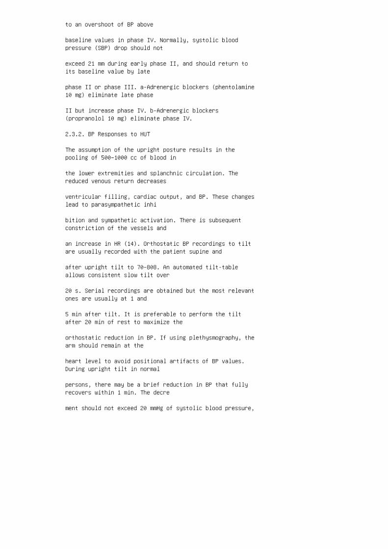
to an overshoot of BP above
baseline values in phase IV. Normally, systolic bloodpressure (SBP) drop should not
exceed 21 mm during early phase II, and should return toits baseline value by late
phase II or phase III. a-Adrenergic blockers (phentolamine10 mg) eliminate late phase
II but increase phase IV. b-Adrenergic blockers(propranolol 10 mg) eliminate phase IV.
2.3.2. BP Responses to HUT
The assumption of the upright posture results in thepooling of 500–1000 cc of blood in
the lower extremities and splanchnic circulation. Thereduced venous return decreases
ventricular filling, cardiac output, and BP. These changeslead to parasympathetic inhi
bition and sympathetic activation. There is subsequentconstriction of the vessels and
an increase in HR (14). Orthostatic BP recordings to tiltare usually recorded with the patient supine and
after upright tilt to 70–808. An automated tilt-tableallows consistent slow tilt over
20 s. Serial recordings are obtained but the most relevantones are usually at 1 and
5 min after tilt. It is preferable to perform the tiltafter 20 min of rest to maximize the
orthostatic reduction in BP. If using plethysmography, thearm should remain at the
heart level to avoid positional artifacts of BP values.During upright tilt in normal
persons, there may be a brief reduction in BP that fullyrecovers within 1 min. The decre
ment should not exceed 20 mmHg of systolic blood pressure,
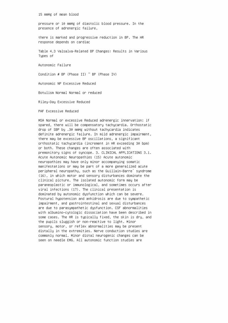
15 mmHg of mean blood
pressure or 10 mmHg of diastolic blood pressure. In thepresence of adrenergic failure,
there is marked and progressive reduction in BP. The HRresponse depends on cardiac
Table 4.3 Valsalva-Related BP Changes: Results in VariousTypes of
Autonomic Failure
Condition # BP (Phase II) " BP (Phase IV)
Autonomic NP Excessive Reduced
Botulism Normal Normal or reduced
Riley–Day Excessive Reduced
PAF Excessive Reduced
MSA Normal or excessive Reduced adrenergic innervation; ifspared, there will be compensatory tachycardia. Orthostaticdrop of SBP by .30 mmHg without tachycardia indicatesdefinite adrenergic failure. In mild adrenergic impairment,there may be excessive BP oscillations, a significantorthostatic tachycardia (increment in HR exceeding 30 bpm)or both. These changes are often associated withpremonitory signs of syncope. 3. CLINICAL APPLICATIONS 3.1.Acute Autonomic Neuropathies (15) Acute autonomicneuropathies may have only minor accompanying somaticmanifestations or may be part of a more generalized acuteperipheral neuropathy, such as the Guillain–Barre´ syndrome(16), in which motor and sensory disturbances dominate theclinical picture. The isolated autonomic form may beparaneoplastic or immunological, and sometimes occurs afterviral infections (17). The clinical presentation isdominated by autonomic dysfunction which can be severe.Postural hypotension and anhidrosis are due to sympatheticimpairment, and gastrointestinal and sexual disturbancesare due to parasympathetic dysfunction. CSF abnormalitieswith albumino–cytologic dissociation have been described insome cases. The HR is typically fixed, the skin is dry, andthe pupils sluggish or non-reactive to light. Minorsensory, motor, or reflex abnormalities may be presentdistally in the extremities. Nerve conduction studies arecommonly normal. Minor distal neurogenic changes can beseen on needle EMG. All autonomic function studies are

markedly abnormal. The disorder follows a monophasiccourse. The prognosis is variable, and approximatelyone-third of patients remain severely disabled. Improvementusually occurs 1 year after the onset. Treatment issymptomatic. Anecdotal reports suggest that plasmapheresisor intravenous immunoglobulin (IVIG) therapy may behelpful. 3.1.1. Botulism Ingestion of food contaminated byClostridium botulinum often leads to cholinergic failure.The manifestations are anhidrosis, xerostomia,xerophthalmia, gastroparesis, ileus, urinary retention, andBP disturbances (6). There are signs of cranialneuropathies, usually extraocular, and subsequentdevelopment of generalized weakness and respiratorydifficulties. The diagnosis can be confirmed byidentification of the toxins in food or serum. 3.1.2.Porphyria Sympathetic overactivity is the most commonabnormality. In acute intermittent porphyria, persistenttachycardia, and hypertension may herald an attack. Bothsympathetic and parasympathetic functions are affected.There is often concomitant large fiber involvement that canbe detected by nerve conduction studies. 3.1.3. Drug orToxin-Induced Autonomic Neuropathies Autonomic neuropathiesmay occur in patients receiving a variety of medicationsincluding cisplatin, vinca alkaloids, amiodarone,perhexiline maleate, and taxol. Postural hypotension oftendominates the presentation, but there are usually othersensory and motor signs of polyneuropathy. Organic solventsalso cause a sensorimotor and autonomic polyneuropathy,with abnormal valsalva maneuver and HR response to deep
breathing (18). Thallium and arsenic intoxication leads toa peripheral neuropathy with
hypertension, tachycardia, and disturbances of sweating(19,20).
3.2. Chronic Autonomic Neuropathies (8)
Autonomic dysfunction is present in many chronic axonalneuropathies, particularly those
involving the small fibers.
3.2.1. Diabetic Autonomic Neuropathy
Autonomic neuropathy is present in at least 25% of diabeticpatients. Parasympathetic
tests are usually impaired before sympathetic tests(21–23). Sweating is often abnormal

(8). Other neuropathic complications of diabetes are commonand include a distal
polyneuropathy, diabetic amyotrophy, lumbosacralradiculo–plexopathy, and truncal
radiculopathy.
3.2.2. Amyloid Neuropathy
Autonomic dysfunction is prominent in the Portuguese typeof familial amyloid
polyneuropathy (FAP type 1). The inheritance is autosomaldominant. The onset is
usually in young adulthood. There is often superimposedsensory neuropathy involving
mainly the small fibers. Motor and reflex abnormalities areinfrequent or late (24). In sporadic systemic amyloidosis,a neuropathy usually develops in mid—to late
adulthood. The neuropathy is usually painful. Autonomicfailure is often severe and
early. Postural hypotension, syncope, and impotence arefrequent. Sudomotor function
is impaired. Gastrointestinal disturbances are common. Somepatients have dysphagia
caused by infiltration of the lower esophagus. Peripheralnerve biopsy shows amyloid
deposits with loss of thinly myelinated and unmyelinatedfibers. The prognosis is worse
than in the inherited form.
3.2.3. Familial Dysautonomia (Riley-Day Syndrome)
Familial dysautonomia is also known as hereditary sensoryand autonomic neuropathy
type III. It begins in childhood with impaired lacrimation,hyperhidrosis, postural hypoten
sion, and poor temperature control (25). Autonomic testing

abnormalities are both sym
pathetic and parasymapthetic.
3.2.4. Pure Autonomic Failure
Pure Autonomic Failure (PAF) is an idiopathic, sporadic,adult-onset, and progressive dis
order of the autonomic nervous system. Bradbury andEggleston first described it in 1925
(26). Patients present with autonomic dysfunction such asurinary retention, erectile dys
function, constipation, and orthostatic hypotension withsyncope (27). The presence of
extrapyramidal deficit, cerebellar deficit, or bothindicates multiple system atrophy
(MSA) rather than PAF.
3.2.5. Multiple System Atrophy
MSA is a progressive neurodegenerative disease that causesparkinsonism and cerebellar,
pyramidal, and autonomic dysfunction in any combination(28,29). Wenning et al. (30)
detailed the clinical features of 203 published cases ofMSA. Men are more commonly
affected than women (ratio of 1.3:1). Autonomic failure ispresent in 74% of patients. Parkinsonism affects 87%,cerebellar ataxia 54%, and pyramidal signs are present in49%. Life expectancy in MSA is usually shorter than in PAF.Anal sphincter electromyography and urodynamic studies areoften abnormal. 3.2.6. Pure Adrenergic NeuropathyOccasional patients with PAF and MSA present with pureadrenergic failure. There is postural hypotension butnormal tests of cardiovagal function. 3.2.7. ChronicIdiopathic Anhidrosis Patients present with heatintolerance. Vasomotor function is usually preserved. Thereis no associated somatic neuropathy. Some patients haveAdie’s tonic pupil (Ross syndrome). Adie’s pupil is often alarge pupil that is poorly reactive to light. Unlike thenormal pupil, it constricts after topical application ofdiluted (2.5%) methacholine. The disorder is often

asymmetric or unilateral. Some tendon reflexes, especiallythe ankle jerks, may be lost. Treatment is usuallyprophylactic with avoidance of heat. 3.2.8. Small-FiberSensory Neuropathies Patients present with painfuldysesthetic feet, allodynia, and sympathetic disturbances,with an impairment of vasomotor function and of sweating.Many have distal sensory nociceptive abnormalities. Themotor examination is normal, and tendon reflexes are oftenpreserved. Nerve conduction studies are not helpful. Causesare diabetes, monoclonal gammopathies, primary systemicamyloidosis, AIDS, nutritional deficiencies, certainhereditary sensory, and autonomic neuropathies, Tangierdisease, and Fabry disease. Many cases are idiopathic(31,32). Quantitative sensory testing reveals abnormalthermal and pain perception modalities (33). Sudomotorfunction is impaired, particularly distally. Cardiovagalabnormalities are less common than sudomotor disturbances.When technically available, intraepidermal nerve fiberanalysis reveals reduced fiber density, especially distally(34). A large component of the treatment is symptomatic.3.3. Orthostatic Hypotension, Syncope, and OrthostaticIntolerance As discussed earlier, the assumption of theerect posture reduces cardiac output and BP. There issubsequent constriction of the vessels and an increase inHR. When these mechanisms fail, the patient developshypotension and syncope. The normal response to standing isa transient fall in systolic BP (,10 mmHg), an increase indiastolic BP (,10 mmHg), and an increase in the pulse rate(,25 bpm). The changes are usually less pronounced thanduring passive upright tilt, in part because of thecompensatory muscular contraction of the lower limbs.Orthostatic hypotension and syncope are the mostincapacitating symptoms of autonomic failure. Patientstypically present with lightheadedness after suddenpostural changes, meals, exertion, or prolonged standing.Other pre-syncopal complaints include generalized fatigue,leg weakness, blurred vision, and headache. Syncope may begradual or sudden, and some patients may have briefmyoclonus, raising the possibility of a seizure. Syncopeoccurs when orthostatic hypotension exceeds the capacity ofcerebral autoregulation. This leads to a transient loss ofconsciousness and postural tone. In addition to all causesof orthostatic hypotension, cardiac disease should be ruledout. The most common types of syncope are noncardiac.Iatrogenic factors particularly in the elderly
may precipitate syncope. Among cardiac causes, vascularobstruction (aortic or pulmon
ary) and arrythmias predominate. Up to 50% of patients may

not have a detectable cause. Orthostatic intolerance, alsoknown as postural orthostatic tachycardia syndrome
(POTS), is characterized by the development orthostatictachycardia that exceeds 30 bpm
after assuming the erect position. HR in excess of 120 bpmis also diagnostic. Patients
often describe dizziness, blurred vision and epigastricdiscomfort. There is occasional
venous pooling in the lower limbs. Around 50–60% ofpatients have evidence of autonomic
neuropathy, primarily adrenergic (35,36). The conditionlikely represents a milder form of
neurogenic orthostatic hypotension and syncope. Themanagement depends on the under
lying etiology, and follows the same guidelines asorthostatic hypotension. Treatment of neurogenicorthostatic hypotension can be difficult (37). Two
approaches are required. Nonpharmacological measuresemphasize patient education.
Patients should stand up gradually, particularly in themorning. A tailored program of
exercise, particularly isotonic, is recommended.Precipitating factors include exercise in
warm weather, hot baths, and showers and particularly whensweating is impaired. Medi
cations that cause orthostatic hypotension should berevised and possibly removed or
modified. Patients often benefit from increasing sodiumintake. Custom fitted elastic stock
ings to the waist minimize peripheral blood pooling, buttend to be poorly tolerated. Pharmacological measures beginwith fludrocortisone acetate. Sympathomimetic
agents can be added to fludrocortisone acetate. Commonlyused a-adrenergic agonists

include ephedrine, pseudoephedrine, and midodrine.Midodrine produces arterial and
venous constriction and is quite effective. For refractorypatients, recombinant human
erythropoietin and somatostatin analogs such as octreotidecan be useful. b-Adrenergic
blockers with intrinsic sympathomimetic activity, such aspindolol, may have a limited
role, particularly when there is symptomatic tachycardia.
1. Kandel ER, Schwartz JH, Jessell TM. The autonomicnervous ystem and the hypothalamus. In: Kandel ER, SchwartzJH, Jessell TM, eds. Principles of Neural Science.Amsterdam: Elsevier, 2000:961–981.
2. Anonymous. Clinical autonomic testing report of theTherapeutics and Technology Assessment Subcommittee of theAmerican Academy of Neurology. Neurology 1996; 46:873–880.
3. Low PA. Laboratory evaluation of autonomic function. In:Low PA, ed. Clinical Autonomic Disorders: Evaluation andManagement. Philadelphia: Lippincott-Raven, 1997:179–208.
4. Low PA. Pitfalls in autonomic testing. In: Low PA, ed.Clinical Autonomic Disorders: Evaluation and Management.Philadelphia: Lippincott-Raven, 1997:391–401.
5. Low PA, Suarez GA, Benarroch EE. Clinical autonomicdisorders: classification and clinical evaluation. In: LowPA, ed. Clinical Autonomic Disorders. Philadelphia:Lippincott-Raven, 1997:3–15.
6. Jaradeh SS, Smith TL, Torrico L et al. Autonomic nervoussystem evaluation of patients with vasomotor rhinitis.Laryngoscope 2000; 110:1828–1831.
7. Fealey RD. The thermoregulatory sweat test. In: Low PA,ed. Clinical Autonomic Disorders: Evaluation andManagement. Philadelphia: Lippincott-Raven, 1997:245–257.
8. Fealey RD, Low PA, Thomas JE. Thermoregulatory sweatingabnormalities in diabetes mellitus. Mayo Clin Proc 1989;64:617–628.
9. Baba M, Watahiki Y, Matsunaga M et al. Sympathetic skinresponse in healthy man. Electromyogr Clin Neurophysiol

1988; 28:277–283.
10. Benarroch EE, Opfer-Gehrking TL, Low PA. Use of thephotoplethysmographic technique to analyze the Valsalvamaneuver in normal man. Muscle Nerve 1991; 14:1165–1172.11. Angelone A, Coulter NA. Respiratory sinus arrhythmia: afrequency dependent phenomenon. J Appl Physiol 1964;19:479–482. 12. Hirsch JA, Bishop B. Respiratory sinusarrhythmia in humans: how breathing pattern modulates heartrate. Am J Physiol 1981; 241:H620–H629. 13. Hirsch J,Leibel RL, Mackintosh R et al. Heart rate variability as ameasure of autonomic function during weight change inhumans. Am J Physiol 1991; 261:R1418–R1423. 14. Borst C,Wieling W, van Brederode JF et al. Mechanisms of initialheart rate response to postural change. Am J Physiol 1982;243:H676–H681. 15. Low PA, McLeod JG. Autonomicneuropathies. In: Low PA, ed. Clinical Autonomic Disorders:Evaluation and Management. Philadelphia: Lippincott-Raven,1997:463–486. 16. Tuck RR, McLeod JG. Autonomic dysfunctionin Guillain-Barre syndrome. J Neurol Neurosurg Psychiatry1981; 44:983–990. 17. Young RR, Asbury AK, Corbett JL,Adams RD. Pure pan-dysautonomia with recovery. Descriptionand discussion of diagnostic criteria. Brain 1975;98:613–636. 18. Matikainen E, Juntunen J. Autonomic nervoussystem dysfunction in workers exposed to organic solvents.J Neurol Neurosurg Psychiatry 1985; 48:1021–1024. 19. BankWJ, Pleasure DE, Suzuki K et al. Thallium poisoning. ArchNeurol 1972; 26:456–464. 20. LeQuesne PM, McLeod JC.Peripheral neuropathy following a single exposure toarsenic. J Neurol Sci 1977; 32:437–451. 21. Bennett T,Fentem PH, Fitton D et al. Assessment of vagal control ofthe heart in diabetes. Measures of R-R interval variationunder different conditions. Br Heart J 1977; 39:25–28. 22.Low PA, Zimmerman BR, Dyck PJ. Comparison of distalsympathetic with vagal function in diabetic neuropathy.Muscle Nerve 1986; 9:592–596. 23. Wheeler T, Watkins PJ.Cardiac denervation in diabetes. Br Med J 1973; 4:584–586.24. Bergfeldt BL, Olofsson BO, Edhag KO. Electrophysiologicevaluation of the cardiac conduction system and itsautonomic regulation in familial amyloid polyneuropathy. AmJ Cardiol 1985; 56:647–652. 25. Axelrod FB. Familialdysautonomia. In: Low PA, ed. Clinical Autonomic Disorders.2nd ed. Philadelphia: Lippincott-Raven Publishers,1997:525–535. 26. Bradbury S, Eggleston C. Posturalhypotension. Report of 3 cases. Am Heart J 1925; 1:73–86.27. Bannister R, Sever P, Gross M. Cardiovascular reflexesand biochemical responses in progressive autonomic failure.Brain 1977; 100:327–344. 28. Anonymous. Consensus statementon the definition of orthostatic hypotension, pureautonomic failure, and multiple system atrophy. Neurology

1996; 46:1470. 29. Cohen J, Low PA, Fealey R et al. Somaticand autonomic function in progressive autonomic failure andmultiple system atrophy. Ann Neurol 1987; 22:692–699. 30.Wenning GK, Tison F, Ben Shlomo Y et al. Multiple systematrophy: a review of 203 pathologically proven cases. MovDisord 1997; 12:133–147. 31. Stewart JD, Low PA, Fealey RD.Distal small fiber neuropathy: results of tests of sweatingand autonomic cardiovascular reflexes. Muscle Nerve 1992;15:661–665. 32. Suarez GA et al. Idiopathic autonomicneuropathy: clinical, neurophysiologic, and follow-upstudies on 27 patients. Neurology 1994; 44:1675–1682. 33.Jamal GA, Hansen S, Weir AI et al. The neurophysiologicinvestigation of small fiber neuropathies. Muscle Nerve1987; 10:537–545. 34. Holland NR, Crawford TO, Hauer P etal. Small-fiber sensory neuropathies: clinical course andneuropathology of idiopathic cases. Ann Neurol 1998;44:47–59. 35. Jaradeh SS, Maas EF. Postural orthostatictachycardia syndrome: diagnostic value of thermoregulatorysweat test. Clin Auton Res 1997; 7:258–259. 36. Low PA,Opfer-Gehrking TL, Textor SC et al. Postural tachycardiasyndrome (POTS). Neurology 1995; 45(suppl 5):S19–S25. 37.Fealey RD, Robertson D. Management of orthostatichypotension. In: Low PA, ed. Clinical Autonomic Disorders:Evaluation and Management. Philadelphia: Lippincott-Raven,1997:763–775.
5
Peripheral Nerve Histology and Pathology
Mark B. Bromberg
University of Utah, Salt Lake City, Utah, USA
Abstract 66
1. Nerve Histology 66 1.1. Gross Structure 66 1.2.Connective Tissue 66 1.3. Vasculature 67
2. Nerve Fiber Histology 67 2.1. Numbers and Sizes 67 2.2.Myelinated Fibers 68 2.3. Unmyelinated Fibers 68
3. Pathologic Changes 70 3.1. Axonal Changes 70 3.2. MyelinChanges 71 3.3. Connective Tissue Changes 73 3.4. VascularChanges 73
4. Nerve Biopsy 74 4.1. Uses and Limitations 74 4.2.Suitable Nerves 74 4.3. The Biopsy 74 4.4. Biopsy Review 754.5. Biopsy Yield 75

5. Classes of Neuropathy 75 5.1. Inflammatory Neuropathies75 5.2. Vasculitic Neuropathies 76 5.3. AmyloidNeuropathies 77 5.4. Hereditary Neuropathies 77 5.5.Diabetic Neuropathies 78 5.6. Toxic Neuropathies 79 5.7.Neuropathies Associated with Cancer 80 5.8. OtherNeuropathies 80
Acknowledgments 80
endoneuronal milieu. Endoneurium, the inner-most layer, isthe infrafascicular connective
tissue surrounding individual nerve fibers. Theinfrafascicular compartment also contains
fine fibrillary material and fluid.
1.3. Vasculature
The blood supply to nerve can be divided into extrinsic andintrinsic systems. The extrinsic
system includes arterioles, capillaries, and venules in theepineurium and perineurium.
Vessels pierce through to connect to an intrinsic systemthat consists of longitudinal
microvessels within the fascicular endoneurium (Fig. 5.2).Anastomoses between the
external and internal vascular systems constitute acollateralized blood supply that helps
maintain blood flow under conditions of ischemia. Nervefibers are also able to use anero
bic metabolism. These factors can account for the relativeinsensitivity of peripheral nerve
to all but extreme degrees of general ischemia. Ischemia atthe microvascular level, such as
in the vasculidities, can cause localized nerve damage.
2. NERVE FIBER HISTOLOGY
2.1. Numbers and Sizes
Individual nerve axons vary in diameter. Larger diameter

axons are individually wrapped
by Schwann cells (myelinated fibers), while smallerdiameter axons are wrapped in groups
by Schwann cells (unmyelinated fibers) (Fig. 5.3).Myelinated fiber diameters range from
2 to 22 mm: sensory fibers have bimodal distributions withpeak diameters at 4 and 10 mm,
and motor fibers have bimodal distributions with peakdiameters at 2 and 6 mm. The
number of myelinated fibers, measured in sensory nerves,ranges from 5000 to 9000,
with considerable variability between individuals.Unmyelinated fibers are challenging
to count and measure, but are 3–6 times more numerous thanmyelinated fibers. Diameters
range from 0.5 to 3.0 mm, with a unimodal peak at �1.5 mm.
Figure 5.2 Schematic arrangement of connective tissues andblood vessels in nerve. (A) Whole
nerve showing epineurium as continuous with connectivetissue. (B) Enlarged section showing
perineurium with circumferential cells. (C) Furtherenlargement showing portion of a myelinated
fiber with compact myelin and endoneurium with longitudinalcells. a, indicates Nutrient artery;
b, Epineurial vessels; c, Perineurial vessels; d, Intrinsicvessels. [Modified from Low (2).] 2.2. Myelinated FibersMyelinated axons are surrounded by a linear series ofSchwann cells. Each Schwann cell is long and flat, andtightly wraps the axon in a spiral fashion (Fig. 5.4). Thewrapping results in compacted layers of membranes, withlittle cytoplasm in between myelin. Schwann cell nuclei lieexternal to the wrapping. Schwann cells are arrangedserially along an axon, with each Schwann cell covering300–2000 mm of axon length, with longer length associatedwith larger diameter axons (Fig. 5.5). The thickness of themyelin wrapping increases with the diameter of the axon.Myelin wrappings are of uniform thickness in the internodal

region, but taper at the perinodal region of the axon.There is a 1 mm space between two adjacent Schwann cellscalled the node of Ranvier. The axon is of uniform caliberin the internodal region, but becomes fluted in theparanodal region, and thinned by 50% at the node ofRanvier. A basement membrane covers both Schwann cells andnodes of Ranvier, and provides a guiding tube forregenerating axons. 2.3. Unmyelinated Fibers Unmyelinatedfibers, in groups of one to six, are loosely invested bysingle Schwann cells, forming a Remake bundle (Fig. 5.3).Schwann cells are arranged serially along axons, with eachcell covering 300–500 mm. The ends of Schwann cellsinterdigitate, leaving no bare axonal segments.Unmyelinated fibers also cross from one Remake bundle toanother. Within a fascicle, unmyelinated and smallmyelinated fibers tend to be clustered. Figure 5.3Photomicrograph and fiber diameter histogram of myelinatedand unmyelinated fibers. [Modified from Ochoa and Mair(3).]
Figure 5.5 Changes in internode length, comparingneuropathies with primary demyelination to
those with primary axonal loss and secondary demyelination.The boundary lines enclose the range
of internode lengths. (A) Normal range of internodelengths, showing linear relationship between
axon diameter and internode length with mild degree ofinternode variability. (B) General reduction
of internode lengths in primary demyelinating neuropathy(animal model of diptheric neuropathy),
with a loss of the linear relationship and greatervariability. (C) Range of internode lengths in sec
ondary demyelinating neuropathy (Wallerian degeneration)with markedly shorter internode lengths
but less variability. [Modified from Fullerton et al. (5).]
Figure 5.4 Composite figure showing components ofmyelinated fibers. The middle of the figure
shows the outline of two myelinated fibers and a node ofRanvier. At the left is an outline of an
unrolled Schwann cell, with the nucleus at the top. Within

this outline is a cross-section electron
micrograph of compact myelin and Schwann cell nucleus. Theelectron micrograph below the
node of Ranvier shows the reduced diameter of the axonthrough this region. The micrograph to
the right shows the crinkled shape of the axon in theparanodal region. The electron micrograph
at lower right shows the lamallar arrangement of compactmyelin. Constituent proteins in myelin
are shown at the upper right. [Modified from Midroni andBilbao (4).] 3. PATHOLOGIC CHANGES The repertoire ofpathologic changes in peripheral nerve is limited. Findingsobserved at the light microscopic level are emphasized.Changes to other structures, such as vessels and connectivetissue, are discussed in detail under individual types ofneuropathy. 3.1. Axonal Changes The cell body isresponsible for metabolic maintenance of the long axonalprocess. It is felt that many acquired neuropathiesprimarily affect some aspect of the cell body function oraxonal transport, leading to early changes in distalportions of the nerve. Metabolic abnormalities and theirmechanistic effects vary among diseases that causeperipheral neuropathies, but specific mechanisms areincompletely understood. Despite this lack of specificknowledge, many disorders lead to a common set ofpathologic observations. This has given rise to a varietyof terms, including dying-back neuropathy or distalaxonopathy. Animal models of toxic neuropathies have beenimportant sources of information on morphologic changes.The pathological changes in dying-back neuropathies aresimilar to those seen with distal Wallerian degenerationfollowing an axonal lesion, except that dying-back changesevolve more slowly and co-exist with regenerative changes.The most distal, large diameter myelinated fibers are thefirst to be affected. A schematic representation of changesis depicted in Fig. 5.6. The initial change is felt to bein the axon, with an accumulation of abnormal mitochondria,organelles, and disorganized microtubules andneurofilaments. Changes to myelin occur soon after acuteinjury (7). Myelin swells at the paranodal regions, leadingto secondary changes in myelin. The earliest observedchanges at the light microscopic level are the formation ofrows of myelin ovoids that are best viewed in teased fiberpreparation (Fig. 5.7). Ovoids Figure 5.6 Schematic drawingof primary axonal neuropathy with secondary demyelination,
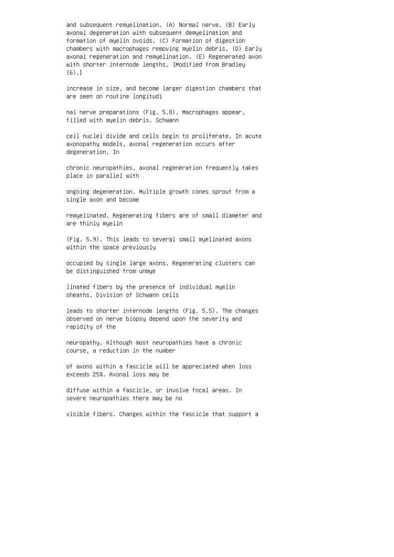
and subsequent remyelination. (A) Normal nerve. (B) Earlyaxonal degeneration with subsequent demyelination andformation of myelin ovoids. (C) Formation of digestionchambers with macrophages removing myelin debris. (D) Earlyaxonal regeneration and remyelination. (E) Regenerated axonwith shorter internode lengths. [Modified from Bradley(6).]
increase in size, and become larger digestion chambers thatare seen on routine longitudi
nal nerve preparations (Fig. 5.8). Macrophages appear,filled with myelin debris. Schwann
cell nuclei divide and cells begin to proliferate. In acuteaxonopathy models, axonal regeneration occurs afterdegeneration. In
chronic neuropathies, axonal regeneration frequently takesplace in parallel with
ongoing degeneration. Multiple growth cones sprout from asingle axon and become
remyelinated. Regenerating fibers are of small diameter andare thinly myelin
(Fig. 5.9). This leads to several small myelinated axonswithin the space previously
occupied by single large axons. Regenerating clusters canbe distinguished from unmye
linated fibers by the presence of individual myelinsheaths. Division of Schwann cells
leads to shorter internode lengths (Fig. 5.5). The changesobserved on nerve biopsy depend upon the severity andrapidity of the
neuropathy. Although most neuropathies have a chroniccourse, a reduction in the number
of axons within a fascicle will be appreciated when lossexceeds 25%. Axonal loss may be
diffuse within a fascicle, or involve focal areas. Insevere neuropathies there may be no
visible fibers. Changes within the fascicle that support a
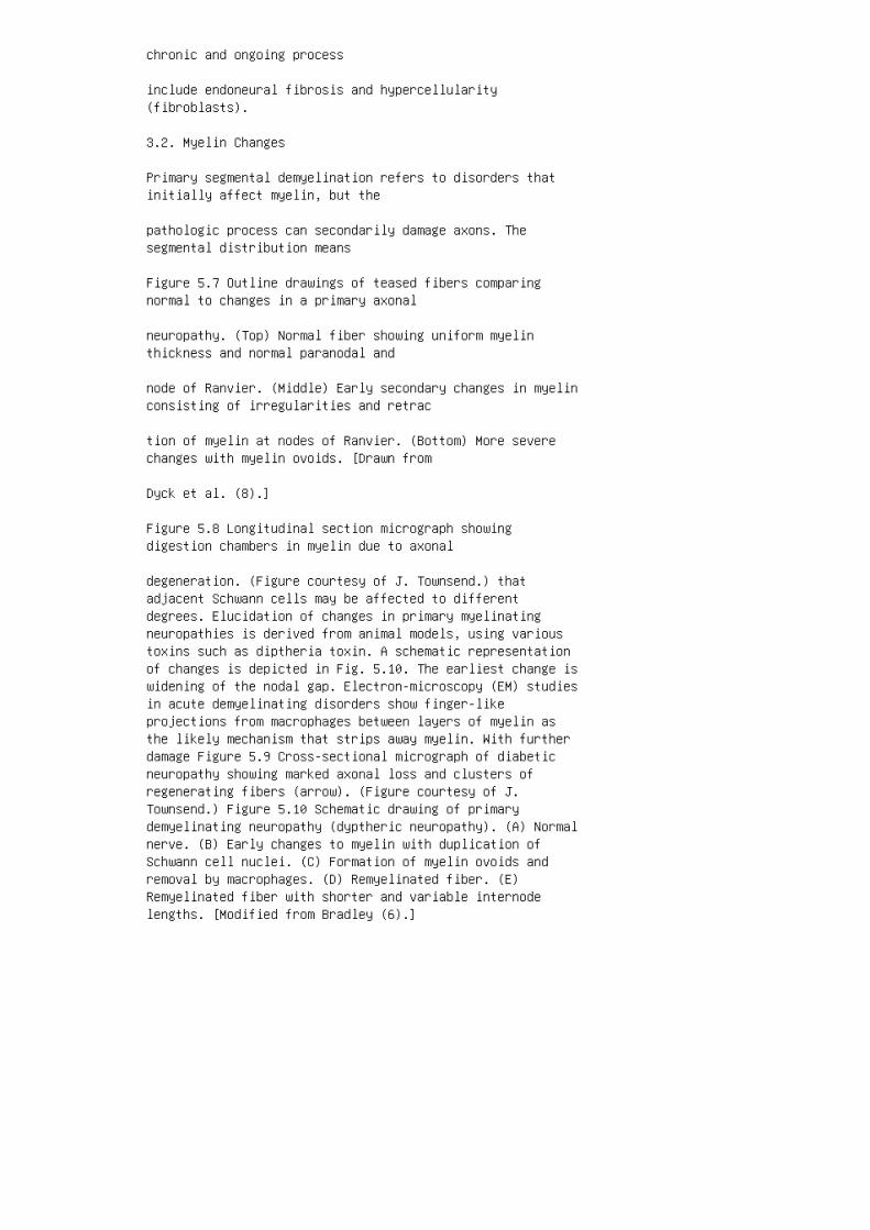
chronic and ongoing process
include endoneural fibrosis and hypercellularity(fibroblasts).
3.2. Myelin Changes
Primary segmental demyelination refers to disorders thatinitially affect myelin, but the
pathologic process can secondarily damage axons. Thesegmental distribution means
Figure 5.7 Outline drawings of teased fibers comparingnormal to changes in a primary axonal
neuropathy. (Top) Normal fiber showing uniform myelinthickness and normal paranodal and
node of Ranvier. (Middle) Early secondary changes in myelinconsisting of irregularities and retrac
tion of myelin at nodes of Ranvier. (Bottom) More severechanges with myelin ovoids. [Drawn from
Dyck et al. (8).]
Figure 5.8 Longitudinal section micrograph showingdigestion chambers in myelin due to axonal
degeneration. (Figure courtesy of J. Townsend.) thatadjacent Schwann cells may be affected to differentdegrees. Elucidation of changes in primary myelinatingneuropathies is derived from animal models, using varioustoxins such as diptheria toxin. A schematic representationof changes is depicted in Fig. 5.10. The earliest change iswidening of the nodal gap. Electron-microscopy (EM) studiesin acute demyelinating disorders show finger-likeprojections from macrophages between layers of myelin asthe likely mechanism that strips away myelin. With furtherdamage Figure 5.9 Cross-sectional micrograph of diabeticneuropathy showing marked axonal loss and clusters ofregenerating fibers (arrow). (Figure courtesy of J.Townsend.) Figure 5.10 Schematic drawing of primarydemyelinating neuropathy (dyptheric neuropathy). (A) Normalnerve. (B) Early changes to myelin with duplication ofSchwann cell nuclei. (C) Formation of myelin ovoids andremoval by macrophages. (D) Remyelinated fiber. (E)Remyelinated fiber with shorter and variable internodelengths. [Modified from Bradley (6).]

to myelin, macrophages can be appreciated on lightmicroscopy to phagocytize myelin
debris. Schwann cell nuclei divide, and remyelinationoccurs early on, initially with
thin layers. Each Schwann cell contributes to theremyelination process, and internodal
lengths are shorter and highly variable (Fig. 5.5). Onionbulb formation refers to intertwined and attenuated Schwanncell processes
surrounding axons (Fig. 5.11). This reflects underlyingpathologic processes of repeated
demyelination and remyelination. Collagen separates Schwanncell processes. Onion
bulb formation is not specific to a particular type ofneuropathy, and is encountered in
both hereditary and acquired neuropathies that involverepeated myelin breakdown. Secondary demyelination refersto degeneration of Schwann cells consequent to
primary axonal degeneration. There is a degree of initialautophagocytosis by the
Schwann cell, and subsequent removal of myelin debris bymacrophages. Changes in
Schwann cells and myelin subsequent to axonal damage havebeen discussed (Fig. 5.5–5.9).
3.3. Connective Tissue Changes
Changes in connective tissue are usually of a nonspecificnature. Vessels course through
the epineurium. Perivascular cuffing with mononuclear cellsis common but nonspecific,
and is not synonymous with vasculitis. Epineurial vascularproliferation is indirect evi
dence for vasculitis. Amyloid deposition may be observed inamyloidosis. Inflammatory
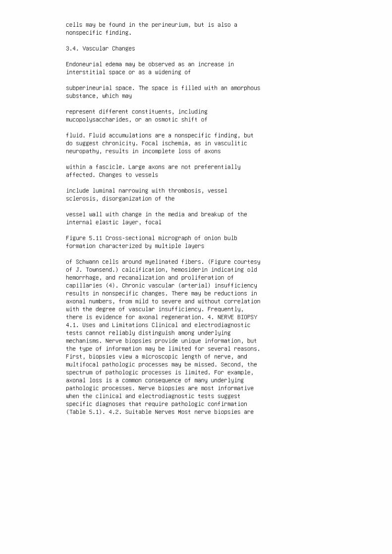
cells may be found in the perineurium, but is also anonspecific finding.
3.4. Vascular Changes
Endoneurial edema may be observed as an increase ininterstitial space or as a widening of
subperineurial space. The space is filled with an amorphoussubstance, which may
represent different constituents, includingmucopolysaccharides, or an osmotic shift of
fluid. Fluid accumulations are a nonspecific finding, butdo suggest chronicity. Focal ischemia, as in vasculiticneuropathy, results in incomplete loss of axons
within a fascicle. Large axons are not preferentiallyaffected. Changes to vessels
include luminal narrowing with thrombosis, vesselsclerosis, disorganization of the
vessel wall with change in the media and breakup of theinternal elastic layer, focal
Figure 5.11 Cross-sectional micrograph of onion bulbformation characterized by multiple layers
of Schwann cells around myelinated fibers. (Figure courtesyof J. Townsend.) calcification, hemosiderin indicating oldhemorrhage, and recanalization and proliferation ofcapillaries (4). Chronic vascular (arterial) insufficiencyresults in nonspecific changes. There may be reductions inaxonal numbers, from mild to severe and without correlationwith the degree of vascular insufficiency. Frequently,there is evidence for axonal regeneration. 4. NERVE BIOPSY4.1. Uses and Limitations Clinical and electrodiagnostictests cannot reliably distinguish among underlyingmechanisms. Nerve biopsies provide unique information, butthe type of information may be limited for several reasons.First, biopsies view a microscopic length of nerve, andmultifocal pathologic processes may be missed. Second, thespectrum of pathologic processes is limited. For example,axonal loss is a common consequence of many underlyingpathologic processes. Nerve biopsies are most informativewhen the clinical and electrodiagnostic tests suggestspecific diagnoses that require pathologic confirmation(Table 5.1). 4.2. Suitable Nerves Most nerve biopsies are

of sensory nerves because permanent sensory loss from thebiopsy is better tolerated than weakness that would resultif a mixed nerve was biopsied. The additional sensory lossfrom the biopsy will be less noticed in the context of thesensory disturbance from the underlying neuropathy.Fortunately, most neuropathies involve both sensory andmotor fibers and a sensory nerve will be representative.The nerve chosen should show clinical and electrodiagnosticevidence of pathologic involvement. Nerves most commonlybiopsied are the sural and superficial peroneal nerves, andoccasionally the superficial radial. Fascicular motor nervebiopsies are feasible in pure motor neuropathies. Asimultaneous muscle biopsy may increase the yield offinding pathologic changes, especially for vasculitis (9).4.3. The Biopsy Biopsies are performed under localanesthesia with minimal morbidity, although wound healingmay be an issue (7). A several centimeter segment of nerveis taken, and cut into a number of pieces for differentpathologic processing. A cross-sectional piece andlongitudinal piece are imbedded in paraffin, and anothercross-sectional piece flashTable 5.1 Neuropathies that canbe Diagnosed by Nerve Biopsy Vasculitis Leprosy SarcoidosisAmyloidoisis Infiltrative neoplasms Hexacarbon inhalationHNPP Charcot–Marie–Tooth Giant axonal neuropathy Storagediseases
frozen for immunologic studies. Another piece is fixed ingluteraldehyde for plastic
embedded 1 mm plastic sections. EM may be performed onplastic sections if necessary.
A segment can be prepared for fiber teasing. Not alltechniques are necessary or available
in a given laboratory (especially teased fiber analysis),and plans for specific preparations
and staining can be made depending upon diagnosticquestions.
4.4. Biopsy Review
Steps in reviewing a nerve biopsy include generalfascicular histology for adequacy of the
preparation, inspection of connective tissue, andassessment of individual fascicules for
changes to axons and myelin. The pathophysiological

questions to be answered are: (1)
Is the biopsy normal, or is there evidence for a peripheralneuropathy? (2) If abnormal,
is there evidence for a primary axonal, a primarydemyelinating, or a mixed neuropathy?
(3) What is the time course, acute or chronic? (4) Arethere pathologic features supporting
a specific diagnosis? Finally, the biopsy findings must beplaced in the context of the clini
cal and electrodiagnostic findings to make a finaldiagnosis that makes sense.
4.5. Biopsy Yield
The question of the diagnostic yield of a nerve biopsy isimportant but difficult to answer
because there is sample bias based on what types ofpatients are sent for biopsy. A biopsy
is most informative if a specific question is asked, andthe most common clinical question
is whether there is a vasculitis (9). Studies in theelderly suggest that vasculitis is not
uncommon, but the clinical and therapeutic significance isnot clear (10). Nerve biopsy
gives little discriminative information in toxicneuropathies, except for hexacarbon tox
icity. Inflammatory neuropathies can be more accuratelydiagnosed by electrodiagnostic
studies than by biopsy (11). Idiopathic peripheralneuropathies represent a common
class for which the question of whether to biopsy isdifficult to answer. When a peripheral
neuropathy has a very slow progressive course with littlefunctional disability, a biopsy is
unlikely to identify a specific cause (12,13).

5. CLASSES OF NEUROPATHY
5.1. Inflammatory Neuropathies
Nerve biopsy findings are similar for the Guillain–Barre´syndrome, or acute inflammatory
demyelinating polyradiculoneuropathy (AIDP), and chronicinflammatory demyelinating
polyradiculoneuropathy (CIDP). Pathologic changes aredependent upon the stage of the neuropathy, and are more
specific in the acute phase of AIDP and less specific inCIDP. Endoneurial mononuclear
cell infiltration is a prominent feature in AIDP, and lesscommon in CIDP. Cells are mostly
lymphocytes and macrophages, with fewer polymorphonuclearleukocytes. The cells are
found in association with venules and precapillaries.Macrophages are observed in close
association with axons. Endoneurial edema is often present.Segmental demyelination
and remyelination is the most important feature (Fig.5.10). This includes combinations
of fibers with normal myelin, thin myelin and bare axons,and macrophages with
myelin debris (Fig. 5.12). Schwann cells proliferate andremyelinate with shorter
internodal segments (Fig. 5.5). Axonal loss may be markedin severe cases of AIDP,
but is more common in CIDP. Onion bulb formations alsooccur in CIDP. 5.2. Vasculitic Neuropathies Thevasculitidies that affect peripheral nerve arecharacterized by small vessel occlusion causing axonalloss. Vessel destruction is characterized by the presenceof inflammatory infiltrates within the vessel wall, leadingto fibrinoid necrosis and thrombosis (Fig. 5.13).Transmural damage is an essential pathologic feature ofvasculitis, and perivascular inflammation without fibrinoid

necrosis is a nonspecific finding and insufficient for thediagnosis of vasculitis. Acute vessel changes includeinfiltration of the muscularis and endothelial layers withinflammatory cells, and fragmentation of the internalelastic lamina is characteristic. Old lesions may berecognized as fibrous obliteration of vessels inassociation with hemosiderin-laden macrophages. The amountof fiber loss varies from none to severe, presumablydepending upon the degree of involvement of the biopsiednerve. The pattern of fiber loss within a fascicle alsovaries, but characteristically affects discrete areas, andis not randomly scattered Figure 5.12 Cross-sectionalmicrograph in CIDP showing axonal loss, hyper cellularityand evidence of remyelination with small diameter fibersand thin myelin. (Figure courtesy of J. Townsend.) Figure5.13 Cross-sectional micrographs in vasculitis. (A) Smallvessel showing fibrinoid necrosis and partial occlusion ofthe vessel lumen. (B) Nerve fascicle showing discrete areaof axonal loss. (Figure courtesy of J. Townsend.)
throughout the fascicle (Fig. 5.13). Vascular lesions maybe focal or patchy in distribution,
and multiple sections along the length of the biopsiednerve should be studied if clinical
suspicion is high and no involved vessels are found withthe initial survey. A combination
biopsy of nerve and neighboring muscle may increase thediagnostic yield (9).
5.3. Amyloid Neuropathies
Amyloid is an extra cellular protein composed ofbeta-pleated sheets that accumulate in
the epineurium, perineurium or endoneurium, or in adiposetissue. Neuropathies attributed
to excess amyloid occur in both acquired and hereditaryforms of amyloidosis. Amyloid
deposition can be identified with Congo red stain by itscharacteristic apple-green
yellow birefringence stain. The primary nerve pathology isaxonal loss, of varying
degrees. The degree of loss may not correlate with the

degree of amyloid observed in
the biopsied nerve segment, and is attributed to theeffects of amyloid at more proximal
locations along the nerve. The mechanism of amyloid-induceddamage is not clear. One
is mechanical pressure on nerves from extraneural depositonwithin connective tissue.
Another is deposition of amyloid within vessel walls (Fig.5.14). Patients with amyloid
neuropathy may not show amyloid deposition in nerve, and ifsuspected, other tissues
should be biopsied, such as rectal tissue and fat aspirates(14).
5.4. Hereditary Neuropathies
Charcot–Marie–Tooth neuropathies represent a heterogeneousgroup of hereditary nerve
disorders. The most common forms have been broadly dividedby electrodiagnostic and
pathologic criteria into hypertrophic (type 1) and neuronal(type 2) forms. Both types
include axonal loss. Further subtyping is based ongenotypes, but genetic subtypes
cannot be distinguished pathologically.
Figure 5.14 Cross-sectional micrograph showing amyloiddeposition (Congo red stain) in a small
vessel. (Figure courtesy of J. Townsend.) Hypertrophic type1 neuropathies are characterized pathologically by enlargednerves due to increased numbers of Schwann cells withfrequent onion bulb formations and a myxoid-appearinginterstitium composed largely of collagen. This results inenlarged fascicles, up to several times normal. There is avariable degree of fiber loss, dependant upon the severityof the neuropathy, and there may be no nerve fibers in adistal biopsy. Unmyelinated fibers are less affected.Neuronal type 2 neuropathies have normal or smallerfascicles, and there is no edema. The number of myelinated

fibers is reduced and frequently there are no fibers in adistal biopsy. Clusters of regenerating fibers are common,but there is no evidence for demyelination or onion bulbformation. Unmyelinated fibers are normal. Hereditarypredisposition to pressure palsies (HNPP) has uniquepathological features. Myelinated fibers with very thickmyelin sheaths (tomaculi) are observed (Fig. 5.15). Somemyelin may be thickened circumferentially or eccentricallyand may have focal outor in-foldings of myelin. The numberof myelinated fibers is usually normal, with littleevidence for axonal degeneration. 5.5. DiabeticNeuropathies Pathologic changes in diabetic distalsymmetric neuropathies are primarily axonal in nature, withsecondary changes to myelin. These pathologic findings arenot specific for diabetes. There is loss of large and smallmyelinated fibers, roughly in proportion to clinicalseverity. Neuropathies classified clinically as affectingpredominantly small fiber function may show correspondingpredominant loss of unmyelinated fibers. Axonal loss leadsto frequent regenerating clusters of axons and fibers withthin myelin, and there is segmental demyelination (Fig.5.9). Figure 5.15 HNPP. (Top) Micrograph comparing thickmyelin of tomaculi with normal myelin. (Bottom) Teasedfiber preparation showing focal enlargement of myelin(tomaculi). [Drawn and modified from Mendell et al. (15).]Pathologic findings in proximal focal diabetic neuropathies(lumbosacral radi
culoplexus neuropathies) are primarily implicated fromdistal nerve biopsies. Focal
nerve fiber loss, reflecting primary axonal damage, isprominent. There is also evidence
for perineural thickening and inflammatory cells inperivascular and vascular location.
These changes are interpreted as reflecting an inflammatorymicrovasculitis leading to
multifocal nerve ischemia (16). In contrast to thevasculidities described earlier, that
involve larger vessels, fibrinoid necrosis is rarelyobserved in lumbosacral radiculoplexus
neuropathies, presumably due to the small size of involvedvessels.
5.6. Toxic Neuropathies

Neuropathies may be caused by a diverse group of exogenoustoxins such as drugs and
industrial solvents. Included in this category areneuropathies associated with altered
metabolic states due to vitamin deficiencies and systemicdiseases. The primary pathology
is axonal loss, in a length-dependent fashion. Largemyelinated fibers are affected earlier
and more severely than small fibers. The degree of axonalloss depends upon the dose and
length of exposure. Early changes are restricted toindividual fibers, and late in the course
there may be no fibers in distal specimens. In chronicdisorders, there will be endoneurial
fibrosis and hypercellularity reflecting Schwann cellproliferation. Of note, the length
dependent pathology includes the central processes ofdorsal root ganglion neurons result
ing in axonal loss in rostral portions of the funiculusgracilis. In severe cases, dorsal root
ganglion and anterior horn cell bodies may undergochromatolytic changes. Several toxins have uniquepathologic features. n-Hexane neuropathy from inhaling
rubber cement results in characteristic fusiform swellingof distal axonal segments due to
accumulation of neurofilaments (Fig. 5.16). Swollensegments are mainly in the internode
segment, and the overlying myelin may be thin. Axonaldiameters in swollen segments
may be several times normal axonal diameters.
Figure 5.16 Micrograph of n-hexane neuropathy showingenlarged axons. Some fibers are cut in
oblique section. (Figure courtesy of A.G. Smith.) 5.7.

Neuropathies Associated with Cancer Neuropathies may beassociated with direct infiltration of malignant cells oras a remote effect (paraneoplastic syndrome). Nerveinfiltration occurs primarily with hematologicmalignancies. In lymphomatous neuropathies, diffuse andmassive infiltration may occur, frequently involving allcompartments (endoneurium, perineurium, and epineurium)(Fig. 5.17). Collections of cells are noted around vessels.Chronic axonal damage with segmental demyelination andremyelination are observed. 5.8. Other Neuropathies Thereare other uncommon conditions with suggestive orcharacteristic pathologic features on nerve biopsy thatwill not be covered because of their rarity and theclinical picture is sufficiently distinct to be diagnostic.These include metachromatic leukodystrophies, globoid cellleukodystrophy, Fabry disease, Niemann–Pick, and Tangierdisease. ACKNOWLEDGMENTS I thank Jeannette Townsend, MD,for her review of the manuscript, and Becky Guertler forher help with the figures. Figure 5.17 Longitudinal sectionmicrograph showing B-cell infiltration. (Figure courtesy ofJ. Townsend.)
1. Sunderland S. Nerves and Nerve Injuries. 2nd ed.Edinburgh: Churchill Livingstone, 1978.
2. Low R. The perineurium and connective tissue ofperipheral nerve. In: Landon D, ed. The Peripheral Nerve.London: Chapman and Hall, 1976:159–187.
3. Ochoa J, Mair W. The normal sural nerve. ActaNeuropathologica 1969; 13:197–216.
4. Midroni G, Bilbao J. Biopsy Diagnosis of PeripheralNeuropathy. Boston: ButterworthHeinemann, 1995.
5. Fullerton P, Gilliatt R, Lancelles R, Morgan-Huges J.Relation between fibre diameter and internodal length inchronic neuropathy. J Physiol 1965; 178:26P–28P.
6. Bradley W. Disorders of Peripheral Nerves. Oxford:Blackwell, 1974.
7. Dyck P, Giannini C, Lais A. Pathologic alternations ofnerves. In: Dyck P, Thomas P, Griffin J, Low P, Poduslo J,eds. Peripheral Neuropathy. 3rd ed. Philadelphia: WBSaunders Company, 1993:514–595.
8. Dyck P, Johnson W, Lambert E, O’Brien P. Segmentaldemyelination secondary to axonal degeneration in uremicneuropathy. Mayo Clin Proc 1971; 46:406–431.

9. Collins M, Periquet M, Mendell J, Sahenk Z, Nagaraja H,Kissel J. Nonsystemic vasculitic neuropathy. Insights froma clinical cohort. Neurology 2003; 61:623–630.
10. Chia L, Fernandez A, Lacroix C, Adams D, Plante´ V,Said G. Contribution of nerve biopsy findings to thediagnosis of disabling neuropathy in the elderly. Aretrospective review of 100 consecutive patients. Brain1996; 119:1091–1098.
11. Krendel D, Parks H, Anthony D, St Clair M, Graham D.Sural nerve biopsy in chronic inflammatory demyelinatingpolyradiculoneuropathy. Muscle Nerve 1989; 12:257–264.
12. Wolfe G, Baker N, Amato A, Jackson C, Nations S,Saperstein D et al. Chronic crytogenic sensorypolyneuropathy. Clinical and laboratory characteristics.Arch Neurol 1999; 56:540–547.
13. Notermans M, Wokke J, Franssen H, van der Graaf Y,Vermeulin M, van den Berg L et al. Chronic idiopathicpolyneuropathy presenting in middle or old age: a clinicaland electrophysiological study of 75 patients. J NeurolNeurosurg Psychiatr 1993; 56:1066–1071.
14. Simmons Z, Blaivas M, Aguilera A, Feldman E, BrombergM, Towfighi J. Low diagnostic yield of sural nerve biopsyin patients with peripheral neuropathy and primaryamyloidosis. J Neurol Sci 1993; 120:60–63.
15. Mendell J, Kissel J, Cornblath D. Diagnosis andManagement of Peripheral Nerve Disorders. Oxford: OxfordUniversity Press, 2001.
16. Dyck P, Norell J, Dyck P. Microvasculitis and ischemiain diabetic lumbosacral radiculoplexus neuropathy.Neurology 1999; 53:2113–2121.
6
Skin Biopsy
A. Gordon Smith
University of Utah, Salt Lake City, Utah, USA
Abstract 83
1. Introduction 83

2. Technique 84
3. Normal Innervation 85
4. Clinical Applications 87
5. When to Perform a Skin Biopsy? 88
6. Research Applications 89
7. Conclusion 89
ABSTRACT
One of the most common clinical situations encountered ingeneral neurologic practice is
numb painful feet. Patients often complain of numbness andsignificant pain, but have
minimal objective findings on neurologic examination. Nerveconduction studies are
often normal. Skin biopsy stained for peripheral nerveaxons is usually abnormal in
these patients and is often the only objective evidence ofperipheral neuropathy. Other
structures including sensory receptors and sweat glands mayalso be observed. Because
the density of epidermal innervation is a quantitative andreproducible measure of small
fiber neuropathy severity, skin biopsy is also a usefulresearch tool and may be used to
follow neuropathy progression and evaluate potentialtherapies.
1. INTRODUCTION
Although the routine use of skin biopsy as a means ofstudying peripheral neuropathy is a
recent development, the existence of epidermal nerve fiberswas first demonstrated by

Langerhans (1) in 1868. Langerhans’ initial description wasaccurate, including a
drawing demonstrating several epidermal fibers branchingfrom the dermal plexus, and 83 ascending in betweenkeratinocytes toward the epidermal surface (Fig. 6.1).Despite the fact that other investigators in the first-halfof the 20th century confirmed his observations (2), thetechnical difficulties encountered in attempts to stain thefine epidermal fibers led to a belief developed that theepidermis was devoid of significant innervation (3). Thedevelopment of an antibody against neuropeptide proteingene product 9.5 (PGP 9.5) in the 1980s (4), whicheffectively stains all axons, allowed Kennedy andWendelschaferCrabb (5) to demonstrate numerous nerve fibersin all layers of human epidermis. Since this confirmationof Langerhans’ initial observation, several groups havedemonstrated the utility of skin biopsy as a means ofevaluating patients with small fiber neuropathy. 2.TECHNIQUE Skin biopsies are performed using a 3 mm punchafter injection of a local anesthetic. The biopsy procedureis generally very well tolerated with only minimal risk ofinfection (1/1000). The biopsy site heals over �2 weeks,similar to a deep scratch, resulting in a small scar thatfades over months. Because the procedure is so welltolerated, it may be repeated over time to measure changesin nerve fiber density. The sites biopsied depend on theclinical question. Most subjects with a diagnostic questionof a distal polyneuropathy undergo biopsy of a distal site(ankle) and a proximal site (thigh) in order to evaluate alength dependent pattern of fiber loss. Biopsy of truncalskin may be useful in evaluating thoracic radiculopathy orsensory ganglionopathy (6). Although rarely usedclinically, biopsy of glabrous skin (finger or toe pad) maybe used to study Meissner’s and Pacinian corpuscles,sensory receptors important in touch and vibrationsensation (Fig. 6.2). Biopsy tissue is immediately placedin fixative (paraformaldehyde lysine phosphate). Afterseveral hours, fixed skin is placed in a cryoprotectant andcut into 50 mm thick sections using a freezing slidingmicrotome. The free floating sections are stained with PGP9.5. Biopsy slides may be analyzed using either standardlight microscopy or confocal microscopy. Confocalmicroscopy facilitates staining with multiple antibodiessimultaneously and provides a three-dimensional picture ofthe skin and is therefore a powerful research technique.However, light microscopy is equally useful for mostclinical purposes. Figure 6.1 A drawing from Langerhans’ in1868 accurately depicts the anatomy of epidermalinnervation. PGP 9.5, the most commonly used antibody,
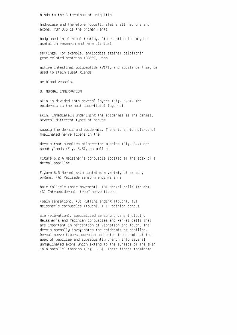
binds to the C terminus of ubiquitin
hydrolase and therefore robustly stains all neurons andaxons. PGP 9.5 is the primary anti
body used in clinical testing. Other antibodies may beuseful in research and rare clinical
settings. For example, antibodies against calcitoningene-related proteins (CGRP), vaso
active intestinal polypeptide (VIP), and substance P may beused to stain sweat glands
or blood vessels.
3. NORMAL INNERVATION
Skin is divided into several layers (Fig. 6.3). Theepidermis is the most superficial layer of
skin. Immediately underlying the epidermis is the dermis.Several different types of nerves
supply the dermis and epidermis. There is a rich plexus ofmyelinated nerve fibers in the
dermis that supplies piloerector muscles (Fig. 6.4) andsweat glands (Fig. 6.5), as well as
Figure 6.2 A Meissner’s corpuscle located at the apex of adermal papillae.
Figure 6.3 Normal skin contains a variety of sensoryorgans. (A) Palisade sensory endings in a
hair follicle (hair movement). (B) Merkel cells (touch).(C) Intraepidermal “free” nerve fibers
(pain sensation). (D) Ruffini ending (touch). (E)Meissner’s corpuscles (touch). (F) Pacinian corpus
cle (vibration). specialized sensory organs includingMeissner’s and Pacinian corpuscles and Merkel cells thatare important in perception of vibration and touch. Thedermis normally invaginates the epidermis as papillae.Dermal nerve fibers approach and enter the dermis at theapex of papillae and subsequently branch into severalunmyelinated axons which extend to the surface of the skinin a parallel fashion (Fig. 6.6). These fibers terminate

immediately beneath the surface of the skin and often havea small terminal enlargement (5). Figure 6.4 Theinnervation of piloerector muscles (arrow) is easilyobserved in skin biopsy. Figure 6.5 (A) Normal innervationof two sweat glands. (B) Absent innervation of sweat glandsin a patient with congenital insensitivity to pain due to amutation of the tyrosine kinase A receptor. The pattern ofskin innervation varies with anatomic location. Forexample, Meissner’s
and Pacinian corpuscles are most commonly observed inglabrous skin of the finger and toe
pads. Epidermal nerve fibers are distributed throughout thebody but density varies with
location, with intraepidermal nerve fiber density (IENFD)highest at distal sites (7). The effects of aging oncutaneous innervation are controversial. Dermal papillae
become lower in height and there is denervation of sweatglands with aging (8). There
is also a reduction in the density of both myelinated andunmyelinated sensory nerve
fibers within the sural nerve with aging (9). In contrastto these observations, the
density of epidermal nerve fibers appears to remainconstant, although few subjects
.75 years old have been studied (7,10). The reason for thediscrepancy between sural
nerve and IENFD data is unknown. However, the stability ofepidermal nerve fiber
density facilitates the use of skin biopsy as a sensitiveclinical tool in the evaluation of
small fiber peripheral neuropathy at advanced ages.
4. CLINICAL APPLICATIONS
Skin biopsy was first used in the study of peripheralneuropathy in the 1960s, when
investigators at the Mayo Clinic demonstrated reducednumbers of Meissner’s corpuscles

from glabrous skin of subjects with a variety of inheritedneuropathies, including
Charcot–Marie–Tooth disease. The first routine use of skinbiopsy for neuropathy
came after the development of PGP 9.5, which allowed forthe easy identification of
Figure 6.6 (A) Normal innervation with nerve fibersextending through the epidermis, parallel to
one another, extending toward the skin surface. (B) Apatient with small fiber neuropathy has a total
absence of epidermal nerve fibers with a single dermalfiber terminating prematurely beneath the
epidermis. epidermal nerve fibers. Several investigatorshave demonstrated reduced density of epidermal fibers in alength dependent fashion in sensory neuropathies caused byHIV, antiretroviral therapy, diabetes, and in idiopathicneuropathy [Fig. 6.6(B)] (11–14). Surviving fibers oftenhave morphologic abnormalities including axonal swellingsor unusual branching patterns [Fig. 6.7(A)]. Thesemorphologic changes are frequently observed at sites wellproximal to the area of symptoms. In areas with reducedepidermal fiber density, dermal fibers may approach thedermal–epidermal junction, but either end abruptly orbranch beneath the epidermis [Fig. 6.7(B)]. Patients withpain and numbness limited to the feet often have thesechanges in skin from the proximal thigh. Analysis of otherstructures, particularly sweat glands, may also be useful,and patients with peripheral neuropathy often havedenervated sweat glands [Fig. 6.5(B)]. Fiber density doesnot correlate with nerve conduction study parameters, anexpected observation given that routine electrophysiologicevaluation is limited to large diameter fibers, whereasskin biopsy specifically evaluates small fibers. Asanticipated, there is good correlation with the number ofsmall unmyelinated fiber in sural nerve biopsy tissue (15).Fiber density correlates with pain but not sudomotorfunction (16,17). These findings suggest that epidermalnerve fibers are normally responsible for pain sensationand damage results in abnormal sensations including pain.Skin biopsy is a very useful tool in the diagnosis ofperipheral neuropathy, particularly for patients who havepainful feet. Approximately 30–50% of patients presentingwith foot pain and numbness have normal nerve conduction

studies (17,18). Even when other specialized tests such asquantitative sensory testing and sudomotor testing areused, one-third of patients with this common clinicalpresentation require skin biopsy to confirm the diagnosisof neuropathy. When a normal skin biopsy is obtained, acareful search for a central nervous system cause for painis warranted (17). Although processing of skin biopsytissue requires a specialized laboratory, biopsy tissue maybe easily shipped to laboratories offering this test.Therefore, no special equipment or training (aside fromperforming the skin biopsy itself ) is necessary to usethis test in routine clinical practice. 5. WHEN TO PERFORMA SKIN BIOPSY? Skin biopsy is most often used in theevaluation of patients with foot pain and numbness but anormal nerve conduction study. In this setting, an abnormalskin biopsy confirms the Figure 6.7 Predegenerativestructural changes in small fiber neuropathy. (A) Axonalswellings at a proximal site (thigh) in a patient withsymptoms limited to the feet. (B) Excessive branching andpremature termination of nerve fibers as they approach theepidermal surface.
clinical suspicion of a small fiber predominant neuropathy.Skin biopsy may also be
useful in patients with suspected painful neuropathy inwhom measurement of distal
lower extremity sensory responses is complicated bytechnical issues (edema and
obesity) or advanced age. A rare syndrome of acute diffuseburning pain, often following a viral illness, has been
described. Most patients describe their pain as resemblinga sunburn. This syndrome is pre
sumably due to postinfectious injury to small cutaneousnerve fibers in a nonlength depen
dent fashion or the dorsal root ganglion neuronsresponsible for small fiber sensory function.
Nerve conduction studies are normal, but skin biopsyreveals diffusely reduced IENFD (19). Broadly speaking,skin biopsy is useful in any situation where thedemonstration of
peripheral sensory axonal loss, especially if it involvessmall fibers, is diagnostically

important, but impractical using other available means. Forexample, skin biopsy may
occasionally be of use in confirming a diagnosis ofthoracic radiculopathy due to
Herpes Zoster. Although this diagnosis is usually straitforward, if there is no clear
history of vesicles, skin biopsy may be used to confirm thepresence of sensory denerva
tion corresponding to a thoracic dermatome. In thisinstance, a biopsy of the involved area
should be compared with another proximal site other thanthe opposite side, because bilat
eral denervation is often observed (20).
6. RESEARCH APPLICATIONS
In addition to providing a clinically useful diagnostictest, skin biopsy is also a useful
research tool. Skin biopsy tissue stained for peripheralnerve can be used to study the neu
robiology of disease. For example, skin biopsy hasdemonstrated that sensory axonal loss
occurs bilaterally in postherpetic neuralgia, even whensymptoms are unilateral (20).
Because skin biopsy is a very sensitive measure of smallfiber sensory integrity, it may
be used to study subclinical peripheral nerve changes. Forexample, many patients with
late onset sporadic restless legs syndrome have evidence ofa neuropathy using skin
biopsy, supporting the role of sensory neuropathy in thedevelopment of restless legs
syndrome (21). Following a skin biopsy, sensory nerves fromadjacent skin reinnervate

the biopsy site. Skin biopsy may therefore be used to studycutaneous reinnervation. Perhaps, the most useful researchapplication is as an endpoint measure for treatment
trials for peripheral neuropathy. Many common neuropathiescause prominent small fiber
injury, including diabetes and HIV. Traditional surrogatemeasures of neuropathy pro
gression measure large fiber loss (e.g., nerve conductionstudies). In neuropathies with
mixed small and large fiber loss, specific therapies maypreferentially target small fiber
function (e.g., nerve growth factor for diabeticneuropathy). Because skin biopsy provides
a reproducible and quantitative means of evaluatingcutaneous innervation, it holds
promise as an endpoint measure for neuropathy trials andhas been used as a secondary
progression measure in one study of nerve growth factor inHIV neuropathy (22). By
causing focal epidermal denervation by injecting capsaicinand the effect of different
agents on the rate or degree of reinnervation may also bestudied.
7. CONCLUSION
Skin biopsy is a useful clinical and research tool in theevaluation of patients with painful
sensory peripheral neuropathy. Epidermal denervation may bethe only laboratory
7
MR Imaging of the Peripheral Nerve
Kevin R. Moore
University of Utah, Salt Lake City, Utah, USA

Abstract 92
1. Introduction 92
2. Neural Structures Amenable to MRPNI 93
3. Role of the Ordering Physician 93
4. Technical Points and Imaging Parameters 94 4.1. Hardware94 4.1.1. MR Scanner 94 4.1.2. Coils 94 4.2. PatientComfort 95 4.3. Technical Imaging Parameters and TheirAdjustment 95 4.3.1. Field of View 95 4.3.2. SpatialResolution 95 4.3.3. Signal-to-Noise Ratio 95 4.3.4.Imaging Time 95 4.3.5. Contrast Resolution 95 4.4. PulseSequences and Imaging Scan Planes 96 4.4.1. Pulse Sequences96 4.4.2. Imaging Scan Planes 96
5. Normal Peripheral Nerve 96
6. Abnormal Nerve Imaging Diagnostic Criteria 97
7. Abnormal Muscle Imaging Diagnostic Criteria 98
8. Imaging Atlas 99
9. Conclusions 99
Appendix: Guidelines for Imaging Specific Neural Structures100
A.1. Brachial Plexus 100
A.2. Lumbosacral Plexus 100
A.3. Sciatic Nerve 101
A.4. Tibial and Peroneal Nerves 103
tional MRI protocols use commonly available general-purposevolume or phase array
coils, and with careful attention to optimizing imagingparameters, can produce acceptable
images. High-resolution MR peripheral nerve imaging(MRPNI), also known as MR neu
rography (1), is the most sensitive imaging modality forcharacterizing peripheral nerve

and plexus abnormalities (1–4) and for directing treatmentplanning (4,5). High-resolution
MRPNI uses a small field of view (FOV), an optimizedsignal-to-noise ratio (SNR), and
high contrast resolution to depict subtle internal nervearchitectural details. MRI is an invaluable adjunct to theclinical examination and electrodiagnostic
evaluation of peripheral nerve disease. It providesanatomic lesion localization and
details about internal nerve architecture, and in somecases, may provide a specific diag
nosis. MRPNI studies must be interpreted in context withpertinent clinical, laboratory,
and electrophysiologic data. Successful high qualityperipheral nerve imaging requires
an understanding of relevant anatomy and close attention totechnical details. MRPNI is
typically more sensitive for the presence of disease thanspecific for a particular disease
process. For example, MRPNI is sensitive to nerveanatomical and signal abnormalities
due to neoplastic and inflammatory lesions, but oftencannot distinguish between them.
However, MRPNI frequently permits a specific answer whenthe primary questions
involve assessment of nerve integrity and the presence orabsence of edema in causes
of nerve trauma. This chapter describes MRI findings ofnormal and abnormal nerves including
abnormalities of nerve size, T2 signal intensity, anddefinition of internal architecture
(3,6,7). MRI of muscles is included because acute andchronic muscle denervation have
distinctive imaging appearances that may help confirm a

suspected peripheral nerve
lesion. In certain situations, it is possible to confirmthe existence of a specific peripheral
nerve lesion based on the pattern of abnormal muscleimaging, when identification the
affected nerve as a discrete structure is impossiblebecause it is too small or the
imaging abnormality too subtle. MRI of muscle may provideuseful corollary information
when a patient is not able to undergo definitiveelectrodiagnostic testing.
2. NEURAL STRUCTURES AMENABLE TO MRPNI
In theory, nerves 4–5 mm or larger in diameter that have aknown course can be imaged
with MRPNI. In practice, however, artifacts caused byadjacent structures may prevent
useful imaging. Commonly imaged nerves include the brachialplexus, lumbosacral
plexus, sciatic nerve, ulnar nerve, and common peronealnerve (at the fibular head).
Less frequently imaged nerves include the median, radial,tibial, and femoral nerves.
In cases where the nerve of interest is too small to beresolved as a discrete structure
(e.g., long thoracic nerve, spinal accessory nerve,posterior interosseous nerve in the
forearm), it is usually possible to follow the nerve’sexpected course and indirectly
deduce anatomical lesions producing the observed clinicalfindings.
3. ROLE OF THE ORDERING PHYSICIAN
It is very difficult for the radiologist to perform andaccurately interpret peripheral nerve

imaging studies without full clinical information.Availability of pertinent clinical data
facilitates designing appropriate imaging of the relevantregion in a time efficient
4.2. Patient Comfort
A comfortable patient is more likely to hold still to yieldhigh quality imaging studies.
Poor patient satisfaction also limits imaging time andwillingness to comply with future
imaging requests. Careful attention should be paid toappropriate placement of supporting
pillows and padding as well as immobilization of the limb.
4.3. Technical Imaging Parameters and Their Adjustment
Successful identification of fascicular morphology andsubtle nerve signal intensity
abnormalities requires optimal scan parameters to maximizeSNR and produce sufficient
spatial and contrast resolution within a reasonable imagingtime. A basic introduction to
the most clinically pertinent technical parameters andtheir importance will help achieve
better imaging quality. The most important technicalparameters to understand are FOV,
spatial resolution, SNR, imaging time, and contrastresolution.
4.3.1. Field of View
It is most important to define the study FOV so that thenerve of interest is displayed as a
large discrete structure. Choosing a large FOV may make itdifficult to resolve the nerve
as a discrete structure, much less discern internalarchitecture. For example, imaging

the sciatic nerve within the pelvis and proximal thighusing a single large FOV results
in a tiny, poorly visualized nerve structure. To detectsubtle differences in nerve size or
signal intensity, it is often best to image each sideseparately or to compare the sympto
matic side to normal imaging studies stored in aninstitutional normative database.
4.3.2. Spatial Resolution
Spatial resolution is maximized by choosing the largestpractical matrix size and thinnest
reasonable slice thickness to make voxels as small aspossible. However, there are trade
offs with an increase in imaging time and decrease in theSNR.
4.3.3. Signal-to-Noise Ratio
SNR may be improved by increasing imaging time (increasingthe number of excitations),
increasing slice thickness, or decreasing the matrix size.Higher SNR produces a smoother
and more pleasing image that facilitates the detection ofsubtle abnormalities.
4.3.4. Imaging Time
Short imaging time is important for patient comfort and formaintaining reasonable patient
throughput. Best quality imaging is usually acquired withinthe first 20 min of the session,
before the patient becomes restless and begins to move.Most patients are intolerant of
individual imaging sequences lasting .8–10 min.
4.3.5. Contrast Resolution

Contrast resolution is the ability to distinguishindividual structures on the basis of differ
ences in intrinsic tissue contrast. T1 weighted images(T1WI) provide excellent anatom
ical definition and fat/muscle contrast resolution and areuseful for delineating underlying
architecture of macroscopic structures, but providerelatively poor contrast resolution for
structures of varying water content. Fat saturated T2weighted image (T2WI) sequences,
on the other hand, do not display underlying anatomy aswell and have relatively poor muscle and fat contrastresolution, but are excellent for portraying relativelysubtle differences in tissue water content. For thesereasons both sequences should be obtained, T1WI foranatomical information and T2WI to identify inflammatory orneoplastic changes manifesting as differences in watercontent. 4.4. Pulse Sequences and Imaging Scan Planes MRPNItechnique exploits differences in water content betweenendoneurium, perineurium, and surrounding epineurium. MRPNIprotocols use high resolution T1 weighted sequences todelineate anatomical detail and fat suppressed T2 weightedor short tau inversion recovery (STIR) sequences to detectabnormal nerve water content due to inflammation orneoplasm. 4.4.1. Pulse Sequences It is not possible todiscern subtle abnormalities in nerve water T2 signalwithout suppressing the fat signal, because increased nervewater secondary to pathology and fat both appear bright onthe T2WI. Intrinsic fat signal on the T2WI is negated usingchemical fat saturation pulses or STIR technique. STIR isan extremely useful pulse sequence that provides T2weighting, excellent homogeneous suppression of fat signal,and accentuates nerve water content by removing theintrinsic high signal from fat containing structures. Fatsaturated T2WI and STIR sequences each have their meritsand limitations. T2WI technique generally provides betterSNR and is more time efficient than STIR sequences, but maybe compromised by inhomogeneous fat suppression. On theother hand, STIR technique gives very homogeneous fatsignal suppression and is effective for demonstrating nervehyperintensity, but is generally a longer sequence withmore background noise and vascular flow artifacts thanT2WI. As a general rule, intravenous contrast agents do notconfer appreciable additive value to current MRPNItechnique. However, the selected use of contrast is helpful
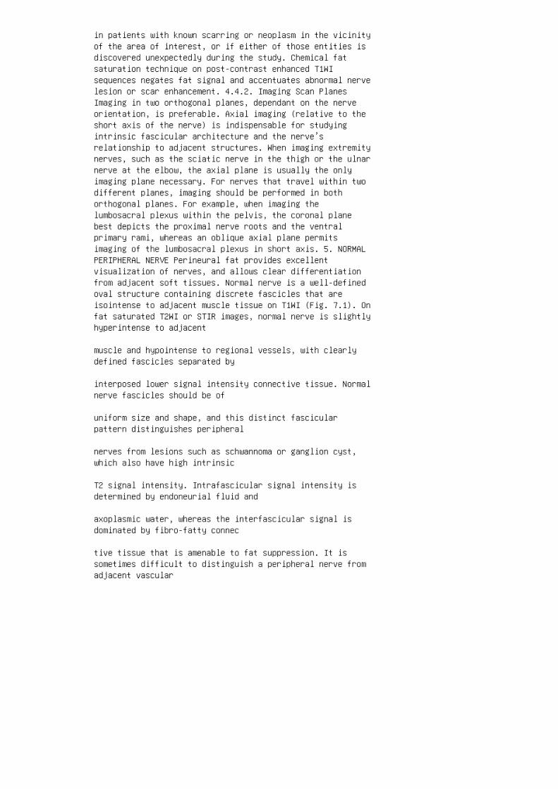
in patients with known scarring or neoplasm in the vicinityof the area of interest, or if either of those entities isdiscovered unexpectedly during the study. Chemical fatsaturation technique on post-contrast enhanced T1WIsequences negates fat signal and accentuates abnormal nervelesion or scar enhancement. 4.4.2. Imaging Scan PlanesImaging in two orthogonal planes, dependant on the nerveorientation, is preferable. Axial imaging (relative to theshort axis of the nerve) is indispensable for studyingintrinsic fascicular architecture and the nerve’srelationship to adjacent structures. When imaging extremitynerves, such as the sciatic nerve in the thigh or the ulnarnerve at the elbow, the axial plane is usually the onlyimaging plane necessary. For nerves that travel within twodifferent planes, imaging should be performed in bothorthogonal planes. For example, when imaging thelumbosacral plexus within the pelvis, the coronal planebest depicts the proximal nerve roots and the ventralprimary rami, whereas an oblique axial plane permitsimaging of the lumbosacral plexus in short axis. 5. NORMALPERIPHERAL NERVE Perineural fat provides excellentvisualization of nerves, and allows clear differentiationfrom adjacent soft tissues. Normal nerve is a well-definedoval structure containing discrete fascicles that areisointense to adjacent muscle tissue on T1WI (Fig. 7.1). Onfat saturated T2WI or STIR images, normal nerve is slightlyhyperintense to adjacent
muscle and hypointense to regional vessels, with clearlydefined fascicles separated by
interposed lower signal intensity connective tissue. Normalnerve fascicles should be of
uniform size and shape, and this distinct fascicularpattern distinguishes peripheral
nerves from lesions such as schwannoma or ganglion cyst,which also have high intrinsic
T2 signal intensity. Intrafascicular signal intensity isdetermined by endoneurial fluid and
axoplasmic water, whereas the interfascicular signal isdominated by fibro-fatty connec
tive tissue that is amenable to fat suppression. It issometimes difficult to distinguish a peripheral nerve fromadjacent vascular

structures, particularly if the nerve is abnormal anddisplays high T2 signal intensity.
Both nerves and vessels are round or ovoid linearstructures, but vessels have character
istics that often permit their distinction from nerves.Vessels demonstrate internal flow
voids, branch at large angles, and show intense contrastenhancement. Nerves on the
other hand do not show flow voids, branch at relativelyacute angles, enhance minimally,
and display a discrete distinctive fascicular architectureon transverse imaging.
6. ABNORMAL NERVE IMAGING DIAGNOSTIC CRITERIA
Abnormal MRPNI findings include segmental nerveenlargement, disruption of normal
nerve anatomy, T2 signal intensity approaching that ofregional blood vessels on fat
saturated T2WI or STIR sequences, or disruption ordistortion of normal fascicular archi
tecture (Fig. 7.2). Goals of MRPNI are localization of thesite of nerve “injury” or tumor, determi
nation of the pathologic process, formulation of atreatment plan, and prognosis. Nerve
injuries result in gross changes in nerve configuration,loss or distortion of the character
istic fascicular pattern, and/or swelling of individualfascicles with abnormally high signal
on T2 weighted and STIR sequences. Abnormal nerves areisointense on T1WI, but
become increasingly hyperintense to muscle on T2WI. Thecause of abnormal high
Figure 7.1 Normal sciatic nerve at the level of piriformismuscle. (A) Oblique axial T1WI

oriented through the sciatic nerve short axis (arrow) showsnormal fascicles that are isointense to
muscle, surrounded by bright fatty connective tissue. (B)Oblique axial fat saturated T2WI shows
mildly hyperintense fascicles with surrounding dark fattyconnective tissue (dark due to the fat
saturation pulse). signal on T2WI and STIR sequences is notdefinitively known, but edema from increased endoneurialfluid due to disordered endoneurial fluid flow or localvenous obstruction may be factors (6). Alterations inaxoplasmic flow may also produce T2 hyperintensity,including impeded flow due to compression (1,4,6). MRPNI issensitive for detecting and discriminating among the threetypes of peripheral nerve injury. Neurapraxia is the leastsevere type of injury, and is characterized by focal damageto the myelin sheath without axonal disruption. It isidentifiable on MRPNI by swollen and hyperintense nervefascicles. Axonotmesis is an intermediate level of crush ortraction injury that produces axonal disruption andsubsequent Wallerian degeneration, but leaves Schwann cellsand endoneurium intact. Axonotmesis results inhomogeneously increased signal intensity with loss offascicular architecture at the injury site. Neurotmesis isthe most severe form of nerve injury, and representscomplete interruption of nerve continuity. MRPNI cannotalways distinguish axonotmesis from neurotmesis, but thelatter may be inferred with there is discontinuity ofsurrounding connective tissues. MRPNI should be considereda companion to electrodiagnostic testing. Abnormal highsignal in nerve fascicles correlates well with clinical andelectrodiagnostic evidence of nerve injury (4).Electromyography does not show denervation until 1–2 weeksfollowing injury, whereas MR neurography can allowassessment of nerve integrity immediately after injury (6).7. ABNORMAL MUSCLE IMAGING DIAGNOSTIC CRITERIA MRPNI issensitive to muscle denervation, and correlates well withdenervation changes on electrodiagnostic studies (10).Denervation results in T2 signal hyperintensity on fatsaturated T2WI or STIR sequences, and normal or increasedmuscle volume (Fig. 7.3). Acute denervation changes are notreliably identified earlier than 4 days post nerve Figure7.2 Lumbosacral trunk neuropathy. A 62-year-old manpresents with pain radiating down the leg and a clinicaldiagnosis of compression of the sciatic nerve within thepiriformis muscle. (A) Oblique axial T1WI shows enlargementof the right lumbosacral trunk (white arrow). The adjacentnormal S1 root (black arrow) may be used for comparison.

(B) Oblique axial fat saturated T2WI confirms abnormalincreased T2 signal intensity, with several enlarged “hot”fascicles that are particularly bright (white arrow). Theadjacent normal S1 root (black arrow) for comparison.
injury (10). Chronic denervation is characterized bydecreased muscle volume (atrophy)
and strands of interspersed high T1 signal intensityrepresenting fatty replacement of
muscle tissue. Acute and chronic changes may coexist indiseases with ongoing dener
vation. Although a preponderance of either type of findingsis useful for characterizing
the length of denervation, subacute denervation can showboth types of findings simul
taneously for considerable time periods.
8. IMAGING ATLAS
The following case studies provide illustrative examples ofhow MRI can be effectively
applied to the evaluation of peripheral nerve disease.Examples of normal and abnormal
nerve and muscle images are provided. A variety ofstructural and nonstructural nerve
lesions are reviewed. The appendix provides details on thespecific imaging techniques
for each major peripheral nerve. All images are used withpermission Electronic
Medical Education Resource Group, Salt Lake City, Utah, USA.
9. CONCLUSIONS
This chapter considers principles necessary to produce andinterpret peripheral nerve
imaging studies. MRI is the only clinically availableimaging technique with suitable
Figure 7.3 Superimposed acute and chronic muscle
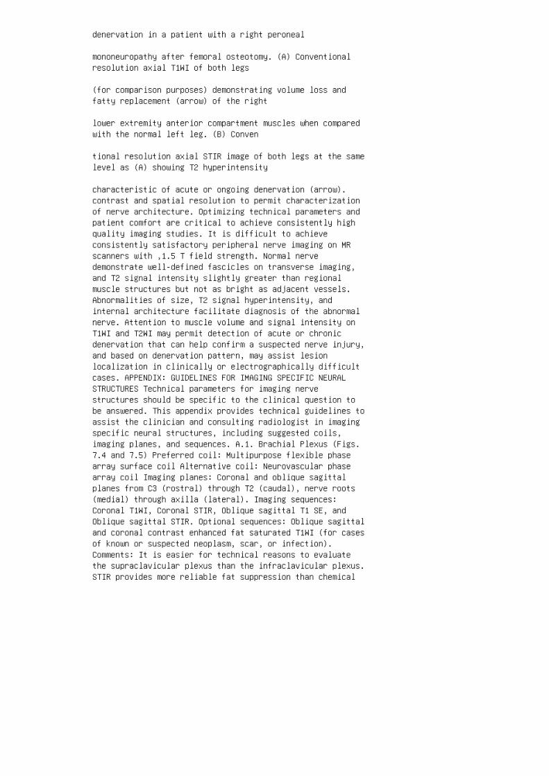
denervation in a patient with a right peroneal
mononeuropathy after femoral osteotomy. (A) Conventionalresolution axial T1WI of both legs
(for comparison purposes) demonstrating volume loss andfatty replacement (arrow) of the right
lower extremity anterior compartment muscles when comparedwith the normal left leg. (B) Conven
tional resolution axial STIR image of both legs at the samelevel as (A) showing T2 hyperintensity
characteristic of acute or ongoing denervation (arrow).contrast and spatial resolution to permit characterizationof nerve architecture. Optimizing technical parameters andpatient comfort are critical to achieve consistently highquality imaging studies. It is difficult to achieveconsistently satisfactory peripheral nerve imaging on MRscanners with ,1.5 T field strength. Normal nervedemonstrate well-defined fascicles on transverse imaging,and T2 signal intensity slightly greater than regionalmuscle structures but not as bright as adjacent vessels.Abnormalities of size, T2 signal hyperintensity, andinternal architecture facilitate diagnosis of the abnormalnerve. Attention to muscle volume and signal intensity onT1WI and T2WI may permit detection of acute or chronicdenervation that can help confirm a suspected nerve injury,and based on denervation pattern, may assist lesionlocalization in clinically or electrographically difficultcases. APPENDIX: GUIDELINES FOR IMAGING SPECIFIC NEURALSTRUCTURES Technical parameters for imaging nervestructures should be specific to the clinical question tobe answered. This appendix provides technical guidelines toassist the clinician and consulting radiologist in imagingspecific neural structures, including suggested coils,imaging planes, and sequences. A.1. Brachial Plexus (Figs.7.4 and 7.5) Preferred coil: Multipurpose flexible phasearray surface coil Alternative coil: Neurovascular phasearray coil Imaging planes: Coronal and oblique sagittalplanes from C3 (rostral) through T2 (caudal), nerve roots(medial) through axilla (lateral). Imaging sequences:Coronal T1WI, Coronal STIR, Oblique sagittal T1 SE, andOblique sagittal STIR. Optional sequences: Oblique sagittaland coronal contrast enhanced fat saturated T1WI (for casesof known or suspected neoplasm, scar, or infection).Comments: It is easier for technical reasons to evaluatethe supraclavicular plexus than the infraclavicular plexus.STIR provides more reliable fat suppression than chemical

fat saturated T2WI. A.2. Lumbosacral Plexus (Fig. 7.6)Preferred coils: Torso wrap-around or pelvis phase arraycoil. Alternative coil: Spine phase array coil Imagingplanes: Coronal and oblique sagittal planes with scancoverage from L3 (rostral) through the ischial tuberosity(caudal), spinal axis (medial) through greater trochanter(lateral). Imaging sequences: Coronal T1WI, coronal STIR orfat saturated T2WI, direct axial or oblique axial T1 spinecho, and direct axial or oblique axial fat saturated T2WIor STIR. Optional sequences: Coronal and oblique axialcontrast enhanced fat saturated T1WI (for cases of known orsuspected neoplasm, scar, or infection). Comments: Comparedwith the torso phase array coil, the spine and pelvis phasearray coils provide inferior SNR in the lateral aspects ofthe pelvis, which may limit evaluation of the proximalsciatic nerve in larger patients. Although the body coilwill provide adequate coverage, it is severely limited bypoor SNR and spatial resolution. The neural foramina,proximal L4 and L5 ventral rami, lumbosacral trunk, S1contribution to the sciatic nerve, and sciatic nervecontinuation into the greater sciatic foramen are bestevaluated in direct coronal and axial planes. The obliqueaxial plane permits optimal visualization of internalarchitecture of the sacral plexus and proximal sciaticnerve.
A.3. Sciatic Nerve (Figs. 7.7 and 7.8) Preferred coil:Torso wrap-around phase array coil Alternative coil:Flexible extremity surface coil Imaging planes: Coronal andoblique axial or direct axial planes (see subsequently).Imaging sequences: Coronal T1WI, coronal STIR or fatsaturated T2WI, direct axial or oblique axial T1 spin echo,and direct axial or oblique axial fat saturated T2WI orSTIR.
Figure 7.4 Normal brachial plexus. (A and B) Coronal STIRimages show normal size and normal
mild T2 hyperintensity of the upper and lower plexuselements (arrows). (C) Axial STIR image
shows the normal size and signal intensity brachial plexus(annotations) passing through the anterior
and middle scalene muscles (asterisks). Optional sequences:Coronal and direct or oblique axial contrast enhanced fatsaturated T1WI (for cases of known or suspected neoplasm,scar, or infection). Comments: Although the body coil willprovide adequate coverage, it is severely limited by poorSNR and spatial resolution. For lesions clinically

localized to proximal sciatic nerve, oblique axial T1WI,and fat saturated T2WI on the symptomatic side from thesacral ala (superior) through the ischial tuberosity(inferior) are preferable. For lesions localized to thesciatic nerve within the thigh (below the ischialtuberosity), direct axial T1WI and fat saturated T2WI onthe symptomatic side from the ischial tuberosity (superior)to the tibioperoneal bifurcation (inferior) are preferable.Figure 7.5 Radiation plexitis following breast cancertreatment. A 55-year-old woman with known metastatic breastcancer presents with arm pain and numbness. MRNI wasperformed in order to differentiate radiation plexitis frommetastatic infiltration. (A) Coronal T1WI shows mildenlargement of the brachial plexus (arrows). (B) CoronalSTIR image shows diffuse enlargement (straight arrows) andincreased T2 signal intensity of the plexus elements thatapproaches that of regional vessels (curved arrow). (C)T1WI fat saturated image following contrast administrationreveals mild patchy enhancement (arrows) without focal massto suggest malignant infiltration.
A.4. Tibial and Peroneal Nerves (Fig. 7.9) Preferred coils:Torso wrap-around phase array coil or knee coil (producesexcellent images but limited to coverage of the commonperoneal nerve at the fibular head).
Figure 7.6 Neurolymphomatosis. A 69-year-old man with ahistory of successfully treated
thoracic nonHodgkin’s lymphoma presents with progressionright leg pain and weakness, clinically
and electrodiagnostically localized to the right lumbarplexus. Coronal STIR image shows diffuse
enlargement and abnormal increased T2 signal intensity(arrows) of the right lumbosacral plexus
that also involves the right L4, L4 and L5 dorsal rootganglia (asterisks), and ventral primary rami.
Figure 7.7 Compressive injury of the sciatic nerve at theobturator internus level. A 24-year-old
woman presents with a 13 month history of pain in the leftbuttock radiating to the left calf. The pain
was exacerbated by long periods of sitting. (A) Obliqueaxial T1WI oriented through the sciatic
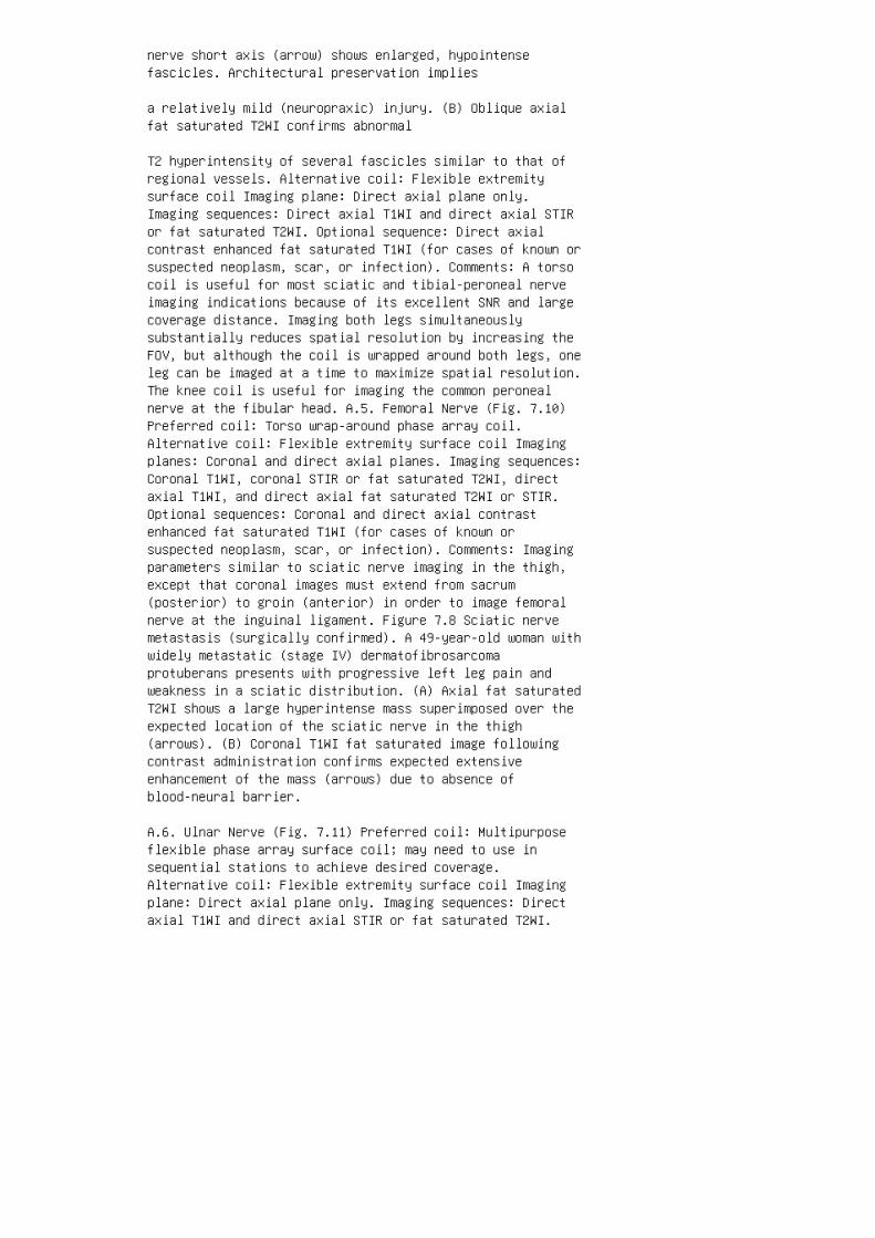
nerve short axis (arrow) shows enlarged, hypointensefascicles. Architectural preservation implies
a relatively mild (neuropraxic) injury. (B) Oblique axialfat saturated T2WI confirms abnormal
T2 hyperintensity of several fascicles similar to that ofregional vessels. Alternative coil: Flexible extremitysurface coil Imaging plane: Direct axial plane only.Imaging sequences: Direct axial T1WI and direct axial STIRor fat saturated T2WI. Optional sequence: Direct axialcontrast enhanced fat saturated T1WI (for cases of known orsuspected neoplasm, scar, or infection). Comments: A torsocoil is useful for most sciatic and tibial-peroneal nerveimaging indications because of its excellent SNR and largecoverage distance. Imaging both legs simultaneouslysubstantially reduces spatial resolution by increasing theFOV, but although the coil is wrapped around both legs, oneleg can be imaged at a time to maximize spatial resolution.The knee coil is useful for imaging the common peronealnerve at the fibular head. A.5. Femoral Nerve (Fig. 7.10)Preferred coil: Torso wrap-around phase array coil.Alternative coil: Flexible extremity surface coil Imagingplanes: Coronal and direct axial planes. Imaging sequences:Coronal T1WI, coronal STIR or fat saturated T2WI, directaxial T1WI, and direct axial fat saturated T2WI or STIR.Optional sequences: Coronal and direct axial contrastenhanced fat saturated T1WI (for cases of known orsuspected neoplasm, scar, or infection). Comments: Imagingparameters similar to sciatic nerve imaging in the thigh,except that coronal images must extend from sacrum(posterior) to groin (anterior) in order to image femoralnerve at the inguinal ligament. Figure 7.8 Sciatic nervemetastasis (surgically confirmed). A 49-year-old woman withwidely metastatic (stage IV) dermatofibrosarcomaprotuberans presents with progressive left leg pain andweakness in a sciatic distribution. (A) Axial fat saturatedT2WI shows a large hyperintense mass superimposed over theexpected location of the sciatic nerve in the thigh(arrows). (B) Coronal T1WI fat saturated image followingcontrast administration confirms expected extensiveenhancement of the mass (arrows) due to absence ofblood-neural barrier.
A.6. Ulnar Nerve (Fig. 7.11) Preferred coil: Multipurposeflexible phase array surface coil; may need to use insequential stations to achieve desired coverage.Alternative coil: Flexible extremity surface coil Imagingplane: Direct axial plane only. Imaging sequences: Directaxial T1WI and direct axial STIR or fat saturated T2WI.
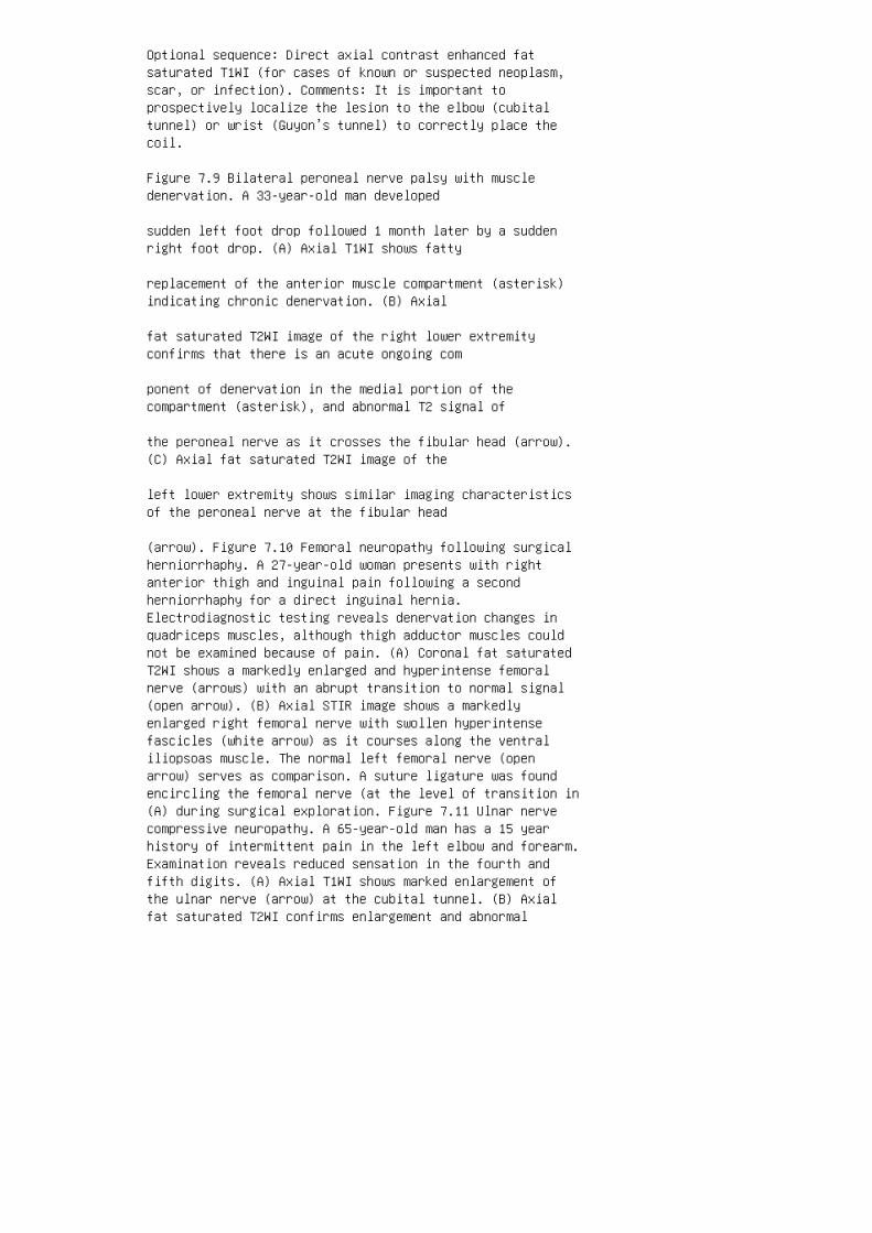
Optional sequence: Direct axial contrast enhanced fatsaturated T1WI (for cases of known or suspected neoplasm,scar, or infection). Comments: It is important toprospectively localize the lesion to the elbow (cubitaltunnel) or wrist (Guyon’s tunnel) to correctly place thecoil.
Figure 7.9 Bilateral peroneal nerve palsy with muscledenervation. A 33-year-old man developed
sudden left foot drop followed 1 month later by a suddenright foot drop. (A) Axial T1WI shows fatty
replacement of the anterior muscle compartment (asterisk)indicating chronic denervation. (B) Axial
fat saturated T2WI image of the right lower extremityconfirms that there is an acute ongoing com
ponent of denervation in the medial portion of thecompartment (asterisk), and abnormal T2 signal of
the peroneal nerve as it crosses the fibular head (arrow).(C) Axial fat saturated T2WI image of the
left lower extremity shows similar imaging characteristicsof the peroneal nerve at the fibular head
(arrow). Figure 7.10 Femoral neuropathy following surgicalherniorrhaphy. A 27-year-old woman presents with rightanterior thigh and inguinal pain following a secondherniorrhaphy for a direct inguinal hernia.Electrodiagnostic testing reveals denervation changes inquadriceps muscles, although thigh adductor muscles couldnot be examined because of pain. (A) Coronal fat saturatedT2WI shows a markedly enlarged and hyperintense femoralnerve (arrows) with an abrupt transition to normal signal(open arrow). (B) Axial STIR image shows a markedlyenlarged right femoral nerve with swollen hyperintensefascicles (white arrow) as it courses along the ventraliliopsoas muscle. The normal left femoral nerve (openarrow) serves as comparison. A suture ligature was foundencircling the femoral nerve (at the level of transition in(A) during surgical exploration. Figure 7.11 Ulnar nervecompressive neuropathy. A 65-year-old man has a 15 yearhistory of intermittent pain in the left elbow and forearm.Examination reveals reduced sensation in the fourth andfifth digits. (A) Axial T1WI shows marked enlargement ofthe ulnar nerve (arrow) at the cubital tunnel. (B) Axialfat saturated T2WI confirms enlargement and abnormal

increased fascicular signal (arrow).
A.7. Median Nerve Preferred coil: Multipurpose flexiblephase array surface coil in sequential stations to achievedesired coverage. Alternative coil: Flexible extremitysurface coil Imaging plane: Direct axial plane only.Imaging sequences: Direct axial T1WI and direct axial STIRor fat saturated T2WI. Optional sequence: Direct axialcontrast enhanced fat saturated T1WI (for cases of known orsuspected neoplasm, scar, or infection). Comments: Nerveentrapment by the flexor retinaculum at the wrist is themost common site, but it is important to extend imagingdistally into the palm to include distal nerve entrapments.
A.8. Radial Nerve (Fig. 7.12) Preferred coil: Multipurposeflexible phase array surface coil Alternative coil:Flexible extremity surface coil Imaging planes: Directaxial plane only. Imaging sequences: Direct axial T1WI anddirect axial STIR or fat saturated T2WI. Optional sequence:Direct axial contrast enhanced fat saturated T1WI (forcases of known or suspected neoplasm, scar, or infection).Comments: It is difficult to image the distal radial nerveafter its bifurcation, but relatively easy within the armalong the spiral groove.
1. Filler AG, Howe FA, Hayes CE, Kliot M, Winn HR, Bell BA,Griffiths JR, Tsuruda JS. Magnetic resonance neurography.Lancet 1993; 341:659–661.
Figure 7.12 Radial neuropathy. A 58-year-old woman withParkinson’s disease awoke in the
morning with a complete wrist drop and numbness of thefirst three fingers. (A) Axial T1WI
shows enlargement of the radial nerve along the spiralgroove (arrow). (B) Axial STIR image
confirms enlargement and abnormal nerve signal intensity(arrow). 2. Aagaard BD, Maravilla KR, Kliot M. Magneticresonance neurography: magnetic resonance imaging ofperipheral nerves. Neuroimaging Clin N Am 2001;11(8):131–146. 3. Filler AG, Kliot M, Howe FA, Hayes CE,Saunders DE, Goodkin R, Bell BA, Winn HR, Griffiths JR,Tsuruda JS. Application of magnetic resonance neurographyin the evaluation of patients with peripheral nervepathology. J Neurosurg 1996; 85:299–309. 4. Kuntz C, BlakeL, Britz G, Filler A, Hayes CE, Goodkin R, Tsuruda J,Maravilla K, Kliot M. Magnetic resonance neurography ofperipheral nerve lesions in the lower extremity.

Neurosurgery 1996; 39:750–756; discussion 756–757. 5. GrantG, Goodkin R, Kliot M. Evaluation and surgical managementof peripheral nerve problems. Neurosurgery 1999;44:825–840. 6. Maravilla K, Aagaard B, Kliot M. MRneurography: MR imaging of peripheral nerves. MRI Clin N Am1998; 6:179–194. 7. Maravilla K, Bowen B. Imaging of theperipheral nervous system: evaluation of peripheralneuropathy and plexopathy. Am J Neuroradiol 1998;19:1011–1023. 8. Hashemi R, Bradley W. MRI: the basics.Lippincott, Williams, and Wilkins, 1997:307. 9. Moore KR,Tsuruda JS, Dailey AT. The value of MR neurography forevaluating extraspinal neuropathic leg pain: a pictorialessay. AJNR Am J Neuroradiol 2001; 22:786–794. 10. West G,Haynor D, Goodkin R, Tsuruda J, Bronstein A, Kraft G,Winter T, Kliot M. Magnetic resonance imaging signalchanges in denervated muscles after peripheral nerveinjury. Neurosurgery 1994; 35:1077–1086.

Inflammatory Demyelinating Neuropathies
1. Guillain G, Barre JA, Strohl A. Sur un syndrome deradiculo-nevrite avec hyperalbuminose du liquidecephalorachidien sans reaction cellulaire: remarques surles caracteres cliniques et graphiques des reflexestendineux. Bull Soc Med Hop Paris 1916; 40:1462–1470.
2. Feasby TE, Gilbert JJ, Brown WF, Bolton CF, Hahn AF,Koopman WF, Zochodne DW. An acute axonal form ofGuillain–Barre´ polyneuropathy. Brain 1986; 109:1115–1126.
3. McKhann GM, Cornblath DR, Griffin JW, Ho TW, Li CY,Jiang Z, Wu HS, Zhaori G, Liu Y, Jou LP, Liu TC, Gao CY,Mao JY, Blaser MJ, Mishu B, Asbury AK. Acute motor axonalneuropathy: a frequent cause of acute Baccid paralysis inChina. Ann Neurol 1993; 33:333–342.
4. Fisher M. An unusual variant of acute idiopathicpolyneuritis (syndrome of ophthalmoplegia, ataxia andareflexia). N Engl J Med 1956; 255:57–65.
5. Schonberger LB, Hurwitz ES, Katona P, Holman RC, BregmanDJ. Guillain–Barre´ syndrome: its epidemiology andassociations with influenza vaccination. Ann Neurol 1981;9(suppl):31–38.
6. Mobley W, Wolinsky J. Scientific overview ofinflammatory demyelinating polyneuropathy and design of theNorth American Collaborative Study of plasma exchange inGuillain–Barre´ syndrome. In: Tindall R, ed. TherapeuticApheresis and Plasma Perfusion. New York: Alan R. Liss,1982:159–187.
7. Griffin JW, Li CY, Ho TW, Tian M, Gao CY, Xue P, MishuB, Cornblath DR, Macko C, McKhann GM, Asbury AK. Pathologyof the motor-sensory axonal Guillain–Barre´ syndrome. AnnNeurol 1996; 39(1):17–28.
8. McKhann GM, Cornblath DR, Ho T, Li CY, Bai AY, Wu HS,Yei QF, Zhang WC, Zhaori Z, Jiang Z et al. Clinical andelectrophysiological aspects of acute paralytic disease ofchildren and young adults in northern China. Lancet 1991;338(8767):593–597.
9. Ho TW, Mishu B, Li CY, Gao CY, Cornblath DR, Griffin JW,Asbury AK, Blaser MJ, McKhann GM. Guillain–Barre´ syndromein northern China: relationship to Campylobacter jejuniinfection and anti-glycolipid antibodies. Brain 1995;118:597–605.

10. Hafer-Macko C, Hsieh ST, Li CY, Ho TW, Sheikh K,Cornblath DR, McKhann GM, Asbury AK, Griffin JW. Acutemotor axonal neuropathy: an antibody-mediated attack onaxolemma. Ann Neurol 1996; 40(4):635–644. 11. Arnason B,Soliven B. Acute inflammatory demyelinating neuropathy. In:Dyck P, Thomas P, eds. Peripheral neuropathy. Philadelphia:WB Saunders, 1993:1437–1497. 12. Ho TW, Willison HJ,Nachamkin I, Li CY, Veitch J, Ung H, Wang GR, Liu RC,Cornblath DR, Asbury AK, Griffin JW, McKhann GM. Anti-GD1aantibody is associated with axonal but not demyelinatingforms of Guillain–Barre´ syndrome. Ann Neurol 1999;45(2):168–173. 13. Ropper A, Wijdicks E, Truax B.Guillain–Barre´ syndrome. Philadelphia: FA Davis, 1991. 14.Service FJ, O’Brien PC. The relation of glycaemia to therisk of development and progression of retinopathy in theDiabetic Control and Complications Trial. Diabetologia2001; 44:1215–1220. 15. Mori M, Kuwabara S, Fukutake T,Yuki N, Hattori T. Clinical features and prognosis ofMiller Fisher syndrome. Neurology 2001; 56(8):1104–1106.16. Emilia–Romagna Study Group on Clinical andEpidemiological Problems in Neurology. Guillain–Barre´syndrome variants in Emilia–Romagna, Italy, 1992–1993:incidence, clinical features, and prognosis. J NeurolNeurosurg Psychiatry 1998; 65(2):218–224. 17. Yuki N, SatoS, Tsuji S, Ohsawa T, Miyatake T. Frequent presence ofanti-GQ1b antibody in Fisher’s syndrome. Neurology 1993;43(2):414–417. 18. Chiba A, Kusunoki S, Obata H, MachinamiR, Kanazawa I. Serum anti-GQ 1b IgG antibody is associatedwith ophthalmoplegia in Miller Fisher syndrome andGuillain–Barre´ syndrome: Clinical and immunohistochemicalstudies. Neurology 1993; 43:1911–1917. 19. Odaka M, Yuki N,Hirata K. Anti-GQ1b IgG antibody syndrome: clinical andimmunological range. J Neurol Neurosurg Psychiatry 2001;70(1):50–55. 20. O’Hanlon GM, Plomp JJ, Chakrabarti M,Morrison I, Wagner ER, Goodyear CS, Yin X, Trapp BD, ConnerJ, Molenaar PC, Stewart S, Rowan, EG, Willison HJ.Anti-GQ1b ganglioside antibodies mediatecomplement-dependent destruction of the motor nerveterminal. Brain 2001; 124(Pt 5):893–906. 21. Buchwald B,Bufler J, Carpo M, Heidenreich F, Pitz R, Dudel J,Nobile-Orzaio E, Toyka KV. Combined preand postsynapticaction of IgG antibodies in Miller Fisher syndrome.Neurology 2001; 56(1):67–74. 22. Chiba A, Kusunoki S, ObataH, Machinami R, Kanazawa I. Ganglioside composition of thehuman cranial nerves, with special reference topathophysiology of Miller Fisher syndrome. Brain Res 1997;745(1–2):32–36. 23. Young RR, Asbury AK, Corbett JL, AdamsRD. Pure pan-dysautonomia with recovery. Description anddiscussion of diagnostic criteria. Brain 1975;

98(4):613–636. 24. Oh SJ, LaGanke C, Claussen GC. SensoryGuillain–Barre´ syndrome. Neurology 2001; 56(1):82–86. 25.Suarez GA, Fealey RD, Camilleri M, Low PA. Idiopathicautonomic neuropathy: clinical, neurophysiologic, andfollow-up studies on 27 patients. Neurology 1994;44(9):1675–1682. 26. Maile LA, Badley-Clarke J, ClemmonsDR. Structural analysis of the role of the beta 3 subunitof the alpha V beta 3 integrin in IGF-I signaling. J CellSci 2001; 114:1417–1425. 27. Asbury AK, Cornblath DR.Assessment of current diagnostic criteria forGuillain–Barre´ syndrome. Ann Neurol 1990;27(supplement):21–29. 28. Andersson T, Siden A. A clinicalstudy of the Guillain–Barre´ Syndrome. Acta Neurol Scand1982; 66(3):316–327. 29. Masucci EF, Kurtzke JF. Diagnosticcriteria for the Guillain–Barre´ syndrome. An analysis of50 cases. J Neurol Sci 1971; 13(4):483–501. 30. Bosch E,Smith B. Disorders of peripheral nerves. In: Bradley W,Daroff R, Fenichel F, Marsden C, eds. Neurology in ClinicalPractice. Boston: Butterworth–Heinemann, 2000: 2079–2086.31. Gordon PH, Wilbourn AJ. Early electrodiagnosticfindings in Guillain–Barre´ syndrome. Arch Neurol 2001;58(6):913–917. 32. Jacobs BC, Meulstee J, van Doorn PA, vander Meche FG. Electrodiagnostic findings related toanti-GM1 and anti-GQ1b antibodies in Guillain–Barre´syndrome. Muscle Nerve 1997; 20(4):446–452.
33. Albers JW, Kelly JJ. Acquired inflammatorydemyelinating polyneuropathies: Clinical andelectrodiagnostic features. Muscle Nerve 1989; 12:435–451.
34. Brown WF, Feasby TE, Hahn AF. Electrophysiologicalchanges in the acute “axonal” form of Guillain–Barre´syndrome. Muscle Nerve 1993; 16(2):200–205.
35. McKhann GM. Guillain–Barre´ syndrome: clinical andtherapeutic observations. Ann Neurol 1990; 27 Suppl:S13–16.
36. Hadden RD, Cornblath DR, Hughes RA, Zielasek J, HartungHP, Toyka KV, Swan AV. Electrophysiological classificationof Guillain–Barre´ syndrome: clinical associations andoutcome. Plasma Exchange/Sandoglobulin Guillain–Barre´Syndrome Trial Group. Ann Neurol 1998; 44(5):780–788.
37. Meulstee J, van der Meche FG. Electrodiagnosticcriteria for polyneuropathy and demyelination: applicationin 135 patients with Guillain–Barre´ syndrome. DutchGuillain–Barre´ Study Group. J Neurol Neurosurg Psychiatry1995; 59(5):482–486.
38. Okumura A, Ushida H, Maruyama K, Itomi K, Ishiguro Y,

Takahashi M, Osuga A, Negoro T, Watanabe K. Guillain–Barre´syndrome associated with central nervous system lesions.Arch Dis Child 2002; 86(4):304–306.
39. Ang CW, Jacobs BC, Brandenburg AH, Laman JD, van derMeche FG, Osterhaus AD, van Doorn PA. Cross-reactiveantibodies against GM2 and CMV-infected fibroblasts inGuillain–Barre´ syndrome. Neurology 2000; 54(7):1453–1458.
40. Rees JH, Soudain SE, Gregson NA, Hughes RAC.Campylobacter jejuni infection and Guillain–Barre´Syndrome. N Engl J Med 1995; 333:1374–1379.
41. Donofrio PD, Wilbourn AJ, Albers JW, Rogers L, SalangaV, Greenberg HS. Acute arsenic intoxication presenting asGuillain–Barre´-like syndrome. Muscle Nerve 1987;10(2):114–120.
42. Greenstein P. Tick paralysis. Med Clin North Am 2002;86(2):441–446.
43. Wollerton E. HIV drug stavudine and symptons mimickingGuillain–Barre´ syndrome. Can Med Assoc J 2002;166(8):1067.
44. Chang VH, Robinson LR. Serum positive botulism withneuropathic features. Arch Phys Med Rehabil 2000;81(1):122–126.
45. Gorson KC, Ropper AH. Nonpoliovirus poliomyelitissimulating Guillain–Barre´ syndrome. Arch Neurol 2001;58(9):1460–1464.
46. Piradov MA, Pirogov VN, Popova LM, Avdunina IA.Diphtheritic polyneuropathy: clinical analysis of severeforms. Arch Neurol 2001; 58(9):1438–1442.
47. The French Cooperative Group on Plasma Exchange inGuillain–Barre´ Syndrome. Appropriate number of plasmaexchanges in Guillain–Barre´ syndrome. Ann Neurol 1997;41(3):298–306.
48. French Cooperative Group on Plasma Exchange inGuillain–Barre´ syndrome. Efficiency of plasma exchange inGuillain–Barre´ syndrome: role of replacement fluids. AnnNeurol 1987; 22(6):753–761.
49. Fletcher DD, Lawn ND, Wolter TD, Wijdicks EF. Long-termoutcome in patients with Guillain–Barre´ syndrome requiringmechanical ventilation. Neurology 2000; 54(12):2311–2315.

50. Hughes RAC, Newsom-Davis JM, Perkin GD, Pierce JM.Controlled trial of prednisolone in acute polyneuropathy.Lancet 1978; 2:750–753.
51. Guillain–Barre´ Syndrome Steroid Trial Group.Double-blind trial of intravenous methylprednisolone inGuillain–Barre´ syndrome. Lancet 1993; 341(8845):586–590.
52. Guillain–Barre´ Syndrome Study Group. Plasmapheresisand acute Guillain–Barre´ syndrome. Neurology 1985;35:1096–1104.
53. van der Meche FG, Schmitz PI. A randomized trialcomparing intravenous immune globulin and plasma exchangein Guillain–Barre´ syndrome. Dutch Guillain–Barre´ StudyGroup. N Engl J Med 1992; 326(17):1123–1129.
54. Dalakas MC. Intravenous immunoglobulin in the treatmentof autoimmune neuromuscular diseases: present status andpractical therapeutic guidelines. Muscle Nerve 1999;22(11): 1479–1497.
55. Gabarra-Niecko V, Keely PJ, Schaller MD.Characterization of an Activated Mutant of Focal AdhesionKinase: SuperFAK. Biochem J 2002; 365(Pt 3):591–603. 56.Tsiouris J, Tsiouris N. Hemiplegia as a complication oftreatment of childhood immune thrombocytopenic purpura withintravenously administered immunoglobulin. J Pediatr 1998;133(5):717. 57. Steg RE, Lefkowitz DM. Cerebral infarctionfollowing intravenous immunoglobulin therapy for myastheniagravis. Neurology 1994; 44(6):1180–1181. 58. Harkness K,Howell SJ, Davies-Jones GA. Encephalopathy associated withintravenous immunoglobulin treatment for Guillain–Barre´syndrome. J Neurol Neurosurg Psychiatry 1996; 60(5):586.59. Constantinescu CS, Chang AP, McCluskey LF. Recurrentmigraine and intravenous immune globulin therapy. N Engl JMed 1993; 329(8):583–584. 60. Finkel AG, Howard JF Jr. MannJD. Successful treatment of headache related to intravenousimmunoglobulin with antimigraine medications. Headache1998; 38(4):317–321. 61. Green DM, Ropper AH. MildGuillain–Barre´ syndrome. Arch Neurol 2001;58(7):1098–1101. 62. Asbury AK, Arnason BG, Adams RD. Theinflammatory lesion in idiopathic polyneuritis: its role inpathogenesis. Medicine 1969; 48:173–215. 63. Prineas JW.Acute idiopathic polyneuritis. An electron microscopestudy. Lab Invest 1972; 26(2):133–147. 64. Hafer-Macko CE,Sheikh KA, Li CY, Ho TW, Cornblath DR, McKhann GM, AsburyAK, Griffin JW. Immune attack on the Schwann cell surfacein acute inflammatory demyelinating polyneuropathy. Ann
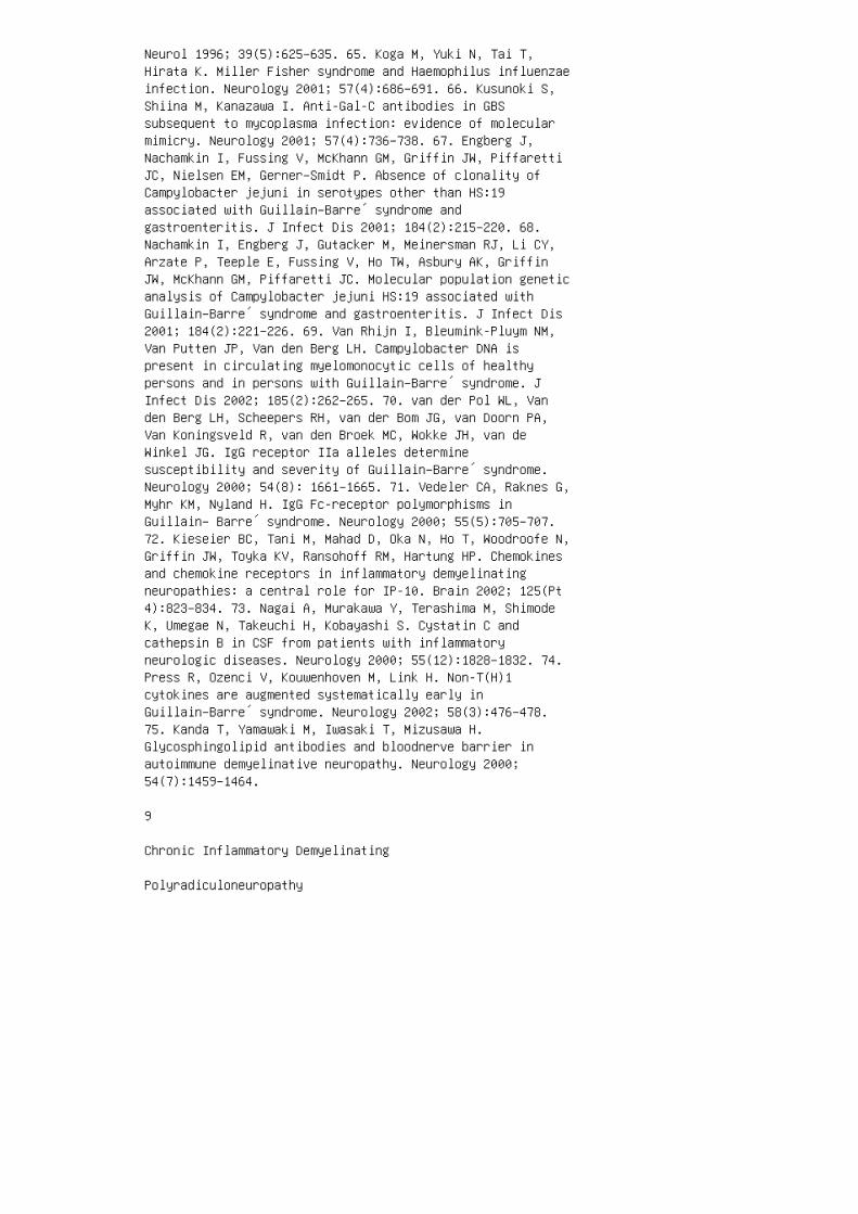
Neurol 1996; 39(5):625–635. 65. Koga M, Yuki N, Tai T,Hirata K. Miller Fisher syndrome and Haemophilus influenzaeinfection. Neurology 2001; 57(4):686–691. 66. Kusunoki S,Shiina M, Kanazawa I. Anti-Gal-C antibodies in GBSsubsequent to mycoplasma infection: evidence of molecularmimicry. Neurology 2001; 57(4):736–738. 67. Engberg J,Nachamkin I, Fussing V, McKhann GM, Griffin JW, PiffarettiJC, Nielsen EM, Gerner–Smidt P. Absence of clonality ofCampylobacter jejuni in serotypes other than HS:19associated with Guillain–Barre´ syndrome andgastroenteritis. J Infect Dis 2001; 184(2):215–220. 68.Nachamkin I, Engberg J, Gutacker M, Meinersman RJ, Li CY,Arzate P, Teeple E, Fussing V, Ho TW, Asbury AK, GriffinJW, McKhann GM, Piffaretti JC. Molecular population geneticanalysis of Campylobacter jejuni HS:19 associated withGuillain–Barre´ syndrome and gastroenteritis. J Infect Dis2001; 184(2):221–226. 69. Van Rhijn I, Bleumink-Pluym NM,Van Putten JP, Van den Berg LH. Campylobacter DNA ispresent in circulating myelomonocytic cells of healthypersons and in persons with Guillain–Barre´ syndrome. JInfect Dis 2002; 185(2):262–265. 70. van der Pol WL, Vanden Berg LH, Scheepers RH, van der Bom JG, van Doorn PA,Van Koningsveld R, van den Broek MC, Wokke JH, van deWinkel JG. IgG receptor IIa alleles determinesusceptibility and severity of Guillain–Barre´ syndrome.Neurology 2000; 54(8): 1661–1665. 71. Vedeler CA, Raknes G,Myhr KM, Nyland H. IgG Fc-receptor polymorphisms inGuillain– Barre´ syndrome. Neurology 2000; 55(5):705–707.72. Kieseier BC, Tani M, Mahad D, Oka N, Ho T, Woodroofe N,Griffin JW, Toyka KV, Ransohoff RM, Hartung HP. Chemokinesand chemokine receptors in inflammatory demyelinatingneuropathies: a central role for IP-10. Brain 2002; 125(Pt4):823–834. 73. Nagai A, Murakawa Y, Terashima M, ShimodeK, Umegae N, Takeuchi H, Kobayashi S. Cystatin C andcathepsin B in CSF from patients with inflammatoryneurologic diseases. Neurology 2000; 55(12):1828–1832. 74.Press R, Ozenci V, Kouwenhoven M, Link H. Non-T(H)1cytokines are augmented systematically early inGuillain–Barre´ syndrome. Neurology 2002; 58(3):476–478.75. Kanda T, Yamawaki M, Iwasaki T, Mizusawa H.Glycosphingolipid antibodies and bloodnerve barrier inautoimmune demyelinative neuropathy. Neurology 2000;54(7):1459–1464.
9
Chronic Inflammatory Demyelinating
Polyradiculoneuropathy
Zachary Simmons
Penn State College of Medicine, Hershey, Pennsylvania, USA
Abstract 128
1. Introduction 128
2. Idiopathic Chronic Inflammatory DemyelinatingPolyradiculoneuropathy in Adults 129 2.1. Clinical Features129 2.2. Laboratory Evaluation 130 2.2.1. Serum LaboratoryStudies 130 2.2.2. Electrodiagnostic Studies 130 2.2.3.Cerebrospinal Fluid 133 2.2.4. Nerve Biopsy 133 2.2.5.Radiologic Studies 134 2.3. Differential Diagnosis 134 2.4.Management 135 2.4.1. Overview of Treatment 135 2.4.2.Corticosteroids 135 2.4.3. Azathioprine 136 2.4.4. PlasmaExchange 136 2.4.5. Intravenous Immunoglobulin 136 2.4.6.Pulse Cyclophosphamide 136 2.4.7. Cyclosporine A 136 2.4.8.Interferon 137 2.4.9. Tacrolimus 137 2.4.10. AutologousStem Cell Transplantation 137 2.4.11. Immunoadsorption 1372.5. An Approach to Treatment of the Patient with CIDP 1372.6. Prognosis 139 2.7. Pathogenesis 139
immunotherapy. CIDP can be associated with a monoclonalgammopathy of undetermined
significance (MGUS). Under these circumstances, theresponse to treatment varies with
the associated monoclonal subtype.
2. IDIOPATHIC CHRONIC INFLAMMATORY DEMYELINATINGPOLYRADICULONEUROPATHY IN ADULTS
CIDP is a chronic sensorimotor disorder most commonlyaffecting sensory and motor
functions. It may follow monophasic, relapsing, orprogressive courses, which distinguish
it from acute inflammatory demyelinating polyradiculopathy(AIDP) or the Guillain–
Barre´ Syndrome (GBS). A variety of names have been used,including chronic relapsing
polyneuropathy, chronic relapsing Guillain–Barre´polyneuritis, relapsing corticosteroid
dependent polyneuritis, steroid-responsive recurrent

polyneuropathy, chronic relapsing
polyneuritis, and chronic inflammatorypolyradiculoneuropathy (1–6). The disorder
was formally defined in 1975, and formal research criteriaestablished in 1991 (7). A
number of patient series have described the spectrum ofclinical and electrodiagnostic
findings, course, and prognosis (8–13).
2.1. Clinical Features
CIDP may affect individuals at any age, including into theninth decade (14). The mean
age in adults varies from 31.6 to 51 years in differentpatient series. It is slightly more
common in men than in women. Antecedent events occur in�30%of patients, most com
monly upper respiratory infections, gastroenteritis, otherinfections, vaccinations, surgery,
and trauma (9,11–13,15). This is in contrast to a muchhigher incidence (�60–70%) of
antecedent events in AIDP/GBS (16,17). In general, CIDPdevelops rather slowly, and the mean time from symptomonset to
initial presentation from different series is 11.7 months(13) to 24 months (15). Research
clinical criteria for CIDP (7) and many clinical seriesrequire a minimum period of
2 months of symptom progression, but others accept ashorter period of progression
(8,12). However, the range is broad, and some patientspresent acutely, resembling
AIDP/GBS, before going on to develop a relapsing coursetypical of CIDP. Most
patient series include up to 20% of patients reaching a
nadir of symptoms within 4
weeks, but they are distinguished from AIDP/GBS bydeveloping a relapsing or a
progressive course that responds to immunotherapy(9,11,13,18). However, a subacute
time course (subacute idiopathic demyelinatingpolyradiculoneuropathy—SIDP) has
been recognized and proposed as a separate clinical entityfor patients who have a mono
phasic course and progression for 4–8 weeks (19). Asawareness of CIDP grows and the
diagnosis is made more rapidly, it is increasingly likelythat decisions regarding diagnosis
and treatment will be made prior to the development of a 2month period of symptoms. CIDP is a strikinglyheterogeneous disease (20). Most patients present with both
sensory and motor symptoms, although small percentagespresent with predominantly
motor (10–22%) or sensory involvement (6%). Weakness may besevere, and in our
series we found that 13 of 77 patients (17%) were severelydisabled (unable to carry
our activities of daily living) at our evaluation. Numbnessand paresthesias, most com
monly in feet and hands, occurred in 64–82% at onset. Painwas a less common presenting
feature (14–20%) (13). Cranial nerve symptoms, which occurin a minority, include
dysarthria, dysphagia, facial numbness, facial weakness,blurred or double vision, and ptosis (8,11–13). Symptoms ofdysautonomia are uncommon, although urinary dysfunction andimpotence have been reported (8,21). Examination findingsinclude both proximal and distal weakness, usually in asymmetric manner, with distal weakness more common andsevere than proximal weakness. Tendon reflexes aredecreased or absent in most patients, with areflexia in 70%

and absent Achilles reflexes in most patients. Sensorydeficits are present in .80% of patients, with vibratoryimpairment more common than deficits to pinprick. Cranialnerve dysfunction can be found in up to 16%, includingophthalmoplegia, facial weakness, and bulbar weakness.Papilledema may also occur (8,11–13). Among CIDP variantpatients who present clinically with only a sensorysyndrome and normal strength, electrodiagnostic evidencefor demyelination in motor nerves may be found in some(22–26). Patients with acquired demyelinating neuropathieslocalized to the upper limbs have been described andincluded as a variant of CIDP (27,28). These patientslikely represent examples of the Lewis–Sumner syndrome ormultifocal motor neuropathy, and are discussed elsewhere.Nerve root hypertrophy can result in cervical myelopathy(29) or in findings of lumbar radiculopathy (30–32)superimposed upon CIDP. 2.2. Laboratory EvaluationEstablishing a set of diagnostic criteria for CIDP has beenchallenging. A set of research criteria have been proposedthat categorize patients as definite, probable, or possibleCIDP-I based upon clinical, electrodiagnostic, pathologic,and cerebrospinal fluid (CSF) findings (Table 9.1) (7).Such criteria are very useful but strict, and many patientswho do not meet criteria for definite CIDP are (and shouldbe) diagnosed with and treated for CIDP. If CIDP issuspected from the history and examination findings, thefollowing laboratory evaluation should be undertaken.2.2.1. Serum Laboratory Studies Few routine metabolicstudies are informative in diagnosing CIDP, and largelyrepresent an exclusion of other causes or concurrentmedical conditions. Erythrocyte sedimentation rate iscommonly normal in CIDP. Anti-DNA titer and other tests forcollagen vascular disease are also usually normal. A 2 hglucose tolerance test may be helpful if a diabeticneuropathy is included as a diagnostic possibility. HIVtiters should be drawn in those patients suspected to be atrisk for HIV infection because CIDP can appear in thissetting. Tests for other causes of polyneuropathy,including thyroid function tests, vitamin B12 level, VDRL,and (in some geographic regions) Lyme titer, may be helpfulif nerve conduction findings are equivocal. Serum and urineimmunofixation or immunoelectrophoresis should be performedlooking for a monoclonal gammopathy in the serum andBence–Jones proteins in the urine. If the IgM level iselevated, antibody titers to myelin-associated glycoprotein(anti-MAG) should be assessed (discussed subsequently). Ifa monoclonal antibody is detected a skeletal X-ray surveyshould be performed and an evaluation by ahematology/oncology specialist is warranted. 2.2.2.Electrodiagnostic Studies Motor nerve conduction

abnormalities are similar to those seen in AIDP/GBS, andinclude multifocal demyelination characterized by prolongeddistal latencies, slowed conduction velocities, prolongedF-wave latencies, and evidence of partial conduction blockor abnormal temporal dispersion (8–13,33) (Fig. 9.1).Several sets of formal
Table 9.1 Research Criteria for Diagnosis of CIDP
CIDP is a diagnosis of pattern recognition, based onclinical symptoms and signs, electrodiagnostic
studies, cerebrospinal fluid examination, laboratory testsappropriate to the specific clinical
situation, and, on occasions, results from nerve biopsy.
Four features are used as the basis of diagnosis: clinical,electrodiagnostic, pathologic, and
cerebrospinal fluid studies. These are further divided into(A) mandatory, (B) supportive, and,
where appropriate (C) exclusion. Mandatory features arethose required for diagnosis and should
be present in all Definite cases. Supportive features arehelpful in clinical diagnosis but by
themselves do not make a diagnosis, and are not part of thediagnostic categories. Exclusion
features strongly suggest alternative diagnoses.
I. Clinical A. Mandatory 1. Progressive or relapsing motorand sensory, rarely, only motor or sensory, dysfunction ofmore than one limb of a peripheral nerve nature, developingover at least 2 months. 2. Hypoor areflexia. This usuallyinvolves all four limbs. B. Supportive 1. Large-fibersensory loss predominates over small-fiber sensory loss. C.Exclusion 1. Mutilation of hands or feet, retinitispigmentosa, ichthyosis, appropriate history of drug ortoxic exposure known to cause a similar peripheralneuropathy, or family history of an inherited peripheralneuropathy. 2. Sensory level. 3. Unequivocal sphincterdisturbance.
II. Electrodiagnostic studies A. Mandatory Nerve conductionstudies including studies of proximal nerve segments inwhich the predominant process is demyelination. Must have

three of following four: 1. Reduction in conductionvelocity (CV) in two or more motor nerves: a. ,80% of lowerlimit of normal (LLN) if amplitude .80% of LLN. b. ,70% ofLLN if amplitude ,80% of LLN. 2. Partial conduction block aor abnormal temporal dispersion b in one or more motornerves: either peroneal nerve between ankle and belowfibular head, median nerve between wrist and elbow or ulnarnerve between wrist and below elbow. 3. Prolonged distallatencies in two or more nerves: a. .125% of upper limit ofnormal (ULN) if amplitude .80% of LLN. b. .150% of ULN ifamplitude ,80% of LLN. 4. Absent F-waves or prolongedminimum F-wave latencies (10–15 trials) in two or moremotor nerves: a. .120% of ULN if amplitude .80% of LLN. b..150% of ULN if amplitude ,80% of LLN. B. Supportive 1.Reduction in sensory CV ,80% of LLN. 2. Absent H reflexes.
III. CSF studies A. Mandatory 1. Cell count ,10/mm 3 ifHIV-seronegative or ,50/mm 3 if HIV-seropositive. 2.Negative VDRL. B. Supportive 1. Elevated protein.(continued ) electrodiagnostic criteria have been proposedbased on these abnormalities to distinguish between primarydemyelinating and primary axonal neuropathies (7,33).However, these abnormalities may not be found in all motornerves, likely reflecting different degrees of nerveinvolvement and different distributions of involvement(e.g., changes in nerve Table 9.1 Continued IV. Pathologicfeatures A. Mandatory Nerve biopsy showing unequivocalevidence of demyelination and remyelination. c B.Supportive 1. Subperineurial or endoneurial edema. 2.Mononuclear cell infiltration. 3. “Onion-bulb” formation.4. Prominent variation in the degree of demyelinationbetween fascicles. C. Exclusion Vasculitis,neurofilamentous swollen axons, amyloid deposits, orintracytoplasmic inclusions in Schwann cells or macrophagesindicating adrenoleukodystrophy, metachromaticleukodystrophy, globoid cell leukodystrophy, or otherevidence of specific pathology. Diagnostic categories forresearch purposes DEFINITE: Clinical A and C,Electrodiagnostic A, CSF A, and Pathology A and C.PROBABLE: Clinical A and C, Electrodiagnostic A, and CSF A.POSSIBLE: Clinical A and C and Electrodiagnostic A.Laboratory studies Depending on the results of thelaboratory tests, those patients meeting the criteria abovewill be classified into the groups listed below. Thefollowing studies are suggested: CBC, ESR, SMA6/ 12, CK,ANA, thyroid functions, serum and urine immunoglobulinstudies (to include either immunofixation electrophoresisor immunoelectrophoresis), and HIV and hepatitis serology.The list of laboratory studies is not comprehensive. Forinstance, in certain clinical circumstances other studies

may be indicated, such as phytanic acid, long-chain fattyacids, porphyrins, urine heavy metals, a-lipoprotein,b-lipoprotein, glucose tolerance test, imaging studies ofthe central nervous system, and lymph node or bone marrowbiopsy. A. Idiopathic CIDP: no concurrent disease. B.Concurrent disease with CIDP (depending on laboratorystudies or other clinical features): 1. Systemic lupuserythematosus. 2. HIV infection. 3. Monoclonal or biclonalgammopathy (macroglobulinemia, POEMS syndrome,osteosclerotic myeloma). 4. Castleman disease. 5.Monoclonal gammopathies of undetermined significance. 6.Diabetes. 7. Central nervous system demyelinating disease.a Criteria suggestive of partial conduction block:.20% dropin area or amplitude with,15% change in duration betweenproximal and distal sites. b Criteria for abnormal temporaldispersion and possible conduction block: .20% drop in areaor amplitude between proximal and distal sites with.15%change in duration between proximal and distal sites. Thesecriteria are only suggestive of partial conduction block asthey are derived from studies of normal individuals.Additional studies, such as stimulation across shortsegments or recording of individual motor unit potentials,are required for confirmation. c Demyelination by eitherelectron microscopy (.5 fibers) or teased fiber studies(.12% of 50 teased fibers, minimum of four internodes each,demonstrating demyelination/remyelination). Source: Adaptedfrom Ad hoc subcommittee of the American Academy ofNeurology AIDS task force (7).
conduction along segments not routinely tested). A reviewof 70 CIDP patients found that
only 48–64% fulfilled any one of the sets ofelectrodiagnostic criteria for CIDP (34).
Attempts to make the criteria less stringent to includeCIDP of lesser severity result in
the inclusion of patients with Charcot–Marie–Tooth (CMT)type I neuropathies (35).
These criteria, therefore, best serve as guidelines andshould be considered in conjunction
with clinical, pathological, and CSF studies.Electrodiagnostic studies are helpful in distinguishingbetween CIDP and CMT type I,
the most common form of hereditary neuropathy (36). Motornerve conduction in CIDP is

characterized by abnormal temporal dispersion andconduction block on proximal stimu
lation, whereas conduction in CMT type I is characterizedby uniform slowing (Fig. 9.2). Sensory responses may varyfrom normal to absent (9–11,13). A unique pattern of
sensory nerve findings observed more commonly in CIDP thanin distal-predominant neuro
pathies is the combination of an abnormal median responseand a normal sural response (37). Needle EMG findingsreflect the degree of secondary axonal degeneration. Abnorm
alities from acute and chronic partial denervation includefibrillation potentials and positive
sharp waves. Recruitment will be decreased and motor unitpotentials will show varying
degrees of increased amplitude, long-duration, andincreased polyphasia (12,33,38,39).
2.2.3. Cerebrospinal Fluid
CSF protein is frequently elevated without a pleocytosis(albuminocytologic dissociation),
with mean values �140 mg/dL (range 20–1200 mg/dL)(8,11–13). Cell counts are
rarely greater than five.
2.2.4. Nerve Biopsy
The findings on nerve biopsy are not sensitive or specificfor CIDP, and a nerve biopsy is
not necessary to make the diagnosis in most patients withsuspected CIDP. When the
Figure 9.1 Ulnar motor nerve conduction study performed ona patient with CIDP, recording with
surface electrodes over the abductor digiti quinti muscleand stimulating at the wrist (A), below
elbow (B), and above elbow (C). There is partial conductionblock and abnormal temporal dispersion

with proximal stimulation. [Adapted from Simmons and Albers(209).] diagnosis is unclear, a biopsy may be helpful ifthe differential diagnosis includes vasculitis,amyloidosis, sarcoidosis, or neoplastic infiltration of thenerve. In CIDP, the degree of demyelination varies widelybetween patients, being predominant in only about half ofpatients (8,9,12,40). Predominant axonal degeneration andmixed axonal and demyelinating patterns also occur (12). Acomparison of sural nerve biopsies from patients with CIDPor chronic idiopathic axonal neuropathies found nodifference in demyelinating features between the two (41).Endoneurial perivascular mononuclear inflammatoryinfiltrates and endoneurial edema are not specific for CIDP(41,42). 2.2.5. Radiologic Studies A skeletal X-ray surveyof bone should be performed in patients with a monoclonalgammopathy. Occasional patients have a combined central andperipheral demyelinating disorder manifest clinically by amyelopathy superimposed upon clinical and electrodiagnosticevidence of CIDP, and a magnetic resonance image (MRI) ofthe brain and spinal cord may be useful. Cervical andlumbosacral MRI scans in CIDP patients may show hypertrophyof roots caused by demyelination and remyelination withonion-bulb formation (29–32,43–45). These findings do notappear to correlate with disease severity (46). MRIscanning of the cord should be performed when a combinedcentral and peripheral disturbance of myelin is suspectedwith evidence of a myelopathy. 2.3. Differential DiagnosisThe differential diagnosis includes hereditary and acquiredneuropathies, disorders of neuromuscular transmission, andprimary diseases of muscle. Patients with an insidiousonset and very slow progression may resemble CMT, but willlack characteristic orthopedic Figure 9.2 Ulnar motor nerveconduction study performed on a patient with Type ICharcot– Marie–Tooth disease, recording with surfaceelectrodes over the abductor digiti quinti muscle andstimulating at the wrist (A), below elbow (B), and aboveelbow (C). The distal latency is prolonged at 6.8 ms(normal�3.5 ms). The conduction velocities in the forearmand across the elbow are 30 m/s and 38 m/s, respectively(normal �49 m/s). Slowing is uniform, and partialconduction block and temporal dispersion are absent, incontrast to CIDP. [Adapted from Simmons and Albers (209).]
abnormalities (high arched feet and hammer toes), and willdemonstrate distinct electro
diagnostic findings (see section on “LaboratoryEvaluation”). Patients who have an acute

onset may initially resemble AIDP/GBS, but will progress toa chronic or relapsing
course. The Lewis–Sumner syndrome and multifocal motorneuropathy are similar to
each other with asymmetric limb involvement, which isdistinct from CIDP. Axonal
neuropathies may resemble CIDP, although clinically theyhave a distal predominance
of sensory loss and weakness, and electrodiagnosticallythey lack conduction block,
temporal dispersion, and severely slow conductionvelocities. Axonal neuropathies are
frequently painful, whereas CIDP is not. The proximalweakness in CIDP may resemble
disorders of neuromuscular junction transmission, butmyasthenia gravis and Lambert–
Eaton myasthenic syndrome frequently include bulbar andocular involvement, and lack
features of acquired demyelination on nerve conductiontesting. Primary disorders of
muscle such as inflammatory myopathies, toxic myopathies,and muscular dystrophies
include proximal weakness but lack the sensory loss andnerve conduction abnormalities
characteristic of CIDP. CIDP may coexist with otherdiseases including systemic lupus erythematosus, HIV
infection, and central nervous system demyelination(47–57). CIDP has rarely been
reported in association with malignancies, includingpancreatic adenocarcinoma, rectosig
moid adenocarcinoma, cholangiocarcinoma, hepatocellularcarcinoma, seminoma, and
malignant melanoma (58–63), but it is not necessary toevaluate every CIDP patient

for all these disorders, but only when clinicallyindicated. CIDP with atypical clinical
findings or a poor response to standard therapies shouldlead to a broader search for
associated diseases. CIDP has been described in associationwith diabetic neuropathy. It can be challen
ging to diagnose and manage CIDP in diabetic patients, butimportant because it is trea
table. Clinical clues to a superimposed primaryinflammatory neuropathy are a rapid
change in clinical symptoms that include proximal weakness.Electrodiagnostic findings
will include features of primary demyelination (64–66).Nerve biopsy does not distinguish
between diabetic neuropathy and CIDP. The inflammatoryneuropathy does respond to
immunotherapy, and a treatment trial should be consideredfor diabetic patients who
meet two of four electrophysiologic criteria for CIDP (66).Overall, patients with CIDP
and diabetes have more axonal loss and respond more poorlyto treatment than those
with idiopathic CIDP (67).
2.4. Management
2.4.1. Overview of Treatment
A variety of immunosuppressive agents have beendemonstrated effective in a large number
of controlled and uncontrolled studies and retrospectivereviews (3–6,8–13). They include
corticosteroids, plasma exchange, intravenousimmunoglobulin (IVIg), azathioprine,
and cyclophosphamide. In most series, the overall response

rate has been good, with
65–95% of patients responding to these therapies, eitheralone or in combination (11–
13,15). However, the response to any single therapy is notpredictable, and failure to
respond to one modality should lead to treatment withanother (8,11–13,15). A summary
of the major treatment modalities, followed by arecommended approach is provided.
2.4.2. Corticosteroids
Corticosteroids represent the earliest effective treatmentfor CIDP (3–6). A prospective,
randomized, placebo-controlled trial of prednisonedemonstrated a small but significant improvement inpatients with CIDP (68). The effects of prednisone are notimmediate, and the mean time for initial improvement is �2months, and the mean time to reach a clinical plateau is �6months (12). 2.4.3. Azathioprine The combination ofazathioprine and prednisone has been assessed in acontrolled trial and was found to be no better thanprednisone alone, at least over a 9 month period (69).However, clinicians commonly add azathioprine as a“steroid-sparing” agent when treating patients with CIDP,particularly those with relative contraindications tosteroids, such as diabetics or those with severeosteoporosis. 2.4.4. Plasma Exchange Randomizedsham-controlled studies have confirmed the efficacy ofplasma exchange in some patients with CIDP (70,71)supplemented by uncontrolled studies (72–79), but not allpatients respond. The time to improvement is generallyshort, 2 days to 3 weeks after beginning plasma exchange(79). Plasma exchange is often given 2–3 times per weekuntil clear improvement is obtained, then tapered to alower frequency. The duration of efficacy varies. Somepatients have a monophasic course and do not relapse afterplasma exchange treatment, but most relapse after 7–14 days(70,71). Thus, concomitant immunosuppressive therapy isfrequently required. 2.4.5. Intravenous ImmunoglobulinThere are a number of reports and studies which areretrospective, uncontrolled, or nonrandomized, and whichappear to demonstrate the efficacy of IVIg in some, but notall, patients with CIDP. Randomized controlled studies,supplemented by uncontrolled studies, confirm that IVIg is

efficacious, being equal to plasma exchange (80–82) orcorticosteroids (83). Traditional dosing is 1–2 g/kgdivided over 3–5 consecutive days (84–90). The responserate is .60% (81,89), and improvement is rapid, but oftentransient. The time to onset of improvement is 3–8 days,with maximal improvement within 2–16 weeks (mean 5 weeks)(86,89). Although some patients have a monophasic coursecharacterized by a sustained response, the duration ofefficacy ranges from 3 to 22 weeks (mean 6.4 weeks)(81,87). IVIg can be effective as long-term treatment forrelapsing CIDP, with responds to intermittent infusions ofIVIg for 4 years or longer (81,89,91). For patients whorequire regular IVIg infusions, dosages, and intervals varygreatly, ranging from 0.025 g/kg every 10 days to 0.4 g/kgonce every 2–4 days (80). For patients who do not improvewith IVIg, improvement with prednisone, azathioprine,plasma exchange, or cyclosporine A has been described(81,89). 2.4.6. Pulse Cyclophosphamide An uncontrolledtrial of pulse cyclophosphamide (1 g/m 2 ) at monthlyintervals for 6 months led to improved strength intwo-thirds of 15 patients (92). Most received concomitantprednisone, with no demonstrable added effect from theprednisone. 2.4.7. Cyclosporine A Cyclosporine A is usuallyreserved patients who are refractory to standard treatment(corticosteroids, azathioprine, and plasma exchange).Uncontrolled trials of cyclosporine A
in patients with idiopathic CIDP and CIDP associated with aMGUS yielded mixed results,
with some responding excellently, whereas others have beenable to reduce the amount of
other immunosuppressive agents (93–95).
2.4.8. Interferon
Case reports suggest that interferona 2a might be effectivefor some patients with CIDP
poorly responsive or unresponsive to standard therapy(96–101). Strikingly, interferona
2a has also been associated with the development of CIDP inpatients with hepatitis C, and
the role of interferona in the treatment of patients withCIDP remains uncertain
(102,103).

2.4.9. Tacrolimus
Benefits have been reported in one patient with CIDP (104).
2.4.10. Autologous Stem Cell Transplantation
Autologous stem cell transplantation resulted in markedclinical and electrodiagnostic
improvement in one patient with chronic CIDP refractory tostandard therapy, and
allowed for a daily prednisone dose of 5 mg/day formaintenance (105).
2.4.11. Immunoadsorption
A single patient responded less well to immunoadsorptionthan to plasma exchange in a
crossover study (106).
2.5. An Approach to Treatment of the Patient with CIDP
The goal should be to induce and maintain remission withthe minimum dosage of immu
nosuppressive medication or procedures to reduce medicationside effects. Periodic
attempts should be made to taper the patient offimmunosuppressive medication. It may
be challenging when patients do not respond to initialmedications among immunosup
pressive medications. A treatment algorithm is provided inFig. 9.3. Patients with mild sensory deficits and noweakness can be observed without treat
ment for days to weeks, because spontaneous improvement mayoccur. If a patient does
not improve or worsens, or has weakness, treatment shouldbe initiated. IVIg, plasma
exchange, and prednisone are beneficial in most CIDPpatients, and the choice of initial

therapy should be based on other factors such as speed ofonset action, side effect
profile, concurrent diseases, availability, convenience,and cost (80). IVIg or plasma
exchange is reasonable as the initial treatment becausesome patients experience sustained
improvement with these treatments, and the adverse effectsof long-term corticosteroid
treatment can be avoided in these responders. Thewidespread availability of IVIg
has made this the initial treatment of choice for mostneurologists. However, both are
very expensive. The standard dose of IVIg is 2 g/kg infusedover 2–3 consecutive days. The onset of
action is rapid, and the side effect profile in mostpatients is rather benign. A single dose of
IVIg may bring about a sustained remission. If relapsesoccur, IVIg at 1 g/kg can be given
again, and at monthly intervals. Maintenance doses shouldbe tapered, and efficacy is often
maintained for years. An alternative if several doses ofIVIg are not fully effective is the
addition of prednisone 60–80 mg/day for 2–3 months. Theprednisone dose is then
gradually tapered over another 6–9 months, aiming for analternate-day regimen at the lowest dose that maintains thepatient in remission. Supplemental calcium and vitamin D,and monitoring of glucose and electrolytes are prudent, andfor those maintained on longterm prednisone, bone densitystudies should be included and aggressive treatment ofosteoporosis may be necessary. Azathioprine 3, 5 mg/kg(with dose escalation starting at 50 mg), can be added inan attempt to minimize the prednisone dose. Long-term IVIgis appropriate for patients in whom prednisone iscontraindicated, such as those with brittle diabetesmellitus, or for those who do not tolerate prednisone. Insome patients, immunosuppressive therapy can be eventuallydiscontinued, but most patients require maintenance

therapy. Plasma exchange is usually given as one plasmavolume per exchange, five times over 2 weeks. As with IVIg,the onset of action generally is rapid. If the effect issustained, no further treatment is required. If relapseoccurs, plasma exchange may be repeated, but prednisoneshould be started according to the earlier guidelines. Forpatients who cannot tolerate prednisone, plasma exchangecan be used alone and tapered to maintain remission.Prednisone can be given without IVIg or plasma exchange forpatients with mild to moderate chronic deficits, or forthose patients for whom IVIg and plasma exchange are notavailable. Although the onset of action is typically slowerthan that with IVIg or plasma exchange, it is easy toadminister and inexpensive. Azathioprine can be added as asteroid-sparing agent. Cyclophosphamide should beconsidered for patients who fail to respond to thesetherapies, or who do not tolerate them. Cyclophosphamidedosages are 1–2 mg/kg orally on a daily basis or 1 g/m 2 ofbody surface area by monthly intravenous infusion.Monitoring of hematologic and renal parameters and urineare essential to manage transient immunosuppresssion andminimize long-term risk for developing malignancies. Figure9.3 An approach to treatment of patients with idiopathicCIDP. [Adapted from Simmons and Albers (209).]
Cyclosporine can be given at 3–5 mg/kg orally in twicedaily doses, with close monitor
ing of renal function. Interferon a-2a may represent analternative, as described earlier.
For cases of refractory CIDP, autologous stem celltransplantation holds promise.
2.6. Prognosis
CIDP may follow monophasic, relapsing or progressivecourses. A monophasic illness
includes a single episode of deterioration followed bysustained improvement. A relapsing
course includes at least two discrete episodes ofdeterioration, separated by at least one
episode of improvement. A progressive course ischaracterized by gradually increasing
deficits with no episodes of improvement. A relapsingcourse has been noted in 34–

65% of patients, although most series have found that lessthan half the patients have
such a course. The proportions identified as havingmonophasic, relapsing, and progress
ive courses vary widely among patient series(8,11,12,107,108). Differences are not sur
prising, because they include a wide variety of treatmentregimens and length of
follow-up times. In our series, 45% of patientsdemonstrated a monophasic course,
whereas 13% had progressive disease (107). Although CIDPmay demonstrate a relapsing course even in the absence oftreat
ment, relapses often occur in conjunction with attempts totaper immunosuppressive medi
cation (3,8–11), or after a course of plasma exchange orIVIg. In the modern treatment era,
it is increasingly difficult to determine whether thenatural course of the disease in a par
ticular individual is relapsing or not. Relapses may occurabruptly or progress gradually (average time to peak impair
ment 5 months, range weeks to years), and the number ofrelapses varies widely
(average number of relapses 4, range 2–7) (107).Respiratory failure requiring ventilatory
support is rare (12%) (13). Prognosis has improved. In theolder literature, 10% of patients died from CIDP,
30% were nonambulatory, and only 65% were able to work (8).Subsequent reports
include a satisfactory outcome in over three-quarters ofpatients (11,107). Good outcomes
are still not universal, and the death rate from CIDPremains 3–6% and 2–4% are unable
to live independently (4%) (11,12,107,108). Continuedlong-term immunosuppressive treatment will be required totreat or
prevent relapses in 60–70% of CIDP patients (12,107). Amajor factor influencing
long-term outcome in CIDP is the degree of axon loss, withgreater axon loss associated
with a poorer prognosis (109).
2.7. Pathogenesis
CIDP is an autoimmune disorder, supported by the clinicalimprovement with immuno
suppressive therapy. Experimental evidence is provided bythe similarity between CIDP
and chronic experimental allergic neuritis (EAN) (108) andby the development of
conduction block and demyelination in rats receivingintraneural or systemic injections
of IgG or serum from patients with CIDP (110). Both humoraland cell-mediated responses
against a variety of myelin-derived antigens have beendetected in CIDP patients
(111–117). It is unclear whether CIDP is due to a singlepathogenic cause or it represents
a syndrome with more than one pathogenetic mechanism. Noone specific antigen has been
identified as the common target in all patients with CIDP,however antibodies to P0 gly
coprotein have been found in the serum of some patientswith CIDP, and these antibodies
have the ability to produce conduction block anddemyelination when injected into rat sciatic nerve (118).Most likely, there are other autoantibodies involved aswell, and the mechanism of CIDP is probably mediated byboth T and B cells (119). 3. IDIOPATHIC CIDP IN CHILDRENCIDP is an uncommon cause of childhood polyneuropathy,

making up 9–12% in several series (90,114,120–126). 3.1.Clinical Features Antecedent events, usually an upperrespiratory infection, have been reported in 30–56% ofpediatric CIDP patients (127–131). CIDP in childrengenerally presents more precipitously than in adults,mimicking AIDP/GBS one-third of the time (128), and thisfeature has been recognized in recently-published clinicalcriteria (132). Symptoms are diffuse and symmetric, withearly leg weakness (gait disturbance) common, and overallgreater impairment of function than in adults (Table 9.2)(128–131). Table 9.2 Presenting Symptoms and Signs:Pediatric vs. Adult CIDP Patients Clinical featurePediatric CIDP (15 patients) Adult CIDP (69 patients)p-value a Initial symptoms b Weakness 100.0 95.7 0.41Numbness/paresthesias 53.3 85.5 0.005 Numbness/paresthesias(age �8 years old) 66.7 85.5 0.11 Gait abnormality 40 130.01 Gait abnormality (age ,10 years only) 100 13 0.0001Pain 0 15.9 0.1 Cranial nerve dysfunction 13.3 5.8 0.3Dysarthria 6.7 0 Blurred or double vision 13.3 4.4 Ptosis 01.5 Strength c Composite MRC score 3.5 (1.5–4.1) 3.5 (0–5)0.49 Sensation b Vibratory impairment 64.3 88.4 0.02Pinprick impairment 35.7 66.7 0.03 Reflexes b Absent bicepsreflex 85.7 73.9 0.35 Absent brachioradialis reflex 92.982.6 0.34 Absent quadriceps reflex 92.9 79.7 0.24 Absentachilles reflex 100.0 92.8 0.30 Total areflexia 85.7 69.60.22 Functional level c Rankin score 3.5 (3–5) 3.3 (1–5)0.3 a Mann–Whitney U-test for ordinal measures and scaledresponses; chi-square analysis for categorical responses. bPercent of patients. c Mean (range). Source: Adapted fromSimmons et al. (128).
3.2. Laboratory Evaluation
As with adult CIDP, the research criteria for pediatricCIDP include “confirmed” or “poss
ible” diagnoses based on clinical, electrodiagnostic, CSF,and nerve biopsy findings
(Table 9.3) (132). For clinical use, the criteria should beused as a guideline so as not
to exclude from treatment patients who do not fulfill allitems in the list. Blood tests for pediatric CIDP aresimilar to those for adults, although the probability
of finding an MGUS or a lymphoproliferative disorder ismuch lower. Electrodiagnostic
findings in children do not differ in most measures from

those in adults
(11,12,120,121,126–131). Slowed conduction velocity,prolonged distal latency, and
partial conduction block or temporal dispersion are themajor findings. As with adult
CIDP, not all children satisfy electrodiagnostic criteria,and an extensive evaluation
should be attempted despite the normal lack of cooperationdemonstrated by some children. CSF protein level is usuallybut not invariably elevated, and mean values do not
differ from those found in adult CIDP(8,9,11,12,120,121,126,128,130,132). Guidelines
for performing a nerve biopsy are similar to those foradults. Nerve biopsies demonstrate
myelinated fiber loss, segmental demyelination andremyelination, and inflammatory
changes consisting of mononuclear cell infiltration(129–131).
3.3. Differential Diagnosis
Differential diagnosis is similar to adult CIDP. Theprimary challenges in children are the
differentiation of CIDP from AIDP/GBS when the onset israpid onset, and hereditary
neuropathies when the onset is gradual. Differentiationfrom AIDP/GBS is often possible
only after a period of observation to determine whether arelapsing or progressive course
ensues. Differentiation from hereditary neuropathies may beparticularly challenging in
very young children because nerve conduction velocityvalues gradually increase with
the process of myelination. Genetic testing for CMT type Ican be helpful making this dis
tinction. In some such patients, a therapeutic trial ofimmunotherapy is warranted.
3.4. Management
Treatment of pediatric CIDP is similar to that in adults.IVIg, plasma exchange, and
predisone have been used, but there are no controlledtrials of these therapies in children.
Children with CIDP respond at least as well, and probablybetter, to initial treatment than
adults because all children in our series and most of thechildren in other series improved
with initial therapy (120,121,126–130). IVIg usually isvery effective (90,129,130). Some patients have a sustainedresponse
and a monophasic course, whereas for other patients theeffect of IVIg is transient and the
course relapsing. As in adults, IVIg can be givenrepeatedly with continued effectiveness for
years (120,129,130). Plasma exchange appears to haveefficacy similar to that seen in adults,
and may have to be repeated periodically (129,130,133).Prednisone is usually effective in
high dose (1 mg/kg for small children, 60–80 mg foradult-sized children), but some chil
dren relapse when the dose is lowered or the prednisone isdiscontinued. Other treatments
used in few children include cyclophosphamide,azathioprine, and methotrexate (130).
3.5. An Approach to Treatment of Pediatric CIDP
The principles of treatment are the same as those foradults. IVIg, plasma exchange, and
prednisone are reasonable first choices. The relativepaucity of side effects, general Table 9.3 DiagnosticCriteria for Childhood CIDP Mandatory clinical criteria A.

Progression of muscle weakness in proximal and distalmuscles of upper and lower extremities over at least 4weeks, or alternatively when rapid progression (GBS-likepresentation) is followed by relapsing or protracted course(.1 year). B. Areflexia or hyporeflexia Major laboratoryfeatures A. Electrophysiological criteria Must demonstrateat least three of the following four major abnormalities inmotor nerves (or two of the major and two of the supportivecriteria): 1. Major electrophysiological criteria a.Conduction block or abnormal temporal dispersion in one ormore major nerves at sites not prone to compression. i.Conduction block: at least 50% drop in negative peak areaor peak-to-peak amplitude of proximal compound muscleaction potential (CMAP) if duration of negative peak ofproximal CMAP is ,130% of distal CMAP duration. ii.Temporal dispersion: abnormal if duration of negative peakof proximal CMAP is .130% of distal CMAP duration.Recommendations: (i) Conduction block and temporaldispersion can be assessed only in nerves where amplitudeof distal CMAP is .1 mV. (ii) Supramaximal stimulationshould always be used. b. Reduction in conduction velocity(CV) in two or more nerves: ,75% of the mean minus standarddeviations (SD) CV value for age. c. Prolonged distallatency (DL) in two or more nerves: .130% of the mean þ2 SDDL value for age. d. Absent F waves or prolonged F-waveminimal latency (ML) in two or more nerves: .130% of themeanþ 2SD F-wave ML for age. Recommendation: F-wave studyshould include a minimum of 10 trials. 2. Supportiveelectrophysiological criteria When conduction block isabsent, the following abnormal electrophysiologicalparameters are indicative of nonuniform slowing and thus ofan acquired neuropathy: a. Abnormal median sensory nerveaction potential (SNAP) while the sural nerve SNAP isnormal. b. Abnormal terminal latency index (TLI) c.Difference of .10 m/s in motor CVs between nerves of upperor lower limbs (either different nerves from the same limbor the same nerve from different sides) B. Cerebrospinalfluid (CSF) a. CSF protein .35 mg/dL b. Cell count ,10cells/mm 3 C. Nerve biopsy features a. Nerve biopsy withpredominant features of demyelination. Exclusion criteriaA. Clinical features or history of a hereditary neuropathy,other diseases or exposure to drugs or toxins that areknown to cause peripheral neuropathy. B. Laboratoryfindings (including nerve biopsy or DNA studies) that showevidence for a different etiology other than CIDP. C.Electrodiagnostic features of abnormal neuromusculartransmission, myopathy, or anterior horn cell disease.(continued )
availability, and lack of need for large-bore needles as

required for plasma exchange have
led many clinicians to use IVIg as initial therapy.Subsequent courses of IVIg may be
given as required. Plasma exchange can be given for severeweakness. Prednisone
1–2 mg/kg per day can be given alone or in conjunction withIVIg, and taper guidelines
outlined for adults can be followed. Failure to respond toone modality of treatment should
lead to treatment with another. One goal is to avoidlong-term immunosuppression if
possible, and the generally favorable response in childrenaids this goal.
3.6. Prognosis
Improvement after initial therapy in children is very rapid(mean 1 week, range 0.3–3
weeks) (128). Relapse rates have been found to vary, from30% to 80% (120,127,128,
133), Relapse episodes develop more rapidly, but improvemore rapidly and completely
than in adults (133). Ventilatory failure and support israre (129,130,133). Long-term prognosis for children withCIDP is excellent, and deaths have not been
reported (120,126,129–131,133). Overall, residual deficits,if present, are minor, and few
children have significant residual disability(127–129,133). Data from one series found
that children who experienced acute onset weakness (peakdisability in ,3 months)
were more likely to have a monophasic course and completerecovery. In contrast, children
with slower progression were more likely to have severelong-term disability, corticoste

roid dependence, and require other immunosuppressive agents(127).
3.7. Pathogenesis
The pathogenesis for CIDP in children is felt to be similarto that in adults.
4. CIDP ASSOCIATED WITH MONOCLONAL GAMMOPATHY OFUNDETERMINED SIGNIFICANCE
MGUS is characterized by low levels of serum monoclonal (M)protein (,3 g/dL), low
levels or absent Bence–Jones proteins in the urine, bonemarrow plasma cell counts of
,5%, and no bone lesions, anemia, hypercalcemia, or renalinsufficiency (134). The
association of CIDP with MGUS is of interest because of thepossible causal pathologic
relationship between the neuropathy and the MGUS,particularly IgM.
Table 9.3 Continued
Diagnostic criteria (must have no exclusion criteria)
A. Confirmed CIDP 1. Mandatory clinical features. 2.Electrodiagnostic and CSF features.
B. Possible CIDP 1. Mandatory clinical features. 2. One ofthe three laboratory findings.
Source: Adapted from Nevo and Topaloglu (132). 4.1.Clinical Features Patients with CIDP and MGUS (CIDP-MGUS)have been compared with those who have idiopathic CIDP(CIDP-I) (12). (Table 9.4). CIDP-MGUS patients generallypresent with more slowly progressing disease. Otherfeatures of CIDP-MGUS compared with CIDP-I are less severeweakness and more sensory impairment (13,135). Comparisonsare problematic because patients with CIDP-MGUS are aheterogeneous group with IgM, IgG, and IgA monoclonalgammopathies, and IgM when associated with high levels ofanti-MAG activity may play a unique role (seePathogenesis). Patients with IgM CIDP-MGUS and high titersof anti-MAG activity likely constitute a relativelyhomogeneous group of predominantly male, middle aged to

elderly patients with chronic, slowly progressivepolyneuropathies characterized by distal sensory loss, gaitataxia, normal strength or mild distal weakness, and somedistinguishing features on their nerve conduction studies(see section on “Laboratory Evaluation”). In contrast, IgMCIDP-MGUS patients who lack reactivity to MAG form a moreheterogeneous group (136–143). Further, IgM CIDPMGUSpatients may differ clinically from those with IgG or IgACIDP-MGUS patients Table 9.4 Presenting Symptoms and Signs:CIDP-I vs. CIDP-MGUS Patients Clinical feature CIDP-I (77patients) CIDP-MGUS (26 patients) p-value a Initialsymptoms b Weakness 94.8 76.9 0.008 Numbness/paresthesias81.8 96.2 0.07 Pain 14.3 7.7 0.38 Gait ataxia 13.0 19.20.44 Cranial nerve dysfunction 7.8 3.9 0.49 Dysarthria 1.33.8 Blurred or double vision 6.4 0.0 Ptosis 1.3 0.0Strength (MRC score) c Proximal upper extremity 3.9 (0–5)4.8 (3–5) 0.0001 Distal upper extremity 3.6 (0–5) 4.5 (2–5)0.0001 Proximal lower extremity 3.6 (0–5) 4.4 (2–5) 0.003Distal lower extremity 2.9 (0–5) 3.9 (0–5) 0.001 AverageMRC 3.5 (0–5) 4.4 (2–5) 0.0001 Sensation b Vibratoryimpairment 87.0 100.0 0.05 Pinprick impairment 63.6 92.30.005 Reflexes b Absent biceps reflex 75.3 53.9 0.04 Absentbrachioradialis reflex 84.4 61.5 0.01 Absent quadricepsreflex 81.8 65.4 0.08 Absent achilles reflex 93.5 88.5 0.41Total areflexia 71.4 50.0 0.05 Functional level c Rankinscore 3.3 (1–5) 2.4 (1–5) 0.0006 a Mann–Whitney U-test forordinal measure and scaled responses; chi-square analysisfor categorical responses. b Percent of patients. c Mean(range). Source: Adapted from Simmons et al. (13).
as the former have frequent sensory loss, more frequentataxia, greater symptom pro
gression, and a higher frequency of nerve conductionabnormalities (13,15,144–147). CIDP is characterized bymultifocal demyelination affecting both distal and proxi
mal segments of nerve. A distal acquired demyelinatingsymmetric neuropathy (DADS)
has been identified in patients with distal, symmetric,sensory or sensorimotor involve
ment, sparing proximal limb, neck, and facial muscles. Mostsuch patients have an IgM
kappa monoclonal gammopathy and elevated titers of anti-MAGantibodies (148,149).
4.2. Laboratory Evaluation

The evaluation is similar to that CIDP-I. Diagnosticcriteria have been proposed for CIDP
MGUS (135) (Table 9.5). The presence of a monoclonalprotein should be further evalu
ated to determine if it represents a plasma cell dyscrasiaby a skeletal X-ray survey and
evaluation by a hematology/oncology specialist. ForCIDP-MGUS patients as a group,
electrodiagnostic findings in motor nerves are similar tothose of patients with CIDP-I,
whereas sensory nerve conduction studies are more abnormal(13,15). However, patients
with IgM-MGUS and high titers of anti-MAG antibodies haveseveral unique features,
including disproportionately prolonged motor distallatencies, as measured by the terminal
latency index, compared to those patients with CIDP-I andto those with CIDP-MGUS
but without elevated anti-MAG titers and less conductionblock (136–143,150).
Table 9.5 Proposal for Criteria for DemyelinatingPolyneuropathy Associated with MGUS
A causal relation between demyelinating polyneuropathy andMGUS should be considered in a
patient with:
1. Demyelinating polyneuropathy according to theelectrodiagnostic AAN criteria for idiopathic
CIDP
2. Presence of an M protein (IgM, IgG, or IgA), withoutevidence of malignant plasma cell
dyscrasias like multiple myeloma, lymphoma, Waldenstrom’smacroglobulinemia, or

amyloidosis
3. Family history negative for neuropathy
4. Age .30 years
The relationship is definite when the following is present:
1. IgM M protein with anti-MAG antibodies
The relationship is probable when at least three of thefollowing are present in a patient without
anti-MAG antibodies:
1. Time to peak of the neuropathy .2 years
2. Chronic slowly progressive course without relapsing orremitting periods
3. Symmetrical distal polyneuropathy
4. Sensory symptoms and signs predominate over motorfeatures
A causal relation is unlikely when at least three of thefollowing are present in a patient without
anti-MAG antibodies:
1. Median time to peak of the neuropathy is within 1 year
2. Clinical course is relapsing and remitting or monophasic
3. Cranial nerves are involved
4. Neuropathy is asymmetrical
5. Motor symptoms and signs predominate
6. History of preceding infection
7. Presence of abnormal median SNAP in combination withnormal sural SNAP
Source: Adapted from Notermans et al. (135). Further, thecombination of an abnormal median sensory nerve actionpotential with a normal sural nerve action potential ismore likely to be present in patients with CIDP-I than inthose with CIDP-MGUS (135). CSF findings are similar to

those in CIDP-I (107). Nerve biopsy is rarely needed, butpatients with anti-MAG activity may demonstrate uniquefindings discussed under Pathogenesis. Increased suralnerve T-cells are seen in patients with a more progressivedisease course and more pronounced weakness (151). MRIscanning of the brachial plexus, when performed, revealsincreased signal intensity on T2-weighted imaging in mostpatients with CIDP-MGUS, and may demonstrate swelling ofthe brachial plexus (152). 4.3. Differential Diagnosis Thedifferential diagnosis of other peripheral nerve disordersis similar to CIDP-I. However, the clinical andelectrodiagnostic features of CIDP-MGUS may be similar oridentical to those seen in the presence of osteoscleroticmyeloma and POEMS syndrome (polyneuropathy, organomegaly,endocrinopathy, M-protein, and skin changes) (153– 156).Amyloidosis may present with neuropathy and a monoclonalprotein in serum or urine, but symptoms usually includemarked pain and the neuropathy is usually axonal (157–161).This pattern warrants a nerve biopsy, which generallydemonstrates amyloid deposition (157,159,162). 4.4.Management Treatment of CIDP-MGUS follows that for CIDP-I.In general, the response is better in patients with IgG andIgA than with IgM (139,163,164). Although lowering IgMlevels by 50% for several months with immunosuppressivetherapy may be beneficial (141), the overall response totreatment is not reliable, and is often limited andtransient, without a consistent relationship between thelowering of antibody titers and improvement in theneuropathy (138,165–169). Experience with IVIg in patientswith IgM-MGUS is varied, but generally poor (165,170–172).Case reports treating with cyclosporine A were favorablepatients with various monoclonal gammopathies(93,94,173,174). Chlorambucil has been reported to produceimprovement in up to one-third of patients with IgMmonoclonal gammopathy (175). Several recent treatmentmodalities show promise. Fludarabine has been efficaciousin a small series (176). Interferona was more beneficialthan IVIg, primarily due to an improvement in the sensorycomponent of the neuropathy (168). This is encouraging,because the poor response of CIDP-MGUS patients to standardimmunomodulating therapy is largely due to persistence ofsensory deficits (13,107). Rituximab has been reportedeffective in patients with IgM and anti-MAG antibodies(177,178). Autologous bone marrow transplantation has beendescribed to induce an improvement in the demyelinatingpolyneuropathy of a patient with Waldenstrom’smacroglobulinemia and elevated titers of anti-MAGantibodies who failed to respond to standardimmunosuppressive therapies (169). 4.5. An Approach toTreatment of Monoclonal Gammopathy of Undetermined

Significance Patients with IgG or IgA MGUS should betreated using the same approach as those with CIDP-I. Aprotocol for patients with CIDP-IgM might start with IVIg;with three monthly treatments before assessment forresponse. Rituximab could be considered for
nonresponders to IVIg. If there is no response toRituximab, a series of plasma exchanges
over several weeks attempting to lower the serum IgM levelto �50% of baseline. The
other agents discussed earlier can then be tried, but theprobability of a response to any
one of them cannot be predicted. Corticosteroids are lesslikely to be beneficial.
4.6. Prognosis
The course of CIDP-MGUS is more likely to be progressivethan that of CIDP-I (107,135).
CIDP-MGUS also evolves more slowly, with a longer time fromonset of deterioration to
peak impairment for each episode, and at their worst,patients with CIDP-MGUS demon
strate a lesser degree of measured weakness than those withCIDP-I. Much of the func
tional impairment in CIDP-MGUS patients is due sensoryimpairment. The overall
prognosis varies among patient series, from a coursesimilar to CIDP-I (15) to a less
favorable one (107). Patients with IgM-MGUS have a moreproblematic course. Most
patients require continued immunosuppressive treatment tomaintain remission (107). Laboratory follow-up is necessaryin patients with CIDP-I or CIDP-MGUS. Those
with CIDP-I may go on to develop CIDP-MGUS or alymphoproliferative disorder months
or years after the onset of the neuropathy (107,179–184).Immunological re-evaluation

should be considered in those patients who have majorrelapses. Particular attention
should be paid to the skeletal survey, since a plasmacytomamay vary greatly in radio
graphic appearance, and may be mistaken for a more commonbenign bone lesion such
as a nonossifying fibroma, fibrous dysplasia, or ananeurysmal bone cyst (155,185–
188). When in doubt, biopsy of such a lesion is indicated.
4.7. Pathogenesis
There is a strong suspicion that IgM monoclonal proteinsplay a unique role in the patho
genesis of some demyelinating polyneuropathies. Antibodiesdirected against MAG have
been found in over half of patients with polyneuropathy andIgM monoclonal gammopathy
(136). Several lines of evidence point to a role for IgM,particularly anti-MAG IgM, in the
pathogenesis of polyneuropathy. (i) Monoclonal IgM andcomplement have been shown to
bind to peripheral nerve myelin (136,189–193), and IgMantibody deposits are associated
with sites of MAG localization (194). (ii) Widening ofmyelin lamellae has been observed
in virtually all patients with peripheral neuropathy, IgMmonoclonal gammopathy, and
anti-MAG antibodies (165,195). There is a good correlationbetween widening of
myelin sheaths and IgM penetration level into myelinatedfibers (196). (iii) Anti-MAG
antibodies alter neurofilament spacing, producing smallerdistances between neurofila
ments in nerves of patients with anti-MAG antibodies thanin those with idiopathic
CIDP or normal control subjects (197). (iv) A demyelinatingneuropathy can be produced
in experimental animals by transfer of serum from patientswith IgM monoclonal gammo
pathy and anti-MAG antibodies either intraneurally orsystemically (198–201). (v) IgM
monoclonal gammopathies are more common compared with IgGamong patients with
peripheral neuropathy and MGUS than among the generalpopulation of patients with
MGUS (144,145). Conversely, the prevalence of peripheralneuropathy is higher in
patients with IgM-MGUS than in patients with IgA-MGUS orIgG-MGUS (202). (vi) A
much higher proportion of patients with IgM monoclonalgammopathy and clinical neuro
pathy had high levels of anti-MAG IgM antibodies thanpatients with IgM monoclonal
gammopathy and no clinical neuropathy (56% vs. 11.5%)(136). (vii) More patients
with high anti-MAG reactivity have a subclinical neuropathycompared with those with
low or no MAG reactivity. At follow-up of patients with asubclinical neuropathy or no neuropathy, more patients withhigh anti-MAG reactivity went on to develop a clinicalneuropathy than patients with low or no MAG reactivity(203). Despite these strong points, there are observationsthat confound a simple pathogenic relationship. Patientsmay have anti-MAG IgM without neuropathy (136). A patientwith CIDP has been observed to develop anti-MAG IgM afterthe onset of the neuropathy (180). 5. LEWIS–SUMNER SYNDROMEThe Lewis–Sumner syndrome is chronic, acquired, asymmetricsensorimotor demyelinating polyneuropathy which clinicallyresembles a mononeuritis multiplex (204–208).Electrodiagnostic studies demonstrate multifocal conductionblock in motor nerves. CSF protein is normal to mildly

elevated. In contrast to multifocal motor neuropathy,patients may improve with prednisone. This clinical patternhas been given various names, including Lewis–Sumnersyndrome, motor and sensory demyelinating mononeuropathymultiplex, multifocal motor and sensory demyelinatingneuropathy, multifocal inflammatory demyelinatingneuropathy, and multifocal acquired demyelinating sensoryand motor (MADSAM) neuropathy. 5.1. Clinical Features Thisappears to be a disease of adults, with a mean age ofsymptom onset in the sixth decade (range of 18–77 years).Progression is slow, commonly over many years. Upperextremities are usually involved before lower extremities.Both weakness and numbness may be present, in an asymmetricpattern, usually in the distribution of discrete nerves.Pain is present in a minority. Cranial neuropathies mayoccur, but are rare. Reflexes are usually decreased in anasymmetric multifocal manner, but rarely are completelyabsent (206–208). 5.2. Laboratory Evaluation Blood andurine tests are the same as those for CIDP. In contrast tomultifocal motor neuropathy, anti-GM1 antibodies are rarelypresent (205–208). Electrodiagnostic studies demonstratefeatures of multifocal demyelination in motor nerves,including prolonged distal latencies, slowed conductionvelocities, temporal dispersion, conduction block, andprolonged F-wave latencies in an asymmetric manner. Sensorystudies are also abnormal. Needle EMG demonstrates activeand chronic denervation. CSF protein is usually normal tomildly elevated, but one patient has been reported with aprotein level of 459 mg/dL. Sural nerve biopsy usually isnot necessary, but shows primary demyelination. MRI of thebrachial plexus may reveal swollen nerves and increasedsignal intensity on T2-weighted imaging (208). 5.3.Differential Diagnosis The Lewis–Sumner syndrome is to bedifferentiated from multifocal motor neuropathy because ofdifferences in response to treatment. The clinical andelectrodiagnostic sensory involvement usually aresufficient to make this distinction. The asymmetricpresentation may mimic motor neuron disease or amononeuritis multiplex, but electrodiagnostic findings ofmultifocal demyelination are not seen in these otherdisorders (except possibly in the acute stage of amononeuritis multiplex). The presence of
sensory involvement and decreased to absent reflexesdistinguishes this syndrome from
motor neuron disease.
5.4. Management and Prognosis

Corticosteroids are effective in greater than half ofpatients (206,207). IVIg has also been
effective (206–208).
5.5. Pathogenesis
This syndrome is considered to be a variant of CIDP, inview of the sensory and motor
involvement, the electrodiagnostic finding of multifocalprimary demyelination, and the
response of some patients to steroids.
6. SUMMARY
CIDP represents a heterogeneous autoimmune disorder of theperipheral nervous system
producing weakness and sensory loss as a result ofmultifocal demyelination in children
as well as in adults. There is no single test diagnosticfor CIDP, and the diagnosis rests
on a considered evaluation starting with a thorough historyand neurologic examination,
an electrodiagnostic evaluation, an analysis of CSF, andserum studies for a monoclonal
protein. Other studies are indicated, such as skeletalX-ray survey and consultation with
a hematologist/oncologist in selected cases. Immunotherapyis effective, including IVIg,
plasma exchange, corticosteroids, and otherimmunomodulating therapies, resulting in a
good prognosis for most adults and virtually all childrenwith CIDP. A treatment algorithm
is provided in Fig. 9.3. CIDP associated with an IgMmonoclonal gammopathy, particularly
in the presence of anti-MAG antibodies, appears to bedistinct clinically, pathologically,

and with regard to treatment response, producing aneuropathy with sensory predominance
and a poor response to many immunotherapies. Rituximab mayplay a role in treating such
neuropathies, as described earlier in this chapter. Anasymmetric CIDP variant, usually
termed Lewis–Sumner syndrome, has been described, andresembles idiopathic CIDP
electrodiagnostically and with regard to its response toimmunotherapy.
1. Hoestermann E. On recurring polyneuritis. Dtsche ZNervenheikd 1914; 51:116–123.
2. Roussy G, Cornil L. Progressive hypertrophicnon-familial neuritis in adults. Ann Med 1919; 6:206–305.
3. Austin JH. Recurrent polyneuropathies and theircorticosteroid treatment. Brain 1958; 81:157–192.
4. Thomas PK, Lascelles RG, Hallpike JF, Hewer RL.Recurrent and chronic relapsing Guillain–Barre´polyneuritis. Brain 1969; 92:589–606.
5. DeVivo DC, Engel WK. Remarkable recovery of asteroid-responsive recurrent polyneuropathy. J NeurolNeurosurg Psychiatry 1970; 33:62–69.
6. Matthews WB, Howell DA, Hughes RC. Relapsingcorticosteroid-dependent polyneuritis. J Neurol NeurosurgPsychiatry 1970; 33:330–337.
7. Ad hoc subcommittee of the American Academy of NeurologyAIDS task force. Criteria for diagnosis of chronicinflammatory demyelinating polyneuropathy (CIDP). Neurology1991; 41:617–618. 8. Dyck PJ, Lais AC, Ohta M, Bastron JA,Okazaki H, Groover RV. Chronic inflammatorypolyradiculoneuropathy. Mayo Clin Proc 1975; 50:621–637. 9.Prineas JW, McLeod JG. Chronic relapsing polyneuritis. JNeurol Sci 1976; 27:427–458. 10. Dalakas MC, Engel WK.Chronic relapsing (dysimmune) polyneuropathy: pathogenesisand treatment. Ann Neurol 1981; 9(suppl):134–145. 11.McCombe PA, Pollard JD, McLeod JG. Chronic inflammatorydemyelinating polyradiculoneuropathy: a clinical andelectrophysiological study of 92 cases. Brain 1987;110:1617–1630. 12. Barohn RJ, Kissel JT, Warmolts JR,

Mendell JR. Chronic inflammatory demyelinatingpolyradiculoneuropathy: clinical characteristics, course,and recommendations for diagnostic criteria. Arch Neurol1989; 46:878–884. 13. Simmons Z, Albers JW, Bromberg MB,Feldman EL. Presentation and initial clinical course inpatients with chronic inflammatory demyelinatingpolyradiculoneuropathy: comparison of patients without andwith monoclonal gammopathy. Neurology 1993; 43:2202–2209.14. McLeod JG, Pollard JD, Macaskill P et al. Prevalence ofchronic inflammatory demyelinating polyneuropathy in NewSouth Wales, Australia. Ann Neurol 1999; 46:910–913. 15.Gorson KC, Allam G, Ropper AH. Chronic inflammatorydemyelinating polyneuropathy: clinical features andresponse to treatment in 67 consecutive patients with andwithout a monoclonal gammopathy. Neurology 1997;48:321–328. 16. Andersonn T, Siden A. A clinical study ofthe Guillain–Barre´ syndrome. Acta Neurol Scand 1982;66:316–317. 17. Ropper AH, Wijdicks EFM, Truax BT.Guillain–Barre´ Syndrome. Philadelphia: FA Davis, 1991:57.18. Mori K, Hattori N, Sugiura M, Koike H, Misu K, IchimuraM, Hirayama M, Sobue G. Chronic inflammatory demyelinatingpolyneuropathy presenting with features of GBS. Neurology2002; 58:979–982. 19. Hughes R, Sanders E, Hall S, AtkinsonP, Colchester A, Payan P. Subacute idiopathic demyelinatingpolyradiculoneuropathy. Arch Neurol 1992; 49:612–616. 20.Rotta FT, Sussman AT, Bradley WG, Ayyar DR, Sharma KR,Shebert RT. The spectrum of chronic inflammatorydemyelinating polyneuropathy. J Neurol Sci 2000;173:129–139; Dyck PJ, Dyck PJB. Atypical varieties ofchronic inflammatory demyelinating neuropathies. Lancet2000; 355:1293–1294. 21. Sakakibara R, Hattori T, KuwabaraS, Yamanishi T, Yasuda K. Micturitional disturbance inpatients with chronic inflammatory demyelinatingpolyneuropathy. Neurology 1998; 50:1179–1182. 22. Oh SJ,Joy JL, Sunwoo I, Kuruoglu R. A case of chronic sensorydemyelinating neuropathy responding to immunotherapies.Muscle Nerve 1992; 15:255–256. 23. Oh SJ, Joy JL, KuruogluR. Chronic sensory demyelinating neuropathy: chronicinflammatory demyelinating polyneuropathy presenting as apure sensory neuropathy. J Neurol Neurosurg Psychiatry1992; 55:677–680. 24. Simmons Z, Tivakaran S. Acquireddemyelinating polyneuropathy presenting as a pure clinicalsensory syndrome. Muscle Nerve 1996; 19:1174–1176. 25.Uncini A, DiMuzio A, De Angelis MV, Gioia S, Lugaresi A.Minimal and asymptomatic chronic inflammatory demyelinatingpolyneuropathy. Clin Neurophysiol 1999; 110:694–698. 26.Rubin M, Mackenzie CR. Clinically and electrodiagnosticallypure sensory demyelinating polyneuropathy. ElectromyogrClin Neurophysiol 1996; 36:145–149. 27. Thomas PK, Claus D,Jaspert A, Workman JM, King RHM, Larner AJ, Anderson M,
Emerson JA, Ferguson IT. Focal upper limb demyelinatingneuropathy. Brain 1996; 119:765–774. 28. Gorson KC, RopperAH, Weinberg DH. Upper limb predominant, multifocal chronicinflammatory demyelinating polyneuropathy. Muscle Nerve1999; 22:758–765. 29. Midroni G, Dyck PJ. Chronicinflammatory demyelinating polyradiculoneuropathy: unusualclinical features and therapeutic responses. Neurology1996; 46:1206–1212.
30. Ginsburg L, Platts AD, Thomas PK. Chronic inflammatorydemyelinating polyneuropathy mimicking a lumbar spinalstenosis syndrome. J Neurol Neurosurg Psychiatry 1995;59:189–191.
31. Goldstein JM, Parks BJ, Mayer PL, Kim JH, Sze G, MillerRG. Nerve root hypertrophy as the cause of lumbar stenosisin chronic inflammatory demyelinatingpolyradiculoneuropathy. Muscle Nerve 1996; 19:892–896.
32. Schady W, Goulding PJ, Lecky BRF, King RHM, Smith CML.Massive nerve root enlargement in chronic inflammatorydemyelinating polyneuropathy. J Neurol Neurosurg Psychiatry1996; 61:636–640.
33. Albers JW, Kelly JJ. Acquired inflammatorydemyelinating polyneuropathies: clinical andelectrodiagnostic features. Muscle Nerve 1989; 12:435–451.
34. Bromberg MB. Comparison of electrodiagnostic criteriafor primary demyelination in chronic polyneuropathy. MuscleNerve 1991; 14:968–976.
35. Nicolas G, Maisonobe T, Le Forestier N, Leger J-M,Bouche P. Proposed revised electrophysiological criteriafor chronic inflammatory demyelinatingpolyradiculoneuropathy. Muscle Nerve 2002; 25:26–30.
36. Lewis RA, Sumner AJ. The electrodiagnostic distinctionsbetween chronic familial and acquired demyelinativeneuropathies. Neurology 1982; 32:592–596.
37. Bromberg MB, Albers JW. Patterns of sensory nerveconduction abnormalities in demyelinating and axonalperipheral nerve disorders. Muscle Nerve 1993; 16:262–266.
38. Dumitru D. Electrodiagnostic Medicine. Philadelphia:Hanley and Belfus, 1995: 763–767.
39. Preston DC, Shapiro BE. Electromyography andNeuromuscular Disorders: ClinicalElectrophysiologic

Correlations. Boston: Butterworth-Heinemann, 1998: 366–367.
40. Krendel DA, Parks HP, Anthony DC, St Clair MB, GrahamDG. Sural nerve biopsy in chronic inflammatorydemyelinating polyradiculoneuropathy. Muscle Nerve 1989;12:257–264.
41. Bosboom WMJ, van den Berg LH, Franssen H, GiesbergenPCLM, Flach HZ, van Putten AM, Veldman H, Wokke JHJ.Diagnostic value of sural nerve demyelination in chronicinflammatory demyelinating polyneuropathy. Brain 2001;124:2427–2438.
42. Bosboom WMJ, Van den Berg LH, De Boer L, Van Son MJ,Veldman H, Franssen H, Logtenberg T, Wokke JHJ. Thediagnostic value of sural nerve T cells in chronicinflammatory demyelinating polyneuropathy. Neurology 1999;53:837–845.
43. De Silva RN, Willison HJ, Doyle D, Weir AI, Hadley DM,Thomas AM. Nerve root hypertrophy in chronic inflammatorydemyelinating polyneuropathy. Muscle Nerve 1994;17:168–170.
44. Mizuno K, Nagamatsu M, Hattori N, Yamamoto M, Goto H,Kuniyoshi K, Sobue G. Chronic inflammatory demyelinatingpolyradiculoneuropathy with diffuse and massive peripheralnerve hypertrophy: distinctive clinical and magneticresonance imaging features. Muscle Nerve 1998; 21:805–808.
45. Duggins AJ, McLeod JG, Pollard JD, Davies L, Yang F,Thompson EO, Soper JR. Spinal root and plexus hypertrophyin chronic inflammatory demyelinating polyneuropathy. Brain1999; 122:1383–1390.
46. Midroni G, de Tilly LN, Gray B, Vajsar J. MRI of thecauda equina in CIDP: clinical correlations. J Neurol Sci1999; 170:36–44.
47. Dalakas MC, Culper EJ. Neuropathies in HIV infection.Baillieres Clin Neurol 1996; 5:199–218.
48. Mendell JR, Kolkin S, Kissel JT, Weiss KL, Chakeres DW,Rammohan KW. Evidence for central nervous systemdemyelination in chronic inflammatory demyelinatingpolyradiculoneuropathy. Neurology 1987; 37:1291–1294.
49. Rubin M, Karpati G, Carpenter S. Combined central andperipheral myelinopathy. Neurology 1987; 37:1287–1290.

50. Thomas PK, Walker RWH, Rudge P, Morgan-Hughes JA, KingRHM, Jacobs JM, Mills KR, Ormerod IEC, Murray NMF, McDonaldWI. Chronic demyelinating peripheral neuropathy associatedwith multifocal central nervous system demyelination. Brain1987; 110:53–76. 51. Thomas PK, Valentine A, Youl BD.Chronic inflammatory demyelinating polyneuropathy withmultifocal CNS demyelination in an Afrid. J NeurolNeurosurg Psychiatry 1996; 61:529–530. 52. Liguori R, RizziR, Vetrugno R, Salvi F, Lugaresi A, Cevoli S, Montagna P.Steroidresponsive multifocal demyelinating neuropathy withcentral involvement. Muscle Nerve 1999; 22:262–265. 53.Waddy HM, Misra VP, King RHM et al. Focal cranial nerveinvolvement in chronic inflammatory demyelinatingpolyneuropathy: clinical and MRI evidence of peripheral andcentral lesions. J Neurol 1989; 236:400–410. 54. Feasby TE,Hahn AF, Koopman WJ, Lee DH. Central lesions in chronicinflammatory polyneuropathy: an MRI study. Neurology 1990;40:476–478. 55. Hawke SHB, Hallinan JM, McLeod JG. Cranialmagnetic resonance imaging in chronic demyelinatingpolyneuropathy J Neurol Neurosurg Psychiatry 1990;53:794–796. 56. Ormerod IEC, Waddy HM, Kermode AG, MurrayNMF, Thomas PK. Involvement of the central nervous systemin chronic inflammatory demyelinating polyneuropathy: aclinical, electrophysiological and magnetic resonanceimaging study. J Neurol Neurosurg Psychiatry 1990;53:789–793. 57. Holtkamp M, Zschenderlein R, Bruck W, WeberJR. Acta Neuropathol 2001; 101: 529–531. 58. Antoine JC,Mosnier JF, Lapras J, Convers P, Absi L, Laurent B, MichelD. Chronic inflammatory demyelinating polyneuropathyassociated with carcinoma. J Neurol Neurosurg Psychiatry1996; 60:188–190. 59. Sugai F, Abe K, Fujimoto T, Nagano S,Fujimura H, Kayanoki Y, Oshikawa O, Yamasaki E, Kawata S,Matsuzawa Y, Yanagihara T. Chronic inflammatorydemyelinating polyneuropathy accompanied by hepatocellularcarcinoma. Inter Med 1997; 36:53–55. 60. Abe K, Sugai F.Chronic inflammatory demyelinating polyneuropathyaccompanied by carcinoma. J Neurol Neurosurg Psychiatry1998; 65:403–404. 61. Greenspan BN, Felice KJ. Chronicinflammatory demyelinating polyneuropathy (CIDP) associatedwith seminoma. Eur Neurol 1998; 39:57–58. 62. Bird SJ,Brown MJ, Shy ME, Scherer SS. Chronic inflammatorydemyelinating polyneuropathy associated with malignantmelanoma. Neurology 1996; 46:822–824. 63. Weiss MD, LucianoCA, Semino-Mora C, Dalakas MC, Quarles RH. Molecularmimicry in chronic inflammatory demyelinatingpolyneuropathy and melanoma. Neurology 1998; 51:1738–1741.64. Krendel DA, Costigan DA, Hopkins LC. Successfultreatment of neuropathies in patients with diabetesmellitus. Arch Neurol 1995; 52:1053–1061. 65. Stewart JD,McKelvey R, Durcan L, Carpenter S, Karpati G. Chronic

inflammatory demyelinating polyneuropathy (CIDP) indiabetics. J Neurol Sci 1996; 142:59–64. 66. Uncini A, DeAngelis MV, Di Muzio A et al. Chronic inflammatorydemyelinating polyneuropathy in diabetics: motorconductions are important in the differential diagnosiswith diabetic polyneuropathy. Clin Neurophysiol 1999;110:705–711. 67. Gorson KC, Roopper AH, Adelman LS,Weinberg DH. Influence of diabetes mellitus on chronicinflammatory demyelinating polyneuropathy. Muscle Nerve2000; 23:37–43. 68. Dyck PJ, O’Brien PC, Oviatt KF,Dinapoli RP, Daube JR, Bartleson JD, Mokri B, Swift T, LowPA, Windebank AJ. Prednisone improves chronic inflammatorydemyelinating polyradiculoneuropathy more than notreatment. Ann Neurol 1982; 11:136–144. 69. Dyck PJ,O’Brien P, Swanson C, Low P, Daube J. Combined azathioprineand prednisone in chronic inflammatory demyelinatingpolyneuropathy. Neurology 1985; 35:1173–1176. 70. Dyck PJ,Daube J, O’Brien P, Pineda A, Low PA, Windebank AJ, SwansonC. Plasma exchange in chronic inflammatory demyelinatingpolyradiculoneuropathy. N Engl J Med 1986; 314:461–465. 71.Hahn AF, Bolton CF, Pillay N, Chalk C, Benstead T, Bril V,Shumak K, Vandervoort MK, Feasby TE. Plasma-exchangetherapy in chronic inflammatory demyelinatingpolyneuropathy: a double-blind, sham-controlled, cross-overstudy. Brain 1996; 119:1055–1066.
72. Levy RL, Newkirk R, Ochoa J. Treating chronic relapsingGuillain–Barre´ syndrome by plasma exchange. Lancet 1979;2:259–260.
73. Gross MLP, Thomas PK. The treatment of chronicrelapsing and chronic progressive idiopathic inflammatorypolyneuropathy by plasma exchange. J Neurol Sci 1981;52:69–78.
74. Conner RK, Ziter FA, Anstall HB. Childhood chronicrelapsing polyneuropathy: dramatic improvement followingplasmapheresis. J Clin Apheresis 1982; 1:46–49.
75. Gross MLP, Legg NJ, Lockwood MC, Pallis C. Thetreatment of inflammatory polyneuropathy by plasmaexchange. J Neurol Neurosurg Psychiatry 1982; 45:675–679.
76. Toyka KV, Augspach R, Wietholter H et al. Plasmaexchange in chronic inflammatory polyneuropathy: evidencesuggestive of a pathogenic humoral factor. Muscle Nerve1982; 5:479–484.
77. Van Nunen SA, Gatenby PA, Pollard JD, Deacon M, ClancyRL. Specificity of plasmapheresis in the treatment of

chronic relapsing polyneuropathy. Aust NZ J Med 1982;12:81–84.
78. Pollard JD, McLeod JG, Gatenby P, Kronenberg H.Prediction of response to plasma exchange in chronicrelapsing polyneuropathy. J Neurol Sci 1983; 58:269–287.
79. Donofrio PD, Tandan R, Albers JW. Plasma exchange inchronic inflammatory demyelinating polyradiculoneuropathy.Muscle Nerve 1985; 8:321–327.
80. Dyck PJ, Litchy WJ, Kratz KM, Suarez GA, Low PA, PinedaAA, Windebank AJ, Karnes JL, O’Brien PC. A plasma exchangeversus immune globulin infusion trial in chronicinflammatory demyelinating polyradiculoneuropathy. AnnNeurol 1994; 36:838–845.
81. Hahn AF, Bolton CF, Zochodne D, Feasby TE. Intravenousimmunoglobulin treatment in chronic inflammatorydemyelinating polyneuropathy: a double-blind,placebo-controlled, cross-over study. Brain 1996;119:1067–1077.
82. Mendell JR, Barohn RJ, Freimer ML et al. Randomizedcontrolled trial of IVIg in untreated chronic inflammatorydemyelinating polyradiculoneuropathy. Neurology 2001;56:445–449.
83. Hughes R, Bensa S, Willison H, Van den Bergh P, Comi G,Illa I, Nobile-Orazio E, van Doorn P, Dalakas M, Bojar M,Swan A. Randomized controlled trial of intravenousimmunoglobulin versus oral prednisolone in chronicinflammatory demyelinating polyradiculoneuropathy. AnnNeurol 2001; 50:195–201.
84. Vermeulen M, van der Meche FDA, Speelman JD, Weber A,Busch JFM. Plasma and gamma-globulin infusion in chronicinflammatory polyneuropathy. J Neurol Sci 1985; 70:317–326.
85. Curro Dossi B, Tezzon F. High-dose intravenousgammaglobulin for chronic inflammatory demyelinatingpolyneuropathy. Ital J Neurol Sci 1987; 8:321–326.
86. Faed JM, Day B, Pollock M, Taylor PK, Nukada H,Hammond-Tooke GD. High-dose intravenous humanimmunoglobulin in chronic inflammatory demyelinatingpolyneuropathy. Neurology 1989; 39:422–425.
87. Van Doorn PA, Brand A, Strengers PFW, Meulstee J,Vermeulen M. High-dose intravenous immunoglobulin treatment

in chronic inflammatory demyelinating polyneuropathy: adoubleblind, placebo-controlled, crossover study. Neurology1990; 40:209–212.
88. Cornblath DR, Chaudhry V, Griffin JW. Treatment ofchronic inflammatory demyelinating polyneuropathy withintravenous immunoglobulin. Ann Neurol 1991; 30:104–106.
89. Van Doorn PA, Vermeulen M, Brand A, Mulder PGH, BuschHFM. Intravenous immunoglobulin treatment in patients withchronic inflammatory demyelinating polyneuropathy. ArchNeurol 1991; 48:217–220.
90. Vedanarayanan VV, Kandt RS, Lewis DV, DeLong GR.Chronic inflammatory demyelinating polyradiculoneuropathyof childhood: treatment with high-dose intravenousimmunoglobulin. Neurology 1991; 41:828–830.
91. Choudhary PP, Hughes RAC. Long-term treatment ofchronic inflammatory demyelinating polyradiculoneuropathywith plasma exchange or intravenous immunoglobulin. Q J Med1995; 88:493–502.
92. Good JL, Chehrenama M, Mayer RF, Koski CL. Pulsecyclophosphamide therapy in chronic inflammatorydemyelinating polyneuropathy. Neurology 1998; 51:1735–1738.93. Hodgkinson SJ, Pollard JD, McLeod JG. Cyclosporin A inthe treatment of chronic demyelinatingpolyradiculoneuropathy. J Neurol Neurosurg Psychiatry 1990;53:327–330. 94. Mahattankul W, Crawford TO, Griffin JW,Cornblath DR, Goldstein JM. Treatment of chronicinflammatory demyelinating polyneuropathy withcyclosporin-A. J Neurol Neurosurg Psychiatry 1996;60:185–187. 95. Barnett MH, Pollard JD, Davies L, McLeodJG. Cyclosporin A in resistant chronic inflammatorydemyelinating polyradiculoneuropathy. Muscle Nerve 1998;21:454–460. 96. Sabatelli M, Mignogna T, Lippi G, Milone M,Di Lazzaro V, Tonali P, Bertini E. Interferonalpha maybenefit steroid unresponsive chronic inflammatorydemyelinating polyneuropathy. J Neurol Neurosurg Psychiatry1995; 58:638–639. 97. Gorson KC, Allam G, Simovic D, RopperAH. Improvement following interferon-alpha 2a in chronicinflammatory demyelinating polyneuropathy. Neurology 1997;48:777–780. 98. Mancuso A, Ardita FV, Leonardi J, ScuderiM. Interferon alpha-2a therapy and pregnancy. Report of acase of chronic inflammatory demyelinating polyneuropathy.Acta Obstet Gynecol Scand 1998; 77:869–872. 99. Gorson KC,Ropper AH, Clark BD, Dew RB, Simovic D, Allam G. Treatmentof chronic inflammatory demyelinating polyneuropathy withinterferon-alpha 2a. Neurology 1998; 50:84–87. 100. Hadden

RDM, Sharrack B, Bensa S, Soudain SE, Hughes RAC.Randomized trial of interferon beta-1a in chronicinflammatory demyelinating polyradiculoneuropathy.Neurology 1999; 53:57–61. 101. Kuntzer T, Radziwill AJ,Lettry-Trouillat R et al. Interferonb1a in chronicinflammatory demyelinating polyneuropathy. Neurology 1999;53:1364–1365. 102. Marzo E, Tintore M, Fabregues O,Montalban X, Codina A. Chronic inflammatory demyelinatingpolyneuropathy during treatment with interferon-alpha. JNeurol Neurosurg Psychiatry 1998;65:604. 103. MeriggioliMN, Rowin J. Chronic inflammatory demyelinatingpolyneuropathy after treatment with interferon-alpha.Muscle Nerve 2000; 23:433–435. 104. Ahlmen J, Andersen O,Hallgren G, Peilot B. Positive effects of tacrolimus in acase of CIDP. Transplantation Proceedings 1998; 30:4194.105. Vermeulen M, Van Oers MH. Successful autologous stemcell transplantation in a patient with chronic inflammatorydemyelinating polyneuropathy. J Neurol Neurosurg Psychiatry2002; 72:127–128. 106. Hadden RDM, Bensa S, Lunn MPT,Hughes RAC. Immunoadsorption inferior to plasma exchange ina patient with chronic inflammatory demyelinatingpolyradiculoneuropathy. J Neurol Neurosurg Psychiatry 2002;72:644–646. 107. Simmons Z, Albers JW, Bromberg MB, FeldmanEL. Long-term follow-up of patients with chronicinflammatory demyelinating polyradiculoneuropathy, withoutand with monoclonal gammopathy. Brain 1995; 118:359–368.108. Harvey GK, Pollard JD, Schindhelm K, Antony J. Chronicexperimental allergic neuritis: an electrophysiological andhistological study in the rabbit. J Neurol Sci 1987;81:215–225. 109. Bouchard C, Lacroix C, Plante´ V, Adams D,Chedru F, Guglielmi J-M, Said G. Clinicopathologic findingsand prognosis of chronic inflammatory demyelinatingpolyneuropathy. Neurology 1999; 52:498–503. 110. Yan WX,Taylor J, Andrias-Kauba S, Pollard JD. Passive transfer ofdemyelination by serum of IgG from chronic inflammatorydemyelinating polyneuropathy patients. Ann Neurol 2000;47:765–775. 111. Koski CL, Humphrey R, Shin ML.Anti-peripheral myelin antibody in patients withdemyelinating neuropathy: quantitative and kineticdetermination of serum antibody by complement component 1fixation. Proc Natl Acad Sci USA 1985; 82:905–909. 112. VanDoorn PA, Brand A, Vermeulen M. Clinical significance ofantibodies against peripheral nerve tissue in inflammatorypolyneuropathy. Neurology 1987; 37:1798–1802. 113. FredmanP, Bedeler CA, Nyland H, Aarli JA, Svennerholm L.Antibodies in sera from patients with inflammatorydemyelinating polyradiculoneuropathy react with gangliosideLM1 and sulphatide of peripheral nerve myelin. J Neurol1991; 238:75–79.

114. Ilyas AA, Mithen FA, Dalakas MC, Chen ZW, Cook SD.Antibodies to acidic glycolipids in Guillain–Barre´syndrome and chronic inflammatory demyelinatingpolyneuropathy. J Neurol Sci 1992; 107:111–121.
115. Connolly AM, Pestronk A, Trotter JL, Feldman EL,Cornblath DR, Olney RK. High-titer selective serumantib-tubulin antibodies in chronic inflammatorydemyelinating polyneuropathy. Neurology 1993; 43:557–562.
116. Khalili-Shirazi A, Atkinson P, Gregson N, Hughes RAC.Antibody responses to P0 and P2 myelin proteins inGuillain–Barre´ syndrome and chronic idiopathicdemyelinating polyradiculoneuropathy. J Neuroimmunol 1993;46:245–251.
117. Simone IL, Annunziata P, Maimone D, Liguori M, LeanteR, Livrea P. Serum and CSF antiGM1 antibodies in patientswith Guillain–Barre´ syndrome and chronic inflammatorydemyelinating polyneuropathy. J Neurol Sci 1993; 114:49–55
118. Yan WX, Archelos JJ, Hartung H-P, Pollard JD. P0protein is a target antigen in chronic inflammatorydemyelinating polyradiculoneuropathy. Ann Neurol 2001;50:286–292.
119. Hughes RAC. Chronic inflammatory demyelinatingpolyradiculoneuropathy. Ann Neurol 2001; 50:281–282.
120. Colan RV, Snead OC, Oh SJ, Benton JW.Steroid-responsive polyneuropathy with subacute onset inchildhood. J Pediatr 1980; 97:374–377.
121. Sladky JT, Brown MJ, Berman PH. Chronic inflammatorydemyelinating polyneuropathy of infancy: acorticosteroid-responsive disorder. Ann Neurol 1986;20:76–81.
122. Ouvrier RA, Mc Leod JG. Chronic peripheral neuropathyin childhood: an overview. Aust Paediatr J 1988;(suppl):80–82.
123. Faleck H, Cruse RP, Levin KH, Estes M. Response of CSFIgG to steroids in an 18-month-old with chronicinflammatory polyradiculoneuropathy: Clev Clin J Med 1989;56:539–541.
124. Beydoun SR, Engel WK, Karofsky P, Schwartz MU.Long-term plasmapheresis therapy is effective and safe inchildren with chronic relapsing dysimmune polyneuropathy.

Rev Neurol (Paris) 1990; 146:123–127.
125. Taku K, Tamura T, Miike T. Gammaglobulin therapy in acase of chronic relapsing dysimmune polyneuropathy. BrainDev 1990; 12:247–249.
126. Uncini A, Parano E, Lange DJ, DeVivo DC, Lovelace RE.Chronic inflammatory demyelinating polyneuropathy inchildhood: clinical and electrophysiological features.Child’s Nerv Syst 1991; 7:191–196.
127. Nevo Y, Pestronk A, Kornberg AJ, Connolly AM, Yee W-C,Iqbal I, Shield LK. Childhood chronic inflammatorydemyelinating neuropathies: clinical course and long-termfollow-up. Neurology 1996; 47:98–102.
128. Simmons Z, Wald JJ, Albers JW. Chronic inflammatorydemyelinating polyradiculoneuropathy in children: I.Presentation, electrodiagnostic studies, and initialclinical course, with comparison to adults. Muscle Nerve1997; 20:1008–1015.
129. Korinthenberg R. Chronic inflammatory demyelinatingpolyradiculoneuropathy in children and their response totreatment. Neuropediatrics 1999; 30:190–196.
130. Ryan MM, Grattan-Smith PJ, Procopis PG, Morgan G,Ouvrier RA. Childhood chronic inflammatory demyelinatingpolyneuropathy: clinical course and long-term outcome.Neuromusc Disord 2000; 10:398–406.
131. Hattori N, Ichimura M, Aoki S-I et al.Clinicopathological features of chronic inflammatorydemyelinating polyradiculoneuropathy in childhood. J NeuroSci 1998; 154:66–71.
132. Nevo Y, Topaloglu H. 88th ENMC international workshop:childhood chronic inflammatory demyelinating polyneuropathy(including revised diagnostic criteria), Naarden, TheNetherlands, December 8–10, 2000. Neuromusc Disord 2002;12:195–200.
133. Simmons Z, Wald JJ, Albers JW. Chronic inflammatorydemyelinating polyradiculoneuropathy in children: II.Long-term follow-up, with comparison to adults. MuscleNerve 1997; 20:1569–1575.
134. Kyle RA. Diagnostic criteria of multiple myeloma. HemOnc Clin North Am 1992; 6:347–369. 135. Notermans NC,Franssen H, Eurelings M, Van der Graaf Y, Wokke JHJ.

Diagnostic criteria for demyelinating polyneuropathyassociated with monoclonal gammopathy. Muscle Nerve 2000;23:73–79. 136. Nobile-Orazio E, Manfredini E, Carpo M,Meucci N, Monaco S, Ferrari S et al. Frequency and clinicalcorrelates of anti-neural IgM antibodies in neuropathyassociated with IgM monoclonal gammopathy. Ann Neurol 1994;36:416–424. 137. Kelly JJ. The electrodiagnostic findingsin peripheral neuropathy associated with monoclonalgammopathy. Muscle Nerve 1983; 6:504–509. 138. Melmed C,Frail D, Duncan I, Braun P, Danoff D, Finlayson M, StewartJ. Peripheral neuropathy with IgM kappa monoclonalimmunoglobulin directed against myelin-associatedglycoprotein. Neurology 1983; 33:1397–1405. 139. Hafler DA,Johnson D, Kelly JJ, Panitch H, Kyle R, Weiner HL.Monoclonal gammopathy and neuropathy: myelin-associatedglycoprotein reactivity and clinical characteristics.Neurology 1986; 36:75–78. 140. Nobile-Orazio E, MarmiroliP, Baldini L, Spagnol G, Barbieri S, Moggio M et al.Peripheral neuropathy in macroglobulinemia: incidence andantigen-specificity of M proteins. Neurology 1987;37:1506–1514. 141. Kelly JJ, Adelman LS, Berkman E, Bhan I.Polyneuropathies associated with IgM monoclonalgammopathies. Arch Neurol 1988; 45:1355–1359. 142. Kaku DA,England JD, Sumner AJ. Distal accentuation of conductionslowing in polyneuropathy associated with antibodies tomyelin-associated glycoprotein and sulphated glucuronylparagloboside. Brain 1994; 117:941–947. 143. Chassande B,Leger J-M, Younes-Chennoufi AB, Bengoufa D, Maisonobe T,Bouche P, Baumann N. Peripheral neuropathy associated withIgM monoclonal gammopathy: correlations between M-proteinantibody activity and clinical/electrophysiologicalfeatures in 40 cases. Muscle Nerve 1998; 21:55–62. 144.Gosselin S, Kyle RA, Dyck PJ. Neuropathy associated withmonoclonal gammopathies of undetermined significance. AnnNeurol 1991; 30:54–61. 145. Suarez GA, Kelly JJ Jr.Polyneuropathy associated with monoclonal gammopathy ofundetermined significance: further evidence that IgM-MGUSneuropathies are different than IgG-MGUS. Neurology 1993;43:1304–1308. 146. Notermans NC, Wokke JHJ, Lokhorst HM,Franssen H, van der Graaf Y, Jennekens FGI. Polyneuropathyassociated with monoclonal gammopathy of undeterminedsignificance: a prospective study of the prognostic valueof clinical and laboratory abnormalities. Brain 1994;117:1385–1393. 147. Simovic D, Gorson KC, Ropper AH.Comparison of IgM-MGUS and IgG-MGUS polyneuropathy. ActaNeurol Scand 1998; 97:194–200. 148. Katz JS, Saperstein DS,Gronseth G, Amato AA, Barohn RJ. Distal acquireddemyelinating symmetric neuropathy. Neurology 2000;54:615–620. 149. Saperstein DS, Katz JS, Amato AA, BarohnRJ. Clinical spectrum of chronic acquired demyelinating

polyneuropathies. Muscle Nerve 2001; 24:311–324. 150.Cocito D, Isoardo G, Ciaramitaro P et al. Terminal latencyindex in polyneuropathy with IgM paraproteinemia andanti-MAG antibody. Muscle Nerve 2001; 24:1278–1282. 151.Eurelings M, Notermans NC, Wokke JH, Bosboom WM, Van denBerg LH. Sural nerve T cells in demyelinatingpolyneuropathy associated with monoclonal gammopathy. ActaNeuropathol 2002; 103:107–114. 152. Eurelings M, NotermansNC, Franssen H, Van Es HW et al. MRI of the brachial plexusin polyneuropathy associated with monoclonal gammopathy.Muscle Nerve 2001; 24:1312–1318. 153. Bardwick PA, ZvaiflerNJ, Gill GN, Newman D, Greenway GD, Resnick DL. Plasma celldyscrasia with polyneuropathy, organomegaly,endocrinopathy, M protein, and skin changes: the POEMSsyndrome. Medicine 1980; 59:311–322. 154. Kelly JJ, KyleRA, Miles JM, Dyck PJ. Osteosclerotic myeloma andperipheral neuropathy. Neurology 1983; 33:202–210.
155. Miralles GD, O’Fallon JR, Talley NJ. Plasma-celldyscrasia with polyneuropathy: the spectrum of POEMSsyndrome. N Engl J Med 1992; 327: 1919–1923.
156. Kyle RA, Dyck PJ. Osteosclerotic myeloma (POEMSsyndrome). In: Dyck PJ, Thomas PK, Griffin JW, Low PA,Poduslo JF, eds. Peripheral Neuropathy. 3rd ed.Philadelphia: WB Saunders, 1993: 288–1293.
157. Kyle RA, Greipp PR. Amyloidosis (AL): clinical andlaboratory features in 229 cases. Mayo Clin Proc 1983;58:665–683.
158. Duston MA, Skinner M, Anderson J, Cohen AS. Peripheralneuropathy as an early marker of AL amyloidosis. ArchIntern Med 1989; 149:358–360.
159. Kyle RA, Dyck PJ. Amyloidosis and Neuropathy. In: DyckPJ, Thomas PK, Griffin JW, Low PA, Poduslo JF, eds.Peripheral Neuropathy. 3rd ed. Philadelphia: WB Saunders,1993:1294–1309.
160. Kyle RA, Gertz MA. Primary systemic amyloidosis:clinical and laboratory features in 474 cases. Sem Hematol1995; 32:45–59.
161. Rajkumar SV, Gertz MA, Kyle RA. Prognosis of patientswith primary systemic amyloidosis who present with dominantneuropathy. Am J Med 1998; 104:232–237.
162. Janssen S, Van Rijswijk MH, Meijer S, Ruinen L, VanDer Hem GK. Systemic amyloidosis: a clinical survey of 144

cases. Neth J Med 1986; 29:376–385.
163. Sherman WH, Olarte MR, McKiernan G, Sweeney K, LatovN, Hays AP. Plasma exchange treatment of peripheralneuropathy associated with plasma cell dyscrasia. J NeurolNeurosurg Psychiatry 1984; 47:813–819.
164. Dyck PJ, Low PA, Windebank AJ, Jaradeh SS, Gosselin S,Bourque P et al. Plasma exchange in polyneuropathyassociated with monoclonal gammopathy of undeterminedsignificance. N Engl J Med 1991; 325:1482–1486.
165. Ellie E, Vital A, Steck A, Boiron J-M, Vital C, JulienJ. Neuropathy associated with “benign”anti-myelin-associated glycoprotein IgM gammopathy:clinical, immunological, neurophysiological, pathologicalfindings and response to treatment in 33 cases. J Neurol1996; 243:34–43.
166. Haas DC, Tatum AH. Plasmapheresis alleviatesneuropathy accompanying IgM anti-myelinassociatedglycoprotein paraproteinemia. Ann Neurol 1988; 23:394–396.
167. Nobile-Orazio E, Baldini L, Barbieri S, Marmiroli P,Spagnol G, Francomano E, Scarlato G. Treatment of patientswith neuropathy and anti-MAG IgM M-proteins. Ann Neurol1988; 24:93–97.
168. Mariette X, Chastang C, Clavelou P, Louboutin J-P,Leger J-M, Brouet J-C, for the IgM-associatedpolyneuropathy study group. A randomised clinical trialcomparing interferona and intravenous immunoglobulin inpolyneuropathy associated with monoclonal IgM. J NeurolNeurosurg Psychiatry 1997; 63:28–34.
169. Rudnicki SA, Harik SI, Dhodapkar M, Barlogie B,Eidelberg D. Nervous system dysfunction in Waldenstro¨m’smacroglobulinemia: response to treatment. Neurology 1998;51:1210–1213.
170. Cook D, Dalakas M, Galdi A, Biondi D, Porter H.High-dose intravenous immunoglobulin in the treatment ofdemyelinating neuropathy associated with monoclonalgammopathy. Neurology 1990; 40:212–214.
171. Leger JM, Younes-Chennoufi AB, Chassande B, Davila G,Bouche P, Baumann N, Brunet P. Human immunoglobulintreatment of multifocal motor neuropathy and polyneuropathyassociated with monoclonal gammopathy. J Neurol NeurosurgPsychiatry 1994; 57(suppl):46–49.

172. Dalakas MC, Quarles RH, Farrer RG, Dambrosia J,Soueidan S, Stein DP, Cupler E, Sekul EA, Otero C. Acontrolled study of intravenous immonoglobulin indemyelinating neuropathy with IgM gammopathy. Ann Neurol1996; 40:792–795.
173. Waterston JA, Brown MM, Ingram DA, Swash M.Cyclosporine A therapy in paraproteinassociated neuropathy.Muscle Nerve 1992; 15:445–448.
174. Barnett MH, Pollard JD, Davies L, McLeod JG.Cyclosporin A in resistant chronic inflammatorydemyelinating polyradiculoneuropathy. Muscle Nerve 1998;21:454–460. 175. Oksenhendler E, Chevret S, Leger J-M,Louboutin L-P, Bussel A, Brouet J-C for the IgMassociatedpolyneuropathy study group. Plasma exchange andchlorambucil in polyneuropathy associated with monoclonalIgM gammopathy. J Neurol Neurosurg Psychiatry 1995;59:243–247. 176. Wilson H, Lunn MPT, Schey S, Hughes RAC.Successful treatment of IgM paraproteinaemic neuropathywith fludarabine. J Neurol Neurosurg Psychiatry 1999;66:575–580. 177. Latov N, Sherman WH. Therapy of neuropathyassociated with anti-MAG IgM monoclonal gammopathy withRituxan. Neurology 1999; 52(suppl 2):A551. 178. Levine TD,Pestronk A. IgM antibody-related polyneuropathies: B-celldepletion chemotherapy using Rituximab. Neurology 1999;52:1701–1704. 179. Julien J, Bital C, Vallat JM et al.Chronic demyelinating neuropathy with IgM producinglymphocytes in peripheral nerve and delayed appearance of“benign” monoclonal gammopathy. Neurology 1984;34:1387–1389. 180. Valldeoriola F, Graus F, Steck AJ, MunozE, de la Fuente M, Gallart T, Ribalta T, Bombi JA, TolosaE. Delayed appearance of anti-myelin-associatedglycoprotein antibodies in a patient with chronicinflammatory demyelinating polyneuropathy. Ann Neurol 1993;34:394–396. 181. Ponsford S, Willison H, Veitch J, MorrisR, Thomas PK. Long-term clinical and neurophysiologicalfollow-up of patients with peripheral neuropathy associatedwith benign monoclonal gammopathy. Muscle Nerve 2000;23:164–174. 182. Kyle RA. “Benign” monoclonal gammopathy: amisnomer? J Am Med Assoc 1984; 251:1849–1854. 183. Kyle RA,Garton JP. The spectrum of IgM monoclonal gammopathy in 430cases. Mayo Clin Proc 1987; 62:719–731. 184. Kyle RA.“Benign” monoclonal gammopathy—after 20–35 years offollow-up. Mayo Clin Proc 1993; 68:26–36. 185. Read D,Warlow C. Peripheral neuropathy and solitary plasmacytoma.J Neurol Neurosurg Psychiatry 1978; 41:177–184. 186.Takatsuki K, Sanada I. Plasma cell dyscrasia withpolyneuropathy and endocrine disorder: clinical and
laboratory features of 109 reported cases. Jpn J Clin Oncol1983; 13:543–556. 187. Nakanishi T, Sobue I, Toyokura Y,Nishitani H, Kuroiwa Y, Satoyoshi E et al. The CrowFukasesyndrome: a study of 102 cases in Japan. Neurology 1984;34:712–720. 188. Simmons Z. Wald J, Albers JW, Feldman EL.The natural history of a “benign” rib lesion in a patientwith a demyelinating polyneuropathy and an unusual variantof POEMS syndrome. Muscle Nerve 1994; 17:1055–1059. 189.Latov N, Sherman WH, Nemni R, Galassi G, Shyong JS, Penn ASet al. Plasma-cell dyscrasia and peripheral neuropathy witha monoclonal antibody to peripheral-nerve myelin. N Engl JMed 1980; 303:618–621. 190. Mendell JR, Sahenk Z, WhitakerJN, Trapp BD, Yates AJ, Griggs RC, Quarles RH.Polyneuropathy and IgM monoclonal gammopathy: studies onthe pathogenetic role of anti-myelin-associatedglycoprotein antibody. Ann Neurol 1985; 17:243–254. 191.Takatsu M, Hays AP, Latov N, Abrams GM, Nemni R, Sherman WHet al. Immunofluorescence study of patients with neuropathyand IgM M proteins. Ann Neurol 1985; 81:173–181. 192. HaysAP, Lee SSL, Latov N. Immune reactive C3d on the surface ofmyelin sheaths in neuropathy. J Neuroimmunol 1988;18:231–244. 193. Monaco S, Bonetti B, Ferrari S, Moretto G,Nardelli E, Tedesco F et al. Complement dependentdemyelination in patients with IgM monoclonal gammopathyand polyneuropathy. N Engl J Med 1990; 322:844–852. 194.Gabriel JM, Erne B, Bernasconi L, Tosi C, Probst A,Landmann L, Steck AJ. Confocal microscopic localization ofanti-MAG autoantibodies in a patient with peripheralneuropathy initially lacking a detectable IgM gammopathy.Acta Neuropathol 1998; 95:540–546. 195. Vital A, Vital C,Julien J, Baquey A, Steck AJ. Polyneuropathy associatedwith IgM monoclonal gammopathy: immunological andpathological study in 31 patients. Acta Neuropathol 1989;79:160–167.
196. Ritz M-F, Erne B, Ferracin F, Vital A, Vital C, SteckAJ. Anti-MAG IgM penetration into myelinated fiberscorrelates with the extent of myelin widening. Muscle Nerve1999; 22:1030–1037.
197. Lunn MPT, Crawford TO, Highes RAC, Griffin JW, SheikhKA. Anti-myelin-associated glycoprotein antibodies alterneurofilament spacing. Brain 2002; 125:904–911.
198. Hays AP, Latov N, Takatsu M, Sherman WH. Experimentaldemyelination of nerve induced by serum of patients withneuropathy and an anti-MAG IgM M-protein. Neurology 1987;37:242–256.
199. Willison HJ, Trapp BD, Bacher JD, Dalakas MC, Griffin

JW, Quarles RH. Demyelination induced by intraneuralinjection of human antimyelin-associated glycoproteinantibodies. Muscle Nerve 1988; 11:1169–1176.
200. Trojaborg W, Galassi G, Hays AP, Lovelace RE, AlkaitisM, Latov N. Electrophysiological study of experimentaldemyelination induced by serum of patients with IgM Mproteins and neuropathy. Neurology 1989; 39:1581–1586.
201. Tatum AH. Experimental paraprotein neuropathy,demyelination by passive transfer of human IgManti-myelin-asociated glycoprotein. Ann Neurol 1993;33:502–506.
202. Nobile-Orazio E, Barbieri S, Baldini L, Marmiroli P,Carpo M, Premoselli S et al. Peripheral neuropathy inmonoclonal gammopathy of undetermined significance:prevalence and immunopathogenetic studies. Acta NeurolScand 1992; 85:383–390.
203. Meucci N, Baldini L, Cappellari A, Di Troia A, AllariaS, Scarlato G, Nobile-Orazio E. Anti-myelin-associatedglycoprotein antibodies predict the development ofneuropathy in asymptomatic patients with IgM monoclonalgammopathy. Ann Neurol 1999; 46:119–122.
204. Lewis RA, Sumner AJ, Brown MJ, Asbury AK. Multifocaldemyelinating neuropathy with persistent conduction block.Neurology 1982; 32:958–964.
205. Gibbels E, Behse F, Kentenich M, Haupt WF. Chronicmultifocal neuropathy with persistent conduction block(Lewis–Sumner syndrome). Clin Neuropathol 1993; 12:343–352.
206. Oh SJ, Claussen GC, Kim DS. Motor and sensorydemyelinating mononeuropathy multiplex (multifocal motorand sensory demyelinating neuropathy): a separate entity ora variant of chronic inflammatory demyelinatingpolyneuropathy? J Periph Nerv Syst 1997; 2:362–369.
207. Saperstein DS, Amato AA, Wolfe GI, Katz JS, NationsSP, Jackson CE, Bryan WW, Burns DK, Barohn RJ. Multifocalacquired demyelinating sensory and motor neuropathy: theLewis–Sumner syndrome. Muscle Nerve 1999; 22:560–566.
208. Van den Berg-Vos RM, Van den Berg LH, Franssen H etal. Multifocal inflammatory demyelinating neuropathy: adistinct clinical entity? Neurology 2000; 54:26–32.
209. Simmons Z, Albers JW. Chronic inflammatory

demyelinating polyradiculoneuropathy and related disorders.In: Katirji B, Kaminski HJ, Preston DC, Ruff RL, ShapiroBE, eds. Neuromuscular Disorders in Clinical Practice.Boston: Butterworth-Heinemann, 2002:567–588.
10
Multifocal Demyelinating Neuropathies
Richard A. Lewis
Wayne State University School of Medicine, Detroit,Michigan, USA
Abstract 161
1. Introduction 162
2. Multifocal Motor Neuropathy with Persistent CB 164 2.1.Background 164 2.2. Clinical Features 165 2.3.Electrodiagnostic Features: Conduction Block 165 2.4.Laboratory Features 168 2.5. Treatment 169 2.5.1.Intravenous Immunoglobulin 169
3. Multifocal Sensorimotor Demyelinating Neuropathy:Lewis–Sumner Syndrome 170 3.1. Clinical Description 1713.2. Electrophysiologic Findings 171 3.3. TreatmentResponse 172
4. Comparison of MMN and LSS 172
ABSTRACT
Chronic immune-mediated demyelinating neuropathies have aspectrum of clinical pre
sentations, electrodiagnostic findings, laboratoryfindings, and responses to treatment.
Over the past 25 years, a number of different forms havebeen recognized on the basis
of specific features, and these include chronicinflammatory demyelinating polyradiculo
neuropathy, multifocal motor neuropathy with conductionblock, and the Lewis–Sumner
Syndrome (multifocal sensorimotor demyelinatingpolyneuropathy with persistent

conduction block or multifocal acquired demyelinationsensory and motor neuropathy).
Distinctions between these entities can be challenging, andit is not clear that they rep
resent truly distinct entities. This chapter reviews therecognition of these disorders
from a historical perspective, clarifying and contrastingthe clinical, diagnostic, and
treatment response features. 161 1. INTRODUCTION Ourunderstanding of the spectrum of neuropathies that can beconsidered to be “chronic immune-mediated demyelinatingneuropathies” has increased over the past 25 years. Priorto 1980, the only disorders under this heading were chronicinflammatory demyelinating polyradiculoneuropathy (CIDP)and the neuropathy associated with multiple myeloma.Currently, there are over 10 different disorders that fitthis classification (Table 10.1). CIDP remains theprototypic immune-mediated neuropathy. It is characterizedby greater weakness than sensory loss involving symmetricproximal and distal regions. Many of the other disordersalso are symmetric. However, some have a strikinglyasymmetric pattern that involves multiple individualnerves, termed mononeuropathy (or mononeuritis) multiplex.The term confluent mononeuropathy multiplex is used whenmany nerves have become involved, and it is not possible toidentify specific nerves. Prior to 1980, the cause ofmononeuritis multiplex was attributed to collagen vasculardisorders and vasculitides, but since then other disordershave been recognized (Table 10.2). Vasculitis is known tocause an ischemic insult leading to Wallerian degenerationas the pathologic consequence of the nerve infarct.Demyelinating mononeuropathy multiplex is seen in only afew conditions. Hereditary neuropathy with liability topressure palsies (HNPP) is one such disorder. Thisinherited disorder due to a deletion of the gene for PMP-22on chromosome 17 presents with multifocal sensorimotorsymptoms related to repetitive activity, minor trauma, ormoderate compression that would not normally affectperipheral nerve function. An immune mechanism has not beenidentified. However, two disorders that appear to beimmune-mediated have now been recognized as causing amultifocal demyelinating neuropathy. This chapter willdescribe and contrast these disorders: multifocal motorneuropathy and the Lewis–Sumner Syndrome (LSS) [multifocalsensorimotor demyelinating polyneuropathy with persistent

conduction block (MSMDN)]. Table 10.1 ChronicImmune-Mediated Polyneuropathies CIDP CIDP variants Sensorypredominant Associated with multiple myeloma Associatedwith MGUS POEMS Distal acquired demyelinating neuropathyAssociated with IgM MGUS Anti-MAG antibodies in 50% CANOMADAssociated with CNS demyelination Associated with systemicdisorders ?Associated with Diabetes mellitus MMN MSMDNNote: CIDP, chronic inflammatory demyelinatingpolyradiculoneuropathy; MGUS, monoclonal gammopathy ofundermined significance; POEMS, polyneuropathy,organomegaly, endocrinopathy, M-protein, and skin changes;Anti-MAG, antibody titers to myelin-associatedglycoprotein; CANOMAD, chronic ataxic neuropathyophthalmoplegia M-protein agglutination disialosylantibodies; CNS, central nervous system; MMN, multifocalmotor neuropathy; MSMDN, multifocal sensorimotordemyelinating polyneuropathy with persistent conductionblock. In 1982 (1), five patients were described who had asensorimotor mononeuropathy
multiplex, but rather than finding prominent Walleriandegeneration, the expected finding
in vasculitic mononeuritis multiplex, these patients hadstriking and persistent multifocal
conduction block (CB) and segmental demyelination. The twopatients who were treated
with prednisone responded in a time frame consistent withrecovery from CB rather than
the slower time course of recovery from Walleriandegeneration. These patients were
described as having MSMDN and were considered to have avariant of CIDP that had
previously been described as a symmetric disorder. In 1988,Parry and Clarke (2), and in a separate report, Pestronk etal. (3), reported
patients, who were initially considered to have lower motorneuron forms of amyotrophic
lateral sclerosis (ALS), but were found to have a motormononeuropathy multiplex related
to persistent CB. Since then, there have been a number ofstudies describing patients with
multifocal motor neuropathy with persistent conductionblock (MMN). Whether patients
with MMN and patients with MSMDN have the same disorderwith varying degrees of
sensory involvement has been debated, and the LSS has beenused interchangeably
by different authors to describe both entities. Recentreports suggest significant differences
between these two disorders which may have therapeutic andpathophysiologic
Table 10.2 Mononeuropathy Multiplex
Demyelinating neuropathies
MMN
MSMDN
Block (LSS)
HNPP
Vasculitic and ischemic neuropathies
Systemic lupus erythematosis
Rheumatoid vasculitis
Systemic sclerosis (Scleroderma)
Periarteritis nodosa
Churg–Strauss
Wegener’s granulomatosis
Paraneoplastic vasculitic neuropathy
Nonsystemic vasculitic neuropathy
Behc¸et Syndrome
Giant cell arteritis

Diabetes mellitus
Lumbosacral radiculoplexopathy
Truncal radiculopathy
Cranial mononeuropathies
Sensory perineuritis
Infectious neuropathies
Leprosy
Herpes zoster
Lyme disease
HIV associated cytomegalovirus
Hepatitis C and cryoglobulinemia
Other causes
Sarcoid
Brachial neuritis (Parsonage–Turner Syndrome)
Monomelic amyotrophy
Malignant infiltration of peripheral nerve
Neurofibromatosis significance. For this reason, the twodisorders will be considered separately and the term LSSwill be used to describe only the sensorimotor disorder.The naming of MSMDN has been a topic of discussion. Thetitle of the original paper does not lend itself to ashort, easily articulated, name. Saperstein et al. (4) havesuggested the term MADSAM, for multifocal acquireddemyelinating sensory and motor neuropathy. Although thischapter’s author may be somewhat biased, the term MADSAMseems less than ideal. It may be confused with Mad CowDisease and also seems somewhat prejudicial as the termconnotes psychiatric pathology (not a feature of thisdisorder). Van den Berg-Vos et al. (5) have suggested“multifocal inflammatory demyelinating neuropathy.” This isonly minimally less awkward than the original title anddoes not truly separate MSMDN from MMN. As such, for thischapter, the author has taken the liberty of using the termLSS as the preferred name for MSMDN. 2. MULTIFOCAL MOTOR

NEUROPATHY WITH PERSISTENT CB 2.1. Background Most reviewspoint to the two reports in 1988 of Parry and Clarke (2)and Pestronk et al. (3) for identifying MMN. However, therewere other reports that preceded these in which the variousaspects of the disorder were described (6–9). The tworeports in 1988 described patients who were initiallyconsidered to have lower motor neuron forms of ALS but werefound to have a multifocal demyelinating neuropathy.Parry’s five cases and Pestronk’s two cases all presentedwith multifocal weakness, atrophy, cramps, andfasciculations without significant sensory complaints.Parry and Clarke emphasized that the symptoms began in thearms, that reflexes were usually normal or lost focally,that cerebrospinal fluid (CSF) protein was normal, and thatthe course was slowly progressive. Treatment responses werevariable. One patient did not respond to plasmapheresis,one stabilized when azathioprine was added to prednisone,and one did not respond to plasmapheresis, prednisone, orcyclophosphamide. The cardinal feature of the disorder wasmultifocal CB and conduction slowing which appeared to beconfined to motor nerve fibers. Distal sensory responseswere normal and somatosensory evoked responses and compoundnerve action potentials were normal across the regions ofmotor block. Pestronk also emphasized early involvement ofthe upper limbs, presence of fasciculations, lack of uppermotor neuron (UMN) signs or bulbar involvement, and thenormal sensory exam. CSF protein was elevated to 69 mg/dLin one patient and normal in the other. They found similarfindings of motor CB as well as conduction slowing withnormal distal sensory conduction. Sural nerve biopsy wasnormal from one patient, but revealed occasionalWallerian-like degeneration from the other. A motor pointbiopsy showed axonal loss and demyelination of theremaining axons. There was no significant response totreatment with prednisone or plasmapheresis, but bothpatients responded to intravenous and/or oralcyclophosphamide. Both patients had IgM antibodies thatreacted with GM1 ganglioside. The titers of the anti-GM1antibodies fell after cyclophosphamide treatment. Bothreports mention that some of the patients had mild andvague sensory symptoms but no significant abnormality onclinical examination, and sensory conduction studies werenormal. Since these reports, there have been a number ofcase reports and small series that have defined theclinical syndrome and the electrophysiologic features. Theyhave addressed the controversial issue of the significanceof elevated titers of anti-GM1 antibody and response tovarious treatments. In particular, many authors haveemphasized intravenous immunoglobulin (IVIg) and/orcyclophosphamide in the treatment of MMN.

2.2. Clinical Features
MMN is a rare motor disorder that is more common in menthan women (2:1 ratio) (4,10–
12). The age of onset is between 20 and 70 years, with mostpatients between 25 and 55 years
of age. The disease usually progresses slowly, with somereports indicating a course of over
20 years (10,12), but some patients will have a more rapiddecline in function (3,9,11). Patients may be misdiagnosedwith a lower motor neuron form of ALS. Presenting
features are usually painless, asymmetric weakness,atrophy, and fasciculations of the
upper extremities in a distribution that may be isolated toone or two nerves. Legs may
become involved, but rarely as the first limb, and theseverity is usually less in legs
than in arms, and patients may remain ambulatory despitesevere upper extremity weak
ness. Although some patients describe vague sensoryphenomena or intermittent mild
sensory symptoms, sensory symptoms are never prominentdespite marked lower motor
neuron changes. Discrete sensory symptoms in thedistribution of the motor abnormalities
are not present. Sensory examination is almost alwaysnormal, even in patients with
sensory symptoms. However, some reports mention distalvibration sense reduction (3)
or sensory symptoms later in the course (2). Atrophy,fasciculations, myokymia, and
cramps are seen in various combinations (9,12). Tinel’ssigns at the sites of the CB
have not been mentioned. Focal hemiatrophy of the tongue
has been described (13), but
bulbar and pseudobulbar dysfunction have not been reported.Deep tendon reflexes tend
to be preserved despite significant weakness. They can belost focally, particularly in
the arms, but it is unusual to have complete areflexiaunless the weakness has become
generalized and profound. Some reports mention relativehyper-reflexia (3) but unequivocal UMN involvement
in the form of Babinski responses or sustained clonus arenot seen. Whether UMN signs
can clinically distinguish ALS from MMN has been addressed(14). In this study of 169
patients with motor neuron disease, 17 were found to haveeither motor CB (10 patients) or
focal temporal dispersion (seven patients). One out of the10 patients with CB had
Babinski signs or sustained clonus, whereas five others hadincongruously brisk reflexes.
Three of the seven patients with no block but abnormaltemporal dispersion had definite
UMN signs, and two had brisk reflexes. The implications ofthis study are that CB, as
opposed to abnormal temporal dispersion, appears to bestdefine the syndrome of
MMN, and that true UMN signs are rare in patients with MMN,but brisk reflexes
may be seen. The diagnosis of MMN should be questioned inpatients with otherwise
unexplained UMN signs.
2.3. Electrodiagnostic Features: Conduction Block
The defining feature of MMN is motor CB. CB occurs whensaltatory transmission is lost

over a focal segment of nerve due to structural orphysiologic changes at the nodes of
Ranvier. This is usually associated with segmentaldemyelination. The pathophysiologic
features of CB have been shown in studies of CB induced bytourniquet (15) and intra
neural injection of anti-galactocerebroside serum (16,17).Although CB can occur
without segmental demyelination, in most conditions inwhich CB causes weakness,
segmental demyelination will eventually occur (17).Exceptions to this would include
the CB due to local anesthetics and acute motor axonalneuropathy (AMAN). AMAN
is a form of Guillain–Barre´ Syndrome associated with IgGantibodies against certain
gangliosides including GM1. The demonstration of CBrequires that the amplitude of the motor response on
stimulation proximal to the lesion must be substantiallyless than the amplitude upon stimulation distal to thelesion (Fig. 10.1). The criterion for determining CBremains controversial. In case series of MMN, amplitudereductions ranging from 20% to 60% have been used asindicative of CB (18,19). The issue is that the amplitudereduction cannot be attributed to abnormal temporaldispersion and, in an attempt to exclude abnormal temporaldispersion, many investigators will assess the negativepeak area for a confirmatory reduction. If the reduction innegative peak area on proximal stimulation compared withdistal stimulation is less than the degree of reduction inamplitude, this would imply that at least some of theamplitude reduction is due to abnormal temporal dispersion(Fig. 10.2), resulting in phase cancellation that reducesthe amplitude on proximal stimulation (20). Although it istheoretically possible for amplitude reductions as small as20% to be due to CB, in reality there are too many othercauses of reduced amplitude to utilize this value for longnerve segments. The degree of amplitude reduction andtemporal dispersion that occurs depends on the length ofnerve studied. A 20% drop in amplitude is likely due to CB

if the length of nerve between stimulating sites is short(,10 cm) but may be normal if the length of nerve is long(.20 cm). For instance, the long length of the tibial nervebetween the ankle and the popliteal fossa may result in anormal amplitude reductions of 20–30% on proximalstimulation. The electrodiagnostic examination must takethis and other technical factors into account beforedeciding that pathophysiologic CB is present. Most authorsrecommend criteria of �50% reduction in both amplitude andarea to determine CB with ,20% increase in duration (21).Figure 10.1 Motor CB. DL, distal latency; AMP, amplitude;CV, conduction velocity. The duration of the response onelbow stimulation is increased by 50% when compared withwrist stimulation suggesting that there is some temporaldispersion but the amplitude and area decrease cannot beaccounted for by temporal dispersion.
Consensus criteria have been established which attempt tobe more specific for different
nerves (22). Although CB is the defining electrophysiologicfeature of MMN, other electrodiag
nostic features of segmental demyelination have been noted(11,21,23–25). These include
prolonged distal motor latencies, prolonged F-wavelatencies, multifocal slow motor con
duction velocities, and abnormal temporal dispersion. Inmost instances, CB is detected in
at least one nerve, along with the other features ofdemyelination that may be observed in
nerves without CB. Whether the diagnosis of MMN can be madewithout evidence of CB remains
controversial. The inability to detect CB may be due todifficulties stimulating above
and below very proximal or very distal lesions, or may bedue to secondary Wallerian
degeneration masking the primary CB lesion. It isreasonable to strongly consider
MMN in patients with an appropriate clinical picture andelectrodiagnostic evidence of

segmental demyelination but without CB. However, responseto treatment does not
appear to be as good in these patients as with those withCB (26–28). The consensus
criteria for the diagnosis of MMN require at least onenerve with CB for definite or
probable MMN (29). Sensory conduction studies are normal inMMN. Those investigators that have
attempted to look for sensory conduction changes in theregions of motor block have
been unable to detect any abnormality utilizing sensoryconduction, compound nerve
action potentials, and somatosensory evoked responses(2,13,21,30).
Figure 10.2 Temporal dispersion. The dispersion on proximalstimulation is 300%. The area is
also reduced but CB, if present, is not the predominantpathophysiologic process. Abbreviations
as in Fig. 10.1. 2.4. Laboratory Features The CSF proteinis normal or mildly elevated (4), reflecting the minimalinvolvement of nerve roots. There are no other significantabnormalities on routine spinal fluid or serum testing. Themost significant, albeit controversial, laboratory findingin MMN is elevated titers of antibody against GM1ganglioside. GM1 is one of a number of gangliosides thatare constituents of cell membranes and have been implicatedin neuroimmunologic disorders. GM1, GD1a, GD1b, GT1a, andGT1b constitute.80% of the total gangliosides in mostmammalian brains (31), and GM1 is the most abundantganglioside in human CNS myelin (32). The GM1 epitope ispresent in motor neurons and their axons and to a lesserextent in dorsal root ganglion cells and sensory axons(33), but there may be differences in the ceramidecomposition of GM1 in motor and sensory axons (34).Although the total amount of GM1 in peripheral nerve isminimal when compared with other constituents, it is amajor peripheral nerve antigen at nodes of Ranvier and onthe axolemma (35). The mechanism by which anti-GM1antibodies might cause conduction block remains unclear.The possibility that the antibodies cause blockade ofsodium channels (36,37) has been challenged (33), and an

alternate explanation of nerve fiber membranehyperpolarization mediated by potassium channels has beensuggested as a mechanism of increasing nodal threshold tothe point of producing CB. The incidence of anti-GM1antibodies in MMN varies from 20% to 80% (38). Thediscrepancy is attributed to multiple factors. It isunclear how high titers must be in order to be consideredsignificant, but the specificity increases if only veryhigh titers are considered significant. Analytic techniquesvary among laboratories, and not all laboratories use thesame disease controls, making it difficult to compare theresults. Pestronk and Choksi (38) report 85% of patientswith MMN have elevated titers to GM1 combined with otherlipids including galactocerebroside and cholesterol,whereas no patient with ALS had elevated titers. Thiscombination appears to increase sensitivity and maintainspecificity of antibody testing. However, Carpo et al.(39), utilizing what they understood to be the sametechnique, noted elevated titers in only 35% of patientswith MMN, which was only an increase from 31% utilizingstandard ELISA technique. Thus, the sensitivity of IgManti-GM1 antibodies remains variable among patients, andpossibly dependent upon laboratory techniques. Specificityhas also been variable, but most recent studies have foundhigh titers primarily in MMN and rarely in ALS, CIDP, orother neurologic disorders. In a meta-analysis, van Schaiket al. (40) noted that different methodologies,particularly the use of detergent and the duration andtemperature of incubation, significantly affect results.ELISA methods utilizing no detergent and longer incubationperiods resulted in a specificity of 90% and sensitivity of38% in a comparison of MMN with other lower motor neurondisorders. The probability of having MMN was between 50%and 85% if titers were highly raised and considered to beclinically significant. Although there is evidence thatanti-GM1 antibodies have the potential for causing CB anddemyelination (41), there is still no clear evidence thatthe antibodies are the specific cause of MMN. Thus, thecurrent state of knowledge points to a correlation betweenhigh titer anti-GM1 antibodies and MMN. High titers arerarely seen in ALS or other neurologic disorders. However,the specificity and sensitivity of high titers is probablynot strong enough to make detection of high titerantibodies in and of themselves diagnostic. At best, theymay be confirmatory of the diagnosis of MMN when theclinical and electrophysiologic findings are unclear. Hightiter antibodies in patients with other diagnoses mightencourage re-evaluation consideration of MMN. On occasion,the

detection of high titer antibody may suggest an empirictrial of immunotherapy in a patient
with an otherwise untreatable condition. It may be verydifficult for a treating physician
not to consider IVIg in a patient with possible or probableALS but who also has high
titers of anti-GM1 antibodies. Van den Berg et al. (26)treated five patients with lower
motor neuron disorders (LMND) who had high titers ofanti-GM1 antibodies but who
did not have CB. Only one patient responded to IVIg, butthe benefit did not persist
despite chronic treatment. The conclusion was that thepresence of high titers of anti
GM1 antibodies did not identify a subgroup of patients withlower motor neuron syn
dromes, but atypical of MMN, who respond to IVIg.Similarly, Azulay et al. (27) in a
placebo-controlled, cross-over trial in 12 patients withLMND and high titers of anti
GM1 antibodies showed that only the five patients with CBresponded. Tsai et al. (28)
reported that 12 patients with LMND and elevated anti-GM1antibody titers without
CB did not improve clinically with intravenouscyclophosphamide despite reductions in
antibody titers. Pestronk et al. (42), on the other hand,noted improvement in four patients
with LMND and high titer anti-GM1 antibodies without CBwhen plasmapheresis and
cyclophosphamide were combined. Although it is enticing toconsider immunomodulating
or immunosuppressant treatment in atypical cases of LMND,the experience to date is not

supportive. It would appear that the routine use ofanti-GM1 antibodies as a diagnostic tool has
limited utility. In those patients who have the typicalclinical presentation and electrophy
siologic abnormalities, the presence or absence of anti-GM1antibodies does not add to
diagnostic specificity, nor does it help predict who willrespond to therapy. The one situ
ation in which it might be helpful, and this requiresfurther study, is when one encounters a
patient with a lower motor neuron syndrome but without CB.In this rare clinical situation,
the presence of IgM anti-GM1 antibodies may suggest thatthe patient has MMN and
might respond to therapy. In summary, the diagnosis of MMNdepends on the combination of clinical findings
including weakness without sensory loss or UMN signs andelectrodiagnostic abnormal
ities of motor CB but normal sensory conduction studies.Currently, there is no role for the
use of antibody testing as part of the diagnostic criteriafor MMN (29).
2.5. Treatment
2.5.1. Intravenous Immunoglobulin
IVIg has been reported to be effective in uncontrolledstudies and its efficacy has been con
firmed in randomized, double-blind, placebo-controlledtrials (27,45–47). More than 50%
of patients show improvement after the initial treatment of2 g/kg, usually administered in
divided doses over 2–5 days (10). However,,10% maintainbenefit over a year after one

treatment, and the majority of patients requireintermittent dosing to sustain functional
recovery (43,48–50). Patients with definite or probable CBwho have disease of shorter dur
ation and less axonal degeneration are more likely torespond to IVIg (51–53). There is no
consensus on whether the presence of anti-GM1 antibodiespredicts response to treatment. The dosage and intervalbetween treatments varies from a low dose given weekly to
a high dose given every 4 weeks. The treatment must beindividualized and the dosing
titrated slowly to determine the lowest possible dose atthe longest interval that maximizes
function. This approach minimizes the cost andinconvenience to the patient without
compromising the clinical status of the patient. Theexpectations of the physician and patient need to berealistic. A muscle with
significant denervation on EMG pointing to secondaryWallerian degeneration will not improve quickly, and isless likely to have complete recovery. It is also knownthat demyelinating CB lesions may take over 4 weeks torecover (15,16). Therefore, lack of improvement after aninitial treatment does not necessarily mean treatmentfailure. Most clinicians would treat for 2–3 months beforeconsidering alternative treatments. However, if a patientdevelops a new CB lesion during IVIg treatment, alternativetherapies should be considered. It may not be necessary totreat every patient with MMN. With greater recognition ofthis disorder, patients are being diagnosed earlier, andsome may have very few lesions and minimal functionaldeficit. In addition, the disorder may be very slow andintermittent with long periods of inactivity. In theseinstances, it may be prudent to withhold treatment untilactive CB lesions develop or functional disability occurs.It is important to monitor treatment carefully.Quantitative muscle testing and functional assessments maybe helpful. Repeat electrodiagnostic studies frequentlyshow at least partial reversal of the CB. There has beensome discrepancy between clinical improvement andelectrophysiologic improvement, although it is difficult tounderstand how strength could improve without the CB
improving. Unfortunately, not all patients benefit fromIVIg and new regions of CB or progressive of axonal lossduring treatment have been documented (43,54). Alternativetreatments of documented benefit are relatively few. Bothoral (4,48,49,55) and high dose intravenous (57)corticosteroid treatment have been remarkably ineffective.The few reports utilizing plasmapheresis without othermedications have also not been encouraging (2).Cyclophosphamide is the only immunosuppressive that hasbeen reported to be effective (42,56,58,59). Although thedrug is most commonly administered intravenously, there isno consensus on the optimal treatment program.Nobile-Orazio (59) used relatively low dose (1.5–3 mg/kgper day) oral cyclophosphamide in two patients and feltthat the frequency of IVIg treatments could be reduced.Pestronk (58), in a review article without published data,recommends six treatments in a monthly basis at 1 g/m 2preceded by two plasma exchanges. In his experience, thisreduces the serum anti-GM1 titer in 60–80% of patients withfunctional benefit in some of these. Pestronk states thatthe remission can last for 1–3 years but relapse frequentlyoccurs and retreatment may be necessary. The risks ofcyclophosphamide include bone marrow suppression,opportunistic infection, and an increased frequency ofneoplasias. It is important to determine whether possiblebenefits warrant the risks of cyclophosphamide treatment.There has been recent interest in rituximab, a monoclonalantibody directed against the CD20 epitope on the surfaceof B-cells. Treatment reduces peripheral B lymphocytecounts by .90% within 3 days of administration. It hastherefore been considered for neuropathies associated withIgM monoclonal gammopathies, including MMN. Several reportshave been encouraging in IgM related neuropathies (60,61)as well as in MMN (62). In MMN, antibody titers are reducedsignificantly associated with clinical improvement, butimprovement has been delayed by as much as 1 year and theimprovement in strength was modest. 3. MULTIFOCALSENSORIMOTOR DEMYELINATING NEUROPATHY: LEWIS–SUMNERSYNDROME In 1982, Lewis et al. (1) reported five patientswho had a sensorimotor mononeuropathy multiplex. All hadsymptoms that included pain and numbness as well asweakness in multiple nerve distributions. Motor neurondisease was not considered in the differential diagnosis.Two patients had episodes of optic neuritis with centralscotomas, afferent pupillary
defects, and prolonged visual evoked responses. CSF proteinwas normal or mildly
increased. Sural nerve biopsy revealed segmental

demyelination and a small amount of
inflammatory cell infiltrate. Two patients treated withcorticosteroids improved. The
unique finding in these patients, which distinguished themfrom patients with vasculitic
mononeuritis multiplex, was the presence of CB, which insome cases was demonstrated
to persist for many years. Prior to 1995, the disorder,which has been frequently called the LSS was mostly
either ignored or lumped with MMN. However, recent seriesof patients with sensorimotor
symptoms (4,5,63,68), consistent with the original LSSpatients, suggest differences
between patients with pure motor symptoms and those withsensory and motor symptoms
(Table 10.3).
3.1. Clinical Description
The report of Viala et al. (68) summarize the clinicalaspects of LSS. It can present at any
age (mean age 40–50 years) and is more common in men thanwomen (M/F ¼ 2.8:1).
Initial symptoms are sensorimotor in the majority andpurely sensory in one-thirds. The
upper extremities are more commonly involved first butlower limb onset can occur.
Cranial nerve involvement is noted in .25% and opticneuritis has been seen in a few
patients. Pain may be significant, and Tinel’s sign may bepresent at the site of the CB.
Significant sensory findings are noted, typically in thedistribution of specific nerves.
Reduction in deep tendon reflexes can be in a multifocalpattern consistent with the

clinical symptoms but can occasionally be generalized. CSFprotein levels are elevated in many, but the elevation isusually mild to mod
erate, rarely .100 mg%. No specific laboratoryabnormalities are noted and there are
no reported cases of high anti-GM1 antibodies titers. Thedisorder can evolve in different patterns over time,including a relapsing–
remitting pattern in one-thirds, but a progressive courseis seen in the majority.
Progression can remain asymmetrical and multifocal, butsome will develop a more
generalized and symmetric disorder. Spontaneous remissionhas been noted. However,
the majority progress to have new symptoms and requiretreatment.
3.2. Electrophysiologic Findings
Similar to MMN, the hallmark of LSS is CB, which is morecommon in the upper extre
mities and may only be seen in proximal segments. Thisemphasizes the importance of
stimulating at very proximal sites. Conduction slowing,prolonged distal motor latencies,
and abnormal F-wave latencies are typically recorded innerves with CB, but are noted
in only a minority of nerves in regions unaffected by CB.Thus, the electrophysiology
Table 10.3 Differences Between MMN and LSS MMN LSS
Sensory symptoms No Yes
Pain and Tinel’s No Yes
Sensory conduction Normal Abnormal
Anti-GM1 Abs High titers in 35–80% Normal in all patients
CSF protein Minimal increase Mild to moderate increase
Prednisone Poor response Good response in some isstrikingly multifocal, consistent with the clinicalsymptoms and signs. Distal sensory amplitudes are reducedin .85%, and if proximal stimulation is performed in nerveswith sensory symptoms and motor CB, abnormalities ofsensory conduction are usually detected. 3.3. TreatmentResponse It appears that 50% of patients have a goodresponse to IVIg (68). However, most require repeatedmonthly infusions. Prednisone as primary and initialtherapy has been associated with remission and improvement.In a small patient series, when prednisone was added toIVIg, IVIg could be discontinued in 33–79% of patients(4,63,68). Plasmapheresis, azathioprine, andcyclophosphamide have all been reported to be beneficial inindividual patients. 4. COMPARISON OF MMN AND LSS Both MMNand LSS are multifocal disorders with CB as the hallmarkphysiologic finding. Both have a male predominance.However, pain, paresthesias, and Tinel’s signs are onlyseen in patients with sensory symptoms. Sural nervebiopsies of patients with LSS reveal significantly moreabnormalities consistent with demyelinating pathology thando biopsies of patients with MMN (4,63,64). High titers ofGM1 antibodies have not been reported in LSS although Oh etal. (63) noted one patient out of 16 with mildly elevatedtiters. CSF protein, while not markedly elevated, tends tobe higher in LSS than in MMN, suggesting that nerve rootsmay be more involved in LSS. A significant number ofpatients with LSS respond to corticosteroids whereaspatients with MMN either have no response or haveaccelerated progression (55,57,59). Most cases of MMN thatresponded to corticosteroids had, at closer view, sensorysigns or symptoms, and more likely had LSS (65,66). Thefindings on motor conduction studies in LSS areindistinguishable from those found in MMN. However, sensoryabnormalities are usually seen, particularly if proximalstimulation is utilized. In contrast to MMN, in whichmultiple reports have shown normal sensory conductionthrough areas of motor block, there is now at least onecase of LSS demonstrating sensory CB that improved withtreatment (67). There does appear to be a “gray zone” inwhich occasional patients cannot easily be labeled as MMNor LSS. A few patients who presented with a pure motorsyndrome go on to develop sensory symptoms years later. Itis also apparent that some patients have few sensorysymptoms or minor changes on sensory conduction studies,and it becomes difficult to decide whether these changesare significant enough to warrant a diagnosis of LSS. These

are reasons why some investigators suggest “lumping” MMNand MSMDN together as a single entity. Although it will benecessary to identify more patients with MMN and LSS beforeit can determined if the clinical and electrodiagnosticdistinctions are significant, for now it appears prudent toseparate the two disorders. First, lumping confuses theissue of GM1 antibodies. The true incidence of theseantibodies in MMN will not be known if patients with LSSare included. Second and most important, there appears tobe a real difference in response to corticosteroid therapybetween the two entities. The potential benefits oflong-term prednisone in LSS may outweigh the risks.However, there is currently no evidence in MMN for the useof corticosteroids, and some believe that they arecontra-indicated. Many authors suggest that LSS is a formof CIDP (1,4,63,68), and the response to
treatment is similar to that of CIDP, but some favorconsidering it separately from
CIDP. One could make a similar argument about MMN as well.It is increasingly clear
that CIDP is not a single disorder, but like its cousin,Guillain–Barre` Syndrome, CIDP
should also be considered a syndrome, with subgroupsincluding MMN, LSS, POEMS,
the neuropathies associated with monoclonal gammopathieswith or without specific
antibodies, and sensory predominant CIDP. Clinicalrecognition of these different dis
orders will hopefully provide clues that will unravel theimmunopathologic causes of
these disorders.
1. Lewis RA, Sumner AJ, Brown MJ, Asbury AK. Multifocaldemyelinating neuropathy with persistent conduction block.Neurology 1982; 32:958–962.
2. Parry GJ, Clarke S. Multifocal acquired demyelinatingneuropathy masquerading as motor neuron disease. MuscleNerve 1988; 11:103–107.
3. Pestronk A, Cornblath DR, Ilyas AA, Baba H et al. Atreatable multifocal neuropathy with antibodies to GM1

ganglioside. Ann Neurol 1988; 24:73–78.
4. Saperstein DS, Amato AA, Wolfe GI, Katz JS, Nations SP,Jackson CE, Bryan WW, Burns DK, Barohn RJ. Multifocalacquired demyelinating sensory and motor neuropathy: theLewis–Sumner Syndrome. Muscle Nerve 1999; 22:560–566.
5. Van den Berg-Vos RM, Van den berg LH, Franssen H,Vermeulen M, Witkamp TD, Jansen GH, Van Es HW, Kerkhoff H,Wokke JH. Multifocal inflammatory demyelinating neuropathy:a distinct clinical entity? Neurology 2000; 54:26–32.
6. Chad DA, Hammer K, Sargent J. Slow resolution ofmultifocal weakness and fasciculation: a reversible motorneuron syndrome. Neurology 1982; 32:958–964.
7. Engel WK, Hopkins LC, Rosenberg BJ. Fasciculatingprogressive muscular atrophy (F-PMA) remarkably responsiveto antidysimmune treatment (ADIT)—a possible clue to moreordinary ALS? Neurology 1985; 335(suppl 1b):72.
8. Freddo L, Yu RK, Latov N, Donofrio PD, Hays AP,Greenberg HS, Albers JW, Allessi AG, Keren D. GangliosidesGM1 and GD1b are antigens for IgM M-protein in a patientwith motor neuron disease. Neurology 1986; 36:454–458.
9. Roth G, Rohr J, Magistris MR, Oschner F. Motorneuropathy with proximal multifocal persistent block,fasciculations and myokymia. Evolution to tetraplegia. EurNeurol 1986; 25:416–423.
10. Bouche P, Moulonguet A, Ben Younes-Chennnoufi A, AdamsD, Baumann N, Meninger V, Le`ger J-M, Said G. Multifocalmotor neuropathy with conduction block: a study of 24patients. J Neurol Neurosurg Psych 1995; 59:38–44.
11. Comi G, Amadio S, Galardi G, Fazio R, Nemni R. Clinicaland neurophysiological assessment of immunoglobulin therapyin five patients with multifocal motor neuropathy. J NeurolNeurosurg Psych 1994; 57(suppl):35–37.
12. Leger J-M, Younes-Chennoufi AB, Chassande B, Davila G,Bouche P, Baumannn N, Brunet P. Human immunoglobulintreatment of multifocal motor neuropathy and polyneuropathyassociated with monoclonal gammopathy. J Neurol NeurosurgPsych 1994; 57(suppl):46–49.
13. Kaji R, Shibasaki H, Kimura J. Multifocal demyelinatingmotor neuropathy: cranial nerve involvement andimmunoglobulin therapy. Neurology 1992; 42:506–509.

14. Lange DJ, Trojaberg W, Latov N, Hays AP et al.Multifocal motor neuropathy with conduction block: is it adistinct clinical entity? Neurology 1992; 42:497–505.
15. Rudge P, Ochoa J, Gilliatt RW. Acute peripheral nervecompression in the baboon. J Neurol Sci 1974; 23:403–420.
16. Saida K, Sumner AJ, Saida T, Brown MJ, Silberberg DH.Antiserum-mediated demyelination: relationship betweenremyelination and functional recovery. Ann Neurol 1980;8:12–24. 17. LaFontaine S, Rasminsky M, Saida T, Sumner AJ.Conduction block in rat myelinated fibres following acuteexposure to anti-galactocerebroside serum. J Physiol 1982;323:287–306. 18. Brown WF, Feasby TE. Conduction block anddenervation in Guillain–Barre polyneuropathy. Brain 1984;107:219–239. 19. Oh SJ, Kim DE, Kuruguolu HR. What is thebest diagnostic index of conduction block and temporaldispersion? Muscle Nerve 1994; 17:489–493. 20. Rhee RK,England JD, Sumner AJ. Computer simulation of conductionblock: effects produced by actual block verse interphasecancellation. Ann Neurol 1990; 28:146–159. 21. Chaudhry V,Cornblath DR, Griffin JW, Corse AM, Kuncl RW, Freimer ML,Griffin JW. Multifocal motor neuropathy: electrodiagnosticfeatures. Muscle Nerve 1994; 17:198–205. 22. AmericanAssociation of Electrodiagnostic Medicine. Consensuscriteria for the diagnosis of partial conduction block.Muscle Nerve 1999; 22:S225–S229. 23. Katz JS, Wolfe GI,Bryan WW, Jackson CE, Amato AA, Barohn RJ.Electrophysiologic findings in multifocal motor neuropathy.Neurology 1997; 48:700–707. 24. Pakiam AS, Parry GJ.Multifocal motor neuropathy without overt conduction block.Muscle Nerve 1998; 21:243–245. 25. Weimer LH, Grewal RP,Lange DJ. Electrophysiologic abnormalities other thanconduction block in multifocal motor neuropathy. MuscleNerve 1994; 9:A1089. 26. Van den Berg LH, Franssen H, VanDoorn PA, Wokke JH. Intravenous immunoglobulin treatment inlower motor neuron disease associated with highly raisedanti-GM1 antibodies. J Neurol Neurosurg Psych 1997;63:674–677. 27. Azulay JP, Blin O, Pouget J, Boucrat J,Bille-Turc F, Carles G, Serratrice G. Intravenousimmunoglobulin treatment in patients with motor neuronsyndromes associated with antiGM1 antibodies: adouble-blind, placebo controlled study. Neurology 1994;44:429–432. 28. Tsai CP, Lin KP, Liao KK, Wang SJ, Wang V,Kao KP, Wu ZA. Immunosuppressive treatment in lower motorneuron syndrome with autoantibodies against GM1. EuropeanNeurology 1993; 33:446–449. 29. Olney RK, Lewis RA, PutnamTD, Campellone JV Jr. Consensus criteria for the diagnosisof multifocal motor neuropathy. Muscle Nerve 2003;

27:117–121. 30. Kaji R, Oka N, Tsuji T, Mezaki T, Nishio T,Akiguchi I, Kimura J. Pathological findings at the site ofconduction block in multifocal motor neuropathy. Ann Neurol1993; 33:152–158. 31. Rapport MM, Donnenfield H, Brunner W,Hungund B, Bartfield H. Ganglioside patterns in amyotrophiclateral sclerosis brain regions. Ann Neurol 1985; 18:60–67.32. Ledeen R. Gangliosides of the neuron. Trends Neurosci1985; 8:12–24. 33. Kaji R, Kimura J. Facts and fallacies onanti-GM1 antibodies: physiology of motor neuropathies.Brain 1999; 122:797–798. 34. Ogawa-Goto K, Funamoto N, AbeT et al. Different ceramide compositions of gangliosidesbetween human motor and sensory nerves. J Neurochem 1990;55:1486–1493. 35. Sheikh KA, Deerinck TJ, Ellisman MH,Griffin JW. The distribution of ganglioside-like moietiesin peripheral nerves. Brain 1999; 122:449–460. 36. TakigawaT, Yasuda H, Kikkawa R, Shigeta Y, Saida T, Kitasato H.Antibodies against GM1 ganglioside affect K þ and Na þcurrents in isolated rat myelinated nerve fibers. AnnNeurol 1995; 37:436–442. 37. Waxman SG. Sodium channelblockade by antibodies: a new mechanism of neurologicaldisease? Ann Neurol 1995; 37:421–423. 38. Pestronk A,Choksi R. Multifocal motor neuropathy. Serum IgM anti-GM1ganglioside antibodies in most patients detected usingcovalent linkage of GM1 to ELISA plates. Neurology 1997;49:1289–1292. 39. Carpo M, Allaria S, Scarlato G,Nobile-Orazio E. Anti-GM1 IgM antibodies in multifocalmotor neuropathy: slightly improved detection with covalinkELISA technique. Neurology 1999; 53:2206–2207. 40. vanSchaik IN, Bossuyt PM, Brand A, Vermeulen M. Diagnosticvalue of GM1 antibodies in motor neuron disorders andneuropathies: a meta-analysis. Neurology 1995;45:1570–1577.
41. Santoro M, Uncini A, Corbo M, Staugaitis SM, Thomas FP,Hays AP, Latov N. Experimental conduction block induced byserum from a patient with anti-GM1 antibodies. Ann Neurol1992; 31:385–390.
42. Pestronk A, Lopate G, Kornberg AJ, Elliott JL, Blume G,Yee WC, Goodnough LT. Distal lower motor neuron syndromewith high-titer serum IgM anti-GM1 antibodies: improvementfollowing immunotherapy with monthly plasma exchange andintravenous cyclophosphamide. Neurology 1994; 44:2027–2031.
43. Van den Berg LH, Franssen H, Wokke JHJ. The long termeffect of intravenous immunoglobulin treatment inmultifocal motor neuropathy. Brain 1998; 121:421–428.
44. Van den Berg LH, Franssen H, Wokke JHJ. Improvement ofmultifocal motor neuropathy during long-term weekly

treatment with human immunoglobulin. Neurology 1995;45:987– 988.
45. Van den Berg LH, Kerkhoff H, Oey PL, Franssen H, MolleeI, Vermeulen M, Jennekens FG, Wokke JH. Treatment ofmultifocal motor neuropathy with high dose intravenousimmunoglobulin:a double blind, placebo controlled study. JNeurol Neurosurg Psych 1995; 59:248–252.
46. Federico P, Zochodne DW, Hahn AF, Brown WF, Feasby TE.Multifocal motor neuropathy improved by IVIg. Randomized,double-blind, pacebo-controlled, study. Neurology 2000;55:1257–1262.
47. Le´ger J-M, Chassande B, Musset L, Meininger V, BoucheP, Bauman N. Intravenous immunoglobulin therapy inmultifocal motor neuropathy: a double-blindplacebo-controlled study. Brain 2001; 124:145–153.
48. Azulay J-Ph, Rihet P, Pouget J, Cador F, Blin O,Boucraut J, Serratrice G. Long term follow up of multifocalmotor neuropathy with conduction block under treatment. JNeurol Neurosurg Psych 1997; 62:391–394.
49. Meucci N, Cappellari A, Barbieri S, Scarlato G,Nobile-Orazio E. Long term effect of intravenousimmunoglobulins and oral cyclophosphamide in multifocalmotor neruopathy. J Neurol Neurosurg Psych 1997;63:765–769.
50. Van den Berg-Vos RM, Franssen H, Wokke JHJ, Van denBerg LH. Multifocal motor neuropathy: long-term clinicaland elctrophysiological assessment of intravenousimmunoglobulin maintenance treatment. Brain 2002;125:1875–1886.
51. Nobile-Orazio E, Meucci N, Carpo M et al. Multifocalmotor neuropathy: clinical and immunological features andresponse to IVIg in relation to the presence and degree ofmotor conduction block. J Neurol Neurosurg Psych 2002;72:761–766.
52. Van den Bergh P, Franssen H, Wokke JHJ, Van Es HV, Vanden Berg LH. Multifocal motor neuropathy: diagnosticcriteria that predict the response to immunoglobulin.Neurology 2000; 48:919–926.
53. Van den Berg-Vos RM, Franssen H, Visser J et al.Disease severity in multifocal motor neuropathy and itsassociation with the response to immunoglobulin treatment.

J Neurol 2002; 249–336.
54. Elliott JL, Pestronk A. Progression of multifocal motorneuropathy during apparently successful treatment withhuman immunoglobulin. Neurology 1994; 44:967–968.
55. Donaghy M, Mills KR, Boniface SJ, Simmons J, Wright I,Gregson N, Jacobs J. Pure motor demyelinating neuropathy:deterioration after steroid treatment and improvement withintravenous immunoglobulin. J Neurol Neurosurg Psych 1994;57:778–783.
56. Feldman EL, Bromberg MB, Albers JW, Pestronk A.Immunosuppressive treatment in multifocal motor neuropathy.Ann Neurol 1991; 30:397–401.
57. Van den Berg LH, Lokhorst H, Wokke JHJ. Pulsedhigh-dose dexamethasone is not effective in patients withmultifocal motor neuropathy. Neurology 1997; 48:1135.
58. Pestronk A. Multifocal motor neuropathy: diagnosis andtreatment. Neurology 1998; 51(suppl 5)S22–S24.
59. Nobile-Orazio E, Meucci N, Barbieri S, Carpo M,Scarlato G. High-dose intravenous immunoglobulin therapy inmultifocal motor neuropathy. Neurology 1993; 43:537–544.
60. Levine TD, Pestronk A. IgM antibody-relatedpolyneuropathies: B-cell depletion using Rituximab.Neurology 1999; 52:1701–1704. 61. Renaud S, Gregor M, FuhrP, Lorenz D, Deuschl G, Gratwohl A, Steck AJ. Rituximab inthe treatment of polyneuropathy associated with anti-MAGantibodies. Muscle Nerve 2003; 27(5):611–615. 62. PestronkA, Florence J, Miller T, Choksi R, Al-Lozi MT, Levine TD.Treatment of IgM antibody associated polyneuropathies usingrituximab. J Neurol Neurosurg Psychiatry 2003; 74:485–489.63. Oh SJ, Claussen GC, Kim DS. Motor and sensorydemyelinating mononeuropathy multiplex (Multifocal motorand sensory demyelinating neuropathy): a separate entity ora variant of chronic inflammatory demyelinatingpolyneuropathy. J Periph Nervous System 1997; 2:362–369.64. Corse AM, Chaudhry V, Crawford TO, Cornblath DR, KunclRW, Griffin JW. Sensory nerve pathology in multifocal motorneuropathy. Ann Neurol 1996; 39:319–325. 65. Parry GJ. Aremultifocal motor neuropathy and Lewis–Sumner Syndromedistinct nosologic entities? Muscle Nerve 1999; 22:557–559.66. Lewis RA. Multifocal motor neuropathy and Lewis–SumnerSyndrome: two distinct entitles. Muscle Nerve 1999;22:1738–1739. 67. Nikhar NJ, Lewis RA. Multifocalsensorimotor demyelinating neuropathy with persistent

conduction block is distinct from multifocal motorneuropathy. Neurology 1998; 50(suppl):A206. 68. Viala K,Renie´ L, Maisonobe T, Be´hin A et al. Follow-up study andresponse to treatment in 23 patients with Lewis–SumnerSyndrome. Brain 2004; 127:2010–2017.

Neuropathies Associated with SystemicDisease
15. Ziegler D, Gries FA, Spuler M, Lessmann F. Theepidemiology of diabetic neuropathy. J Diab Comp 1992;6:49–57.
16. Archer A, Watkins PJ, Thomas PK. The natural history ofacute painful neuropathy in diabetes mellitus. J NeurolNeurosurg Psych 1983; 46:491–497.
17. Tesfaye S, Malik R, Harris N, Jakubowski JJ, Mody C,Rennie IG, Ward JD. Arterio-venous shunting andproliferating new vessels in acute painful neuropathy ofrapid glycaemic control (insulin neuritis). Diabetologia1996; 39:329–335.
18. Burchiel KJ, Russell LC, Lee RP, Sima AAF. Spontaneousactivity of primary afferent neurons in diabetic BB/wistarrats: a possible mechanism of chronic diabetic neuropathicpain. Diabetes 1985; 36:1210–1213.
19. Boulton A. What causes neuropathic pain? J Diab Comp1992; 6:58–63.
20. Guy RJ, Clark CA, Malcolm PN, Watkins PJ. Evaluation ofthermal and vibration sensation in diabetic neuropathy.Diabetologia 1985; 28:131–137.
21. Thomas PK. Classification, differential diagnosis, andstaging of diabetic peripheral neuropathy. Diabetes 1997;(suppl 2):S54–S57.
22. Brown MJ, Asbury AK. Diabetic neuropathy. Ann Neurol1984; 15:2–12.
23. Dunsmuir WD, Holmes SA. The aetiology and management oferectile, ejaculatory, and fertility problems in men withdiabetes mellitus. Diab Med 1996; 13:700–708.
24. Periquet MI, Novak V, Collins MP, Nagaraja HN, Erdem S,Nash SM, Freimer ML, Sahenk Z, Kissel JT, Mendel JR.Painful sensory neuropathy: prospective evaluation usingskin biopsy. Neurology 1999; 53:1641–1647.
25. Holland NR, Crawford TO, Hauer P, Cornblath DR, GriffinJW, McArthur JC. Small-fiber sensory neuropathies: clinicalcourse and neuropathology of idiopathic cases. Ann Neurol1998; 44:47–59.
26. Freeman R, Chase K, Risk M. Quantitative sensorytesting cannot differentiate simulated sensory loss fromsensory neuropathy. Neurology 2003; 60:465–470.
27. Smith AG, Tripp C, Singleton JR. Skin biopsy findingsin patients with neuropathy associated with diabetes andimpaired glucose tolerance. Neurology 2001; 57:1701.
28. Dyck PJ, Lais A, Karnes JL, O’Brien P, Rizza R. Fiberloss is primary and multifocal in sural nerves in diabeticpolyneuropathy. Ann Neurol 1986; 19:425–439.
29. Timperley WR, Boulton AJ, Davies-Jones GA, Jarrett JA,Ward JD. Small vessel disease in progressive diabeticneuropathy associated with good metabolic control. J ClinPathol 1985; 38:1030–1038.
30. Gianni C, Dyck PJ. Basement membrane thickening andpericyte degeneration precede development of diabetespolyneuropathy and are associated with its severity. AnnNeurol 1995; 37:498–504.
31. Herrmann D, Griffin J, Hauer P, Cornblath DR, McArthurJC. Epidermal nerve fiber density: association with suralnerve morphometry and electrophysiology in peripheralneuropathies. Neurology 1999; 52:A309–A310.
32. Low PA. Diabetic autonomic neuropathy. Sem Neurol 1996;16:143–151.
33. Vinik AI, Maser RE, Mitchell BD, Freeman R. Diabeticautonomic neuropathy. Diabetes Care 2003; 26:1553–1579.
34. Deutsch S, Sherman L. Previously unrecognized diabetesmellitus in sexually impotent men. J Am Med Assoc 1980;244:2430–2432.
35. Sairam K, Kulinskaya E, Boustead GB, Hanbury DC,McNicholas TA. Prevalence of undiagnosed diabetes mellitusin male erectile dysfunction. BJU Int 2001; 88:68–71.
36. Maatman TJ, Montague DK, Martin LM. Erectiledysfunction in men with diabetes mellitus. Urology 1987;29:589–592.
37. Bemelmans BL, Meuleman EJ, Doesburg WH, Notermans SL,Debruyne FM. Erectile dysfunction in diabetic men: theneurological factor revisited. J Urol 1994; 151:884–889.
38. Benvenuti F, Boncinelli L, Vignoli GC. Male sexual

impotence in diabetes mellitus: vasculogenic versusneurogenic factors. Neurourol Urodyn 1993; 12:145–151.
39. Lustman PJ, Clouse RE. Relationship of psychiatricillness to impotence in men with diabetes. Diabetes Care1990; 13:893–895. 40. Low PA. Autonomic nervous systemfunction. J Clin Neurophys 1993; 10:14–27. 41. AmericanDiabetes Association. Consensus statement: role ofcardiovascular risk factors in prevention and treatment ofmacrovascular disease in diabetes. Diabetes Care 1993;16:72–78. 42. Stevens MJ, Feldman EL, Thomas T, Greene DA.Pathogenesis of diabetic neuropathy. In: Veves A, ed.Clinical Management of Diabetic Neuropathy. Totowa: HumanaPress, 1998. 43. Maser RE, Pfeifer MA, Dorman JS, KullerLH, Becker DJ, Orchard TJ. Diabetic autonomic neuropathyand cardiovascular risk. Pittsburgh Epidemiology ofDiabetes Complications Study III. Arch Intern Med 1990;150:1218–1222. 44. Orchard T, CE L, Maser R, Kuller L. Whydoes diabetic autonomic neuropathy predict IDDM mortality?An analysis from the Pittsburgh Epidemiology of DiabetesComplications Study. Diabetes Res Clin Pract 1996;34(suppl):S165–S171. 45. Fava S, Azzopardi J, Muscatt HA,Fenech FF. Factors that influence outcome in diabeticsubjects with myocardial infarction. Diabetes Care 1993;16:1615–1618. 46. Schiller LR, Santa Ana CA, Schmulen AC,Hendler RS, Harford WV, Fordtran JS. Pathogenesis of fecalincontinence in diabetes mellitus: evidence forinternal-anal-sphincter dysfunction. N Eng J Med 1982;307:1666–1671. 47. Taborsky GJ, Ahren B, Havel PJ.Autonomic mediation of glucagon secretion duringhypoglycemia: implications for impaired alpha-cellresponses in type I diabetes. Diabetes 1998; 47:995–1007.48. Powell AM, Sherwin RS, Shulman GI. Impaired hormonalresponses to hypoglycemia in spontaneously diabetic andrecurrently hypoglycemic rats. J Clin Invest 1993;92:2667–2674. 49. Bolli GB, De Feo P, Compagnucci P,Cartechini MG, Angeletti G, Santeusanio F, Brunetti P,Gerich JE. Abnormal glucose counterregulation ininsulin-dependent diabetes mellitus: interaction ofanti-insulin antibodies and impaired glucagon andepinephrine secretion. Diabetes 1983; 32:134–141. 50.Feldman EL, Stevens MJ, Russell JW, Greene DA. Diabeticneuropathy. In: Taylor S, ed. Current Review of Diabetes.New York, 1999. 51. Cameron N, Eaton S, Cotter M, TesfayeS. Vascular factors and metabolic interactions in thepathogenesis of diabetic neuropathy. Diabetologia 2001;44:1973–1988. 52. Dyck P, Hansen S, Karnes J, O’Brien P,Yasuda H, Windebank A. Capillary number and percentageclosed in human diabetic sural nerve. Proc Natl Acad Sci1985; 82:2513–2517. 53. Britland S, Young R, Sharma A,

Clarke B. Relationship of endoneurial capillaryabnormalities to type and severity of diabetic neuropathy.Diabetes 1990; 39:909–913. 54. Malik R, Newrick PG, SarmaAK, Jennings A, Ah-See AK, Mayhew TM, Jakubowski J, BoultonAJ, Ward JD. Microangiopathy in human diabetic neuropathy:relationship between capillary abnormalities and theseverity of neuropathy. Diabetologia 1989; 32:92–102. 55.Ibrahim S, Harris N, Radatz M, Selmi F, Rajbhandari S,Brady L, Jakubowski J, Ward JD. A new minimally invasivetechnique to show nerve ischaemia in diabetic neuropathy.Diabetologia 1999; 36:737–742. 56. Newrick P, Wilson A,Jakubowski J, Boulton A, Ward J. Sural nerve oxygen tensionin diabetes. Brit Med J 1986; 293:1053–1054. 57. Reja A,Tesfaye S, Harris N, Ward J. Is ACE inhibition withlisiopril helpful in diabetic neuropathy? Diabetic Med1995; 12:307–309. 58. Obrosova I, Van Huysen C, FathallahL, Cao X, Stevens M, Greene D. Evaluation ofa1-adrenoceptor antagonist on diabetes-induced changes inperipheral nerve function, metabolism, and antioidativedefense. FASEB J 2000; 14:1548–1558. 59. Malik R,Williamson S, Abbott C, Carrington A, Iqbal J, Schady W,Boulton A. Effect of the angiotensin converting enzymeinhibitor trandalopril on human diabetic neuropathy: arandomised controlled trial. Lancet 1998; 352:1978–1981.60. Steinberg HO, Brechtel G, Johnson A, Fineberg N, BaronAD. Insulin mediated skeletal muscle vasodilation is nitricoxide dependent: a novel action of insulin to increasenitric oxide release. J Clin Invest 1994; 94:1172–1179.
61. Shankar RR, Wu Y, Shen HQ, Zhu JS, Baron AD. Mice withgene disruption of both endothelial and neuronal nitricoxide synthase exhibit insulin resistance. Diabetes 2000;49:684–687.
62. Windebank A, Feldman E. Diabetes and the nervoussystem. In: Aminoff M, ed. Neurology and General Medicine.Philadelphia, PA: Churchill Livingstone, 2001:341–364.
63. Singh R, Barden A, Mori T, Beilin L. Advanced glycationend-products: a review. Diabetologia 2001; 44:129–146.
64. Cameron N, Cotter M, Achibald V, Dines K, Maxfield E.Anti-oxidant and pro-oxidant effects on nerve conductionvelocity, endoneurial blood flow and oxygen tension innon-diabetic and streptozotocin diabetic rats. Diabetologia1994; 37:449–459.
65. Ward K, Low P, Schmelzer J, Zochodne D. Prostacyclinand moradrenaline in peripheral nerve of chronicexperimental diabetes in rats. Brain 1989; 112:197–208.

66. Maxfield EK, Cameron NE, Cotter MA. Effects of diabeteson reactivity of sciatic vasa nervorum in rats. J DiabetComplications 1997; 11:47–55.
67. Takahashi K, Ghatei M, Lam H, O’Halloran D, Bloom S.Elevated plasma endothelin levels in patients with diabetesmellitus. Diabetologia 1990; 33:306–310.
68. Suzuki K. Neurological disorders associated withimpaired glucose tolerance. Nippon Rinsho 1996;54:2704–2708.
69. Rezende KF, Melo A, Pousada J, Rezende ZF, Santos NL,Gomes I. Autonomic neuropathy in patients with impairedglucose tolerance. Arq Neuropsiquiatr 1997; 55:703–711.
70. Way KJ, Katai N, King GL. Protein kinase C and thedevelopment of diabetic vascular complications. Diabet Med2001; 18:945–959.
71. Jiang ZY, Lin YW, Clemont A, Feener EP, Hein KD,Igarashi M, Yamauchi T, White MF, King GL. Characterizationof selective resistance to insulin signaling in thevasculature of obese Zucker (fa/fa) rats. J Clin Invest1999; 104:447–457.
72. King RHM. The role of glycation in the pathogenesis ofdiabetic polyneuropathy. J Clin Pathol Mol Pathol 2001;54:400–408.
73. McClain DA, Crook ED. Hexosamines and insulinresistance. Diabetes 1996; 45:1003–1009.
74. Yagihashi S, Yamagishi S, Wada R, Baba M, Hohman T,Yabe-Nishimura C, Kokai Y. Neuropathy in diabetic miceoveresxpressing human aldose reductase and effects ofaldose reductase inhibitor. Brain 2001; 124:2448–2458.
75. Pfeifer MA, Schumer MP, Gelber DA. Aldose reductaseinhibitors: the end of an era or the need for differenttrial designs? Diabetes 1997; 46:82–89.
76. Sugimoto K, Nishizawa Y, Horiuchi S, Yagihashi S.Localization in human diabetic peripheral nerve ofN(epsilon)-carboxymethyllysine-protein adducts, an advancedglycation endproduct. Diabetologia 1997; 40:1380–1387.
77. Sima AA, Sugimoto K. Experimental diabetic neuropathy:an update. Diabetologia 1999; 42:773–778.

78. Brownlee M. Biochemistry and molecular cell biology ofdiabetic complications. Nature 2001; 414:813–820.
79. Pierson CR, Zhang W, Murakawa Y, Sima AA. Early generesponses of trophic factors in nerve regeneration differin experimental type 1 and type 2 diabeticpolyneuropathies. J Neuropathol Exp Neurol 2002;61:857–871.
80. Faradji V, Sotelo J. Low serum levels of nerve growthfactor in diabetic neuropathy. Acta Neurol Scand 1990;81:402–409.
81. Tomlinson DT, Mayer JH. Defects of axonal transport indiabetes mellitus: a possible contribution to the aetiologyof diabetic neuropathy. J Auton Pharmacol 1984; 4:59–68.
82. Migdalis IN, Kalogeropoulou K, Kalantzis L, NounopoulosC, Bouloukos A, Samartzis M. Insulin-like growth factor-Iand IGF-I receptors in diabetic patients with neuropathy.Diabet Med 1995; 12:823–827.
83. Delaney CL, Russell JW, Cheng HL, Feldman EL.Insulin-like growth factor-I and overexpression of Bcl-xLprevent glucose-mediated apoptosis in Schwann cells. JNeuropathol Exp Neurol 2001; 60:147–160.
84. Russell JW, Feldman EL. Insulin-like growth factor-Iprevents apoptosis in sympathetic neurons exposed to highglucose. Horm Metab Res 1999; 31:90–96. 85. Christianson J,Riekhof J, Wright D. Restorative effects of neurotrophintreatment on diabetes induced cutaneous axon loss in mice.Exp Neurol 2003; 179:188–199. 86. Apfel S, Schwartz S,Adornato B, Freeman J, Biton V, Rendell M, Ninik A,Giuliani M, Stevens J, Barbano R, Dyck P. Efficacy andsafety of recombinant human nerve growth factor in patientswith diabetic polyneuropathy a randomized controlled trial.J Am Med Assoc 2000; 284:2215–2221. 87. Ejskjaer NT, ZanoneMM, Peakman M. Autoimmunity in diabetic autonomicneuropathy: does the immune system get on your nerves? DiabMed 1998; 15:723–729. 88. Pittenger GL, Liu D, Vinik AI.The neuronal toxic factor in serum of type 1 diabeticpatients is a complement-fixing autoantibody. Diab Med1995; 12:380–386. 89. Srinivasan S, Stevens MJ, Sheng H,Hall KE, Wiley JW. Serum from patients with type 2 diabeteswith neuropathy induces complement-independent,calcium-dependent apoptosis in cultured neuronal cells. JClin Invest 1998; 102:1454–1462. 90. Ristic H, SrinivasanS, Hall KE, Sima AA, Wiley JW. Serum from diabetic BB/W

rats enhances calcium currents in primary sensory neurons.J Neurophys 1998; 80:1236–1244. 91. Stewart JD, McKelvey R,Durcan L, Carpenter S, Karpati G. Chronic inflammatorydemyelinating polyneuropathy (CIDP) in diabetics. J NeurolSci 1996; 142:59–64. 92. Navaro X, Sutherland DE, KennedyWR. Long-term effects of pancreatic transplantation ondiabetic neuropathy. Ann Neurol 1997; 42:727–736. 93.Diabetes Control and Complications Trial Research Group.Effect of intensive diabetes treatment on nerve conductionin the diabetes control and complications trial. Ann Neurol1995; 38:869–880. 94. Diabetes Control and ComplicationsResearch Group. The effect of intensive treatment ofdiabetes on the development and progression of long-termcomplications in insulin-dependent diabetes mellitus. NewEngl J Med 1993; 329:977–986. 95. Diabetes Control andComplications Research Group. Hypoglycemia in the diabetescontrol and complications trial. Diabetes 1997; 46:271–286.96. Diabetes Control and Complications Trial ResearchGroup. The absence of a glycemic threshold for thedevelopment of long-term complications: the perspective ofthe diabetes control and complications trial. Diabetes1996; 45:1289–1298. 97. Diabetes Prevention ProgramResearch Group. Reduction in the incidence of type 2diabetes with lifestyle intervention or metformin. NEJM2002; 346:393–403. 98. Krentz A, Honigsberger L, Ellis S,Hardman M, Nattrass M. A 12-month randomized controlledstudy of the aldose reductase inhibitor ponalrestat inpatients with chronic symptomatic diabetic neuropathy.Diabet Med 1992; 9:463–468. 99. Dyck P, O’Brien P.Meaningful degrees of prevention or improvement of nerveconduction in controlled clnical trials of diabeticneuropathy. Diabetes Care 1989; 12:649–652. 100. Greene D,Arezzo J, Brown M, the Zenarestat Study Group. Effect ofaldose reductase inhibition on nerve conduction andmorphometry in diabetic neuropathy. Neurology 1999;53:580–591. 101. Pfeifer MA, Schumer MP. Clinical trials ofdiabetic neuropathy: past, present, and future. Diabetes1995; 44:1355–1361. 102. Dahmen R, Haspels R, Koomen B,Hoeksma AF. Therapeutic footwear for the neuropathic foot:an algorithm. Diabetes Care 2001; 24:705–709. 103. AmericanDiabetes Association. Preventive foot care in people withdiabetes. Diab Care 2002; 25(suppl 1):S69–S70. 104. AbbottC, Carrington A, Ashe H, Bath S, Every L, Griffiths J, HannA, Hussein A, Jackson N, Johnson K, Ryder C, Torkington R,Van Ross E, Whalley A, Widdows P, Williamson S, Boulton A.The North-West Diabetes Foot Care Study: incidence of, andrisk factors for, new diabetic foot ulceration in acommunity-based patient cohort. Diabet Med 2002;19:377–384. 105. Pham H, Armstrong DG, Harvey C, HarklessLB, Giurini JM, Veves A. Screening techniques to identify

people at high risk for diabetic foot ulceration: aprospective multicenter trial. Diabetes Care 2000;23:606–611.
106. Perry RC, Baron AD. Impaired glucose tolerance: why isit not a disease? Diabetes Care 1999; 22:883–885.
107. Bonora E, Keichl S, Oberhollenzer, Egger G, BonadonnaRC, Muggeo M, Willeit J. Impaired glucose tolerance, typeII diabetes mellitus and carotid atherosclerosis:prospective results from the Bruneck Study. Diabetologia2000; 42:156–164.
108. Dormandy J, Heeck L, Vig S. Predictors of earlydisease in the lower limbs. Seminars in Vascular Surgery1999; 12:109–117.
109. Tominga M, Eguchi H, Igarashi K, Kato T, Sekikawa A.Impaired glucose tolerance is a risk factor forcardiovascular disease but not impaired fasting glucose.The Funagata Diabetes Study. Diabetes Care 1999;22:920–924.
110. Tuomilehto J, Del Prato S. Mealtime glucose regulationin type 2 diabetes. Int J Clin Pract 2001; 55:380–383.
111. Garland H. Diabetic myelopathy. Br Med J 1955;2:1405–1408.
112. Bruns L. Ueber neuritsche Lahmungen beim diabetesmellitus. Berlin Klin Wochenschr 1890; 27:509.
113. Dyck PJ, Windebank AJ. Diabetic and nondiabeticlumbosacral radiculoplexus neuropathies: new insights intopathophysiology and treatment. Muscle Nerve 2002;25:477–491.
114. Dyck P, Norell J, Dyck PJ. Microvasculitis andischemia in diabetic lumbosacral radiculoplexus neuropathy.Neurology 1999; 53:2113–2121.
115. Jaradeh S, Prieto T, Lobeck L. Progressivepolyradiculoneuropathy in diabetes: correlation ofvariables and clinical outcome after immunotherapy. JNeurol Neurosurg Psychiatry 1999; 67:607–612.
116. Katz J, Saperstein D, Wolfe G, Nations S, Alkhersam H,Amato A, Barohn R. Cervicobrachail involvement in diabeticradiculoplexopathy. Muscle Nerve 2001; 24:794–798.
117. Barohn RJ, Sahenk Z, Warmolts J, Mendell J. TheBruns–Garland syndrome (diabetic amyotrophy) revisited 100years later. Arch Neurol 1991; 48:1130–1135.
118. Bastron J, Thomas J. Diabetic polyradiculopathyclinical and electromyographic findings in 105 patients.Mayo Clin Proc 1981; 56:725–732.
119. Bradley W, Chad D, Verghese J, Liu H, Good P, GabbaiA, Adelman L. Painful lumbosacral plexopathy with elevatederythrocyte sedimentation rate: a treatable inflammatorysyndrome. Ann Neurol 1984; 15:725–732.
120. Dyck P, Norell J, Dyck P. Non-diabetic lumbosacralradiculoplexus neuropathy natural history outcome andcomparison with the diabetic variety. Brain 2001;124:1197–1207.
121. Raff M, Asbury A. Ischemic mononeuropathy andmononeuropathy multiplex in diabetes mellitus. N Engl J Med1968; 279:17–22.
122. Raff M, Sangalang V, Asbury A. Ischemic mononeuropathymultiplex associated with diabetes mellitus. Arch Neurol1968; 18:487–499.
123. Said G, Goulon-Goeau C, Lacroix C, Moulonguet A. Nervebiopsy findings in different patterns of proximal diabeticneuropathy. Ann Neurol 1994; 35:559–569.
124. Said G, Elgrably F, Lacroix C, Plante V, Talamon C,Adams D, Tager M, Slama G. Painful proximal diabeticneuropathy: inflammatory nerve lesions and spontaneousfavorable outcome. Ann Neurol 1997; 41:762–770.
125. Kelkar P, Masood M, Parry G. Distinctive pathologicfindings in proximal diabetic neuropathy (diabeticamyotrophy). Neurology 2000; 55:83–88.
126. Llewelyn J, Thomas P, King R. Epineurialmicrovasculitis in proximal diabetic neuropathy. J Neurol1998; 245:159–165.
127. Krendel DA, Costigan DA, Hopkins LC. Successfultreatment of neuropathies in patients with diabetesmellitus. Arch Neurol 1995; 52:1053–1061.
128. Pascoe MK, Low PA, Windebank AJ, Litchy WJ. Subacutediabetic proximal neuropathy. Mayo Clin Proc 1997;72:1123–1132.

129. Stewart J. Diabetic truncal neuropathy: topography ofthe sensory deficit. Ann Neurol 1989; 25:233–238.
130. Sun S, Streib E. Diabetic thoracoabdominal neuropathy:clinical and electrodiagnostic features. Ann Neurol 1981;9:75–79. 131. Waxman S, Sabin T. Diabetic truncalpolyneuropathy. Arch Neurol 1981; 38:46–47. 132. Parry G,Floberg J. Diabetic truncal neuropathy presenting asabdominal hernia. Neurology 1989; 39:1488–1490. 133. LauriaG, McArthur J, Hauer P, Griffin J, Cornblath D.Neuropathological alterations in diabetic truncalneuropathy: evaluation by skin biopsy. J Neurol NeurosurgPsychiatry 1998; 65:762–766. 134. Rowbotham MC, Davies PS,Verkempinck C, Galer BS. Lidocaine patch: double-blindcontrolled study of a new treatment method forpost-herpetic neuralgia. Pain 1996; 65:39–44. 135. GalerBS, Rowbotham MC, Perander J, Friedman E. Topical lidocainepatch relieves postherpetic neuralgia more effectively thana vehicle topical patch: results of an enriched enrollmentstudy. Pain 1999; 80:533–538. 136. Richards B, Jones F,Younge B. Causes and prognosis in 4,278 cases of paralysisof the oculomotor, trochlear, and abducens cranial nerves.Am J Ophthalmol 1992; 113:489–496. 137. Jacobson D. Pupilinvolvement in patients with diabetes-associated oculomotorpalsy. Arch Ophthalmol 1998; 116:723–727. 138. Goldstein J,Cogan D. Diabetic ophthalmoplegia with special reference tothe pupil. Arch Ophthalmol 1960; 64:592–600. 139. Rucker C.The causes of paralysis of the third, fourth and sixthcranial nerves. Am J Ophthalmol 1966; 61:1293–1298. 140.Kissel J, Burde R, Klingele T, Zeiger H. Pupil-sparingoculomotor palsies with internal carotid-posteriorcommunicating artery aneurysms. Ann Neurol 1983;13:149–154. 141. Asbury A, Aldredge H, Hershberg R, FisherC. Oculomotor palsy in diabetes mellitus: aclinico-pathological study. Brain 1970; 93:555–566. 142.Hopf H, Gutmann L. Diabetic 3rd nerve palsy: evidence for amesencephalic lesion. Neurology 1990; 40:1041–1045. 143.Aminoff M, Miller A. The prevalence of diabetes in patientswith Bell’s palsy. Acta Neurol Scandinav 1972; 48:381–384.144. Pecket P, Schattner A. Concurrent Bell’s palsy anddiabetes mellitus: a diabetic mononeuropathy. J NeurolNeurosurg Psychiatry 1982; 45:652–655. 145. Korczyn A.Bell’s palsy and diabetes mellitus. The Lancet 1971;1:108–109. 146. Ellenberg M. Diabetic neuropathic cachexia.Diabetes 1974; 23:418–423. 147. D’Costa D, Price D, BurdenA. Diabetic neuropathic cachexia associated withmalabsorption. Diabet Med 1992; 9:203–205. 148. Jackson CE,Barohn RJ. Diabetic neuropathic cachexia: report of arecurrent case. J Neurol Neurosurg Psychiatry 1998;

64:785–787. 149. Weintrob N, Josefsberg Z, Galazer A, VardiP, Karp M. Acute painful neuropathic cachexia in a youngtype 1 diabetic woman. Diabetes Care 1997; 20:290–291. 150.Yuen K, Day J, Flannagan D, Rayman G. Diabetic neuropathiccachexia and acute bilateral cataract formation followingrapid glycaemic control in a newly diagnosed type 1diabetic patient. Diabet Med 2001; 18:854–857. 151.Llewelyn J, Thomas PK, Fonseca V, King RH, Dandona P. Acutepainful diabetic neuropathy precipitated by strictglycaemic control. Acta Neuropathol 1986; 72:157–163. 152.Caravati C. Insulin neuritis: a case report. VA Med Mon1933; 59:745–746. 153. Patel V, Rassam S, Newsom R, Wiek J,Kohner E. Retinal blood flow in diabetic retinopathy. BrMed J 1992; 305:678–683. 154. Wilbourn AJ. Diabeticentrapment and compression neuropathies. In: Dyck PJ,Thomas PK, eds. Diabetic Neuropathy. Philadelphia: WBSaunders, 1999:481–508. 155. Wilbourn A. Diabeticneuropathies. In: Brown W, Bolton C, eds. ClinicalElectromyography. Boston: Butterworth-Heinemann,1993:477–515. 156. Albers J, Brown M, AA S, Greene D.Frequency of median neuropapthy in patients with milddiabetic neuropathy in the early diabetes interventiontrial. Tolrestat Study Group for EDIT (early diabetesintervention trial). Muscle Nerve 1996; 19:140–146. 157.Becker J, Nora D, Gomes I, Stringari F, Seitensus F,Panosso J, Ehlers J. An evaluation of gender, obesity, ageand diabetes mellitus as risk factors for carpal tunnelsyndrome. Clin Neurophys 2002; 113:1429–1434.
12
Endocrine Neuropathies
Patrick M. Grogan and Jonathan S. Katz
Stanford University School of Medicine, Stanford,California, USA
Abstract 205
1. Introduction 206
2. Hypothyroidism 206 2.1. Clinical Features 206 2.2.Laboratory 207 2.3. Pathology/Pathophysiology 207 2.4.Treatment 208
3. Hyperthyroidism 208 3.1. Clinical Features 208
4. Acromegaly 208 4.1. Clinical Features 208 4.2.Laboratory 209 4.3. Pathology/Pathophysiology 209 4.4.

Treatment 209
5. Hypoparathyroidism 210
6. Insulinoma 210 6.1. Clinical Features 210 6.2.Laboratory 210 6.3. Pathology/Pathophysiology 210 6.4.Treatment 211
7. POEMS Syndrome 211
ABSTRACT
Peripheral neuropathies may occur in the setting of variousendocrine disorders, but are
rare with improvements in the laboratory detection andtreatment of the endocrine dis
orders. The neuropathies generally develop in the settingof general symptoms of the
underlying endocrine disorder. Most present with distalextremity sensory loss and
paresthesias. Exceptions include predominant motorinvolvement of upper limbs with 205 insulinoma andgeneralized weakness in addition to sensory loss with POEMSdisease. Focal neuropathies, particularly carpal tunnelsyndrome, are also commonly observed. Electrodiagnosticabnormalities vary depending on the type of neuropathy. Thediagnosis is confirmed by laboratory identification of theendocrinopathy, and treatment involves reversal of theendocrine imbalance. The neuropathic abnormalities usuallyresolve once this is achieved. 1. INTRODUCTION Peripheralneuropathies may occur in association with severalendocrine diseases other than diabetes mellitus. However,severe endocrinopathies and associated neuropathies arerelatively rare today, as modern testing allows for earlierdetection and intervention. Much of the literature is froman earlier era, but knowledge of these disorders isimportant, as endocrine disorders can be overlooked and arereadily treatable. Moreover, earlier associations haveinfluenced modern practice, and thyroid function studiesremain in the battery of studies commonly ordered forpatients presenting with newly diagnosed peripheralneuropathies. The evidence to determine whether thispractice is justified can only be gleaned from this earlierwork, although objective support is scant. This chapterconsiders the clinical features, evaluation,pathophysiology, and treatment of endocrine-related

neuropathies. 2. HYPOTHYROIDISM 2.1. Clinical FeaturesPeripheral neuropathy has been reported in up to 40% ofpatients with hypothyroidism (1,2). Using a less stringentdefinition of neuropathy, subjective sensory complaintsoccur in 40–100% of hypothyroid patients (3–6) andobjective sensory loss occurs in 10–60% (5,6). Distalextremity sensory loss associated with pain andparesthesias is the most common neuropathic complaint (3).These symptoms may occur at any time while the patient ishypothyroid, but increase in severity with the degree andduration of hormone deficiency (7). Women reportneuropathic complaints more commonly than men, attributedto the increased incidence of hypothyroidism in womenrather than a gender-related predilection to develophypothyroid neuropathy (1,4). Reduced deep tendon reflexesare common in hypothyroidism, but this may result fromother neuromuscular conditions, including myopathy. “Hungup” reflexes are distinct diagnostic findings inhypothyroidism that may or may not be associated with aneuropathy (4,5). Myoedema, an electrically silent, focalmuscle contraction produced by tapping the muscle, may alsobe seen but is generally not considered part of theneuropathy. The neuropathy characteristically occurs alongwith other systemic features of hypothyroidism, includinggeneralized fatigue, cold intolerance, dry skin and hair,weight gain, and constipation. However, in rare instances,the neuropathy may precede the development of generalizedhypothyroidism, and this serves as a justification to checkthyroid function tests in routine screening for patientspresenting with distal neuropathies (3,8). It should benoted that mild hypothyroidism and distal neuropathy arerelatively common entities, and there is likely to be adegree of coincidental overlap between idiopathic distalneuropathy and hypothyroidism. Thus, abnormal thyroidstudies in a patient without other features ofhypothyroidism may be coincidental, and it is questionablewhether there is sufficient evidence to justify thyroidstudies in chronic axonal polyneuropathies. Entrapmentneuropathies also commonly occur in hypothyroidism. Phalen(9) noted
hypothyroidism in only 0.7% of his large series of carpaltunnel syndrome (CTS), but
whether this represents an increase over the expectednumber of cases in the general popu
lation is unclear. Smaller studies suggest directcausation, because as many as 75% of

patients diagnosed with hypothyroidism report CTS symptoms(10,11). Another review
reports that hypothyroid patients are 70% more likely to bereferred for carpal tunnel
release than the general population (12). Symptoms ofmedian neuropathy at the wrist
are identical to patients without hypothyroidism (13).Bilateral symptoms are seen in at
least 50% of patients (10,11,14). Tarsal tunnel syndromehas also been reported in
hypothyroidism, although nonspecific clinical andelectrophysiologic criteria were used
(15). Hypothyroidism has also been associated with focalneuropathy of cranial nerve
VIII, manifest by hearing loss without vestibulardysfunction. A causal association was
assumed based on improvements in subjective hearing androutine audiometric testing
with thyroid hormone replacement (16). However, otherstudies were unable to corrobo
rate these findings and also noted no improvement inbrainstem auditory evoked response
testing with thyroid replacement (17,18).
2.2. Laboratory
By definition in hypothyroid neuropathy, thyroidstimulating hormone levels are elevated
and thyroxine levels are depressed, whereas routinelaboratory tests are normal. Elevated
serum creatine kinase levels that are seen in the myopathyare not associated with thyroid
neuropathy (2,19,20). Cerebral spinal fluid (CSF) proteinlevels are typically mild-to
moderately elevated in hypothyroidism. Older studies

suggest that the elevation is
related to disruptions in the blood–brain barrier,alterations in CSF drainage, or even
antibody-mediated nerve damage (6,21). CSF proteinnormalizes with therapy for the
hypothyroidism (21). Nerve conduction studies inhypothyroid neuropathy typically show features of
demyelination with reduced motor and sensory velocities inboth upper and lower extre
mities, sometimes with superimposed conduction blocks orabnormal temporal dispersion
(8,22–26). Reduced amplitudes are less common (22,25).Nerve conduction abnormalities
can be detected in hypothyroid patients even in the absenceof neuropathic symptoms
(23,26). Needle electromyography (EMG) may show overlappingmyopathic changes
(2,27), acute denervation (28,29), or myotonic discharges(30), owing to the mixture of
myopathic and neuropathic disorders related tohypothyroidism. Electrodiagnostic abnormalities in CTSrelated to hypothyroidism are similar to
findings in patients with normal thyroid function. Clinicaland electrodiagnostic abnorm
alities improve with treatment and return to the euthyroidstate (10,14).
2.3. Pathology/Pathophysiology
The pathophysiology of hypothyroid neuropathy has not beenclearly elucidated. Early
studies reported changes consistent with primary axonaldegeneration and a “mucinous
substance” infiltrating the endoneurium and perineurium inmany patients (6). Later

studies challenged the findings of a primary axonopathy,reporting instead primarily
demyelinating changes, including decreased myelin fiberdensity and segmental demyeli
nation (22,31). Glycogen accumulations within nerve fiberson EM, a feature also seen on
muscle biopsy in hypothyroid myopathy, have been reported(22). The pendulum
has swung back and recent studies to report axonal changesincluding disintegrated neurofilaments, altered axoplasmicorganelles, and increased numbers of regenerative clusters(24,32,33). It has been theorized that median nervecompression (CTS) occurs as a result of swollen,myxoedematous tissues under the flexor retinaculum (10,14).However, no biopsy studies of hypothyroid-related medianneuropathy have been performed to further clarify thishypothesis. Edematous muscle tissue, the primary diagnosticfeature of a subtype of hypothyroid myopathy known asHoffman’s syndrome (28), may cause CTS by similarphysiologic mechanisms. 2.4. Treatment Neuropathic symptomsand clinical examination abnormalities resolve withcorrection of the hypothyroid state (2,3,24,32,33).Prolonged recovery, up to several years may be noted,although slow and steady improvement is typical (2,7).Nerve conduction study abnormalities (8,22,31), needle EMGchanges (30,31), and CSF protein (21) normalize withmanagement of the hypothyroidism. 3. HYPERTHYROIDISM 3.1.Clinical Features The association between hyperthyroidismand polyneuropathy is unclear. In a small series,electrodiagnostic abnormalities resolved with hyperthyroidmanagement in a quarter of cases (34). Theelectrodiagnostic findings necessary for inclusion in thisunblinded study were decreased motor unit recruitment andincreased motor unit action potential amplitude on needleEMG in intrinsic hand and foot muscles, but neither nerveconduction nor clinical histories were reported (34). Otherefforts to establish an association between hyperthyroidismand peripheral neuropathy have not lead to a firmconclusion. One report addressing peripheral nervous systemdysfunction focused on myopathy, and the small number ofpatients with neuropathic complaints were not studied indetail (35). It has also been suggested thathyperthyroidism predisposes to Guillain–Barre´ syndrome,but no clear association is apparent from a small caseseries (36). Basedow’s paraplegia, a rare progressivesensorimotor loss restricted to the legs, has been linked

to hyperthyroidism, but responses to hyperthyroidmanagement have been questionable and incomplete (37,38). Arelationship between thyrotoxicosis and a motor neurondisease-like presentation has also been proposed based on asingle case report of a patient with limb and bulbarweakness, diffuse hyperreflexia, and acute denervation onEMG that responded to hyperthyroid management (39). Aslightly better argument has been made for focalcompressive neuropathies with hyperthyroidism. Two caseseries associate various entrapment neuropathies, includingCTS, peroneal neuropathy at the fibular head, and lateralfemoral cutaneous neuropathy (meralgia paresthetica), withthyrotoxicosis, many of which display symptomaticimprovement to hyperthyroid therapy (40,41). A recentprospective study reported that 5% of hyperthyroid patientsdevelop clinical and electrophysiologic evidence of CTS,and the majority of these patients respond to hyperthyroidmanagement alone (42). 4. ACROMEGALY 4.1. Clinical FeaturesAcromegaly is caused by elevated levels of growth hormone(GH), usually due to a pituitary adenoma. Neuropathiccomplaints, suggestive of a generalized polyneuropathy,
occur in 30–40% of patients and commonly includeparesthesias, numbness, and pain
involving the distal extremities (43–45). Distal extremityweakness may also occur
(46). Peripheral nerves may be palpably enlarged (44,47). Amyopathy, manifested by
proximal limb weakness of insidious onset, may develop latein the disease course, and
may confound the clinical picture if the two occurconcurrently (48). Compression neuropathies, in particularCTS, have been reported to occur with
increased frequency in acromegaly, with incidences in35–65% of patients, and include
bilateral symptoms in many patients (46,49,50).
4.2. Laboratory
Nerve conduction studies in acromegalic polyneuropathyreveal mild-to-moderate conduc
tion velocity slowing in both motor and sensory nerves.Sural sensory amplitudes are reduced

(44,45). Needle EMG typically shows mild chronicdenervation (51), but shows nonspecific
myopathic abnormalities if the myopathy occurs concurrently(48). Nerve conduction
studies in CTS show prolonged median motor and sensorylatencies in only 50% of cases.
This raises questions about the accuracy of the clinicalassessment and the reported incidence
of CTS in acromegaly (50,52). A magnetic resonance imaging(MRI) study of the median
nerve in symptomatic acromegalic patients notedhyperintense signals on T2-weighted
images within the nerve suggesting intraneural edema (53).However, this finding is also
seen in typical CTS and is again not specific foracromegaly. The features of a polyneuro
pathy due to acromegaly are not specific, the diagnosis ofthe underlying condition rests
on recognizing the presence of other features of thecondition and demonstrating elevated
GH levels. It should be noted that diabetes is commonlyassociated with acromegaly and
may affect the interpretation of electrodiagnostic featuresof the neuropathy (44,45,47,52).
4.3. Pathology/Pathophysiology
Nerve biopsies in acromegalic patients reveal demyelinatingchanges, including paranodal
and internodal loss of myelin, decrease in myelin fiberdensity, and frequent “onion bulb”
formations (44,46). The severity of the demyelinatingabnormalities has been directly cor
related with the duration of excess GH levels (46). Massivecollagen deposits in the epineur

ium and perineurium, and axons packed with membranousdebris have been observed on
electron microscopy, which may explain the clinicallyenlarged nerves occasionally seen
on examination (51). The pathophysiology of thepolyneuropathy in acromegaly is unknown. GH alters
energy metabolism toward an anabolic state. Elevated GHlevels may induce the conversion
from carbohydrate to lipid utilization for energy, increasefatty acid oxidation, impair gly
colysis, increase protein synthesis, and reduce proteinbreakdown. In CTS, the observation
that patients who symptomatically improve if GH levelsnormalize, whereas those with
ongoing elevations do not also suggests direct causation(49). Edematous, hypertrophied
synovial tissue, with or without intraneural edema of themedian nerve, is considered the
most likely pathophysiologic mechanism (44,53–57). GH issuspected to trigger the pro
nounced soft tissue enlargement and edema, given its knownanabolic properties. GH also
produces electrolyte abnormalities, including sodiumretention, which may cause soft
tissue edema (54). A reduction in the size of the carpaltunnel from bony overgrowth is unli
kely given the rapid symptomatic improvement that is oftenseen following treatment (52).
4.4. Treatment
Clinical (44,54,55) and electrophysiologic (50,53)improvement with treatment and resol
ution of elevated GH levels is well documented. The mostsignificant benefits have been obtained with pituitary
surgery to remove a GH-producing adenoma, but radiotherapyof the pituitary gland has also produced clinicalimprovement (49). The improvement mainly occurs within afew weeks of the procedure (49,54–56). Bromocriptine, whichcan block excess GH levels, has also produced rapidsymptomatic improvement (56). Routine carpal tunnel releaseis a procedure of last resort (49). 5. HYPOPARATHYROIDISMHypoparathyroidism and pseudohypoparathyroidism are notknown to cause a polyneuropathy. The most commonneuromuscular problem associated with hypoparathyroidism istetany, which is the result of hyperexcitable peripheralnerves. Tetany is elicited by hyperventilation, tapping theface (Chvostek’s sign) or impeding venous return in a limbto induce carpopedal spasm (Trousseau’s sign). Otherneurologic complaints that may be confused with peripheralneuropathy include paresthesias, carpopedal spasms,fatigue, and cramps, all of which are related to nervehyperexcitability associated with low levels of calcium andmagnesium (48,58). The tetany and paresthesias resolve withcalcium and vitamin D replacements (58,59). Magnesiumsupplements are also recommended, if levels are reduced. 6.INSULINOMA 6.1. Clinical Features A rare neuropathy withdistinct features occurs in patients with recurrenthypoglycemic episodes due to an insulinoma of the pancreas(60). Men are affected more than women, and the mean age atpresentation is 40 years. Episodic confusion anddisorientation are the hallmarks of insulinoma, and theneuropathy develops after variable time periods followingthe onset of a relatively severe episodic disturbance inmental status (61). The unique features are a symmetricpredominantly motor neuropathy marked by weakness andatrophy affecting the upper extremities more than the lowerwith burning pain and paresthesias (61). There may be anobjective sensory loss, and deep tendon reflexes arereduced or absent on examination. 6.2. Laboratory Diagnosisis based on demonstrating low glucose levels afterovernight fasting and inappropriately elevated insulinlevels (60). Nerve conduction studies typically reveal asensorimotor polyneuropathy marked by reduced motor andsensory nerve amplitudes with normal or mildly reducedconduction velocities (60). Computed tomography imaging ofthe abdomen is used to identify single or multiplepancreatic lesions. Tumors associated with neuropathy aremost commonly islet-cell adenomas, although islet-cellcarcinomas are described (61). There are two reports ofpatients with multiple endocrine neoplasia (MEN-1), withinsulinoma and parathyroid adenomas who developed a similarpolyneuropathy (61,62). Nerve biopsies display evidence ofprimarily axonal neuropathy (61). 6.3.Pathology/Pathophysiology The underlying pathophysiology is
not known. Historically, mild distal sensorypolyneuropathies were described in psychiatric patientsreceiving insulin shock therapy.
However, the phenotype of this neuropathy differs from thatseen in insulinoma, and hypo
glycemic episodes were induced much less frequently andover longer periods of time,
suggesting an alternative underlying cause (61). Prolongedhypoglycemia is not likely a
direct cause as peripheral nerves are less dependent onglucose and may utilize alternate
nutrient sources compared with central nervous systemneurons (60). Other theories
include toxic effects of elevated insulin, unknown factorsreleased by the tumor, or a para
neoplastic syndrome (60,61).
6.4. Treatment
Surgical removal of the insulinoma reliably results inresolution in sensory abnormalities.
Resolution of motor abnormalities is less common, althoughdramatic improvements in
strength are reported (61).
7. POEMS SYNDROME
POEMS syndrome [polyneuropathy, organomegaly(hepatosplenomegaly or lymphadeno
pathy), endocrinopathy, M protein, and skin changes] is anuncommon plasma cell dys
crasia with diverse manifestations. It is also known asCrow–Fukase syndrome. The
multisystem dysfunction occurs frequently in associationwith osteosclerotic myeloma.
Castleman’s syndrome (multicentric angiofollicular lymphnode hyperplasia) may have
some features POEMS such as polyneuropathy, Addison’sdisease, and sclerotic bone
lesions. In contrast to the other polyneuropathies in thischapter, the endocrine disorder
is not thought to be the direct cause of the neuropathicabnormalities, but instead, an
underlying plasma-cell dyscrasia causes both the neuropathyand the endocrinopathy.
However, POEMS should be kept in the differential diagnosisof patients with both endo
crine dysfunction and polyneuropathy. Of theendocrinopathies associated with POEMS
syndrome, diabetes mellitus is the most common, occurringin 30–50% (63,64).
Thyroid dysfunction has been reported in 40–60% in nonAsianpopulations (63). Other
endocrine abnormalities are less common and includeimpotence, gynecomastia, amenor
rhea, hypogonadism, hyperestrogenemia, andhyperprolactenemia (63,64). The onset of the neuropathy issubacute affecting both proximal and distal strength
in a symmetric distribution involving all extremities (63).Sensory loss is distal in distri
bution and may affect the hands and the feet. Examinationshows generalized areflexia or
hyporeflexia. Electrodiagnostic testing reveals sensory andmotor axonal changes, with
varying degrees of superimposed conduction slowing. Thelatter finding may be near
the range of that seen in chronic demyelinatingpolyneuropathy. The combination of a recent-onset endocrinedisorder and a generalized, subacute
polyneuropathy may be an initial clue for POEMS syndrome.Overlooking the significance

of the endocrinopathy has frequently led to themisdiagnosis of chronic inflammatory
demyelinating polyradiculoneuropathy (CIDP) and toendocrine-related neuropathies in
this clinical setting. Many patients with POEMS have beendiagnosed with diabetes and
a diabetic neuropathy, but most neuropathies in POEMS havea relatively more rapid
onset than a diabetic neuropathy. Similar time coursesapply to patients who have a
recent onset of hypothyroidism and a neuropathy. Thefinding of a monoclonal gammopa
thy, most commonly IgG or IgA l, is an important clue tothe diagnosis. The quantity of
monoclonal immunoglobulin is commonly small, and may bemissed on protein electro
phoresis, and immunofixation testing is recommended.Nonneurologic abnormalities,
22. Dyck PF, Lambert EH. Polyneuropathy associated withhypothyroidism. J Neuropathol Exp Neurol 1970; 29:631–658.
23. Yamamoto K, Saito K, Takai T. Unusual manifestations inprimary hypothyroidism. Prog Clin Biol Res 1983;116:169–187.
24. Nemni R, Bottacchi E, Fazio R. Polyneuropathy inhypothyroidism: electrophysiologic and morphologic findingsin 4 cases. J Neurol Neurosurg Psychiatry 1987;50:1454–1460.
25. Fincham RW, Cape CA. Neuropathy in myxedema: a study ofsensory nerve conduction in the upper extremities. ArchNeurol 1968; 19:464–466.
26. Bastron JA. Neuropathy in diseases of the thyroid andpituitary glands. In: Dyck PJ, Thomas PK, Lambert EH etal., eds. Peripheral Neuropathy. 2nd ed. Philadelphia: WBSaunders, 1986:1833–1846.
27. Khaleeli AA, Griffith DG, Edwards RHT. The clinical

presentation of hypothyroid myopathy and its relationshipto abnormalities in structure and function of skeletalmuscle. Clin Endocrinol 1983; 19:365–376.
28. Klein I, Parker M, Shebert R, Ayyar DR, Levey GS.Hypothyroidism presenting as muscle stiffness andpsudohypertrophy: Hoffman’s syndrome. Am J Med 1981;70:891–894.
29. Frank B, Schonle P, Klingelhofer J. Autoimmunethyroiditis and myopathy: reversibility of myopathicalterations under thyroxine therapy. Clin Neurol Neurosurg1989; 91(3):251–255.
30. Venables GS, Bates D, Shaw A. Hypothyroidism with truemyotonia. J Neurol Neurosurg Psychiatry 1978; 41:1013–1015.
31. Shirabe T, Tawara S, Terao A, Araki S. Myxoedematouspolyneuropathy: a light and electron microscopy study ofperipheral nerve and muscle. J Neurol Neurosurg Psychiatry1975; 38:241–247.
32. Pollard JD, McLeod JG, Angel Honnibal TG, VerheijdenNA. Hypothyroid polyneuropathy: clinical,electro-physiological, and nerve biopsy findings in twocases. J Neurol Sci 1982; 53:461–471.
33. Meier C, Bischoff A. Polyneuropathy in hypothyroidism:clinical and nerve biopsy study of 4 cases. J Neurol 1977;215:103–114.
34. McComas AJ, Sica REP, McNabb AR, Goldberg WM, UptonARM. Neuropathy in thyrotoxicosis. N Engl J Med 1973;289:219–220.
35. Ludin HP, Spiess H, Koenig MP. Neuromusculardysfunction associated with thyrotoxicosis. Eur Neurol1969; 2:269–278.
36. Bronsky D, Kaganiec GI, Waldstein SS. An associationbetween the Guillain–Barre syndrome and hyperthyroidism. AmJ Med Sci 1964; 247:196–200.
37. Feibel JH, Campa JF. Thyrotoxic neuropathy (Basedow’sparaplegia). J Neurol Neurosurg Psychiatry 1976;39:491–497.
38. Pandit L, Shankar SK, Gayathri N, Pandit A. Acutethyrotoxic neuropathy: Basedow’s paraplegia revisited. JNeurol Sci 1998; 155(2):211–214.

39. Fisher M, Mateer JE, Ullrich I, Gutrecht JA. Pyramidaltract deficits and polyneuropathy in hyperthyroidism:combination clinically mimicking amyotrophic lateralsclerosis. Am J Med 1985; 78:1041–1044.
40. Beard L, Kumar A, Estep HL. Bilateral carpal tunnelsyndrome caused by Graves’ disease. Arch Intern Med 1985;145:345–346.
41. Ijichi S, Niina K, Tara M. Mononeuropathy associatedwith hyperthyroidism. J Neurol Neurosurg Psychiatry 1990;53:1109.
42. Roquer J, Cano JF. Carpal tunnel syndrome andhyperthyroidism: some new observations. J Neurol Sci 1987;77:237–248.
43. Davidoff LM. Studies in acromegaly III: the anamnesisand symptomatology in 100 cases. Endocrinology 1926;10:461–483.
44. Low PA, McLeod JG, Turtle JR, Donnelly P, Wright RG.Peripheral neuropathy in acromegaly. Brain 1974;97(1):139–152.
45. Jamal GA, Kerr DJ, McLellan AR, Weir AI, Davies DL.Generalised peripheral nerve dysfunction in acromegaly: astudy by conventional and novel neurophysiologicaltechniques. J Neurol Neurosurg Psychiatry 1987;50(7):886–894. 46. Dinn JJ, Dinn EI. Natural history ofacromegalic peripheral neuropathy. Q J Med 1985;57(224):833–842. 47. Stewart BM. The hypertrophicneuropathy of acromegaly: a rare neuropathy associated withacromegaly. Arch Neurol 1966; 14(1):107–110. 48. Anagnos A,Ruff RL, Kaminski HJ. Endocrine neuromyopathies. NeurolClin 1997; 15:673–696. 49. O’Duffy JD, Randall RV, MacCartyCS. Median neuropathy (CTS) in acromegaly: a sign ofendocrine overactivity. Ann Intern Med 1973; 78:379–383.50. Baum H, Ludecke DK, Herrmann HD. Carpal tunnel syndromeand acromegaly. Acta Neurochir 1986; 83:44–45. 51. SandbankU, Bornstein B, Najanson T. Acidophilic adenoma of thepituitary with polyneuropathy. J Neurol NeurosurgPsychiatry 1974; 37:324–329. 52. Pickett JBE, Layzer RB,Levin SR, Schneider V, Campbell MJ, Sumner AJ.Neuromuscular complications of acromegaly. Neurology 1975;25:638–645. 53. Jenkins PJ, Sohaib SA, Akker S, PhillipsRR, Spillane K, Wass JA, Monson JP, Grossman AB, Besser GM,Reznek RH. The pathology of median neuropathy inacromegaly. Ann Intern Med 2000; 133(3):197–201. 54.

Johnston AW. Acroparaesthesiae and acromegaly. Br Med J1960; 1:1616–1618. 55. Schiller F, Kolb FO. Carpal tunnelsyndrome in acromegaly. Neurology 1954; 4:271–282. 56.Luboschitzky R, Barzilai D. Bromocriptine for anacromegalic patient and improvement in cardiac function andcarpal tunnel syndrome. J Am Med Assoc 1980; 244:1825–1827.57. Woltman HW. Neuritis associated with acromegaly. ArchNeurol Psychiatry 1941; 45:680–682. 58. Ruff RL, WeissmannJ. Endocrine myopathies. Neurol Clin 1988; 6(3):575–592.59. Barber J, Butler RC, Davie MW, Sewry CA.Hypoparathyroidism presenting as myopathy with raisedcreatine kinase. Rheumatology 2001; 40(12):1417–1418. 60.Heckmann JG, Dietrich W, Hohenberger W, Klein P, Hanke B,Neundorfer B. Hypoglycemic sensorimotor polyneuropathyassociated with insulinoma. Muscle Nerve 2000;23:1891–1894. 61. Jaspan JB, Wollman RL, Bernstein L,Rubenstein AH. Hypoglycemic peripheral neuropathy inassociation with insulinoma: implication of glucopeniarather than hyperinsulinism. Medicine 1982; 61:33–44. 62.Conri C, Ducloux G, Lagueny A, Ferrer M, Vital C.Polyneuropathy in type I multiple endocrine syndrome.Presse Med 1990; 19(6):247–250. 63. Soubrier MJ, Dubost JJ,Sauvezie BJM. POEMS syndrome: a study of 25 cases and areview of the literature. Am J Med 1994; 97:543–553. 64.Nakanishi T, Sobue I, Toyokura Y, Nishitani H, Kuroiwa Y,Satoyoshi E, Tsubaki T, Igata A, Ozaki Y. The Crow–Fukasesyndrome: a study of 102 cases in Japan. Neurology 1984;34:712–720. 65. Feddersen RM, Burgdorf W, Foucar K, EliasL, Smith SM. Plasma cell dyscrasia: a case of POEMSsyndrome with a unique dermatologic presentation. J Am AcadDermatol 1989; 21:1061–1068.
13
Neuropathy and Rheumatologic Disease
Rachel A. Nardin and Seward B. Rutkove
Beth Israel Hospital, Boston, Massachusetts, USA
Abstract 216
1. Introduction 216
2. Clinical Features 217 2.1. Mononeuropathy Multiplex 2172.1.1. Systemic Necrotizing Vasculitis 219 2.1.2.Hypersensitivity Vasculitis 220 2.1.3. Giant Cell Arteritis220 2.1.4. Nonsystemic Vasculitic Neuropathy 220 2.1.5.Vasculitis from Direct Infection 220 2.2. SensoryNeuronopathy 221 2.3. Polyneuropathy 221 2.3.1. Axonal

Polyneuropathy 221 2.3.2. Demyelinating Polyneuropathy 2222.4. Trigeminal Neuropathy 222 2.5. CompressionNeuropathies 222 2.5.1. Median Neuropathy 223 2.5.2. UlnarNeuropathy 223 2.5.3. Peroneal Neuropathy 223 2.5.4. TibialNeuropathy 223 2.5.5. Other Compression Neuropathies 2232.6. Autonomic Neuropathy 223
3. Laboratory Evaluation 223 3.1. Screening Tests withoutKnown Rheumatologic Disease 223 3.2. Evaluation in Patientswith Known or Suspected Rheumatologic Disease 224 3.2.1.Mononeuropathy Multiplex 224 3.2.2. Sensory Neuronopathy225 3.2.3. Symmetric Polyneuropathy 226 3.2.4. TrigeminalNeuropathy 226 3.2.5. Compression Neuropathy 226 3.2.6.Autonomic Neuropathy 226
neuropathies (Table 13.1). These neuropathies are mostchallenging when they are the pre
senting symptoms of a rheumatologic disease. Given the widevariety of rheumatologic
disorders, we have organized our discussion by peripheralnervous system manifestation
rather than by rheumatologic disease type. An algorithmassists in the assessment of indi
viduals with known rheumatologic disease who present withinvolvement of the peripheral
nervous system (Fig. 13.1).
2. CLINICAL FEATURES
2.1. Mononeuropathy Multiplex
Mononeuropathy multiplex is a multifocal neuropathycharacterized by sensory loss,
weakness, and frequently pain, in the distribution of oneor more peripheral or cranial
nerves. In the setting of known rheumatologic disease,mononeuropathy multiplex is
essentially always due to vasculitis, and the onset istypically acute or subacute. Nerve
pathology in vasculitis is multifocal nerve ischemia andinfarction. Vasculitis usually

affects one nerve at a time, in a stepwise fashion, and in20–40% of patients, nerve invol
vement is sufficiently discreet that individually involvednerves can be identified clini
cally. Certain nerves are more commonly involved, perhapsdue to restricted collateral
blood flow. In the lower extremities, the peroneal divisionof the sciatic nerve is involved
90% of the time, often in the watershed zone in themid-thigh (1). In the upper extremity,
the ulnar nerve is affected 35–40%, localized most commonlyto the upper arm (1). Other
nerves include the sural (85%), the posterior tibial nerve(40–70%), and the median nerve
(25%) (2–13). There are frequent exceptions to the classicstepwise presentation. Multiple mono
neuropathies may be extensive, and involvement ofindividual nerves may not be dis
tinguishable clinically (3–5,10–12). In 25–30% of patients,overlap results in a distal,
symmetric “stocking-glove” pattern. With questioning,patients may have a history of
asymmetric onset or stepwise progression to suggest avasculitic etiology. Nerve ischemia affects both motor andsensory and large and small fibers (2,16).
However, isolated motor or sensory involvement can occur(4,17). Systemic lupus erythe
matosis (SLE) and Sjo¨gren’s syndrome are also associatedwith predominantly asym
metric sensory neuropathies with vasculitis of epineurialvessels (14,15). Although
burning, dysesthetic pain is common in affected limbs(80%), pain may be absent

despite clear motor involvement (16). The typical course isacute or subacute, with
steady progression over weeks to months, but occasionalpatients have an indolent
course over years or a stepwise course with quiescentperiods of weeks to months. Spon
taneous remissions and exacerbations are rare.
Table 13.1 Types of Neuropathy Complicating the MajorRheumatic Diseases
Disease Neuropathy Sensorimotor Sensory neuronopathyMononeuropathy multiplex Demyelinating neuropathyCompressive neuropathies Trigeminal neuropathy
Vasculitis þ 2 þ 2 2 2
RA þ 2 þ 2 þ þ
Sjo¨gren’s þ þ þ þ þ þ
SLE þ 2 þ þ 2 þ
Scleroderma þ 2 2 2 þ þ
MCTD þ 2 2 2 2 þ
Note: RA, rheumatoid arthritis; SLE, systemic lupuserythematosus; MCTD, mixed connective tissue disease. Theterm “vasculitis” refers to the pathologic condition ofinflammation and necrosis of blood vessel walls. Vasculitiscan be primary or secondary. In primary vasculitis, thevasculides is the defining feature of the condition. Almostall corrective tissue diseases can cause a secondaryvasculitis, or secondary vasculitis can result fromexposure to an exogenous antigen such as a drug orinfectious agent. The vasculitis involving peripheralnerves can be divided into five main categories based onthe size of involved vessels and presumed immunopathologicmechanisms (Table 13.2). As a general rule, an underlyingsystemic necrotizing vasculitis should be considered in anypatient with neuropathy and clinical or laboratory evidenceof dysfunction in kidneys, skin, lung, bowel, or liver,particularly if systemic symptoms such as fatigue,anorexia, weight loss, arthralgias, and fevers are alsopresent. Patient with known rheumatologic disease whodevelops neuropathic symptoms (excluding isolated

trigeminal neuropathy) Consider compression neuropathyor vasculitis Consider surgical or symptomatictreatment Consider generalized polyneuropathy, confluentmononeuropathy multiplex Consider vasculitic disease,sensory neuronopathy, focal compression EMG Aggressiveimmunosuppression with cyclophosphamide and prednisoneSupportive care Consider trial of IVIg or prednisoneSymptomatic treatment. Treatment of underlying disease,consider drug toxicity IVIG, plasmapheresis,immunosuppression, consider drug toxicity Nerve/muscleBiopsy EMG EMG c/w focal compression c/w vasculitis c/wfocal compression c/w vasculitis Asymmetric or multifocalc/w sensory neuronopathy c/w vasculitis Not c/wvasculitis Focal or multifocal, acute painful andsevere symptoms Focal or multifocal indolent symptomsGeneralized or distal symmetric symptoms AxonalDemyelinating Figure 13.1 Algorithm for evaluation ofneuropathic symptoms in patient with known rheumatologicdisease.
2.1.1. Systemic Necrotizing Vasculitis
Peripheral nerve involvement occurs in the setting of aheterogeneous group of potentially
life-threatening rheumatologic diseases that affectmultiple organs, including polyarteritis
nodosum (PAN), microscopic polyangiitis, Churg–Strausssyndrome, Wegener’s granuloma
tosis, and vasculitis associated with connective tissuediseases. Most of these disorders involve
smalland medium-sized arteries, and all but PAN may involvesmaller vessels as well. The
incidence of peripheral nerve vasculitis varies: 60% withPAN, 20% with Churg–Strauss
syndrome and microscopic polyangiitis, and 15% withWegener’s granulomatosis (18–24). Systemic symptoms in thesystemic vasculitides include fever, fatigue, anorexia,
weight loss, and arthralgias. Each rheumatologic diseasehas a characteristic pattern of
organ involvement and laboratory abnormalities. PAN mostcommonly involves the

kidneys, skin, muscle, gastrointestinal tract, and testes.Thirty percent of patients have
evidence of hepatitis B infection. Microscopic polyangiitisis typically associated with
rapidly progressive glomerulonephritis and palpablepurpura, with concurrent pulmonary
involvement in 50% of patients (22). Churg–Strauss syndromeis characterized by
eosinophilia and asthma, with vasculitis affecting multipleorgans, but with a predilection
Table 13.2 Classification of the Vasculitides
Vasculitides resulting from immunologic mechanisms
Systemic necrotizing vasculitis
PAN (classic or hepatitis B-associated)
Antineutrophil cytoplasmic antibody (ANCA)-associatedMicroscopic polyangiitis Churg–Strauss syndrome Wegener’sgranulomatosis
Vasculitis associated with connective tissue diseaseRheumatoid arthritis Sjo¨gren’s syndrome Systemic lupuserythematosus Others
Hypersensitivity vasculitis a
Cryoglobulinemic vasculitis b
Henoch–Schonlein purpura
Drug-induced vasculitis b
Vasculitis associated with malignancy or infection b
Giant cell arteritis
Temporal arteritis
Takayasu arteritis
Nonsystemic vasculitic neuropathy
Miscellaneous vasculitides

Behc¸et’s disease
Kawasaki disease
Relapsing polychondritis
Vasculitides resulting from direct infection
Bacterial (syphilis, tuberculosis, Lyme)
Fungal (Cryptococcus, aspergillosis)
Rickettsial (Rocky Mountain spotted fever)
Viral (VZV, CMV, HIV)
a Also called cutaneous leukocytoclastic angiitis if skinis the only
organ involved.
b Also can produce a systemic necrotizing vasculitis. forlung (2,25). Despite the fact that Churg–Strauss syndromeis also known as allergic angiitis and granulomatosis,eosinophilic granulomas are rare in any organ (20%) (25).Wegener’s granulomatosis is characterized by granulomas inthe respiratory system and prominent involvement of thesinuses, lungs, and kidneys (26,27). Necrotizing vasculitiscan also occur in association with connective tissuediseases, especially RA, and less frequently withSjo¨gren’s syndrome and SLE (28). Vasculitic neuropathy israre in scleroderma (29–31), Behc¸et’s syndrome (32), andmixed connective tissue disease (33). Nearly 10% ofpatients with rheumatoid arthritis (RA) develop vasculiticneuropathy, usually in the setting of severe and chronicskin changes, nonarticular manifestations of the disease,and accompanying systemic manifestations such as fever,malaise, weight loss, and cutaneous vasculitis (34,35). Theprevalence of vasculitic neuropathy in Sjo¨gren’s syndromeis estimated at 5–10% and that in SLE at 5%. Thepresentation is often a mononeuropathy multiplex(15,36,37), but vasculitic changes have been seen on biopsyin Sjo¨gren’s patients with a purely sensory neuropathy andin SLE patients with symmetric motor . sensory neuropathy,and a lumbosacral polyradiculopathy (38). 2.1.2.Hypersensitivity Vasculitis The term hypersensitivityvasculitis originally described a purely cutaneousvasculitis characterized pathologically by aleukocytoclastic reaction, with a clear history of exposure
to a precipitating antigen and clinical resolution within afew weeks. However, the concept has evolved to include awider range of disorders (Table 13.2), some without clear aprecipitant, some with involvement of other organs andrequiring immunosuppressive therapy. Vasculitic neuropathyis most common in essential mixed cryoglobulinema (39–41).It is uncommon with other hypersensitivity vasculitides,including the hypersensitivity vasculitis occurring as animmunologic reaction to nearly any type of infection or toa variety of drugs, both illicit (amphetamines, cocaine,and heroin), and prescription (multiple antibiotics,cyclophosphamide, methotrexate, and interferon-alpha)(42,43). Hypersensitivity vasculitis occurs seen as a trueparaneoplastic syndrome resulting from an immune responseto the tumor with increased frequency in hematologicmalignancies, especially hairy cell leukemia (44). Theoccurrence of hypersensitivity vasculitic neuropathy inassociation with solid tumors is debated (44–49). 2.1.3.Giant Cell Arteritis Giant cell arteritis includes Takayasuarteritis and temporal arteritis, which cause giant cellformations in largeand medium-sized arteries. Vasculitis ofthe peripheral nerves has been reported rarely in temporalarteritis only (50). 2.1.4. Nonsystemic VasculiticNeuropathy This is the second most common cause ofvasculitic neuropathy. Like PAN, nonsystemic vasculiticneuropathy involves smalland medium-sized arteries of theepineurium and perineurium. As the name implies, clinicalinvolvement is restricted to the peripheral nervous system(4,13), although subclinical involvement of other organs,particularly skeletal muscle, can be demonstrated on biopsy(16,51). Whether this condition is a true organ-specificvasculitis or a restricted form of PAN is unclear. Thecourse of nonsystemic vasculitic neuropathy is moreindolent, and the prognosis more benign, than that of PAN.2.1.5. Vasculitis from Direct Infection Peripheral nervevasculitis has been reported in association with manybacterial, fungal, rickettsial, and viral agents (Table13.2). In cases with herpes zoster and cytomegalovirus
infection, the vasculitis may result from direct invasionof the vessel wall, but most cases
are thought to be due to secondary deposition of immunecomplexes in involved vessels.
2.2. Sensory Neuronopathy
Patients with sensory neuronopathy present withparesthesias and dysesthesias involving

limbs, trunk, and face. At onset, the distribution of thesensory abnormalities is typically
asymmetric and not length-dependent, but often becomessymmetric with disease pro
gression. Loss of proprioception leads to sensory ataxia,and this disorder is also called
ataxic sensory neuronopathy. Accompanying autonomicdysfunction is common,
causing tonic pupils, orthostatic hypotension, anhidrosis,and facial flushing. Onset
varies from acute to chronic. Examination shows prominentlarge-fiber sensory loss,
with less prominent loss of small-fiber sensory modalities.Sensory ataxia, pseudoatheto
sis, and areflexia are usually seen. Strength is normal(52–55).
2.3. Polyneuropathy
2.3.1. Axonal Polyneuropathy
Symmetric axonal sensorimotor neuropathy is a commonmanifestation of most rheuma
tologic diseases. Although such neuropathies may resultfrom chronic vasculitis affecting
multiple nerves, differentiating confluent vasculiticmononeuropathies from nonvasculitic
sensorimotor neuropathies is not always straightforward.More often, there is no clear
vasculitic component. A history of discrete multiplemononeuropathies or a prominent
asymmetry supports a vasculitic cause. Nerve biopsy canhelp with the diagnosis. When
a definite distinction between a nonvasculitic sensorimotorneuropathy and a confluent
vasculitic mononeuropathy multiplex cannot be made,

treatment is guided by the severity
and progression of the neuropathy. Rheumatoid Arthritis.The prevalence of neuropathy in RA varies markedly
from 10% to 75% (56,57). This is due in part to differentcriteria used to assess the pre
sence or absence of neuropathy, with the higher percentagewhen the diagnosis is based
on detailed clinical and electrophysiologic examinations.Patients typically present with
distal paresthesias and sensory loss involving allmodalities. Neuropathic pain may also
be present. In more advanced cases, gait problems may alsobe present, but these may
be difficult to sort out from underlying arthriticproblems. Although the neuropathy
may present as a symptomatic disorder, it is often foundincidentally on examination. Systemic Lupus Erythematosus.Approximately 5–20% of patients with SLE
develop symptoms and signs of a distal symmetric neuropathy(58,59). Mild paresthesias,
pain, and sensory loss in the feet are the most commonpresenting symptoms (14,15).
Nerve conduction abnormalities greater than expected forage occurred in 33% of patients
followed over a 7 year period (60). There is no clearassociation between the deteriorating
nerve function and medications or other disease-associatedfactors. GM1 antibodies have
also been found to be elevated in SLE patients with andwithout neuropathy, whereas anti
sulfatide antibody levels are normal (61). Although biopsygenerally reveals distal axonal
loss, demyelinating and vasculitic pathology may alsooccur. Sjo¨gren’s Syndrome. Sjo¨gren’s syndrome is strongly

associated with a sensory
neuronopathy, but a distal sensory neuropathy occurs morefrequently, affecting three
quarters of patients (62–64). Patients present with typicaldistal sensory loss, pain, and
paresthesias affecting the feet. This is in contrast to thesymptoms of sensory neuronopa
thy, in which paresthesias affect the arm, trunk, and facewith predominantly “large-fiber” symptoms including ataxia.The absence of generalized autonomic manifestations (suchas orthostatic hypotension) also suggests neuropathy ratherthan sensory neuronopathy. Electrodiagnostic tests arehelpful in distinguishing the two entities. InSjo¨gren’s-associated neuropathy, abnormalities tend to bedistally predominant and relatively symmetric, whereas insensory neuronopathy, sensory responses are reduced morediffusely and asymmetrically. In some patients, however,differentiating neuropathy from sensory neuronopathy maynot be possible, either clinically orelectrodiagnostically. Scleroderma. Neuropathy wasinitially thought to occur only rarely in scleroderma (65).However, with careful examination and electrodiagnostictesting, it is clear that mild, nonvasculitic neuropathyprobably occurs in up to 25% (30,31,66). Behc¸et’s. Axonalpolyneuropathy appears to be exceptionally rare inBehc¸et’s syndrome (67). Other Diseases. Symmetricneuropathy has been reported in mixed connective tissuedisease (68), and in the rare syndrome of relapsingpolychondritis (69). We have identified two patients withsymptoms of neuropathy in the setting of psoriaticarthritis (unpublished data). 2.3.2. DemyelinatingPolyneuropathy Rare reports of demyelinating neuropathieshave been associated with nearly every rheumatologicdisease. Chronic inflammatory demyelinatingpolyradiculoneuropathy (CIDP) has been associated with RA(70). Predominantly demyelinating neuropathy has beenreported in Sjo¨gren’s and Behc¸et’s syndromes (71–73). SLEappears most strongly associated with demyelinatingneuropathies. Acute inflammatory demyelinatingpolyradiculoneuropathy has occurred as a presenting symptomof SLE (74–76), and there are multiple reports of CIDPassociated with SLE (77–80). Drug-induced SLE has also beenassociated with CIDP (81). The Miller–Fisher syndrome hasalso been described in SLE (82). Mixed connective tissuedisease has also been associated with a CIDP-likeneuropathy (83,84). Scleroderma appears to be the only

major connective tissue disease with which a demyelinatingneuropathy has not been associated. 2.4. TrigeminalNeuropathy Trigeminal neuropathy, affecting sensory fibers,occurs as a complication of Sjo¨gren’s syndrome, SLE,scleroderma, and mixed connective tissue disease (85–87).It is the most common neurological complication ofscleroderma and complicates scleroderma more often thanother connective tissue diseases (86). When it occurs inassociation with Sjo¨gren’s syndrome, it is usually part ofa more widespread sensory neuronopathy (88,89). Patientswith trigeminal neuropathy present with facial numbnessinvolving the second, third, or less commonly, the firstdivision of the nerve. Oropharyngeal involvement is common.Onset is usually gradual, although can be acute or subacute(86,90). More than half of patients eventually havebilateral symptoms and pain is common (91). Motor fibersare usually spared. Symptoms progress for months to a fewyears and then stabilize, with occasional spontaneousremissions described (86). Other cranial nerves can beaffected, and patients with Sjo¨gren’s syndrome rarely havea syndrome of cranial polyneuropathy that is self-limitedbut occasionally recurrent (89). 2.5. CompressionNeuropathies Compression mononeuropathies are more commonthan vasculitic neuropathies in patients with knownrheumatologic disease.
2.5.1. Median Neuropathy
The incidence of carpal tunnel syndrome in RA is 10–40%(92–94). The incidence is
5–20% of patients with Sjo¨gren’s syndrome (62,95) and 25%with SLE (96). Compressive
neuropathies are attributed to fibrotic changes, calciumdeposition, and tophaceous gout
(99–101).
2.5.2. Ulnar Neuropathy
RA may predispose to ulnar neuropathy at the elbow, with anincidence of 15–20% (102).
Tophaceous gout has been found in the ulnar groove (99) andproximal forearm (103).
Proximal ulnar neuropathy can be due to vasculitis anddistinguishing between the two

can be difficult, but electrodiagnostic studies are helpful.
2.5.3. Peroneal Neuropathy
Peroneal neuropathy at the fibular head occurs rarely inthe rheumatologic disorders and
often arises as a consequence of prolonged bed rest (104).Peroneal neuropathy has been
associated with the formation of Baker’s cysts in patientswith RA (104). Isolated peroneal
neuropathy at the fibular head could be confused with avasculitic lesion affecting the
proximal peroneal nerve, and electrodiagnostic studies arehelpful.
2.5.4. Tibial Neuropathy
Tibial neuropathy at the ankle (tarsal tunnel syndrome) israre, but electrodiagnostic
evidence may be found in 10% (105).
2.5.5. Other Compression Neuropathies
A variety of other focal compressive neuropathies have beenfound in RA, including
anterior and posterior interosseous neuropathies, digitalneuropathy, and sciatic neuro
pathy (104,106,107).
2.6. Autonomic Neuropathy
Autonomic neuropathy has been associated with manyrheumatologic disorders. Dysfunc
tion in lacrimal and salivary glands in Sjo¨gren’s syndromemay include autonomic
dysfunction (108). Autonomic dysfunction has been found inRA (109) and SLE (110).
Autonomic dysfunction in scleroderma has been related tosympathetic over activity

and parasympathetic under activity and possibly associatedwith secondary cardiac
events (111). Abnormalities in sympathetic skin responsehave also been identified in
scleroderma (112). Acute autonomic neuropathy has also beendescribed in mixed connec
tive tissue disease (113). Prominent autonomic symptoms andabnormal testing have also
been reported in Behc¸et’s syndrome (114–116).
3. LABORATORY EVALUATION
3.1. Screening Tests without Known Rheumatologic Disease
When a patient presents with an acute picture ofmononeuropathy multiplex, suspicion for
an underlying rheumatologic disorder is high and a completework-up, often including
tissue biopsy, is warranted. However, in patients whopresent with more indolent problems, such as distalsymmetric neuropathy or compression neuropathies, screeninglaboratory tests are appropriate and include complete bloodcount, erythrocyte sedimentation rate, antinuclearantibodies, and rheumatoid factor. 3.2. Evaluation inPatients with Known or Suspected Rheumatologic Disease3.2.1. Mononeuropathy Multiplex Diagnostic evaluationcenters on determining whether the neuronal injury isvasculitic, and if so, determining the responsibleunderlying disease. An expanded list of laboratory testsincludes serum chemistries, liver and renal functionstudies, urinalysis, serum auto-antibody assays (Table13.3), and a chest X-ray. Serologic testing forantineutrophil cytoplasmic antibodies (ANCAs) can also behelpful to diagnose Churg–Strauss syndrome, Wegener’sgranulomatosis, and microscopic polyangiitis. ANCAs havebeen divided into c-ANCA, with a cytoplasmic stainingpattern, and p-ANCA, with a perinuclear pattern; c-ANCA andp-ANCA are usually directed against proteinase 3 andmyeloperoxidase, respectively. c-ANCA is a sensitive andspecific marker for Wegener’s granulomatosis, whereasp-ANCA has good sensitivity but has poor specificity forChurg–Strauss syndrome and microscopic polyangiitis (121).Electrodiagnostic studies can demonstrate a patternconsistent with mononeuropathy multiplex, and nerve

conduction studies can assist in identifying a nerveappropriate for biopsy (i.e., one which is mildly tomoderately affected). Electrodiagnostically, vasculiticmononeuropathy multiplex is an axonal neuropathy involvingindividual nerves. Nerve conduction studies show reducedmotor and sensory amplitudes with normal or mildly slowedconduction velocities. An ischemic “pseudo-conductionblock” has been described within 7–10 days of acuteischemic injury, but is transient and disappears with axondegeneration (117–119). Widespread vasculitic neuropathymay appear confluent on electrodiagnostic testing, butdisparities in motor or sensory amplitudes, either betweenindividual nerves in a single limb or between the samenerve on both sides, or greater Table 13.3 Serologic Testsin Rheumatic Diseases Test RA Sjo¨gren’s SLE SclerodermaMCTD WG PAN Normals Rheumatoid factors 80–90 75–90 20–4025–30 50–60 50–60 15–20 5–20 a ANCA 15–25 5–15 10–20 1–585–100 5–10 ANA screen 30–40 80–95 90–100 75–95 95–10020–25 5–7 Centromere 60–80 Topoisomerase 20–40 dsDNA 15–4060–70 ssDNA 15–20 10–20 60–70 10–20 10–20 Histones 15–2050–70 25–30 10–20 Smith (Sm) 30–40 U1-RNP 35–50 5–15 SS-A(Ro) 60–70 25–40 SS-B (La) 40–60 10–45 a Prevalenceincreases with age. Note: Prevalence as percent of allcases. Nonspecific tests: C3, C4, CH50, absolute eosinophilcount.
amplitude reduction in the upper compared to the lowerlimbs, suggest a multifocal,
nonlength-dependent process. Needle electromyography (EMG)usually reveals evidence of subacute denervation,
consisting of fibrillation potentials and positive sharpwaves, and decreased motor unit
action potential recruitment. If neurogenic injury is ,2–3weeks old, only reduced
motor unit potential recruitment will be seen, and ifinjury is old (chronic) long-duration,
polyphasic motor unit action potentials will be seen.Rarely, myopathic motor units are
seen suggesting additional involvement of muscle (120).Abnormalities are distributed
in a nonlength-dependent fashion, often with significantproximal involvement. Nerve biopsy should be performed inmost patients with suspected vasculitic neuro

pathy to confirm the diagnosis. Nerve biopsy may not berequired in a patient with vascu
litis demonstrated on biopsy of an alternative tissue inthe setting of a classic, painful,
axonal, mononeuropathy multiplex. The diagnostic yield isincreased if muscle is also
biopsied (10,16). Sural, superficial peroneal, superficialradial, or the intermediate
cutaneous nerve of the thigh are candidate nerves. Thepathologic appearance of vasculitis in peripheral nervevaries according to the
age of the lesion. T-cell and macrophage infiltration withnecrosis of vessel walls is seen in
early, active lesions, and these changes are necessary fora definite diagnosis of vasculitis
(12,122–124). Epineurial and perineurial vessels arepreferentially involved over endo
neurial vessels (2–5). Perivascular inflammatoryinfiltrates alone can occur in other
types of neuropathy, and are insufficient for diagnosingvasculitis. Vasculitic changes
are associated with axon degeneration and loss, usually ofdiffering severity and extent
between adjacent fascicles and within individual fascicles(1,3–5,12,125). Immunohistochemical staining shows vasculardeposits of immunoglobulin and
complement in 80% of cases (5,12,13,124). More chroniclesions show thrombosis and
vascular recanalization, intimal proliferation, medialhypertrophy, and hemosiderin depos
its (126). However, because vasculitis is a multifocalprocess, an involved vessel may be
missed simply due to restricted nerve sampling. Thesensitivity of combined nerve and
muscle biopsy for vasculitis is 60% (16). In some patientswith a suggestive clinical
and laboratory picture, a diagnosis must be madepresumptively based on less specific
biopsy findings which only indirectly suggest vasculitis;these include perivascular inflam
mation, occlusion of vessels with recanalization, vascularhemosiderin or immunoglobulin
deposits, and asymmetric nerve fiber loss (4,13,16,127).
3.2.2. Sensory Neuronopathy
Electrodiagnostic testing shows reduced sensory responseamplitudes with normal or
minimally slowed conduction velocities, and in more severecases, responses may be
entirely absent. H-reflexes and blink reflexes aretypically absent (52–55). Motor nerve
conduction studies and F-wave latencies are preserved.Needle EMG is usually normal,
although mild denervation may be present (54). Magneticresonance imaging (MRI) of
the cervicothoracic spine may show dorsal column signalabnormalities consistent with
degeneration of central dorsal root ganglion projections(128). Nerve biopsy shows loss
of large myelinated fibers and scattered perivascularinflammatory cells (52,54). Dorsal
root ganglion biopsies can be done in difficult cases; aganglionopathy with cytotoxic
CD 8þ T cells surrounding neurons is diagnostic of thissyndrome (54,55). Laboratory testing in patients withsensory neuronopathy is directed at confirming
Sjo¨gren’s syndrome and includes a Schirmer test forreduced tear production or rose

bengal staining for keratoconjunctivitis, minor salivarygland biopsy showing mononuclear
cell infiltrates, and tests of salivary gland dysfunctionsuch as salivary scintigraphy, parotid sialography, ordocumentation of reduced unstimulated salivary flow (�1.5mL in 15 min). Antibodies to Ro/SS-A have a sensitivity of60–70%, and antibodies to La/SS-B have a sensitivity of40–60% (129). 3.2.3. Symmetric PolyneuropathyElectrodiagnostic testing is used to identify the symmetricdistribution of the neuropathy and to determine the primarypathology, axonal, or demyelinating. 3.2.4. TrigeminalNeuropathy This is largely a clinical diagnosis.Electrodiagnostic testing may show abnormal blink reflexesand needle EMG of facial muscles is usually normal (86).MRI helps to exclude alternative causes. 3.2.5. CompressionNeuropathy Nerve conduction studies are used todifferentiate focal compression neuropathy frommononeuritis mutiplex. 3.2.6. Autonomic Neuropathy Bedsidestests for autonomic neuropathy, such as pupillary functionand orthostatic blood pressure measurements, can besupplemented by tilt table testing, quantitative sudomotorautonomic reflex testing, and heart rate variability withdeep breathing. 4. DIFFERENTIAL DIAGNOSIS 4.1. VasculiticNeuropathy Other asymmetric, nonvasculitic neuropathiesinclude multifocal demyelinating neuropathy (Lewis–Sumnersyndrome), diabetic or idiopathic lumbosacralradiculoplexopathy, multifocal motor neuropathy,sarcoidosis, Lyme neuropathy, multiple nerve entrapments,malignant infiltration of nerve, and hereditary neuropathywith liability to pressure palsies. 4.2. SensoryNeuronopathy Other causes of sensory neuronopathy includeparaneuplastic syndromes which are frequently associatedwith anti-Hu antibodies (130), high doses of pyridoxine(131), and monoclonal gammopathies (132). 4.3.Polyneuropathy Other relatively common causes ofneuropathies include diabetes, uremia, and CIDP. Anotherconsideration is medication toxicity. Medications used totreat rheumatologic disease can produce neuropathiesincluding chloroquine, dapsone, tacrolimus (FK-506),thalidomide, gold, D-penicillamine, colchicine, andcyclosporine (133). Most cause an axonal peripheralneuropathy, whereas tacrolimus can produce a predominantlydemyelinating disorder. 4.4. Trigeminal Neuropathy Isolatedtrigeminal neuropathy occurs rarely as the solemanifestation of other disorders. Brainstem demyelinationand infectious processes (Lyme disease, syphilis) should beconsidered.

4.5. Compression Neuropathies
A differential diagnosis is usually not warranted. Inpatients who have multiple compres
sive neuropathies, hereditary neuropathy withpredisposition to pressure palsies should
be considered.
4.6. Autonomic Neuropathies
Autonomic dysfunction can occur with diabetes. Rare centralcauses include multiple
system atrophy with dysautonomia (Shy–Drager syndrome).
5. MANAGEMENT AND PROGNOSIS
5.1. Mononeuropathy Multiplex
Treatment focuses on the underlying vasculitis and hasthree main components: removal
of antigenic stimuli, immunosuppression, and supportivecare. Most vasculitides have no
identifiable antigenic stimulus, but possible stimuliinclude drugs, infectious agents, or
concurrent malignancy (134–138). Immunosuppression isrequired for almost all patients with vasculiticneuropathy.
Corticosteroids alone are appropriate for nonsystemicvasculitic when the neuropathy is
mild and relatively stable (13). In the setting of SLE orSjo¨gren’s syndrome and a
distal, predominantly sensory neuropathy, corticosteroidsalone are also effective. In
mild cases, prednisone can be given orally, and in moresevere cases corticosteroids
can be given as intravenous methylprednisolone (1 gm/dayfor the first 5–6 days,
followed by oral prednisone 1.5 mg/kg per day) (143). After

3–4 weeks of therapy,
prednisone is usually switched to an every other dayschedule, then gradually tapered
by 5 mg every 2–3 weeks. If relapse occurs during tapering,intravenous methylpredniso
lone or higher doses of oral prednisone can be given. Inmore severe cases of systemic vasculitis, addition of acytotoxic agent, such as
cyclophosphamide, is appropriate (139–142).Cyclophosphamide is frequently given con
currently with corticosteroids. Options include oralcyclophosphamide (1.5–2.5 mg/kg
per day, or up to 5 mg/kg per day) (140–144), usuallycontinued for at least 1 year
after disease stabilization and then tapered over weeks tomonths. Intravenous pulse cyclo
phosphamide (15 mg/kg, every 1–4 weeks) has fewer adverseeffects than oral delivery,
although a higher relapse rate has been reported (20–50%)(145–148). A second approach
to minimize the bladder toxicity and other adverse effectsof cyclophosphamide is to
switch to an alternative immunosuppressant once remissionis achieved, usually after
3–6 months (142,149). Small, uncontrolled studies haveshown that remission can also
be maintained with other immunosuppressive agents includingmethotrexate (150),
cyclosporine (148), and mycophenolate (151). For refractorydisease, intravenous immunoglobulin (IVIG) has been showneffec
tive (152–155). Plasma exchange does not appear to beeffective (156) except in some
cases of hepatitis B-associated or cryoglobulinemic
vasculitis (134,157). Other nonstan
dard therapies include cyclosporine (158), antithymocyteglobulin (159), and monoclonal
anti-T-cell antibodies (160). Investigational therapiesinclude interferon-alpha (139),
leflunomide (139), tumor necrosis factor antagonists likeetanercept (161), anti-adhesion
molecule, or anti-T-cell monoclonal antibodies (139,162),and high-dose chemotherapy
with autologous stem cell rescue (163). It is critical forthe treating clinician to monitor carefully for medicationside effects,
which are common with the drugs used to treat vasculiticneuropathy. Up to 70% of vasculitis patients experiencetreatment-related morbidity due to medication toxicity,opportunistic infections, or treatment-related malignancy(26,147). The most common side effects seen withcorticosteroid treatment are hypertension, glucoseintolerance, electrolyte imbalance, weight gain, glaucoma,cataracts, osteoporosis, steroid myopathy, and avascularnecrosis of the hip and other bones (164). Ideally,patients should have a tuberculin skin test beforetreatment and receive antituberculous therapy if positive.A low-calorie, low-sodium, low-carbohydrate, high-proteindiet will limit weight gain and minimize hypertension andhyperglycemia. Calcium (1000–1500 mg/day) and vitamin D(400–800 IU/day) supplementation, estrogen replacement inpostmenopausal women, exercise involving axial stress, andfor elderly patients or patients with a low bone density onbaseline bone density screening, bisphosphonates(aledronate 5–10 mg/day or 70 mg/week or risedronate 5mg/day) will limit osteoporosis and reduce the risk ofpathologic fracture (165,166). Patients who do suffer afracture likely merit calcitonin therapy. Pneumocystiscarinii pneumonia occurs in up to 20% of vasculitispatients treated with corticosteroids and another cytotoxicagent; in these patients, long-term trimethoprim/sulfamethoxazole prophylaxis three times per week isrecommended (147,167,168). For patients treated withcyclophosphamide, hemorrhagic cystitis occurs in up to 40%of patients; patients should be monitored for microscopichematuria monthly, should drink at least eight glasses ofwater/day and urinate frequently (26). There is a 30-foldincreased risk of bladder cancer in patients treated with

cyclophosphamide compared to controls, with a risk of 5% at10 years and 16% at 15 years (26,169). Complete bloodcounts must be monitored monthly due to the risk of bonemarrow suppression. The prognosis for vasculitic neuropathyis dependent on the underlying disease. In nonsystemicvasculitis, mortality is,10%. The neuropathy has arelatively good prognosis, with 75% experiencing a goodrecovery. Relapses occur in 20% and spread to other organsystem in 35% over 5 years (2–7,9,13,170,171). Withsystemic vasculitis, neuropathy mortality is �30%.Significant improvement in the neuropathy occurs in 65%(2–7,9,13,170). 5.2. Sensory Neuronopathy There areanecdotal reports of improvement of sensory neuronopathydue to Sjo¨gren’s syndrome with IVIG (172), prednisone(54), and D-penicillamine (173). 5.3. SymmetricPolyneuropathy There are no specific treatments for axonalpolyneuropathy associated with the rheumatologic diseases,and treatment is directed to the underlying vasculitis.Demyelinating neuropathies may be treated withcorticosteroids, plasmapheresis, and IVIG. 5.4. TrigeminalNeuropathy There is no specific treatment for trigeminalneuropathy, and after a period of progression, deficitsusually remain stable or occasionally spontaneously remit(86). 5.5. Compression Neuropathy Surgical release isappropriate if symptoms are severe or there is axonaldamage (99). 5.6. Autonomic Neuropathy There are nospecific treatments for the autonomic neuropathy. Severeorthostasis can be treated with compression stockings,alpha agonists, or fludrocortisone is often very helpful.
6. PATHOGENESIS
6.1. Mononeuropathy Multiplex
Immune-mediated inflammation and necrosis of blood vesselwalls results in compromise
of the vessel lumen. The nerve ischemia causes axonaldegeneration. Unmyelinated fibers
are relatively resistant to ischemia, but can be damagedwith extensive vasculitis (125).
Small blood vessels (50–300 mm) in the vasa nervorum areaffected, most prominent in
watershed areas between the distributions of major nutrientarteries of proximal nerves
(1). Although there is rich anastomotic blood supply of

peripheral nerve, the poor auto
regulatory capacity of peripheral nerve and the widespacing of endoneurial capillaries
in the center of nerves compared with other tissuescontribute to the susceptibility of
peripheral nerve to ischemia (174). True nerve infarcts arerare. It is likely that several mechanisms trigger andperpetuate the immunologic pro
cesses depending on the inciting agent (175). In immunecomplex-mediated vasculitis,
antigen–antibody complexes form in the circulation anddeposit in blood vessel walls
or antibody may be directed against endothelial cellantigens directly. The immune com
plexes activate complement, resulting in formation ofmembrane attack complex, and gen
erate chemotactic factors which attract neutrophils to thevessels, damaging them via toxic
oxygen radical formation and release of proteolyticenzymes. This process is termed
leukocytoclasia, and hence the term leukocytoclasticvasculitis. This mechanism may
be important in vasculitis associated with the connectivetissue diseases where multiple
auto-antibodies are generated, such as SLE, or thehypersensitivity vasculitis where an
exogenous antigen can be identified. Antibody mechanismsmay also be important in
ANCA-positive vasculitis (176). Vasculitis may includecell-mediated cytotoxicity. Cytotoxic T-cells are observedin
nerve biopsies from patients with vasculitic neuropathy(2,12,124). Cytotoxic T-cells
may be directed against endogenous or exogenous antigens

expressed by endothelial
cells (175).
6.2. Sensory Neuronopathy
In Sjo¨gren’s syndrome, sensory neuronopathy results fromextraglandular infiltration of
dorsal root ganglia by lymphocytes, largely cytotoxicT-cells, with resultant degeneration
of sensory neurons (54,55). The trigger of thisT-cell-mediated attack on dorsal root
ganglia cells is unknown.
6.3. Symmetric Polyneuropathy
Symmetric peripheral neuropathy may develop in therheumatologic diseases for a number
of reasons. In RA, intimal thickening has been found in theendoneurial and epineurial
vessels with associated perivascular mononuclearinfiltration (177,178). Vasculitis
has also been found in some patients with mild sensorimotorneuropathy (35). Anti
ganglioside antibodies have been observed in 40% ofpatients with RA, and a correlation
between the severity of the neuropathy and the level ofantibodies was also found (179).
Elevations of an antineuroblastoma cell antibody andantibodies to neurofilament
polypeptides have also been found in RA patients with aneuropathy (180,181). In SLE,
perivascular mononuclear cell infiltration and variableintimal thickening have been
described without evidence of frank vasculitis, suggestinginvolvement of another
immune mechanism (15). Although the explanation for

neuropathy development in
9. Harati Y, Niakan E. The clinical spectrum ofinflammatory-angiopathic neuropathy. J Neurol NeurosurgPsychiatry 1986; 49:1313–1316.
10. Said G, Lacroix-Ciaudo C, Fujimura H, Blas C, Faux N.The peripheral neuropathy of necrotizing arteritis: aclinicopathological study. Ann Neurol 1988; 23:461–465.
11. Nicolai A, Bonetti B, Lazzarino L, Ferrari S, Monaco S,Rizzuto N. Peripheral nerve vasculitis: aclinico-pathological study. Clin Neuropathol 1995;14:137–141.
12. Panegyres P, Blumbergs P, Leong A-Y, Bourne A.Vasculitis of peripheral nerve and skeletal muscle:clinicopathological correlation and immuno-pathogenicmechanisms. J Neurol Sci 1990; 100:193–202.
13. Davies L, Spies J, Pollard J, McLeod J. Vasculitisconfined to peripheral nerves. Brain 1996; 119:1441–1448.
14. Omdal R, Henriksen OA, Mellgren SI, Husby G. Peripheralneuropathy in systemic lupus erythematosus. Neurology 1991;41:808–811.
15. McCombe PA, McLeod JG, Pollard JD, Guo YP, Ingall TJ.Peripheral sensorimotor and autonomic neuropathy associatedwith systemic lupus erythematosus. Clinical, pathologicaland immunological features. Brain 1987; 110:533–549.
16. Collins M, Mendell J, Periquet M, Sahenk Z, Amato A,Gronseth G, Barohn R, Jackson C, Kissel J. Superficialperoneal nerve/peroneus brevis muscle biopsy in vasculiticneuropathy. Neurology 2000; 55:636–643.
17. Lacomis D, Giuliani M, Steen V, Powell H. Small fiberneuropathy and vasculitis. Arthritis Rheum 1997;40:1173–1177.
18. Guillevin L, Cohen P, Gayraud M, Lhote F, Jarrousse B,Casassus P. Churg–Strauss syndrome. Clinical study andlong-term follow-up of 96 patients. Medicine (Baltimore)1999; 78:26–37.
19. Guillevin L, Du L, Godeau P, Jais P, Wechsler B.Clinical findings and prognosis of polyarteritis nodosa andChurg–Strauss angiitis: a study in 165 patients. Br JRheumatol 1988; 27:258–264.

20. Oh S, Herrasa G, Spalding D. Eosinophilic vasculiticneuropathy in the Churg–Strauss syndrome. Arthritis Rheum1986; 29:1173–1175.
21. Marazzi R, Pareyson D, Boiardi A, Corbo M, Scaioli V,Sghirlanzoni A. Peripheral nerve involvement inChurg–Strauss syndrome. J Neurol 1992; 239:317–321.
22. Guillevin L, Durand-Gasselin B, Cevallos R, Gayraud M,Lhote F, Callard P, Amouroux J, Casassus P, Jarrousse B.Microscopic polyangiitis: clinical and laboratory findingsin eighty-five patients. Arthritis Rheum 1999; 42:421–430.
23. Duna G, Galperin C, Hoffman G. Wegener’sgranulomatosis. Rheum Dis Clin North Am 1995; 21:949–986.
24. Nishino H, Rubino F, Parisi J. The spectrum ofneurologic involvement in Wegener’s granulomatosis.Neurology 1993; 43:1334–1337.
25. Masi A, Hunder G, Lie J, Michel B, Bloch D, Arend W,Calabrese L, Edworthy S, Fauci A, Leavitt R et al. TheAmerican College of Rheumatology 1990 criteria for theclassification of Churg–Strauss syndrome. Arthritis Rheum1990; 33:1094–1100.
26. Hoffman G, Kerr G, Leavitt R, Hallahan C, Lebovics R,Travis W, Rottem M, Fauci A. Wegener granulomatosis: ananalysis of 158 patients. Ann Intern Med 1992; 116:488–498.
27. Leavitt R, Fauci A, Bloch D, Michel B, Hunder G, ArendW, Calabrese L, Fries J, Lie J, Lightfoot R. The AmericanCollege of Rheumatology 1990 criteria for classification ofWegener’s granulomatosis. Arthritis Rheum 1990;33:1101–1107.
28. Bacon P, Carruthers D. Vasculitis associated withconnective tissue disorders. Rheum Dis Clin North Am 1995;21:1077–1096.
29. Dyck P, Hunder G, Dyck P. A case-control and nervebiopsy study of CREST multiple mononeuropathy. Neurology1997; 49:1641–1645.
30. Hietaharju A, Jaaskelainen S, Kalimo H, Hietarintu M.Peripheral neuromuscular manifestations in systemicsclerosis (scleroderma). Muscle Nerve 1993; 16:1204–1212.
31. Schady W, Sheard A, Hassell A, Holt L, Jayson MI,

Klimiuk P. Peripheral nerve dysfunction in scleroderma. Q JMed 1991; 80:661–675. 32. Serdaroglu P, Yazici H, OzdemirC, Yurdakul S, Bahar S, Aktin E. Neurologic involvement inBehc¸et’s syndrome. A prospective study. Arch Neurol 1989;46:265–269. 33. Bennett E, O’Connell D. Mixed connectivetissue disease: a clinicopathologic study of 20 cases.Semin Arthritis Rheum 1980; 10:25–51. 34. Scott D, Bacon P,Tribe C. Systemic rheumatoid vasculitis: a clinical andlaboratory study of 50 cases. Medicine 1981; 60:288–297.35. Puechal X, Said G, Hilliquin P, Coste J, Job-DeslandreC, Lacroix C, Menkes CJ. Peripheral neuropathy withnecrotizing vasculitis in rheumatoid arthritis. Aclinicopathologic and prognostic study of thirty-twopatients. Arthritis Rheum 1995; 38:1618–1629. 36. Tsokos M,Lazaron S, Moutsopoulos H. Vasculitis in Sjo¨gren’ssyndrome: histologic classification and clinicalpresentation. Am J Clin Pathol 1987; 88:26–31. 37. MellgrenS, Conn D, Steven J, Dyck P. Peripheral inflammatoryvascular disease in Sjo¨gren’s syndrome: association withnervous system complications. Arthritis Rheum 1989;28:1341–1347. 38. Stefurak TL, Midroni G, Bilbao JM.Vasculitic polyradiculopathy in systemic lupuserythematosus. J Neurol Neurosurg Psychiatry 1999;66:658–661. 39. Nemni R, Corbo M, Fazio R, Quattrini A,Comi G, Canal N. Cryoglobulinemic neuropathy. A clinical,morphological and immunocytochemical study of 8 cases.Brain 1988; 111:541–552. 40. Gemignani F, Pavesi G, FiocchiA, Manganelli P, Ferraccioli G, Marbini A. Peripheralneuropathy in essential mixed cryoglobulinemia. J NeurolNeurosurg Psychiatry 1992; 55:116–120. 41. Ferri C,LaCivita L, Cirafisi C, Siciliano G, Longombardo G,Bombardieri S, Rossi B. Peripheral neuropathy in mixedcryoglobulinemia: clinical and electrophysiologicinvestigations. J Rheumatol 1992; 19:889–895. 42. CalabreseL, Duna G. Drug-induced vasculitis. Curr Opin Rheumatol1996; 8:34–40. 43. Dubost J, Souteyrand P, Sauvezie B.Drug-induced vasculitides. Baillieres Clin Rheumatol 1991;5:119–138. 44. Vincent D, Dubas F, Hauw J, Godeau P,Lhermitte F, Buge A, Castaigne P. Nerve and musclemicrovasculitis in peripheral neuropathy. A remote effectof cancer? J Neurol Neurosurg Psychiatry 1986;49:1007–1010. 45. Oh S, Slaughter R, Harrell L.Paraneoplastic vasculitic neuropathy: a treatableneuropathy. Muscle Nerve 1991; 14:152–156. 46. Oh S.Paraneoplastic vasculitis of the peripheral nervous system.Neurol Clin 1997; 15:849–863. 47. Johnson P, Rolak L,Hamilton R, Laguna F. Paraneoplastic vasculitis of nerve: aremote effect of cancer. Ann Neurol 1979; 5:437–444. 48.Kurzrock R, Cohen P, Markowitz A. Clinical manifestationsof vasculitis in patients with solid tumors: a case report

and review of the literature. Arch Intern Med 1994;154:334–340. 49. Matsumuro K, Izumo S, Umehara F, ArisatoT, Maruyama I, Yonezawa S, Shirahama H, Sato E, Osame M.Paraneoplastic vasculitic neuropathy: immunohistochemicalstudies on a biopsied nerve and post-mortem examination. JIntern Med 1994; 236:225–230. 50. Caselli R, Daube J,Hunder G, Whisnant J. Peripheral neuropathic syndromes ingiant cell (temporal) arteritis. Neurology 1988;38:685–689. 51. Kissel J, Mendell J. Vasculitic neuropathy.Neurol Clin 1992; 10:761–781. 52. Sobue G, Yasuda T, KachiT, Sakakibara T, Mitsuma T. Chronic progressive sensoryataxic neuropathy: clinicopathological features ofidiopathic and Sjo¨gren’s syndrome-associated cases. JNeurol 1993; 240:1–7. 53. Font J, Valls J, Cervera R, PouA, Ingelmo M, Graus F. Pure sensory neuropathy in patientswith primary Sjo¨gren’s syndrome: clinical, immunological,and electromyographic findings. Ann Rheum Dis 1990;49:775–778. 54. Griffin J, Cornblath D, Alexander E,Campbell J, Low P, Bird S, Feldman E. Ataxic sensoryneuropathy and dorsal root ganglionitis associated withSjo¨gren’s syndrome. Ann Neurol 1990; 27:305–315.
55. Malinow K, Yannakakis G, Glusman S, Edlow D, Griffin J,Pestronk A, Powell D, RamseyGoldman R, Eidelman B, MedsgerT et al. Subacute sensory neuronopathy secondary to dorsalroot ganglionitis in primary Sjo¨gren’s syndrome. AnnNeurol 1986; 20:535–537.
56. Good A, Christopher R, Koepke G, Bender L, Tarter M.Peripheral neuropathy associated with rheumatoid arthritis.Ann Intern Med 1965; 63:87–99.
57. Chamberlain MA, Bruckner FE. Rheumatoid neuropathy.Clinical and electrophysiological features. Ann Rheum Dis1970; 29:609–616.
58. Feinglass E, Arnett F, Dorsch C, Zizic M, Stevens M.Neuropsychiatric manifestations of systmeic lupuserythematosus: diagnosis, clinical spectrum, andrelationship to other features of the disease. Medicine1976; 55:323–339.
59. Hochberg M, Boyd R, Ahearn J, Arnett F, Bias W, ProvostT, Stevens M. Systemic lupus erythematosus: a review ofclinico-laboratory features and immunogenetic markers in150 patients with emphasis on demographic subsets. Medicine(Baltimore) 1985; 64:285–295.
60. Omdal R, Loseth S, Torbergsen T, Koldingsnes W, HusbyG, Mellgren SI. Peripheral neuropathy in systemic lupus

erythematosus—a longitudinal study. Acta Neurol Scand 2001;103:386–391.
61. Zeballos RS, Fox RI, Cheresh DA, McPherson RA.Anti-glycosphingolipid autoantibodies in rheumatologicdisorders. J Clin Lab Anal 1994; 8:378–384.
62. Mellgren SI, Conn DL, Stevens JC, Dyck PJ. Peripheralneuropathy in primary Sjo¨gren’s syndrome. Neurology 1989;39:390–394.
63. Gemignani F, Marbini A, Pavesi G, Di Vittorio S,Manganelli P, Cenacchi G, Mancia D. Peripheral neuropathyassociated with primary Sjo¨gren’s syndrome. J NeurolNeurosurg Psychiatry 1994; 57:983–986.
64. Andonopoulos A, Lagos G, Drosos A, Moutsopoulos H. Thespectrum of neurological involvement in Sjo¨gren’ssyndrome. Br J Rheumatol 1990; 29:21–23.
65. Tufanelli D, Winkelmann R. Systemic scleroderma: aclinical study of 727 cases. Arch Dermatol 1961;84:359–371.
66. Lee P, Bruni J, Sukenik S. Neurological manifestationsin systemic sclerosis (scleroderma). J Rheumatol 1984;11:480–483.
67. Ben Taarit C, Turki S, Ben Maiz H. Neurologicalmanifestations in Behcet’s disease. Forty observations in acohort of 300 patients. J Mal Vasc 2002; 27:77–81.
68. Bennett R, Bong D, Spargo B. Neuropsychiatric problemsin mixed connective tissue disease. Am J Med 1978;65:955–962.
69. Isaak BL, Liesegang TA, Michet CJ Jr. Ocular andsystemic findings in relapsing polychondritis.Ophthalmology 1986; 93:681–689.
70. McCombe PA, Klestov AC, Tannenberg AE, Chalk JB, PenderMP. Sensorimotor peripheral neuropathy in rheumatoidarthritis. Clin Exp Neurol 1991; 28:146–153.
71. Larrue V, Moulinier L, Arne-Bes MC, Voisin D, Bes A.Chronic inflammatory polyneuropathy and Behc¸et’s syndrome.Presse Med 1987; 16:732–733.
72. Erdem E, Tan E. Peripheral neuropathy in Behcet’sdisease. Neuromuscul Disord 1999; 9:483.

73. Gross M. Chronic relapsing inflammatory polyneuropathycomplicating sicca syndrome [letter]. J Neurol NeurosurgPsychiatry 1987; 50:939–940.
74. Chaudhuri KR, Taylor IK, Niven RM, Abbott RJ. A case ofsystemic lupus erythematosus presenting as Guillain–Barresyndrome. Br J Rheumatol 1989; 28:440–442.
75. Moreira Filho PF, Nascimento OJ, Cinnicinatus D, PortoFJ, Freitas MR, Santos PC. Guillain–Barre syndrome as amanifestation of systemic lupus erythematosus. Report of acase. Arq Neuropsiquiatr 1980; 38:165–170.
76. Robson MG, Walport MJ, Davies KA. Systemic lupuserythematosus and acute demyelinating polyneuropathy[clinical conference]. Br J Rheumatol 1994; 33:1074–1077.
77. Rechthand E, Cornblath DR, Stern BJ, Meyerhoff JO.Chronic demyelinating polyneuropathy in systemic lupuserythematosus. Neurology 1984; 34:1375–1377.
78. Sigal LH. Chronic inflammatory polyneuropathycomplicating SLE: successful treatment with monthly oralpulse cyclophosphamide. J Rheumatol 1989; 16:1518–1519. 79.Sindern E, Stark E, Haas J, Steck AJ. Serum antibodies toGM1 and GM3-gangliosides in systemic lupus erythematosuswith chronic inflammatory demyelinatingpolyradiculoneuropathy. Acta Neurol Scand 1991; 83:399–402.80. Millette TJ, Subramony SH, Wee AS, Harisdangkul V.Systemic lupus erythematosus presenting with recurrentacute demyelinating polyneuropathy. Eur Neurol 1986;25:397–402. 81. Sahenk Z, Mendell JR, Rossio JL, HurtubiseP. Polyradiculoneuropathy accompanying procainamide-inducedlupus erythematosus: evidence for drug-induced enhancedsensitization to peripheral nerve myelin. Ann Neurol 1977;1:378–384. 82. Hess DC, Awad E, Posas H, Sethi KD, AdamsRJ. Miller–Fisher syndrome in systemic lupus erythematosus.J Rheumatol 1990; 17:1520–1522. 83. Luostarinen L, HimanenSL, Pirttila T, Molnar G. Mixed connective tissue diseaseassociated with chronic inflammatory demyelinatingpolyneuropathy. Scand J Rheumatol 1999; 28:328–330. 84.Katada E, Ojika K, Uemura M, Maeno K, Mitake S, Tsugu Y,Otsuka Y, Iwase T. Mixed connective tissue diseaseassociated with acute polyradiculoneuropathy. Intern Med1997; 36:118–124. 85. Farrell DA, Medsger TA. Trigeminalneuropathy in progressive systemic sclerosis. Am J Med1982; 73:57–62. 86. Hagen N, Stenens J, Michet C.Trigeminal sensory neuropathy associated with connectivetissue diseases. Neurology 1990; 40:891–896. 87. Searles R,Mladinich E, Messner R. Isolated trigeminal sensory

neuropathy: early manifestation of mixed connective tissuedisease. Neurology 1978; 28:1286–1289. 88. Govoni M,Bajocchi G, Rizzo N, Tola M, Caniatti L, Tugnoli V,Colamussi P, Trotta F. Neurological involvement in primarySjo¨gren’s syndrome: clinical and instrumental evaluationin a cohort of Italian patients. Clin Rheumatol 1999;18:299–303. 89. Grant IA, Hunder GG, Homburger HA, Dyck PJ.Peripheral neuropathy associated with sicca complex.Neurology 1997; 48:855–862. 90. Lecky BR, Hughes RA, MurrayNM. Trigeminal sensory neuropathy. A study of 22 cases.Brain 1987; 110:1463–1485. 91. Tajima Y, Mito Y, Owada Y,Tsukishima E, Moriwaka F, Tashiro K. Neurologicalmanifestations of primary Sjo¨gren’s syndrome in Japanesepatients. Intern Med 1997; 36:690–693. 92. Smukler N,Patterson J, Lorenz H, Weiner L. The incidence of carpaltunnel syndrome in patients with rheumatoid arthrits.Arthritis Rheum 1963; 6:298–299. 93. Barnes C, Currey H.Carpal tunnel syndrome in rheumatoid arthritis; a clinicaland electrodiagnostic survey. Ann Rheum Dis 1967;26:226–233. 94. Chamberlain M, Corbett M. Carpal tunnelsyndrome in early rheumatoid arthritis. Ann Rheum Dis 1970;29:149–152. 95. Mauch E, Volk C, G K, Krapf H, Kornhuber H,Laufen H, Hummel K. Neurological and neuropsychiatricdysfunction in primary Sjo¨gren’s sydnrome. Acta NeurolScand 1994; 89:31–35. 96. Machet L, Vaillant L, Machet M,Esteve E, Muller C, Khallouf R, Lorette G. Carpal tunnelsyndrome and systemic sclerosis. Dermatology 1992;185:101–103. 97. Sidiq M, Kirsner A, Sheon R. Carpal tunnelsyndrome: first manifestation of systemic lupuserythematosus. J Am Med Assoc 1992; 222:1416–1417. 98.Omdal R, Mellgren SI, Husby G, Salvesen R, Henriksen OA,Torbergsen T. A controlled study of peripheral neuropathyin systemic lupus erythematosus. Acta Neurol Scand 1993;88:41–46. 99. Akizuki S, Matsui T. Entrapment neuropathycaused by tophaceous gout. J Hand Surg [Br] 1984;9:331–332. 100. Janssen T, Rayan GM. Gouty tenosynovitisand compression neuropathy of the median nerve. Clin Orthop1987:203–206. 101. Vincent FM, Van Houzen RN. Trigeminalsensory neuropathy and bilateral carpal tunnel syndrome:the initial manifestation of mixed connective tissuedisease. J Neurol Neurosurg Psychiatry 1980; 43:458–460.
102. Hanna B, Robertson F, Ansell B, Maudsley R. Nerveentrapment at the elbow in rheumatoid arthritis. RheumatolRehabil 1975; 14:212–217.
103. Wang HC, Tsai MD. Compressive ulnar neuropathy in theproximal forearm caused by a gouty tophus. Muscle Nerve1996; 19:525–527.

104. Nakano K. The entrapment neuropathies of rheumatoidarthritis. Orthop Clin North Am 1975; 6:837–860.
105. McGuigan L, Burke D, Fleming A. Tarsal tunnel syndromeand peripheral neuropathy in rheumatoid disease. Ann RheumDis 1983; 42:128–131.
106. Dawson D, Hallett M, Wilbourn A. EntrapmentNeuropathy. Philadelphia: Lippincott-Raven, 1999.
107. Westkaemper JG, Varitimidis SE, Sotereanos DG.Posterior interosseous nerve palsy in a patient withrheumatoid synovitis of the elbow: a case report and reviewof the literature. J Hand Surg [Am] 1999; 24:727–731.
108. Wright RA, Grant IA, Low PA. Autonomic neuropathyassociated with sicca complex. J Auton Nerv Syst 1999;75:70–76.
109. Tan J, Akin S, Beyazova M, Sepici V, Tan E.Sympathetic skin response and R–R interval variation inrheumatoid arthritis. Two simple tests for the assessmentof autonomic function. Am J Phys Med Rehabil 1993;72:196–203.
110. Lagana B, Tubani L, Maffeo N, Vella C, Makk E, BarattaL, Bonomo L. Heart rate variability and cardiac autonomicfunction in systemic lupus erythematosus. Lupus 1996;5:49–55.
111. Dessein PH, Joffe BI, Metz RM, Millar DL, Lawson M,Stanwix AE. Autonomic dysfunction in systemic sclerosis:sympathetic overactivity and instability. Am J Med 1992;93:143–150.
112. Raszewa M, Hausmanowa-Petrusewicz I, Blaszczyk M,Jablonska S. Sympathetic skin response in scleroderma.Electromyogr Clin Neurophysiol 1991; 31:467–472.
113. Edelman J, Gubbay S, Zilko P. Acute pandysautonomiadue to mixed connective tissue disease. Aust NZ J Med 1981;11:68–70.
114. Bayramlar H, Hepsen IF, Uguralp M, Boluk A, Ozcan C.Autonomic nervous system involvement in Behc¸et’s disease:a pupillometric study. J Neuroophthalmol 1998; 18:182–186.
115. Tellioglu T, Robertson D. Orthostatic intolerance inBehc¸et’s disease. Auton Neurosci 2001; 89:96–99.

116. Aksoyek S, Aytemir K, Ozer N, Ozcebe O, Oto A.Assessment of autonomic nervous system function in patientswith Behc¸et’ s disease by spectral analysis of heart ratevariability. J Auton Nerv Syst 1999; 77:190–194.
117. Briemberg H, Levin K, Amato A. Multifocal conductionblock in peripheral nerve vasculitis. J Clin NeuromusculDisord 2002; 3:153–158.
118. Jamieson P, Guiliani M, Martinez A. Necrotizingangiopathy presenting with multifocal conduction blocks.Neurology 1991; 41:442–444.
119. McCluskey L, Feinberg D, Cantor C, Bird S.“Pseudo-conduction block” in vasculitic neuropathy. MuscleNerve 1999; 22:1361–1366.
120. Battaglia M, Mitsumoto H, Wilbourn A, Estes M. Utilityof electromyography in the diagnosis of vasculiticneuropathy. Neurology 1990; 40(suppl 1):427.
121. Bajema I, Hagen E. Evolving concepts about the role ofantineutrophil cytoplasm autoantibodies in systemicvasculitides. Curr Opin Rheumatol 1999; 11:34–40.
122. Lie J. Histopathological specificity of systemicvasculitis. Rheum Dis Clin North Am 1995; 21:883–909.
123. Englehardt A, Lorler H, Neundorfer B. Immunochemicalfindings in vasculitic neuropathies. Acta Neurol Scand1993; 87:318–321.
124. Kissel J, Riethman J, Omerza J, Rammohan K, Mendell J.Peripheral nerve vasculitis: immune characterization of thevascular lesions. Ann Neurol 1989; 25:291–297.
125. Fujimura H, LaCroix C, Said G. Vulnerability of nervefibers to ischaemia: a quantitative light and electronmicroscopic study. Brain 1991; 114:1929–1942.
126. Lie J. Systemic and isolated vasculitis: a rationalapproach to classification and pathologic diagnosis. PatholAnnu 1989; 24:25–114. 127. Claussen G, Thomas T, Goyne C,Vazquez L, Oh S. Diagnostic value of nerve and musclebiopsy in suspected vasculitis cases. J Clin NeuromusculDisord 2000; 1:117–123. 128. Lauria G, Pareyson D, GrisoliM, Sghirlanzoni A. Clinical and magnetic resonance imagingfindings in chronic sensory ganglionopathies. Ann Neurol2000; 47:104–109. 129. Vitali C, Bombardieri S,Moutsopoulos H, Balestrieri G, Bencivelli W, Bernstein R,
Bjerrum K, Braga S, Coll J, de Vita S et al. Preliminarycriteria for the classification of Sjo¨gren’s syndrome.Arthritis Rheum 1993; 36:340–347. 130. Chalk D, Lennon V,Stevens J, Windebank A. Seronegativity for type 1antineuronal nuclear antibodies (“anti-Hu”) in subacutesensory neuronopathy patients without cancer. Neurology1993; 43:2209–2211. 131. Schaumburg H, Kaplan J, WindebankA, Vick N, Rasmus S, Pleasure D, Brown MJ. Sensoryneuropathy from pyridoxine abuse. A new megavitaminsyndrome. N Engl J Med 1983; 309:445–448. 132. Dalakas M.Chronic idiopathic ataxic neuropathy. Ann Neurol 1986;19:545–554. 133. Kissel J, Collins M. Vasculiticneuropathies and neuropathies of connective tissuediseases. In: Katirji B, Kaminski H, Preston D, Ruff R,Shapiro B, eds. Neuromuscular Disorders in ClinicalPractice. Boston: Butterworth–Heinemann, 2002:669–702. 134.Guillevin L, Lhote F, Cohen P, Sauvaget F, Jarrousse B,Lortholary O, Noel L, Trepo C. Polyarteritis nodosa relatedto hepatitis B virus: a prospective study with long-termobservation of 41 patients. Medicine 1995; 74:238–253. 135.Maclachlan D, Battegay M, Jacob A, Tyndall A. Successfultreatment of hepatitis B-associated polyarteritis nodosawith a combination of lamivudine and conventionalimmunosuppressive therapy: a case report. Rheumatology2000; 39:106–108. 136. Boonyapisit K, Katirji B. Severeexacerbation of hepatitis C-associated vasculiticneuropathy following treatment with interferon alpha: acase report and literature review. Muscle Nerve 2002;25:909–913. 137. Khella S, Frost S, Hermann G, Leventhal L,Whyatt S, Sajid M, Scherer S. Hepatitis C infection,cryoglobulinemia, and vasculitic neuropathy: treatment withinterferon alfa: case report and literature review.Neurology 1995; 45:407–411. 138. Misiani R, Bellavita P,Fenili D, Vicari O, Marchesi D, Sironi P, Zilio P,Vernocchi A, Massazza M, Vendramin G et al. Interferonalfa-2a therapy in cryoglobulinemia associated withhepatitis C virus. N Eng J Med 1994; 330:751–756. 139.Gross W. New concepts in treatment protocols for severesystemic vasculitis. Curr Opin Rheumatol 1999; 11:41–46.140. Fauci A, Haynes B, Katz P, Wolff S. Wegener’sgranulomatosis: prospective and therapeutic experience with85 patients for 21 years. Ann Intern Med 1983; 98:76–85.141. Langford C, Klippel J, Balow J, James S, Sneller M.Use of cytotoxic agents and cyclosporine in the treatmentof autoimmune disease. Part 2: inflammatory bowel disease,systemic vasculitis, and therapeutic toxicity. Ann InternMed 1998; 129:49–58. 142. Jayne D. Evidence-based treatmentof systemic vasculitis. Rheumatology 2000; 39. 143.Guillevin L, Rosser J, Cacoub P, Mousson C, Jarrousee B.Methylprednisolone in the treatment of Wegener’s

granulomatosis, polyarteritis nodosa and Churg–Straussangiitis. APMIS 1990; 19(suppl ):52–53. 144. Fauci A, KatzP, Haynes B, Wolff S. Cyclophosphamide therapy of severenecrotizing vasculitis. N Engl J Med 1979; 301:235–238.145. Adu D, Pall A, Luqmani R, Richards N, Howie A, EmeryP, Michael J, Savage C, Bacon P. Controlled trial of pulseversus continuous prednisolone and cyclophosphamide in thetreatment of vasculitis. Q J Med 1997; 90:401–409. 146.Gayraud M, Guillevin L, Cohen P, Lhote F, Cacoub P, DebloisP, Godeau B, Ruel M, Vidal E, Piontud M, Ducroix J,Lassoued S, Christoforov B, Babinet P. Treatment ofgood-prognosis polyarteritis nodosa and Churg–Strausssyndrome: comparison of steroids and oral or pulsecyclophosphamide in 25 patients. French Cooperative StudyGroup for Vasculitides. Br J Rheumatol 1997; 36:1290–1297.
147. Guillevin L, Cordier J-F, Lhote F, Cohen P, JarrousseB, Royer I, Lesavre P, Jacquot C, Bindi P, Bielefeld P,Desson J, Detree F, Dubois A, Hachulla E, Hoen B, Jacomy D,Seigneuric C, Lauque D, Stern M, Longy-Boursier M. Aprospective, multicenter, randomized trial comparingsteroids and pulse cyclophosphamide versus steroids nd oralcyclophosphamide in the treatment of generalized Wegener’sgranulomatosis. Arthritis Rheum 1997; 40:2187–2198.
148. Haubitz M, Koch K, Brunkhorst R. Cyclosporin for theprevention of disease reactivation in relapsingANCA-associated vasculitis. Nephrol Dial Transplant 1998;13:2074–2076.
149. Jayne D, Gaskin G. Randomized trial ofcyclophosphamide versus azathioprine during remission inANCA-associated vasculitis (CYCAZAREM). J Am Soc Nephrol1999; 10:105A.
150. Langford C, Talar-Williams C, Barron K, Sneller M. Astaged approach to the treatment of Wegener’sgranulomatosis: induction of remission with glucocorticoidsand daily cyclophosphamide switching to methotrexate forremission maintenance. Arthritis Rheum 1999; 42:2666–2673.
151. Nowack R, Gobel U, Klooker P, Hergesell O, Andrassy K,van der Woude F. Mycophenolate mofetil for maintenancetherapy of Wegener’s granulomatosis and microscopicpolyangiitis: a pilot study in 11 patients with renalinvolvement. J Am Soc Nephrol 1999; 10:1965–1971.
152. Jayne D, Chapel H, Adu D, Misbah S, O’Donoghue D,Scott D, Lockwood C. Intravenous immunoglobulin forANCA-associated systemic vasculitis with persistent disease

activity. Q J Med 2000; 93:433–439.
153. Jayne D, Lockwood C. Pooled intravenous immunoglobulinin the management of systemic vasculitis. Adv Exp Med Biol1993; 336:469–472.
154. Richter C, Schnabel A, Csernok E, De Groot K,Reinhold-Keller E, Gross W. Treatment of anti-neutrophilcytoplasmic antibody (ANCA)-associated vasculitis withhigh-dose intravenous immunoglobulin. Clin Exp Immunol1995; 101:2–7.
155. Levy Y, Sherer Y, George J, Langevitz P, Ahmed A,Bar-Dayan Y, Fabbrizzi F, Terryberry J, Peter J, ShoenfeldY. Serologic and clinical response to treatment of systemicvasculitis and associated autoimmune disease withintravenous immunoglobulin. Int Arch Allergy Immunol 1999;119:231–238.
156. Guillevin L, Lhote F, Cohen P, Jarrousse B, LortholaryO, Genereau T, Leon A, Bussel A. Corticosteroids plus pulsecyclophosphamide and plasma exchanges versuscorticosteroids plus pulse cyclophosphamide alone in thetreatment of polyarteritis nodosa and Churg– Strausssyndrome patients with factors predicting poor prognosis.Arthritis Rheum 1995; 38:1638–1645.
157. Lamprecht P, Gause A, Gross W. Cryoglobulinemicvasculitis. Arthritis Rheum 1999; 42:2507–2516.
158. Georganas C, Ioakimidis D, Iatrou C, Vidalaki B,Iliadou K, Athanassiou P, Kontomerkos T. RelapsingWegener’s granulomatosis: successful treatment withcyclosporin-A. Clin Rheumatol 1996; 15:189–192.
159. Hagen E, de Keizer R, Andrassy K, van Boven W, BruijnJ, van Es L, van der Woude F. Compassionate treatment ofWegener’s granulomatosis with rabbit anti-thymocyteglobulin. Clin Nephrol 1995; 43:351–359.
160. Lockwood C, Thiru S, Isaacs J, Hale G, Waldmann H.Long-term remission of intractable systemic vascuitis withmonoclonal antibody therapy. Lancet 1993; 341:1620–1622.
161. Stone J, Uhlfelder M, Hellman D, Crook S, Bedocs N,Hoffman G. Etanercept in Wegener’s granulomatosis: asix-month open-label trial to evaluate safety. ArthritisRheum 2000; 43(suppl):S404.
162. Lockwood C, Elliot J, Brettman L, Hale G, Rebello P,

Frewin M, Ringler D, Merrill C, Waldmann H. Anti-adhesionmolecule therapy as an interventional strategy forautoimmune inflammation. Clin Immunol 1999; 93:93–106.
163. Tyndall A, Fassas A, Passweg J, Ruiz de Elvira C,Attal M, Brooks P, Black C, Durez P, Finke J, Forman S,Fouillard L, Furst D, Holmes J, Joske D, Jouet J, Kotter I,Locatelli F, Prentice H, Marmont A, McSweeney P, Musso M,Peter H, Snowden J, Sullivan K, Gratwohl A et al.Autologous haematopoietic stem cell transplants forautoimmune disease: feasibility and transplant-relatedmortality. Bone Marrow Transplant 1999; 24:729–734. 164.Boumpas D. Glucocorticoid therapy for immune diseases:basic and clinical correlates. Ann Intern Med 1993;119:1198–1208. 165. Dalakas M. Pharmacologic concerns ofcorticosteroids in the treatment of patients withimmune-related neuromuscular diseases. Neurol Clin 1990;8:93–118. 166. Eastell R, Reid D, Compston J, Cooper C,Fogelman I, Francis R, Hosking D, Purdie D, Ralston S,Reeve J, Russell R, Stevenson J, Torgerson D. A UKConsensus Group on management of glucocorticoid-inducedosteoporosis: an update. J Intern Med 1998; 244:271–292.167. Ognibene F, Shelhamer J, Hoffman G, Kerr G, Reda D,Fauci A, Leavitt R. Pneumocystis carinii pneumonia: a majorcomplication of immunosuppressive therapy in patients withWegener’s granulomatosis. Am J Respir Crit Care Med 1995;151:795–799. 168. Chung J, Armstrong K, Schwartz J, AlbertD. Cost-effectiveness of prophylaxis against Pneumocystiscaranii pneumonia in patients with Wegener’s granulomatosisundergoing immunosuppressive therapy. Arthritis Rheum 2000;43. 169. Talar-Williams C, Hijazi Y, Walther M, Linehan W,Hallahan C, Lubensky I, Kerr G, Hoffman G, Fauci A, SnellerM. Cyclophosphamide-induced cystitis and bladder cancer inpatients with Wegener granulomatosis. Ann Intern Med 1996;124:477–484. 170. Singhal BS, Khadilkar SV, Gursahani RD,Surya N. Vasculitic neuropathy: profile of twenty patients.J Assoc Physicians India 1995; 43:459–461. 171. Said G.Necrotizing peripheral nerve vasculitis. Neurol Clin 1997;15:835–848. 172. Molina JA, Benito-Leon J, Bermejo F,Jimenez-Jimenez FJ, Olivan J. Intravenous immunoglobulintherapy in sensory neuropathy associated with Sjo¨gren’ssyndrome. J Neurol Neurosurg Psychiatry 1996; 60:699. 173.Asahina M, Kuwabara S, Nakajima M, Hattori T.D-penicillamine treatment for chronic sensory ataxicneuropathy associated with Sjo¨gren’s syndrome. Neurology1998; 51:1451–1453. 174. McManis P, Low P, Lagerund T.Microenvironment of nerve: blood flow and ischemia. In:Dyck P, Thomas P, eds. Peripheral Neuropathy. Philadelphia:WB Saunders, 1993:453–475. 175. Jennette J, Falk R, MillinD. Pathogenesis of vasculitis. Semin Neurol 1994;

14:291–299. 176. Ben-Baruch A, Michiel D, Oppenheim J.Signals and receptors involved in recruitment ofinflammatory cells. J Biol Chem 1995; 270:11703–11706. 177.Conn DL, McDuffie FC, Dyck PJ. Immunopathologic study ofsural nerves in rheumatoid arthritis. Arthritis Rheum 1972;15:135–143. 178. Weller RO, Bruckner FE, Chamberlain MA.Rheumatoid neuropathy: a histological andelectrophysiological study. J Neurol Neurosurg Psychiatry1970; 33:592–604. 179. Salih AM, Nixon NB, Gagan RM, HeathP, Hawkins CP, Dawes PT, Mattey DL. Anti-gangliosideantibodies in patients with rheumatoid arthritiscomplicated by peripheral neuropathy. Br J Rheumatol 1996;35:725–731. 180. Salih AM, Nixon NB, Dawes PT, Mattey DL.Prevalence of antibodies to neurofilament polypeptides inpatients with rheumatoid arthritis complicated byperipheral neuropathy. Clin Exp Rheumatol 1998; 16:689–694.181. Salih AM, Nixon NB, Dawes PT, Mattey DL. Antibodies toneuroblastoma cells in rheumatoid arthritis: a potentialmarker for neuropathy. Clin Exp Rheumatol 2000; 18:23–30.182. Molina R, Provost T, Alexander E. Peripheralneuropathy in primary Sjo¨gren’s syndrome. Arthritis Rheum1985; 28:1341–1347. 183. Kaplan JG, Rosenberg R, Reinitz E,Buchbinder S, Schaumburg HH. Invited review: peripheralneuropathy in Sjo¨gren’s syndrome. Muscle Nerve 1990;13:570–579. 184. Balagtas-Balmaseda O, Grabois M, BalmasedaP, Lidsky M. Cubital tunnel syndrome in rheumatoidarthritis. Arch Phys Med Rehabil 1983; 64:163–166. 185.Straub RH, Zeuner M, Lock G, Rath H, Hein R, Scholmerich J,Lang B. Autonomic and sensorimotor neuropathy in patientswith systemic lupus erythematosus and systemic sclerosis. JRheumatol 1996; 23:87–92.
14
Neuropathy Associated with Cancer
David R. Renner and Deborah T. Blumenthal
University of Utah, Salt Lake City, Utah, USA
Abstract 240
1. Introduction 240
2. Neuropathy from Direct Tumor Involvement 241 2.1.Cranial Neuropathies 241 2.1.1. Clinical Features 2412.1.2. Evaluation 242 2.1.3. Differential Diagnosis 2432.1.4. Management and Prognosis 243 2.2. Radiculopathies243 2.2.1. Clinical Features 243 2.2.2. Evaluation 2442.2.3. Differential Diagnosis 245 2.2.4. Management and

Prognosis 246 2.3. Plexopathies (Brachial and Lumbosacral)246 2.3.1. Clinical Features 246 2.3.2. Evaluation 2462.3.3. Differential Diagnosis 247 2.3.4. Management andPrognosis 248 2.4. Peripheral Neuropathies(Polyneuropathies and Mononeuropathies) 248 2.4.1. ClinicalFeatures 248 2.4.2. Evaluation 248 2.4.3. DifferentialDiagnosis 249 2.4.4. Management and Prognosis 249
3. Neuropathy Secondary to Cancer Treatment 250 3.1.Surgical Injury 250 3.2. Chemotherapy 250 3.3. RadiationInjury 251 3.4. Management and Prognosis 252
2. NEUROPATHY FROM DIRECT TUMOR INVOLVEMENT
2.1. Cranial Neuropathies
2.1.1. Clinical Features
Cranial neuropathies are often the product of skull-basedand leptomeningeal metastases,
though they can also be related to opportunisticinfections, late effects of radiation therapy,
or nonmalignant processes. Although most skull-basedmetastases are from breast cancer,
other neoplasms include lymphoma, lung, head and neckcarcinoma, thyroid, renal,
melanoma, and prostate cancers (9). Presenting symptomsinclude headache, nausea,
blurred or decreased vision, diplopia, abnormal facialsensation and pain, chin numbness,
impaired hearing, hoarseness, dysphagia, retro-auricularpain, dysarthria, and occasionally
syncope. The most common cranial nerve affected (regardlessof tumor type) is the abducens
nerve (CN VI), followed by the oculomotor (CN III),trigeminal (CN V), and hypoglossal
(CN XII) (9). Melanoma, and head and neck (squamous cell)carcinoma can be neuro
tropic, and progress along the course of cranial nerves(trigeminal, facial) from their
primary site. Lymphoma frequently involves cranial nervesas they exit the subarachnoid
spaces peripherally. Leptomeningeal disease commonlypresents with one or more cranial
neuropathies. Olfactory Nerve (CN I). Hyposmia and anosmia,and occasionally dysgeusia can
result from olfactory groove compression, most commonlywith inferiofrontal tumors such
as meningioma, esthesioneuroblastoma, or from directextension to the olfactory nerve by
nasopharyngeal carcinoma. Optic Nerve (CN II). Loss ofvisual acuity and eventual blindness can occur from
optic neuropathy related to leptomeningeal disease.Elevated intracranial pressure from
space-occupying intracranial tumors can produce papilledemaand subsequent visual
scotoma. Monocular visual loss may occur with optic nervegliomas and nerve sheath
meningiomas. Oculomotor Nerve (CN III). Patients maycomplain of blurred or double vision
with oculomotor paresis (ipsilateral midriasis with ptosisand an eye that looks down and
out in primary gaze). Breast, lung, and prostrate cancerare most common, with initial
Table 14.1 Etiology of Neuropathy in Patients with SystemicMalignancy
Direct effect of malignancy
Diffuse/restricted invasion or compression of nerve roots,brachial or lumbosacral plexuses, or peripheral and cranialnerve
Iatrogenic effect of treatment
Neurotoxicity of chemotherapeutic and immunosuppressiveagents

Neurotoxicity of radiation
Surgery
Infection
Alteration of immune system after bone marrowtransplantation
Paraneoplastic effect
Indirect effect of malignancy
Organ failure
Malnutrition
Malabsorption
Critical Illness
Cryptogenic involvement of bone and extension into thecavernous sinus. CN IV, VI, and V-1 and V-2 pass throughthe cavernous sinus, and may also be involved. TrochlearNerve (CN IV). Double vision may result from trochlearnerve involvement in cavernous sinus syndrome, inassociation with other cranial neuropathies from nervespassing through the cavernous sinus. Trigeminal Nerve (CNV). Proptosis, chemosis, and hyperesthesia, the orbitalsyndrome, involves V-1. Mental or inferior alveolar nerveneuropathy, numb chin syndrome, is commonly due toneoplastic involvement. Abducens Nerve (CN VI). This is themost commonly involved cranial nerve, presenting withdouble vision. Its vulnerability is due to its longintracranial course, its vulnerability to compression,changes in intracranial pressure, and irritation fromleptomeningeal processes. Facial Nerve (CN VII).Progressive facial pain and weakness distinguishescancer-related facial neuropathy from acute weakness inidiopathic Bell’s palsy. Leptomeningeal disease usuallyinvolves multiple nerves. Parotid gland malignanciesimpinge upon the nerve in 5–25% of cases (10). Dysgeusiamay result from facial nerve branch injury secondary toradiation and may be permanent in patients treated withhigh doses (.50 Gy) for head and neck tumors (11).Decreased salivary gland secretion from radiation canresult in xerostomia leading to an increased incidence ofdental caries. Vestibulocochlear Nerve (CN VIII). Tumors inthe cerebellopontine angle are typically benign. Metastatic

lesions can also reach this area and involve the eighthnerve, as well as the nearby facial and trigeminal nerves.Presenting symptoms include disequilibrium, ear fullness,vertigo, tinnitus, and hearing loss. Glossopharyngeal Nerve(CN IX) and Vagus Nerve (CN X). Hoarseness and dysphagiaare common presenting symptoms. Esophageal metastases caninvolve the glossopharyngeal or vagal nerve branches, andcause swallow syncope. The jugular foramen syndrome is morecommon, and may produce glossopharyngeal neuralgia. Othertumors include schwannomas of the glossopharyngeal nerve,metastases (melanoma, renal cell, and prostate),meningiomas, and glomus tumors (12). Spinal Accessory Nerve(CN XI). Weakness occurs in the sternocleidomastoid andtrapezius muscles. This nerve is rarely compromised bytumor, but may be injured during radical and modifiedradical neck dissections and lymph node biopsy surgery.Hypoglossal Nerve (CN XII). Tongue weakness with deviationto the affected side and lingual dysarthria and dysphagiaare common symptoms of hypoglossal neuropathy (13). Tumorsat the base of the skull/clivus (most commonly metastases)may impinge on the hypoglossal foramina. Cancers of thehead and neck (typically nasopharyngeal carcinoma) canaffect the nerve either by erosion into the hypoglossalcanal, or by perineural infiltration. 2.1.2. EvaluationSuspicion should be high for metastatic disease when acranial neuropathy occurs in the setting of a knownmalignancy. Skull-based and leptomeningeal processesusually result in multiple cranial neuropathies. Computedtomography (CT) scanning with bone windows is helpful inthe evaluation of osseous metastases. A bone scan mayreveal abnormal uptake in the skull base or mandible.High-resolution magnetic resonance imaging (MRI) is used toinvestigate infiltrative or compressive lesions. MRI withgadolinium enhancement and fine cuts through the skull basemay reveal abnormal enhancement of nerves either at theirorigin or along their course. MR neurography can
be helpful if the nerve has been clinically localized.Nerve conduction studies (NCS) of the
facial nerve (CN VII) may reveal decreased compound muscleaction potential (CMAP)
amplitude. Blink reflex studies may show absent orprolonged R1 and R2 if CN V
or VII are involved. Needle electromyography (EMG) can beused to evaluate muscle
supplied by affected cranial nerves. Denervation will be
evident by increased insertional
activity, sustained positive waves and fibrillationpotentials. Voluntary motor units show
reduced recruitment and varying degrees of waveformcomplexity dependent upon the
chronicity of the lesion (14). Serologic testing mayidentify serum markers associated with the primaryneoplasm,
and a rising serum marker level can support clinicalsuspicion of active metastatic
involvement. Active systemic lymphoma (but not primary CNSlymphoma) may be
associated with rising lactate dehydrogenase enzymes (15).
2.1.3. Differential Diagnosis
CNS structures are more vulnerable to radiation injury thanperipheral nerves. Cranial
nerves are more resistant than brachial plexus nerves, butcranial neuropathies can
occur in patients who have had external beam treatment tothe head and neck (16,17).
The five lowest cranial nerves are most frequently affectedbecause of their proximity
to sites of cancer included in radiation portals (18).Radiation-related cranial neuropathies
can be distinguished from other causes when myokymicdischarges are observed on
needle EMG. Cranial nerves may be affected by infectiousprocesses in the setting of immunosup
pression from cancer and chemotherapy. Varicella-zostervirus infection occurs more fre
quently in patients with systemic cancer, most notablylymphoma. Although the infection
usually presents with dysesthesias, hyperpathia, allodynia,

and vessicular eruption follow
ing a dermatome distribution, facial weakness occurs in 10%(19). Weakness usually
presents acutely over hours to subacutely over days, andmay lag behind the rash by
up to 5 months (20).
2.1.4. Management and Prognosis
Most osseous metastases respond to focal irradiation, butthe neuropathy may not fully
respond, although early treatment is associated withoverall better functional recovery.
Focal irradiation may also slow progression of involvementto neighboring nerves (21). Diplopia may be managed withalternating eye patching or prisms. Painful dysesthe
sias from trigeminal and glossopharyngeal neuralgiasrespond to tricyclic antidepressants,
conventional and atypical antiseizure medications, andoccasionally high dose NSAIDs
or opiates. Facial weakness from facial neuropathy maycause xerophthalmia and
exposure keratoconjunctivitis, and lubricating gels andnocturnal eye patching should
be used. Vestibular rehabilitation protocols can help withperipherally mediated
disequilibrium/vertigo. Lower peripheral cranial nervelesions may produce dysphagia
and dysarthria, predisposing patients to aspirationpneumonia. In these instances,
speech pathology can suggest swallowing strategies.
2.2. Radiculopathies
2.2.1. Clinical Features
Monoor polyradiculopathies are caused by metastases or

local spread to epidural or
paravertebral spaces. Damage is due to direct invasion orindirect compression of neural structures. The incidence ofepidural metastases in patients with systemic cancer is1–5%, and the most common tumors are breast, lung, andprostate, multiple myeloma and other lymphoproliferativedisorders. The thoracic spine is the most frequent site ofepidural cord or root compression, followed by thelumbosacral spine and cervical spine (22). Back pain is acommon symptom in cancer patients: one-third have vertebralmetastases without epidural or nerve involvement, andone-third have epidural tumor (23). Symptoms of spinalnerve root involvement include lateralized back painexacerbated by provocative maneuvers producing duraltension, lateralized sensory dysesthesias (electric jolt,tingling numbness, or nonspecific pain), and weakness inthe involved myotome. Signs include abnormal sensation in adermatomal distribution, but weakness is less common. Areduced tendon reflex in the suspected nerve rootdistribution is supportive. Lesions may also produce amyeloradiculopathy, manifested clinically by lower motorneuron findings at the level of the compression, and uppermotor neuron signs below the level of compression. 2.2.2.Evaluation Acute lesions should be evaluated with imagingstudies. A myeloradiculopathy is a neurologic emergency,and careful neurologic examination for signs of myelopathyis imperative. Chronic lesions should follow a step-wiseplan. Electrodiagnostic studies can localize the level ofspinal roots involved in chronic disorders. NCS will rarelyshow reduced sensory nerve action potentials (SNAP) andCMAP amplitudes, or reduced conduction velocities.Prolonged or absent F-wave responses in the involvedsegments may be an early indicator of nerve rootinvolvement (24). Needle EMG of paraspinal and limb musclesis the most sensitive indicator of nerve root dysfunction(25). Abnormal spontaneous activity is indicative ofdenervation. Myokymia in the setting of radiation therapysupports radiation-induced nerve damage, as opposed toneoplastic involvement (26). When a lesion has beenlocalized to a specific nerve root, imaging studies canhelp determine the etiology and extent of the lesion.Spinal cord tumors and masses can be classified into threecatagories (Table 14.2) (27). Extradural lesions oftendisplace dura and cord away from the pedicle or vertebralbody, resulting in indistinct margins on imaging and afeathering on myelography (28). Intradural metastases arerare. Drop metastases occur with specific intracranialtumors, most notably medulloblastoma, ependymoma, and germcell tumors (29). Intradural extramedullary lesions are

best evaluated Table 14.2 Spinal Cord Tumor/MassClassification by Location (27) Extradural lesions Osseousspine Epidural space Paraspinous soft tissues Intraduralextramedullary lesions Nerve roots Leptomeninges CSF spacesIntramedullary lesions Cord parenchyma Piamater
with myelography or MRI. Classic findings include acapping-type defect on
myelography, displacement of the cord away from the duralmargin, widening of the
subarachnoid space on the side of the lesion, and usuallyin the absence of any bony
abnormality (with the exception of dumbell lesions).Contrast enhanced MRI is generally
the imaging procedure of choice, whereas CT-myelography isappropriate when MRI is
contra-indicated. Most metastases are osteolytic, althoughbreast and prostate cancer can produce
osteoblastic or osteosclerotic lesions (30). These lesionsare evident on plain films when
40–50% of bone area is involved. CT-myelography is usefulfor identifying mass effect
from tumor, and determining whether CSF blockage hasoccurred. Bone scintigraphy is
sensitive in detecting altered local bone metabolism.Single photon emission computed
tomographic imaging may differentiate between benign andmalignant lesions. Non
enhanced CT scans can identify both osteolytic orosteoblastic lesions, and intrathecal con
trast is necessary to precisely evaluate the epiduralspace. MRI can delineate epidural from
paraspinous soft tissue involvement, and spinal cordinvolvement. Lytic lesions are often
multifocal, and are characterized by low signal onT1-weighted, and high signal on

T2-weighted sequences. Sclerotic lesions are hypo-intenseon both T1and T2-weighted
sequences. Contrast enhancement may be required to evaluatefor metastatic disease.
Lesion degree and pattern of enhancement varies among tumortypes. MRI standard
sequences, along with fat suppression sequences andprecontrast enhanced scans, may
further clarify tumor characteristics.
2.2.3. Differential Diagnosis
Extramedullary spine lesions can be either intradural orextradural. Metastatic masses are
the most common extramedullary, extradural neoplasm,whereas primary extramedullary,
extradural malignant neoplasms such as chordomas,lymphomas, and sarcomas are
uncommon. In adults, almost half of all spine metastaseswith epidural cord compression
are from breast, lung, or prostate cancers, whereas primarytumors are from lymphoma,
renal cancer, multiple myeloma, melanoma, and sarcoma. Themost common CNS primary tumors that metastasize to thesubarachnoid space
are grade 3 and 4 astrocytomas (anaplastic astrocytomas andglioblastoma multiformae,
respectively), ependymomas, and primitive neuro-ectodermaltumors/medulloblastomas,
and less commonly choroid plexus papillomas and carcinomas,germinomas, and pineal
tumors (pinealblastomas) (31). Nerve sheath tumors(schwannomas, neurofibromas, and ganglioneuromas) and
meningiomas account for 80–90% of all intraduralextramedullary masses. They have a

predilection for the dorsal sensory nerve roots, andtherefore they often manifest with radi
cular symptoms that mimic those associated with theuncommon intradural disc herniation
syndrome. Enlarged nerve roots may extend intradurally, asseen in Dejerine–Sottas
disease, Charcot–Marie–Tooth disease, Guillain–Barre´Syndrome, nonHodgkins lym
phoma, hexane toxicity, carcinomatous meningitis, lymphoma,and leukemia. The most
common intradural extramedullary lesion is a meningioma,classically seen in middle
aged females and most commonly localized to the thoracicspine. Almost all are benign
and occur in isolation. Occasionally, they can occur bothintradurally and extradurally,
and when present, give a dumbell appearance on imaging.Conus and filum terminale
ependymomas can look identical to schwannomas. Neurologicsymptoms and signs in a multifocal root pattern raisesuspicion for
neoplastic meningitis. Neoplastic meningitis presents withcauda equine syndrome
(uncommonly mimicking Guillain–Barre´ Syndrome) ormononeuritis multiplex. 2.2.4. Management and PrognosisTreatment for focal lesions is similar to that for cranialnerve involvement. Treatment of systemic disease may notproduce relief of symptoms, and is often supplemented bypalliative treatment (focal radiation). When neoplasticradiculopathy occurs with spinal cord compression, urgentirradiation is indicated. 2.3. Plexopathies (Brachial andLumbosacral) 2.3.1. Clinical Features BrachialPlexopathies. Primary nerve tumors surround the nerve andare slow growing. Secondary tumors more commonly infiltratenerves, and the primary tumor is usually known. Pancoasttumors are an exception, and represent the presentingsymptom of nonsmall cell carcinoma of the lung withinvasion of C8 and T1 spinal nerves. Overall, the lower

trunk is affected in 60% (because of its proximity to theaxillary lymph nodes and direct invasion from the apex ofthe lung), the upper and middle trunks in 10%, and alltrunks in 30% (32). Presenting symptoms of middle trunklesions are dysesthesias and anesthesia in the medial handand forearm. Lower trunk lesions present with intrinsichand weakness, sensory loss in the distribution of themedial hand, forearm, posteromedial arm and axilla.Horner’s syndrome may be present. Posterior cord lesionspresent with weakness of elbow, wrist, and fingerextension. Upper trunk lesions present with weakness of armabduction and elbow flexion. Muscle atrophy andfasiculations may be seen in long-standing axonal brachialplexopathies. Sensory findings are less reliable inlocalizing plexus injuries, and deep tendon reflexes may bedepressed or even absent (8). Lumbosacral Plexopathies.Lumbosacral plexopathies are twice as common as brachialplexopathies, and are often the result of direct tumorextension or infiltration from pelvic and abdominal cancer.Pain is the most common presenting symptom, which istypically worse at night while lying down, and amelioratedwith standing or walking. With progression, weakness mayensue, and L4–S2 muscles are more commonly affected. Theremay be associated reflex asymmetries. Sympathetic gangliaare rarely involved from extension to retroperitoneal andpara-aortic areas, but can produce a painful, warm,anhidrotic foot (dry foot syndrome) (33). 2.3.2. EvaluationWhen a Pancoast tumor is suspected, plain film chest x-raysor CT of the chest and thorax are appropriate imagingmodalities. For other brachial plexopathies, cervical spineMRI without and with contrast may be needed to ensure thata cervical polyradiculopathy is not mimicking a plexopathy.For lumbosacral plexopathies, routine x-rays of thelumbosacral spine are appropriate to exclude pathologicfracture or osseous destruction producing a monoorpolyradiculopathy. Bone scans may provide corroborativeinformation, revealing increased uptake at areas ofinvolvement. MRI of the intradural spinal nerves of thecauda equina can reveal a cauda equine syndrome orarachnoiditis. When electrodiagnostic testing localizes thelesion, MR-neurography may determine whether the lesion isexternally compressing or locally infiltrating the plexus.Electrodiagnostic testing is helpful in defining a plexuslesion. NCS may demonstrate reduced or absent sensoryresponses. Conduction velocities are usually preserved, butF-waves may be prolonged or absent. Needle EMG is the mostsensitive indicator of denervation, and can be used todetermine that the distribution of axonal damage isconsistent with a plexopathy (8,34) (Table 14.3).
2.3.3. Differential Diagnosis
Primary nerve tumors are most commonly benign, and includeschwannomas or neuro
fibromas, and less common lipomas, ganglioneuromas,desmoids, lymphangiomas, and
myoblastomas. Secondary tumors are most commonly frombreast, lung, colon, uterine,
cervical, and prostrate primary sources. Lymphomas may beneurotropic, and infiltrate
along the nerve course to involve the brachial plexus (35)(Fig. 14.1). Parsonage–Turner Syndrome (idiopathic brachialplexitis) traditionally presents
with acute onset of shoulder pain that subsides over weeks,and segues into weakness
and atrophy of proximal, and less distal, muscles. It isunclear whether Parsonage–
Turner Syndrome is more common in lymphoma.
Table 14.3 High Yield Brachial Plexus Injury Study (8)
Suspected site of plexus lesion SNAP/MUAP
Lateral cord SNAP: lateral antebrachial cutaneous sensorynerve SNAP: median sensory nerve MUAP: musculocutaneousmotor nerve
Posterior cord SNAP: radial sensory nerve MUAP: radialmotor nerve MUAP: axillary motor nerve
Medial cord SNAP: ulnar sensory nerve SNAP: medialantebrachial cutaneous sensory nerve MUAP: ulnar motornerve MUAP: median motor nerve
Note: MUAP, motor unit action potential.
Figure 14.1 Infiltrating nerve lesions. Right lumbosacralspine infiltrating hyperintense mass. Herniated discs inthe lumbosacral region tend to involve L5–S1 and L4–L5nerve roots, producing pain and lower motor neuronweakness, and hypoto areflexia. Diabetic proximalneuropathy (diabetic amyotrophy) is a syndrome with similarclinical features to Parsonage–Turner syndrome, but in the

lower extremity. Diffuse infiltration of the psoas muscleby cancer can produce anterior thigh pain and a tendency tokeep the hip flexed, with pain provoked by leg extension(malignant psoas syndrome). 2.3.4. Management and PrognosisFocal radiation can palliate symptoms, and may restore orstabilize function. Pain management may require narcoticanalgesics and centrally acting drugs. Corticosteroids maybe used to temporize symptoms. Weakness may reduceindependence of function, and physical and occupationaltherapy may be useful for home safety assessment,orthotics, and rehabilitation. Prognosis is dependent uponthe nature of the underlying cancer. 2.4. PeripheralNeuropathies (Polyneuropathies and Mononeuropathies) 2.4.1.Clinical Features Polyneuropathies secondary to cancer orits treatment are initially perceived as dysesthesia,hypoesthesia, or anesthesia, starting in the feet. Withtime, sensory complaints may include hyperpathia orallodynia. Progression of the neuropathy follows a stockingand glove distribution, reflecting the length of nervesinvolved. Less common are complaints of weakness in thefeet, with a tendency to catch the toes when ambulating onuneven surfaces because of weakness in ankle dorsiflexion.Mononeuropathies due to direct tumor infiltration orcompression of a nerve begin as pain, dysesthesias, andweakness in the distribution of a peripheral nerve.Occasionally, referred pain can be experienced. Physicalexamination findings in polyneuropathies are reducedtemperature and vibration indiscrimination prior toinvolvement of light touch, pinprick, and proprioception.Weakness and muscle atrophy occur first in distallyinnervated muscles (extensor digitorum brevis and anteriortibialis). When intrinsic foot muscles become weak andatrophic, the normal architecture of the foot can change,producing high arches, collapsing arches when standing, andhammer toes. Deep tendon reflexes are hypo-active or absentin a distal to proximal distribution. Patients withpolyneuropathies may give mute responses when assessing forthe Babinski response because of interruption of theafferent limb of the muscle stretch reflex. Physicalexamination findings in mononeuropathies are sensoryabnormalities and weakness confined to the peripheral nervesensory distribution, although at times this may bedifficult to ascertain. When weak muscles are superficial,atrophy and fasciculations may be observed. The deep tendonreflexes may become reduced or absent if the mononeuropathyin question carries a reflex arc. 2.4.2. Evaluation NCS andneedle EMG can accurately define which nerves are involved,as well as characterize the neuropathy (axonal vs.demyelinating), and indicate the degree of severity ofdenervation, all of which may provide information for

rehabilitation potential. Primary axonal neuropathiesresult in reduction of absent SNAP and CMAP amplitudes,with relative preservation of conduction velocities, distallatencies, and late
responses (F-waves). Primary demyelinating neuropathies arecharacterized by prolonged
distal latencies, slowed conduction velocities, andprolonged or absent late responses.
Waveforms show abnormal temporal dispersion, and possiblyconduction block. Needle
EMG evidence of axonal loss is increased insertionalactivity, sustained positive waves,
and fibrillation potentials. Motor units show reducedrecruitment and increased amplitude
to a degree dependent upon the severity of axonal loss.These EMG abnormalities may
occur in primary demyelinating neuropathies because theyfrequently include some
degree of axonal damage. Electrodiagnostic studies canconfirm involvement of specific nerves. In these
situations, sensory responses are reduced or absent, andneedle EMG shows evidence
for denervation confined to muscles innervated by thesuspected nerve. MR neurography can be used to imageindividual nerves in question. When infiltra
tion of nerve is considered, nerve biopsy may be useful. Inindividuals with preserved
reflexes, distal painful pin prick dysesthesias, and normalelectrodiagnostic testing, skin
biopsy is useful to assess for the presence of a smallfiber neuropathy via analysis of
the intra-epidermal nerve fiber density. Even when anunderlying cancer has been well established and it ispresumed that a
patient’s neuropathy is cancer related, it is appropriate
to evaluate for reversible causes of
peripheral neuropathies to ensure that a patient does notharbor more than one etiology for
the neuropathy. This includes testing for B12, thyroidstimulating hormone and for T4,
RPR, serum protein electrophoresis with immunofixation,fasting blood sugar and 2-h
oral glucose tolerance test, and ESR and a urinalysis formicrohematuria.
2.4.3. Differential Diagnosis
Other metabolic and systemic causes for peripheralneuropathy, including unsuspected
diabetes or impaired glucose tolerance, should be excluded(36). Azotemia, malnutrition,
metabolic derangements, hypoxemia, toxic metaboliteaccumulations from hepatic insuf
ficiency, and medication related etiologies are common incancer patients. In individuals
hospitalized for acute illness requiring the use ofglucocorticoid steroids without or with
chemical paralytics, critical illness neuropathy/myopathymay present as a new-onset
subacute progressive weakness and dysesthesias. Cancertherapy treatments, including
medications and radiation, must be considered. Suspicionfor a paraneoplastic neuropathy
is frequently raised, but it is very rare (seesubsequently). Lymphomatous neuropathy (lymphomatosis) isthe direct infiltration of neoplastic
cells into peripheral nerves. It is most common in patientswith B-cell lymphomas, and
is diagnosed by nerve biopsy. It presents as a relativelyasymmetric, slowly progressive,

painful axonal sensorimotor neuropathy (orpolyradiculoneuropathy). Acute inflammatory
demyelinating polyradiculoneuropathy (AIDP) or theGuillain–Barre´ Syndrome, and
chronic inflammatory demyelinating polyradiculoneuropathy(CIDP) are seen with Hodg
kins and nonHodgkins lymphoma. Overall, the etiology ofmost distal predominant sen
sorimotor axonal peripheral neuropathies in patients withmalignancy remains unknown.
2.4.4. Management and Prognosis
Recovery is dependent upon identifying and stopping theunderlying cause, and these two
factors are frequently elusive. The degree of recovery isprimarily a function of the degree
of axonal damage. Motor nerves undergo collateralre-innervation, which can preserve
strength. Axonal regeneration is slow, and frequentlyincomplete under the best of
circumstances. The list of directly treatable neuropathiesis short. B12 replacement may result in a return ofproprioceptive discrimination, but the degree of neurologicdamage determines whether permanent underlying disabilityremains. Paraneoplastic neuropathies may improve upontreatment of the underlying cancer. Acquired inflammatoryneuropathies (AIDP and CIDP) may respond to immune-mediatedtherapies such as prednisone, IVIg, and plasmaphoresis.Lymphomatosis is best treated by chemotherapies directed atthe underlying malignancy. Management of sensory symptomsinvolves pharmacologic masking of positive symptoms withmedications that include tricyclic antidepressants,centrally acting antiseizure medications such asgabapentin, lamotrigine, and tiagabine. Topical lidocainegels, and occasionally opioid analgesics may be requiredfor pain control. Distal motor weakness is treatedsymptomatically with the use of ankle–foot orthoses. Musclecramps may be treated with quinine sulfate. 3. NEUROPATHYSECONDARY TO CANCER TREATMENT 3.1. Surgical InjuryPeripheral nerve injuries may occur during the surgicaltreatment of cancers. The brachial plexus can be injured

during breast reconstructive surgery involving pedicle TRAMflaps and tissue expansion (37). Postmastectomy lymphedemacan cause nerve compression syndromes involving thebrachial plexus and median nerve in the carpal tunnel (38).The spinal accessory nerve is often damaged in the courseof radical neck dissection, or cervical node dissection, orbiopsy. Pelvic surgeries for rectal and gynecologic cancerscan injure the pelvic and pudendal nerves and cause urinarysphincter dysfunction and incontinence (39). 3.2.Chemotherapy A number of chemotherapeutic agents are toxicto peripheral nerves, but only vincristine, cisplatin(Oxaliplatin), taxol, and thalidomide are discussed. Thevinca alkaloids include vincristine and vinblastine, whichbind to tubulin and prevent microtubule formation andmetaphase arrest. Neurotoxicity is less common withvinblastine, but sensorimotor neuropathy is dose-limitingfor vincristine. Symptoms begin with distal numbness andtingling, muscle cramps, loss of tendon reflexes, andweakness of distal muscles (wrist extension and ankledorsiflexion). Symptoms may progress over several weeks tomonths after the treatment, a phenomenon known as coasting.Unilteral or focal neuropathies also occur. Autonomic nerveinvolvement is noted in over one-third of patients.Cisplatin is a heavy metal compound that binds to DNA basesand disrupts function. The drug enters and accumulates inthe dorsal root ganglion and peripheral nerves, causingsensory nerve toxicity. It can also cause CNS toxicity andototoxicity. Intra-arterial infusions for head and neckcancers have been associated with cranial neuropathies (CNVII–XII) (40). Peripheral neuropathy can occur at doses of200 mg/m 2 , and is expected at doses .400 mg/m 2 . Theneuropathy initially involves the distal extremities, withnumbness and tingling. Pin prick and temperaturediscrimination are spared, although proprioceptive loss canbe significant, producing sensory ataxia. Strength istypically preserved. Symptoms may worsen for several months(coasting) after treatment stops. Neuropathy improves, andmay even resolve over months. Histologic examination ofaffected nerve reveals axonal loss with secondarydemyelination. The ventral root is
classically spared. Protective agents, including amifostine(a thiophosphate cytoprotec
tant), have been used, but there is no effective treatmentonce the neuropathy has
developed (41). Oxaliplatin (Eloxatin) is a new platinumcompound that inhibits DNA synthesis by

causing interand intrastrand DNA crosslinks. It is notassociated with nephrotoxicity or
ototoxicity. However, it can cause a cold-sensitive,hyperpathic sensory neuropathy that is
a dose-limiting toxicity, but appears to be largelyreversible once therapy is stopped. The
mechanism may be an increase in the refractory period ofperipheral nerves via voltage
gated Na þ channels. An acute laryngeal reaction can beseen, also cold-triggered and
probably more prevalent in the winter in colder regions(42). Paclitaxel (Taxotere) and docetaxel (Taxol) areagents that affect microtubule struc
ture by promoting polymerization. The most commonlyassociated neuropathy seen is a
sensory type, but reversible motor neuropathy occurs.Electrodiagnostic studies suggest
either a distal axonopathy, or denervation of proximalmuscles innervated by the anterior
horn cell or nerve root, although more than one mechanismmay be involved. The peripheral sensory neuropathy seenwith paclitaxel occurs in doses
.200 mg/m 2 , and increases with cumulative dose. Whenweakness occurs, it is usually
distal predominant, although can be occasionally proximalor generalized. Sensory neuro
pathy from docetaxel is less common (doses 50–720 mg/m 2 ),although is more often seen
in patients previously treated with cisplatin. Weakness israre, is felt to be idiosyncratic,
and can occur at any stage of the treatment (43). Severeneuropathy may be seen with
cumulative doses .600 mg/m 2 (44). Nerve biopsies show anaxonal neuropathy with
preferential loss of large myelinated fibers (45).Thalidomide works through complex effects on tumorangiogenesis, the immune
system, and adhesion molecules. A peripheral neuropathyoccurs in 25% of patients
treated with doses of 400–800 mg, and older patients may bemore susceptible to toxic
effects. The neuropathy is characterized as a distalaxonopathy involving large diameter
motor and sensory fibers, and may be permanent.Paresthesias in the feet are the most
common clinical manifestation, but hyperesthesia and asensation of tightness in the
feet are also described. Muscle cramps, intrinsic muscleweakness, and decreased
stretch reflexes can follow. Fifty percent of patientsreport subjective symptoms of neuro
pathy without objective evidence on electrophysiologicstudies. The neuropathic symp
toms appear to be related to dose and duration of treatment.
3.3. Radiation Injury
Any part of the nervous system exposed to ionizingradiation can be damaged. Damage is
caused by the breaking DNA strands, which leads to celldeath. Radiation-induced damage
is dependent upon the dose reaching nervous tissue. Opticneuropathy develops between 7 months to 2 years afterradiation treatment.
Axonal loss, demyelination, gliosis, and vesselhyalinization cause visual loss that is
not reversible. Optic nerves can be spared by shielding theeyes, but some damage
occurs with whole-brain radiation. Dysgeusia may resultfrom facial nerve branch

injury secondary to radiation and may be permanent inpatients treated with high doses
(.50 Gy) for head and neck tumors. Decreased salivary glandsecretion from radiation
can result in xerostomia and dental caries, which may beamenable to pilocarpine.
Radiation injury to the peripheral nerves and brachial andlumbosacral plexuses is dose
limiting for treatment of many tumors. Radiation-inducedplexopathy occurs typically one or more years followingtreat
ment, although as soon as 4–7 months after intra-operativepelvic irradiation (Table 14.4). A multilevel lower motorneuron lumbosacral polyradiculopathy or cauda equinasyndrome may occur months to decades after para-aorticradiation for malignant testicular cancers and lymphoma(26). 3.4. Management and Prognosis Delayed radiationtherapy injury is typically irreversible, althoughtreatment with anticoagulation, anti-oxidant therapy, andhyperbaric oxygen, may slow or halt the progression ofinjury, or even reverse the damage. Urinary incontinencefrom lower sacral nerve involvement may warrantpharmacologic treatment, and possible suprapubiccatheterization to prevent repeated overflow incontinencewith recurrent urinary tract infections. Dysesthesias andpain can be treated with anticonvulsants, topical analgesicpreparations such as lidocaine gel, and rarely opioids maybe utilized. 4. PARANEOPLASTIC SYNDROMES 4.1.Paraneoplastic Neuropathies and Neuronopathies 4.1.1.Clinical Features A paraneoplastic syndrome is a disorderassociated with neoplasia, but not caused by direct tumorinvasion or metastases. Neurologic syndromes attributableto toxicity from cancer therapy (i.e., chemotherapy andradiation therapy), cerebrovascular effect, infection,toxic/metabolic insult, or coagulopathy from malignancy arenot included. Only syndromes involving the peripheralnervous system will be discussed in this section (Table14.5). Syndromes affecting peripheral nerve include sensoryneuronopathy, acute and chronic demyelinatingpolyneuropathy, multifocal motor neuropathy, mononeuritismultiplex, small fiber painful/autonomic neuropathy, distalsensorimotor axonal polyneuropathy, Lambert–Eatonmyasthenic syndrome, and myasthenia gravis. Sensoryneuronopathies produce symptoms of burning dysesthesias,

aching, and lancinating pain. The distribution tends toinvolve the torso and face early in the course of symptoms,distinguishing it from length-dependent peripheralneuropathy. The dense loss of sensation often leads to ade-afferented sensory state, and patients may complain ofimbalance and frequent falls, but rarely of weakness.Physical examination reveals disproportionateindiscrimination of vibration and proprioception, thoughall sensory modalities are usually affected. Tendonreflexes are reduced or absent in affected limbs. Trueweakness is rarely observed, but rather there is a motorimpersistence with difficulty in sustaining effort againstmanual motor testing when the limb being tested is notvisualized. The loss of sensory input may further lead toan altered sensorium (encephalopathy). Uncommon findingsinclude ocular dysmotility, dysarthria, dysphagia,cerebellar ataxia, corticospinal tract findings, asymmetriclower motor neuron weakness, or autonomic findings (32).Although sensory neuronopathies tend to occur in isolation,other paraneoplastic disorder, such as Lambert–Eatonmyasthenic syndrome, motor neuronopathy, and peripheralnerve vasculitis, may occur in combinations. Acute andchronic demyelinating polyneuropathies producelength-dependent tingling dysesthesias, hyperpathia, andoccasionally allodynia. The clinical distinction betweenacute and chronic demyelinating polyneuropathies is basedupon the duration of symptom progression. Chronicneuropathies progress over the course of �6 weeks, whereasacute demyelinating neuropathies reach peak disability by 4weeks. Physical examination in both disorders is notablefor distal predominant loss of sensory T a b l e 1 4 . 4 No r m a l T i s s u e T o l e r a n c e t o T h e r a p e ut i c I r r a d i a t i o n O r g a n T D 5 / 5 v o l u m eT D 5 0 / 5 v o l u m e 1 / 3 2 / 3 3 / 3 1 / 3 2 / 3 3 / 3E n d p o i n t B r a i n 6 0 0 0 5 0 0 0 4 5 0 0 7 5 0 0 65 0 0 6 0 0 0 N e c r o s i s I n f a r c t i o n B r a i ns t e m 6 0 0 0 5 3 0 0 5 0 0 0 – – 6 5 0 0 N e c r o s i sI n f a r c t i o n O p t i c n e r v e I & I I N o p a r ti a l v o l u m e 5 0 0 0 – – 6 5 0 0 B l i n d n e s s C hi a s m N o p a r t i a l v o l u m e 5 0 0 0 N o p a r t ia l v o l u m e 6 5 0 0 B l i n d n e s s S p i n a l c o rd 5 c m / 5 0 0 0 1 0 c m / 5 0 0 0 2 0 c m / 4 7 0 0 5 c m/ 7 0 0 0 1 0 c m / 7 0 0 0 2 0 c m / – M y e l i t i s N ec r o s i s C a u d a e q u i n a N o v o l u m e e f f e ct 6 0 0 0 N o v o l u m e e f f e c t 7 5 0 0 C l i n i c al l y a p p a r e n t n e r v e d a m a g e B r a c h i a lp l e x u s 6 2 0 0 6 1 0 0 6 0 0 0 7 7 0 0 7 6 0 0 7 5 0 0C l i n i c a l l y a p p a r e n t n e r v e d a m a g e Ey e l e n s I a n d I I N o p a r t i a l v o l u m e 1 0 00 – – 1 8 0 0 C a t a r a c t r e q u i r i n g i n t e r v

e n t i o n E y e r e t i n a I a n d I I N o p a r t i a lv o l u m e 4 5 0 0 – – 6 5 0 0 B l i n d n e s s E a r m id / e x t e r n a l 3 0 0 0 3 0 0 0 3 0 0 0 4 0 0 0 4 0 0 04 0 0 0 A c u t e s e r o u s o t i t i s E a r m i d / e xt e r n a l 5 5 0 0 5 5 0 0 5 5 0 0 6 5 0 0 6 5 0 0 6 5 0 0C h r o n i c s e r o u s o t i t i s discriminationaffecting all sensory modalities, and weakness affectingboth distal and proximal musculature. Muscle atrophy ismore common in axonal sensorimotor polyneuropathies, butthe distinction between axonal and demyelinatingpolyneuropathies cannot be made on physical examinationalone, but is aided by electrodiagnostic studies. Patientswith multifocal motor neuropathy have progressive orstep-wise loss of strength in muscles confined to a singleperipheral nerve distribution in any limb. There is atrophyand fasciculations in these involved muscles, and tendonreflexes are reduced or absent. Mononeuritis multiplexincludes sensory or mixed nerves. Physical examinationfindings of weakness and sensory loss are confined tosingle nerves. 4.1.2. Laboratory Evaluation Symptoms ofparaneoplastic syndromes frequently develop months to yearsbefore discovery of cancer. Paraneoplastic neuromuscularsyndromes are not clinically distinguishable from those ofnonparaneoplastic origin, thus complicating theirrecognition. When a paraneoplastic syndrome is suspected,antineuronal antibodies may be assayed (Table 14.6).Although antibody panels are commercially available, theyshould not be relied upon, because not all patients withparaneoplastic syndromes express antineuronal antibodies.In addition, there is clinical heterogeneity within mostsyndromes. Accordingly, clinical examination and testingshould be pursued simultaneously with antibody testing. Insensory neuronopathy NCS reveal reduced or absent SNAPs ina non-length dependent fashion, with preservation of motorpotentials, latencies, and conduction velocities.Demyelinating polyneuropathies show prolonged distallatencies, markedly slow conduction velocities, andprolonged F-wave latencies. This is in contrast to distalpredominant sensorimotor axonal polyneuropathies, wherebysensory nerve and motor unit action potentials are low inamplitude, with preservation of distal latencies,conduction velocities, and late responses. Multifocal motorneuropathy is characterized by an abrupt drop in CMAPamplitudes by .50%, with preservation of conductionvelocities, distal latencies, and late responses. This isin contrast to mononeuritis multiplex, whereby both SNAPand CMAP have low amplitudes with preservation ofconduction velocities, distal latencies, and lateresponses. Painful small fiber peripheral neuropathies mayhave normal sensory and motor responses. Testing for

Lambert–Eaton myasthenic syndrome and Table 14.5Neuromuscular Paraneoplastic Clinical Syndromes andAssociated Tumors Clinical syndrome Associated tumor(s)Sensory neuronopathy Small cell lung carcinoma Acutedemyelinating polyneuropathy Hodgkin lymphoma Variety ofsolid tumors (rare) Chronic demyelinating polyneuropathyOsteosclerotic myeloma POEMS syndrome Other plasma celldyscrasias Multifocal motor neuropathy Plasma celldyscrasias Mononeuritis multiplex Variety of solid tumorsCryoglobulinemia Small fiber painful/autonomic neuropathyAmyloidosis Plasma cell dyscrasias Distal sensorimotoraxonal polyneuropathy Variety of solid tumors Plasma celldyscrasias
myasthenia gravis entails repetitive nerve stimulation atboth high and low frequencies,
and occasionally single fiber EMG measurement of jitter.Patients with associated Sicca syndrome (xerophthalmia,xerostomia) should be
interrogated for Sjo¨gren’s syndrome via an extractablenuclear antigen panel, as subacute
sensory neuronopathies have been described in thesepatients. Ultimately, a dorsal root
ganglion biopsy may prove diagnostic. In those with ahistory of smoking, routine chest
imaging is appropriate. Suspicious pulmonary parenchymal orbronchial masses can be
biopsied by CT-guided biopsy, video assisted thoracoscopy,or via mediastinoscopy/
bronchoscopy. Sensory neuronopathy is associated withparaneoplastic encephalomy
eloneuritis, and MRI of the brain with and without contrastcan be helpful in making
the diagnosis. CSF analysis in patients with a pleocytosisoften benefit from a high
volume tap for spin-down cytology, in addition to flowcytometry for lymphoma markers.
Table 14.6 Paraneoplastic Disorders and Autoantibodies (37)

Clinical syndrome Associated tumor(s) Auto-antibodies
Multifocal encephalomyelitis/
sensory neuronopathy Small cell lung carcinoma Anti-Hu(ANNA-1) Anti-CV2 Anti-Amphiphysin
Cerebellar degeneration Breast, ovarian, adnexal Anti-YoSmall cell lung carcinoma Anti-Ri (ANNA-2) Hodgkin lymphomaAnti-Hu Various carcinomas Anti-CV2 PCA-2 Anti-TrAnti-mGluR1 Anti-MA
Limbic encephalopathy Small cell lung carcinoma Anti-HuAnti-CV2 PCA-2 Testicular germ cell tumors Anti-Hu Anti-CV2PCA-2 Testicular germ cell tumors Anti-amphiphysin Anti-TaBreast carcinoma Anti-voltage gated Thymoma Potassiumchannel
Opsoclonus/myoclonus Breast, ovarian carcinomas Anti-RiAnti-Hu Small cell lung carcinoma Anti-Hu NeuroblastomaAnti-Hu Testicular Anti-Ta
Stiff person syndrome Small cell lung carcinomaAnti-amphiphysin Breast carcinoma Anti-amphiphysin Anti-GAD
Retinal degeneration Small cell lung carcinomaAnti-recoverin Melanoma Anti-bipolar cell
Neuromyotonia Thymoma Anti-voltage gated potassium channel
Lambert–Eaton myasthenic
syndrome Small cell lung carcinoma Anti-voltage gatedcalcium channel 4.1.3. Differential Diagnosis Thedifferential diagnosis of paraneoplastic neuromusculardisorders depends upon whether a diagnosis of cancer isestablished. In patients with an established diagnosis ofcancer, chemotherapy neurotoxicity should be considered aspotential etiologies, with particular attention to priorcisplatin and carboplatin therapy. Coasting phenomena canoccur weeks to months following platin therapy. Sensoryneuropathies are more likely to occur when thesemedications are paired with taxol (paclitaxel) anddocetaxel (taxotere), and are likely mediated by bothcumulative drug dose and single dose intensity. Chronicsensory neuropathies have been described in patients withIgM paraproteinemia and polyclonal hypergammaglobulinemia,some of which possess concomitant elevated antibody titersagainst chondroitin sulfate, sulfatide, anddisialogangliosides. Subacute sensory neuropathies may

produce the presenting symptoms heralding a diagnosis ofSjo¨gren’s syndrome, and thus warrant continued queries forSicca syndrome. HIV infection can produce dorsal rootganglionitis and dorsal column degeneration, thuswarranting ELISA testing with Westernblot confirmation.Pyridoxime hypervitaminosis (vitamin B6) causes a subacutesensory neuropathy, although the presenting symptoms ofvibratory and proprioceptive loss tends to occur in adistal-predominant manner with associated Lhermitte’ssymptom (because of both central dorsal column andperipheral nerve involvement). 4.1.4. Management andPrognosis Management involves identification of the occultcancer, its treatment, and suppression of theautoimmune-mediated cascade producing peripheral nervedysfunction. Suppression of the autoimmune-mediated cascadeis recommended in patients with a clinical diagnosis of asensory neuronopathy and documented serum anti-Huantibodies, even in the absence of detectable neoplasm.Although no concensus exists regarding the best way totreat these patients, therapies tend to initiate high dosesteroid treatment (prednisone 1 mg/kg PO QD). Most patientswith a sensory neuronopathy experience deterioration overthe course of weeks to months, with subsequentstabilization at a level of significant disability.However, some experience a progressive or a step-wise lossof function. In patients who seem to pass away from theirneuropathy, as opposed to the cancer, a dysautonomia isbelieved to be the etiology of sudden death in a minorityof patients. 5. OTHER 5.1. Organ Failure and MetabolicDerangements Various organs may be affected by cancer ortherapy-related side effects, or by opportunisticinfection. End organ dysfunction may ultimately causeperipheral nerve damage through the accumulation of toxicsubstances (in the case of both liver and kidney damage),and through the accumulation of metabolic substrates(hyperglycemia and malnutrition in pancreatic illness). 6.CRYPTOGENIC ETIOLOGIES When a cancer patient developsneuropathy, there is a tendency to assume that it must besomehow related to the cancer or its therapy.Unfortunately, despite an aggressive search for aparsimonious etiology to unite the neuropathy and a cancer,or to another cause, one is often not found, and theneuropathy is said to be cryptogenic.
1. Ampil FL. Radiotherapy for carcinomatous brachialplexopathy. A clinical study of 23 cases. Cancer 1985;56(9):2185–2188.
2. Croft PB, Wilkinson M. Carcinomatous neuromyopathy. Itsincidence in patients with carcinoma of the lung and

carcinoma of the breast. Lancet 1963; 1:184–188.
3. Croft PB, Wilkinson M. The incidence of carcinomatousneuromyopathy in patients with various types of carcinoma.Brain 1965; 88:427–434.
4. McLeod JG. Peripheral neuropathy associated withlymphomas, leukemias and polycythemia vera. In: Dyck PJ,Thomas PK, Griffin JW et al., eds. Peripheral Neuropathy.3rd ed. Philadelphia: WB Saunders, 1993b:1591–1598.
5. Moody JF. Electrophysiologic investigations into theneurological complications of carcinoma. Brain 1965;88:1023–1036.
6. Trojaborg W, Frantzen E, Andersen I. Peripheralneuropathy and myopathy associated with carcinoma of thelung. Brain 1969; 92:71–82.
7. Paul T, Katiyar BC, Misra S, Pant GC. Carcinomatousneuromuscular syndromes. A clinical and quantitativeelectrophysiological study. Brain 1978; 101:53–63.
8. Amato AA, Collins MP. Neuropathies associated withmalignancy. Semin Neurol 1998; 18(1):125–144.
9. Gupta SR, Zdonczyk DE, Rubino FA. Cranial neuropathy insystemic malignancy in a VA population. Neurology 1990;40:997–999.
10. Batsakis JG. Nerves and neurotropic carcinomas. AnnOtol Rhinol Laryngol 1985; 94:426–427.
11. Giese WL, Kinsella TJ. Radiation injury to peripheraland cranial nerves. In: Gutin PH, Leibel SA, Sheline GE,eds. Radiation Injury to the Nervous System. New York:Raven Press, 1991:383–403.
12. Robbins KT, Fenton RS. Jugular foramen syndrome. JOtolaryngol 1980; 9:505–516.
13. Chong VF, Fan YF. Hypoglossal nerve palsy innasopharyngeal carcinoma. Eur Radiol 1998; 8: 939–945.
14. Collins MP, Amato AA. Peripheral Nervous SystemDiseases Associated with Malignancy. In: Pyck PJ, ThomasPK, Griffin JW, Low PA, Poduslo JF, eds. PeripheralNeuropathy. 3rd ed. Philadelphia: WB Saunders, 1993:1188.
15. Fasola G, Fanin R, Gherlinzoni F et al. Serum LDH

concentration in nonHodgkin’s lymphomas. Relationship tohistologic type, tumor mass, and presentation features.Acta Haematol 1984; 72:231–238.
16. Thomas PK, Holdorff B. Neuropathy due to physicalagents. In: Dyck PJ, Thomas PK, Griffin JW, eds. PeripheralNeuropathy. 3rd ed. Philadelphia: WB Saunders,1993:990–1013.
17. Berger PS, Bataini JP. Radiation-induced cranial nervepalsy. Cancer 1977; 40:152–155.
18. Chew NK, Sim BF, Tan CT et al. Delayed post-irradiationbulbar palsy in nasopharyngeal carcinoma. Neurology 2001;57:529–531.
19. Haanpaa M, Hakkinen V, Nurmikko T. Motor involvement inacute herpes zoster. Muscle Nerve 1997; 14:1043–1049.
20. Merchunt MP, Gruener G. Segmental zoster paresis oflimbs. Electromyogr Clin Electrophysiol 1996; 36:369–375.
21. Pollock BE, Brown PD, Foote RL, Stafford SL, SchombergPJ. Properly selected patients with multiple brainmetastases may benefit from aggressive treatment of theirintracranial disease. J Neuro Oncol 2003; 61(1):73–80.
22. Chamberlain MC, Kormanik PA. Epidural spinal cordcompression: a single institution’s retrospectiveexperience. Neuro Oncol 1999; 1(2):120–123.
23. Clouston PD, DeAngelis LM, Posner JB. The spectrum ofneurological diseases in patients with systemic cancer. AnnNeurol 1992; 31:268–273.
24. Argov Z, Siegel T. Leptomeningeal metastases:peripheral nerve and root involvement: clinical andelectrophysiological study. Ann Neurol 1985; 17:593–597.25. Kaplan JG, Portenoy RK, Pack DR, DeSouza T.Polyradiculopathy in leptomeningeal metastasis: the role ofEMG and late response studies. J Neuro Oncol 1990;9:219–224. 26. Lamy C, Mas JL, Varet B et al. Postradiationlower motor neuron syndrome presenting as monomelicamyotrophy. J Neurol Neurosurg Psychiatry 1991; 54:648–649.27. Osborn AG. Diagnostic Neuroradiology. Chapter 21. St.Louis: Mosby, 1994:876. 28. Kirkwood JR. Essentials ofNeuroimaging. Chapter 13. New York: Churchill Livingstone,1990:412. 29. Kirkwood JR. Essentials of Neuroimaging.Chapter 13. New York: Churchill Livingstone, 1990:414. 30.Osborn AG. Diagnostic Neuroradiology. Chapter 21. St.

Louis: Mosby, 1994:894. 31. Schunknecht B, Hubner P, BullerB, Nadjmi M. Spinal leptomeningeal neoplastic disease. EurNeurol 1992; 32:11–16. 32. Posner JB. NeurologicComplications of Cancer. Philadelphia: FA Davis, 1995. 33.Aguilera Navarro JM, Lopez Domingues JM, Gil Neciga E, GilPeralta A. Neoplastic lumbosacral plexopathy and “hotfoot.” Neurologia 1993; 8(8):271–273. 34. Harper CM, ThomasJE, Cascino TL, Litchy WJ. Distinction between neoplasticand radiation-induced brachial plexopathy, with emphasis onthe role of EMG. Neurology 1989; 39:502–506. 35. StubgenJP, Elliot JJ. Malignant radiculopathy and plexopathy. In:Vecht CJ, ed. Handbook of Clinical Neurology. Vol. 25 (69),part III. New York: Elsevier Science, 1997:71–103. 36.Singleton JR, Smith AG, Bromberg MB. Painful sensorypolyneuropathy associated with impaired glucose tolerance.Muscle Nerve 2001; 24(9):1225–1228. 37. Ganel A, Engel J,Sela M, Brooks M. Nerve entrapments associated withpostmastectomy lymphedema. Cancer 1979; 44:2254–2259. 38.Godfrey PM, Godfrey NV, Fast A, Kemeny M. Bilateralbrachial plexus palsy after immediate breast reconstructionwith TRAM flaps. Palst Reconstr Surg 1994; 93:1078–1079.39. Hoffman MS, Roberts WS, Cavanagh D. Neuropathiesassociated with radical pelvic surgery for gynecologiccancer. Gynecol Oncol 1988; 31:462–466. 40. Frustaci S,Barzan L, Comoretto R et al. Local neurotoxicity afterintra-arterial cisplatin in head and neck cancer. CancerTreat Rep 1987; 71:257–259. 41. DiPaola RS, Schuchter L.Neurologic protection by amifostine. Semin Oncol 1999;26:82–88. 42. Adelsberger H, Quasthoff S, Grosskreutz J etal. The chemotherapeutic oxaliplatin alters voltage-gaitedNa þ channel kinetics on rat sensory neurons. Eur JPharmacol 2000; 406:25–32. 43. Freilich RJ, Balmaceda C,Seidman AD et al. Motor neuropathy due to docetaxel andpaclitaxel. Neurology 1996; 47:115–118. 44. Hilkens PH,Verweij J, Vecht CJ et al. Clinical characteristics ofsevere peripheral neuropathy induced by docetaxel(Taxotere). Ann Oncol 1997; 8:187–190. 45. Fazio R,Quattrini A, Bolognesi et al. Docetaxel neuropathy: adistal axonopathy. Acta Neuropathol 1999; 98:651–653.
15
Paraproteinemias and Acquired
Amyloidosis
Mark E. Landau and William W. Campbell
Walter Reed Army Medical Center, Washington DC, USA

Abstract 259
1. Introduction 260
2. Monoclonal Gammopathy of Undetermined Significance(MGUS) 261 2.1. IgM-MGUS 262 2.2. IgGand IgA-MGUS 263
3. Cryoglobulinemia 264
4. Waldenstro¨m’s Macroglobulinemia 265
5. Light Chain Amyloidosis 266
6. Multiple Myeloma 267
7. Osteosclerotic Myeloma 268
8. Clinical Evaluation 269
ABSTRACT
Approximately 10% of individuals with an idiopathicpolyneuropathy harbor a mono
clonal protein (M-protein) in serum or urine. In someinstances, the M-protein is etiopatho
genic, whereas in others it may be an epiphenomenona. AnM-protein may be seen in
demyelinating or axonal polyneuropathies. Serum and urineprotein electrophoresis are
the most common means for detecting an M-protein, butimmunofixation electrophoresis
provides the highest sensitivity and specificity. Thischapter discusses the various disease
entities associated with M-proteins and polyneuropathy, themost common being
The opinions or assertions contained herein are the privateviews of the authors and are not to be
construed as official or as reflecting the views of theDepartment of the Army or the Department of
Defense. 259 monoclonal gammopathy of undeterminedsignificance. Optimal diagnosis and management occurs when

a solid working relationship is formed with a hematologyconsultant. 1. INTRODUCTION The paraproteinemias, alsocalled monoclonal gammopathies and plasma cell dyscrasias,are a group of heterogeneous clinical diseases andsyndromes that have in common a monoclonal immunoglobulinin serum or urine. Proliferation of a single plasma cellclone in bone marrow is responsible for production of themonoclonal immunoglobulin. Any monoclonal protein isusually referred to as an M-protein, regardless of itsimmunoglobulin type, and “M” means monoclonal and hasnothing to do with IgM. Other terms are also used andinclude M-protein, M-component, M-spike, and paraprotein.The M-protein consists of two heavy polypeptide chains andtwo light chains. The M-protein is called IgG, IgA, IgM,IgD, or IgE when the heavy chain is g, a, m, d, or 1,respectively. The light chain may be either k or l. In apolyclonal gammopathy, there is an increase of one or moreheavy chains and both light chains and indicates a reactiveor inflammatory process. A monoclonal gammopathy isindicative of a malignant or potentially malignant process.The paraproteinemias associated with peripheral neuropathy(PN) are listed in Table 15.1. Various methods areavailable to detect the M-protein, including serum proteinelectrophoresis (SPEP), urine protein electrophoresis(UPEP), immunoelectrophoresis (IEP), and immunofixationelectrophoresis (IFE). These techniques vary in regard tothe expense, skill, and intensity of labor involved.High-resolution SPEP can detect up to 96% of smallM-spikes, whereas low-resolution techniques may only detect28–60% (1). IFE is the most sensitive and critical indifferentiating a monoclonal from a polyclonal gammopathy,but is more expensive and may not be readily available.Laboratories may not want to perform an IFE in the settingof a normal SPEP, but routine SPEP may miss small M-spikes,particularly when they migrate outside the gamma regioninto the alpha or beta region. In certain situations, theclinician should insist upon an IFE despite a normal SPEP(Table 15.2). An SPEP prior to the IFE helps ensure anoptimal dilution of the serum. Urine for detection ofmonoclonal light chains should be performed from 24 hsamples and may be positive when no serum M-spike isdetected (2). The presence of a M-protein is notnecessarily indicative of disease as it may be detected inotherwise asymptomatic individuals. Epidemiologic studiesindicate that a M-protein is found in �1% of individuals.25 years and 3% of those .70 years (3). The association ofmonoclonal gammopathies and peripheral neuropathies cannotbe Table 15.1 Monoclonal Gammopathies Associated withPeripheral Neuropathies Monoclonal gammopathy ofundetermined significance IgM with anti-MAG—distal

demyelinating neuropathy IgM without anti-MAG—axonal,distal demyelinating, or CIDP neuropathy IgG orIgA—CIDP-like or small fiber axonal Light chain amyloidosisMultiple myeloma Osteosclerotic myeloma Waldenstro¨m’smacroglobulinemia Cryoglobulinemia
explained by chance alone. Although, up to 3% of allpatients.70 years have a detectable
serum M-protein, as many as 10% of patients with anotherwise idiopathic PN have detect
able levels (4). Upon detection of an M-protein, a fullhematological evaluation, including
bone marrow biopsy and skeletal survey, is indicated. If nounderlying etiology is found on
initial evaluation, then the diagnosis of monoclonalgammopathy of undetermined signifi
cance (MGUS) is made. These patients are at risk fordeveloping multiple myeloma (MM),
Waldenstro¨ms macroglobulinemia (WM), light chainamyloidosis (AL), or other prolif
erative disease and must be carefully followed.Longitudinal studies show that up to
30% of patients with MGUS will develop one of thesedisorders, if followed for �25
years (5). There are no initial laboratory or clinicalfeatures that predict which patients
are at risk. When levels of the M-protein are .3 g/dL, MMis more likely to be found
at initial evaluation. The pathogenetic role of theimmunoglobulin in neuropathy is not always clear. In
some instances, the M-spike may represent an epiphenomenon,as in the cases of chronic
inflammatory demyelinating polyneuropathy (CIDP) associatedwith an IgG-MGUS. Evi
dence for a causative role of the M-protein exists in somecases of IgM-MGUS, where

pathologic studies show antibody to myelin-associatedglycoprotein (anti-MAG) to inter
calated between the myelin lamellae on EM studies (6). Thediscovery of other antibodies
that recognize nerve proteins, such as anti-SGPG (sulfatedglucuronyl-paragloboside),
anti-Gd1b (diasialyl ganglioside), anti-GM1(ganglioside-monosialic), anti-chondroitin
sulfate C, and others, supports a pathogenetic role inneuropathy (7). The neurologist’s goal is to determine anetiology for a neuropathy based on clinical
presentation, but the literature mainly describes onlyclinical syndromes related to specific
monoclonal antibodies, and there is rarely a distinctphenotypic presentation associated
with a particular serological finding. For instance, an IgGM-protein may be seen in
MM where the neuropathy is generally an axonal motor andsensory polyneuropathy, in
AL where the neuropathy is typically a small fiber sensoryand autonomic neuropathy,
or in MGUS where there are a variety of clinicalpresentations. These factors preclude
a rigid clinical approach to neuropathy patients withM-spikes.
2. MONOCLONAL GAMMOPATHY OF UNDETERMINED SIGNIFICANCE (MGUS)
MGUS is characterized by the presence of a serum M-protein,3 g/dL, ,5% plasma
cells in the bone marrow, no or minimal Bence Jones proteinin the urine, absence of
lytic bone lesions, and no related anemia, hypercalcemia,or renal failure. Of patients
with a PN and monoclonal gammopathy, approximately

two-thirds fall into this category.
In patients with MGUS and a neuropathy, 60% are IgM, 30%IgG, and 10% IgA. In
patients without neuropathy, the most common subclass isIgG (8). There is pathological
Table 15.2 When to Order an IFE
Electrodiagnosis indicative of a demyelinatingpolyneuropathy
Hypergammaglobulinemia on SPEP without M-spike
Evidence of a small sensory fiber polyneuropathy early inthe clinical course
Evidence of systemic disease (cardiac conduction defect,liver, renal, or
dermatological abnormalities) and immunological evidence tosupport a causal role of the immunoglobulin when IgM ispresent. Clinical, electrophysiological, and immunologicaldata from patients with MGUS correlate imperfectly, defyingsimplistic classification. There is significant overlap ofthese neuropathies with chronic acquired dysimmunepolyneuropathies (CADP) and multiple nomenclatures exist todescribe similar yet not necessarily distinct entities(Table 15.3). CIDP is associated with the M-protein �20% ofthe time, and the term CIDP–MGUS encompasses all patientswith CIDP, or its clinical variants, who harbor an IgM,IgG, or IgA subclass (9). Studies that compared idiopathicCIDP (CIDP-I) with CIDP–MGUS demonstrated a more indolentcourse, less functional impairment, and less severeweakness, but greater imbalance, leg ataxia, and vibrationloss in the MGUS group (10). The term distal acquireddemyelinating symmetric neuropathy (DADS) has been proposedto distinguish this phenotypic variation (11). DADS-M isassociated with a M-protein (IgM k in 90% and IgG in theremainder). DADS-I refers to patients with similar clinicalmanifestations, but no monoclonal gammopathy. DADS-I may bemore responsive to immunosuppressive agents than DADS-M. Ithas been suggested that the term CIDP be applied topatients with predominantly motor manifestations in aproximal and distal distribution, and note that particularpatients with an M-protein usually have IgG. The variousMGUS neuropathies have been described primarily from thevantage point of the immunoglobulin subtype. 2.1. IgM-MGUSNeuropathies associated with IgM-MGUS are relatively
homogeneous. Most have antiMAG antibodies, and may bereferred to as anti-MAG neuropathy or Latov’s syndrome,emphasizing the pathophysiology (12). The othernomenclature is DADS-M emphasizing the clinical andelectrophysiologic data (11). The typical patient is anolder man who presents with distal, ascending parasthesias,numbness, and sensory ataxia. Pain is relatively uncommonand weakness is confined to the distal muscles. Examinationdiscloses distal joint position and vibration sensationdecreased to a greater degree than pain and temperature anda positive Romberg test. Autonomic involvement is rare andhelpful in excluding amyloidosis. Atrophy and weakness areconfined to the distal lower extremities. Early in Table15.3 Chronic Acquired Dysimmune Polyneuropathies Chronicinflammatory demyelinating polyneuropathy 1. Iatrogenic 2.Concurrent illnesses a. MGUS—IgG .IgM l b. Osteoscleroticmyeloma c. Lymphoma d. HIV e. Diabetes mellitus Distalacquired demyelinating symmetric neuropathy (DADS) 1.DADS-M (M-protein identified)—IgM k a. Anti-MAG activity b.No anti-MAG 2. DADS-I (no M-protein) Multifocal motorneuropathy Multifocal acquired demyelinating sensory andmotor neuropathy
the course, the illness may resemble a pure sensoryneuropathy. Reflexes are markedly
decreased or absent in the lower extremities and mildlydecreased in the arms. Nerves
may be thickened and firm to palpation. A postural upperextremity tremor may be
present in many (13). The disease is chronic, progressingover months to years. Electrophysiological testing revealsa demyelinating polyneuropathy, with pro
longed distal latencies, decreased nerve conductionvelocities (NCVs), and conduction
blocks (14). The terminal latency index, which compares theNCV of a distal segment
of a nerve to a proximal segment, is decreased, indicativeof greater degrees of demyelina
tion in the more distal segments (15). Sensory potentialsare usually absent in the legs and
diminutive in the arms. Cerebrospinal fluid shows a highprotein concentration and normal

cell count. Approximately two-thirds of patients haveanti-MAG antibodies, a glyco
protein constituent of peripheral and central nervoussystem myelin with neural adhesion
properties (11). Nerve pathology reveals a characteristicwide spacing between the myelin lamellae
and, immunopathological studies show IgM binding to myelinsheaths (16). Furthermore,
the anti-MAG antibody has been found to be intercalatedbetween densely packed layers
of myelin (17). Induction of a similar neuropathic picturein animal models through
passive transfer of anti-MAG serum or by direct intraneuralinjection of anti-MAG also
support a causative role for the antibody (18,19). Rarely,a patient’s serum may be positive for anti-MAG antibodieswhen no M-spike
is present on IEP, although this would not technicallyrepresent an MGUS (20). Thus, it is
appropriate in the clinical setting of a predominantlydistal demyelinating features on
electrodiagnostic studies to test serum for MAG antibodies,even in the presence of a
normal IFE. The incidence of this syndrome, even in majormedical centers, is low. Therefore
large, randomized, controlled trials do not exist, limitingtreatment recommendations.
Further, the slow progression of the neuropathy influencesmanagement. When symptoms
are mild, benefits of immunologic therapy are likelyoutweighed by the risks. The time to
initiate therapy is done on a case-by-case basis. Long-termprognosis is favorable, and
therapies may only be effective in half of the patients(21). Treatment modalities
include intravenous immunoglobulin (IVIg), plasma exchange(PE), cyclophosphamide,
prednisone, chlorambucil, and interferon a (11,22–25).There is no literature on the
use of mycophenolate mofetil. Patients treated with PE mayshow fluctuations in
symptoms and signs that correlate with serum IgM levels,supporting a causal role of
the paraprotein (26). Approximately one-third of patientswith IgM-MGUS do not have anti-MAG anti
bodies. Detailed electrodiagnostic, clinical, andimmunological studies have been per
formed to determine whether patients with anti-MAG antibodydiffer from those
without. Anti-MAG antibody negative patients tend to bemore heterogeneous with
respect to the clinical and electrodiagnostic features anddiffer from anti-MAG antibody
positive patients (12,13,27,28). Patients withdemyelination, irrespective of the presence
or absence of anti-MAG, were clinically indistinguishable,with sensory greater than
motor manifestations, and these have been classified asDADS-M (11).
2.2. IgGand IgA-MGUS
There is less evidence implicating IgGor IgA-MGUSimmunoglobulins in the pathogen
esis of the neuropathy. Anti-MAG antibodies are rarelyfound. It is not clear that the PN in
patients with IgG or IgA differ from those with IgMgammopathy (27–29). Some patients

with IgGor IgA-MGUS neuropathy are indistinguishable fromidiopathic CIDP and do not differ in response to therapy.Progression may be subacute, chronic, or relapsing andremitting, with greater motor than sensory symptoms.Weakness is symmetric, affecting proximal and distalmuscles. Other patients may have an axonal polyneuropathythat is chronic and mild. These patients tend to be olderand have predominantly sensory symptoms with dysesthesiasand autonomic dysfunction, resembling the idiopathicsensory neuropathy seen in the elderly. These symptoms aresimilar to those in amyloidosis, but progress at a slowerrate. However, features such as anemia, elevatedsedimentation rate, proteinuria, and cardiac conductionabnormalities should raise suspicion for amyloidosis.Treatment recommendations for neuropathies associated witha MGUS are limited. Dyck et al. (30) performed a controlledtrial comparing PE and sham exchanges on 39 patients withpolyneuropathy and MGUS. Testing for MAG antibody was notperformed. They concluded that PE appeared to beefficacious, particularly for IgGor IgA-MGUS and less sofor IgM-MGUS. As stated earlier, there is clinical andelectrodiagnostic heterogeneity within each of thesesubtypes, and to what degree any of these factors played arole is unknown. If a patient has clinical andelectrodiagnostic criteria consistent with CIDP, then theyshould be treated the same, regardless of the presence ofan IgGor IgA-MGUS (9). Using strict electrodiagnosticcriteria, Gorson and Ropper (31) performed a study ofpatients with MGUS and polyneuropathy comparing axonal vs.demyelinating subtypes. Fewer patients with an axonalpolyneuropathy responded to immunomodulatory treatment. 3.CRYOGLOBULINEMIA Cryoglobulins are immunoglobulins thatprecipitate when cooled. They may be formed by oneimmunoglobulin class or from the interaction of two or moredifferent isotypes (32,33). Type I cryoglobulins areisolated monoclonal immunoglobulins, usually IgG or IgM,and self-aggregate. Type II cryoglobulins have rheumatoidfactor (RF) activity, and monoclonal IgM, more commonlythan IgG or IgA, and are directed against a polyclonalimmunoglobulin, usually IgG. Type III cryoglobulins consistof a polyclonal RF, usually IgM, and other immuoglobulins.Types II and III are referred to as mixed cryoglobulins. RFis present in .87% of patients with mixed cryoglobulinemia.The presence of cryoglobulins does not necessarilyestablish disease, as approximately one-half of normalpatients have detectable, albeit low, levels.Cryoglobulinemia is defined as having a cryocrit.1% for atleast 6 months, a positive serum RF, and at least two ofthe following: purpura, weakness, arthralgia, or C4 ,8mg/dL. It is considered to be essential cryoglobulinemia

when no underlying condition exists and secondary whenassociated with connective tissue disease, chronic liverdisease, a lymphoproliferative disorder, or infection. Themost characteristic clinical manifestations ofcryoglobulinemia are those related to cold sensitivity,usually affecting acral and dependent regions of the body,such as Raynaud’s phenomenon, petechiae, and purpuriceruptions (34). Other possible manifestations includeulcerations, gangrene, oronasal and retinal hemorrhages,liver abnormalities, and a membranoproliferativeglomerulonephritis (32). Clinical peripheral nerveinvolvement is noted in 17–56% of patients withcryoglobulinemia (32,35,36). Electrophysiological evidenceof nerve involvement can be detected in 80% and istypically axonal (37). Clinical features are a progressive,distal, sensory greater than motor, polyneuropathy.Asymmetry is common early on. Dysesthetic pain is the mostcommon complaint (38). The neuropathy may not progresssignificantly when other aspects of the disease are wellcontrolled (39). Other presentations include mononeuritismultiplex and subacute sensory ataxia (40). The mainpathological
abnormalities are axonal degeneration with few signs ofaxonal regeneration (41). Pro
posed pathogenetic factors include ischemia related tothrombosis or increased blood vis
cosity, immunologically mediated damage, or cellularinfiltration. Proper collection and processing of serum iscritical for detection of cryoglobulins
(32). A minimum of 10–20 cc of blood must be allowed toclot at 378C for 30–60 min.
Following centrifugation, the supernatant is kept at 48Cand inspected daily for up to
1 week for cryoprecipitate. If cryoprecipitate forms, itshould be resolubilized at 378C
to prove it is a cryoglobulin. Other laboratoryabnormalities in essential cryoglobulinemia
include a polyclonal hypergammaglobulinemia on SPEP (only15% of patients have a
visible M-spike), anemia, elevated erythrocytesedimentation rate (ESR), depression of

the complement factors C1q and C4, and abnormal liverfunction tests. Antibodies to
hepatitis C virus (HCV) have been found in .50% of patientswith types II and III cryo
globulins. Treatments have included corticosteroids,cyclophoshamide, and PE. Interferon
A can also be used when anti-HCV antibodies are found.
4. WALDENSTRO¨M’S MACROGLOBULINEMIA
Waldenstro¨m’s macroglobulinemia (WM) is a rare diseasecharacterized by diffuse pro
liferation of small lymphocytes, plasmacytoid lymphocytes,and plasma cells in bone
marrow (42). An IgM M-protein is present in serum. The term“macroglobulin” refers to
the high molecular weight of IgM. Direct tumor infiltrationand effects of high circulating
levels of IgM contribute to clinical manifestations.Although the malignant cells divide
slowly, they can disseminate throughout the lymphaticsystem. Monoclonal IgM can
form complexes with other serum proteins resulting invascular damage in skin or glomeruli.
Alternatively, it can act as an autoantibody that attachesto antigens in the nervous system,
skin, or skeletal muscle. Clinical manifestations of WMinclude weakness, fatigue, nasal and oral bleeding,
impaired vision, splenomegaly, and lymphadenopathy. Ahyperviscosity syndrome occurs
in one-third of patients, causing bleeding tendencies,headache, tinnitus, vertigo,
decreased hearing, ataxia, or blurred vision. Fundoscopyreveals distended retinal veins,

hemorrhages, or papilledema. PN occurs in �50% of patientsand can be the presenting
feature (43). Less common manifestations include lytic bonelesions, amyloidosis,
renal, and pulmonary involvement. The neuropathicmanifestations are similar to those seen in IgM-MGUS,including
anti-MAG antibodies detectable in 50% (43). Clinically, theneuropathy follows a chronic
and slowly progressive course, with distal numbness andparasthesias to a greater degree
than distal weakness, sensory ataxia, and tremor. Pain isuncommon. In patients without
anti-MAG activity, the clinical manifestations do notdiffer from those with anti-MAG,
but the electrophysiologic findings are less homogeneous,with demyelinating and
axonal features. Morphologic studies show demyelination andremyelination. Amyloid
deposition and cryoglobulins can also occur, and amyloidshould be suspected with the
development of dysautonomia. Subclinical,electrophysiologic evidence of neuropathy
can also be seen. There is no correlation between the IgMlevel and the presence oseverity
of a neuropathy. Laboratory abnormalities include anemia,Bence Jones proteinuria, elevated b-2
microglobulin, and hyperviscosity. The IgM immunoglobulinsmay behave as a cryoglo
bulin. The median survival is�5 years, with at least 20% ofpatients surviving.10 years.
Death is due to the complications of a heavily infiltratedbone marrow or transformation to
other malignancies such as lymphoma or acute lymphoblastic
leukemia. Primary therapy for WM consists of cytotoxicagents. Patients are often diagnosed in an asymptomaticstage, and the course may be indolent for years, andtherapy is typically withheld until complications develop.In patients whose neuropathy features predominate,immunosuppressive therapy with prednisone, chlorambucil,IVIg, or plasmapheresis can be considered (44). There isone published case of a successful response to autologousbone marrow transplantation (45). 5. LIGHT CHAINAMYLOIDOSIS AL is a monoclonal plasma cell disorder closelyrelated to MM with some patients fulfilling diagnosticcriteria for MM. The amyloid diseases are characterized bythe accumulation of insoluble protein fibrils in theextracellular spaces of various organs. Twenty differenttypes of fibrils have been described in human amyloidosis,each with a different clinical picture (46). In AL, thefibril protein is an immunoglobulin light chain or lightchain fragment. Rarely, the fibril deposits consist of theheavy chain component (AH). AL and AH are also referred toas immunoglobulin-related amyloidosis. Prior to thedescription of the fibril component, AL was called primarysystemic amyloidosis or when the burden of monoclonalplasma cells was large, myeloma-associated amyloidosis. ALis a systemic disorder with cardiac, gastrointestinal,renal and pulmonary manifestations in addition to a PN. Theincidence is �1/100,000 per year (47). The clinicalpresentation is variable depending on which manifestationspredominate. Seventy percent of patients are men, with amedian age of 64 (47). Prognosis is poor with the medianduration of survival 13–25 months (47,48), and only 7% ofpatients survive .5 years. Serum elevations of b-2microglobulin are associated with particularly poorprognosis (49). Rarely, survival .10 years occurs.Prolonged survival is generally seen in younger patientswith a high initial platelet count (50). Patients whopresent with a PN tend to have a somewhat longer mediansurvival (25–35 months) (47,51,52). Death is due tosystemic involvement and not due to the PN. PN occurs in17% and may be the presenting abnormality (47,53). Smallmyelinated and unmyelinated fibers that subserve pain andtemperature are commonly affected to a greater extent thanlarger myelinated sensory and motor fibers (54). Usualsymptoms are numbness, parasthesias, and dysesthesias inthe distal lower extremities. Dysesthesias may beincapacitating, precluding weight bearing, and requiringnarcotic medications. Weakness and atrophy are milder,limited to distal lower extremities, and occur later in thecourse of the illness. Autonomic fibers are alsosignificantly involved and patients can have distressingorthostatic hypotension. Other autonomic features include
bowel or bladder dysfunction, impotence, xerostomia, andanhidrosis. Superimposed carpal tunnel syndrome (CTS) dueto deposition of amyloid into the flexor retinaculum is notuncommon. CTS may occur prior to the other manifestationsof the PN. Electrodiagnostic testing reveals symmetricaxonal degeneration with small or absent sensory nerveaction potentials (SNAPs) and decreased compound muscleaction potentials (CMAPs). Conduction velocities and distallatencies are minimally affected. Needle EMG disclosesdistal denervation. Autonomic testing may show absentsympathetic skin responses and fixed heart rate. Rarely,amyloid neuropathy simulates lower motor neuron disease(55) or causes a myopathy associated with skeletal musclepseudohypertrophy and macroglossia (56). In the latter,serum creatine kinase is normal and needle EMG disclosessmall, short duration, polyphasic units. AL should besuspected in patients presenting with symptoms of a smallfiber sensory neuropathy. Many patients with AL haveconstitutional symptoms such as weight loss,
fatigue, and postural lightheadedness. Non-neurologicalsigns include periorbital and facial
purpura, peripheral edema from cardiac or renal disease,and hepatomegaly. Laboratory
clues to the diagnosis include anemia or pancytopenia,elevated ESR, and proteinuria.
Other organ involvement, such as cardiac failure, nephroticsyndrome, and a malabsorption
syndrome, should raise further suspicion. A classic patternon ECG is a low voltage QRS
complex in the limb leads due to infiltration of amyloid incardiac conduction tissue. A
M-spike is noted in the urine or serum of 90% of patients(47). Serum IEP showed an
M-spike in 70%, an intact immunoglobulin (usually IgG) in50%, and a free monoclonal
light chain in 25% (usually l). The median M-spike is 1.4g/dL and rarely .3.0 g/dL.
The M-spike may be detected in the urine when the serum isnegative, supporting the

need to order UPEP. CSF protein is moderately elevated inmost (average 66 mg/dL).
CSF is acellular in most, but a modest pleocytosis can beobserved (up to 19 cells/mm 3 ) (54). Definitive diagnosisis histological. Amyloid can be found in epineurium and
epineural blood vessels or in endoneurium between the nervefibers. Congo red staining
reveals apple-green birefringence under polarized light,and electron microscopy identifies
b-pleated fibrils. The sensitivity of nerve biopsy rangesfrom 33% to 90% (47,54,57). One
factor for lower sensitivities in some patients is thetiming of the biopsy relative to the pattern
of amyloid deposition. Proximal portions of the nervoussystem are thought to be involved
first, and a negative study should not preclude biopsy ofthe contralateral sural nerve or other
tissues based on clinical circumstances. The sensitivity ofrectal and fat pad biopsy is�70–
80%. Muscle is another tissue to consider, and amyloiddeposits may be found in or around
blood vessel walls or in connective tissue, but not withinmuscle fibers (58). The selection of
the biopsy site is dictated by evidence of clinicalinvolvement. There are currently no medications that haveproven successful in stopping the
relentless progression of the disease. Combination therapywith melphalan and prednisone
has prolonged survival (59). High-dose dexamethasone hasbeen occasionally beneficial in
untreated and previously treated patients (60).Iododoxorubicin, a molecule that binds to
and solubilizes amyloid fibrils, is undergoing clinicalinvestigation (61).

6. MULTIPLE MYELOMA
MM is a neoplastic disorder characterized by theinfiltration of malignant plasma cells in
the bone marrow. Onset is usually in the third to fifthdecade of life. Clinical evidence of a
PN is noted in �3–5% of patients (62). On the other hand,electrophysiological evidence
of neuropathy is present in up to 39% (63). Otherneurological manifestations are more
common, including radiculopathy and myelopathy fromvertebral bone infiltration and
those secondary to hypercalcemia. Non-neurologicalmanifestations include bone pain,
renal disease, elevated ESR, pancytopenia, and a highincidence of infections. Major
criteria for diagnosis include a plasmacytoma, bone marrowinfiltration of plasma cells
.30%, serum IgG .3.5 g/dL, serum IgA .2.0 g/dL, and urineprotein .1.0 g/24 h
(64). Other criteria include lytic bone lesions and areciprocal decrease in the levels of
the nonmonoclonal immunoglobulins. Neuropathy in MM isuncommon and usually is mild, with distal sensory and motor
manifestations (63). Sensory symptoms are symmetric, mostprominently in the distal
lower extremities. The course is slowly progressive andtends to parallel the progression
of the MM. Primarily axonal features are notedelectrodiagnostically and pathologically
(63,65). The neuropathy is usually not disabling, but canherald the onset of MM. Less
common is a sensory neuropathy manifested by disablingproprioception and vibration

loss. Symptoms progress subacutely over months and thentend to stabilize without improvement or worsening. Leastcommon is a subacute or chronic demyelinatingpolyradiculoneuropathy manifested by symmetric progressionof weakness both distally and proximally. Approximatelyone-third of patients with MM and neuropathy have evidenceof amyloid deposition, similar to patients with AL (63). Inthese cases, it is commonly stated that MM can beassociated with mononeuritis multiplex because of painful,asymmetric neuropathic signs and symptoms, but these aremore likely secondary to lytic bone lesions andpolyradiculopathy. 7. OSTEOSCLEROTIC MYELOMA Osteoscleroticmyeloma (OSM) represents ,3% of all cases of myeloma (63).It has a more indolent course, bone marrow is notinfiltrated by plasma cells, the concentration of theM-spike is low, and anemia, renal disease, andhypercalcemia are uncommon. However, PN is noted in �50% ofcases. OSM is due to a plasmacytoma that is usually foundin a sclerotic bone lesion, otherwise proliferation ofplasma cells is confined to the lymph nodes. Scleroticlesions may be solitary or multiple and are typicallylocated in the spine, ribs, or pelvis, with relativesparing of the skull and distal long bones. The neuropathyis relatively homogeneous with motor manifestationsdominating (66). Patients present with progressive,symmetric weakness beginning distally and spreadingproximally. Symptoms progress over months to years. Sensoryataxia with imbalance, distal hypesthesias, andparasthesias occurs, but to a lesser extent than weakness.Other manifestations include an action tremor and palpablenerves. The clinical and electrodiagnostic phenomena arenot distinctly different from idiopathic CIDP. CSF isacellular with an elevated protein. Somatosensory evokedpotentials show proximal abnormalities, suggesting that thesyndrome is a polyradiculoneuropathy (67). The neuropathydoes not respond to conventional immunotherapy, but doesremit with radiation of the tumor. The diagnosis of OSM maybe difficult. A serum M-spike of low concentration, usuallyIgG or IgA with the light chain more commonly l, is notedin �75% of patients (66). A skeletal bone survey isnecessary to detect the sclerotic lesions. Radioisotopebone scanning is not helpful. Often the tumors areinadvertently ascribed to benign processes such as fibrousdysplasia or vertebral hemangioma. In the appropriateclinical setting, biopsy of such lesions is necessary.Radiation therapy effectively treats solitary lesions, withsubsequent improvement of the neurological manifestationsand decreased titers of the M-spike. Patients must befollowed after treatment, as return of the neurologicalsymptoms or elevations of the M-spike can signify tumor
recurrence. Multiple lesions are more difficult to treatdue to radiation therapy dose limitations. Chemotherapy isless effective in treating the tumors or the neuropathy.OSM may also be associated with a systemic process that isreferred to as either the POEMS (polyneuropathy,organomegaly, endocrinopathy, M-proteins, and skin changes)syndrome (68) or the Crow–Fukase syndrome (69). Again, thepolyneuropathy is CIDP-like. Organomegaly can be noted inthe liver, spleen, or lymph nodes. Endocrine abnormalitiesinclude glucose intolerance, hypothyroidism, gynecomastia,amenorrhea, or impotence. Dermatologic manifestationsinclude hypertrichosis, hyperpigmentation, clubbed fingers,and sclerodermatous changes. Edema is very common and canbe severe. Patients may have ascites or anasarca. Ocularcomplications such as optic disc swelling, preretinalhemorrhages, or uveitis also occur. Patients can have anynumber of manifestations, but rarely fulfill all criteriafor POEMS (70). Approximately 90% of patients will have adetectable M-spike, either IgA or IgG (70). The course ofthe
illness is slowly progressive, with median survival of 33months and death in .50% of
patients. Long-term treatment with melphalan andprednisolone may be effective in a
few patients (71). A related neoplasm, angiofollicularlymph node hyperplasia, or Castleman’s disease
may be associated with the same systemic manifestations asOSM. It also causes a primar
ily demyelinating motor greater than sensorypolyneuropathy, associated with an IgG or
IgA M-spike and l light chains (72).
8. CLINICAL EVALUATION
The clinical manifestations of peripheral neuropathiesassociated with monoclonal gam
mopathies are diverse. Routine screening with SPEP and 24 hUPEP is recommended
for all patients having a PN without an obvious etiology.SPEP is not the most sensitive
test for detecting a M-spike, but it is the cheapest and
most readily available. The clinician
must decide when to order the more sensitive, but also moreexpensive, IFE when the
SPEP is negative. Suggestions are listed in Table 15.2.When a M-spike is detected, all
patients should have a hematology consultation and skeletalsurvey. Further workup is
dependent upon the clinical and electrodiagnostic data. 1.CIDP presentation: IFE is necessary if SPEP/UPEP isnegative. When a M-spike is found, the differentialdiagnosis includes CIDP–MGUS and OSM. A skeletal survey isnecessary for detecting sclerotic lesions, and a nuclearbone scan is insensitive. Evidence of the Crow–Fukasesyndrome (POEMS) must be sought by clinical evaluation.Serum anti-MAG is recommended if the M-spike is IgM.CIDP–MGUS is managed the same as CIDP-I, except thatperiodic re-evaluations for OSM are required. 2. DADSpresentation: IFE is necessary if SPEP/UPEP is negative.DADS-M can be seen in MGUS or in WM, and bone marrow biopsyis the most useful tool to differentiate between them.Other laboratory abnormalities in WM include anemia,elevated b-2 microglobulin, and hyperviscosity. Testingserum for anti-MAG is recommended regardless of thepresence or absence of a M-spike. 3. Axonal neuropathyinvolving unmyelinated and small sensory fibers andautonomic fibers: IFE not required when SPEP/UPEP isnegative unless other clinical features suggestcryoglobulinemia or AL. If an M-spike is detected, serumevaluation for cryoglobulins, RF, ESR, complement levels,and HCV are recommended. Sural nerve, rectal or fat padbiopsy is also recommended. The absence of a M-spike doesnot obviate the need for biopsy under the appropriateclinical settings. 4. Axonal, sensory, and motorneuropathy: IFE not needed when SPEP/UPEP is negative. Ifpositive, skeletal survey for osteolytic lesions andhematologic consultation recommended.
1. Keren DF. Procedures for the evaluation of monoclonalimmunoglobulins. Arch Pathol Lab Med 1999; 123:126–132.
2. Kyle RA, Gertz MA. Primary systemic amyloidosis:clinical and laboratory features in 474 cases. SeminHematol 1995; 32:45–59. 3. Kyle RA, Finklestein S, ElvebackLR, Kurland LT. Incidence of monoclonal proteins in aMinnesota community with a cluster of multiple myeloma.Blood 1972; 40:719–724. 4. Kelly JJ Jr, Kyle RA, O’Brien

PC, Dyck PJ. Prevalence of monoclonal protein in peripheralneuropathy. Neurology 1981; 31:1480–1483. 5. Kyle RA,Therneau TM, Rajkumar SV, Offord JR, Larson DR, Plevak MF,Melton LF. A longterm study of prognosis in monoclonalgammopathy of undetermined significance. N Engl J Med 2002;346:564–569. 6. Trojaborg W, Hays AP, Van Den Berg L,Younger DS, Latov N. Motor conduction parameters inneuropathies associated with anti-MAG antibodies and othertypes of demyelinating and axonal neuropathies. MuscleNerve 1995; 18:730–735. 7. Nobile-Orazio E, Manfredini E,Carpo M, Meucci N, Monaco S, Ferari S, Boneti B, CavalettiG, Gemignani F, Durelli L, Barbieri S, Allaria S, Sgarzi M,Scarlato G. Frequency and clinical correlates ofanti-neural IgM antibodies in neuropathy associated withIgM monoclonal gammopathy. Ann Neurol 1994; 36:416–424. 8.Ropper AH, Gorson KC. Neuropathies associated withparaproteinemia. N Engl J Med 1998; 338:1601–1607. 9.Gorson KC, Allam G, Ropper AH. Chronic inflammatorydemyelinating polyneuropathy: clinical features andresponse to treatment in 67 consecutive patients with andwithout a monoclonal gammopathy. Neurology 1997;48:321–328. 10. Simmons Z, Albers JW, Bromberg MB, FeldmanEL. Presentation and initial clinical course in patientswith chronic inflammatory demyelinatingpolyradiculoneuropathy: comparison of patients without andwith monoclonal gammopathy. Neurology 1993; 43:2202–2209.11. Katz JS, Saperstein DS, Gronseth G, Amato AA, BarohnRJ. Distal acquired demyelinating symmetric neuropathy.Neurology 2000; 54:615–620. 12. Kelly JJ. Neuropathies ofmonoclonal gammopathies of undetermined significance.Hematol Oncol Clin North Am 1999; 13:1203–1210. 13.Chassande B, Leger J, Younes-Chennoufi AB, Bengoufa D,Maisonobe T, Bouche P, Baumann N. Peripheral neuropathyassociated with IgM monoclonal gammopathy: correlationsbetween M-protein antibody activity andclinical/electrophysiological features in 40 cases. MuscleNerve 1998; 21:55–62. 14. Kelly JJ. The electrodiagnosticfindings in polyneuropathies associated with IgM monoclonalgammopathies. Muscle Nerve 1990; 13:1113–1117. 15. Kaku DA,England JD, Sumner AJ. Distal accentuation of conductionslowing in polyneuropathy associated with antibodies tomyelin-associated glycoprotein and sulphated glucornylparagloboside. Brain 1994; 117:941–947. 16. Vital A, VitalC, Julien J, Baquey A, Steck AJ. Polyneuropathy associatedwith IgM monoclonal gammopathy. Immunological andpathological study in 31 patients. Acta Neuropathol 1989;79:160–167. 17. Nemni R, Galassi G, Latov N, Sherman WH,Olarte MR, Hays AP. Polyneuropathy in nonmalignant IgMplasma cell dyscrasia: a morphological study. Ann Neurol1983; 14:43–54. 18. Tatum AH. Experimental paraprotein

neuropathy, demyelination by passive transfer of human IgManti-myelin-associated glycoprotein. Ann Neurol 1993;33:502–506. 19. Hays AP, Latov N, Takatsu M, Sherman WH.Experimental demyelination of nerve induced by serum ofpatients with neuropathy and an anti-MAG IgM M protein.Neurology 1987; 37:242–256. 20. Nobile-Orazio E, Latov N,Hays AP, Takatsu M, Abrams GM, Sherman WH, Miller JR,Messito MJ, Saito T, Tahmoush A. Neuropathy and anti-MAGantibodies without detectable serum M-protein. Neurology1984; 34:218–221. 21. Nobile-Orazio E, Meucci N, Baldini L,Di Troia A, Scarlato G. Long-term prognosis of neuropathyassociated with anti-MAG IgM M-proteins and itsrelationship to immune therapies. Brain 2000; 123:710–717.22. Haas DC, Tatum AH. Plasmapheresis alleviates neuropathyaccompanying IgM anti-myelin associated glycoproteinparaproteinemia. Ann Neurol 1988; 23:394–396.
23. Blume G, Pestronk A, Goodnough LT. Anti-MAG antibodyassociated polyneuropathies: improvement followingimmunotherapy with monthly plasma exchange and IVcyclophosphamide. Neurology 1995; 45:1577–1580.
24. Nobile-Orazio E, Baldini L, Barbieri S, Marmiroli P,Spagnol G, Francomano E, Scarlato G. Treatment of patientswith neuropathy and anti-MAG IgM M-proteins. Ann Neurol1988; 24:93–97.
25. Mariette X, Chastang C, Clavelou P, Louboutin JP, LegerJM, Brouet JC. A randomized clinical trial comparinginterferona and intravenous immunoglobulin inpolyneuropathy associated with monoclonal IgM. J NeurolNeurosurg Psychiatry 1997; 63:28–34.
26. Sherman WH, Olarte MR, McKiernan G, Sweeney K, Latov N,Hays AP. Plasma exchange treatment of peripheral neuropathyassociated with plasma cell dyscrasia. J Neurol NeurosurgPsychiatry 1984; 813–819.
27. Suarez GA, Kelly JJ. Polyneuropathy associated withmonoclonal gammopathy of undetermined significance: furtherevidence that IgM-MGUS neuropathies are different thanIgG-MGUS. Neurology 1993; 43:1304–1308.
28. Gosselin S, Kyle RA, Dyck PJ. Neuropathy associatedwith monoclonal gammopathies of undetermined significance.Ann Neurol 1991; 30:54–61.
29. Simovic D, Gorson KC, Ropper AH. Comparison of IgM-MGUSand IgG-MGUS polyneuropathy. Acta Neurol Scand 1998;97:194–200.

30. Dyck PJ, Low PA, Windebank AJ, Jaradeh SS, Gosselin S,Bourque P, Smith BE, Kratz KM, Karnes JL, Evans BA, PinedaAA, O’Brien PC, Kyle RA. Plasma exchange in polyneuropathyassociated with monoclonal gammopathy of undeterminedsignificance. N Engl J Med 1991; 325:1482–1486.
31. Gorson KC, Ropper AH. Axonal neuropathy associated withmonoclonal gammopathy of undetermined significance. JNeurol Neurosurg Psychiatry 1997; 63:163–168.
32. Dispenzieri A, Gorevic PD. Cryoglobulinemia. HematolOncol Clin North Am 1999; 13:1315–1349.
33. Brouet JC, Clauvel JP, Danon F, Klein M, Seligmann M.Biologic and clinical significance of cryoglobulins. Areport of 86 cases. Am J Med 1974; 57:775–788.
34. Logothetis J, Kennedy WR, Ellington A, Williams RC.Cryoglobulinemic neuropathy. Incidence and clinicalcharacteristics. Arch Neurol 1968; 19:389–397.
35. Cavaletti G, Petruccioli MG, Crespi V, Pioltelli P,Marmiroli P, Tredici G. A clinicopathological and follow upstudy of 10 cases of essential type II cryoglobulinaemicneuropathy. J Neurol Neurosurg Psychiatry 1990; 53:886–889.
36. Logothetis J, Kennedy WR, Ellington A, Williams RC.Cryoglobulinemic neuropathy. Arch Neurol 1968; 19:389–397.
37. Ferri C, La Civita L, Cirafisi C, Siciliano G,Longombardo G, Bombardieri S, Rossi B. Peripheralneuropathy in mixed cryoglobulinemia: clinical andelectrophysiologic investigations. J Rheumatol 1992;19:889–895.
38. Garcia-Bragado F, Fernandez JM, Navarro C, Villar M,Bonaventura I. Peripheral neuropathy in essential mixedcryoglobulinemia. Arch Neurol 1988; 45:1210–1214.
39. Crespi V, Cavaletti G, Pioltelli P, Zincone A, TrediciG, Mamiroli P, Petruccioli MG. Cryoglobulinaemicneuropathy: lack of progression in patients with goodhematological control. Acta Neurol Scand 1995; 92:372–375.
40. Lippa CF, Chad DA, Smith TW, Kaplan MH, Hammer K.Neuropathy associated with cryoglobulinemia. Muscle Nerve1986; 9:626–631.
41. Nemni R, Corbo M, Fazio R, Quattrini A, Comi G, Canal

N. Cryoglobulinaemic neuropathy: a clinical, morphologicaland immunocytochemical study of 8 cases. Brain 1988;111:541–552.
42. Dimopoulos MA, Galani E, Matsouka C. Monoclonalgammopathies and related disorders: Waldenstro¨m’smacroglobulinemia. Hematol Oncol Clin North Am 1999;13:1351–1366.
43. Nobile-Orazio E, Marmiroli P, Baldini L, Spagnol G,Barbieri S, Moggio M, Polli N, Polli E, Scarlato G.Peripheral neuropathy in macroglobulinemia: incidence andantigen-specificity of M proteins. Neurology 1987;37:1506–1514. 44. Dalakas MC, Flaum MA, Rick M, Engel WK,Gralnick HR. Treatment of polyneuropathy in Waldentstro¨m’smacroglobulinemia: role of paraproteinemia and immunologicstudies. Neurology 1983; 33:1406–1410. 45. Rudnicki SA,Harik SI, Dhodapkar M, Barlogie B, Eidelberg D. Nervoussystem dysfunction in Waldenstro¨m’s macroglobulinemia:response to treatment. Neurology 1998; 51:1210–1213. 46.Westermark P, Araki S, Benson MD. Nomenclature of amyloidfibril proteins. Report from the meeting of theInternational Nomenclature Committee on Amyloidosis, Aug8–9, 1998. Part 1. Amyloid 1999; 6:63–66. 47. Kyle RA,Gertz MA. Primary systemic amyloidosis: clinical andlaboratory features in 474 cases. Semin Hematol 1995;32:45–59. 48. Gertz MA, Kyle RA. Amyloidosis: prognosis andtreatment. Semin Arthritis Rheum 1994; 24:124–138. 49.Gertz MA, Kyle RA, Greipp PR, Katzmann JA, O’Fallon WM.Beta 2-microglobulin predicts survival in primary systemicamyloidosis. Am J Med 1990; 89:609–614. 50. Kyle RA, GertzMA, Greipp PR, Witzig TE, Lust JA, Lacy MQ, Therneau TM.Long-term survival (10 years or more) in 30 patients withprimary amyloidosis. Blood 1999; 93:1062– 1066. 51. DustonMA, Skinner M, Anderson J, Cohen AS. Peripheral neuropathyas an early marker of AL amyloidosis. Arch Intern Med 1989;149:358–360. 52. Rajkumar SV, Gertz MA, Kyle RA. Prognosisof patients with primary systemic amyloidosis who presentwith dominant neuropathy. Am J Med 1998; 104:232–237. 53.Kyle RA, Bayrd ED. Amyloidosis: review of 236 cases.Medicine (Baltimore) 1975; 54:271–299. 54. Kelly JJ, KyleRA, O’Brien PC, Dyck PJ. The natural history of peripheralneuropathy in primary systemic amyloidosis. Ann Neurol1979; 6:1–7. 55. Quattrini A, Nemni R, Sferrazza B,Ricevuti G, Dell’Antonio G, Lazzerini A, Iannaccone S.Amyloid neuropathy simulating lower motor neuron disease.Neurology 1998; 51:600–602. 56. Whitaker JN, Hashimoto K,Quinones M. Skeletal muscle pseudohypertrophy in primaryamyloidosis. Neurology 1977; 27:47–54. 57. Simmons Z,Blaivas M, Aguilera AJ, Feldman EL, Bromberg MB, Towfighi

J. Low diagnostic yield of sural nerve biopsy in patientswith peripheral neuropathy and primary amyloidosis. JNeurol Sci 1993; 120:60–63. 58. Trotter JL, Engel WK,Ignaczak TF. Amyloidosis with plasma cell dyscrasia: anoverlooked cause of adult onset sensorimotor neuropathy.Arch Neurol 1977; 34:209–214. 59. Kyle RA, Gertz MA, GreippPR, Witzig TE, Lust JA, Lacy MQ, Therneau TM. A trial ofthree regimens for primary amyloidosis: colchicines alone,melphalan and prednisone, and melphalan, prednisone, andcolchicines. N Engl J Med 1997; 336:1202–1207. 60. GertzMA, Lacy MQ, Lust JA, Greipp PR, Witzig TE, Kyle RA. PhaseII trial of high-dose dexamethasone for untreated patientswith primary systemic amyloidosis. Med Oncol 1999;16:104–109. 61. Gianni L, Bellotti V, Gianni AM, Merlini G.New drug therapy of amyloidosis: resorption of AL-TypeDeposits with 4 0 -Iodo-4 0 -deoxydoxorubicin. Blood 1995;3:855–861. 62. Kelly JJ, Kyle RA, Miles JM, O’Brien PC,Dyck PJ. The spectrum of peripheral neuropathy in myeloma.Neurology 1981; 31:24–31. 63. Walsh JC. The neuropathy ofmultiple myeloma: an electrophysiological and histologicalstudy. Arch Neurol 1971; 25:404–414. 64. Grethlein S.Multiple myeloma. EMedicine J 2004. Available atwww.emedicine.com/med/ topic1521.htm. 65. Maurice V, BankerBQ, Adams RD. The neuropathy of multiple myeloma. J NeurolNeurosurg Psychiatry 1958; 21:73–88. 66. Kelly JJ, Kyle RA,Miles JM, Dyck PJ. Osteosclerotic myeloma and peripheralneuropathy. Neurology 1983; 33:202–210.
67. Shibasaki H, Ohnishi A, Kuroiwa Y. Use of SEPs tolocalize degeneration in a rare polyneuropathy: studies onpolyneuropathy associated with pigmentation,hypertrichosis, edema, and plasma cell dyscrasia. AnnNeurol 1982; 12:355–360.
68. Bardwick PA, Zvaifler NJ, Gill GN, Newman D, GreenwayGD, Resnick DL. Plasma cell dyscrasia with polyneuropathy,organomegaly, endocrinopathy, M-proteins, and skin changes:the POEMS syndrome. Report on two cases and a review of theliterature. Medicine 1980; 59:311–324.
69. Nakanishi T, Sobue I, Toyokura Y, Nishitani H, KuroiwaY, Satoyoshi E, Tsubake T, Igata A, Ozaki Y. TheCrow–Fukase syndrome: a study of 102 cases in Japan.Neurology 1984; 34:712–720.
70. Miralles GD, O’Fallon JR, Talley NJ. Plasma-Celldyscrasia with polyneuropathy: the spectrum of POEMSSyndrome. N Engl J Med 1992; 327:1919–1923.
71. Kuwabara S, Hattori T, Shimoe Y, Kamitsukasa I. Long

term melphalan-prednisolone chemotherapy for POEMSsyndrome. J Neurol Neurosurg Psychiatry 1997; 63:385–387.
72. Donaghy M, Hall P, Gawler J, Gregson NA, Leibowitz S,Jitpimolmard S, King RH, Thomas PK. Peripheral neuropathyassociated with Castleman’s disease. J Neurol Sci 1989;89:253–267.
16
Peripheral Neuropathy in HIV Infection
Lydia Estanislao and David Simpson
Mount Sinai Medical Center, New York, New York, USA
Abstract 276
1. Introduction 276
2. Distal Symmetrical Polyneuropathy 276 2.1. Epidemiology276 2.2. Clinical Manifestations and Examination Findings277 2.3. Laboratory and Electrophysiologic Findings 2772.4. Pathology 277 2.5. Pathogenesis 278 2.6. Treatment 278
3. Toxic Neuropathy 280 3.1. Antiviral Drugs 280 3.2.Thalidomide 280 3.3. Isoniazid 280 3.4. Pyridoxine 281 3.5.Ethambutol 281 3.6. Metronidazole 281 3.7. Dapsone 281 3.8.Vincristine 281 3.9. Hydroxyurea 282
4. Inflammatory Demyelinating Polyneuropathy 282 4.1.Epidemiology 282 4.2. Clinical Manifestations andExamination Findings 282 4.3. Laboratory andElectrophysiologic Findings 282 4.4. Pathogenesis 282 4.5.Treatment 283
stage is the most important risk factor (5–8). Plasma HIVviral load is predictive of the
development and severity of DSP (8). HIV RNA levels .10,000copies/mL are associ
ated with a 2.3-fold greater hazard of developing aneuropathy, and HIV viral load is
correlated with severity of pain and quantitative sensorytest (QST) abnormalities (9).
Other risk factors include diabetes mellitus, alcoholabuse, illicit drug abuse, older age,
and other neurologic disorders.
2.2. Clinical Manifestations and Examination Findings
DSP presents with symmetric dysesthesias, paresthesias, ornumbness in the legs which
may progress to above the ankles. The pain is oftencharacterized as burning, and may
be so intense that gait is altered to avoid pressure on thesoles of the feet (10). The soft
touch of socks or bed sheets may be very painful. Decreasedsensory perception to pinprick and temperature is found inthe distal
lower extremities and, in advanced stages, in the distalupper extremities. Joint position
sense is usually preserved, except in severe cases. Anklereflexes are absent or depressed
compared with knee reflexes. However, in the setting of acoexisting HIV-associated
myelopathy, knee reflexes may be hyperactive, whereas anklereflexes will be normal
or minimally depressed. Muscle weakness involving onlydistal intrinsic muscles of the
feet may occur late in the disease.
2.3. Laboratory and Electrophysiologic Findings
Laboratory investigations are usually uninformative, but itis prudent to look for reversible
cause of neuropathy, including serum vitamin B12 andglucose levels and 2 h glucose tol
erance test. Cerebrospinal fluid (CSF) analysis is usuallynot necessary, unless there is sus
picion of an infectious process. Nerve biopsy is rarelynecessary except in cases with
atypical features. Skin biopsy for intraepidermal nerve
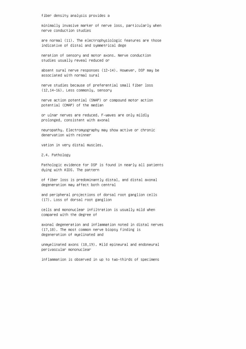
fiber density analysis provides a
minimally invasive marker of nerve loss, particularly whennerve conduction studies
are normal (11). The electrophysiologic features are thoseindicative of distal and symmetrical dege
neration of sensory and motor axons. Nerve conductionstudies usually reveal reduced or
absent sural nerve responses (12–14). However, DSP may beassociated with normal sural
nerve studies because of preferential small fiber loss(12,14–16). Less commonly, sensory
nerve action potential (SNAP) or compound motor actionpotential (CMAP) of the median
or ulnar nerves are reduced. F-waves are only mildlyprolonged, consistent with axonal
neuropathy. Electromyography may show active or chronicdenervation with reinner
vation in very distal muscles.
2.4. Pathology
Pathologic evidence for DSP is found in nearly all patientsdying with AIDS. The pattern
of fiber loss is predominantly distal, and distal axonaldegeneration may affect both central
and peripheral projections of dorsal root ganglion cells(17). Loss of dorsal root ganglion
cells and mononuclear infiltration is usually mild whencompared with the degree of
axonal degeneration and inflammation noted in distal nerves(17,18). The most common nerve biopsy finding isdegeneration of myelinated and
unmyelinated axons (18,19). Mild epineural and endoneuralperivascular mononuclear
inflammation is observed in up to two-thirds of specimens

(18–20). T-lymphocytes and
activated macrophages, with suppressor/cytotoxic cells(CD8) predominating over helper/inducer (CD4) cellsendoneurially are observed (18). No deposition ofimmunoglobulin, complement, or fibrin is observed.Demyelination is occasionally observed, but does not appearto be macrophage-mediated or follow a segmental pattern(18–22). 2.5. Pathogenesis The pathogenesis is not known.HIV infection appears to be a factor because aggressiveantiretroviral therapy and suppression of plasma HIV-1viral burden improves sensory function in HIV-infectedpatients, as measured by QST, but it is not clear if thisrepresents a direct or indirect mechanism (23). There hasbeen no consistent demonstration of HIV in dorsal rootganglion cells, nerve roots or peripheral nerves(16,18,19,24), and a paucity of evidence for significant invivo infection of neurons. Possible indirect mechanismsinclude cytokine dysregulation and immunologic dysfunction.Elevated levels of tumor necrosis factora, interleukin-1,and interleukin-6 have been identified in peripheral nerveand dorsal root ganglia of patients with AIDS(3,14,18,19,21,25–27), and their expression may be inducedthough low levels of HIV-1 replication acting on cytotoxicT-lymphocytes, resulting in degeneration of sensory neuronswithin the dorsal root ganglion (27). Paraproteinemia maybe a factor in some cases, with reactivity of IgM and IgGagainst myelin proteins identified in a majority of testedsamples from HIV-infected individuals (28). High titers ofantisulfatide antibodies have been found which showimmunofluorescence at paranodal regions (29,30).Cytomegalovirus (CMV) inclusions or antigens have beenidentified in peripheral nerve, nerve roots, and dorsalroot ganglion cells of some patients with DSP (17–19,24).Although some patients with painful neuropathy,particularly in advanced disease stages, have an underlyingsystemic CMV infection (31), there is no compellingevidence that direct CMV infection causes DSP and accountsfor DSP in patients with early HIV infection. 2.6.Treatment Control of pain is the primary treatment goal. Ofnote, up to 85% of patients with AIDS-related pain areunder treated (32). Guidelines established by the worldHealth Organization (WHO) to manage cancer pain can beadapted for HIV-associated DSP (33). In addition to the useof mild analgesics, adjunctive analgesic medications maynecessary. These include tricyclic antidepressantsamitriptyline or desipramine and gabapentin andlamotrigine. Topical anesthetic agents may also be used(lidocaine patch or gel or capsaicin). Narcotic analgesicsare often necessary to control pain in severe cases. The
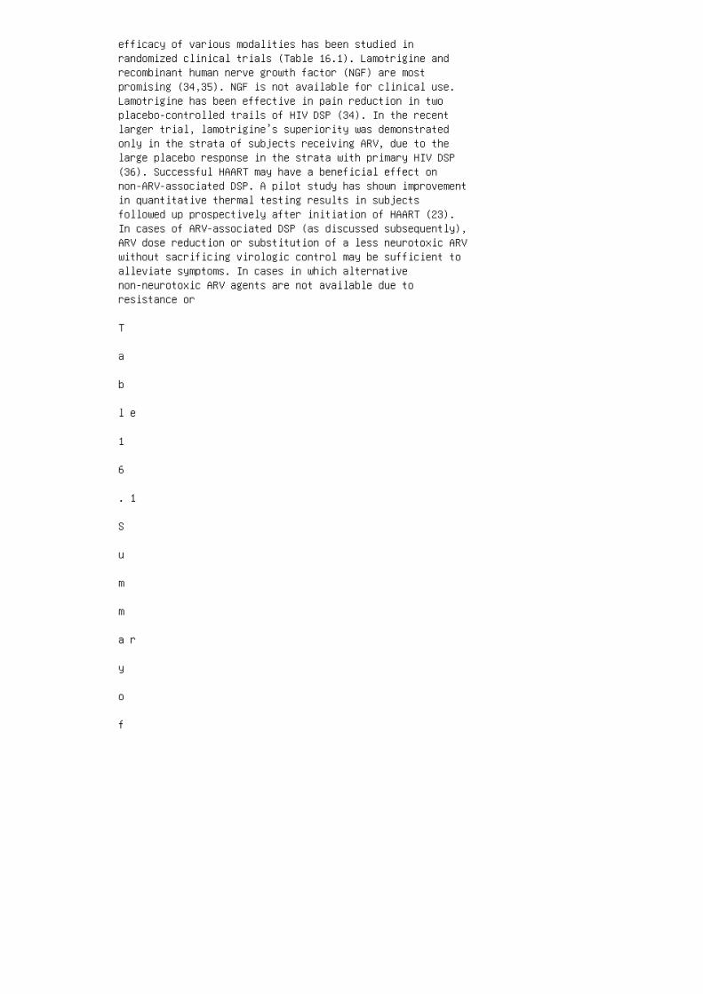
efficacy of various modalities has been studied inrandomized clinical trials (Table 16.1). Lamotrigine andrecombinant human nerve growth factor (NGF) are mostpromising (34,35). NGF is not available for clinical use.Lamotrigine has been effective in pain reduction in twoplacebo-controlled trails of HIV DSP (34). In the recentlarger trial, lamotrigine’s superiority was demonstratedonly in the strata of subjects receiving ARV, due to thelarge placebo response in the strata with primary HIV DSP(36). Successful HAART may have a beneficial effect onnon-ARV-associated DSP. A pilot study has shown improvementin quantitative thermal testing results in subjectsfollowed up prospectively after initiation of HAART (23).In cases of ARV-associated DSP (as discussed subsequently),ARV dose reduction or substitution of a less neurotoxic ARVwithout sacrificing virologic control may be sufficient toalleviate symptoms. In cases in which alternativenon-neurotoxic ARV agents are not available due toresistance or
T
a
b
l e
1
6
. 1
S
u
m
m
a r
y
o
f

R
a n
d
o
m
i z
e d
C
l i
n
i c
a l
T
r i
a l
s
o
f
E
f fi
c a
c y
a n
d
/ o

r
S
a f
e t
y
o
f
A
g
e n
t s
f o
r
D
S
P
P
a i
n
i n
H
I V
I
n
f e
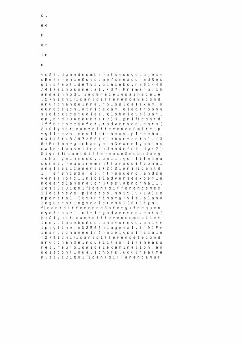
c t
e d
P
a t
i e
n
t s S t u d y a n d n u m b e r o f s t u d y s u b j e c ts R e f e r e n c e O u t c o m e / s m e a s u r e d R e su l t s P e p t i d e T v s . p l a c e b o , n ¼ 8 1 ( 4 0/ 4 1 ) S i m p s o n e t a l . ( 3 7 ) P r i m a r y : c ha n g e i n m o d i fi e d G r a c e l y p a i n s c a l e( 2 ) S i g n i fi c a n t d i f f e r e n c e S e c o n da r y : c h a n g e i n n e u r o l o g i c a l e x a m , ne u r o p s y c h i a t r i c e x a m , e l e c t r o p h ys i o l o g i c s t u d i e s , g l o b a l e v a l u a t io n , a n d C D 4 c o u n t s ( 2 ) S i g n i fi c a n t di f f e r e n c e S a f e t y : a d v e r s e e v e n t s (2 ) S i g n i fi c a n t d i f f e r e n c e A m i t r i pt y l i n e v s . m e x i l e t i n e v s . p l a c e b o ,n ¼ 1 4 5 ( 4 8 / 4 7 / 5 0 ) K i e b u r t z e t a l . ( 38 ) P r i m a r y : c h a n g e i n G r a c e l y p a i n sc a l e a t b a s e l i n e a n d e n d o f s t u d y ( 2 )S i g n i fi c a n t d i f f e r e n c e S e c o n d a r y: c h a n g e i n m o o d , q u a l i t y o f l i f e m e as u r e s , r e q u i r e m e n t f o r a d d i t i o n a la n a l g e s i c a g e n t s ( 2 ) S i g n i fi c a n t di f f e r e n c e S a f e t y : f r e q u e n c y a n d s ev e r i t y o f c l i n i c a l a d v e r s e e x p e r i en c e a n d l a b o r a t o r y t e s t a b n o r m a l i ti e s ( 2 ) S i g n i fi c a n t d i f f e r e n c e M e xi l e t i n e v s . p l a c e b o , n ¼ 1 9 ( 9 / 1 0 ) K em p e r e t a l . ( 3 9 ) P r i m a r y : v i s u a l a n al o g u e r a t i n g s c a l e ( V A S ) ( 2 ) S i g n ifi c a n t d i f f e r e n c e S a f e t y : f r e q u e nc y o f d o s e l i m i t i n g a d v e r s e e v e n t s (þ ) S i g n i fi c a n t d i f f e r e n c e m e x i l e ti n e . p l a c e b o A c u p u n c t u r e v s . a m i t ri p t y l i n e , n ¼ 2 5 0 S h l a y e t a l . ( 4 0 ) P ri m a r y : c h a n g e i n G r a c e l y p a i n s c a l e( 2 ) S i g n i fi c a n t d i f f e r e n c e S e c o n da r y : c h a n g e i n q u a l i t y o f l i f e m e a s ur e s , n e u r o l o g i c a l e x a m i n a t i o n , a nd d i s c o n t i n u a t i o n o f s t u d y t r e a t m en t s ( 2 ) S i g n i fi c a n t d i f f e r e n c e N G F

( 2 d o s e s ) v s . p l a c e b o , n ¼ 2 7 0 ( 9 0 / 9 0/ 9 0 ) M c A r t h u r e t a l . ( 3 5 ) P r i m a r y : ch a n g e i n G r a c e l y p a i n s c a l e ( þ ) S i g ni fi c a n t d i f f e r e n c e S e c o n d a r y : p r ac t i t i o n e r a n d p a t i e n t a s s e s s m e n t of g l o b a l i m p r o v e m e n t , c h a n g e i n n e ur o l o g i c a l e x a m , u s e o f p r e s c r i p t i on a n a l g e s i c s , a n d Q S T ( þ ) S i g n i fi c an t d i f f e r e n c e ( 2 ) d i f f e r e n c e S u b s et : c h a n g e i n e p i d e r m a l n e r v e fi b e r de n s i t y a s d e t e r m i n e d b y p u n c h s k i n bi o p s y ( 2 ) S i g n i fi c a n t d i f f e r e n c e La m o t r i g i n e v s . p l a c e b o , n ¼ 2 9 ( 9 / 2 0) S i m p s o n e t a l . ( 3 4 ) P r i m a r y : c h a n ge i n m o d i fi e d G r a c e l y p a i n s c a l e : a ve r a g e d a i l y p a i n a n d w o r s t p a i n ( þ ) Si g n i fi c a n t d i f f e r e n c e ( 2 ) S i g n i fi ca n t d i f f e r e n c e L a m o t r i g i n e v s . p l ac e b o , n ¼ 2 2 7 ( d d r u g ¼ 9 2 : 6 2 / 3 0 w / o d dr u g ¼ 1 3 5 : 8 8 / 4 7 ) S i m p s o n e t a l . ( 3 6 )S e c o n d a r y : c h a n g e i n n e u r o l o g i c a le x a m , u s e o f c o n c o m i t a n t a n a l g e s i cs , g l o b a l p a i n r e l i e f ( 2 ) S i g n i fi c an t d i f f e r e n c e P r i m a r y : G r a c e l y p a in s c a l e ; v i s u a l a n a l o g u e s c a l e ; M c Gi l l p a i n a s s e s s m e n t ; p a t i e n t a n d c li n i c a l g l o b a l i m p r e s s i o n o f c h a n g e( þ ) S i g n i fi c a n t d i f f e r e n c e i n a l l th r e e m e a s u r e s i n t h o s e w i t h d r u g n e ur o p a t h y toxicity, and substitution is not possiblewithout jeopardizing virologic control, symptomaticanalgesic treatment while continuing the neurotoxic ARV maybe appropriate. 3. TOXIC NEUROPATHY 3.1. Antiviral DrugsMedications used to treat HIV infection may cause a DSPindistinguishable from that caused by the infection itself.Among the currently approved antiretrovirals, only thedideoxynucleosides analogs (d-drugs) are clearlyneurotoxic. These include didanosine (ddI), zalcitabine(ddC), and stavudine (d4T) (41–45). DSP associated withdideoxynucleoside analogs is clinically andelectrophysiologically similar to DSP associated with HIV.Although the symptoms and signs of DSP associated withneurotoxicity bear a temporal relationship to the drug, itis often difficult to determine the date of symptom onsetrelative to the frequent changes in a patient’santiretroviral regimen. Accordingly, the incidence ofd-drug-induced DSP is difficult to determine. Thepathogenesis of toxic DSP is not known. One hypotheses isthat ddC, containing a cytosine base, interferes with theproduction of sphingomyelin via the formation of a
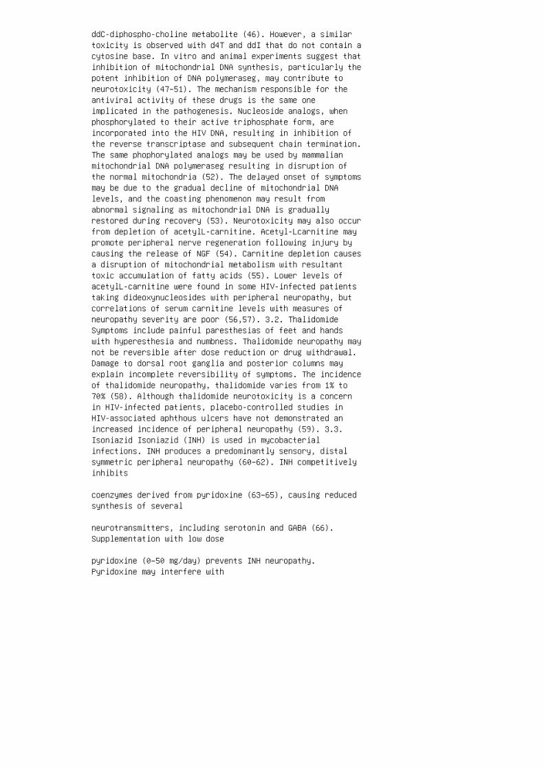
ddC-diphospho-choline metabolite (46). However, a similartoxicity is observed with d4T and ddI that do not contain acytosine base. In vitro and animal experiments suggest thatinhibition of mitochondrial DNA synthesis, particularly thepotent inhibition of DNA polymeraseg, may contribute toneurotoxicity (47–51). The mechanism responsible for theantiviral activity of these drugs is the same oneimplicated in the pathogenesis. Nucleoside analogs, whenphosphorylated to their active triphosphate form, areincorporated into the HIV DNA, resulting in inhibition ofthe reverse transcriptase and subsequent chain termination.The same phophorylated analogs may be used by mammalianmitochondrial DNA polymeraseg resulting in disruption ofthe normal mitochondria (52). The delayed onset of symptomsmay be due to the gradual decline of mitochondrial DNAlevels, and the coasting phenomenon may result fromabnormal signaling as mitochondrial DNA is graduallyrestored during recovery (53). Neurotoxicity may also occurfrom depletion of acetylL-carnitine. Acetyl-Lcarnitine maypromote peripheral nerve regeneration following injury bycausing the release of NGF (54). Carnitine depletion causesa disruption of mitochondrial metabolism with resultanttoxic accumulation of fatty acids (55). Lower levels ofacetylL-carnitine were found in some HIV-infected patientstaking dideoxynucleosides with peripheral neuropathy, butcorrelations of serum carnitine levels with measures ofneuropathy severity are poor (56,57). 3.2. ThalidomideSymptoms include painful paresthesias of feet and handswith hyperesthesia and numbness. Thalidomide neuropathy maynot be reversible after dose reduction or drug withdrawal.Damage to dorsal root ganglia and posterior columns mayexplain incomplete reversibility of symptoms. The incidenceof thalidomide neuropathy, thalidomide varies from 1% to70% (58). Although thalidomide neurotoxicity is a concernin HIV-infected patients, placebo-controlled studies inHIV-associated aphthous ulcers have not demonstrated anincreased incidence of peripheral neuropathy (59). 3.3.Isoniazid Isoniazid (INH) is used in mycobacterialinfections. INH produces a predominantly sensory, distalsymmetric peripheral neuropathy (60–62). INH competitivelyinhibits
coenzymes derived from pyridoxine (63–65), causing reducedsynthesis of several
neurotransmitters, including serotonin and GABA (66).Supplementation with low dose
pyridoxine (0–50 mg/day) prevents INH neuropathy.Pyridoxine may interfere with
the antimicrobial activity of INH (67) and in high doses,may in itself cause a
neuropathy (68–70).
3.4. Pyridoxine
Pyridoxine is neurotoxic when given in doses .200 mg/day.Symptoms include numb
ness and tingling, and are often reversible afterdiscontinuation of the vitamin, although
a coasting period during which they may intensify for 2–3weeks after discontinuation
may be observed (70). The neuropathy is thought to resultfrom competitive inhibition
of the more active form, pyridoxal phosphate, by the lessactive form, pyridoxine. Satur
ation of the activating enzymes results in paradoxicaldeficiency of pyridoxine (69).
3.5. Ethambutol
Ethambutol, an anti-tuberculosis bacteriostatic drug,causes both a distal sensory neuropa
thy and an optic neuropathy. DSP may occur with doses of 20mg/kg or more. Neuropathy
is less common than optic neuropathy, but can be disabling(71), and may herald the onset
of optic neuropathy (72). Ethambutol can aggravateneurotoxicity of other drugs
commonly used in combination (e.g., INH). Discontinuationof ethambutol is generally
followed by prompt improvement of symptoms.
3.6. Metronidazole
Metronidazole is an antimicrobial used to treat anaerobicinfections. DSP has been

reported with doses of 1.5 gm/day or drug usage .1 month(73–75). Symptoms
include numbness, painful paresthesias and dysesthesias ina stocking, and glove distri
bution. A proposed mechanism is binding of the drug toribonucleic acid (RNA) in
nerve cells causing inhibition of neuronal proteinsynthesis leading to peripheral axonal
degeneration (76). Metronidazole neuropathy is generallyreversible after dose reduction
or withdrawal, but coasting has been reported (77).
3.7. Dapsone
Dapsone is used for prophylaxis for Pneumocystis cariniipneumonia in patients with sul
fonamide intolerance (78). Unlike other toxic neuropathies,dapsone-induced peripheral
neuropathy involves primarily motor nerves with lessprominent sensory symptoms
(79–85). Progressive muscle weakness of both proximal anddistal muscles occurs in
upper and lower extremities. Electrophysiologic findingsdemonstrate motor axonopathy.
Symptoms begin from 6 weeks to several years afterinitiation of the drug, and are associ
ated with cumulated doses from 4 g to 600 g. Withdrawal ofthe drug results in improve
ment of symptoms. The mechanism is not known, but mayrepresent an idiosyncratic
reaction, or occur in individuals who have slow acetylation(84).
3.8. Vincristine
Vinca alkaloids are used as chemotherapy for AIDS-relatedlymphoma and Kaposi’s

sarcoma. DSP is a major dose-limiting effect, occurringwith a cumulative dose of
15–20 mg, and particularly with vincristine (86). Symptomsare distal paresthesias, with reduced or absent anklereflexes in early stages, and objective sensory findingsand motor weakness in later stages. Cranial neuropathiesand autonomic dysfunction may also occur (86–88).Vincristine neurotoxicity is thought due to impairedmicrotube assembly, resulting in disruption of axonaltransport. 3.9. Hydroxyurea Hydroxyurea is an adjunctiveagent for HIV infection. It is neurotoxic, particularlywhen used in combination with ddI and d4T (89). The highincidence of neuropathy has lead to reduced use ofhydroxyurea. 4. INFLAMMATORY DEMYELINATING POLYNEUROPATHY4.1. Epidemiology IDP occurs in acute and chronic forms.The prevalence of IDP in HIV patients is unknown, but theacute form often presents at the time of HIV seroconversionor primary infection. 4.2. Clinical Manifestations andExamination Findings The clinical features of the acuteform in HIV-positive patients are similar to acuteinflammatory demyelinating polyradiculoneuropathy (AIDP),or Guillain–Barre´ syndrome in non-HIV-infected patients(90). There is rapidly progressive limb weakness andgeneralized areflexia in a symmetric distribution. Sensorysymptoms are mild or absent. Cranial nerve involvement iscommon, but autonomic dysfunction is rare. Recovery beginswithin 2–4 weeks from the nadir. Chronic inflammatorydemyelinating polyradiculoneuropathy (CIDP) has a slowercourse, progressing over weeks to months, and usually has aless favorable long-term prognosis. 4.3. Laboratory andElectrophysiologic Findings CSF analysis reveals acytoalbumin dissociation typical of that seen innon-HIV-infected AIDP patients, but a lymphocyticpleocytosis (10–50 cells/mm 3 ) is more common inHIV-infected patients (10). In advanced HIV disease, searchfor opportunistic infections, notably CMV, should beconsidered. Electrophysiologic findings are similar tothose reported in non-HIV-uninfected patients, and areconsistent with a primary demyelinating neuropathy (REF).These include markedly reduced SNAP and CMAP responseamplitudes, prolonged distal and F-wave latencies, anddecreased conduction velocities with evidence forconduction block. Superimposed axonal loss may coexist,particularly in CIDP. 4.4. Pathogenesis IDP is likelyimmune-mediated, particularly early in the course of HIVinfection, when CD4 lymphocyte counts are relatively high.In patients with CD4 counts ,100 cells/ mm 3 , CMVinfection may cause IDP and should be considered in these

cases (91). IDP associated with CMV infection may includemore severe sensory involvement (92). Other laboratoryabnormalities that parallel the course of the disease inIDP include increased CSF levels of soluble CD8 andneopterin and anti-peripheral nerve myelin antibody titers(93).
4.5. Treatment
Case series indicate responsiveness to immunomodulatingtherapy, including as cortico
steroids, intravenous immunoglobulin (0.5–1.0 gm/kg for 2days), or plasmapheresis
(four to five exchanges). In patients with advanced HIVinfection, antiviral therapy
against CMV may be indicated.
5. PROGRESSIVE POLYRADICULOPATHY
5.1. Epidemiology
PP usually occurs in advanced AIDS as a complication of CMVinfection. The use of
HAART is associated with a reduction in both the incidenceof systemic CMV infection
and PP. Less common causes of PP should therefore beconsidered, including neurosyphilis,
herpes radiculitis, tuberculous radiculopathy, andlymphomatous meningoradiculopathy.
5.2. Clinical Manifestations and Examination Findings
The primary manifestations of PP are lower extremity painand weakness which may pro
gress rapidly to flaccid paraparesis with sphincterdysfunction (urinary retention or incon
tinence). Examination findings are weakness in a caudaequina distribution, areflexia in the
legs, sensory loss, and occasionally a thoracic sensorylevel (94).

5.3. Laboratory and Electrophysiologic Data
Contrast-enhanced lumbar spine MRI may reveal increased T2signal of the cauda equina
and enhanced, thickened, and clumped lumbosacral roots. InCMV polyradiculopathy,
CSF shows marked polymorphonuclear pleocytosis (mean, 651cells+ 1053 10 6 /L),
elevated protein (mean, 2.28+ 1.78 g/L), and decreasedglucose (mean CSF : plasma
glucose ratio, 0.48+ 0.17). Detection of CMV by polymerasechain reaction (PCR)
analysis of CSF has 92% sensitivity and 94% specificity inCMV-associated PP (95–
97). For most of the other etiologies of PP, CSF revealspredominantly lymphocytic pleo
cytosis, and CSF should be assayed for varicella-zostervirus (VZV) PCR, TB PCR,
VDRL, and cytology. Electrophysiologic evaluation revealsevidence of a lumbosacral polyradiculopathy.
Nerve conduction studies reveal diminished CMAP amplitude,but conduction velocity is
normal. SNAP amplitude is normal. Electromyography revealswidespread denervation
which helps differentiate PP from MM or IDP.
5.4. Pathogenesis
PP is due to opportunistic infections while CMV is the mostcommon pathogen; with the
advent of HAART, other etiologies have been identified,such as treponema pallidum,
herpes zoster, herpes simplex 2, mycobacteriumtuberculosis, and lymphoma, and
should be considered. Early recognition and treat isimportant to minimize irreversible

nerve root damage.
5.5. Treatment
PP secondary to CMV is a neurologic emergency, becauseuntreated mortality is 100%.
Combination therapy with ganciclovir and foscarnet may bemore effective (98).
Therapy appropriate for other etiologies include penicillinfor syphilitic radiculopathy, acyclovir for zosterradiculitis, anti-tuberculosis regimen for tuberculosisradiculopathy, and radiotherapy and/or chemotherapy forlymphomatous radiculitis. 6. MONONEUROPATHY MULTIPLEX 6.1.Epidemiology MM can be seen early in the course of HIVinfection when CD4 counts are.200 cells/mL (99). When MMoccurs late in the course of HIV infection, it may be aresult of CMV infection (100). A Sjo¨gren’s syndrome-likedisease is a rare cause of MM and other peripheralneuropathies in HIV. It is associated with a diffuseinterstitial lymphocytic syndrome (101). 6.2. ClinicalManifestations and Examination Findings MM is anaccumulation of mononeuropathies, frequently presenting asa wrist drop (radial nerve palsy) or foot drop (peronealneuropathy). There may also involvement of cutaneous nerveswith paresthesias and numbness in patchy areas in the trunkor extremities, or with facial or extraocular muscleweakness (cranial neuropathies) (102). 6.3. Laboratory andElectrophysiologic Findings CSF in HIV-associated MM isfrequently abnormal, but findings are nonspecific, andinclude elevated protein and mild mononuclear pleocytosis.When MM occurs in latestage of AIDS, PCR for CMV should beperformed on CSF. Electrophysiologic studies are helpful todocument asymmetric axonal damage with reduced CMAP andSNAP amplitudes. Electromyography shows neurogenicdenervation in the distribution of individual nerves. Nervebiopsy findings range from axonal degeneration with orwithout endoneural and epineural perivascular inflammatoryinfiltrates in early-onset MM, to numerouspolymorphonuclear infiltrates with mixed axonal anddemyelinative lesions in late-onset MM. Vasculitis has beennoted in several cases (103). CMV inclusions are detectedin peripheral nerve and in some cases of late-onset MM.6.4. Pathogenesis Early-onset MM is thought to be mediatedby immune mechanisms and is usually a self-limited disease(4,104). In the setting of advanced HIV disease, concurrentCMV infection is implicated (105). 6.5. TreatmentEarly-onset MM may resolve spontaneously within weeks to
several months (4,104). For patients with progressive MM orwho have incomplete recovery, immunomodulatory therapiesincluding corticosteroids, plasmaphoresis, and IVIG haveall been shown to be effective in uncontrolled reports. Inlate-onset MM occurring in advanced HIV infection, empirictherapy for CMV with ganciclovir, foscarnet or cidofovirshould be considered. T a b l e 1 6 . 2 T y p e s o f P e ri p h e r a l N e u r o p a t h y i n H I V I n f e c t i on N e u r o p a t h y t y p e a n d C D 4 s t a g e o f o cc u r r e n c e C l i n i c a l m a n i f e s t a t i o n sN e u r o l o g i c a l s i g n s D i a g n o s t i c s t ud i e s T r e a t m e n t D S P C D 4 , 2 0 0 P a r e s t he s i a s , d y s e s t h e s i a , d i s t a l w e a k n es s i n s e v e r e c a s e s ( a n d i n d a p s o n e n eu r o p a t h y ) S e n s o r y ( p i n p r i c k a n d v ib r a t i o n ) d e fi c i t s , s t o c k i n g a n d g lo v e d i s t r i b u t i o n , d e p r e s s e d d i s t al r e fl e x e s E M G / N C V : a b n o r m a l s e n s or y n e r v e a m p l i t u d e s , d i s t a l a x o n o pa t h y A n a l g e s i c s ( p r i m a r y a n d a d j u va n t ) , n e u r o t o x i c d r u g w i t h d r a w a l or d o s e r e d u c t i o n ; a d e q u a t e v i r o l o gi c s u p p r e s s i o n I D P E a r l y : C D 4 . 2 0 0 La t e : C D 4 , 1 0 0 P r o g r e s s i v e w e a k n e s s, p a r e s t h e s i a s W e a k n e s s , m i l d s e n so r y d e fi c i t s , a r e fl e x i a C S F : l y m p h oc y t i c p l e o c y t o s i s ( 1 0 – 5 0 c e l l s / u L) , h i g h p r o t e i n E M G / N C V : d e m y e l i n at i o n I m m u n o m o d u l a t i n g t h e r a p y , c on s i d e r a n t i C M V t h e r a p y i n l a t e o n s et I D P M M E a r l y : C D 4 . 2 0 0 L a t e : C D 4 , 1 00 F o o t o r w r i s t d r o p , f a c i a l w e a k n e ss , f o c a l p a i n M u l t i f o c a l c r a n i a l a nd p e r i p h e r a l n e u r o p a t h y E M G / N C V : mu l t i f o c a l a x o n a l n e u r o p a t h y I m m u no m o d u l a t i n g t h e r a p y , c o n s i d e r a n ti C M V t h e r a p y i n l a t e o n s e t M M P P E a r ly : C D 4 . 2 0 0 L a t e : C D 4 , 1 0 0 L o w e r e x t re m i t y w e a k n e s s , p a r e s t h e s i a s , s p hi n c t e r d y s f u n c t i o n F l a c c i d p a r a p ar e s i s , s a d d l e d i s t r i b u t i o n a n e s t he s i a , d e p r e s s e d r e fl e x e s C S F : p o l ym o r p h o n u c l e a r p l e o c y t o s i s ( i n C M Vi n f e c t i o n ) ; l y m p h o c y t i c p l e o c y t os i s ( i n l y m p h o m a , s y p h i l i s , h e r p e s) ; E M G / N C V : p o l y r a d i c u l o p a t h y A n ti C M V t h e r a p y ( i f C S F C M V P C R þ ) ; a c y cl o v i r f o r h e r p e s r a d i c u l i t i s ; P C N fo r n e u r o s y p h i l i t i c r a d i c u l o p a t h y; c h e m o t h e r a p y w / o r w / o R T f o r l y m p h

o m a t o u s r a d i c u l o p a t h y 7. CONCLUSIONPeripheral neuropathies are the most common neurologiccomplications in HIV infection. They may result from theHIV infection itself, (directly or indirectly), fromneurotoxic properties of HIV drugs, from opportunistic orother superimposed infections, or from a combination. DSPis the most common form and is the most disabling. Otherneuropathies are much less common. A summary of thedifferent forms of peripheral neuropathy that occur in HIVinfection is given in Table 16.2. An algorithm summarizingthe appropriate approach to an HIV-infected individual withneuropathy is shown in Fig. 16.1. Figure 16.1 Algorithm formanagement of peripheral neuropathy in HIV-infectedindividuals.
1. Brinley F, Pardo C, Verma A. Human immunodeficiencyvirus and the peripheral nervous system workshop. ArchNeurol 2001; 58:1561–1566.
2. Pardo CA, McArthur JC, Griffin JW. HIV neuropathy:insights in the pathology of HIV peripheral nerve disease.J Peripher Nerv Syst 2001; 6(1):21–27.
3. So YT, Holtzman DM, Abrams DI, Olney RK. Peripheralneuropathy associated with acquired immunodeficiencysyndrome. Prevalence and clinical features from apopulationbased survey. Arch Neurol 1988; 45(9):945–948.
4. Simpson D, Olney R. Peripheral neuropathies associatedwith human immunodeficiency virus infection. PeripheralNeuropathies: New Concepts Treat 1992; 685–711.
5. Schifitto G, McDermott M, McArthur J, Marder K, SacktorN, Epstein L. Incidence of and risk factors forHIV-associated distal sensory polyneuropathy. Neurology2002; 58(12):1764–1768.
6. Barohn RJ, Gronseth GS, LeForce BR, McVey AL, McGuireSA, Butzin CA. Peripheral nervous system involvement in alarge cohort of human immunodeficiency virus-infectedindividuals [published erratum appears in Arch Neurol 1993Apr; 50(4):388]. Arch Neurol 1993; 50(2):167–171.
7. Tagliati M, Grinnell J, Godbold J, Simpson DM.Peripheral nerve function in HIV infection: clinical,electrophysiologic, and laboratory findings. Arch Neurol1999; 56(1):84–89.
8. Childs EA, Lyles RH, Selnes OA, Chen B, Miller EN, CohenBA. Plasma viral load and CD4 lymphocytes predict
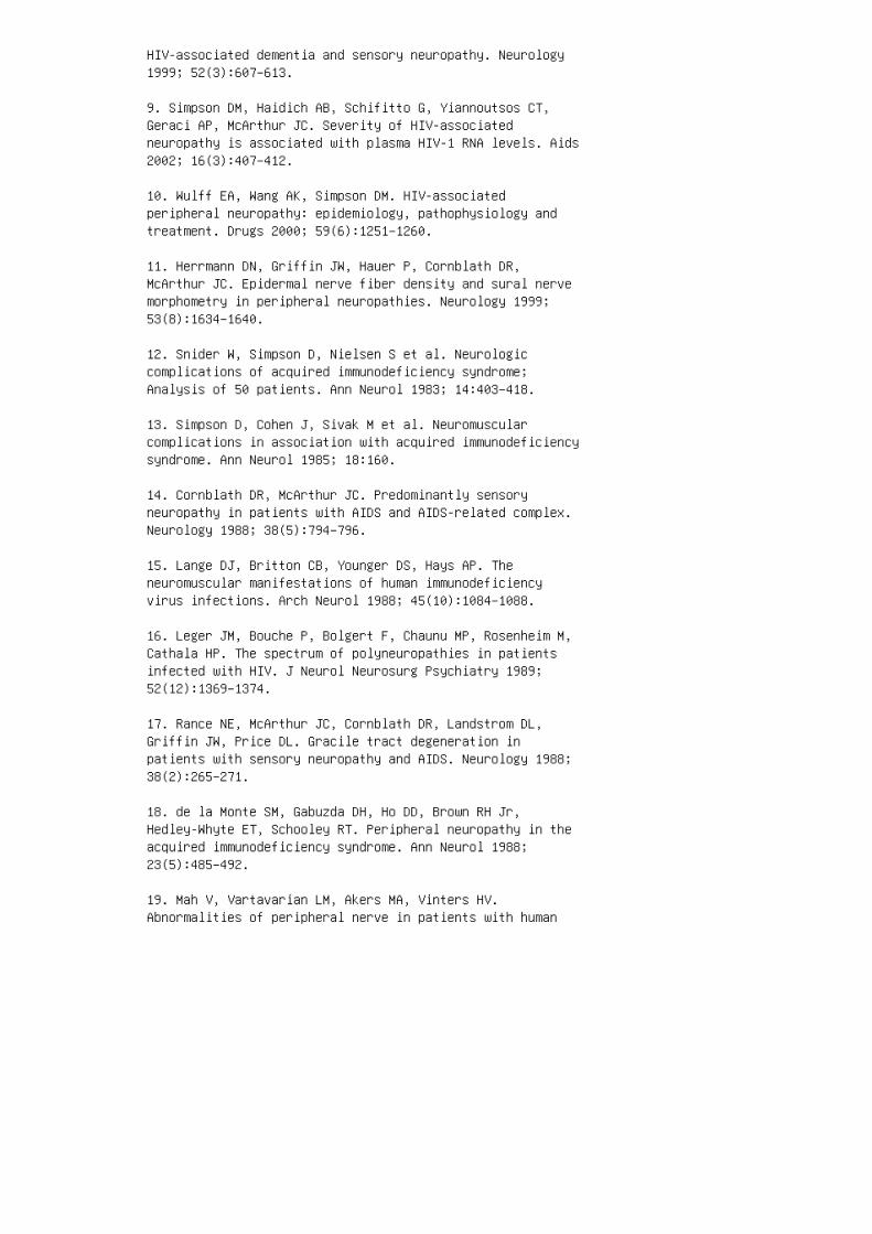
HIV-associated dementia and sensory neuropathy. Neurology1999; 52(3):607–613.
9. Simpson DM, Haidich AB, Schifitto G, Yiannoutsos CT,Geraci AP, McArthur JC. Severity of HIV-associatedneuropathy is associated with plasma HIV-1 RNA levels. Aids2002; 16(3):407–412.
10. Wulff EA, Wang AK, Simpson DM. HIV-associatedperipheral neuropathy: epidemiology, pathophysiology andtreatment. Drugs 2000; 59(6):1251–1260.
11. Herrmann DN, Griffin JW, Hauer P, Cornblath DR,McArthur JC. Epidermal nerve fiber density and sural nervemorphometry in peripheral neuropathies. Neurology 1999;53(8):1634–1640.
12. Snider W, Simpson D, Nielsen S et al. Neurologiccomplications of acquired immunodeficiency syndrome;Analysis of 50 patients. Ann Neurol 1983; 14:403–418.
13. Simpson D, Cohen J, Sivak M et al. Neuromuscularcomplications in association with acquired immunodeficiencysyndrome. Ann Neurol 1985; 18:160.
14. Cornblath DR, McArthur JC. Predominantly sensoryneuropathy in patients with AIDS and AIDS-related complex.Neurology 1988; 38(5):794–796.
15. Lange DJ, Britton CB, Younger DS, Hays AP. Theneuromuscular manifestations of human immunodeficiencyvirus infections. Arch Neurol 1988; 45(10):1084–1088.
16. Leger JM, Bouche P, Bolgert F, Chaunu MP, Rosenheim M,Cathala HP. The spectrum of polyneuropathies in patientsinfected with HIV. J Neurol Neurosurg Psychiatry 1989;52(12):1369–1374.
17. Rance NE, McArthur JC, Cornblath DR, Landstrom DL,Griffin JW, Price DL. Gracile tract degeneration inpatients with sensory neuropathy and AIDS. Neurology 1988;38(2):265–271.
18. de la Monte SM, Gabuzda DH, Ho DD, Brown RH Jr,Hedley-Whyte ET, Schooley RT. Peripheral neuropathy in theacquired immunodeficiency syndrome. Ann Neurol 1988;23(5):485–492.
19. Mah V, Vartavarian LM, Akers MA, Vinters HV.Abnormalities of peripheral nerve in patients with human

immunodeficiency virus infection. Ann Neurol 1988;24(6):713–717.
20. Lipkin WI, Parry G, Kiprov D, Abrams D. Inflammatoryneuropathy in homosexual men with lymphadenopathy.Neurology 1985; 35(10):1479–1483.
21. Bailey RO, Baltch AL, Venkatesh R, Singh JK, Bishop MB.Sensory motor neuropathy associated with AIDS. Neurology1988; 38(6):886–891. 22. Bradley WG, Shapshak P, Delgado S,Nagano I, Stewart R, Rocha B. Morphometric analysis of theperipheral neuropathy of AIDS. Muscle Nerve 1998;21(9):1188–1195. 23. Martin C, Solders G, Sonnerborg A,Hansson P. Antiretroviral therapy may improve sensoryfunction in HIV-infected patients: a pilot study [seecomments]. Neurology 2000; 54(11):2120–2127. 24. Grafe M,Wiley C. Spinal cord and peripheral nerve pathology inAIDS: the roles of cytomegalovirus and humanimmunodeficiency virus. Ann Neurol 1989; 25:561–566. 25.Wesselingh S, Glass J, McArthur J, Griffin J, Griffin D.Cytokine Dysregulation in HIV-Associated NeurologicalDisease. Adv Neuroimmunol 1994; 4(3):199–206. 26. Tyor WR,Glass JD, Griffin JW, Becker PS, McArthur JC, Bezman L,Griffin DE. Cytokine expression in the brain during theacquired immunodeficiency syndrome. Ann Neurol 1992;31:349–360. 27. Yoshioka M, Shapshak P, Srivastava AK,Stewart RV, Nelson SJ, Bradley WG. Expression of HIV-1 andinterleukin-6 in lumbosacral dorsal root ganglia ofpatients with AIDS. Neurology 1994; 44(6):1120–1130. 28.Petratos S, Turnbull V, Papadopoulos R et al. Antibodiesagainst peripheral myelin glycolipids in people with HIVinfection. Immunol Cell Biol 1998; 76:535–541. 29. PetratosS, Turnbull V, Papadopoulos R. High titer anti-sulfatideantibodies in HIV-infected individuals. Neuroreport 1999;10:2557–2562. 30. Petratos S, Turnbull V, Papadopoulos R etal. Peripheral nerve binding patterns of anti-sulphatideantibodies in HIV-infected individuals. Neuroreport 1999;10:1659–1664. 31. Fuller GN, Jacobs JM, Guiloff RJ.Association of painful peripheral neuropathy in AIDS withcytomegalovirus infection [see comments]. Lancet 1989;2(8669):937–941. 32. Breitbart W, Rosenfeld BD, Passik SD,McDonald MV, Thaler H, Portenoy RK. The undertreatment ofpain in ambulatory AIDS patients. Pain 1996;65(2–3):243–249. 33. Grond S, Zech D, Schug S, Lynch J,Lehmann K. Validation of World Health Organizationguidelines for cancer pain relief during the last days andhours of life. J Pain Symptom Manage 1991; 6(7):411–422.34. Simpson D, Olney R, McArthur J et al. Aplacebo-controlled trial of lamotrigine for painfulHIV-associated neuropathy. Neurology 2000; 54:2115–2119.

35. McArthur JC, Yiannoutsos C, Simpson DM, Adornato BT,Singer EJ, Hollander H. A phase II trial of nerve growthfactor for sensory neuropathy associated with HIVinfection. AIDS Clinical Trials Group Team 291 [seecomments]. Neurology 2000; 54(5):1080–1088. 36. Simpson DM,McArthur JC, Olney R, et al. Lamotrigine for HIV-associatedpainful sensory neuropathies: a placebo-controlled trial.Neurology 2003; 60:1508–1514. 37. Simpson D, Dorfman D,Olney R, McKinley G, Dobkin J, So Y. Peptide T in thetreatment of painful distal neuropathy associated withAIDS; Results of a placebo-controlled trial. The Peptide TNeuropathy Study Group. Neurology 1996; 47:1254–1259. 38.Kieburtz K, Simpson D, Yiannoutsos C, Max MB, Hall CD,Ellis RJ. A randomized trial of amitriptyline andmexiletine for painful neuropathy in HIV infection. AIDSClinical Trial Group 242 Protocol Team. Neurology 1998;51(6):1682–1688. 39. Kemper CA, Kent G, Burton S,Deresinski SC. Mexiletine for HIV-infected patients withpainful peripheral neuropathy: a double-blind,placebo-controlled, crossover treatment trial. J AcquirImmune Defic Syndr Hum Retrovirol 1998; 19(4):367–372. 40.Shlay JC, Chaloner K, Max MB, Flaws B, Reichelderfer P,Wentworth D. Acupuncture and amitriptyline for pain due toHIV-related peripheral neuropathy: a randomized controlledtrial. Terry Beirn Community Programs for Clinical Researchon AIDS [see comments]. J Am Med Assoc 1998;280(18):1590–1595. 41. Yarchoan R, Perno C, Thomas R,Klecker R, Allain J, Wills R. Phase I studies of 2 0 3 0deoxycytidinein severe human immuno-deficiency virusinfection as a single agent and alternating withzidovudine. Lancet 1988; 1:76–81.
42. Merigan T, Skowron G, Bozzette S, Richman D,Uttamchandani R, Fischl M. Circulating p24 antigen levelsand responses to dideoxycytidine in human immunodeficiencyvirus (HIV) infections. Ann Intern Med 1989; 110:189–194.
43. Cooley T, Kunches L, Saunders C, Ritter J, Perkins C,Mclaren C. Once-daily administration of 2 0 3 0didexyinosine (ddI) in patients with the acquiredimmunodeficiency syndrome or AIDS-related complex. N Engl JMed 1990; 322:1340–1345.
44. Lambert J, Seidlin M, Reichman R, Plank C, Laverty M,Morse G. Dideoxyinosine (ddI) in patients with the acquiredimmunodeficiency syndrome or AIDS-related complex: resultsof phase I trial. N Engl J Med 1990; 322:1333–1340.
45. Browne MJ, Mayer KH, Chafee SB, Dudley MN, Posner MR,Steinberg SM. 2 0 ,3 0 -didehydro3 0 -deoxythymidine (d4T)

in patients with AIDS or AIDSrelated complex: a phase Itrial. J Infect Dis 1993; 167(1):21–29.
46. Cooney D, Dalal M, Mitsuya H, McMahon J et al. Initialstudies on the cellular pharmacology of 2 0 3 0-dideoxycytidine, an inhibitor of HTLV-III infectivity.Biochem Pharmacol 1986; 35:2065–2068.
47. Pezeshkpour G, Krarup C, Buchthal F, Dimauro S,Bresolin N, McBurney J. Peripheral neuropathy inmitochondrial disease. J Neurol Sci 1987; 77:285–304.
48. Balzarini J, Herdewlin P, De Clercq E. Differentialpatterns of intracellular metabolism of 2 0 3 0-dideoxythymidine and 3 0 -azido-2 0 3 0 -dideoxythymidine,two potent anti-human immunodeficiency virus compounds. JBiol Chem 1989; 264:6127–6133.
49. Chen CH, Cheng YC. Delayed cytotoxicity and selectiveloss of mitochondrial DNA in cells treated with theanti-human immunodeficiency virus compound 2 0 3 0-dideoxycytidine. J Biol Chem 1989; 264:11934–11937.
50. Martin J, Brown C, Davis N, Reardon J. Effects ofantiviral nucleoside analogs on human DNA polymerases andmitchondrial DNA synthesis. Antimicrob Agents Chemother1994; 38:2743–2749.
51. Cui L, Locatelli L, Xie M, Sommadossi J. Effect ofnucleoside analogs on neurite regeneration andmitochondrial DNA synthesis in PC-12 cells. The Journal ofPharmacology and Experimental Therapeutics 1997;280(3):1228–1234.
52. Gasnault J, Pinganaud C, Kousignian P et al. Subacuteascendant polyneuropathy revealing a nucleoside-inducedmitochondrial cytopathy. Presented at The Seventh EuropeanConference on Clinical Aspects and Treatment of HIVInfection. Lisbon, Portugal, Oct 23–27, 1999.
53. Moyle G, Sadler M. Peripheral neuropathy withnucleoside antiretrovirals. Drug Saf 1998; 19(6):481–494.
54. Angelucci L, Ramacci M, Taglialatela G et al. Nervegrowth factor binding in aged rat central nervous system:effect of acetyl-L-carnitine. J Neurosci Res 1988;20:491–496.
55. Colucci W, Gandour R. Carnitine acetyltransferase: areview of its biology, enzymology and bioorganic chemistry.
Bioorg Chem 1988; 16:307–334.
56. Famularo G, Moretti S, Marcellini S, Trinchieri V,Tzantzoglou S, Santini G. Acetyl-carnitine deficiency inAIDS patients with neurotoxicity on treatment withantiretroviral nucleoside analogues. Aids 1997;11(2):185–190.
57. Simpson DM, Katzenstein D, Haidich B, Millington D,Yiannoutsos C, Schifitto G. Plasma carnitine inHIV-associated neuropathy. Aids 2001; 15(16):2207–2208.
58. Tseng S, Pak G, Washenick K et al. Rediscoveringthalidomide: a review of its mechanism of action, sideeffects, and potential uses. J Am Acad Dermatol 1996;35:969–979.
59. Jacobson J, Greenspan J, Spritzler J et al. Thalidomidefor the treatment of oral aphthous ulcers in patients withhuman immunodeficiency virus infection. N Eng J Med 1997;336:1487–1493.
60. Biehl J, Skavlem J. Toxicity of isoniazid. Am RevTuberc 1953; 68:296–297.
61. Gammon G, Burge F, King G. Neural toxicity intuberculous patients treated with isoniazid (isonicotinicacid hydrazide). AMA Archs Neurol Psychiatry 1953;70:64–69.
62. Jones W, Jones G. Peripheral neuropathy due toisoniazid. Report of two cases. Lancet 1953; 1:1073–1074.63. Biehl J, Vilter R. Effect of isonazid on vitamin B12metabolism, its possible significance in producingisoniazid neuritis. Proc Soc Exp Biol Med 1954; 85:389–395.64. Wiegand R. The formation of pyridoxal and pyridoxal5-phosphate hydrazones. J Am Chem Soc 1956; 78:5307. 65.Price J, Brown R, Larson F. Quantitative studies on humanurinary metabolites of tryptophan as affected by isoniazidand deoxypyridoxine. J Clin Invest 1957; 36:1600. 66.Girling D. Adverse effects of antituberculosis drugs. Drugs1982; 23:56–74. 67. Snider D. Pyridoxine supplementationduring isoniazid therapy. Tubercle 1980; 61:191–196. 68.Schaumburg H, Kaplan J, Windebank A. Sensory neuropathyfrom pyridoxine abuse. N Engl J Med 1983; 309:445–448. 69.Nisar M, Watkin SW, Bucknall RC, Agnew RA. Exacerbation ofisoniazid induced peripheral neuropathy by pyridoxine.Thorax 1990; 45(5):419–420. 70. Berger A, Schaumburg H,Schroeder C, Appel S, Reynolds R. Dose response, coasting,and differential fiber vulnerability in human toxic

neuropathy: a prospective study of pyridoxineneurotoxicity. Neurology 1992; 42:1367–1370. 71. Tugwell P,James S. Peripheral neuropathy with ethambutol. PostgradMed J 1972; 48(565):667–670. 72. Nair V, LeBrun M, Kass I.Peripheral neuropathy associated with ethambutol. Chest1980; 77(1):98–100. 73. Duffy L, Daum F, Fisher S, SelmanJ, Vishnubhakat S, Aiges H. Peripheral neuropathy inCrohn’s disease. Patients treated with metronidazole.Gastroenterology 1985; 88:681–684. 74. Boyce E, Cookson E,Bond W. Persistent metronidazole-induced peripheralneuropathy. Ann of Pharmacotherapy 1990; 24:19–21. 75.Learned-Coughlin S. Peripheral neuropathy induced bymetronidazole (letter). Ann Pharmacother 1994; 28:536. 76.Bradley W, Karlsson I, Rassol C. Metronidazole neuropathy.Br Med J 1977; 2(6087):610–611. 77. Coxon A, Pallis C.Metronidazole neuropathy. J Neuro Neurosurg and Psych 1976;39:403–405. 78. Medina I, Mills J, Leoung G. Oral therapyfor Pneumocystis carinii pneumonia in the acquiredimmunodeficiency syndrome: a controlled trial oftrimethoprim-sulfamethoxazole versus trimethoprim-dapsone.N Engl J Med 1990; 323:776–782. 79. Saqueton A, Lorinez A,Vick N, Hamer R. Dapsone and peripheral motor neuropathy.Arch Dermatol 1969; 100:214–217. 80. Rapoport A, Guss S.Dapsone-induced peripheral neuropathy. Arch Neurol 1972;27:184–185. 81. Epstein F, Bohm M. Dapsone-inducedperipheral neuropathy. Arch Dermatol 1976; 112:1761–1762.82. Fredericks E, Kugelman R, Kirsch N. Dapsone-inducedmotor polyneuropathy. Arch Dermatol 1976; 112:1158–1160.83. Guttman L, Martin J, Welton W. Dapsone motorneuropathy—an axonal disease. Neurology 1976; 26:514–516.84. Koller W, Gehlmann L, Malkinson F, Davis F.Dapsone-induced peripheral neuropathy. Arch Neurol 1977;34:644–646. 85. Navarro S, Rosales R, Ordinario A, Izumo S,Osame M. Acute dapsone-induced peripheral neuropathy.Muscle Nerve 1989; 12:604–606. 86. Tuxen M, Hansen S.Neurotoxicity secondary to antineoplastic drugs. CancerTreat Rev 1994; 20:191–214. 87. Legha S. Vincristineneurotoxicity: pathophysiology and management. Med Toxicol1986; 1:421–427. 88. Macdonald D. Neurologic complicationsof chemotherapy. Neurol Clin 1991; 9:955–967. 89. Moore RD,Wong WM, Keruly JC, McArthur JC. Incidence of neuropathy inHIV-infected patients on monotherapy versus those oncombination therapy with didanosine, stavudine andhydroxyurea. Aids 2000; 14(3):273–278.
90. Cornblath DR, McArthur JC, Kennedy PG, Witte AS,Griffin JW. Inflammatory demyelinating peripheralneuropathies associated with human T-cell lymphotropicvirus type III infection. Ann Neurol 1987; 21(1):32–40.

91. Misha B, Sommers W, Koski C et al. Acute inflammatorydemyelinating polyneuropathy in the acquiredimmunodeficiency syndrome. Ann Neurol 1985; 18:131–132.
92. Visser L, Van der Meche´ F, Meulstee J et al.Cytomegalovirus infection and Guillain–Barre´ syndrome: theclinical, electrophysiologic, and prognostic features.Neurology 1996; 47:668–673.
93. Griffin D, McArthur J, D C. Soluble interleukin-2receptor and soluble CD8 in serum and cerebrospinal fluidduring human immunodeficiency virus-associated neurologicdisease. J Neuroimmunol 1990; 28:97–109.
94. Eidelberg D, Sotrel A, Vogel H, Walker P, Kleefield J,Crumpacker CSd. Progressive polyradiculopathy in acquiredimmune deficiency syndrome. Neurology 1986; 36(7):912–916.
95. Gozlan J, El Amrani M, Baudrimont M et al. Aprospective evaluation of clinical criteria and polymerasechain reaction assay of cerebrospinal fluid for thediagnosis of cytomegalovirusrelated neurological diseasesduring AIDS. AIDS 1995; 9:253–260.
96. Shinkai M, Spector S. Quantitation of humancytomegalovirus (HCMV) DNA in cerebrospinal fluid bycompetitive PCR in AIDS patients with different HCMVcentral nervous system diseases. Scand J Infect Dis 1995;27:559–561.
97. Vogel J, Cinatl J, Lux A et al. New PCR assay for rapidand quantitative detection of human cytomegalovirus incerebrospinal fluid. J Clin Microbiol 1996; 34:482–483.
98. The studies of ocular complication of AIDS ResearchGroup in collaboration with the AIDS Clinical Trials Group.Combination foscarnet and ganciclovir therapy versusmonotherapy for the treatment of relapsed cytomegalovirusretinitis in patients with AIDS. Arch Ophthalmol 1996;114:23–32.
99. Simpson D, Olney RK. Peripheral neuropathies associatedwith human immunodeficiency virus infection. Neurol Clin1992; 10(3):685–711.
100. Said G, Lacroix C, Chemouilli P, Goulon-Goeau C,Roullet E, Penaud D. Cytomegalovirus neuropathy in acquiredimmunodeficiency syndrome: a clinical and pathologicalstudy. Ann Neurol 1991; 29(2):139–146.

101. Gherardi RK, Chretien F, Delfau-Larue MH, Authier FJ,Moulignier A, Roulland-Dussoix D. Neuropathy in diffuseinfiltrative lymphocytosis syndrome: an HIV neuropathy, nota lymphoma [see comments]. Neurology 1998; 50(4):1041–1044.
102. Miller RG, Parry GJ, Pfaeffl W, Lang W, Lippert R,Kiprov D. The spectrum of peripheral neuropathy associatedwith ARC and AIDS. Muscle Nerve 1988; 11(8):857–863.
103. Bradley WG, Verma A. Painful vasculitic neuropathy inHIV-1 infection: relief of pain with prednisone therapy.Neurology 1996; 47(6):1446–1451.
104. So Y, Olney R. The natural history of mononeuritismultiplex and simplex in HIV infection. Neurology 1991;41(suppl 1):375.
105. Roullet E, Asseurus J, Gozlan J et al. Cytomegalovirusmultifocal neuropathy in AIDS: analysis of 15 consecutivepatients. Neurology 1994; 44:2174–2182.
17
Infectious and Granulomatous
Neuropathies
Sharon P. Nations, Jaya R. Trivedi, and Gil I. Wolfe
University of Texas Southwestern Medical Center, Dallas,Texas, USA
Abstract 294
1. Leprosy 294 1.1. Introduction 294 1.2. Clinical Features294 1.3. Laboratory Evaluation 296 1.4. DifferentialDiagnosis 297 1.5. Management and Prognosis 298 1.6.Pathophysiology 300
2. Lyme Disease 300 2.1. Introduction 300 2.2. ClinicalFeatures 300 2.3. Laboratory Evaluation 301 2.4.Differential Diagnosis 302 2.5. Management 302 2.6.Prevention 303 2.7. Prognosis 303 2.8. Pathophysiology 303
3. Sarcoidosis 304 3.1. Introduction 304 3.2. ClinicalFeatures 304 3.3. Polyneuropathy 304 3.4. Mononeuropathyand Mononeuritis Multiplex 305 3.5. Polyradiculopathy 3053.6. Guillain–Barre´ Syndrome 305 3.7. LumbosacralPlexopathy with Chronic Sensorimotor Polyneuropathy 3063.8. Subclinical Neuropathy 306 3.9. Laboratory Evaluation

306
3.10. Differential Diagnosis 306
3.11. Management and Prognosis 307
hypopigmented, anesthetic skin lesions. If there isinvolvement of a nearby nerve trunk, a
mononeuropathy develops. Biopsy of the skin lesion revealswell-formed epithelioid gran
ulomas from subcutaneous tissue through the dermis (5). AFBare absent or only rarely
detected in biopsy specimens and are not detected inslit-skin (tissue fluid) smears of
the lesion or of earlobes, knees or elbows, which aretypical sites of infection. Granulomas
involving nerves may caseate and abscesses occasionallyform. In lepromatous leprosy, the patient fails to mount acell-mediated immune response
to the organism, and widespread infection occurs. Numerouserythematous macules,
plaques, and nodules may spread over the face, trunk, andextensor surfaces of the extre
mities, sparing the scalp, crurae, and axillae. Loss ofeyelashes and eyebrows (madarosis)
and infiltration of nose and earlobes lead to thecharacteristic facial deformity (Fig. 17.1).
Nasal congestion is a frequent complaint, and nasal mucussmears stain positive for AFB
(6). Nasal septal perforation is seen in severe cases. Asymmetric distal sensory polyneuro
pathy involving primarily pain, temperature, and autonomicmodalities progresses proxi
mally. A mononeuritis multiplex may become superimposed aslarger superficial nerve
trunks become involved. Skin biopsy reveals histiocytic
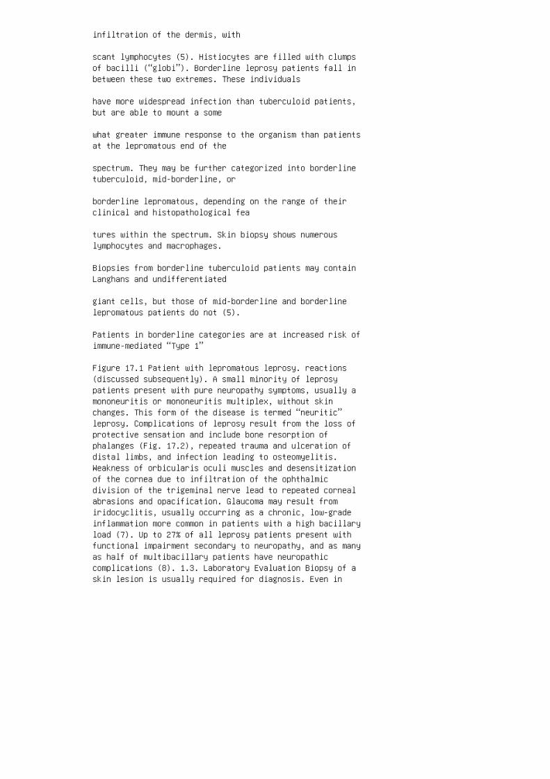
infiltration of the dermis, with
scant lymphocytes (5). Histiocytes are filled with clumpsof bacilli (“globi”). Borderline leprosy patients fall inbetween these two extremes. These individuals
have more widespread infection than tuberculoid patients,but are able to mount a some
what greater immune response to the organism than patientsat the lepromatous end of the
spectrum. They may be further categorized into borderlinetuberculoid, mid-borderline, or
borderline lepromatous, depending on the range of theirclinical and histopathological fea
tures within the spectrum. Skin biopsy shows numerouslymphocytes and macrophages.
Biopsies from borderline tuberculoid patients may containLanghans and undifferentiated
giant cells, but those of mid-borderline and borderlinelepromatous patients do not (5).
Patients in borderline categories are at increased risk ofimmune-mediated “Type 1”
Figure 17.1 Patient with lepromatous leprosy. reactions(discussed subsequently). A small minority of leprosypatients present with pure neuropathy symptoms, usually amononeuritis or mononeuritis multiplex, without skinchanges. This form of the disease is termed “neuritic”leprosy. Complications of leprosy result from the loss ofprotective sensation and include bone resorption ofphalanges (Fig. 17.2), repeated trauma and ulceration ofdistal limbs, and infection leading to osteomyelitis.Weakness of orbicularis oculi muscles and desensitizationof the cornea due to infiltration of the ophthalmicdivision of the trigeminal nerve lead to repeated cornealabrasions and opacification. Glaucoma may result fromiridocyclitis, usually occurring as a chronic, low-gradeinflammation more common in patients with a high bacillaryload (7). Up to 27% of all leprosy patients present withfunctional impairment secondary to neuropathy, and as manyas half of multibacillary patients have neuropathiccomplications (8). 1.3. Laboratory Evaluation Biopsy of askin lesion is usually required for diagnosis. Even in

cases of pure neuritic leprosy, skin biopsy from ananesthetic area may demonstrate inflammation and AFB,especially if the specimen is thick enough to includedermal nerves (9). However, a nerve biopsy is oftennecessary, preferably of an involved sensory nerve such asthe sural, the superficial radial, or the superficialperoneal (Figs. 17.3 and 17.4). When the biopsy confirmsthe diagnosis, slit-skin smears are obtained to look forbacteria and aid in classification of the patient as either“paucibacillary” or “multibacillary” (see Section 1.5.).These require availability of personnel trained in thetechnique. A small incision is made in the skin ofearlobes, elbows, and knees bilaterally with a scalpelblade. The inner edges of the incision are scraped tocollect tissue fluid, which is then applied to a microscopeslide. Samples of nasal mucus may also be collected viaswab. Specimens are stained for AFB. The number oforganisms per high power (oil immersion) field may bequantitated in log units on a scale from 0 to 6þ (thebacillary index or BI). The BI includes both viable andnonviable organisms. Smears should be negative intuberculoid patients, but are strongly positive inlepromatous patients. Skin smears may be performed yearlyfor monitoring response to treatment or may be repeated incases of suspected relapse. A decline in the BI of �1 logunit per year is expected with treatment (10). Themorphological index (MI) is the number of viable(solid-staining) organisms compared to nonviable (granularstaining) organisms and can also be determined from Figure17.2 Hands of patient with lepromatous leprosy.
slit-skin smears. Interpretation of biopsy and slit-skinsmear specimens is available
through the National Hansen’s Disease Center in BatonRouge, LA, USA (1-800-642
2477). Polymerase chain reaction (PCR) for M. leprae may beobtained using nasal
mucus or tissue specimens (11). Nerve conduction studies(NCS) may be normal in patients with clinical
involvement of only superficial dermal nerves. In patientswith more extensive nerve
involvement, NCS reveal slowed conduction velocities,particularly in the ulnar nerve
across the elbow (12,13), the median nerve in the forearm,

and the peroneal nerve in
the popliteal fossa (14).
1.4. Differential Diagnosis
Leprosy should be readily recognized when a patient from anendemic area, which
includes Texas and Louisiana in the United States, presentswith a typical skin rash and
Figure 17.3 Cross-section of superficial radial nerve inneuritic leprosy demonstrating lympho
cytic infiltration of the periphery (small arrow) and a fewscattered remaining nerve axons (large
arrow).
Figure 17.4 Electron microscopy demonstrating M. leprae(small arrow) within Schwann cell
surrounding a degenerating nerve axon (large arrow). aneuropathy. The diagnosis should also be considered in anypatient from an endemic area who presents with neuropathy,particularly mononeuropathy or mononeuritis multiplex,without skin lesions. Recognition is more challenging inpatients with pure neuritic forms. Preservation ofproprioception and deep tendon reflexes are featurescharacteristic of leprous neuropathy that may help todifferentiate it from other causes of neuropathy. Althoughthe skin manifestations of sarcoidosis, granulomamultiforme, and granuloma annulare include erythematousraised macules and plaques, they are not anesthetic.Histologically, the granulomas of these other diseases donot typically extend to the epidermal margin, whereastuberculoid leprosy does. Conversely, epitheliod granulomasassociated with normal nerve bundles argue against thepresence of leprosy (5). 1.5. Management and Prognosis Thenumber of leprosy cases registered in treatment programs in82 reporting countries in 1999 was about 730,000 (15), downfrom 5.4 million in 1991, due primarily to introduction ofmultidrug therapy (MDT) by the World Health Organization(WHO) in 1982. To facilitate the determination of theappropriate treatment regimen by healthcare workers inleprosy control programs, the classification of patientswas simplified. The term “paucibacillary” refers topatients with five or fewer skin lesions. Neuropathy in thepaucibacillary classification is limited to involvement of
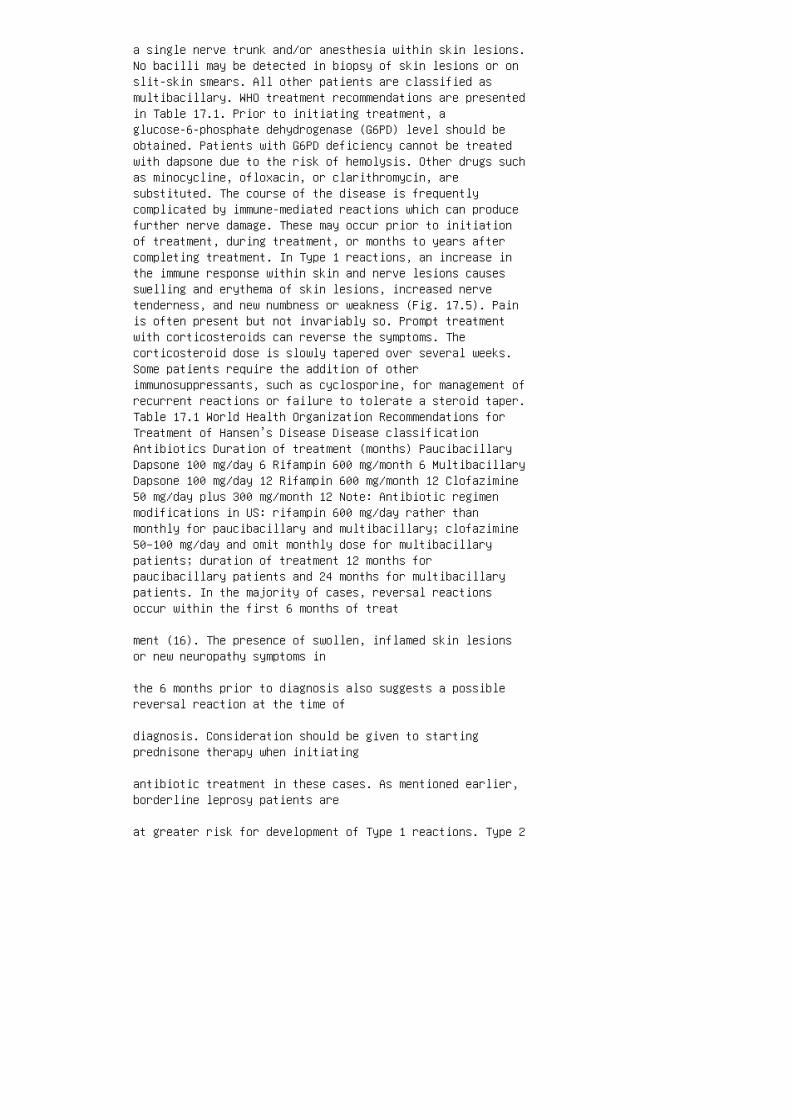
a single nerve trunk and/or anesthesia within skin lesions.No bacilli may be detected in biopsy of skin lesions or onslit-skin smears. All other patients are classified asmultibacillary. WHO treatment recommendations are presentedin Table 17.1. Prior to initiating treatment, aglucose-6-phosphate dehydrogenase (G6PD) level should beobtained. Patients with G6PD deficiency cannot be treatedwith dapsone due to the risk of hemolysis. Other drugs suchas minocycline, ofloxacin, or clarithromycin, aresubstituted. The course of the disease is frequentlycomplicated by immune-mediated reactions which can producefurther nerve damage. These may occur prior to initiationof treatment, during treatment, or months to years aftercompleting treatment. In Type 1 reactions, an increase inthe immune response within skin and nerve lesions causesswelling and erythema of skin lesions, increased nervetenderness, and new numbness or weakness (Fig. 17.5). Painis often present but not invariably so. Prompt treatmentwith corticosteroids can reverse the symptoms. Thecorticosteroid dose is slowly tapered over several weeks.Some patients require the addition of otherimmunosuppressants, such as cyclosporine, for management ofrecurrent reactions or failure to tolerate a steroid taper.Table 17.1 World Health Organization Recommendations forTreatment of Hansen’s Disease Disease classificationAntibiotics Duration of treatment (months) PaucibacillaryDapsone 100 mg/day 6 Rifampin 600 mg/month 6 MultibacillaryDapsone 100 mg/day 12 Rifampin 600 mg/month 12 Clofazimine50 mg/day plus 300 mg/month 12 Note: Antibiotic regimenmodifications in US: rifampin 600 mg/day rather thanmonthly for paucibacillary and multibacillary; clofazimine50–100 mg/day and omit monthly dose for multibacillarypatients; duration of treatment 12 months forpaucibacillary patients and 24 months for multibacillarypatients. In the majority of cases, reversal reactionsoccur within the first 6 months of treat
ment (16). The presence of swollen, inflamed skin lesionsor new neuropathy symptoms in
the 6 months prior to diagnosis also suggests a possiblereversal reaction at the time of
diagnosis. Consideration should be given to startingprednisone therapy when initiating
antibiotic treatment in these cases. As mentioned earlier,borderline leprosy patients are
at greater risk for development of Type 1 reactions. Type 2

reactions, or erythema nodosum leprosum (ENL), are theresult of deposition
of antigen–antibody complexes in various tissues. Patientsat the lepromatous end of
the spectrum are at greatest risk. Symptoms include fever,erythematous and tender
skin nodules, neuritis, arthritis, orchitis, iridocyclitis,and nephritis. Biopsy of skin
nodules reveals polymorphonuclear cells, differentiatingthis lesion from skin lesions
of reversal reaction (17). Thalidomide 100–300 mg/day innonpregnant patients is the
treatment of choice. Thalidomide has well-known teratogeniceffects, and male as well
as female patients must use adequate contraception, as thedrug is present in seminal
fluid. Corticosteroids are also used in the treatment ofENL, especially if neuritis is
present. With MDT, recurrence rates in leprosy areextremely low. New skin lesions or neu
ropathy symptoms associated with a twofold or greaterincrease in the BI of slit-skin
smears suggest relapse. The presence of AFB in skin ornerve biopsy specimens in
treated patients cannot be used as evidence for recurrentinfection, as nonviable bacilli
may persist for years before being cleared by the immunesystem (18,19). To avoid repeated trauma to insensitivehands and feet, patients must be instructed in
proper foot care and in coping behaviors to prevent burns.Physical therapy and occu
pational therapy are important, particularly to preventdeformity and contractures.
Regular ophthalmologic examinations are essential for early
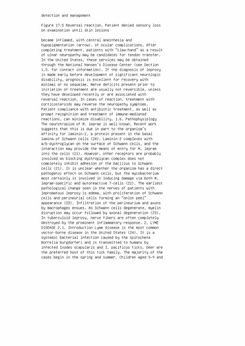
detection and management
Figure 17.5 Reversal reaction. Patient denied sensory losson examination until skin lesions
became inflamed, with central anesthesia andhypopigmentation (arrow). of ocular complications. Aftercompleting treatment, patients with “claw-hand” as a resultof ulnar neuropathy may be candidates for tendon transfer.In the United States, these services may be obtainedthrough the National Hansen’s Disease Center (see Section1.3. for contact information). If the diagnosis of leprosyis made early before development of significant neurologicdisability, prognosis is excellent for recovery withminimal or no sequelae. Nerve deficits present prior toinitiation of treatment are usually not reversible, unlessthey have developed recently or are associated withreversal reaction. In cases of reaction, treatment withcorticosteroids may reverse the neuropathy symptoms.Patient compliance with antibiotic treatment, as well asprompt recognition and treatment of immune-mediatedreactions, can minimize disability. 1.6. PathophysiologyThe neurotropism of M. leprae is well known. Recent worksuggests that this is due in part to the organism’saffinity for laminin-2, a protein present in the basallamina of Schwann cells (20). Laminin-2 complexes witha/b-dystroglycan on the surface of Schwann cells, and theinteraction may provide the means of entry for M. lepraeinto the cells (21). However, other receptors are probablyinvolved as blocking dystroglycan complex does notcompletely inhibit adhesion of the bacillus to Schwanncells (21). It is unclear whether the organism has a directpathogenic effect on Schwann cells, but the mycobacteriummost certainly is involved in inducing damage via both M.leprae-specific and autoreactive T-cells (22). The earliestpathological change seen in the nerves of patients withlepromatous leprosy is edema, with proliferation of Schwanncells and perineurial cells forming an “onion peel”appearance (23). Infiltration of the perineurium and axonsby macrophages ensues. As Schwann cells degenerate, myelindisruption may occur followed by axonal degeneration (23).In tuberculoid leprosy, nerve fibers are often completelydestroyed by the prominent inflammatory response. 2. LYMEDISEASE 2.1. Introduction Lyme disease is the most commonvector-borne disease in the United States (24). It is asystemic bacterial infection caused by the spirocheteBorrelia burgdorferi and is transmitted to humans byinfected Ixodes scapularis and I. pacificus ticks. Deer arethe preferred host of this tick family. The majority of thecases begin in the spring and summer. Children aged 5–9 and

adults aged 45–54 have the highest reported incidence (25).Annual incidence is 0.5% in endemic areas, which includethe Northeast and upper Midwest of the United States (26).2.2. Clinical Features Lyme disease is classified intothree stages. Stage 1 consists of erythema migrans (EM),which occurs at the site of the tick bite. The rash erupts1–30 days after the tick bite and is almost pathognomonicfor Lyme disease. EM classically appears as a nonpruriticbull’s-eye lesion with central clearing, although it canalso be raised, irregular, vesicular, or pruritic (27). EMis usually accompanied by malaise, fatigue, headache,arthralgias, myalgias, fever, and regional lymphadenopathy.Stage 2 follows days to weeks later in
a disseminated form affecting the nervous system, heart, orjoints. Stage 3 follows weeks
to months later and consists of arthritis and/or neurologicfeatures (26,28). In the early stages of Lyme disease,cranial neuropathy, radiculopathy, or lympho
cytic meningitis may occur independently or in combination,a disease complex known as
the Garin–Bujadoux–Bannwarth syndrome. A wide variety ofcranial neuropathies are
associated with Lyme disease, either as a mononeuropathy orin combination. Facial
nerve palsy occurs most commonly and may be unilateral orbilateral (26,29,30). The radiculoneuritis seen in earlystages of infection is relatively severe, predomi
nantly axonal, and tends to resolve spontaneously. Theinterval between the appearance of
cutaneous symptoms and the neurological presentation rangesfrom 18 days to 2 months
(31). Symptoms consist of asymmetric pain, often in aradicular distribution, followed by
weakness in proximal leg muscles. Reflexes are decreased toabsent and sensory loss is
limited to affected nerve roots. Electrophysiologic testingis remarkable for markedly
reduced compound motor action potentials, some prolongation

of distal and F-wave
latencies, and reduced amplitudes of sensory responses.Needle EMG reveals rare fibrilla
tion potentials. Sural nerve biopsies demonstrate loss ofmyelinated and unmyelinated
fibers and myelin ovoids, consistent with axonaldegeneration (31). About one-third of
patients with early Lyme disease have peripheral neuritis.Patterns include thoracic
sensory radiculitis, motor radiculitis, mononeuritismultiplex, mononeuritis, and brachial
plexitis (30). Occasional patients in this early stage maypresent with acute inflammatory
demyelinating polyneuropathy (32). Other rare neurologicalcomplications are phrenic
nerve palsy (33) and autonomic neuropathy (34). In laterstages of Lyme disease, chronic peripheral nerve disordersare common.
Chronic neuropathy occurs in �5% of untreated patients,sometimes after long periods
of latent infection. These neuropathies may affect any limbor an body segment in
either a symmetrical or an asymmetrical pattern. Symptomsusually are reported as par
esthesias or radicular pain. In one study of patients withLyme disease and chronic periph
eral neuropathy, about half presented with distalparesthesias and the other half with
painful radiculopathy. Asymptomatic sensory loss was rarelyobserved, and some patients
reported only numbness. Radicular pain occurred incervical, thoracic, or lumbosacral
distributions. For many patients, paresthesias andradicular symptoms were intermittent.

Physical examination revealed sensory loss for bothsmalland large-fiber modalities.
Weakness and hyporeflexia were less common. Most patientswith either radicular pain
or distal paresthesias had needle EMG evidence of proximalnerve segment involvement
with denervation in paraspinal musculature as well asproximal and distal limb muscles.
Isolated conduction block of the peroneal nerve between theankle and the fibular head
was noted in one patient (29). Associated central nervoussystem (CNS) features in late stages of B. burgdorferi
infection include progressive encephalopathy andneuropsychiatric disorders. Acroderma
titis chronica atrophicans (ACA) is a late manifestation ofLyme disease, occurring in up to
two-thirds (35). It is a cutaneous eruption localized toacral parts of the extremities and
appears as bluish-red skin lesions. In one series, anassociated distal sensory polyneuro
pathy was found in 64% of subjects with ACA (35).
2.3. Laboratory Evaluation
The diagnosis of Lyme disease is usually based on therecognition of the characteristic
clinical findings such as EM, a history of exposure in anendemic area, and antibody
response to B. burgdorferi by enzyme-linked immunosorbentassay (ELISA) and
western blot. Cultivation of B. burdorferi from skin orblood is the gold standard for demonstration of activeinfection, but it is expensive and lacks clinicalsensitivity (36). The urine antigen test is unreliable andnot commonly used (26). There may be little or nodetectable serum antibody in many patients during the first

4–6 weeks of the illness. IgM antibodies are first toappear and suggest recent infection. This is followed 6weeks later by the detection of IgG antibodies. Ifantibiotics are given early in the course of Lyme disease,seroconversion may not occur. Positive IgM serology aloneis not helpful in diagnosing active disease, when theillness has been present longer than a month due to thehigh frequency of false-positive results. If serology isnegative in suspected early Lyme disease, paired acuteandconvalescent-phase serum samples should be tested forevidence of seroconversion. False-positive serologies mayoccur in patients with other infectious and autoimmunedisorders such as syphilis, leptospirosis, yaws, bejel,malaria, subacute bacterial endocarditis, rheumatoidarthritis, and systemic lupus erythematosus (26). IgM orIgG antibody responses to B. burgdorferi may persist for10–20 years without indicating the presence of activeinfection (37). The recombinant OspA Lyme disease vaccinemay give positive IgG results by ELISA, as OspA is themajor outer surface protein expressed in cultured B.burgdorferi. Thus, any seroassay that uses the wholeorganism as its antigen source cannot differentiate betweenvaccinated subjects and those who are naturally infected. Anew sensitive and specific ELISA technique that utilizesrecombinant chimeric borrelia proteins devoid of OspA(rNon-OspA) has been developed and can differentiatebetween patients who have been vaccinated and those withactive or prior infection (38). Cerebrospinal fluid (CSF)can also be tested for the presence of B. burgdorferiantibodies. Nonspecific CSF abnormalities includemononuclear pleocytosis, elevated protein, and normalglucose. PCR is a highly sensitive means of detectingmicrobial DNA or RNA. However, borrelia DNA or RNA has notbeen detected reliably in blood, urine, or CSF of patientswith early or late forms of Lyme disease (39). PCR has beenfound to be the most sensitive and specific test fordetection of B. burgdorferi in skin biopsies from patientswith dermatoborreliosis. For patients withneuroborreliosis, B. burgdorferi DNA was detectable in only17–21% of CSF samples (40). There was a tendency for thePCR positive rate to be higher in patients with earlyneuroborreliosis (,2 weeks) when compared with those withlonger disease duration. Thus, PCR can be helpful in earlyneuroborreliosis. However, PCR does not differentiatebetween the DNA of dead or live spirochetes (36) and will,therefore, not verify the presence of active infection.False-positive results from poor specimen handling andcontamination must also be considered at the time ofinterpretation. 2.4. Differential Diagnosis Thedifferential diagnosis includes other disorders that can

present with cranial and peripheral neuropathies. Theseinclude a number of other infectious diseases includinghuman immunodeficiency virus (HIV), syphilis, mycobacteriaincluding leprosy, and fungal infections. Connective tissuedisorders and malignancy, such as lymphoma, should also beconsidered. 2.5. Management Treatment of early Lyme diseaseusually prevents progression. Most patients withneuroborreliosis require parenteral antibiotics. Exceptionsare patients with isolated facial nerve palsy and normalCSF examinations (30,41). These patients can be treatedwith a number
of oral agents including doxycycline 100 mg p.o. bid,amoxicillin 500 mg p.o. tid, or
tetracycline 500 mg p.o. tid for 2–4 weeks. Doxycycline iscontraindicated during
pregnancy and in children under the age of 8. In otherclinical scenarios, patients are treated with 2–4 weeks ofintravenous cef
triaxone 2 g/day. Other parenteral antibiotics that may beused are intravenous cefotaxime
2 g every 8 h or penicillin G 3.3 million units every 4 h(27). Some authors advocate a
combination of macrolide and hydroxychloroquine to treatlate Lyme disease symptoms
(39). Clinical features of acute neuroborreliosis usuallyresolve within weeks of antibiotic
therapy, but those of chronic neuroborreliosis improve moreslowly, over periods of
several months (28). Complete recovery is rare in patientswith longstanding disease (42).
2.6. Prevention
Patient education regarding proper clothing, carefulinspection for ticks, and avoidance of
infested habitats plays a key role in disease prevention.Chemoprophylaxis with a single
dose of 200 mg doxycycline may be provided within 72 h ofexposure to a tick bite
(28,43). For immunoprophylaxis of high-risk individuals inendemic areas, a recombinant
outer-surface protein (rOspA) Lyme disease vaccine(LYMErix) is available. The vaccine
is given intramuscularly at a dose of 30 mg at 0, 1, and 12months. A 76% efficacy in
preventing infection has been demonstrated after the thirddose (44). Booster doses may
be needed every 1–3 years. Only transient mild-to-moderatevaccine-associated adverse
events have been reported. The vaccine is not approved foruse in children under the
age of 15 or in pregnant women (45) and is licensed onlyfor persons between 15 and
70 years of age.
2.7. Prognosis
Patients usually do well if diagnosed in early stages ofinfection after receiving appropriate
antibiotic therapy (27). Response to therapy is lessfavorable in those with longer disease
duration. Opinions differ on the outcome of chronicneuroborreliosis (42,46). Some have
reported reversal of peripheral nerve manifestations withappropriate antibiotics (46),
whereas others have reported more than a third of patientswith chronic neurological mani
festations that do not improve and about half who have achronic progressive course (42).
Poor outcomes were attributed to failure to eradicate thespirochete completely despite a 2
week course of intravenous antibiotics or to irreversiblenervous system damage.

2.8. Pathophysiology
The pathogenesis of the nerve injury in Lyme diseaseremains unclear. Although injury
directly related to the spirochete is a possibility,parainfectious immunologic mechanisms
have also been proposed (41,47). Nerve biopsies showmeningoradiculitis, lymphocytes,
and plasma cells surrounding many epineurial, perineurial,and endoneurial vessels,
without evidence of vessel wall necrosis. There is loss ofboth myelinated and unmyeli
nated fibers. Myelin ovoids consistent with axonaldegeneration are seen (31). Sural
nerve biopsy from a patient with chronic neuroborreliosisrevealed complement mem
brane attack complexes and macrophages surroundingepineurial vessels and within the
endoneurium. PCR demonstrated B. burgdorferi DNA from thenerve tissue (48). 3. SARCOIDOSIS 3.1. IntroductionSarcoidosis is an immune-mediated, multisystemgranulomatous disorder that presents with hilar adenopathy,pulmonary infiltration, and ocular and skin lesions.Sarcoidoisis was first recognized in 1869, but the causeremains unknown. No infectious agent has been identified.Immunoreactivity is enhanced in sarcoidosis with activatedT-cells and macrophages producing a variety ofproinflammatory mediators at sites of granuloma formation(49). Environmental exposures are believed to interact withgenetic factors in determining the pattern of presentation,progression, and prognosis (50). Sarcoidosis commonlyaffects young and middle-age adults (51) with a slightfemale predominance in symptomatic cases. In the UnitedStates, the prevalence rate in African-Americans is roughly10 times higher than that in Caucasians (51). 3.2. ClinicalFeatures The frequency of neurological involvement insarcoidosis is �5% (52). Most of these patients have othersystemic manifestations of the disease. In two series ofneurosarcoidosis, 48–65% of patients had neurologicdysfunction as their presenting manifestation (52,53).Neurologic manifestations are protean and may involve thecentral or peripheral nervous system. CNS involvement

usually occurs in the early phase of the disease, whereasperipheral neuromuscular involvement is characteristicallyseen in the chronic stages (53). Neurosarcoid patients mayhave single or multiple cranial nerve palsies (52), basilarmeningitis, hydrocephalus, parenchymal disease of thenervous system, peripheral neuropathy, or myopathy (Table17.2). Cranial nerve palsies occur in .50% of patients(54), with the facial nerve being most commonly affected(55). Most other cranial nerves may also be involvedincluding I, II, III, IV, V, VI, VIII, IX, and X. Theincidence of noncranial peripheral neuropathy insarcoidosis varies from 24% to 40% (54,55). The variouspatterns of peripheral neuropathy in sarcoidosis are listedin Table 17.3. 3.3. Polyneuropathy Chronic sensorimotorpolyneuropathy is the most common pattern of peripheralneuropathy in sarcoidosis. The polyneuropathy usuallystarts months to years after systemic features first appear(56). Patients typically present with a subacute to chronicprogressive course of paresthesias, burning or shootingpain, and numbness distally in the hands and feet.Occasional patients develop respiratory failure secondaryto diaphragmatic weakness (56,57). On examination, distalpan-sensory loss consistent with a length-dependentprocess, areflexia, and mild distal weakness are seen.Table 17.2 Comparison of Clinical Manifestations inNeurosarcoidosis Chapelon et al. (55) (n ¼ 35) (%) Stern etal. (52) (n ¼ 33) (%) Sharma (54) (n ¼ 37) (%) Cranialnerve palsies 37 73 52 Meningitis 40 18 24 Peripheralinvolvement 40 6 24 Myopathy 26 12 8 NCS reveal an axonalneuropathy with low-amplitude compound muscle action
potentials and reduced to absent sensory nerve actionpotentials. Needle EMG may
show active denervation in distal muscles.
3.4. Mononeuropathy and Mononeuritis Multiplex
This type of presentation is more often associated withcranial neuropathies as opposed to
a neuropathy involving the limbs (58). Patients withmononeuropathy present with focal
sensory and motor or sensorimotor symptoms that may includepatchy sensory loss over
the trunk (56). Onset is typically subacute withprogressive involvement (59). Electrophysiologic studiestypically reveal an axonal pattern of injury. A multifocal
demyelinating pattern has also been reported withasymmetric onset of weakness and
numbness in the distal extremities that was responsive tocorticosteroid therapy (59).
Electrophysiologic studies in this report revealedconduction block in the median, ulnar,
and peroneal nerves and a reduced sural amplitude.
3.5. Polyradiculopathy
This pattern is rarely seen in sarcoidosis. Presentation isusually in the fourth decade with
back pain, lower extremity weakness most prominentproximally, sphincter dysfunction,
and areflexia in the lower limbs (60). Thoracic, lumbar,and sacral roots are involved
more frequently than the cervical distribution (55). Aconcurrent myelopathy may be
present. Radicular involvement may arise from contiguous,hematogenous, or gravita
tional seeding of nerve root sleeves. Active denervationconsistent with a nerve root
localization can be demonstrated on needle EMG.
3.6. Guillain–Barre´ Syndrome
There have been rare cases of a Guillain–Barre´ syndrome(GBs)-like presentation in the
setting of sarcoidosis (56). One case describes a young manwith sarcoidosis who had
subacute onset of facial and bulbar weakness accompanied byleg weakness. Electrophy
siologic testing revealed prolonged distal latencies,low-amplitude motor amplitudes,
prolonged late responses in the legs, absent sensorypotentials, and prolonged blink

reflexes. He improved with steroids, but had a mild relapse3 years later. Other cases
can present acutely leading to fulminant respiratoryfailure (59). Chronic inflammatory
Table 17.3 Patterns of Peripheral Neuropathy inNeurosarcoidosis
† Symmetric polyneuropathy
W Sensorimotor (49,50)
W Pure sensory (49,51)
W Pure motor (55)
† Mononeuritis multiplex
† Mononeuropathy
† Polyradiculopathy (50,52,55)
† Guillain–Barre´ syndrome (49)
† Lumbosacral plexopathy (49)
† Phrenic neuropathy (53)
† Multifocal sensorimotor neuropathy with conduction block(51) demyelinating polyradiculoneuropathy (CIDP) may alsooccur in association with sarcoidosis (58). 3.7.Lumbosacral Plexopathy with Chronic SensorimotorPolyneuropathy Proximal involvement is rare. A case reportdescribes a patient with foot drop, ipsilateral absence ofpatellar reflex, bilaterally absent achilles reflexes, andvibratory loss in feet. Electrophysiologic studiesdemonstrated a left lumbosacral plexopathy superimposed ona sensorimotor polyneuropathy (56). 3.8. SubclinicalNeuropathy Subclinical mononeuritis multiplex appears to becommon in sarcoidosis. In a comparison study ofasymptomatic sarcoid patients to a similar number ofage-matched control subjects, abnormal sensory amplitudesin one or more nerves were found in two-thirds of sarcoidpatients when compared with few controls. Mean sensoryamplitudes for the median, ulnar, and sural nerves werelower in patients than in controls. Asymptomaticcompression of nerves by granulomata is the proposedmechanism (61). 3.9. Laboratory Evaluation The diagnosis of

sarcoidosis may be challenging when patients present withneurologic symptoms in the absence of systemic features. Asthe incidence of systemic involvement is high, routinechest radiography and ophthalmologic evaluation must bedone to assess for hilar adenopathy, uveitis, or fundusgranulomas (53). Chest radiographs are abnormal in 78–90%cases (54). Routine chemistry must be performed to excludeinvolvement of other organ systems. Hypercalcemia is seenin 17% of cases. Serum angiotensin converting enzyme (ACE)levels are elevated in 54–65% of cases, but are not highlysensitive or specific (54,55). Serum lysozyme is also usedas a marker of sarcoidosis activity. It has a highersensitivity, but lower specificity than ACE levels (62).CSF analysis may be normal (56) or may reveal an elevatedprotein (mean value, 326 mg/dL), lymphocytosis (mean value,156 cells/mm 3 ), and hypoglycorrhachia (60). Thesefindings, in the absence of positive cytology and culturesfor bacteria, AFB, and fungi, are supportive ofneurosarcoidosis. Myelography may demonstrate nerve rootthickening and irregularity in patients withpolyradiculopathy. A definitive diagnosis of sarcoidosisrequires histologic confirmation of noncaseating granulomasin the setting of a compatible clinical or radiologicpicture. Muscle, nerve, skin, lymph node, conjunctival, orminor salivary gland biopsies can be performed forhistologic confirmation. Because pulmonary involvement isvery common with sarcoidosis, transbronchial biopsy ormediastinoscopy are options. Gallium scan or computedtomography of the chest may also be helpful in theevaluation. 3.10. Differential Diagnosis Sarcoidosis is amultisystem disease and neurologic involvement may occur inisolation without the typical systemic features. Hence,when patients present with the various cranial andperipheral manifestations, etiologies such as HIVinfection, mycobacterial and fungal infections includingleprosy, lymphoma, syphilis, and Lyme disease must be
kept in mind. Vasculitides in isolation or in associationwith connective tissue disorders
should also be considered. Common causes ofpolyradiculopathy and sensorimotor
polyneuropathy, such as diabetes mellitus, must also beexcluded.
3.11. Management and Prognosis
Treatment is not indicated for asymptomatic or minimallysymptomatic disease (63), and

these patients may improve spontaneously (53). In patientswith more significant disease,
corticosteroids remain the mainstay of treatment.Prednisone is usually sufficient in con
trolling the underlying inflammatory process (52).Prednisone doses range from 0.5 to
1 mg/kg daily, depending on the clinical scenario.Treatment at this higher dosage
should continue for at least 2 months, with lower dosescontinued for longer periods to
maintain functional improvement (55,63). In patients withrelapses, intravenous cortico
steroids (solumedrol 1 g/day 3 days) should be considered.If patients fail to respond
to this regimen, azathioprine, chlorambucil, methotrexate,cyclosporine, or cyclophospha
mide are options. Decompressive laminectory may beappropriate when there is mass
effect and it also provides tissue for diagnosis (60).Patients presenting with a clinical
picture consistent with GBS or CIDP should be treated withplasma exchange or intra
venous gammaglobulin (58), as one would in cases notassociated with sarcoidosis. The overall prognosis ofneurosarcoidosis is relatively favorable. A positive
outcome has been observed in two-thirds of well-documentedepisodes of neurologic dys
function (52). However, outcomes for patients withperipheral nerve involvement are
somewhat less favorable than for neurosarcoidosis of theCNS, two-thirds of patients
with CNS manifestations recovered compared with 42% withperipheral neuropathy

(55). In patients with chronic neurosarcoidosis, theprognosis is relatively poor, with
death occurring in 20% from complications related tosarcoidosis (54).
3.12. Pathophysiology
Nerve biopsy specimens show noncaseating granulomas in theepineurial and perineurial
spaces, periangiitis, panangiitis, and axonal degeneration(64). Axonal degeneration is
probably a result of involvement of the vasa nervorum anddirect compression by the
granulomas. Immune dysfunction may also play a role in thepathogenesis as a number
of autoimmune disorders are associated with sarcoidosis.
4. HERPES ZOSTER
4.1. Introduction
Radiculitis or cranial neuritis due to herpes zoster,commonly referred to as “shingles,” is
the result of re-activation of latent varicella zostervirus (VZV) in sensory ganglia.
Decreased immune responsiveness associated with advancingage, malignancy, use of
cytotoxic agents, or infection with HIV increases the riskof developing herpes zoster.
4.2. Clinical Features
A vesicular rash appears in a trigeminal or dermatomaldistribution, associated with
radicular pain and paresthesias that usually conform to thesame body region. Loss of
pain and temperature sensation is typically patchy withinthe dermatome. Pain
may precede the rash by a week or more. Fever, malaise, and

headache may also occur. In nonimmunocompromised patients,the vesicles resolve in 2–3 weeks, usually with nosequelae. Patients may develop focal weakness in aradicular distribution. Ramsay–Hunt syndrome, thedevelopment of facial paresis after herpes zoster oticus,is the most common example. In general, myotomalinvolvement correlates to the dermatome(s) affected byrash, although rarely weakness may not coincideanatomically with the cutaneous eruption (65–68). Weaknessprogresses over hours or days, then plateaus. Most of thetime, the rash precedes the weakness but rarely, weaknessmay antedate the rash. Focal weakness in a spinal nerveroot or cranial nerve distribution is rare (5%), with alatency varying from 1 day to 5 weeks after eruption of therash (65). Motor involvement ranges from mild weakness in asingle myotome to nearly complete paralysis of anextremity. Deep tendon reflexes are reduced, often out ofproportion to the degree of muscle weakness, consistentwith involvement of the afferent arc of the spinal reflex.Oculomotor paresis, both with and without pupillaryinvolvement, may occur in association with rash in thedistribution of the ophthalmic division of the trigeminalnerve. Rarely, in both immunocompetent andimmunocompromised individuals, focal sensory and motorsymptoms may occur in the absence of the vesicular rash,the so-called zoster sine herpete (69–71). In the absenceof rash, diagnosis is challenging. PCR for VZV in CSF ordetection of VZV antibody in CSF can be helpful inestablishing the diagnosis (70,72). Recurrentpolyneuropathies without cutaneous manifestations of herpeszoster have been diagnosed in the setting of presumedchronic VZV infection that was based on the presence of VZVantibodies in the CSF (73). Other uncommon neurologicalcomplications of VZV are aseptic meningitis, encephalitis,transverse myelitis, and urinary retention. Thesecomplications occur in ,1% of immunocompetent patients, butin as many as 35% of immunocompromised hosts (74).Post-herpetic neuralgia (PHN) is a chronic pain syndromethat is seen in greater frequency in older patients. Thenumber of patients reporting persistent pain �3 monthsafter resolution of the rash ranges from 11% in patients,30years (75) to 39% in patients .50 years (76,77). 4.3.Laboratory Evaluation In the setting of the trademarkvesicular rash and uncomplicated sensory symptoms, thediagnosis is straightforward and requires no specificlaboratory testing. In cases without rash or when severecomplications arise, further investigation is indicated.Magnetic resonance imaging (MRI), T1-weighted sequencesbefore and after infusion of gadolinium may showenhancement of spinal nerve roots or cranial nerves
(68,78). T2 and proton density sequences may showhigh-signal intensity in the spinal cord in cases ofmyelitis (79) and in subcortical white matter or brainstemin cases of encephalitis (79–83). However, MRI may benormal even in cases of encephalitis (81). CSF analysisoften shows mild-tomoderate elevation of protein and avariable lymphocytic pleocytosis with normal glucose. PCRfor VZV in CSF is an important and specific method ofdetecting infection, as are immunosorbent assays for IgGand IgM antibodies to VZV in the CSF (84). In the cases ofsegmental paresis, NCS reveal normal sensory and motorresponses in most patients (65). However, other studieshave shown reduced sensory nerve action potentials in asmany as 52% (85). Motor nerve responses may be normal orhave lowamplitudes (65,85). Needle EMG performed �2 weeksafter onset of weakness usually demonstrates activedenervation potentials in clinically weak muscles, but havealso been reported in a myotomal distribution correspondingto the affected dermatomes even when no clinical weaknessis apparent (86).
4.4. Differential Diagnosis
In cases of zoster sine herpete, the differential diagnosisincludes other structural, inflam
matory, or infectious causes of radiculopathy or cranialneuropathy, including herniated
vertebral disc, diabetes, or Lyme disease. In the settingof myelitis or encephalitis, viral
infection including HIV or lupus or other inflammatoryprocesses should be considered.
4.5. Management and Prognosis
To reduce the time for healing of cutaneous lesions andpossibly to reduce the incidence of
segmental paresis (85), immunocompetent patients should betreated with valacyclovir
1000 mg p.o. three times daily for 7 days, famciclovir 500mg p.o. three times daily for
7 days, or acyclovir 800 mg p.o. five times daily for 7–10days. Treatment started
within 72 h of onset of the rash is the most advantageous.

The incidence of PHN is not
reduced by antiviral therapy, but the duration of pain maybe shortened (85,87). Intra
venous acyclovir is effective in preventing disseminationof herpes zoster in immuno
compromised individuals. The use of steroids is probablynot indicated and should be
avoided in immunocompromised patients. For PHN,amitriptyline and other tricyclic antidepressants andgabapentin are effec
tive. Carbamazepine may be especially useful for treatinglancinating pain. Short-term use
of narcotics should be considered, especially in refractorypatients. The prognosis for full recovery is good. Evenpatients who develop focal weakness
usually regain normal strength or have only slight residualnumbness or weakness.
4.6. Pathophysiology
With re-activation of VZV, replication and release of thevirus occur in dorsal root ganglia
and spread to the entire ganglion. Involved dorsal rootganglia on autopsy show inflam
mation and necrosis (88). Destruction of sensory neuronsleads to temporary or permanent
sensory loss in the corresponding dermatome, but alsoremoves reservoirs for the virus
(89). The active infection explains the pleocytosis seen inCSF. VZV spreads along the
nerve to the skin. Light microscopy demonstratesperivascular lymphocytic infiltration
in the early erythematous cutaneous stage. During thevesicular stage, viral antigens
may be detected in epidermis and in capillary and venuleendothelial cells and the peri

neurium of dermal nerves (90). The pathogenesis of PHN isnot well understood. PHN may arise from structural or
physiologic changes in the DRG (89). Alternatively, C-fibernociceptors may become sen
sitized, with development of spontaneous or stimulus-evokedpain. Further, continuous
nociceptor discharge may lead to central hyperexcitabilityof spinal DRG. These neurons
may have exaggerated responses to input and may contributeto development of allodynia
(91). Skin biopsies of patients with pain 3 months afterresolution of HZV rash show
significantly fewer epidermal nerve endings than dobiopsies of patients without pain (92).
1. Rees RJW, McDougall AC. Airborne infection withMycobacterium leprae in mice. Int J Lepr 1976; 44:99–103.
2. Abraham S, Mozhi NM, Joseph GA, Kurian N, Rao PSSS, JobCK. Epidemiological significance of first skin lesion inleprosy. Int J Lepr 1998; 66:131–139. 3. Lara CB, NolascoJO. Self-healing, or abortive, and residual forms ofchildhood leprosy and their probable significance. Int JLepr 1956; 24:245–263. 4. Ridley DS, Jopling WH.Classification of leprosy according to immunity. Afive-group system. Int J Lepr 1966; 34:255–273. 5. FieldsJP, Meyers WM. Mycobacterial infections. In: Farmer ER,Hood AF, eds. Pathology of the Skin. 2nd ed. McGraw-Hill:New York, 2000:553–578. 6. Davey TF, Rees RJW. The nasaldischarge in leprosy: clinical and bacteriological aspects.Lepr Rev 1974; 45:121–134. 7. Lewallen S. Prevention ofblindness in leprosy: an overview of the relevant clinicaland programme-planning issues. Ann Trop Med Parasitol 1997;91:341–348. 8. van Brakel WH. Peripheral neuropathy inleprosy and its consequences. Lepr Rev 2000;71(suppl):S146–S153. 9. Rodriguez G, Sanchez W, Chatela JG,Soto J. Primary neuritic leprosy. J Am Acad Dermatol 1993;29:1050–1052. 10. Amenu A, Saunderson P, Desta K, Byass P.The pattern of decline in bacillary index after 2 years ofWHO recommended multiple drug therapy: the AMFES cohort.Lepr Rev 2000; 71:332–337. 11. De Wit MY, Douglas JT,McFadden J, Klaster PR. Polymerase chain reaction fordetection of Mycobacterium leprae in nasal swab specimens.

J Clin Microbiol 1993; 31:502–506. 12. Hackett ER, ShipleyDE, Livengood R. Motor nerve conduction velocity studies ofthe ulnar nerve in patients with leprosy. Int J Lepr 1968;36:282–287. 13. Antia NH, Pandya SS, Dastur DK. Nerves inthe arm in leprosy: I. Clinical, electrodiagnostic andoperative aspects. Int J Lepr 1970; 38:12–29. 14. Swift TR,Hackett ER, Shipley DE, Miner KM. The peroneal and tibialnerves in lepromatous leprosy: clinical andelectrophysiologic observations. Int J Lepr 1973; 41:25–34.15. Anonymous. Global leprosy situation, September, 1999.Wkly Epidemiol Rec 1999; 74:313–316. 16. Roche PW, LeMaster J, Butlin CR. Risk factors for type 1 reactions inleprosy. Int J Lepr 1997; 65:450–455. 17. Naafs B. Currentviews on reactions in leprosy. Indian J Lepr 2000;72:97–122. 18. Sharma A, Sharma VK, Rajwanshi A, Das A,Kaur I, Kumar B. Presence of M. leprae in tissues in slitskin smear negative multibacillary (MB) patients afterWHO-MBR. Lepr Rev 1999; 70:281–286. 19. Ebenezer GJ, Job A,Abraham S, Arunthathi S, Rao PSSS, Job CK. Nasal mucosa andskin of smear-positive leprosy patients after 24 months offixed duration MDT: histopathological and microbiologicalstudy. Int J Lepr 1999; 67:292–297. 20. Rambukkana A,Salzer JL, Yurchenco PD, Tuomanen EI. Neural targeting ofMycobacterium leprae mediated by the G domain of thelaminin-a2 chain. Cell 1997; 88:811–821. 21. Rambukkana A,Yamada H, Zanazzi G, Mathus T, Salzer JL, Yurchenco PD,Campbell KP, Fischetti VA. Role of a-dystroglycan as aSchwann cell receptor for Mycobacterium leprae. Science1998; 282:2076–2079. 22. Spierings E, de Boer T, ZulianelloL, Ottenhoff THM. The role of Schwann cells, T cells andMycobacterium leprae in the immunopathogenesis of nervedamage in leprosy. Lepr Rev 2000; 71(suppl):S121–S129. 23.Job CK. Pathology of peripheral nerve lesions inlepromatous leprosy—a light and electron microscopic study.Int J Lepr 1971; 39:251–268. 24. Piacentino JD, SchwartzBS. Occupational risk of Lyme disease: an epidemiologicalreview. Occup Environ Med 2002; 59:75–84. 25. Orloski KA,Hayes EB, Campbell GL, Dennis DT. Surveillance for Lymedisease-United States, 1992–1998. MMWR CDC Surveill Summ2000; 49:1–11. 26. Verdon ME, Sigal LH. Recognition andmanagement of Lyme disease. Am Fam Physician 1997;56:427–436. 27. Coyle PK, Schutzer SE. Neurologic aspectsof Lyme disease. Med Clin North Am 2002; 86:261–284.
28. Steere AC. Lyme disease. N Engl J Med 2001; 345:115–125.
29. Logigian EL, Steere AC. Clinical and electrophysiologicfindings in chronic neuropathy of Lyme disease. Neurology1992; 42:303–311.

30. Pachner AR, Steere AC. The triad of neurologicmanifestations of Lyme disease: meningitis, cranialneuritis, and radiculoneuritis. Neurology 1985; 35:47–53.
31. Vallat JM, Hugon J, Lubeau M, Leboutet MJ, Dumas M,Desproges-Gotteron R. Tick-bite meningoradiculoneuritis:clinical, electrophysiologic, and histologic findings in 10cases. Neurology 1987; 37:749–753.
32. Crisp D, Ashby P. Lyme radiculoneuritis treated withintravenous immunoglobulin. Neurology 1996; 46:1174–1175.
33. Faul JL, Ruoss S, Doyle RL, Kao PN. Diaphragmaticparalysis due to Lyme disease. Eur Respir J 1999;13:700–702.
34. Chatila R, Kapadia CR. Intestinal pseudoobstruction inacute Lyme disease: a case report. Am J Gastroenterol 1998;93:1179–1180.
35. Kindstrand E, Nilsson BY, Hovmark A, Pirskanen R,Asbrink E. Peripheral neuropathy in acrodermatitis chronicaatrophicans—a late Borrelia manifestation. Acta NeurolScand 1997; 95:338–345.
36. Bunikis J, Barbour AG. Laboratory testing for suspectedLyme disease. Med Clin North Am 2002; 86:311–340.
37. Kalish RA, McHugh G, Granquist J, Shea B, Ruthazer R,Steere AC. Persistence of immunoglobulin M orimmunoglobulin G antibody responses to Borrelia burgdorferi10–20 years after active Lyme. Clin Infect Dis 2001;33:780–785.
38. Gomes-Solecki MJ, Wormser GP, Schreifer M, Neuman G,Hannafey L, Glass JD, Dattwyle RJ. Recombinant assay forserodiagnosis of lyme disease regardless of OspAvaccination status. J Clin Microbiol 2002; 40:193–197.
39. Donta ST. Late and chronic Lyme disease. Med Clin NorthAm 2002; 86:341–349.
40. Lebech AM. Polymerase chain reaction in diagnosis ofBorrelia burgdorferi infections and studies on taxonomicclassification. APMIS 2002; 105(suppl):1–40.
41. Logigian EL. Peripheral nervous system Lymeborreliosis. Semin Neurol 1997; 17:25–30.
42. Logigian EL, Kaplan RF, Steere AC. Chronic neurologic

manifestations of Lyme disease. N Engl J Med 1990;323:1438–1444.
43. Nadelman RB, Nowakowski J, Fish D, Falco RC, Freeman K,McKenna D, Welch P, Marcus R, Aguero-Rosenfeld ME, DennisDT, Wormser GP. Tick Bite Study Group. Prophylaxis withsingle-dose doxycycline for the prevention of Lyme diseaseafter an Ixodes scapularis tick bite. N Engl J Med 2001;345:79–84.
44. Poland GA, Jacobson RM. The prevention of Lyme diseasewith vaccine. Vaccine 2001; 19:2303–2308.
45. Rahn DW. Lyme vaccine: issues and controversies. InfectDis Clin North Am 2001; 15:171–187.
46. Halperin J, Luft BJ, Volkman DJ, Dattwyler RJ. Lymeneuroborreliosis. Peripheral nervous system manifestations.Brain 1990; 113:1207–1221.
47. Sigal LH. Immunologic mechanisms in Lymeneuroborreliosis: the potential role of autoimmunity andmolecular mimicry. Semin Neurol 1997; 17:63–68.
48. Maimone D, Villanova M, Stanta G, Bonin S, MalandriniA, Guazzi GC, Annunziata P. Detection of Borreliaburgdorferi DNA and complement membrane attack complexdeposits in the sural nerve of a patient with chronicpolyneuropathy and tertiary Lyme disease. Muscle Nerve1997; 20:969–975.
49. Moller DR. Etiology of sarcoidosis. Clin Chest Med1997; 18:695–706.
50. Martinetti M, Dugoujon JM, Tinelli C, Cipriani A,Cortelazzo A, Salvaneschi L, Casali L, Semenzato G, CucciaM, Luisetti M. HLA-Gm/kappam interaction in sarcoidosis.Suggestions for a complex genetic structure. Eur Respir J2000; 16(1):74–80.
51. Hosoda Y, Yamaguchi M, Hiraga Y. Global epidemiology ofsarcoidosis. What story do prevalence and incidence tellus? Clin Chest Med 1997; 18:681–694
52. Stern BJ, Krumholz A, Johns C, Scott P, Nissim J.Sarcoidosis and its neurological manisfestations. ArchNeurol 1985; 42:909–917. 53. Delaney P. Neurologicalmanifestations in sarcoidosis. Review of the literature,with a report of 23 cases. Ann Intern Med 1977; 87:336–345.54. Sharma OP. Neurosarcoidosis. A personal perspective

based on the study of 37 patients. Chest 1997; 112:220–228.55. Chapelon C, Ziza JM, Piette JC, Levy Y, Raguin G,Wechsler B, Bitker MC, Bletry O, Laplane D, Bousser MG,Godeau P. Neurosarcoidosis: signs, course and treatment in35 confirmed cases. Medicine 1990; 69:261–276. 56. ZunigaG, Ropper AH, Frank J. Sarcoid peripheral neuropathy.Neurology 1991; 41:1558–1561. 57. Robinson LR, BrownsbergerR, Raghu G. Respiratory failure and hypoventilationsecondary to neurosarcoidosis. Am J Respir Crit Care Med1998; 157:1316–1318. 58. Mendell JR, Kissel JT, CornblathDR. Diagnosis and Management of Peripheral Nerve Disorders.Oxford University Press, Inc., 2001. 59. Said G, Lacroix C,Plante-Bordeneuve V, Page LL, Pico F, Presles O, Senant J,Remy P, Rondepierre P, Mallecourt J. Nerve granulomas andvasculitis in sarcoid peripheral neuropathy. Aclinicopathological study of 11 patients. Brain 2002;125:264–275. 60. Koffman B, Junck L, Elias SB, Feit HW,Levine SR. Polyradiculopathy in sarcoidosis. Muscle Nerve1999; 22:608–613. 61. Challenor YB, Felton CP, Brust JC.Peripheral nerve involvement in sarcoidosis: anelectrodiagnostic study. J Neurol Neurosurg Psychiatry1984; 47:1219–1222. 62. Tomita H, Sato S, Matsuda R,Sugiura Y, Kawaguchi H, Niimi T, Yoshida S, Morishita M.Serum lysozyme levels and clinical features of sarcoidosis.Lung 1999; 177:161–167 63. Johns CJ, Michele TM. Theclinical management of sarcoidosis. A 50-year experience atthe Johns Hopkins hospital. Medicine 1999; 78:65–111. 64.Oh SJ. Sarcoid polyneuropathy: a histologocally provedcase. Ann Neurol 1980; 7:178–181. 65. Thomas JE, Howard FM.Segmental zoster paresis—a disease profile. Neurology 1972;22:459–466. 66. Taterka JH, O’Sullivan ME. The motorcomplications of herpes zoster. J Am Med Assoc 1943;122:737–739. 67. Kendall D. Motor complications of herpeszoster. Br Med J 1957; 2:616–618. 68. Hanakawa T, HashimotoS, Kawamura J, Nakamura M, Suenago T, Matsuo M. Magneticresonance imaging in a patient with segmental zosterparesis. Neurology 1997; 49:631–632. 69. Osaki Y,Mutsubayashi K, Okumiya K, Wada T, Doi Y. Polyneuritiscranialis due to varicellazoster virus in the absence ofrash. Neurology 1995; 45:2293. 70. Gilden DH, Wright RR,Schneck SA, Gwaltney JM, Mahalingam R. Zoster sine herpete,a clinical variant. Ann Neurol 1994; 35:530–533. 71.Elliott KD. Other neurological complications of herpeszoster and their management. Ann Neurol 1994;35(suppl):S57–S61. 72. Gilden DH, Dueland AN, Devlin ME.Varicella-zoster reactivation without rash. J Infect Dis1992; 166(suppl 1):S30–S34. 73. Fox RJ, Galetta SL,Mahalingamm R, Wellish M, Forghani B, Gilden DH. Acute,chronic, and recurrent varicella zoster virus neuropathywithout zoster rash. Neurology 2001; 57:351–354. 74. Weller

TH. Varicella and herpes zoster: changing concepts of thenatural history, control and importance of a not-so-benignvirus (part two). N Engl J Med 1983; 309:1434–1440. 75. GohCL, Khoo L. A retrospective study of the clinicalpresentation and outcome of herpes zoster in a tertiarydermatology outpatient referral clinic. Int J Dermatol1997; 36:667–672. 76. Ragozzino MW, Melton IJ, Kurland LT.Population based study of herpes zoster and its sequelae.Medicine 1982; 21:310–316. 77. de Moragas JM, Kierland RR.The outcome of patients with herpes zoster. Arch Dermatol1957; 75:193–196. 78. Osumi A, Tien RD. MR findings in apatient with Ransay–Hunt syndrome. J Comput Assist Tomogr1990; 14:991–993. 79. Haanpaa M, Dastidar P, Weinberg A,Levin M, Miettinen A, Lapinlampi A. CSF and MRI finding inpatients with acute herpes zoster. Neurology 1998;51:1405–1411.
80. Tien RD, Felsberg GJ, Osumi AK. Herpes virus infectionsin the CNS: MR findings. Am J Roentgenol 1993; 161:167–176.
81. De la Blanchardiere A, Rozenberg F, Caumes E, Picard O,Lionnet F, Livartowski J, Coste J, Sicard D, Lebon P,Salmon-Ceron D. Neurological complications ofvaricella-zoster virus infection in adults with humanimmunodeficiency virus infection. Scand J Infect Dis 2000;32:263–269.
82. Kidd D, Duncan JS, Tompson EJ. Pontine inflammatorylesion due to shingles. J Neurol Neurosurg Psychiatry 1998;65:208.
83. Weaver S, Rosenblum MK, DeAngelis LM. Herpes varicellazoster encephalitis in immunocompromised patients.Neurology 1999; 52:193–197.
84. Kleinschmidt-DeMasters, Gilden DH. Varicella-zostervirus infections of the nervous system. Arch Pathol Lab Med2001; 125:770–780.
85. Mondelli M, Romano C, Passero S, Della Porta P, RossiA. Effects of acyclovir on sensory axonal neuropathy,segmental motor paresis and postherpetic neuralgia inherpes zoster patients. Eur Neurol 1996; 36:288–292.
86. Greenberg MK, McVey AL, Hayes T. Segmental motorinvolvement in herpes zoster: an EMG study. Neurology 1992;42:1122–1123.
87. Alper BS, Lewis PR. Does treatment of acute herpeszoster prevent or shorten postherpetic neualgia? A

systematic review of the literature. J Fam Pract 2000;49:256–264.
88. Lungo O, Ammunziato PW, Gershon A, Staugaitis SM,Josephson D, LaRussa P, Silverstein SJ. Reactivated andlatent varicella-zoster virus in human dorsal root ganglia.Proc Natl Acad Sci USA 1995; 92:10980–10984
89. Steiner I. Human herpes viruses latent infection in thenervous system. Immunol Rev 1996; 152:158–173.
90. Muraki R, Baba T, Iwasaki T, Sata T, Kurata T.Immunohistochemical study of skin lesions in herpes zoster.Pathol Anat Histopathol 1992; 420:71–76.
91. Bennett GJ. Hypotheses on the pathogenesis of herpeszoster—associated pain. Ann Neurol 1994; 35(suppl):S38–S40.
92. Oaklander AL. The density of remaining nerve endings inhuman skin with and without postherpetic neuralgia aftershingles. Pain 2001; 92:139–145.
18
Critical Illness Polyneuropathy and
Myopathy
J. Robinson Singleton and Estelle S. Harris
University of Utah, Salt Lake City, Utah, USA
Abstract 315
1. Introduction 316
2. Differential Diagnosis 316
3. Clinical Presentation of CIP 317
4. Electrodiagnosis of CIP 318
5. Critical Illness Myopathy 318
6. Distinguishing CIP and CIM 319
7. Risk Factors and Pathogenesis 320
8. Prognosis in CIP and CIM 321

9. Treatment 321
ABSTRACT
Weakness is common in patients requiring intensive care andis often attributable to injury
to either peripheral nerves or muscle. Critical illnesspolyneuropathy (CIP) is a distal
axonal neuropathy observed in severely ill patients withsepsis and multiple organ
system failures. Critical illness myopathy (CIM) is acatabolic injury to skeletal muscle
that occurs in the same setting. There is controversy overthe relative contribution of
these two entities to the spectrum of clinical weaknessobserved in critically ill patients.
Up to 70% of patients receiving intensive care developweakness associated due to CIP,
CIM, or both. Diagnosis of either entity may be delayedbecause superimposed medical
problems can obscure clinical weakness. This chapter willreview the differential diagno
sis, clinical and electrodiagnostic features of CIP, anddescribe the common pathogenesis
shared by CIP and the closely related CIM. 315 1.INTRODUCTION Weakness is common in critically ill patients.In 1984, Bolton et al. (1) formulated the concept that theattendant metabolic and immune stressors of sepsis andcritical illness cause reversible injury to peripheralnerve and muscle (2). Since that time, there has been anapparent increased prevalence of critical illnesspolyneuropathy (CIP) as neurologists and intensive carespecialists became familiar with these disorders.Prevalence figures also depend on diagnostic criteria, witholder studies identifying few patients with severe andprotracted weakness, to identification of 70% of patientswith electrodiagnostic abnormalities consistent withmotor-predominant axonal injury (3,4). Recent studiesquestion whether these patients may in fact have a criticalillness myopathy (CIM) instead of or in addition to, CIP.

It is challenging to detect and diagnose weakness incritically ill patients. They are usually intubated, oftensedated and sometimes paralyzed. Many have alteredconsciousness or mental status, and questions aboutweakness or sensory symptoms are limited or impossible.Physical examination may be restricted by the paraphernaliaof critical illness, including intravenous and arteriallines, endotracheal tubes, splints, and alternatingcompression devices. Weakness is often undetected untiltransfer to convalescent care or until weakness leads todifficulty weaning from assisted ventilation. Accordingly,ICU care providers should be attentive to evidence ofweakness in critically ill patients and not mistakenlyascribe weakness to drugs or sedation. It is important torequest that neuromuscular blocking agents and sedation beheld, if possible, before the neurological examination.Simple resistive strength testing can establish the degreeof weakness and help to distinguish symmetric weaknessassociated with CIP or CIM from other causes of weakness.2. DIFFERENTIAL DIAGNOSIS The broad differential diagnosisof weakness in this setting includes metabolicabnormalities, disorders of the central nervous system, anddisorders of the peripheral nervous system. Stroke ordiffuse anoxic brain injury frequently accompany acutemyocardial infarction or respiratory arrest. Vertebraldissection or traumatic myelopathy can be late orunrecognized consequences of head and neck trauma (5).Ischemic myelopathy can be an initially unnoticedcomplication following major thoracic or abdominal surgicalprocedures. Psychomotor retardation from advanced dementia,severe bradykinesia in parkinsonism, catatonia, and avariety of metabolic abnormalities can mimic neuromuscularweakness. The differential diagnosis of neuromuscularcauses of weakness includes diseases that can occur inconjunction with critical illness or are caused by criticalillness. Among, the former is Guillain–Barre´ Syndrome(GBS) which causes rapidly progressive ascending limbweakness accompanied by sensory complaints and diffuse backand body pain. Diaphragmatic weakness impairs ventilationin 20% and requires intubation in 5–10%, leading to ICUadmission. Patients with limb weakness that precedesrespiratory compromise usually do not present a diagnosticchallenge. However, GBS can also be precipitated by acritical illness. Although the most common form of GBS,acute inflammatory demyelinating polyradiculoneuropathy(AIDP), typically causes primary multisegmentaldemyelination, axonal forms of GBS are also described.Acute motor and sensory axonal neuropathy (AMSAN) isrecognized as a severe form of GBS with prolonged weaknessand poor prognosis. These AIDP variants are frequently
associated with specific
anti-ganglioside antibodies (anti-GM1 and anti-GD1A) (6,7),but are likely to be difficult to
distinguish from CIP clinically or electrodiagnostically(8). Exacerbations of myasthenia gravis can lead torespiratory compromise or pneumo
nia during an exacerbation. Although rare, sepsis, organfailure, or surgery can precipitate
a myasthenic crisis in a patient not known to have thedisease. Botulism, acetylcholinester
ase inhibitors, and a variety of toxins cause precipitousweakness, respiratory failure, and
autonomic dysfunction. Prolonged neuromuscular blockadewithout evidence of myo
pathy has been described after withdrawal of neuromuscularblocking agents (especially
vecuronium) and in association with aminoglycosides (9,10).Acute heavy metal intoxi
cations (thallium and arsenic) and porphyria canprecipitate metabolic dysequilibrium
and diffuse neuropathy that occur simultaneously. CIP andCIM are unique as neuromuscular diseases because theyappear to be
directly caused by critical illness. CIP is more common inpatients with sepsis and
multiorgan failure, whereas glucocorticoids andneuromuscular blocking agents predis
pose to CIM. Distinguishing CIM from CIP can be difficultboth clinically and electrodiag
nostically. Because of overlap between the two syndromes interms of setting, clinical
presentation, electrodiagnostic findings, and pathogenesis,they may be best regarded as
poles in a spectrum of neuromuscular disease associated

with critical illness.
3. CLINICAL PRESENTATION OF CIP
CIP is an acute or subacute, axonal, length-dependentneuropathy that occurs in severely
ill patients in proportion to the acuity of systemicdisease. The neuropathy is monophasic
and self-limited and generally gradually resolves if thepatient survives. Confusion exists
about the clinical boundaries of CIP. The prototypicalpresentation is rapid development of
profound weakness affecting limbs in a distal to proximalgradient days to weeks after
acquiring a severe illness. Acute axonal injury has beenreported to occur within 72 h
after ICU admission (11–13). In severe cases, muscles ofrespiration are also involved,
resulting in respiratory failure, but more commonly asfailure to wean from ventilatory
support. Weakness is accompanied by muscle atrophy andhyporeflexia. Complete
areflexia is rare, even in severely affected patients,distinguishing CIP from AIDP
clinically. Sensory symptoms should be a key feature indistinguishing CIP from CIM, but
sensory involvement is variable (14). It is often difficultor impossible to clinically evalu
ate sensory loss in the acute care setting. A forced choicetechnique, such as joint position
sense testing, has the greatest chance of providingobjective data. However, CIP is viewed
as a motor predominant or exclusive disease by most authors(15–18), and this view is
generally borne out by electrodiagnostic testing. Ancillary

laboratory tests are rarely instructive. Creatinephosphokinase (CPK) is
normal or minimally elevated. CPK elevations greater thantwice the upper limit of
normal suggest CIM, whereas CPK values.2000 IU/dL morelikely reflect myonecrosis.
Cerebral spinal fluid protein is normal (in contrast tofindings in GBS) with no elevation of
white blood cells. Little pathological data are available.A review of autopsy specimens for nine
patients with clinical CIP who died of their criticalillness (19) showed primary axonal
degeneration of both motor and sensory nerve fibers, butlittle or no evidence of inflam
mation, vascular injury, or demyelination. Skeletal muscleshowed extensive atrophy
described as being due to denervation. 4. ELECTRODIAGNOSISOF CIP Electrodiagnostic testing in the ICU has specialchallenges. Sedation may preclude voluntary muscleactivation. Bedside instruments and equipment, peripheraledema, and skin breakdown can limit placement of recordingelectrodes. Electrical interference can obscure thedetection of low amplitude responses. Interference can beminimized by plugging the EMG machine into the same bank ofoutlets used for the ventilator, intravenous pumps, andother patient care machines. This also minimizes the riskof electrical shock to the patient by eliminating possibledifferences in ground potentials. If 60 cycle/sinterference significantly obscures the recording, allnonessential equipment should be turned off during sensorynerve conduction and assessment for abnormal spontaneousactivity during EMG recording. Sensory nerve actionpotentials (SNAPs) are of low amplitude (5–30 mV) and caneasily be obscured by 60-cycle electrical interference orblunted by distal limb edema. Assessing the radial sensoryresponse is useful because it is rarely absent in CIP orother severe axonopathies and serves as a check that asensory response can be discerned. Averaging sensoryresponses is also helpful. The electrodiagnostic featuresof CIP are consistent with a distal, motor-predominantaxonal polyneuropathy. Compound motor action potentials(CMAPs) are reduced in amplitude or entirely absent in
severe cases. CMAP amplitude reduction is not specific, forCIP, and can be due to pre-existing polyneuropathy, CIM,AIDP or AMSAN. Features of demyelination, such asprolongation of distal latency, proximal nerve conductionslowing, or F-wave latency prolongation are typically notpresent or mild. Prominent conduction slowing or blocksuggests AIDP. Repetitive stimulation should be performedto exclude unrecognized neuromuscular junction disorders.Assessment of distal SNAP amplitudes is helpful todistinguish CIM from CIP, because SNAP amplitude is normalin CIM. However, the degree of sensory nerve involvement inCIP is variable, and mean SNAP amplitudes tend to benormal, whereas mean CMAP amplitudes are 60% of the lowerlimit of normal (16,20). Abnormal spontaneous activity onneedle EMG examination develops within 1–3 weeks of theonset of weakness, consistent with an acute axonal injury.If the patient is able to voluntarily activate muscles,decreased recruitment of normal appearing motor units istypical early in the disease course. During convalescence,increased motor unit complexity or size supportsre-innervation. A mixture of myopathic and neuropathicelectromyographic features is common and consistent withthe frequent coexistence of CIM in these patients. Datafrom quantitative motor unit analysis techniques indicatethat myopathic features are more common and prominent thanneuropathic features (18). 5. CRITICAL ILLNESS MYOPATHYMyopathy is a recognized complication of critical illness.The term “acute quadriplegic myopathy” is applied toseverely affected patients, but we prefer the moreinclusive term CIM to emphasize the close association withCIP, because most patients probably have relatively mildweakness (21–24). CIM is recognized clinically in 1–5% ofcritically ill patients as rapidly progressive weakness oflimbs and diaphragm. Patients are typically septic,intubated, and receiving nondepolarizing neuromuscularjunction blocking agents to aid ventilation (24). High doseglucocorticoids predispose patients to CIM. As with CIP,the incidence of CIM diagnosed by clinical observation isan underestimate. CIM was first recognized as a failure towean from ventilation in young asthmatics receiving highdose steroids, and the diagnosis is still often overlookedunless the patient is very weak. Biopsy of affected muscleshows severe atrophy of myofibers, especially in type II
fibers, and sarcomeric disruption with profound loss ofmyosin thick filaments, a condition
often referred to as “sarcomeric disarray” (25). As aresult, a low myosin/actin ratio after

electrophoretic separation of muscle proteins has beensuggested as a reliable and rapid
method to establish the diagnosis of CIM (26). Thesechanges in sacromeric protein are
similar in form, but more severe, than those recognized inmyopathy produced by
chronic glucocorticoid use and underscore the central roleof steroid administration to
the pathogenesis of this myopathy (27). In contrast tomyopathy associated with chronic
steroid use, myotubular fragmentation and lysis often occurin CIM and results in elevated
serum CPK, abnormal spontaneous activity onelectromyography, and evidence of scat
tered myotubular necrosis on muscle biopsy (24,28).Finally, there is disruption of electri
cal signaling in affected myotubes. Murine models of CIMcombining high dose
intravenous steroids with acute focal denervation confirmthe observation in human
patients of electrical inexcitability of many myofibers inCIM (29). These findings
suggest that weakness in CIM results, in part, fromreversible changes in sodium and
chloride channels causing inhibition of muscledepolarization (30).
6. DISTINGUISHING CIP AND CIM
The overlap in clinical and electrodiagnostic features, andthe common pathogenetic
factors, supports abandoning the polar terms of CIP and CIMin favor of more global
and descriptive terms of “critical illness weakness” or“critical illness polyneuropathy

and myopathy” (31). However, one reason to distinguishbetween the two polar terms is
that the prognosis for patients with clear CIP appears tobe worse than for those with
CIM alone. Routine electromyography is of little value indistinguishing CIP from CIM
in the acute time period, as both diseases feature reducedCMAP amplitudes and abnormal
spontaneous activity on needle EMG, and patients usuallycannot voluntarily recruit motor
units to distinguish between neurogenic and myopathicrecruitment patterns. Howerver,
special electrodiagnostic testing may help to distinguishbetween the two (Table 18.1).
In severe CIM, muscle is inexcitable to direct electricalstimulation (29). Conversely, in
CIP, axonal injury prevents neuromuscular junctiontransmission, but leaves individual
muscle fibers able to be depolarized in response to directelectrical stimulation. Motor
unit number estimation (MUNE) estimates the number of motorunits participating in
Table 18.1 Features of CIP and CIM
Parameter CIP CIM
Weakness Rapidly progressive, distal predominant Rapidlyprogressive, distal and proximal
Diaphragm involvement Occasionally Frequently
Sensory loss Frequently No
Nerve conduction studies Low amplitude or absent CMAPs Lowamplitude or absent CMAPs Low amplitude SNAPs PreservedSNAPs Normal conduction velocities Normal conductionvelocities
Electromyography Abundant ASP ASP Excitable muscle

Inexcitable muscle
CPK Normal to 2� normal Normal to 20� normal
Note: ASP, abnormal spontaneous potentials. the CMAP. Whenweakness is due to a neuropathy MUNE values should bereduced, whereas when it is due to a myopathy MUNE valueswill be normal, if a CMAP can be obtained. Using thesespecial techniques, the prevalence of CIM exceeds that ofCIP, or put another way, myopathic injury is a more commoncomponent of critical illness weakness than axonal injury(18,29,32,33). 7. RISK FACTORS AND PATHOGENESIS CIP occursin patients with the systemic inflammatory responsesyndrome (SIRS), which includes sepsis and multiple organdysfunction syndrome (MODS, i.e., renal failure). Sepsisresults in more than 200,000 deaths in the United Stateseach year, more than from breast, colon/rectal, pancreatic,and prostate cancer combined. Severe sepsis, defined assepsis accompanied by acute dysfunction of one or moreorgans (i.e., renal failure), has a mortality rate of 40%with a high rate of morbidity in the surviours. SIRS is thegeneralized inflammatory host response that occurs as aconsequence of severe illness. Clinically, SIRS is heraldedby increased heart rate, elevated respiratory rate, andincreased or decreased temperature or leukocyte count(Table 18.2). The SIRS response is often characterized byuncontrolled inflammation via a variety of“proinflammatory” mediators including IL-1, TNFa, and IL-6that lead to widespread tissue injury. Extension of thispathologic inflammatory process to normal organs causesMODS. Common examples include altered consciousness ordepressed mental status, acute respiratory failure ordistress syndrome (ARDS), and acute liver or renal failure.CIP has been considered to be organ dysfunction or failureof peripheral nerves. Up to 70% of patients with sepsishave a length-dependent motor and sensory axonal neuropathy(4,14,34). Explanations for the development of nerve damagein CIP include direct physical injury, exposure to toxins,circulating serum factors (IL-1 and IL-6), electrolyteabnormalities, hyperosmolar states, and vascularinsufficiency. Inflammation has been demonstrated in musclebiopsies from patients with CIP or CIM. Evidence ofinflammation in nerve biopsies includes perivascularlymphocytic infiltrates, perimysial lymphocytes,macrophages, and cytokine expression (IL-1 b, INFg, andIL-12) (35). These changes are consistent with adestructive immune response as a contributor to CIP or CIMpathogenesis. A single small and uncontrolled clinicalstudy reported that early treatment with intravenousimmunoglobulin (IVIG) reduced or prevented the development

of CIP in patients with gram negative sepsis and MODS (36).Unfortunately, no subsequent prospective trials haveevaluated IVIG treatment in the prevention or attenuationof CIP. Another pathogenic factor for both CIP and CIM issepsis-induced maldistribution of microvascular blood flowleading to decreased capillary oxygen extraction and localischemia to nerves and muscle. A rat model of sepsisdemonstrated a maldistribution of oxygen transport from twoprincipal mechanisms: stopped or “clogged” blood flow inlow flow capillaries and a twofold increase in diffusiondistances across swollen membranes in regions of tissuescontaining normal or high flow capillaries (37). Table 18.2Diagnostic Criteria for SIRS Two or more of the followingmust be present: Temperature .388C or ,368C Heart rate .90beats/min Respiratory rate .20 breaths/min or PaCo 2 ,32mmHg Abnormal white blood cell count [.12,000 cells/mm 3 ,,4000 cells/mm 3 , or .10% immature (band) forms]
The maldistribution of microvascular blood flow in tissuesis felt to be similar to the
ventilation and perfusion mismatch in the lung observed inmany respiratory diseases
including ARDS. Nutritional status and hyperglycemia likelycontribute to the development of CIP.
Serum albumin levels were significantly lower in criticallyill patients who developed
CIP (34). Hyperalimentation formulas lacking essentialnutrients may lead to a “nutritional
neuropathy” in critically ill patients who receiveprolonged total parenteral nutrition (1).
CIP has also been associated with “hyperosmolar” states,including hyperalimentation,
hypernatremia, and hyperglycemia (38). Hyperglycemia iscommon in sepsis and trauma
and has been identified as an independent risk factor forthe development of CIP (34,39).
The concept of acute hyperglycemia-induced neurotoxicity isstrengthened by prospective
evidence that tight glycemic control using an insulininfusion reduced the incidence of CIP

in surgical intensive care unit patients by 44% (incidenceof 20.5% vs. 26.3%) (39).
8. PROGNOSIS IN CIP AND CIM
CIP occurs in critically ill patients with multiorganinvolvement, and thus is a marker for
severe illness. As a consequence, the short-term prognosisis poor due to the underlying
illness. In-hospital mortality for electrodiagnosticallyconfirmed CIP is 84% when com
pared with 56% mortality for patients matched for age andAPACHE score who did not
have axonal injury (40). It is not known whether CIP itselfincreases mortality, but the
length of mechanical ventilation and hospitalization issignificantly longer among patients
with CIP who are discharged alive from the ICU (40). Thereare little data on the long-term functional recovery ofpatients with CIP or
CIM. Approximately 10% have persistent severe limb weaknessand ventilator depen
dency (34). Most, if not all, patients haveelectrodiagnostic evidence of persistent
axonal injury, and most clinical evidence of persistentweakness 1–2 years after discharge.
However, evidence of any residual myopathy has not beenfound, and CIM may be associ
ated with a much better long-term recovery than CIP (41).
9. TREATMENT
No specific treatment has been effective for CIP or CIM. Atthis stage, the focus must be
on prevention including early and aggressive physicaltherapy. Recognition and treatment

of risk factors may decrease the incidence of CIP. The useof glucocorticoids and neuro
muscular blocking agents should be minimized. Rigorouscontrol of hyperglycemia
may also be practical CIP prophylaxis. Maintenance of serumglucose between 80 and
110 mg/dL reduced incidence of CIP by 44% in a largeprospective study of surgical
intensive care patients (39). Early and aggressivetreatment of sepsis, including surgical
drainage of infectious foci, can limit the severity ofsevere sepsis and posibly attenuate
CIP. Additional recommendations about prevention and/ortreatment of critical illness
weakness are lacking until further work in this area isforthcoming.
1. Bolton CF, Lavert DA, Brown JD. Polyneuropathy incritically ill patients. J Neurol Neurosurg Psychiatry1984; 45:1223–1231.
2. Bolton CF. Sepsis and the systemic inflammatory responsesyndrome: neuromuscular manifestations. Crit Care Med 1996;24:1408–1416. 3. Wijdicks EF, Litchy WJ, Harrison BA,Gracey DR. The clinical spectrum of critical illnesspolyneuropathy. Mayo Clin Proc 1994; 69:955–959. 4. BerekK, Margreiter J, Willeit J, Berek A, Schmutzhard E, MutzNJ. Polyneuropathies in critically ill patients: aprospective evaluation. Intensive Care Med 1996;22:849–855. 5. Schellinger P, Schwab S, Krieger D, FiebachJ, Steiner T, Hund E, Hacke W, Meinck H. Masking ofvertebral artery dissection by severe trauma to thecervical spine. Spine 2001; 26:314–319. 6. Yuki N, KuwabaraS, Koga M, Hirata K. Acute motor axonal neuropathy andacute motorsensory axonal neuropathy share a commonimmunological profile. J Neurol Sci 1999; 168:121–126. 7.Ho TW, Willison HJ, Nachamkin I, Li CY, Veitch J, Ung H,Wang GR, Liu RC, Cornblath DR, Asbury AK, Griffin JW,McKhann GM. Anti-GD1a antibody is associated with axonalbut not demyelinating forms of Guillain–Barre syndrome. AnnNeurol 1999; 45:168–173. 8. de Letter MA, Visser LH, vander Meche FG, Ang W, Savelkoul HF. Distinctions betweencritical illness polyneuropathy and axonal Guillain–Barre

syndrome. J Neurol Neurosurg Psychiatry 2000; 68:397–398.9. Segredo V, Caldwell JE, Matthay MA, Sharma ML, GruenkeLD, Miller RD. Persistent paralysis in critically illpatients after long-term administration of vecuronium. NEngl J Med 1992; 327:524–528. 10. Gooch J. AAEM case report#29: prolonged paralysis after neuromuscular blockade.Muscle Nerve 1995; 18:937–942. 11. Tennila A, Salmi T,Pettila V, Roine RO, Varpula T, Takkunen O. Early signs ofcritical illness polyneuropathy in ICU patients withsystemic inflammatory response syndrome or sepsis.Intensive Care Med 2000; 26:1360–1363. 12. Woittiez AJ,Veneman TF, Rakic S. Critical illness polyneuropathy inpatients with systemic inflammatory response syndrome orseptic shock. Intensive Care Med 2001; 27:613. 13. TepperM, Rakic S, Haas JA, Woittiez AJ. Incidence and onset ofcritical illness polyneuropathy in patients with septicshock. Neth J Med 2000; 56:211–214. 14. Latronico A, FenziF, Recupero D, Guarneri B, Tomelleri G, Tonin P, De MariaG, Antonini, Rizzuto N, Candiani A. Critical illnessmyopathy and neuropathy. Lancet 1996; 347:1579–1582. 15.Gorson KC, Ropper AH. Acute respiratory failure neuropathy:a variant of critical illness polyneuropathy. Crit Care Med1993; 21:267–271. 16. Hund E, Genzwurker H, Bohrer H, JakobH, Thiele R, Hackle W. Predominant involvement of motorfibres in patients with critical illness polyneuropathy. BrJ Anaesth 1997; 78:274–278. 17. Zifko U, Zipko H, Bolton C.Clinical and electrophysiological findings in criticalillness polyneuropathy. J Neurol Sci 1998; 19:186–193. 18.Trojaborg W, Weimer L, Hays A. Electrophysiologic studiesin critical illness weakness: myopathy or neuropathy—areappraisal. Clin Neurophysiol Col 2001; 112:1586–1593. 19.Zochodne DW, Bolton CF, Wells GA, Gilbert JJ, Hahn AF,Brown JD, Sibbald WA. Critical illness polyneuropathy. Acomplication of sepsis and multiple organ failure. Brain1987; 110:819–841. 20. Hinds C, Yarwood G, Coakley J.Acquired neuromuscular abnormalities in intensive carepatients. In: Vincent J ed. Yearbook of Intensive Care andEmergency Medicine. Berlin: Springer-Verlag, 1994:655–667.21. Kaminski HJ, Ruff RL. Endocrine myopathies (hyperandhypofunction of adrenal, thyroid, pituitary and parathyroidglands and iatrogenic corticosteroid myopathy). In: EngelAG, Franzini-Armstrong C, eds. Myology. New York: McGrawHill, 1995:1726–1747. 22. MacFarlane IA, Rosenthal FD.Severe myopathy after status asthmaticus. Lancet 1977;2:615. 23. Kaplan KH, Rocha W, Sanders DB, D’Souza B, SpockA. Acute steroid-induced tetraplegia following statusasthmaticus. Pediatrics 1986; 78:121–123. 24. Danon MJ,Carpenter S. Myopathy with thick filament (myosin) lossfollowing prolonged paralysis with vecuronium duringsteroid treatment. Muscle Nerve 1991; 14:1131–1139.

25. Al-Lozi MT, Pestronk A, Yee WC, Flaris N, Cooper J.Rapidly evolving myopathy with myosin-deficient musclefibers. Ann Neurol 1994; 5:273–279.
26. Stibler H, Edstrom L, Ahlbeck K, Remahl S, Ansved T.Electrophoretic determination of the myosin/actin ratio inthe diagnosis of critical illness myopathy. Intensive CareMed 2003; 29:1515–1527.
27. Gutmann L, Blumenthal D, Gutmann L, Schochet SS. Acutetype II myofiber atrophy in critical illness. Neurology1996; 46:600–601.
28. Zochodne DW, Ramsay DA, Saly V, Shelley S, Moffatt S.Acute necrotizing myopathy of intensive care:electrophysiologic studies. Muscle Nerve 1994; 17:285–292.
29. Rich MM, Teener JW, Raps EC, Schotland DL, Bird SJ.Muscle is electrically inexcitable in acute quadriplegicmyopathy. Neurology 1996; 46:731–736.
30. Rich MM, Pinter MJ, Kraner SD, Barchi RL. Loss ofelectrical excitability in an animal model of acutequadriplegic myopathy. Ann Neurol 1998; 43:171–179.
31. De Letter MA, Schmitz PI, Visser LH, Verheul FA,Schellens RL, Op de Coul DA, van der Meche FG. Risk factorsfor the development of polyneuropathy and myopathy incritically ill patients. Crit Care Med 2001; 29:2281–2286.
32. Lacomis D, Petrella JT, Giuliani MJ. Cause ofneuromuscular weakness in the intensive care unit: a studyof ninety-two patients. Muscle Nerve 1998; 21:610–617.
33. Bednarik J, Lukas Z, Vondracek P. Critical illnesspolyneuromyopathy: the electrophysiological components of acomplex entity. Intensive Care Med 2003; 29:1505–1514.
34. Witt NJ, Zochodne DW, Bolton CF, Grand’Maison F, WellsG, Young B, Sibbald WJ. Peripheral nerve function in sepsisand multiple organ failure. Chest 1991; 99:176–184.
35. De Letter MA, van Doorn PA, Savelkoul HF, Laman JD,Schmitz PI, Op de Coul AA, Visser LH, Kros JM, Teepen JL,van der Meche FG. Critical illness polyneuropathy andmyopathy (CIPNM): evidence for local immune activation bycytokine-expression in the muscle tissue. PG-206-13. JNeuroimmunol 2000; 106:206–213.

36. Mohr M, Englisch L, Roth A, Burchardi H, Zielmann S.Effects of early treatment with immunoglobulin on criticalillness polyneuropathy following multiple organ failure andgram-negative sepsis. Intensive Care Med 1997;23:1144–1149.
37. Ellis CG, Bateman RM, Sharpe MD, Sibbald WJ, Gill R.Effect of a maldistribution of microvascular blood flow oncapillary O(2) extraction in sepsis. Am J Physiol HeartCirc Physiol 2002; 282:H156–H164.
38. Bolton CF, Young GB. Critical illness polyneuropathydue to parenteral nutrition (letter). Intensive Care Med1997; 23:924–925.
39. van den Berghe G, Wouters P, Weekers F, Verwaest C,Bruyninckx F, Schetz M, Vlasselaers D, Ferdinande P,Lauwers P, Bouillon R. Intensive insulin therapy in thecritically ill patients. N Engl J Med 2001; 345:1359–1367.
40. Garnacho-Montero J, Madrazo-Osuna J, Garcia-GarmendiaJL, Ortiz-Leyba C, Jimenez-Jimenez FJ, Barrero-Almodovar A,Garnacho-Montero MC, Moyano-Del-Estad MR. Critical illnesspolyneuropathy: risk factors and clinical consequences. Acohort study in septic patients. Intensive Care Med 2001;27:1288–1296.
41. Fletcher SN, Kennedy DD, Ghosh IR, Misra VP, Kiff K,Coakley JH, Hinds CJ. Persistent neuromuscular andneurophysiologic abnormalities in long-term survivors ofprolonged critical illness. Crit Care Med 2003;31:1012–1016.
19
Nutritional Neuropathies
Patrick C. Nolan and James W. Albers
University of Michigan, Ann Arbor, Michigan, USA
Abstract 326
1. Introduction 327
2. Alcohol-Associated Neuropathy 327 2.1. Clinical Features328 2.2. Laboratory Evaluation 328 2.2.1. Serology 3282.2.2. Electrodiagnostic Testing 328 2.2.3. PathologicFeatures 328 2.3. Differential Diagnosis 329 2.4.Management and Prognosis 330 2.5. Pathogenesis 330
3. Thiamine (Vitamin B 1 ) Deficiency Neuropathy 330 3.1.Clinical Features 332 3.2. Laboratory Evaluation 333 3.2.1.Serology 333 3.2.2. Electrodiagnostic Testing 333 3.2.3.Pathologic Features 333 3.3. Differential Diagnosis 3333.4. Management and Prognosis 334 3.5. Pathogenesis 334
4. Pyridoxine (Vitamin B 6 ) Deficiency Neuropathy 335 4.1.Clinical Features 335 4.2. Laboratory Evaluation 335 4.2.1.Serology 335 4.2.2. Electrodiagnostic Testing 335 4.2.3.Pathologic Features 336 4.3. Differential Diagnosis 3374.4. Management and Prognosis 337 4.5. Pathogenesis 337
1. INTRODUCTION
Nutritional neuropathies continue to be diagnosticallychallenging and demand a high
clinical suspicion for initial recognition. Theseneuropathies are preventable and generally
respond best to therapy when diagnosed and treated in theearly stages. Neuropathies
arising from nutritional deficiencies were historicallyrecognized among patients suffering
from prolonged caloric restriction or chronic alcoholism.Although malnutrition is now
less common in Western medicine, recent research hasidentified multiple patient popu
lations at high risk for specific nutritional neuropathies.Gastric resection, malabsorption
syndromes, fad diets, prolonged parenteral nutrition,genetic disorders, and parasitic infec
tion can result in neuropathies associated with specificnutritional deficits. Understanding
the neuropathies arising from individual nutritionaldeficiencies and toxicities provides a
starting point to understand the epidemic neuropathicsyndromes that continue to arise
throughout the world. Neuropathy associated with a specificnutritional deficit commonly presents as a

constellation of neurological and systemic symptoms andsigns that can help differentiate
individual nutritional neuropathies from other etiologies.As in all facets of medicine, a
careful history and detailed physical examination canestablish the presence of neuropathy
and focus the evaluation that may require serological,electrophysiologic, or histologic
evaluation to identify the underlying etiology. However,many patients who present
with complaints suspicious of a nutritional neuropathy havecomplex medical conditions
or complicated nutritional histories. A lack of definitiveresearch continues to fuel debate
in many areas discussed in this chapter. Areas of debateinclude the etiology of alcohol
associated neuropathy, the existence of neuropathy in thesetting of single vitamin
deficiencies, and the biochemical mechanism responsible formany of the neuropathies
discussed. In the text that follows, “neuropathy” is usedsynonymously with the terms
peripheral neuropathy or polyneuropathy to indicate aperipheral disorder generally
showing a symmetric stocking-glove distribution of sensoryloss or weakness.
2. ALCOHOL-ASSOCIATED NEUROPATHY
Neuropathy is commonly associated with evidence of chronicalcoholism, yet the etiology
of the neuropathy is surrounded with controversy.Alcohol-associated neuropathy has been
well described among patients with chronic alcohol exposurewho also have poor nutritional

status (68,70). Although the alcoholic population does notoften present for medical evalu
ation, estimates of neuropathy among patients withalcoholism range from 9% (68) to 67%
(7). The higher estimates include evidence of subclinicalneuropathy based on use of
electrophysiological tests to evaluate peripheral nervefunction. There may be gender differ
ences, with females more frequently and more severelyaffected by the neuropathy associ
ated with alcoholism than males (7,10,44). Because thecaloric content of alcohol can
replace food, many patients who abuse alcohol developmultiple nutrient deficiencies.
Alcohol is known to inhibit the absorption and metabolismof thiamine, and alcohol
associated neuropathy has many similarities to thiaminedeficiency neuropathy. Some
argue that alcohol-associated neuropathy isindistinguishable from thiamine neuropathy
(68,70,76). Support for a nutritional cause includesevidence that individuals who
consume excessive amounts of alcohol but also receivenutritional supplements do not
develop neuropathy (70). This view contrasts to theobservations of others who report
that a typical alcohol-associated neuropathy may occur inthe setting of normal nutrition
(10,44). Extensive research involving humans and laboratoryanimals has investigated whether alcohol has direct toxiceffects on peripheral nerves. Although this questionremains unanswered, clinical treatment ofalcohol-associated neuropathy should address both alcoholabstinence and thiamine (vitamin B 1 ) supplementation.2.1. Clinical Features The initial symptoms of

alcohol-associated neuropathy include painful, burningdysesthesias and lancinating leg pains historicallydescribed as “pseudotabes” (68). The pain is accompanied bysubjective numbness that progresses in a stocking-glovepattern. Clinical signs include distal, symmetric loss offine touch, pain, temperature, and vibration sensations(19,44,53,76). Reflexes are diminished or absent at theankles. Weakness of the distal lower extremities developslate in the course of the neuropathy but can be severe.Progression of the neuropathy is generally gradual, overmonths to years, but can also occur subacutely over months.Autonomic abnormalities are prominent in alcohol-associatedneuropathy. Hyperhydrosis, hair loss, and thinning of theskin go along with the distal distribution of sensory loss.Sympathetic-mediated orthostasis, parasympathetic-mediatedcardiac irregularities, and esophageal dysmotility can bedemonstrated in up to one-quarter of patients known toabuse alcohol (44,53). Alcohol-associated neuropathyusually is accompanied by other stigmata of chronicalcoholism that reflect direct alcohol toxicity orconcomitant thiamine deficiency. Myopathy is common andpresents chronically as diffuse cramps, local pain, andselective loss of proximal type II muscle fibers. A rareform of acute myopathy also can occur after alcohol binges(19). Other neurological findings include truncal ataxiawith wide-based gait related to vermal cerebellardegeneration, amblyopia, and the Wernicke–Korsakoffsyndrome. Systemic abnormalities found with chronicalcoholism include evidence of liver damage that canprogress to cirrhosis, anemia, and hypoglycemia withketoacidosis (Table 19.1). 2.2. Laboratory Evaluation2.2.1. Serology Evaluation of serum alcohol levels can behelpful to establish acute intoxication. Chronic excessivealcohol use can result in elevated transaminases and gglutamyltransferase with a relative red cell macrocytosis.If liver toxicity has progressed to cirrhosis,transaminases can be low or normal but liver syntheticactivity, evaluated by serum albumin and clotting factors,will be diminished (19,68,76). 2.2.2. ElectrodiagnosticTesting Nerve conduction studies reveal low amplitudesensory responses, especially in the lower extremities;motor responses are usually normal early in the course ofneuropathy, but may be of very reduced amplitude,particularly when recorded from weak distal leg muscles(7,10,44,53). Motor and sensory conduction velocitiesrecorded from the upper and lower extremities show onlymild slowing with prolonged distal latency withoutconduction block (7,10,44). The combined findings are thoseof a sensory or sensorimotor neuropathy of the axonal type.Axonal loss is confirmed by evidence of denervation and

reinnervation on needle EMG examination (10). 2.2.3.Pathologic Features Recent studies by Koike et al. (44)found reduced myelinated fiber density, especially in smallmyelinated and unmyelinated fibers, whereas Behse andBuchthal (10) found a
substantial loss of large myelinated fibers among patientswith excessive alcohol con
sumption but normal serum thiamine levels. Unmyelinatedfibers also showed evidence
of degeneration. Axonal sprouting was noted to be abundantonly among patients with
a prolonged (.2 years) course of neuropathy (44). Teasedfiber preparations showed
axonal degeneration with rare segmental demyelination(10,44).
2.3. Differential Diagnosis
The differential diagnosis for primary sensory greater thanmotor neuropathies of the
axonal type is broad and included in Table 19.3. Multiplenutritional neuropathies also
show an axonal predominance with painful dysesthesias. Acareful history and accompa
nying clinical abnormalities should help to focus theneuropathy evaluation. Prescription
Table 19.1 Additional Signs and Symptoms
Associated with Vitamin Deficiencies
Alcohol toxicity
Wernicke’s encephalopathy/Korsakoff syndrome
Cerebellar ataxia/gait disturbances
Myopathy (chronic and acute)
Amblyopia
Liver cirrhosis

Anemia
Thiamine (vitamin B 1 ) deficiency
Wernicke’s encephalopathy/Korsakoff syndrome
Dilated cardiac hypertrophy (acute)
Peripheral edema (acute)
Chorea/myoclonus
Pyridoxine (vitamin B 6 ) deficiency
Lethargy/anorexia/nausea
Dermatological changes Facial seborrheicdermatitis/cheliosis
Conjunctivitis/glossitis/oral ulcers
Seizures in congenital deficiency state
Cyanocobalamin (vitamin B 12 ) deficiency
Myelopathy (dorsal columns)
Encephalopathy (dementia, memory loss, psychosis)
Altered smell and taste/anosmia
Visual impairment/optic atrophy
Megaloblastic anemia
Glossitis/diarrhea/yellow-pigmented skin and nails
Hypospermia
a-Tocopherol (vitamin E) deficiency
Myelopathy (dorsal columns)
Ataxia
Dysarthria
Tongue fasciculations

Pes cavus and kyphoscoliosis
Phosphate deficiency
Dysarthria
Confusion/obtundation/coma
Seizures
Rhabdomyolysis drugs and hereditary causes for neuropathyare often overlooked. Diabetic neuropathy, amyloidosis, andFabry’s disease can produce neuropathy with painfuldysesthesias and autonomic symptoms, although Fabry’sdisease usually presents in early adulthood. 2.4.Management and Prognosis Abstinence of alcohol is theprimary treatment for alcohol-associated neuropathy. Asmost clinical presentations of alcoholic neuropathy areassociated with malnutrition, vitamin supplementation withB complex vitamins is also recommended. Noticeableimprovement in dysesthesias and sensory deficits oftenrequires months to years of abstinence and is oftenincomplete (10,44,68,76). 2.5. Pathogenesis Determinationof a patient’s alcohol intake is highly unreliable, butcareful retrospective studies of patients diagnosed withalcoholism demonstrate a good correlation between thelifetime dose of alcohol and the presence of neuropathy(7,53). These studies suggest that a cumulative dose .15 kgof alcohol per kilogram of body weight is an independentrisk factor for peripheral and autonomic neuropathies. Inpractical terms, this equates to 300 mL of distilledspirits daily for 25 years. However, Behse and Buchthal(10) found neuropathic evidence among patients imbibing100 mL of alcohol per day for at least 3 years. Thepathogenesis of alcoholic neuropathy is still debated andmay relate to a combination of malnutrition with thiamine(vitamin B 1 ) deficiency, direct toxic effects of alcoholand its metabolites on peripheral nerves, as well asgenetic predisposition. Recent comprehensive reviews andnew human data suggest alcohol may play a direct role inneurotoxicity (10,28,44,68,70,76). However, in the clinicalsetting where most patients presenting with alcoholicneuropathy are malnourished, both alcohol abstinence andthiamine (vitamin B 1 ) supplementation should be stressed.3. THIAMINE (VITAMIN B 1 ) DEFICIENCY NEUROPATHY Theneuropathy associated with thiamine deficiency was termedberiberi by Western medicine in 1611 (68). This termtranslates to extreme weakness, which is a prominentfeature of thiamine deficiency. After multiple epidemics ofneuropathy in the next 300 years, it was determined that a

diet of white rice stripped of the thiamine-containingpericarp resulted in what was, by then, a well-describeddisease in both humans and domestic fowl. Research in theearly 1900s defined thiamine as the essential agent in thisneuropathy and introduced the concept of the vitamin tomedicine (35,68). Although pure thiamine deficiency isrelatively rare, some countries continue to have endemicberiberi resulting from a diet high in polished rice (21).In the West, faddiets, chronic dialysis, and surgicalgastrectomy have resulted in patients with signs of bothacute or “wet” beriberi and the more chronic or “dry”beriberi, although some of these syndromes may be relatedto multiple deficiencies (33,34,43,57,66). Other causes ofthiamine deficiency are listed in Table 19.2 and includediets rich in thiaminase found in raw fish (72). The acutepresentation of thiamine deficiency (wet beriberi) isassociated with dilated cardiomyopathy causing congestiveheart failure and resultant edema in association withneuropathy and other CNS findings. Dry beriberi refers to a
Table 19.2 Clinical Conditions Associated with
Metabolic Neuropathies
Thiamine (vitamin B 1 ) deficiency
Alcoholism
Malnutrition Fad diets HIV Cachexia Malabsorption
Peritoneal dialysis and hemodialysis
Raw fish diet (thiaminases)
Pyridoxine (vitamin B 6 ) deficiency
Isolated deficiency in neonatal formula
Prescription drug inactivation of vitamin CycloserineHydralazine Isoniazid Penicillamine Phenelzine
Cyanocobalamin (vitamin B 12 ) deficiency
Genetic mutations affecting B 12 metabolism
Malabsorption Intrinsic factor deficiency Pernicious anemiaCeliac and tropical sprue Gastric resection Geneticdisorders affecting absorption Pancreatic insufficiencyAchlorhydria [common in elderly (16–40%)] Absent terminalileum (resection or disease)

Consumption of cobalamin Blind loop syndrome (bacterialovergrowth) Fish tapeworm infection (D. latum)
Vegetarianism–Veganism
Nitrous oxide exposure (inactivates cobalamin)
a-Tocopherol (vitamin E) deficiency
Genetic mutations affecting vitamin E metabolismAbetalipoproteinemia (Bassen–Kornzweig syndrome) Vitamin Etransporter deficiency
Malabsorption Chronic cholestasis/liver disease Cysticfibrosis Bowel disease with steatorrhea Celiac/tropicalsprue Whipple’s disease Pancreatitis Inflammatory boweldisease Parenteral nutrition (chronic)Postgastrectomy/short bowel syndrome (continued ) moregradual development of thiamine deficiency resulting in thesame neurological symptoms without cardiomyopathy andedema. 3.1. Clinical Features Acute thiamine deficiency inthe absence of excessive alcohol use presents with edema,sensory loss, and extremity weakness. Symptoms related toneuropathy include distal sensory loss and weakness. Inclassic thiamine deficiency associated with a dietexclusively of polished white rice, the numbness isassociated with tingling paresthesias but without thelancinating or dysesthetic pain described inalcohol-associated neuropathy (57,66). Examination revealsdiminished lower extremity sensation for all sensorymodalities, weakness of foot dorsiflexors and wristextensors, and depressed or absent patellar and anklereflexes. Distal weakness and sensory loss with or withoutperipheral edema can appear within 2–12 months of beginninga thiamine-restricted diet or may develop insidiously overyears without any signs of cardiac abnormalities(33,57,66). Cranial nerve involvement was reported in earlyaccounts of thiamine deficiency neuropathy, characterizedby weakness of laryngeal, facial, and tongue muscles (76).More recent studies of patients developing neuropathy afterconsuming exclusive rice diets have not reported cranialnerve involvement (57,66). Acute thiamine deficiency canoccur in the setting of malnutrition and alcoholism. Anacute alcoholic binge followed by increased metabolicstress can result in Wernicke’s encephalopathy described asa triad of confusion, impaired extraocular movements withnystagmus, and truncal ataxia (68). Wernicke’sencephalopathy can develop within hours or evolve overdays. The mental status changes include apathy, confusion,
and poor concentration. Eye movement abnormalities are mostoften associated with nystagmus and lateral rectus paresis.Rapid thiamine supplementation is crucial to preventbrainstem and hypothalamic damage. In 80% of patientspresenting with this acute encephalopathy, retrograde andanterograde amnesia persists. The more chronic mentalstatus abnormality associated with thiamine deficiency istermed Korsakoff syndrome and consists of an amnesia thatis primarily retrograde. Although confabulation has beenincluded in the classic description of Korsakoff syndrome,it is not a requisite component. Recently, patients whowere undergoing peritoneal dialysis or hemodialysis werefound to have poor nutritional status, reduced thiaminelevels, and many clinical characteristics of thiaminedeficiency. Neuropathy and cardiac abnormalities due toother causes may confuse the presentation among patientsundergoing dialysis, but subacute onset of chorea,confusion, and myoclonus have shown remarkable improvementin association with thiamine supplementation (33,34). Manyof these patients did not show the typical ataxia and eyemovement abnormalities found in classic Wernicke’sencephalopathy. Table 19.2 Continued Phosphate deficiencyCarbohydrate infusions Hyperparathyroidism Respiratoryalkalosis Phosphate-binding antacids Extensive burnsPancreatitis
Systemic abnormalities associated with thiamine deficiencyinclude congestive heart
failure in the acute syndrome with secondary tachycardiaand lower extremity edema.
Heart failure is associated with dilated cardiomyopathy(66) (Table 19.1).
3.2. Laboratory Evaluation
3.2.1. Serology
Modern evaluation of serum thiamine employs whole-bloodhigh performance liquid
chromatography (HPLC) for thiamine level evaluation (43).Thiamine stores can also
be evaluated by assays of blood cell transketolaseactivity. The response can then be com
pared with the activity after addition of thiaminepyrophosphate (TPP) (35,72,76). If clini

cal suspicion for thiamine deficiency is high, promptsupplementation is warranted before
evaluation of vitamin levels.
3.2.2. Electrodiagnostic Testing
Most reports of electrodiagnostic evaluation of patientswho have thiamine deficiency are
complicated by secondary vitamin deficiencies or onlylimited evaluations. Koike et al.
(43) described 17 patients who developed thiaminedeficiency and occasional other
vitamin B deficiencies postgastrectomy. In this population,most patients showed
marked reduction of lower limb sensory and motor responses.They also showed only
mild slowing of sensory and motor conduction velocities anddistal latencies. These find
ings are similar to reports of studies in humans withrestricted diets of polished rice
(21,66). Further evaluation revealed prolonged H-reflexlatencies with only mildly
slowed F-wave latencies (21). Needle EMG evaluation ofdistal limbs in all studies
reflected neurogenic changes with some evidence ofdenervation and reinnervation.
3.2.3. Pathologic Features
Sural nerve pathology among patients eating diets of onlypolished rice demonstrates a
preferential degeneration of large myelinated axons(57,66). Teased fiber preparations
demonstrate rare to absent segmental demyelination andonion bulbs, although one
study in two patients found some segmental demyelination

that was attributed to axonal
regeneration (57,66). Studies of patients with thiaminedeficiency and occasional other
vitamin deficiencies or alcohol abuse associated withthiamine deficiency show a more
global axonal loss with less preference for large fibers(43,73). These pathological findings
are consistent with the clinical observations of sensoryand autonomic symptoms in the
patients studied. Endoneural edema was noted in all nervebiopsy studies performed
before thiamine replacement therapy (43,57,66).
3.3. Differential Diagnosis
As with alcohol-associated neuropathy, the preferentialloss of small axons can be seen in
many neuropathies, including multiple vitamin deficiencies(Table 19.3). Microscopic
evaluation of distal innervation in experimental animalsdeprived of dietary thiamine
shows a “dying back” neuropathy also seen in numerous toxicneuropathies, including
those associated with vincristine and acrylamide. As withall metabolic neuropathies,
the accompanying clinical syndrome (Table 19.1) and adetailed history about diet and
alcohol intake should help to limit the diagnosticevaluation before serological tests are
required. 3.4. Management and Prognosis In the acutepresentation of Wernicke’s encephalopathy among chronicallymalnourished patients who abuse alcohol, rapidadministration of 100 mg of thiamine intravenously usuallyresults in improvement of ataxia, confusion, and eyemovement abnormalities over hours. Thiamine should beadministered before glucose, which can increase metabolicstress and produce subsequent metabolic lactic acidosis. If
left untreated, these abnormalities can becomeirreversible. A dose of 100 mg of thiamine should becontinued intramuscularly for 3–5 days and then continuedorally. Oral supplements are then recommended at doses of50–100 mg daily thereafter. The same oral dose also isbeneficial for chronic thiamine deficiency and shouldprobably be continued indefinitely. Parenteralsupplementation may be preferred for patients who haveevidence of malabsorption or who are undergoing dialysis.The response to thiamine supplementation can result inimprovement of neuropathy symptoms in a few weeks and insome cases show complete resolution (57,66). Pathologicstudies of sural nerve show active regeneration in patientswith thiamine supplementation (66). Edema and presumedcardiac pathology can also respond dramatically in acutethiamine deficiency within weeks (43,57). 3.5. PathogenesisThiamine is a ubiquitous water-soluble vitamin found inmany foods, including cereal grains, meat, yeast, and ricehusks. It is converted to TPP and is required for theoxidative decarboxylation of pyruvate into acetyl CoA.Under conditions of extreme metabolic Table 19.3 AxonalNeuropathies with Sensory Greater than Motor LossAcromegaly Amyloidosis Chronic illness neuropathyConnective tissue disease Rheumatoid arthritisPeriarteritis nodosa Churg–Strauss vasculitis Degenerativedisorders Friedreich’s ataxia Olivopontocerebellar atrophyGout Hypothyroidism Metals Arsenic Gold Lithium MercuryMultiple myeloma Myotonic dystrophy Nutritionaldeficiencies Chromium Cobalamin (vitamin B 12 ) Thiamine(vitamin B 1 ) Pyridoxine (vitamin B 6 ) PostgastrectomyPharmaceuticals Amitriptyline Colchicine EthambutolIsonicotine hydrazine Metronidazole Nitrous oxide PhenytoinThallium Vincristine Polycythemia vera Sarcoidosis ToxicAcrylamide Carbon disulfide Ethyl alcohol HexacarbonsOrganophosphorus esters Source: Adapted from Donofrio andAlbers (1990).
activity, TPP is also required in the pentose phosphateshunt. In the absence of TPP,
pyruvate can accumulate and result in an increase oflactate in metabolically active
tissues (35). Tissues of the central nervous system,especially brain stem structures, are
most sensitive to acute changes in lactate concentrations(45). The primary pathology associated with thiaminedeficiency neuropathy involves a

preferential loss of the largest myelinated axons. Thedistal distribution is that of a
dying-back process, consistent with defective axonaltransport. As fast axoplasmic trans
port requires oxidative metabolism for nerve health, aproposed mechanism for thiamine
induced neuropathy may be related directly to its role inenergy metabolism (18,60,66).
4. PYRIDOXINE (VITAMIN B 6 ) DEFICIENCY NEUROPATHY
Pyridoxine is an essential vitamin necessary for themetabolism of many substances,
including proteins, carbohydrates, and fatty acids. Theneuropathy associated with
pyridoxine deficiency is rarely encountered in isolation,although it may play an important
role in the neuropathy observed with combined deficiency ofthe B vitamins in malnu
trition. Because pyridoxine is an essential vitamin, boththe deficiency and toxicity
state, which can result in phenotypically distinctneuropathies, are considered in this
chapter. Because of the ubiquitous nature of pyridoxine inmost foods, isolated pyridoxine
deficiency rarely results from dietary restriction. Rather,it is usually encountered in the
setting of pharmacologic therapy. For example, isoniazidcompetitively inhibits the
action of pyridoxine, and pyridoxine supplementation duringisoniazid therapy is necess
ary to prevent development of a pyridoxine deficiencyneuropathy (64). Since the description of pyridoxinedeficiency neuropathy after the introduction
of isoniazid for tuberculosis therapy, it has been relatedto therapy with cycloserine,

hydralazine, penicillamine, and phenelzine.
4.1. Clinical Features
Pyridoxine deficiency neuropathy is a distal, symmetric,sensorimotor neuropathy. Symp
toms of lower extremity numbness, tingling, and painfulparesthesias dominate the clinical
presentation. Examination reveals decreased lower extremitysensation, mild distal
weakness, and depressed ankle, and occasionally patellar,reflexes (2,30,55). Clinically
evident pyridoxine neuropathy can appear within 4–6 monthsof initiating isoniazid at
dosages of 200–500 mg/day (2). The rate of progression isrelated to the dose of isoniazid,
the patient’s nutritional status, and the rate at which thepatient inactivates isoniazid (2).
Other medications show a variable rate of presentation ofneuropathic symptoms (30,61).
Additional systemic symptoms sometimes attributed topyridoxine deficiency include
cognitive changes, muscle cramps, skin changes, andglossitis (Table 19.4) (55,71).
4.2. Laboratory Evaluation
4.2.1. Serology
Pyridoxine levels may be directly measured in the clinicalsetting with HPLC.
4.2.2. Electrodiagnostic Testing
The electrophysiological findings depend on the severity ofthe neuropathy, and some
patients thought to have a mild pyridoxine deficiencyneuropathy may demonstrate no nerve conduction studyabnormalities. The limited published descriptions of

abnormal electrodiagnostic results attributed to pyridoxinedeficiency show evidence suggestive of a generalsensorimotor neuropathy (2,30). 4.2.3. Pathologic FeaturesMicroscopic studies of human nerve taken fromisoniazid-treated patients with clinically evidentneuropathy reveal axonal degeneration of medium-sized nervefibers and sparing of large myelinated fibers (55). Animalstudies show similar changes in peripheral nerves andsuggest more severe changes distally in the nerve (2,69).Table 19.4 Clinical Characteristics of MetabolicNeuropathies Alcohol toxicity Poorly defined, distalsymmetric sensory loss (pain/temperature .vibration/proprioception) Severe paresthesias with shootingpains in lower legs (“pseudotabes”) Calf tenderness onpalpation Autonomic involvement Hyperhydrosis, hair loss,and thinning skin over affected areas OrthostasisIncontinence Esophageal dysmotility Thiamine (vitamin B 1 )deficiency Distal, symmetric sensory loss (all modalities)Mild lower extremity paresthesias or burning feet(variable) Early weakness of foot and wrist extensors withmuscle wasting Cranial nerve involvement (disputed)Pyridoxine (vitamin B 6 ) deficiency Distal, symmetricsensory loss (all modalities) Burning distal paresthesiasPyridoxine (vitamin B 6 ) toxicity Rapidly progressive,symmetric sensory loss (all modalities) Painless Autonomicinvolvement in severe cases Ileus Urinary retentionCyanocobalamin (vitamin B 12 ) deficiency Distal, symmetricsensory loss (vibration/proprioception . pain/temperature)Tingling paresthesias both upper and lower extremitiesearly Variable distal deep tendon reflexes (often withplantar extensor reflex) Autonomic involvement OrthostasisImpotence Incontinence a-Tocopherol (vitamin E) deficiencyDistal, profound, symmetric sensory loss(vibration/proprioception . pain/temperature) PainlessVariable distal deep tendon reflexes (often with plantarextensor reflex) Phosphate deficiency Mild, distal,symmetric sensory loss (all modalities) Mild lowerextremity and perioral paresthesias Early, profound,ascending weakness Ataxia out of proportion to sensory loss
4.3. Differential Diagnosis
A careful exposure history, including prescription andnonprescription medications and
drug exposure, is required to distinguish this neuropathyfrom the many other causes of
sensorimotor axonal neuropathies (Table 19.3). The presenceof painful paresthesias

may be helpful to differentiate this neuropathy, but theevidence in the literature for the
aforementioned cognitive and dermatological changes isscant. A high degree of suspicion
and confirmatory serum levels of pyridoxine may be helpfulat the time of treatment.
4.4. Management and Prognosis
The recommended daily allowance for a normal adult dietis�2.0 mg/day. During isonia
zid therapy, oral supplementation should be between 50 and100 mg daily (2). However,
adult supplementation should not exceed 150 mg daily.Response to pyridoxine sup
plementation can reverse symptoms in early stages butusually only halts progression in
cases showing electromyographic changes (30,61,71).
4.5. Pathogenesis
Pyridoxine is a common water-soluble vitamin found ingrains, meats, and vegetables and
fruits. It is especially plentiful in oranges, bananas, andavocados. It is converted by
pyridoxal kinase or pyridoxine phosphate oxidase topyridoxal phosphate, an essential
coenzyme in both decarboxylation and transaminationreactions. Proposed essential reac
tions associated with neuropathy may relate to theessential role of pyridoxal phosphate in
synthesizing sphingomyelin with serine palmityl transferase(2,59,61). The aldehyde
group on pyridoxal phosphate is available to bond thecarbonyl group on isoniazid and
hydralazine as well as the other drugs noted earlier.

Formation of a bond forms a hydro
zone compound that inactivates the coenzyme function ofpyridoxal phosphate (30,61,71).
Why a perturbation in the availability of pyridoxalphosphate results in a sensorimotor
axonal neuropathy has not been fully explored.
5. PYRIDOXINE (VITAMIN B 6 ) INTOXICATION NEUROPATHY
Unlike most water-soluble vitamins, pyridoxine isneurotoxic at doses higher than pro
vided in a normal diet. Following excessive exposure,pyridoxine produces a pure
sensory axonal neuropathy. At relatively low, yet stillexcessive, exposures to this essen
tial vitamin, sensory axons degenerate in alength-dependent distribution. At higher exces
sive doses, the neuronal cell bodies of the dorsal rootganglion (DRG) degenerate,
resulting in a severe sensory neuronopathy. Pyridoxineintoxication requires ingestion far beyond what can beobtained in a
normal diet; however, modern vitamin supplements allow easyaccess to toxic levels.
Most patients who present with pyridoxine-inducedneuropathy do so in the setting of
self-treatment for water retention, bodybuilding, or faddiets. Occasionally, misinterpreta
tion of symptoms of pyridoxine intoxication has beenconfused with the indication for
additional treatment (as in the treatment of some types ofmushroom poisoning), prompt
ing the use of supratherapeutic doses and a resultantpoorly reversible sensory neuronopa
thy (5). Medical trials for schizophrenia and autism have

also employed neurotoxic doses
of pyridoxine (62). 5.1. Clinical Features The initialsymptoms of a pyridoxine-induced neuropathy are lowerextremity numbness and paresthesias. Gait unsteadiness isoften the presenting complaint. Occasional patientsdescribe symptoms consistent with Lhermitte’s sign (12,58).Symptoms can progress to involve the upper extremities,depending on cumulative dose. Examination reveals reducedtouch, vibratory, and joint position sensations. Sensoryataxia and choreoathetoid movements (pseudoathetosis)develop in association with decreased proprioceptivesensation. At this level of sensory impairment, markedfunctional impairment develops, and patients are sometimesthought to show motor involvement. Close evaluation revealsnormal strength but with activation limited by poorproprioception. With mild neuropathy, reflexes may beglobally hypoactive or absent only at the ankles. Withincreasing severity, areflexia develops, sparing only themasseter reflex. Autonomic dysfunction has been describedin the setting of large, acute doses of pyridoxine (2 g/kg)(5). Evidence of dysautonomia includes ileus and urinaryretention. The severity and rate of symptom onset arerelated to the total dosage. Symptoms can occur subacutelywith massive doses extending to gradual onset of symptomsin 36 months at doses of 200 mg/day (5,16,58). 5.2.Laboratory Evaluation 5.2.1. Electrodiagnostic Testing Inearly stages, electrodiagnostic testing may give normalresults (12). As severity increases, nerve conductionstudies reveal reduced sensory amplitudes progressing toabsent sensory responses in all nerves tested; motorresponses remain normal (12,58). In case reports ofprofound toxicity, mild motor abnormalities consisting ofmildly reduced amplitude and conduction, presumablyattributable to limb disuse have been described (5). Needleelectromyography reveals no abnormalities, even amongpatients with severe pyridoxine intoxication. 5.2.2.Pathologic Features Pyridoxine toxicity results in asensory neuronopathy at the level of the dorsal root andtrigeminal ganglion. Dose dependence has been demonstratedwith careful histological studies in animals (77). Chroniclow-level exposure results in proximal axonal atrophy inthe presence of normal appearing neurons; higher-levelexposure results in death of sensory neurons in the DRG.Humans appear to be the most sensitive to the neurotoxiceffects of pyridoxine on a weight per dose basis of allanimals studied (77). The results of human sural nervebiopsy in pyridoxine intoxication are consistent withpathological investigations in animals and show moderatereduction in myelinated fiber density without evidence of

segmental demyelination or regeneration (58). 5.3.Differential Diagnosis The sensory neuronopathy ofpyridoxine deficiency has a limited differential diagnosis.The most common systemic disorders causing a sensoryneuronopathy include paraneoplastic antineuronal antibodies(Hu or ANNA-1) most often related to small-cell lungcancer, Sjo¨gren’s syndrome, and idiopathic sensoryganglionopathy. Medications that produce sensory neuropathywith or without loss of DRG cells include cisplatin,metronidazole, and thalidomide.
5.4. Management and Prognosis
Therapy involves prompt discontinuation of pyridoxinesupplementation. Upon discon
tinuation of pyridoxine, paresthesias and sensory loss maycontinue to progress for
many weeks. This phenomenon is termed “coasting” and isobserved in many other
toxic neuropathies (12,16). Recovery can be complete if thepresenting symptoms are
mild. In the setting of high doses and severe presentation,recovery may only be partial
after pyridoxine discontinuation (4,58).
5.5. Pathogenesis
The dose of pyridoxine required to produce neuropathy inhumans has been suggested to
be as low as 200 mg daily. This dose results in aninsidious presentation after prolonged
exposure. In contrast, much higher doses of pyridoxine(many grams over a short period of
time) can result in rapid onset of severe neuropathicsymptoms. The present theory of
pathogenesis suggests that an overabundance of pyridoxineis directly toxic to neurons.
Support for this theory is the observation that dosagestrength results in a differential tox

icity to DRG neurons (58,77). Further, in the case ofextreme doses (2 g/kg), autonomic
abnormalities were noted with profound sensory loss (5).Selective toxicity of the DRG
neurons and, in higher doses, autonomic neurons may be dueto reduced permeability
of the blood–nerve barrier at these locations. Pathologicchanges occur in the largest
diameter DRG neurons and axons, preferentially. Theseneurons are purported to have
higher metabolic requirements and may reflect impairedenergy metabolism (18,77).
6. COBALAMIN (VITAMIN B 12 ) DEFICIENCY NEUROPATHY
In the early part of the 20th century, the distal sensoryloss associated with cobalamin
deficiency was attributed to myelopathy (68). The clinicalfindings were named subacute
combined degeneration in reference to the degeneration ofthe dorsal and lateral columns
of the spinal cord. However, the clinical findings ofvariably depressed reflexes and
electromyographic studies demonstrating peripheral nerveabnormalities suggested a con
current neuropathy. Neuropathological studies of peripheralnerves later confirmed the
presence of peripheral axonal loss and occasionaldemyelination (51,76). The frequency
and clinical relevance of myelopathy in relation to theneuropathy associated with
cobalamin deficiency have not been conclusively settled(6,68,76). Most studies have
been limited by sample size (20,68) or limitedelectrophysiological or histological inves
tigation of peripheral nerve (29). Unlike most nutritionalneuropathies, cobalamin deficiency typically occurs in
patients with normal caloric intake and no signs ofmalnutrition. Most causes are related
to gastric malabsorption. The majority of cases (80%) occurin the setting of pernicious
anemia, although as many as 25% of patients with cobalamindeficiency do not have the
expected megaloblastic anemia (29,31,41). Patients withchronic atrophic gastritis and sub
sequent achlorhydria also are at great risk for cobalamindeficiency (6,29). Other causes of
cobalamin deficiency include genetic defects affectingcobalamin metabolism and compe
tition for cobalamin stores with intestinal flora andoccasional parasites (Table 19.2).
6.1. Clinical Features
Early clinical findings of cobalamin deficiency neuropathyinclude ill-defined paresthesias
of the extremities. In contrast to typical distalneuropathies that first involve the feet, the hands arereported to be affected equally or more severely than thefeet in cobalamin deficiency neuropathy (31,68,70).Painful, tingling paresthesias can be disabling. Vibrationand position sensations are variably impaired out ofproportion to decreased pain and temperature sensations.Distal leg weakness and gait ataxia typically developwithin 4 months of the initial sensory complaints (29).Reflexes are variable, but may be depressed from theneuropathy early in the course of disease or brisk withextensor planter responses reflecting a simultaneousmyelopathy (29,31). The onset of cobalamin deficiencyneuropathy is insidious and the clinical signs ofneuropathy exist in the setting of confounding myelopathicfindings. In a retrospective study of patients withcobalamin deficiency, Healton et al. (29) found that 114 of153 patients had neurologic symptoms at the time of thelaboratory diagnosis. Coexisting signs of neuropathy andmyelopathy were present in 41% of these patients, whereasneuropathy alone was present in 25% and myelopathic changes

alone were present in 12% of the patients. A recent largeretrospective study of patients with cobalamin deficiencyfound that the laboratory diagnosis normally resulted inprompt treatment without electrophysiologic testing (29).This common clinical practice may explain theunder-representation of neuropathy in the classicdescriptions of cobalamin deficiency. Symptoms of cobalamindeficiency normally develop gradually. However, patientswith borderline-low cobalamin stores may develop symptomsacutely after exposure to nitrous oxide (48). Nitrous oxideoxidizes the cobalt in cobalamin, inactivatinggastrointestinal stores of methylcobalamin. Nitrous oxideis commonly used in surgical and dental procedures, and italso has illicit recreational usage. Historically, evensingle exposures can result in symptoms and should beconsidered in any postoperative patient with new extremityparesthesias (40). Other neurologic manifestations ofcobalamin deficiency include autonomic neuropathy withorthostasis, impotence, and bladder dysfunction (29,31,51).Evidence of orthostatic hypotension may suggest involvementof small unmyelinated axons (51). Cognitive symptoms andsigns, as well as dysgeusia, dysosmia, and visualdisturbances occasionally occur (29,31). Impaired centralvision may reflect a bilateral optic neuropathy. Cognitiveabnormalities range from mild short-term memory deficits toclinically evident dementia with psychomotor slowing.Occasionally, depression and psychosis develop (49).Nonneurological manifestations include glossitis, diarrhea,and yellow-pigmented skin and fingernails (26) (Table19.1). 6.2. Laboratory Evaluation 6.2.1. Serology Thecharacteristic blood abnormality associated with cobalamindeficiency is a megaloblastic anemia. Serum levels ofcobalamin can be measured, and levels ,200 pg/mL shouldarouse suspicion of a cobalamin deficit. As describedsubsequently, folate levels should be measured at the sametime. Deficiency of cobalamin is confirmed by demonstratingelevated serum methylmalonic acid and homocysteine levels,and both should be evaluated even when cobalamin levels arein the low-normal range (29,41). The cause of cobalamindeficiency should be first addressed by testing forintrinsic factorblocking antibody, although elevatedantibody levels are identified only in �50% of cases ofpernicious anemia (41). When intrinsic factor-blockingantibodies are not found, but there remains a highsuspicion for pernicious anemia, a Schilling test canfurther differentiate between pernicious anemia and defectsinvolving absorption (terminal ileum damage) or digestivecompetition for cobalamin (blind loop or infection with the
fish tapeworm Diphyllobothrium latum). In pernicious

anemia, the Schilling test will be
abnormal after the first administration of radiolabeledcobalamin (Part I). Vegetarians
and patients who have achlorhydria demonstrate a normalresponse to Part I of the test
because of the purified supply of cobalamin provided in thetest. When combined with
intrinsic factor (Part II), absorption is corrected inpernicious anemia. Complications of
intestinal absorption, such as blind loop syndrome,parasite infection, pancreatic
disease, or ileal malabsorption, produce abnormal resultsof Part II.
6.2.2. Electrodiagnostic Testing
Depending on the severity and duration of symptoms,patients with cobalamin deficiency
may demonstrate no nerve conduction or electromyographicabnormalities (31). Electro
physiologic evaluation of 20 patients with perniciousanemia identified 13 (65%) who
showed evidence of neuropathy (20). When abnormalities arepresent, they are consistent
with an axonal neuropathy, although occasional patientshave findings suggestive of
demyelination. Most reports include evidence of lowamplitude sensory and motor
responses (25,29,31). In contrast, conduction velocities,including F response latencies,
are only mildly slowed, consistent with an axonalneuropathy (20,25,29,31). Severely
slowed nerve conduction and coexisting partial conductionblock have been described
as reversible with cobalamin replacement in two individual

patients (3,31). Needle
EMG studies show evidence of distal denervation andreinnervation of distal muscles of
patients with severe neuropathy (29).
6.2.3. Pathologic Features
Sural nerve pathology in cobalamin deficiency neuropathyshows predominant axonal loss
of large myelinated fibers (51). McCombe and McLeod foundno evidence of primary
demyelination in the three sural nerves evaluated in theirstudy but did note axonal degenera
tion, which was severe in two of the three cases.Neuropathological studies of spinal cord
demonstrate multifocal axonal loss and demyelination,predominantly in the dorsal and
lateral columns. Similar lesions are found in the cerebrumin late stages of the disease (76).
6.3. Differential Diagnosis
The clinical presentation of cobalamin deficiency is highlyvariable, and the neuropathy
may be present without evidence of myelopathy or cognitiveimpairment. The differential
diagnosis for distal paresthesias in the setting of a mild,nonspecific axonal neuropathy is
broad (Table 19.3). In the more classic combined syndromeof myelopathy and neuro
pathy, with or without cognitive changes, the differentialdiagnosis includes Friedreich’s
ataxia, a-tocopherol (vitamin E) deficiency, andadrenoleukodystrophy. These disorders
also have neurophysiological and neuropathological findingssimilar to those of cobalamin

deficiency (25).
6.4. Management and Prognosis
Before cobalamin was purified, Nobel Prize researchperformed in 1926 demonstrated that
feeding animal liver to patients with subacute combinedsystem disease resulted in clinical
improvement (52). The response to treatment of cobalamindeficiency can be dramatic
if replacement is initiated before significant myelopathyoccurs. In the setting of perni
cious anemia, typical therapy consists of promptadministration of subcutaneous or
intramuscular cobalamin at a dose of 100–1000 mg daily for5 days, followed by 100–1000 mg monthly (26). There is alsoevidence that oral supplementation of cobalamin at doses of2 mg daily in patients with pernicious anemia is aseffective as the intramuscular 1 mg administered monthly(46). The normalization of serum methylmalonic acid can bemonitored as a therapeutic marker in the setting of normalhematological parameters. Maximal improvement ofneurological symptoms and signs may require up to 18months, although paresthesias usually improve rapidly.Clinical symptoms may respond much more thanelectrophysiologic parameters, although some studiessuggest abnormal nerve conduction studies improvesignificantly (20). Autonomic symptoms, visual changes, andeven cognitive changes have been reported to improve orresolve after cobalamin supplementation (29,49,51).Myelopathy deficits usually do not respond fully toreplacement (29,31,51). 6.5. Pathogenesis Because of theprevalence of cobalamin in animal-related proteins and alarge capacitance of hepatic stores, cobalamin is presentin most diets at adequate levels and deficiency syndromesusually develop slowly. Pernicious anemia is responsiblefor.70% of cases of cobalamin deficiency (29). Other causesare presented in Table 19.2. Cobalamin has a complexmechanism for absorption. It is initially released fromdietary animal proteins in the low-pH environment of thestomach and binds to salivary proteins. Pancreatic enzymesin the duodenum and jejunum cleave the binding proteins andthe cobalamin binds with intrinsic factor secreted fromgastric parietal cells. The cobalamin–intrinsic factorcomplex is absorbed through a receptor-mediated mechanism

in the terminal ileum where it is bound to serum proteinstranscobalamin I and II. The cobalamin bound totranscobalamin II is biologically active and available forintracellular enzymatic reactions or is stored in theliver. Tissue stores of cobalamin usually last 3–4 years.Cobalamin acts as a coenzyme for two essential metabolicreactions. Although significant debate exists, it isunknown which reaction is the essential mechanism resultingin the neurological abnormalities (9). 7. FOLATE DEFICIENCYNEUROPATHY The metabolic linkage between folate andcobalamin (vitamin B 12 ) metabolism results in aninterdependence that can mask alternate deficiencies (41).Consequently, folate supplementation in the setting of acobalamin deficiency can improve a megaloblastic anemia butmay not improve and may even worsen the neurologic symptomsof cobalamin deficiency (29). The presence of peripheralneuropathy in the setting of isolated folic acid deficiencycontinues to be debated in the literature. The clinicalsymptoms are reported to be similar to cobalamindeficiency, often including a myelopathy and neuropathy(6,13). Botez et al. (13) described two patients whodisplayed clinical and electrophysiologic evidence of bothmyelopathy and peripheral neuropathy and three patients whoshowed evidence of an isolated neuropathy. All had reducedserum folate levels and all improved or resolved afterfolate supplementation. All patients had longstandinggastrointestinal disease and normal serum cobalamin levels.Other published studies are often complicated by coexistingvitamin deficiencies or alcoholism limiting theunderstanding of an isolated deficiency. 8. a-TOCOPHEROL(VITAMIN E) DEFICIENCY NEUROPATHY The tocopherols ( a, b,g, and d) are a group of fat-soluble antioxidants thoughtto prevent peroxidation of fatty acids in cell membranes(39). Vitamin E or a-tocopherol is the most
common and potent antioxidant in this group of compounds.The ubiquitous nature of the
tocopherols makes dietary insufficiency nearly impossible.However, functional defi
ciencies related to vitamin metabolism and absorption havebeen described (Table 19.2).
Malabsorption and steatorrhea comprise the most commoncause of a-tocopherol
deficiency as observed in celiac disease, Whipple’sdisease, and pancreatitis (36).

Genetic defects associated with vitamin metabolism havealso been described, including
defects in lipid transport proteins (32). A well-definedautosomal recessive disorder resulting in a-tocopheroldeficiency
is abetalipoproteinemia (Bassen–Kornzweig Syndrome). Thecongenital absence of
apoprotein B results in deficits in vitamins E and A. Theclinical characteristics of
this disease are a progressive ataxia, areflexia,steatorrhea, and retinitis pigmentosa
attributable to both vitamin E and A deficiencies (6,15). Asecond, autosomal recessive
defect in the a-tocopherol transporter protein results ininadequate incorporation of
a-tocopherol into very low-density lipoproteins in livercells and in an isolated
a-tocopherol deficiency (32). Patients exhibiting either ofthese disorders have normal
gastrointestinal function.
8.1. Clinical Features
The neuropathy associated with a-tocopherol deficiency isrelatively mild in early stages
and usually not painful. Most often it is limited to thelower extremities. Similar to
the presentation of cobalamin deficiency, it most oftenpresents as a mixed syndrome of
neuropathy and myelopathy. The presentation usuallyincludes an unsteady gait and com
plaints of lower extremity numbness. The onset of symptomsis insidious and progression
slow. In the setting of genetic defects in metabolism,impaired gait may develop between

the ages of 5 and 10 years (27). When the defect is partialor acquired, abnormalities may
present after intervals of 5–20 years (6,36,76).Examination reveals decreased propriocep
tion and vibration sensation with preserved pinprick,temperature, and light-touch sen
sation. Diminished proprioception, when severe, results inpseudoathetosis. Reflexes are
depressed or absent, and the response to plantarstimulation is variable. Truncal and extre
mity ataxia as well as tremor and dysarthria attributableto cerebellar dysfunction can also
be observed. Opthalmoplegia, retinitis pigmentosa, andoptic nerve abnormalities are
common findings in combined fat-soluble vitamin disordersbut have not been described
in isolated a-tocopherol deficiency states (11,36,42). Overtime, pes cavus and kypho
scoliosis often develop in the setting of the progressiveneuropathy and myelopathy.
8.2. Laboratory Evaluation
8.2.1. Serology
Serum level of a-tocopherol is a reasonable measure of bodystores, having a normal range
of 5.5–17.0 mg/L. gand b-tocopherols also can be assayed.Although the ratio of
a-tocopherol to total serum lipids may be a more accuratemeasure that corrects for hyper
lipidemia, in most patients with a-tocopherol deficiency,levels are undetectable. A trial
loading dose of 2–5 g of a-tocopherol can be followed over5 days to establish the effec
tiveness of vitamin absorption (76).

8.2.2. Electrodiagnostic Testing
Occasionally, nerve conduction and electromyographicstudies may be normal in sympto
matic a-tocopherol deficiency neuropathy (36,39). Asseverity of the neuropathy
increases, sensory responses decline in amplitude and maybe unobtainable in the legs. The accompanying reduction insensory conduction velocity reflects the loss of sensoryaxons. Needle EMG is normal (14,75). 8.2.3. PathologicFeatures Dorsal root neurons appear most sensitive toa-tocopherol deficiency. The results of sural nervebiopsies can be normal or show retrograde degeneration oflarge-caliber myelinated axons (36,54,67). Most patientswith abetalipoproteinemia show a marked loss oflargediameter fibers (75). The distal projection of thecentral afferents of the dorsal root neuron shows markeddegeneration in the fasciculi gracilis and cuneatus(47,76). 8.3. Differential Diagnosis As a neuropathy,a-tocopherol deficiency resembles a sensory ganglionopathywith its painless loss of large-fiber modalities. However,when considering the combined syndrome of neuropathy,myelopathy, and ataxia, the differential diagnosis shouldinclude Friedreich’s ataxia, cobalamin (vitamin B 12 )deficiency, and Refsum’s disease. 8.4. Management andPrognosis The normal recommended daily allowance ofa-tocopherol is 10 mg/day for males and 8 mg/day forfemales. There have been no reported toxic effects ofa-tocopherol, and doses of 200 mg to 2 g/day have been usedfor therapeutic replacement in the event of a demonstrateddeficiency. Patients with malabsorption require 1–4 g/dayor may require a water-soluble a-tocopherol preparation(36,39). In abetalipoproteinemia, the recommended dose ofa-tocopherol is 100–200 mg/kg per day in addition tovitamin A and vitamin K supplementation (39). The goal ofall therapy in a-tocopherol deficiency is to arrestneurologic progression. In the setting of early neuropathy,case reports have described substantial clinical andelectrophysiological improvement after a-tocopherolsupplementation (17,42,50). There is poor evidence forsubstantial recovery after progression includes prominentcerebellar dysfunction and other central signs. 8.5.Pathogenesis Tocopherols are a component of vegetableproducts, and they are especially concentrated in vegetableoils and wheat germ. They are solubilized with bile acidsand absorbed in the small intestine as mixed micelles andincorporated into chylomicrons. a-tocopherol is

specifically transferred through a binding protein into LDLand VLDL particles for systemic use. The mechanism ofuptake in neural tissues is unknown, but the loss of theantioxidant function is thought to result in the pathologicchanges associated with a-tocopherol deficiency (36). 9.PHOSPHATE DEFICIENCY NEUROPATHY Severe hypophosphatemia canresult in a reversible neuropathy that resembles acuteinflammatory demyelinating polyradiculoneuropathy (AIDP).The clinical presentation includes generalized weakness,perioral paresthesias, acral sensory loss, dysarthria,ataxia, and absent reflexes (63). Weakness can rapidlyprogress and result in respiratory failure and need forassisted ventilation. Electrodiagnostic evaluations havesuggested
the presence of a demyelinating neuropathy with partialconduction block and conduction
slowing. However, the rapid resolution of clinical andelectrodiagnostic abnormalities
over days to weeks suggests a metabolic mechanism for theconduction abnormalities. Hypophosphatemia is most oftenfound in profoundly ill patients in the setting of
poorly supplemented parenteral nutrition. Acutecarbohydrate loads can cause a shift of
phosphate to the intracellular space in a patient withalready depleted stores resulting in
severe weakness (65). Hypophosphatemia frequently is notrecognized, and it can be
caused or worsened by alcoholism, phosphate-bindingantacids, severe burns, respiratory
alkalosis, and hyperparathyroidism (65,78). Othermanifestations of severe hypophospha
temia include rhabomyolysis, delirium, seizure, and coma(63,65). Following intravenous replacement of phosphate,the neuropathy can be reversed
within days to weeks. It is proposed to result fromdecreased phosphate metabolism and
recruitment of ATP in the largest axons that have thehighest metabolism (18,63,74,78).

10. CHROMIUM DEFICIENCY NEUROPATHY
A rare neuropathy related to chromium deficiency has beendescribed in three patients in
the setting of prolonged total parenteral nutrition (37).This syndrome consists of a sensor
imotor neuropathy and impaired glucose tolerance, whichboth resolved following
chromium replacement in the parenteral nutritionformulation (37,38).
11. CUBAN EPIDEMIC NEUROPATHY
Between 1991 and 1994, an epidemic of neuropathy and opticneuropathy occurred in
Cuba, with over 50,000 reported cases (23). The “epidemicneuropathy” was initially
thought to represent an exposure to some unrecognizedneurotoxicant. However, the epi
demic appeared in association with acute worsening of theeconomic situation, and the
syndrome was eventually attributed to acute nutritionaldeficiencies caused by malnu
trition, possibly in combination with alcohol consumption(tobacco–alcohol amblyopia)
and exposure to other unidentified substances (56). Supportfor a deficiency syndrome
hypothesis was provided by disappearance of additionalcases after widespread vitamin
supplementation (23).
12. POSTGASTRECTOMY NEUROPATHY
Clinically evident nutritional neuropathies often representcombined deficiencies related
to dietary insufficiency or malabsorption. Because of thepresence of overlapping

deficiency syndromes, diagnosis can be challenging. One ofthe most common presenta
tions of nutritional neuropathy occurs in the setting ofremote gastric resection or diver
sion. A recent review suggests that �5% of patientsundergoing gastric restriction
surgery for morbid obesity developed some neurologiccomplication (1). The resultant
neuropathy reflects the type of gastric resection and theresultant malabsorption syndrome.
In the setting of gastric resection for carcinoma or severegastric ulcer disease, the neuro
pathy presents 5–15 years after the procedure. The clinicalsymptoms associated with postgastrectomy neuropathy includedistal
dysesthesias and weakness, most severe in the lower limbs.Examination shows distal
3. Al-Shubaili AF, Farah SA, Hussein JM, Trontelj JV,Khuraibet AJ. Axonal and demyelinating neuropathy withreversible proximal conduction block, an unusual feature ofvitamin B12 deficiency. Muscle Nerve 1998; 21:1341–1343.
4. Albin RL, Albers JW. Long-term follow-up ofpyridoxine-induced acute sensory neuropathy– neuronopathy.Neurology 1990; 40:1319.
5. Albin RL, Albers JW, Greenberg HS, Townsend JB, Lynn RB,Burke JM, Alessi AG. Acute sensory neuropathy–neuronopathyfrom pyridoxine overdose. Neurology 1987; 37:1729–1732.
6. Albers JW, Nostrant TT, Riggs JE. Neurologicmanifestations of gastrointestinal disease. Neurol Clin1989; 7:525–548.
7. Ammendola A, Tata MR, Aurilio C, Ciccone G, Gemini D,Ammendola E, Ugolini G, Argenzio F. Peripheral neuropathyin chronic alcoholism: a retrospective cross-sectionalstudy in 76 subjects. Alcohol & Alcoholism 2001;36:271–275.
8. Banerji NK, Hurwitz LJ. Nervous system manifestationsafter gastric surgery. Acta Neurol Scand 1971; 47:485–513.

9. Beck WS. Cobalamin and the nervous system. New Engl JMed 1988; 318(26):1752–1754.
10. Behse F, Buchthal F. Alcoholic neuropathy: clinical,electrophysiological, and biopsy findings. Ann Neurol 1977;2:95–110.
11. Benomar A, Yahyaoui M, Marzouki N, Birouk N, Bouslam N,Belaidi H, Amarti A, Ouazzani R, Chkili T. Vitamin Edeficiency ataxia associated with adenoma. J Neurol Sci1999; 162:97–101.
12. Berger AR, Schaumburg HH, Schroeder C, Apfel S,Reynolds R. Dose response, coasting, and differential fibervulnerability in human toxic neuropathy: a prospectivestudy of pyridoxine neurotoxicity. Neurology 1992;42:1367–1370.
13. Botez MI, Peyronnard J, Bachevalier J, Charron L.Polyneuropathy and folate deficiency. Arch Neurol 1978;35:581–584.
14. Brin MF, Fetell MR, Green PHA, Kayden HJ, Hays AP,Behrens MM, Baker H. Blind loop syndrome, vitamin Emalabsorption, and spinocerebellar degeneration. Neurology1985; 35:338–342.
15. Brin MF, Pedley TA, Lovelace RE, Emerson RG, Gouras P,MacKay C, Kayden HJ, Levy J, Baker H. Electrophysiologicfeatures of abetalipoproteinemia: functional consequencesof vitamin E deficiency. Neurology 1986; 36:669–673.
16. Bromberg, MB. Peripheral neurotoxic disorders. NeurolClin 2000; 18:681–694.
17. Burck U, Goebel HH, Kuhlendahl HD, Meier C, Goebel KM.Neuromyopathy and vitamin E deficiency in man.Neuropediatrics 1981; 12:267–278.
18. Cavanagh JB. The problems of neurons with long axons.Lancet 1984; 1:1284–1287.
19. Charness ME, Simon RP, Greenberg DA. Medical progress:ethanol and the nervous system. N Engl J Med 1989;321:442–454.
20. Cox-Klazinga M, Endtz LJ. Peripheral nerve involvementin pernicious anemia. J Neurol Sci 1980; 45:367–371.

21. Djoenaidi W, Notermans SLH. Electrophysiologicevaluation of beri-beri neuropathy. Electromyogr ClinNeurophysiol 1990; 30:97–103.
22. Donofrio PD, Albers JW. Polyneuropathy: classificationby nerve conduction studies and electromyography. MuscleNerve 1990; 13:889–903.
23. Anonymous. Epidemic neuropathy—Cuba, 1991–1994. MMWRMorb Mortal Wkly Rep 1994; 43:189–192.
24. Feit H, Glasberg M, Ireton C, Rosenberg RN, Thal E.Peripheral neuropathy and starvation after gastricpartitioning for morbid obesity. Ann Int Med 1982;96:453–455.
25. Fine EJ, Hallett M. Neurophysiological study ofsubacute combined degeneration. J Neurol Sci 1980;45:331–336.
26. Green R, Kinsella LJ. Current concepts in the diagnosisof cobalamin deficiency. Neurology 1995; 45:1435–1440.
27. Guggenheim MA, Jackson V, Lilly J, Silverman A, GrabertBE. Progressive neuromuscular disease in children withchronic cholestasis and vitamin E deficiency: diagnosis andtreatment with alpha-tocopherol. J Pediatr 1982; 100:51–58.28. Hallett M, Fox JG, Rogers AE, Nicolosi R, Schoene W,Goolsby HA, Landis DMD, Pezeshkpour G. Controlled studieson the effects of alcohol ingestion on peripheral nerves ofmacaque monkeys. J Neurol Sci 1987; 80:65–71. 29. HealtonEB, Savage DG, Brust JC, Garrett TJ, Lindenbaum J.Neurologic aspects of cobalamin deficiency. Medicine 1991;70(4):229–245. 30. Heller CA, Friedman PA. Pyridoxinedeficiency and peripheral neuropathy associated withlong-term phenelzine therapy. Am J Med 1983; 75:887–888.31. Hemmer B, Glocker FX, Schumacher M, Deuschl G, LuckingCH. Subacute combined degeneration: clinical,electrophysiological, and magnetic resonance imagingfindings. J Neurol Neurosurg Psychiatry 1998; 65(6):822–827. 32. Hentati A, Deng HX, Hung WY, Nayer M, AhmedMS, He X, Tim R, Stumpf DA, Siddique T. Human a-tocopheroltransfer protein: gene structure and mutations in familialvitamin E deficiency. Ann Neurol 1996; 39(3):295–300. 33.Hung SC, Hung SH, Tarng DC, Yang WC, Chen TW, Huang TP.Thiamine deficiency and unexplained encephalopathy inhemodialysis and peritoneal dialysis patients. Am J KidneyDis 2001; 38:941–947. 34. Ihara M, Ito T, Yanagihara C,Nishimura Y. Wernicke’s encephalopathy associated withhemodialysis: a report of two cases and review of the

literature. Clin Neurol Neurosurg 1999; 101:118–121. 35.Itokawa Y. Thiamine and nervous system function: anhistorical sketch. Metabol Brain Dis 1996; 11:1–7. 36.Jackson CE, Amato AA, Barohn RJ. Isolated vitamin Edeficiency. Muscle Nerve 1996; 19:1161–1165. 37. JeejeebhoyKN, Chu RC, Marliss EB, Greenburg GR, Bruce-Robertson A.Chromium deficiency, glucose intolerance, and neuropathyreversed by chromium supplementation, in a patientreceiving long-term total parenteral nutrition. Am J ClinNutr 1977; 30:531–538. 38. Jeejeebhoy KN. Chromium andparenteral nutrition. J Trace Elem Exp Med 1999; 12:85–89.39. Kayden HK. The neurological syndrome of vitamin Edeficiency: a significant cause of ataxia. Neurology 1993;43:2167–2169. 40. Kinsella LJ, Green R. “Anesthetesiaparesthetica”: nitrous oxide-induced cobalamin deficiency.Neurology 1995; 45:1608–1610. 41. Klee GG. Cobalamin andfolate evaluation: measurement of methylmalonic acid andhomocysteine vs vitamin B 12 and folate. Clin Chem 2000;46:1277–1283. 42. Ko HY, Park-Ko I. Electrophysiologicrecovery after vitamin E-deficient neuropathy. Arch PhysMed Rehabil 1999; 80:964–967. 43. Koike H, Misu K, HattoriN, Ito H, Hirayama M, Nagamatsu M, Sasaki I, Sobue G.Postgastrectomy polyneuropathy with thiamine deficiency. JNeurol Neurosurg Psychiatry 2001; 71:357–362. 44. Koike H,Mori K, Misu K, Hattori N, Ito H, Hirayama M, Sobue G.Painful alcoholic polyneuropathy with predominantsmall-fiber loss and normal thiamine status. Neurology2001; 56:1727–1732. 45. Kril JJ. Neuropathology of thiaminedeficiency disorders. Metabol Brain Dis 1996; 11:9–17. 46.Kuzminski AM, Del Giacco EJ, Allen RH, Stabler SP,Lindenbaum J. Effective treatment of cobalamin deficiencywith oral cobalamin. Blood 1998; 94:1191–1198. 47.Landrieu, P. Peripheral nerve involvement in children withchronic cholestasis and vitamin E deficiency.Neuropediatrics 1985; 16:194–201. 48. Layzer RB, FishmanRA, Schafer JA. Neuropathy following abuse of nitrousoxide. Neurology 1978; 28:504–506. 49. Lindenbaum J,Healton EB, Savage DG, Brust JCM, Garrett TJ, Podell ER,Marcell PD, Stabler SP, Allen RH. Neuropsychiatricdisorders caused by cobalamin deficiency in the absence ofanemia or macrocytosis. New Engl J Med 1988; 318:1720–1728.50. Martinello F, Fardin P, Ottina M, Ricchieri GL, KoenigM, Cavalier L, Trevisan CP. Supplemental therapy inisolated vitamin E deficiency improves the peripheralneuropathy and prevents the progression of ataxia. J NeurolSci 1998; 156:177–179.
51. McCombe PA, McLeod JG. The peripheral neuropathy ofvitamin B 12 deficiency. J Neurol Sci 1984; 66:117–126.
52. Minot GR, Murphy WP. Treatment of pernicious anemia bya special diet. J Am Med Assoc 1926; 87:470–476.
53. Monforte R, Estruch R, Valls-Sole J, Nicolas J,Villalta J, Urbano-Marquez A. Autonomic and peripheralneuropathies in patients with chronic alcoholism: adose-related toxic effect of alcohol. Arch Neurol 1995;52:45–51.
54. Nelson JS. Neuropathological studies of chronic vitaminE deficiency in mammals including humans. Ciba Found Symp1983; 101:92–105.
55. Ochoa J. Isoniazid neuropathy in man: quantitativeelectron microscope study. Brain 1970; 93:831–850.
56. Ordunez-Garcia PO, Nieto FJ, Espinosa-Brito AD,Caballero B. Cuban epidemic neuropathy, 1991–1994: historyrepeats itself a century after the “amblyopia of theblockade.” Am J Public Health 1996; 86:738–743.
57. Ohnishi A, Tsuji S, Igisu H, Murai Y, Goto I, KuriowaY, Tsujihata M, Takamori M. Beriberi neuropathy:morphometric study of sural nerve. J Neurol Sci 1980;45:177–190.
58. Parry GJ, Bredessen DE. Sensory neuropathy withlow-dose pyridoxine. Neurology 1985; 35:1466–1468.
59. Pellock JM, Howell J, Kendig EL, Baker H. Pyridoxinedeficiency in children treated with isoniazid. Chest 1985;5:658–661.
60. Prineas J. Peripheral nerve changes inthiamine-deficient rats. Arch Neurol 1970; 23:541–548.
61. Raskin NH, Fishman RA. Pyridoxine-deficiency neuropathydue to hydralazine. New Engl J Med 1965; 25:1182–1185.
62. Schaumburg HH, Kaplan J, Windebank AJ, Vick N, RasmusS, Pleasure D, Brown MJ. Sensory neuropathy from pyridoxineabuse: a new megavitamin syndrome. New Engl J Med 1983;309:445–448.
63. Siddiqui MF, Bertorni TE. Hypophosphatemia-inducedneuropathy: clinical and electrophysiologic findings.Muscle Nerve 1998; 21:650–652.
64. Snider DE Jr. Pyridoxine supplementation duringisoniazid therapy. Tubercle 1980; 61:191–196.

65. Subramanian R, Khardori R. Severe hypophosphatemia:pathophysiologic implications, clinical presentations andtreatment. Medicine 2000; 79:1–8.
66. Takahishi K, Nakamura H. Axonal degeneration inberiberi neuropathy. Arch Neurol 1976; 33:836–841.
67. Traber MG, Sokol RJ, Ringel SP, Neville HE, ThellmanCA, Kayden HJ. Lack of tocopherol in peripheral nerves ofvitamin E-deficient patients with peripheral neuropathy.New Engl J Med 1987; 317:262–265.
68. Victor M. Polyneuropathy due to nutritional deficiencyand alcoholism. In: Dyck PJ, Thomas RK, Bunge RP, eds.Peripheral Neuropathy. Vol. 2. 2nd ed. Philadelphia: WBSaunders, 1984:1899–1940.
69. Victor M, Adams RD. The neuropathy of experimentalvitamin B6 deficiency in monkeys. Am J Clin Nutr 1956;4:346–353.
70. Victor M, Adams RD, Collins GH. The Wernicke–Korsakoffsyndrome and related neurologic disorders due to alcoholismand malnutrition. Contemporary Neurology Series. Vol. 3.Philadelphia: FA Davis, 1989.
71. Vilter RW, Mueller JF, Glazer HS, Jarrold T, Abraham J,Thompson C, Hawkins VR. The effect of vitamin B6 deficiencyinduced by desoxypyridoxine in human beings. J Lab Clin Med1953; 42:335–357.
72. Vimokesant SL, Hilker DM, Nakornchai S, Rungruangsak K,Dhanamitta S. Effects of betel nut and fermented fish onthe thiamine status of northeastern Thais. Am J Clin Nutr1975; 28:1458–1463.
73. Walsh JC, McLeod JG. Alcoholic neuropathy: anelectrophysiological and histological study. J Neurol Sci1970; 10:457–469.
74. Weintraub MI. Hypohposphatemia mimicking acuteGuillain–Barre´–Strohl syndrome: a complication ofhyperalimentation. J Am Med Assoc 1976; 235:1040–1041. 75.Wichman A, Buchthal F, Pezeshkpour GH, Gregg RE. Peripheralneuropathy in abetalipoproteinemia. Neurology 1985;35:1279–1289. 76. Windebank AJ. Polyneuropathy due tonutritional deficiency and alcoholism. In: Dyck PJ, ThomasRK, eds. Peripheral Neuropathy. Vol. 2. 3rd ed. WBSaunders, 1993:1310–1321. 77. Xu Y, Sladky JT, Brown MJ.

Dose-dependent expression of neuronopathy afterexperimental pyridoxine intoxication. Neurology 1989;39:1077–1083. 78. Yagnik P, Singh N, Burns R. Peripheralneuropathy with hypophosphatemia in a patient receivingintravenous hyperalimentation. South Med J 1985;78:1381–1384.
20
Toxic Neuropathy
Eric J. Sorenson and A. Gordon Smith
University of Utah, Salt Lake City, Utah, USA
Abstract 352
1. Introduction 352
2. Occupational and Environmental Exposures 354 2.1. HeavyMetals 354 2.1.1. Lead 354 2.1.2. Arsenic 355 2.1.3.Thallium 357 2.1.4. Mercury 358 2.2. Organophosphates 3582.3. Organic Solvents 359 2.3.1. Acrylamide 360 2.3.2.Carbon Disulfide 360 2.3.3. Ethylene Oxide 361 2.3.4.Triorthocresylphosphate 361
3. Recreational Substance Abuse 361 3.1. Alcohol 361 3.2.Hexacarbons 362 3.3. Nitrous Oxide 363
toxic neuropathy. Most neurotoxins cause an axonal sensorymotor peripheral neuropathy.
Sensory nerve action potential amplitudes are reduced orabsent in a length dependent
fashion. Compound muscle action potential amplitudes may bereduced if there is signifi
cant motor axonal loss. Fibrillations and reducedrecruitment of large motor units may be
observed in distal muscles. Rare toxic neuropathies maycause primary demyelination, and
the finding of demyelination of nerve conduction studies isone of the useful diagnostic
clues (Table 20.2).
Table 20.1 The Bradford Hill Criteria Outline an Organized

Approach to Determine a Causal
Relationship Between a Potential Toxin and Neuropathy
Strength of association: The stronger the relationshipbetween the neuropathy and the toxic agent,
the less likely it is that the relationship is due to someother variable.
Temporality: The toxic exposure must precede development ofneuropathy.
Consistency: Multiple independent observations of anassociation by different investigators, using
multiple techniques increase the likelihood of a casualassociation.
Theoretical plausibility: The relationship betweenneuropathy and the toxic agent should make
theoretical sense.
Coherence: A cause-and-effect relationship is most likelywhen there is no conflict with what is
known about the toxic agent and exposure and when there areno plausible competing theories or
rival hypotheses.
Specificity in the causes: Ideally, the outcome (thespecific type of neuropathy) should have only one
cause. This criterion is uncommonly met in toxicneuropathy. However, the more distinctive the
clinical syndrome, the more likely there is an association.
Dose–response relationship: Larger doses (exposures) shouldcause more severe neuropathy. The
larger the exposure (dose) the more severe the neuropathyshould be.
Experimental evidence: Cell culture and animal models ofthe toxic neuropathy support a direct
causal relationship.

Analogy: Sometimes a commonly accepted phenomenon in onearea can be applied to another area
(e.g., nucleoside analog reverse transcriptase inhibitors).
Note: The more of the criteria are fulfilled, the morelikely the association is valid and causative.
Table 20.2 Demyelinating
Toxic Neuropathies:
Demyelinating Neuropathies
are Uncommon
n-Hexane
Suramin
Amiodarone
Perhexiline
Chloroquine
Cytosine arabinoside
Arsenic (rarely in the acute stage)
Tacrolimus
Procainamide
Gold
Note: When encountered, exposure to
these agents should be sought. Toxins
in italics are discussed in further
detail in the text. Patients with pre-existing peripheralneuropathy may be at risk to develop significant worseningof their neuropathy following exposure to neurotoxicmedication (6). The best example of this phenomenon is thedevelopment of a severe motor neuropathy in patients withunrecognized Charcot–Marie–Tooth disease who receivevincristine chemotherapy (7). Care should be exercised when
prescribing potentially neurotoxic medication to patientswith underlying peripheral neuropathy, and alternativetreatments should be used whenever possible. 2.OCCUPATIONAL AND ENVIRONMENTAL EXPOSURES Occupational andenvironmental peripheral nerve toxicity is relativelyuncommon in developed countries. Care must be taken whencounseling patients with a possible exposure given thesignificant medical legal implications. The Bradford Hillcriteria should be carefully evaluated. A number ofchemicals have been studied and their role in peripheralneuropathy have been established. These include heavymetals, organophosphates, and organic solvents. 2.1. HeavyMetals Occupational exposure to heavy metals has becomeincreasingly rare with improvements in worker safety.Currently, the most common source of heavy metal exposureleading to peripheral neuropathy is intentional poisoning.The diagnosis of heavy metal neuropathy requires a highdegree of suspicion. Although the neuropathy may benon-specific, the associated systemic features of heavymetal toxicity should suggest the correct diagnosis. 2.1.1.Lead Because of its ease of malleability lead hashistorically been a popular agent in metallurgy. This isdespite recognition of its adverse health effects even inantiquity. Lead toxicity most commonly occurs followingingestion of lead containing paints and from the miningindustry. Toxicity from lead paint occurs in children whoingest paint flakes. Low socio-economic status is a majorrisk factor due to inhabitation of older homes that havenot been renovated. Childhood lead toxicity causes anencephalopathy rather than neuropathy. Lead neuropathyusually occurs following occupational exposure. Industriesat risk include lead smelting and refining, steel and ironfactories, gasoline stations, and battery manufacturing(8). Miners may be exposed via inhalation during thesmelting of ore to extract silver (9). There are severalreports of lead neuropathy due to retained bullet fragmentsmany years after a gunshot wound (10). Fortunately as aresult of increasing governmental regulations significantindustrial exposure to lead has diminished considerably. Aswith other heavy metals, the systemic manifestations oftensuggest the diagnosis. Abdominal pain, constipation, andanemia are prominent in acute lead poisoning (11).Abdominal pain and constipation may not be as prominentwith chronic toxicity. Occasionally, a lead line may appearin the gingiva of the mouth suggesting chronic exposure.Lead inhibits d-aminolevulinic acid dehydrase, the ratelimiting enzyme in heme biosynthesis. This results in amicrocytic, hypochromic anemia with basophilic stippling.d-Aminolevulinic can be detected in the urine. Leadneuropathy is classically motor-predominant with little
sensory involvement. In adults, it has a predilection forthe upper limbs, in particular the nerves innervating
wrist and finger extensor muscles. When the legs areinvolved, the neuropathy affects
ankle dorsiflexors resulting in foot drop. Involvement ofthe lower extremities is more
likely to occur in children than in adults (12). Morerecent data have called into question
this classic pattern of weakness and suggest chronicindustrial exposure leads to a sensory
predominant neuropathy primarily involving the legs (13).It seems likely that two forms
of lead neuropathy exist: an acute intoxication leading tothe classically described pattern
of wrist drop and a chronic intoxication leading to alength-dependent sensory greater than
motor neuropathy. Most of the clinical experience predatesthe development of electromyography, and
the electrophysiological changes are not well described.Similarly, because of
co-morbidities and misdiagnoses, the pathology of thecondition also has not been
firmly established in humans. In a rat model of leadneuropathy, segmental demyelination
is prominent (14). In humans, however, the availableevidence suggests that the primary
process is axonal loss (15). The diagnosis of leadneuropathy is usually suggested by a typical pattern ofweak
ness in association with anemia. If exposure is ongoing,the diagnosis may be confirmed by
demonstrating elevated lead levels in the blood or a 24 hurine specimen. d-Aminolevulinic
levels are elevated in the urine of all patients with lead

toxicity. If the exposure is not
ongoing, these tests may be normal. It has been suggestedthat chelation with ethylene
diaminetetra-acetic acid (EDTA) may mobilize tissue leadthat can then be measured
in the urine (16). This approach has not been well studied,however, and diagnosis of
lead neuropathy months after the exposure usually rests onrecognition of the typical
neuropathy, a plausible route of exposure, and exclusion ofother etiologies. Treatment must first be directed atremoving the patient from the exposure. While
controlled studies are lacking, chelation therapy isgenerally recommended, particularly
if the blood levels are .10 mg/dL (17). There are severalchelating agents available:
dimercaprol (BAL), penicillamine, succimer, and EDTA. Whichagent and route of
administration are most effective remains debated. Lead isbelieved to have its clinical effects via inhibition ofheme synthesis by
blocking the activity of d-aminolevulinic acid dehydratase(18,19). This enzyme converts
d-aminolevulinic acid to porphobilinogen (a pyrole ring).Synthesis of hemoglobin and
heme containing enzymes (e.g., mitochondrial electrontransport chain cytochrome
P450 enzymes) is disturbed.
2.1.2. Arsenic
Arsenic has had many uses throughout history. In antiquity,it was used for medicinal
purposes. As recently as the early 20th century arsenic wasused to treat syphilis and

other infections because of its antibacterial effects. Inmore recent years, it has been
used in pesticides and in the lumber industry as a woodpreservative. Inorganic
forms of arsenic are far more toxic than the organic form.Most organic exposures to
arsenic are the result of seafood ingestion. Fish andshellfish absorb arsenic from the
water in which they live. Organic exposures are relativelynon-toxic and the arsenic
is quickly excreted in the urine. Industrial exposure toinorganic arsenic has been
described in the smelting industry (20,21). Neuropathy mayoccur following inhalation
of smoke emanating from burning wood preserved with arsenicor from contaminated
dust or soil (22). Exposure to contaminated water viawading or oral consumption
may also result in toxicity (23). However, intentionalpoisoning, often with a rodenti
cide, is currently the most common cause of arsenicintoxication in the developed
world (Fig 20.1). As with lead, acute arsenic intoxicationcommonly presents with systemic symptoms including severeabdominal pain, nausea, and diarrhea. Acute tubularnecrosis may result in the need for hemodialysis (24).Acute arsenic poisoning often causes hematologicabnormalities including anemia and pancytopenia (25). Insevere cases, vasomotor collapse and death occur withinhours. Those that survive the cardiovascular insult developa progressive neuropathy over weeks (26). The initialmanifestations are distal pain and sensory loss. Weaknesstypically follows in a length dependent fashion. Whensevere, proximal muscles are also affected. Reflexes arepreserved early on, suggesting a primary axonal rather thandemyelinating insult. Given the prodrome of abdominal painand diarrhea, acute arsenic poisoning may be confused withthe Gullain–Barre´ syndrome (acute inflammatorydemyelinating polyradiculoneuropathy). Diagnostic clues for

arsenic toxicity include prominent sensory loss, pain,distal predominance, and hematologic abnormalities. Avariety of cutaneous manifestations of arsenic poisoninghave been described. Acute dermatologic findings includefacial edema and desquamation of the palms and soles (Fig.20.2) (27). A pale transverse line in the finger and toenails (Mee’s lines) is the most commonly recognized finding(28). Mee’s are visible several weeks following acuteintoxication, and are only useful diagnostically later inthe disease course, or in patients with repeated exposures.Chronic arsenic exposure may also cause slowly progressivedistal sensory loss, pain, and mild weakness. Abdominalpain and diarrhea are not characteristic. Other systemicfeatures include chronic lung disease and hepatotoxicitydue to liver fibrosis. Dermatologic features includehyperkeratosis and skin cancer (23,29). The results ofelectrodiagnostic studies depend on the timing of thestudy. If performed within the first 1–2 weeks of theillness elements of partial motor conduction block may beobserved (30). This finding is thought to be due to acuteaxonal injury rather than segmental demyelination. Afterseveral weeks of progression, axonal features predominateincluding prominent fibrillation potentials. Pathologicdata support primary axonal loss. There is no specificnerve biopsy change associated with arsenic toxicity (26).Diagnosis of arsenic intoxication rests on recognition ofthe systemic features in association with thecharacteristic neuropathy. A history of recognized exposureis usually absent, or difficult to establish, especiallybefore the diagnosis is confirmed. The diagnosis isconfirmed by measuring urinary arsenic in a 24 h sample.Because arsenic is deposited in hair and nails, it may bedetected in these sites for many weeks or months followingexposure. CSF examination reveals elevated protein withoutpleocytosis. The mechanisms underlying arsenic toxicity tonerve are unclear. Arsenic binds to sulfhydryl groups,resulting in the disruption of over 200 enzymes. Cellularenergy Figure 20.1 The most common source of arsenic isrodenticides. Rough on Rats was a late 19th century ratpoison. A patient with acute arsenic neuropathy attemptedsuicide by ingesting this poison.
systems, including respiratory chain enzymes, are affectedand oxidative phosphorylation
is uncoupled. Different mechanisms may underliecarcinogenesis (31). Treatment is similar to that of leadintoxication. The principal therapy is removal
from the exposure. Chelating agents have been utilized to

both increase arsenic excretion
and block its biological effects. Rigorous examination onthe effects of chelating agents
has failed to demonstrate clear benefit. In addition, itshould be noted that the neuropathy
of arsenic intoxication may continue to worsen for a fewweeks after treatment is
begun. The chelating agent of choice remains debated.Traditionally, intramuscular
dimercaprol (BAL) has been used at a dose of 3–5 mg/kg ofbody weight administered
intramuscularly every 4 h for 2 days, every 6 h for 1 day,and then every 12 h for
10 days (Hu, 2001). Because intramuscular BAL is painfuland has a low therapeutic
index, newer chelating agents have been suggested toreplace BAL. These include 2,3
dimercaptopropanesulphonate sodium (DMPS) andmeso-2,3-demercaptosuccinic acid
(DMSA). It remains debated if these newer agents result inimproved outcomes (32).
In cases of renal failure, hemodialysis may be necessary toremove the chelator-arsenic
compounds. Recovery of the neuropathy following arsenicintoxication is prolonged
and often incomplete.
2.1.3. Thallium
Thallium was utilized in the 1800s as a treatment fortuberculosis and is currently used in
commercial insecticides. As with arsenic, a majority ofthallium intoxications today occur
in the form of intentional poisoning. Exposure to thalliumcan be through skin, inhalation,

or via oral ingestion. The presentation is very similar toarsenic intoxication. Acute intoxication causes
abdominal pain, nausea, and diarrhea followed by anascending sensorimotor neuropathy.
Figure 20.2 Arsenic toxicity may cause a variety ofcutaneous manifestations, including peeling
of the skin on the hands and feet. Acrylamide toxicity mayalso cause peeling palms. Unlike arsenic neuropathy,cranial nerve involvement with severe thallium intoxicationis not uncommon (33). The hallmark of thallium intoxicationis alopecia, which typically begins �2 weeks after theexposure and involves the scalp more than other areas ofhair growth (34). The diagnostic approach is the same asfor the other metals: confirming increased level in blood,a 24 h urine sample, or hair and nails. Treatment issimilar to the other metals, with elimination of the sourceof toxicity as the first step. There is no evidence thatchelating agents are useful (35). Other treatmentsattempted have included forced diuresis, dialysis, andPrussian blue therapy, but none are of proven efficacy(35). Treatment at this point is largely supportive.Recovery is slow given the axonal nature to the neuropathy.If the neuropathy is severe, recovery is often incomplete.2.1.4. Mercury Mercury toxicity in the past has generallyoccurred as an occupational exposure. During the 19thcentury, inorganic mercury salts were used in felt hatindustry, leading to the phrase “Mad as a Hatter.” Mercuryexposure can come in the form of elemental, inorganic, ororganic mercury. Elemental mercury is poorly absorbed fromthe gastrointestinal tract, with nearly all beingeliminated in feces. However, vaporized elemental mercuryis readily absorbed by inhalation. Inorganic mercury isprincipally absorbed via the gastrointestinal tract ortransdermally. Organic mercury can be absorbed via thegastrointestinal tract or via inhalation (36). Mercurytoxicity primarily affects the central nervous system,resulting in cognitive disturbance, tremor, and ataxia.Gingivitis may be observed (36). A sensory predominantperipheral neuropathy is common, although it is nearlyalways overshadowed by central nervous system disease.Dorsal root ganglia neurons may be particularly sensitiveto mercury salts (37). The principal pathologic lesions arelocated within the visual cortex, cerebellar vermis, andthe postcentral cortex (38). Diagnosis is made bydemonstrating increased mercury content in the 24 h urinesample and confirmed by quantitating mercury content in the

blood. Treatment focuses on removal from the exposure andchelation. The chelation agent of choice depends on thetemporal profile of the exposure. In acute intoxicationswith mercury salts dimercaprol (BAL) is preferred (39).N-Acetyl penicillamine was designed specifically as amercury chelator and may ultimately be the preferredchelator in humans (40). It may have a superior side effectprofile compared with D, L-penicillamine, and a similarside effect profile to D-penicillamine with strongerbinding of mercury than either. N-Acetyl penicillamineremains experimental and is not widely available for humanuse (41). 2.2. Organophosphates Organophosphates are potentinhibitors of acetylcholinesterase, the enzyme responsiblefor degrading acetylcholine. Because of this action, theseagents have been utilized as pesticides in the agrochemicalindustry and as agents of chemical warfare.Organophosphates have industrial application as petroleumadditives and softeners in the plastics industry. Exposuremay occur transdermally, via the respiratory tree or fromthe gastrointestinal tract. The most common source ofexposure is pesticide use in the agricultural industry,although cases of intentional poisoning may also occur.Organophosphate intoxication occurs in two phases: an acutesyndrome with prominent neuromuscular weakness andautonomic features and a delayed length-dependentperipheral neuropathy. The acute phase is due to blockadeof both muscarinic and nicotinic
acetylcholine receptors resulting in acute cholinergicoverload. Abdominal cramping,
diarrhea, excessive lacrimation and salivation occur withinthe first few hours following
exposure. Weakness follows due to depolarizing blockade ofthe post-synaptic neuromus
cular junction (42). Weakness is generalized, involvingproximal, distal, and cranial nerve
innervated muscles. With supportive therapy, weaknessresolves in 1–2 weeks. The acute phase may be followed by alength-dependent peripheral neuropathy.
Onset is usually several weeks following exposure and itmay be progressive and
severe (42). Distal weakness and atrophy are prominent andovershadow sensory loss.

Reflexes remain relatively preserved, suggesting that theneuropathy is predominately
axonal. Diagnosis requires the identification of anappropriate exposure. Quantitation of
erythrocyte acetylcholinesterase or plasma cholinesterasecan be used for the confirmation
of acute toxicity. Autonomic symptoms begin at 40–50%enzymatic inhibition and
neuromuscular blockage at 80% inhibition (43).Electrodiagnostic testing may reveal mul
tiple afterpotentials following the compound muscle actionpotential due to depolarizing
blockade of post-synaptic acetylcholine receptors. Adecremental response to repetitive
nerve stimulation may also be observed, reflecting thedefect of neuromuscular trans
mission that accompanies the acute form of toxicity. Thesefindings are similar to those
seen in some congenital myasthenic syndromes. Diagnosis oforganophosphate-induced neuropathy rests on recognition ofan
appropriate exposure in a patient with a progressive motorgreater than sensory neuro
pathy. Electrodiagnostic studies demonstrate an axonalneuropathy. There are no specific
pathologic features and nerve biopsy reveals axonal losswithout evidence of
demyelination (44). Organophosphates neuropathy is thoughtto be due to inhibition of the enzyme
neurotoxic esterase (45). The organophosphate esters thatcause permanent inhibition of
neurotoxic esterase are the most toxic. Less potent agentsrequire repeated or prolonged
exposure before causing neuropathy. Occasionally,

neuropathy may occur following a
single large exposure of the more common but less potentagents. There is no known effec
tive treatment of the organophosphate-induced delayedneuropathy. Only supportive and
symptomatic care is available. Acute toxicity can betreated with atropine to block the
muscarinic effects (symptoms of gastrointestinal distress,excessive lacrimation and
salivation). Atropine does not prevent nicotinic mediatedsymptoms (neuromuscular
blockade). Pralidoxime (PAM) may prevent weakness if givensoon after the exposure.
PAM reactivates acetylcholinesterase if theorganophosphates have not yet irreversibly
inactivated the enzyme (43). Randomized controlled clinicaltrials of PAM in acute
organophosphate poisoning, however, have not performed (46).
2.3. Organic Solvents
Patients often question if their neuropathy could be due tooccupational exposure to toxic
chemicals. Concern is often highest among individuals whohave a history of exposure to
organic solvents. In considering the question of exposure,the clinician must carefully
evaluate the Bradford Hill criteria. Often, the exposureoccurred years before the onset
of symptoms, and an association is doubtful. In otherinstances, the chemical in question
has not been linked to peripheral neuropathy andexamination of other exposed individuals
may be informative. Several organic solvents have beenshown to cause neuropathy,

including acrylamide, carbon disulfide, ethylene oxide, andtriorthocresylphosphate. For
tunately, neuropathy secondary to industrial solventexposure is very rare in the developed
world. N-Hexane is probably the most common neurotoxicsolvent. Because exposure typically occurs in the settingof recreational drug abuse, this agent is considered in thefollowing section. 2.3.1. Acrylamide Acrylamide is usedindustrially as either a monomer or a polymer. The polymeris widely used as a flocculator (a substance used toseparate suspended solids from liquids) and as a groutingagent. The polymer itself is generally considered to benontoxic. However, acrylamide monomer, which serves as aprecursor to the various polymers, is highly toxic.Acrylamide is absorbed through many routes, includingtransdermally and transbronchially. The transdermal routeis believed to be more important than inhalation in thedevelopment of neuropathy (47). Acrylamide toxicity causesa length-dependent sensory and motor neuropathy with distalweakness involving the hands and feet and sensory loss toall modalities. Ataxia and generalized hyporeflexia aremore prominent that in other axonal neuropathies. Data fromanimal models suggest that this is due to a direct toxiceffect on muscle spindles (48,49). An important diagnosticclue is the frequent development of an exfoliativedermatitis involving the hands prior to the onset ofneuropathy (50). Patients may develop bothersomehyperhidrosis of the hands and feet, a finding unusual inother causes of peripheral neuropathy (51). The pathologyof acrylamide neuropathy has been established primarily inanimal models. There is a loss of large fibers withoutevidence of demyelination. The characteristic finding isaxonal swellings, particularly at the nerve terminals andthe prenodal spaces. This is the result of accumulation ofneurofilaments due to impaired axonal transport (52). Thismay give the appearance of paranodal demyelination. Limiteddata from human pathology is consistent with the animaldata (53). There is more recent data suggesting there isalso prominent injury to small unmeylinated cutaneous nervefibers and sudomotor fibers (54,55). In humans, theelectrophysiological studies are in keeping with an axonalneuropathy. The sensory nerve action potential and compoundmuscle action potential amplitudes are reduced withpreservation of the conduction velocities and distallatencies. The findings are in general symmetrical. CSFexamination is usually normal (51). Treatment is limited toremoving the offending exposure. Recovery generally depends

on the severity of the deficits. Those with mild deficitsmay recover without sequelae. Those with more advanceddeficits are typically left with some residual distal motorand sensory deficits. 2.3.2. Carbon Disulfide Carbondisulfide is used primarily in the production of syntheticpolymers and cellophane. Toxicity results from inhalation.In acute high-dose exposures, psychosis and delirium arethe principal manifestations. With chronic low doseexposure, a peripheral neuropathy is common. This may ormay not occur in the setting of an extrapyramidal disorder(56). The neuropathy associated with chronic low-doseexposure is a length-dependent axonal neuropathy. There isdistal loss of sensation to all modalities and weakness ina symmetrical fashion. The electrophysiological featuresare consistent with axonal loss. Peripheral nerve pathologyis similar to acrylamide neuropathy including axonalswelling secondary to neurofilament accumulation. Therealso appears to be some central effects with loss of fibersin the spinal cord tracts (57). There is no treatment otherthan removal from the exposure. As with acrylamideneuropathy, recovery depends on the severity of thedeficits. Residual deficits are common in more severelyaffected patients.
2.3.3. Ethylene Oxide
Ethylene oxide is an epoxide used to sterilize materialsthat are otherwise heat sensitive
and cannot be sterilized in other manners. Toxic exposurein humans is believed to
occur via inhalation. An outbreak has been reported amongoperating room staff
exposed to residual ethylene oxide from sterilization ofoperating room gowns (58). A
sensory and motor neuropathy may be the only manifestationof this agent’s toxicity,
but encephalopathy also occurs (58,59). Residual ethyleneoxide within dialyzers follow
ing sterilization may contribute to the peripheralneuropathy in chronic renal failure
patients undergoing dialysis, and there are some data fromcell culture models supporting

this hypothesis (60,61). Electrodiagnostic studies areconsistent with an axonal neuro
pathy. Pathology from animal models demonstrates a distalaxonopathy (62,63).
2.3.4. Triorthocresylphosphate
Triorthocresylphosphate (TOCP) is an organophosphate ester.Because of its industrial use
and similarities to the other industrial neurotoxins, it isdiscussed here rather than with the
organophosphates above. TOCP is used primarily as asoftener and a lubricant in industrial
plastics production. Toxic exposure may occurtransdermally, via inhalation or through the
gastrointestinal tract. Currently, the most common sourceof intoxication is by ingestion of
contaminated cooking oil (64). TOCP is of significanthistorical interest in the USA. In the
early 1930s, there was a large outbreak of paralysis due toingestion of a ginger extract
(Jamaica Ginger of “Jake”) containing TOCP that wasintended to circumvent alcohol pro
hibition, and many thousands were left with permanentparalysis before the association
with the “Jake” was confirmed (65). Acute exposure maycause gastrointestinal distress
due to cholinergic overactivity, although neuromuscularblockade does not occur. One
to two weeks after exposure, a motor greater than sensoryaxonal peripheral neuropathy
develops. Spasticity and gait ataxia develop following theneuropathy due to a super
imposed myelopathy. Although some recovery is possible,most patients are left with

distal weakness and spasticity. As with otherorganophosphates, TOCPs neurotoxicity
likely due to inhibition of neurotoxic esterase.
3. RECREATIONAL SUBSTANCE ABUSE
Substance abuse commonly causes central nervous systemtoxicity. However, the fre
quency and importance of peripheral neuropathy isunderappreciated. Several agents
including alcohol, n-hexane, and nitrous oxide have beenassociated with peripheral
nerve disease. As with most toxic neuropathies, a highindex of suspicion and a careful
history will disclose a possible association. The medicalhistory may be particularly chal
lenging in a suspected substance abuser and establishing atrusting relationship with the
patient is particularly important.
3.1. Alcohol
The association between heavy alcohol use and peripheralneuropathy has been recognized
for over 200 years. It has been estimated that peripheralneuropathy occurs in �10% of
chronic alcoholics (66). The most common form of alcoholicneuropathy is an indolently
progressive sensory predominant axonal neuropathy. Severedysesthetic pain is common
and sural nerve biopsy confirms prominent small fiberinjury (67). As the neuropathy pro
gresses, motor weakness and atrophy may develop in alength-dependent fashion. More recently it has beenrecognized alcoholics may develop an acute neuropathyresembling the Guillain–Barre´ syndrome (67,68). Acuteneuropathy is typically painful and is often associatedwith other systemic features of malnutrition and hepatic

dysfunction. Differentiation from Guillain–Barre´ syndromeis made by the spinal fluid findings (normal protein andcell count with alcohol) and by electrophysiology (axonalloss rather than demyelination with alcohol) (68). Suralnerve biopsies typically show loss of large and smallmyelinated and unmyelinated fibers without evidence ofdemyelination or inflammation. In one series, five subjectswere treated with oral vitamin supplementation andabstinence of alcohol and all improved (68). The etiologyof alcoholic neuropathy continues to be debated. Chronicalcohol use is associated with a number of possibleconfounding co-morbidities including alcoholic liverdisease and malnutrition. The extent to which neuropathy isdirectly due to the toxic effects of alcohol as opposed tochronic illness and malnutrition is unclear. Animal modelshave resulted in conflicting evidence. In one rat model,alcohol induces minor axonal injury when nutritional statusis controlled (69). In another rat model, no peripheralnerve abnormalities could be induced with chronic alcoholingestion (70). Primate models have also failed todemonstrate any evidence of neurotoxicity with chronicethanol ingestion (71). There is substantial evidence tosuggest that in humans malnutrition is a major contributorto the peripheral neuropathy in alcoholics, on the basis ofsimilarities of the clinical findings between alcoholicneuropathy and beri–beri (66,72,73). The link was furthersupported when beri–beri was found to be due to thiaminedeficiency and the well known risk of thiamine deficiencyin chronic alcoholics. More recent clinical andpathological data indicate that patients with alcoholicneuropathy are clinically and pathologically distinct fromnon-alcoholic patients with thiamine deficiency. The formerhave prominent pain and small fiber loss on biopsy, whereasthe latter have a more acute, motor predominant neuropathywith large fiber loss on nerve biopsy. Alcoholic patientswith thiamine deficiency have variable pathological andclinical features (67). These data suggests both directtoxicity and nutritional deficiency are important clinicalfactors. Treatment of alcohol-associated neuropathy isdirected at improving nutritional intake and abstinencefrom alcohol. In the acute setting, intravenous orintramuscular vitamin therapy should be given. 3.2.Hexacarbons N-Hexane is a commonly used organic solventthat has industrial application as a solvent and degreaser,and household application in glues, rubber cement, andspray paint. Chronic exposure in an industrial settingleads to a progressive sensory neuropathy followed byprogressive distal weakness. Pain is common. Theelectrophysiological features are those of axonal loss. Awide variety of industries use n-hexane and outbreaks have

been reported in the automotive maintenance, printing, ballmanufacturing, roofing, and shoe industries among others(74–78). The most common route of exposure of n-hexane isvia recreational inhalation of glue, rubber cement, orspray paint. Abuse of these substances is gaining inpopularity because they are legal and inexpensive. Patientstypically present with a subacute progressive neuropathywith prominent weakness and amyotrophy (3). Workers who areexposed to large quantities may have neuropathy similar tothat seen in huffers. In extreme cases, there may beconfusion with Guillain–Barre´ syndrome. A characteristicfeature of n-hexane neuropathy is continued progression forweeks after removal from exposure (coasting) (3). In thesubacute setting, multifocal conduction block is commonlyobserved on elec
trodiagnostic studies, presumably due to thinning of themyelin over areas of axonal enlar
gements (79,80). A failure to recognize this pattern maylead to a misdiagnosis of acute or
chronic inflammatory demyelinating polyradiculoneuropathy.As the neuropathy pro
gresses, conduction block resolves and there is aprogressive decline in motor amplitudes
are observed because of axonal injury. The diagnosis shouldbe suspected in any individual with a subacute neuropathyor
in those with a progressive neuropathy and a plausiblesource of exposure. In the subacute
setting, nerve conduction studies may be very useful. 2–5Hexanedione, the toxic metab
olite of n-hexane, may be measured in acid hydrolyzed urinesamples (81). This test is only
useful in patients with ongoing exposure. Unlike most othertoxic neuropathies, nerve
biopsy is of significant diagnostic utility. In addition toa loss of large myelinated
axons, prominent axonal swellings are observed (Fig. 20.3).These “giant axons” are mor

phologically similar to those seen in acrylamide andinherited giant axonal neuropathy
(53). They are due to cross linking of intermediateneurofilaments by 2–5 hexanedione.
The crosslinking of neurofilaments leads to neuropathy bydisrupting axonal transport. Treatment of n-hexaneneuropathy is directed at removal of the offendinginhalant.
As with other forms of substance abuse, this can provechallenging. Recovery from mild
neuropathy is excellent (82). Patients with more severeneuropathy may have incomplete
recovery.
3.3. Nitrous Oxide
Abuse of nitrous oxide, a commonly used inhalationalanesthetic, may cause a length
dependent peripheral neuropathy and myelopathy clinicallyindistinguishable from the
Figure 20.3 Axonal swellings are observed in severalneuropathies, most prominently n-hexane
toxicity and giant axonal neuropathy. Sural nerve biopsyfrom a patient with n-hexane toxicity
demonstrates axonal enlargements due to accumulation ofneurofilaments (arrow). severe combined degeneration ofvitamin B 12 deficiency, despite normal serum B 12 levels(83). Patients presents with progressive sensory loss,myelopathic findings, and other describe a Lhermitte’s sign(83). It has been proposed that nitrous oxide interfereswith the metabolism or effect of vitamin B 12 ; an animalmodel supports this hypothesis (84). Given ease of access,dentists represent the majority of subjects with nitrousoxide induced neuropathy due to abuse. However, recreationinhalation of nitrous oxide canisters used in whipped creamdispensers represents an increasingly common source ofexposure (85,86). Patients with unrecognized B 12deficiency may be at high risk of developing a malignantform of severe combined degeneration following nitrousoxide anesthesia. A complete evaluation for subclinical B12 deficiency should be undertaken prior to nitric oxide

exposure in any individual with symptoms of neuropathy orlaboratory findings suggesting B 12 deficiency (mildmacrocytosis) (87–90). Treatment of nitrous oxidemyeloneuropathy includes replacement of vitamin B 12 inthose patients who are deficient. There is one report thatmethionine therapy may be beneficial (91). 4. IATROGENICTOXIC NEUROPATHIES Peripheral neuropathy is a reported sideeffect for a large number of medications, and iatrogenicneuropathies have become increasingly prevalent as thenumber of pharmacological agents increases (Table 20.3).The following section focuses on agents for which theperipheral neuropathy is a prominent complication and, inmany instances, the doselimiting side effect. Table 20.3Commonly Used Drugs Known to Induce Peripheral NeuropathyAllopurinol Indomethacin Amiodarone Isoniazid AmitriptylineLevamisole Carbamide Levodopa Carboplatin LithiumChlorambucil Metronidazole Chloramphenicol NitrofurantoinChloroquine Oxaliplatin Cisplatin Pentazocine ColchicinePerhexiline Cyclobenzaprine Phenelzine Dapsone PhenytoinDisopyramide Procainamide Disulfiram Propafenone EthanolPropranolol Fluorouracil Pyridoxine GentamycinSulfasalazine Glutethimide Suramin Gold ThalidomideHydralazine Vancomycin Hydroxyurea Vincristine Imipramine
4.1. Antineoplastic Agents
Many cancer chemotherapeutic agents cause a peripheralneuropathy. The neuropathy is
frequently dose-dependent and in many cases dose limiting.The neuropathy often
improves following discontinuation of treatment; however,the degree of recovery
depends on the magnitude of the maximal deficit, and inmore severe cases recovery is
incomplete.
4.1.1. Platinum Agents
The prototypical agent is cisplatin. Cisplatin exerts itsantitumor effects by binding to
DNA and preventing replication. It is commonly used totreat ovarian and other germ
line tumors. Neuropathy is dose-dependent, and theincidence of neuropathy approaches
100% with high doses (92). The clinical features aredistinctive. Sensory loss occurs in
a length-dependent fashion, and in severe cases, a gaitataxia results that may leave the
patient wheelchair bound. Weakness does not occur.Neuropathy often worsens for
months after therapy has been discontinued (93). Althoughmost patients improve, residual
signs and symptoms are common. The electrophysiologicalfindings are consistent with an
exclusively sensory axonal neuropathy (reduced amplitude orabsent sensory nerve action
potentials). There are cell culture, animal, and pathologicdata suggesting cisplatin is directly
toxic to dorsal root ganglia (94–96). The mechanism ofneurotoxicity is unknown but
likely involves binding of platinum to sensory neuron DNAcausing apoptotic cell
death (96,97). There is also experiment evidence ofabnormal axonal transport and
protein synthesis (96,98,99). There is interest inpreventing cisplatin neuropathy to allow higher doses to be
given. Preclinical data suggest a fragment of adrenalcorticotrophic hormone (ACTH
4–9) is effective (100), but clinical trials have providedmixed results (101,102). More
recent data suggest delivery of nerve growth factor (NGF)or neurotrophic factor 3
(NT3) to dorsal root ganglia neurons by infection with aherpes simplex virus vector
may prevent neuropathy in a rat model (103). No controlledhuman studies of NT3 or
NGF have been performed to date. Carboplatin is another

platinum based anti-tumor agent that also causes peripheral
neuropathy similar to that due to cisplatin, however, it ismuch less common and less
severe when it occurs. Furthermore, unlike with cisplatinneuropathy, motor involvement
occasionally occurs with carboplatin toxicity (104).
4.1.2. Vinca Alkaloids
Vincristine is the most neurotoxic of the vinca alkaloids.Vincristine is derived from the
periwinkle plant and is used predominately againstmyelogenous neoplasms. Vincristine
disrupts microtubule assembly and degradation, thusinterfering with axonal transport.
The neuropathy associated with vincristine can be severeand may limit its use. The
neuropathy is typically length dependent affecting bothmotor and sensory modalities.
A prominent feature is the early loss of reflexes due todirect muscle spindle toxicity.
Despite this, the H-reflex is preserved onelectrodiagnostic testing, a finding distinct
from other neuropathies (105). Although cranialneuropathies are uncommon, they may
occur (106). The neuropathy typically improves aftertreatment has been discontinued.
The degree of improvement is dependent on the magnitude ofthe maximum deficit.
Nerve conduction studies are consistent with an axonalneuropathy. Very early in the neuropathy, there is theunique finding of a preserved H-reflex with an absentAchilles tendon reflex due to isolated muscle spindleinjury. Patients with Charcot–Marie–Tooth disease type 1may be at risk to develop a very severe neuropathy whenexposed to vincristine (7,107–109). Patients with a familyhistory of CMT 1 should have genetic testing. 4.1.3.

Taxanes Paclitaxel and its synthetic analog, decetaxel, aretaxanes that have tumorcidal properties against a varietyof solid and myelogenous neoplasms. With development ofbone marrow stimulating proteins, peripheral neuropathy hasbecome a dose limiting side effect. Like vincristine, theseagents interfere with microtubule processing. Unlikevincristine, the neuropathy is sensory-predominant (110).As with cisplatin the neuropathy may progress after therapyhas been discontinued; however, progression typically doesnot occur beyond �1 month (111). The electrophysiology isconsistent with an axonal sensory greater than motorperipheral neuropathy. 4.1.4. Suramin Suramin was initiallyused as an anti-parasitic agent, but recently it has beenused to treat a variety of cancers. The mechanism of itsanti-tumor effects is uncertain but is believed to be basedon its interaction with trophic pathways (112). Itsmechanism of neurotoxicity is unknown. Suramin causes twotypes of peripheral neuropathy: a more common chronicaxonal length-dependent sensory and motor neuropathy, andan uncommon subacute demyelinating polyradiculoneuropathy.It is not clear whether the chronic form is a toxic effectof a high peak level or a cumulative effect of an ongoingexposure, but it is typically mild and resolves slowlyafter the suramin is discontinued (113). The subacutedemyelinating polyradiculoneuropathy is more severe andoccurs in �10% of patients treated with suramin (113). Theclinical syndrome and pathology are indistinguishable fromacute inflammatory demyelinating polyradiculoneuropathy(Guillain–Barre´ syndrome). The response to immunotherapyhas been indeterminate (113–115). 4.1.5. CytosineArabinoside Cytosine Arabinoside, or Ara-C, is ananti-neoplastic agent used to treat myelogenous neoplasms.Although neuropathy is a rare complication, it can besevere and disabling. The peripheral neuropathy typicallyoccurs in one of two forms: a pure sensory neuropathy or anacute demyelinating polyradiculoneuropathy. The acutedemyelinating polyradiculoneuropathy may be severe and, aswith suramin, is indistinguishable from acute inflammatorydemyelinating polyradiculoneuropathy (Guillain–Barre´syndrome). Sural nerve biopsy demonstrates segmentaldemyelination mixed with axonal degeneration with Walleriandegeneration (116). The mechanism of neurotoxicity is notknown. 4.1.6. Thalidomide Thalidomide was approved in theearly 1960s as a sedative. Reports of neurotoxicity at thattime were overshadowed by its teratogenic effects, leadingto its withdrawal from the market in 1962 (117). It is nowprescribed for treatment of hematological malignancies anddermatitis herpetaformis. Peripheral neuropathy is commonand is frequently the dose-limiting side effect (118). Inone series, 53% of subjects receiving thalidomide

had to stop treatment because of peripheral neuropathy(119). The risk for neuropathy
appears to be dose dependent (119,120). The neuropathy isprimarily sensory with distal painful paresthesias. Motorinvolve
ment may occur, but is less prominent (121). There isevidence suggesting involvement of
the dorsal root ganglia (122). Recovery following removalof the thalidomide is variable;
in one series of long-term follow-up (4–6 years)approximately one-forth recovered
completely, one-fourth improved with residual deficits andone-half failed to improve
(123). Nerve conduction studies have been proposed as ameans to screen for the early
development of peripheral neuropathy. Baseline nerveconduction studies should be
obtained within 3 months of starting and every 3–6 monthsthereafter. If sensory nerve
action potential amplitudes drop by �50% then thethalidomide should be discontinued
(124). However, in one series clinical features precededelectrophysiological findings in
41% of their subjects, indicating careful clinicalexaminations are necessary so that
treatment can be discontinued prior to the development of amore clinically significant
neuropathy (119).
4.2. Amphiphilic Cationic Drugs
Amphiphilic cationic drugs are a group of lipophilic agentsthat are resistant to lysosomal
degradation; drugs in this class include amiodarone,perhexiline, and chloroquine. These

agents may cause a peripheral neuropathy, and while theclinical features vary, each agent
causes the accumulation of osmiophilic lamellar lysosomalinclusions in fibroblasts and
Schwann cells (125). Amiodarone and perhexline aredistinctive in that they cause demye
linating neuropathy, likely because of direct Schwann cellinjury due to lysosomal dys
function (126). Chloroquine primarily causes a prominentmyopathy and will not be
discussed further.
4.2.1. Amiodarone
Amiodarone is a potent anti-arrhythmic used in cardiacdisease. It is unique in that it has
an extremely long half-life, exceeding 30 days in mostindividuals. The neuropathy typi
cally begins months after treatment has begun and becauseof the long half-life continues
to worsen for months after the drug is discontinued.Numbness and tingling of the feet are
the first symptoms. Progressive weakness may follow and inextreme cases can become
quite severe. Reflexes are reduced or absent. Othermanifestations include a tremor and
ataxia (127–130). Unlike most toxic neuropathies,electrophysiologic testing reveals a
demyelinating neuropathy. CSF examination may show mildlyelevated protein.
Because of these findings, severe cases of amiodaroneinduced neuropathy may be con
fused with CIDP (131). Serum creatinine phosphokinaselevels may be increased due to
a concomitant myopathy. Patients may experience significantimprovement, although
recovery is slow because of the prolonged half-life.
4.2.2. Perhexiline
Perhexiline is used to treat coronary artery disease. Theneuropathy involves both sensory
and motor fibers, and like amiodarone neuropathy it canbecome very severe, resulting in
both proximal and distal weakness. Facial weakness ofpapilledema may occur. Electro
diagnostic testing reveals a demyelinating neuropathy(132–134). Pathologic examination
reveals segmental demyelination in addition to axonal loss.Lysosomal lamellar inclusions
are observed in Schwann cells (135). The neuropathyimproves with discontinuation of the
perhexiline. 4.3. Other Drugs 4.3.1. Anti-humanImmunodeficiency Virus Drugs There are several classes ofanti-HIV drugs including nucleoside analog reversetranscriptase inhibitors, non-nucleoside analog reversetranscriptase inhibitors, and protease inhibitors. Allmembers of the first class cause peripheral neuropathy,except for zidovudine, which causes a myopathy (Table 20.4)(136). The neuropathy is clinically similar to the painfulsensory neuropathy of HIV. Weight loss, low CD4 count,diabetes, pre-existing neuropathy and substance use may bepredisposing factors (137,138). Symptoms may have a rapidonset, a useful feature in distinguishing toxic from HIVneuropathy (4). Symptoms improve after discontinuation ofthe suspected agent, although a several week period ofcoasting is expected. Electrodiagnostic testing revealssensory predominant axonal loss and nerve biopsy hasrevealed preferential injury to small nerve fibers. Themechanism of nerve injury is thought to be related totoxicity to mitochondrial DNA (136,139). Zidovudine, whichdoes not cause a peripheral neuropathy, causes a myopathywith features of mitochondrial dysfunction. Non-nucleosideanalogs and protease inhibitors do not cause neuropathy.The combination of two reverse transcriptase inhibitors anda protease inhibitors (highly active antiretroviraltherapy—HAART) has resulted in a dramatic reduction in HIV

related disease. HAART therapy may cause a lipodystrophythat consists of central adiposdity and peripheral fatwasting. Peripheral neuropathy may be observed. Aprospective study of 112 patients suggested the combinationof dadanosine (ddI) and stavudine (d4T) may be particularlyprone to cause neuropathy. 4.3.2. Colchicine Colchicine isa potent inhibitor of microtubule assembly and as a resulthas anti-mitotic properties. Its use in high doses commonlycauses a unique neuromuscular syndrome consisting of a mildlength-dependent sensory and motor peripheral neuropathy incombination with a severe proximal myopathy (140–142).Electrodiagnostic testing reveals an axonal neuropathy withfibrillations in proximal muscles where there is increasedrecruitment of myopathic appearing motor units (143).Because of the neuropathy and proximal weakness, the unwaryexaminer may mistakenly conclude the patient suffers from apolyradiculopathy. Serum CPK is elevated an muscle biopsyconfirms the presence of the myopathy with vacuolarinclusions distributed centrally within the fibers or inthe subsarcolemma zone. The neuropathy is not a significantcause of disability. Both the neuropathy and myopathyimprove following discontinuation of colchicine. 4.3.3.Pyridoxine Pyridoxine, vitamin B 6 , is an over-the-counternutritional supplement. Pyridoxine has been prescribed as atreatment for a variety of ailments including carpal tunnelsyndrome Table 20.4 Nucleoside Analog Reverse TranscriptaseInhibitors Known to Cause Peripheral Neuropathy StavudineFialuridine Didanosine Emtricitabine Lamivudine Zalcitabine
and pre-menstrual syndrome. High doses of pyridoxine (gramsper day) may cause a
severe sensory ganglionopathy with little recovery afterdiscontinuation of the agent
(144,145). Patients present with rapidly progressive lossof large fiber sensory modalities
including joint position sense resulting in sensory ataxiaand pseudoathetosis. The pathol
ogy demonstrates profound neuronal loss in the dorsal rootganglion the presumed site of
action for the pyridoxine induced toxicity. Chronic lowerdose exposure (several hundred
milligram per day) may cause a less severe length-dependentsensory neuropathy. As with

other toxic neuropathy, continued progression of symptomsfollowing discontinuation has
been described (5,146). The mechanism of sensory neuroninjury is unknown. Data from a
rat model suggest gene therapy with transfer of NT3 todorsal root ganglia using herpes
simplex virus is protective (147).
4.3.4. Nitrofurantoin
Nitrofurantoin is a commonly used antibiotic in thetreatment of urinary tract infections.
Neuropathy is a rare complication (148). Its importance isout of proportion to its fre
quency because the neuropathy causes rapidly progressivelarge fiber sensory loss and
weakness (149,150). Elderly patients with renal failure areat highest risk (151). The
only therapy is withdrawal of the agent. Early recognitionof the neuropathy is very
important given recovery is typically incomplete. Themechanism is not known.
5. SUMMARY
Toxins are an important cause of peripheral neuropathy.Exposure can occur in the work
place, from medical therapy or from substance abuse. Mosttoxins cause an axonal
sensory predominant neuropathy. In these cases, diagnosisrests on the recognition of
associated systemic features and the history of exposure.Some toxic neuropathies
(e.g., high dose pyridoxine) cause a characteristicclinical syndrome that should
suggest the correct diagnosis. Typically, once theappropriate exposure is recognized,
the diagnosis is easy to confirm. While specific therapy isavailable for some toxins, treat
ment for most consists of removing the patient from theoffending exposure. Prevention
is the most important approach to toxic neuropathy.Industry and the Federal Government
have made considerable progress in the last 50 years ifefforts to protect workers from
occupational exposure to known neurotoxins. Education ofmedical professionals regard
ing the neurotoxic side effects of commonly usedmedications is an important preven
tative strategy. Future treatment options may includegrowth factor therapy such as
NT3 or NGF.
1. Hill A. The environmental and disease: association orcausation. Proc Royal Soc Med 1965; 58:295–300.
2. Greenberg RS, Daniels SR, Flanders WD, Eley JW, BoringJR. Interpretation of Epidemiological Literature.McGraw-Hill Companies, 2002.
3. Smith AG, Albers JW. n-Hexane neuropathy due to rubbercement sniffing. Muscle Nerve 1997; 20(11):1445–1450.
4. Berger AR, Arezzo JC, Schaumburg HH et al. 2 0 ,3 0-dideoxycytidine (ddC) toxic neuropathy: a study of 52patients. Neurology 1993; 43(2):358–362.
5. Berger AR, Schaumburg HH, Schroeder C, Apfel S, ReynoldsR. Dose response, coasting, and differential fibervulnerability in human toxic neuropathy: a prospectivestudy of pyridoxine neurotoxicity. Neurology 1992;42(7):1367–1370. 6. Chaudhry V, Chaudhry M, Crawford TO,Simmons-O’Brien E, Griffin JW. Toxic neuropathy in patientswith pre-existing neuropathy. Neurology 2003;60(2):337–340. 7. Hogan-Dann CM, Fellmeth WG, McGuire SA,Kiley VA. Polyneuropathy following vincristine therapy intwo patients with Charcot–Marie–Tooth syndrome. J Am MedAssoc 1984; 252(20):2862–2863. 8. Agency for ToxicSubstances and Disease Registry (ATSDR). Case Studies in
Environmental Medicine, Lead Toxicity. Atlanta: PublicHealth Service US, 1992. 9. Richards RT. Lead poisoning.Rocky Mt Med J 1967; 64(4):59–65. 10. Wu PB, Kingery WS,Date ES. An EMG case report of lead neuropathy 19 yearsafter a shotgun injury. Muscle Nerve 1995; 18(3):326–329.11. Goyer RA, Rhyne BC. Pathological effects of lead. IntRev Exp Pathol 1973; 12:1–77. 12. Seto DS, Freeman JM. Leadneuropathy in childhood. Am J Dis Child 1964; 107:337–342.13. Rubens O, Logina I, Kravale I, Eglite M, Donaghy M.Peripheral neuropathy in chronic occupational inorganiclead exposure: a clinical and electrophysiological study. JNeurol Neurosurg Psychiatry 2001; 71(2):200–204. 14.Ohnishi A, Schilling K, Brimijoin WS, Lambert EH, FairbanksVF, Dyck PJ. Lead neuropathy. (1) Morphometry, nerveconduction, and choline acetyltransferase transport: newfinding of endoneurial edema associated with segmentaldemyelination. J Neuropathol Exp Neurol 1977;36(3):499–518. 15. Hyslop GH, Draus WM. The pathology ofmotor paralysis by lead. Arch Neurol Psychiatry 1923;10:444. 16. Reiders F. Current concepts in the therapy oflead poisoning. In: Metal Binding in Medicine.Philadelphia: JB Lippincott, 1960:143. 17. Force UPST.Guide to Clinical Preventive Services. Baltimore, MD:Lippincott, Williams and Wilkins, 1996. 18. Sassa S, KappasA. Prenatal diagnosis of acute intermittent porphyria. NEngl J Med 1976; 295(24):1381. 19. Devalin TM. Biochemistrywith Clinical Correlations. Wiley Medical Publications,1986. 20. Blom S, Lagerkvist B, Linderholm H. Arsenicexposure to smelter workers. Clinical andneurophysiological studies. Scand J Work Environ Health1985; 11(4):265–269. 21. Feldman RG, Niles CA, Kelly-HayesM et al. Peripheral neuropathy in arsenic smelter workers.Neurology 1979; 29(7):939–944. 22. Gerr F, Letz R, Ryan PB,Green RC. Neurological effects of environmental exposure toarsenic in dust and soil among humans. Neurotoxicology2000; 21(4):475–487. 23. Mukherjee SC, Rahman MM, ChowdhuryUK et al. Neuropathy in arsenic toxicity from groundwaterarsenic contamination in West Bengal, India. J Environ SciHealth Part A Tox Hazard Subst Environ Eng 2003;38(1):165–183. 24. Mathieu D, Mathieu-Nolf M,Germain-Alonso M, Neviere R, Furon D, Wattel F. Massivearsenic poisoning—effect of hemodialysis and dimercaprol onarsenic kinetics. Intensive Care Med 1992; 18(1):47–50. 25.Kyle RA, Pease GL. Hematologic aspects of arsenicintoxication. N Engl J Med 1965; 273:18–23. 26. Le QuesnePN, McLeod JG. Peripheral neuropathy following a singleexposure to arsenic. Clinical course in four patients withelecetrophysiological and histological studies. J NeurolSci 1977; 32(3):437–451. 27. Uede K, Furukawa F. Skinmanifestations in acute arsenic poisoning from the Wakayama

curry-poisoning incident. Br J Dermatol 2003;149(4):757–762. 28. Mees RA. Een verschijnsel Bijpolyneuritis arsenicosa. Nederl T Geneesk 1919; 1:391. 29.Guha Mazumder DN. Chronic arsenic toxicity: clinicalfeatures, epidemiology and treatment: experience in WestBengal. J Environ Sci Health Part A Tox Hazard SubstEnviron Eng 2003; 38(1):141–163. 30. Albers JW, Wald JJ.Industrial and environmental toxic neuropathies. In: BrownWF, Bolton CF, Aminoff MJ, eds. Neuromuscular Function andDisease: Basic, Clinical and Electrodiagnostic Aspects.Philadelphia: WB Saunders Publishing, 2002:1143–1168.
31. Abernathy CO, Liu YP, Longfellow D et al. Arsenic:health effects, mechanisms of actions, and research issues.Environ Health Perspect 1999; 107(7):593–597.
32. Muckter H, Liebl B, Reichl FX, Hunder G, Walther U,Fichtl B. Are we ready to replace dimercaprol (BAL) as anarsenic antidote? Hum Exp Toxicol 1997; 16(8):460–465.
33. Atsmon J, Taliansky E, Landau M, Neufeld MY. Thalliumpoisoning in Israel. Am J Med Sci 2000; 320(5):327–330.
34. Tromme I, Van Neste D, Dobbelaere F et al. Skin signsin the diagnosis of thallium poisoning. Br J Dermatol 1998;138(2):321–325.
35. Hoffman RS. Thallium toxicity and the role of Prussainblue in therapy. Toxicol Rev 2003; 22(1):29–40.
36. Satoh H. Occupational and environmental toxicology ofmercury and its compounds. Ind Health 2000; 38(2):153–164.
37. Ohnishi A, Murai Y, Ikeda ML, Koroiwa Y. Experimentalmethylmercury intoxication. Morphometric analysis of thenerve fiber of the sural nerve, lumbar spinal roots, andGoll’s tract. Neurol Med 1978; 9:564.
38. Korogi Y, Takahashi M, Okajima T, Eto K. MR findings ofMinamata disease—organic mercury poisoning. J Magn ResonImaging 1998; 8(2):308–316.
39. Longcope WT, Luetschar JA Jr. The uses of BAL (BritishAnit-Lewisite) in the treatment of the injurious effects ofarsenic, mercury, and other metallic poisons. Ann InternMed 1949; 31:545–554.
40. Aposhian HV, Aposhian MM. N-AcetylDL-penicillamine, anew oral protective agent against the lethal effects ofmercuric chloride. J Pharmacol Exp Ther 1959;
126(2):131–135.
41. Hu H. Heavy metal poisoning. In: Braunwald, Fauci,Isselbacher et al., eds. Harrison’s Principles of InternalMedicine. New York: McGraw-Hill, 2001.
42. Marrs TC. Organophosphate poisoning. Pharmacol Ther1993; 58(1):51–66.
43. Kwong TC. Organophosphate pesticides: biochemistry andclinical toxicology. Ther Drug Monit 2002; 24(1):144–149.
44. Cavanagh JB. The toxic effects of tri-ortho-cresylphosphate on the nervous system. J Neurol NeurosurgPsychiatry 1954; 17:163.
45. Van de Neucker K, Vanderstraeten G, De Muynck M, DeWilde V. The neurophysiologic examination inorganophosphate ester poisoning. Case report and review ofthe literature. Electromyogr Clin Neurophys 1991;31(8):507–511.
46. Eddleston M, Szinicz L, Eyer P, Buckley N. Oximes inacute organophosphorus pesticide poisoning: a systematicreview of clinical trials. Q J Med 2002; 95(5):275–283.
47. Schaumberg HH, Spencer PS. Human toxic neuropathy dueto industrial agents. In: Dyck PJ, Thomas PK, Griffin JW,Low PA, Poduslo JF, eds. Peripheral Neuropathy.Philadelphia: WB Saunders Publishing, 1993.
48. Lowndes HE, Baker T, Michelson LP, Vincent-Ablazey M.Attenuated dynamic responses of primary endings of musclespindle: a basis for depressed tendon responses inacrylamide neuropathy. Ann Neurol 1978; 3(5):433–437.
49. Summer AJ, Asbury AK. Physiological studies of thedying–back phenomenon. Muscle stretch afferents inacrylamide neuropathy. Brain 1975; 98(1):91–100.
50. He FS, Zhang SL, Wang HL et al. Neurological andelectroneuromyographic assessment of the adverse effects ofacrylamide on occupationally exposed workers. Scand J WorkEnviron Health 1989; 15(2):125–129.
51. Kulig B. Acrylamide. In: de Wolff F, ed. Handbook ofClinical Neurology. Amsterdam: Elsevier, 1994:63–80.
52. Prineas J. The pathogenesis of dying–backpolyneuropathies. II. An ultrastructural study of

experimental acrylamide intoxication in the cat. JNeuropathol Exp Neurol 1969; 28(4):598–621.
53. Davenport JG, Farrell DF, Sumi M. “Giant axonalneuropathy” caused by industrial chemicals:neurofilamentous axonal masses in man. Neurology 1979;26(10):919–923.
54. Navarro X, Verdu E, Guerrero J, Buti M, Gonalons E.Abnormalities of sympathetic sudomotor function inexperimental acrylamide neuropathy. J Neurol Sci 1993;114(1):56–61.
55. Ko MH, Chen WP, Hsieh ST. Neuropathology of skindenervation in acrylamide-induced neuropathy. Neurobiol Dis2002; 11(1):155–165. 56. Vigliani EC. Carbon disulphidepoisoning in viscose rayon factories. Br J Ind Med 1954;11(4):235–244. 57. Spencer PS, Schaumberg HH.Central-peripheral distal axonopathy—the pathology ofdying–back polyneuropathies. In: Zimmerman H, ed. Progressin Neuropathology. New York: Grune and Stratton, 1976:253.58. Brashear A, Unverzagt FW, Farber MO, Bonnin JM, GarciaJG, Grober E. Ethylene oxide neurotoxicity: a cluster of 12nurses with peripheral and central nervous system toxicity.Neurology 1996; 46(4):992–998. 59. Gross JA, Haas ML, SwiftTR. Ethylene oxide neurotoxicity: report of four cases andreview of the literature. Neurology 1979; 29(7):978–983.60. Garnaas KR, Windebank AJ, Blexrud MD, Kurtz SB.Ultrastructural changes produced in dorsal root ganglia invitro by exposure to ethylene oxide from hemodialyzers. JNeuropathol Exp Neurol 1991; 50(3):256–262. 61. WindebankAJ, Blexrud MD. Residual ethylene oxide in hollow fiberhemodialysis units is neurotoxic in vitro. Ann Neurol 1989;26(1):63–68. 62. Ohnishi A, Inoue N, Yamamoto T et al.Ethylene oxide neuropathy in rats. Exposure to 250 ppm. JNeurol Sci 1986; 74(2–3):215–221. 63. Ohnishi A, Inoue N,Yamamoto T et al. Ethylene oxide induces central-peripheraldistal axonal degeneration of the lumbar primary neurons inrats. Br J Ind Med 1985; 42(6):373–379. 64. Senanayake N.Tri-cresyl phosphate neuropathy in Sri Lanka: a clinicaland neurophysiological study with a three year follow up. JNeurol Neurosurg Psychiatry 1981; 44(9):775–780. 65. MorganJP, Penovich P. Jamaica ginger paralysis. Forty-seven-yearfollow-up. Arch Neurol 1978; 35(8):530–532. 66. Victor M,Adams RD. The effect of alcohol on the nervous system. ResPubl Assoc Res Nerv Ment Dis 1953; 32:526–573. 67. Koike H,Iijima M, Sugiura M et al. Alcoholic neuropathy isclinicopathologically distinct from thiamine-deficiencyneuropathy. Ann Neurol 2003; 54(1):19–29. 68. Wohrle JC,Spengos K, Steinke W, Goebel HH, Hennerici M.

Alcohol-related acute axonal polyneuropathy: a differentialdiagnosis of Guillain–Barre´ syndrome. Arch Neurol 1998;55(10):1329–1334. 69. Bosch EP, Pelham RW, Rasool CG et al.Animal models of alcoholic neuropathy: morphologic,electrophysiologic, and biochemical findings. Muscle Nerve1979; 2(2):133–144. 70. Lieber CS, DeCarli LM. The feedingof alcohol in liquid diets: two decades of applications and1982 update. Alcohol Clin Exp Res 1982; 6(4):523–531. 71.Hallett M, Fox JG, Rogers AE et al. Controlled studies onthe effects of alcohol ingestion on peripheral nerves ofmacaque monkeys. J Neurol Sci 1987; 80(1):65–71. 72. VictorM, Adams RD. On the etiology of the alcoholic neurologicdisease with special reference to the role of nutrition. AmJ Clin Nutr 1961; 9:379–397. 73. Wright H. On theClassification and Pathology of Beri-Beri. London: JohnBale Sonsand Danielleson, 1903. 74. Chang CM, Yu CW, FongKY et al. N-Hexane neuropathy in offset printers. J NeurolNeurosurg Psychiatry 1993; 56(5):538–542. 75. Chang AP,England JD, Garcia CA, Summer AJ. n-Hexane-relatedperipheral neuropathy among automotivetechnicians—California, 1999–2000. Focal conduction blockin n-hexane polyneuropathy. MMWR Morb Mortal Wkly Rep 2001;50(45):1011–1013. 76. Cianchetti C, Abbritti G, PerticoniG, Siracusa A, Curradi F. Toxic polyneuropathy ofshoeindustry workers. A study of 122 cases. J NeurolNeurosurg Psychiatry 1976; 39(12):1151–1161. 77. Herbert R,Gerr F, Luo J, Harris-Abbott D, Landrigan PJ. Peripheralneurologic abnormalities among roofing workers: sentinelcase and clinical screening. Arch Environ Health 1995;50(5):349–354. 78. Huang CC, Shih TS, Cheng SY, Chen SS,Tchen PH. n-Hexane polyneuropathy in a ball-manufacturingfactory. J Occup Med 1991; 33(2):139–142. 79. Pastore C,Izura V, Marhuenda D, Prieto MJ, Roel J, Cardona A. Partialconduction blocks in N-hexane neuropathy. Muscle Nerve2002; 26(1):132–135.
80. Chang AP, England JD, Garcia CA, Summer AJ. Focalconduction block in n-hexane polyneuropathy. Muscle Nerve1998; 21(7):964–969.
81. Cardona A, Marhuenda D, Marti J, Brugnone F, Roel J,Perbellini L. Biological monitoring of occupationalexposure to n-hexane by measurement of urinary2,5-hexanedione. Int Arch Occup Environ Health 1993;65(1):71–74.
82. Chang YC. Patients with n-hexane inducedpolyneuropathy: a clinical follow up. Br J Ind Med 1990;47(7):485–489.

83. Layzer RB, Fishman RA, Schafer JA. Neuropathy followingabuse of nitrous oxide. Neurology 1978; 28(5):504–506.
84. Dinn JJ, McCann S, Wilson P, Reed B, Weir D, Scott J.Animal model for subacute combined degeneration. Lancet1978; 2(8100):1154.
85. Sahenk Z, Mendell JR, Couri D, Nachtman J.Polyneuropathy from inhalation of N 2 O cartridges througha whipped-cream dispenser. Neurology 1978; 28(5):485–487.
86. Diamond AL, Diamond R, Freedman SM, Thomas FP.“Whippets”-induced cobalamin deficiency manifesting ascervical myelopathy. J Neuroimaging 2004; 14(3):277–280.
87. Sesso RM, Iunes Y, Melo AC. Myeloneuropathy followingnitrous oxide anesthaesia in a patient with macrocyticanaemia. Neuroradiology 1999; 41(8):588–590.
88. Marie RM, Le Biez E, Busson P et al. Nitrous oxideanesthesia-associated myelopathy. Arch Neurol 2000;57(3):380–382.
89. Nestor PJ, Stark RJ. Vitamin B12 myeloneuropathyprecipitated by nitrous oxide anaesthesia. Med J Aust 1996;165(3):174.
90. Holloway KL, Alberico AM. Postoperativemyeloneuropathy: a preventable complication in patientswith B12 deficiency. J Neurosurg 1990; 72(5):732–736.
91. Stacy CB, Di Rocco A, Gould RJ. Methionine in thetreatment of nitrous-oxide-induced neuropathy andmyeloneuropathy. J Neurol 1992; 239(7):401–403.
92. Gregg RW, Molepo JM, Monpetit VJ et al. Cisplatinneurotoxicity: the relationship between dosage, time, andplatinum concentration in neurologic tissues, andmorphologic evidence of toxicity. J Clin Oncol 1992;10(5):795–803.
93. LoMonaco M, Milone M, Batocchi AP, Padua L, RestucciaD, Tonali P. Cisplatin neuropathy: clinical course andneurophysiological findings. J Neurol 1992; 239(4):199–204.
94. Muller LJ, Gerritsen van der Hoop R, Moorer-van DelftCM, Gispen WH, Roubos EW. Morphological andelectrophysiological study of the effects of cisplatin andORG.2766 on rat spinal ganglion neurons. Cancer Res 1990;50(8):2437–2442.

95. Walsh TJ, Clark AW, Parhad IM, Green WR. Neurotoxiceffects of cisplatin therapy. Arch Neurol 1982;39(11):719–720.
96. Gill JS, Windebank AJ. Cisplatin-induced apoptosis inrat dorsal root ganglion neurons is associated withattempted entry into the cell cycle. J Clin Invest 1998;101(12):2842–2850.
97. Meijer C, de Vries EG, Marmiroli P, Tredici G, FrattolaL, Cavaletti G. Cisplatin-induced DNA-platination inexperimental dorsal root ganglia neuronopathy.Neurotoxiciology 1999; 20(6):883–887.
98. Cece R, Petruccioli MG, Cavaletti G, Barajon I, TrediciG. An ultrastructural study of neuronal changes in dorsalroot ganglia (DRG) of rats after chronic cisplatinadministrations. Histol Histopathol 1995; 10(4):837–845.
99. Russell JW, Windebank AJ, McNiven MA, Brat DJ,Brimijoin WS. Effect of cisplatin and ACTH4-9 on neuraltransport in cisplatin induced neurotoxicity. Brain Res1995; 676(2):258–267.
100. Windebank AJ, Smith AG, Russell JW. The effect ofnerve growth factor, ciliary neurotrophic factor, and ACTHanalogs on cisplatin neurotoxicity in vitro. Neurology1994; 44(3 Pt 1): 488–494.
101. van der Hoop RG, Vecht CJ, van der Burg ME et al.Prevention of cisplatin neurotoxicity with an ACTH(4-9)analogue in patients with ovarian cancer. N Engl J Med1990; 322(2):89–94.
102. Roberts JA, Jenison EL, Kim K, Clarke-Pearson D,Langleben A. A randomized, multicenter, double-blind,placebo-controlled, dose-finding study of ORG 2766 in theprevention or delay of cisplatin-induced neuropathies inwomen with ovarian cancer. Gynecol Oncol 1997;67(2):172–177. 103. Chattopadhyay M, Goss J, Wolfe D et al.Protective effect of herpes simplex virus-mediatedneurotrophin gene transfer in cisplatin neuropathy. Brain2004; 127(Pt 4):929–939. 104. Canetta R, Rozencweig M,Carter SK. Carboplatin: the clinical spectrum to date.Cancer Treat Rev 1985; 12(suppl A):125–136. 105. Pal PK.Clinical and eletrophysiological studies in vincristineinduced neuropathy. Electromyogr Clin Neurophys 1999;39(6):323–330. 106. Legha SS. Vincristine neurotoxicity.Pathophysiology and management. Med Toxicol 1986;

1(6):421–427. 107. Mercuri E, Poulton J, Buck J et al.Vincristine treatment revealing asymptomatic hereditarymotor sensory neuropathy type 1A. Arch Dis Child 1999;81(5):442–443. 108. Moudgil SS, Riggs JE. Fulminantperipheral neuropathy with severe quadriparesis associatedwith vincristine therapy. Ann Pharmacother 2000;34(10):1136–1138. 109. Graf W, Chance P, Lensch M, Eng L,Lipe H, Bird T. Severe vincristine neuropathy inCharcot–Marie–Tooth disease type 1A. Cancer 1996;77:1356–1362. 110. Forsyth PA, Balmaceda C, Peterson K,Seidman AD, Brasher P, DeAngelis LM. Prospective study ofpaclitaxel-induced peripheral neuropathy with quantitativesensory testing. J Neurooncol 1997; 35(1):47–53. 111. vanden Bent MJ, van Raaij-van den Aarssen VJ, Verweij J, DoornPA, Sillevis Smitt PA. Progression of paclitaxel-inducedneuropathy following discontinuation of treatment. MuscleNerve 1997; 20(6):750–752. 112. Sullivan KA, Kim B, BuzdonM, Feldman EL. Suramin disrupts insulin-like growthfactor-II (IGF-II) mediated autocrine growth in humanSH-SY5Y neuroblastoma cells. Brain Res 1997;744(2):199–206. 113. Chaudhry V, Eisenberger MA, SinibaldiVJ, Sheikh K, Griffin JW, Cornblath DR. A prospective studyof suramin-induced peripheral neuropathy. Brain 1996;119(Pt 6):2039–2052. 114. La Rocca RV, Meer J, Gilliatt RWet al. Suramin-induced polyneuropathy. Neurology 1990;40(6):954–960. 115. Soliven B, Dhand UK, Kobayashi K et al.Evaluation of neuropathy in patients on suramin treatment.Muscle Nerve 1997; 20(1):83–91. 116. Paul M, Joshua D,Rahme N et al. Fatal peripheral neuropathy associated withaxonal degeneration after high-dose cytosine arabinoside inacute leukaemia. Br J Haematol 1991; 79(3):521–523. 117.Cohen S. Thalidomide polyneuropathy. Nord Hyg Tidskr 1962;266:1268. 118. Wulff CH, Hoyer H, Asboe-Hansen G,Brodthagen H. Development of polyneuropathy duringthalidomide therapy. Br J Dermatol 1985; 112(4):475–480.119. Bastuji-Garin S, Ochonisky S, Bauche P et al.Incidence and risk factors for thalidomide neuropathy: aprospective study of 135 dermatologic patients. J InvestDermatol 2002; 119(5):1020–1026. 120. Offidani M, CorvattaL, Marconi M et al. Common and rare side-effects oflow-dose thalidomide in multiple myeloma: focus on thedose-minimizing peripheral neuropathy. Eur J Haematol 2004;72(6):403–409. 121. Tseng S, Pak G, Washenik K, PomeranzMK, Shupack JL. Rediscovering thalidomide: a review of itsmechanism of action, side effects, and potential used. J AmAcad Dermatol 1996; 35(6):969–979. 122. Giannini F. VolpiN, Rossi S, Passero S, Fimiani M, Cerase A.Thalidomide-induced neuropathy: a ganglionopathy? Neurology2003; 60(5):877–878. 123. Fullerton PM, O’Sullivan DJ.Thalidomide neuropathy: a clinical electrophysiological,
and histological follow-up study. J Neurol NeurosurgPsychiatry 1968; 31(6):543–551. 124. Gardner-Medwin JM,Smith NJ, Powell RJ. Clinical experience with thalidomidein the management of severe oral and genital ulceration inconditions such as Behc¸et’s disease: use ofneurophysiological studies to detect thalidomideneuropathy. Ann Rheum Dis 1994; 53(12):828–832.
125. Jacobs JM, Costa-Jussa FR. The pathology of amiodaroneneurotoxicity. II. Peripheral neuropathy in man. Brain1985; 108(Pt 3):753–769.
126. Fardeau M, Tome FM, Simon P. Muscle and nerve changesinduced by perhexiline maleate in man and mice. MuscleNerve 1979; 2(1):24–36.
127. Palakurthy PR, Iyer V, Meckler RJ. Unusualneurotoxicity associated with amiodarone therapy. ArchIntern Med 1987; 147(5):881–884.
128. Silva Oropeza E, Peralta Rosado HR, Valero Elizondo C,Sadowinski E, Magana Serrano JA. A case of amiodarone andneuromyopathy. Rev Invest Clin 1997; 49(2):135–139.
129. Charness ME, Morady F, Scheinman MM. Frequentneurologic toxicity associated with amiodarone therapy.Neurology 1984; 34(5):669–671.
130. Abarbanel JM, Osiman A, Frisher S, Herishanu Y.Peripheral neuropathy and cerebellar syndrome associatedwith amiodarone therapy. Isr J Med Sci 1987; 23(8):893–895.
131. Zea BL, Leonard JA, Donofrio PD. Amiodarone producingan acquired demyelinating and axonal polyneuropathy. MuscleNerve 1985; 8:612.
132. Goble AJ, Horowitz JD. Perhexilene neuropathy: areport of two cases. Aust NZ J Med 1984; 14(3):279.
133. Mandelcorn M, Murphy J, Colman J. Papilledema withoutperipheral neuropathy in a patient taking perhexilinemaleate. Can J Ophthalmol 1982; 17(4):173–175.
134. Lorentz IT, Shortall M. Perhexilene neuropathy: areport of two cases. Aust NZ J Med 1983; 13(5):517–518.
135. Said G. Perhexiline neuropathy: a clinicopathologicalstudy. Ann Neurol 1978; 3(3):259–266.
136. Dalakas MC. Peripheral neuropathy and antiretroviral

drugs. J Peripher Nerv Syst 2001; 6(1):14–20.
137. Blum AS, Dal Pan GJ, Feinberg J et al. Low-dosezalcitabine-related toxic neuropahy: frequency, naturalhistory, and risk factors. Neurology 1996; 46(4):999–1003.
138. Lopez OL, Becker JT, Dew MA, Caldararo R. Riskmodifiers for peripheral sensory neuropathy in HIVinfection/AIDS. Eur J Neurol 2004; 11(2):97–102.
139. Dalakas MC, Semino-Mora C, Leon-Monzon M.Mitochondrial alterations with mitochondrial DNA depletionin the nerves of AIDS patients with peripheral neuropathyinduced by 2 0 ,3 0 -dideoxycytidine (ddC). Lab Invest2001; 81(11):1537–1544.
140. Altiparmak MR, Pamuk ON, Pamuk GE, Hamuryudan V,Ataman R, Serdengecti K. Colchicine neuromyopathy: a reportof six cases. Clin Exp Rheumatol 2002; 20(4 suppl26):S13–S16.
141. Kuncl RW, Duncan G, Watson D, Alderson K, Rogawski MA,Peper M. Colchicine myopathy and neuropathy. N Engl J Med1987; 316(25):1562–1568.
142. Younger DS, Mayer SA, Weimer LH, Alderson LM,Seplowitz AH, Lovelace RE. Colchicine-induced myopathy andneuropathy. Neurology 1991; 41(6):943.
143. Kuncl RW, Cornblath DR, Avila O, Duncan G.Electrodiagnosis of human colchicine myoneuropathy. MuscleNerve 1989; 12(5):360–364.
144. Albin RL, Albers JW, Greenberg HS et al. Acute sensoryneuropathy–neuronopathy from pyridoxine overdose. Neurology1987; 37(11):1729–1732.
145. Schaumburg H, Kaplan J, Windebank A et al. Sensoryneuropathy from pyridoxine abuse. A new megavitaminsyndrome. N Engl J Med 1983; 309(8):445–448.
146. Parry GJ, Bredesen DE. Sensory neuropathy withlow-dose pyridoxine. Neurology 1985; 35(10):1466–1468.
147. Chattopadhyay M, Wolfe D, Huang S et al. In vivo genetherapy for pyridoxine-induced neuropathy by herpes simplexvirus-mediated gene transfer of neurotrophin-3. Ann Neurol2002; 51(1):19–27.
148. Holmberg L, Boman G, Bottiger LE, Eriksson B, Spross

R, Wessling A. Adverse reactions to nitrofurantoin.Analysis of 921 reports. Am J Med 1980; 69(5):733–738.
149. White WT, Harrison L, Dumas J II. Nitrofurantoinunmasking peripheral neuropathy in a type 2 diabeticpatient. Arch Intern Med 1984; 144(4):821.
150. Toole JF, Parrish ML. Nitrofurantoin polyneuropathy.Neurology 1973; 23(5):554–559.
151. Yiannikas C, Pollard JD, McLeod JG. Nitrofurantoinneuropathy. Aust NZ J Med 1981; 11(4):400–405.

Hereditary Neuropathies
1. Charcot J, Marie P. Sue une forme particulaired’atrophie musculaire progressive souvent familial debutantpar les pieds et les jamber et atteingnant plus tard lesmains. Re Med 1886; 6:97–138.
2. Tooth H. The peroneal type of progressive muscularatrophy. London: Lewis, 1886.
3. Dyck PJ, Lambert EH. Lower motor and primary sensoryneuron diseases with peroneal muscular atrophy. II.Neurologic, genetic, and electrophysiologic findings invarious neuronal degenerations. Arch Neurol 1968;18(6):619–625.
4. Lupski JR, de Oca-Luna RM, Slaugenhaupt S et al. DNAduplication associated with Charcot– Marie–Tooth diseasetype 1A. Cell 1991; 66(2):219–232. 5. Raeymaekers P,Timmerman V, Nelis E et al. Duplication in chromosome17p11.2 in Charcot–Marie–Tooth neuropathy type 1a (CMT 1a).The HMSN Collaborative Research Group. Neuromuscul Disord1991; 1(2):93–97. 6. Krajewski KM, Lewis RA, Fuerst DR etal. Neurological dysfunction and axonal degeneration inCharcot–Marie–Tooth disease type 1A. Brain 2000; 123(Pt7):1516–1527. 7. Thomas PK, Marques W Jr, Davis MB et al.The phenotypic manifestations of chromosome 17p11.2duplication. Brain 1997; 120(Pt 3):465–478. 8. Anani T,Sindou P, Richard L, Diot M, Vallat JM. Ultrastructuralimmunocytochemical abnormalities of peripheral myelinproteins in hereditary sensory-motor neuropathies: 12cases. Ann N Y Acad Sci 1999; 883:186–195. 9. Hayasaka K,Ohnishi A, Takada G, Fukushima Y, Murai Y. Mutation of themyelin P0 gene in Charcot–Marie–Tooth neuropathy type 1.Biochem Biophys Res Commun 1993; 194(3):1317–1322. 10.Warner LE, Hilz MJ, Appel SH et al. Clinical phenotypes ofdifferent MPZ (P0) mutations may includeCharcot–Marie–Tooth type 1B, Dejerine–Sottas, andcongenital hypomyelination. Neuron 1996; 17(3):451–460. 11.Shy ME, Jani A, Krajewski K et al. Phenotypic clustering inMPZ mutations. Brain 2004; 127(Pt 2):371–384. 12. StreetVA, Bennett CL, Goldy JD et al. Mutation of a putativeprotein degradation gene LITAF/SIMPLE inCharcot–Marie–Tooth disease 1C. Neurology 2003;60(1):22–26. 13. Warner LE, Mancias P, Butler IJ et al.Mutations in the early growth response 2 (EGR2) gene areassociated with hereditary myelinopathies. Nat Genet 1998;18(4):382–384. 14. De Jonghe P, Timmerman V, Nelis E et al.A novel type of hereditary motor and sensory neuropathycharacterized by a mild phenotype. Arch Neurol 1999;

56(10):1283–1288. 15. Yoshihara T, Kanda F, Yamamoto M etal. A novel missense mutation in the early growth response2 gene associated with late-onset Charcot–Marie–Toothdisease type 1. J Neurol Sci 2001; 184(2):149–153. 16.Bergoffen J, Scherer SS, Wang S et al. Connexin mutationsin X-linked Charcot–Marie–Tooth disease. Science 1993;262(5142):2039–2042. 17. Tabaraud F, Lagrange E, Sindou P,Vandenberghe A, Levy N, Vallat JM. Demyelinating X-linkedCharcot–Marie–Tooth disease: unusual electrophysiologicalfindings. Muscle Nerve 1999; 22(10):1442–1447. 18. Hahn AF,Ainsworth PJ, Naus CC, Mao J, Bolton CF. Clinical andpathological observations in men lacking the gap junctionprotein connexin 32. Muscle Nerve 2000; 9(suppl):S39–S48.19. Chance PF, Alderson MK, Leppig KA et al. DNA deletionassociated with hereditary neuropathy with liability topressure palsies. Cell 1993; 72(1):143–151. 20.Gabreels-Festen A, Wetering RV. Human nerve pathologycaused by different mutational mechanisms of the PMP22gene. Ann N Y Acad Sci 1999; 883:336–343. 21. Vance JM. Themany faces of Charcot–Marie–Tooth disease. Arch Neurol2000; 57(5):638–640. 22. Zuchner S, Mersiyanova IV, MugliaM et al. Mutations in the mitochondrial GTPase mitofusin 2cause Charcot–Marie–Tooth neuropathy type 2A. Nat Genet2004; 36(5):449–451. 23. Verhoeven K, De Jonghe P, Coen Ket al. Mutations in the small GTP-ase late endosomalprotein RAB7 cause Charcot–Marie–Tooth type 2B neuropathy.Am J Hum Genet 2003; 72(3):722–727. 24. Klein CJ,Cunningham JM, Atkinson EJ et al. The gene for HMSN2C mapsto 12q23-24: a region of neuromuscular disorders. Neurology2003; 60(7):1151–1156. 25. Sambuughin N, Sivakumar K,Selenge B et al. Autosomal dominant distal spinal muscularatrophy type V (dSMA-V) and Charcot–Marie–Tooth diseasetype 2D (CMT2D) segregate within a single large kindred andmap to a refined region on chromosome 7p15. J Neurol Sci1998; 161(1):23–28.
26. Antonellis A, Ellsworth RE, Sambuughin N et al. GlycyltRNA synthetase mutations in Charcot–Marie–Tooth diseasetype 2D and distal spinal muscular atrophy type V. Am J HumGenet 2003; 72(5):1293–1299.
27. Mersiyanova IV, Perepelov AV, Polyakov AV et al. A newvariant of Charcot–Marie–Tooth disease type 2 is probablythe result of a mutation in the neurofilament-light gene.Am J Hum Genet 2000; 67(1):37–46.
28. Kamholz J, Menichella D, Jani A et al.Charcot–Marie–Tooth disease type 1: molecular pathogenesisto gene therapy. Brain 2000; 123(Pt 2):222–233.

29. Baxter RV, Ben Othmane K, Rochelle JM et al.Ganglioside-induced differentiation-associated protein-1 ismutant in Charcot–Marie–Tooth disease type 4A/8q21. NatGenet 2002; 30(1):21–22.
30. Cuesta A, Pedrola L, Sevilla T et al. The gene encodingganglioside-induced differentiationassociated protein 1 ismutated in axonal Charcot–Marie–Tooth type 4A disease. NatGenet 2002; 30(1):22–25.
31. Ben Othmane K, Hentati F, Lennon F et al. Linkage of alocus (CMT4A) for autosomal recessive Charcot–Marie–Toothdisease to chromosome 8q. Hum Mol Genet 1993;2(10):1625–1628.
32. Liu H, Nakagawa T, Kanematsu T, Uchida T, Tsuji S.Isolation of 10 differentially expressed cDNAs indifferentiated Neuro2a cells induced through controlledexpression of the GD3 synthase gene. J Neurochem 1999;72(5):1781–1790.
33. Bolino A, Muglia M, Conforti FL et al.Charcot–Marie–Tooth type 4B is caused by mutations in thegene encoding myotubularin-related protein-2. Nat Genet2000; 25(1):17–19.
34. Senderek J, Bergmann C, Weber S et al. Mutation of theSBF2 gene, encoding a novel member of the myotubularinfamily, in Charcot–Marie–Tooth neuropathy type 4B2/11p15.Hum Mol Genet 2003; 12(3):349–356.
35. Senderek J, Bergmann C, Stendel C et al. Mutations in agene encoding a novel SH3/TPR domain protein causeautosomal recessive Charcot–Marie–Tooth type 4C neuropathy.Am J Hum Genet 2003; 73(5):1106–1119.
36. Kalaydjieva L, Gresham D, Gooding R et al. N-mycdownstream-regulated gene 1 is mutated in hereditary motorand sensory neuropathy-Lom. Am J Hum Genet 2000;67(1):47–58.
37. Boerkoel CF, Takashima H, Stankiewicz P et al. Periaxinmutations cause recessive Dejerine– Sottas neuropathy. Am JHum Genet 2001; 68(2):325–333.
38. Rogers T, Chandler D, Angelicheva D et al. A novellocus for autosomal recessive peripheral neuropathy in theEGR2 region on 10q23. Am J Hum Genet 2000; 67(3):664–671.
39. Irobi J, Van Impe K, Seeman P et al. Hot-spot residue

in small heat-shock protein 22 causes distal motorneuropathy. Nat Genet 2004; 36(6):597–601.
40. Evgrafov OV, Mersiyanova I, Irobi J et al. Mutant smallheat-shock protein 27 causes axonal Charcot–Marie–Toothdisease and distal hereditary motor neuropathy. Nat Genet2004; 36(6):602–606.
41. Chen YZ, Bennett CL, Huynh HM et al. DNA/RNA HelicaseGene Mutations in a Form of Juvenile Amyotrophic LateralSclrosis (ALS4). Am J Hum Genet 2004; 74(6):1128–1135 Epub2004 Apr 21.
42. Chance PF. Survey of inherited peripheral nervediseases. Electroencephalogr Clin Neurophysiol 1999;50(suppl):121–128.
43. Kuhlenbaumer G, Stogbauer F, Timmerman V, De Jonghe P.Diagnostic guidelines for hereditary neuralgic amyotrophyor heredofamilial neuritis with brachial plexuspredilection. On behalf of the European CMT Consortium.Neuromuscul Disord 2000; 10(7):515–517.
44. Lewis RA, Sumner AJ. The electrodiagnostic distinctionsbetween chronic familial and acquired demyelinativeneuropathies. Neurology 1982; 32(6):592–596.
45. Lewis RA, Sumner AJ, Shy ME. Electrophysiologicalfeatures of inherited demyelinating neuropathies: areappraisal in the era of molecular diagnosis. Muscle Nerve2000; 23(10):1472–1487.
46. Li J, Krajewski K, Shy ME, Lewis RA. Hereditaryneuropathy with liability to pressure palsy: theelectrophysiology fits the name. Neurology 2002;58(12):1769–1773. 47. Lewis RA, Shy ME. Electrodiagnosticfindings in CMTX: a disorder of the Schwann cell andperipheral nerve myelin. Ann N Y Acad Sci 1999;883:504–507. 48. Nicholson G, Nash J. Intermediate nerveconduction velocities define X-linked Charcot– Marie–Toothneuropathy families. Neurology 1993; 43(12):2558–2564. 49.Hildebrandt G, Holler E, Woenkhaus M et al. Acutedeterioration of Charcot–Marie–Tooth disease IA (CMT IA)following 2 mg of vincristine chemotherapy. Ann Oncol 2000;11(6):743–747. 50. Uno S, Katayama K, Dobashi N et al.Acute vincristine neurotoxicity in a non-Hodgkin’s lymphomapatient with Charcot–Marie–Tooth disease. Rinsho Ketsueki1999; 40(5):414–419.
22
Hereditary Sensory Autonomic
Neuropathies
Victoria H. Lawson
University of Utah, Salt Lake City, Utah, USA
Abstract 396
1. Introduction 396
2. Clinical Features 396
2.1. HSAN1 396
2.2. HSAN2 397
2.3. HSAN3 397
2.4. HSAN4 398
2.5. HSAN5 399
3. Laboratory Evaluation 399
3.1. Electrodiagnostic Studies 399
3.2. Nerve Biopsy Studies 399
3.3. Autonomic Studies 399 3.3.1. Histamine test 400 3.3.2.Sudomotor Function 401 3.3.3. Postural Hypotension 4013.3.4. Pupil Abnormalities 402 3.3.5. GastrointestinalMotility 402 3.3.6. Lacrimation 402
4. Laboratory Findings 402
4.1. HSAN1 402
4.2. HSAN2 402
4.3. HSAN3 403
4.4. HSAN4 403
4.5. HSAN5 403
5. Management 403
5.1. Tissue Complications 403
5.2. Pain Control 404
5.3. Autonomic Dysfunction 404
6. Prognosis 404
7. Differential Diagnosis 404
laxity, excessive mobility, and crepitus suggest neurogenicarthropathy (6). Loss of pain
and temperature sensation is profound in a symmetricdistribution, predominantly affect
ing the distal portions of the lower extremities andoccasionally the upper extremities.
There is relative preservation of light touch and pressureperception (1). Distal weakness
is minimal. Ankle jerks are reduced or absent, but reflexesare normal elsewhere. HSAN1 is the only type with anautosomal dominant pattern of inheritance. The
gene has been mapped to chromosome 9 (9q22) and codes forserine palmitoyltransferase,
long-chain base subunit 1 (SPTLC1). SPTLC1 is an enzymeimportant for the generation
of the sphingolipids ceramide, and sphingomyelin (16,17)and mutation may disrupt a
modulatory function of sphingolipids or, given thatceramide is a signaling factor in pro
gramed cell death, disrupt apoptosis of differentiatingneurons (17,18). There is genetic
heterogeneity within the HSAN1 phenotype as two familieswith the phenotype have
not been linked to the 9q locus, suggesting at least oneadditional locus for HSAN1
(2,16,19,20).
2.2. HSAN2

HSAN2 is inherited in an autosomal recessive manner(1,21–23) and presents during
infancy or early childhood. It tends to affect the limbsmore proximally as well as the
trunk and involves the upper extremities to a greaterextent than the lower extremities
(22–24). Lower extremity manifestations are recurrent footfractures, pressure point
ulcers, and limb deformations (1,22,24). Autonomicdysfunction becomes prominent
with neurogenic bladder or impotence (22). Tonic pupilshave been observed (13). The
autosomal recessive inheritance, early onset, and truncalsensory loss distinguish this
type from HSAN1. Examination findings include paronychia,painless whitlows, and ulcers of the
fingers and plantar surfaces of the feet. Acral mutilationmay be observed in the hands
as well as the feet due to secondary osteomyelitis and maylead to shortened or clubbed
fingers. Sensory loss involves all modalities buttouch-pressure and position senses are
affected earlier and more significantly than pain ortemperature and occurs in a more prox
imal distribution than that in HSAN1. Deep tendon reflexesare absent but muscle strength
is preserved. Hearing loss has been reported (22,25). HSAN2remains unlinked to a chromosomal locus. Genes for one ofthe neurotro
phins, brain derived neurotrophic factor (BDNF), and genesfor three of the neurotrophin
receptors [tyrosine kinase A (TRKA), TRKB, and p75] havebeen screened and excluded
as candidate genes for HSAN2 (26).
2.3. HSAN3
HSAN3 [also referred to as familial dysautonomia or theRiley–Day syndrome] (27) is an
autosomal recessive disorder, which presents in infancy andpredominantly in Ashkenazi
Jews (28). This is a severe disorder with marked autonomicsymptoms. Infants suffer from
decreased lacrimation (alacrima or “tearless cry”),hyperhidrosis (which may vary from
erratic to excessive sweating), labile blood pressure, skinblotching, and difficulties with
body temperature control (29–31). A set of five criteriahave been proposed, which
include (i) lack of axon flare following intradermalhistamine administration, (ii) pupillary
miosis following instillation of 2.5% methacholinechloride, (iii) alacrima, (iv) absence of
fungiform papillae on the tongue, and (v) diminished deeptendon reflexes (32). There are other clinical issues.Infants have difficulties in sucking and swallowing,
failure to thrive, recurrent pulmonary infections, episodicfever, and episodic vomiting.
The oral problems may improve with age, but give way tocontinued delays in development and growth. Alacrima canlead to corneal abrasions (33). Recurrent pulmonaryinfections are related to autonomic gastrointestinaldysfunction with aspiration. Even when the gastric symptomsare well managed to minimize aspiration, respiratorycontrol difficulties occur with abnormal responses tohypercapnia and hypoxia (34). These difficulties maymanifest as drowning or near-drowning during underwaterswimming, syncope and convulsions with high altitude or airtravel, and cyanosis or decerebrate posturing withbreath-holding (34). Examination findings in older childrenmay include kyphoscoliosis from compression fractures ofthe vertebra, neurogenic arthropathy relating to recurrentfractures of the feet and long bones, and absence of

fungiform papillae on the tongue (35,36). Cranial nerveexamination may reveal a diminished gag reflex and cornealinsensitivity and abrasions (28,37) as well as defectivetaste perception and hypersalivation. Pain and temperatureperception is reduced. Strength is normal but deep tendonreflexes are absent. Autonomic dysfunction is indicated byskin color changes such as generalized erythema orblotching (the latter related to emotional upset), rednessand cold of the hands (from peripheral vasoconstriction),and rubor of the feet. If the patient is anxious oragitated, excessive sweating may be noted as well ashypertension and orthostatic hypotension (38). Sphincterand sexual function are usually normal (1). HSAN3 is linkedto chromosome 9q31. Mutations in the gene IKBKAP (inhibitorof kappa light polypeptide gene enhancer in B cells, kinasecomplex-associated protein), which codes for a proteinthought to be involved in transcriptional elongation, havebeen identified (39,40). 2.4. HSAN4 HSAN4 (also referred toas congenital insensitivity to pain with anhidrosis) isautosomal recessive with onset at birth or during earlychildhood. It is characterized by episodic fever,anhidrosis, absence of response to painful stimuli,oral/acral mutilation (with associated neurogenicarthropathy), and mental retardation (1,41). The episodicfever is due to loss of thermoregulation from anhidrosis,and recurrent febrile episodes are related to exposure toheated environments. The more diffuse nature of theanhidrosis may help to distinguish HSAN4 from HSAN1 and 2in which anhidrosis is coincident with areas of sensoryloss. Typical patient reports include “biting off the tipof the tongue,” “burning the tips of fingers with anautomobile lighter,” and “burning hands and fingers on ahot oven” due to the inability to perceive pain (41).Normal lacrimation separates HSAN4 from HSAN3. Although thefeatures described earlier are typical for HSAN4, it maypresent at any time from birth to the seventh decade withsymptoms so mild as to remain unrecognized by the affectedindividual (42). Whether this phenotypic heterogeneitycorresponds to genetic heterogeneity has yet to bedetermined. Clinical examination may reveal mutilation orloss of fingers, toes, tongue, or ears, and there may bejoint deformities. Responses to superficial pain (pinprickand burning heat) and deep pain (testicular/tendoncompression, ischemia, esophageal balloon dilatation, coldwater immersion of a limb) are absent, but touch,vibration, and position sensation are relatively intact.Cranial nerves, strength, and deep tendon reflexes arenormal. There is a risk of spinal cord compression relatedto compression fractures of spinal vertebral bodies (43).HSAN4 is due to mutations of the NTRK1 or TRKA gene on

chromosome 1q22. NTRK1 encodes a receptor (tyrosine kinase)for nerve growth factor and may be important for thedevelopment of small, primary neurons. Nerve growth factorreleased from target cells such as skin, muscle, andglandular tissues recognize this receptor in neural crestcells
and stimulate their survival. Mutations in the receptordisrupt this interaction and result in
the developmental defect responsible for the phenotype(44–46).
2.5. HSAN5
HSAN5 is the least common and well defined of the HSANtypes. It is thought to be auto
somal recessive, but may include dominant inheritance.Clinical features are similar to
HSAN4 with no response to painful stimuli, but isdifferentiated from other types by an
absence of autonomic dysfunction (particularly anhidrosis)and mental retardation (47).
Symptom onset is at birth or infancy with the typicalfeatures of ulceromutilating
changes of the limbs and joints. Examination reveals acralulcerations, painless fractures, and neuropathic joint
degeneration. There is absent pain and temperaturesensation over the limbs with preser
vation of other sensory modalities. Muscle strength, tone,and deep tendon reflexes are
normal. The phenotypes of HSAN5 and HSAN4 are similar withthe exception of anhidrosis
and mental retardation, but mutations in the TRKA gene havenot been demonstrated (48),
and chromosomal linkage remains to be established.
3. LABORATORY EVALUATION
3.1. Electrodiagnostic Studies

Routine nerve conduction studies should include sensory andmotor nerve testing. Needle
EMG is less informative.
3.2. Nerve Biopsy Studies
A nerve biopsy may be indicated. Histologic evaluationshould include both a distal lower
extremity nerve (sural nerve) and muscle (such asgastrocnemius). Cryostat sections of
muscle may be taken for standard histochemical reactionsand visualization with light
and electron microscopies. Semi-thin sections of nerveshould be evaluated by light
microscopy for the density of myelinated fibers. Electronmicroscopy is necessary to
assess unmyelinated fibers, axonal contents, myelin sheathstructure, and the endoneurial
space (49). Histologic changes parallel electrophysiologicchanges with marked reduction
of unmyelinated fibers (C-fibers) and milder degrees ofloss of small myelinated fibers (A
d-fibers). Large myelinated fibers (A a-fibers) are leastaffected (50). Fiber loss is greater
distally than proximally in the lower limb (1).
3.3. Autonomic Studies
The history should include inquiries about sweatingabnormalities, skin blotching, temp
erature instability, trouble swallowing, episodic vomiting,bowel/bladder incontinence,
and impotence. Physical examination should includeassessment of the skin for blotching,
excessive sweatiness, or dryness as well as assessment forpostural hypotension and absent

compensatory tachycardia. A battery of autonomic tests,which may only be available in special laboratories, will
be reviewed. Figure 22.1 presents an algorithm of the mainfeatures of autonomic testing. 3.3.1. Histamine testIntradermal histamine injection is used to demonstrate thedermal autonomic response. The resulting axonal flare maybe absent in any disorder with a sensory dysfunction andtherefore lacks specificity, but is useful to establishthat sensory dysfunction exists (51). The test should beperformed with dilute (1:10,000) histamine phosphate toavoid false-normal results with more concentrated solutions(52). Equivocal results may occur in children ,6 weeks oldand in children on antihistamines (51,53). Histamine Test(Dilute histamine phosphate 1:10,000) No axon flare(constricted reddened area with sharp borders surroundingthe wheal) Sweating Increased or decreased HSAN3 HSAN1(distal), HSAN4 (truncal), HSAN3 Normal Consider HSAN2,HSAN5 Blotching Postural hypotension HSAN3, HSAN4 HSAN1,HSAN2, HSAN 5 YES NO HSAN3, HSAN4 HSAN1, HSAN2, HSAN4 HSAN5 YES NO Methacholine Chloride Test Abnormal Normal HSAN3,HSAN4, ?HSAN2, ?HSAN1 HSAN5 GI motility Abnormal NormalHSAN2, HSAN3 HSAN4, HSAN5, HSAN1 Overflow tearing YES NOHSAN1, HSAN2 (but may be delayed) HSAN4, HSAN5 HSAN3Normal triple response (erythema, wheal, axonflare) Figure22.1 Algorithm of the main features of autonomic testing inthe setting of a suspected HSAN.
3.3.2. Sudomotor Function
The pattern of sweating under conditions of stress oragitation can be assessed by four auto
nomic reflex tests: (i) quantitative sudomotor axon reflextest (QSART) (54,55); (ii) sympath
etic skin response (SSR) (56); (iii) silastic imprints ofsweat glands (57,58); and (iv) the
thermoregulatory sweat test (59). QSART relies on theorthodromic propagation of an
action potential from the unmyelinated post-ganglionic axonof an eccrine sweat gland in
response to iontophoresis of acetylcholine. The sweatresponse is quantified using a capsule
adherent to the skin that is connected to a sudorometer.

The response is obtained at specific
distal and proximal sites on the lower extremity andcompared to normative values. Sudo
motor axon damage is indicated by a reduced or excessiveresponse. The SSR refers to a change in skin surfacevoltage as a result of sudomotor activity
in response to deep inspiration or noxious stimuli (60). Itcan be easily performed during
routine nerve conduction studies using disc electrodes onthe dorsal and ventral surfaces of
the hand or foot. Only the absence of a response isconsidered to be abnormal. The sweat gland is stimulateddirectly by inotophoresis of 1% pilocarpine or acetyl
choline in the silastic imprint test. Silastic is thenspread onto the skin and allowed to
harden to obtain a cast of the sweat droplets emerging fromthe ducts. Because it assesses
the response of the sweat gland itself without relying onthe axon reflex, the silastic imprint
test less directly reflects nerve fiber status whencompared with QSART. The thermoregulatory test assessessweating of the whole body using a covering of ali
zarin red, cornstarch, and sodium carbonate, while thesubject is heated in a hot chamber. The
body pattern of anhidrosis indicates the anatomicdistribution of affected efferent sympathetic
nerves (59). Sweating is decreased distally in HSAN1,decreased diffusely in HSAN4,
increased or decreased diffusely in HSAN3, and normal inHSAN2 and HSAN5.
3.3.3. Postural Hypotension
Postural hypotension is defined as a difference of at least20 mmHg in systolic blood
pressure or 10 mmHg in diastolic blood pressure between
reclining and standing.
Although this may be performed in the office, it may bedifficult for patients with arthroses
or other orthopedic problems and does not assesssympathetic modulation alone (61). In
the setting of physical disability, cardiac disturbances,or other non-neurologic causes
of postural hypotension (Table 22.1), referral to anautonomic testing laboratory is
Table 22.1 Non-neurologic Causes of Postural Hypotension
Cardiac
Cardiac output block
Mitral valve prolapse
Aortic stenosis
Hypertrophic cardiomyopathy
Cardiac myxoma or thrombus
Cardiac dysrhythmias
Congestive heart failure
Non-cardiac
Effective blood volume
depletion
Dehydration
Hyponatremia
Addison’s disease Venous pooling Pregnancy Varicose veinsDrugs Endocrine/metabolic disorders Diabetes Addison’sdisease Hypopituitarism Myxedema ThyrotoxicosisPheochromocytoma Hypokalemia Carcinoid syndrome Increasedage indicated. The presence of postural hypotensionsupports the diagnosis of HSAN3 and 4; its absence suggestsHSAN1, 2 or 5, but does not rule out HSAN4. 3.3.4. PupilAbnormalities HSAN3, HSAN4, and sometimes HSAN1 result in

parasympathetic pupillary denervation. Application of lowdose parasympathetic agents (2.5% methacholine chloride or0.0625% pilocarpine) results in pupillary constriction dueto denervation supersensitivity. However, corneal lesionsand reduced drug dilution because of lack of tearing maycause falseabnormal responses (51,62). Pupillary functioncan also be assessed with infrared pupillometry. 3.3.5.Gastrointestinal Motility An early and frequent sign ofgastric autonomic dysfunction is oropharyngealincoordination. Gastric and esophageal dysmotility as wellas oropharyngeal incoordination may be assessed usingcinesophagrams and manometric studies (34). 3.3.6.Lacrimation Lacrimation can be assessed using Schirmer’stest for eye moisture. In this test, sterile filter paperis placed over the border of the lower eyelid and thedegree of moisture is measured after 5 min;,8 mm wetting isabnormal (63). Parenteral methacholine can also be used todistinguish whether alacrima is due to neurogenicpathology, because methacholine will produce tears in aneurogenic lesion but not in a glandular lesion. 4.LABORATORY FINDINGS 4.1. HSAN1 Nerve conduction studiesreveal reduced or absent sensory nerve action potentialswith relatively preserved motor nerve action potentials.Mild slowing of conduction velocities or prolongation ofdistal latencies may be seen (3,10,13,64,65). Needle EMGmay include abnormal spontaneous activity and largepolyphasic motor units suggestive of chronic partialdenervation (13,64). In vitro study of excised sural nervesegments shows striking reduction of C-fibers(unmyelinated) and lesser reductions of A a-fibers (largemyelinated) and A d-fibers (small myelinated) (50).Histologic changes parallel electrophysiologic changes withmarked reduction of unmyelinated C-fibers and milderdegrees of loss of small myelinated fibers (A d-fibers).Numbers of large myelinated fibers (A a-fibers) are leastreduced (50). Fiber loss is greater distally thanproximally in the lower limb (1). Anhidrosis is observed inthe distribution of sensory loss. 4.2. HSAN2 Nerveconduction findings include absent sensory nerve actionpotentials with mildly decreased amplitude and increasedlatency motor potentials (24,66,67). Mild needle EMGabnormalities have been reported, including minimalfibrillation potentials and polyphasic, large motor units,suggesting some chronic motor axon loss. In vivo sural andsuperficial peroneal nerve studies reveal undetectable Ad(small myelinated), A a(large myelinated), and C(unmyelinated)-fiber potentials (24). Pathologic findingsare marked loss of myelinated fibers with balloonedfibroblasts in the endoneurium

(1,21,24,65,67). This loss is more pronounced distally withevidence of ongoing segmental
demyelination and remyelination in a small proportion offibers. Unmyelinated fibers are
also lost but to a variable degree and, in some instances,displaying histological evidence
of abortive regeneration. These findings suggested anactive, slowly progressive process
rather than a static, congenital defect (1,24). Sweating isusually absent.
4.3. HSAN3
Nerve conduction studies reveal reduced sensory nerveaction potential amplitudes in the
lower extremities with mildly slowed motor nerve conductionvelocities (68). Sural nerve
biopsy reveals decreased numbers of nerve fibers of allsizes. Larger unmyelinated nerve
fibers are most severely reduced, whereas the largermyelinated fibers (.12 mm) are
relatively less affected. There is no evidence for fiberdegeneration or segmental demye
lination (68,69). Loss of autonomic, small sensory, andg-motoneurons have been demon
strated (69–74). Despite near-normal numbers ofa-motoneurons, inspection reveals distal
axonal atrophy (73,74). Autopsy studies reported volumeloss in the brainstem and cranial
nerves of severely affected children (75,76). Volume lossin neuronal populations without
pathologic hallmarks of active degeneration is a consistentfinding and has been inter
preted as a disturbance in early neuronal differentiation.Sweating may be increased or

decreased.
4.4. HSAN4
Electrodiagnostic evaluation reveals intact sensory andmotor responses, although entrap
ment neuropathies may occur secondary to neurogenicarthropathy and recurrent fractures.
Nerve biopsy shows an absence of unmyelinated afferentsensory neurons (C-fibers) and
an absence of sympathetic fibers to sweat glands,supporting the idea that HSAN4 is due a
defect during neural crest differentiation, rather than aneurodegenerative process (41,77–
80). SSR is absent (81).
4.5. HSAN5
Sensory and motor nerve action potentials are normal(47,82). Sural nerve biopsy reveals
a selective severe decrease in the numbers of smallmyelinated fibers and a less
severe decrease in the numbers of unmyelinated fibers(47,82,83). Sweating studies are
normal.
5. MANAGEMENT
5.1. Tissue Complications
Plantar ulcerations, fractures, cellulitis, osteomyelitis,osteolysis, and neurogenic arthro
pathy can be avoided with proper attention to foot care(1). Patients should be advised
to regularly and meticulously examine their feet, seekproper fitting shoes, minimize
activities that will put them at risk for foot injury(excessive standing or walking; heavy
lifting), and seek medical attention early in the event ofan ulcer. Patients with HSAN4
may require specific devices to prevent injury to theirmouth and tongue, such as soft
polyvinyl mouth guards (84). 5.2. Pain Control Lancinatingand burning pain should be treated with pharmacologicagents that are appropriate for neuropathic pain, whereaspain associated with cellulitis and osteomyelitis is moreappropriately treated with analgesics. 5.3. AutonomicDysfunction Anhidrosis can lead to life-threateninghyperthermia and febrile episodes, and individuals shouldbe advised to avoid heated environments, sun exposure, andintense physical activity. Oropharyngeal incoordinationshould be treated with a gastrotomy tube to preventaspiration and dehydration. Dysmotility and reflux shouldbe treated conservatively with H2-blockers, thickenedfeeds, and appropriate positioning. If these interventionsfail, fundoplication should be considered (85). Vomitingcrises may be treated with avoidance of physical oremotional stress as well as pharmacologically with diazepam(0.2 mg/kg q3h up to maximum dose of 10 mg) and chloralhydrate suppositories (30 mg/kg q6h up to a maximum dose of2 g) (34). Postural hypotension may be alleviated byelastic stockings, hydration, exercise of the lowerextremities, and pharmacologically with fludrocortisone ormidodrine (86). Increased blood pressure can be treatedwith avoidance of precipitants (pain and anxiety) but mayalso be treated with clonidine or diazepam (34). Profoundhypotension and cardiac arrest may occur during generalanesthesia and this should be performed only with aknowledgeable anesthetist and with pretreatment withadequate hydration (34,87). Corneal ulcerations fromdefective lacrimation and corneal insensitivity can beprevented with good general hydration, artificial tears,and soft contact lenses (which also promote healing). Inrefractory cases, procedures that block tear drainage (tearduct puncta cautery) or tarsorrhaphy may be required (34).6. PROGNOSIS Data on prognosis are anecdotal, with theexception of HSAN3 for which there are two detailedsurvival studies. HSAN1 is slowly progressive, and survivalhas been linked to appropriate and meticulous care of footulcers with prevention of secondary severe andlife-threatening infections (1). HSAN2 does not show aclinical progression, but pathologic data point to a slowprogression (1). HSAN3 is progressive but survival has beenshown to be longer when patients participate in aspecialized clinic (88,89). Sudden death is common,particularly during sleep, and sudden deaths that occur

during the day are thought to be due to cardiac orvasovagal causes. HSAN4 is a static condition related to adefect during embryogenesis (3), but episodes ofhyperpyrexia may be lifethreatening, especially in thefirst 3 years of life (90). Data on survival andprogression of disease in HSAN5 is lacking given its rareoccurrence. 7. DIFFERENTIAL DIAGNOSIS The clinical featuresof HSAN are distinctive, but other diagnoses should beconsidered in the setting of mild symptoms (Tables22.2–22.4). The presence of marked distal weakness andwasting with ulceromutilation also suggests hereditarymotor and sensory neuropathy (HMSN or CMT) type 2B (91).Marked atrophy and weakness without ulceromutilatingchanges suggest CMT. Porphyric neuropathy is a familialdisorder and involves both
small myelinated and unmyelinated nerve fibers, but ischaracterized by episodic attacks
of psychosis, abdominal pain, weakness, and constipation.Fabry disease is an X-linked
disorder due to the deficiency of a-galactosidase A andpresents with burning pain and
dysesthesias along with angiokeratoses of the skin andcorneal opacities. Skin biopsy
Table 22.2 Differential Diagnosis of HSAN InheritedNon-inherited
HSAN1 Autosomal dominant amyloidosis Syringomyelia CMT2BDiabetes HSAN3 AIDP Porphyria Distal small fiber neuropathy
HSAN2 Congenital sesnsory neuropathy with skeletaldysplasia Congenital autonomic dysfunction with universalpain loss
HSAN3 Biemond congenital and familial analgesia HSAN4
HSAN4 Amyloid neuropathy Fabry disease Diabetic small fiberneuropathy Tangier disease Acute pandysautonomia Anhidroticectodermal dysplasia HSAN5 HSAN3
HSAN5 Amyloid neuropathy Fabry disease Diabetic small fiberneuropathy Tangier disease Acute pandysautonomia HSAN4Anhidrotic ectodermal dysplasia
Table 22.3 Clinical Differentiation of the HSAN PatientHSAN1 HSAN2 HSAN3 HSAN4 HSAN5

Inheritance AD AR AR AR AR
Fungiform
papillae
of the
tongue No No Yes May be present No
Corneal
reflexes Yes No No Yes Yes
DTRs Reduced/ absent Reduced/ absent Reduced/ absent(normal in 5%) Normal Normal
Visceral pain
appreciation Normal Abnormal Normal Abnormal Abnormal
Paronychia No Yes No Yes Yes
Sensory loss Pain/ temperature All Pain/ temperature Pain/temperature Pain/ temperature
Self-mutilation Intermediate Intermediate Rare ProminentProminent
Mental retardation No Rare No Mild No
Sphincter/sexual
10. Horoupian DS. Hereditary sensory neuropathy withdeafness: a familial multisystem atrophy. Neurology 1989;39:244–248.
11. Van Epps C, Kerr HD. Familial lumbosacralsyringomyelia. Radiology 1940; 35:160–173.
12. Spillane JD, Wells CEC. Acrodystrophic Neuropathy: ACritical Review of the Syndrome of Trophic Ulcers, SensoryNeuropathy, and Bony Erosion, Together with an Account of16 Cases in South Wales. London: Oxford University Press,1969.
13. Miller RG, Nielsen SL, Sumner AJ. Hereditary sensoryneuropathy and tonic pupils. Neurology 1976; 26:931–935.

14. England AC, Denny-Brown D. Severe sensory changes, andtrophic disorder, in peroneal muscular atrophy(Charcot–Marie–Tooth type). Arch Neurol Psychiatry 1952;67:1.
15. Riley HA. Syringomyelia or myelodysplasia. J Nerv MentDis 1930; 72:1.
16. Dawkins JL, Hulme DJ, Brahmbhatt SB, Auer-Grumbach M,Nicholson GA. Mutations in SPTLC1, encoding serinepalmitoyltransferase, long chain base subunit-1, causehereditary sensory neuropathy type I. Nat Genet 2001;27:309–312.
17. Bejaoui K, Wu C, Scheffler MD et al. SPTLC1 is mutatedin hereditary sensory neuropathy, type 1. Nat Genet 2001;27:261–262.
18. Kolesnick RK, M. Annu Rev Physiol 1998; 60:643–655.
19. Kwon JM, Elliott JL, Yee WC et al. Assignment of asecond Charcot–Marie–Tooth type II locus to chromosome 3q.Am J Hum Genet 1995; 57:853–858.
20. Auer-Grumbach M, Wagner K, Timmerman V, de Jonghe P,Hartung HP. Ulcero-mutilating neuropathy in an Austriankinship without linkage to hereditary motor and sensoryneuropathy IIB and hereditary sensory neuropathy I loci.Neurology 2000; 54:45–52.
21. Ogryzlo M. A familial peripheral neuropathy of unknownetiology resembling Morvan’s disease. Can Med Assoc J 1946;54:547–553.
22. Nukada H, Pollock M, Haas LF. The clinical spectrum andmorphology of type II hereditary sensory neuropathy. Brain1982; 105(Pt 4):647–665.
23. Heller IH, Robb P. Hereditary sensory neuropathy.Neurology 1955; 5:15–29.
24. Ota M, Ellefson RD, Lambert EH, Dyck PJ. Hereditarysensory neuropathy, type II. Clinical, electrophysiologic,histologic, and biochemical studies of a Quebec kinship.Arch Neurol 1973; 29:23–37.
25. Munro M. Sensory radicular neuropathy in a deaf child.Br Med J 1956; 1:541–544.
26. Davar G, Shalish C, Blumenfeld A, Breakfield XO.
Exclusion of p75NGFR and other candidate genes in a familywith hereditary sensory neuropathy type II. Pain 1996;67:135–139.
27. Riley CM, Day RL, Greeley DM, Langford WS. Centralautonomic dysfunction with defective lacrimation. I. Reportof five cases. Pediatrics 1949; 3:468.
28. Brunt PW, McKusick VA. Familial dysautonomia. A reportof genetic and clinical studies, with a review of theliterature. Medicine (Baltimore) 1970; 49:343–374.
29. Riley CMR. Familial dysautonomia differentiated fromrelated disorders: case reports and discussion of currentconcepts. Pediatrics 1966; 37:435–446.
30. Riley C. Familial dysautonomia. Advances Pediat 1957;9:157–190.
31. Moses SW, Rotem Y, Jagoda N, Talmor N, Eichbom F, LevinS. A clinical, genetic and biochemical study of familialdysautonomia in Israel. Israel J Med Sci 1967; 3:358–371.
32. Axelrod FB, Nachtigal R, Dancis J. Familialdysautonomia: diagnosis, pathogenesis and management. AdvPediatr 1974; 21:75–96.
33. Mahloudji M, Brunt PW, McKusick VA. Clinical neurologicaspects of familial dysautonomia. J Neurol Sci 1970;11:383.
34. Axelrod FB. Familial dysautonomia. In: Low PA, ed.Clinical Autonomic Disorders. New York: Lippincot–RavenPublishers, 1997:525–535.
35. Henkin RKI. Abnormalities of taste and smell thresholdsin familial dysautonomia: improvement with methacholine.Life Sci 1964; 3:1319–1325.
36. Dancis JSAA. Familial dysautonomia. J Pediatr 1970;77:174.
37. Fogelson MH, Rorke LB, Kaye R. Spinal cord changes infamilial dysautonomia. Arch Neurol 1967; 17:103. 38.Fellner MJ. Manifestations of familial autonomicdysautonomia: report of a case, with an analysis of 125cases in the literature. Arch Dermatol 1964; 89:190. 39.Anderson SL, Coli R, Daly IW et al. Familial dysautonomiais caused by mutations of the IKAP gene. Am J Hum Genet2001; 68:753–758. 40. Slaugenhaupt SA, Blumenfeld A, Gill
SP et al. Tissue-specific expression of a splicing mutationin the IKBKAP gene causes familial dysautonomia. Am J HumGenet 2001; 68:598–605. 41. Swanson AG. Congenitalinsensitivity to pain with anhidrosis. Arch Neurol 1963;8:299–306. 42. Thrush DC. Congenital insensitivity to pain.Brain 1973; 96:369–386. 43. Piazza MR, Bassett GS, BunnellWP. Neuropathic spinal arthropathy in congenitalinsensitivity to pain. Clin Orthop 1988; 175–179. 44. GrecoA, Villa R, Fusetti L, Orlandi R, Pierotti MA. TheGly571Arg mutation, associated with the autonomic andsensory disorder congenital insensitivity to pain withanhidrosis, causes the inactivation of the NTRK1/nervegrowth factor receptor. J Cell Physiol 2000; 182:127–133.45. Smeyne RJ, Klein R, Schnapp A et al. Severe sensory andsympathetic neuropathies in mice carrying a disruptedTrk/NGF receptor gene. Nature 1994; 368:246–249. 46. IndoY, Tsuruta M, Hayashida Y et al. Mutations in the TRKA/NGFreceptor gene in patients with congenital insensitivity topain with anhidrosis. Nat Genet 1996; 13:485–488. 47. LowPA, Burke WJ, McLeod JG. Congenital sensory neuropathy withselective loss of small myelinated fibers. Ann Neurol 1978;3:179–182. 48. Toscano E, Simonati A, Indo Y, Andria G. Nomutation in the TRKA (NTRK1) gene encoding a receptortyrosine kinase for nerve growth factor in a patient withhereditary sensory and autonomic neuropathy type V. AnnNeurol 2002; 52:224–227. 49. Gherardi RK. Neuromusuclarpathology. In: Poirier J, Gray F, Escourolle R, eds. Manualof Basic Pathology. Philadelphia: WB Saunders, 1990. 50.Dyck PJ, Lambert EH, Nichols PC. Quantitative measurementof sensation related to compound action potential andnumber and sizes of myelinated and unmyelinated fibers ofsural nerve in health, Friedreich’s ataxia, hereditarysensory neuropathy, and tabes dorsalis. In: Cobb WA, ed.Handbook of Electroencephalography and ClinicalNeurophysiology. Vol. 9. Amsterdam: Elsevier PublishingCo., 1971. 51. Axelrod FB, Pearson J. Congenital sensoryneuropathies. Diagnostic distinction from familialdysautonomia. Am J Dis Child 1984; 138:947–954. 52. SmithAAD, J. Response to intradermal histamine in familialdysautonomia: a diagnostic sign. J Pediatr 1963;64:889–894. 53. Perlman M, Benady S, Saggi E. Neonataldiagnosis of familial dysautonomia. Pediatrics 1979;63:238–241. 54. Low PA, Opfer-Gehrking TL, Proper CJ,Zimmerman I. The effect of aging on cardiac autonomic andpostganglionic sudomotor function. Muscle Nerve 1990;13:152–157. 55. Low PA, Caskey PE, Tuck RR, Fealey RD, DyckPJ. Quantitative sudomotor axon reflex test in normal andneuropathic subjects. Ann Neurol 1983; 14:573–580. 56.Shahani BT, Halperin JJ, Boulu P, Cohen J. Sympathetic skinresponse—a method of assessing unmyelinated axon
dysfunction in peripheral neuropathies. J Neurol NeurosurgPsychiatry 1984; 47:536–542. 57. Kennedy WR, Sakuta M,Sutherland D, Goetz FC. Quantitation of the sweatingdeficiency in diabetes mellitus. Ann Neurol 1984;15:482–488. 58. Kennedy WR, Navarro X. Sympatheticsudomotor function in diabetic neuropathy. Arch Neurol1989; 46:1182–1186. 59. Fealey R. The thermoregulatorysweat test. In: Low P, ed. Clinical Autonomic Disorders:Evaluation and Management. Boston: Little, Brown,1993:217–229. 60. Novak V, Mendell JR. Evaluation of theperipheral neuropathy patient using autonomic reflex tests.In: Jerry R Mendell, John T Kissel, David R Cornblath, eds.Diagnosis and Managment of Peripheral Nerve Disorders. NewYork: Oxford University Press, 2001:43–66. 61. Hilz MJ,Axelrod FB, Braeske K, Stemper B. Cold pressor testdemonstrates residual sympathetic cardiovascular activationin familial dysautonomia. J Neurol Sci 2002; 196:81–89.
62. Korczyn AD, Rubenstein AE, Yahr MD, Axelrod FB. Thepupil in familial dysautonomia. Neurology 1981; 31:628–629.
63. Holland NR, Crawford TO, Hauer P, Cornblath DR, GriffinJW, McArthur JC. Small-fiber sensory neuropathies: clinicalcourse and neuropathology of idiopathic cases. Ann Neurol1998; 44:47–59.
64. Danon MJ, Carpenter S. Hereditary sensory neuropathy:biopsy study of an autosomal dominant variety. Neurology1985; 35:1226–1229.
65. Schoene WC, Asbury AK, Astrom KE, Masters R. Hereditarysensory neuropathy. A clinical and ultrastructural study. JNeurol Sci 1970; 11:463–487.
66. Johnson RH, Spalding JM. Progressive sensory neuropathyin children. J Neurol Neurosurg Psychiatry 1964; 27:125.
67. Winkelman RK, Lambert EH, Hayles AB. Congenital absenceof pain: report of a case and experimental studies. ArchDermatol 1962; 85:325.
68. Aguayo AJ, Nair CP, Bray GM. Peripheral nerveabnormalities in the Riley–Day syndrome. Findings in asural nerve biopsy. Arch Neurol 1971; 24:106–116.
69. Pearson J, Dancis J, Axelrod F, Grover N. The suralnerve in familial dysautonomia. J Neuropathol Exp Neurol1975; 34:413–424.
70. Pearson J, Pytel BA. Quantitative studies of

sympathetic ganglia and spinal cord intermediolateral graycolumns in familial dysautonomia. J Neurol Sci 1978;39:47–59.
71. Pearson J, Pytel B. Quantitative studies of ciliary andsphenopalatine ganglia in familial dysautonomia. J NeurolSci 1978; 39:123–130.
72. Pearson J, Pytel BA, Grover-Johnson N, Axelrod F,Dancis J. Quantitative studies of dorsal root ganglia andneuropathologic observations on spinal cords in familialdysautonomia. J Neurol Sci 1978; 35:77–92.
73. Low PA. Clinical autonomic disorders. Philadelphia:Lippincot–Raven Publishers, 1997.
74. Dyck PJ, Kawamura Y, Low PA, Shimono M, Solovy JS. Thenumber and sizes of reconstructed peripheral autonomic,sensory and motor neurons in a case or dysautonomia. JNeuropathol Exp Neurol 1978; 37:741–755.
75. Cohen PS, Solomon NH. Familial dysautonomia: casereport with autopsy. J Pediatr 1955; 46:663.
76. Brown WJ, Beauchemin JA, Linde LM. A neuropathologicalstudy of familial dysautonomia (Riley–Day syndrome) insiblings. J Neurol Neurosurg Psychiatry 1964; 27:131.
77. Goebel HH, Veit S, Dyck PJ. Confirmation of virtualunmyelinated fiber absence in hereditary sensory neuropathytype IV. J Neuropathol Exp Neurol 1980; 39:670–675.
78. Sztriha L, Lestringant GG, Hertecant J, Frossard PM,Masouye I. Congenital insensitivity to pain withanhidrosis. Pediatr Neurol 2001; 25:63–66.
79. Rafel E, Alberca R, Bautista J, Navarrete M, Lazo J.Congenital insensitivity to pain with anhidrosis. MuscleNerve 1980; 3:216–220.
80. Langer J, Goebel HH, Veit S. Eccrine sweat glands arenot innervated in hereditary sensory neuropathy type IV. Anelectron-microscopic study. Acta Neuropathol (Berl) 1981;54:199–202.
81. Shorer Z, Moses SW, Hershkovitz E, Pinsk V, Levy J.Neurophysiologic studies in congenital insensitivity topain with anhidrosis. Pediatr Neurol 2001; 25:397–400.
82. Dyck PJ, Mellinger JF, Reagan TJ et al. Not

“indifference to pain” but varieties of hereditary sensoryand autonomic neuropathy. Brain 1983; 106(Pt 2):373–390.
83. Donaghy M, Hakin RN, Bamford JM et al. Hereditarysensory neuropathy with neurotrophic keratitis. Descriptionof an autosomal recessive disorder with a selectivereduction of small myelinated nerve fibres and a discussionof the classification of the hereditary sensoryneuropathies. Brain 1987; 110(Pt 3):563–583.
84. Littlewood SJ, Mitchell L. The dental problems andmanagement of a patient suffering from congenitalinsensitivity to pain. Int J Paediatr Dent 1998; 8:47–50.
85. Axelrod FB, Gouge TH, Ginsburg HB, Bangaru BS, Hazzi C.Fundoplication and gastrostomy in familial dysautonomia. JPediatr 1991; 118:388–394. 86. Axelrod FB, Krey L,Glickstein JS, Allison JW, Friedman D. Preliminaryobservations on the use of midodrine in treatingorthostatic hypotension in familial dysautonomia. J AutonNerv Syst 1995; 55:29–35. 87. Axelrod FB, Donenfeld RF,Danziger F, Turndorf H. Anesthesia in familialdysautonomia. Anesthesiology 1988; 68:631–635. 88. AxelrodFB, Abularrage JJ. Familial dysautonomia: a prospectivestudy of survival. J Pediatr 1982; 101:234–236. 89. AxelrodFB, Goldberg JD, Ye XY, Maayan C. Survival in familialdysautonomia: impact of early intervention. J Pediatr 2002;141:518–523. 90. Rosenberg S, Marie SKN, Kleimann S.Congenital insensitivity to pain with anhidrosis(hereditary sensory and autonomic neuropathy type IV).Pediatr Neurol 1994; 11:50–56. 91. Auer-Grumbach M, DeJonghe P, Wagner K, Verhoeven K, Hartung HP, Timmerman V.Phenotype–genotype correlations in a CMT2B family withrefined 3q13–q22 locus. Neurology 2000; 55:1552–1557. 92.Onishi A, Dyck PJ. Loss of small peripheral sensory neuronsin Fabry disease. Histologic and morphometric evaluation ofcutaneous nerves, spinal ganglia, and posterior columns.Arch Neurol 1974; 31:120–127. 93. Yao JK, Herbert PN,Fredrickson DS et al. Biochemical studies in a patient witha Tangier syndrome. J Neuropathol Exp Neurol 1978;37:138–154. 94. Brown MJ, Martin JR, Asbury AK. Painfuldiabetic neuropathy. A morphometric study. Arch Neurol1976; 33:164–171. 95. Low PA, Walsh JC, Huang CY, McLeodJG. The sympathetic nervous system in diabetic neuropathy.A clinical and pathological study. Brain 1975; 98:341–356.
23
Other Hereditary Neuropathies

Jun Li
Wayne State University, Detroit, Michigan, USA
A. Gordon Smith
University of Utah, Salt Lake City, Utah, USA
Abstract 412
1. Introduction 412
2. Porphyric Neuropathy 413
2.1. Clinical Features 413
2.2. Laboratory Evaluation 415
2.3. Genetics 416
2.4. Management and Prognosis 416
2.5. Pathogenesis 416
3. Leukodystrophies 417
3.1. Adrenomyeloneuropathy 417 3.1.1. Clinical Features 4173.1.2. Laboratory Evaluation 418 3.1.3. DifferentialDiagnosis 419 3.1.4. Management and Prognosis 419 3.1.5.Pathogenesis 419
3.2. Metachromatic Leukodystrophy 419 3.2.1. ClinicalFeatures 419 3.2.2. Laboratory Evaluation 420 3.2.3.Differential Diagnosis 420 3.2.4. Management and Prognosis421 3.2.5. Pathogenesis 421
3.3. Krabbe’s Disease 421 3.3.1. Clinical Features 4213.3.2. Laboratory Evaluation 421 3.3.3. DifferentialDiagnosis 422 3.3.4. Management and Prognosis 422 3.3.5.Pathogenesis 422
4. Fabry Disease and Hereditary Amyloidosis 422
porphyria, leukodystrophies, Krabbe’s disease (KD), Fabrydisease, hereditary amyloido
sis, giant axonal neuropathy, and Tangier disease. Althoughthey are rare, familiarity with
their clinical features is important. They have distinctive

clinical presentation, differential
diagnosis, and diagnostic approaches.
2. PORPHYRIC NEUROPATHY
The term porphyria is derived from the Greek wordporphuros, meaning purple, and
refers to the reddish-purple color of urine owing toexcessive excretion of pigmented
porphyrins. Although rare, this disease has captured publicinterest because King
George III and van Gogh may have had porphyria (1,2). Thefirst description of an
acute porphyric attack, occurring secondary to exposure toa sedative drug (sulfonal)
dates to 1889 (3). Two decades later, this disorder wasrecognized as an inborn
error of metabolism (4). However, a full appreciation ofthe clinical and biochemical
features did not occur until the mid 20th century withanalysis of high prevalence popu
lations (5–7). The genes coding for enzymes of theporphyrin synthetic pathway have
been identified and sequenced, and genotype–phenotypecorrelations are clarifying the
pathogenesis (8).
2.1. Clinical Features
Porphyrias are autosomal dominant or autosomal recessiveand owing to mutations affect
ing genes encoding enzymes in the biosynthetic pathway ofheme. There are seven types of
porphyria, and the nervous system may be affected in fourof them (Table 23.1). Neuro
porphyrias may be further divided into two groups: 1)

neurovisceral porphyrias, including
acute intermittent porphyria (AIP) and d-aminolevulinatedehydratase (ALAD) deficiency
and 2) neurocutaneous porphyrias, including hereditarycoproporphyria (HCP) and varie
gate porphyria (VP) which often cause skin lesions (1),which are absent in the neurovisc
eral porphyries (Fig. 23.1). AIP is the most common type ofporphyria in North America
(prevalence of 1–2 per 100,000), whereas ALAD is extremelyrare (nine reported cases).
VP is rare in most regions of the world, but has highprevalence in others, particularly
South Africa (prevalence of 3 per 1000) (9–11). Regardlessof the specific form of porphyria, neuropathy usuallypresents with single
or intermittent acute attacks. Abdominal pain, vomiting,and constipation are almost
Table 23.1 The Classification of Neuro-Porphyria
Disorder Defective enzyme Gene location Mutations Porphyrinprofile
AIP Porphobilinogen deaminase AD 11q24 .90 """ PBG, ALA inurine; normal in feces.
HCP Coproporphyrinogen oxidase 3q12 16 "" PBG, ALA, COPROin urine; """ COPRO in feces.
VP Protoporphyrinogen oxidase 1q23 5 "" PBG, ALA, COPRO inurine; "" PROTO . COPRO
ALAD
deficiency ALAD 9q34 9 """ ALA, "or normal PBG in urine;normal in feces. universal features during the attacks,often leading to extensive inconclusive gastroenterologicalevaluations (12,13). Autonomic instability, includingtachycardia and hypertension, is common, but oftenoverlooked. Acute attacks may progress to include acutemotor neuropathy, similar to the acute motor sensory axonal

neuropathy variant of the Guillain–Barre´ Syndrome (GBS).However, weakness may start in proximal upper extremitymuscles, a pattern atypical for GBS. After severalrecurrent attacks, distal weakness may occur. Mildneuropathic sensory symptoms, such as numbness andtingling, may occur, but usually do not represent a majorcomplaint. Sensory disturbances often have a “bathingtrunk” distribution, with reduced touch and pin-prick overthe trunk and thighs, a pattern atypical for GBS (10). Deeptendon reflexes may be normal in some patients. Factorswhich increase heme synthesis may provoke porphyricattacks. Cytochrome P450 contains heme, and any substancethat stimulates synthesis of this enzyme may provoke anattack. Drugs are the most common provocative factors,(Table 23.2) (9,10,14). Estrogen and progesterone compoundsinduce cytochrome P450, and oral contraceptives orpregnancy may also provoke attacks. Reduced caloric intakeand stress are also risk factors (15). Recognition ofdermatological abnormalities may facilitate the diagnosis.Photosensitivity is a hallmark of neurocutaneousporphyrias, with fragile skin and development of bullouslesions after exposure to sunlight (Fig. 23.2). Facialhypertrichosis and hyperpigmentation may also be observed(9,10). Psychiatric symptoms are common in porphyria, butare frequently ignored. Because these symptoms are unusualin other acute neuropathies, their recognition may expeditethe diagnosis. Anxiety, restlessness, insomnia, andpsychosis are common and may occur years prior to the acuteattack. During the attack, confusion, delusion,hallucinations, and agitation are common (16). Othercentral nervous system manifestations including seizuresand inappropriate secretion of anti-diuretic hormone(SIADH) (17,18). Care must be Figure 23.1 The hemebiosynthetic pathway. The enzyme defects responsible forthe various forms of porphyria are noted. The neurovisceralporphyrias result in accumulation of water soluble,upstream, metabolites that may be measured in urine. Theneurocutaneous porphyrias result in accumulation of fatsoluble metabolites best measured in stool.
taken when treating seizures because many anticonvulsantscan exacerbate porphyria
(Table 23.2).
2.2. Laboratory Evaluation
Electrodiagnostic evaluation is consistent with an axonalmotor polyneuropathy (19–21).

Compound muscle action potential (CMAP) amplitudes arereduced, whereas conduction
velocities are normal or mildly slowed. Sensory nerveaction potential amplitudes are
normal or minimally reduced. Needle electromyography (EMG)reveals fibrillations and
positive waves and decreased motor unit recruitment.Denervation changes may be
widespread, including proximal muscles. Differentiationfrom acute inflammatory demye
linating polyradiculoneuropathy (AIDP) is straightforwardbecause the electrodiagnostic
features are indicative of a primary acquired demyelinatinglesion. However, distinction
from axonal variants of GBS may be difficult on the basisof electrodiagnostic testing alone.
Table 23.2 Drugs that may Precipitate the Acute Porphyria
Unsafe Safe
Barbiturates Narcotic analgesics
Sulfonamide antibiotics Aspirin
Meprobamate Acetaminophen
Glutethimide Phenothiazines
Chlorpropamide Penicillin and derivatives
Diphenylhydantoin Streptomycin
Mephenytoin Glucocorticoids
Succinimide Propranolol
Carbamazepine Digoxin
Valproic acid Bromides
Pyrazolones Insulin

Griseofulvin Atropine
Ergots Diazepam
Synthetic estrogens
and progestins Diphenhydramine Ether
Danazol Nitrous oxide
Alcohol Neostigmine
Source: Modified from Barohn et al. (14).
Figure 23.2 Porphyric skin lesions. Neurocutaneousporphyrias case photosensitive skin lesions
that may be bullous (right panel). (Image courtesy of DrJohn Zone, University of Utah Department
of Dermatology.) Nerve biopsy is not required, and axonaldegeneration and regeneration are the primary pathologicfeatures (21,22). Laboratory evaluation may revealleukocytosis during acute attacks. Hyponatremia may besecondary to SIADH (9,13). Cerebrospinal fluid (CSF) isusually acellular with normal or elevated protein (9,10). Adefinitive diagnosis relies on the identification ofexcessive porphyrins in urine or feces. The Watson–Schwartztest is a qualitative test for urinary porphyrins but haslow sensitivity, and a negative result does not exclude thediagnosis. Quantitative porphyrins in a 24 h urine or fecescollection should be performed when the diagnosis issuspected. The pattern of porphyrins in urine and feces mayallow differentiation among porphyria types (Table 23.1).Porphyrins in the first half of the synthetic pathway arewater-soluble, and thus appear in the urine, but theinterpretation of urine results may be problematic andmeasurement of stool levels may be necessary to make aconclusive diagnosis. Porphyrins from the later half of thepathway are fat-soluble and may only be measured in stool.Patients with suspected cutaneous porphyrias should havestool sampled first. Lead may interfere with hemebiosynthesis, resulting in elevated porphyrins with leadpoisoning, and a lead level may need to be checked whenlead poisoning is also being considered (9). 2.3. GeneticsThe genes responsible for each relevant enzyme have beenidentified, cloned, and sequenced, and leading to mousemodels (23,24). Urinary or fecal porphyrins may beincreased in genetically involved, but clinicallynonpenetrant, family members (9), and genetic testing may

be the most accurate means of determining if other familymembers are affected (8). DNA testing is not available forroutine clinical use and is only available in researchlaboratories. 2.4. Management and Prognosis Patients withporphyria may be asymptomatic until a precipitating factoris encountered. When the disease is known, avoidance ofprovocative factors is most important. During an acuteattack, hydration is important, but with attention topossible hyponatremia from SIADH. Fasting increasesd-aminolevulinic acid (ALA) synthase activity andcarbohydrate intake reduces it, therefore glucose infusionrepresents a therapy during the acute attack. Pain ismanaged with analgesics or opiates. Seizures may becontrolled with safe medications (Table 23.2).Cardiovascular function should be monitored because ofpossible dysautonomia. Specific treatment with hematin, ahydroxide of heme, suppresses the accumulation ofporphyrins by down-regulating ALA synthase. Although arandomized trial did not show significant clinical benefit(25), most clinicians still use hematin to treat acuteporphyric attacks. Hematin is administered intravenously ata dose of 1–4 mg/kg daily for several days to two weeksdepending on response. Prediction of subsequent attacks isnot possible. VP with homozygosity tends to have an earlieronset and more severe phenotype. In general, recurrentattacks are more common in AIP than in other types ofporphyrias. Porphyrias may be associated with an increasedincidence of hepatocellular carcinoma (9,10). 2.5.Pathogenesis Porphyrins are intermediate products in thebiosynthesis of heme, which is a critical component ofmultiple proteins essential to human life, includingcytochrome P-450 oxidase,
hemoglobin, and myoglobin (Fig. 23.1) (8). Hemebiosynthesis starts in the mitochondria.
Glycine and succinyl CoA are converted to ALA, a stepcatalyzed by the rate-limiting
enzyme ALA synthase. ALA moves to the cytoplasm whereadditional steps occur.
When coproporphyrinogen III is synthesized, it re-entersthe mitochondria, where
protoporphyrin IX is produced. Heme forms with theinsertion of an iron into the center
of the protoporphyrin IX ring. Once produced, heme exerts anegative feedback on

ALA synthase activity. For this reason, a defect in any ofthe synthetic enzymes results
in reduced heme formation and reduced negative feedback,resulting in an increased
activity of ALA synthase. There is overproduction of theporphyrins upstream from the
deficiency and underproduction down-stream. Thepathophysiologic mechanisms in the development of porphyricneuropathy are
unclear, but it is assumed that excessive accumulation ofintermediate porphyrins is toxic
to peripheral nerve. Acute porphyric attacks are alwaysaccompanied by increased activity
of ALA synthase and excessive urinary excretion of ALA.These biochemical features also
occur in the lead toxicity, which may cause a motorneuropathy with prominent upper
extremity involvement. The toxic porphyrins may be taken upby nerve terminals and
transported to the cell body via retrograde axonaltransport. This hypothesis would
explain why proximal upper extremity muscles arepreferentially involved because they
are supplied by the shortest motor nerves (26). The toxichypothesis does not explain the observation of excessiveALA concen
trations in asymptomatic patients, and other mechanisms maycontribute. Reduced
levels of heme may result in reduced activity of hemecontaining enzymes relevant for per
ipheral nerve function.
3. LEUKODYSTROPHIES
Clinically significant peripheral neuropathy may occur in

several leukodystrophies, includ
ing adrenomyeloneuropathy (AMN), metachromaticleukodystrophy (MLD), and KD.
Because peripheral nerves are normal or minimally abnormalin early-onset forms of adre
noleukodystrophy, only AMN is discussed in this chapter.The leukodystrophies share
several common features (Table 23.3) (27). Motor symptomsor signs, including slowly pro
gressive spasticity, weakness, ataxia, and pyramidal tractsigns, usually dominate the clini
cal presentation. Patients may manifest corticalabnormalities such as seizures and cognitive
and behavioral disturbances. Urinary symptoms are common.Despite these similarities, the
differences among the leukodystrophies are considerable,and the distinctive features of
each disorder will be reviewed. The genes responsible forthe three leukodystrophies have been characterized, but
genetic testing for these disorders is presently performedonly in research laboratories (28).
3.1. Adrenomyeloneuropathy
3.1.1. Clinical Features
AMN is one of the two most common phenotypes of X-linkedadrenoleukodystrophy.
Neurological manifestations mainly involve the spinal cordand peripheral nerve. Men
typically present in the third decade with slowlyprogressive spasticity. Proximal leg
weakness contributes to the gait disturbance. Bowel andbladder dysfunction are
common. Peripheral nerve manifestations are subtle, andsymptoms of tingling and pain

are uncommon. Sensation is reduced to all modalities, andproprioceptive loss can be severe (27,28). A milder set ofsymptoms can be seen in 20% of female carriers, with onsetin the fourth or later decades (28). Central nervous systemdysfunction eventually occurs in half of patients in theform of cognitive impairment (28–30). Occasional patientsmay present with a progressive cerebellar disorderimitating spinocerebellar ataxia (SCA) (28). Microscopicevidence of adrenal involvement is observed in allpatients, but in a substantial number adrenal insufficiencyremains asymptomatic. Symptoms of adrenal insufficiencyinclude weakness, fatigue, weight loss, and dermatologicalchanges, including increasing pigmentation of the oralmucosa and skin around the nipples and over the elbows,knees, and scrotum (28,31). Women carriers do not developadrenal insufficiency or hyperpigmentation (28). 3.1.2.Laboratory Evaluation The diagnosis is suggested byneuroradiologic findings of demyelination affectingposterior white matter on brain MRI in half of patientswith AMN (Table 23.3) (28). Table 23.3 Clinical andLaboratory Features of Leukodystrophies LeukodystrophiesAMN MLD KD Age of onset 3rd–4th decade Infantile orjuvenile or adult Infantile (90%) or juvenileþ adult (10%)Inheritance and gene location X-linked, Xq28 .406 mutationsAR, ch 22q13 .70 mutations AR, ch 14q31 .60 mutationsUnique clinical features Adrenal insufficiency, skinhyperpigmentation, gynecomastia Prominent behaviordisturbance and dementia Seizures and vision loss withoptic disc pallor MRI Symmetric increased signals ofT2-weighted image in parietal-occipital region, which maynot occur in AMN Diffuse and symmetric high signals in thedeep white matter of bilateral hemispheres with U-fibersparing Similar to MLD Electrophysiology Axonal in mostcases Demyelinating with uniform slowing Demyelinating withuniform slowing CSF Normal or mildly elevated ElevatedHighly elevated in many cases Biochemical analysis SerumVLCFA elevated Low arylsulfatase-A activity and excessivesulfatides in urine Low galactocerebrosidase activity inleukocytes or skin fibroblasts Nerve biopsy Axonaldegeneration as the dominant feature, may have somedemyelinating changes Segmental demyelination andremyelination. “Tuffstone” bodies and lamellated inclusionswith periodicity under EM Segmental demyelination andremyelination. Nonorientated straight or curved prismaticinclusions in Schwann cells under EM
Spinal cord imaging usually reveals atrophy of the thoraciccord (32). CSF examination is

normal, differentiating it from multiple sclerosis (33).Nerve conduction studies reveal evidence for an axonalneuropathy, but some
patients demonstrate features of demyelinatingpolyneuropathy (34). Somatosensory
evoked potentials are usually abnormal in asymptomaticfemale carriers. Brainstem audi
tory evoked potentials are abnormal in most men and 50% ofwomen. Visual evoked
potentials are abnormal in one-fifth of patients (35–37).Nerve biopsy findings are consistent with axonal loss withsome associated demye
lination (38). The diagnosis is made by documentingelevated very long chain fatty acids (VLCFA)
in serum (28). Approximately 10% of female carriers havenormal VLCFA levels (28).
3.1.3. Differential Diagnosis
Other disorders with chronic spastic paraparesis andperipheral neuropathy include HIV
induced myelopathy/neuropathy, syphilis myelopathy, HTLV1,vitamin B12 or vitamin E
deficiency, transverse myelitis, and hereditary spasticparaparesis.
3.1.4. Management and Prognosis
Patients without clinical or neuroimaging evidence of CNSinvolvement may live a normal
lifespan, although wheelchair dependency is common.Patients with cognitive deficits and
an abnormal brain MRI scan may have a more aggressivecourse, reminiscent of childhood
adrenoleukodystrophy. Female carriers have a benign course(28). There is no effective treatment for AMN. Bone marrowtransplantation (BMT)
results in normalization of VLCFA levels, with encouraging

results in patients with adre
noleukodystrophy without cerebral involvement, but therisks of BMT are significant.
Patients with more severe manifestations may not beappropriate candidates because
they may not tolerate the risk of BMT (39,40). Dietarymodification with restricted VLCFA intake andglyceroltrioleate sup
plementation (“Lorenzo’s oil”) has been evaluated in bothchildhood adrenoleukodystro
phy and AMN. Although the treatment results in significantreduction of VLCFA levels,
there is no effect on clinical manifestations orprogression (41). Treatment of adrenal
insufficiency with steroids is essential (28).
3.1.5. Pathogenesis
The ALD protein is a peroxisomal membrane protein belongingto the ATP-binding cas
sette (ABC) protein family. ALD protein may be involved inthe transportation of VLCFA
into peroxisomes, where VLCFA are oxidized. Mutation of theALD gene may impair the
degradation of VLCFA and lead to the accumulation of VLCFA(42). Although VLCFA
levels correlate with adrenal dysfunction, there is a poorcorrelation with neurologic mani
festations (43,44). A variety of immunologic derangementshave been described, but
treatment with immune modulating therapy has beenunsuccessful, suggesting these find
ings may be a secondary process (45,46).
3.2. Metachromatic Leukodystrophy

3.2.1. Clinical Features
MLD has three broad phenotypes, infantile, the juvenile,and the adult forms. The infantile
form presents with progressive mental regression andataxia, and features of a myelopathy
that are similar to those in adrenoleukodystrophy.Peripheral neuropathy occurs in almost every case,resulting in decreased or absent deep tendon reflexes(27,31). Clinical manifestations in the juvenile and adultforms are similar to the infantile form, except forseverity and age of onset. MLD may present with isolatedperipheral neuropathy, resulting in slowly progressivedistal weakness, sensory loss, and pes cavus (47). 3.2.2.Laboratory Evaluation Brain MRI is useful in diagnosingMLD, showing diffuse, symmetric demyelination with sparingof U fibers. Nerve conduction studies reveal uniformly slowconduction velocities which differs from the multifocalpattern observed with acquired demyelinating neuropathies,such as CIDP (48). Characteristic abnormalities aresegmental demyelination and remyelination and metachromicaggregates in Schwann cells apparent with toluidine bluestaining, and “tuffstone” bodies and lamellated inclusionswith periodicity observed with electron microscopy (Fig.23.3) (38). Patients with MLD have excessive sulfatides inurine, and arylsulfatase-A activity is absent or reduced inleukocytes. However, the activity of arylsulfatase-A may bereduced in patients with arylsulfatase-A pseudodeficiency,a condition caused by mutations affecting thepolyadenylation of the arylsulfatase-A mRNA. Patients withpseudodeficiency are usually normal clinically. Adefinitive diagnosis of MLD may be more easily establishedby nerve biopsy. 3.2.3. Differential Diagnosis Peripheralneuropathy is not usually a dominant feature in patientswith early onset MLD, and the differential diagnosis forthese patients includes many hereditary neurodegenerativeFigure 23.3 Metachromatic leukodystrophy pathology.Semithin transverse (�400) and longitudinal (�1000)sections stained with toluidine blue show a mild reductionin myelinated fibers, many thinly myelinated fibers, andSchwann cell inclusions (arrows). (C) Electron microscopydemonstrates prismatic inclusions within a Schwann cell(�39,000 before reduction). (D) At higher magnification,these inclusions show a periodicity of 5.6–5.8 nm (�73,000before reduction). [From Felice et al. (110) withpermission.]
and metabolic disorders. The diagnosis of MLD should be

considered in an adult patient
with dementia, psychiatric disturbances, spasticity, andperipheral neuropathy. The differ
ential diagnosis is similar to that for AMN. Occasionally,MLD causes an isolated peripheral
neuropathy, and consideration should be given todemyelinating forms of Charcot–Marie–
Tooth (CMT1) disease (47).
3.2.4. Management and Prognosis
There is no specific treatment available for MLD.Management is mainly symptomatic
with physical therapy and orthotics. Psychiatric treatmentis often required. BMT is
under investigation (49).
3.2.5. Pathogenesis
Mutations of arylsulfatase-A are causative for MLD.Impairment of arylsulfatase-A results
in the accumulation of excessive sulfatides, which lead toneuronal degeneration and
demyelination by unknown mechanisms (50).
3.3. Krabbe’s Disease
3.3.1. Clinical Features
KD has four phenotypes: infantile, late-infantile,juvenile, and adult-onset forms. The
infantile form represents 90%, and affected children areirritable and rigid with tonic
spasms, and gradually develop spasticity, dysarthria,weakness, visual loss, seizures,
and mental regression. Peripheral neuropathy is common,resulting in reduced or absent
deep tendon reflexes, but is clinically over-shadowed by

manifestations of central
nervous system involvement. Late-onset forms have a similarpresentation, but long
track signs may be more prominent (27,31). Several cases ofKD have been described
with slowly progressive isolated peripheral neuropathy withpes cavus deformities (51).
3.3.2. Laboratory Evaluation
Nerve conduction studies reveal uniform slowing of motorconduction velocity, differen
tiating it from acquired demyelinating neuropathies(38,48). CSF protein is very high
(52). Nerve biopsy is helpful with the finding ofnonorientated straight or curved pris
matic inclusions in Schwann cells visualized with electronmicroscopy (Fig. 23.4) (38).
Figure 23.4 KD. (A) A transverse section of the sural nervereveals moderate reduction in
myelinated fibers and with uniformly thin myelin sheaths.The interstitial space in the endoneurium
is enlarged. (B) An electron micrograph of Schwann cellcytoplasm reveals curvilinear lamellar
inclusions, with low density granular material. [FromMatsumoto (111) with permission.] Biochemical diagnosis isby demonstrating deficient b-galactocerebrosidase (GALC)activity (27,31). 3.3.3. Differential Diagnosis Thedifferential diagnosis includes other types ofleukodystrophies, inherited neurodegenerative, andmetabolic diseases. For patients with isolated peripheralneuropathy, the phenotype is similar to, but distinguishedfrom CMT1 by genetic testing (51,53). 3.3.4. Management andPrognosis There is no specific treatment available for KD,but BMT is investigational. 3.3.5. Pathogenesis KD iscaused by the mutations of GALC gene. Dysfunction of GALCleads the accumulation of excessive galactosylceramides,which may cause neuronal degeneration and demyelination(31). 4. FABRY DISEASE AND HEREDITARY AMYLOIDOSIS Fabrydisease and hereditary amyloidosis are distinct in that

they are small fiber neuropathies. In both, pain is acardinal feature, and autonomic dysfunction is common. 4.1.Fabry Disease (FD) 4.1.1. Clinical Features FD is anX-linked disorder that usually presents in childhood withattacks of severe burning pain with elevation oftemperature and associated with anhidrosis or hypohidrosis.The distribution of symptoms does not follow alength-dependent nerve pattern, and pain may occur in anylocation, including the trunk or face. Heat intolerance iscommon. Motor nerve function is spared. Neurologicexamination is surprisingly normal aside from subtlesensory findings, reduced ankle reflexes and diminishedsweating (54,55). Systemic manifestations are common and ofdiagnostic importance. Cutaneous angiokeratomas are smallreddish lesions that may be slightly raised, and do notblanch with pressure. Lesions are most frequent in a“bathing trunk” distribution, including the periumbilical,gluteal, and perineal regions (Fig. 23.5). Their densityincreases with age. A variety of characteristic eyefindings may also herald the diagnosis. Cornealglyosphingolipid deposits forming whorls are seen in mostpatients and carriers. Characteristic lens deposits forming“spokes” or “propellers” may be observed, but do not causesymptomatic visual loss (56,57). Symptomatic involvement ofother organ systems develops with disease progression, andmay occasionally represent the presenting complaint.Cerebral small vessel disease develops at a young age andmay result in strokes similar to those associated withdiabetes and hypertension (55,58,59). Renal failuredevelops with age. Late in the disease, glycosphingolipidsaccumulate within neurons, resulting in cognitive decline(60). Occasional patients with residual a-galactosidase-Aactivity present later in life with a milder phenotype,sometimes with isolated cardiac involvement (61,62).Occasional female carriers develop sensory complaints orcardiomyopathy (63).
4.1.2. Laboratory Evaluation
Nerve conduction studies are usually normal or conductionvelocity may be mildly
reduced (64). Sympathetic skin response may be diminishedor absent. Other autonomic
testing may reveal evidence of orthostatic tachycardia andabnormal sweating (65),
suggesting a small fiber axonal sensory polyneuropathy thatcan be confirmed by skin

biopsy showing reduced epidermal nerve fiber density(64,66). Nerve biopsy reveals axonal loss. Electronmicroscopy demonstrates lamellar
inclusions in pericytes and endothelial, smooth muscle andperineurial cells (Fig. 23.6).
Similar inclusions may also be demonstrated in othertissues including muscle and
kidney (38,54,58). A definitive diagnosis is usually madeby measuring a-galactosidase-A activity in
serum, plasma, leukocytes, or skin fibroblasts. Thediagnosis is strengthened with the dem
onstration of accumulation of globotriaosylceramides, themost redundant metabolites in
FD (54,58). The enzymatic activity is usually absent inhemizygotes (affected males), but
may have some activity in heterozygotes (female carriers).Occasional male patients may
have a mutation allowing expression of somea-galactosidase-A. Genetic testing may be
necessary to confirm the diagnosis in manifesting carriersor males with atypical
mutations, but DNA testing is limited to researchlaboratories (66).
Figure 23.5 Angiokeratomas: Angiokeratomas are small redlesions, typically located in a
“bathing trunk” distribution (umbilicus, gluteal cleft,scrotum). Although many may be observed
in one location (upper panel), isolated lesions occur, anda detailed skin examination should be per
formed on every patient with a painful sensory neuropathy,particularly those of young age. 4.1.3. DifferentialDiagnosis Neuropathies that affect small fibers includehereditary sensory autonomic neuropathy (HSAN), diabeticneuropathy, and idiopathic painful sensory neuropathy. Theage of onset, clinical and electrodiagnostic features

usually allow easy differentiation from diabeticneuropathy. Every patient with a painful sensory neuropathyshould undergo a thorough dermatologic examination forangiokeratomas. Ophthalmologic evaluation may also beillustrative. Biochemical testing should be pursued inselected patients. Nerve biopsy and genetic testing may behelpful in rare individuals. 4.1.4. Management andPrognosis Neuropathic pain may be debilitating and shouldbe aggressively treated. Because of systemic involvement,cardiac and renal consultation is important, and manypatients may require dialysis or renal transplantation(58,59). There has been progress in the enzymatic treatmentof FD. Replacement with recombinant a-galactosidase-A hasbeen evaluated in a double-blind trial with successfulremoval of microvascular endothelial deposits ofglobotriaosylceramide from the kidneys, heart, and skin(67). 4.1.5. Pathogenesis Mutations are distributed in allexons and are usually unique to a specific family. Ahemizygous male manifests typical FD, whereas aheterozygous carrier may or may not be symptomaticdepending on the lyonization (random inactivation) of the Xchromosome (66). When the enzyme is deficient,glycosphingolipids accumulates in different tissues. Theprimary deposit is globotriaosylceramide, whereasgalabiosylceramide is less common. These deposits mayinjury organs and cause abnormalities, such as neuropathy,renal insufficiency, or vascular endothelial cell damage(54,59,67). Figure 23.6 Fabry disease pathology. Anelectron micrograph reveals lamellar osmiophilic inclusionsin glomerular visceral epithelial cell (arrowheads).Similar inclusions may be seen in a sural nerve biopsy(original magnification�20,000). [From Branton et al.,Medicine 2002, with permission.] Lower: Depiction of thebluish-purple, macular rash in the “bathing trunk”distribution typical of Fabry disease. [From Menkes (112)with permission.]
5. HEREDITARY AMYLOIDOSIS
Amyloidosis as a diagnostic entity may cause confusion,partly because the term
“amyloidosis” is used to refer to a variety of acquired andgenetic disorders. Further,
the nomenclature of genetic amyloidosis is confusingbecause of recent advances in the
understanding of the genetic basis of the different forms.Although this chapter will

focus on hereditary amyloidosis, a brief review ofamyloidosis in general will provide a
useful foundation. All forms of amyloidosis involvedeposition of fibrillary protein aggregates in the
extracellular space of multiple tissues. Amyloid depositsmay be comprised of a
number of proteins, and 18 amyloidogenic proteins have beenidentified so far (68). All
forms of amyloid share a similar beta pleated sheetconformation, and it is this confor
mation that explains the affinity of amyloid for Congo reddye as well as its birefringence
under polarized light (Fig. 23.7). There are differentforms of amyloidosis. Acquired
Figure 23.7 Hereditary amyloidosis pathology: This suralnerve biopsy demonstrates endoneurial
deposits of amyloid using both H&E staining (A) and Congored staining with polarized views (B).
[From Yazaki et al. (82) with permission.] amyloidosis isdue to light chain disease (AL), and has been termedprimary amyloidosis. Reactive systemic amyloidosis (AA)refers to amyloid deposition secondary to a systemicinflammatory condition, and does not result in peripheralnerve injury. Hereditary amyloidosis refers to a group ofautosomal dominant genetic disorders involving alterationsto proteins resulting in the formation and deposition ofamyloid in various organs. Like amyloidosis in general, thenomenclature of hereditary amyloidosis is confusing.Traditionally, different forms were named for thegeographic area where the particular forms were endemic. Adifferent classification system based on clinical syndromeshas also been used, and divides forms involving peripheralnerve into familial amyloid polyneuropathy (FAP) typesI–IV. Although this classification has advantages over thegeographic designations, it also has drawbacks. Forexample, not every patient with a mutation known to cause aparticular type of FAP has polyneuropathy. There is alsosignificant phenotypic overlap. Now that the genetic andprotein products underlying each form are betterunderstood, the preferred classification system is based on

the protein name itself with reference to the specificmutation. Although recognition of the different clinicalpatterns is important, this system is most reflective ofthe underlying pathophysiology. Table 23.4 summarizes thedifferent classification systems and the salient clinicalfeatures of each clinical subtype. The overwhelmingmajority of patients with hereditary amyloidosis havemutations in the transthyretin (TTR) gene. This sectionwill focus on TTR, but other forms will be brieflyreviewed. The prevalence of hereditary amyloidosis variesgeographically. In northern Portugal, gene frequency isestimated at 1/538, whereas in the USA it is between1/100,000 and 1/1,000,000 (69). However, hereditaryamyloidosis may be frequently misdiagnosed as ALamyloidosis, and in one series of patients who carried thediagnosis of AL, 10% were found to have hereditaryamyloidosis. Table 23.4 Hereditary Amyloidosis Protein GeneProtein function Syndrome Geography Clinical featuresTransthyretin 18q11.2 Transports thyroxine and retinol FAPI Portuguese Swedish Japanese Painful neuropathy, autonomicdysfunction CTS, cardiac, vitreous opacity FAP II IndianaMaryland Swiss German Painful neuropathy, CTS, earlyvitreous opacity, cardiac Apolipoprotein A1 11q23 Majorapoprotein of HDL Transports cholesterol from tissue to theliver FAP III Iowa Renal failure, gastric ulcer, neuropathyGelsolin 9q34 Calcium binding protein that helps clearactin from plasma FAP IV Finnish Cranial neuropathies,corneal lattice dystrophy, neuropathy
5.1. Transthyretin Amyloidosis (TTR)
5.1.1. Clinical Features
TTR amyloidosis usually presents in early adulthood (thirdto fourth decade) with a
painful sensory neuropathy. The syndrome is relentlesslyprogressive, leading to death
in 10–15 years without treatment. In the Portuguesepopulation, the mean age of onset
is 33 years of age, although 10% present during their 40sor 50s or later (70). Late
onset is more frequently observed in sporadic cases and inother endemic regions, includ
ing Sweden, France, and the UK (69). Unlike FD, sensorysymptoms are length-depen

dent. Symptomatic carpal tunnel syndrome is common. Theneuropathy progresses over
years, eventually involving motor nerves as well. Autonomicdysfunction is very
common, consistent with prominent early small fiber injury.Autonomic gastrointestinal
symptoms include alternating diarrhea and constipation,severe constipation, and gastro
paresis. Orthostatic hypotension may be severe. Impotenceoccurs early and other
bladder complaints may follow later in the disease(69,71,72). Nonneurologic manifestations are common.Cardiac dysfunction includes arrhyth
mias and direct cardiac involvement with congestive heartfailure. Patients may present
with an isolated cardiomyopathy (73–75). Mild renalimpairment is rare in TTR. Vitreous
opacities may also be observed early in the disease course.In rare instances, amyloid deposition may occur in themeninges and brain, and
dementia, strokes, ataxia and seizures have been described(76–78). There is great phenotypic variability amongpatients from distinct endemic regions,
with specific mutations tending to have similar syndromes.Patients from Portugal,
Sweden, and Japan (FAP I) present as described earlier,whereas cardiac disease and
vitreous opacities are more frequently observed in kindredsfrom Indiana and Maryland
(FAP II). However, even within one kindred, phenotypicmanifestations may overlap
substantially.
5.1.2. Other Forms of Hereditary Amyloidosis

Hereditary amyloidosis can be due to abnormalities ofproteins unrelated to TTR.
Mutations in the gene for apolipoprotein A1 (FAP III)results in a progressive peripheral
neuropathy similar to that seen with TTR mutations, butwith prominent gastric ulcers and
renal involvement (79). Mutations in the gene encodinggelsolin, a calcium binding
protein important in the clearing of actin filaments,results in a characteristic syndrome
of cranial polyneuropathy, starting with weakness of theupper part of the face. Corneal
lattice dystrophy occurs due to amyloid deposition incorneal nerves (69,80). Other
forms that do not involve the peripheral nervous systemresulting from mutations in a
variety of unrelated genes (Fibrinogen A a, lysozyme,amyloid precursor protein, and
cystatin C).
5.1.3. Laboratory Evaluation
Nerve conduction studies reveal reduced sensory and motoramplitudes with normal or
mildly slowed conduction velocities (81,82). Needle EMGreveals signs of denervation
and reinnervation that may involve distal and proximal limbmuscles, supporting a poly
radiculopathy likely due to meningeal amyloid deposition.Cardiovagal and sudomotor
testing may also be abnormal, supporting small fiberinvolvement (81). Definitive testing requires demonstrationof amyloid deposition or identification of a
causative mutation. Although nerve biopsy is the logicalchoice in a patient with peripheral

neuropathy, diagnostic sensitivity may not be as high asother tissues (83). Full thickness rectal biopsy may be themost sensitive procedure and should be performed if nervebiopsy fails to reveal amyloid deposition (69). Aspirationof abdominal fat pad tissue provides a noninvasivediagnostic test. Tissue amyloid deposition is demonstratedby Congo red staining with an “apple green” birefringencewhen viewed under polarized light (Fig. 23.4). Scintigraphywith specific ligand to amyloids can be used to confirmcardiac or hepatic accumulation of amyloids (72).Histologic tests do not distinguish between different formsof amyloidosis (82,84). Genetic testing is commerciallyavailable. For the TTR gene, a substitution of methioninefor valine at position 30 is the most prevalent mutation,although other mutations have been identified. Mutationsfor Apolipoprotein A-I or Gelsolin-related amyloidosis arerelatively few (69,85,86). In certain situations, genetictesting may be used as the first line diagnostic test.Patients who are from an endemic area, have a suggestiveclinical scenario or positive family history should undergogenetic testing. 5.1.4. Differential Diagnosis Amyloidneuropathy must be differentiated from other causes ofpredominantly small fiber neuropathy, including HSAN,diabetic neuropathy, and idiopathic painful sensoryneuropathy. Prominent autonomic dysfunction is usually seenonly in diabetes and amyloidosis. 5.1.5. Management andPrognosis Liver transplantation is the only therapycurrently available that directly influences diseaseprogression, and has been used primarily in patients withTTR amyloidosis (69,87). TTR is synthesized in the liver,and transplantation results in normalization of serum TTRlevels within days (87). The survival rates in the severalyears following transplantation are between 60% and 80%,with most deaths resulting from cardiac or septiccomplications (69,87). Transplantation results instabilization of peripheral neuropathy and improved overalloutcome, but cardiac disease and autonomic dysfunction donot respond (87–89). Neuropathic pain and orthostatichypotension should be aggressively treated. Cardiovascularand renal function should be monitored and treated (72,90).Vitrectomy in patients with vitreous opacities may restorevision. 5.1.6. Pathogenesis The mechanisms of proteinaggregation and amyloid deposition, and how amyloiddeposits result in tissue injury are unknown (69,72,91).Vascular deposition may result in nerve ischemia (71). Avariety of biochemical and immunologic derangements maycontribute to axonal loss, and TTR amyloid deposits resultin oxidative stress and stimulation of the receptor foradvanced glycation end products (92,93). There is alsoevidence of complement deposition on amyloid deposits in

peripheral nerve (94). 6. GIANT AXONAL NEUROPATHY 6.1.Clinical Features Giant axonal neuropathy (GAN) is anautosomal recessive disorder that usually presents prior toage seven with clumsy gait, poor motor skills andimbalance. Distal weakness and impaired sensation developalong with reduced or absent tendon reflexes. Cranialnerves may be affected with facial weakness, ptosis orvocal cord paralysis. Central nervous system involvementoccurs later with dysarthria, tremor, nystagmus, seizures
and cognitive decline. Pale and tightly curled hair (“kinkyhair”) is distinctive, but is not
always present.
6.2. Laboratory Evaluation
Nerve conduction studies show significantly reduced sensoryand motor amplitudes with
preserved conduction velocities, consistent with an axonalsensory motor neuropathy
(95–98). Brain MRI scan may show scattered areas ofdysmyelination. Most cases have
mild to moderated cerebral atrophy (97–99). Nerve biopsyshows “giant” swollen axons. The axonal enlargements tendto occur
at paranoidal regions, and overlying myelin is absent orthin. Large axons are filled with
packed neurofilaments displacing other axonal organelles tothe peripheral areas of
axoplasm (Fig. 23.8) (95,97–99).
6.3. Management and Prognosis
There is no specific treatment. Pain should be treatedaggressively. The disease is relent
lessly progressive and most patients die by the thirddecade (95–99).
Figure 23.8 Giant axonal neuropathy pathology: (A) Semithinsections reveal numerous giant
axons (original magnification �100). (B) An electron

micrograph reveals a giant axon filled
with neurofilaments: no myelin sheath is visible (bar ¼ 5mm). [From Zemmouri et al. (97) with
2. Loftus L, Arnold W. Vincent van Gogh’s illness: acuteintermittent porphyria? Br Med J 1991; 303:1589–1591.
3. Stokvis B. Over twee zeldsame Kleuerstoffen in Urine vanZieken. Ned Tijdschr Geneeskd 1889; 13:409–411.
4. Garrod A. Inborn errors of metabolism. 2nd ed. Hodderand Sloughton, 1923.
5. Waldenstrom J. The porhyria as inborn errors ofmetabolism. Am J Med 1957; 22:758–772.
6. Dean G. The porphyries—a story of inheritence andenvironment. 2nd ed. New York: Lippincott, 1971.
7. Eales L. Porphyrias as seen in Cape Town—a survey of 250cases and some recent studies. S Afr J Lab Med 1963;9:145–151.
8. Shreiber W. A molecular view of the neurologicporphyries. Clin Lab Med 1997; 17:73–83.
9. Tefferi A, Colgan J, Solberg LJ. Acute porphyrias:diagnosis and management. Mayo Clin Proc 1994; 69:991–995.
10. Elder G, Hift R, Meissner P. The acute porphyrias.Lancet 1997; 349:1613–1617.
11. Maruno M, Furuyama K, Akagi R et al. Highlyheterogeneous nature of delta-aminolevulinate dehydratase(ALAD) deficiencies in ALAD porphyria. Blood 2001;97:2972–2978.
12. McEneaney D, Hawkins S, Trimble E, Smye M. Porphyricneuropathy—a rare and often neglected differentialdiagnosis of Guillain–Barre´ Syndrome. J Neurol Sci 1993;114:231–232.
13. Suarez J, Cohen M, Larkin J et al. Acute intermittentporphyria: clinicopathologic correlation. Report of a caseand review of the literature. Neurology 1997; 48:1678–1683.
14. Barohn R, Sanchez J, Anderson K. Acute peripheralneuropathy due to hereditary coproporphyria. Muscle Nerve1994; 17:793–799.
15. Felsher B, Redeker A. Acute intermittent porphyria:effect of diet and griseofulvin. Medicine (Baltimore),1967; 46:217–223.
16. Crimlisk H. The little imitator—porphyria: aneuropsychiatric disorder. J of Neurol Neurosurg Psychiatry1997; 62:319–328.
17. Scane A, Wright J, Godwin-Austen R. Acute intermittentporphyria presenting as epilepsy. Br Med J 1986;292:946–947.
18. Muraoka A, Suehiro I, Fuji M, Murakami K.Aminoelvulinic acid dehydratase deficiency porphyria (ADP)with syndrome of inappropriate secretion of antidiuretichormone (SIADH) in a 69-year-old woman. Kobe J Med Sci1995; 41:23–31.
19. Albers J, Robertson WJ, Daube J. Electrodiagnosticfindings in acute porphyric neuropathy. Muscle Nerve 1978;1:292–296.
20. Flugel K, Druschky K. Electromyogram and nerveconduction in patients with acute intermittent porphyria. JNeurol 1977; 214(4):267–279.
21. Defanti C, Sghirlanzoni A, Bottachhi E, Peluchetti D.Porphyric neuropathy: a clinical, neurophysiological andmorphological study. Ital J Neurol Sci 1985; 6:521–526.
22. Thorner P, Bilbao J, Sima A, Briggs S. Porphyricneuropathy: an ultrastructural and quantitative case study.Can J Neurol Sci 1981; 8(4):281–287.
23. Lindberg R, Porcher C, Grandchamp B et al.Porphobilinogen deaminase deficiency in mice causes aneuropathy resembling that of human hepatic porphyria. NatGenet 1996; 12(2):195–199.
24. Lindberg R, Martini R, Baumgartner M et al. Motorneuropathy in porphobilinogen deaminase-deficient miceimitates the peripheral neuropathy of human acuteporphyria. J Clin Invest 1999; 103(8):1127–1134.
25. Herrick A, McColl K, Moore M, Cook A, Goldberg A.Controlled trial of haem arginate in acute hepaticporphyria. Lancet 1989; 1:1295–1297.
26. Case Reports of the Massachusetts General Hospital
(case 39-1984). N Engl J Med 1984; 311:839–847.
27. Aicardi J. The inherited leukodystrophies: a clinicaloverview. J Inherit Metab Dis 1993; 16(4):733–743.
28. Moser H. Adrenoleukodystrophy: phenotype, genetics,pathogenesis and therapy. Brain 1997; 120:1485–1508. 29.Edwin D, Speedie L, Kohler W et al. Cognitive and brainmagnetic resonance imaging findings inadrenomyeloneuropathy. Ann Neurol 1996; 40:675–678. 30.Edwin D, Speedie L, Naidu S, Moser H. Cognitive impairmentin adult-onset adrenoleukodystrophy. Mol Chem Neuropathol1990; 12:167–176. 31. Kaye E. Update on genetic disordersaffecting white matter. Pediatr Neurol 2001; 24:11–24. 32.Kumar A, Kohler W, Kruse B et al. MR findings inadult-onset adrenoleukodystrophy. Am J Neuroradiol 1995;16:1227–1237. 33. Griffin J, Li C, Ho T et al. Pathology ofthe motor-sensory anobal Guillain–Barre´ syndrome. AnnNeurol 1996; 39:17–28. 34. vanGeel B, Koelman J, Barth P,Ongerboer de Visser B. Peripheral nerve abnormalities inadrenomyeloneuropathy: a clinical and electrodiagnosticstudy. Neurology 1996; 46:112–118. 35. Tobimatsu S, FukuiR, Kato M et al. Multimodality evoked potential in patientsand carries with adrenoleukodystrophy andadrenomyeloneuropathy. Electroenceph Clin Neurophsiol 1985;62:18–24. 36. Kaplan P, Tusa R, Rignani J, Moser H.Somatosensory evoked potentials in adrenomyeloneuropathy.Neurology 1997; 48:1662–1667. 37. Kaplan P, Kruse B, Tusa Ret al. Visual system abnormalities inadrenomyeloneuropathy. Ann Neurol 1995; 37:550–552. 38.Graham D, Lantos P. Greenfield’s neuropathology. Vol. 2.6th ed. New York: Arnold, 1997. 39. Moser H. Clinical andtherapeutic aspects of adrenoleukodystrophy andadrenomyeloneuropathy. J Neuropathol Exp Neurol 1995;54(5):740–745. 40. Aubourg P, Blanche S, Jambaque I et al.Reversal of early neurologic and neuroradiologicmanifestations of X-linked adrenoleukodystrophy by bonemarrow transplantation. N Engl J Med 1990; 322:1860–1866.41. Aubourg P, Adamsbaum C, Lavallard-Rousseau M et al. Atwo-year trial of oleic acid and erucic acids (“Lorenzo’soil”) as treatment for adrenomyeloneuropathy. N Engl J Med1993; 329:745–752. 42. Tanaka A, Tanabe K, Morita M. ATPbinding/hydrolysis by and phosphorylation of peroxisomalATP-binding cassette proteins PMP70 (ABCD3) andadrenoleukodystrophy protein (ABCD1). J Biol Chem 2002;277:40142–40147. 43. Powers J, Schaumburg H, Johnson A,Raine C. A correlative study of the adrenal cortex inadrenoleukodystrophy: evidence for a fatal intoxicationwith very long chain fatty acids. Invest Cell Pathol 1980;3:353–376. 44. Chaudhry V, Moser H, Cornblath D. Nerve
conduction in adrenomyeloneuropathy. J Neurol NeurosurgPsychiatry 1996; 61:181–185. 45. Moser H, Moser A, Smith Ket al. Adrenoleukodystrophy: phenotypic variability andimplications for therapy. J Inherit Metab Dis 1992;15:645–664. 46. Naidu S, Bresnan M, Griffin D et al.Intensive immunosuppression fails to alter neurologicalprogression in childhood adrenoleukodystrophy. Arch Neurol1988; 45:846–848. 47. Comabella M, Waye J, Raguer N et al.Late-onset metachromatic leukodystrophy clinicallypresenting as isolated peripheral neuropathy: compoundheterozygosity for the IVS2þ 1G ! A Mutation and a newlyidentified missense mutation (Thr408IIe) in a SpanishFamily. Ann Neurol 2001; 50:108–112. 48. Lewis R, Sumner A,Shy M et al. Electrophysiological features of inheriteddemyelinating neuropathies: A reappraisal in the era ofmolecular diagnosis. Muscle Nerve 2000; 23:1472–1487. 49.Koc O, Day J, Nieder M et al. Allogeneic mesenchymal stemcell infusion for treatment of metachromatic leukodystrophy(MLD) and Hurler syndrome (MPS-IH). Bone Marrow Transplant2002; 30:215–222. 50. Hageman A, Gabreels F, de Jong J etal. Clinical symptoms of adult metachromatic leukodystrophyand arylsulfatase A pseudodeficiency. Arch Neurol 1995;52:408–413. 51. Marks H, Scavina M, Kolodny E, Palmieri M,Childs J. Krabbe’s disease presenting as a peripheralneuropathy. Muscle Nerve 1997; 20:1024–1028.
52. Lyon G, Hagberg B, Evrard P et al. Symptomatology oflate onset Krabbe’s leukodystrophy: the Europeanexperience. Dev Neurosci 1991; 13:240–244.
53. Krajewski K, Lewis R, Fuerst D et al. Neurologicaldysfunction and axonal degeneration in Charcot–Marie–Toothdisease type 1A. Brain 2000; 123:1516–1527.
54. Brady R, Schiffmann R. Clinical features of and recentadvances in therapy for Fabry disease. JAMA 2000;284:2771–2775.
55. Menkes D. The cutaneous stigmata of Fabry disease. ArchNeurol 1999; 56:487.
56. Spaeth G, Frost P. Fabry’s disease. Its ocularmanifestations. Arch Ophthalmol 1965; 74(6):760–769.
57. Sher N, Letson R, Desnick R. The ocular manifestationsof Fabry’s disease. Arch Ophthalmol 1979; 97(4):641–646.
58. Branton M, Schiffmann R, Sabnis S et al. Naturalhistory of Fabry renal disease: influence ofalpha-galactosidase A activity and genetic mutations on

clinical course. Medicine (Baltimore) 2002; 81:122–128.
59. Dashe J. Case records of the Massachusetts GeneralHospital. N Engl J Med 1998; 339:1914–1923.
60. deVeber G, Schrwarting G, Kolodny E, Kowall N. Fabrydisease: immunocytochemical characterization of neuronalinvolvement. Ann Neurol 1992; 31(4):409–415.
61. Clarke J, Knaack J, Crawhall J, Wolfe L. Ceramidetrihexosidosis (Fabry’s disease) without skin lesions. NEngl J Med 1971; 284(5):233–235.
62. Ogawa K, Sugamata K, Funamoto N et al. Restrictedaccumulation of globotiaosylceramide in the hearts ofatypical cases of Fabry disease. Hum Pathol 1990;21:1067–1073.
63. Broadbent J, Edwards W, Gordon H et al. Fabrycardiomyopathy in the female confirmed by endomyocardialbiopsy. Mayo Clin Proc 1981; 56:623–628.
64. Sheth K, Swick H. Peripheral Nerve conduction in Fabrydisease. Ann Neurol 1980; 7:319–323.
65. Luciano CA, Russell JW, Banerjee TK et al.Physiological characterization of neuropathy in Fabry’sdisease. Muscle Nerve 2002; 26(5):622–629.
66. Ashton-Prolla P, Tong B, Shabbeer J et al. Fabrydisease: twenty-two novel mutations in thealpha-galactosidase A gene and genotype/phenotypecorrelations in severely and mildly affected hemizygotesand heterozygotes. J Investig Med 2000; 48:227–235.
67. Eng C, Guffon N, Wilcox W et al. Safety and efficacy ofrecombinant human alphagalactosidase A—replacement therapyin Fabry’s disease. N Engl J Med 2001; 345:9–16.
68. Buxbaum J, Tagoe C. The genetics of the amyloidoses.Annu Rev Med 2000; 51:543–569.
69. Hund E, Linke R, Willig F, Grau A.Transthyretin-associated neuropathic amyloidosis.Pathogenesis and treatment. Neurology 2001; 56(4):431–435.
70. Sousa A, Coelho T, Barros J. Genetic epidemiology offamilial amyloidotic polyneuropathy (FAP_-type I in Povoado Varzim and Vila do Conde (north of Portugal). Am J MedGenet 1995; 60:512–521.

71. Adams D. Hereditary and acquired amyloid neuropathies.J Neurol 2001; 248:647–657.
72. Falk R, Comenzo R, Skinner M. The systemic amyloidosis.N Engl J Med 1997; 898–909.
73. De Freitas F. The heart in Portuguese amyloidosis.Postgrad Med J 1986; 62:601–605.
74. Jacobson D, Pan T, Kyle R. Transthyretin Ile20, a newvariant associated with late-onset cardiac amyloidosis.Human Mutat 1997; 9:83–85.
75. Jacobson D, Pastore R, Yaghoubian R. Variantsequencetransthyretin (insoleucine 122) in late-onset cardiacamyloidosis in black Americans. N Engl J Med 1997;336:466–473.
76. Benson M. Leptomeningeal amyloid and varianttransthyretins [comment]. Am J Pathol 1996; 148:351–354.
77. Garzuly F, Vidal R, Wisniewski T. Familialmeningocerebrovascular amyloidosis, Hungarian type, withmutan transthyretin (TTR Asp18Gly). Neurology 1996;47:1562–1567.
78. Petersen R, Goren H, Cohen M. Transthyretinamyloidosis: a new mutation associated with dementia. AnnNeurol 1997; 41:307–313.
79. Van Allen M, Frohlick J, Davis J. Inheritedpredisposition to generalized amyloidosis. Neurology 1969;19:10–25. 80. Yin H, Kwiatkowski D, Mole J et al. Structureand biosynthesis of cytoplasmic and secreted variants ofgelsolin. J Biol Chem 1984; 259:5271–5276. 81. Montagna P,Marchello L, Plasmati R et al. Electromyographic findingsin transthyretin (TTR)-related familial amyloidpolyneuropathy (FAP). Electroenceph Clin Neurophysiol 1996;101(5):423–430. 82. Yazaki M, Yamashita T, Kincaid J et al.Rapidly progressive amyloid polyneuropathy associated witha novel variant transthyretin serine 25. Muscle Nerve 2002;25:244–250. 83. Simmons Z, Blaivas M, AJ A. Low diagnosticyield of sural nerve biopsy in patients with peripheralneuropathy and primary amyloidosis. J Neurol Sci 1993;120:60–63. 84. Gorson K. Case Records of the NassachusettsGeneral Hospital. N Engl J Med 2001; 344:917–923. 85. BoothD, Tan S, Booth S et al. Hereditary hepatic and systemicamyloidosis caused by a new deletion/insertion mutation inthe apolipoprotein Al gene. J Clin Invest 1996;

97:2714–2721. 86. Maury C, Liljestrom M, Boysen G et al.Danish type gelsolin related amyloidosis: 654G-T mutationis associated with a disease pathogenetically andclinically similar to that caused by the 654G-A mutation(familial amyloidosis of the Finnish type). J Clin Pathol,2000; 53:95–99. 87. Adams D, Samuel D, Goulon-Goeau C etal. The course and prognostic factors of familial amyloidpolyneuropathy after liver transplantation. Brain 2000;123(Pt 7):1495–1504. 88. Dubrey S, Davidoff R, Skinner M etal. Progression of ventricular wall thickening after livertransplantation for familial amyloidosis. Transplantation1997; 64:74–80. 89. Pomfret E, Lewis W, Jankins R et al.Effect of orthotic liver transplantation on the progressionof familial amyloidotic polyneuropathy. Transplantation,1998; 65:918–925. 90. Low P. Autonomic neuropathies. CurrOpin Neurol 2002; 15:605–609. 91. Sousa M, Cardoso I,Fernandes R, Guimaraes A, Saraiva M. Deposition oftransthyretin in early stages of familial amyloidoticpolyneuropathy: evidence for toxicity of nonfibrillaraggregates. Am J Pathol 2001; 159(6):1993–2000. 92. SousaM, Du Yan S, Fernandes R et al. Familial amyloidpolyneuropathy: receptor for advanced glycation endproducts-dependent triggering of neuronal inflammatory andapoptotic pathways. J Neurosci 2001; 21(19):7576–7586. 93.Nyhlin N, Anan I, El S, Ando Y, Suhr O. Reduction of freeradical activity in amyloid deposits following livertransplantation for familial amyloidotic polyneuropathy. JInternal Med 2002; 251(2):136–141. 94. Hafer-Macko C, DyckPJ, Koski C. Complement activation in acquired andhereditary amyloid neuropathy. J Peripher Nerv Syst 2000;5(3):131–139. 95. Ben Hamida M, Hentati F, Ben Hamida C.Giant axonal neuropathy with inherited multisystemdegernation in a Tunisian kindred. Neurology 1990;40:245–250. 96. Flanigan K, Crawford T, Griffin J et al.Localization of the giant axonal neuropathy gene tochromosome 16q24. Ann Neurol 1998; 43:143–148. 97. ZemmouriR, Azzedine H, Assami S et al. Charcot–Marie–Tooth 2-likepresentation of an Algerian family with giant axonalneuropathy. Neuromuscul Disord 2000; 10:592–598. 98.Kuhlenbaumer G, Young P, Oberwittler C et al. Giant axonalneuropathy (GAN): case report and two novel mutations inthe gigaxonin gene. Neurology 2002; 58:1273–1276. 99.Malandrini A, Dotti M, Battisti C et al. Giant axonalneuropathy with subclinical involvement of the centralnervous system: case report. J Neurol Sci 1998;158:232–235. 100. Bomont P, Cavalier L, Blondeau F et al.The gene encoding gigaxonin, a new member of thecytoskeletal BTB/kelch repeat family, is mutated in giantaxonal neuropathy. Nat Genet 2000; 26(3):370–374. 101.Timmerman V, De Jonghe P, Van Broeckhoven C. Of giant axons

and curly hair. Nat Genet 2000; 26(3):254–255. 102.Herrmann D, Griffin J. Intermediate filaments: a commonthread in neuromuscular disorders. Neurology 2002;58:1141–1143. 103. Siao P, Cros D. Case records of theMassachusetts General Hospital. Weekly clinicopathologicalexercises. Case 16-1996. A 36-year-old woman with bilateralfacial and hand weakness and impaired truncal sensation. NEngl J Med 1996; 334(21):1389–1394.
104. Pietrini V, Rizzuto N, Vergani C, Zen F, Ferro MiloneF. Neuropathy in Tangier disease: A clinicopathologic studyand a review of the literature. Acta Neurol Scand 1985;72(5):495–505.
105. Oram JF. Molecular basis of cholesterol homeostasis:lessons from Tangier disease and ABCA1. Trends Mol Med2002; 8(4):168–173.
106. Gibbels E, Schaefer HE, Runne U et al. Severepolyneuropathy in Tangier disease mimicking syringomyeliaor leprosy. Clinical, biochemical, electrophysiological,and morphological evaluation, including electron microscopyof nerve, muscle, and skin biopsies. J Neurol 1985;232(5):283–294.
107. Dyck PJ, Ellefson RD, Yao JK, Herbert PN. Adult-onsetof Tangier disease: 1. Morphometric and pathologic studiessuggesting delayed degradation of neutral lipids afterfiber degeneration. J Neuropathol Exp Neurol 1978;37(2):119–137.
108. Remaley AT, Rust S, Rosier M et al. Human ATP-bindingcassette transporter 1 (ABC1): genomic organization andidentification of the genetic defect in the originalTangier disease kindred. Proc Natl Acad Sci USA 1999;96(22):12685–12690.
109. Brousseau ME, Schaefer EJ, Dupuis J et al. Novelmutations in the gene encoding ATP-binding cassette 1 infour tangier disease kindreds. J Lipid Res 2000;41(3):433–441.
110. Felice KJ, Gomez Lira M, Natowicz M et al. Adult-onsetMLD: a gene mutation with isolated polyneuropathy.Neurology 2000; 55(7):1036–1039.
111. Matsumoto R, Oka N, Nagahama Y, Akiguchi I, Kimura J.Peripheral neuropathy in late-onset Krabbe’s disease:histochemical and ultrastructural findings. ActaNeuropathol (Berl) 1996; 92(6):635–639.

112. Menkes DL. Images in neurology. The cutaneous stigmataof Fabry disease: an X-linked phakomatosis associated withcentral and peripheral nervous system dysfunction. ArchNeurol 1999; 56(4):487.

Focal Peripheral Neuropathy
22. David CP, Barbara ES. Radiculopathy. In:Electromyography and Neuromuscular Disorders:Clinical-Electrophysiologic Correlations. 1st ed.Butterworth–Heinemann Medical, 1998:413–433.
23. Humphreys SC, Eck JC. Clinical evaluation and treatmentoptions for herniated lumbar disc. Am Fam Physician 1999;59(3):575–582, 587.
24. Patel AT, Ogle AA. Diagnosis and management of acutelow back pain. Am Fam Physician 2000; 61(6):1779–1786,1789–1790.
25. Hall S, Bartleson JD, Onofrio BM, Baker HL Jr, OkazakiH, O’Duffy JD. Lumbar spinal stenosis: clinical features,diagnostic procedures, and results of surgical treatment in68 patients. Ann Intern Med 1985; 103:271–275.
26. Malmivaara A, Ha¨kkinen U, Aro T, Heinrichs ML,Koskenniemi L, Kuosma E, Lappi S, Paloheimo R, Servo C,Vaaranen V, Hernberg S. The treatment of acute low backpain— bed rest, exercises, or ordinary activity? N Engl JMed 1995; 332:351–355.
27. van Tulder MW, Koes BW, Bouter LM. Conservativetreatment of acute and chronic nonspecific low back pain: asystematic review of randomized controlled trials of themost common interventions. Spine 1997; 22:2128–2156.
28. Atlas SJ, Keller RB, Robson D, Deyo RA, Singer DE.Surgical and nonsurgical management of lumbar spinalstenosis: four-year outcomes from the Maine Lumbar SpineStudy. Spine 2000; 25:556–562.
25
Plexopathies
John C. Kincaid
Indiana University School of Medicine, Indianapolis,Indiana, USA
Abstract 453
1. Introduction 454
2. Brachial Plexus 454 2.1. Anatomy and Function 454 2.2.

Brachial Plexopathies 455 2.2.1. Traumatic Causes 4552.2.2. Inflammatory Causes 456 2.2.3. Inherited BrachialPlexopathy 457 2.2.4. Neoplastic Causes 458 2.2.5.Postradiation Causes 458 2.2.6. Thoracic Outlet Syndrome459
3. Lumbo-Sacral Plexus 459 3.1. Anatomy and Function 4593.2. Lumbo-Sacral Plexopathies 461 3.2.1. Traumatic Causes461 3.2.2. Hematoma 461 3.2.3. Inflammatory Causes 4623.2.4. Diabetes-Associated Plexopathy 462 3.2.5. NeoplasticCauses 463 3.2.6. Postradiation Causes 464
ABSTRACT
Plexopathies are lesions that affect the majorinterchanging segments between the nerve
roots and the peripheral nerves. Plexopathy should beconsidered when the pattern of
nerve dysfunction cannot be explained by a lesion of asingle nerve root or peripheral
nerve. Plexopathies usually occur as recognizablesyndromes. In most instances the
diagnosis is made clinically, with support fromelectrodiagnostic and imaging studies.
Treatment and prognosis depend on the underlying condition.453 1. INTRODUCTION The diagnosis of a plexopathy can bechallenging because of the complexity of the underlyingaxonal interchanges. A firm grasp of anatomy is essential.However, the basic diagnostic principle is to distinguishradicular and peripheral nerve trunk lesions. There areclinical patterns of neuropathy that can be recognized aslikely reflecting a plexopathy, and will be outlinedsubsequently. Electrodiagnostic studies have an importantrole in making these distinctions. The lesion site inplexopathies is distal to the sensory ganglion cell, andboth motor (compound muscle action potential: CMAP) andsensory (sensory nerve action potential: SNAP) responseswill be reduced or absent. Lesions proximal to the level ofthe sensory ganglion cells, such as radiculopathies ornerve root avulsions, affect the CMAP but not the SNAP.Needle EMG examination in a plexopathy will usually beabnormal in anterior myotomes (limb muscles), but normal inposterior myotomes (paraspinal muscles). Plexopathies canalso include damage to nerve roots, in the settings oftrauma, and diabetes. Needle EMG is essential in

determining the territory of involvement, and can helpestimate severity, chronology, prognosis. In acuteplexopathies, it may take up to 4 weeks for the full extentof denervation to become evident. Low amplitude polyphasic,unstable motor unit potentials indicate re-innervation.Long duration, high amplitude, stable configuration motorunit potentials indicate complete re-innervation. Mixedpopulations of complex and simple potentials suggestongoing denervation and re-innervation. Myokymicdischarges, in the appropriate clinical context, supportradiation-induced plexopathy. Magnetic resonance imaging(MRI) may identify lesion sites within the plexus either byhyperintense signals in the nerve or by demonstratingcompressive structures such as hematomas or neoplasm (1).Spinal MRI or myelography followed by a postmyelogram CT ishelpful to determine whether radiculopathy is present. 2.BRACHIAL PLEXUS 2.1. Anatomy and Function The brachialplexus is formed from axons arising from the ventral ramiof C5–T1 spinal levels (Fig. 25.1). The innervation ofdermatomes follows an orderly pattern through the trunksand cords of the plexus. The C5 root innervates the radialside of the forearm and C6 root the thumb and index finger.The axons pass through the upper trunk and lateral orposterior cords of the plexus. The C7 root innervates inthe middle and ring fingers, and axons pass through themiddle trunk and lateral cord. The C8 root supplies thefifth finger and ulnar side of the hand and T1 the ulnarside of the forearm. The axons pass through the lower trunkand medial cord. The innervation of myotomes is lesslogical, but conforms to an understandable pattern. Thelong thoracic nerve originates directly from C5–C7 roots(proximal to the plexus) and innervates the serratusanterior muscle, which stabilizes the shoulder blade. Thedorsal scapular, suprascapular, axillary, andmusculocutaneous nerves originate from C5–C6 roots andinnervate muscles that control shoulder motion and elbowflexion. These nerves are derivatives of the upper trunk.The median and radial nerves originate from C6–C7 roots andinnervate muscles that extend and flex the wrist. Axonspass through the upper and middle trunks and the lateraland posterior cords. The C7 root is the major innervationof elbow extension. Axons pass through the middle trunk andposterior cord into the radial nerve. The median and radialnerves innervate the muscles that flex and extend thefingers, respectively, and are innervated
by C7–C8 roots, the axons pass through the middle and lowertrunks and posterior and
medial cords. The median and ulnar nerves innervate

intrinsic hand muscles. They are
supplied by C8–T1 roots and derived from lower trunk andmedial cord.
2.2. Brachial Plexopathies
Etiologies of brachial plexus lesions include trauma,inflammation, genetic, neoplastic
infiltration, prior therapeutic radiation, and thoracicoutlet syndrome (2,3).
2.2.1. Traumatic Causes
Obstetric brachial plexus palsy is the most commontraumatic cause, with an incidence of
0.5–2.0 per thousand births. Weakness is detected at orshortly after birth when an arm is
noted not to move properly (4,5). Patterns of plexus injurymay involve the upper, middle
or lower trunks. Upper trunk lesions, Erb’s palsy, producedeficits in abduction of the
shoulder, external rotation of the arm, and flexion of theelbow. Deficits of both upper
and middle trunk function are common in this setting. Lowerplexus lesions, Klumpke’s
palsy, produce impairment of finger function. Isolateddeficits of this type are rare, and
impairment of finger function are more often encounteredwith pan-plexus lesions. Traumatic plexopathies can becaused by motor vehicle accidents, falls associated
with shoulder dislocations, and penetrating knife or gunshot wounds (2,3). Any portion
of the plexus is susceptible in such circumstances.Surgical procedures which require
the arm to be positioned in an externally rotated andabducted posture while the head is
rotated to the opposite side may damage the plexus. Lower

trunk deficits can occur
after median sternotomy. These can be confused initiallywith postoperative ulnar neuro
pathy, but abnormalities of median and ulnar-innervatedhand muscles confirm a lower
trunk pattern. Plexopathies can develop during thepostoperative period, when positioning
Figure 25.1 Anatomy of the brachial plexus. or theoperative site are not factors. The clinical course issimilar to that of an idiopathic inflammatory typeplexopathy. “Stingers or burners” are another type oftraumatic plexopathy, occurring most often during contactsports, like football (6). The player performing a block ortackle experiences sudden downward force on the shoulderand reports an immediate stinging or burning sensation inthe arm. Sensory and motor symptoms tend to clear over afew minutes. Motor deficits in shoulder abduction and elbowflexion may occur, but longlasting sensory and motordeficits are unusual. Heavy, improperly fitting back packsworn for several consecutive days can cause plexopathy bycombined downward and retracting forces on the shouldersand plexus. With acute skeletal and penetrating injuries,neurological deficits may not be fully assessable in theimmediate medical setting. Plain X-ray films can identifyfractures and dislocations of the vertebrae, upper ribs,and shoulders that may impinge on the plexus. The scope ofneurological deficit should be defined as soon asconditions permit. Pain should be managed with appropriatelevels of analgesics. Pathologic features vary with thenature of the injury. Traction forces act on myelinsheaths, axons, perineurium and epineurium, and vasculaturestructures of the plexus. In some circumstances, forces actmaximally at the nerve root level, rather than in theplexus, and avulsion of nerve rootlets from the spinal cordmay occur in extreme circumstances. Severe lesions mayproduce a combination of plexus and nerve root injury. Thecourse of recovery varies based on the etiology. Myelinsheath damage tends to clear over days to weeks, andrecovery will likely be good. Axonal injury requires monthsto years to improve, and may not be complete. Deficits dueto nerve root avulsion do not improve. Penetrating injuresdue to a sharp object may be considered for early surgicalexploration and repair. Blunt force and traction typeinjuries are usually observed for several months beforesurgical intervention is considered. Obstetric palsieswhich have not shown improvement by 3 months should be

considered for surgical intervention. If motor and sensorydeficits are still present 2–3 weeks after onset,electrodiagnostic studies can help to determine the extentand nature of the lesions. Needle EMG can also be helpfulfor following the course by detecting signs ofre-innervation. Occupational and physical therapy formaintaining range of joint motion is important forenhancing recovery. Splinting may be required to preventcontracture and to maximize the function of other portionsof the involved limb. Active exercise should be started assoon as recovery of strength permits. 2.2.2. InflammatoryCauses Brachial plexopathy arising secondary to systemicinflammatory responses has been given the eponymParsonage–Turner syndrome or neuralgic amyotrophy (7,8).These plexopathies begin most often with spontaneous painin the upper and posterior regions of the shoulder andproximal arm. The pain, which may be excruciating,increases over a few days to weeks, may worsen with arm andshoulder motion, but not with coughing, sneezing, orstraining. The pain is bilateral in one-third of cases.Weakness and sensory symptoms become apparent between a fewdays or weeks after onset. Weakness is most often found inshoulder abduction, external rotation, and elbow flexion.Scapular winging is common. Localization is usually toseveral nerves of the upper portion of the plexus, butoccasionally one nerve is affected in apparent isolation.Sensory loss is most often along the radial side of theforearm or hand and in the distribution of the axillarynerve. Pain severity lessens over weeks to several months,but some degree of pain may
linger for months, and may be at least partly related toshoulder motion abnormalities
that have developed secondary to weakness. Motor andsensory deficits tend to improve
over several months to several years. A small percent ofpatients have permanent deficits.
Recurrence is rare and raises the question of an inheritedform of brachial plexopathy. Cervical radiculopathy andlesions of the shoulder joint are the main considerations.
The onset of weakness and sensory symptoms concurrent withpain somewhat favors acute
radiculopathy over plexopathy. Significant worsening ofpain with active or passive

motions of the shoulder favors an intrinsic shoulder lesionover plexopathy. A confident
diagnosis of an idiopathic plexopathy may require severalweeks for the typical syndrome
of pain and then weakness plus sensory loss to evolve.Sensory nerve conduction studies may reveal postganglioninvolvement, but the
nerve affected may be challenging to study. Needle EMGprovides information on the dis
tribution and severity of denervation. Muscles showing thegreatest abnormality in the
symptomatic limb should be sampled contralaterally, asbilateral involvement may be
present at a subclinical level. Follow-up studies inseveral months can help define progno
sis by demonstrating signs of re-innervation. MRI ormyelogram can exclude a radicular
lesion that could produce the same clinical deficits. MRImay directly support the diagno
sis of plexopathy by showing local enlargement andincreased T2 signals in the involved
portions of the plexus (1). There are no controlled trialsfor the use of immune modulating therapy such as
corticosteroids. One challenge in assessing treatment isthat a secure diagnosis often
cannot be made until several weeks after onset, thusmissing a therapeutic time window.
If steroid therapy is used, a high dose intravenous regimensimilar to that used for an
acute attack of multiple sclerosis, followed by an oraltaper should likely be chosen.
Management focuses on treating the pain and maintainingrange of motion. Narcotic
analgesics are frequently needed, but may be only partly

effective. Pain may severely
limit the patient’s willingness to move the involved limb.Active strengthening therapy
should be started when signs of recovery appear. Thepathogenesis is presumed to be an autoimmune inflammatoryattack on the plexus.
There are no abnormalities in serum characteristic of aninflammatory process. Support
for an inflammatory etiology is the acute onset and that ina number of patients, the onset
is within days to weeks after a systemic infection orvaccination. Plexopathies with these
same clinical characteristics may also occur after anepisode of overly vigorous exertion,
or after a surgical procedure in which limb positioningcannot be easily linked to the
lesion. The primary target of the inflammatory response ispresumed to be the vascular
supply of the plexus or intramuscular nerves. It has beensuggested that it is a localized
form of mononeuritis multiplex which affects individualnerves arising from the plexus.
2.2.3. Inherited Brachial Plexopathy
A familial form of brachial plexopathy occurs as adominantly inherited disorder (9).
There are associated mild dysmorphic features includinghypotelorism and epicanthal
folds. The clinical characteristics of acute episodes aresimilar to those described earlier
for inflammatory plexopathies. Systemic infections oroverly vigorous exertion, and
women giving birth, can precipitate an attack. Recurrencesare unique to this form of

plexopathy. This entity is distinguished clinically fromhereditary liability to pressure
palsy, where weakness involves individual nerves, and isusually in association with main
tenance of a position that compresses the nerve. There ismuch less pain than in the
plexopathy. Biopsies of upper extremity nerves during anattack show inflammatory changes in vascular structures andaxonal degeneration. The genetic basis of this type ofplexopathy has not yet been fully established. Linkage witha locus on chromosome 17 has been demonstrated in some butnot in all families (10). When the diagnosis of hereditaryplexopathy is established, immune modulating treatment canbe started much earlier than in the sporadic form.Treatment with high dose intravenous methylprednisolone hasshown some benefit in terms of lessening the pain (11). Itis not clear, though, whether this treatment alters thecourse of the motor and sensory deficits. A case reportdescribes benefit from intravenous gamma globulin treatmentin this condition (12). 2.2.4. Neoplastic Causes Primarynerve tumors of the brachial plexus are exceedingly rare,but metastatic spread occurs primarily from breast and lungcancers (13). A neoplastic plexopathy may develop at anytime as part of the original symptom complex, or months toyears later as a tumor recurrence. The inferior portion ofthe plexus tends to be most often involved, and pain is anearly symptom, usually located in the axilla, medial arm,or shoulder. The pain tends to be less severe compared withidiopathic plexopathies. Weakness occurs in both ulnar andmedian innervated intrinsic hand muscles and forearmmuscles. Sensory symptoms of parasthesia and numbness occurin the fourth and fifth fingers and the medial forearm. AHorner’s syndrome may be present. The diagnosis may besuspected in the setting of a known cancer. Plain X-rayfilms of the spine can detect signs of bone erosion, as thevertebral body is a common site of metastasis. CT and MRIcan demonstrate a mass lesion in the plexus, and contrastenhancement may identify the lesion site. Electrodiagnosticstudies can help differentiate between lesions of the nerveroot, lower plexus, and ulnar nerve. Lower plexus lesionsmay result in reduced or absent SNAP responses in the ulnarand medial antebrachial cutaneous nerves. Ulnar and medianmotor conduction studies tend to show mild diffuse slowingof velocity and more marked reduction in CMAP amplitude.Needle EMG should show denervation in limb muscles suppliedby the involved portion of the plexus but spare paraspinalmuscles. If a mass lesion is demonstrated in the plexus by

imaging and a primary neoplasm has not been found, or ifthe primary is thought to be in remission, biopsy of theplexus lesion may be required to clarify the diagnosis. Thedifferential diagnosis includes cervical radiculopathy atthe C8 or T1 level, combined ulnar and median neuropathy,and lesions of the shoulder joint. The latter does notproduce neurological signs, but can cause shoulder,axillary, and arm pain. When a plexopathy develops yearsafter previous treatment of the neoplasm, both tumorrecurrence and plexopathy secondary to previous radiationtreatment should be considered. Management is dictated bythe biology of the primary neoplasm. Additionalchemotherapy and local radiation therapy may be options.Evaluation and treatment by the rehabilitation serviceswill help improve the patient’s ability to deal with thedeficits. The pathogenesis of neoplastic plexopathy is mostoften direct invasion of the plexus from adjacentstructures such as the apex of the lung or a vertebralbody, or by metastatic spread through the lymphatics.2.2.5. Postradiation Causes Therapeutic radiation treatmentcan cause a brachial plexopathy, with a latency of 5–10years (14,15). The condition is characterized by insidiousprogressive weakness, muscle
atrophy, fasciculations, and sensory loss. Pain is usuallynot severe. The upper trunk is
most often involved, with weakness most prominent inshoulder and elbow flexor
muscles. Sensory loss is most often located in themusculocutaneous, superficial radial,
and median sensory areas. These features can inexorablyworsen over years to complete
limb dysfunction. Laboratory evaluation is directed todetermine whether there is recurrent tumor or a
radiculopathy. The absence of anatomic findings on MRI ofthe plexus and neck is against
these and for post-radiation plexopathy. Needle EMG issupportive of a postradiation
cause if myokymic discharges can be demonstrated. Myokymicdischarges are spon
taneous, time-locked groups of motor unit potentialdischarges that recur in a rhythmic

pattern. The pathogenesis is thought to be a small calibervasculopathy affecting the vasa
nervorum of the nerves located in the radiation portals.Susceptibility of the upper trunk
is not understood, and can occur when treatment is withinaccepted radiation dosage
ranges. A similar process can involve the lumbo-sacralplexus. There is no direct treatment
for this condition at this time.
2.2.6. Thoracic Outlet Syndrome
This is a loosely defined entity, often invoked to accountfor otherwise unexplainable
activity-related pain, weakness, fatigability, and sensorydisturbance in the arm. Within
this symptom complex is a clearly definable entity termedtrue neurogenic thoracic
outlet syndrome (1,2,16). This is a very rare condition,but has a consistent, clinical,
and electrodiagnostic pattern. It typically occurs inwomen, and includes a long history
of unilateral axillary and medial arm pain. Weakness andatrophy of intrinsic hand and
medial forearm muscles is obvious, with greater weakness ofmedian-innervated thenar
muscles than ulnar-innervated muscles. Many patients havehad carpal tunnel surgery,
without benefit. Sensory loss occurs in the ulnar aspect ofthe hand and the medial forearm. Electrodiagnosticevaluation is remarkable for reduced or absent SNAPresponses in
ulnar and medial antebrachial nerves, but normal mediannerve SNAP responses. Median
CMAP responses are lower than ulnar responses. Needle EMG

shows chronic neurogenic
abnormalities in median, ulnar, and radial musclesinnervated by the lower trunk of the
plexus. Plain X-rays of the neck demonstrate either acervical rib or an elongated transverse
process of the seventh cervical vertebra. These anatomicvariations are usually present
bilaterally but may be somewhat asymmetric. The cervicalrib, or a fibrous band which
extends from the elongated transverse process to the firstrib, impinges abnormally on
the lower portion of the plexus. Surgical removal of thecervical rib, or the fibrous band which extends from thever
tebral body’s lateral process down to the first rib, mayimprove pain, but usually does not
improve motor or sensory deficits. One reason for therelatively poor response to surgery
may be that the deficits have accumulated over a many yeartime span and have produced
significant axonal pathology.
3. LUMBO-SACRAL PLEXUS
3.1. Anatomy and Function
The lumbar and sacral plexes arise on different sides ofthe pelvic brim and have some
what distinct functions, see Fig. 25.2. Motor functions ofthe lumbar plexus are hip flexion, leg adduction, and kneeextension. The psoas and iliacus muscles are the majorflexors of hip flexion, and rectus femoris a lessor flexor.The psoas is innervated by branches arising at the mostproximal aspect of the plexus, whereas the iliacus issupplied by branches from the proximal portion of thefemoral nerve in the pelvis. The rectus femoris is suppliedby the femoral nerve distal to the inguinal ligament. Legadduction is controlled by the adductor muscle group viathe obturator nerve. All of these muscles are innervated by
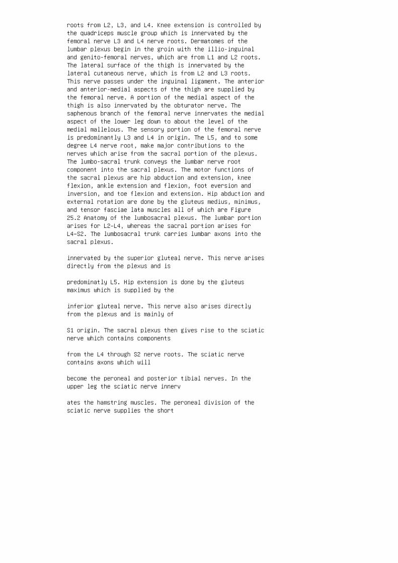
roots from L2, L3, and L4. Knee extension is controlled bythe quadriceps muscle group which is innervated by thefemoral nerve L3 and L4 nerve roots. Dermatomes of thelumbar plexus begin in the groin with the illio-inguinaland genito-femoral nerves, which are from L1 and L2 roots.The lateral surface of the thigh is innervated by thelateral cutaneous nerve, which is from L2 and L3 roots.This nerve passes under the inguinal ligament. The anteriorand anterior-medial aspects of the thigh are supplied bythe femoral nerve. A portion of the medial aspect of thethigh is also innervated by the obturator nerve. Thesaphenous branch of the femoral nerve innervates the medialaspect of the lower leg down to about the level of themedial mallelous. The sensory portion of the femoral nerveis predominantly L3 and L4 in origin. The L5, and to somedegree L4 nerve root, make major contributions to thenerves which arise from the sacral portion of the plexus.The lumbo-sacral trunk conveys the lumbar nerve rootcomponent into the sacral plexus. The motor functions ofthe sacral plexus are hip abduction and extension, kneeflexion, ankle extension and flexion, foot eversion andinversion, and toe flexion and extension. Hip abduction andexternal rotation are done by the gluteus medius, minimus,and tensor fasciae lata muscles all of which are Figure25.2 Anatomy of the lumbosacral plexus. The lumbar portionarises for L2–L4, whereas the sacral portion arises forL4–S2. The lumbosacral trunk carries lumbar axons into thesacral plexus.
innervated by the superior gluteal nerve. This nerve arisesdirectly from the plexus and is
predominatly L5. Hip extension is done by the gluteusmaximus which is supplied by the
inferior gluteal nerve. This nerve also arises directlyfrom the plexus and is mainly of
S1 origin. The sacral plexus then gives rise to the sciaticnerve which contains components
from the L4 through S2 nerve roots. The sciatic nervecontains axons which will
become the peroneal and posterior tibial nerves. In theupper leg the sciatic nerve innerv
ates the hamstring muscles. The peroneal division of thesciatic nerve supplies the short

head of the biceps femoris muscle, whereas the otherhamstring muscles are innervated
by the posterior tibial division. L5 is the predominantnerve root supplying these
muscles except for the short head of biceps femoris, whichsome consider to be predomi
nantly S1 innervated. Proximal to the knee the peroneal andposterior tibial divisions sep
arate into individual nerves. The peroneal nerve controlsankle and toe extension, and
ankle eversion. L5 and S1 are the major nerve roots. Theposterior tibial nerve controls
ankle flexion and inversion, and toe flexion. L5–S2 are thecontributing nerve roots. The sensory pattern of the sacralplexus derived nerves include the posterior aspect
of the thigh, the lateral and posterior areas of the lowerleg, and both the dorsal and plantar
aspects of the foot.
3.2. Lumbo-Sacral Plexopathies
Etiologies include trauma, gun shot wounds, pelvicfractures, compression by hematomas,
inflammation with or without concomitant diabetes, invasionby neoplasms, and damage
from prior therapeutic radiation (17,18).
3.2.1. Traumatic Causes
Major trauma to the abdomen or pelvis can also injure thelumbo-sacral plexus (19–21).
Neurological deficits may not be apparent initially in thesetting of severe injury. Gun shot
wounds tend to involve the lumbar portion of the plexus andproduce deficits of femoral
and obturator nerve function. A shielding effect of thebony pelvis may explain why the

sacral portion of the plexus is less likely to be involved.Crush injuries of the pelvis can
produce deficits in both the lumbar and sacral plexus, withthe latter being more commonly
involved. Pelvic fractures in the region of thesacro-illiac joint can involve axons of the
sacral plexus with weakness in the distribution of L4–S1roots. Plain X-rays can assess fractures, but CT or MRI isnecessary to assess soft tissue
lesions. Electrodiagnostic evaluation can help define thedistribution and severity of
plexus involvement, but will not be informative until about2–4 weeks after the injury.
Follow-up studies at several month intervals can also behelpful for defining the longer
term prognosis and course of recovery.
3.2.2. Hematoma
Hematoma in the retroperitoneal space can produce lumbarplexopathy (22,23). Anticoa
gulated patients with therapeutic levels are the mostsusceptible, and hemophiliacs are also
at risk. Bleeding may be spontaneous, associated with minortrauma, or follow diagnostic
procedures which require vascular access through thefemoral vessels. Unilateral pain in
the back, lower abdomen, or groin is the initial symptom.Pain may intensify over hours to
a day or so. Movement of the hip, active or passive, oftenworsens the pain. Unilateral
weakness of hip flexion, knee extension, and often hipadduction appears in the same time
frame. The patellar reflex is reduced or is absent. Sensoryloss and parasthesia occur in the anterior thigh and medial

lower leg. Signs of hypovolemia and anemia due to bloodloss from the hemorrhage may be present. Diagnosis isconfirmed by CT or MRI studies of the pelvis.Anti-coagulation therapy should be stopped. Transfusion maybe required. It is not clear if surgical evacuation of thehematoma improves the neurological outcome. The level ofpain can be significant and should be treated withappropriate analgesics. Electrodiagnostic studies performed3–4 weeks after onset can define prognosis. Evaluation byrehabilitative services is appropriate as soon as stabilityof the clinical status permits. The neurological deficitstend to improve but may be long lasting in some. Thepathogenesis is compression of the lumbar plexus or femoralnerve secondary to the hematoma formation. A combination ofdemyelination and axonopathy likely occurs.The hematomaforms in the retroperitoneal space either within the psoasmuscle, or beneath the illiacus fascia in the groovebetween the psoas and illiacus muscles in the pelvis. Theproximal lesion tends to produce deficits in both femoraland obturator nerve territories, whereas the more distalone involves only the femoral territory. 3.2.3.Inflammatory Causes Lumbo-sacral plexopathies can arisesecondary to a presumed inflammatory attack on the plexus,as in the brachial plexus (24–26). The clinical featuresare similar to those described subsequently fordiabetes-associated plexopathy. The upper, lower, or bothportions of the plexus can be involved. Patients may havean elevated sedimentation rate, and some will have evidenceof a systemic process like Wegener’s granulomatosis orpolyarteritis nodosa. Biopsy of the sural, superficialperoneal, or lateral femoral cutaneous nerves may aid thediagnosis by showing inflammatory cells in epineuralvessels. The electrodiagnostic findings are similar tothose of diabetes-associated plexitis described in thefollowing section. Weakness improves spontaneously overmonths. There is some support for treatment withimmune-modulating drugs. As with inflammatory brachialplexitis there may be a several week delay between onsetand the confirmation of the diagnosis, making it difficultto judge the efficacy of therapy. The choice of agents isdictated by whether a systemic condition is present, andcorticosteroids plus cyclophosphamide may be required. Ifthe inflammatory process appears to be limited to theplexus alone, single agent treatment with a corticosteroidor intravenous gamma globulin may be adequate. 3.2.4.Diabetes-Associated Plexopathy Diabetic plexopathy is themost common lumbosacral plexopathy. Symptoms usually beginwith spontaneous pain in the back, hip, or proximal leg(27–29). The pain is perceived “deep in the muscles orbone,” and tends to be unilateral initially. It intensifies
over days to weeks, and often reaches a very high levelthat is incompletely responsive to major oral analgesics.The intensity decreases after 2–6 weeks, and can linger ata lower level for several months. Within a few weeks afteronset of the pain, weakness and sensory loss develop in theaffected limb. Weakness is most commonly in the territoryof the lumbar plexus, and produces deficits of hip flexion,adduction, and knee extension. Weakness can also occur morecaudally and produce a sciatic distribution deficit.Occasionally, both lumbar and sacral territories areinvolved concurrently. Weakness evolves for several months,often to a severe degree, and significant muscle atrophyoccurs. Patients may become wheelchair bound due to thesevere proximal weakness. Muscle stretch reflexes arereduced or lost
in the involved territories. Sensory deficits occur mostoften in the anterior thigh and
medial lower leg. Loss of sensation tends to be moreprominent than paresthesia. Some patients develop a similarsyndrome on the opposite side several months later.
Thoracic radiculopathies, characterized by band-liketruncal pain and sensory loss, may
also develop in the same time frame as the leg symptoms.Symptoms and signs of
polyneuropathy are often absent or present to only aminimal degree. Patients may experience significantanorexia and weight loss during the period of
maximum pain. It is not clear whether this is an indirecteffect of the severe pain or due
to a concomitant enteropathy. It is possible that poorblood glucose control prior to
onset of the plexopathy explains the associated weightloss. The onset of the plexopathy
may occur shortly after the diagnosis of diabetes, or laterwhen enhanced efforts at
blood sugar control have been instituted. Overall, patientswho experience this type of
plexopathy do not have worse blood glucose control (29).The type of plexopathy is common and readily recognized.

Laboratory studies
should be directed towards establishing the status ofglucose control, and identifying sys
temic inflammatory conditions. When studied, spinal fluidprotein level is usually mildly
elevated. Electrodiagnostic studies demonstrate activedenervation in clinically involved
muscles. Contralateral muscles should also be evaluated, assubclinical involvement
may be present. Interestingly, the lumbar paraspinals alsooften show active denervation
at multiple levels, supporting a combined radicular andplexus process rather than a
plexopathy alone. Nerve conduction studies usually showaxonal type abnormalities in
the involved nerves. The condition tends to improvespontaneously over months to �2 years, but signifi
cant motor deficits may persist. Traditionally, managementhas been limited to pain
control and rehabilitation. Treatment withimmuno-modulators like corticosteroids or
intravenous gamma globulin though may lessen the evolutionof the deficits and shorten
the course. Both oral and intravenous steroid treatment hasbeen used. The pathogenesis of this lesion is notcompletely established. Type II diabetics are
much more commonly afflicted than Type I patients.Metabolic, ischemic, and inflamma
tory etiologies have been postulated. A multifocalvasculitis affecting small caliber vessels
in the nerve roots and plexus is the current hypothesis,but the relationship of glucose and
immune-mediated process is not currently understood.

3.2.5. Neoplastic Causes
The lumbo-sacral plexus can be infiltrated by neoplasmsthat arise locally from the colon,
cervix, or bladder (30,31). Retroperitoneal lymphomas andsarcomas can also invade the
plexus. Less common are metastatic lesions from breastcancers or melanomas. The cancer
is usually known, and rarely the initial manifestation.Pain is usually the initial symptom, and is located in theback, hip, or proximal leg.
The intensity increases over days to a few weeks. Weaknessand sensory loss usually
develop several weeks later. The distribution of thesesigns corresponds with the
portion of the plexus involved, lumbar, sacral, or diffuse.The lumbar portion is more
often involved when the neoplasm directly infiltrates theplexus, whereas metastatic
lesions tend to involve the sacral portion. Bilateralinvolvement occurs in �25% of
cases. Leg edema may be present and is due to venous orlymphatic compromise. The diagnosis is confirmed by CT orMRI imaging of the pelvis. MRI has a higher
yield than CT. Biopsy may be required to determine thespecific tissue type if the primary
is not known. Electrodiagnostic testing will show a patternof acute or subacute axonopa
14. Thomas JE, Colby MY. Radiation-induced or metastaticbrachial plexopathy. J Am Med Assoc 1972; 11:1392–1395.
15. Harper CM, Thomas JE, Cascino TL, Litchy WJ.Distinction between neoplastic and radiation-inducedbrachial plexopathy, with emphasis on the role of EMG.Neurology 1989; 39:502–506.
16. Gilliatt RW, LeQuense PM, Logue V, Sumer AJ. Wasting ofthe hand associated with a cervical rib or band. J Neurol
Neurosurg Psychiatry 1970; 33:615–624.
17. Donaghy M. Lumbosacral plexus disorders. In: Dyck PJ,ed. Peripheral Neuropathy. Philadelphia: WB Saunders,1993:951–959.
18. Weber M. Lumbosacral plexopathies. In: Brown WF, BoltonCF, Aminoff MJ, eds. Neuromuscular Function and Disease.Philadelphia: WB Saunders, 2002:852–864.
19. Weis EB. Subtle neurological injuries in pelvicfractures. J Trauma 1984; 24:983–985.
20. Kutsy RL, Robinson LR, Routt ML. Lumbosacral plexopathyin pelvic trauma. Muscle Nerve 2000; 23:1757–1760.
21. Chou-Tan FY, Efenbaum M, Chan K-T, Song J. Lumbosacralplexopathy in gunshot wounds and motor vehicle accidents.Am J Phys Med Rehabil 2001; 80:280–285.
22. Chiu WS. The syndrome of retroperitoneal hemorrhage andlumbar plexus neuropathy during anticoagulant therapy.South Med J 1976; 69:595–599.
23. Emery S, Ochoa J. Lumbar plexus neuropathy resultingfrom retroperitoneal hemorrhage. Muscle Nerve 1978;1:330–334.
24. Evans BA, Stevens JC, Dyck PJ. Lumbosacral plexusneuropathy. Neurology 1981; 31:1327–1330.
25. Bradley WG, Chad D, Verghese JP, Liu HC, Good P, GabbaiAA, Adelman LS. Painful lumbosacral plexopathy withelevated sedimentation rate: a treatable inflammatorysyndrome. Ann Neurol 1984; 15:457–464.
26. Triggs WJ, Young MS, Eskin T, Valenstein E. Treatmentof idiopathic lumbosacral neuropathy with intravenousimmunoglobulin. Muscle Nerve 1997; 20:244–246.
27. Barohn RJ, Sahenk Z, Warmolts JR, Mendell JR. TheBruns–Garland syndrome (Diabetic Amytorophy) revisited 100years later. Arch Neurol 1991; 48:1130–1135.
28. Krendel DA, Costigan DA, Hopkins LC. Successfultreatment of neuropathies in patients with diabetesmellitus. Arch Neurol 1995; 2:1053–1061.
29. Dyck PJB, Windebank AJ. Diabetic and nondiabeticlumbosacral radiculoplexus neuropathies: new insights into

pathophysiology and treatment. Muscle Nerve 2002;25:477–491.
30. Thomas JE, Cascino TL, Earle JD. Differential diagnosisbetween radiation and tumor plexopathy of the pelvis.Neurology 1985; 35:1–7.
31. Jaeckle KA, Young DF, Foley KM. The natural history oflumbosacral plexopathy in cancer. Neurology 1985; 35:8–15.
26
Median Mononeuropathies
A. Gordon Smith
University of Utah, Salt Lake City, Utah, USA
Abstract 467
1. Anatomy 467
2. Carpal Tunnel Syndrome 469 2.1. Clinical Features 4692.2. Diagnostic Evaluation 471 2.3. Prognosis andManagement 473 2.4. Pathophysiology 475
3. Median Mononeuropathies in the Region of the Elbow 4753.1. Pronator Teres Syndrome 476 3.2. Anterior InterosseousNeuropathy 477
4. Other Causes of Median Mononeuropathy 477
ABSTRACT
The median nerve is the most frequently injured nerve.Median mononeuropathy at the
wrist (carpal tunnel syndrome) is one of the most commonreferral questions for electro
diagnostic testing, and is a major cause of work-relateddisability. The median nerve may
also be injured in the forearm, in the region of thepronator teres muscle, but the pronator
syndrome is controversial. The anterior interosseous nervemay also be injured in the
forearm. More proximal lesions of the median nerve in the
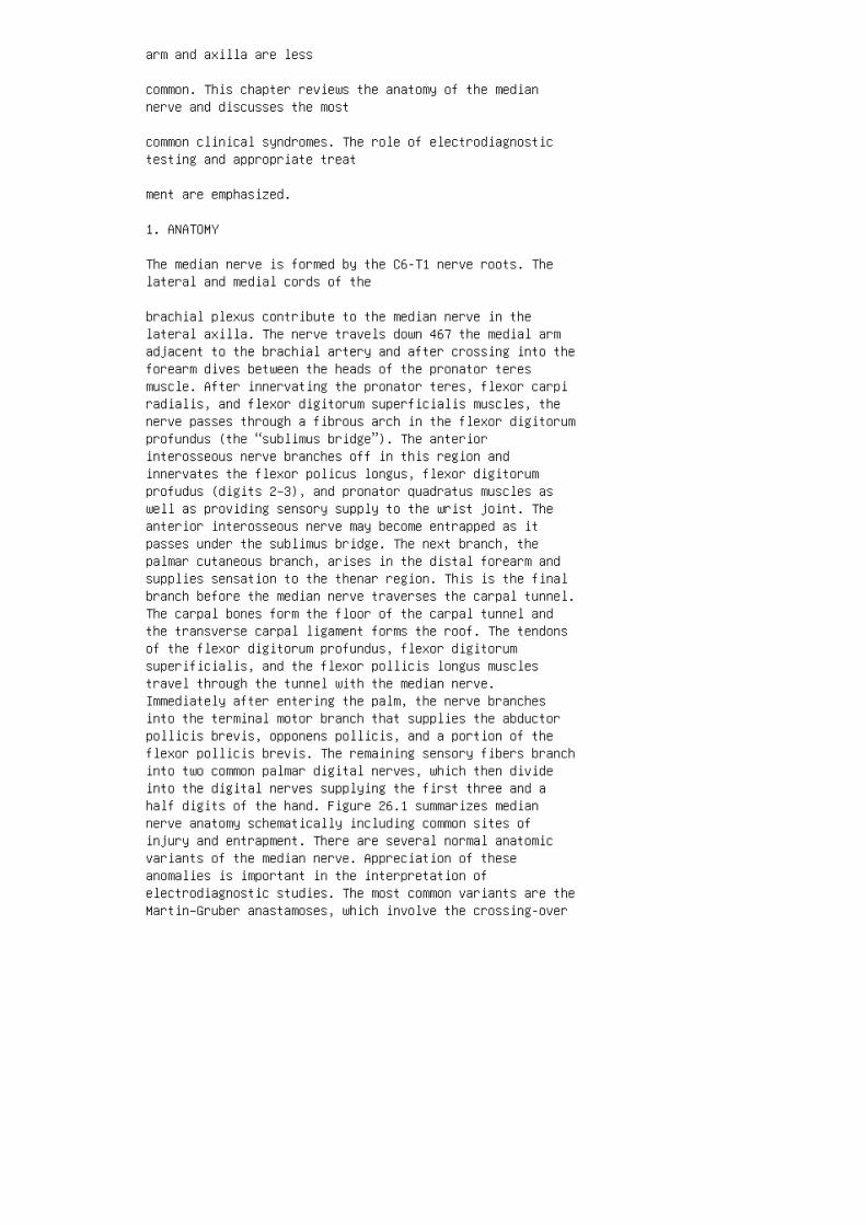
arm and axilla are less
common. This chapter reviews the anatomy of the mediannerve and discusses the most
common clinical syndromes. The role of electrodiagnostictesting and appropriate treat
ment are emphasized.
1. ANATOMY
The median nerve is formed by the C6-T1 nerve roots. Thelateral and medial cords of the
brachial plexus contribute to the median nerve in thelateral axilla. The nerve travels down 467 the medial armadjacent to the brachial artery and after crossing into theforearm dives between the heads of the pronator teresmuscle. After innervating the pronator teres, flexor carpiradialis, and flexor digitorum superficialis muscles, thenerve passes through a fibrous arch in the flexor digitorumprofundus (the “sublimus bridge”). The anteriorinterosseous nerve branches off in this region andinnervates the flexor policus longus, flexor digitorumprofudus (digits 2–3), and pronator quadratus muscles aswell as providing sensory supply to the wrist joint. Theanterior interosseous nerve may become entrapped as itpasses under the sublimus bridge. The next branch, thepalmar cutaneous branch, arises in the distal forearm andsupplies sensation to the thenar region. This is the finalbranch before the median nerve traverses the carpal tunnel.The carpal bones form the floor of the carpal tunnel andthe transverse carpal ligament forms the roof. The tendonsof the flexor digitorum profundus, flexor digitorumsuperificialis, and the flexor pollicis longus musclestravel through the tunnel with the median nerve.Immediately after entering the palm, the nerve branchesinto the terminal motor branch that supplies the abductorpollicis brevis, opponens pollicis, and a portion of theflexor pollicis brevis. The remaining sensory fibers branchinto two common palmar digital nerves, which then divideinto the digital nerves supplying the first three and ahalf digits of the hand. Figure 26.1 summarizes mediannerve anatomy schematically including common sites ofinjury and entrapment. There are several normal anatomicvariants of the median nerve. Appreciation of theseanomalies is important in the interpretation ofelectrodiagnostic studies. The most common variants are theMartin–Gruber anastamoses, which involve the crossing-over

Figure 26.1 Median nerve anatomy. This schematicrepresentation of the median nerve demonstrates major nervebranches, the muscles they innervate, and common sites ofinjury. Important causes for median mononeuropathy arelisted by anatomic site.
of fibers from the median to the ulnar nerve. Martin–Gruberanastamoses have an esti
mated prevalence of �30% based on carefulelectrophysiologic evaluation (1), but are
observed much less frequently during routineelectrodiagonstic testing. There are
several subtypes, all of which include a branch from themedian nerve in the forearm in
the region of the anterior interosseous nerve that joinsthe ulnar nerve in the forearm.
The crossed fibers usually cross back to the median nerveafter the wrist and supply
thenar muscles. They may also supply hypothenar muscles.Patients with Martin–
Gruber anastamoses supplying hyopthenar muscles may developunexpected hand intrin
sic weakness with lesions of the anterior interosseousnerve (2). Martin–Gruber anasta
moses may create some confusion in the electrodiagnosticevaluation of carpal tunnel
syndrome (CTS). When the crossed fibers innervate thenarmuscles via the ulnar nerve,
they are not slowed at the wrist. This results ininappropriately fast median motor conduc
tion velocity in the forearm. Because the musclesinnervated by the crossed fibers are
usually not directly under the recording electrode, a smallpositive deflection may be
seen when the median nerve is stimulated at the elbow.Observation of either pattern

should prompt a careful evaluation for CTS. Crossovers inthe palm innervating intrinsic
hand muscles (Riche–Cannieu anastamoses) are less common.
2. CARPAL TUNNEL SYNDROME
Median nerve entrapment at the wrist, CTS, is the mostcommon entrapment neuropathy
and �2–3% of the population suffers from CTS (3).Variability in prevalence data is
likely due to variations in occupational and industrialwork exposure. A majority of
patients attribute their CTS to work exposure and CTS is asignificant cause of work
related morbidity; 90% have a good treatment outcome andare able to return from
work, but 10% are permanently disabled (4).
2.1. Clinical Features
CTS typically presents with paresthesias, numbness, andpain involving the wrist hand and
fingers. Symptoms often awaken the patient and are worse inthe morning. They are exacer
bated by provocative activities involving repetitiveflexion and/or extension of the wrist.
Shaking of the hands often relieves the numbness andparesthesias (5). Driving, typing,
writing, or using hand tools may worsen symptoms.Individuals working in industries that
involve repetitive forceful wrist flexion and extension areat increased risk (6,7). Most patients appreciate moresignificant symptoms in the first three digits.
However, over half describe paresthesias involving thefifth digit and 40% have pain or
paresthesias in the forearm or above, with a significant

minority (7%) experiencing
shoulder pain (Fig. 26.2) (8). Numbness may be limited toonly one digit. Therefore,
differentiation of CTS from other causes of limb pain canbe problematic and careful phys
ical and electrodiagnostic evaluation are of significanthelp. Many patients also experience subjective handweakness. This is typically described
as a tendency to drop objects or a sense of incoordinationwith fine motor tasks. The weak
ness is often due to pain inhibition or sensory loss thatimpairs fine motor control. The physical examination may benormal, particularly in mild CTS. Many patients
have reduced sensation to pinprick in a median nervesensory distribution. The finding of
reduced pin on the lateral side of the fourth digitcompared with the medial side is helpful,
although not observed in all patients because the mediannerve sometimes innervated both
sides of the fourth finger. Typically, the skin over thethenar eminence has normal sensation because the palmarcutaneous branch of the median nerve, which arises proximalto the carpal tunnel, provides sensory innervation to thisregion. Care must be taken not to over-interpret thesensory examination, as there is significant normalvariation in sensory distributions. Furthermore, thesensory examination is inherently subjective, and patientsmay describe reduced sensation in unexpected areas (8). Thefinding of subjective sensory change in regions notsupplied by the median nerve should not exclude thediagnosis or discourage further diagnostic evaluation in apatient with suggestive symptoms. Patients with more severedisease have weakness of thumb abduction and opposition andthose with advanced disease have thenar atrophy. Elderlypatients may present with thenar atrophy and weaknesswithout significant sensory complaints or pain. A varietyof provocative physical examination maneuvers have beendescribed. The most commonly sought is the Tinel’s sign.Tinel first described paresthesias with nerve percussion inpatients recovering from nerve injury (9). The “flick sign”refers to a history of shaking the hands after awakening

with numbness or pain. Paresthesias provoked by armextension and wrist flexion is termed the Phalen’s sign.Other signs Figure 26.2 Distribution of sensory symptoms inCTS. This figure, modified from Stevens et al. (8),illustrates patterns of sensory complaints in patients withCTS. A patient completed each drawing. (A) Sensory symptomsmay be limited to the median nerve distribution, or (B) mayoccur in a glove pattern or (C) ulnar nerve distribution.[Reproduced from Stevens et al. (8) with permission.]
focus on reproduction of symptoms with compression over thecarpal tunnel. The diagnos
tic accuracy of these various maneuvers is poor and theirpositive and negative predictive
values are low (10). In one study of 142 patients referredfor possible CTS, the negative
predictive values were all,40% (11). Because many normalindividuals have these signs,
and many CTS patients do not, their clinical value islimited.
2.2. Diagnostic Evaluation
The diagnosis of CTS is complicated by neurologicco-morbidity. Patients with an under
lying peripheral neuropathy are challenging because theyoften describe hand numbness
that is worse at night, but they also have an increasedrisk of developing CTS. Patients
with superimposed cervical spine disease or ulnarmononeuropathy also pose a clinical
challenge because they have numbness, pain, and weaknessthat extends out of the
median nerve distribution. Given the variability inclinical findings and complaints
among patients with suspected CTS, supportive diagnosticstudies play a very important
role in the evaluation. Electrodiagnostic evaluation withnerve conduction studies

(NCS) and electromyography (EMG) is the most commonly usedstudy. A large variety
of specific electrodiagnostic techniques have beendescribed. This review focuses on
the general principles and the most commonly employedtechniques. The earliest electrophysiological changes inCTS are due to demyelination of large
sensory nerve fibers. Median sensory conduction velocityacross the carpal tunnel is
slowed, and thus the median sensory distal latency becomesprolonged. Diagnostic criteria
are based on the difference between the median and ulnarsensory distal latency or conduc
tion velocity (Fig. 26.3). The median sensory response maybe recorded antidromically by
stimulating proximal to the wrist and recording the sensorynerve action potential at the
digit, or orthodromically by stimulating at the digit or inthe palm, and recording above
the wrist (Fig. 26.4). Use of a short segment orthodromictechnique, stimulating at the
palm and recording at the wrist, maximizes diagnosticsensitivity because the area of
nerve conduction slowing across the carpal tunnel regionrepresents a larger fraction of
the total length of the nerve examined compared withstudies investigating longer segments
(12,13). Focal slowing can be detected by stimulating thenerve in 1 cm increments starting
proximal to the wrist and extending into the palm(“inching”). Sensory nerve action potential
amplitude may be reduced due to sensory conduction block atthe wrist (which can be

detected by inching). With more severe lesions, there isinjury to the axons themselves.
This is recognized as diminished sensory amplitudes, evenwhen stimulating distally. Motor NCS are usually normal inmild CTS. In more moderate disease, there is pro
longation of the motor distal latency due to demyelinationof motor nerve fibers. With
severe CTS, there is a decline in the compound muscleaction potential amplitude due
to axonal injury. Because of the effects of reinnervation,the motor amplitude remains
normal for some time despite ongoing axonal injury. Forthis reason, needle EMG is
useful to demonstrate the presence of axonal loss. Avariety of specific NCS diagnostic criteria for CTS havebeen proposed. In general,
NCS are reproducible and have a high sensitivity (.85%) andspecificity (.95%) (14). A
review of the literature demonstrated that the mostsensitive and specific parameter was
median sensory or mixed nerve conduction study from thepalm to the wrist over a
short distance (7–8 cm). The American Association ofNeuromuscular Electrodiagnostic
Medicine CTS Practice Parameter recommends starting theevaluation with a median
sensory NCS across the wrist using a distance of 14 cm. Ifthis is normal, the more sensitive
palm to wrist study should be performed (14). It has beensuggested that a combination
of multiple median nerve studies maximizes diagnosticaccuracy (15). However, if any
individual parameter is significant abnormal (orthodromicpalmar median versus ulnar difference of .0.3 ms orantidromic ring difference of .0.4), further sensory

studies are not necessary (16). Electrodiagnostic studiesserve several purposes in the diagnostic evaluation ofsuspected CTS. The first is to confirm the diagnosis ofCTS. They are also used to exclude Figure 26.3 Mediansensory nerve conduction technique. (A) An orthodromicsensory nerve conduction study over a short segment (8 cm)is the most sensitive technique for detection of slowingacross the wrist. The nerve is stimulated in the palm andthe response recorded immediately proximal to the wrist.(B) A standard antidromic sensory study over a longersegment (14 cm) is often used as a screening study. Figure26.4 Sensory nerve conduction in CTS. Median (top) andulnar (bottom) sensory nerve action potentials from (A) anormal subject, (B) a patient with moderate CTS withreduced median sensory amplitude (17 mV compared with 37 mVfor the ulnar response) and prolonged distal latency (peaklatency of 4.8 ms compared with 3.1 ms for the ulnarresponse), and (C) a patient with more severe CTS and anabsent median sensory response.
other disorders (neuropathy, radiculopathy, othermononeuropathies). It is very important
that the results of the electrodiagnostic study beinterpreted in clinical context. The finding
of slowed median nerve conduction across the wrist on NCSdoes not necessarily imply
that CTS is the cause for the patient’s symptoms. Theelectrodiagnostic consultant
should try to place the findings in context and make ajudgement concerning their clinical
relevance. Electrodiagnostic studies can be used forprognostic purposes and to direct
therapy. Mild CTS with pure sensory demyelination is likelyto be more responsive to con
servative therapy than more severe disease. Conversely,those with reduced motor ampli
tude and axonal loss on EMG have a poorer prognosis andshould be treated aggressively.
The electrodiagnostic evaluation for suspected CTS shouldtherefore include motor

conduction studies and needle examination of multiple limbmuscles in order to
exclude a superimposed radiculopathy or ulnar or radialmononeuropathy (14). Although NCS are very useful, up to25% of patients with clinically suspected CTS
may have a normal study (17). Some authors, particularly inthe surgical literature, have
suggested NCS are not very useful because of thisobservation (18). A prospective study
of sequential patients referred for CTS addressed thisquestion. When the clinical history
and examination were strongly suggestive of CTS, the NCSresults were typically abnor
mal (92%). However, when the clinical data was lessdefinitive, but CTS was still sus
pected (e.g., mild CTS), NCS results could not be predictedbased on clinical data
(positive predictive value of 67%). NCS were thereforeoften necessary to confirm the
diagnosis of CTS (17). There is also evidence that patientswith clinically suspected
CTS but normal NCS are less likely to experience resolutionof symptoms following sur
gical release (51%) when compared with those with abnormalNCS (65–77%). Patients
with moderate CTS are most likely to respond and those withsevere CTS are least likely
to respond (19). This suggests many patients with suspectedCTS but normal NCS prob
ably do not have CTS. Although patients with normal NCS maystill have true CTS, sur
gical intervention should only be considered in those whohave failed conservative
therapy and have a classic syndrome. A repeat NCS may be

useful in patients whose
initial NCS was normal. Other procedures have been assessedfor the diagnosis of suspected CTS. A variety
of quantitative sensory testing devices have been studiedand have been found to have a
low diagnostic accuracy (20–22). Quantitative sensorytesting is a psychophysiological
test and results are significantly influenced by patientbias. For example, normal subjects
without any training, can simulate neuropathic sensory loss(23). Radiologic evaluation for CTS has shown greaterpromise. MRI of the carpal tunnel
may be used to evaluate structural or anatomicabnormalities, although it is not necessary
for the routine evaluation of idiopathic CTS (24). Patientswith a mass in the wrist, trauma,
or persistent symptoms following surgical evaluation maybenefit from MRI. There are
also data suggesting that ultrasound may be a usefuldiagnostic technique. Ultrasound typi
cally demonstrates nerve swelling proximal to the site ofcompression with flattening dis
tally (25). Careful measurement of nerve area may have asensitivity of up to 67% with a
specificity of 97% (26).
2.3. Prognosis and Management
Treatment of CTS should start by identification andmodification of activities that provoke
or exacerbate symptoms. Treatment of underlying diseasesassociated with CTS (rheuma
toid arthritis, hypothyroidism) may result in improvement.Up to 10% or more of pregnant
individuals develop CTS, presumably due to hand swelling or

hypertension. Conservative
therapy is generally warranted given most patients havecomplete resolution of symptoms within weeks of delivery(27,28). Most patients with idiopathic CTS of mild tomoderate severity also respond to conservative therapy.However, patients with more severe symptoms, andparticularly those with thenar atrophy, weakness, ordenervation on electrodiagnostic testing should be referredfor definitive surgical therapy. The most commonly employedconservative therapy for CTS is wrist splinting. Up to 80%of patients with CTS have resolution of their symptomswithin days to weeks of using splints at nighttime (29). Avariety of off-the-shelf and custom wrist splints areavailable. Splinting in the neutral position reducesnocturnal intra-carpal tunnel pressures more than splintingin relative extension (30). Any wrist splint that iscomfortable and in the neutral position is acceptable. Acourse of nonsteroidal anti-inflammatory medication isoften prescribed in combination with wrist splinting.However, this treatment has not been well studied. Therehas been only one randomized study comparing placebo,nonsteroidal anti-inflammatory medication, diuretic, andoral prednisolone. Only the prednisolone group, which took20 mg daily for 2 weeks and 10 mg daily for 2 weeks noted asignificant benefit. Patients were only followed for 4weeks, so the long term outcome is unknown (31). Vitamin B6(pyridoxine) has been suggested in the past but smallstudies suggest a lack of efficacy (32). Use of pyridoxineis discouraged, as high doses may result in a sensoryneuropathy that can be very severe (33). Small studies ofdiuretics alone also failed to show evidence for efficacy(32). For patients who fail neutral wrist splinting, localsteroid injection into the carpal tunnel is a therapeuticoption. It has been suggested that this approach is limitedby a relatively low rate of long term success (34).However, several studies suggest a significant fraction ofpatients with mild to moderate disease have sustainedbenefit (35,36). One prospective study demonstrated that10% of patients remained symptom free at 1 year (37). Inanother study patients, one-third had normal NCS at 1 year(38). There is a very small risk of injuring the mediannerve during the injection. Local steroid injection is anappropriate therapy for patients with mild to moderatedisease who wish to avoid carpal tunnel release. For thosewho have recurrent symptoms, up to 3 injections yearly maybe safely performed (39). Because it is less invasive thansurgery, steroid injection may also be more appropriate forpatients with an uncertain diagnosis and can serve as adiagnostic procedure, for those who respond are more likely
to respond to carpal tunnel release than those who do notrespond (40). Iontophoresis of dexamethasone may be aneffective and safe therapy. It involves applying a smallelectrical current to an electrode containing a medication,which propels it through the skin. In a prospective studyof 19 patients who had failed splinting andanti-inflammatory medication, 58% had benefit at 6 months,a rate superior to that in most studies of local injection(41). Because repeat iontophoresis treatments are welltolerated and inexpensive, this therapy holds significantpromise and deserves more careful study. Patients who havefailed conservative management, have severe disease withaxonal injury, or wish immediate definitive therapy shouldbe referred for carpal tunnel release. Surgical release ishighly successful, with 90% of patients experiencing asuccessful outcome (42). A variety of techniques have beendescribed, including endoscopic approaches and “mini” openrelease. Each technique works well. Proponents ofendoscopic release argue that it results in an earlierreturn to work. However, there may also be a higher risk ofinjury to the median nerve (39). A recent meta-analysis ofthe literature suggests the various techniques may beessentially equivalent, both for long-term side effects andefficacy (43). The most prudent approach is to refer thepatient to a surgeon with significant expertise in carpaltunnel release.
2.4. Pathophysiology
There are two general mechanisms underlying CTS: a reducedcanal size and a predisposition
to nerve compression. Any lesion within the carpal tunnelmay compress the median nerve.
Medical causes include rheumatoid arthritis andsarcoidosis. Structural causes include
ganglion cysts, tumors, and vascular malformations. Wristfractures or hematomas may
result in acute CTS. A variety of medical conditions maypredispose the median nerve to
compression, including peripheral neuropathy, particularlythat associated with diabetes
(Table 26.1). As many as one in three patients with CTShave a predisposing condition (44). It has been suggestedthat proximal nerve compression predisposes to a more

distal
injury along the nerve’s course (the “double crush”hypothesis) (45). Although there has
been significant debate about the double crush hypothesis,there is no convincing animal or
clinical data to support it (46,47). An EMG examination toexclude a radiculopathy as a
confounding clinical feature is suggested, but routineneuroimaging of the cervical
spine is not recommended. The etiology of idiopathic CTS isunknown. There has been significant debate as to
whether patients with CTS have a congenitally smaller canalthan those who do not.
Although there are some data to support this concept (48),most evidence suggests conge
nital canal narrowing is not a predisposing factor (49,50).Occupational exposure is
important, and repetitive forceful extension and flexion ofthe wrist places individuals
at increased risk.
3. MEDIAN MONONEUROPATHIES IN THE REGION OF THE ELBOW
The second most common site for median nerve injury is inthe proximal forearm and near
the elbow. Supracondylar humeral fractures may injure themedian nerve proximal to the
elbow. Often, only the fascicles supplying the anteriorinterosseous nerve are involved
(51). A significant minority of individuals have a bonyspur above the medial humeral
condyle (surpacondylar spur). There may be a ligament thatruns from the spur to the
humerus (ligament of Struthers) that can compress themedian nerve (Fig. 26.5). In this
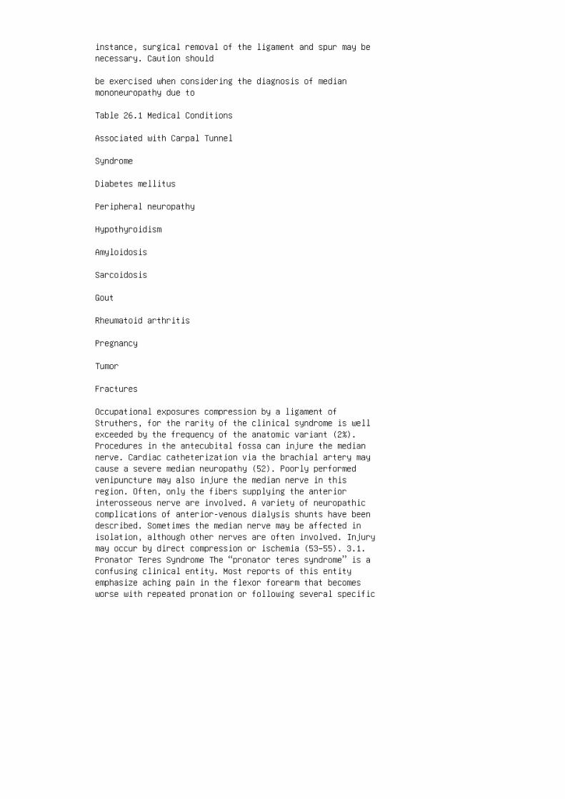
instance, surgical removal of the ligament and spur may benecessary. Caution should
be exercised when considering the diagnosis of medianmononeuropathy due to
Table 26.1 Medical Conditions
Associated with Carpal Tunnel
Syndrome
Diabetes mellitus
Peripheral neuropathy
Hypothyroidism
Amyloidosis
Sarcoidosis
Gout
Rheumatoid arthritis
Pregnancy
Tumor
Fractures
Occupational exposures compression by a ligament ofStruthers, for the rarity of the clinical syndrome is wellexceeded by the frequency of the anatomic variant (2%).Procedures in the antecubital fossa can injure the mediannerve. Cardiac catheterization via the brachial artery maycause a severe median neuropathy (52). Poorly performedvenipuncture may also injure the median nerve in thisregion. Often, only the fibers supplying the anteriorinterosseous nerve are involved. A variety of neuropathiccomplications of anterior-venous dialysis shunts have beendescribed. Sometimes the median nerve may be affected inisolation, although other nerves are often involved. Injurymay occur by direct compression or ischemia (53–55). 3.1.Pronator Teres Syndrome The “pronator teres syndrome” is aconfusing clinical entity. Most reports of this entityemphasize aching pain in the flexor forearm that becomesworse with repeated pronation or following several specific

provocative maneuvers (56). Proponents of the syndromesuggest that the pain is due to compression of the mediannerve as it passes through the two heads of the pronatorteres muscle. Reports of true neurogenic pronator teressyndrome are rare however. In one series, 39 patientsunderwent a thorough electrodiagnostic evaluation, and mildmedian nerve injury was found in only two individuals.Despite this, surgical decompression resulted in goodimprovement in 28, fair improvement in five, and no changein three patients (57). The explanation for the clinicalimprovement following decompression despite a lack ofevidence for median nerve injury is uncertain. It has beensuggested that pronator teres syndrome is really a forearmcompartment syndrome rather than a nerve compressionsyndrome, and that release of the median nerve in theregion is effective because the procedure includes afasciotomy (58). Patients with suspected pronator teressyndrome should undergo a careful electrodiagnostic study.If there is evidence of median nerve injury, MRIneurography of the region may be useful and surgicaldecompression is a reasonable therapeutic option if thereis significant axonal injury. In the absence of mediannerve injury, conservative therapy should be pursued, andsurgical intervention reserved for patients with severesymptoms refractory to this approach. Any surgical approachshould include a fasciotomy. Figure 26.5 Supracondylar spurand ligament of Struthers. A reproduction of Struthers’original sketch demonstrates the bony and ligamentousanatomy (A). The brachial artery and median nerve maytravel between the humerus and ligament (B). [Withpermission from Stewart (63).]
3.2. Anterior Interosseous Neuropathy
Anterior interrosseous mononeuropathies are the second mostcommon median mono
neuropathy following CTS. As already noted, lesions in theelbow or distal arm may
cause preferential injury to the fibers supplying theanterior interosseous nerve. Direct
trauma to the nerve may also occur, as described earlier.External compression by a
cast has also been described (59). The most common cause ofanterior interosseous
neuropathy is idiopathic. Patients may experience forearm

pain due to involvement of
sensory nerves supplying the wrist joint. However, theprimary complaint is weakness
of flexion of the first two fingers and the thumb affectingpinching. When patients
attempt to make an O with their thumb and first finger,they are unable to do so. The
distal thumb and first finger collapse against one another,forming a tear drop shape
(the “pinch sign”) (Fig. 26.6). Weakness of the thirdmuscle supplied by the nerve, the pro
nator quadratus, does not cause clinical findings. Inpatients who have a Martin–Gruber
anastamosis, intrinsic hand muscles may be weak becausefibers innervating ulnar hand
muscles travel with the crossed fibers in the anteriorinterosseous nerve (2). The cause
for spontaneous anterior interosseous neuropathy is oftendifficult to define. The nerve
may become compressed as it passes through the tendons ofthe pronator teres and flexor
digitorum superficialis. Idiopathic brachial plexusneuropathy (“Parsonage–Turner
Syndrome” or “brachial plexitis”) may cause preferentialinjury to the anterior interos
seous nerve (60). Clues to this diagnosis include patchyinvolvement of other muscles
in the arm and the presence of shoulder pain. Most patientsexperience spontaneous
improvement over several months. If there is noimprovement, MRI of the region may
be necessary to exclude an anatomic or structuralabnormality. It is important to remember

that more proximal lesions may cause isolated anteriorinterosseous injury, so exclusion of
a ligament of Struthers and imaging of the entire mediannerve course may be necessary.
4. OTHER CAUSES OF MEDIAN MONONEUROPATHY
The median nerve may be injured in the axilla. Generally,however, multiple nerves are
involved and isolated median mononeuropathy is rare. Distalmedian nerve branches
may also be injured. The recurrent motor branch of themedian nerve arises just distal
to the carpal tunnel and supplies thenar muscles. It may becompressed by the transverse
Figure 26.6 The pinch sign. Patients with anteriorinterosseous neuropathy are unable to form an
21. Werner RA, Franzblau A, Johnston E. Quantitativevibrometry and electrophysiological assessment in screeningfor carpal tunnel syndrome among industrial workers: acomparison. Arch Phys Med Rehabil 1994; 75(11):1228–1232.
22. Franzblau A, Werner RA, Johnston E, Torrey S.Evaluation of current perception threshold testing as ascreening procedure for carpal tunnel syndrome amongindustrial workers. J Occup Med 1994; 36(9):1015–1021.
23. Freeman R, Chase K, Risk M. Quantitative sensorytesting cannot differentiate simulated sensory loss fromsensory neuropathy. Neurology 2003; 60:465–470.
24. Cosgrove J. Magnetic resonance imaging in theevaluation of carpal tunnel syndrome: a literature review.J Clin Neuromusc Dis 2000; 1:175–180.
25. Buchberger W, Schon G, Strasser K, Jungwirth W.High-resolution ultrasonography of the carpal tunnel. JUltrasound Med 1991; 10(10):531–537.
26. Nakamichi K, Tachibana S. Ultrasonographic measurementof median nerve cross-sectional area in idiopathic carpaltunnel syndrome: Diagnostic accuracy. Muscle Nerve 2002;26(6):798–803.
27. McLennan HG, Oats JN, Walstab JE. Survey of handsymptoms in pregnancy. Med J Aust 1987; 147(11–12):542–544.
28. Wand JS. Carpal tunnel syndrome in pregnancy andlactation. J Hand Surg [Br] 1990; 15(1):93–95.
29. Burke DT, Burke MM, Stewart GW, Cambre A. Splinting forcarpal tunnel syndrome: in search of the optimal angle.Arch Phys Med Rehabil 1994; 75(11):1241–1244.
30. Gelberman RH, Hergenroeder PT, Hargens AR, Lundborg GN,Akeson WH. The carpal tunnel syndrome. A study of carpalcanal pressures. J Bone Joint Surg Am 1981; 63(3):380–383.
31. Chang MH, Chiang HT, Lee SS, Ger LP, Lo YK. Oral drugof choice in carpal tunnel syndrome. Neurology 1998;51(2):390–393.
32. O’Connor D, Marshall S, Massy-Westropp N. Non-surgicaltreatment (other than steroid injection) for carpal tunnelsyndrome. Cochrane Database Syst Rev 2003(1):CD003219.
33. Schaumburg H, Kaplan J, Windebank A et al. Sensoryneuropathy from pyridoxine abuse a new megavitaminsyndrome. N Engl J Med 1983; 309:445–448.
34. Demirci S, Kutluhan S, Koyuncuoglu HR et al. Comparisonof open carpal tunnel release and local steroid treatmentoutcomes in idiopathic carpal tunnel syndrome. RheumatolInt 2002; 22(1):33–37.
35. Dammers JW, Veering MM, Vermeulen M. Injection withmethylprednisolone proximal to the carpal tunnel:randomised double blind trial. BMJ 1999; 319(7214):884–886.
36. Ayhan-Ardic FF, Erdem HR. Long-term clinical andelectrophysiological results of local steroid injection inpatients with carpal tunnel syndrome. Funct Neurol 2000;15(3):157–165.
37. Graham RG, Hudson DA, Solomons M, Singer M. Aprospective study to assess the outcome of steroidinjections and wrist splinting for the treatment of carpaltunnel syndrome. Plast Reconstr Surg 2004; 113(2):550–556.
38. Hagebeuk EE, de Weerd AW. Clinical andelectrophysiological follow-up after local steroidinjection in the carpal tunnel syndrome. Clin Neurophysiol2004; 115(6):1464–1468.

39. Katz JN, Simmons BP. Clinical practice. Carpal tunnelsyndrome. N Engl J Med 2002; 346(23):1807–1812.
40. Edgell SE, McCabe SJ, Breidenbach WC, LaJoie AS, AbellTD. Predicting the outcome of carpal tunnel release. J HandSurg [Am] 2003; 28(2):255–261.
41. Banta CA, Bland JD, You H et al. A prospective,nonrandomized study of iontophoresis, wrist splinting, andantiinflammatory medication in the treatment of early-mildcarpal tunnel syndrome. J Occup Med 1994; 36(2):166–168.
42. Scholten RJ, Gerritsen AA, Uitdehaag BM, van Geldere D,de Vet HC, Bouter LM. Surgical treatment options for carpaltunnel syndrome. Cochrane Database Syst Rev2002(4):CD003905.
43. Thoma A, Veltri K, Haines T, Duku E. A systematicreview of reviews comparing the effectiveness of endoscopicand open carpal tunnel decompression. Plast Reconstr Surg2004; 113(4):1184–1191.
44. Atcheson SG, Ward JR, Lowe W. Concurrent medicaldisease in work-related carpal tunnel syndrome. Arch InternMed 1998; 158(14):1506–1512. 45. Upton AR, McComas AJ. Thedouble crush in nerve entrapment syndromes. Lancet 1973;2(7825):359–362. 46. Morgan G, Wilbourn AJ. Cervicalradiculopathy and coexisting distal entrapmentneuropathies: double-crush syndromes? Neurology 1998;50(1):78–83. 47. Wilbourn AJ, Gilliatt RW. Double-crushsyndrome: a critical analysis. Neurology 1997; 49(1):21–29.48. Horch RE, Allmann KH, Laubenberger J, Langer M, StarkGB. Median nerve compression can be detected by magneticresonance imaging of the carpal tunnel. Neurosurgery 1997;41(1):76–82. 49. Cobb TK, Bond JR, Cooney WP, Metcalf BJ.Assessment of the ratio of carpal contents to carpal tunnelvolume in patients with carpal tunnel syndrome: apreliminary report. J Hand Surg [Am] 1997; 22(4):635–639.50. Pierre-Jerome C, Bekkelund SI, Mellgren SI, NordstromR. Quantitative MRI and electrophysiology of preoperativecarpal tunnel syndrome in a female population. Ergonomics1997; 40(6):642–649. 51. Spinner M, Schreiber SN. Anteriorinterosseous-nerve paralysis as a complication ofsupracondylar fractures of the humerus in children. J BoneJoint Surg Am 1969; 51(8):1584–1590. 52. Kennedy AM,Grocott M, Schwartz MS, Modarres H, Scott M, Schon F.Median nerve injury: an underrecognised complication ofbrachial artery cardiac catheterisation? J Neurol NeurosurgPsychiatry 1997; 63(4):542–546. 53. Redfern AB, ZimmermanNB. Neurologic and ischemic complications of upper

extremity vascular access for dialysis. J Hand Surg [Am]1995; 20(2):199–204. 54. Ergungor MF, Kars HZ, Yalin R.Median neuralgia caused by brachial pseudoaneurysm.Neurosurgery 1989; 24(6):924–925. 55. Bolton CF, DriedgerAA, Lindsay RM. Ischaemic neuropathy in uraemic patientscaused by bovine arteriovenous shunt. J Neurol NeurosurgPsychiatry 1979; 42(9):810–814. 56. Tetro AM, Pichora DR.High median nerve entrapments. An obscure cause ofupper-extremity pain. Hand Clin 1996; 12(4):691–703. 57.Hartz CR, Linscheid RL, Gramse RR, Daube JR. The pronatorteres syndrome: compressive neuropathy of the median nerve.J Bone Joint Surg Am 1981; 63(6):885–890. 58. Stewart J,Jablecki C. Median nerve. In: Brown W, Bolton C, AminoffMJ, eds. Neuromuscular Function and Disease Basic Clinicaland Electrodiagnostic Aspects. 1st ed. Philadelphia: W.B.Saunders, 2002:873. 59. Gardner-Thorpe C. Anteriorinterosseous nerve palsy: spontaneous recovery in twopatients. J Neurol Neurosurg Psychiatry 1974;37(10):1146–1150. 60. Rennels GD, Ochoa J. Neuralgicamyotrophy manifesting as anterior interosseous nervepalsy. Muscle Nerve 1980; 3(2):160–164. 61. Bennett JB,Crouch CC. Compression syndrome of the recurrent motorbranch of the median nerve. J Hand Surg [Am] 1982;7(4):407–409. 62. Lederman RJ. Neuromuscular andmusculoskeletal problems in instrumental musicians. MuscleNerve 2003; 27(5):549–561. 63. Stewart JD. Focal PeripheralNeuropathies. 2nd ed. New York: Raven Press, 1993:164.
27
Radial Neuropathy
Michael Stanton
University of Rochester Medical Center, Rochester, NewYork, USA
Abstract 481
1. Radial Nerve Anatomy 481
2. Clinical Features 482
3. Evaluation 483
4. Etiologies 483
5. Treatment 484
ABSTRACT

Radial neuropathies are relatively infrequent, and mayresult from multiple causes.
Depending on the site of lesion, they result in numbness inthe arm, forearm, and hand
in addition to weakness of extensor movements of the elbow,wrist, and digits. Knowledge
of radial nerve anatomy aids in localizing lesion sites,and is aided by electrodiagnostic
studies. Treatment depends upon underlying causes.
1. RADIAL NERVE ANATOMY
The radial nerve (1–3) is derived from C5 to C8 andoccasionally T1 nerve roots. Fibers
from these roots traverse the upper, middle, and lowertrunks, and the posterior divisions
of the brachial plexus (Fig. 27.1). In the infra-clavicularregion, the fibers contribute to
the posterior cord, which is named for its anatomicrelationship to the axillary artery. The
proper radial nerve arises from the posterior cord in thelower part of the axilla. The
nerve descends into the arm along the junction of thelatassimus dorsi and the long head
of the triceps muscles. It then passes medially between thelong and medial heads of the
triceps muscle. The spiral groove is located atmid-humerus, and it is here that the radial
nerve passes from the medial to lateral side of the arm.Below the spiral groove, the
nerve pierces the lateral intermuscular septum, crossingthe elbow joint anterior to the
lateral epicondyle in its path, through the supinatormuscle. The path from the lateral inter

muscular septum to the supinator muscle is known as theradial tunnel. 481 The afferent fibers from the posteriorcutaneous nerve of the arm join the radial nerve at thejunction of the arm and axilla. Afferent fibers from theposterior cutaneous nerve of the forearm join the radialnerve just proximal to the spiral groove. The lower lateralcutaneous nerve of the arm joins the radial nerve at thespiral groove or in common with the posterior cutaneousnerve of the forearm. Afferent fibers from the posteriorand lateral cutaneous nerves of the forearm join the radialnerve in the radial tunnel. In the distal portion of theradial tunnel (4,5), the afferent fibers of the superficialradial nerve join the efferent motor fibers of theposterior interosseous nerve. The superficial radial nerveremains anterior to the supinator. It receives cutaneousafferents from the dorsum of the lateral hand,posterolateral thumb, and proximal dorsal surface of digits(6–8). The triceps is innervated by several branches of thenerve between the axilla and the upper arm, proximal to thespiral groove. Motor branches to the brachialis muscle, thebrachioradialis muscle, and the extensor carpi longusmuscle arise in the radial tunnel. The extensor carpibrevis has a variable innervation in the radial tunnel fromeither the radial nerve, posterior interosseous nerve orarising from the superficial radial nerve (9). Thesupinator muscle receives branches proximal to and withinthe supinator (9). The posterior interosseous nerve piercesthe supinator muscle through a fibrous opening, the Arcadeof Frohse (10,11). It then innervates the extensordigitorum communis, extensor carpi ulnaris, extensor digitiminimi, abductor pollicis longus, extensor pollicis longusand brevis, and the extensor indicis muscles. 2. CLINICALFEATURES Injury to the radial nerve in the axilla resultsin weakness in elbow extension, forearm supination, wristextension, digit extension, and thumb abduction (Fig.27.1). Sensation is lost in the dorsolateral hand anddorsal forearm, and dorsolateral arm. Injury to the Figure27.1 Diagram of radial nerve showing major points andbranches to individual muscles. Major entrapment sitesmarked by gray bars, and associated numbers refer to tablewith differential diagnoses and causes.
nerve at the spiral groove results in weakness in wristextension, elbow supination, digit
extension, and thumb abduction. Sensation is lost asdescribed earlier. Injury to the pos
terior interosseous nerve at the Arcade of Frohse resultsin mild wrist extensor weakness,
with radial deviation, digit extension weakness, and thumbabduction weakness. There is
no sensory loss. Injury to the superficial radial nerve inthe forearm causes loss of sensation
in the dorsolateral hand.
3. EVALUATION
Electrophysiologic evaluation is useful in the localizationand has the ability to distinguish
between weakness predominantly due to conduction block fromthat due to axonal
injury. Evaluation includes sensory and motor nerveconduction studies and needle
electromyography. Motor nerve conduction studies can becarried out recording from the following
muscles: extensor digitorum, extensor indicis proprius,middle of the abductor pollicis
lungus, and extensor pollicis longus and stimulation can becarried out at the following
sites (1,12–14): the elbow between the brachioradialismuscle and biceps tendon,
below the spiral groove over the lateral humerus atapproximately the distal one-third
of the arm, above the spiral groove over the medial humerusat approximately the proximal
one-third of the arm, and at Erb’s point. Sensory responsesare evaluated by distal stimu
lation of the radial sensory nerve along the lateral radiuswith recording from the snuff box
(1). Assessment of the degree of axonal loss is made byside-to-side comparisons of distal
sensory nerve action potential and compound muscle actionpotential amplitudes (15).

Acute and subacute conduction block can be demonstrated bymotor studies (12,15). Con
sensus criteria for partial conduction block requireseither a 50% amplitude reduction or a
40% area reduction if there is mild temporal dispersionand.60% amplitude or 50% area
if moderate temporal dispersion is present (16).Intra-operative nerve action potentials
(NAPs) are useful in assessing axonal continuity forsurgical intervention (17,18). Ultrasound has been used toevaluate continuity of the nerve in the setting of
humeral shaft fracture (19). MRI has been used to evaluateradial neuropathies at the
elbow (20). Its advantage is that it can evaluate forintrinsic and extrinsic mass lesions.
Focal and diffuse thickening and increased T2 signal areassociated with nerve injury.
4. ETIOLOGIES
Etiologies of radial mononeuropathy and its branchesinclude trauma, prolonged com
pression, entrapment, systemic illness, and neoplasia.Trauma is the main cause of radial neuropathy proximal tothe spiral groove. Trau
matic injury to the radial nerve in the axilla can occurfrom stretch, contusion, or gunshot
wound (17,18,21). Traumatic injury of the radial nerve atthe arm or elbow can occur
with mid-shaft and distal humerus fracture (22–25), bluntcontusion, laceration, or
gunshot wound. Risk of radial nerve transection isincreased with oblique fracture of the
distal one-third of the humerus (22). The radial nerve andposterior interosseous nerves
can be injured from fracture near the head of the radius

(3,17,21,25–27) and by a Monteggia
lesion, a fracture of the proximal ulna with radial headdislocation (2,27,28). Entrapment in
callus formation and at the lateral intermuscular septumcan occur as a sequelae of humerus
fracture (17,22,29). In the supinator syndrome, there isentrapment of the posterior inteross
4. Ozkan M, Bacakoglu AK, Gul O, Ekin A, Magden O. Anatomicstudy of posterior interosseous nerve in the arcade ofFrohse. J Shoulder Elbow Surg 1999; 8(6):617–620.
5. Thomas SJ, Yakin DE, Parry BR, Lubahn JD. The anatomicalrelationship between the posterior interosseous nerve andthe supinator muscle. J Hand Surg [Am] 2000; 25(5):936–941.
6. Alzagatiti BI, Bertorini TE, Horner LH, Maccarino VS,O’Brien T. Focal myositis presenting with radial nervepalsy. Muscle Nerve 1999; 22(7):956–959.
7. Amato AA, Kissel JT, Mendel JR. Neuropathies associatedwith organ system failure. In: Mendel JR, Kissel JT,Cornblath DR, eds. Diagnosis and Management of PeripheralNervous System Disorders. Oxford University Press,2001:565–591.
8. Atroshi I, Johnsson R, Ornstein E. Radial tunnelrelease. Unpredictable outcome in 37 consecutive cases witha 1–5 year follow-up. Acta Orthop Scand 1995;66(3):255–257.
9. Branovacki G, Hanson M, Cash R, Gonzalez M. Theinnervation pattern of the radial nerve at the elbow and inthe forearm. J Hand Surg [Br] 1998; 23(2):167–169.
10. Eaton CJ, Lister GD. Radial nerve compression. HandClin 1992; 8(2):345–357.
11. Spinner M. The arcade of Frohse and its relationship toposterior interosseous nerve paralysis. J Bone Joint SurgBr 1968; 50(4):809–812.
12. Brown WF, Watson BV. AAEM case report #27: acuteretrohumeral radial neuropathies. Muscle Nerve 1993;16(7):706–711.
13. Dumitru D, Amato AA, Zwarts MJ. Nerve conduction

studies. In: Dumitru D, Amato AA, Zwarts MJ, eds.Electrodiagnostic Medicine. Philadelphia: Hanley & Belfus,Inc., 2002:159–223.
14. Young AW, Redmond MD, Hemler DE, Belandres PV. Radialmotor nerve conduction studies. Arch Phys Med Rehabil 1990;71(6):399–402.
15. Watson BV, Brown WF. Quantitation of axon loss andconduction block in acute radial nerve palsies. MuscleNerve 1992; 15(7):768–773.
16. Olney RK. Consensus criteria for the diagnosis ofpartial conduction block. Muscle Nerve 1999; 22(suppl8):S225–S229.
17. Kim DH, Kam AC, Chandika P, Tiel RL, Kline DG. Surgicalmanagement and outcome in patients with radial nervelesions. J Neurosurg 2001; 95(4):573–583.
18. Kline DG, Hudson HR. Acute injuries of peripheralnerves. In: Youmans JR, ed. Neurological Surgery: aComprehensive Reference Guide to the Diagnosis andManagement of Neurosurgical Problems. 4th ed. Philadelphia:WB Saunders Company, 1996:2103–2181.
19. Bodner G, Buchberger W, Schocke M et al. Radial nervepalsy associated with humeral shaft fracture: evaluationwith US—initial experience. Radiology 2001; 219(3):811–816.
20. Rosenberg ZS, Bencardino J, Beltran J. MR features ofnerve disorders at the elbow. Magn Reson Imaging Clin N Am1997; 5(3):545–565.
21. Kline DG. Macroscopic and microscopic concomitants ofnerve repair. Clin Neurosurg 1979; 26:582–606.
22. Bostman O, Bakalim G, Vainionpaa S, Wilppula E, PatialaH, Rokkanen P. Immediate radial nerve palsy complicatingfracture of the shaft of the humerus: when is earlyexploration justified? Injury 1985; 16(7):499–502.
23. Mast JW, Spiegel PG, Harvey JP Jr, Harrison C.Fractures of the humeral shaft: a retrospective study of240 adult fractures. Clin Orthop 1975; 112:254–262.
24. Pollock FH, Drake D, Bovill EG, Day L, Trafton PG.Treatment of radial neuropathy associated with fractures ofthe humerus. J Bone Joint Surg Am 1981; 63(2):239–243.

25. Sturzenegger M, Rutz M. Radial nerve paralysis—causes,site, and diagnosis. Analysis of 103 cases. Nervenarzt1991; 62(12):722–729.
26. Cravens G, Kline DG. Posterior interosseous nervepalsies. Neurosurgery 1990; 27(3):397–402.
27. Galbraith KA, McCullough CJ. Acute nerve injury as acomplication of closed fractures or dislocations of theelbow. Injury 1979; 11(2):159–164.
28. Stewart JD. Focal Peripheral Neuropathies.Philadelphia: Lippincott, Williams & Wilkins, 2000.
29. Chesser TJ, Leslie IJ. Radial nerve entrapment by thelateral intermuscular septum after trauma. J Orthop Trauma2000; 14(1):65–66. 30. Fernandez AM, Tiku ML. Posteriorinterosseous nerve entrapment in rheumatoid arthritis.Semin Arthritis Rheum 1994; 24(1):57–60. 31. Grant AC, CookAA. A prospective study of handcuff neuropathies. MuscleNerve 2000; 23(6):933–938. 32. Prochaska V, Crosby LA,Murphy RP. High radial nerve palsy in a tennis player.Orthop Rev 1993; 22(1):90–92. 33. Sinson G, Zager EL, KlineDG. Windmill pitcher’s radial neuropathy. Neurosurgery1994; 34(6):1087–1089 (discussion 9–90). 34. Streib E.Upper arm radial nerve palsy after muscular effort: reportof three cases. Neurology 1992; 42(8):1632–1634. 35.Ben-David B, Stahl S. Axillary block complicated byhematoma and radial nerve injury. Reg Anesth Pain Med 1999;24(3):264–266. 36. Gaur SC, Swarup A. Radial nerve palsycaused by injections. J Hand Surg [Br] 1996; 21(3):338–340.37. Lin CC, Jawan B, de Villa MV, Chen FC, Liu PP. Bloodpressure cuff compression injury of the radial nerve. JClin Anesth 2001; 13(4):306–308. 38. Sheu JJ, Yuan RY.Superficial radial neuropathy following venepuncture. Int JClin Pract 2001; 55(6):422–423. 39. Vialle R, Pietin-VialleC, Cronier P, Brillu C, Villapadierna F, Mercier P.Anatomic relations between the cephalic vein and thesensory branches of the radial nerve: how can nerve lesionsduring vein puncture be prevented? Anesth Analg 2001;93(4):1058–1061. 40. Said G. Necrotizing peripheral nervevasculitis. Neurol Clin 1997; 15(4):835–848. 41. ChapelonC, Ziza JM, Piette JC et al. Neurosarcoidosis: signs,course and treatment in 35 confirmed cases. Medicine(Baltimore) 1990; 69(5):261–276. 42. Parry GJ. AAEM casereport 30: multifocal motor neuropathy. Muscle Nerve 1996;19(3):269–276. 43. Saperstein DS, Amato AA, Wolfe GI et al.Multifocal acquired demyelinating sensory and motorneuropathy: the Lewis–Sumner syndrome. Muscle Nerve 1999;22(5):560–566. 44. Baba M, Matsunaga M, Ozaki I. Posterior
interosseous syndrome in acute Guillain–Barre´ syndrome.Electromyogr Clin Neurophysiol 1994; 34(6):367–371. 45.Erdem S, Kissel JT, Mendel JR. Toxic neuropathies: drugs,metals, and alcohol. In: Mendel JR, Kissel JT, CornblathDR, eds. Diagnosis and Management of Peripheral NervousSystem Disorders. Oxford University Press, 2001:297–343.46. Arteriovenous shunts and nerve damage. Lancet 1981;1(8213):211. 47. Shergill G, Bonney G, Munshi P, Birch R.The radial and posterior interosseous nerves. Results of260 repairs. J Bone Joint Surg Br 2001; 83(5):646–649. 48.Lister GD, Belsole RB, Kleinert HE. The radial tunnelsyndrome. J Hand Surg [Am] 1979; 4(1):52–59. 49. Roles NC,Maudsley RH. Radial tunnel syndrome: resistant tennis elbowas a nerve entrapment. J Bone Joint Surg Br 1972;54(3):499–508. 50. Rosenbaum R. Disputed radial tunnelsyndrome. Muscle Nerve 1999; 22(7):960–967. 51. SotereanosDG, Varitimidis SE, Giannakopoulos PN, Westkaemper JG.Results of surgical treatment for radial tunnel syndrome. JHand Surg [Am] 1999; 24(3):566–570.
28
Ulnar Mononeuropathies
John D. Steffens
University of Utah, Salt Lake City, Utah, USA
Abstract 487
1. Introduction 487
2. Anatomy 488
3. Clinical Features 489
4. Laboratory Evaluation 490
5. Differential Diagnosis 491
6. Management and Prognosis 492
7. Conclusions 492
ABSTRACT
The ulnar nerve is the major nerve to the hand and issusceptible to injury at the elbow and
wrist. Injury to the nerve may be associated with pain at

the site of the lesion, although
insidious atrophy and/or weakness of ulnar innervatedmuscles is common. Ulnar mono
neuropathy should be considered when the fifth or fourthand fifth digits of the hand
develop altered sensation; or when intrinsic hand musclesbecome weak and/or atrophic.
Documentation of nerve injury and localization are aided byelectrodiagnostic evaluation.
The most common cause of injury is compression at theelbow, which generally responds
well to conservative or simple decompressive interventions.Other disorders can be
confused with ulnar neuropathies. Underlying metabolic,genetic, or acquired illnesses
may be exacerbating factors.
1. INTRODUCTION
Ulnar mononeuropathies are common, and compression at theelbow is second in fre
quency to median nerve compression at the wrist.Compression of the ulnar nerve can
also occur at other sites. There are a number ofpathological processes that may injure
the nerve. An understanding of normal ulnar nerve anatomyand common anatomic 487 variants is important for correctinterpretation of the clinical and electrodiagnosticexamination, facilitates accurate lesion localization, andleads to a better understanding of underlying causes. 2.ANATOMY The ulnar nerve is a mixed motor and sensory nerve.Motor fibers originate from cell bodies located in thecervical spinal cord, at the C8 and T1 levels. Sensoryfibers originate from dorsal root ganglia at levels C7–8and T1. Both motor and sensory fibers from C8 and T1combine to form the lower trunk of the brachial plexus,whereas C7 fibers form the middle trunk (Fig. 28.1).Variations occur rarely (3–5%) and are associated with aprefixed or post-fixed brachial plexus. In these

situations, the spinal root levels contributing to thebrachial plexus and its branches may be one higher or onelower, respectively. The anterior division of the lowertrunk combines with C7 fibers from the middle trunk to formthe medial cord, with the ulnar nerve proper as the largestbranch. The nerve then travels through the axilla bycrossing under the pectoralis minor and traverses along themedial arm to pass through the ulnar groove and then thecubital tunnel at the elbow. The latter structure is afairly restrictive canal, bounded above by the aponeurosisbetween the olecranon and the medial epicondyle (thehumeral–ulnar aponeurosis, composed of the tendinous archof the two heads of the flexor carpi ulnaris) and below bythe medial ligament of the elbow (Fig. 28.2). Movement atthe elbow (especially flexion) narrows this canal and is animportant mechanism for local nerve compression. The firsttwo branches of the ulnar nerve arise at or below thispoint and provide innervation to the flexor carpi ulnarisand flexor digitorum profundus III and IV (proximal anddistal branches, respectively). The nerve then continuesthrough the forearm, crossing the wrist lateral to thetendon of the flexor carpi ulnaris, and enters the handthrough Guyon’s canal, which is a boney canal boundedmedially by the pisiform, and laterally by the hook of thehamate. Sensory branches usually leave the nerve proximalto Guyon’s canal, whereas the remaining motor nervebranches occur within or distal to the canal. Figure 28.1Diagram of ulnar nerve showing branches to individualmuscles. Major entrapment sites marked by gray bars andassociated numbers refer to table with differentialdiagnoses and causes. The ulnar nerve innervates 10muscles. Branches to the flexor carpi ulnaris and flexor
digitorum profundus III and IV leave the nerve just belowthe elbow, distal to or within the
cubital tunnel. All other muscular branches are given offdistal to the wrist, in or near
Guyon’s canal. The hypothenar group consists of theabductor, opponens, and flexor
digiti minimi. The thenar group consists of the adductorpollicis and deep head of the
flexor pollicis brevis. The remaining muscles are thedorsal and volar interossei, lumbricals
III, IV, and the palmaris brevis. All distal muscles areinnervated by C8 and T1 roots, with

the exception of the flexor carpi ulnaris and flexordigitorum profundus III and IV which
have C7 and C8 fibers. The flexor carpi ulnaris also hasvariable T1 contributions. Ulnar-to-median (1) andmedian-to-ulnar (Martin–Gruber anastamosis) communi
cation in the forearm occur in �17% of people (2). Thereare four variations of the
Martin–Gruber anastamosis. Type I (60%) has median-to-ulnarmotor crossover, with
preservation of standard median and ulnar muscleinnervation. Type II (35%) has
median-to-ulnar motor crossover, with resultant medianfibers innervating ulnar
muscles. Type III (3%) and IV (1%) send motor fibers fromthe ulnar to the median
nerve with ulnar innervation of median muscles (III) orulnar innervation of ulnar
muscles (IV) (3). There are occasional examples ofulnar-to-median communication of
sensory fibers as variations to the pattern noted earlier(4). Sensory branches of the ulnar nerve include (fromproximal to distal) the palmar
cutaneous, dorsal cutaneous [which arises an average of 5.5cm proximal to the head of
the ulna (5)], and superficial terminal branches. Similarto the distal motor branches,
sensory nerves are largely of fibers of C8 origin. Theymediate sensation to the skin over
lying the hypothenar eminence, the dorsal ulnar portion ofthe hand, including half of the
fourth and all the fifth digits, and palmar aspect of thefifth and half of the fourth digits.
3. CLINICAL FEATURES

Clinical symptoms and examination findings are dependentupon the location of the lesion
(Fig. 28.1) (6). In the cubital syndrome, sensory symptomspredominate with paresthesias
occurring in most patients but pain in less than half,whereas motor symptoms are noted in
two-thirds. Physical examination findings parallelsymptoms, with sensory deficits and
motor weakness noted in 75–80% and atrophy noted intwo-thirds (7). Early presentations
may be restricted to motor symptoms with ill-defined lossof strength and dexterity.
Figure 28.2 Diagram of the ulnar nerve at the elbow showingthe cubital tunnel. Cold may exacerbate symptoms, andnonmotor symptoms may fluctuate or even disappear forperiods of time. Severe injury to the ulnar nerve at orabove the elbow may result in a “claw hand” deformity (mainen griffe) with intrinsic muscle atrophy, hyperextension ofthe fourth and fifth digits (and sometimes the third digit)at the metacarpophalangeal joint and flexion at theinterphalangeal joints, loss of ulnar flexion, and alteredsensation over the ulnar aspect of the hand. Mild injury atthe elbow can result in sparing of flexor carpi ulnarisfunction. Lesions in the forearm or hand may cause findingssimilar to those of more proximal lesions (i.e., beassociated with intrinsic muscle wasting etc.), but willspare the flexor carpi ulnaris and flexor digitorumprofundus III and IV, with variable sparing of ulnarsensation. Excluding postoperative or immediatelypost-traumatic situations, compressive etiologies areusually due to repetitive insults, such as in over-usesyndromes, with manual labor activities. Tardy ulnar palsyrefers to situations of remote trauma to the elbow that isthought to trigger arthritic changes, which in turn lead tocompression at the elbow. With compression at Guyon’s canalthere may be similar symptoms and signs, but typicallywithout sensory disturbance. Lesions to the ulnar nerve inthe hand result in variable involvement of distal motorbranches, thereby having a range of clinical presentations,which may include finger tremor (8). Some authors separatecompressive lesions of the ulnar nerve in the hand intothree (9) or four (10) groups based upon location of thecompression relative to Guyon’s canal. Group I refers toproximal lesions in Guyon’s canal, with resultant muscle

weakness in all innervated hand muscles and involvement ofthe superficial terminal branches of the ulnar nerve beforethey branch. Group II is divided into IIa and IIb bydifferent authors (10). These groups are characterized bymore distal lesions within Guyon’s canal, resulting inweakness of ulnar innervated muscles but without sensorynerve involvement (group II or IIb) or weakness that spareshypothenar muscles (group IIa). The former involves lesionsof the proximal portion of the deep motor branch of thenerve, and the latter involves more distal lesions afterthe branch to hypothenar muscles. Group III is very distalin Guyon’s canal and involves only superficial terminalsensory nerves. Historical factors are relevant indeciphering the etiology; repetitive movements associatedwith ulnar neuropathy include Nordic skiing (11), meatpacking (12), wheelchair athletes (13), some types of videogame playing (14), and playing musical instruments [15–17],carrying mail (18), unusual sleep positions (19), telesalesrepresentatives (20), use of an axillary crutch (21),bicycling (22–24), and driving (25). Surgery is thought tobe a risk factor, attributed to positioning during theprocedure and tilting the patient (26), but a retrospectivereview of over 1 million patients undergoing diagnostic andnoncardiac procedures at one institution (Mayo Clinic)revealed a rate of persistent (.3 months) ulnar neuropathyof 1 per 2729 patients. There was no relation to the typeof anesthetic technique or patient positioning. Both verythin and obese body habitus were positive risk factors(27). Cardiac procedures were excluded from the study, assternal splitting procedures are associated with a 5–37%rate of post-operative nerve damage, with 40% having anulnar neuropathy, 40% a brachial plexopathy, and 20% bothneuropathies (28,29). Fortunately, up to 92% (range 57–93%)of patients had full recovery by 3 months after surgery(30). Other iatrogenic causes of ulnar neuropathy includearteriovenous fistula for dialysis (31,32), injections(33), subdermal contraceptive implantation (34), liquidnitrogen cryotherapy (35), and one case of ulnar neuropathyof the hand after high pressure air injection (36). 4.LABORATORY EVALUATION The electrodiagnostic evaluation isthe first step in further evaluation. Nerve conductionpractices have been reviewed and delineated by the AmericanAssociation of
Neuromuscular Electrodiagnostic Medicine and the AmericanAcademy of Neurology
(37). When performing studies, care should be taken toexclude measurement errors

(38). The elbow should be in a position of moderate flexion(70–908 from horizontal),
with stimulation and measurement performed in the samepositions. Across-elbow dis
tances should be�10 cm, and stimulation of the ulnarnerve.3 cm distal to the medial epi
condyle should be avoided as the nerve usually lies deep atthis point. The diagnosis of a
focal ulnar nerve lesion at the elbow is most convincingwhen several internally consistent
abnormalities are noted, rather than an isolatedabnormality (38). Abnormalities supportive
of a lesion include: an absolute motor nerve conductionvelocity (NCV) across the elbow of
,50 m/s; NCV across the elbow segment that is at least 10m/s slower than the NCV from
below the elbow to the wrist segment; and a compound muscleaction potential amplitude
across the elbow segment that is at least 20% lower thanthat recorded at the wrist. Needle EMG examination shouldinclude the first dorsal interosseous muscle, as this
is the most frequent muscle to demonstrate abnormalities inulnar nerve lesions. In
addition, ulnar innervated forearm muscles, nonulnar C8innervated muscles of the
hand, and cervical paraspinous muscles may be tested inorder to exclude a process invol
ving the medial cord or lower trunk of the brachial plexus,cervical (C8) roots, or motor
neurons. Note that changes limited to the first dorsalinterosseous (i.e., sparing the
forearm muscles) do not exclude an ulnar nerve lesion at orabove the elbow. Because of the long list of possiblecauses for compressive ulnar mononeuropathies

(either at the elbow or at the wrist), imaging studies maybe of use. Magnetic resonance
imaging (MRI) may help to delineate a typical compressiondue to repetitive movements
from other entities such as tophaceous gout or calciumpyrophosphate crystal deposition,
intraneural or extraneural growths, accessory muscles, orother described entities. In ordin
ary compressive neuropathy, there is increased ulnar nervesignal within 3–4 cm of the
compressed region in almost all cases and no signal changein normal controls (39).
5. DIFFERENTIAL DIAGNOSIS
The diagnosis of ulnar neuropathy may appearstraightforward, but a wide range of entities
can masquerade as or contribute to ulnar mononeuropathy.There is a wide range of etiol
ogies. One large study of ulnar mononeuropathy at the elbowrevealed that 33% had local
or systemic factors contributing to or responsible for thelesion (7). Identified factors
included diabetes, alcohol abuse, and polyneuropathy ofuncertain cause. Compressive
ulnar neuropathy is a common manifestation of hereditaryneuropathy with a liability to
pressure palsy, and other forms of hypertrophicneuropathies (40) including focal demye
linating neuropathy of the ulnar nerve presenting with aGuyon’s canal compressive
neuropathy (41). A painful ulnar neuropathy asparaneoplastic process has been described
(42). Other systemic processes include systemic scleroderma(43), drug induced lupus
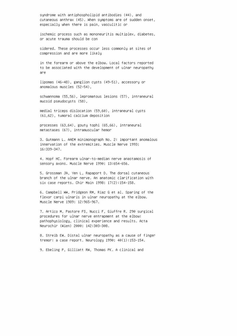
syndrome with antiphospholipid antibodies (44), andcutaneous anthrax (45). When symptoms are of sudden onset,especially when there is pain, vasculitic or
ischemic process such as mononeuritis multiplex, diabetes,or acute trauma should be con
sidered. These processes occur less commonly at sites ofcompression and are more likely
in the forearm or above the elbow. Local factors reportedto be associated with the development of ulnar neuropathyare
lipomas (46–48), ganglion cysts (49–51), accessory oranomalous muscles (52–54),
schwannoma (55,56), lepromatous lesions (57), intraneuralmucoid pseudocysts (58),
medial triceps dislocation (59,60), intraneural cysts(61,62), tumoral calcium deposition
processes (63,64), gouty tophi (65,66), intraneuralmetastases (67), intramuscular hemor
3. Gutmann L. AAEM minimonograph No. 2: important anomalousinnervation of the extremities. Muscle Nerve 1993;16:339–347.
4. Hopf HC. Forearm ulnar-to-median nerve anastamosis ofsensory axons. Muscle Nerve 1990; 13:654–656.
5. Grossman JA, Yen L, Rapaport D. The dorsal cutaneousbranch of the ulnar nerve. An anatomic clarification withsix case reports. Chir Main 1998; 17(2):154–158.
6. Campbell WW, Pridgeon RM, Riaz G et al. Sparing of theflexor carpi ulnaris in ulnar neuropathy at the elbow.Muscle Nerve 1989; 12:965–967.
7. Artico M, Pastore FS, Nucci F, Giuffre R. 290 surgicalprocedures for ulnar nerve entrapment at the elbow:pathophysiology, clinical experience and results. ActaNeurochir (Wien) 2000; 142:303–308.
8. Streib EW. Distal ulnar neuropathy as a cause of fingertremor: a case report. Neurology 1990; 40(1):153–154.
9. Ebeling P, Gilliatt RW, Thomas PK. A clinical and

electrical study of ulnar nerve lesions of the hand. JNeurol Neurosurg Psychiatry 1960; 23:1–9.
10. Cavallo M, Poppi M, Martinelli P, Gaist G. Distal ulnarneuropathy from carpal ganglion: a clinical andelectrophysiologic study. Neurosurgery 1988; 22:902–905.
11. Fulkerson JP. Transient ulnar neuropathy from Nordicskiing. Clin Orthop 1980; 153:230–231.
12. Streib EW, Sun SF. Distal ulnar neuropathy in meatpackers. An occupational disease? J Occup Med 1984;26(11):842–843.
13. Burnham RS, Steadward RD. Upper extremity peripheralnerve entrapments among wheelchair athletes: prevalence,location, and risk factors. Arch Phys Med Rehabil 1994;75(5):519–524.
14. Friedland RP, St John JN. Video-game palsy: distalulnar neuropathy in a video game enthusiast. N Engl J Med1984; 311(1):58–59.
15. Lederman RJ. Neuromuscular and musculoskeletal problemsin instrumental musicians. Muscle Nerve 2003;27(5):549–561.
16. Charness ME, Ross MH, Shefner JM. Ulnar neuropathy anddystonic flexion of the fourth and fifth digits: clinicalcorrelation in musicians. Muscle Nerve 1996; 19(4):431–437.
17. Lederman RJ. AAEM minimonograph No. 43: neuromuscularproblems in the performing arts. Muscle Nerve 1994;17(6):569–577.
18. Massey EW. Dimple sign in mail carrier’s ulnarneuropathy. Neurology 1989; 39(8):1132.
19. Finsterer J. Ulnar neuropathy at the elbow due tounusual sleep position. Eur J Neurol 2000; 7(1):115–117.
20. Lewis MB. Telesales neuropathy. Postgrad Med J 2000;76(902):793–794.
21. Veerendrakumar M, Taly AB, Nagaraja D. Ulnar nervepalsy due to axillary crutch. Neurol India 2001;49(1):67–70.
22. Eckman PB, Perstein G, Altrocchi PH. Ulnar neuropathyin bicycle riders. Arch Neurol 1975; 32(2):130–132.

23. Mellion MB. Common cycling injuries. Management andprevention. Sports Med 1991; 11(1):52–70.
24. Patterson JM, Jaggers MM, Boyer MI. Ulnar and mediannerve palsy long-distance cyclists. A prospective study. AmJ Sports Med 2003; 31(4):585–589.
25. Abdel-Salam A, Eyres KS, Cleary J. Drivers’ elbow: acause of ulnar neuropathy. J Hand Surg [Br] 1991;16(4):436–437.
26. Lee CT, Espley AJ. Perioperative ulnar neuropathy inorthopaedics: association with tilting the patient. ClinOrthop 2002; 396:106–111.
27. Warner MA, Warner ME, Martin JT. Ulnar neuropathy.Incidence, outcome, and risk factors in sedated oranesthetized patients. Anesthesiology 1994;81(6):1332–1340.
28. Morin JE, Long R, Elleker MG, Eisen AA, Wynands E,Ralphs-Thibodeau S. Upper extremity neuropathies followingmedian sternotomy. Ann Thorac Surg 1982; 34(2):181–185.
29. Seyfer AE, Grammer NY, Bogumill GP, Provost JM, ChandryU. Upper extremity neuropathies after cardiac surgery. JHand Surg [Am] 1985; 10(1):16–19.
30. Casscells CD, Lindsey RW, Ebersole J, Li B. Ulnarneuropathy after median sternotomy. Clin Orthop 1993;291:259–265. 31. Klein C, Halevy A, Gandelman-Marton R,Halpern Z, Weissgarten J, Averbukh Z, Arlazoroff A. Nerveconduction abnormalities in the arms of patients witharteriovenous fistula. Ren Fail 1996 18(1):85–89 32.Knezevic W, Mastaglia FL. Neuropathy associated withBrescia–Cimino arteriovenous fistulas. Arch Neurol 1984;41(11):1184–1186. 33. Geiringer SR, Leonard JA Jr.Injection related ulnar neuropathy. Arch Phys Med Rehabil1989; 70(9):705–706. 34. Marin R, McMillian D. Ulnarneuropathy associated with subdermal contraceptive implant.South Med J 1998; 91(9):875–878. 35. Finelli PF. Ulnarneuropathy after liquid nitrogen cryotherapy. Arch Dermatol1975; 111(10):1340–1342. 36. Markal N, Celebioglu S.Compression neuropathy of the hand after high-pressure airinjection. Ann Plast Surg 2000 44(6):680–681. 37. QualityAssurance Committee of the AAEM and Quality StandardsSubcommittee of the AAN. Practice parameter:Electrodiagnostic studies in ulnar neuropathy at the elbow.Neurology 1999; 52:688–690. 38. Landau ME, Diaz MI, Barner

KC, Campbell WW. Changes in nerve conduction velocityacross the elbow due to experimental error. Muscle Nerve2002; 26:838–840. 39. Britz GW, Haynor DR, Kuntz C, GoodkinR, Gitter A, Maravilla K, Kliot M. Ulnar nerve entrapmentat the elbow: correlation of magnetic resonance imaging,clinical, electrodiagnostic, and intraoperative findings.Neurosurgery 1996; 38:458–465. 40. Taras J, Melone CP.Hypertrophic neuropathy presenting with ulnar nervecompression: a case report. J Hand Surg [Am] 1995;20(2):233–234. 41. Dhillon MS, Chu ML, Posner MA.Demyelinating focal motor neuropathy of the ulnar nervemasquerading as compression in Guyon’s canal: a casereport. J Hand Surg [Am] 2003; 28(1):48–51. 42. Sharief MK,Robinson SF, Ingram DA, Geddes JF, Swash M. Paraneoplasticpainful ulnar neuropathy. Muscle Nerve 1999; 22(7):952–955.43. Mouthon L, Halimi C, Muller GP, Cayre-Castel M, BegueT, Masquelet AC, Guillevin L. Systemic sclerodermaassociated with bilateral ulnar nerve entrapment at theelbow. Rheumatology (Oxford) 2000; 39(6):682–683. 44.Graham LE, Bell AL. Minocycline associated lupus-likesyndrome with ulnar neuropathy and antiphospholipidantibody. Clin Rheumatol 2001; 20(1):67–69. 45. TerziogluA, Aslan G. Ulnar nerve lesion due to cutaneous anthrax.Ann Plast Surg 1999; 43(6):644–645. 46. Zahrawi F. Acutecompression ulnar neuropathy at Guyon’s canal resultingfrom lipoma. J Hand Surg [Am] 1984; 9(2):238–239. 47.Galeano M, Colonna M, Risitano G. Ulnar tunnel syndromesecondary to lipoma of the hypothenar region. Ann PlastSurg 2001; 46(1):83–84. 48. Sakai K, Tsutsui T, Aoi M,Sonobe H, Murakami H. Ulnar neuropathy caused by a lipomain Guyon’s canal—case report. Neurol Med Chir (Tokyo) 2000;40(6):335–338. 49. Shu N, Uchio Y, Ryoke K, Yamamoto S, OaeK, Ochi M. Atypical compression of the deep branch of theulnar nerve in Guyon’s canal by a ganglion. Case report.Scand J Plast Reconstr Surg Hand Surg 2000; 34(2):181–183.50. Sharma RR, Pawar SJ, Delmendo A, Mahapatra AK.Symptomatic epineural ganglion cyst of the ulnar nerve inthe cubital tunnel: case report and brief review of theliterature. J Clin Neurosci 2000; 7(6):542–543. 51. CavalloM, Poppi M, Martinelli P, Gaist G. Distal ulnar neuropathyfrom carpal ganglia: a clinical and electrophysiologicstudy. Neurosurgery 1998; 22(5):902–905. 52. Masear VR,Hill JJ Jr, Cohen SM. Ulnar compression neuropathysecondary to the anconeus epitrochlearis muscle. J HandSurg [Am] 1988; 13(5):720–724. 53. Ruocco MJ, Walsh JJ,Jackson JP. MR imaging of ulnar nerve entrapment secondaryto an anomalous wrist muscle. Skeletal Radiol 1998;27(4):218–221. 54. Sheppard JE, Prebble TB, Rahn K. Ulnarneuropathy caused by an accessory abductor digiti minimimuscle. Wis Med J 1991; 90(11):628–631.

55. Shank CP, Friedman WA. Ulnar neuropathy in a patientwith multiple schwannomas of the ulnar nerve. Surg Neurol1987; 28(2):153–157.
56. Spinner RJ, Tiel RL. High ulnar nerve compression by atriceps branch schwannoma. J South Orthop Assoc 2003;12(1):41–42.
57. Bhushan C. Solitary tuberculoid Hansen lesion of theulnar nerve. J Neurosurg 2000; 93(5):898.
58. Chick G, Alnot JY, Silbermann-Hoffman O. Intraneuralmucoid pseudocysts. A report of ten cases. J Bone JointSurg Br 2001; 83(7):1020–1022.
59. Spinner RJ, Davids JR, Goldner RD. Dislocating medialtriceps and ulnar neuropathy in three generations of onefamily. J Hand Surg [Am] 1997; 22(1):132–137.
60. Spinner RJ, O’Driscoll SW, Jupiter JB, Goldner RD.Unrecognized dislocation of the medial portion of thetriceps: another cause of failed ulnar nerve transposition.J Neurosurg 2000; 92(1):52–57.
61. Harway RA. Ulnar neuropathy due to intraneural cyst.Orthopedics 1997; 20(4):354–355.
62. Inhofe PD, Moneim MS. Compression of the ulnar nerve atthe elbow by an intraneural cyst: a case report. J HandSurg [Am] 1996; 21(6):1094–1096.
63. Aoyama T, Takahashi S. Cubital tunnel syndrome causedby tumoral deposition of calcium pyrophosphate dihydratecrystals: a case report. J Shoulder Elbow Surg 2001;10(2):194–196.
64. Cofan F, Garcia S, Campistol JM, Combalia A,Oppenheimer F, Ramon R. Ulnar nerve compression—a case ofgiant uremic tumoral calcinosis. Acta Orthop Scand 1997;68(3):302–304.
65. Nakamichi K, Tachibana S. Cubital tunnel syndromecaused by tophaceous gout. J Hand Surg [Br] 1996;21(4):559–560.
66. Wang HC, Tsai MD. Compressive ulnar neuropathy in theproximal forearm caused by gouty tophus. Muscle Nerve 1996;19(4):525–527.

67. Griswold W, Piza-Katzer H, Jahn R, Herczeg E.Intraneural nerve metastasis with multiplemononeuropathies. J Peripher Nerv Syst 2000; 5(3):163–167.
68. Vijayakumar R, Nesathurai S, Abbott KM, Eustace S.Ulnar neuropathy from diffuse intramuscular hemorrhage: acase report. Arch Phys Med Rehabil 2000; 81(8):1127–1130.
69. Yoshii S, Ikeda K, Murakami H. Ulnar nerve compressionsecondary to ulnar artery true aneurysm at Guyon’s canal. JNeurosurg Sci 1999; 43(4):295–297.
70. Grossman JA, Becker GA. Ulnar neuropathy caused by athrombosed ulnar vein. Case report and literature review.Ann Chir Main Memb Super 1996; 15(4):244–247.
71. Scelsa SN. Syringomyelia presenting as ulnar neuropathyat the elbow. Clin Neurophysiol 2000; 111(9):1527–1530.
72. Hochman MS, DePrima SJ, Leon BJ. Pseudoulnar palsy froma small infarct of the precentral knob. Neurology 2000;55(12):1939–1941.
73. Escobar PL, Ballesteros J. Teres minor. Source ofsymptoms resembling ulnar neuropathy or C8 radiculopathy.Am J Phys Med Rehabil 1988; 67(3):120–122.
74. Husain S, Mishra B, Prakash V, Malaviya GN. Results ofsurgical decompression of ulnar nerve in leprosy. ActaLeprol 1998; 11(1):17–20.
75. Dellon AL, Hament W, Gittelshon A. Non-operativemanagement of cubital tunnel syndrome: an 8 yearprospective study. Neurology 1993; 43:1673–1677.
76. Friedman RJ, Cochran TP. A clinical andelectrophysiological investigation of anteriortransposition for ulnar neuropathy at the elbow. ArchOrthop Trauma Surg 1987; 106:375–380.
77. Black BT, Barron OA, Townsend PF, Glickel SZ, Eaton RG.Stabilized subcutaneous ulnar nerve transposition withimmediate range of motion. J Bone Joint Surg 2000;82-A(11):1544–1551.
29
Other Upper Extremity Neuropathies
J. Steven Schultz

University of Michigan Health System, Ann Arbor, Michigan,USA
Abstract 498
1. Introduction 498
2. Long Thoracic Neuropathy 498 2.1. Anatomy 498 2.2.Clinical Features 499 2.3. Laboratory Evaluation 500 2.4.Differential Diagnosis 501 2.5. Management 502 2.6.Pathogenesis 502
3. Spinal Accessory Neuropathy 504 3.1. Anatomy 504 3.2.Clinical Features 504 3.3. Differential Diagnosis 505 3.4.Laboratory Evaluation 505 3.5. Management 506 3.6.Pathogenesis 506
4. Suprascapular Neuropathy 506 4.1. Anatomy 506 4.2.Clinical Features 507 4.3. Laboratory Evaluation 508 4.4.Differential Diagnosis 509 4.5. Management 510 4.6.Pathogenesis 510
root. The branch from the seventh nerve root is absent in�8% of patients and the nerve
occasionally receives a contribution from the eighthcervical root (1). The branches from
the fifth and sixth roots unite immediately after piercingthe scalenus medius muscle.
The branch arising from the seventh root joins the nerve asit travels across the scalenus pos
terior. The nerve then courses downward and laterally justposterior to the midaxillary line,
supplying each digitation of the serratus anterior as itprogresses. The lower portion of the
serratus is innervated by intercostal nerves in �20% ofindividuals (1).
2.2. Clinical Features
Patients with a long thoracic neuropathy typically presentwith a dull, aching sensation in
the periscapular area, and difficulty reaching overhead.

The distinguishing clinical feature
of serratus anterior weakness is scapular winging. Sittingin a hard-backed chair is often
uncomfortable, owing to scapular prominence. In many cases,patients are unaware of the
winging until it is brought to their attention. Scapularwinging from serratus anterior weak
ness has characteristic findings on physical examination.At rest, there is often a slight
outward flaring of the inferior angle of the scapula (Fig.29.2). Flexion of the arms
forward produces winging of the entire medial scapularborder (Fig. 29.3). The serratus
functions primarily as a scapular stabilizer and is uniquein its ability to maintain the
medial border of the scapula against the thorax as the armsare thrust forward. This move
ment results in a posteriorly directed force upon thescapula that cannot be counter
balanced if the serratus is weak. Winging of this type isaccentuated by resisted scapular
protraction, pressing against a wall, or performing apush-up. There is little if any trans
location of the scapula laterally because of thestabilizing action of the middle trapezius.
The serratus anterior also functions as an upward rotatorof the scapula; consequently,
active abduction of the affected arm is often limited.However, abduction may be full
due to compensatory action of the upper trapezius (2,3).Scapulohumeral rhythm is
Figure 29.1 Long thoracic nerve. [From Haymaker andWoodhall (24).] uniformly abnormal in these patients. Thecomparative features of serratus and trapezius winging are
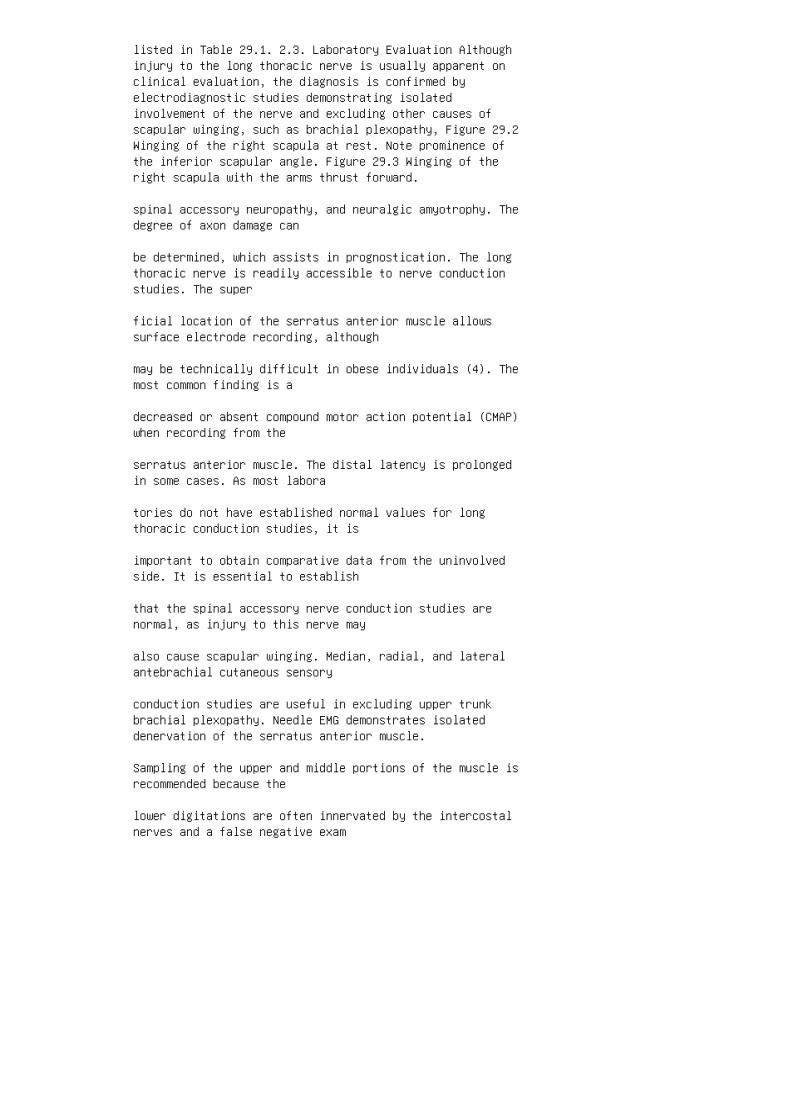
listed in Table 29.1. 2.3. Laboratory Evaluation Althoughinjury to the long thoracic nerve is usually apparent onclinical evaluation, the diagnosis is confirmed byelectrodiagnostic studies demonstrating isolatedinvolvement of the nerve and excluding other causes ofscapular winging, such as brachial plexopathy, Figure 29.2Winging of the right scapula at rest. Note prominence ofthe inferior scapular angle. Figure 29.3 Winging of theright scapula with the arms thrust forward.
spinal accessory neuropathy, and neuralgic amyotrophy. Thedegree of axon damage can
be determined, which assists in prognostication. The longthoracic nerve is readily accessible to nerve conductionstudies. The super
ficial location of the serratus anterior muscle allowssurface electrode recording, although
may be technically difficult in obese individuals (4). Themost common finding is a
decreased or absent compound motor action potential (CMAP)when recording from the
serratus anterior muscle. The distal latency is prolongedin some cases. As most labora
tories do not have established normal values for longthoracic conduction studies, it is
important to obtain comparative data from the uninvolvedside. It is essential to establish
that the spinal accessory nerve conduction studies arenormal, as injury to this nerve may
also cause scapular winging. Median, radial, and lateralantebrachial cutaneous sensory
conduction studies are useful in excluding upper trunkbrachial plexopathy. Needle EMG demonstrates isolateddenervation of the serratus anterior muscle.
Sampling of the upper and middle portions of the muscle isrecommended because the
lower digitations are often innervated by the intercostalnerves and a false negative exam

ination may be recorded if only inferior portions arestudied. Examination of the trapezius
and other limb muscles excludes lesions of the spinalaccessory nerve and brachial plexus.
2.4. Differential Diagnosis
The differential diagnosis of long thoracic neuropathy islisted in Table 29.2. Among nerve
injuries, spinal accessory neuropathies cause scapularwinging, but with a different clinical
picture from serratus anterior winging (Table 29.1).Traction injuries to the brachial plexus
with root avulsion or involvement of preganglionic fibersmay also result in scapular
Table 29.1 Comparative Features of Serratus Anterior andTrapezius Scapular Winging Serratus anterior wingingTrapezius scapular winging
Muscle atrophy Minimal Often prominent in upper trapezius
Appearance at rest Flaring of the inferior scapular angleDrooped shoulder on affected side
Winging accentuated by Forward flexion and resistedprotraction Abduction
Winging minimized by Abduction Forward flexion
Lateral translocation
of scapula Minimal Often pronounced
Table 29.2 Differential
Diagnosis of Long Thoracic
Neuropathy
Spinal accessory neuropathy
Dorsal scapular neuropathy
Brachial plexopathy

C7 radiculopathy
Neuralgic amyotrophy
Fascioscapulohumeral dystrophy
Scapuloperoneal dystrophy
Limb-girdle dystrophy
Hypermobility syndromes winging, but clinical andelectrophysiologic abnormalities are not limited to thelong thoracic nerve. Winging is occasionally seen as amanifestation of C7 radiculopathy (5). Neuralgic amyotrophyshould be considered with winging of the scapula,especially when preceded by acute severe and sharp pain inthe shoulder girdle unassociated with a specific event oractivity or a symptom free interval following trauma. Thiscondition is characterized by single or multiple upperextremity mononeuropathies and patchy involvement of thebrachial plexus. The most common nerves affected are thelong thoracic, axillary, and suprascapular (6). When needleEMG abnormalities are isolated to the long thoracic nerve,a careful history will distinguish between neuralgicamyotrophy and other etiologies. Various types of musculardystrophy are associated with scapular winging, notablyfascioscapulohumeral (FSH), scapuloperoneal, andlimb-girdle types. These disorders are easily distinguishedfrom long thoracic nerve injury by family history, otherassociated findings, and histopathologic features. Rarely,winging not associated with pain is seen as a normalvariant in patients with hypermobility syndromes. 2.5.Management Initial treatment includes relative rest andavoidance of activities that cause overstretching of theserratus anterior muscle, especially those that require thearm to assume a forward flexed position. A properlydesigned scapular orthotic device may be helpful (7–9).Active range of motion exercises are performed in thesupine position, utilizing the weight of the body tostabilize the scapula and prevent serratus overstretch(10). Much of the pain results from biomechanical overloadand contracture of periscapular and antagonist muscles.Accordingly, attention is given to stretching thelatissimus dorsi, levator scapulae pectoralis minor, andrhomboid muscles. Serratus strengthening is initiated aspain and range of motion improve. Other stabilizers of thescapula, especially the upper and lower trapezius must alsobe strengthened. Exercising these muscles in their capacityas scapular rotators, such as raising the arm overhead

while supine is recommended in the early stages of recovery(7). As strength improves, more advanced closed chainexercises such as push-ups are prescribed to enhancedynamic stability of the shoulder girdle (11). Surgicalexploration and neurolysis is considered with severe axonloss. Neurolysis of the supraclavicular portion of thenerve has been successful in patients with compressivelesions in the scalene muscles (12). Different surgicalprocedures have been proposed to substitute for the weakserratus anterior and stabilize the scapula against thethorax. The optimal procedure is transfer of the sternalhead of the pectoralis major to the inferior angle of thescapula (13,14). Transposition of the pectoralis minor,rhomboid, or teres major muscles has also been performed(15–17). Scapular fixation to the chest wall utilizingfascial slings is less successful, as it fails to providedynamic stability to the shoulder (14). Prognosis islargely dependent upon the mechanism of injury. Tractioninjuries have a favorable outcome, with recovery rates of90–100% (2,10,11,18). Although patients typically regainfull functional use, and a mild degree of scapular wingingmay persist (2,11,19). Prognosis in traumatic axon losslesions is generally poor, with maximum recovery 6 months–2years, (average time, 9 months) (2,10,11). If there is noelectrophysiologic evidence of nerve function at 2 years,the prognosis for spontaneous recovery is poor and surgicalstabilization of the scapula is recommended (11). 2.6.Pathogenesis Long thoracic mononeuropathy has beenattributed to many causes (Table 29.3). The superficiallocation of the nerve in the lateral thorax renders itsusceptible to direct
trauma, including motor vehicle accidents (3,20), carryinga knapsack (21), lifting (20),
sleeping on an outstretched arm (19), cervical chiropracticmanipulation (22), and electric
shock (14,23). Surgical procedures in the axillary regionmay also damage the nerve,
including mastectomy (24), axillary node dissection (10),and minimally invasive mitral
valve surgery (25). The proximal portion of the nerve isrelatively fixed by the scalenus
medius muscle and is subject to traction when the arm israised overhead or outstretched

(11,26). A tight fascial band extending from the middlescalene to the first rib has been
described such that abduction and external rotation of thearm may cause the long thoracic
nerve to “bow string” over the fascial band, resulting in astretch injury to the nerve (27).
Thus, stretch-induced injury, including cases involving asingle strenuous exertion, appears
to play a significant causative role and may be the primarymechanism of injury in patients
Table 29.3 Causes of Long
Thoracic Neuropathy
Direct trauma
Motor vehicle crashes
Falls
Carrying knapsack
Post surgical
Thoracotomy
First rib resection
Radical mastectomy
Axillary lymph node
dissection
Indirect trauma
Electrocution
Chopping wood
Sleeping on outstretched arm
Carrying tray overhead
Sports-related

Tennis
Weightlifting
Football
Hockey
Golf
Baseball
Basketball
Gymnastics
Wrestling
Archery
Discus
Bowling
Ballet
Target shooting
Nontraumatic
Post infectious
Post immunization
Post partum
Familial
Neuralgic amyotrophy
Idiopathic without a history of direct trauma (2,11,19).However, when there is shoulder pain in the setting ofminor trauma, neuralgic amyotrophy should be considered(28). Athletic activities have been associated with longthoracic neuropathy. The most common is tennis, especiallywith serving (2,11,28–30). Other sports includeweightlifting (2,11), baseball (2), football (31,32),wrestling (3), basketball (32), golf (2), archery (28),shooting a rifle (33), gymnastics, bowling, hockey, and

soccer (11). Isolated injuries to the long thoracic nervehave been reported to occur after infection (3,7,19,28),immunization (3,7,31), childbirth, and cold exposure (31).Occasionally, a causative agent cannot be identified, andmost likely represent a variant of neuralgic amyotrophy(6,7,19,28,30,31,34). There is a familial neuralgicamyotrophy that includes recurrent long thoracicneuropathies (35). 3. SPINAL ACCESSORY NEUROPATHY 3.1.Anatomy The spinal accessory, or eleventh cranial nerve, isa motor nerve that supplies the sternocleidomastoid andtrapezius muscles. The spinal portion of the nerve isformed by efferent branches of the first five cervicalnerve roots which ascend and enter the skull through theforamen magnum, where they briefly unite with the bulbarportion of the nerve. The spinal portion then exits theskull through the jugular foramen together with theglossopharygeal and vagus nerves. Initially, the nerve liesin close proximity to the internal carotid artery andinferior jugular vein, then descends obliquely in theposterolateral neck to innervate the sternocleidomastoidmuscle. The nerve continues deep to this muscle and crossesthe posterior triangle of the neck. Here, the nerve lies ina superficial location, rendering it susceptible to trauma.It first supplies the upper trapezius and then runs on thedeep surface of this muscle to innervate the middle andlower portions. Although the spinal accessory nerve doesnot contain sensory fibers, the ventral rami of C3 and C4from the cervical plexus often provide proprioceptiveinnervation from the upper trapezius (36). C2 andoccasionally C3 give off sensory branches to thesternocleidomastoid. The trapezius may also receive anefferent contribution from C3 and C4 (37,38). 3.2. ClinicalFeatures Shoulder pain and weakness are the presentingcomplaints. Pain is variable and results from contractureand overload on other musculotendinous structures in theneck and shoulder girdle. Shoulder weakness is usuallypronounced and typically causes more disability than thatseen in serratus anterior paresis due to the trapezius’greater role in scapular stabilization. Weakness manifestsprimarily as inability to fully abduct the arm, andpatients have significant difficulty performing overheadactivities. Intracranial or intraspinal involvement of thenerve also results in weakness and atrophy of thesternocleidomastoid resulting in difficulty turning thehead to the opposite side. Atrophy of the trapezius is mostnoticeable in the relatively thick upper portion of themuscle, resulting in a loss of normal contour of the top ofthe shoulder. When atrophy is severe, the superior borderof the scapula is visible above the shoulder when viewedfrom the front, which has been termed “moonrise scapula”

(39). The upper fibers of the trapezius are the only musclecapable of elevating the point of the shoulder. When thismuscle is nonfunctional, the lateral angle of the scapulais left unsupported and pulled downward by the weight ofthe limb. Clinically, this results in a drooped shoulder onthe affected side. The most consistent feature of spinalaccessory neuropathy is scapular
winging which differs from that associated with longthoracic nerve injury. With trapezius
weakness, abduction of the involved shoulder results inwinging of the entire medial
border of the scapula (Fig. 29.4). This is in contrast toserratus anterior winging which
is most prominent at the inferior angle and is precipitatedupon forward flexion. The tra
pezius as a whole, acts to retract the scapula, andweakness of this muscle together with
unopposed action of the serratus and pectoralis musclesresults in lateral translocation
of the scapula during the act of abduction. Additionally,active abduction is limited to
908 due to the pivotal role played by the upper and lowerfibers of the trapezius in
upward rotation of the scapula. Shoulder shrug is minimallyaffected because of the
action of the levator scapulae.
3.3. Differential Diagnosis
Scapular winging is also a prominent clinical feature oflong thoracic neuropathy;
however, this results in a different clinical picture thatshould not be confused with
spinal accessory neuropathy (Table 29.1). Other conditionsthat can cause winging of
the scapula are listed in Table 29.2. Scapular winging thatoccurs following surgical

procedures in the neck is far more likely to be due tospinal accessory nerve damage.
3.4. Laboratory Evaluation
The diagnosis of spinal accessory neuropathy is confirmedby electrodiagnostic studies.
A CMAP can be obtained by stimulating the nerve along theposterior border of the ster
nocleidomastoid with recording over the upper, middle, orlower portions of the trapezius
(4,37,40). As most injuries are axonal, the most constantfinding is a reduced CMAP (41).
A prolonged distal latency is occasionally observed infocal compression of the nerve.
Comparative values from the uninvolved side are necessaryto determine the significance
of any abnormalities seen. Needle EMG demonstratesdenervation in the trapezius. Examination of all three
portions of the muscle is helpful when looking for signs ofrecovery following a complete
Figure 29.4 Winging of the right scapula with abduction.injury. Involvement of the sternocleidomastoid indicates anintracranial or intraspinal lesion. It is important toexamine the serratus anterior and other scapular muscles toconfirm that the abnormalities are isolated to thetrapezius. In cases where intracranial involvement issuspected, cranial computerized tomography (CT) or magneticresonance imaging (MRI) is recommended (42). 3.5.Management Early treatment consists of stretching of therhomboids, levator scapulae, latissimus dorsi, andpectoralis minor, which have a tendency to becomecontracted and painful. Bracing may offer pain relief insome patients by stabilizing the scapula. Exercises tostrengthen the serratus anterior, the other primary upwardrotator of the scapula are given in an effort to compensatefor the nonfunctional trapezius. Specific strengthening ofthe trapezius can begin once reinnervation is evident onelectrodiagnostic studies. Surgical exploration andneurolysis is considered in severe closed injuries if norecovery is seen after several months. Exploration is

occasionally done earlier when the nerve is injured as aresult of a surgical complication. Cable grafting utilizingthe greater auricular nerve is often performed when thenerve is unavoidably sacrificed during radical neckdissection (38,43). Muscle transfer utilizing the levatorscapulae and rhomboid muscles is successful in decreasingpain and improving function in many patients who fail orare not candidates for nerve repair (44–46). 3.6.Pathogenesis Causes of spinal accessory neuropathy arelisted in Table 29.4. The majority of injuries involve theperipheral portion of the nerve as it courses through theneck. Lesions of the intracranial portion are rare andtypically result from meningioma, schwanoma, or metastasesto the base of the skull (47–49). In the Vernet syndrome,the spinal accessory, glossopharygeal and vagus nerves aredamaged as they pass through the jugular foramen. Patientswith carotid body tumors may also present with accessoryneuropathy (50). The majority of spinal accessoryneuropathies are iatrogenic, resulting from surgicalprocedures in the posterior triangle of the neck. Injury tothe nerve can occur as a complication of lymph node biopsy(51–53), carotid endarterectomy (54–56), facelift (57),coronary artery bypass grafting (58), and internal jugularvein cannulation (59,60). The spinal accessory nerve isdeliberately sacrificed during radical neck dissection, andmay be injured during modified neck dissection (61,62).Intraoperative monitoring of the nerve can decrease therisk of injury (63). Blunt trauma to the neck, such asslashing in hockey or lacrosse can damage the nerve. Acuteand chronic stretch injuries to the nerve have also beenreported (64–66). Whiplash-type injury may also damage thenerve (67). Uncommon etiologies include suicide attempts byhanging (52), radiation (68), and penetrating trauma (42).Spinal accessory neuropathy is a rare manifestation ofneuralgic amyotrophy. 4. SUPRASCAPULAR NEUROPATHY 4.1.Anatomy The suprascapular nerve is derived from the fifthand sixth cervical nerve roots and usually arises directlyfrom the upper trunk of the brachial plexus (Fig. 29.5). Itmay also arise directly from the fifth cervical root andoccasionally receives a contribution from C4.
It courses behind the brachial plexus between the fascia ofthe middle and the anterior
scalene muscles, then travels deep to the omohyoid andtrapezius to reach the superior
border of the scapula. Here, it passes through thesuprascapular foramen, a true fibro

osseous tunnel, formed by the scapular notch and overlyingsuperior transverse scapular
ligament. It first gives off a motor branch to thesupraspinatus then two small sensory
twigs to the acromioclavicular and glenohumeral joints. Italso supplies sensory and sym
pathetic fibers to the spinatii and periarticular tissues.The nerve then continues downward
and laterally in the supraspinatus fossa, then makes asharp turn to pass through the spino
glenoid notch of the scapular spine, which may be coveredby an inferior transverse
scapular (spinoglenoid) ligament. It terminates in theinfraspinatus muscle.
4.2. Clinical Features
The diagnosis of suprascapular neuropathy is often delayedor unrecognized due to the
diffuse and insidious nature of symptoms. Posteriorshoulder pain and weakness are the
primary presenting complaints. Pain ranges from mild tosevere, depending on the degree
of afferent branch involvement. Pain is typicallyaggravated with overhead activities.
Athletes engaged in throwing or overhead sports, such asbaseball pitchers, volleyball,
and tennis players, complain of increased pain in thecocking and release phases.
Table 29.4 Causes of Spinal
Accessory Neuropathy
Intracranial
Meningioma
Schwannoma

Sarcoidosis
Vernet syndrome
Metastases to base of skull
Intraspinal
Tumor
Syringomyelia
Motor neuron disease
Poliomyelitis
Peripheral
Idiopathic Neck dissection Lymph node biopsy FaceliftCarotid endarterectomy Coronary artery bypass graftingInternal jugular vein cannulation
Blunt Trauma Slashing in hockey Lacrosse Attempted suicideby hanging
Traction Rowing Backpack Shoulder dislocation
Radiation
Idiopathic Weakness and atrophy of the infraspinatus arethe principal physical findings, and the majority ofpatients will demonstrates weakness of external rotation ofthe shoulder. Supraspinatus atrophy is often masked by theoverlying upper fibers of the trapezius. Weakness of thesupraspinatus can be overlooked with a normally functioningdeltoid, as the latter muscle is a much more powerfulabductor of the arm. Sensory examination is normal, as thenerve has no cutaneous distribution. Forced adduction ofthe arm places the nerve on stretch and elicits pain insome patients (69,70). Tenderness over the suprascapularnotch is a common finding in many patients with entrapmentat this site (69). Occasionally, patients demonstratetenderness over the spinoglenoid notch. Scapular winging isnotably absent in these individuals. 4.3. LaboratoryEvaluation Suprascapular nerve conduction studies aregenerally less helpful than needle electromyography.Conduction studies to the supraspinatus and infraspinatusare obtained by stimulating the nerve at Erb’s point, butbecause of the overlying trapezius muscle, surface

electrode recording is not reliable (71). Consequently,nerve conduction studies using an intramuscular needlerecording electrode can be used. CMAP amplitude obtained inthis manner is of little diagnostic utility, owing to thelimited recording area of a needle electrode. Conductionvelocities are likewise of doubtful validity, due todifficulties obtaining accurate measurements in theshoulder. Thus, distal latency is the most useful Figure29.5 Suprascapular nerve. [From Haymaker and Woodhall(24).]
parameter. The latency to the infraspinatus may beprolonged, although there is no
consensus as to what constitutes a clinically significantdelay or side-to-side difference.
A difference of .0.4 ms correlates with denervation onneedle EMG (72). Median,
radial, and lateral antebrachial cutaneous sensory nerveaction potential (SNAP) ampli
tudes are helpful in excluding upper trunk brachialplexopathy. Needle EMG can confirm the diagnosis anddifferentiate from other neurologic con
ditions that may mimic suprascapular neuropathy (73).Abnormalities are found in the
infraspinatus when the lesion is localized to either thesuprascapular notch or spinoglenoid
notch, whereas the supraspinatus is involved in mostsuprascapular notch injuries but is
spared in spinoglenoid entrapment. The supraspinatus isoccasionally spared in proven
lesions at the scapular notch, presumably because thebranch to the supraspinatus was
given off proximal to the scapular notch (74).Alternatively, partial lesions of the nerve
may selectively affect those fascicles that supply theinfraspinatus while sparing those
that supply the supraspinatus (73,75). Examination of othershoulder girdle muscles, ser

ratus anterior, and paraspinals will distinguish isolatedsuprascapular neuropathy from C5
or C6 radiculopathy, upper trunk brachial plexopathy, andneuralgic amyotrophy. Radiographs of the scapula may revealfractures or excessive callus formation in
traumatic cases. MRI is the study of choice for theevaluation of cystic lesions or other
compressive etiologies, and is particularly useful when noapparent cause can be ident
ified. Additionally, MRI can assess denervation atrophy ofthe spinati muscles, and there
fore be of value when electrophysiologic studies are normal(76).
4.4. Differential Diagnosis
Injury to the suprascapular nerve should be considered inthe differential diagnosis of
rotator cuff tendinopathies, glenohumeral disorders, andmyofascial pain syndrome
(Table 29.5). Supraspinatus tendinosis also causes shoulderpain and weakness and is
exceedingly common in athletes. Complete avulsion of therotator cuff at its insertion typi
cally results in disuse atrophy of the supraspinatus andinfraspinatus and profound
shoulder weakness. Patients with chronic adhesivecapsulitis will also manifest scapular
atrophy with restricted external rotation and abduction.Patients with C5 or C6 radiculopathy or upper trunkplexopathy have a similar clini
cal picture; however, sensory and reflex abnormalitiesshould differentiate these lesions
from suprascapular neuropathy. Spinal accessory neuropathycauses scapular winging,

while suprascapular nerve injury does not. Abrupt onset ofsevere sharp pain followed
by weakness without a clear inciting event should raiseconcern for neuralgic amyotrophy.
Table 29.5 Differential
Diagnosis of Suprascapular
Neuropathy
C5, C6 radiculopathy
Upper trunk brachial plexopathy
Spinal accessory neuropathy
Neuralgic amyotrophy
Rotator cuff tendinopathy/tear
Glenohumeral disorders
Myofascial pain syndrome 4.5. Management Conservativetreatment is successful in most patients without acompressive lesion. Relative rest and range of motionexercises are prescribed in the early stage followingsuprascapular nerve injury. Activities that cause increasedtraction on the nerve, such as repetitive or sustainedadduction or external rotation should be limited. Physicaltherapy is recommended for strengthening and to addressother painful biomechanical dysfunctions of the shoulder.Suprascapular nerve block at the suprascapular notch ishelpful in alleviating pain in some patients. Patients witha documented axon loss lesion in the absence of aspace-occupying lesion who fail to improve withconservative measures should be referred for surgicalexploration and decompression of the scapular orspinoglenoid notch (77–80). Sectioning of the transversescapular ligament and widening of the notch has results ina favorable outcome in the majority of patients (69,70,81).Ganglion cysts may be aspirated under CT guidance orresected arthroscopically, however, large or invasivelesions usually require open excision (82,83). 4.6.Pathogenesis A variety of causes of suprascapularneuropathy have been reported (Table 29.6). Blunt trauma,such as scapular fractures, acromioclavicular separation,and rotator cuff tears are believed to cause acute tractioninjury to the nerve (84–88). The suprascapular Table 29.6

Causes of Suprascapular Neuropathy Blunt trauma Scapularfracture Shoulder dislocation Acromioclavicular separationFracture of proximal humerus Post surgical Rotator cuffrepair Radical neck dissection Supraclavicular lymph nodebiopsy Distal clavicle resection Traction Sustainedshoulder adduction Forcible shoulder depression Repetitiveexternal rotation Anabolic steroid use Sports-relatedVolleyball Baseball Tennis Football Weightlifting WrestlingMass lesions Ganglion cysts Chondrosarcoma SynovialsarcomaEwing’s sarcoma Metastatic disease
nerve is damaged with surprising frequency in anteriordislocation of the shoulder
(86,89,90). Suprascapular neuropathy occurs as aninfrequent complication of surgical
procedures involving the neck and shoulder girdle (91,92).Chronic repetitive traction, especially in athletes, is onemechanism. Sustained
shoulder adduction is thought to cause a stretch injury tothe proximal portion of the
nerve, which is tethered at the suprascapular notch (84).Suprascapular neuropathy
isolated to the infraspinatus muscle has been described inthrowing and overhead athletes.
Most of these have been reported to occur in the dominantarm of elite volleyball
players (93–96), baseball pitchers (74,77,97), tennisplayers (87,98), weightlifters
(69,81,97–100), a dancer (101), a fencer (102), and a skeetshooter (97). Many of these
injuries were postulated to occur from repetitive stretchto the terminal portion of the
nerve as it makes a sharp turn around the lateral aspect ofthe scapular spine and the rela
tive fixation of the nerve at the spinoglenoid notch(78,102). Alternatively, repetitive
throwing may cause intimal damage to the suprascapular oraxillary arteries, leading to

microembolization of the vaso nervorum and ultimately nervedamage (74). One case
was attributed to intramuscular compression of the terminalbranches from rapid hyper
trophy associated with anabolic steroid use (100). Trueentrapment of the suprascapular nerve can occur at thesuprascapular and spino
glenoid foramina. On the basis of the relative dimensionsand shape of the notch and
foramen, six distinct types of suprascapular notches havebeen described (103). Patients
with a deep, V-shaped notch are at higher risk ofdeveloping entrapment at the supra
scapular notch (103). Other anatomic risk factors includehypertrophy or ossification of
the superior and inferior transverse scapular ligaments anda small diameter foramen
(79,102–105). Entrapment of the nerve has also been foundto occur proximally between
the fascia of the anterior and middle scalene muscles,between the omyhyoid and sub
clavius fascia and distally within the suprascapular fossa,and between the suprascapular
fascia and base of the coracoid process (74). Compressionof the suprascapular nerve also occurs from space-occupyinglesions,
such as a ganglion cyst, which are being identified withincreasing frequency, due in large
part to the routine use of MRI in the evaluation ofshoulder pain (82,83,106–108). Cysts
are also seen in asymptomatic patients and rarely becomeclinically significant unless
they compress the suprascapular nerve (109). The etiologyof these cysts is controversial.

The proximity of many ganglion cysts to the glenohumeraljoint has led some investigators
to theorize that the cysts are formed by the leakage ofjoint fluid from glenoid labrial tears
(110). Additionally, ganglia are typically located in theposterior superior aspect of the
joint space, corresponding to the relative weakness of thecapsule above the posterior
band of the inferior glenohumeral ligament, therebyallowing extravasation of joint fluid
into the suprascapular and spinoglenoid foramina (110).Other malignant masses reported
to cause suprascapular nerve compression includechondrosarcoma, synoviosarcoma,
Ewing’s sarcoma, and metastatic renal cell carcinoma,although these are quite rare (82).
5. AXILLARY NEUROPATHY
5.1. Anatomy
The axillary nerve is a mixed nerve that carries fibersfrom the fifth and sixth cervical roots
and arises directly from the posterior cord of the brachialplexus at the level of the coracoid
process (Fig. 29.6). It courses under the coracoid alongthe anterior surface of the subsca
pularis then passes through the quadrilateral spaceaccompanied by the posterior circum
flex humeral artery. The quadrilateral space is boundedsuperiorly by the teres minor and subscapularis, laterallyby the surgical neck of the humerus, inferiorly by theteres major and medial by the long head of the tricepstendon. Here, the nerve divides into posterior and anteriorbranches. The posterior branch innervates the posteriordeltoid and teres minor, then continues as the lateralbrachial cutaneous nerve which supplies cutaneous sensationto the lateral aspect of the shoulder. The anterior branch
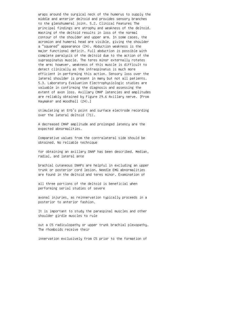
wraps around the surgical neck of the humerus to supply themiddle and anterior deltoid and provides sensory branchesto the glenohumeral joint. 5.2. Clinical Features Theprincipal findings are atrophy and weakness of the deltoid.Wasting of the deltoid results in loss of the normalcontour of the shoulder and upper arm. In some cases, theacromion and humeral head are visible, giving the shouldera “squared” appearance (24). Abduction weakness is themajor functional deficit. Full abduction is possible withcomplete paralysis of the deltoid due to the action of thesupraspinatus muscle. The teres minor externally rotatesthe arm; however, weakness of this muscle is difficult todetect clinically as the infraspinatus is much moreefficient in performing this action. Sensory loss over thelateral shoulder is present in many but not all patients.5.3. Laboratory Evaluation Electrophysiologic studies arevaluable in confirming the diagnosis and assessing theextent of axon loss. Axillary CMAP latencies and amplitudesare reliably obtained by Figure 29.6 Axillary nerve. [FromHaymaker and Woodhall (24).]
stimulating at Erb’s point and surface electrode recordingover the lateral deltoid (71).
A decreased CMAP amplitude and prolonged latency are theexpected abnormalities.
Comparative values from the contralateral side should beobtained. No reliable technique
for obtaining an axillary SNAP has been described. Median,radial, and lateral ante
brachial cutaneous SNAPs are helpful in excluding an uppertrunk or posterior cord lesion. Needle EMG abnormalitiesare found in the deltoid and teres minor. Examination of
all three portions of the deltoid is beneficial whenperforming serial studies of severe
axonal injuries, as reinnervation typically proceeds in aposterior to anterior fashion.
It is important to study the paraspinal muscles and othershoulder girdle muscles to rule
out a C5 radiculopathy or upper trunk brachial plexopathy.The rhomboids receive their
innervation exclusively from C5 prior to the formation of
the upper trunk, therefore are
particularly useful in excluding both of these diagnoses.Additionally, sampling of the
latissimus dorsi and radial innervated muscles allow anisolated axillary nerve injury to
be distinguished from a posterior cord lesion.
5.4. Differential Diagnosis
Axillary neuropathy can mimic other neurologic conditionsthat cause shoulder pain and
weakness (Table 29.7). These disorders are often difficultto distinguish solely on clinical
grounds, highlighting the importance of theelectrodiagnostic examination in the evalu
ation of these patients. Moreover, axillary nerve lesionscommonly occur in combination
with injuries of the brachial plexus and other peripheralnerve, further complicating the
clinical diagnosis. An important diagnostic considerationin patients with deltoid weakness is C5 radi
culopathy. In contrast to isolated axillary nerve injury,weakness, and atrophy are rarely
severe in radiculopathy. Additionally, neck pain and adiminished biceps reflex are typi
cally associated with C5 root lesions. Patients with uppertrunk lesions demonstrate more
widespread weakness of the shoulder girdle and biceps aswell as sensory loss in the
thumb. As noted above, lesions of the posterior cord alsoresult in deltoid weakness;
however, involvement of radial innervated muscles is also afeature of this disorder. As
it the case with other nerves arising from the brachialplexus, the axillary nerve is com

monly involved in neuralgic amyotrophy. This diagnosis isconsidered when pain is
severe and no distinct injury has occurred. Shoulder painand abduction weakness are
seen in patients with rotator cuff tears and adhesivecapsulitis, although these disorders
do not result in isolated atrophy and weakness of thedeltoid.
5.5. Management
Treatment is guided by the severity of injury as determinedby clinical and electrophysio
logic examination (42). If the injury is mild, thenphysical therapy is appropriate for range
Table 29.7 Differential
Diagnosis of Axillary
Neuropathy
C5, C6 radiculopathy
Upper trunk brachial plexopathy
Posterior cord plexopathy
Neuralgic amyotrophy
Rotator cuff tendinopathy
Adhesive capsulitis of shoulder of motion andstrengthening. The prognosis for spontaneous recovery inthese cases is good (111). Patients with severe axonaldamage should be assessed monthly for signs of improvement(42). If there is no evidence of recovery by 3–4 months,surgical exploration is recommended (112–115). Given theshort distance between the site of injury and the deltoid,the prognosis for spontaneous recovery beyond this time ispoor. Surgical options include neurolysis, neurotization,and nerve grafting, which result in a good outcome in themajority of patients (112,113,115). 5.6. PathogenesisCauses of axillary neuropathy are listed in Table 29.8.Injury to the nerve is most likely to result from shoulder

dislocation and fracture of the surgical neck of thehumerus (86,89,116). In one large series, the incidence ofaxillary nerve injuries in patients with these conditionswas 37% (89). Isolated damage to the axillary nerve wasfound in 8%; the remaining injuries occurred in variouscombinations with the suprascapular, musculocutaneous andradial nerves (89). Elderly patients and those withhematoma formation after shoulder trauma appear to be athigher risk of injuring the axillary nerve (89). Reductionof a shoulder dislocation has been reported to damage thenerve (117). Blunt trauma to the deltoid withoutdislocation can also damage the axillary nerve (118).Various sports have been reported to injure the nerve inthis manner, including football, hockey, wrestling, boxing,and basketball (119,120). The nerve is also injured frompenetrating trauma to the shoulder girdle. Injury to theanterior branch of the axillary nerve has been reported ina paraplegic patient following vigorous exercise (121).Hypertrophy of the muscles of the quadrilateral space weretheorized to compress the nerve in this patient, thus thismay represent the first reported case of quadrilateralspace syndrome Table 29.8 Causes of Axillary NeuropathyTraumatic Shoulder dislocation Proximal humerus fractureShoulder contusion Vigorous exercise Sleeping on a firmmattress Penetrating injuries Iatrogenic Intramuscularinjection Reduction of shoulder dislocation Shoulderarthroscopy Anterior shoulder surgery Radiofrequencycapsulorrhaphy Sports-related Football Hockey BasketballWrestling Boxing Quadrilateral space syndrome Neuralgicamyotrophy
(discussed subsequently). Compression of the anteriorbranch from sleeping on a firm
mattress has also been described in a patient withhereditary neuropathy with liability
to pressure palsies (122). Iatrogenic causes have also beendescribed, including complication of shoulder
arthroscopy (123), anterior shoulder surgery (124), andintramuscular injection into the
deltoid (125). Axillary neuropathy is occasionally seenfollowing radiofrequency capsular
shrinkage of the shoulder (126). Indirect thermal injury tothe nerve from heating of sur
rounding tissues was believed to be the cause in these
cases. A controversial cause of axillary neuropathy hasbeen reported to occur in the quad
rilateral space. In this syndrome, it is hypothesized thatmuscle hypertrophy and repeated
external rotation and abduction of the arm common inthrowing athletes results in a
decrease in the size of the quadrilateral space, therebycompressing the axillary nerve
and posterior circumflex humeral artery between surroundingmuscle and proximal
humerus (127). Fibrous bands have also been found tocompress the nerve in the space
(128). Clinically, patients with this syndrome present withlocalized tenderness in the
quadrilateral space and provocation of symptoms withsustained abduction and external
rotation of the affected limb. In initial reports of thesyndrome, all patients had abnormal
dynamic subclavian arteriograms, demonstrating occlusion ofthe posterior circumflex
humeral artery with the arm abducted and externally rotated(128–130). Neurologic
and electromyographic examinations were normal in allcases. Nevertheless, injury to
the axillary nerve as felt to be a defining characteristicof the disorder (128). Most of
these patients underwent decompression of the quadrilateralspace with axillary neurolysis
being performed in one case (128,130). In another series,EMG abnormalities were found
in 3 of 5 patients presumed to have the syndrome; however,other associated peripheral
nerve lesions were also observed (131). It is probable thatthese patients actually had

neuralgic amyotrophy rather than axillary nerve compression(42). Evidence for isolated
involvement of the axillary nerve in the quadrilateralspace was provided in a report of two
elite volleyball players who presented with painlessweakness and atrophy of the deltoid
muscle (127). Both had decreased axillary CMAP amplitudesand isolated denervation
of the deltoid on electrophysiologic studies. Completerecovery occurred with rest and
conservative measures. This suggests that the quadrilateralspace syndrome is a vascular
phenomenon in the majority of patients and compression ofthe axillary nerve in the quad
rilateral space is exceedingly rare.
6. MUSCULOCUTANEOUS NEUROPATHY
6.1. Anatomy
The musculocutaneous nerve usually originates from thelateral cord of the brachial plexus
and carries fibers from the C5, C6, and occasionally C7nerve roots (Fig. 29.7). Rarely, it
comes off directly from the median nerve or posterior cord.Immediately after supplying
the coracobracialis muscle, the nerve pierces this muscleand comes to lie between the
biceps and the brachialis muscles. It innervates both headsof the biceps and brachialis
then continues distally as the lateral antebrachialcutaneous nerve. The brachialis often
receives an additional branch from the radial nerve. Themusculocutaneous nerve
occasionally gives off a branch to the pronator teres and

flexor carpi radialis. The
lateral antebrachial cutaneous nerve travels lateral to thebiceps tendon in the antecubital
fossa, then pierces the brachial fascia. It divides intoanterior and posterior branches sup
plying sensation to the radial aspect of the forearm. 6.2.Clinical Features The typical presentation is painlessweakness of the arm flexors (132), although severe pain canoccur in some patients (133). Sensory abnormalities in theradial forearm are commonly found. Atrophy of the bicepsand brachialis can be impressive in chronic injuries.Weakness of the arm flexion and supination are the mostprominent deficits; however, elbow flexion is usuallypreserved even in complete paralysis of these muscles owingto the actions of the brachioradialis and pronator teres.Weakness of the coracobrachialis is difficult to detectclinically. It assists in flexion of the arm at theshoulder; however, the anterior deltoid is the chief moverand can effectively mask a paralyzed coracobrachialismuscle. Similarly, the coracobrachialis is a weak adductorof the arm, although this action is brought about primarilyby the pectoralis major, latissimus dorsi, and teres major.A diminished biceps reflex is frequently seen. 6.3.Laboratory Evaluation Electrodiagnostic examination willconfirm involvement of the musculocutaneous nerve, provideinformation regarding extent of injury and exclude otherneurologic conditions Figure 29.7 Musculocutaneous nerve.[From Haymaker and Woodhall (24).]
that produce a similar clinical picture. A musculocutaneousSNAP can reliably be obtained
from the lateral antebrachial cutaneous nerve (134,135). Areduced or absent SNAP
amplitude is expected in musculocutaneous neuropathy andupper trunk plexopathy.
Radial and median SNAPs recording from the thumb are usefulin excluding the latter
possibility. The lateral antebrachial cutaneous SNAP willbe normal in patients with C6
radiculopathy. The musculocutaneous CMAP amplitude,obtained by surface recording
over the biceps and stimulating at Erb’s point (71), oraxilla (134), is decreased in axon
loss injuries to the nerve. Needle EMG findings are foundin the coracobrachialis, biceps, and brachialis,
although sparing of the coracobrachialis is seen in distallesions. Examination of other
shoulder girdle and paraspinal muscles will discriminateisolated musculocutaneous neu
ropathy from C6 radiculopathy and upper trunk plexopathy.Sampling of the pronator teres
and flexor carpi radialis is useful in excluding a lesionof the lateral cord.
6.4. Differential Diagnosis
The differential diagnosis of musculocutaneous neuropathyis listed in Table 29.9. Weak
ness of arm flexion and a decreased biceps reflex arecommonly seen in patients with C6
radiculopathy; however associated weakness of the deltoidand brachioradialis should
differentiate this from musculocutaneous neuropathy. Uppertrunk and lateral cord injuries
are ruled out by electrodiagnostic studies. Themusculocutaneous nerve is commonly
involved in neuralgic amyotrophy in association with otherperipheral nerves. The clinical
presentation of neuralgic amyotrophy is distinctive andcontrasts with isolated musculo
cutaneous injury, in which pain is generally not aprominent feature. Spontaneous
rupture of the long head of the biceps tendon also producespainless weakness of arm
flexion and loss of the biceps reflex. In this disorder,attempted arm flexion at the elbow

causes the biceps to bunch up into a spherical shapedistally, producing a very character
istic appearance. Because the brachialis and other armflexors remain functional, rupture
of the tendon results in less weakness than doesmusculocutaneous neuropathy. Addition
ally, sensation in the radial forearm is normal in patientswith a ruptured biceps tendon.
6.5. Management
Patients with partial lesions of the musculocutaneous nervebenefit from relative rest and
physical therapy. Emphasis is placed on maintaining rangeof motion and strengthening of
the involved muscles. The prognosis for recovery is good(132,136–140). In severe closed
injuries, serial physical and electrodiagnosticexaminations should be performed to
monitor for recovery. If no electrophysiologic evidence ofrecovery is seen in 3–4
months, surgical exploration is warranted, and neurolysisor nerve grafting considered,
Table 29.9 Differential
Diagnosis of Musculocutaneous
Neuropathy
C6 radiculopathy
Upper trunk plexopathy
Lateral cord plexopathy
Neuralgic amyotrophy
Rupture of biceps tendon with good results reported withgrafting (141). Initial treatment of patients with isolatedinjury to the lateral antebrachial cutaneous nerve consistsof analgesics, splinting, and corticosteroid injections.

Surgical decompression of the biceps aponeurosis in theantecubital fossa is indicated when conservative measuresfail, with a favorable outcome reported in most patients(142,143). 6.6. Pathogenesis Most injuries to themusculocutaneous nerve result from traction, compression,or a combination of both (Table 29.10). It is more commonfor the nerve to be injured in conjunction with the uppertrunk and lateral cord than in isolation. Blunt trauma tothe anterior shoulder resulting in contusions, proximalhumeral, or clavicular fractures can cause direct injury tothe nerve (137,144,145). Penetrating trauma such as stab orbullet wounds may also damage the nerve (42,146).Musculocutaneous neuropathy occurs in association withshoulder dislocation (89,116). Strenuous physical activityand violent elbow extension have been reported to injurethe nerve (132–134,138,146). The coracobrachialis wasspared in these cases, indicating compression of the nervewithin this muscle, possibly due to forceful contraction orhypertrophy (132). An additional case of intramuscularcompression has been reported in a weightlifter, which wasbelieved to be due to rapid hypertrophy from anabolicsteroid use (100). An unusual case of musculocutaneousneuropathy from carrying rolls of carpet has been reported(139). Iatrogenic musculocutaneous neuropathy occurs as acomplication of surgical procedures of the shoulder(123,124,140). The musculocutaneous nerve is at risk duringshoulder arthroscopy (123). Three mechanisms of injuryunique to this procedure have been described, includingexcessive glenohumeral distraction from longitudinaltraction on the arm, massive extravasation of fluid in theanterior shoulder, and direct injury to the nerve via theanterior portal (123). Prolonged positioning of the arm inhyperextension, external rotation, or abduction duringsurgical procedures has been reported to damage themusculocutaneous nerve (136,147–149). Compression from ablood pressure cuff was hypothesized to be a contributingfactor in one case (149). Musculocutaneous neuropathyoccurs in association with neuralgic amyotrophy but rarelyas an isolated finding. Isolated lateral antebrachialcutaneous nerve injury is most commonly seen fromcompression or direct trauma in the antecubital fossa. Manyof these are iatrogenic, including venipuncture of thecephalic vein and radial artery bypass in coronary arterybypass operations (150–154). Compression of the nerve bythe biceps tendon and aponeurosis has been described(142,143,155,156). Lateral antebrachial nerve compressionhas been reported in quadriplegics (157), and from carryinga heavy handbag (158), or food tray Table 29.10 Causes ofMusculocutaneous Neuropathy Blunt trauma to shoulderShoulder dislocation Strenuous physical activity Violent

elbow extension Anabolic steroid use Shoulder surgeryShoulder arthroscopy General anesthesia Blood pressure cuff
(159) across the forearm. Vigorous extension and pronationof the forearm (160) and
cryotherapy (161) may also damage the nerve.
7. CONCLUSIONS
Focal neuropathy of the shoulder is a frequently overlookedcause of upper extremity pain
and dysfunction. These lesions often mimic other softtissue and neurologic disorders,
highlighting the critical role of EMG in establishing thediagnosis. Additionally, EMG
is unique in its ability to assess pathophysiology andseverity of injury, providing valuable
information to guide clinical decision-making. As many ofthese conditions occur in dis
tinctive settings or as a result of specific activities, itis important to obtain an accurate
injury history. With prompt recognition, most proximalneuropathies respond favorably
to conservative measures, although surgical management isnecessary in some cases.
1. Horwitz MT, Tocantins LM. An anatomic study of the roleof the long thoracic nerve and the related scapular bursaein the pathogenesis of local paralysis of the serratusanterior muscle. Anat Rec 1938; 71:375–385.
2. Schultz JS, Leonard JA. Long thoracic neuropathy fromathletic activity. Arch Phys Med Rehabil 1992; 73:87–90.
3. Goodman CE, Kenrick MM, Blum MV. Long thoracic nervepalsy: a follow-up study. Arch Phys Med Rehabil 1975;56:352–355.
4. Liveson JA, Ma DM. Laboratory Reference for ClinicalNeurophysiology. Philadelphia: FA Davis, 1992:48–52.
5. Makin GV, Brown WF, Ebers GC. C7 radiculopathy:

importance of scapular winging in clinical diagnosis. JNeurol Neurosurg Psychiatry 1986; 49:640–644.
6. Parsonage MJ, Turner JA. Neuralgic amyotrophy: theshoulder-girdle syndrome. Lancet 1948; 1:973–978.
7. Johnson JH, Kendall HO. Isolated paralysis of theserratus anterior muscle. J Bone Joint Surg (Am) 1955;37:567–574.
8. Truong XT, Rippel DV. Orthotic devices for serratusanterior palsy: some biomechanical considerations. ArchPhys Med Rehabil 1979; 60:66–69.
9. Marin R. Scapula winger’s brace: a case series on themanagement of long thoracic nerve palsy. Arch Phys MedRehabil 1998; 79:1226–1230.
10. Duncan MA, Lotze MT, Gerber LH, Rosenberg SA.Incidence, recover, and management of serratus anteriormuscle palsy after axilary node dissection. Phys Ther 1983;63:1243–1247.
11. Gregg JR, Labosky D. Harty M, Lotke, P, Ecker M,DiStefano V, Das M. Serratus anterior paralysis in theyoung athlete. J Bone Joint Surg (Am) 1979; 61:825–832.
12. Disa JJ, Wang B, Dellon AL. Correction of scapularwinging by supraclavicular neurolysis of the long thoracicnerve. J Reconstr Microsurg 2001; 17(2):79–84.
13. Wiater JM, Flatow EL. Long thoracic nerve injury. ClinOrthop 1999; 368:17–27.
14. Marmor L, Bechtol CO. Paralysis of the serratusanterior due to electric shock relieved by transplantationof the pectoralis major muscle. J Bone Joint Surg (Am)1963; 45:156–160.
15. Rapp IH. Seratus anterior paralysis treated bytransplantation of the pectoralis minor. J Bone Joint Surg(Am) 1954; 36:852–854.
16. Herzmark MH. Traumatic paralysis of the serratusanterior relieved by transplantation of the rhomboidei. JBone Joint Surg (Am) 1951; 33:235–238.
17. Harmon PH. Surgical reconstruction of the paralyticshoulder by multiple muscle transplantations. J Bone JointSurg (Am) 1950; 32:583–595. 18. Fardin P, Negrin P, Dainese

R. The isolated paralysis of the serratus anterior muscle:clinical and electromyographical follow-up of 10 cases.Electromyogr Clin Neurophysiol 1978; 18:379–386. 19.Overpeck DO, Ghormley RK. Paralysis of the serratus magnusmuscle caused by lesions of the long thoracic nerve. J AmMed Assoc 1940; 114:1994–1996. 20. Hauser CU, Martin WF.Two additional cases of traumatic winged scapula occurringin the armed forces. J Am Med Assoc 1943; 121:667–668. 21.Ilfeld FW, Holder HG. Winged scapula: case occurring in asoldier from a knapsack. J Am Med Assoc 1942; 120:448–449.22. Oware A, Herskovitz S. Berger AR. Long thoracic nervepalsy following cervical chiropractic manipulation. MuscleNerve 1995; 18:1351. 23. Still JM, Law EJ, Duncan JW,Hughes HF. Long thoracic nerve injury due to an electricburn. J Burn Care Rehabil 1996; 17:562–564. 24. Haymaker W,Woodhall B. Peripheral Nerve Injuries: Principles ofDiagnosis. 2nd ed. Philadelphia: WB Saunders, 1953:222–238.25. Chaney MA, Morales M, Bakhos M. Severe incisional painand long thoracic nerve injury after port-access minimallyinvasive mitral valve surgery. Anesth Analg 2000;91:288–290. 26. Ebraheim NA, Lu J, Porshinsky B, Heck BE,Yeasting RA. Vulnerability of long thoracic nerve: ananatomic study. J Shoulder Elbow Surg 1998; 7:458–461. 27.Hester P, Caborn DN, Nyland J. Cause of long thoracic nervepalsy: a possible dynamic fascial sling cause. J ShoulderElbow Surg 2000; 9:31–35. 28. Foo CL, Swann M. Isolatedparalysis of the serratus anterior. J Bone Joint Surg (Br)1983; 65:552–556. 29. Foucar HO. The “clover leaf” sling inparalysis of the serratus magnus. Br Med J 1933; 2:865–866.30. Goodman CE. Unusual nerve injuries in recreationalactivities. Am J Sports Med 1983; 11:224–227. 31. HannsonKG. Serratus magnus paralysis. Arch Phys Med Rehabil 1948;29:156–161. 32. Kaplan PE. Electrodiagnostic confirmationof long thoracic nerve palsy. J Neurol Neurosurg Psychiatry1980; 43:50–52. 33. Woodhead AB III. Paralysis of theserratus anterior in a world class marksman: a case study.Am J Sports Med 1985; 13:359–362. 34. Lederman RJ. Longthoracic neuropathy in instrumental musicians: an oftenunrecognized cause of shoulder pain. Med Probl Perform Art1996; 11:116–119. 35. Phillips LH. Familial long thoracicnerve palsy: a manifestation of brachial plexus neuropathy.Neurology 1986; 36:1251–1253. 36. Jenkins DB. Hollinshead’sFunctional Anatomy of the Limbs and Back. 6th ed.Philadelphia: WB Saunders, 1991:91. 37. Fahrer J, Ludin HP,Mumenthaler M, Neiger M. The innervation of the trapeziusmuscle: an electrophysiologic study. J Neurol 1974;207:183–188. 38. Weisberger EC. The efferent supply of thetrapezius muscle: a neuroanatomic basis for thepreservation of shoulder function during neck dissection.Laryngoscope 1987; 97:435–445. 39. Nelson KR, Bicknell JM.

Oblique pectoral crease and moonrise scapula as signs oftrapezius muscle weakness. Neurology 1985; 35(suppl 1):334.40. Green RF, Brien M. Accessory nerve latency to themiddle and lower trapezius. Arch Phys Med Rehabil 1985;66:23–24. 41. Dimitru D, Zwarts M, Amato A.Electrodiagnostic Medicine. 2nd ed. Philadelphia: Hanleyand Belfus, 2002:692. 42. Stewart JD. Focal PeripheralNeuropathies. 3rd ed. Philadelphia: Lippincott, 2000:88–91,157–181. 43. Anderson R, Flowers RS. Free grafts of thespinal accessory nerve during radical neck dissection. Am JSurg 1969; 118:796–799. 44. Bigliani LU, Compito CA,Duralde XA, Wolfe IN. Transfer of the levator scapulae,rhomboid major and rhomboid minor for paralysis of thetrapezius. J Bone Joint Surg 1996; 78:1534–1540.
45. Coessens BC, Wood MB. Levator scapulae transfer andfascia lata fasciodesis for chronic spinal accessory nervepalsy. J Reconstr Microsurg 1995; 11:277–280.
46. Wiater JM, Bigliani LU. Spinal accessory nerve injury.Clin Orthop 1999; 368:5–16.
47. Greenberg HS. Metastasis to the base of the skull:clinical findings in 43 patients. Neurology 1981;31:530–537.
48. Kaye AH. Jugular foramen schwannomas. J Neurosurg 1984;60:1045–1053.
49. Ortiz O, Reed L. Spinal accessory nerve schwannomainvolving the jugular foramen. Am J Neuroradiol 1995;16(suppl 4):986–989.
50. Clark AJ, Chalmers RT. Bilateral carotid body tumourspresenting with accessory nerve palsy. Eur J Vas EndovascSurg 2002; 23:87–88.
51. Norden A. Peripheral injuries to the spinal accessorynerve. Acta Chir Scand 1946; 94:515–531.
52. Berry H, MacDonald EA, Mrazek AC. Accessory nervepalsy: a review of 23 cases. Can J Neurol Sci 1991;18:337–341.
53. King RJ, Motta G. Iatrogenic spinal accessory nervepalsy. Ann R Coll Surg Engl 1983; 65:35–37.
54. Sweeney PJ, Wilbourn AJ. Spinal accessory (11th) nervepalsy following carotid endarterectomy. Neurology 1992;42:674–675.

55. Yagnik PM, Chong PS. Spinal accessory nerve injury: acomplication of carotid endarterectomy. Muscle Nerve 1996;19:907–909.
56. Swann KW, Heros RC. Accessory nerve palsy followingcarotid endarterectomy: report of two cases. J Neurosurg1985; 63:630–632.
57. Blackwell KE, Landman MD, Calceterra TC. Spinalaccessory nerve palsy: an unusual complication ofrhytidectomy. Head Neck 1994; 16:181–185.
58. Marini SG, Rook JL, Green RF, Nagler W. Spinalaccessory nerve palsy: an unusual complication of coronaryartery bypass. Arch Phys Med Rehabil 1991; 72:247–249.
59. Hoffman JC. Permanent paralysis of the accessory nerveafter cannulation of the internal jugular vein.Anesthesiology 1984; 58:583–584.
60. Burns S, Herbison GJ. Spinal accessory nerve injury asa complication of internal jugular vein cannulation. AnnInt Med 1996; 125:700.
61. Ewing MR, Martin H. Disability following “radical neckdissection”: an assessment based on the postoperativeevaluation of 100 patients. Cancer 1952; 5:873–883.
62. Remmler D, Byers R, Scheetz J, Shell B, White G,Zimmerman S, Goepfert H. A prospective study of shoulderdisability resulting from radical and modified neckdissections. Head Neck Surg 1986; 8:280–286.
63. Midwinter K, Willatt D. Accessory nerve monitoring andstimulation during neck surgery. J Laryngol Otol 2002;116:272–274.
64. Logigian EL, McInnes JM, Berger AR, Busis NA, LehrichJR, Shahani BT. Stretch-induced spinal accessory nervepalsy. Muscle Nerve 1988; 11:146–150.
65. Dellon AL, Campbell JN, Cornblath D. Stretch palsy ofthe spinal accessory nerve: case report. J Neurosurg 1990;72:500–502.
66. Cohn BT, Brahms MA, Cohn M. Injury to the eleventhcranial nerve in a high school wrestler. Orthop Rev 1986;15:590–595.

67. Bodack MP, Tunkel RS, Marini SG, Nagler W. Spinalaccessory nerve palsy as a cause of pain after whiplashinjury: case report. J Pain Symptom Manage 1998;15:321–328.
68. Berger PS, Bataini JP. Radiation-induced cranial nervepalsy. Cancer 1977; 40:152–155.
69. Callahan JD, Scully TB, Shapiro SA, Worth RM.Suprascapular nerve entrapment: a series of 27 cases. JNeurosurg 1991; 74:893–896.
70. Antoniadis G, Richter HP, Rath S, Braun V, Moese G.Suprascapular nerve entrapment: experience with 28 cases. JNeurosurg 1996; 85:1020–1025.
71. Kraft GH. Axillary, musculocutaneous and suprascapularnerve latency studies. Arch Phys Med Rehabil 1972;53:383–387.
72. Edgar TS, Lotz BP. A nerve conduction technique for theevaluation of suprascapular neuropathies. Muscle Nerve1998; 21:1580. 73. Bird SJ, Williams GR, Iannotti JP.Clinical and EMG features of suprascapular neuropathy.Muscle Nerve 1997; 20:1063. 74. Ringel SP, Treihaft M,Carry M, Fisher R, Jacobs P. Suprascapular neuropathy inpitchers. Am J Sports Med 1990; 18:80–86. 75. Stewart JD.Suprascapular neuropathies. 2000 AAEM Course C: UnusualMononeuropathies. Philadelphia, PA: American Association ofElectrodiagnostic Medicine 47th Annual Scientific Meeting,2000. 76. Zeiss J, Woldenberg LS, Saddemi SR, Ebraheim NA.MRI of suprascapular neuropathy in a weight lifter. JComput Assist Tomogr 1993; 17:303–308. 77. Cummins CA,Bowen M, Anderson K, Messer T. Suprascapular nerveentrapment at the spinogelnoid notch in a professionalbaseball pitcher. Am J Sports Med 1999; 27:810–812. 78.Rengarchary SS, Neff J, Singer PA, Brackett CE.Suprascapular entrapment neuropathy: a clinical,anatomical, and comparative study. Part 1: clinical study.Neurosurgery 1979; 5:441–446. 79. Garcia G, McQueen D.Bilateral suprascapular-nerve entrapment syndrome. Casereport and review of the literature. J Bone Joint Surg1981; 63A:491–492. 80. Clein LJ. Suprascapular entrapmentneuropathy. J Neurosurg 1975; 43:337–342. 81. Berry H, KongK, Hudson AR, Moulton RJ. Isolated suprascapular nervepalsy: a review of nine cases. Can J Neurol Sci 1995;22:301–304. 82. Fritz RC, Helms CA, Steinbach LS, GenantHK. Suprascapular nerve entrapment: evaluation with MRimaging. Radiology 1992; 182:437–444. 83. Iannotti JP,Ramsey ML. Arthroscopic decompression of a ganglion cyst

causing suprascapular nerve compression. Arthroscopy 1996;12:739–745. 84. Kopell HP, Thompson WA. PeripheralEntrapment Neuropathies. 2nd ed. Huntington, New York:Krieger, 1976:147–159. 85. Kaplan PE, Kernahan WT. Rotatorcuff rupture: management with suprascapular neuropathy.Arch Phys Med Rehabil 1984; 65:273–275. 86. Wardner JM,Leonard JA, Schultz JS, Jackson MD. Concurrentsuprascapular and axillary mononeuropathies. Arch Phys MedRehabil 1989; 70. 87. Yoon TN, Grabois M, Guillen M.Suprascapular nerve injury following trauma to theshoulder. J Trauma 1981; 21:652–655. 88. Brown TD, NewtonPM, Steinman SP, Levine WN, Bigliani LU. Rotator cuff tearsand associated nerve injuries. Orthopedics 2000;23:329–332. 89. DeLaat ET, Visser CJ, Coene LM, PahlplatzPM, Tavy DJ. Nerve lesions in primary shoulder dislocationsand humeral neck fractures. J Bone Joint Surg 1994;76-B:381–383. 90. Zoltan JD. Injury to the suprascapularnerve associated with anterior dislocation of the shoulder:case report and review of the literature. J Trauma 1979;19:203–206. 91. Zanotti RM, Carpenter JE, Blaiser RB,Greenfield ML, Adler RS, Bromberg MB. The low incidence ofsuprascapular nerve injury after primary repair of massiverotator cuff tears. J Shoulder Elbow Surg 1997; 6:258–264.92. Mallon WJ, Bronec PR, Spinner RJ, Levin LS.Suprascapular neuropathy after distal clavicle excision.Clin Orthop 1996; 207–211. 93. Ferretti A, Cerullo G, RussoG. Suprascapular neuropathy in volleyball players. J BoneJoint Surg 1987; 69-A:260–263. 94. Montagna P, Colonna S.Suprascapular neuropathy restricted to the infraspinatusmuscle in volleyball players. Acta Neurol Scand 1993;87:248–250. 95. Holzgraefe M, Kukowski B, Eggert S.Prevalence of latent and manifest suprascapular neuropathyin high-performance volleyball players. Br J Sport Med1994; 28:177–179. 96. Mittal S, Turcinovic M, Gould ES,Vishnubhakat SM. Acute isolated suprascapular nerve palsylimited to the infraspinatus muscle: a case report. ArchPhys Med Rehabil 2002; 83:565–567. 97. Liveson JA, BronsonMJ, Pollack MA. Suprascapular nerve lesions at thespinoglenoid notch: report of three cases and review of theliterature. J Neurol Neurosurg Psychiatry 1991; 54:241–243.
98. Black KP, Lombardo JA. Suprascapular nerve injurieswith isolated paralysis of the infrapinatus. Am J SportsMed 1990; 18:225–228.
99. Agree JC, Ash N, Cameron MC, House J. Suprascapularneuropathy after intensive progressive resistive exercise:case report. Arch Phys Med Rehabil 1986; 68:236–238.
100. Bird SJ, Brown MJ. Acute focal neuropathy in male

weightlifters. Muscle Nerve 1996; 19:897–899.
101. Kukowski B. Suprascapular nerve lesion as anoccupational neuropathy in a semiprofessional dancer. ArchPhys Med Rehabil 1993; 74:768–769.
102. Aiello I, Serra G, Traina GC, Tugnoli V. Entrapment ofthe suprascapular nerve at the spinoglenoid notch. AnnNeurol 1982; 12:314–316.
103. Rengachary SS, Burr D, Lucas S, Hassanein KM, Mohn MP,Matzke H. Suprascapular entrapment neuropathy: a clinical,anatomical, and comparative study. Part 2: anatomicalstudy. Neurosurgery 1979; 5:447–451.
104. Kiss G, Komar J. Suprascapular nerve compression atthe spinogelnoid notch. Muscle Nerve 1990; 13:556–557.
105. Kapsi A, Yanai J, Pick CG et al. Entrapment of thedistal suprascapular nerve. An anatomical study. Int Orthop1988; 12:273–275.
106. Ganzhorn RW, Hocker JT, Horowitz M, Switzer HE.Suprascapular nerve entrapment. A case report. J Bone JointSurg 1981; 63-A:492–494.
107. McCluskey L, Feinberg D, Dolinskas C. Suprascapularneuropathy related to a glenohumeral joint cyst. MuscleNerve 1999; 22:772–777.
108. Fehrman DA, Orwin JF, Jennings RM. Suprascapular nerveentrapment by ganglion cysts: a report of six cases witharthroscopic findings and review of the literature.Arthroscopy 1995; 1:727–734.
109. Catalono JB, Fenlin JM. Ganglion cysts about theshoulder girdle in the absence of suprascapular nerveinvolvement. J Shoulder Elbow Surg 1995; 3:34–41.
110. Tirman PF, Feller JF, Janzen DL, Peterfy CG, BergmanAG. Association of glenoid labral cysts with labral tearsand glenohumeral instability: radiologic findings andclinical significance. Radiology 1994; 190:653–658.
111. Quan K, Iannotti JP, Williams GR, Bird SJ. Clinicaland prognostic features of isolated axillary neuropathy.Muscle Nerve 1998; 21:1572.
112. Petrucci FS, Morelli A, Raimondi PL. Axillary nerveinjuries-21 cases treated by nerve graft and neurolysis. J

Hand Surg 1982; 7:271–278.
113. Steinman SP, Moran EA. Axillary nerve injury:diagnosis and treatment. J Amer Acad Orthop Surg 2001;9:328–335.
114. Perlmutter GS, Apruzzese W. Axillary nerve injuries incontact sports: recommendations for treatment andrehabilitation. Sports Med 1998; 26:351–361.
115. Bonnard C, Anastakis DJ, VanMelle G, Narakas AO.Isolated and combined lesions of the axillary nerve. Areview of 146 cases. J Bone Joint Surg 1999; 81-B:212–217.
116. Liveson JA. Nerve lesions associated with shoulderdislocation; an electrodiagnostic study of 11 cases. JNeurol Neurosurg Psychiatry 1984; 47:742–744.
117. Wilbourn A, Lederman R, Sweeney P. Brachialplexopathy: a complication of closed reduction of shoulderdislocation. Can J Neurol Sci 1992; 19:300.
118. Berry J, Bril V. Axillary nerve palsy following blunttrauma to the shoulder region: a clinical andelectrophysiological review. J Neurol Neurosurg Psychiatry1982; 45:1027–1032.
119. Wilbourn AJ. Electrodiagnostic testing of neurologicinjuries in athletes. Clin Sports Med 1990; 9:229–245.
120. Perlmutter GS, Leffert RD, Zarins B. Direct injury tothe axillary nerve in athletes playing contact sports. Am JSports Med 1997; 25:65–68.
121. Kirby JF, Kraft GH. Entrapment neuropathy of anteriorbranch of axillary nerve: report of a case. Arch Phys MedRehabil 1972; 53:338–340.
122. Simonetti S. Lesion of the anterior branch of axillarynerve in a patient with hereditary neuropathy withliability to pressure palsies. Eur J Neurol 2000;7:577–579. 123. Stanish WD. Peterson DC. Shoulderarthroscopy and nerve injury: pitfalls and prevention.Arthroscopy 1995; 11:458–466. 124. Richard RR, Hudson AR,Bertoia JT, Urbaniak JR, Waddell JP. Injury to the brachialplexus during Putti-platt and Bristow procedures. Am JSports Med 1987; 15:374–380. 125. Choi HR, Kondo S, MishimaS, Shimizu T, Hasegawa Y, Hirayama M, Iwata H. Axillarynerve injury caused by intradeltoid muscular injection: acase report. J Shoulder Elbow Surg 2001; 10:493–495. 126.

Gries PE, Burks RT, Schickendantz MS, Sandmeier R. Axillarynerve injury after thermal capsular shrinkage of theshoulder. J Shoulder Elbow Surg 2001; 10:231–235. 127.Paladini D, Dellantonio R, Cinti A, Angeleri F. Axillaryneuropathy in volleyball players: report of two cases andliterature review. J Neurol Neurosurg Psychiatry 1996;60:345–347. 128. Cahill BR, Palmer RE. Quadrilateral spacesyndrome. J Hand Surg 1983; 8:65–69. 129. Redler MR, RulandLJ, McCue FC. Quadrilateral space syndrome in a throwingathlete. Am J Sports Med 1986; 14:511–513. 130. McKowen HC,Voories RM. Axillary nerve entrapment in the quadrilateralspace. Case report. J Neurosurg 1987; 66:932–934. 131.Francel TJ, Dellon AL, Campbell JN. Quadrilateral spacesyndrome: diagnosis and operative decompression technique.Plast Reconst Surg 1991; 87:911–916. 132. Braddom RL, WolfeC. Musculocutaneous nerve injury after heavy exercise. ArchPhys Med Rehabil 1978; 59:290–293. 133. Kim SM, GoodrichJA. Isolated musculocutaneous nerve palsy: case report.Arch Phys Med Rehabil 1984; 65:735–736. 134. Trojaborg W.Motor and senory conduction in musculocutaneous nerve. JNeurol Neurosurg Psychiatry 1976; 39:890–899. 135. Izzo KL,Aravabhumi S, Jafri A, Sobel E, Demopoulos JT. Medial andlateral antebrachial cutaneous nerves: standardization oftechnique, reliability and age effect on healthy subjects.Arch Phys Med Rehabil 1985; 66:592–597. 136. Karakousis CP,Hena MA, Emrich LJ, Driscoll DL. Axillary node dissectionin malignant melanoma: results and complications. Surgery1990; 108:10–17. 137. Bateman JE. Nerve injuries about theshoulder in sports. J Bone Joint Surg 1967; 49-A:785–792.138. Simonetti S. Musculocutaneous nerve lesion afterstrenuous physical activity. Muscle Nerve 1999; 22:647–649.139. Sander HW, Quinto CM, Elinzano H, Chokroverty S.Carpet carrier’s palsy: musculocutaneous neuropathy.Neurology 1997; 48:1731–1732. 140. Capsi I, Ezra E, NerubayJ, Horoszovski H. Musculocutaneous nerve injury aftercoracoid process transfer for clavicle instability. Reportof three cases. Acta Orthop Scand 1987; 294–295. 141.Osborne AW, Birch RM, Munshi P, Bonney G. Themusculocutaneous nerve. J Bone Joint Surg 2000;82-B:1140–1142. 142. Bassett FH, Nunley JA. Compression ofthe musculocutaneous nerve at the elbow. J Bone Joint Surg1982; 64-A:1050–1052. 143. Davidson JJ, Bassett FH, NunleyJA. Musculocutaneous nerve entrapment revisited. J ShoulderElbow Surg 1998; 7:250–255. 144. Kuhlman KA, Batley RJ.Bilateral musculocutaneous nerve palsy. A case report. Am JPhys Med Rehabil 1996; 75:227–231. 145. Bartosh RA, DugdaleTW, Nielsen R. Isolated musculocutaneous nerve injurycomplicating closed fracture of the clavicle. A casereport. Am J Sports Med 1992; 20:356–359. 146. MumenthalerM, Schliack H. Peripheral Nerve Lesions. Diagnosis and

Therapy. New York: Thieme, 1991:223–224. 147. Dundore DE,DeLisa JA. Musculocutaneous nerve palsy: an isolatedcomplication of surgery. Arch Phys Med Rehabil 1979;60:130–133. 148. Lachman E, Rosenberg P, Gino G, Levine S,Goldberg S, Borstein M. Axonal damage to the leftmusculocutaneous nerve of the left biceps muscle duringlaparoscopic surgery. J Am Assoc Gyn Laparoscop 2001;8:453–455.
149. Heitzner J, Miller TA. Infrequently reportedvulnerability of the musculocutaneous nerve to routineactivities. Muscle Nerve 1998; 21:1571.
150. Yuan RT, Cohen MJ. Lateral antebrachial cutaneousnerve injury as a complication of phlebotomy. PlastReconstr Surg 1985; 76:299–300.
151. Stitik TP, Foye PM, Nadler SF, Brachman GO.Phlebotomy-related lateral antebrachial cutaneous nerveinjury. Am J Phys Med Rehabil 2001; 80:230–234.
152. Berry PR, Walls WE. Venipuncture nerve injuries.Lancet 1977; 1:1236–1237.
153. Horowitz SH. Peripheral nerve injury and causalgiasecondary to routine venipuncture. Neurology 1994;44:962–964.
154. Brodman RF, Frame R, Camacho M, Hu E, Chen A,Hollinger I. Routine use of unilateral and bilateral radialarteries for coronary artery bypass graft surgery. J AmColl Cardiology 1996; 28:959–963.
155. Gillingham BL, Mack GR. Compression of the lateralantebrachial cutaneous nerve by the biceps tendon. JShoulder Elbow Surg 1996; 5:330–332.
156. Dailiana ZH, Roulot E, Le Viet D. Surgical treatmentof compression of the lateral antebrachial cutaneous nerve.J Bone Joint Surg 2000; 82-B:420–423.
157. Farrage JR, Salcido R, Atchison JW, Haglund BL.Compression syndrome involving the lateral antebrachialcutaneous nerve in high level quadriplegia. J Am ParaplegiaSoc 1994; 17:12–14.
158. Hale BR. Handbag paresthesia. Lancet 1976; 2:470.
159. Young AW, Redmond D, Belandres PV. Isolated lesion ofthe lateral cutaneous nerve of the forearm. Arch Phys Med

Rehabil 1990; 71:251–252.
160. Felsenthal G, Mondell DL, Reischer MA, Mack RH.Forearm pain secondary to compression of the lateralcutaneous nerve of the forearm. Arch Phys Med Rehabil 1984;65:139–141.
161. Bassett FH, Kirkpatrick JS, Engelhardt DL, Malone TR.Cryotherapy-induced nerve injury. Am J Sports Med 1992;20:516–518.
30
Sciatic Neuropathy
A. Mouaz Sbei
University of Utah, Salt Lake City, Utah, USA
Abstract 528
1. Sciatic Nerve Anatomy 528 1.1. Sciatic Mononeuropathies529 1.1.1. Intrapelvic Region 529 1.1.2. Compromise at theSciatic Notch 530 1.1.3. Focal Involvement in the Thigh 5301.1.4. Symptoms 530 1.1.5. Examination Findings 531 1.1.6.Differential Diagnosis 531 1.1.7. Management and Prognosis532
2. Peroneal Nerve Anatomy 532 2.1. Common PeronealMononeuropathies 533 2.1.1. Compressive Neuropathies 5332.1.2. Trauma 534 2.1.3. Popliteal Masses 534 2.1.4.Mononeuropathy Multiplex Syndrome 534 2.1.5. AnteriorCompartment Syndrome 534 2.1.6. Lateral CompartmentSyndrome 535 2.1.7. Anterior Tarsal Tunnel Syndrome 5352.1.8. Differential Diagnosis 535 2.1.9. Management andPrognosis 535
3. Tibial Nerve Anatomy 536 3.1. Tibial Mononeuropathies536 3.1.1. Above the Ankle 536 3.1.2. Tarsal TunnelSyndrome 537 3.1.3. Morton’s Neuroma 537 3.1.4. SuralNeuropathy 538 3.1.5. Diagnosis 538 3.1.6. Management andPrognosis 538
for the sensory innervation below the knee, except for themedial aspect of the leg and foot
supplied by the saphenous nerve, a terminal branch of thefemoral nerve. The sciatic nerve
is responsible for the motor innervation of the hamstring

muscles and all the muscles
below the knee. All the hamstring muscles are innervated bythe medial trunk (tibial
nerve) except for the short head of the biceps femorismuscle, which is supplied by the
lateral trunk (common peroneal nerve). The adductor magnusmuscle is mainly innervated
by the obturator nerve, but also receives some innervationfrom the medial trunk of the
sciatic nerve.
1.1. Sciatic Mononeuropathies
Sciatic nerve injuries can be divided into three regions:(1) intrapelvic, (2) at the sciatic
notch, and (3) in the thigh. In some cases, localization isuncertain despite appropriate
diagnostic testing.
1.1.1. Intrapelvic Region
Trauma is the most frequent cause of sciatic nerve damagein this region. Motor vehicle
accidents result in fractures and/or posterior dislocationsof the hip or the sacroiliac joint
and are felt to represent stretch injuries. A less commoncause is a post-traumatic osseous
tunnel (7). The frequency of sciatic neuropathy associatedwith total hip arthroplasty is
0.7–3.7% (17,18). Specific surgical procedures areacetabular reconstruction in congenital
dysplasia and revision arthroplasties. Other nerves thatcan also be injured after total hip
arthroplasty are the superior gluteal, femoral, andobturator nerves (14). The mechanism of
injury is unknown, but stretch and direct nerve trauma
appear to have roles. Although
sciatic neuropathies usually develop days to weeks afterarthroplastic surgeries, they
can be delayed by months or years after surgery (19,1).Operations that do not involve
the hip joint or the nerve itself, but that are done in thelithotomy position with prolonged
hip flexion, may infrequently cause stretch and compressionof the sciatic nerve in the
gluteal region resulting in sciatic neuropathy (16).Tumors, including neurofibromas, lymphomas, and lipomas,can compress the
sciatic nerve in the pelvis and elsewhere along its course.Less common compressive
sciatic nerve injuries result from intramuscular hematomasin the gluteal muscles
(surgical, spontaneous, traumatic, or anticoagulantrelated) and iliac arteries aneurysms. The piriformissyndrome is a very rare and vaguely defined syndrome, andis over
diagnosed. It is thought to be due to proximal sciaticnerve compression by the piriformis
muscle as the nerve passes under it or between its tendons(5). However, these anatomic
findings are noted in 20% of people at autopsy (11).Moreover, some patients given a diag
nosis of a piriformis syndrome based on piriformis musclehypertrophy by imaging studies
are subsequently found to have lumbosacral radiculopathiesor sacral plexopathies on sur
gical exploration (13). Pain is the most commonpresentation ascribed to this syndrome.
Tenderness of the sciatic nerve on deep palpation in thebuttock area is often found in

patients with lumbosacral radiculopathies or tumors anddoes not necessarily define a pir
iformis syndrome. If weakness and parasthesia in thesciatic nerve distribution are present
at all, they are usually very mild. The Freiberg and Pacemaneuvers have been advocated
as having diagnostic value (8). The Freiberg maneuver isperformed by passive internal
rotation and adduction of the thigh (stretching of thepiriformis muscle against the
sciatic nerve) (10). The Pace test is active externalrotation and abduction of the thigh against resistance(active contraction of the piriformis muscle) (10). Thebest method has yet to be clearly established, and it isprudent, therefore, to exclude all other well-definedcauses of sciatic neuropathy and posterior leg pain.Surgical intervention should only be pursued if aggressiveconservative therapy fails. 1.1.2. Compromise at theSciatic Notch Injection palsy of the sciatic nerve canresult from intramusclar injections in the buttock, but isnow rare (4). Nerve damage can result from a direct toxiceffect of the injected drug or the sequelae of repeatedinjections at the same site with resultant muscle fibrosis(delayed compressive injection palsy). Injuries to othernotch nerves, the gluteal nerve or posterior cutaneousnerve of the thigh, may also be seen with buttockinjections (3). Causalgia usually appears immediately afterthe injection and most patients recover poorly (2).Endometriosis is an infrequent cause of sciatic neuropathymost commonly by nerve compression at the sciatic notch(7). The accompanying radiating posterior leg pain may varywith the menstrual cycle (catamenial sciatica). Otherreported causes include delayed effect from pelvicirradiation and nerve infarction due to vasulitis, diabetesmellitus, or atherosclerosis of the iliac artery (6,7,16).1.1.3. Focal Involvement in the Thigh Traumatic lacerations(open femur fracture or knife wounds) and compression(hematomas) are frequent causes (20). Treatment of openfemur fractures may lead to nerve entrapment by slowencapsulation by fibrosis (21). Less commonly, the nervecan become damaged from external compression from the useof a thigh tourniquet. External compression of the sciaticnerve with prolonged immobilization in comatose patientsmay result in sciatic neuropathy from mechanical orischemic disruption or, less commonly, as a result of

muscle necrosis as seen in the posterior compartmentsyndrome (7). Other causes of external compression includeprolonged operations with the patient in a sitting orsupine position and prolonged sitting on a hard edge seat(27). The sciatic nerve can be injured as a result of thehemodynamic instability and hypotension with reducedcapillary perfusion as seen in prolonged surgeries (cardiacsurgeries) (9). Entrapment by a myofascial band and tumorsare also rare etiologies (16,22). 1.1.4. Symptoms Thelateral trunk of the sciatic nerve, which eventuallyconstitutes the common peroneal nerve, is more susceptibleto damage (especially when associated with hip replacement)than the medial trunk (the tibial portion). Explanationsfor this greater susceptibility include: (1) the lateraltrunk has larger and fewer fascicles with less supportiveconnective tissue, (2) it is more securely fixed andangulated at the sciatic notch and the fibular neck andmore likely to be compressed by stretch injury, and (3) theperoneal division lies superficial to the tibial divisionin the hip region. The clinical presentation of sciaticneuropathy includes pain, numbness, and weakness. Almost,all patients with sciatic neuropathies complain of pain atsome point during the course. The pain may be feltimmediately or within weeks to months after the injury.This dysesthetic pain (burning sensation or sharp pain) isusually in the posterior aspect of the thigh and leg andextends to the foot. Less commonly, pain is predominantlyin the foot. There may be associated low back pain. Inpatients with tumors, pain may be prominently nocturnal.Numbness or paresthesia may be present along the sciaticcutaneous distribution. Foot drop is usually the mostprominent motor symptom because of tibialis
anterior muscle weakness. Complete isolated injury of theproximal sciatic nerve is very
rare and may result in severe paralysis (flail lower leg)and sensory loss.
1.1.5. Examination Findings
Physical findings vary depending on the lesion site. Thestraight leg raising test, although
not specific, may intensify pain. Weakness (most commonlytoe flexion/extension weak
ness followed by ankle plantar/dorsiflexion), diminished orabsent ankle reflexes, and

varying sensory deficits may be found. Hamstring musclesare relatively spared owing
to the fact that they are the most proximal innervatedmuscles by the sciatic nerve and
will be involved only with proximal lesions, and the medialtrunk of the sciatic nerve
(which innervates most of the hamstring muscles) is lesssusceptible to injury than the
lateral trunk. Tenderness along the sciatic nerve may beseen in infiltrative tumors.
1.1.6. Differential Diagnosis
The differential diagnosis of sciatic mononeuropathiesincludes: (1) lumbosacral radiculo
pathies, (2) lumbosacral plexopathy, (3) common peronealmononeuropathy, and (4) tibial
mononeuropathy. The clinical distinction between theearlier entities and a sciatic mono
neuropathy is not always readily apparent. As a result,electrodiagnostic tests and imaging
studies are often required to distinguish between theseconditions. Electrodiagnostic Tests. Motor conductionstudies of the common peroneal nerve
are helpful for detecting conduction block across thefibular head. Sensory conduction
studies of the sural and superficial peroneal sensory nervemay show post-ganglionic
axonal damage with reduced amplitudes in sciaticmononeuropathy and lumbosacral
plexopathy. An attempt to establish an objective method forthe diagnosis of the piriformis
syndrome is based on the H-reflex. Prolongation of thelatency with hip flexion, adduction,
and internal rotation (FAIR test) has been reported to havea sensitivity of 0.88 and a speci

ficity of 0.83 (13). Another electrodiagnostic method isbased on showing an increased
latency of cauda equina action potentials recorded withepidural electrodes at L3–4
after stimulation of the peroneal nerve at the fibular head(12). Needle electromyography (EMG) is most helpful inlocalizing the site of the nerve
lesion by recording abnormal spontaneous activity (positivewaves and fibrillation poten
tials), reflecting denervation, and motor units withreduced recruitment, increased ampli
tude, and greater complexity, reflecting collateralreinnervation. Gluteal muscle
involvement indicates either a very proximal sciatic nerveinjury at the sciatic notch
level (where coexisting gluteal nerves lesions could occuras they emerge from the
notch) or a lesion proximal to the origin of the sciaticnerve (L5–S1 radiculopathy or a
lumbosacral plexopathy). Involvement of the short head ofthe biceps femoris muscle con
firms that the lateral trunk of the sciatic nerve isinjured, rather than the common peroneal
nerve, a distinction that is sometimes difficult to makeclinically. The needle EMG exam
ination should also include the femoral nerve-innervatedmuscles and lumbosacral para
spinal muscles (to exclude a radiculopathy or more diffuseprocess) (23). Imaging Studies. Magnetic resonance (MR)neurogram provides the best resol
ution in investigating sciatic neuropathy and lumbosacralplexopathy and can be particu
larly helpful in identifying masses and infiltratinglesions such as tumors and

neurosarcoidosis. Computed tomography (CT) may be helpfulin investigating compres
sive lesions associated with surgery or coagulopathichematomas and bony abnormalities.
If an arterial aneurysm or thrombosis is suspected,interventional angiography is indicated. Laboratory Tests.Laboratory tests should be selected on the basis of thehistory,
physical examination, and electrodiagnostic testing. Ifnoncompressive nerve lesions, such as vasculitis (a rarecause of sciatic neuropathy), are possible, appropriateserologic and pathologic evaluation should be pursued.1.1.7. Management and Prognosis Dysesthetic sciaticneuropathy pain, regardless of etiology, is usuallyresponsive to pharmacological therapy (tricyclicantidepressants, carbamazepine, or gabapentin) or topicalmedications (lidocaine patch). Foot drop can be compensatedfor by an ankle-foot orthoses (AFO). Additional treatmentdepends on the type of the lesion, the etiology, and needleEMG findings. Evidence of re-enervation suggests thatspontaneous recovery is expected and, as such, a waitingapproach is recommended. Early surgical exploration of thesciatic nerve should be carried out for resection of anycompressive lesions such as hematoma, tumor, osseous orfibrosis formation, or endometriosis. Full recovery of acomplete sciatic nerve palsy occurring after an open femurfracture was reported with neurolysis of scars around thesciatic nerve (21). The appropriate medical managementshould be focused on the suspected predisposing factorssuch as diabetes mellitus. Treatment of the piriformissyndrome includes steroid and botulinum toxin injections ofthe piriformis muscle (15). Results are variable, andconservative and surgical outcomes are similar in a 10 yearstudy with two-thirds of patients diagnosed with piriformissyndrome clinically or using the FAIR test. The prognosisof sciatic mononeuropathy is largely dependent on theetiology, the severity of the nerve injury, and the abilityto remove any triggering factors. In general, sciaticneuropathy as a complication of total hip arthroplastycarries a relatively good prognosis, and a nearly fullrecovery is expected to 2–3 years after the injury. Theworst prognosis is seen in patients with complete motor andsensory deficits and in patients with causalgic pain. Afavorable prognosis can be predicted when a compound motoraction potential can be recorded from the extensordigitorum brevis muscle 3 weeks after injury, because it

signifies the presence of uninterrupted axons. 2. PERONEALNERVE ANATOMY The lateral trunk of the sciatic nervebecomes the common peroneal nerve at the level of the upperpopliteal fossa. Two sensory nerves branches form at theupper popliteal fossa: (1) a sural communicating branchjoins the main sural nerve from the tibial nerve and (2)the lateral cutaneous nerve of the calf. The commonperoneal nerve then winds around the fibular neck, where itis covered mainly by skin. It then pierces the superficialhead of the peroneus longus muscle, through a tendinoustunnel between the edge of the peroneus longus muscle andthe fibula (the fibular tunnel), to reach the anteriorcompartment of the leg. The nerve divides into two mainbranches as it exits from the fibular tunnel, thesuperficial peroneal nerve and the deep peroneal nerve(Fig. 30.2). The superficial peroneal nerve passes throughthe lateral compartment of the leg along the shaft of thefibula to the lateral malleolus, where it pierces the deepfascia and divides into: (1) sensory branches to the skinof the anterolateral aspect of the lower leg; (2) motorsupply to the peroneus longus and brevis muscle; (3)accessory deep peroneal nerve, an anomalous branch found asa normal variant in �30% of people, that supplies all orpart of the extensor digitorum brevis muscle (which usuallyreceives innervation from the deep peroneal nerve only);and (4) medial and lateral
terminal cutanous branches that supply the dorsal aspect ofthe foot, except for a web
between the first and the second toes. The deep peronealnerve passes through the anterior compartment of the leg tothe
ankle where it passes under a thin extensor retinaculum andgives rise to lateral and medial
branches which supply: (1) motor innervation of theanterior tibialis, extensor hallucis
longus, extensor digitorum longus, and peroneus tertiusmuscles; (2) lateral motor terminal
branch to the extensor digitorum brevis muscle; and (3)medial sensory branch, which
passes under the tendon of the extensor hallucis brevismuscle, which supplies the web
space between the first and the second toe.

2.1. Common Peroneal Mononeuropathies
2.1.1. Compressive Neuropathies
Compressive neuropathy of the common peroneal occurs morecommonly at the fibular
head (where it is covered with skin for �10 cm) or rarelyat the fibular tunnel. Symptoms
include acute or subacute foot drop, ankle instability, andparesthesias over the antero
lateral leg and the dorsum of the foot. Pain is uncommon,and when present, is vague
and deeply aching at the knee. Physical findings includeweakness restricted to the
ankle dorsiflexion and toe extension and to a lesser degreefoot eversion due to innervation
from the superficial branch. The superficial peroneal nerveis usually less involved than the
deep peroneal nerve due to the topographical anatomicalarrangement of the common per
oneal nerve. At the fibular head, the deep peroneal nervefascicles are placed medially and
in direct contact with the fibular bone, whereas thesuperficial peroneal branch is located
laterally away from direct pressure. Testing foot inversionin the setting of a complete foot
Figure 30.2 Anterolateral view of the course and branchesof the common peroneal nerve. drop should be done with theankle passively dorsiflexed to 908 to avoid apparent butfalse ankle inversion weakness. Deep tendon reflexes in thelower extremity are usually normal. A sensory deficit overthe anterolateral leg and the dorsum of the foot iscommonly elicited. External compression at the fibular headoccurs in a number of situations, including prolonged bedpositioning (comatose patients), long operations with sidepositioning or when the leg is held to the table by asupport beside the knee, below-the-knee casts or orthoticdevices, and use of knee supports during labor and

delivery. It is also observed in patients who are unawareof parasthesias, as occurs during sleep or as a result ofexcessive alcohol or drugs, and crossing of the legs forprolonged periods of time. Leg crossing causing subacuteperoneal mononeuropathy is frequently associated withmarked weight loss, which presumably reduces the thinprotective tissues over the nerve at the fibular head.Observing a slight depression in the soft tissues over thefibular head supports the diagnosis of leg crossing as apossible cause of peroneal neuropathy (16). Internalpressure and entrapment of the peroneal nerve (within thefibular tunnel or between tendons of anterior compartmentmuscles and the head of the fibula) is a rare cause.Factors that may predispose to the external and internalcompressive etiologies are the presence of underlyingperipheral neuropathy, diabetes, dieting resulting inmarked weight loss, and squatting for long periods of time.2.1.2. Trauma Direct blows and lacerations, dislocation ofthe knee, fracture of the head or neck of the fibula, andsurgical and arthroscopic knee procedures are commoncauses. Acute plantar flexion–inversion injuries at theankle due to excessive stretching of the peroneus longusmuscle or the common peroneal nerve itself (e.g., whileskiing) have been described (24). 2.1.3. Popliteal MassesBaker’s cysts arising from thegastrocnemius-semimembranosus bursa are common causes,whereas nerve sheath tumors, lipomas, callus from old bonefractures, and hematomas are rare. 2.1.4. MononeuropathyMultiplex Syndrome The common peroneal nerve is the mostfrequently involved nerve in vasculitis and other causesincluding diabetes and leprosy. 2.1.5. Anterior CompartmentSyndrome Acute compartmental syndromes usually result frombleeding, trauma, post-ischemia, or spontaneously afterexcessive exercise and represent an emergency syndrome thatrequires rapid diagnosis and treatment. Clinical findingsare (1) severe spontaneous pain and tenderness to palpationover the anterior leg compartment, (2) pain with passivefoot plantar flexion and toe flexion, and (3) weakness ofthe anterior compartment muscles (tibialis anterior,extensor hallicis longus, and extensor digitorum longus)manifesting by foot drop or less commonly, an isolated “toedrop.” The deep peroneal nerve may or may not be involved,and the extensor digitorum brevis muscle (located distal tothe compartment) becomes weakened only when the deepperoneal nerve is involved by ischemia as a
result of the syndrome. As ischemia of the deep peronealnerve evolves, a sensory deficit
manifests itself in the web between the first and the

second toes.
2.1.6. Lateral Compartment Syndrome
The superficial peroneal nerve is primarily involved inthis rather uncommon syndrome.
Clinical findings include pain and tenderness over thelateral aspect of the leg, pain
with passive foot inversion, and ankle eversion weakness(due to involvement of the
peronei muscles). When the superficial peroneal nervebecomes involved, sensory loss
over develops over the dorsum of the foot.
2.1.7. Anterior Tarsal Tunnel Syndrome
This extremely rare syndrome is observed when there isinvolvement of the distal portion
of the deep peroneal nerve at the ankle and results fromankle injuries associated with
swelling. Predisposing factors are tight boots orhigh-heeled shoes with a tight strap or
a high top. The most common symptoms are pain anddysesthesia in the ankle and
dorsum of the foot with nocturnal exacerbation. Walkingusually provides a partial
relief. Weakness is often restricted to the extensordigitorum brevis (24,25,27).
2.1.8. Differential Diagnosis
The differential diagnosis includes: (1) common peronealnerve mononeuropathy (and less
commonly, isolated deep peroneal mononeuropathy), (2)sciatic mononeuropathy, (3)
lumbosacral plexopathy, and (4) lumbar radiculopathy (L5and rarely L4). Less
common causes are motor neuron disease (ALS), peripheral

polyneuropathy (especially
hereditary types), muscular dystrophies with distalinvolvement and distal myopathies,
upper motor neuron weakness (parasagital cerebral orthoraco-lumbar lesions), and
focal foot dystonia. Electrodiagnostic Tests. In peronealmononeuropathies, the sural response
should be normal, but the superficial peroneal sensoryresponse will be absent or
reduced in amplitude. Peroneal motor nerve conductionstudies across the fibular head
are informative in localizing the site of a lesion.Recordings should be made from the
anterior tibialis muscle, if the response from the extensordigitorum muscle is absent. Needle EMG is very sensitive todenervation and can localize lesions when nerve
conduction studies are normal. Muscles that distinguishamong radiculopathies, proximal
sciatic neuropathies, and peroneal neuropathies areanterior tibialis muscle (L4–5 radicu
lopathy, deep peroneal nerve injury), peroneal muscles(L5–S1 radiculopathy, superficial
peroneal nerve), short head of the biceps femoris (injuryto the lateral trunk of the sciatic
nerve—common peroneal above knee), posterior tibialismuscle (L4–5 radiculopathy,
tibial nerve injury), gluteal muscles, tensor facia lata(L5–S1 radiculopathy, nonperoneal,
and nontibial nerves injured), and lumbosacral paraspinalmuscles (lumbosacral radiculo
pathy) (25,26). Imaging Studies. Magnetic resonance imaging(MRI) and MR neurograms can
detect compressing or infiltrative masses (cysts, bonylesions, ganglia, and tumors), hema

tomas, or vascular lesions.
2.1.9. Management and Prognosis
Conduction slowing or block across the fibular headindicates a compressive neuropathy at
that site. These lesions are caused by segmentaldemyelination and, with removal of the
causative compression, prognosis is excellent and usually,recovery is expected in 2–3 months (16,26). The prognosiswith axonal damage depends upon the degree of axonal lossand may result in a degree of permanent ankle dorsiflexionweakness. AFOs or high boots are helpful. If no improvementis observed .4–6 months (no evidence of re-innervation intibialis anterior or peroneus longus muscles by needleEMG), surgical exploration should be considered even in theabsence of structural lesions on imaging studies. Animmediate surgical intervention is warranted if a completetransection is suggested by lacerating trauma and in thesetting of acute compartment syndrome. When the transectionis incomplete or in the case of blunt trauma, the likelyinjury is neurapraxia or axonotmesis and a waiting approachfor spontaneous improvement is recommended. 3. TIBIAL NERVEANATOMY The tibial nerve is the continuation of the medialtrunk of the sciatic nerve (L5, S1, and S2 roots). Thisnerve separates from the sciatic nerve above the poplitealfossa, passes through the fossa, and then runs deep to thecalf muscles. At the foot, the nerve runs under the flexorretinaculum (the roof of the tarsal tunnel) before givingoff the calcaneal branch and the two plantar nerves (medialand lateral). In�30% of people, these branches come offwithin the tarsal tunnel itself. The flexor retinaculum,unlike the thick volar carpal tunnel ligament, is a thinfibrous band. The plantar nerves end by dividing into theinterdigital nerves that pass between the distal heads ofthe metatarsal bones, crossing the deep transversemetatarsal ligaments (Fig. 30.1). The tibial nerveinnervates the following: (1) Popliteal fossa. The suralnerve (which also receives a communicating branch from thecommon peroneal nerve) descends in the midline of the calfand passes behind the lateral malleolus to supply the skinover the lateral aspect of the ankle and the foot up to thebase of the fifth toe. (2) Calf. Motor branches supply theplantaris, popliteus, gastrocnemius, soleus, tibialisposterior, flexor digitorum longus, and flexor hallucislongus muscles. (3) Tarsal tunnel. (a) The calcaneal branchprovides sensation to the medial portion of the heel and

part of the calcaneus. (b) The medial plantar nervesupplies sensory branches (the interdigital nerves and themedial plantar proper digital nerve) to the anteriortwo-thirds of the medial aspect of the sole of the foot,and to the first three toes, plus half of the fourth toe.It also supplies the abductor hallucis, flexor digitorumbrevis, and flexor hallicus brevis muscles. (c) The lateralplantar nerve supplies sensation to the anterior two-thirdsof the lateral aspect of the sole of the foot and to thefifth toe and lateral half of the fourth toe. It alsosupplies abductor digiti quinti, abductor hallucis, flexordigiti quinti, interossei, and quadratous plantae muscles.3.1. Tibial Mononeuropathies Tibial nerve entrapmentneuropathies are relatively rare compared with sciatic andperoneal neuropathies, in part because it lies deep in theleg. Tibial entrapment neuropathies can be divided bylesion site. 3.1.1. Above the Ankle Deep posteriorcompartment syndromes are characterized by pain localizingto the distal posteromedial leg, tenderness over the distalposteromedial leg, pain with passive foot dorsiflexion andtoe extension, and weakness of plantar flexion/footinversion/toes flexion and intrinsic foot muscles weakness.Other etiologies are external trauma to the knee
and leg or fracture, localized hypertrophic mononeuropathy,and stretching injury from
ankle sprains.
3.1.2. Tarsal Tunnel Syndrome
Although frequently linked to ankle and foot paresethiasand pain, true tibial nerve injury
within the tarsal tunnel is uncommon. The tarsal tunnel islocated behind and below the
medial malleolus. Any space occupying or narrowing lesionswithin this fibro-osseous
tunnel can compress the tibial nerve and result in thetarsal tunnel syndrome (TTS).
Trauma is the most common etiology and includes anklefractures and dislocations,
severe ankle twisting, and post-traumatic fibrosis. Vesselabnormalities in the form of
dilated or varicose veins constricting the tibial nerve

branches, account for 17% of the
TTS cases. Other causes include external compression (tightshoes or casts), idiopathic
thickening of the flexor retinaculum, tendon cysts“ganglia,” the presence of an accessory
or hypertrophied abductor hallucis muscle, rheumatoidarthritis of the foot joints, teno
synovitis, and, more rarely, nerve sheath tumors. Commonsymptoms include burning pain and paresthesias in the toesand the sole of
the foot with occasional radiation to the calf. Symptomsmay be exacerbated at night
(15–42% of cases), aggravated by ambulation, and relievedby rest and removal of the
shoes. Examination may reveal tenderness to palpation overthe flexor retinaculum and
sensory deficit over the cutaneous distribution of thetibial nerve or its branches. A positive
Tinel’s sign over the tibial nerve (percussion behind themedial malleolus) is positive in
nearly all cases. Although weakness is almost neverelicited, atrophy of the abductor
hallucis muscle may be detected (30,31). Most patients witha presumptive diagnosis of TTS are found to have distalsensory
neuropathy, especially when symptoms are bilateral. In trueTTS, there should be no
objective sensory loss over the dorsum of the foot.Additional diagnostic considerations
are plantar fascitis, in which pain tends to occur in themorning and improves with activity,
calcaneal bursitis, and tenosynovitis.
3.1.3. Morton’s Neuroma
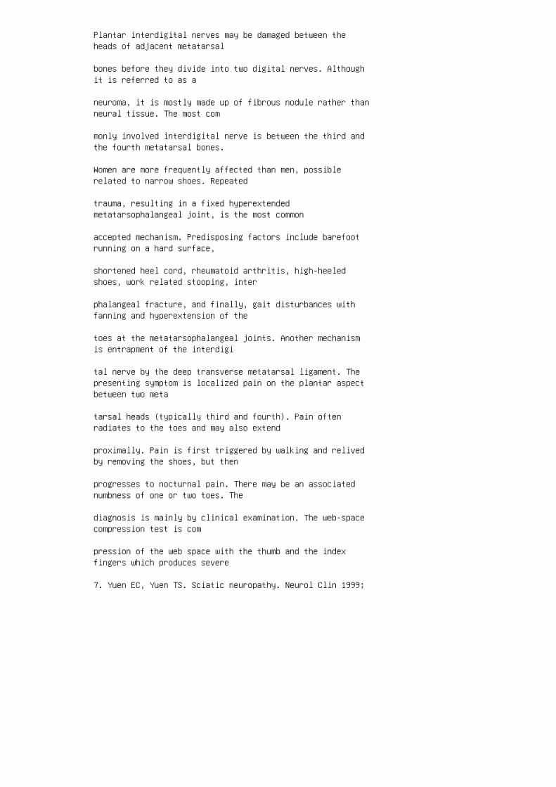
Plantar interdigital nerves may be damaged between theheads of adjacent metatarsal
bones before they divide into two digital nerves. Althoughit is referred to as a
neuroma, it is mostly made up of fibrous nodule rather thanneural tissue. The most com
monly involved interdigital nerve is between the third andthe fourth metatarsal bones.
Women are more frequently affected than men, possiblerelated to narrow shoes. Repeated
trauma, resulting in a fixed hyperextendedmetatarsophalangeal joint, is the most common
accepted mechanism. Predisposing factors include barefootrunning on a hard surface,
shortened heel cord, rheumatoid arthritis, high-heeledshoes, work related stooping, inter
phalangeal fracture, and finally, gait disturbances withfanning and hyperextension of the
toes at the metatarsophalangeal joints. Another mechanismis entrapment of the interdigi
tal nerve by the deep transverse metatarsal ligament. Thepresenting symptom is localized pain on the plantar aspectbetween two meta
tarsal heads (typically third and fourth). Pain oftenradiates to the toes and may also extend
proximally. Pain is first triggered by walking and relivedby removing the shoes, but then
progresses to nocturnal pain. There may be an associatednumbness of one or two toes. The
diagnosis is mainly by clinical examination. The web-spacecompression test is com
pression of the web space with the thumb and the indexfingers which produces severe
7. Yuen EC, Yuen TS. Sciatic neuropathy. Neurol Clin 1999;

17(3):617–631.
8. Kline DG. Comment to the piriformis muscle syndrome: asimple diagnostic maneuver. Neurosurgery 1994; 34:514.
9. McManis PG. Sciatic nerve lesions during cardiacsurgery. Neurology 1994; 44:684–687.
10. Parziale JR, Hudgins TH, Fishman LM. The piriformissyndrome. Am J Orthop 1996; 25:819–823.
11. Pecina M. Contribution to the etiological explanationof the piriformis syndrome. Acta Anta (Basel) 1979;105:181–187.
12. Nakamura H et al. Piriformis syndrome diagnosed bycauda equina action potentials: report of two cases. Spine2003; 15:28(2):E37–E40.
13. Fishman LM et al. Piriformis syndrome: diagnosis,treatment, and outcone-a 10-year study. Arch Phys MedRehabil 2002; 83(3):295–301.
14. McCrory P, Bell S. Nerve entrapment syndromes as acause of pain in the hip, groin, and buttock. Sports Med1999; 27(4):261–274.
15. Fishman LM, Anderson C, Rosner B. BOTOX and physicaltherapy in the treatment of piriformis syndrome. Am J PhysMed Rehabil 2002; 81(2):936–942.
16. Stewart JD. Focal Peripheral Neuropathies, LippencottWilliams & Wilkins, 3rd ed. 2000.
17. Schmaizried TP, Amstutz HC, Dorey FJ. Nerve palsyassociated with total hip replacement. Risk factors andprognosis. J Bone Joint Surg 1991; 73A:1074–1080.
18. DeHart MM, Riley LH Jr. Nerve injuries in total hiparthroplast. J Am Acad Ortho Surg 1999; 7(2):101–111.
19. Katsimihas M, Hutchinson et al. Delayed transientsciatic nerve palsy after total hip arthroplasty. JArthroplasy 2002; 17(3):379–381.
20. Aboulafia AJ et al. Neuropathy secondary to pigmentedvillondular synovitis of the hip. Clin Ortho 1996;325:174–180.
21. Tomaino MM. Complete sciatic nerve palsy after open

femur fracture: successful treatment with neurolysis 6months after injury. Am J Orthop 2002; 31(10):585–588.
22. Venna N, Bielawski M, Spatz EM. Sciatic nerveentrapment in a child. Case report. J Neurosurg 1991;75:652–654.
23. Yuen EC, So YT, Oiney RK. The electrophysiologicfeatures of sciatic neuropathy in 100 patients. MuscleNerve 1995; 18:414–420.
24. Katirji B. Peroneal neuropathy. Neurol Clin 1999;17(3):567–591.
25. Preston DC, Shapiro BE. Electromyography andneuromuscular disorders. Clinicalelectrophysiologiccorrelations. Boston: Butterworth-Heinemann, 1998.
26. Sourkes M, Stewart JD. Common peroneal neuropathy: astudy of selective motor and sensory involvement. Neurology1991; 41:1029–1033.
27. Howard JF. Syllabus of Muscle and Nerve Disease.University of North Carolina at Chapel Hill, 2002.
28. Oh SJ, Meyer RD. Entrapment neuropathy of the tibial(posterior tibial) nerve. Neurol Clin 1999; 17(3):593–615.
29. Refaeian M et al. Isolated sural neuropathy presentingas lateral ankle pain. Am J Phys Med Rehabil 2001;80(7):543–546.
30. Lau JT, Daniels TR. Tarsal tunnel syndrome: a review ofthe literature. Foot Ankle Int 1999; 20(3):201–209.
31. Reade BM et al. Tarsal tunnel syndrome. Clin Podiat MedSurg 2001; 18(3):395–408.
32. Erickson SJ, Quinn SF, Kneeland JB et al. MR imaging ofthe tarsal tunnel and related spaces: normal and abnormalfindings with anatomic correlation. Am J Roentgenol 1990;155:323–328.
33. Shapiro PP, Shapiro SL. Sonographic evaluation ofinterdigital neuroma. Foot Ankle Int 1995; 16:604–605.
34. Hammerschlag WA, Goldner JL, Bassett FH. Posteriortibial nerve entrapment: a thirty-three year review. AmOrthop Foot Ankle Soc 1998; July.

31
Focal Neuropathies of the Femoral,
Obturator, Lateral Femoral
Cutaneous Nerve, and Other Nerves
of the Thigh and Pelvis
Kevin J. Felice
University of Connecticut Health Center, Farmington,Connecticut, USA
Abstract 542
1. Focal Neuropathies of the Femoral Nerve 542 1.1. Anatomy542 1.2. Clinical Features and Differential Diagnosis 5431.3. Etiologies 543 1.4. Diagnostic Studies 544 1.5.Management 545
2. Focal Neuropathies of the Obturator Nerve 546 2.1.Anatomy 546 2.2. Clinical Features and DifferentialDiagnosis 546 2.3. Etiologies 546 2.4. Diagnostic Studies546 2.5. Management 547
3. Focal Neuropathies of the Lateral Femoral CutaneousNerve 547 3.1. Anatomy 547 3.2. Clinical Features andDifferential Diagnosis 548 3.3. Etiologies 548 3.4.Diagnostic Studies 549 3.5. Management 549
4. Focal Neuropathies of Other Nerves of Thigh and Pelvis549 4.1. Posterior Femoral Cutaneous Nerve of the Thigh 5494.2. Iliohypogastric Nerve 549 4.3. Ilioinguinal Nerve 5504.4. Genitofemoral Nerve 551 4.5. Pudendal Nerve 552 4.6.Diagnostic Studies 554 4.7. Management 554
1.2. Clinical Features and Differential Diagnosis
The clinical features of femoral mononeuropathy depend onthe site of injury (1,2).
Lesions of the proximal segment in the lumbar plexus orwithin the pelvis are associated
with weakness of hip flexion (psoas, iliacus, and rectusfemoris involvement), knee exten
sion (quadriceps involvement), and lateral thigh rotation

(sartorius involvement). Sensory
loss and paresthesias occur along the anteromedial thighand inner foreleg. The patellar
reflex is hypoactive or absent. Quadriceps muscle atrophymay occur within weeks,
especially with axonal nerve injuries. Lesions at or nearthe ilioinguinal ligament spare
hip flexion because motor branches to the psoas and theiliacus come off more proximally.
Lesions within the femoral triangle or upper thigh maycause selective motor or sensory
symptoms. Sensory symptoms limited to the medial knee andforeleg may result from iso
lated lesions of the saphenous nerve in thigh or knee.Damage to the infrapatellar branch
of the saphenous nerve results in numbness and paresthesiasin the skin over the patella
(gonyalgia paresthetica) (3). Other localizationconsiderations in patients with weakness
or sensory symptoms in the distribution of the femoralnerve include lesions of the
lumbar plexus, L2-4 spinal roots or segments, and lateralfemoral cutaneous nerve.
1.3. Etiologies
Trauma is the most common cause (Table 31.1) (1,2,4). Manytraumatic cases are iatro
genic from nerve injury sustained during surgicalprocedures (5–7). The mechanisms
of femoral nerve injury in these cases include compression,blunt trauma, stretch, and
laceration. Other causes include noniatrogenic trauma(compression from hematoma,
abscess, tumors, or pseudoaneurysm), diabetes mellitus,

localized hypertrophic neuro
pathy, vasculitis, radiation-induced neuropathy, andidiopathic neuropathy (1,2,8–13).
Unilateral or bilateral femoral mononeuropathies may occurin surgical or obstetric
patients while in the lithotomy position, presumedly due tonerve compression against
the inguinal ligament or excessive stretch of the nerve(14). Isolated injury of the saphe
nous nerve may occur in thigh, knee, or foreleg (Table31.2). Iatrogenic trauma caused by
Figure 31.2 Innervation of the skin of the inguinal areaand upper thigh. [From Stewart (2),
p. 491, with permission.] knee surgeries, arthroscopy, andsaphenous vein harvesting are the most common causes ofsaphenous mononeuropathy (1,2,15–18). 1.4. DiagnosticStudies The clinical examination is most informative forlocalization of femoral nerve lesions. Electrodiagnosticstudies are useful to verify the clinical diagnosis andexclude other sites of injury (e.g., femoral neuropathy vs.L2-4 radiculopathy), to provide more precise localization(e.g., femoral neuropathy proximal vs. distal to theinnervation of the iliacus), to determine the type of nerveinjury (e.g., axonal, demyelinating, or mixed), todetermine whether or not surgical intervention isnecessary, and to predict recovery and prognosis. Routineelectrodiagnostic studies include nerve conduction studies(NCS) and needle electromyography (EMG). General knowledgeof the pathophysiology and electrodiagnostic features offocal demyelinating injury, Wallerian degeneration, andreinnervation via both collateral sprouting and axonalregeneration is necessary in order to accurately interpretthese studies (19,20). Saphenous sensory and femoral motorNCS are optimally performed 10 days following an acutenerve injury. Techniques for performing these studies havebeen reported (21–23). As Wallerian degeneration iscomplete at this point, such studies are better able todistinguish among focal demyelinating, axonal, and mixednerve injuries. A preserved saphenous sensory nerve actionpotential (SNAP) at day 10 post-injury in a patient with asuspected Table 31.1 Causes of Femoral Neuropathy TraumaIatrogenic Inguinal herniorrhaphy Total hip replacementIntraabdominal surgeries Gynecological surgeries Lumbar

sympathectomy Laparoscopic procedures Other trauma Bulletand stab wounds Blunt trauma Hip and pelvic fracturesStretch injuries Lithotomy position Childbirth Surgicalprocedures Compression Hematoma Abscess TumorsPseudoaneurysm Diabetes mellitus Localized hypertrophicneuropathy Vasculitis Radiation-induced Idiopathic
femoral neuropathy suggests either selective injury to thefemoral nerve without involve
ment of saphenous sensory fibers, or proximal focaldemyelinating injury to the femoral
nerve, or nerve injury proximal to the dorsal root ganglioncells (e.g., L2-4 radiculopathy).
At day 10 post-injury, a preserved femoral compound muscleaction potential (CMAP)
from a paralyzed rectus femoris muscle also attests to aprimary demyelinating injury,
and predicts a good recovery and prognosis. Attenuated orabsent saphenous SNAP and
femoral CMAP responses are consistent with axonal or mixedinjuries, and predict a pro
longed recovery. EMG provides information about activeaxonal injury (e.g., presence of
fibrillations and positive sharp waves) and extent of nerverepair (e.g., nascent motor unit
action potentials suggest repair via axonal regeneration).Neuroimaging, including ultrasound computed tomography andmagnetic resonance
imaging (MRI), may be used to assess for compressive orinfiltrative nerve lesions
(e.g., hemorrhage and metastases), nerve sheath tumors,focal edema, and sources of
nerve entrapment (24). In general, MRI provides the bestsoft tissue contrast and anatom
ical detail among imaging modalities. In addition, therecent technique of magnetic

imaging neurography can identify small focal areas of nerveedema, entrapment, or
compression (24–26).
1.5. Management
Initial management of femoral nerve injuries is depends onthe suspected type of injury
(2). With mild to moderate nerve injuries (e.g., blunttrauma and stretch), restitution by
remyelination, collateral sprouting, or both may be evidentwithin weeks to months.
Severe axonal injuries require reinnervation by axonalregeneration and repair may not
be evident for 6–12 months. If complete disruption (e.g.,laceration) of the nerve is sus
pected, early exploration and surgical repair is indicated.EMG studies can document
Table 31.2 Causes of Saphenous Neuropathy
Thigh
Compression Fibrous bands Branches of femoral vessels
Entrapment at subsartorial canal exit
Lacerations
Arterial surgery
Nerve tumors
Knee
Knee surgery and arthroscopy
External compression (e.g., surfer’s neuropathy)
Meniscal cyst
Foreleg
Saphenous vein harvest for coronary artery bypass surgery

Saphenous vein cannulation
Ankle injuries
Trauma
Lacerations
Infrapatellar branch
Blunt trauma, compression, and lacerations
Knee surgery and arthroscopy
Entrapment at the sartorius tendon nerve continuity ordiscontinuity, and serial studies can assess early(subclinical) axonal regeneration. Management of iliacushematomas includes reversal of anticoagulation andtreatment of pain (2). Surgical evacuation should beconsidered in patients with large hematomas and substantialclinical deficits (9,11). 2. FOCAL NEUROPATHIES OF THEOBTURATOR NERVE 2.1. Anatomy The obturator nerve is a mixednerve that originates from the anterior divisions of theventral rami of the L2-4 spinal nerves and traverses thelumbar plexus (Fig. 31.1) (1,2). The nerve passes throughthe pelvis via the obturator canal, and within the canalsupplies the obturator externus muscle (L2-4). Outside ofthe canal, it divides into the anterior and posteriordivisions which descend along the medial thigh. Theanterior division supplies the pectineus (L2-4), adductorlongus (L2-4), adductor brevis (L2-4), and gracilis (L2-4).The anterior division terminates in a sensory branch thatinnervates the skin along the medial thigh (Fig. 31.2). Theposterior division supplies the obturator externus (L2-4),adductor magnus (L2-4) (which also receives innervationfrom the sciatic nerve), and adductor brevis muscle (L2-4).2.2. Clinical Features and Differential Diagnosis Lesionsof the obturator nerve cause weakness of thigh adduction,wasting of the medial thigh muscles (with axonal injuries),and sensory loss or paresthesias affecting the medialthigh. Patients usually note difficulties with hip andthigh stabilization, especially during certain activities(e.g., squatting and sexual intercourse). Otherlocalization sites to consider in patients with weakness orsensory symptoms in the distribution of the obturator nerveinclude lesions of the lumbar plexus, or L2-4 spinal rootsor segments. 2.3. Etiologies Causes of obturatormononeuropathy include trauma, compression, nerve tumors,entrapment within the obturator foramen, diabetes mellitus,

myositis ossificans, and idiopathic injury (Table 31.3)(1,2,4,27–32). Within the lumbar plexus, the obturatornerve may be damaged during pelvic surgical or laparoscopicprocedures, and by compression or infiltration frommetastatic disease or endometriosis (28,32). Damage mayresult from obturator hernias, following cancer surgery inthe pelvis, iliopsoas hemorrhage, pelvic fractures, orduring hip surgery. Prolonged hip flexion during certainurological surgical procedures may stretch the nerve at thebony obturator foramen. Obturator mononeuropathy may resultfrom prolonged labor or difficult childbirth due tocompression of the nerve between the fetal head and thebony pelvis (30). Nerve entrapment by thickened fasciaoverlying the adductor brevis muscle has been described inathletes (31). We recently evaluated a unilateral obturatormononeuropathy attributed to stretch or compression duringan endoscopic hysterectomy. 2.4. Diagnostic Studies Thereare no NCS techniques for evaluating the obturator nerve(21) and electrodiagnostic evaluation is limited to needleEMG of hip adductor muscles. Focal demyelinating lesions orneurapraxic injury of the obturator nerve can be inferredby reduced recruitment (higher
discharge rates) and from normal-appearing motor unitaction potentials in hip adductor
muscles. In distinction, mixed and axonal injuries fromnoniatrogenic trauma would be
associated with fibrillation potentials and positive sharpwaves during the subacute to
early chronic phase of injury, and motor unit actionpotentials (nascent units) reflecting
reinnervation during the chronic phase. MRI of the thighmay provide additional infor
mation in selected situations. For example, prominentatrophy of the hip adductor
muscles would be consistent with severe axonal or mixednerve injury, whereas
normal-appearing or mildly atrophic muscles assessedseveral months post-injury would
be more consistent with neuropraxic nerve injury.
2.5. Management

Good clinical outcome is reported in most patientsregardless of etiology (27). Patients
with partial injuries should be observed because recoveryby remyelination or collateral
reinnervation occurs over a period of several months.Severe or complete injuries may
require early surgical exploration and nerve grafting (34).Obturator neuropathy following
hip arthroplasty may be caused by nerve encasement insurgical cement, early surgical
exploration is indicated. Surgical exploration for possiblenerve entrapment within the
obturator canal, nerve infiltration by metastatic disease,or endometriosis should be con
sidered in patients with progressive and unexplainedsymptoms.
3. FOCAL NEUROPATHIES OF THE LATERAL FEMORAL CUTANEOUS NERVE
3.1. Anatomy
The lateral femoral cutaneous nerve of the thigh (LFCN)originates from the anterior
primary rami of the L2-3 spinal roots within the psoasmuscle (Fig. 31.1) (1,2).
Table 31.3 Causes of
Obturator Neuropathy
Trauma
Iatrogenic Pelvic surgery Hip surgery Laparoscopic surgeryFemoral artery procedures
Pelvic fractures
Compression
Tumors

Endometriosis
Obturator hernias
Fibrous bands
Childbirth
Nerve tumors
Entrapment in obturator foramen
Diabetes mellitus
Myositis ossificans
Idiopathic The nerve emerges from the lateral border of thepsoas muscle, crosses the iliacus muscle, passes under thelateral part of the inguinal ligament medial to theanterior superior iliac spine, and enters the upper thighbeneath the fascia lata. In the upper thigh, the LFCNdivides into anterior and posterior divisions. The anteriordivision innervates the skin of the anterior thigh to theknee whereas the posterior division supplies the skin alongthe upper and lateral aspects of the thigh (Fig. 31.2).3.2. Clinical Features and Differential Diagnosis LFCNlesions cause sensory loss, paresthesias, or pain involvingthe anterolateral thigh (meralgia paresthetica). Symptomsare worsened when standing or walking, especially in obesepatients, presumably due to compression of the nerve alongthe anterior superior iliac spine by the protuberantabdomen. Other localization considerations in patients withsymptoms of meralgia paresthetica include lesions of thefemoral nerve, lumbar plexus, or L2-4 spinal roots orsegments. 3.3. Etiologies There are multiple sites fornerve injury, including the abdomen, pelvis, inguinalligament, and thigh (Table 31.4). In the abdomen, the LFCNmay be compressed by psoas hemorrhage, abscess, or tumor(2,4,35). Surgical trauma may result from renaltransplants, hysterectomies, or other surgeries (36–38).Procurement of iliac bone for grafting may injure the LFCNin the pelvis (39). Obesity and constricting garments orbelts are potential causes of external compression of thenerve near the anterior superior iliac spine or theinguinal ligament, and can be associated with bilateralmeralgia paresthetica (2,4,40,41). Table 31.4 Causes ofLateral Femoral Cutaneous Neuropathy Abdomen Iliopsoashemorrhage Psoas tumor Surgery (e.g., renal transplants andabdominal hysterectomy) Pelvis Iliac bone procurement forgrafting Surgery in the iliac fossa Inguinal ligament

External compression and angulation Corsets, belts, andother constricting garments Prolonged sitting in the lotusposition Protuberant abdomen in obese patient Seat-belttrauma Entrapment Trauma Surgical Injury Idiopathic ThighExternal compression Trauma Lacerations Injections Surgicalinjury
Other causes are trauma, compression, lacerations,injection injuries, and surgical
injuries (42,43).
3.4. Diagnostic Studies
Several techniques are available for recording the LFCNsensory nerve action potentials
(21,44,45). The nerve is stimulated with either surface orneedle electrodes just medial
to the anterior superior iliac spine and the SNAP isrecorded along the anterolateral
thigh. However, NCS of the LFCN are technically difficultto perform in normal subjects,
and particularly in obese subjects. A major limitation isthat the SNAP is of low amplitude
(2–10 mV) and usually requires signal averaging.Comparative studies on the asympto
matic side are required in an patient found to have anabsent LFCN response in the symp
tomatic limb. Given these limitations, lateral femoralcutaneous mononeuropathy remains
primarily a clinical diagnosis. When appropriate, imagingstudies of the retroperitoneal
space and pelvis are needed to assess for metastaticdisease or hemorrhage.
3.5. Management
In most patients, symptoms resolve spontaneously withinweeks to months (2,46), and
conservative management of idiopathic meralgia paresthetica

is appropriate. Patients
should be educated on potential causes (e.g., obesity andconstricting garments or belts)
of meralgia paresthetica and instructed on appropriatemodifications. Analgesics and
medications for neuropathic pain may be appropriate forpatients with especially bother
some symptoms. Local anesthetics and corticosteroids mayprovide pain relief and can be
repeated at intervals (46,47). Surgical decompression ofthe nerve at the inguinal ligament
may be appropriate for patients with recalcitrant painsymptoms (46). Nerve transection is
preferred over decompression by some surgeons; however, thepaucity of reported cases
hampers comparative analyses (48).
4. FOCAL NEUROPATHIES OF OTHER NERVES OF THIGH AND PELVIS
4.1. Posterior Femoral Cutaneous Nerve of the Thigh
Sensory nerve fibers of the posterior femoral cutaneousnerve of the thigh (PFCN) orig
inate from the anterior primary rami of S1-3 spinal roots(Fig. 31.3) (1,2). The PFCN
exits the pelvis via the greater sciatic notch and descendsthe posterior thigh. The nerve
is initially deep to the gluteus maximus muscle, butdescends superficially down the
back of the thigh to the knee. The PFCN innervates the skinof lower buttock, posterior
thigh, and popliteal fossa, and lesions cause paresthesiasor sensory loss in these areas.
The PFCN may be damaged in sacral plexus, greater sciaticforamen, buttocks, or upper

thigh (Table 31.5). Posterior femoral cutaneous neuropathymay be due to compression or
infiltration (malignancies), trauma (falls on the buttock),injection injuries, compression
from bicycle seat, and lacerations (1,2,49–52).
4.2. Iliohypogastric Nerve
The iliohypogastric nerve is a mixed nerve originating fromthe anterior primary rami of
T12-L1 spinal roots. It emerges from the lateral border ofthe psoas muscle (Fig. 31.4)
(1,2) and traverses with the ilioinguinal nerve around theabdominal wall and along the upper border of the iliaccrest. In the abdomen, the iliohypogastric and ilioinguinalnerves provide motor branches to the internal oblique andtransversus abdominis muscles. The terminal branches of theiliohypogastric nerve are the lateral and anteriorcutaneous sensory branches. The lateral branch supplies theskin along the upper aspect of the buttock and the anteriorbranch supplies a small patch of skin over the pubis (Fig.31.2). Lesions of the iliohypogastric nerve are associatedwith sensory loss, paresthesias, or pain in thedistribution of the lateral and anterior branches.Iliohypogastric neuropathies may result from lesions in thelumbar plexus or abdomen (Table 31.6). Damage may be causedby compression or infiltration by tumors, surgicalincisions, trauma, or compression (1,2). 4.3. IlioinguinalNerve The ilioinguinal nerve is a mixed nerve originatingfrom the anterior primary rami of the L1 spinal root (Fig.31.4) (1,2), and travels just below and parallel to theiliohypogastric nerve in the abdomen. The nerve pierces theinternal oblique and transversus abdominis muscles, andsupplies motor branches to these muscles, and then entersthe inguinal canal and passes through the superficialinguinal ring together with the spermatic cord in men orround ligament in woman. Sensory fibers emerge from thecanal to supply the skin along the medial thigh below theinguinal ligament, symphysis pubis and external genitalia(Fig. 31.2). Lesions of the ilioinguinal nerve areassociated with lower abdominal Figure 31.3 Course of theposterior femoral cutaneous nerve. [From Stewart (2), p.377, with permission.] Table 31.5 Causes of PosteriorFemoral Cutaneous Neuropathy Sacral plexus MalignancyButtocks Falls on to the buttocks Injections Prolongedsitting (e.g., bicycle riding) Trauma Lacerations

pain, sensory loss or paresthesias in this distribution,and tenderness below the anterior
superior iliac spine. The ilioinguinal nerve may be injuredin the lumbar plexus,
abdomen, and inguinal canal (Table 31.7) (1,2). In thelumbar plexus or retroperitoneal
space, the ilioinguinal nerve may be damaged by tumorcompression, infiltration, or sur
gical injuries (53,54). Along the anterior abdominal wall,the nerve may be damaged by
surgical incisions, trauma, or entrapment within abdominalmuscles (55). The inguinal
canal is a potential site for injury due to hernia surgery,or compression by endometriosis,
lipoma, or leiomyoma. Neuropathy may occur after childbirthdue to stretching of the
nerve (2,56). Ilioinguinal neuropathy or neuralgia may beidiopathic.
4.4. Genitofemoral Nerve
Mixed nerve fibers of the genitofemoral nerve originatefrom the anterior primary rami of
L1-2 spinal roots (Fig. 31.4) (1,2). The nerve passesthrough the psoas muscle and travels
downward to the inguinal ligament where is divides into theexternal spermatic (genital)
and lumboinguinal (femoral) branches. The genital branchenters the deep inguinal ring
and traverses the inguinal canal where it terminates in amotor branch innervating the
cremaster muscle and sensory branch supplying the skinalong the scrotum (or labia majoris)
Figure 31.4 Course of the iliohypogastric, ilioinguinal,and genitofemoral nerves. [From Stewart
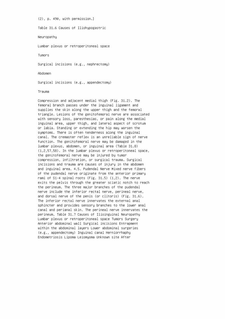
(2), p. 490, with permission.]
Table 31.6 Causes of Iliohypogastric
Neuropathy
Lumbar plexus or retroperitoneal space
Tumors
Surgical incisions (e.g., nephrectomy)
Abdomen
Surgical incisions (e.g., appendectomy)
Trauma
Compression and adjacent medial thigh (Fig. 31.2). Thefemoral branch passes under the inguinal ligament andsupplies the skin along the upper thigh and the femoraltriangle. Lesions of the genitofemoral nerve are associatedwith sensory loss, paresthesias, or pain along the medialinguinal area, upper thigh, and lateral aspect of scrotumor labia. Standing or extending the hip may worsen thesymptoms. There is often tenderness along the inguinalcanal. The cremaster reflex is an unreliable sign of nervefunction. The genitofemoral nerve may be damaged in thelumbar plexus, abdomen, or inguinal area (Table 31.8)(1,2,57,58). In the lumbar plexus or retroperitoneal space,the genitofemoral nerve may be injured by tumorcompression, infiltration, or surgical trauma. Surgicalincisions and trauma are causes of injury in the abdomenand inguinal area. 4.5. Pudendal Nerve Mixed nerve fibersof the pudendal nerve originate from the anterior primaryrami of S1-4 spinal roots (Fig. 31.5) (1,2). The nerveexits the pelvis through the greater sciatic notch to reachthe perineum. The three major branches of the pudendalnerve include the inferior rectal nerve, perineal nerve,and dorsal nerve of the penis (or clitoris) (Fig. 31.6).The inferior rectal nerve innervates the external analsphincter and provides sensory branches to the lower analcanal and perianal skin. The perineal nerve innervates theperineum, Table 31.7 Causes of Ilioinguinal NeuropathyLumbar plexus or retroperitoneal space Tumors SurgeryAnterior abdominal wall Surgical incisions Entrapmentwithin the abdominal layers Lower abdominal surgeries(e.g., appendectomy) Inguinal canal HerniorrhaphyEndometriosis Lipoma Leiomyoma Unknown site After
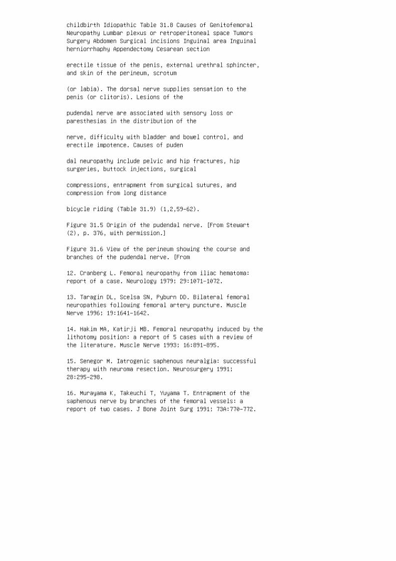
childbirth Idiopathic Table 31.8 Causes of GenitofemoralNeuropathy Lumbar plexus or retroperitoneal space TumorsSurgery Abdomen Surgical incisions Inguinal area Inguinalherniorrhaphy Appendectomy Cesarean section
erectile tissue of the penis, external urethral sphincter,and skin of the perineum, scrotum
(or labia). The dorsal nerve supplies sensation to thepenis (or clitoris). Lesions of the
pudendal nerve are associated with sensory loss orparesthesias in the distribution of the
nerve, difficulty with bladder and bowel control, anderectile impotence. Causes of puden
dal neuropathy include pelvic and hip fractures, hipsurgeries, buttock injections, surgical
compressions, entrapment from surgical sutures, andcompression from long distance
bicycle riding (Table 31.9) (1,2,59–62).
Figure 31.5 Origin of the pudendal nerve. [From Stewart(2), p. 376, with permission.]
Figure 31.6 View of the perineum showing the course andbranches of the pudendal nerve. [From
12. Cranberg L. Femoral neuropathy from iliac hematoma:report of a case. Neurology 1979; 29:1071–1072.
13. Taragin DL, Scelsa SN, Pyburn DD. Bilateral femoralneuropathies following femoral artery puncture. MuscleNerve 1996; 19:1641–1642.
14. Hakim MA, Katirji MB. Femoral neuropathy induced by thelithotomy position: a report of 5 cases with a review ofthe literature. Muscle Nerve 1993; 16:891–895.
15. Senegor M. Iatrogenic saphenous neuralgia: successfultherapy with neuroma resection. Neurosurgery 1991;28:295–298.
16. Murayama K, Takeuchi T, Yuyama T. Entrapment of thesaphenous nerve by branches of the femoral vessels: areport of two cases. J Bone Joint Surg 1991; 73A:770–772.

17. Adar R, Meyer E, Zweig A. Saphenous neuralgia: acomplication of vascular reconstructions below the inguinalligament. Ann Surg 1979; 190:609–613.
18. Luerssen TG, Campbell RL, Defalque RJ, Worth RM.Spontaneous saphenous neuralgia. Neurosurgery 1983;13:238–241.
19. Robinson L. Traumatic injury to peripheral nerves.Muscle Nerve 2000; 23:863–873.
20. Chaudhry V, Cornblath DR. Wallerian degeneration inhuman nerves: serial electrophysiological studies. MuscleNerve 1992; 687–693.
21. Oh SJ. Clinical Electromyography: Nerve ConductionStudies. 2nd ed. Philadelphia: Williams and Wilkins, 1993.
22. Stohr M, Shumm F, Ballier R. Normal sensory conductionin the saphenous nerve in man. EEG Clin Neurophysiol 1978;44:172–178.
23. Ertekin C. Saphenous nerve conduction in man. J NeurolNeurosurg Psychiatry 1969; 31:28–33.
24. Halford H, Graves A. Imaging techniques. In: BertoriniTE, ed. Clinical Evaluation and Diagnostic Tests forNeuromuscular Disorders. Boston: Butterworth and Heinemann,2002:565–593.
25. Filler AG, Howe FA, Hayes CE et al. Magnetic resonanceneurography. Lancet 1993; 341:659–661.
26. Filler AG, Kliot M, Howe FA et al. Application ofmagnetic resonance neurography in the evaluation ofpatients with peripheral nerve pathology. J Neurosurg 1996;85:299–309.
27. Sorenson EJ, Chen JJ, Daube JR. Obturator neuropathy:causes and outcome. Muscle Nerve 2002; 25:605–607.
28. Rogers LR, Borkowski GP, Albers JW, Levin KH, BarohnRJ, Mitsumoto H. Obturator mononeuropathy caused by pelviccancer: six cases. Neurology 1993; 43:1489–1492.
29. Pellegrino MJ, Johnson EW. Bilateral obturator nerveinjuries during urologic surgery. Arch Phys Med Rehabil1988; 69:46–47.
30. Warfield CA. Obturator neuropathy after forceps
delivery. Obstet Gynecol 1984; 64:47S–48S.
31. Bradshaw C, McCory P, Bell S, Brukner P. Obturatornerve entrapment: a cause of groin pain in athletes. Am JSports Med 1997; 25:402–408.
32. Redwine DB, Sharpe DR. Endometriosis of the obturatornerve: a case report. J Reprod Med 1990; 35:434–435.
33. Scotto V, Rosica G, Valeri B et al. Benign schwannomaof the obturator nerve: a case report. Am J Obstet Gynecol1998; 179:816–817.
34. Vasilev SA. Obturator nerve injury: a review ofmanagement options. Gynecol Oncol 1994; 53:152–155.
35. Amoiridis G, Wo¨hrle J, Grunwald I, Przuntek. Malignanttumour of the psoas: another cause of meralgiaparesthetica. Electromyogr Clin Neurophysiol 1993;33:109–112.
36. Jablecki CK. Postoperative lateral femoral cutaneousneuropathy. Muscle Nerve 1999; 1129–1131.
37. Yamout B, Tayyim A, Farhat W. Meralgia paresthetica asa complication of laparoscopic cholescystectomy. ClinNeurol Neurosurg 1994; 96:143–144.
38. Rao T, Kim H, Mathrubhutham M, Lee KN. Meralgiaparesthetica: unusual complication of inguinalherniorrhaphy. J Am Med Assoc 1977; 237:2525.
39. Weikel AM, Habal MB. Meralgia paresthetica: acomplication of iliac bone procurement. Plast Reconstr Surg1977; 60:572–574. 40. Jefferson D, Eames RA. Subclinicalentrapment of the lateral femoral cutaneous nerve: anautopsy study. Muscle Nerve 1979; 2:145–154. 41. Richer LP,Shevell MI, Stewart J, Poulin C. Pediatric meralgiaparesthetica. Pediatr Neurol 2002; 26:321–323. 42. EckerAD, Woltman HW. Meralgia paresthetica: a report of onehundred and fifty cases. J Am Med Assoc 1938;110:1650–1652. 43. Beresford HR. Meralgia parestheticaafter seat-belt trauma. J Trauma 1971; 11:629–630. 44.Butler ET, Johnson EW, Kaye AZ. Normal conduction velocityin the lateral femoral cutaneous nerve. Arch Phys MedRehabil 1974; 55:31–32. 45. Sarala PK, Nishihira T, Oh SJ.Meralgia paresthetica: electrophysiological study. ArchPhys Med Rehabil 1979; 60:30–31. 46. Williams PH, Trzil KP.Management of meralgia paresthetica. J Neurosurg 1991;74:76–80. 47. Dureja GP, Gulaya V, Jayalakshmi TS, Mandal
P. Management of meralgia paresthetica: a multimodalityregimen. Anesth Analg 1995; 80:1060–1061. 48. van EertenPV, Polder TW, Broere CA. Operative treatment of meralgiaparesthetica: transection versus neurolysis. Neurosurgery1995; 37:63–65. 49. McKain CW, Urban BJ. Pain and clunealneuropathy following intragluteal injection. Anesth Analg1978; 57:138–141. 50. Arnoldussen WJ, Korten JJ. Pressureneuropathy of the posterior femoral cutaneous nerve. ClinNeurol Neurosurg 1980; 82:57–60. 51. Chutkow JG. Posteriorfemoral cutaneous neurolagia. Muscle Nerve 1988;11:1146–1148. 52. Iyer VG, Shields CB. Isolated injectioninjury to the posterior femoral cutaneous nerve.Neurosurgery 1989; 25:835–838. 53. Stultz P, Pfeiffer KM.Peripheral nerve injuries resulting from common surgicalprocedures in the lower portion of the abdomen. Arch Surg1982; 117:324–327. 54. Sippo WC, Gomez AC. Nerve entrapmentsyndromes from lower abdominal surgery. J Fam Pract 1987;25:585–587. 55. Kanai H, Hirakawa M, Arimura Y, TaniguchiM, Tsuruya K, Masutani K, Ninomiya T, Sugawara K, Koga Y,Hirakata H. Ilioinguinal neuralgia complicatingpercutaneous renal biopsy. J Neurol 2001; 248:708–709. 56.Hahn L. Clinical findings and results of operativetreatment in ilioinguinal nerve entrapment syndrome. Br JObstet Gynaecol 1989; 96:1080–1083. 57. Laha RK, Rao S,Pidgeon CN. Genito-femoral neuralgia. Surg Neurol 1977;8:280–282. 58. Obrien MD. Genito-femoral neuropathy. Br MedJ 1979; 1:1052. 59. Hofmann A, Jones RE, Schoenvogel R.Pudendal-nerve neuropraxia as a result of traction on thefracture table: a report of four cases. J Bone Joint SurgAm 1982; 64:136–138. 60. Goodson JD. Pudendal neuritis frombiking. N Engl J Med 1981; 304:365. 61. Gibson GR.Impotence following fractured pelvis and ruptured urethra.Br J Urol 1970; 42:86–88. 62. Allen RE, Hosker GL, SmithARB, Warrell DW. Pelvic floor damage and childbirth: aneurophysiological study. Br J Obstet Gynecol 1990;97:770–779. 63. Dumitru D, Nelson MR. Posterior femoralcutaneous nerve conduction. Arch Phys Med Rehabil 1990;71:979–982. 64. Ellis RJ, Geisse H, Holub BA, Swenson MR.Ilioinguinal nerve conduction. Muscle Nerve 1992; 15:1194.65. Kiff ES, Swash M. Slowed conduction in the pudendalnerves in idiopathic faecal incontinence. Br J Surg 1984;71:614–616. 66. Snooks SJ, Swash M. Perineal nerve andtranscutaneous spinal stimulation: new method forinvestigation of the urethral striated musculature. Br JUrol 1984; 56:406–409. 67. Clawson D, Cardenas D. Dorsalnerve of the penis nerve conduction velocity: a newtechnique. Muscle Nerve 1991; 14:845–849. 68. Kaneko S,Bradley WE. Penile electrodiagnosis. Value ofbulbocavernosus reflex latency versus conduction velocityof the dorsal nerve of the penis in diagnosis of diabetic

impotence. J Urol 1986; 137:933–935.
69. Kothari MJ, Bauer SB. Urodynamic and neurophysiologicevaluation of patients with diastematomyelia. J ChildNeurol 1997; 12:97–100.
70. Kothari MJ, Kelly M, Darbey M, Bauer S, Scott RM.Neurophysiologic assessment of urinary dysfunction inchildren with thoracic syringomyelia. J Child Neurol 1995;10:451–454.
71. Fowler CJ. Pelvic floor neurophysiology. Methods ClinNeurophysiol 1991; 2:1–24.
72. Rizzo MA. Successful treatment of painful traumaticmononeuropathy with carbamazepine: insights into a possiblemolecular pain mechanism. J Neurol Sci 1997; 152:103–106.
32
Management of Traumatic
Mononeuropathies
Lisa DiPonio, James A. Leonard Jr, and John E. McGillicuddy
University of Michigan Medical Center, Ann Arbor, Michigan,USA
Abstract 560
1. Clinical Features/Differential Diagnosis 560 1.1.Clinical Findings and Classification System of NerveInjuries 560 1.2. Anatomic Correlates 561 1.2.1. SpinalAccessory Nerve 561 1.2.2. Suprascapular Nerve 562 1.2.3.Axillary Nerve 562 1.2.4. Radial Nerve 562 1.2.5.Musculocutaneous Nerve 564 1.2.6. Median Nerve 564 1.2.7.Ulnar Nerve 565 1.2.8. Femoral Nerve 566 1.2.9. ObturatorNerve 566 1.2.10. Gluteal Nerves 566 1.2.11. Sciatic Nerve567 1.2.12. Tibial Nerve 567 1.2.13. Peroneal Nerve 5671.3. Screening Tests 568
2. Pathogenesis and Nerve Response to Injury 569 2.1.Neural Injury in Neurotmesis or Sunderland’s Fifth-DegreeLesion 569 2.2. Neural Regeneration After Sunderland’sNeurotmesis or Fifth-Degree Lesions 569 2.3. Neural Injuryand Regeneration in Sunderland’s Fourthand Third-DegreeLesions 570 2.4. Neural Injury and Regeneration inAxonotmesis or Sunderland’s Second-Degree Lesion 570 2.5.Neural Injury and Regeneration in Neurapraxia 570

ism. Seddon’s classification of nerve injury describesthree degrees of severity of nerve
injury (1). Neurapraxia refers to focal demyelination thatis sufficient to interrupt saltatory
nerve conduction. The clinical result is sensory or motorloss despite axonal continuity.
Symptoms in the acute phase are as severe as those fromnerve transection, but prognosis
for spontaneous recovery is excellent. Axonotmesis refersto loss of axonal continuity with
preservation of structural and supportive neural tubules.It also carries a good prognosis for
spontaneous recovery, although not as quickly asneurapraxia. Neurotmesis refers to dis
continuity of the axons as well as their supportingstructures. This carries a worse progno
sis and is the only injury benefiting from surgical repairor grafting. An updated classification proposed bySunderland has five degrees of injury (2).
Sunderland’s first-degree injury is identical to Seddon’sneurapraxia. Sunderland’s
second, third, and fourth-degree injuries correspond todifferent severities of Seddon’s
axonotmesis. A second-degree injury is axonal disruptionbut sparing of endoneurium,
perineurium, and epineurium. A third-degree injury issparing of perineurium and epineur
ium, and a fourth-degree injury is sparing of epineuriumonly. Sunderland’s fifth-degree of
injury is identical to Seddon’s neurotmesis.
1.2. Anatomic Correlates
1.2.1. Spinal Accessory Nerve

This is the sole motor innervation to the trapezius muscle,and also innervates the sterno
cleidomastoid muscle. It has a superficial location in thesubcutaneous tissue on the floor
of the posterior cervical triangle which is bordered by thetrapezius posteriorly, the ster
nocleidomastoid anteriorly, and the clavicle inferiorly. Itis most commonly injured
during surgical procedures, but is susceptible to trauma byblunt or penetrating injuries
to the neck or by traction injury from dislocations of thesternoclavicular or acromiocla
vicular joints. The trapezius muscle serves to elevate,retract, and rotate the scapula, and
Table 32.1 Orthopedic Injuries and Commonly AssociatedPeripheral
Nerve Injuries
Orthopedic injury Commonly associated nerve injury
Shoulder dislocation Axillary
Humerus fracture Radial
Elbow dislocation Median
Monteggia fracture Radial
Wrist Median, ulnar, or radial
Pelvis Cauda equina, radicular, plexus
Acetabular fracture Sciatic
Hip dislocation Femoral, sciatic
Femur fracture Sciatic, tibial/peroneal components
Distal femur fracture Tibial (medial), peroneal (lateral)
Tibial fracture Tibial, deep peroneal

Fibular fracture Common or superficial peroneal
Medial malleolus (tibia) Tibial
Lateral malleolus (fibula) Sural
Metatarsals Interdigital nerves disruption leads to lateralwinging of the scapula and weakness of arm abduction past908. This nerve has no cutaneous innervation (Table 32.2).1.2.2. Suprascapular Nerve This nerve originates from theupper trunk of the brachial plexus and carries fibers fromC5 and C6. It travels behind the plexus, under thetrapezius muscle, to the scapula, where it passes throughthe suprascapular notch and innervates the supraspinatusmuscle, and then around the lateral edge of the scapularspine to innervate the infraspinatus muscle. The nerve canbe injured by scapular fracture, blunt trauma to theshoulder girdle, or traction through the nerve by alongitudinal pull on the upper extremity. The supraspinatusmuscle helps elevate the arm over the first 908 and theinfraspinatus muscle assists in external rotation of thearm. It has no cutaneous sensory innervation. 1.2.3.Axillary Nerve This is a branch of the posterior cord andcontains fibers from C5 and C6. It branches from theposterior cord near the lateral scapular border and sends abranch to teres minor. It then bifurcates into cutaneousand motor branches and courses posteriorly around theproximal humerus, supplies the posterior, middle, andanterior deltoid muscle. The sensory branch goes on tosupply a small cutaneous segment in the lateral upper arm.Axillary mononeuropathy results in weakness of shoulderabduction or sensory loss on the lateral upper arm. Injuryoccurs with proximal humeral fractures and, less commonly,with scapular fractures. The nerve is susceptible totraction injury of the plexus because it is tethered to thedeltoid muscle close to the shoulder. The nerve can also beinjured from scar tissue formation in the quadrilateralspace (formed by the teres major muscle, triceps, humerus,and teres minor) through which the nerve passes or whenother injuries occur, which require the use of axillarycrutches (crutch paralysis). 1.2.4. Radial Nerve This isthe continuation of the posterior cord and carries fibersfrom C5, C6, C7, and C8. After leaving the plexus, thenerve travels posterior to the axillary artery and givesbranches to the posterior cutaneous nerve of the arm andthe triceps and anconeus muscles. At this level the nervemay be damaged by traction or tourniquet application. Itthen travels in the spiral groove of the humerus where itis vulnerable to damage related to humeral fractures orcompression. Distal to the spiral groove, it pierces the
lateral intermuscular septum and travels anterior to thelateral condyle of the humerus, giving branches to thebrachialis, brachioradialis, and extensor carpi radialislongus muscles. At this point, it bifurcates into theposterior interosseous nerve (PIN) and the superficialbranch. The PIN passes through the arcade of Frohse andthen travels along the posterior interosseous membrane,where it innervates the other extensors muscles of theforearm. It travels along with the posterior interosseousartery and may be compressed by a traumatic aneurysm. Itmay also be injured by proximal radial fractures orpenetrating injuries. The superficial branch continuessuperficially along the lateral border of the forearm whereit is vulnerable to injuries associated with fractures ofthe distal and proximal radius or forced wristhyperpronation. Lesions in the axilla or proximal to thespiral groove may lead to sensory loss on the posterior armabove the elbow (posterior cutaneous nerve of arm) andweakness and dropped triceps reflex. Lesions within thespiral groove lead to sensory loss on the extensor surfaceof the forearm (posterior cutaneous nerve of forearm) andcomplete wrist drop.
T
a
b
l e
3
2
. 2
S
u
m
m
a r
y
o

f
N
e r
v
e
I n
j u
r i
e s
a n
d
A
s s
o
c i
a t
e d
M
u
s c
l e
s ,
F
u
n

c t
i o
n
a l
D
e fi
c i
t s
,
a n
d
F
u
n
c t
i o
n
a l
I n
t e
r v
e n
t i
o
n

s N e r v e S u p r a s c a p u l a r A x i l l a r y R a di a l M u s c u l o c u t a n e o u s M e d i a n U l n a rM a j o r m u s c l e s i n n e r v a t e d S p i n a t i De l t o i d T r i c e p s , f o r e a r m e x t e n s o r sB i c e p s b r a c h i i P r o n a t o r t e r e s , fl ex o r d i g i t o r u m p r o f u n d u s 1 a n d 2 , t h um b o p p o s i t i o n F l e x o r d i g i t o r u m p r of u n d u s 1 a n d 2 , h a n d , a n d i n t r i n s i c sF u n c t i o n a l d e fi c i t W e a k n e s s o f i n te r n a l a n d e x t e r n a l r o t a t i o n W e a k n es s o f a r m a b d u c t i o n b e y o n d 3 0 8 W e a k ne s s o f a r m e x t e n s i o n , w r i s t d r o p , l os s o f p o w e r g r a s p W e a k n e s s o f a r m fl ex i o n , s u p i n a t i o n W e a k n e s s o f a r m p ro n a t i o n , d i s t a l i n d e x a n d m i d d l e fin g e r fl e x i o n , t h u m b o p p o s i t i o n W e ak n e s s o f g r i p , h a n d f u n c t i o n F u n c t io n a l i n t e r v e n t i o n S h o u l d e r s u p p o rt s l i n g S h o u l d e r s u p p o r t s l i n g S p l in t i n g w r i s t f u s i o n m u s c l e t e n d o n t ra n s f e r F u n c t i o n a l a r m s u p p o r t s l i ng , e l b o w f u s i o n , m u s c l e t e n d o n t r a ns f e r S p l i n t i n g , m u s c l e t e n d o n t r a ns f e r S p l i n t i n g , m u s c l e t e n d o n t r a ns f e r N e r v e G l u t e a l s : s u p e r i o r a n d in f e r i o r F e m o r a l S c i a t i c P e r o n e a l Ti b i a l M a j o r m u s c l e s i n n e r v a t e d S u pe r i o r : g l u t e u s m e d i u s a n d m i n i m u s ,t e n s o r f a s c i a l a t a . I n f e r i o r : g l u te u s m a x i m u s Q u a d r i c e p s H a m s t r i n g s, a n k l e d o r s i fl e x o r s , p l a n t a r fl e x or s , e v e r t e r s , a n d i n v e r t e r s A n k l e do r s i fl e x o r s a n d e v e r t e r s A n k l e p l an t a r fl e x o r s a n d i n v e r t e r s M a j o r f un c t i o n a l d e fi c i t S u p e r i o r : T r e n d el e n b e r g g a i t I n f e r i o r : w e a k n e s s o fh i p e x t e n s i o n W e a k n e s s o f k n e e e x t en s i o n F l a i l f o o t F o o t d r o p W e a k n e s so f t o e o f f F u n c t i o n a l i n t e r v e n t i o nS u p e r i o r : c a n e i n c o n t r a l a t e r a l h an d . I n f e r i o r : u s e h a n d r a i l o r c a n e fo r s t a i r s K A F O w i t h l o c k i n g k n e e j o in t o r k n e e e x t e n s i o n a s s i s t , o r g r o un d r e a c t i o n A F O D o u b l e a c t i o n o r s o li d a n k l e A F O A F O w i t h d o r s i fl e x i o n as s i s t A F O w i t h d o r s i fl e x i o n s t o pLesions distal to the spiral groove, but proximal to theelbow, such as can be seen with a distal fracture of thehumerus, may lead to wrist drop with radial deviation and

sparing of the forearm sensation and the brachioradialisreflex. Lesions involving the PIN give a purely motorsyndrome of finger drop with radial deviation (due to thesparing of the extensor carpi radialis branch locatedproximal to the elbow). Lesions involving the superficialbranch of the radial nerve would give a purely sensorysyndrome affecting the dorsal radial hand, also known ascheiralgia paresthetica. 1.2.5. Musculocutaneous Nerve Themusculocutaneous nerve is a branch of the lateral cordcontaining fibers from C5 and C6. It travels between theaxillary artery and median nerve, enters thecoracobrachialis muscle which it supplies, and descendsbetween the biceps and brachialis muscles which it alsosupplies. As it travels past the shoulder, it is relativelyprotected from injury related to humeral fractures becauseof its location between these muscles. Sensory fibers ofthe nerve continue distally past the elbow in theantecubital fossa where it could be injured by venipunctureor cutdown procedures due to its location directly underthe median cubital vein. The nerve continues as the lateralantebrachial cutaneous nerve where it supplies cutaneoussensation to the lateral forearm. The musculocutaneousnerve is often damaged in plexus injuries involving thelateral cord. Diagnosis of musculocutaneous mononeuropathyis suspected when there is a penetrating injury to the arm.A lesion in the upper arm leads to weakness of elbowflexion, especially with the forearm supinated. Thebrachioradialis is a strong flexor of the elbow, which isinnervated by the radial nerve, and can mask bicepsweakness. Lesions of the musculocutaneous nerve alsoproduce sensory loss in the lateral forearm. Lesionslocated closer to the elbow, such as in the cubital fossa,produce a purely sensory syndrome with loss at the lateralforearm. 1.2.6. Median Nerve This nerve forms in the axillafrom branches of the lateral cord (containing C6 and C7fibers) and the medial cord (containing C8 and T1 fibers).The nerve travels down the medial side of the arm in closeproximity to the brachial artery into the cubital fossa. Itis vulnerable to injury from penetrating injuries or sheathhemorrhage. No branches are given off proximal to theelbow, making it impossible to determine the location of anerve injury in the shoulder or upper arm based on thesensorimotor findings alone. After crossing into theforearm between the two heads of the pronator teres muscle,it sends a branch, the anterior interosseous nerve (AIN).The AIN travels along the interosseous membrane and givesbranches to the flexor digitorum profundus, flexor pollicislongus, and pronator quadratus muscles. Pseudo-AIN syndromeis a condition where median nerve injuries at the elbowpreferentially affect the fibers of the AIN. This occurs
with supracondylar humeral fractures, proximal radialfractures, and trauma from venipuncture or cut down at thecubital fossa. The symptoms of pseudo-AIN syndrome aresimilar to AIN syndrome (discussed subsequently), withrelative sparing of the other motor branches, but there maybe subtle median sensory changes in the hand. The AINtravels along the interosseous membrane and is susceptibleto injury from penetrating injuries or secondarily due toscar tissue formation in the anterior forearm. After theAIN branch, the median nerve continues in the volarforearm. Proximal to the flexor retinaculum, it gives offthe palmar cutaneous branch, and then passes through theflexor retinaculum into the hand where it divides into theterminal sensory and motor branches. The palmar cutaneousbranch can be damaged by lacerations in the forearm. In15% of individuals, anomalous innervation between themedian and ulnar
nerves occurs in the forearm (Martin–Gruber anastamosis).Most cases involve motor
branches from the median to the ulnar nerve that innervatehand muscles that are normally
median innervated. In this case, a median nerve injury inthe forearm distal to the anasta
mosis would spare some of the median innervated muscles inthe hand. Martin–Gruber
anastamosis can also involve motor branches sent from themedian to the ulnar nerve
that go on to innervate hand muscles that are normallyulnar innervated. In this case, a
median nerve injury proximal to the anastamosis wouldinvolve ulnar innervated
muscles in the hand. Less commonly, fibers can be sent fromthe ulnar to the median
nerve that sometimes innervate median and sometimes ulnarmuscles. Anomalous inner
vations involving the median and ulnar nerves also occur inthe hand. In as many as 2% of
individuals, the abductor pollicis brevis and flexorpollicis brevis are innervated by the

ulnar nerve, and in as many individuals, the adductorpollicis is innervated by the
median nerve. In 1% of individuals, the first dorsalinterosseous muscle is innervated
by the median nerve. Awareness of common anomalousinnervations can help avoid
mistaken diagnosis. Median mononeuropathy is suspected whenthere is sensory loss in the thenar aspect
of the palm and the volar first through third digits,together with weakness of wrist prona
tion, flexion of the distal phalanges of the thumb, indexand middle finger, and abduction
and opposition of the thumb. These latter two movements arerelatively easily substituted
for by other ulnar innervated muscles of the hand, and toexclude a median neuropathy,
careful examination, including palpation for contractionsof the muscles in question, is
required. Lesions at or proximal to the elbow produce allof the symptoms described.
Lesions of the AIN produce weakness of flexion of thedistal phalanges and thumb,
making it difficult for the patient to form the “OK” sign.Lesions in the distal forearm
will produce weakness of thenar opposition and abduction.Palmar sensation is spared if
the lesion is distal to the palmar cutaneous branch in theforearm. Lesions of the digital
sensory branches in the hand commonly occur fromlacerations in the palm or fingers.
Digital nerve lacerations in the palm cause sensory loss ofthe adjacent sides of two
fingers, or if distal, the side of one finger.

1.2.7. Ulnar Nerve
The ulnar nerve is a branch of the medial cord of thebrachial plexus and carries fibers from
C7, C8, and T1. It leaves the axilla and travels in theupper arm medial to the brachial artery.
At the elbow, it enters the cubital tunnel formed by themedial ligament of the elbow joint
and the aponeurosis between the olecranon and medialepicondyle of the humerus. In the
arm, the nerve is vulnerable to traction injuries. At theelbow, it is vulnerable to injury
related to distal humeral fractures or fracture ordislocation of the elbow. Distal to the
elbow, it sends motor branches to the flexor carpi ulnarisand flexor digitorum profundus
muscles. The former muscle is a wrist flexor with ulnardeviation and the latter flexes the
distal phalanges of the fourth and fifth digits. In theforearm, it is vulnerable to injuries
from forearm fractures (Colles’ fracture). In the mid todistal forearm, two sensory branches
are given off, the palmar cutaneous and the dorsalcutaneous, which supply sensory inner
vation to the palmar and dorsal hand, respectively. Thenerve then passes through a tunnel
formed by the pisiform and hamate bones of the wrist(Guyon’s Canal) and enters the hand.
There it divides into a superficial sensory branch whichsupplies the fourth and fifth digits
and a deep motor branch which innervates the muscles of thehand. Ulnar nerve lesions are suspected when there issensory loss on the lateral hand and
weakness of finger abduction and adduction. Ulnar nerve

lesions can be confused with C8
radiculopathies because of similar sensory distributions,but are differentiated by lack of
weakness of distal index and middle finger flexors (mediannerve) and extension of the index finger (radial nerve), orthe presence of Froment’s sign (discussed subsequently).Lesions at or proximal to the elbow cannot be localizedbased on sensorimotor physical examination findings alonebecause the nerve gives no branches proximal to the elbow.Lesions just distal to mid forearm spare the flexor carpiulnaris and flexor digitorum profundus muscles but mayinvolve the palmar or dorsal cutaneous branches, dependingon the location. Lesions at or distal to the wrist sparecutaneous sensation on the hypothenar aspects of the palmand dorsal hand. Froment’s prehensile test is a test forweakness of ulnar muscles in the hand. To demonstrate, thepatient is given a sheet of paper and told to hold itbetween the thumbs and index fingers of both hands and pulllaterally. Because of weakness of the first dorsalinterosseous muscle, the median innervated flexor pollicislongus muscle will substitute for it. The result isextension of the proximal phalynx of the thumb and flexionof the distal phalynx, which is known clinically asFroment’s sign. 1.2.8. Femoral Nerve The femoral nervearises from the posterior rami of L2, L3, and L4. It formswithin the psoas and travels between the psoas and iliacusmuscles, both of which it supplies. It then descendsunderneath the inguinal ligament, lateral to the femoralartery, into the thigh. After passing the inguinal ligamentit bifurcates into anterior and posterior divisions. Theanterior division supplies the sartorius muscle (a thighflexor and external rotator) and continues as the medialcutaneous nerve of the thigh. The posterior divisiondivides in the proximal thigh into the saphenous sensorynerve, which travels posteriomedial to the knee andprovides sensory innervation to the medial leg, and motorbranches which supply the pectineus and quadriceps muscles(knee extensors). The most common cause of femoralneuropathy is trauma, usually iatrogenic from hipreplacement or attempts at femoral artery cannulation. Thenerve is vulnerable to injury from traction placed forpelvic, acetabular or femoral fractures. It can also bedamaged or compressed from iliopsoas hemorrhage. Femoralmononeuropathy is diagnosed by weakness of knee extensionand loss of the patellar reflex. It is accompanied bysensory loss on the medial leg as far as the ankle. 1.2.9.Obturator Nerve The obturator nerve arises from theanterior rami of L2, L3, and L4. It travels near the

midline of the pelvis through the obturator canal, where itis vulnerable to injury from pelvic fractures or iliopsoashemorrhage. Obturator mononeuropathy causes weakness ofthigh adduction with the leg extended and sensory loss ofthe distal medial thigh. 1.2.10. Gluteal Nerves Thesuperior gluteal nerve arises from L4, L5, and S1, leavesthe pelvis via the sciatic notch, and supplies the tensorfascia lata, the gluteus minimus, and the gluteus mediusmuscles, all of which abduct and internally rotate thethigh. This nerve is vulnerable to injury from posteriorpelvic fractures and with hip fracture and dislocation. Alesion of this nerve produces a Trendelenberg sign duringgait, which is a drop of the contralateral side of thepelvis while the contralateral leg is elevated. Theinferior gluteal nerve arises from L5, S1, and S2 andtravels under the piriformis muscle, after which itinnervates the gluteus maximus muscle. It can be injured bypelvic fractures especially near the sciatic foramen. Alesion to the inferior gluteal nerve causes weakness of hipextension that may not become apparent until the patientattempts to get up from a low chair or climb stairs.
1.2.11. Sciatic Nerve
The sciatic nerve arises from L4, L5, S1, and S2 roots.After leaving the sacral plexus, it
exits the pelvis under the piriformis muscle and enters thethigh. It travels through the pos
terior thigh, where it sends branches to the hamstringmuscles (semimembranosis,
semitendonosis, and biceps femoris, which are kneeflexors). Proximal to the popliteal
fossa, it bifurcates into the tibial nerve(posterolaterally) and the peroneal nerve (medially).
The sciatic nerve in the proximal thigh is vulnerable toinjury from hip fracture dis
location, gluteal hemorrhage, or blunt trauma. In the midor distal thigh, the nerve is
vulnerable to injury from femur fractures or penetratinginjuries. Sciatic nerve lesions
in the proximal thigh lead to weakness of knee flexion,sensory loss on the medial leg

and the entire foot, and a flail foot.
1.2.12. Tibial Nerve
The tibial nerve leaves the sciatic nerve just above thepopliteal fossa. As it passes through
the popliteal fossa, it is vulnerable to trauma frompopliteal hemorrhage or knee dislocation.
The tibial nerve gives off the medial sural cutaneousnerve, which supplies the posterolat
eral proximal calf and later joins the lateral suralcutaneous nerve (branch of the common
peroneal nerve) to form the sural nerve. Distal to thepopliteal fossa, the tibial nerve supplies
the gastrocnemius and soleus, popliteus and plantarismuscles. It then travels distally
between the gastroc-soleus and posterior tibialis musclesin the deep posterior compartment
of the leg. There it supplies the posterior tibialis,flexor digitorum longus, and flexor hallicis
longus muscles. Then, along with the tendons of thesemuscles and the posterior tibial
artery, it enters the tarsal tunnel, an irregularly shaped,fibroosseous tunnel formed by
the medial malleolus, calcaneous, deltoid ligaments, andflexor retinaculum of the
medial ankle. Here, the nerve is vulnerable to compressionor damage from hyperpronation,
inversion injuries, fractures, or dislocations. It can alsobe compressed secondarily by an ill
fitting cast. In the distal tunnel, the nerve divides intomedial and lateral plantar branches
and also sends a small, medial calcaneal branch whichsupplies the medial heel. The
medial plantar nerve supplies the skin on the medialtwo-thirds of the sole of the foot and
the first and second lumbricals, abductor hallicis, flexordigitorum brevis, and flexor hallicis
brevis muscles. The lateral plantar nerve supplies the skinon the lateral one-third of the
sole, the abductor digiti minimi, flexor digiti minimi,adductor hallicis, interossei, and
third and fourth lumbrical muscles. The medial and lateralplantar nerves are vulnerable
to damage by tarsal and metatarsal fractures. Tibialmononeuropathy is suspected when there is weakness of ankleplantarflexion
and inversion with sparing of dorsiflexion and eversion.Sensory changes occur on the sole
of the foot and medial heel. Tibial mononeuropathy at thetarsal tunnel leads to sensory
loss or burning paresthesias of the sole of the foot andweakness of foot intrinsics, with
sparing of plantarflexion and inversion strength. Tibialcompression at the tarsal tunnel
may preferentially affect the medial or lateral plantarnerve branches within the tunnel.
Compression of the medial or lateral plantar nerves in thefoot leads to weakness of
foot intrinsics medially or laterally and sensory loss orparesthesias on the medial or
lateral sole of the foot.
1.2.13. Peroneal Nerve
The common peroneal nerve leaves the sciatic nerve justproximal to the popliteal fossa. It
travels in the lateral part of the fossa where it gives offa sensory branch, the lateral sural

cutaneous nerve, and subsequently the lateral cutaneousnerve of the calf. The common peroneal nerve continues tomove laterally and hooks around the fibular head, where itis vulnerable to external compression because of itssuperficial position. From there, it enters the lateralcompartment peroneus longus muscle where it bifurcates intosuperficial and deep branches. The superficial branchcontinues through the lateral compartment of the leg, whereit is vulnerable to damage from fibular fractures orcompartment pressures and supplies the peroneus longus andbrevis muscles. It then exits the lateral compartment andforms two terminal sensory branches, the intermediate andmedial dorsal cutaneous branches which supply the dorsum ofthe foot, excepting the lateral most portion and the webspace between the first and second toes. In aboutone-fourth of individuals, an accessory peroneal nervebranches from the superficial peroneal nerve to innervatethe extensor digitorum brevis muscle. The deep peronealnerve enters the anterior compartment where it travelsalong the interosseous membrane with the anterior tibialartery and veins. It shares the anterior compartment withthe tibialis anterior, extensor hallicis longus, andextensor digitorum longus muscles, which it supplies. It isvulnerable to injury from tibial fractures or compartmentsyndrome. It continues under the extensor retinaculum whereit then innervates the extensor hallicis brevis andextensor digitorum brevis muscles, then continues as asensory nerve to supply the web space between the first andsecond toes. Peroneal mononeuropathy is suspected whenthere is foot drop with sensory loss on the dorsum of thefoot. Common peroneal mononeuropathies produce loss ofsensation and weakness in the entire peroneal distributionand weakness of dorsiflexion and eversion. Lesions of thesuperficial branch would eversion weakness and sensory losson the dorsum of the foot. Damage to the terminalsuperficial branch, vulnerable to laceration on theanterior lower leg, causes sensory loss on the dorsum(sparing the web space between the first and second toes)without eversion weakness. A lesion of the deep peronealnerve, as seen in anterior compartment syndrome, causesweakness of ankle dorsiflexion with sparing of eversion andsensory loss confined to the web space between the firstand second toes. Peroneal mononeuropathy can be difficultto differentiate from L5 radiculopathy because of the highrepresentation of L5 fibers in the peroneal nerve. It ishelpful to know certain anatomical tests. A peronealmononeuropathy affects ankle eversion and not inversion,whereas an L5 radiculopathy affects both. This may bedifficult to differentiate, however, because of the abilityof the medial gastrocnemius to substitute for the action of

the posterior tibialis. A peroneal mononeuropathy does notaffect knee flexion or hip abduction, which may be affectedby an L5 radiculopathy. A peroneal mononeuropathy mayproduce a Tinel’s sign at the fibular head. Ultimately,further laboratory evaluation including anelectrodiagnostic study is helpful. 1.3. Screening TestsSimple screening tests for peripheral nerve injuries can beadministrated in the field. In the upper extremity, theability to make the “OK sign” (thumb and index finger forma circle) and intact sensation on the volar index fingersuggests neuronal continuity of the median nerve and medialcord of the plexus. However, thumb opposition can often beaccomplished with a median nerve lesion by substitution,and is therefore not a reliable indicator of median nervecontinuity unless contraction of the opponens pollicis ispalpated. Abduction of the fifth and index fingers(especially including palpation of first dorsalinterosseous muscle contraction) indicates continuity ofthe ulnar nerve and lower trunk of the plexus. Distalphalanx extension or the “hitchhiker’s sign” (extension ofthe thumb) indicates continuity of the radial nerve andposterior cord. In the lower extremity, extension andflexion of the great toe indicates continuity of theperoneal and tibial branches of
the sciatic nerve. Intact sensation on the medial ankle, orthe ability to extend the knee
actively, indicates continuity of the femoral nerve.
2. PATHOGENESIS AND NERVE RESPONSE TO INJURY
2.1. Neural Injury in Neurotmesis or Sunderland’sFifth-Degree Lesion
Following a complete nerve transection, the nerve endsretract due to the elasticity of
endoneurium. All components of the axon, including theepineurium, perineurium, and
endoneurial tubes, are disrupted. Within hours, thefragment distal to the site of injury
shows disintegration of neurofibrils, and within 24–48 hthe axons begin to fragment,
beginning at the nodes of Ranvier and progressing to theinternodal segments. Schwann

cells are also affected, and myelin is retracted beginningat the nodes of Ranvier, first at
the portion just distal to the site of injury, andprogressing further distally over the next
24–36 h. Schwann cells show an increase in size of thecell’s nucleus. By 72 h, the
myelin sheath forms segmented myelin ovoids and an increasein the Schwann cell
nucleus mitotic activity occurs. Over the next severaldays, Schwann cells continue to pro
liferate, engulfing and breaking down the residualcomponents of the axons within the
endoneurial tubes. There is profound proliferation at theproximal end of the severed
nerve, perhaps attempting to bridge the gap that was formedby the transection and retrac
tion. By the 10th day postinjury, the entire axon distal tothe transection consists of a series
of myelin ovoids and by the 35th day, all axonal materialhas been removed and digested
by these myelin ovoids. The residual endoneurial tubesshrink in diameter but can remain
intact throughout the nerve distal to the injury forseveral months, waiting for some axonal
regrowth from a proximal segment of the nerve. Proximal tothe site of injury, axonal degeneration takes place aswell. If the endoneurial
tube is disrupted, axonal degeneration can progress severalcentimeters proximally. In
addition, if the nerve is severely injured, reactionsaffecting the cell body may occur. Proximal
and sensory nerves are more likely than distal and motornerves to experience axonal reaction.
When this reaction occurs, the first phase is known as the

reactive phase. During this period,
the cell body and the nucleus become swollen and beginproducing substances needed for
regeneration. Most peripheral nerve cells then enter arecovery phase over the next 10–20
weeks, during which the cell body returns to normal. Fullrecovery is dependent upon the
ability of the proximal severed nerve ending to bridge thegap and establish axonal continuity
with the end organ. If axonal continuity does not occur,the cell body is more likely to enter a
degenerative phase and eventually disintegrate over weeksto several months.
2.2. Neural Regeneration After Sunderland’s Neurotmesis orFifth-Degree Lesions
In some cases, within days proliferating Schwann cells andfibroblasts at the proximal end
of the severed nerve are able to close the gap. This bridgeof Schwann cells, debris, and
fibroblasts is referred to as the bands of Bunger. Theproximal axons send out collateral
sprouts which penetrate the bands of Bunger in adisorganized fashion. If a sprout encoun
ters scar tissue, blood clot, and so on, it may changedirection, double back, bifurcate, or
form a terminal bulb. If an axonal sprout is able to reachan endoneurial tube, the
Schwann cells align about the advancing axon and the myelinsheath is reformed from
the myelin ovoids. The formation of the myelin sheath takesseveral days longer than
the advancement of the axon, and therefore remyelinationlags behind axonal regrowth.

Full maturation can take a year or more. The success of theregeneration process has several factors. These includepatient age, length of the gap, length of time since theinjury, amount and severity of bleeding and scar tissueformation in the area, and location of the nerve injury.The closer the injury is to the cell body, the faster therate of axonal sprout advancement. Therefore, injuries thatare more proximal have faster rates of axonal regrowth. Intrue neurotmesis, however, functional recovery in theabsence of surgical intervention is rare. The axonalreaction limits the number of surviving cell bodies capableof supporting any axons. The axons that do survive havelimited chance of locating an endoneurial tube to enter,particularly if there is a true separation of nerve ends orsignificant clot or scarring. Even if axons do findendoneurial tubes to enter, many will enter aninappropriate type of tube, that is, a motor axon enteringsensory nerve endoneurial tube, resulting in a nerve thatis in continuity but not functional. If a nerve issurgically grafted, the regeneration process takes place atthe proximal end of the graft and the proximal end of thedistal nerve stump. Thus, there is twice the opportunityfor advancing neurites both to get lost in clot or scartissue and to enter inappropriate endoneurial tubes. Forsuccessful recovery to take place after a graft, a neuritemust find and enter an endoneurial tube in the graft, andthe same axon from the distal end of the graft needs toenter the endoneurial tube of the same type of axon at thedistal nerve stump. Studies suggest that inappropriateregeneration (neurites entering the wrong type ofendoneurial tube) is more of a problem than lack ofregeneration (3). 2.3. Neural Injury and Regeneration inSunderland’s Fourthand Third-Degree Lesions In fourthandthird-degree lesions, the endoneurium remains intact. Inthird-degree lesions, perineurium and epineurium arespared, and in fourth-degree lesions, epineurium is spared.Response to injury is similar to that in neurotmesis, butwith continuity of perineurium and/or epineurium, some ofthe funincular organization of the nerve is spared. Thismay improve the chance of advancing neurites for successfulregeneration. Particularly in fourth-degree lesions,advancing neurites can be blocked by scar or clot and forma tangle of disorganized neurites (neurofibrillar brush)and form a neuroma in continuity. This is histologicallysimilar to a neuroma at a severed nerve end, but thepresence of intact epineurial and/or perineurial structurescause the nerve to appear to be in continuity. Palpation ofthe nerve at this location during surgical explorationreveals a focal increase in nerve diameter, raising thesuspicion of a neuroma in continuity. 2.4. Neural Injury

and Regeneration in Axonotmesis or Sunderland’sSecond-Degree Lesion With axonotmesis, endoneurial tubesare intact. The previously described events of Walleriandegeneration occur proximally and distally to the site ofinjury. However, when entering the regeneration phase, theadvancing neuritis simply follow the intact endoneurialtubes to reinnervate the distal segments and eventuallyreach the appropriate end organs with no chance of aberrantinnervation. 2.5. Neural Injury and Regeneration inNeurapraxia In Neurapraxia, segmental demyelination occursbut axons and their supporting structures are intact. Theelectrophysiologic correlate of neurapraxia is conductionblock, or the loss of saltatory conduction across a segmentof nerve, resulting in loss of motor and/sensory
function. In the mildest cases, neurapraxia involves onlyfocal nerve ischemia which is
reversible within minutes to hours. More severe cases ofneurapraxia involve moderate
or severe demyelination, and remyelination occursspontaneously. Moderate cases will
resolve within 4 weeks, but more severe cases may take 3–6months. When remyelination
is prolonged, secondary axonal loss can occur at the siteof demyelination. Overall,
however, the prognosis for recovery in neurapraxia isexcellent.
3. LABORATORY EVALUATION
3.1. Blood and Serum Testing
Blood and serum testing is minimally helpful in diagnosingtraumatic mononeuropathies.
Evidence for a superimposed neuropathy might be detectedfrom fasting glucose,
hemoglobin A 1C , and vitamin B 12 .
3.2. Imaging Studies
Radiographs are useful in the initial workup in the settingof multiple trauma when nerve

damage is suspected. Soft tissues and other structures arenot adequately visualized on
plain radiographs, and nerve or other soft tissue traumacertainly cannot be excluded
based on normal radiographs. A chest radiograph may behelpful to look for a clavicle
fracture (which may lead to lateral divisional brachialplexopathy), diaphragmatic paraly
sis (suggesting pathology of proximal roots of C3, C4, orC5), or fracture of the first or
second rib. Myelography is useful if root avulsion issuspected. Magnetic resonance
neurography can visualize peripheral nerves and demonstratenerve discontinuity,
abnormal enlargement, and abnormal signal in peripheralnerves (4). It is not, however,
a substitute for a clinical examination. Traditionalmagnetic resonance imaging techniques
can also be used to demonstrate soft tissue swelling orinjury in the vicinity of peripheral
nerves. High resolution ultrasound can detect abnormalitiesin soft tissue and nerves, and
can identify nerve compression, entrapment, anddiscontinuity of nerves (Fig. 32.1).
3.3. Electrodiagnostic Evaluation
Electrodiagnostic studies are valuable because they assessthe continuity of nerves. The
major limitation in the setting of acute traumaticmononeuropathies is that electrodiagno
sis cannot provide information about the pathology of thelesion, and full characterization
of the severity of a nerve injury is not possible until 3weeks have elapsed. Electrodiagnostic studies are dividedinto nerve conduction studies (NCS) and

needle electromyography (EMG). NCS can document nervedamage, but during the first
several days may not be able to differentiate betweenneurapraxia (conduction block),
axonotmesis, and neurotmesis. They can be useful atlocalizing a site of injury using
the “inching” technique. With a complete nerve transection,this technique is less useful,
because after the first 24–48 h, Wallerian degenerationmakes it impossible to stimulate
the nerve at or distal to the injury to allow forlocalization. Serial electrodiagnostic evaluationsperformed every 1–3 months following the
injury can provide information pertinent to management.With neurapraxia, an EMG
study several weeks after injury will reveal no abnormalspontaneous activity in the
affected nerve’s distribution, supporting an excellentprognosis of spontaneous recovery.
With axonotmesis, abnormal spontaneous activity will beseen, and the presence of recruited motor units withvoluntary activation signifies axonal regrowth into themuscle. With neurotmesis, the continued presence ofabnormal spontaneous activity and absence of recruitablemotor unit potentials suggests no active regrowth andsurgical repair or nerve graft (or other intervention)should be considered. Intraoperative electrodiagnosticstudies provide information about axonal continuity thatcannot be determined from surface recording. Intraoperativerecording is useful in the acute setting to determine ifaxonal continuity is present, and in the subacute settingto see if axonal regrowth has begun to take place. Becausethe nerves can be studied at a very Figure 32.1 A28-year-old female with radial nerve transection afterhumeral fracture. (A) Sonogram longitudinal to the radialnerve shows the retracted, enlarged, and abnormallyhypoechoic stump of the radial nerve (arrow) at the site ofthe humerus fracture (curved arrow). (B) Sonogram distal to(A) shows the separation of the proximal and distal stumpradial nerve stumps (between crosshairs). Ultrasound images

courtesy of Jon Jacobson, University of Michigan MedicalCenter, Department of Radiology.
proximal location, axonal regrowth can be detected beforethere are clinical signs of recov
ery. Surgically exposed nerves are directly stimulated andresponses recorded across a sus
pected lesion before and after neurolysis. This techniqueis useful in all but Sunderland’s
fifth-degree lesions. A nerve that has the externalappearance of continuity may have endo
neurial or perineurial disruption and may not regenerate.Conversely, a nerve that appears
unhealthy on direct inspection may have regenerated. Theintraoperative study can also be
used to detect the proximal margin of a nerve lesion. Oftenhemorrhage, ischemia, or
infection may lead to fibrosis that may extend aconsiderable distance proximal to the
obvious lesion. Moving the electrodes proximally until anormal section of nerve is
located will allow the identification of the distal-mostsegment of undamaged nerve.
Unfortunately, Wallerian degeneration prevents the distalmargin of injury from being
similarly identified electrodiagnostically.
4. SURGICAL AND NONSURGICAL MANAGEMENT
4.1. Open Injuries
Traumatic nerve injury in the setting of an open woundrequires immediate surgical explora
tion, provided there are no contraindications from otherinjuries or the patient’s overall
status. If the nerve is transected, and the two endsidentified, immediate end-to-end repair

offers the best opportunity for nerve recovery. If thetransection resulted in jagged nerve
endings, they must be trimmed back to a clean normalappearing nerve before anastamosis
can be attempted. With retraction and trimming of a damagednerve end, it may not be poss
ible to approximate the ends for a repair, and a nervegraft will be required. Sural nerve is
typically used as the graft donor. Grafting a gap of .14 cmwill not likely result in good
recovery of nerve function. Repair of transected nerve,even delayed repair, at the earliest
surgically appropriate time will yield the best recoveryresults. A 2–4 week delay after tran
section results in the loss of nerve elasticity withgreater difficulty approximating the ends
for primary repair without putting significant tension onthe sutures (5). If the wound is contaminated or thepatient’s overall status does not permit, the nerve
cannot be repaired acutely. In this case, the ends of thetransected nerve, if identified, can
be marked with surgical clips to facilitate repair at alater date. It is also possible that
during surgery, the suspect peripheral nerve may appear tobe in continuity and even
normal in appearance. In this situation, the nerve injurymay represent neurapraxia or
axonotmesis, and should be left alone. Later explorationand repair is often made complex by retraction of thetransected
nerve endings and scar formation at the previous operativefield/injury site. If the nerve
was cleanly transected, but the ends have retracted,dissection of the nerve endings

from surrounding scar as well as along the course of thenerve proximally and distally
will likely be required to free up the sufficient nerve toallow approximation and suturing
with as little tension at the suture line as possible.
4.2. Closed Injuries
Closed peripheral nerve injuries present more diagnosticand management challenges than
open injuries. Examination at presentation in the emergencyroom may identify a nerve
injury and even the likely anatomic location, but themechanism or degree of injury may
be uncertain. Electrodiagnostic studies and somatosensoryevoked potential studies are
useful to help assess the degree of nerve injury, whetherneurapraxia, axonotmesis, or
neurotmesis. They can determine whether there is continuityof the nerve and to what degree. Serial studies can alsodetermine evidence of early recovery. They can also providesome information about prognosis for functional recoverythat will help guide decisions to pursue nonoperative oroperative treatment strategies. Imaging techniques are ableto provide additional information about the anatomy of theperipheral nerve complementing the functional andphysiologic information of the clinical andelectrodiagnostic examination. Neurapraxic lesions have anexcellent functional recovery. Management is directedtoward avoiding complications such as contracture andadditional trauma to insensate or unstable body parts. Ifthere is incomplete axon loss, the amplitudes of the motorand sensory evoked responses on NCS provide anapproximation percentage of preserved nerve function. Thetime course of recovery depends upon the site of the lesionand the distance to the various motor branches of theinvolved nerve distal to the site of the lesion. Generally,recovery of motor and sensory function occurs in asequential order from proximal to distal if completereinnervation/recovery occurs. Regenerating axons are saidto grow approximately one millimeter per day. An additionalfactor in motor recovery is terminal collateral sprouting

from the remaining intact axons. This process typicallytakes 6–12 weeks and is documented during EMG by anincrease in motor unit potential complexity amplitude.Treatment interventions will be required for longer periodsof time and the need for splinting or orthotics is morelikely to compensate for altered function or to preventadditional complications while awaiting recovery. After asufficient period of time, generally 6 months (depending onthe nerve and site of injury), if there has been little orno evidence of recovery or improvement in function,surgical exploration should be considered to determine ifscar, neuroma in continuity, or surgically improvablelesion is present. A complete lesion, based on clinicalexamination and electrodiagnostic testing, should befollowed for evidence of recovery with serial examinations.In the absence of any recovery .3 months, surgicalexploration of the nerve to look for a repairable lesionshould be considered. External neurolysis and direct visualevaluation of the nerve fascicle-by-fascicle, furtherassisted by direct intraoperative electrophysiologicevaluation of the individual nerve bundles, can identifyfunctioning and nonfunctioning fascicles. An increase inthe diameter of a nerve suggests a neuroma in continuity.Those which demonstrate the presence of recordable nerveaction potentials need not be repaired or grafted and thosethat demonstrate no response can be repaired in the hopesof improving recovery and function. When consideringdelayed repair and grafting procedures, appropriate timingcan maximize a patient’s chances of functional recovery.Surgery and grafting performed too early, before it isdetermined that neurotmesis is truly present, can negatethe opportunity for spontaneous recovery. If done too late,axonal stenosis can affect distal segments before theaxonal regeneration reaches them, making it difficult forregenerating axons to reach their end organs.Contraindications to surgical repair include neurapraxia oraxonotmesis, nerve root avulsion, spinal cord injury orother lesion that would impede functional recovery, and if.2 years have passed since the initial injury. Poorfunctional outcome following nerve grafting is more likelyto be from aberrant regeneration rather than from a lack ofregeneration. This is why it is appropriate to allow timefor axonotmetic lesions to show signs of recovery. If thereis any chance that there is axonotmesis and notneurotmesis, graft should not be attempted because theaberrant regeneration will cause significant functionalloss. 4.3. Physical and Occupational Therapy The sametherapies are appropriate for both open and closedinjuries. Physical and occupational therapy may benecessary to maintain range of motion and strength, or

provide
strategies to deal with altered mobility or activities ofdaily living. Splinting in the upper
extremities or bracing in the lower extremities may berequired for protection or stabiliz
ation, especially if there are other associated injuriessuch as fractures in the involved
extremity.
4.4. Compensatory Surgical Options
When functional recovery does not occur, with or withoutsurgical interventions, consider
ation can be given to reconstructive surgery such as muscleor tendon transfer or joint
stabilization. This is especially appropriate in the upperextremity.
4.5. Compensatory Orthotic Options
4.5.1. Axillary Neuropathy
A deltoid aid, Swedish sling, overhead sling, orcounterbalance arm sling can help position
the arm for exercise activities in therapy. Elbow and wriststraps can be suspended from
overhead support that can be freestanding or attached to awheelchair or table which
allows the arm to swing as a pendulum to support a weakdeltoid. The bulk of this
system makes its use outside of an institutional settingimpractical. To reduce pain from
a chronically distracted shoulder, an arm sling such as afigure of eight or universal
sling can be considered.
4.5.2. Musculocutaneous Neuropathy
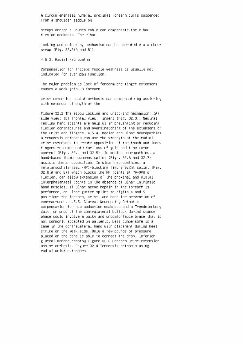
A circumferential humeral proximal forearm cuffs suspendedfrom a shoulder saddle by
straps and/or a Bowden cable can compensate for elbowflexion weakness. The elbow
locking and unlocking mechanism can be operated via a cheststrap [Fig. 32.2(A and B)].
4.5.3. Radial Neuropathy
Compensation for triceps muscle weakness is usually notindicated for everyday function.
The major problem is lack of forearm and finger extensorscauses a weak grip. A forearm
wrist extension assist orthosis can compensate by assistingwith extensor strength of the
Figure 32.2 The elbow locking and unlocking mechanism: (A)side view; (B) frontal view. fingers (Fig. 32.3). Neutralresting hand splints are helpful in preventing or reducingflexion contractures and overstretching of the extensors ofthe wrist and fingers. 4.5.4. Median and Ulnar NeuropathiesA tenodesis orthosis can use the strength of the radialwrist extensors to create opposition of the thumb and indexfingers to compensate for loss of grip and fine motorcontrol (Figs. 32.4 and 32.5). In median neuropathies, ahand-based thumb opponens splint (Figs. 32.6 and 32.7)assists thenar opposition. In ulnar neuropathies, ametatarsophalangeal (MP)-blocking figure eight splint [Fig.32.8(A and B)] which blocks the MP joints at 70–908 offlexion, can allow extension of the proximal and distalinterphalangeal joints in the absence of ulnar intrinsichand muscles. If ulnar nerve repair in the forearm isperformed, an ulnar gutter splint to digits 4 and 5positions the forearm, wrist, and hand for prevention ofcontractures. 4.5.5. Gluteal Neuropathy Orthoticcompensation for hip abduction weakness and a Trendelenberggait, or drop of the contralateral buttock during stancephase would involve a bulky and uncomfortable brace that isnot commonly accepted by patients. Less cumbersome is acane in the contralateral hand with placement during heelstrike on the weak side. Only a few pounds of pressureplaced on the cane is able to correct the drop. Inferiorgluteal mononeuropathy Figure 32.3 Forearm-wrist extensionassist orthosis. Figure 32.4 Tenodesis orthosis usingradial wrist extensors.
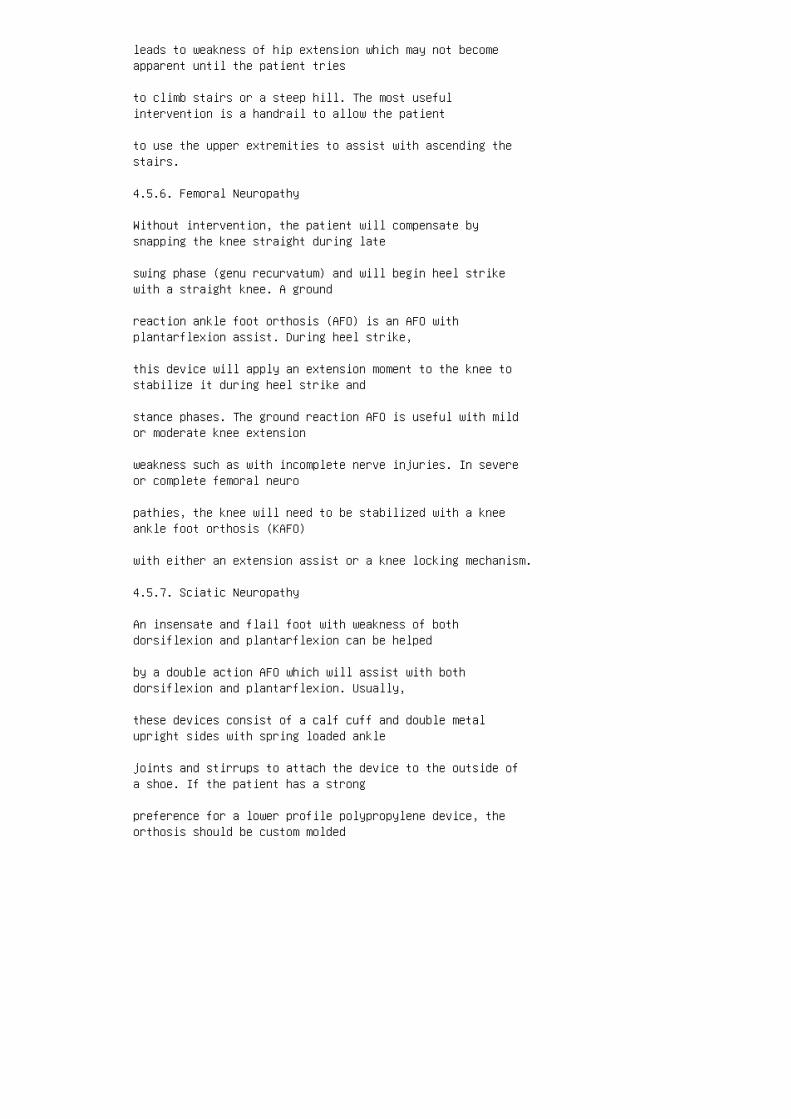
leads to weakness of hip extension which may not becomeapparent until the patient tries
to climb stairs or a steep hill. The most usefulintervention is a handrail to allow the patient
to use the upper extremities to assist with ascending thestairs.
4.5.6. Femoral Neuropathy
Without intervention, the patient will compensate bysnapping the knee straight during late
swing phase (genu recurvatum) and will begin heel strikewith a straight knee. A ground
reaction ankle foot orthosis (AFO) is an AFO withplantarflexion assist. During heel strike,
this device will apply an extension moment to the knee tostabilize it during heel strike and
stance phases. The ground reaction AFO is useful with mildor moderate knee extension
weakness such as with incomplete nerve injuries. In severeor complete femoral neuro
pathies, the knee will need to be stabilized with a kneeankle foot orthosis (KAFO)
with either an extension assist or a knee locking mechanism.
4.5.7. Sciatic Neuropathy
An insensate and flail foot with weakness of bothdorsiflexion and plantarflexion can be helped
by a double action AFO which will assist with bothdorsiflexion and plantarflexion. Usually,
these devices consist of a calf cuff and double metalupright sides with spring loaded ankle
joints and stirrups to attach the device to the outside ofa shoe. If the patient has a strong
preference for a lower profile polypropylene device, theorthosis should be custom molded

and covered with a soft material to protect the insensatefoot from skin breakdown.
Figure 32.5 Tenodesis orthosis using radial wrist extensors.
Figure 32.6 A hand-based thumb opponens splint. 4.5.8.Tibial Neuropathy An AFO in neutral position assists withtoe-off will assist with weakness of ankle plantarflexionand inversion. If a polypropylene device is selected, itshould be custom molded and have a soft cover to protectthe insensate sole against skin breakdown. 4.5.9. PeronealNeuropathy An AFO with dorsiflexion assistance preventsfoot drop and tripping, and the compensatory hip-hike gaitthat may lead to hip and knee pathology over time. Whenfitting the AFO, the orthotist must be careful not to fit acalf cuff that puts excessive pressure at Figure 32.7Hand-based thumb opponens splint. Figure 32.8Metatarsophalongeal-blocking figure light splint.
the fibular neck, as this could further compress theperoneal nerve and prevent functional
recovery that would otherwise happen.
5. PROGNOSIS
Prognosis varies according to the mechanism and location ofinjury. The most important
prognosticating factor is the severity of nerve injury, andfew reports include the severities
of nerve injuries according to the Seddon or Sunderlandclassifications. When taking into
account all the injuries, however, spontaneous recovery isthe rule rather than the excep
tion (7). Closed injuries have better prognosis forspontaneous recovery than do open inju
ries. Seddon (8) reported 83.5% spontaneous recovery incases of neuropathy associated
with closed upper extremity fracture, but only 17% in thoseassociated with open fracture.
Nerve injuries associated with joint dislocations havepoorer prognosis than those associ
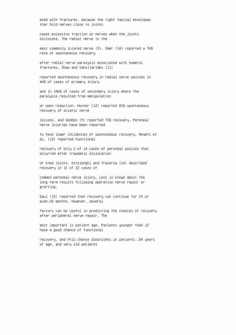
ated with fractures, because the tight fascial envelopesthat hold nerves close to joints
cause excessive traction on nerves when the jointsdislocate. The radial nerve is the
most commonly injured nerve (9). Omer (10) reported a 70%rate of spontaneous recovery
after radial nerve paralysis associated with humeralfractures. Shaw and Sakellarides (11)
reported spontaneous recovery in radial nerve palsies in40% of cases of primary injury
and in 100% of cases of secondary injury where theparalysis resulted from manipulation
or open reduction. Hunter (12) reported 83% spontaneousrecovery of sciatic nerve
lesions, and Seddon (9) reported 73% recovery. Peronealnerve injuries have been reported
to have lower incidences of spontaneous recovery. Meyers etal. (13) reported functional
recovery of only 2 of 14 cases of peroneal palsies thatoccurred after traumatic dislocation
of knee joints. Ottolenghi and Traversa (14) describedrecovery in 12 of 22 cases of
common peroneal nerve injury. Less is known about thelong-term results following operative nerve repair orgrafting.
Gaul (15) reported that recovery can continue for 24 oreven.50 months. However, several
factors can be useful in predicting the chances of recoveryafter peripheral nerve repair. The
most important is patient age. Patients younger than 12have a good chance of functional
recovery, and this chance diminishes in patients .50 yearsof age, and very old patients

experience poorer functional recovery (7). Another factoris the type of nerve involved.
Nerves that contain a single type of fiber have higherincidence of recovery than mixed
nerves, explained by the high incidence of aberrantregeneration possible in mixed nerves.
Other factors include the type of trauma that caused theneuropathy. Crush injuries that
include injury to neural vascular support, contaminated orinfected wounds do worse than
cleanly transected nerves. The time of repair of a primarylaceration will also affect recovery,
as nerve retraction will affect the amount of tensionplaced on the sutures. When jagged nerve
endings have to be trimmed back to allow for end-to-endrepair, the resulting end-to-end gap
will result in increased tension on the sutures and morechance of fibrous scarring at the site.
When a gap is present that is greater than a fewcentimeters, nerve grafting will achieve better
results than end-to-end repair due to the necessary tensionto close such a gap. The ultimate prognosis for bothconservatively and operatively managed nerve inju
ries will be improved by a rehabilitative program designedto increase the patient’s ability
to use new or altered patterns of activation. Prognosis isdependent not only on neural
regeneration and correct reinnervation, but also on thepatient’s age, health, social
support, motivation, beliefs, perseverance, and activitylevel. Nerve regeneration and col
lateral sprouting can occur up to 3 years after an injury.However, reorganization and
refinement of central neural pathways and coordination of

movement can continue for
at least 5 years and longer in the young patient. APPENDIX:TREATMENT ALGORITHM FOR TRAUMATIC MONONEUROPATHIESIdentification of Nerve Injury by Clinical FunctionalDeficit Primary Repair if Possible Primary Repair notpossible explore Therapy, Splinting, Prevention ofContractures Serial EMG and Clinical Examinations every1-3 months for 3-12 Months Evidence of Nerve Recovery NoEvidence of Nerve Recovery Consider Delayed Repair orGraft Ongoing Therapy and Splinting as Needed ConsiderMuscle/Tendon Transfer Open exploration, intraoperativeEMG Evidence of Nerve Recovery No or inadequate Evidenceof Nerve Recovery Delayed Repair or Graft open closed
1. Seddon HJ. Three types of nerve injury. Brain 1943;66:237–288.
2. Sunderland S. A classification of peripheral nerveinjuries producing loss of function. Brain 1951;74:491–516.
3. Toby et al. Changes in the structural properties ofperipheral nerves after transection. J Hand Surg 1996;21A:1086–1090.
4. Aagaard BD. Magnetic resonance neurography: magneticresonance imaging of peripheral nerves. Neuroimaging Clin NAm 2001; 11(1):131–146.
5. Martinoli C. Tendon and nerve sonography. Radiol ClinNorth Am 1999; 37(4):691–711.
6. Kline, DG. Nerve Injuries: Operative results for majornerve injuries, entrapmentz, and tumors. WB Saunders 1995.
7. Omer G, Spinner M, van Beek A, eds. Management ofPeripheral Nerve Problems. 2nd ed. WB Saunders Co., 1998.
8. Seddon HJ. Nerve lesions complicating certain closedbone injuries. J Am Med Assoc 1947; 135:691–694.
9. Seddon HJ. Surgical Disorders of the Peripheral Nerves.2nd ed. Edinburgh: Churchill Livinstone, 1975:691–694.
10. Omer GE, Results of untreated peripheral nerveinjuries. Clin Orthop 1982; 163:15–19.
11. Shaw J, Sakellarides H. Radial-nerve paralysisassociated with fractures of the humerus. A review of

forty-five cases. J Bone Joint Surg 1967; 49A:899–902.
12. Hunter GA. Posterior dislocation and fracturedislocation of the hip, a review of fifty-seven patients. JBone Joint Surg 1969; 51B:38–44.
13. Meyers MH, Moore TM, Harvey JP. Follow-up notes onarticles previously published in the Journal: traumaticdislocation of the knee joint. J Bone Joint Surg 1975;57A:430–433.
14. Ottolenghi CE. Traversa CH. Vascular and nervouscomplications in injuries of the knee joint. [JournalArticle]. Reconstr Surg Traumatol 1974; 14:114–135.
15. Gaul S. Intrinsic motor recovery: a long term study ofulnar nerve repair. J Hand Surg 1982; 7:502–508.
33
Cranial Mononeuropathies
Mark B. Bromberg
University of Utah, Salt Lake City, Utah, USA
Abstract 584
1. CN III, IV, and VI (Oculomotor, Trochlear, and Abducens)584 1.1. Anatomy 584 1.2. Symptoms and Signs 585 1.3.Causes 585 1.4. Diagnostic Testing 586 1.5. Treatment andPrognosis 586
2. CN V (Trigeminal) 586 2.1. Anatomy 586 2.2. Symptoms andSigns 586 2.3. Causes 587 2.4. Diagnostic Testing 587 2.5.Treatment and Prognosis 587 2.6. Cavernous Sinus Syndrome587
3. CN VII (Facial) 587 3.1. Anatomy 587 3.2. Symptoms andSigns 588 3.3. Causes 588 3.4. Diagnostic Testing 588 3.5.Treatment and Prognosis 589
4. CN VIII (Acoustic and Vestibular) 589 4.1. Anatomy 5894.2. Symptoms and Signs 590 4.3. Causes 590 4.4. DiagnosticTesting 590 4.5. Treatment and Prognosis 590
leaves the dorsal brainstem below the inferior cerebellarpeducles and curves around
the brainstem. It passes between the posterior cerebral and
superior cerebellar arteries.
It also passes through the cavernous sinus and enters theorbit through the superior
orbital fissure to innervate the superior oblique muscle.CN VI (abducens) rootlets arise from the pontomedullaryjunction and join to form a
nerve trunk of about 7000 fibers. It passes through thecavernous sinus and enters the orbit
through the superior orbital fissure to innervate thelateral rectus muscle. Within the cavernous sinus, CN III,IV, and VI and the ophthalmic and maxillary
divisions of CN V are in close proximity.
1.2. Symptoms and Signs
Neuropathies of these nerves result in diplopia and ptosisipsilateral to the lesion. Note,
lesions of CN IV may rarely be associated withcontralateral ptosis. Lesions of CN III
lead to diplopia with inability to adduct or look down andup, and as a consequence,
the eye assumes a position of downward abduction. There isalso ptosis and a dilated
pupil. Lesions of CN IV lead to diplopia with inability torotate the eye down and out,
and the eye assumes a slightly elevated position. Thepatient can easily compensate for
weakness of the superior oblique muscle by rotating(tilting) the head to the opposite
side. Lesions of CN VI lead to diplopia because ofinability to abduct the eye.
1.3. Causes
Head trauma is the cause of double vision in 15% of cases,affecting CN VI most com
monly, followed by CN III, with CN IV being the least

affected (1,2). There is controversy
whether CN VI’s greater susceptibility to injury is solelydue to its greater intracranial
length (3). Tumors are the cause of another 15% ofdiplopia, and most commonly affect CN VI,
alone or in combination (1,2). Primary brainstem tumors andmetastatic tumors are equally
represented, and affect intramedullary nerve tracts.Pituitary tumors affect these nerves as
they pass through the cavernous sinus, and pituitaryapoplexy causes loss of vision as well
as diplopia. Isolated tumors of CN III, IV, and VI arerare. Disorders affecting the caver
nous sinus may involve multiple CNs, and are discussed withCN V. Vascular disease is felt to be the cause in 15% ofneuropathies affecting CN III, IV,
and VI (1,2). The collateral blood supply is relativelypoor, and atherosclerosis associated
with hypertension and hyperlipidemia is felt to lead tonerve ischemia, and ocular palsies
are therefore more common in the elderly. Diabetic-basedocular palsies are perhaps the
most common cause, and have a classic presentation ofsudden onset associated with
severe retro-orbital pain and weakness attributed to CN IIIand VI and less frequently
to CN IV (1). Pupillary function is spared inthree-quarters of patients. Clinical features
are similar for patients with impaired glucose tolerance(4). The lesion appears to be
primary demyelinating, with lesser axonal damage (5). Ananeurysm is a classic cause of CN III palsy, and is usuallyassociated with pupil
lary dysfunction; however, with normal pupillary function

minimal palsy has also been
described (1,6). The aneurysm is most commonly in theinternal carotid artery, in the
region of the posterior communicating artery. Nerve palsymay result from direct mass
effects in unruptured aneurysms or the effects of thehemorrhage in ruptured aneurysms. Historically, syphiliswas a common cause of ocular palsies (7). Lyme borreliosis
may involve CN III, IV, and VI as the only neurologicmanifestation of infection (8). The Tolosa–Hunt syndromeincludes recurrent unilateral retro-orbital pain followedby paresis of CN III, IV, VI, and the ophthalmic divisionof CN V with spontaneous resolution (9). Pathologicinvestigation indicates granulomatous tissue in thecavernous sinus. Neurosarcoidosis less commonly involves CNIII, IV, and VII than CN VII (10). Approximately 25% ofneuropathies involving CN III, IV, and VI are ofundetermined cause (1,2). There is also a benign CN VIpalsy described in children (11). 1.4. Diagnostic TestingClinical assessment of eye movements is the most usefultest to distinguish among nerves causing diplopia, and thediagnostic palsies are described earlier. Imaging studies(MRI, CT, and ultrasound) can help isolate the site of thelesion and suggest causes (12,13). Among patients withknown vascular risks, imaging studies identify analternative cause in 15% (13). 1.5. Treatment and PrognosisIn the setting of trauma, the nature of the injuriesdetermines the degree of CN damage. Complete nerve avulsionhas a poor prognosis. Lesser injuries lead to spontaneousrecovery within weeks, and axonal injuries take 4–6 months.Neuropathies attributed to vascular causes improve withouttreatment, and those due to diabetes have a good recoverybecause of the demyelinating nature of the lesion. For thesame reasons, idiopathic neuropathies also have a favorableprognosis. Associated movements are those that are outsideof the intended activation pattern. This can lead tochronic diplopia in certain directions of gaze. Oneexplanation is aberrant (anomalous) regeneration fromaxonal damage leading to inappropriate re-innervation ofmuscles, whereas another is based on central (nuclear)reorganization or release (14). With neuropathies due totumor, the nature and treatment of the underlying tumor arethe most important prognostic factors. 2. CN V (TRIGEMINAL)2.1. Anatomy CN V has prominent sensory and motorfunctions. The sensory component supplies the face and isorganized into three divisions: ophthalmic, maxillary, and

mandibular. The pathways from the receptors mainly are asfollows. The ophthalmic division (CN V1) supplies sensationto the forehead, cornea, and sinuses. The nerve passesthrough the superior orbital fissure and cavernous sinus.The maxillary division (CN V2) supplies sensation to themiddle portion of the face. The nerve passes through thecavernous sinus and the foramen rotundum. The mandibulardivision (CN V3) provides sensation from the jaw, teeth,and anterior two-thirds of the tongue. It passes throughthe foramen ovale. All three divisions join at thetrigeminal ganglion, which enters the brainstem at thepons. The motor division is much smaller, and suppliesprimarily the muscles of mastication. It arises from thelateral portion of the pons, where it joins the sensoryfibers of the mandibular division and passes through theforamen ovale to innervate the tensor palatini and tensortympani, the medial pterygoid, the masseter, the lateralpterygoid, and finally the temporalis muscle. 2.2. Symptomsand Signs Numbness, and less so pain, in the distributionof one or more branches is the most common presentingsymptom, whereas weakness of mastication is less common(15). There may be
an alteration in taste (16,17). The rate of progression ofsymptoms is largely dependent
upon the pathophysiologic process and its location alongthe three pathways of CN V. In
progressive conditions, numbness may begin in one divisionand spread to others.
The maxillary and mandibular divisions appear to be morecommonly affected than the
ophthalmic division. Symptoms may also start on one sideand spread to the other side.
2.3. Causes
There is an association with connective tissue diseases,including undifferentiated sclero
derma and Sjo¨gren syndrome (16,18). Because only sensoryfunction is involved with con
nective tissue diseases, a ganglionopathy is postulated.Primary neoplasms include
cerebellopontine angle neuromas and meningiomas. An

interesting presentation of meta
static neoplasms is initial involvement of the mental nervebranch, giving rise to a numb
chin. Chin numbness may herald the diagnosis of cancer, andprimary tumor types cover a
wide spectrum (19). Undiagnosed or idiopathic trigeminalneuropathies frequently
develop suddenly with facial numbness, which may spreadfrom one division to another.
2.4. Diagnostic Testing
Initial diagnosis is by clinical examination.Electrophysiologic testing with blink reflexes
can help define CN V involvement of the ophthalmicdivision, but the reflex may be
normal (16). MRI is appropriate because of the highfrequency of underlying neoplasms.
2.5. Treatment and Prognosis
For patients with connective tissue diseases, treatment ofthe underlying disease does not
reliably result in lessening of symptoms (18). Among thosewith no identifiable cause, half
experienced a clinical remission without treatment (17).Corticosteroids have been tried
with variable success, but there are no controlled trials.
2.6. Cavernous Sinus Syndrome
CN III, IV, V1 and V2, and VI pass through the cavernoussinus, and all but CN V2 also
pass through the superior orbital fissure. Thus, it isdifficult to precisely localize a lesion to
either of these two structures by clinical examination.Symptoms include ptosis, diplopia,
and small and poorly reactive pupils. There is also

frequent retro-orbital pain, conjunctival
rubor, and proptosis. Causes of cavernous sinus syndromeinclude aneurysms, menin
giomas, pituitary tumors, and infectious and inflammatoryprocesses. Diagnostic evaluation
includes MRI or CT to further localize the lesion and helpdetermine pathology type.
Treatment is based on the pathological processes.
3. CN VII (FACIAL)
3.1. Anatomy
CN VII leaves the brainstem at the cerebellopontine angleand enters the internal auditory
meatus with CN VIII. It follows a tortuous pathway in thetemporal bone. Initially, it lies in
the internal auditory canal for 7–8 mm, and then enters thefacial canal for 3–4 mm. It
makes a sharp turn and travels 12–13 mm close to the middleear. The geniculate ganglion
is in this segment and supplies fibers to mucus membranesand lacrimal glands via the
greater superficial petrosal nerve. There follows a 10–14mm mastoid segment where the motor nerve to the stapediusmuscle branches off. The chorda tympani conveys taste fromthe anterior two-thirds of the tongue and joins CN VII inthis segment. The nerve leaves the skull at thestylomastoid foramen, and the first motor branches are tothe posterior belly of the stylohyoid, digastric, andoccipital muscles. The nerve then travels through theparotid gland, and terminal motor branches are thetemporal, zygomatic, buccal, mandibular and cervical toinnervate muscles of facial expression. 3.2. Symptoms andSigns The primary symptom is facial weakness. Thiscondition was first written about by Sir Charles Bell in1829, when he described a benign condition that was alreadyfamiliar to contemporary clinicians. Clinical features arean acute onset, or progression over several hours, ofunilateral weakness of facial muscles that is complete in70% and partial in 30% of patients. There are also

manifestations of loss of other functions subserved by CNVII, such as decreased lacrimation, and altered taste (20).Abnormal sensation also occurs, including dysesthesias inthe trigeminal and glossopharyngeal nerve distributions.There may be pain on the affected side of the face andtemporary alterations in hearing (21). The incidence ofBell palsy is �20/100,000 in the general population,affecting adults more than children (22). It appears to bethree times more common in pregnant women (23). CN VIIweakness may occur in the setting of a rash involving theexternal auditory meatus, the Ramsay Hunt syndrome (24).Slow progressive facial weakness (over days) is lessconsistent with Bell palsy. 3.3. Causes The majority ofcases of CN VII palsies, which include Bell palsy, areclassified as idiopathic, and nerve damage probably occurssecondary to compression from edema within the confines ofthe boney canal. There are data implicating an underlyingviral infection as the cause for these cases (25). Evidencefor herpes simplex virus in association with 80% ofidiopathic Bell palsy is from DNA analysis by polymerasechain reaction followed by hybridization with Southern blotanalysis of endoneurial fluid at the time of decompressionsurgery (26). The most clearly defined viral cause of CNVII neuropathy is the Ramsay Hunt syndrome, facial weaknesswith erythematous vesicular rash in the ear (24). Anassociated underlying condition or cause is found in theminority of patients. The incidence of diabetes mellitus ishigh, leading to the conclusion that Bell palsy mayrepresent a diabetic mononeuropathy in these patients (27).Otitis media is a rare cause. Bell palsy occurs morefrequently in the setting of Lyme disease, and Borelliaburgdorferi may account for 25% of cases in endemic areas,many without other clinical features of Lyme disease (28).Neurosarcoid presenting as an isolated CN VII palsy is lesscommon than in association with other cranial neuropathies(10). Surgical trauma to CN VII may occur during procedureson the temporal bone. Close head trauma resulting infractures of the temporal bone is another cause. The degreeof nerve fiber damage ranges from neurapraxia toaxonotemesis. Within a patient there may be varyingproportions of each process, which probably account for therange of clinical outcomes (20). 3.4. Diagnostic TestingFunctional tests include measurement of submandibularsalivary flow and electrodiagnostic testing. Measurementsof salivary flow require cannulation of Wharton ducts andis
technically difficult. Nerve conduction measurements focuson the nerve segment distal to

the site of nerve damage in the temporal bone. The evokedresponse to activation of the
nerve at the level of the parotid gland will be within thenormal range for the first 2
days after acute onset of weakness, even if there iscomplete axonal damage (29). After
the third day, if there is significant axonal damage(axotemesis lesion), the evoked
motor response will fall in proportion to the number ofviable axons. Nerve fibers affected
by conduction block (neurapraxic lesion) will contribute tothe evoked potential, but not to
voluntary activation. If the evoked response is .10% of thecontralateral (normal) side,
prognosis is favorable because this indicates that thereare a significant number of
viable fibers. Needle EMG assesses the distribution ofdenervation in the form of abnormal
spontaneous activity (positive sharp waves and fibrillationpotentials). However, it takes
5 days for abnormal spontaneous activity to appear. Imagingstudies using quantitative
MRI suggest some degree of prognostication, with poorerresults among patients with
greater signal abnormalities (30).
3.5. Treatment and Prognosis
The proposed inflammatory pathology has led to the use ofcorticosteroids, and the pro
posed viral pathogenesis has led to the use of anti-viralagents. Although some reports
have favored these agents, the Cochrane Database SystematicReview has analyzed data
for corticosteroids and acyclovir or valaciclovir. The

Cochrane Review selects random
ized or quasirandomized trials for the various agents, assole agent or in combination.
Appropriately conducted trials are reviewed andmeta-analyses are preformed. Improved
outcome has not been demonstrated for any of the agents,either alone or in combination
(31,32). A Cochrane Review of acupuncture indicated thatacupuncture alone might be
more associated with a better outcome than medications orthe combination, but methodo
logical differences among trials prevented a meta-analysis(33). The inaccessible site of pathology means that aclinical determination of the degree
of neurapraxia and axonotemesis by electrodiagnostictesting cannot be determined until
after the fourth day of onset. The full extent of nervedamage has probably occurred by this
time, and surgical intervention is unwarranted (20,29).Overall prognosis from retrospective reviews indicates that80–85% of patients
have a full or satisfactory return of strength (22). Theremay be residual symptoms that
include crocodile tears and other forms of aberrantre-innervation such as lower facial
muscle twitching with eye blinking (synkensis) (14,34).Prognostic factors have been chal
lenging to identify, but age .55 years, complete paralysis,and hypertension are negative
factors.
4. CN VIII (ACOUSTIC AND VESTIBULAR)
4.1. Anatomy
There are two divisions, one serving the vestibular system

and the other the auditory
system. The vestibular division originates from the haircells in the semicircular canals
and the macula of the utricle and saccule, whereas thecochlear division originates from
hair cells in the organ of Corti. The vestibular nervecontains about 20,000 fibers and
the cochlear nerve about 30,000 fibers. Both divisions liein the temporal bone in close
association with CN VII. CN VIII leaves the skull throughthe internal auditory meatus
and joins the brainstem at the pontomedullary junction inthe cerebellopontine angle.
The intracranial portion of the nerve includes a junctionalzone between the proximal segment, which is a central fibertract with neuroglial support cells and a more distalsegment, which is a peripheral nerve with Schwann cellsupport cells. 4.2. Symptoms and Signs Damage to the nervecan result in both hearing loss and tinnitus and vertigo invarious proportions. Acute causes tend to include symptomsin both divisions, whereas slow progressive causes tend topresent with hearing loss. The Ramsay Hunt syndrome beginswith deep ear pain, followed in several days by vesiculareruptions in the external auditory canal eventually leadingto hearing loss and vertigo (24). CN VII is frequentlyinvolved. Acute labrynthitis is associated with vertigowith spontaneous nystagmus (35). Vestibular symptoms may beassociated with other cranial neuropathies (CN V, VII, X,XI) (36). 4.3. Causes Trauma involving the temporal bonecan damage both CN VIII and CN VII. The injury can be astretching of the nerve on a complete severance. Acousticnerve Schwannomas tend to arise within the internal auditormeatus because the junction between the proximal extensionof the neuroglial investment of the nerve and the distalSchwann cell investment is close to the meatus. Up to 10%of unilateral tumors are associated with neurofibromatosis.Viral infections may affect the labrynth (labrynthitis) orthe nerve, although direct evidence for this etiology isoften lacking because of the isolated location of thenerve. The Ramsay Hunt syndrome is due to herpes zoster(24). The consensus is that diabetes mellitus does notcontribute to acute hearing loss (15). Sarcoidosisuncommonly affects CN VII in isolation (10). Evidence for
borrelia infection has been found in patients with vertigo(37). 4.4. Diagnostic Testing Batteries of tests forcochlear and vestibular functions require special equipmentand include audiograms, tone decay, evoked responses, andelectronystagrams with provocative tests (vestibulo-ocular,caloric) (38). Imaging tests are important to excludetumors. 4.5. Treatment and Prognosis Surgical and radiationtreatments of tumors are appropriate with outcome dependentupon many variables (39). Vertigo associated with presumedviral etiology resolve spontaneously (36). 5. CN IX(GLOSSOPHARYNGEAL) 5.1. Anatomy The nerve arises from aline of rootlets in a sulcus along the lateral border ofthe medulla. The nerve is associated with CN X, and bothpass through the jugular foramen and into the neck. Motorfibers innervate the stylopharyngeus muscle. Sensory fiberssubserve taste along the posterior two-thirds of the tongueand sensation of the soft palate and related structures,and a branch supplies sensation to part of the external earand the tympanic
membrane. There are parasympathetic fibers in the lessersuperficial petrosal nerve that
controls secretions of the parotid gland. The carotid sinusnerve is a branch of CN IX.
5.2. Symptoms and Signs
Because of its close proximity with CN X, lesions usuallyinvolve both nerves, and symp
toms are more often related to that nerve. There may bedifficulties in swallowing and
impaired taste from posterior portions of the tongue.Glosspharyngeal neuralgia refers
to episodes of sudden onset of lancinating unilateral painat the base of the tongue and
faucial region with similarities to trigeminal neuralgia.
5.3. Causes
Cerebropontine angle tumors may also affect CN IX. Lesionsin the jugular foramen may
affect both CN IX and X, giving rise to a jugular foramensyndrome (Vernet syndrome).

Causes include glomus tumors and other neoplasms.
5.4. Diagnostic Testing
Assessment of taste perception of the posterior portion ofthe tongue can be performed.
Cutaneous stimulation for the gag reflex is supplied bythis nerve, and can be tested by
lightly stroking the posterior pharynx.
5.5. Treatment and Prognosis
These are dependent upon underlying causes, especially inthe setting of tumors. Crocodile
tears (paroxysmal lacrimation with gustatory stimuli) mayoccur after injury or surgery on
the ear or after CN VII neuropathies. This is due to theclose proximity of fibers in the inner
ear and cross-innervation of sprouting fibers that involvethe lesser superficial petrosal
nerve which supplies the parotid gland (34).
6. CN X (VAGUS)
6.1. Anatomy
The nerve is formed from rootlets emerging from a lateralsulcus of the medulla. The nerve
is joined by fibers from CN XI that have ascended throughthe foramen magnum, although
they remain as a discrete fascicle. The combined nervepasses through the jugular foramen.
In the neck, the pharyngeal nerve supplies all pharyngealconstrictor muscles except the
stylopharynegeus and tensor palati, as well as thepalatoglossus muscle of the tongue.
The superior laryngeal nerve also innervates constrictormuscles and the cricothyroid
muscle. The recurrent laryngeal nerve follows an asymmetriccourse to supply the remain
ing muscles of the larynx. Visceral afferent fibers supplysensation from the pharynx,
larynx, trachea, esophagus, and viscera. Visceral efferentfibers supply the heart and visc
eral organs.
6.2. Symptoms and Signs
Unilateral lesions usually include both CN IX and X andresult in voice hoarseness and
difficulty swallowing and nasal regurgitation due toweakness of soft palate elevation which normally closes offthe nasopharynx during swallowing. Lesions of the recurrentlaryngeal nerve also cause voice hoarseness. 6.3. CausesIsolated lesions may result from tumors affecting the baseof the skull and the jugular vein. Recurrent laryngealnerve lesions occur most commonly in association withsurgery of the carotid artery, the thyroid, and thoraciccavity (40). One-quarter to one-third of cases are thoughtto be idiopathic (40,41). Men are more often affected thanwomen, and the left recurrent laryngeal nerve more than theright, perhaps because of its greater length. Concurrentdiabetes may be a contributing factor in a small percentage(41). Idiopathic superior laryngeal nerve palsies are feltto be common and are due to reactivation of herpes simplexinfection (42). 6.4. Diagnostic Testing Inspection revealsasymmetry of the soft palate, lower on the affected side,and deviation of the uvula to the opposite side. Indirectlaryngoscopy can detect abnormal vocal cord function.Needle EMG can distinguish between superior laryngeal andrecurrent laryngeal lesions (41). Testing of cardiacautonomic function will not be discussed. 6.5. Treatmentand Prognosis Lesions associated with cancers have avariable prognosis (43). Lesions associated with carotidand thyroid surgery may recover spontaneously, and are thuspresumed to be due to tissue swelling. With idiopathicrecurrent laryngeal neuropathies, 85% show some degree ofspontaneous recovery and 35% resolve completely (43,44). 7.CN XI (SPINAL ACCESSORY) 7.1. Anatomy Rootlets contributingto CN IX include the most caudal region of themesencephalon and the first five to seven cervicalsegments. The cervical rootlets form a nerve that ascendsthrough the foramen magnum and joins with the cranialrootlets and CN X. The combined nerves pass through the

jugular foramen. CN XI descends in the carotid sheath, butleaves it and penetrates the sternocleidomastoid musclewhich it supplies. The nerve then crosses the posteriortriangle of the neck to reach the trapezius muscle andsupplies the upper one-third. 7.2. Symptoms and SignsLesions of the deep, proximal portion, result in weaknessof the sternocleidomastoid muscle affecting head rotationto the opposite side and weakness of the upper trapeziuswith poor shoulder and arm elevation above the horizontalaxis on the same side. Lesions of the superficial, distalportion in the posterior triangle result in weakness onlyof the trapezius muscle. There may be mild winging of thescapula.
7.3. Causes
The portion of the nerve in the jugular foramen and carotidsheath may be affected by
tumors. The most common causes of nerve injury in the neckare from surgical procedures,
notably radical neck dissections and cervical lymph nodebiopsy, but neuropathies can also
occur when the nerve is in continuity (45). Delayedweakness after surgical procedures
suggests lesions from scar tissue (45). Less common causesare carotid endarterectomy
and central line placement. Weakness attributed to focalradiation for cancer may occur
years after treatment (46). Idiopathic lesions occur andmay be heralded by pain along
the posterior border of the sternocleidomastoid muscle (47).
7.4. Diagnostic Testing
Needle EMG can document denervation of thesternocleidomastoid and upper trapezius
muscles.
7.5. Treatment and Prognosis
Varying degrees of spontaneous improvement may occur whendamage is due to surgery,

but some patients accept a certain degree of weakness (45).Surgical grafting following
injury from radical neck surgery has been reported to besuccessful (48). Spontaneous
neuropathies may or may not show improvement with time (47).
8. CN XII (HYPOGLOSSAL)
8.1. Anatomy
The nerve arises from rootlets along the medulla, mergingto form two bundles that pass
through the hypoglossal foramen. It passes down from theangle of the jaw and innervates
the thyrohyoid muscle and intrinsic muscles of the tongue(hyoglossus, styloglossus,
genioglossus, and geniohyoid muscles).
8.2. Symptoms and Signs
Unilateral lesions result in tongue deviation to the sideof the lesion. Atrophy of the
affected half may occur. Swallowing is affected to agreater degree than speech. Glosso
dynia is a syndrome with burning pain in the tongue or theoral cavity, which may be
similar to trigeminal neuralgia.
8.3. Causes
Tumors in the region of the hypoglossal foramen or neck arerare causes as are also trauma
and dental extractions. Idiopathic mononeuropathies aredescribed (49).
8.4. Diagnostic Testing
22. Katausic S, Beard C, Wiederholt W, Bergstralh E,Kurland L. Incidence, clinical features, and prognosis inBell’s palsy, Rochester, Minnesota, 1968–1982. Ann Neurol
1986; 20:622–627.
23. Gillman G, Schaitkin B, May M, Klein S. Bell’s palsy inpregnancy: a study of recovery outcomes. Otolaryngol HeadNeck Surg 2002; 126:26–30.
24. Sweeney C, Gliden D. Ramsay Hunt syndrome. J NeurolNeurosurg Psychiatry 2001; 71:149–154.
25. Baringer J. Herpes simplex virus and Bell palsy. AnnIntern Med 1996; 124:63–65.
26. Murakami S, Mizobuchi M, Nakashiro Y, Doi T, Hato N,Yanagihara N. Bell palsy and herpes simplex virus:identification of viral DNA in endoneurial fluid andmuscle. Ann Intern Med 1996; 124:27–30.
27. Pecket P, Schattner A. Concurrent Bell’s palsy anddiabetes mellitus: a diabetic mononeuropathy? J NeurolNeurosurg Psychiatry 1982; 45:652–655.
28. Halpern J, Golightly M. Long Island NeuroborreliosisCollaborative Study Group. Lyme borreliosis in Bell’spalsy. Neurology 1992; 42:1268–1270.
29. Albers J, Bromberg M. Bell’s palsy. In: Johnson R, ed.Current Therapy in Neurologic Disease. 3rd ed.Philadelphia: Dekker, 1990:376.
30. Kress B, Griesbeck R, Stippich C, Bahren W, Sartor K.Bell palsy: quantitative analysis of MR imaging data as amethod of predicting outcome. Radiology 2004; 230:504–509.
31. Allen D, Dunn L. Aciclovir or valaciclovir for Bell’spalsy (idiopathic facial paralysis). Cochrane Database SystRev 2004; (3):CD001869.
32. Salinas R, Alvarez G, Alvarez M, Ferreira J.Corticosteroids for Bell’s palsy (idiopathic facialparalysis). Cochrane Database Syst Rev 2002; (1):CD001942.
33. He L, Zhou D, Wu B, Li N, Zhou M. Acupuncture forBell’s palsy. Cochrane Database Syst Rev 2004;(1):CD002914.
34. Boyer F, Gardner W. Paroxysmal lacrimation (syndrome ofcrocodile tears) and its surgical treatment. Arch NeurolPsychiatry 1949; 61:56–64.
35. Dix M, Hallpike C. The pathology, symptomatology and

diagnosis of certain common disorders of the vestibularsystem. Ann Otol Rinol Laryngol 1952; 61:987–1016.
36. Adour K, Sprague M, Hilsinger R. Vestibular vertigo. Aform of polyneuritis? J Am Med Assoc 1981; 246:1564–1567.
37. Rosenhall U, Hanner P, Kaijser B. Borrelia infectionand vertigo. Acta Otolaryngol 1988; 106:111–116.
38. Luxon L. Diseases of the eighth cranial nerve. In: DyckP, Thomas P, Griffin J, Low P, Poduslo J, eds. PeripheralNeuropathy. 3rd ed. Philadelphia: WB Saunders Co,1993:837–868.
39. Chakrabarti I, Apuzzo M, Giannota S. Acoustic neuroma:decision making with all the tools. Clin Neurosurg 2003;50:293–312.
40. Williams R. Idiopathic recurrent laryngeal nerveparalysis. J Laryngol Otol 1959; 73:161–166.
41. Berry H, Blair R. Isolated vagus nerve palsy and vagalmononeuritis. Arch Otolaryngol 1980; 106:333–338.
42. Abour K, Schneider G, Hilsinger R. Acute superiorlaryngeal nerve palsy: Analysis of 78 cases. OtolaryngolHead Neck Surg 1980; 88:418–424.
43. Huppler E, Schmidt H, Devine K, Gage R. Ultimateoutcome of patients with vocal-cord paralysis ofundetermined cause. Am Rev Tuberc 1956; 72:52–60.
44. Blau J, Kapadia R. Idiopathic palsy of the recurrentlaryngeal nerve: a transient cranial mononeuropathy. BMJ1972; 4:259–261.
45. Gordon S, Graham W, Black J, Miller S. Accessory nervefunction after surgical procedures in the posteriortrangle. Arch Surg 1977; 112:264–268.
46. Berger P, Bataini J. Radiation-induced cranial nervepalsy. Cancer 1977; 40:152–155.
47. Eisen A, Bertrand G. Isolated accessory nerve palsy ofspontaneous origin. Arch Neurol 1972; 27:496–502.
48. Ballantyne A, Guinn G. Reduction of shoulder disabilityafter neck dissections. Am J Surg 1962; 112:662–665.
49. Afifi A, Rifai Z, Faris K. Isolated, reversible,

hypoglossal nerve palsy. Arch Neurol 1984; 41:1218.

Treatment of Peripheral Neuropathy
16. Nodera H, Logigian EL, Herrmann DN. Class of nervefiber involvement in sensory neuropathies: clinicalcharacterization and utility of the plantar nerve actionpotential. Muscle Nerve 2002; 26:212–217.
17. Carter GT, Jensen MP, Galer BS, Kraft GH, Crabtree LD,Beardsley RM, Abresch RT, Bird TD. Neuropathic pain inCharcot–Marie–Tooth disease. Arch Phys Med Rehab 1998;79:1560–1564.
18. Wolfe GI, Baker NS, Amato AA, Jackson CE, Nations SP,Saperstein DS, Cha CH, Katz JS, Bryan WW, Barohn RJ.Chronic cryptogenic sensory polyneuropathy: clinical andlaboratory characteristics. Arch Neurol 1999; 56:540–547.
19. Periquet MI, Novak V, Collins MP, Nagaraja HN, Erdem S,Nash SM, Freimer ML, Sahenk Z, Kissel JT, Mendell JR.Painful sensory neuropathy: prospective evaluation usingskin biopsy. Neurology 1999; 53:1641–1647.
20. Herrmann DN, Griffin JW, Hauer P, Cornblath DR,McArthur JC. Epidermal nerve fiber density and sural nervemorphometry in peripheral neuropathies. Neurology 1999;53:1634–1640.
21. Said G, Lacroix-Ciaudo C, Fujimura H, Blas C, Faux N.The peripheral neuropathy of necrotizing arteritis: aclinicopathological study. Ann Neurol 1998; 23:461–465.
22. Herrmann DN, Logigian EL. Approach to peripheral nervedisorders. In: Katirji B, Kaminski HJ, Preston DC, Ruff RL,Shapiro BE, eds. Neuromuscular Disorders—in ClinicalPractice. Boston: Butterworth Heinemann, 2002:501–512.
23. Gaist D, Jeppesen U, Andersen M, Garcı´a Rodrı´guez LA,Hallas J, Sindrup SH. Statins and risk of polyneuropathy: acase–control study. Neurology 2002; 58:1333–1337.
24. Behse F, Buchthal F. Alcoholic neuropathy: clinical,electrophysiological, and biopsy findings. Ann Neurol 1977;2:95–110.
25. Koike H, Mori K, Misu K, Hattori N, Ito H, Hirayama M,Sobue G. Painful alcoholic polyneuropathy with predominantsmall-fiber loss and normal thiamine status. Neurology2001; 56:1727–1732.
26. Spencer PS, Schaumberg HH, eds. Experimental and

Clinical Neurotoxicology. 2nd ed. New York: OxfordUniversity Press, 2000.
27. Archer AG, Watkins PJ, Thomas PK, Sharma AK, Payan J.The natural history of acute painful neuropathy in diabetesmellitus. J Neurol Neurosurg Psychiatry 1983; 46:491–499.
28. Galer BS, Gianas A, Jensen MP. Painful diabeticpolyneuropathy: epidemiology, pain description, and qualityof life. Diabetes Res Clin Prac 2000; 47:123–128.
29. Malik RA, Veves A, Walker D, Siddique I, Lye RH, SchadyW, Boulton AJ. Sural nerve fibre pathology in diabeticpatients with mild neuropathy: relationship to pain,quantitative sensory testing and peripheral nerveelectrophysiology. Acta Neuropathol 2001; 101:367–374.
30. Singleton JR, Smith AG, Bromberg MB. Painful sensorypolyneuropathy associated with impaired glucose tolerance.Muscle Nerve 2001; 24:1225–1228.
31. McManis PG, Windebank AJ, Kiziltan M. Neuropathyassociated with hyperlipidemia. Neurology 1994;45:2119–2120.
32. David WS, Mahdavi Z, Nance M, Khan M. Hyperlipidemiaand neuropathy. Electromyogr Clin Neurophysiol 1999;39:227–230.
33. Drory VE, Groozman GB, Rubinstein A, Korczyn AD.Hyperlipidemia may cause a subclinical peripheralneuropathy. Electromyogr Clin Neurophysiol 1999; 39:39–41.
34. So YT, Holtzman DM, Abrams DI, Olney RK. Peripheralneuropathy associated with acquired immunodeficiencysyndrome. Prevalence and clinical features from apopulation-based survey. Arch Neurol 1988; 45:945–948.
35. Brannagan TH, Nuovo GJ, Hays AP, Latov N. Humanimmunodeficiency virus infection of dorsal root ganglionneurons detected by polymerase chain reaction in situhybridization. Ann Neurol 1997; 42:368–372.
36. Glass JD, Wesselingh SL. Microglia in HIV-associatedneurological diseases. Microsc Res Tech 2001; 54:95–105.
37. Brady R, Schiffmann R. Clinical features of and recentadvances in therapy for Fabry disease. JAMA 2000;284:2771–2775. 38. Filling-Katz MR, Merrick HF, Fink JK,Miles RB, Sokol J, Barton NW. Carbamazepine in Fabry’s

disease: effective analgesia with dose-dependentexacerbation of autonomic dysfunction. Neurology 1989;39:598–600. 39. Kelly JJ, Kyle RA, O’Brien PC, Dyck PJ. Thenatural history of peripheral neuropathy in primarysystemic amyloidosis. Ann Neurol 1979; 6:1–7. 40.Kuhlenba¨umer G, Young P, Kiefer R, Timmerman V, Wang JF,Schroeder JM, Weis J, Ringelstein EB, Van Broeckhoven C,Stoegbauer F. A second family with autosomal dominantburning feet syndrome. Ann NY Acad Sci 1999; 883:445–448.41. Novak V, Freimer ML, Kissel JT, Sahenk Z, Periquet IM,Nash SM, Collins MP, Mendell JR. Autonomic impairment inpainful neuropathy. Neurology 2001; 56:861–868. 42. GibbelsE, Shaefer HE, Runne U, Schro¨der JM, Haupt WF, Assmann G.Severe polyneuropathy in Tangier disease mimickingsyringomyelia or leprosy. J Neurol 1985; 232:283–294. 43.Moulin DE, Hagen N, Feasby TE, Amireh R, Hahn A. Pain inGuillain–Barre´ syndrome. Neurology 1997; 48:328–331. 44.McCombe PA, Pollard JD, McLeod JG. Chronic inflammatorydemyelinating polyradiculoneuropathy. A clinical andelectrophysiological study of 92 cases. Brain 1987;110:1617–1630. 45. Di Guglielmo G, Di Muzio A, Torrieri F,Repaci M, De Angelis MV, Uncini A. Low back pain due tohypertrophic roots as presenting symptom of CIDP. Ital JNeurol Sci 1997; 18:297–299. 46. Vrethem M, Cruz M, Wen-XinH, Malm C, Holmgren H, Ernerudh J. Clinical,neurophysiological and immunological evidence ofpolyneuropathy in patients with monoclonal gammopathies. JNeurol Sci 1993; 114:193–199. 47. Dabby R, Weimer LH, HaysAP, Olarte M, Latov N. Antisulfatide antibodies inneuropathy: clinical and electrophysiologic correlates.Neurology 2000; 54:1448–1452. 48. Sterman AB, SchaumburgHH, Asbury AK. The acute sensory neuronopathy syndrome: adistinct clinical entity. Ann Neurol 1980; 7:354–358. 49.Hawke SH, Davies L, Pamphlett R, Guo YP, Pollard JD, McLeodJG. Vasculitic neuropathy. A clinical and pathologicalstudy. Brain 1991; 114:2175–2190. 50. Yamamoto M, Ito Y,Mitsuma N, Li M, Hattori N, Sobue G. Pathology-relateddifferential expression regulation of NGF, GDNF, CNTF, andIL-6 mRNAs in human vasculitic neuropathy. Muscle Nerve2001; 24:830–833. 51. McQuay HJ, Trame`r M, Nye BA, CarrollD, Wiffen PJ, Moore RA. A systematic review ofantidepressants in neuropathic pain. Pain 1996; 68:217–227.52. Galer BS. Painful neuropathy. Neurol Clin 1998;16:791–811. 53. Sindrup SH, Jensen TS. Pharmacologictreatment of pain in polyneuropathy. Neurology 2000;55:915–920. 54. Kiayias JA, Vlachou ED, Lakka-Papadodima E.Venlafaxine HCl in the treatment of painful peripheraldiabetic neuropathy. Diabetes Care 2000; 23:699. 55.Semenchuk MR, Sherman S, Davis B. Double-blind, randomizedtrial of bupropion SR for the treatment of neuropathic
pain. Neurology 2001; 57:1583–1588. 56. Sindrup SH, GramLF, Brosen K, Eshoj O, Mogensen EF. The selective serotoninreuptake inhibitor paroxetine is effective in the treatmentof diabetic neuropathy symptoms. Pain 1990; 42:135–144. 57.Sindrup SH, Bjerre U, Dejgaard A, Brosen K, Aaes-JorgensenT, Gram LF. The selective serotonin reuptake inhibitorcitalopram relieves the symptoms of diabetic neuropathy.Clin Pharmacol Ther 1992; 52:547–552. 58. Hardy JR, ReesEA, Gwilliam B, Ling J, Broadley K, A’Hern R. A phase IIstudy to establish the efficacy and toxicity of sodiumvalproate in patients with cancer-related neuropathic pain.J Pain Symptom Manage 2001; 21:204–209. 59. Backonja M,Beydoun A, Edwards KR, Schwartz SL, Fonseca V, Hes M,LaMoreaux L, Garofalo E. Gabapentin for the symptomatictreatment of painful neuropathy in patients with diabetesmellitus: a randomized controlled trial. J Am Med Assoc1998; 280:1831–1836. 60. Gorson KC, Schott C, Herman R,Ropper AH, Rand WM. Gabapentin in the treatment of painfuldiabetic neuropathy: a placebo controlled, double blind,crossover trial. J Neurol Neurosurg Psychiatry 1999;66:251–252.
61. Gee NS, Brown JP, Dissanayake VU, Offord J, Thurlow R,Woodruff GN. The novel anticonvulsant drug, gabapentin(Neurontin), binds to the alpha2delta subunit of a calciumchannel. J Biol Chem 1996; 271:5768–5776.
62. Eisenberg E, Lurie Y, Braker C, Daoud D, Ishay A.Lamotrigine reduces painful diabetic neuropathy: arandomized, controlled study. Neurology 2001; 57:505–509.
63. Simpson DM, Olnery R, McArthur JC, Khan A, Godbold J,Ebel-Frommer K. A placebo-controlled trial of lamotriginefor painful HIV-associated neuropathy. Neurology 2000;54:2115–2119.
64. Devers A, Galer BS. Topical lidocaine patch relieves avariety of neuropathic pain conditions: an open-labelstudy. Clin J Pain 2000; 16:205–208.
65. Galer BS, Rowbotham MC, Perander J, Friedman E. Topicallidocaine patch relieves postherpetic neuralgia moreeffectively than a vehicle topical patch: results of anenriched enrollment study. Pain 1999; 80:533–538.
66. Dejgard A, Petersen P, Kastrup J. Mexiletine fortreatment of chronic painful neuropathy. Lancet 1988;1(8575–8576):1550–1555.
67. Stracke H, Meyer UE, Schumacher HE, Federlin K.

Mexiletine in the treatment of diabetic neuropathy.Diabetes Care 1992; 15:1550–1555.
68. Oskarsson P, Ljunggren JG, Lins PE. Efficacy and safetyof mexiletine in the treatment of painful diabeticneuropathy. The Mexiletine Study Group. Diabetes Care 1997;20:1594–1597.
69. Sindrup SH, Andersen G, Madsen C, Smith T, Brosen K,Jensen TS. Tramadol relieves pain and allodynia inpolyneuropathy: a randomised, double-blind, controlledtrial. Pain 1999; 83:85–90.
70. Harati Y, Gooch C, Swenson M, Edelman S, Greene D,Raskin P, Donofrio P, Cornblath D, Sachdeo R, Siu CO, KaminM. Double-blind randomized trial of tramadol for thetreatment of the pain of diabetic neuropathy. Neurology1998; 50:1842–1846.
71. Nelson KA, Park KM, Robinovitz E, Tsigos C, Max MB.High-dose oral dextromethorphan versus placebo in painfuldiabetic neuropathy and postherpetic neuralgia. Neurology1997; 48:1212–1218.
72. Galer BS, Twilling LL, Harle J, Cluff RS, Friedman E,Rowbotham MC. Lack of efficacy of riluzole in the treatmentof peripheral neuropathic pain conditions. Neurology 2000;55:971–975.
73. Low PA, Opfer-Gehrking TL, Dyck PJ, Litchy WJ, O’BrienPC. Double-blind, placebocontrolled study of theapplication of capsaicin cream in chronic distal painfulpolyneuropathy. Pain 1995; 62:163–168.
74. Capsaicin Study Group. Treatment of painful diabeticneuropathy with topical capsaicin. A multicenter,double-blind, vehicle-controlled study. Arch Intern Med1991; 151:2225–2229.
75. Nolano M, Simone DA, Wendelschafer-Crabb G, Johnson T,Hazen E, Kennedy WR. Topical capsaicin in humans: parallelloss of epidermal nerve fibers and pain sensation. Pain1999; 81:135–145.
76. DePalma MT, Weisse CS. Psychological influences on painperception and non-pharmacologic approaches to thetreatment of pain. J Hand Therapy 1997; 10:183–191.
77. Haythornthwaite JA, Benrud-Larson LM. Psychologicalassessment and treatment of patients with neuropathic pain.

Curr Pain Headache Rep 2001; 5:124–129.
78. Melzack R, Wall P. Pain mechanisms: a new theory.Science 1965; 150:171–179.
79. He LF. Involvement of endogeneous opioid peptides inacupuncture analgesia. Pain 1987; 31:99–121.
80. Shlay JC, Chaloner K, Max MB, Flaws B, Reichelderfer P,Wentworth D, Hillman S, Brizz B, Cohn DL for the TerryBeirn Community Programs for Clinical Research on AIDS.Acupuncture and for pain due to HIV-related peripheralneuropathy: a randomized controlled trial. JAMA 1998;280:1590–1595.
81. Kumar D, Marschall HJ. Diabetic peripheral neuropathy:amelioration of pain with transcutaneouselectrostimulation. Diabetes Care 1997; 20:1702–1705.
82. Hamza MA, White PF, Craig WF, Ghoname ES, Ahmed HE,Proctor TJ, Noe CE, Vakharia AS, Gajraj N. Percutaneouselectrical nerve stimulation: a novel analgesic therapy fordiabetic neuropathic pain. Diabetes Care 2000; 23:365–370.83. Apfel SC, Schwartz S, Adornato BT, Freeman R, Biton V,Rendell M, Vinik A, Giuliani M, Stevens JC, Barbano R, DyckPJ. Efficacy and safety of recombinant human nerve growthfactor in patients with diabetic polyneuropathy: arandomized controlled trial. rhNGF Clinical InvestigatorGroup. JAMA 2000; 284:2215–2221. 84. McArthur JC,Yiannoutsos C, Simpson DM, Adornato BT, Singer EJ,Hollander H, Marra C, Rubin M, Cohen BA, Tucker T, NaviaBA, Schifitto G, Katzenstein D, Rask C, Zaborski L, SmithME, Shriver S, Millar L, Clifford DB, the AIDS ClinicalTrials Group Team. A phase II trial of nerve growth factorfor sensory neuropathy associated with HIV infection.Neurology 2000; 54:1080–1088. 85. Schifitto G, YiannoutsosC, Simpson DM, Adornato BT, Singer EJ, Hollander H, MarraCM, Rubin M, Cohen BA, Tucker T, Koralnik IJ, KatzensteinD, Haidich B, Smith ME, Shriver S, Millar L, Clifford DB,McArthur JC. The AIDS Clinical Trials Group Team 291.Long-term treatment with recombinant nerve growth factorfor HIV-associated sensory neuropathy. Neurology 2001;57:1313–1316.
35
Intravenous Immunoglobulin in the
Treatment of Peripheral Neuropathy

Reem F. Bunyan
King Faisal University, Al-Khobar, Saudi Arabia
Abstract 622
1. Introduction 622
2. Immunoglobulins 622
3. Autoimmunity 623
4. Comparison Between Plasma Exchange and IVIg 623
5. Intravenous Immune Globulin 624
6. IVIg Pharmacology 624 6.1. Route of Administration 6246.2. Kinetics 624 6.3. Interactions 624 6.4. Monitoring 625
7. Mechanism of Action 625 7.1. Anti-idiotypes 625 7.2. FcReceptor 626 7.3. Complement 626 7.4. Cytokines 626 7.5.Accelerated Antibody Clearance 626 7.6. Remyelination 626
8. Dose 626
9. Viral Safety 627
10. Contraindications 627
11. Side Effects 627
12. Indications 628
membranes. It is also secreted in milk and tears. IgMexists as a pentamer and is mostly in
blood. It has a short half-life of 2–5 days and has a rolein the primary immune response.
IgD functions as a surface receptor on mature B cells. IgEplays a role in allergic reactions
and immune responses to parasitic infestations.
3. AUTOIMMUNITY
Autoimmunity results from reactivity of the immune systemagainst self. It is a result of a
breakdown in the normal mechanisms of self-tolerance. Most

autoimmune disorders are
multifactorial and result from complex interactions betweengenetic and environmental
factors. Some of these disorders result from autoantibodyproduction, whereas others
result from abnormal self-reactive T cell responses. Whilemost autoimmune disorders
run a chronic course, some are monophasic and seldom recur(e.g., Guillain-Barre´
syndrome—GBS). Myelin components are a common target ofautoimmune responses. This may be
partly explained by the natural sequestration of neuralstructures by the blood–brain
and blood–nerve barriers. Antigenic components of certaininfectious agents are similar
to myelin. This explains post-infectious autoimmune GBSfollowing infection by Campy
lobacter jejuni bacteria. Treatment of autoimmune disordersis by administering immune modulating or
immunosuppressive agents. Most of these agents arenonspecific and exert their effect
on the entire immune system.
4. COMPARISON BETWEEN PLASMA EXCHANGE AND IVIg
Plasma exchange (PE) or plasma apheresis is a process ofseparating blood into cellular
components and plasma. Plasma is then discarded andreplaced by a suitable colloid to
Figure 35.1 Basic antibody components. reconstitute blood.The mechanism of action of PE relates to discarding thehumoral factors involved in the autoimmune process.Although this therapeutic modality is paradoxically theopposite of IVIg, both are often used to achieve rapid andaggressive recovery in similar immune conditions. Inaddition, neither is associated with long-term toxicmanifestations of immunosuppressant drugs and both have
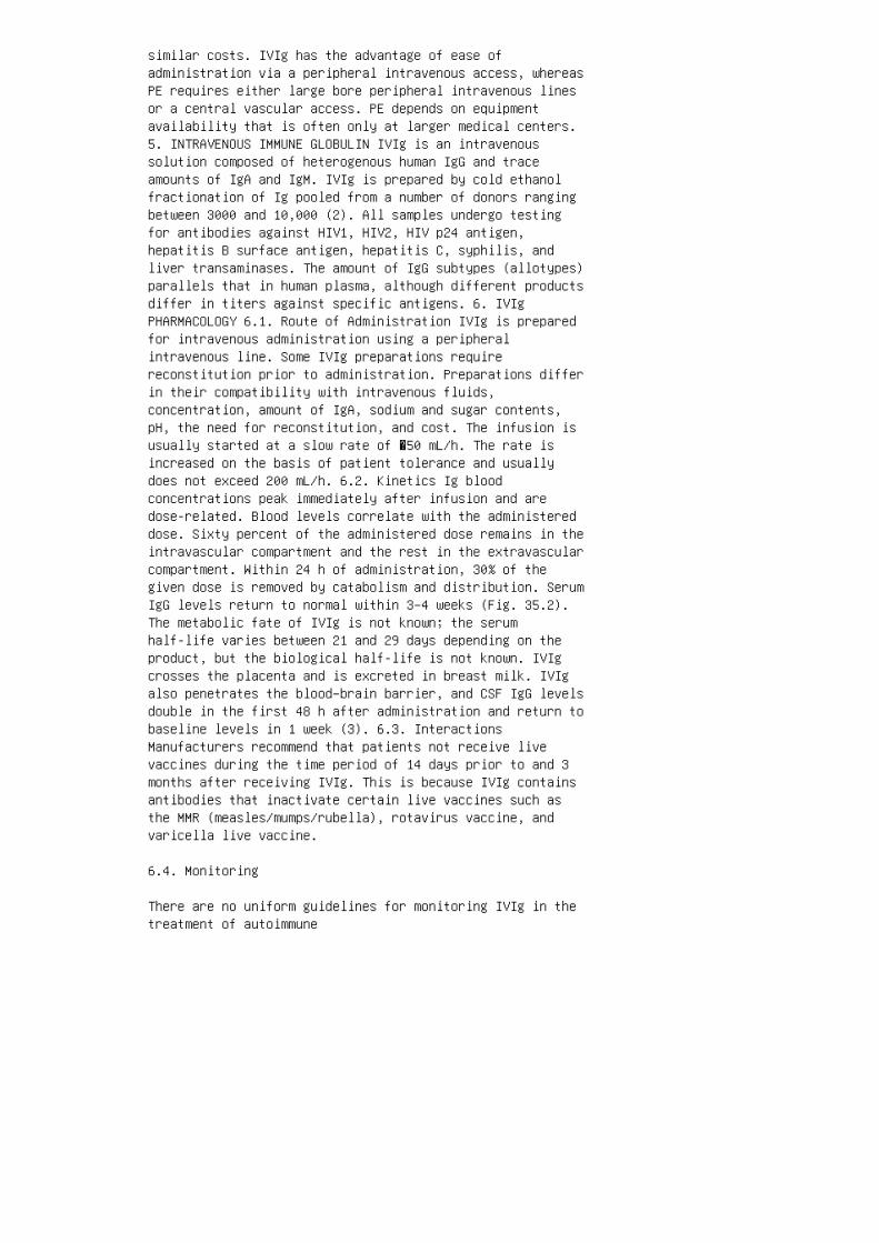
similar costs. IVIg has the advantage of ease ofadministration via a peripheral intravenous access, whereasPE requires either large bore peripheral intravenous linesor a central vascular access. PE depends on equipmentavailability that is often only at larger medical centers.5. INTRAVENOUS IMMUNE GLOBULIN IVIg is an intravenoussolution composed of heterogenous human IgG and traceamounts of IgA and IgM. IVIg is prepared by cold ethanolfractionation of Ig pooled from a number of donors rangingbetween 3000 and 10,000 (2). All samples undergo testingfor antibodies against HIV1, HIV2, HIV p24 antigen,hepatitis B surface antigen, hepatitis C, syphilis, andliver transaminases. The amount of IgG subtypes (allotypes)parallels that in human plasma, although different productsdiffer in titers against specific antigens. 6. IVIgPHARMACOLOGY 6.1. Route of Administration IVIg is preparedfor intravenous administration using a peripheralintravenous line. Some IVIg preparations requirereconstitution prior to administration. Preparations differin their compatibility with intravenous fluids,concentration, amount of IgA, sodium and sugar contents,pH, the need for reconstitution, and cost. The infusion isusually started at a slow rate of �50 mL/h. The rate isincreased on the basis of patient tolerance and usuallydoes not exceed 200 mL/h. 6.2. Kinetics Ig bloodconcentrations peak immediately after infusion and aredose-related. Blood levels correlate with the administereddose. Sixty percent of the administered dose remains in theintravascular compartment and the rest in the extravascularcompartment. Within 24 h of administration, 30% of thegiven dose is removed by catabolism and distribution. SerumIgG levels return to normal within 3–4 weeks (Fig. 35.2).The metabolic fate of IVIg is not known; the serumhalf-life varies between 21 and 29 days depending on theproduct, but the biological half-life is not known. IVIgcrosses the placenta and is excreted in breast milk. IVIgalso penetrates the blood–brain barrier, and CSF IgG levelsdouble in the first 48 h after administration and return tobaseline levels in 1 week (3). 6.3. InteractionsManufacturers recommend that patients not receive livevaccines during the time period of 14 days prior to and 3months after receiving IVIg. This is because IVIg containsantibodies that inactivate certain live vaccines such asthe MMR (measles/mumps/rubella), rotavirus vaccine, andvaricella live vaccine.
6.4. Monitoring
There are no uniform guidelines for monitoring IVIg in thetreatment of autoimmune
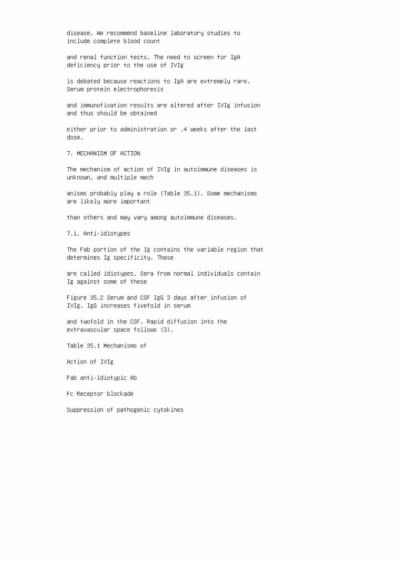
disease. We recommend baseline laboratory studies toinclude complete blood count
and renal function tests. The need to screen for IgAdeficiency prior to the use of IVIg
is debated because reactions to IgA are extremely rare.Serum protein electrophoresis
and immunofixation results are altered after IVIg infusionand thus should be obtained
either prior to administration or .4 weeks after the lastdose.
7. MECHANISM OF ACTION
The mechanism of action of IVIg in autoimmune diseases isunknown, and multiple mech
anisms probably play a role (Table 35.1). Some mechanismsare likely more important
than others and may vary among autoimmune diseases.
7.1. Anti-idiotypes
The Fab portion of the Ig contains the variable region thatdetermines Ig specificity. These
are called idiotypes. Sera from normal individuals containIg against some of these
Figure 35.2 Serum and CSF IgG 3 days after infusion ofIVIg. IgG increases fivefold in serum
and twofold in the CSF. Rapid diffusion into theextravascular space follows (3).
Table 35.1 Mechanisms of
Action of IVIg
Fab anti-idiotypic Ab
Fc Receptor blockade
Suppression of pathogenic cytokines

Inhibition of complement
Accelerated Ab clearance
Remyelination variable regions and are calledanti-idiotypes, that is, antibodies to antibodies. Incontrast to IgG from a single individual, 40% of IVIg’s IgGis in the dimeric form as a result of two IgG moleculesspecifically binding to each other at the Fab region (2).This proportion is higher with a higher number of donorsand indicates the wider variety of anti-idiotypes.Anti-idiotypic antibodies bind and neutralize circulatingautoantibodies. They also down-regulate their production byB cells. This mechanism of action plays an important rolein antibody-mediated disease like anti-myelin associatedglycoprotein (MAG) Ab neuropathy, antiGM1 Ab neuropathy,and Lambert Eaton Myasthenic Syndrome. In a small studyusing IVIg to treat pemphigus foliaceus, IVIg therapyresulted in a selective drop of autoantibody titers(anti-desmoglein 1 antibodies) (4). 7.2. Fc Receptor The Fcportions of IgG saturate surface Ig receptors onmacrophages, resulting in nonspecific macrophage Fcreceptor blockade. This leads to the inhibition ofmacrophage-mediated phagocytosis. This mechanism plays animportant role in idiopathic thrombocytopenic purpura (ITP)by inhibiting platelet uptake by macrophages. It may alsoplay a role in macrophage-mediated myelin destruction. 7.3.Complement IgG Fc portions bind complement, resulting ininability of the available C3 molecules to further activatethe remainder of the complement cascade and the formationof the membranolytic attack complex. This role of IVIg isimportant in dermatomyosits which includescomplement-mediated vascular damage, and there is a .90%disappearance of complement from the tissue after IVIgadministration in this disease (5). 7.4. CytokinesNon-specific binding of Fc portion of IgG with macrophagesinhibits the production of IL1 and TNF a. This plays a rolein controlling vascular inflammation in Kawasaki disease.Controlling the production of pro-inflammatory cytokines isan important mechanism of action in most autoimmuneprocesses. 7.5. Accelerated Antibody Clearance In vitrostudies reveal that IVIg infusion results in a drop in thelevel of endogenous IgG by �40%, 3 days after a singleinfusion. Computational studies to predict this effect inhumans reveal a possible maximal reduction by 25% 3–4 weeksafter administering a 2 g/kg dose. This prolonged andsustained effect is attributed to accelerated clearance ofIgG (6). 7.6. Remyelination In vitro experimental allergicencephalitis models show that polyclonal Ig promotesremyelination (7,8). This might explain some of the effects

of IgG in demyelinating disease. The exact mechanism ofenhancing remyelination is not known. 8. DOSE The dosegenerally used in the treatment of autoimmune disorders is2 g/kg divided over 2–5 days. This is the dose used in theinitial trial of IVIg in ITP (1). Dose–response
relationships likely vary among autoimmune disorders. Therequired maintenance dose
and frequency is not known. Dosing schedules vary, withsome clinicians using fixed
dose and variable dosing intervals and others using fixedintervals and reduced doses.
This variability arises from lack of knowledge of thebiological half-life and the likelihood
that maintenance doses vary between disease states.
9. VIRAL SAFETY
IVIg is a blood product derived from donor plasma. Donorscreening includes a medical
history and physical examination. Plasma processing takesplace when all tests prove nega
tive and after 60 days of quarantine (9). At that pointviral inactivation and removal methods
are used to eliminate possible contaminants and ensuresafety. Viral inactivation methods
vary among manufacturers and include physical methods (suchas dry heat, pasteurization)
and chemical methods (such as solvents, detergents, low pHincubation, caprylate). Viral
removal takes place by using physical methods such aspartitioning, chromatography, or fil
tration. IVIg is generally a safe product and has a goodviral safety record. Apart from the
outbreak of hepatitis C in 1992–1993, no other cases wereidentified (10,11).
10. CONTRAINDICATIONS

IVIg is contraindicated in patients with IgA deficiency.These patients carry the risk of
anaphylaxis by circulating IgE antibodies against IgA. Theincidence of IgA deficiency
is approximately 1:700, but reported anaphylactic reactionsare rare (9). Preparations
devoid of IgA are available and suitable for thesepatients. Relative contraindications
include conditions that predispose to renal toxicity or ahypercoagulable state (Table 35.2).
11. SIDE EFFECTS
Most side effects to IVIg are mild, self-limited, and easyto treat (Table 35.3). Headache is
the commonest side effect and occurs more often in patientswith a history of migraine
headaches. Others include fever, malaise, body aches, andchills. Renal failure is a serious adverse reaction to theuse of IVIg. In most cases, this is
attributed to osmolar renal tubular damage. Sucrose andmaltose-containing preparations
Table 35.2 Contraindications to the Use of IVIg
Absolute IgA deficiency
Relative Agammaglobulinemia HypogammaglobulinemiaDehydration Pregnancy Lactation Renal disease Cardiacdisease Cerebrovascular disease Diabetes mellitus areassociated with a higher frequency of renal impairment. Itis often reversible. BUN and creatinine levels return tobaseline values in �20 days. Vascular adverse reactionsinclude cardiovascular and cerebrovascular thrombosis as aresult of increased serum viscosity. Caution should betaken when treating patients with risk factors tothrombotic disease. The American Red Cross guidelinesindicate using IVIg at concentrations of 5% or less inpatients with signs of thrombosis. A low concentration andslow rate of infusion is recommended to avoid thromboticevents in high-risk patients. Anaphylactic reactions due toIgA deficiency are rare and occur at a rate of

1.3:1,000,000 transfusions of blood or blood products inthe USA. The infusion should be stopped immediately at theearliest sign of anaphylaxis. IgA depleted IVIg is asuitable alternative for these patients. Aseptic meningitisis an uncommon reaction to IVIg. This, too, is reported tooccur more frequently in patients with a history ofmigraine headaches. It is characterized by a severeheadache and meningismus. CSF studies are those of asepticmeningitis. Treatment is symptomatic. A number of rarereactions to IVIg are reported, such as hemolytic anemia(12) and cryoglobulinemic vasculitis (13). 12. INDICATIONSIVIg is generally used for three purposes: as a replacementtherapy, to treat immunodeficiency states, and to treatautoimmune disorders (Table 35.4). Table 35.3 Side Effectsof IVIg Serious Anaphylactic shock Renal tubular necrosisOsmotic nephrosis Stroke Myocardial infarction StevensJohnson syndrome Hemolytic anemia Less serious HeadacheFever Chills Diaphoresis Hypotension Nausea Vomiting Backpain Arthralgia Myalgia Aseptic meningitis Chest pain RashInjection site reactions Phlebitis Pruritus
13. THERAPEUTIC USES OF IVIg FOR PERIPHERAL NEUROPATHY
IVIg has been shown to be effective in the treatment ofautoimmune disorders of the
peripheral nerve.
13.1. Guillain–Barre´ Syndrome
GBS is a syndrome of acute areflexic quadriparesis. Themost common type is acute
inflammatory demyelinating polyradiculoneuropathy (AIDP).Other types include Fisher
syndrome and axonal GBS. A number of trials of IVIg in GBShave been conducted (Table 35.5). In the 1980s,
two large randomized controlled trials showed the efficacyof PE in treatment of AIDP
(14,15). Several randomized double blind placebo-controlledtrials comparing IVIg to
PE, confirmed that IVIg (at a dose of 0.4 g/kg/day for 5days) is equally effective as
PE (16,17). Another study compared IVIg alone, PE alone,and PE followed by IVIg.

Recovery rates in all groups were similar after 4 weeks,with no additional benefit in
the combination group (18). A small study compared IVIg and500 mg of methylprednisolone (MP) intrave
nously with IVIg alone. Although this group was notrandomized and controlled, the
group receiving both IVIg and MP showed a more rapidrecovery (19). Another small ran
domized study included PE alone, selective Ig adsorption byextracorporeal elimination, or
a combination of Ig absorption and IVIg. The combinationwas more efficacious (20). A double-blindplacebo-controlled comparison of two doses of IVIg in AIDP
patients with contraindications to PE in Ref. (21). A doseof 0.4 g/kg per day for 3
days vs. the same dose for 6 days showed a trend toward abetter outcome in the group
Table 35.4 Uses of IVIg
Immunodeficiency
Agammaglobulinemia/hypogammaglobulinemia
Bacterial infection
Bone marrow transplantation
Autoimmune disease
Guillain–Barre´ syndrome
Chronic inflammatory demyelinating polyneuropathy
Multifocal motor neuropathy
Graft vs. host disease
Dermatomyositis
Polymyositis

Kawasaki disease
Myasthenia gravis
Lambert Eaton myasthenic syndrome
Systemic lupus erythematosus
Antiphospholipid syndrome
Vasculitis
Stiff person syndrome
Multiple sclerosis
Rasmussen’s encephalitis
Intractable epilepsy in children
Idiopathic thrombocytopenic purpura T a b l e 3 5 . 5 C l in i c a l T r i a l s o f t h e U s e o f I V I g i n A I DP P r i n c i p a l a u t h o r / s t u d y g r o u p n T re a t m e n t g r o u p s O u t c o m e m e a s u r e s R es u l t s D u t c h g r o u p / v a n d e r M e c h e a n dS c h m i t z ( 1 6 ) 1 4 7 R O C T o f P E 2 0 0 – 2 5 0 mL / k g o v e r 4 – 5 d a y s v s . I V I g 0 . 4 g / k g pe r d a y f o r 5 d a y s I m p r o v e m e n t b y 1 g r ad e s o n t h e G B S F D S I V I g i s a t l e a s t a s ef f e c t i v e a s P E , p o s s i b l y m o r e e f fi ca c i o u s P E / s a n d o g l o b u l i n G B S t r i a lg r o u p ( 1 8 ) 3 8 3 M u l t i c e n t e r , R C B T o fP E a l o n e , I V I g a l o n e , P E f o l l o w e d b yI V I g T h e G B S F D S N o d i f f e r e n c e i n t h et h r e e g r o u p s B r i l e t a l . ( 1 7 ) 5 0 R O T of I V I g 0 . 4 g / k g p e r d a y f o r 4 d a y s v s . PE T i m e t o i m p r o v e o n e d i s a b i l i t y g r ad e , d i s a b i l i t y g r a d e a t 1 m o n t h , a n dt i m e t o r e a c h F D S g r a d e 1 B o t h g r o u p sd i d e q u a l l y w e l l D u t c h ( 1 9 ) 2 5 P i l o ts t u d y o f I V I g a n d M P 5 0 0 m g v s . I V I g I mp r o v e m e n t b y 1 g r a d e s o n G B S F D S I V I ga n d M P s u p e r i o r t o I V I g a l o n e H a u p t et a l . ( 2 0 ) 4 5 R a n d o m i z e d t r i a l o f P E al o n e , s e l e c t i v e I A b y e x t r a c o r p o r ea l e l i m i n a t i o n , I g a d s o r p t i o n p l u sI V I g C l i n i c a l s c o r e S e l e c t i v e I A p lu s I V I g i s s u p e r i o r t o P E a l o n e R a p h ae l e t a l . ( 2 1 ) 3 9 M u l t i c e n t e r , R D B T of I V I g 0 . 4 g / k g p e r d a y f o r 3 d a y s v s . 0
. 4 g / k g p e r d a y f o r 6 d a y s ( 3 . 8 % a l b u mi n w a s g i v e n o n 4 t h t o 6 t h d a y s i n t h e gr o u p t r e a t e d f o r 3 d a y s ) T i m e n e e d e dt o r e g a i n a b i l i t y t o w a l k 5 m w i t h a s si s t a n c e ( G B S d i s a b i l i t y g r a d e 3 ) T he 6 d a y g r o u p d i d b e t t e r ; n o t s t a t i s ti c a l l y s i g n i fi c a n t . T h e s u b g r o u p re q u i r i n g v e n t i l a t o r y a s s i s t a n c e di d b e t t e r ; s t a t i s t i c a l l y s i g n i fi ca n t N o t e : R O C T , r a n d o m i z e d o p e n c o nt r o l l e d t r i a l ; R C B T , r a n d o m i z e d c on t r o l l e d b l i n d e d t r i a l ; R O T , r a n d om i z e d o p e n t r i a l ; G B S , G u i l l a i n – B ar r e´ s y n d r o m e ; F D S , f u n c t i o n a l d i sa b i l i t y s c a l e ; P E , p l a s m a e x c h a n g e; M P , m e t h y l p r e d n i s o l o n e ; I A , I g a ds o r p t i o n .
that was treated for 6 days. This difference wasstatistically significant in patients requiring
mechanical ventilation, which comprised approximately halfof the patients. Some patients with GBS haveantiganglioside antibodies to GM1 (usually of the IgG
or IgA type). The role of these antibodies remains unclear;however, their presence
probably predicts quick reversibility of the demyelinatingprocess with early therapy. In
a nonrandomized uncontrolled study patients with GBS andanti-GM1 Ab were treated
with either IVIg or PE. The groups treated with IVIg had afaster recovery (22). IVIg
might be the treatment of choice in this subgroup of GBS.
13.1.1. GBS Variants
Variants of GBS have been less systematically studied dueto their lower incidence. The
axonal form of GBS is strongly associated with exposure toCampylobacter jejuni
infection and has a poor prognosis regardless of themodality of treatment (23). The Fisher variant is generallyconsidered a milder form with fewer patients requir

ing mechanical ventilation. A case of a 3-year-old childwith a severe course improved
with IVIg (24).
13.1.2. Treatment Failure and Relapses
Patients with GBS who do not respond to IVIg or PE areconsidered treatment failures. The
rate of treatment failure with PE and IVIg are probablysimilar. Though uncommon, GBS
can recur months or years after the initial illness.Relapses occur with similar frequencies
in patients treated with PE or IVIg (25).
13.2. Chronic Inflammatory DemyelinatingPolyradiculoneuropathy
Chronic inflammatory demyelinating polyradiculoneuropathy(CIDP) is a chronic auto
immune demyelinating polyneuropathy. Even though both AIDPand CIDP are acquired
inflammatory demyelinating neuropathies, CIDP is considereda different disease and not
simply a chronic form of GBS. Clinically, CIDP runs achronic course with sensorimotor
and/or autonomic symptoms lasting .8 weeks. Patients mayhave relapses and remis
sions. In distinction to GBS, CIDP is responsive tocorticosteroids. CIDP is also responsive
to PE, IVIg, and other immunosuppressants. Severalrandomized controlled trials showed the efficacy of IVIg inCIDP
(Table 35.6). Mendell et al. conducted a multicenter,randomized, double-blind placebo
controlled trial of IVIg in untreated patients with CIDPusing 1 g/kg per day on days 1

and 2 then 1 g/kg on day 21 compared with 5% albumin.Seventy-six percent of patients
showed improvement as early as 10 days after the firstinfusion (30). Another smaller cross
over trial comparing oral prednisolone and IVIg showed thatboth arms were equally effica
cious, with the IVIg limb showing slightly greatershort-term efficacy (31). Long-term
effects of IVIg have been studied, and two out of threeresponders to IVIg had benefits
up to 6 and 12 months after a single treatment of 0.4 g/kgfor 5 days (29).
13.2.1. CIDP in Diabetes Mellitus
CIDP may occur at a higher frequency in patients withdiabetes mellitus (DM) than in non
diabetics (32,33). There appears to be no epidemiologic orclinical differences in CIDP
between diabetics and nondiabetics (34). In an uncontrolledtrial of IVIg in patients
with diabetic demyelinating polyneuropathy, clinicallysignificant improvement was
seen in 21 out of 26 patients (35). However, diabetics aremore prone to developing T a b l e 3 5 . 6 C l i n i c a lT r i a l s o f t h e U s e o f I V I g i n t h e T r e a tm e n t o f C I D P P r i n c i p a l a u t h o r / s t u dy g r o u p n T r e a t m e n t g r o u p s P r i m a r y ou t c o m e m e a s u r e s R e s u l t s V e r m e u l e ne t a l . ( 2 6 ) 2 8 D B P C T o f I V I g 0 . 4 g / k g pe r d a y f o r 5 c o n s e c u t i v e d a y s v s . p l ac e b o ( a l b u m i n ) S i x p o i n t R a n k i n s c al e , s u m o f M R C s c a l e o f 3 a r m a n d 3 l e g mu s c l e s I V I g s u p e r i o r t o p l a c e b o D y ck e t a l . ( 2 7 ) 2 0 R S B C O T o f P E t w i c e w e ek l y f o r 3 w e e k s f o l l o w e d b y o n c e w e e kl y f o r 3 w e e k s v s . I V I g 0 . 4 g / k g o n c e aw e e k f o r 3 w e e k s f o l l o w e d b y 0 . 2 g / k go n c e a w e e k f o r 3 w e e k s . T h e w a s h o u t pe r i o d w a s a t l e a s t 6 w e e k s N e u r o p a t hy d i s a b i l i t y s c o r e ( N D S ) , s u m m a t e d

C M A P o f u l n a r , m e d i a n a n d p e r o n e a l ne r v e s B o t h m o d a l i t i e s r e s u l t e d i n st a t i s t i c a l l y s i g n i fi c a n t i m p r o v em e n t . N o d i f f e r e n c e b e t w e e n t h e t w oH a h n e t a l . ( 2 8 ) 3 0 D B P C C O T o f I V I g 0 .4 g / k g p e r d a y f o r 5 c o n s e c u t i v e d a y sv s . p l a c e b o ( 1 0 % d e x t r o s e ) . V a r i a bl e w a s h o u t p e r i o d s N D S , f u n c t i o n a lc l i n i c a l g r a d e , a n d g r i p s t r e n g t h IV I g s u p e r i o r t o p l a c e b o T h o m p s o n e ta l . ( 2 9 ) 7 D B P C C O T o f I V I g 0 . 4 g / k g p er d a y f o r 5 d a y s v s . p l a c e b o ( a l b u m i n) f o l l o w e d b y e n r o l l i n g p a t i e n t s t ha t r e l a p s e d i n t o a r m 2 : I V I g 0 . 4 g / k ge v e r y 3 w e e k s v s . p l a c e b o ( a l b u m i n )I m p r o v e m e n t i n t h r e e m e a s u r e s o f : AI , 1 0 m w a l k t i m e , e x p a n d e d M R C s u m s co r e , 9 h o l e p e g t e s t t i m e , m e a n m y o m et e r s c o r e o r t w o m u s c l e g r o u p s , a n d HM A S T h r e e p a t i e n t s r e s p o n d e d , t w o of t h e t h r e e r e s p o n d e r s r e m a i n e d s t ab l e 6 m o n t h s a n d 1 y e a r a f t e r i n f u s i on . T h e 3 r d p a t i e n t r e m a i n e d s t a b l e wi t h t h e r e g i m e n i n a r m 2 o f t h e s t u d y .M e n d e l l e t a l . ( 3 0 ) 5 0 M u l t i c e n t e r ,R D B P C T o f I V I g 1 g / k g o n d a y 1 , 2 a n d 2 1v s . p l a c e b o ( 5 % a l b u m i n ) C h a n g e i n mu s c l e s t r e n g t h f r o m b a s e l i n e t o d a y4 2 b y m a n u a l m u s c l e t e s t i n g I V I g s u pe r i o r t o p l a c e b o H u g h e s e t a l . ( 3 1 ) 24 M u l t i c e n t e r R D B C T o f o r a l p r e d n i so l o n e f o l l o w e d b y I V I g v s . I V I g f o l lo w e d b y p r e d n i s o l o n e . D o s e s : p r e d ni s o l o n e : 6 0 m g q a m f o r 2 w e e k s , 4 0 m g qa m f o r 1 w e e k , 3 0 m g q a m f o r 1 w e e k , 2 0 mg q a m f o r 1 w e e k , t h e n 1 0 m g q a m f o r 1 w ee k , I V I g : 1 g / k g p e r d a y f o r 2 d a y s o r 2g / k g i n 2 4 h , p l a c e b o ( 6 % a l b u m i n ) N ov e l d i s a b i l i t y s c a l e : I N C A T d i s a b il i t y s c a l e B o t h t r e a t m e n t a r m s e q u al l y e f fi c a c i o u s N o t e : D B P C T , d o u b le b l i n d p l a c e b o c o n t r o l l e d t r i a l ; RD B P C T , r a n d o m i s e d d o u b l e b l i n d p l ac e b o c o n t r o l l e d t r i a l ; R S B C O T , r a nd o m i z e d s i n g l e b l i n d e d c r o s s o v e r tr i a l ; D B P C C O T , D o u b l e b l i n d p l a c e bo c o n t r o l l e d c r o s s o v e r t r i a l ; N D S ,n e u r o p a t h y d i s a b i l i t y s c a l e ; C M A P, c o m p o u n d m u s c l e a c t i o n p o t e n t i a l; A I , a m b u l a t i o n i n d e x ; H M A S , H a m m e

r s m i t h m o t o r a b i l i t y s c o r e .
renal impairment. Whether diabetics should receive IVIgwith low sucrose or maltose to
prevent renal toxicity was not confirmed by clinicalstudies.
13.3. Multifocal Motor Neuropathy
Multifocal motor neuropathy (MMN) is an acquiredinflammatory demyelinating poly
neuropathy that has both similarities and differences fromCIDP (36). Patients with
MMN develop a chronic asymmetric, predominantly distal,lower motor neuron syndrome
with electrodiagnostic findings of demyelination. Thisdisorder is uncommon, but there
have been four small randomized, double-blind,placebo-controlled trials confirming the
efficacy of IVIg (37–40). Patients with MMN often haveanti-GM1 antibody (usually
IgM), but neither the presence nor the titer predictresponse to therapy.
13.4. Peripheral Neuropathy Associated with MonoclonalGammopathy
Autoimmune peripheral neuropathies may be associated withmonoclonal antibodies. In
most situations, these neuropathies are acquireddemyelinating polyneuropathies and
are considered special subtypes of CIDP.
13.4.1. Anti-MAG Ab
Anti-MAG antibody neuropathy is a chronic predominantlysensory slowly progressive
symmetric neuropathy. MAG is a protein essential in keepingthe myelin sheath tightly

wound around the axon. This neuropathy responds poorly toIVIg in no more than 18%
of patients and may be less effective than PE (41,42).
13.4.2. Anti-GM1 Ab
Anti-ganglioside GM1 antibodies are present in patientswith MMN (usually of the IgM
type) and in some patients with GBS (usually of the IgG orIgA type). Patients with
MMN respond to IVIg, but the presence of antibody does notseem to predict responsive
ness to therapy (discussed earlier). GBS associated withanti-GM1 antibodies has a good
prognosis when treated with IVIg (22).
13.4.3. Monoclonal Gammopathy of Undetermined Significance
Neuropathies associated with monoclonal gammopathy ofundetermined significance
(MGUS) are variable in clinical and electrodiagnosticfeatures. Patients often have a
slowly progressive, distal, predominantly sensory symmetricpolyneuropathy. MGUS
associated neuropathies associated with IgG and IgA aremore responsive to therapy
with IVIg than those associated with IgM (43).
13.4.4. Multiple Myeloma
Multiple myeloma (MM) is a plasma cell malignancy resultingin the production of large
amounts of monoclonal antibody. Overall, peripheralneuropathy associated with MM is
rare. Osteosclerotic myeloma (OM) is an uncommon type of MMcharacterized by sclero
tic bony lesions. Up to half of these patients have ademyelinating peripheral neuropathy
with predominantly distal symmetric motor deficits. IVIg isnot generally useful in the
treatment of OM associated neuropathy; but there is areport of a rapidly progressive
2. Dalakas MC. Mechanism of action of intravenousimmunoglobulin and therapeutic considerations in thetreatment of autoimmune neurologic diseases. Neurology1998; 51(6 suppl 5): S2–S8.
3. Dalakas MC. Mechanisms of action of IVIg and therapeuticconsiderations in the treatment of acute and chronicdemyelinating neuropathies. Neurology 2002; 59(12 suppl 6):S13–S21.
4. Sami N, Bhol KC, Ahmed AR. Influence of IVIg therapy onautoantibody titers to desmoglein 1 in patients withpemphigus foliaceus. Clin Immunol 2002; 105(2):192–198.
5. Dalakas MC et al. A controlled trial of high-doseintravenous immune globulin infusions as treatment fordermatomyositis. N Engl J Med 1993; 329(27):1993–2000.
6. Bleeker WK, Teeling JL, Hack CE. Acceleratedautoantibody clearance by intravenous immunoglobulintherapy: studies in experimental models to determine themagnitude and time course of the effect. Blood 2001;98(10):3136–3142.
7. Rodriguez M, Lennon VA. Immunoglobulins promoteremyelination in the central nervous system. Ann Neurol1990; 27(1):12–17.
8. van Engelen BG et al. Promotion of remyelination bypolyclonal immunoglobulin in Theiler’s virus-induceddemyelination and in multiple sclerosis. J Neurol NeurosurgPsychiatry 1994; 57(suppl):65–68.
9. Gelfand E. In: Dalakas M, Spath P, eds. Safety inIntravenous Immunoglobulin Therapy, IntravenousImmunoglobulins in the Third Millenium. The Parthenonpublishing groups, 2004:71–75.
10. Outbreak of hepatitis C associated with intravenousimmunoglobulin administration—United States, October1993–June 1994. Morbid Mortal Weekly Rep 1994; 43:505–509.
11. Breese J, Mast E, Coleman PJ. Hepatitis C virus

infection associated with administration of intravenousimmune globulin. A cohort study. J Am Med Assoc 1996;267:1563–1567.
12. Wilson JR, Bhoopalam H, Fisher M. Hemolytic anemiaassociated with intravenous immunoglobulin. Muscle Nerve1997; 20(9):1142–1145.
13. Odum J et al. Cryoglobulinaemic vasculitis caused byintravenous immunoglobulin treatment. Nephrol DialTransplant 2001; 16(2):403–406.
14. Plasmapheresis and acute Guillain-Barre syndrome. TheGuillain-Barre syndrome Study Group. Neurology 1985;35(8):1096–1104.
15. Efficiency of plasma exchange in Guillain-Barresyndrome: role of replacement fluids. French CooperativeGroup on Plasma Exchange in Guillain-Barre syndrome. AnnNeurol 1987; 22(6):753–761.
16. van der Meche FG, Schmitz PI. A randomized trialcomparing intravenous immune globulin and plasma exchangein Guillain-Barre syndrome. Dutch Guillain-Barre StudyGroup. N Engl J Med 1992; 326(17):1123–1129.
17. Bril V et al. Pilot trial of immunoglobulin versusplasma exchange in patients with GuillainBarre syndrome.Neurology 1996; 46(1):100–103.
18. Randomised trial of plasma exchange, intravenousimmunoglobulin, and combined treatments in Guillain-Barresyndrome. Plasma Exchange/Sandoglobulin Guillain-BarreSyndrome Trial Group. Lancet 1997; 349(9047):225–230.
19. Treatment of Guillain-Barre syndrome with high-doseimmune globulins combined with methylprednisolone: a pilotstudy. The Dutch GuillainBarre Study Group. Ann Neurol1994; 35(6):749–752.
20. Haupt WF et al. Sequential treatment of Guillain-Barresyndrome with extracorporeal elimination and intravenousimmunoglobulin. J Neurol Sci 1996; 137(2):145–149.
21. Raphael JC et al. Intravenous immune globulins inpatients with Guillain-Barre syndrome and contraindicationsto plasma exchange: 3 days versus 6 days. J NeurolNeurosurg Psychiatry 2001; 71(2):235–238.
22. Kuwabara S et al. Intravenous immunoglobulin therapy

for Guillain-Barre syndrome with IgG anti-GM1 antibody.Muscle Nerve 2001; 24(1):54–58.
23. Chowdhury D, Arora A. Axonal Guillain-Barre syndrome: acritical review. Acta Neurol Scand 2001; 103(5):267–277.24. Arakawa Y et al. The use of intravenous immunoglobulinin Miller Fisher syndrome. Brain Dev 1993; 15(3):231–233.25. Sater RA, Rostami A. Treatment of Guillain-Barresyndrome with intravenous immunoglobulin. Neurology 1998;51(6 suppl 5):S9–S15. 26. Vermeulen M. et al. Intravenousimmunoglobulin treatment in patients with chronicinflammatory demyelinating polyneuropathy: a double blind,placebo controlled study. J Neurol Neurosurg Psychiatry1993; 56(1):36–39. 27. Dyck PJ et al. A plasma exchangeversus immune globulin infusion trial in chronicinflammatory demyelinating polyradiculoneuropathy. AnnNeurol 1994; 36(6):838–845. 28. Hahn AF et al. Intravenousimmunoglobulin treatment in chronic inflammatorydemyelinating polyneuropathy. A double-blind,placebo-controlled, cross-over study. Brain 1996; 119(Pt4): 1067–1077. 29. Thompson N et al. A novel trial designto study the effect of intravenous immunoglobulin inchronic inflammatory demyelinating polyradiculoneuropathy.J Neurol 1996; 243(3):280–285. 30. Mendell JR et al.Randomized controlled trial of IVIg in untreated chronicinflammatory demyelinating polyradiculoneuropathy.Neurology 2001; 56(4):445–449. 31. Hughes R et al.Randomized controlled trial of intravenous immunoglobulinversus oral prednisolone in chronic inflammatorydemyelinating polyradiculoneuropathy. Ann Neurol 2001;50(2):195–201. 32. Sharma KR et al. Demyelinatingneuropathy in diabetes mellitus. Arch Neurol 2002; 59(5):758–765. 33. Lozeron P et al. Symptomatic diabetic andnon-diabetic neuropathies in a series of 100 diabeticpatients. J Neurol 2002; 249(5):569–575. 34. Rotta FT etal. The spectrum of chronic inflammatory demyelinatingpolyneuropathy. J Neurol Sci 2000; 173(2):129–139. 35.Sharma KR et al. Diabetic demyelinating polyneuropathyresponsive to intravenous immunoglobulin therapy. ArchNeurol 2002; 59(5):751–757. 36. Chaudhry V. Multifocalmotor neuropathy. Semin Neurol 1998; 18(1):73–81. 37.Azulay JP et al. Intravenous immunoglobulin treatment inpatients with motor neuron syndromes associated withanti-GM1 antibodies: a double-blind, placebo-controlledstudy. Neurology 1994; 44(3 Pt 1):429–432. 38. Van den BergLH et al. Treatment of multifocal motor neuropathy withhigh dose intravenous immunoglobulins: a double blind,placebo controlled study. J Neurol Neurosurg Psychiatry1995; 59(3):248–252. 39. Federico P et al. Multifocal motorneuropathy improved by IVIg: randomized, double-blind,

placebo-controlled study. Neurology 2000; 55(9):1256–1262.40. Leger JM et al. Intravenous immunoglobulin therapy inmultifocal motor neuropathy: a double-blind,placebo-controlled study. Brain 2001; 124(Pt 1):145–153.41. Dalakas MC et al. A controlled study of intravenousimmunoglobulin in demyelinating neuropathy with IgMgammopathy. Ann Neurol 1996; 40(5):792–795. 42. Gorson KCet al. Treatment experience in patients withanti-myelin-associated glycoprotein neuropathy. MuscleNerve 2001; 24(6):778–786. 43. Gorson K, Allam G, Ropper A.Chronic inflammatory demyelinating polyneuropathy: clinicalfeatures and response to treatment in 67 consecutivepatients with and without a monoclonal gammopathy.Neurology 1997; 2(48):321–328. 44. Benito-Leon J et al.Rapidly deteriorating polyneuropathy associated withosteosclerotic myeloma responsive to intravenousimmunoglobulin and radiotherapy. J Neurol Sci 1998;158(1):113–117. 45. Mowzoon N, Bradley W. Successfulimmunosuppressant therapy of severe progressive cerebellardegeneration and sensory neuropathy. J Neurol Sci 2000;1(178):63–65. 46. Newburger JW et al. The treatment ofKawasaki syndrome with intravenous gamma globulin. N Engl JMed 1986; 315(6):341–347. 47. Harada K. Intravenousgamma-globulin treatment in Kawasaki disease. Acta PaediatrJpn 1991; 33(6):805–810.
48. Boletis JN et al. Intravenous immunoglobulin comparedwith cyclophosphamide for proliferative lupus nephritis.Lancet 1999; 354(9178):569–570.
49. Levy Y et al. A study of 20 SLE patients withintravenous immunoglobulin–clinical and serologic response.Lupus 1999; 8(9):705–712.
50. Levy Y et al. Serologic and clinical response totreatment of systemic vasculitis and associated autoimmunedisease with intravenous immunoglobulin. Int Arch AllergyImmunol 1999; 119(3):231–238.
51. Jayne DR et al. Intravenous immunoglobulin forANCA-associated systemic vasculitis with persistent diseaseactivity. Q J Med 2000; 93(7):433–439.
52. Levy Y et al. Marked improvement of Churg-Straussvasculitis with intravenous gammaglobulins. South Med J1999; 92(4):412–414.
53. Lucas GF et al. Effect of IVIgG treatment on fetalplatelet count, HPA-1a titre and clinical outcome in a caseof feto-maternal alloimmune thrombocytopenia. Br J Obstet

Gynaecol 2002; 109(10):1195–1198.
54. Yaginuma Y, Kawamura M, Ishikawa M.Landry-Guillain-Barre-Strohl syndrome in pregnancy. JObstet Gynaecol Res 1996; 22(1):47–49.
55. Yamada H et al. Massive intravenous immunoglobulintreatment in pregnancy complicated by Guillain-BarreSyndrome. Eur J Obstet Gynecol Reprod Biol 2001;97(1):101–104.
56. Zafeiriou DI et al. Single dose immunoglobulin therapyfor childhood Guillain-Barre syndrome. Brain Dev 1997;19(5):323–325.
57. Uldry PA, Bogousslavsky J, Regli F. Guillain-Barresyndrome in a 93-year-old woman: rapid improvement ofneurologic function following intravenous immunoglobulin.Schweiz Arch Neurol Psychiatr 1991; 142(4):301–305.
58. Rana SS, Rana S. Intravenous immunoglobulins versusplasmapheresis in older patients with Guillain-Barresyndrome. J Am Geriatr Soc 1999; 47(11):1387–1388.
36
Treatment of Immune-Mediated
Peripheral Neuropathy
Orly Vardeny and A. Gordon Smith
University of Utah, Salt Lake City, Utah, USA
Abstract 640
1. Prednisone 641 1.1. Efficacy 641 1.2. Dosing 641 1.3.Pharmacodynamics 641 1.4. Pharmacokinetics 641 1.5. SideEffects 642 1.5.1. Central Nervous System 642 1.5.2.Hypertension 643 1.5.3. Electrolyte Disturbances 643 1.5.4.Osteoporosis 643 1.5.5. Cutaneous Effects 643 1.5.6.Glucose Intolerance 643 1.5.7. Aseptic Necrosis 643 1.5.8.Peptic Ulcer Disease 644 1.5.9. Infections 644 1.5.10.Ocular Effects 644 1.5.11. Adrenal Suppression 644 1.6.Pregnancy Issues 644
1. PREDNISONE
1.1. Efficacy

Prednisone was demonstrated to be effective in a case of arelapsing neuropathy that failed
to respond to placebo (1). Subsequent open-label, andretrospective reviews, and one con
trolled trial have confirmed the efficacy of prednisone inchronic inflammatory demyeli
nating polyradiculopathy (CIDP) (2–6). Prednisone isineffective for acute inflammatory demyelinatingpolyradiculo
pathy (AIDP) and multifocal motor neuropathy, even thoughthey are felt to be immune
mediated (7–11).
1.2. Dosing
There are little data on optimal dosing, duration of highdose, and taper rates for CIDP
(12). Most reports start with doses of �60 mg/day, and theonly controlled trial started
with 120 mg QOD. High doses are usually maintained for 4weeks and then converted
to alternate-day schedule before tapering. Taper schedulesvary markedly and are com
monly adjusted on the basis of patient response, withtemporary increases if the patient’s
condition relapses (2,4,6,13).
1.3. Pharmacodynamics
The therapeutic mechanism of prednisone is poorlyunderstood, and there are multiple
possible sites of action. Corticosteroids inhibit T-cellproliferation, T-cell dependent
immunity, and cytokine gene transcription (including theIL-1, IL-2, IL-6, interferon g,
and tumor necrosis factora genes). They prevent macrophageactivation, antigen presen

tation, IL-1 production, and inhibit their function aseffector cells. Corticosteroids inhibit
the generation of cytotoxic T-cells. They have a lessmarked effect on B-cell function.
(14–16).
1.4. Pharmacokinetics
Prednisone is a pro-drug, and equilibrium strongly favorsthe formation of prednisolone,
the active agent (17). Conversion is achieved largely onfirst-pass metabolism through the
liver (18). Small variations in the rate of prednisoloneavailability are unlikely to be of
clinical significance because of its multiple sites ofaction and long-term immunologic
effects (19,20). Prednisolone is bound reversibly toalbumin and to alpha-1 glycoprotein, a corticos
teroid-binding globulin (transcortin). Protein binding isconcentration-dependent and is
diminished in hypoproteinemic states. Prednisolone isprimarily eliminated by hydroxy
lation and conjugation, and only 2.5% of a dose ofprednisone appears in the urine in
its unmetabolized form. The mean half-life of prednisoloneis 2.6–5 h (21). There is cir
cadian variation in the clearance of prednisolone and isresponsible for a greater effect per
dose when taken in the morning. This is due to highermorning cortisol concentrations,
resulting in a competitive decrease in the metabolism ofprednisolone, leading to its
decreased clearance (22). These circadian effects favor thechoice of morning doses

rather than evening doses (22). Enzyme inducing drugs,including barbiturate agents, phenytoin, and rifampin, sig
nificantly increase the metabolic clearance ofcorticosteroid agents. The steroid dosage
should be increased during concomitant administration(18,23,24). 1.5. Side Effects The medical complications ofglucocorticoid therapy are listed in Table 36.1 (12). Therisks are theoretically doseand time-dependent.Alternate-day dosing is felt to reduce side effects, butcontrolled trials are lacking (12,25). Doses up to 100 mgof prednisone daily may be taken for up to 3 weeks withoutsignificant risk for major adverse effects. It is notpossible to stipulate a daily dose at which the risk ofside effects is nonexistent. In adults, it is likely that10 mg daily of prednisone will eventually lead to some sideeffects (26). Even, smaller daily doses of 7.5 mg, if takenin the long-term, are still associated with complicationsin the elderly (26). 1.5.1. Central Nervous System Mildeuphoria, depression, and insomnia may occur within thefirst 1–2 weeks of therapy. Other central nervous systemside effects include insomnia, personality changes, mania,and hallucinations. A dose reduction results in abatementwithin 2 weeks in 50% of patients (27). Severe depressionand psychosis rarely occurs at doses of .40 mg/day (5%risk) (28,29). Preexisting psychological problems increasethe risk for adverse Table 36.1 Medical Complications ofCorticosteroids Early Insomnia Emotional lability Enhancedappetite and weight gain With co-morbidities HypertensionDiabetes mellitus Peptic ulcer disease Acne vulgaris Withsustained treatment Cushingoid habitusHypothalamic-pituitary–adrenal suppression Infectiondiathesis Osteonecrosis Myopathy Impaired wound healingInsidious and delayed (dose related) Osteoporosis Skinatrophy Cataracts Atheroscerlosis Growth retardation Fattyliver Rare and unpredictable Psychosis Pseudotumor cerebriGlaucoma Epidural lipomatosis Pancreatitis Source: Adaptedfrom Boumpas (14).
psychiatric reactions (30). If there is no therapeuticalternative, steroid psychosis can be
managed with phenothiazines or lithium carbonate.
1.5.2. Hypertension
Fluid retention, enhancement of the vasoconstrictor effectsof endogenous substances, and
increased concentrations of renin substrate lead tohypertension (31).
1.5.3. Electrolyte Disturbances
Prednisone increases the distal renal tubular reabsorptionof sodium in exchange for
potassium, hydrogen, and ammonium ions, leading tohypernatremia and hypokalemic
alkalosis in some individuals (32).
1.5.4. Osteoporosis
Osteoporosis occurs in up to 50% of patients on long-termglucocorticoid therapy and is
due to decreased bone formation through direct inhibitionof osteoblastic activity and
increased bone resorption. Resorption is a consequence ofsecondary hyperparathyroidism
caused by corticosteroid-induced hypercalciuria andinhibition of intestinal calcium
absorption, as well as increased urinary calcium excretion(33). Trabecular bone in the
spine and ribs is preferentially affected (34).Postmenopausal women and elderly or
immobilized patients are at greater risk. Bone loss is moreprominent in the early stages
of corticosteroid therapy. Alternate-day treatment appearsto have no advantage over
daily use (35). Guidelines for preventing steroid-inducedosteoporosis are similar to idiopathic
osteoporosis and focus on exercise and adequate diet.Recommendations for calcium
and vitamin D intake (dietary and supplemental) are1200–1500 mg/day of calcium
and 800 IU of vitamin D. Higher vitamin D doses are notrecommended for routine use

because of propensity for calcium urolithiasis (36).Baseline bone density scans are rec
ommended for patients utilizing corticosteroids forlong-term therapy. If osteopenia or
osteoporosis is present, treatment with bisphosphanates iswarranted (37–39).
1.5.5. Cutaneous Effects
Easy bruising, purpura, and ecchymoses are common in olderpatients on long-term cor
ticosteroid treatment. These complications are attributedto diminished phagocytosis of
extravasated blood and changes in connective tissue.Hirsutism occurs in �10% of
patients on long-term therapy (31,32).
1.5.6. Glucose Intolerance
Corticosteroids impair carbohydrate metabolism byincreasing hepatic gluconeogenesis
and by decreasing peripheral tissue utilization of glucose.There is also evidence for a
component of insulin resistance (40). Steroid-induceddiabetes is usually mild and can
be managed by diet and oral hypoglycemic agents. Overtdiabetes is unusual and
usually resolves with discontinuance of steroid.Individuals with preexisting diabetes
are at greatest risk for developing diabetes (40,41).
1.5.7. Aseptic Necrosis
Corticosteroids can cause aseptic necrosis of the femoralhead. Fat microemboli are
thought to occlude subchrondral end-arterioles leading tobone-cell death (42). Necrosis is heralded by pain anddiagnosed by MRI showing a lucent area between the
collapsed bone and the overlying cartilage (42).Hyperuricemia, alcoholism, hyperlipidemia, and polycythemiaare predisposing risk factors (42). 1.5.8. Peptic UlcerDisease There is a small but significant associationbetween corticosteroid therapy and peptic ulceration (43).Multivariate analysis demonstrated that risk factors fordeveloping ulcers are previous history of peptic ulcerdisease, cumulative prednisone dose of .1000 mg or therapy.30 days, and concurrent nonsteroidal anti-inflammatorydrug (NSAID) therapy (43,44). Prophylaxis with a histamineH2-receptor antagonist is appropriate if two or more riskfactors are present or if the patient is receivingconcurrent treatment with a NSAID (44). 1.5.9. InfectionsCorticosteroids impair cell-mediated immunity and inhibitmonocyte responsiveness to chemotactic factors. However,the degree of increased susceptibility to infections isdifficult to assess on the basis of clinical trials but islikely low (45). The risk may be higher for patientsreceiving corticosteroids in combination with otherimmunosuppressant agents. 1.5.10. Ocular Effects The riskof posterior subcapsular cataracts increases withcorticosteroid therapy .1 year or doses.10 mg of prednisoneper day. Other ocular effects include increased intraocularpressure, more often in patients with myopia or diabetesmellitus (46,47). 1.5.11. Adrenal Suppression Interruptionof adrenal function occurs after a few days of high-dosecorticosteroid therapy, and long-term daily therapy maysuppress hypothalamic–pituitary–adrenal function for 9–12months after cessation of treatment (48,49). However,examples of clinical adrenal suppression are rare.Traditional taper schedules appear to be reasonable, butsupplemental administration of corticosteroid therapyduring physiological stress is reasonable (50). 1.6.Pregnancy Issues The risk for human teratogenesis is lowwith corticosteroids. This is due to inactivation ofprednisolone in the placenta, allowing only 10% of theactive drug to reach the fetus (51). 2. AZATHIOPRINE 2.1.Efficacy Only one randomized controlled study comparingprednisone alone or with azathioprine in CIDP has beenperformed, and it did not show a benefit to additionalazathioprine (52). However, there are uncontrolled trialssupporting its use as a steroid-sparing agent (2,53). Anuncontrolled trial in AIDP yielded inconclusive results(54).
2.2. Dosing
Doses of 1.5–3 mg/kg per day in divided doses to minimizegastrointestinal side effects are

common, but optimum dosing has not been established forCIDP. A mild leukopenia has
been used as a therapeutic guide, although it is notrequired for a beneficial effect (2,55).
2.3. Pharmacodynamics
Azathioprine, a purine analog, inhibits cellularproliferation by serving as a purine anta
gonist. Its greatest effect is on rapidly dividing celllines such as the bone marrow.
2.4. Pharmacokinetics
Azathioprine has a half-life of 4.5–5 h. Inactivemetabolites are excreted in the urine (56).
It is metabolized by xanthine oxidase, and concomitantadministration of allopurinol, a
xanthine oxidase inhibitor, delays its activation,potentially causing bone marrow suppres
sion. The dose of azathioprine should be reduced toone-fourth when administered
together with allopurinol (57).
2.5. Side Effects
2.5.1. Hematologic
Bone marrow suppression can occur at any dose and timeduring therapy, although it is
most common 7–14 days after the initiation of therapy (15).Mild leukopenia and macrocytosis are the most commonly seenside effects, and
occasionally, anemia, thrombocytopenia, and pancytopeniamay be noted. Macrocytosis
correlates poorly with the dose of azathioprine, andhepatic enzymes, folate, and B12
levels are usually normal (58–60). Generally, even mildleukopenia should be monitored closely. The dose should be

lowered when WBC falls below 4000/mm, and therapy should bediscontinued when
counts reach 2500/mm, or if the absolute neutrophil countfalls below 1000/mm (61).
Therapy may be re-initiated after 2–3 weeks provided thepractitioner monitors therapy
closely.
2.5.2. Hepatotoxicity
Elevated alkaline phosphatase and total bilirubin levelsare markers of azathioprine hepato
toxicity, but elevations in AST and ALT also occur, usuallyearly in the course of therapy.
Levels more than three times the upper limit of normalshould prompt discontinuation of
therapy. The etiology of hepatic dysfunction has beendifficult to ascertain, but enzymes
return to normal with discontinuation of therapy. Severehepatic toxicity leading to
liver failure is very rare (62).
2.5.3. Gastrointestinal
Nausea and vomiting are common side effects, particularlyat the onset of therapy with
azathioprine. Usually, these side effects are notbothersome enough to discontinue
therapy, and they generally abate with time. Increasing thenumber of daily doses and
taking azathioprine after meals often abolishes thisdiscomfort (63). An idiosyncratic reac
tion of azathioprine occurs in �15% of patients and isheralded by nausea and vomiting.
Rechallenge is rarely successful (63–65). 2.5.4. CarcinomaThe degree to which azathioprine increases the risk ofmalignancy remains unclear given the underlying disorder
may also increase risk (63). Neoplasias reported includetransitional cell carcinoma of the bladder, adrenocorticalcarcinoma, acute leukemia, and cervical atypia in women.2.6. Pregnancy Issues Active metabolites may cross theplacenta (66), and there are reports of chromosomalaberrations and neonatal lymphopenia and thrombocytopeniain children born to women receiving azathioprine (67).Small amounts appear in breast, but harmful effects havenot been reported (67). 3. CYCLOPHOSPHAMIDE 3.1. EfficacyEfficacy in CIDP has been limited to open-label studies.Combinations of oral pulse cyclophosphamide and prednisonehave been effective in CIDP associated with a monoclonalgammopathy of undetermined significance (68). A goodresponse to monthly pulse IV cyclophosphamide treatment hasbeen reported in patients refractory to other treatmentmodalities (69). Cyclophosphamide (both oral andintravenous) has been used with success in multifocal motorneuropathy (8) and peripheral nerve vasculitis (70). 3.2.Dosing Doses range from 2 mg/kg orally daily for 6–12months (71) to 1 g/m 2 intravenously monthly for 6 months(69). Another regimen is 200 mg/kg intravenously over 4days accompanied by forced diuresis and mesna (72). Pulsedosing is better tolerated. 3.3. PharmacodynamicsCyclophosphamide covalently bonds with nucleophiliccellular substances (alkylation) leading to disruption ofthe nucleic acids of DNA and abnormal cell replication(73). Pharmacologic activity requires metabolism by hepaticmicrosomal enzymes to form the active derivatives includingphosphoramide mustard. The effects of cyclophosphamide uponimmunologically competent cells include depletion oflymphocytes and changes in function of remaining cells. Itis toxic to lymphocytes and reduces B-lymphocyte function(74,75). Cyclophosphamide has immunopotentiating propertieswhich include selective inhibition of T-suppressor cells(76). Cyclophosphamide inhibits the inflammatory responsein addition to its immunosuppressant actions (77). 3.4.Pharmacokinetics Oral cyclophosphamide is metabolized bythe liver into active metabolites (phosphoramide mustard)and inactive metabolites (4-keto cyclophosphamide,carboxyphospramide, acroelein). Inactive metabolites areexcreted in the urine (56). Acrolein contributes tohemorrhagic cystitis (78).
3.5. Side Effects
3.5.1. Hematologic
Cyclophosphamide causes a dose-related leukopenia, with thenadir between 7 and 14

days following treatment and recovery between 21 and 25days (79). Thrombocytopenia
may rarely occur.
3.5.2. Gastrointestinal
Nausea and vomiting are prominent, especially withintravenous therapy, due to direct
stimulation of the chemoreceptor trigger zone in themedulla (80). Individual patient tol
erance varies greatly with psychological preconditioningplaying a large role (80). Oral
cyclophosphamide is better tolerated after a mealconsisting of cold food.
3.5.3. Hair
Scalp hair loss occurs in 20–50% of patients, and axillary,extremity, and pubic hair loss
may occur with long-term therapy. Hair loss occurs within1–2 weeks and is maximal at
1–2 months and may be minimized by gentle scalp careincluding short-term tourniquets
or ice bag application. Alopecia is generally reversiblewith cessation of therapy.
Regrowth may be preceded by unusual texture and differentshade but often returns to
normal after therapy ends (79).
3.5.4. Genitourinary
Hemorrhagic cystitis occurs in up to 20–30% of subjectsreceiving cyclophosphamide due
to acrolein. Aggressive hydration prior to cyclophosphamidetherapy reduces accumu
lation (79). Sodium-2-mercaptoethane sulfonate (mesna) hasbeen shown to reduce the
risk of hemorrhagic cystitis. There is an increased

frequency of bladder fibrosis and car
cinoma, which may be unrelated to cystitis (79).
3.5.5. Carcinoma
The most ominous side effect is an elevated long-term riskof cancer including transitional
cell bladder cancer and lymphoma. Risk is related to thecumulative dose over time and
previous exposure to cytotoxic therapy (79).
3.5.6. Pulmonary
Interstitial pneumonitis and pulmonary fibrosis are rarebut can be life-threatening (81,82).
3.5.7. Infections
Leukopenia and granulocytopenia, especially during thenadir, can predispose to infections.
3.6. Pregnancy Issues
The frequency of infertility increases with the duration oftherapy. In postpubertal males,
cyclophosphamide causes a selective inhibition ofspermatogenesis, with little or no effect
on testosterone production (56,79). Postpubertal femalescan experience a medication
induced menopause associated with decreased serum andurinary estrogen levels and
increased levels of urinary gonadotrophins. There is anabsence of ova, loss of follicular
maturation, and fibrosis. Although there have been reportsof renewed fertility in males and females followingcessation of therapy (especially after short courses ofcyclophosphamide), the loss of fertility is usuallypermanent. If cyclophosphamide is used before the onset ofpuberty, testicular and ovarian tissues generally matureand function normally. Patients of childbearing age may beoffered sperm or egg banking prior to initiation oftherapy. It is currently unclear whether cyclophosphamide

is teratogenic. Its use during the second and thirdtrimesters does not appear to be associated with neurologicabnormalities or birth defects, although transientinfantile bone marrow suppression has been reported (51).Cytotoxic material may also appear in the breast milk andmay induce leukopenia in breast-fed infants. 4.MYCOPHENOLATE MOFETIL 4.1. Efficacy Mycophenolate mofetilis commonly used to prevent organ rejection in transplantpatients. Data from neuromuscular diseases are scarce.Small case series with CIDP have shown moderateimprovements in strength and the ability to reduce steroidrequirements (83,84). 4.2. Dosing In neuromuscular disease,the common starting dose is 500 mg daily, with an increaseto 1 g twice daily over 2–3 weeks (83). In renal transplantpatients, higher doses do not result in better outcomes andoften lead to an increased incidence of adverse effects.Dosages should be interrupted or reduced in patients withan absolute neutrophil count ,1.3 10 3 /mm 3 . 4.3.Pharmacodynamics Mycophenolate mofetil inhibits theproliferation of B and T lymphocytes throughnoncompetitive, reversible inhibition of inosinemonophosphate dehydrogenase, a key enzyme in the de novosynthetic pathway of guanine nucleotides. There are twopathways for purine biosynthesis, a de novo pathway and asalvage pathway. Lymphocytes can only use the de novopathway, whereas other cells use both pathways.Mycophenolate blocks the synthesis of guanosine andenhances the synthesis of adenosine, resulting ininhibition of purine synthesis only in lymphocytes(16,85–87). 4.4. Pharmacokinetics Mycophenolate mofetil ishydrolyzed in the liver to its biologically activemetabolite, mycophenolic acid (MPA). MPA subsequentlyundergoes glucuronidation in the liver and kidney to forman inactive metabolite which is excreted in the urine andbile. The intestinal microfloral B-glucuronidasedeconjugates the inactive metabolite in the GI tract toform MPA, leading to a second plasma peak at 6–12 h. MPA ishighly protein bound to albumin and displays an eliminationhalf-life of �17 h. Severe renal impairment decreasesbinding of MPA to albumin, therefore, increasing its freefraction in serum. Studies of drug interactions withmycophenolate mofetil are limited. The absorption ofmycophenolate mofetil is decreased with administration ofcholestyramine and antacids
containing magnesium and aluminum; therefore, mycophenolateshould not be simul
taneously administered with these agents (85).

4.5. Side Effects
Adverse effects are mild and rarely require discontinuationof therapy. The most common
adverse effects seen at dose of 1 g twice daily in renaltransplant patients are gastrointes
tinal (diarrhea, abdominal pain, nausea, dyspepsia,vomiting), hematologic (leukopenia,
anemia, thrombocytopenia, pancytopenia), and opportunisticinfections (CMV, herpes
simplex, candida). Lymphomas were rare (0.6%) and occurredless frequently compared
with rates seen with azathioprine. Gastrointestinalcomplaints may respond to a dose div
ision of three or four times per day. Hematologic adverseeffects generally reverse within a
week of discontinuation.
5. CYCLOSPORINE
5.1. Efficacy
There are no controlled studies in CIDP. The largest caseseries included an initial high
dose of 3–10 mg/kg per day, which was tapered to amaintenance dose 2–5 mg/kg per
day. Most patients had an improvement by at least onefunctional grade or a reduction
in annual relapse rates. Side effects were common andincluded nephrotoxicity, hyperten
sion, nausea, hirsutism, and edema, which are consistentwith known limiting side effects
of cyclosporine (88–90).
5.2. Dosing
Common starting doses are 3–5 mg/kg per day, withmaintenance doses of 2 mg/kg per
day with periodic serum concentration monitoring. Empiricdata indicate doses.7 mg/kg
per day are associated with higher incidence of sideeffects (89). The dose of cyclosporine
should be divided to limit toxicity.
5.3. Pharmacodynamics
The mechanism of action depends on the formation of aheterodimeric complex with
cyclophilin. The complex binds calcineurin and inhibits itsphosphatase activity,
thereby inhibiting de novo expression of nuclear regulatoryproteins and helper T
cell activation genes encoding for IL-2. This results ininhibition of the induction
phase of the immune response in a dose-related fashion byblocking gene transcription
and production of IL-2 by helper T-cells, preventingIL-2-driven clonal expansion. The
combination of protein kinase C activation and increasedintracellular calcium ions is
responsible for full T-cell activation. Cyclosporine mayinterfere with these calcium
mediated pathways. Reduced IL-2 expression inhibitsproduction of a wide range of
other cytokines, including gamma interferon, thus blockingrecruitment and activation
of macrophages (16,91).
5.4. Pharmacokinetics
Oral cyclosporine absorption exhibits large intraandinter-patient variability. Low
absorption may be due to the presence of cytochrome P-450enzymes and P-glycoprotein (a transport protein responsible

for preventing drugs from penetrating the cell) in thegastrointestinal tract. Absorption also varies with type ofcyclosporine formulation. Fatty food decreases the areaunder the curve; therefore, cyclosporine should be taken ona consistent basis with regard to food. Cyclosporine is 90%protein bound primarily to lipoproteins. It undergoesextensive hepatic metabolism through the CYP 3A4 enzyme,with minor gut and kidney metabolism. It is widely excretedthrough bile and displays a serum half-life of 8 h (range5–15 h). Cyclosporine is an inhibitor of 3A4 andP-glycoprotein, therefore, drugs that are substrates forthese display altered concentrations. Both inhibitors andinducers of the CYP 3A4 enzyme and P-glycoproteinsignificantly alter cyclosporine levels. Increasedcyclosporine concentrations occur with inhibitors of 3A4such as antifungal agents (ketoconazole, fluconazole,itraconazole), antibiotics (clarithromycin, erythromycin),selective serotonin re-uptake inhibitors (fluvoxamine,sertraline), calcium channel blockers (diltiazem,verapamil), HMG CoA reductase inhibitors (atorvastatin,simvastatin, losartan), and grapefruit juice. Decreasedcyclosporine levels occur during co-administration withinducers of 3A4 or P-glycoprotein such as carbamazepine,phenobarbital, phenytoin, rifamin, and St. John’s wort(92,93). 5.5. Side Effects Nephrotoxicity and hypertensionare the most serious side effects. Patients withpreexisting moderate or severe renal impairment should beexcluded from therapy; serum creatinine, creatinineclearance, and cyclosporine levels should be monitored andan effort made to maintain the lowest effective dose.Nausea, edema, and hirsutism are less serious and did notlead to discontinuation of therapy in clinical studies.5.6. Pregnancy Issues Cyclosporine is extensively excretedin breast milk and should therefore be avoided duringbreastfeeding (94). Reviews of case series of pregnanciesin women receiving cyclosporine after liver transplantationfound no increased risk of congenital malformations ormaternal complications (95,96). Development did not appearto be adversely affected in the offspring (children up to 8years old), although longer-term consequences are notknown. In several case reports, cyclosporine use aftertransplantation did not result in fetal anomalies (51,67).6. RITUXIMAB A few case series of rituximab therapy inanti-MAG and other antibody-related polyneuropathies haveprovided encouraging results (97–99) with few side effects.The dose of rituximab used is 375 mg/m 2 weekly for 4 weeksand may be continued once every 10 weeks thereafter (98).Pretreatment with acetaminophen and diphenhydramine limitsinfusion-related side effects and reactions. Rituximab is achimeric mouse–human monoclonal antibody that specifically

binds the surface membrane protein CD20 on B-lymphocytes,thus eliminating B-cells from the circulation. It inducesantibody-dependent cell and complement-mediatedcytotoxicity in these cells (100). The half-life rangesfrom 59 h (after the first infusion) to 174 h (after thefourth infusion). Rituximab is detected in serum up to 3–6months after completion of therapy. As its effects on thefetus and infant are unknown, it should be avoided duringpregnancy and breastfeeding. Common side effects arerelated to infusion and include hypotension, nausea, vomit
ing, rash, fever, and chills. Others include anemia andthrombocytopenia, necessitating
monitoring complete blood counts with differentials atregular intervals throughout
therapy.
7. OTHER IMMUNOSUPPRESSANT AGENTS
A number of other immunosuppressive agents, includingmethotrexate and interferons,
have been given to patients who are refractory toconventional drugs. All data are from
small trials and case reports. Methotrexate does not appearto be effective in immune
mediated neuropathies. Both interferon beta (101,102) andinterferon alpha (103) have
been used in neuropathy with some success (104). Table 36.2delineates appropriate monitoring parameters as well asmanagement of
common complications from immunosuppressant agents.
Table 36.2 Monitoring and Management of ImmunosuppressantTherapies
Agent Complication Monitoring/management
Corticosteroids Adrenal suppression Taper slowly afterprolonged therapy Fluid/electrolyte disturbances Monitorelectrolytes Cataracts Regular eye exams Peptic ulcer GIprotection H2 blockers Proton pump inhibitors OsteoporosisPeriodic bone density scans Bisphosphanates for treatmentor prevention Diabetes mellitus Periodic fasting/random

blood glucose monitoring
Azathioprine Leukopenia, thrombocytopenia Monitor WBC andadjust dose Liver toxicity Monitor LFTs D/C if 2X uppernormal limit or 3X baseline levels
Cyclosporine Nephrotoxicity Therapeutic drug monitoringMonitor serum creatinine Evaluate drug interactions
Cyclophosphamide Neutropenia Monitor WBC Periodic dosecycling Gastrointestinal Administer at bedtime withsedative Administer oral therapy with food Utilizeantiemetic therapy Urotoxicity Administer with mesna Properhydration Periodic urine cytology
Mycophenolate
mofetil Neutropenia Monitor WBC Gastrointestinal Divideoral doses
24. Leger U, Benet L. Marked alterations in prednisoloneelimination for women taking oral contraceptives. ClinPharmacol Ther 1982; 31:243.
25. Frey F, Ru¨egsegger M, Frey B. The dose-dependentsystematic availability of prednisone: one reason for thereduced biological effect of alternate-day prednisone. Br JClin Pharmacol 1986; 21:183–189.
26. Thomas TPL. The complications of systemiccorticosteroid therapy in the elderly. Gerontology 1984;30:60–65.
27. Silva RG, Tolstunov L. Steroid-induced psychosis:report of case. J Oral Maxillofac Surg 1995; 53:183–186.
28. Peet M, Peters S. Drug-induced mania. Drug Safe 1995;12:146–153.
29. Hall RC, Popkin MK, Stickney SK, Gardner ER.Presentation of the steroid psychoses. J Nerv Ment Dis1979; 167:229–236.
30. Dujorne CA, Azarnoff DL. Clinical complications ofcorticosteroid therapy. Med Clin N Am 1973; 57:1331–1342.
31. Kida-Kimble MA, Young L, eds. Applied Therapeutics: TheClinical Use of Drugs, 5th ed. Vancouver, WA: AppliedTherapeutics, Inc., 1992.

32. David DS, Greico MH, Cushman P. Adrenal glucocorticoidsafter 20 years: a review of their clinically relevantconsequences. J Chron Dis 1970; 22:637–711.
33. Baylink DJ. Glucocorticoid-induced osteoporosis. N EnglJ Med 1983; 309:306–308.
34. Lukert BP, Raisz LG. Glucocorticoid-inducedosteoporosis: pathogenesis and management. Ann Intern Med1990; 112:352–364.
35. Gluck OS, Murphy WA, Hahn TJ, Hahn B. Bone loss inadults receiving alternate day glucocorticoid therapy: acomparison with daily therapy. Arthritis Rheum 1981;24:892–898.
36. Buckley LM, Leib ES, Cartularo KS, Vacek PM, Cooper SM.Calcium and vitamin D3 supplementation prevents bone lossin the spine secondary to low-dose corticosteroids inpatients with rheumatoid arthritis: a randomized,double-blind, placebo-controlled trial. Ann Intern Med1996; 125:961–968.
37. Adachi JD, Olszynski WP, Hanley DA et al. SeminArthritis Rheum 2000; 29(4):228–251.
38. Saag KG, Emkey R, Schnitzer TH et al. Alendronate forthe prevention and treatment of glucocorticoid-inducedosteoporosis. N Engl J Med 1998; 339:292–299.
39. Homik J, Cranney A, Shea B et al. Bisphosphonates forsteroid-induced osteoporosis (Cochrane Review). TheCochrane Library 2000; (Issue 2).
40. Braithwaite SS, Barr WG, Rahman A, Quddusi S. Managingdiabetes during glucocorticoid therapy: how to avoidmetabolic emergencies? Postgrad Med 1998;104:163–166,171,175–177.
41. Hoogwerf B, Danese RD. Drug selection and themanagement of corticosteroid-related diabetes mellitus.Rheum Dis Clin N Am 1999; 25(3):489–505.
42. Richards M, Santiago M, Klaustermeyer B. Asepticnecrosis of the femoral head in corticosteroid-treatedpulmonary disease. Arch Intern Med 1980; 140:1473–1475.
43. Messer J, Reitman D, Sacks HS, Smith H Jr, Chalmers TC.Association of adrenocorticosteroid therapy and pepticulcer disease. N Engl J Med 1983; 309:21–24.

44. Twycross R. The risks and benefits of corticosteroidsin advanced cancer. Drug Safe 1994; 11:163–178.
45. Dale DC, Petersdorf RG. Corticosteroids and infectiousdiseases. Med Clin N Am 1973; 57:1277–1287.
46. David DS, Berkowitz JS. Ocular effects of topical andsystemic corticosteroids. Lancet 1969; 2:149–151.
47. Gourley DR, McKenzie C. Glaucoma. In: Herfindal ET,Gourley DR, Hart LL, eds. Clinical Pharmacy andTherapeutics. 4th ed. Baltimore, MD: Williams & Wilkins,1988.
48. Chamberlin P, Meyer WJ. Management of pituitary–adrenalsuppression secondary to corticosteroid therapy. Pediatrics1981; 67:245–251.
49. Hodding GC, Jann M, Ackerman IP. Drug withdrawalsyndromes. Western J Med 1980; 133:383–391.
50. Krasner AS: Glucocorticoid-induced adrenalinsufficiency. JAMA 1999; 282(7):671–676. 51. Esplin MS,Branch DW. Immunosuppressive drugs and pregnancy. ObstetGyn Clin N Am 1997; 24(3):601–616. 52. Dyck PJ, O’Brien P,Swanson C, Low P, Daube J. Combined azathioprine andprednisone in chronic inflammatory demyelinatingpolyneuropathy. Neurology 1985; 35:1173–1176. 53. WalkerGL. Progressive polyradiculoneuropathy. Treatment withazathioprine. Aust NZ J Med 1989; 9:184–187. 54. Yuill BG,Win burn WR, Liversedge LA: Treatment of polyneuropathywith azathioprine. Lancet 1970; 2:854. 55. Corley CC,Lessner HE, Larsen WE. Azathioprine therapy of “autoimmune”diseases. Am J Med 1966; 41:404–412. 56. Clements PJ, DavisJ. Cytotoxic drugs: their clinical application to therheumatic diseases. Semin Arthritis Rheu 1986; 15(4):231–254. 57. Nashel DJ. Mechanisms of action and clinicalapplications of cytotoxic drugs in rheumatic disorders. MedClin N Am 1985; 69(4):817–840. 58. Klippel JH, Decker JL.Relative macrocytosis in cyclophosphamide and azathioprinetherapy. JAMA 1974; 229:180–181. 59. Wickramasinghe SN,Dodsworth H, Rault RM, Hulme B. Observations on theincidence and cause of macrocytosis in patients onazathioprine therapy following renal transplantation.Transplantation 1974; 18:443–446. 60. Declerck YA, EttengerRB, Ortega JA, Pennisi AJ. Macrocytosis and pure RBC anemiacaused by azathioprine. Am J Dis Child 1980; 134:377–379.61. Kissel JT, Levy RJ, Mendell JR, Griggs RC. Azathioprinetoxicity in neuromuscular disease. Neurology 1986;

36:35–39. 62. Sparberg M, Simon N, DelGreco F. Intrahepaticcholestasis due to azathioprine. Gastroenterology 1969;57:439–441. 63. Matell G. Immunosuppressive drugs:azathioprine in the treatment of myasthenia gravis. Ann NYAcad Sci 1987; 505:589–594. 64. Cunningham T, BarracloughD, Muirden K. Azathioprine-induced shock. Br Med J 1981;283:823–824. 65. King JO, Laver MC, Fairley KF, Ames GA.Sensitivity to azathioprine. Med J Aust 1972; 2:939–941.66. Saarikoski S, Seppala M. Immunosuppression duringpregnancy: transmission of azathioprine and its metabolitesfrom mother to the fetus. Am J Obstet Gynecol 1973;115:1100–1106. 67. Little BB. Immunosuppressant therapyduring gestation. Semin Perinatol 1997; 21(2):143–148. 68.Notermans NC, Lokhorst HM, Franssen H et al. Intermittentcyclophosphamide and prednisone treatment of polyneuropathyassociated with monoclonal gammopathy of undeterminedsignificance. Neurology 1996; 47:1227–1233. 69. Good JL,Chehrenama M, Mayer RF, Koski CL. Pulse cyclophosphamidetherapy in chronic inflammatory demyelinatingpolyradiculoneuropathy. Neurology 1998; 51(6):1735–1738.70. Collins MP, Periquet MI, Mendell JR, Sahenk Z, NagarajaHN, Kissel JT. Nonsystemic vasculitic neuropathy: insightsfrom a clinical cohort. Neurology 2003; 61(5):623–630. 71.Bouchard C, Lacroix C, Plante V et al. Clinicopathologicfindings and prognosis of chronic inflammatorydemyelinating polyneuropathy. Neurology 1999; 52:498–503.72. Brannagan TH III, Pradhan A, Heiman-Patterson T et al.High-dose cyclophosphamide without stem-cell rescue forrefractory CIDP. Neurology 2002; 58:1856–1858. 73. FisherDS. Alkylating agents. In: Fischer DS, Marsh JC, eds.Cancer Therapy. Boston: GK Hall Medical Publishers, 1982.74. Cupps TR, Edgar LC, Fauci AS. Suppression of human Blymphocyte function by cyclophosphamide. J Immunol 1982;128:2453–2457. 75. Oterness IG, Chang YH. Comparative studyof cyclophosphamide, 6-mercaptopurine, azathiopurine andmethotrexate: relative effects on the humoral and thecellular response in the mouse. Clin Exp Immunol 1976;26:346–354.
76. Kerckhaert JA, Hofhuis FM, Willers JM. Effects ofvariation in time and dose of cyclophosphamide injection ondelayed hypersensitivity and antibody formation. CellImmunol 1977; 29:232–237.
77. Stevens JE, Willoughby DA. The anti-inflammatory effectof some immunosuppressive agents. J Pathol 1969;97:367–373.
78. Cox PJ. Cyclophosphamide cystitis—identification ofacrolein as the causative agent. Biochem Pharmacol 1979;

28:2045–2049.
79. Frasier LH, Sarathchandra K, Kehrer JP.Cyclophosphamide toxicity: characterizing and avoiding theproblem. Drugs 1991; 42(5):781–795.
80. Dorr RT, Fritz WL, eds. Cancer Chemotherapy Handbook.New York: Elsevier, 1980.
81. Cooper AJD, White DA, Matthay RA. Drug-inducedpulmonary disease. Am Rev Respir Dis 1986; 133:321–340.
82. Maxwell I. Reversible pulmonary edema followingcyclophosphamide treatment. JAMA 1974; 229:137–138.
83. Mowzoon N, Sussman A, Bradley WG. Mycophenolate(Cellcept) treatment of myasthenia gravis, chronicinflammatory polyneuropathy, and inclusion body myositis. JNeurol Sci 2001; 185:119–122.
84. Chaudhry V, Cornblath DR, Griffin JW, O’Brien R,Drachman DB. Mycophenolate mofetil: a safe and promisingimmunosuppressant in neuromuscular diseases. Neurology2001; 56:94–96.
85. Sievers TM, Rossi SJ, Ghobrial RM et al. Mycophenolatemofetil. Pharmacotherapy 1997; 17(6):1178–1197.
86. Sievers TM, Simmons WD, Rayhill SC, Sollinger HW.Preliminary risk-benefit assessment of mycophenolatemofetil in transplant rejection. Drug Saf 1997; 17:75–92.
87. Allison AC, Kowalski WJ, Muller CD, Eugui EM.Mechanisms of action of mycophenolic acid. Ann NY Acad Sci1993; 696:63–87.
88. Mahattanakul W, Crawford TO, Griffin JW, Goldstein JM,Cornblath DR. Treatment of chronic inflammatorydemyelinating polyneuropathy with cyclosporine-A. J NeurolNeurosurg Psychiatr 1996; 60(2):185–187.
89. Barnett MH, Pollard JD, Davies L, McLeod JG.Cyclosporin A in resistant chronic inflammatorydemyelinating polyradiculoneuropathy. Muscle Nerve 1998;21:454–460.
90. Hodgkinson SJ, Pollard JD, McLeod JG. Cyclosporin A inthe treatment of chronic demyelinatingpolyradiculoneuropathy. J Neurol Neurosurg Psychiatr 1990;53(4):327–330.

91. Hess AD, Colombani PM. Mechanism of action ofcyclosporine: a unifying hypothesis. Adv Exp Med Biol 1987;213:309–330.
92. Levy GL. Long-term immunosuppression and druginteractions. Liver Transplant 2001; 11:S53–S59.
93. Lo A, Burckart GP. P-glycoprotein and drug therapy inorgan transplantation. J Clin Pharm 1999; 39:995–1005.
94. Flechner SM, Katz AR, Rogers AJ, Van Buren C, Kahan BD.The presence of cyclosporine in body tissues and fluidsduring pregnancy. Am J Kidney Dis 1985; 5:60–63.
95. Rayes N, Neuhaus R, David M, SteinMuller T, BechsteinWO, Neuhaus P. Pregnancies following livertransplantation—how safe are they? A report of 19 casesunder cyclosporine A and tacrolimus. Clin Transplant 1998;12(5):396–400.
96. Wu A, Nathan B, Messner U et al. Outcome of 22successful pregnancies after liver transplantation. ClinTransplant 1998; 12(5):454–464.
97. Levine TD, Pestronk A. IgM antibody-relatedpolyneuropathies: treatment with B-cell depletionchemotherapy using rituximab. Neurology 1999; 52:1701–1704.
98. Pestronk A, Florence J, Miller T, Choksi R, Al-Lozi MT,Levine D. Treatment of IgM antibody associatedpolyneuropathies using rituximab. J Neurol NeurosurgPsychiatr 2003; 74:485–489.
99. Renaud S, Gregor M, Fuhr P et al. Rituximab in thetreatment of polyneuropathy associated with anti-MAGantibodies. Muscle Nerve 2003; 27:611–615.
100. Johnson PW, Glennie MJ. Rituximab: mechanisms andapplications. Br J Cancer 2001; 85:1619–1623. 101. MartinaIS, van Doorn PA, Schmitz PI, Meulstee J, van der Meche FG.Chronic motor neuropathies: response to interferon-beta1aafter failure of conventional therapies. J Neurol NeurosurgPsychiatr 1999; 66:197–201. 102. Hadden RD. Sharrack B,Bensa S, Soudain SE, Hughes RA. Randomized trial ofinterferon beta-1a in chronic inflammatory demyelinatingpolyradiculoneuropathy. Neurology 1999; 53:57–61. 103.Gorson KC, Allam G, Simovic D, Ropper AH. Improvementfollowing interferon-alpha 2a in chronic inflammatorydemyelinating polyneuropathy. Neurology 1997; 48:777–780.

104. Saperstein DS, Katz JS, Amato AA, Barohn RJ. Clinicalspectrum of chronic acquired demyelinatingpolyneuropathies. Muscle Nerve 2001; 24:311–324.
37
Managing Joint and Skin Complications
from Peripheral Neuropathy
M. Catherine Spires
University of Michigan, Ann Arbor, Michigan, USA
Abstract 657
1. Introduction 657
2. Clinical Features and Treatment 658 2.1. Type I Foot 6582.2. Type II Foot 660 2.3. Type III and Type IV Feet 6612.3.1. Type III A Foot 661 2.3.2. Type III B Foot 6622.3.3. Type III C Foot 662 2.3.4. Type IV A Foot 662 2.3.5.Type IV B Foot 663 2.3.6. Type IV C Foot 664
ABSTRACT
Peripheral neuropathy is frequently associated with skinand joint complications which
can progress to lower extremity ulceration, Charcotarthropathy, and eventual limb loss.
A classification of the complications is presented that isbased on the sensorimotor periph
eral neuropathy associated with diabetes mellitus. Medicalmanagement focuses on pre
vention of skin, soft tissue, and bone complication whilepreserving normal foot
anatomy and function. Treatment interventions focus onorthoses, shoe modifications,
and physical therapy.
1. INTRODUCTION
Peripheral neuropathy is a relatively common problem, andhealth care costs associated
with neuropathy exceeds several billion dollars per year inthe USA. Neuropathy from 657 diabetes mellitus is the mostcommon form, and 50% of diabetics who have had the diseasefor .15 years are affected (1). Diabetic neuropathy is,therefore, used as the prototype for the nonsurgicalmanagement of the skin and joint complications ofperipheral neuropathies. Goals are to provide a rationaltreatment plan for the complications of neuropathy thatemphasizes interventions prior to foot ulceration or limbloss. Control of locomotion is dependent not only uponefferent nerve activity, but also on the quality ofafferent information reaching the central nervous system.The autonomic nervous system is also important in tissuehomeostasis. In peripheral neuropathies, efferent,afferent, and autonomic dysfunction can occur. In diabeticneuropathy, sensory loss includes the modalities of pain,proprioception, and thermal perception and leads to thepatient unknowingly subjecting their feet and lowerextremities to repeated mechanic, chemical, or thermaltrauma. In addition, the overall importance of the foot isoften overlooked in primary care settings. The anatomy ofthe foot is complex, reflecting its biomechanical functionsof weight bearing, sensory feedback, balance, shockabsorption, and propulsion of the body during gait.Pertinent factors include: 1. The lateral foot, composed ofthe fourth and fifth metatarsal, toes, cuboid, andcalcaneus, provides stability for stance and balance. 2.The medial foot includes the longitudinal arch, which iscreated by the cunieform bones, the navicular and talus,provides energy storing, torque, and energy transfer forforward propulsion for gait. 3. The ligaments of the footpassively support the arch of the foot. 4. The toes addbalance and length to the foot’s lever arm for forwardpropulsion. 5. Intrinsic and extrinsic foot musclesfunction to provide motor strength and control. Intrinsicsmuscles of the foot act to modulate the actions of largerand stronger extrinsics muscles to create a stable, butflexible weight bearing limb. 2. CLINICAL FEATURES ANDTREATMENT A clinical classification of soft tissue andjoint complications that aids clinical decision making ispresented in Table 37.1 and a treatment algorithm in Table37.2. 2.1. Type I Foot A type I foot includes impairedsensation when testing for large and small fiber nervefunctions, but skin is intact and there is normal range ofmotion (ROM) and foot architecture. Some individuals havepremorbid foot abnormalities such as pes planus and halluxvalgus. The evidence of autonomic dysfunction is oftenpresent at this early stage, in the form of anhydrosis,leading to dry skin and progressive fissuring of the skin
which can predispose to ulcers and other complications.Clinical goals include soft tissue protection andmaintenance of the normal shape and architecture of thefoot. Management includes custom or off-the-shelf footorthoses (FOs) to protect the skin from shear and stressforces. Patients with premorbid deformities often requirecustom orthoses. Orthoses made of soft materials, ratherthan rigid materials, are indicated. Patients must beeducated in foot care, including moisturizing skin,selecting appropriate shoe wear, and learning the risksassociated with peripheral neuropathy. It has been reportedthat 70% of those with diabetic neuropathy had not hadtheir feet measured for shoes in 10 years (2).
Table 37.1 Clinical Classification of Neuropathic
Foot Pathology
Type I
Impaired sensation
Skin intact with normal architecture
Type II
Soft tissue stiffness
Joint contracture
Impaired sensation
Intact skin
Type III
A Atrophy of soft tissue Callus formation
B Biomechanical and bone deformities
C Combination deformity Moderate skin, soft tissue, and bone
Type IV
A Ulcer formation Wagner classification of ulcers
B Acute Charcot joint deformity
C End stage limb Severe deformity of skin, soft tissue, andbone

Table 37.2 Treatment of Neuropathic Foot Deformities
Type I foot
Goal: soft tissue protection and maintenance of the normalshape and architecture
of the foot
Treatment
Foot orthosis (FO) to correct or accommodate premorbid footdeformities
Protect the skin Education regarding peripheral neuropathyFO with a soft bed Moisturizing and other foot hygiene Shoewear
Type II foot
Goal: maintain normal foot and ankle range of motion (ROM)and strength
Treatment
Physical therapy for exercise instruction in ROM,strengthening and balance
Custom FOs may be needed
Type III foot
Goal: correct and normalize pressure distribution over thefoot
Accommodate bony abnormalities and abnormal biomechanics
Reduce/eliminate callus and ulcer formation
Prevent further deterioration of the foot function andarchitecture
Treatment
Accommodative custom FOs
Callus reduction and removal
Shoe modifications or custom shoes (continued ) 2.2. Type

II Foot Type II foot adds loss of normal ROM of the footand ankle to sensory loss. Movement deficits include lossof dorsiflexion, great toe extension, and subtalar motionemerge. Early signs of foot intrinsic muscle wasting may beseen. Loss of normal joint motion or abnormal motionincreases the risk of soft tissue trauma. Contracture ofthe subtalar joint and the ankle increases plantarpressures. Limitations of the shock absorbing function ofthe ankle and subtalar joint can lead to inadequatedissipation of the vertical and shear forces of walking. Asa result, reduction of ankle dorsiflexion to ,58 of neutraland subtalar motion to ,308 are associated with asignificant increase in skin breakdown (3–5). Another earlyfinding is atrophy of the metatarsal fat pad and migrationaway from the metatarsal heads. The metatarsal heads aresubsequently exposed to the forces of weight bearing.Increased pressure over the plantar surface of themetatarsal heads is highly correlated with foot ulcers (6),particularly at the first metatarsal (7,8). In diabetics,biomechanical properties of the skin change (9). Thestability of collagen and keratin in the stratum corneum,attributed to secondary nonenzymatic glycosylation,increases the stiffness and thickening of skin and othersoft tissues, including Table 37.2 Continued Type IV A footGoal: healing and prevention of further ulceration Eventualreturn to accommodative custom orthotics and shoesTreatment: Meticulous wound care Sharp and mechanicaldebridement Antibiotics as needed Reducing pressure at thesite of the ulcer to facilitate: Custom bivalved ambulatoryankle foot orthosis (AFO) Total contact cast (TCC) Otherdevices Type IV B: acute Charcot deformity and/or fracturesoccur Goal: osseous healing Maintaining foot architectureand function Return to ambulation Accommodative customorthoses and foot wear Treatment of acute phase Rest,nonweight bearing and immobilization of the foot Custombivalved AFO Treat pain if present Treatment of the subacute reparative phase Begin weight bearing on limitedbasis Gradual weaning from AFO Wean to appropriateorthotics and shoe wear Custom shoe may be necessary TypeIV C: end stage deterioration of the foot Goal: Amputationwith functional residual limb Maximize remaining lowerextremity function Protection of the remaining foot andlimb Treatment Aggressive care of remaining lower extremityand foot Prosthetic restoration and rehabilitation
tendon and joint structures. This is particularly notablein areas where the stratum corneum
of the skin is thick, such as the plantar surface of feet.Initial treatment goals are restoration of normal ROM and

protection of soft tissues.
Sensorimotor neuropathy affects static and dynamic balance.Physical therapy consul
tation is appropriate for ROM and for balance exercises toachieve unipedal stance of
10 s or greater to help prevent falls and foot and ankleinjuries. Exercises are appropriate
for stretching of the hind foot in subtalar neutralposition to avoid abnormal stretching of
ligaments of the medial foot as well as strengthening ofthe foot intrinsic muscles. These
exercises must ultimately be performed at home on anongoing basis. Once ROM is normal or has plateaued, customcorrective FOs are indicated to pre
serve the ROM gained and protect soft tissues, ligamentousand bony architecture of the
foot. The overall goal is to place and maintain the foot ina normal, or as normal as possible
position, to avoid abnormal pressure distribution on thefoot, particularly the plantar
aspect, the most common site of ulcer formation. Areas ofhigh-pressure sites are often
precursors to ulcer formation, FOs should re-distribute thepressure to achieve even
pressure distribution. In the case where normaldorsiflexion has been achieved but a per
sistent pes planus is present, the orthosis should positionthe foot with a medial longitudi
nal arch. The orthosis is made to facilitate the normallongitudinal arch by controlling the
position of the sustentaculum tali rather than creating amound of orthotic materials to lift
the arch. If normal ROM cannot be achieved, orthoses whichcompensate for the fixed

deformities are appropriate. For example, the patient witha fixed pes planus deformity
requires that the orthosis be fabricated to the shape oftheir foot rather than trying to
correct the deformity.
2.3. Type III and Type IV Feet
These are clinically more complicated and aresubclassified. Subtype A reflects patients
whose problems are primarily the result of muscle atrophyand loss of other soft tissue
functions. Subtype B includes patients with predominantlybony architecture changes
and complications. Subtype C combines soft tissue and boneinvolvement.
2.3.1. Type III A Foot
Damage is due to pressure and shear forces. Normal skinresponds to chronic pressure and
shear with hyperkeratosis and callus formation. Callus isassociated with increased risk of
ulceration and is more predictive of ulcer formation thanincreased plantar pressure alone.
The first and fifth toes are the most common sites ofcallus and subsequent ulceration.
Callus increases local pressure by 29% and increasesulceration risk by 11-fold (10). Progressive intrinsicmuscle leads to hammer and claw toe deformities. The dorsal
surfaces of the toes, specifically the interphalangealjoints, are exposed to shear from the
toe box of a shoe with inadequate toe box depth. Callusformation or evidence of shear
requires prompt intervention as the soft tissue coverage ofthe interphalangeal joint is

thin and a septic joint can readily occur resulting in toeloss. In addition to metatarsal
plantar foot atrophy and migration, the imbalance of thepull of the extrinsic foot
muscles over-power the foot intrinsic muscles causesdisproportionate weight bearing
on the plantar surface of the metatarsal heads. It is notsurprising that the majority of
plantar ulcers occur over the metatarsal joints (7,8).Treatment involves decreasing abnormal stresses. Thisincludes sharp debridement
of callus, teaching patients to use pumice to reducecallus, and prescription for custom
orthoses that evenly distribute plantar weight bearingpressure. Simple callus removal or reduction is associatedwith decreased pressure. At times, callus removal revealsan underlying ulcer referred to as a malperforans ulcer.Callus with hemosiderin staining also suggest an underlyingulcer (11). Custom orthoses require meticulous fitting bythe orthotist or pedorthotist. FOs need regular evaluationand inspection. Replacement may be required as often asevery 6 months. To optimize custom FOs, the patient must befitted with shoes that act synergistically with the FOs.For example, the patient experiencing too much pressureover the metatarsal heads may benefit from a rocker solethat decreases stance time over the metatarsal headsfacilitating push off. As shoe needs become more specific,the orthotist or pedorthotist can order special shoes notavailable in typical shoe stores. 2.3.2. Type III B FootDeterioration of the bony architecture of the foot istypically seen first in the midfoot. The longitudinal archbegins to collapse and the navicula becomes increasinglyprominent and descends. As the navicula descends, callusdevelops over the navicula. The keystone of the midfoot isthe second metatarsal base recessed in the intercuneiformmortise. This is subject to deterioration, allowing themetatarsals to displace laterally precipitating cuboid andcuneiform collapse. The hind foot also becomes involved.With progression, the navicula drops further and becomescoplanar with the calcaneus and metatarsal heads. Thiscauses the foot to be flat and rigid. Navicular changes canbe very severe with the navicula descending below the planeof the foot creating a rocker foot deformity. The key is toprevent further deterioration and architectural

stabilization to prevent further bone disruption. Mildchanges are managed with custom FOs and appropriate shoes.As the foot architecture deteriorates, regular follow upand monitoring for callus and ulcers become increasinglyimportant. FOs may require more frequent replacement andcustom made shoes may be required to prevent soft tissueand further bone changes. 2.3.3. Type III C Foot Clinicalchanges affect both the soft and the bony tissues of thefoot and ankle. The triad of increased plantar footpressure, bony abnormalities, and impaired joint mobilityare associated with ulceration and limb loss (12,13).Static plantar foot pressures can be measured using manydevices, including the Harris mat or foot plate.Approximately 35% of the patients with foot pressures .12.3kg/cm 2 (static pressure) developed foot ulcers (13).Aggressive follow up is necessary to prevent furtherdeterioration and limb loss. Custom FOs with creative useof thermoplastic materials to prevent ulceration arecritical. Materials such as selective use of silicone canreduce shear. Socks with silicon between layers decreasesshear, but many patients complain that the silicone layerfurther disrupts their balance. 2.3.4. Type IV A Foot Footulcers distinguish a type IV A foot. A commonclassification system includes the following grades (14).Grade 1: ulcers are superficial and do not invadesubcutaneous tissue. Grade 2: ulcers expose tendons,capsules, or bone, but there is no abscess orosteomyelitis. Grade 3: ulcers invade the joint space withabscess, tendon infection, or osteomyelitis present and arethreatening to limb and life threatening. Grade 4:localized gangrene is present. Grade 5: wound severityprecludes any possibility of limb salvage. Meticulous woundmanagement is essential. Blood glucose must be wellcontrolled.
Antibiotics are indicated for the infected ulcers. Localdebridement of surrounding callus
or devitalized tissue is needed on a regular basis and canbe done in the clinic or after
hydrotherapy. Hydrotherapy will mechanically debride loosetissues and soften other
tissues to make debridement more effective. Trainedphysical therapists and nurse prac
titioners can provide wound care between physician visitsin selected patients. Growth
factors and specialized wound healing devices, such as thevacuum wound healing
devices, are available, although scientific literaturesupporting their use is limited. Reducing weight-bearingtime on an ulcer facilitates wound healing. The time
required for healing is related to high pressure at thesite of the wound (3,15,16). There
are limited studies to validate the effectiveness of themany and various offloading
devices for ulcers, but clinical experience indicates thatthe most effective methods
include bivalved ambulatory ankle foot orthosis (AFO) andtotal contact casts (TCCs).
The bivalved ambulatory AFOs encase the entire lower legand foot, with weight
bearing distributed over the entire area of contact. Thisreduces the vertical and shear
forces associated with ambulation. Areas of specific reliefcan be included in the design
to off-load the ulcer. The bivalved AFO is durable, easilyremoved for wound care, and
readily withstands the forces of ambulation over longperiods. When fitting a patient
with this type of AFO, one must make sure that the heightof shoe on the other foot is cor
rected if a leg length discrepancy is created, because aleg length discrepancy may precipi
tate back pain or disrupt balance. Other disadvantagesinclude cost and require an
experienced orthotist to fit and fabricate. TCCs are basedon the principles of decreasing
motion and redistributing pressures. TCCs are relativelyinexpensive but are effective for
the patient who is not compliant. These need frequent

removal for wound care. With use of
the bivalved AFO or the TCCs, patients with impairedbalance may require the use of a
walker or cane until the devices are discontinued.Commercial off the shelf devices are available, but it isdifficult to achieve the
control and off loading afforded by the bivalved AFO or theTCCs. However, these are
valuable for interim use while a bivalved AFO is made.Additional temporizing options
include commercially available shoes that do not allowweight bearing over affected
areas of the foot. These shoes may have the sole designedsuch that it is open under the
fore foot or heel. Long-term studies indicate that once anulcer has healed, the patient who returns to
normal foot wear has a 90% chance of ulcer recurrence.Patients prescribed modified shoes
and orthoses; however, have a recurrence rate of 19%(17,18). If ulcers are recurrent or
protracted, the physician should prepare the patient forthe possibility that the ulcer
may not heal, may advance, and be life threatening.Patients need to be aware that
surgery may be required, including major limb amputation.
2.3.5. Type IV B Foot
Charcot arthropathy or foot fractures characterize the typeIV B foot. Charcot arthro
pathies occurs abruptly: the foot or ankle is erythematous,warm and swollen, and patients
may or may not experience pain. Inexperienced cliniciansmay misdiagnoses this con
dition as cellutitis, osteomyelitis, acute inflammatory
arthritis, or other infectious
process, and patients may undergo bone biopsy when they donot respond to antibiotics
leading to chronic deep nonhealing wounds. Blood studiesdemonstrate normal or slightly
elevated white blood counts, but without a left shift. Thesedimentation rate and acute
phase reactants will be elevated. Bone scans are not oftenhelpful in distinguishing
7. Apelqvist J, Larsson J, Agardh CD. The influence ofexternal precipitating factors and peripheral neuropathy onthe development and outcome of diabetic foot ulcers. JDiabet Complications 1990; 4(1):21–25.
8. Birke JA, Sims DS. Plantar sensory threshold in theulcerative foot. Lepr Rev 1986; 57(3):261–267.
9. Delbridge L, Ellis CS, Robertson K, Lequesne LP.Non-enzymatic glycosylation of keratin from the stratumcorneum of the diabetic foot. Br J Dermatol 1985;112(5):547–554.
10. Murray HJ, Young MJ, Hollis S, Boulton AJ. Theassociation between callus formation, high pressures andneuropathy in diabetic foot ulceration. Diabet Med 1996;13(11):979–982.
11. Young MJ, Cavanagh PR, Thomas G, Johnson MM, Murray H,Boulton AJ. The effect of callus removal on dynamic plantarfoot pressures in diabetic patients. Diabet Med 1992;9(1):55–57.
12. Ctercteko GC, Dhanendran M, Hutton WC, Le Quesne LP.Vertical forces acting on the feet of diabetic patientswith neuropathic ulceration. Br J Surg 1981; 68(9):608–614.
13. Veves A, Murray HJ, Young MJ, Boulton AJ. The risk offoot ulceration in diabetic patients with high footpressure: a prospective study. Diabetologia 1992;35(7):660–663.
14. Wagner FW Jr. The dysvascular foot: a system fordiagnosis and treatment. Foot Ankle 1981; 2(2):64–122.
15. Armstrong DG, Lavery LA, Bushman TR. Peak foot

pressures influence the healing time of diabetic footulcers treated with total contact casts. J Rehabil Res Dev1998; 35(1):1–5.
16. Boninger ML, Leonard JA Jr. Use of bivalved ankle-footorthosis in neuropathic foot and ankle lesions. J RehabilRes Dev 1996; 33(1):16–22.
17. Dyck PJ, Karnes JL, Daube J, O’Brien P, Service FJ.Clinical and neuropathological criteria for the diagnosisand staging of diabetic polyneuropathy. Brain 1985; 108(Pt4):861–880.
18. Rubin G, Cohen E, Rzonca EC. Prostheses and orthosesfor the foot and ankle. In: Frykberg RG, ed. The High RiskFoot in Diabetes Mellitus. New York: Churchill Livingstone,1991.
38
West Nile Virus
Patrick Luedtke and John E. Greenlee
University of Utah, Salt Lake City, Utah, USA
Abstract 667
1. Introduction 668
2. Clinical Features 669 2.1. West Nile Fever 669 2.2. WestNile Meningitis 669 2.3. West Nile Encephalitis 669 2.4.Acute Flaccid Paralysis 670 2.5. Other NeurologicalManifestations 670
3. Laboratory Evaluation 670 3.1. Routine LaboratoryStudies 670 3.2. Neurologic Studies 671 3.3. Diagnosis ofWNV Infection 671
4. Differential Diagnosis 671
5. Management and Prognosis 672
6. Pathogenesis 672
ABSTRACT
West Nile virus is a mosquito-borne flavivirus initiallyidentified in Africa and associated
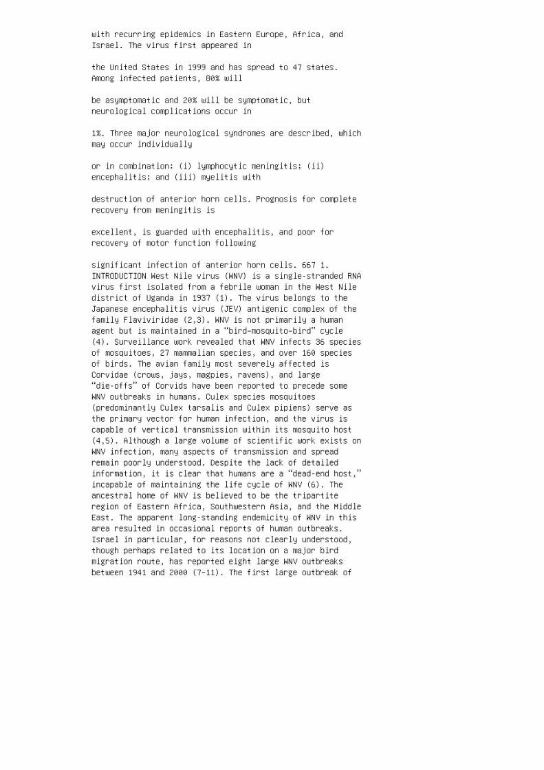
with recurring epidemics in Eastern Europe, Africa, andIsrael. The virus first appeared in
the United States in 1999 and has spread to 47 states.Among infected patients, 80% will
be asymptomatic and 20% will be symptomatic, butneurological complications occur in
1%. Three major neurological syndromes are described, whichmay occur individually
or in combination: (i) lymphocytic meningitis; (ii)encephalitis; and (iii) myelitis with
destruction of anterior horn cells. Prognosis for completerecovery from meningitis is
excellent, is guarded with encephalitis, and poor forrecovery of motor function following
significant infection of anterior horn cells. 667 1.INTRODUCTION West Nile virus (WNV) is a single-stranded RNAvirus first isolated from a febrile woman in the West Niledistrict of Uganda in 1937 (1). The virus belongs to theJapanese encephalitis virus (JEV) antigenic complex of thefamily Flaviviridae (2,3). WNV is not primarily a humanagent but is maintained in a “bird–mosquito–bird” cycle(4). Surveillance work revealed that WNV infects 36 speciesof mosquitoes, 27 mammalian species, and over 160 speciesof birds. The avian family most severely affected isCorvidae (crows, jays, magpies, ravens), and large“die-offs” of Corvids have been reported to precede someWNV outbreaks in humans. Culex species mosquitoes(predominantly Culex tarsalis and Culex pipiens) serve asthe primary vector for human infection, and the virus iscapable of vertical transmission within its mosquito host(4,5). Although a large volume of scientific work exists onWNV infection, many aspects of transmission and spreadremain poorly understood. Despite the lack of detailedinformation, it is clear that humans are a “dead-end host,”incapable of maintaining the life cycle of WNV (6). Theancestral home of WNV is believed to be the tripartiteregion of Eastern Africa, Southwestern Asia, and the MiddleEast. The apparent long-standing endemicity of WNV in thisarea resulted in occasional reports of human outbreaks.Israel in particular, for reasons not clearly understood,though perhaps related to its location on a major birdmigration route, has reported eight large WNV outbreaksbetween 1941 and 2000 (7–11). The first large outbreak of

WNV in Europe occurred in Romania in 1996. Between July andOctober of that year, 393 cases of WNV infection requiringhospitalization were reported (12). Following thisoutbreak, the Volgograd region of Russia reported anoutbreak in 1999 during which 480 persons with WNV werehospitalized (13). These two outbreaks, coupled with the1999 New York City outbreak and the large Israeli outbreakof 2000, highlight an apparent change in the tropism orvirulence of WNV. In prior outbreaks, there was a mildflu-like illness with only a rare case of neuroinvasivedisease, in contrast to recent outbreaks which have amarked increase in both the occurrence and the severity ofneuroinvasive disease (10,14–16). WNV first appeared in theUnited States in 1999, with infection appearing in birds,exotic animals in zoos, and humans in close temporalproximity (10,14,17). Since arriving in North America, WNVhas marched inexorably across the continent. In the UnitedStates, after first appearing in humans in New York statein 1999, WNV caused human disease in three states in 2000,10 states in 2001, 41 states in 2002, and 47 states by theend of 2004 (Alaska, Hawaii, and Washington had notreported human WNV disease as of mid-2005). In Canada, asimilar pattern has emerged, with WNV first appearing inthe province of Ontario in 2001, then marchingprogressively westward to affect six of the 10 provinces bythe end of 2004 (18). Data from Mexico are less complete,and although no confirmed human cases of WNV (i.e., withouta travel history to an affected area) have been reported inMexico, evidence exists for widespread circulation of thevirus in horses since at least 2002 (19,20). WNV is mostcommonly acquired from the bite of an infected mosquito.However, transmission has been documented from organtransplantation, transfusion (blood, stem cell, and bonemarrow), and breast-feeding (21–24). Intrauterine WNVinfection has occurred in isolated cases, resulting insevere congenital disease (25,26). In most instances,however, WNV infection during pregnancy does not appear toresult in fetal illness (27,28).
2. CLINICAL FEATURES
The incubation period for WNV human infection is 2–14 days.Present data suggest that
80% of all human WNV infections are clinically silent (29).Symptomatic human WNV
infection usually presents as one of four clinicalsyndromes: West Nile fever (WNF),

West Nile meningitis (WNM), West Nile encephalitis (WNE),and acute flaccid paralysis.
These syndromes may occur individually or in combination. Afifth category, which
encompasses a wide variety of neurological presentations,will be discussed separately.
Approximately 20% of symptomatic WNV cases present as WNF.Less than 1% of
WNV infections manifest as neuroinvasive disease (e.g.,meningitis, encephalitis, acute
flaccid paralysis). The CDC case definition for WNVinfection is found in Table 38.1.
Clinical features of each of the neurological syndromesassociated with WNV infection
are as follows.
2.1. West Nile Fever
WNF is an acute flu-like illness manifested by the suddenonset of malaise, fatigue, anorexia,
headache, nausea, vomiting, myalgia, fever, eye pain, and anonspecific maculopapular rash.
Typically, these symptoms last ,7 days, although recentreports have indicated that a min
ority of patients may remain symptomatic for as long as 6weeks (2) (Ned Calonge, MD,
MPH, Colorado State Epidemiologist, personalcommunications, 2003–2004).
2.2. West Nile Meningitis
WNM presents with meningismus (fever, headache, and nuchalrigidity), nausea, vomit
ing, and weakness. There are no unique clinical featuresthat allow it to be distinguished
from other viral meningitides (2,30).

2.3. West Nile Encephalitis
WNE typically presents as a diffuse encephalitis withfever, headache abnormal mental
status, and in more severe cases, altered level ofconsciousness, abnormal mental state,
Table 38.1 CDC Diagnostic Criteria for WNV Infection
Case Classification
A clinically compatible illness, plus:
Confirmed
1. Fourfold or greater change in WNV-specific serumantibody titer.
2. Isolation of WNV from or demonstration of specific WNviral antigen or genomic sequences in tissue, blood, CSF,or other bodily fluid.
3. WNV-specific IgM antibodies demonstrated in serum byantibody-capture enzyme immunoassay and confirmed bydemonstration of WNV-specific serum neutralizing antibodiesin the same or a later specimen.
Probable
1. WNV-specific serum IgM antibodies detected byantibody-capture enzyme immunoassay but with no availableresults of a confirmatory test for WNV-specific serumneutralizing antibodies in the same or a later specimen.
Source:
Blood chemistry may be normal, although a mild elevation inliver transaminases may be
seen, and hyponatremia may be present in patients withinappropriate secretion of anti
diuretic hormone (47). Cerebrospinal fluid (CSF)examination typically shows a mild
elevation in pressure (,250 mm CSF), lymphocyticpleocytosis, mild elevation of
protein, and a normal ratio of blood glucose to CSF

glucose. Cell count is usually
50–260 cells/mm 3 , but may reach 2600 cells/mm 3 . Cellsmay be polymorphonuclear
leukocytes, and at presentation, these may account for themajority of cells (3,48).
In one series of West Nile myelitis, cell count was normalin 20% of the patients (49).
In occasional patients, oligoclonal bands may be present.
3.2. Neurologic Studies
Tests to delineate the extent and localization ofneurological disease include imaging and
electrodiagnosis. CT scans are not usually helpful.Experience with magnetic resonance
imaging (MRI) changes is inconclusive, and there is arecommendation for an MRI registry
of WNV cases (50) MRI may be normal, particularly early inthe disease. In individual
cases, areas of altered signal have been detected in thesubstantia nigra, basal ganglia,
and thalamus (51,52). Other abnormalities includesubcortical or leptomeningeal enhance
ment and abnormal signals in the cervical spinal cord(53–55). In cases with acute flaccid
paralysis, nerve conduction studies show normal sensorynerve action potentials, whereas
compound motor action potentials are normal or of reducedamplitude, but conduction velo
cities are normal. This pattern is distinct from that seenin GBS. Electromyography in acute
flaccid paralysis shows fibrillation potentials anddecreased motor unit recruitment (55,56).
3.3. Diagnosis of WNV Infection

A single, acute, CSF specimen positive for WNV-specific IgMantibodies is diagnostic of
current WNV infection (2,57). Paired sera positive forWNV-specific IgM antibodies
(fourfold or greater rise in titer in a convalescent serumsample obtained 14–21 days
after illness onset compared with acute serum sampleobtained 0–7 days after symptom
onset) will also suffice as serological confirmation.Although serological testing is
readily available in the private sector, it must be notedthat acute and convalescent
testing will not result in a timely diagnosis of WNVinfection. There is a recent report
detecting IgM antibodies to WNV in CSF up to 199 days afterclinical presentation, indi
cating that detection of IgM antibodies in CSF is notinvariably indicative of the acute
phase of disease (58). WNV genomic sequences have beenidentified by polymerase
chain reaction (PCR) in blood or in CSF (59–61).Standardized PCR methods are not uni
formly available, however, and detection of IgM antibodiesremains the diagnostic method
of choice. Viral culture methods are not routinelyavailable, and WNV may be inactivated
during transportation at ambient temperature (62).
4. DIFFERENTIAL DIAGNOSIS
WNV infection should be suspected in any individualdeveloping meningitis, encephalitis,
or a syndrome of flaccid paralysis, especially in areas inwhich cases of WNV are being
actively reported. In patients presenting with meningeal

involvement, however, bacterial
meningitis must be excluded. Enteroviralmeningoencephalitis has the same seasonal pre
dominance as WNV meningoencephalitis, as do the olderarthropod-borne encephalitides
in the United States, which include St. Louis encephalitis,Eastern equine encephalitis, Western equine encephalitis,and California-LaCrosse encephalitis. Herpes simplexencephalitis, which has no seasonal predilection, may beindistinguishable from severe cases of WNV encephalitis. Inpatients with a history of travel outside the UnitedStates, consideration should be given to otherarthropod-borne agents, whose likelihood will be dependentupon the region of travel (e.g. Japanese encephalitis orMurray Valley encephalitis in individuals traveling toAfrica, Asia, or Australasia). Rapidly ascending weaknessin a previously healthy patient may represent GBS ratherthan WNV myelitis. Rabies, which may also cause anascending flaccid paralysis, should be considered withknown exposure to potentially infected animals or with ahistory of travel to areas where rabies vaccination is notused. It should be kept in mind, however, that a history ofanimal bite is often absent. A wide range of potentiallytreatable agents may mimic viral meningoencephalitis, whichincludes a large number of bacterial agents, spirochetalagents such as Lyme disease, Mycoplasma, fungi, Rickettsialagents, and Ehrlichia, as well as postinfectious illnessesand a variety of noninfectious systemic disorders (3). 5.MANAGEMENT AND PROGNOSIS Management is supportive. Specifictherapy has not been developed. For the majority of WestNile fever cases, simple measures such as rest, analgesics,and antipyretic medications suffice. Cases of meningitisand, in particular encephalitis and acute flaccidparalysis, require hospitalization. In cases of flaccidparalysis, the possibility of respiratory failure must beconsidered. Ribavirin has not been proven effective intreatment of WNV encephalitis or flaccid paralysis. Amulticenter study employing intravenous immunoglobulin Gcontaining high titers of anti-WNV antibody is in progress.A vaccine is available for use in horses, but is not yetavailable for humans. The prognosis of WNV infection isdetermined by the degree of involvement. Patients with WNFuniformly return to their baseline level of functioning,usually within a few weeks of presentation. Patients withWNM respond similarly, with return to baseline functioningwithin several months. Those patients with WNE appear tofare much worse, and in one study, only six of eight
patients returned to their premorbid level of functioningat 6 months and five of the eight had significant posturalor kinetic tremor at 8 months (30). Additionally, most hadpersistent fatigue and myalgias, and 50% had persistentcognitive deficits at 8 months from presentation. Data fromthe 1999 New York epidemic noted that only 37% of patientswith CNS involvement had returned to baseline by 1 year.The prognosis for recovery is significantly worse in theelderly, and age of.75 is an independent risk factor fordeath from WNV (14). Prognosis for recovery in patientswith acute flaccid paralysis is poor, similar to that inpoliomyelitis, and is unlikely if significant numbers ofmotor neurons have been lost (30,55). 6. PATHOGENESIS WNVinfection in nature begins with introduction of the virusinto its human host through the bite of a mosquito, butinfection may also be transmitted by blood transfusion orbreast milk. The virus is believed to replicate initiallyin the skin and regional lymph nodes. This replicationproduces a primary viremia that ultimately reaches thereticuloendothelial system (RES). A secondary viremia fromthis “seeding” of the RES may then lead to spreadthroughout the body, including the central nervous system(4). At the tissue level, the pathogenesis of WNV infectionis poorly understood. Perivascular inflammatory
infiltrates and microglial nodules have been observed infatal cases of encephalitis
(4,18,63). Cases of acute flaccid paralysis reveal anteriorhorn cell loss, gliosis, patchy
neuronophagia, and infiltrates of lymphocytes andmacrophages (4,40,64). The respective
roles of humoral and cell-mediated immune response incontrol of WNV infection have
not been fully defined. Animal models of WNV infectionsuggest that antibodies are essen
tial in preventing dissemination to the central nervoussystem and that CD 8þ lymphocytes
play an important role in eradication of viral infectionand prevention of viral persistence
(65–67). The occurrence of severe WNV infection intransplant patients suggest that
lymphocyte-mediated immune response may well be essential

for successful eradication
of the virus in man, as well.
1. Smithburn KC, Hughes TP et al. A neurotropic virusisolated from the blood of a native of Uganda. Am J TropMed Hyg 1940; 20:471–492.
2. Petersen LR, Marfin AA. West Nile virus: a primer forthe clinician. Ann Intern Med 2002; 137(3):173–179.
3. Solomon T, Whitley RJ. Arthropod-borne viralencephalitides. In: Scheld WM, Whitley RJ, Marra CM, eds.Infections of the Central Nervous System. Philadelphia:Lippincott Williams & Wilkins, 2004:205–230.
4. Petersen LR, Marfin AA et al. West Nile virus. J Am MedAssoc 2003; 290(4):524–528.
5. Baqar S, Hayes CG et al. Vertical transmission of WestNile virus by Culex and Aedes species mosquitoes. Am J TropMed Hyg 1993; 48(6):757–762.
6. Epidemic/Epizootic West Nile Virus in the United States:Guidelines for Surveillance, Prevention, and Control.August, 2003. 2003.
7. Goldblum N, Sterk VV et al. West Nile fever. Theclinical features of the disease and the isolation of WestNile virus from the blood of nine human cases. Am J TropMed Hyg 1954; 59:89–103.
8. Goldblum N, Jasinka-Klinberg W et al. The naturalhistory of West Nile fever. I. Clinical observations duringan epidemic in Israel. Am J Hyg 1956; 64(3):259–269.
9. Spigland W, Jacinska-Klinberg W et al. Clinical andlaboratory observations in an outbreak of West Nile feverin Israel in 1957. Harefuah 1958; 54:275–281.
10. Giladi M, Metzkor-Cotter E et al. West Nileencephalitis in Israel, 1999: the New York connection.Emerg Infect Dis 2001; 7(4):659–661.
11. Chowers MY, Lang R et al. Clinical characteristics ofthe West Nile fever outbreak, Israel, 2000. Emerg InfectDis 2001; 7(4):675–678.
12. Tsai TF, Popovici F et al. West Nile encephalitisepidemic in southeastern Romania. Lancet 1998;

352(9130):767–771.
13. Platonov AE, Shipulin GA et al. Outbreak of West Nilevirus infection, Volgograd region, Russia, 1999. EmergInfect Dis 2001; 7(1):128–132.
14. Nash D, Mostashari F et al. The outbreak of West Nilevirus infection in the New York City area in 1999. N Engl JMed 2001; 344(24):1807–1814.
15. Briese T, Rambaut A et al. Phylogenetic analysis of ahuman isolate from the 2000 Israel West Nile virusepidemic. Emerg Infect Dis 2002; 8(5):528–531.
16. Klein C, Kimiagar I et al. Neurological features ofWest Nile virus infection during the 2000 outbreak in aregional hospital in Israel. J Neurol Sci 2002;200(1–2):63–66.
17. Briese T, Jia XY et al. Identification of a Kunjin/WestNile-like flavivirus in brains of patients with New Yorkencephalitis. Lancet 1999; 354(9186):1261–1262.
18. Pepperell C, Rau N et al. West Nile virus infection in2002: morbidity and mortality among patients admitted tohospital in southcentral Ontario. CMAJ 2003;168(11):1399–1405.
19. Blitvich BJ, Fernandez-Salas I et al. Serologicevidence of West Nile virus infection in horses, CoahuilaState, Mexico. Emerg Infect Dis 2003; 9(7):853–856. 20.Lorono-Pino MA, Blitvich BJ et al. Serologic evidence ofWest Nile virus infection in horses, Yucatan State, Mexico.Emerg Infect Dis 2003; 9(7):857–859. 21. Pealer LN, MarfinAA et al. Transmission of West Nile virus through bloodtransfusion in the United States in 2002. N Engl J Med2003; 349(13):1236–1245. 22. Kleinschmidt-DeMasters BK,Marder BA et al. Naturally acquired West Nile virusencephalomyelitis in transplant recipients: clinical,laboratory, diagnostic, and neuropathological features.Arch Neurol 2004; 61(8):1210–1220. 23. Williams K. Modes oftransmission for West Nile virus. Clin Lab Sci 2004;17(1):56. 24. From the Centers for Disease Control andPrevention. Possible West Nile virus transmission to aninfant through breast-feeding—Michigan, 2002. J Am MedAssoc 2002; 288(16):1976–1977. 25. Intrauterine West Nilevirus infection—New York, 2002. Morb Mortal Wkly Rep 2002;51(50):1135–1136. 26. Alpert SG, Fergerson J et al.Intrauterine West Nile virus: ocular and systemic findings.Am J Ophthalmol 2003; 136(4):733–735. 27. Bruno J, Rabito

FJ Jr et al. West Nile virus meningoencephalitis duringpregnancy. J La State Med Soc 2004; 156(4):204–205. 28.Interim guidelines for the evaluation of infants born tomothers infected with West Nile virus during pregnancy.Morb Mortal Wkly Rep 2004; 53(7):154–157. 29. Mostashari F,Bunning ML et al. Epidemic West Nile encephalitis, NewYork, 1999: results of a household-basedseroepidemiological survey. Lancet 2001; 358(9278):261–264.30. Sejvar JJ, Haddad MB et al. Neurologic manifestationsand outcome of West Nile virus infection. J Am Med Assoc2003; 290(4):511–515. 31. Ceausu E, Erscoiu S et al.Clinical manifestations in the West Nile virus outbreak.Rom J Virol 1997; 48(1–4):3–11. 32. Kanagarajan K, Ganesh Set al. West Nile virus infection presenting as cerebellarataxia and fever: case report. South Med J 2003;96(6):600–601. 33. Sayao AL, Suchowersky O et al. Calgaryexperience with West Nile virus neurological syndromeduring the late summer of 2003. Can J Neurol Sci 2004;31(2):194–203. 34. Arya SC. Japanese encephalitis virus andpoliomyelitis-like illness. Lancet 1998; 351(9120):1964.35. Ahmed S, Libman R et al. Guillain–Barre´ syndrome: anunusual presentation of West Nile virus infection.Neurology 2000; 55(1):144–146. 36. Leis AA, Stokic DS etal. Clinical spectrum of muscle weakness in human West Nilevirus infection. Muscle Nerve 2003; 28(3):302–308. 37.Fratkin JD, Leis AA et al. Spinal cord neuropathology inhuman West Nile virus infection. Arch Pathol Lab Med 2004;128(5):533–537. 38. Omalu BI, Shakir AA et al. Fatalfulminant pan-meningo-polioencephalitis due to West Nilevirus. Brain Pathol 2003; 13(4):465–472. 39. Doron SI,Dashe JF et al. Histopathologically proven poliomyelitiswith quadriplegia and loss of brainstem function due toWest Nile virus infection. Clin Infect Dis 2003;37(5):e74–e77. 40. Sejvar JJ, Leis AA et al. Acute flaccidparalysis and West Nile virus infection. Emerg Infect Dis2003; 9(7):788–793. 41. Bouffard JP, Riudavets MA et al.Neuropathology of the brain and spinal cord in human WestNile virus infection. Clin Neuropathol 2004; 23(2):59–61.42. Anninger WV, Lomeo MD et al. West Nile virus-associatedoptic neuritis and chorioretinitis. Am J Ophthalmol 2003;136(6):1183–1185. 43. Bains HS, Jampol LM et al. Vitritisand chorioretinitis in a patient with West Nile virusinfection. Arch Ophthalmol 2003; 121(2):205–207. 44.Hershberger VS, Augsburger JJ et al. Chorioretinal lesionsin nonfatal cases of West Nile virus infection.Ophthalmology 2003; 110(9):1732–1736. 45. Vandenbelt S,Shaikh S et al. Multifocal choroiditis associated with WestNile virus encephalitis. Retina 2003; 23(1):97–99. 46.Hassin-Baer S, Kirson ED et al. Stiff-person syndromefollowing West Nile fever. Arch Neurol 2004; 61(6):938–941.

47. Jeha LE, Sila CA et al. West Nile virus infection: anew acute paralytic illness. Neurology 2003; 61(1):55–59.
48. Tyler KL. Viral myelitis. In: Scheld WM, Whitley RJ,Marra CM, eds. Infections of the Central Nervous System.Philadelphia: Lippincott Williams & Wilkins, 2004:323–330.
49. Leis AA, Stokic DS et al. A poliomyelitis-like syndromefrom West Nile virus infection. N Engl J Med 2002;347(16):1279–1280.
50. Robertson HJ, Sejvar JJ. The need for a West Nile virusMRI registry. Am J Neuroradiol 2003; 24(9):1741–1742.
51. Rosas H, Wippold FJ. West Nile virus: case report withMR imaging findings. Am J Neuroradiol 2003;24(7):1376–1378.
52. Bosanko CM, Gilroy J et al. West Nile virusencephalitis involving the substantia nigra: neuroimagingand pathologic findings with literature review. Arch Neurol2003; 60(10):1448–1452.
53. Brilla R, Block M et al. Clinical and neuroradiologicfeatures of 39 consecutive cases of West Nile virusmeningoencephalitis. J Neurol Sci 2004; 220(1–2):37–40.
54. Olsan AD, Milburn JM et al. Leptomeningeal enhancementin a patient with proven West Nile virus infection. Am JRoentgenol 2003; 181(2):591–592.
55. Al Shekhlee A, Katirji B. Electrodiagnostic features ofacute paralytic poliomyelitis associated with West Nilevirus infection. Muscle Nerve 2004; 29(3):376–380.
56. Li J, Loeb JA et al. Asymmetric flaccid paralysis: aneuromuscular presentation of West Nile virus infection.Ann Neurol 2003; 53(6):703–710.
57. Shi PY, Wong SJ. Serologic diagnosis of West Nile virusinfection. Expert Rev Mol Diagn 2003; 3(6):733–741.
58. Kapoor AH, Signs BK et al. Persistence of West NileVirus (WNV) IgM antibodies in cerebrospinal fluid frompatients with CNS disease. J Clin Virol 2004;31(4):289–291.
59. Huang C, Slater B et al. First isolation of West Nilevirus from a patient with encephalitis in the United

States. Emerg Infect Dis 2002; 8(12):1367–1371.
60. Lanciotti RS, Kerst AJ et al. Rapid detection of WestNile virus from human clinical specimens, field-collectedmosquitoes, and avian samples by a TaqMan reversetranscriptase– PCR assay. J Clin Microbiol 2000;38(11):4066–4071.
61. Briese T, Glass WG et al. Detection of West Nile virussequences in cerebrospinal fluid. Lancet 2000;355(9215):1614–1615.
62. Mayo DR, Beckwith WH III. Inactivation of West Nilevirus during serologic testing and transport. J ClinMicrobiol 2002; 40(8):3044–3046.
63. Sampson BA, Armbrustmacher V. West Nile encephalitis:the neuropathology of four fatalities. Ann NY Acad Sci2001; 951:172–178.
64. Kelley TW, Prayson RA et al. Spinal cord disease inWest Nile virus infection. N Engl J Med 2003;348(6):564–566.
65. Shrestha B, Diamond MS. Role of CD 8þ T cells incontrol of West Nile virus infection. J Virol 2004;78(15):8312–8321.
66. Wang Y, Lobigs M et al. CD 8þ T cells mediate recoveryand immunopathology in West Nile virus encephalitis. JVirol 2003; 77(24):13323–13334.
67. Diamond MS, Shrestha B et al. Innate and adaptiveimmune responses determine protection against disseminatedinfection by West Nile encephalitis virus. Viral Immunol2003; 16(3):259–278.
39
Diagnostic Yield for Peripheral Neuropathy
A. Gordon Smith
University of Utah, Salt Lake City, Utah, USA
Abstract 677
1. Introduction 677
2. Confirmation of Peripheral Neuropathy 678

3. Classification of Peripheral Neuropathy 680
4. The Evaluation of Prototypic Neuropathy 680
5. Idiopathic Neuropathy 683
6. Summary 684
ABSTRACT
Accurate and efficient diagnosis of peripheral neuropathyrequires an organized approach.
Use of a standardized set of laboratory tests for allpatients with suspected peripheral
neuropathy is expensive, often misses the correctdiagnosis, and frequently leads to
false positive tests. Use of a rational diagnosticalgorithm maximizes diagnostic yield
and minimizes risk of misdiagnosis. Patients with chronicdistal symmetric sensory pre
dominant neuropathy require relatively few routinediagnostic tests.
1. INTRODUCTION
The diagnostic evaluation of peripheral neuropathy mayprovoke feelings of diagnostic
nihilism if one is not familiar with peripheral nervedisease. There are hundreds of
causes for peripheral neuropathy and the range of symptomsreferable to peripheral
nerve dysfunction is limited. Consequently, a “shotgun”diagnostic approach is often
used, employing a standard set of diagnostic tests (Table39.1). Such an approach may
miss the diagnosis because the particular diagnostic testmay not be on the “shotgun”
panel. On the other hand, false positive diagnostic tests
are not uncommon. Because of
the large number of tests, several of which must be sent tospecific reference laboratories, 677 this approach isexpensive. However, although expensive, the sensitivity andspecificity of this panel are low. There are several teststhat are only relevant for specific patient populations.For example, antiganglioside antibody panels may includeanti-GM1 antibodies, antimyelin associated glycoproteinantibodies, or both. The former is relevant only for motorneuropathies, particularly multifocal motor neuropathy withconduction block, and the later is relevant only for asubset of patients with chronic inflammatory demyelinatingpolyradiculoneuropathy (CIDP). These antibodies may bedetectable in low levels in normal subjects or patientswith unrelated neuropathies, leading the unwary clinicianto wrongly associate the false positive laboratory testwith the clinical syndrome. Experienced clinicians oftenarrive at a diagnosis using a pattern recognition approach.Particular patterns of disease are recognized and thediagnostic approach is limited to few diagnostic tests.When successful, this diagnostic approach is very efficientand cost effective. However, pattern recognition may onlybe employed by those with both excellent clinical skillsand a wealth of experience with peripheral nerve disease.If the particular disorder has not been previouslyobserved, if there is an atypical presentation, or if theclinician (despite excellent skills) does not have a greatdeal of experience, the pattern may not be recognized andthe diagnosis missed. For most clinicians, includingexperienced peripheral nerve experts encountering anunusual disorder, a rational diagnostic approach isnecessary. Several well-reasoned approaches to thediagnosis of peripheral neuropathy have been suggested andeach is appropriate (1–5). This chapter will outline anorganized approach to the classification of peripheralneuropathies and will review the evidence for the routineuse of commonly ordered diagnostic tests in the evaluationof distal symmetric sensory predominant neuropathy. 2.CONFIRMATION OF PERIPHERAL NEUROPATHY The first step in theevaluation of a patient with a peripheral nerve disorder isconfirmation of the localization. The diagnosis ofperipheral neuropathy is usually straightforward. However,patients with isolated foot pain and numbness may present adiagnostic challenge. There are many different causes forfoot pain including orthopedic issues Table 39.1 The“Shotgun” Approach to Peripheral Neuropathy Consists of theRoutine Measurement of a Standardized Panel of DiagnosticTests in all Patients Vitamin B12 Folate RPR Thyroidstimulating hormone Sedimentation rate Rheumatoid factor

Antinuclear antibodies Extractible nuclear antibodies Serumprotein electrophoresis Antiganglioside antibody titersParaneoplastic antibody titers Note: This is a list oftests often included in a shotgun panel. This approach isexpensive and inefficient, and it carries significant riskof false positives.
and central nervous system dysfunction; differentiationfrom small fiber predominant neu
ropathy may be difficult on clinical grounds alone.Confirmation of a suspected diagnosis
of peripheral neuropathy depends on one of severalsupportive diagnostic tests. Nerve
conduction studies are the most commonly used test forperipheral neuropathy. Nerve con
duction studies primarily reflect large fiber function andare therefore insensitive to small
fiber injury. The sympathetic skin response may be used toassess small fiber function, but
is relatively insensitive. Therefore, other tests mustoften be used to confirm the presence
of a suspected small fiber predominant neuropathy.Quantitative sensory testing (QST) is an accurate andreproducible method to deter
mine the threshold for perceiving vibratory or coolingstimuli. Vibration threshold
measures large fiber function and cooling reflects small,lightly myelinated nerve function.
QST is abnormal in 57–72% of patients with small fiberneuropathy (6,7). QST is non
specific in that other causes of sensory loss (e.g.,radiculopathy, central nervous system
processes) can increase perception thresholds. QST alsorequires patient cooperation
and meticulous attention to the testing environment (e.g.,temperature, noise, and other

distractions). Unlike Nerve Conduction Studies (NCS), QSTis a psychophysiologic
measure, rather than a physiologic measure. As a result,QST is unable to differentiate
between true sensory loss and simulated sensory loss (8).Therefore, QST should not be
used as the primary means of differentiating functionalsensory symptoms or signs from
peripheral neuropathy. Some degree of autonomic dysfunctionis demonstrable in most patients with painful
peripheral neuropathy. Autonomic testing provides asensitive means of diagnosing small
fiber nerve injury (Chapter 4). The most commonlyavailable, and most sensitive tests
measure sudomotor (sweat gland) function. Quantitativesudomotor axon reflex testing
(QSART) is the easiest and most widely available procedure.A small circular chamber
is placed on the skin. Acetylcholine is iontophoresed intothe skin and stimulates terminal
unmyelinated axons supplying sweat glands, resulting inreflex activation of surrounding
sweat glands. The resulting sweat volume is measuredquantitatively. QSART is abnormal
in 70% of painful neuropathy patients (9). Unlike QST,QSART is specific for postgang
lionic adrenergic autonomic function. As with QST, specialequipment is necessary, and
careful attention must be paid to the testing protocol inorder to obtain reliable results. The only test thatdirectly evaluates unmyelinated somatic sensory fibers isskin
biopsy. A 3 mm punch biopsy is performed and the tissuestained with an antibody that

stains peripheral nerve axons (Chapter 6). Unmeylinatedaxons subserving pain sensation
may be easily observed coursing through the epidermis innormal subjects. In those with
neuropathy, these fibers are depleted at distal sites. LikeQSART, skin biopsy is both
sensitive and specific (6). Skin biopsy of the distal legis abnormal in subjects whose symp
toms are limited to the feet. Unlike nerve biopsy, skinbiopsy is minimally invasive, and
often better tolerated than NCS. There is a minimal risk ofinfection, and no significant
complications are reported in the literature. The biopsysite heals within 1–2 weeks in
most subjects (similar to abrasion wounds) (10). The biopsyresults in a scar that usually
fades and may disappear after several months. The biopsyprocedure is simple and rapidly
performed (�10–15 min). When tissue processing and analysisrequire skill and time, the
tissue may be easily shipped to a central processinglaboratory. The choice of which test to perform in apatient with suspected peripheral neuro
pathy and normal nerve conduction studies is based onpatient symptoms and which equip
ment is available. If QST and QSART equipments are notavailable, a skin biopsy may be
performed and shipped to a laboratory where the tissue canbe processed. Overall, there is
a poor correlation among QSART, QST and skin biopsy,suggesting that they measure
separate functions (7). Patients may have an abnormality onone but not on the others. Nerve biopsy is never indicatedsolely for the purpose of confirming a diagnosis ofperipheral neuropathy. Even when electron microscopy is
performed for analysis of small unmyelinated nerve fibers,nerve biopsy may be normal when other specific and lessinvasive tests, such as skin biopsy, are abnormal (11).Nerve biopsy should only be performed when there issuspicion of certain disorders, particularly amyloidosis,vasculitis, and tumor infiltration. Nerve biopsy is rarelynecessary for the diagnosis of CIDP and should primarily beused to exclude other etiologies. 3. CLASSIFICATION OFPERIPHERAL NEUROPATHY Most books of peripheral neuropathy(including this one) classify neuropathies based onunderlying pathophysiology (inflammatory, metabolic,neoplastic, toxic etc.). This organization is useful forthe reader interested in learning about a particulardiagnosis. For the clinician attempting to diagnose anindividual patient with an unknown peripheral neuropathy,this classification scheme is less useful. In the clinicalsetting, an algorithmic approach to the classification ofindividual peripheral neuropathies is necessary to limitthe differential diagnosis and permit a reasoned andfocused diagnostic evaluation. Peripheral neuropathies canbe classified based on system involvement (sensory, motor,and autonomic), anatomic pattern (distal, proximal, andasymmetric), temporal course (acute, subacute, andchronic), electrodiagnostic features (axonal,demyelinating, and mixed), or the presence of severe painor positive family history (Table 39.2). Most patients withperipheral neuropathy have a chronic, distal predominant,sensory greater than motor neuropathy with positive sensorysymptoms (tingling, prickling, and burning) and at leastsome degree of discomfort or pain. This prototypic patternof neuropathy represents one of the most common problemsseen by general neurologists and requires only a focuseddiagnostic evaluation that will be outlined suceedingly.However, recognition of the prototypic pattern necessitatesa careful search for any atypical features suggestive of adifferent neuropathy pattern. These features, and selecteddisorders which characteristically cause them, are outlinedin Table 39.2. 4. THE EVALUATION OF PROTOTYPIC NEUROPATHYPatients with prototypic neuropathy comprise a relativelyhomogeneous group, presenting with numbness, positivesensory symptoms, and discomfort. Symptoms often developover a period of months, then stabilize or progress slowlyover years. Numbness and pain are distal predominant,primarily involving the feet and distal leg. While mildweakness of toe extension and ankle dorsiflexion may beobserved, significant weakness is uncommon. Pain may besevere. Autonomic dysfunction is usually limited tosudomotor dysfunction in the feet, which often have a redor purple discoloration, and erectile dysfunction (9). Thepresence of bony abnormalities such as high arches, hammer

toes, or neurogenic arthropathy (Charcot joint) shouldprompt an evaluation for an inherited neuropathy. Once thispattern is recognized, the evaluation may be focused. Allpatients with suspected peripheral neuropathy should have athorough medical history and require a careful generalphysical examination. Subtle historical details orexamination findings may be very helpful in focusing thediagnostic evaluation, regardless of the neuropathypattern. For example, several small angiokeratoma on ayoung male patient with a painful neuropathy suggest thediagnosis of Fabry disease. For patients with prototypicneuropathy, a careful search should be made for clinicalevidence of toxic exposure, connective
tissue disease, or endocrine dysfunction. A differentialdiagnosis of prototypic neuropathy
is displayed in Table 39.3, with symptoms or signssuggestive of each diagnosis. While the differentialdiagnosis of prototypic neuropathy appears lengthy, inpatients
lacking clinical features suggestive of any particulardisorder, the list of potential diagnos
tic possibilities is much shorter. The laboratoryevaluation includes tests for disorders that
directly cause nerve dysfunction or injury (e.g., vitaminB12 and diabetes) as well as tests
for conditions that may be associated with neuropathy butare not necessarily causative
(monoclonal gammopathy). Several studies have addressed thediagnostic yield of labora
tory evaluation among this patient population. Among 93patients with distal pain, numb
ness or tingling without symptoms of motor weakness, theonly abnormality identified after
Table 39.2 The Differential Diagnosis can be Focused byRecognizing Atypical Features
Prominent Motor Involvement Prominent Sensory Involvement
Charcot Marie Tooth

CIDP and variants (e.g., distal acquired demyelinatingperipheral neuropathy)
Lead
Dapsone
Porphyria
Multifocal motor neuropathy
Motor neuron disease
Prominent Autonomic Involvement
Diabetes
Amyloidosis
Hereditary sensory autonomic
neuropathy
Severe Pain
Idiopathic
Diabetes
HIV
Amyloidosis
Fabry
Vasculitis
Proximal Involvement
(non-length dependent)
CIDP
Diabetic and non-diabetic lumbosacral
radiculoplexus neuropathies
(“diabetic amyotrophy”)
Porphyria

Infliltrative or infectious polyradiculitis
(e.g., carcinoma)
Dorsal root ganglionopathy Paraneoplastic IdiopathicIdiopathic Diabetes Vitamin B12 deficiency Pyridoxinetoxicity Drug toxicity (e.g., nucleoside analogs CisplatinDorsal root ganglionopathy Paraneoplastic IdiopathicSjogrens HIV Leprosy Hereditary sensory autonomicneuropathy Amyloidosis Asymmetry Vasculitis Primaryperipheral nerve Systemic vasculitis Hereditary neuropathywith predisposition to pressure palsies Diabetic andnon-diabetic lumbosacral radiculoplexus neuropathies(“diabetic amyotrophy”) CIDP (“Lewis Sumner” variant)Multifocal motor neuropathy Leprosy Acute or SubacuteTimecourse Inflammatory Guillain Barre Syndrome VasculitisToxic Exposure Metals (e.g., arsenic) Solvents (e.g.,n-Hexane) Porphyria Infectious neuropathies
Note: A selected list of diagnoses suggested by eachatypical feature is provided. extensive laboratoryevaluation was a monoclonal gammopathy in 4/83 (5%) (12).The recommendation was that CBC, sedimentation rate, serumchemistry panel, serum protein electrophoresis (SPEP) andimmunofixation (IFIX), and vitamin B12 levels be measuredin all patients (1). In another study of 117 patients withsensory neuropathy, the only laboratory abnormalities werehypertryglyceridemia (34%), hypercholestoreamia (28%),monoclonal gammopathy in 4 (3.4%), positive extractiblenuclear antigens (ENA) consistent with Sjo¨gren’s diseasein 2 (0.2%), and positive antiribonucleoprotein (RNP)antibody consistent with mixed connective tissue disease in1 (0.1%). Two patients had amyloidosis (one primary and oneinherited) and both had symtpomatic orthostatic hypotension(7). Our data is consistent with these series. Among 138consecutive patients with prototypic neuropathy, 1.6% had alow B12 level, and 2.8% had a monoclonal gammopathy ofuncertain significance (MGUS). The most frequentabnormality was impaired glucose tolerance (IGT) defined asa serum glucose of 140–200, 2 h after a 75 g ahydrousdextrose oral load. Among 87 patients who underwent therecommended oral glucose tolerance test, 39 (45%) had IGTand 12 (13%) frank diabetes (13). This is consistent withretrospective data suggesting over one-third of patientswith otherwise idiopathic neuropathy have IGT (14,15). Theother neuropathy patient series described earlier did notinclude routine OGTT. The studies outlined earlier suggestonly selected laboratory tests need to be routinelyperformed in patients with prototypic neuropathy who lack

signs or symptoms suggesting a systemic disorder. Allpatients should undergo an oral glucose tolerance test toexclude diabetes or IGT. The Diabetes Control andComplications Trial demonstrated that glycemic controlslows progression of diabetic neuropathy and the DiabetesPrevention Program demonstrated that diet and exerciseintervention reduces the risk of progression of IGT tofrank diabetes (16,17). Research is underway to determineif similar lifestyle intervention slows the rate ofprogression of neuropathy associated with IGT. Peripheralneuropathy caused by diabetes and IGT is reviewed inChapter 11. IGT is part of the Metabolic Syndrome ofinsulin resistance, obesity, hyperlipidemia, andhypertension, and is associated Table 39.3 The DifferentialDiagnosis of Prototypic Peripheral Neuropathy IdiopathicNeuropathy Diabetes Impaired Glucose Tolerance Vitamin B12Deficiency Connective tissue disease Sjogren’s SyndromeRheumatoid Arthritis a Systemic Lupus Erythematosis aHypothyroidism a Toxic Exposure MedicationsChemotherapeutic agents (vincristine, taxol, platinum)Pyridoxine (high dose causes ganglionopathy) MacrodantinMetronidazole Amiodarone Solvents (chronic low level)Familial a Rarely the presenting symptom
with increased risk of large vessel atheroscleroticdisease. The high incidence of hyperli
pidemia in patients with painful sensory neuropathysuggests a high likelihood of insulin
resistance (7). Because the Metabolic Syndrome maycontribute to peripheral neuropathy
and is a significant risk factor for coronary artery andcerebrovascular disease, all neuro
pathy patients should be have serum lipids and cholesterolmeasured. A serum B12 level should be measured in allpatients. Those with borderline serum
levels (200–300 pg/mL) should also have methylmalonic acidand homocysteine levels
tested to exclude a mild deficiency state (18). Other testsare of less certain yield in the absence of clinicalfeatures suggesting a
systemic disorder. Routine testing of thyroid functionfailed to reveal an abnormality in

the neuropathy series reviewed. However, neuromuscularcomplaints are common among
patients with hypothyroidism (19). Signs and symptoms ofhypothyroidism should be
carefully sought in all patients with peripheral neuropathyand diagnostic testing
performed only when found. Monoclonal gammopathy of unknownsignificance (MGUS) is associated with CIDP,
but the relationship between MGUS and axonal neuropathy andis less certain. The population
prevalence of MGUS rises to 3% among those over 70 years ofage (20,21). In the neuropathy
patient series described previously, the prevalence of MGUSranged 1–5%, a rate not much
higher than the population prevalence. There are noclinical differences between patients with
idiopathic neuropathy and those with axonal neuropathyassociated with MGUS (22). One
argument in favor of screening neuropathy patients is thatover 20 years of follow-up,
20% of patients with MGUS develop a malignantlymphoproliferative disorder (21,23).
However, the impact of early diagnosis of MGUS on ultimateoutcome is unknown. While systemic evidence of a connectivetissue disease, such as rash, inflammatory
arthritis, or keratoconjunctivitis sicca should prompt acareful serologic evaluation, routine
testing for serum autoantibodies is of very low diagnosticyield. Up to 12% of healthy blood
donors have a positive antinuclear antibody (ANA) at a lowtiter (24,25). Abnormal ANA
tests in neuropathy subjects are usually of low titer andare of doubtful significance.
A history of dry eyes or mouth raises the question of

Sjo¨gren’s syndrome and serum
SSA and SSB antibodies should be measured. While specific,these antibodies are absent
in most patients with neuropathy due to Sjo¨gren’s syndromeand a minor salivary gland
biopsy may be necessary to confirm the diagnosis (26).There is significant commercial pressure to order antibodypanels on patients with
peripheral neuropathy. These panels typically includevarious combinations of antigan
glioside and paraneoplastic antibodies. With the exceptionof antibodies associated with
multifocal motor neuropathy (GM1), the Fisher variant ofGuillain Barre Syndrome
(GQ1b), defined paraneoplastic syndromes such as sensoryganglionopathy (Hu) and
certain patients with CIDP (MAG), there is no benefit tothe routine ordering of these
tests. Antisulfatide antibodies in particular are ofuncertain clinical significance (27). It
is usually best to order only the specific antibody ofclinical relevance, as the marketed
panels may include several antibody tests that are notrelevant. Panels may include
antibodies associated with both motor (GM1) and sensory(MAG, Hu) neuropathies. Anti
bodies may be positive in a patient for whom the laboratoryfinding is clinically irrelevant,
occasionally leading to inappropriate therapy.
5. IDIOPATHIC NEUROPATHY
The percentage of patients for whom no abnormality is found(e.g., idiopathic neuropathy)
6. Holland NR, Crawford TO, Hauer P, Cornblath DR, Griffin

JW, McArthur JC. Small-Fiber sensory neuropathies: clinicalcourse and neuropathology of idiopathic cases. Ann Neurol1998; 44:47–59.
7. Periquet MI, Novak V, Collins MP et al. Painful sensoryneuropathy prospective evaluation using skin biopsy.Neurology 1999; 53:1641–1647.
8. Freeman R, Chase K, Risk M. Quantitative sensory testingcannot differentiate simulated sensory loss from sensoryneuropathy. Neurology 2003; 60:465–470.
9. Novak V, Freimer ML, Kissel JT et al. Autonomicimpairment in painful neuropathy. Neurology 2001;56(7):861–868.
10. Chien HF, Tseng TJ, Lin WM et al. Quantitativepathology of cutaneous nerve terminal degeneration in thehuman skin. Acta Neuropathol 2000; 102:455–461.
11. Herrmann D, Griffin J, Hauer P, Cornblath D, McArthurJ. Epidermal nerve fiber density and sural nervemorphometry in peripheral neuropathies. Neurology 1999;53:1634–1640.
12. Wolfe G, Baker N, Amato A et al. Chronic cryptogenicsensory polyneuropathy clinical and laboratorycharacteristics. Arch Neurol 1999; 56:540–547.
13. Smith AG, Singleton JR. The diagnostic yield of astandardized approach to idiopathic sensorypredominantneuropathy. Arch Intern Med 2004; 164(9):1021–1025.
14. Singleton JR, Smith AG, Bromberg MB. Increasedprevalence of impaired glucose tolerance in patients withpainful sensory neuropathy. Diab Care 2001;24(8):1448–1453.
15. Novella SP, Inzucchi SE, Goldstein JM. The frequency ofundiagnosed diabetes and impaired glucose tolerance inpatients with idiopathic sensory neuropathy. Muscle Nerve2001; 24:1229–1231.
16. Diabetes Control and Complications Research Group. Theeffect of intensive treatment of diabetes on thedevelopment and progression of long-term complications ininsulin-dependent diabetes mellitus. New Engl J Med 1993;329:977–986.
17. Diabetes Prevention Program Research Group. Reduction

in the incidence of type 2 diabetes with lifestyleintervention or metformin. NEJM 2002; 346:393–403.
18. Saperstein DS, Barohn RJ. Peripheral Neuropathy Due toCobalamin Deficiency. Curr Treat Options Neurol 2002;4(3):197–201.
19. Duyff R, Van den Bosch J, Laman D, van Loon B, LinssenW. Neuromuscular findings in thyroid dysfunction aprospective clinical and electrodiagnostic study. J NeurolNeurosurg Psychiatry 2000; 68:750–755.
20. Kelly J, Kyle R, O’Brien P, Dyck P. Prevalence ofmonoclonal proteins in peripheral neuropathy. Neurology1981; 31:1480–1483.
21. Kyle RA, Finkelstein S, Elveback LR, Kurland LT.Incidence of monoclonal proteins in a Minnesota communitywith a cluster of multiple myeloma. Blood 1972; 40:719–724.
22. Notermans N, Wokke J, Lokhorst H, Franssen H, van derGraaf Y. Polyneuropathy associated with monoclonalgammopathy of undetermined significance a prospective studyof the prognostic value of clinical and laboratoryabnormalities. Brain 1994; 117:1385–1393.
23. Kyle RA, Therneau TM, Rajkumar SV et al. A long-termstudy of prognosis in monoclonal gammopathy of undeterminedsignificance. N Engl J Med 2002; 346(8):564–569.
24. Azizah MR, Azila MN, Zulkifli MN, Norita TY. Theprevalence of antinuclear, anti-dsDNA and anti-RNPantibodies in a group of healthy blood donors. Asian Pac JAllergy Immunol 1996; 14:125–128.
25. de Vlam K, De Keyser F, Vergraggen G et al. Detectionand identification of antinuclear antibodies in serum ofnormal blood donors. Clin Exp Rheumatol 1993; 11:393–397.
26. Grant IA, Hunder GG, Homburger HA, Dyck PJ. Peripheralneuropathy associated with sicca complex. Neurology 1997;48(4):855–862.
27. Wolfe G, El-Feky W, Katz J, Brayan W, Wians F, BarohnR. Antibody panels in idiopathic polyneuropathy and motorneuron disease. Muscle and Nerve 1997; 20:1275–1283.
28. Smith A, Howard J, Kroll R, Singleton J. Microvasculardysfunction in diabetic and idiopathic peripheralneuropathy. Neurology 2003; 60:A386.

29. Notermans N, Wokke J, van der Graaf Y, Franssen H, vanDijk G, Jennekens F. Chronic idiopathic axonalpolyneuropathy a five year follow up. J Neurol NeurosurgPsychiatry 1994; 57:1525–1527.