Longitudinal Changes in the Corpus Callosum following Pediatric Traumatic Brain Injury
Upload
independentCategory
view
0download
0
REPORT
Whole-Exome Sequencing Identifies MutatedC12orf57 in Recessive Corpus Callosum Hypoplasia
Naiara Akizu,1 Nuri M. Shembesh,2 Tawfeg Ben-Omran,3 Laila Bastaki,4 Asma Al-Tawari,5 Maha S. Zaki,6
Roshan Koul,7 Emily Spencer,1 Rasim Ozgur Rosti,1 Eric Scott,1 Elizabeth Nickerson,8 Stacey Gabriel,8
Gilberto da Gente,9 Jiang Li,9 Matthew A. Deardorff,10,11 Laura K. Conlin,12 Margaret A. Horton,10
Elaine H. Zackai,10,11 Elliott H. Sherr,9,* and Joseph G. Gleeson1,*
The corpus callosum is the principal cerebral commissure connecting the right and left hemispheres. The development of the corpus
callosum is under tight genetic control, as demonstrated by abnormalities in its development in more than 1,000 genetic syndromes.
We recruited more than 25 families in which members affected with corpus callosum hypoplasia (CCH) lacked syndromic features
and had consanguineous parents, suggesting recessive causes. Exome sequence analysis identified C12orf57 mutations at the initiator
methionine codon in four different families.C12orf57 is ubiquitously expressed and encodes a poorly annotated 126 amino acid protein
of unknown function. This protein is without significant paralogs but has been tightly conserved across evolution. Our data suggest that
this conserved gene is required for development of the human corpus callosum.
The corpus callosum (CC), the largest commissural white-matter tract in the mammalian brain, is essential forinterhemispheric integration of sensory, motor, andhigher-order cognitive information. It is traditionallydivided into four segments, the rostrum, genu, body, andsplenium. Development of the CC occurs during weeks6–20 of gestation, and developmental defects, observedin 0.3%–0.7% of individuals undergoing brain imaging,are among the most common of brain malformations.1
Abnormalities of the CC are noted in more than 1,000unique OMIM entries, suggesting that CC developmentis very sensitive to genetic perturbations. Additionally,twin studies have suggested a high heritability of the CCarea.2 However, well-described disorders that specificallyaffect the CC in the absence of other major abnormalitiesare quite rare.We previously described subtypes of CC abnormalities
after having conducted detailed structural analysis frombrain MRI scans from a cohort of 30 affected individualsfrom 19 families with a history of parental consan-guinity.3 The initial presenting feature was developmentaldelay or epilepsy in all cases,4 and brain MRIs showed CCabnormalities without othermajor neuroanatomic defects.We focused on parental consanguinity to enrich the studyfor cases that were likely to involve recessive inheritance,in which phenotypes were more likely to be fully expres-sive and fully penetrant. We delineated onemajor categoryconsisting of CC hypoplasia (CCH) without dysplasia.Most notable was the finding that even within a given
CCH-affected family, whose members in all likelihoodshare a single homozygous-recessive allele, siblings candisplay a spectrum of severity ranging from almost normalappearance to complete agenesis. The data suggest thatthere might be wide-ranging phenotypic severity evenwith presumably identical genetic mutations in siblingsand that complete agenesis might be a common endphenotype.For this study we prioritized families for genetic analysis
by focusing on those with first- or second-degree consan-guinity and two or more affected members in the settingof CCH; these criteria made it likely that a recessive geneticorigin was involved. All family members underwenta routine clinical brain MRI, including midline sagittaland axial views. After worldwide recruitment of individ-uals identified from 1999–2012 as having neurodevelop-mental disease, we focused on those for whom a brainMRI either showed isolated CCH or showed CCH as themajor imaging anomaly. Individuals with other majorstructural brain anomalies or overwhelming evidence ofother dysmorphic or syndromic causes were excluded.We genetically evaluated 27 families, four of which weresimplex. The remaining familes, including the previouslyascertained 19 families, were multiplex with similarlyaffected siblings. The study was approved by appropriateethics committees, and the families provided informedconsent.We used QIAGEN reagents to extract blood DNA, in
some cases after performing a 5K whole-genome linkage
1Department of Neurosciences and Pediatrics, Howard Hughes Medical Institute, University of California, San Diego, CA 92093, USA; 2Department ofPediatrics, Al-Fatah Children’s Hospital, Al-Arab Medical University, Benghazi, Libya; 3Clinical and Metabolic Genetics Division, Department of Pediatrics,Hamad Medical Corporation, 3050, Doha, Qatar; 4Kuwait Medical Genetics Centre, Maternity Hospital, Safat 13041, Kuwait; 5Neurology Department,Children’s Unit Al Sabah Hospital, Safat, Kuwait City 72252 Kuwait; 6Clinical Genetics Department, Human Genetics and Genome Research Division,National Research Centre, Cairo, 12311, Egypt; 7Department of Child Health (Neurology), Sultan Qaboos University Hospital, College of Medicine andHealth Sciences, Muscat, 123, Oman; 8The Broad Institute of MIT and Harvard, Cambridge, MA 02141, USA; 9Department of Neurology, University ofCalifornia, San Francisco, CA 94158 USA; 10Division of Genetics, The Children’s Hospital of Philadelphia, Philadelphia, PA, 19104, USA; 11Departmentof Pediatrics, The University of Pennsylvania Perelman School of Medicine, Philadelphia, PA, 19104, USA; 12Department of Pathology and LaboratoryMedicine, The Children’s Hospital of Philadelphia, Philadelphia, PA, 19104, USA*Correspondence: [email protected] (E.H.S.), [email protected] (J.G.G.)http://dx.doi.org/10.1016/j.ajhg.2013.02.004. !2013 by The American Society of Human Genetics. All rights reserved.
392 The American Journal of Human Genetics 92, 392–400, March 7, 2013

scan on all informative family members by using theIllumina Linkage IVb mapping panel.5 We analyzed theDNA with easyLinkage-Plus software6 to calculate multi-point LOD scores. In each family, we generated whole-exome sequence on two affected individuals if two wereavailable (20 families), one affected individual plus bothparents in simplex families (four families), or a singleaffected individual from either type of family (three fami-lies). We used the Agilent SureSelect Human All Exome50 Mb kit to capture exomes and an Illumina HiSeq2000instrument to sequence them, resulting in ~94% recoveryat >103 coverage. We used the Genome Analysis Toolkit7
for SNP and INDEL variant identification and SeattleSeqfor annotation. We then filtered out variants that wererepresented with an allele frequency of more than 1%in dbSNP, the Exome Variant Server, or our in-house exomedata set of 1400 individuals (dbGaP study ID phs000288).Finally, we prioritized the variants according to scores fromprediction programs (PolyPhen, Grantham, Phastcon, orGenomic Evolutionary Rate Profiling).8–11
Two families had mutations in genes already associatedwith diseases. Family 566 included two affected girlsfrom a first-cousin consanguineous marriage, resulting infour unaffected children. Brain MRIs showed that theaffected individuals had CCH, and they also displayedprofound developmental delay, visual and motor impair-ment, and hypogenitalism, and the older child had micro-cornea; all of this suggested possible Warburg Microsyndrome (MIM 600118). CCH is one of the major find-ings in Warburg Micro syndrome,12 and not surprisingly,in both affected individuals exome sequencing identifieda chr2:135890463A>G homozygous variant at the canon-ical splice-acceptor site for exon 11 of RAB3GAP (Refseqaccession number NM_001172435.1), a gene mutated inthis condition.13 Family 1158 included doubly consan-guineous parents and three affected children, all three ofwhom displayed CCH, epilepsy, and delayed milestones.There was evidence of mild progressive symptoms, butmetabolic studies were negative. Linkage analysis demon-strated a single linkage peak at chr18q11.2 with maximumLOD score 3.62. Exome sequenceing of two affected indi-viduals identified a shared chr18:21148840G>A homozy-gous variant, predicting a p.Thr137Met transversion ina conserved threonine in NPC1 (RefSeq accession numberNM_000271.4). NPC1 mutations lead to Niemann-Picktype C disease (MIM 257220), a neurovisceral disordercharacterized by lysosomal accumulation.14 The majorfeatures, including upgaze nystagmus and organomegaly,were not prominent in these individuals, whereas mildphenotypes and CCH are well documented.15–17 Thesedata support the finding of CCH as a part of manydescribed conditions.Although the remaining families had potentially causa-
tive variants, for only one gene were we able to validatethe association in two independent CCH-affected families.In these families (542 and 1414; Figure 1), we identifieda homozygous variant, chr12:7053285A>G, predicted to
result in substitution of the initiator methionine ofC12orf57 (c.1A>G, p.Met1?, Refseq accession numberNM_138425.1, Entrez ID uc001qrz.3). Family 542, fromKuwait, consisted of first-cousin parents and nine children,three of whom were similarly affected. Family 1414, fromthe Eastern part of Libya, consisted of nine living childrenfrom11pregnancies (onewith twins); two of these childrenwere similarly affected. Concurrently, we identified twoadditional consanguineous multiplex families with thesame chr12:7053285A>G mutation in C12orf57. Family1183, clinically reported,18 presented with four healthyindividuals and two affected individuals with CCH, opticcoloboma, craniofacial, and skeletal abnormalities reminis-cent of Temtamy syndrome (MIM 218340). The family wasof Palestinian origin and displayed parental consanguinity.Family EZ1, from the United Arab Emirates, consisted offirst-cousin parents and six living children, three ofwhom were similarly affected. One other child had diedin adolescence from unrelated causes. Using similarmethodology to perform whole-exome sequencing in twoaffected members per family led to independent identifica-tion of this mutation in both of these families. In family542, we performed SNP analysis of six of the children andboth parents by using the Illumina 5K Linkage IVbmapping panel5 to generate LOD scores under a strictlyrecessivemode of inheritance and identified several linkagepeaks with pLOD scores of more than 3.0 on chromosomes2, 10, and 12 (Figure 1E), which included C12orf57. Infamily EZ1, we performed SNP analysis by using theIllumina OMNI1 Quad platform on three affected indi-viduals and three unaffected individuals and identifiedseveral loci of homozygosity; one of these was at chr12:6488450–23121965 (Figure S1 in the Supplemental Dataavailable with this article online). Thus, the linkage andgenotyping data from the two families analyzed with SNParray was consistent with a locus containing C12orf57.The clinical phenotype included hypotonia, moderate to
severe intellectual disability, and various forms of epilepsyin eight of the ten affected members (Table 1). Four of theten affected individuals showed evidence of abnormalvisual function; such evidence included documented opticatrophy, coloboma, or an abnormal visual evoked poten-tial. All ten individuals displayed autistic features, such asclinical automatisms, absent language, and poor socialinteractions. The two individuals in family 1183 hadlower-extremity spasticity in conjunction with truncalhypotonia. Thus, the clinical phenotype is nonspecific.The affectedmembers in the four families displayed clear
and specific abnormalities of the corpus callosum(Figure 2); these abnormalities fell within the spectrum ofhypoplasia to agenesis. In two individuals from familyEZ1, imaging was not available for review. The CC wasabsent in three of the remaining eight individuals andhypoplastic in five. In family 542, the two older members(III-3 and III-5) displayed CC hypoplasia. Interestingly, theyoungest affected member (III-9) displayed complete agen-esis of the CC. In family 1414, the older individual (III-1)
The American Journal of Human Genetics 92, 392–400, March 7, 2013 393

displayed CC hypoplasia, whereas the younger individual(III-12) displayed complete agenesis. In family 1183, theyoungest child had complete callosal agenesis, and theolder sibling had partial agenesis of the CC. Individual1183-III-6 had a midline colloid cyst in the anterior aspectof the third ventricle; this cyst could be incidental ormightsuggest an underlying midline defect. In each member,notable hypoplasia of the thalamus produced a V-shaped,enlarged third ventricle, best appreciated in coronal images(Figure S2). When we evaluated the remaining 25 familiesfrom our CCH cohort, we found no others with thisspecific third-ventricle defect. Furthermore, enlargementof the third ventricle was apparent in only 2% of themore than 800 brain MRI scans in the UCSF Brain DisorderResearch Program database, suggesting a highly specific,possibly pathognomonic radiographic hallmark.We confirmed the mutation on the basis of Sanger
sequencing in the affected members in all four families,and we additionally confirmed that the mutation segre-gated according to a strictly recessive model with full pene-trance. All tested parents were heterozygous carriers (oneparent in family 1183 was unavailable for testing), whereasthe unaffected siblings were either heterozygous or homo-zygous wild-type. The mutation was not identified in anyother individuals from more than 1,400 individuals for
whom whole-exome sequencing was available (of these1,400, ~1,000 are of matching Arab descent), nor was itevident in any public SNP database. There were no otherpotentially deleterious variants in this gene in our CCHcohort. We also tested an ethnically matched Kuwaitiand Libyan cohort of 100 control individuals each—Kuwait and Libya are the countries of origin for family542 and 1414, respectively—and found no carriers forthe mutation. Furthermore, other variants identifiedfrom exome sequencing were either nonconserved or didnot segregate according to a recessive mode of inheritancein family 542 or 1414 (Table S1). For family 1183, only oneother variant that we tested from the exome report wasfound to segregate appropriately, in ANKMY1 (Refseqaccession number NM_016552.2), but mutations in thisgene were not encountered in any of the other families.The data were consistent with the C12orf57 c.1A>G muta-tion’s being causative in these four families.Because the four families were of Arab descent, although
of different geo-ethnic origin, we considered that c.1A>Gmight represent a founder mutation. This same mutationwas presented as part of a list of 50 candidate genesassociated with nonspecific intellectual disability,19 buta haplotype and detailed phenotype were not available.We analyzed the haplotype in families 542, 1414, and
A B
C D
E F
Figure 1. Families 542, 1414, 1183, and EZ1 with a Homozygous C12orf57Mutation Encoding a p.Met1? Substitution Leading to CCH(A–D) Pedigrees demonstrating affected and unaffected individuals, all from first-cousin marriages. An asterisk indicates that DNA wasascertained.(E) Multipoint linkage plot resulting from the analysis of a 5K SNP array performed in all members of 542. Highest pLOD scores werefound in chromosomes 3, 10, and 12. A red arrow indicates the location of C12orf57.(F) Invariant conservation of the Met1 amino acid position of the predicted protein (red arrow). Nonconserved amino acids are in gray.
394 The American Journal of Human Genetics 92, 392–400, March 7, 2013

Table 1. Phenotype of C12orf57-Mutated Individuals
Family ID 542-III-3 542-III-5 542-III-9 1414-III-1 1414-III-12 1183-III-5 1183-III-6 EZ1-III-5 EZ1-III-6 EZ1-III-8
Family Data
Consanguineous ! ! ! ! ! ! ! ! ! !
Multiplex ! ! ! ! ! ! ! ! ! !
Number of affected individuals 3 3 3 2 2 2 2 3 3 3
Clinical Findings
Dysmorphic facies " " " ! ! ! ! ! ! !
Hypotonia ! ! ! " ! " ! ! ! !
Microcephaly " " " " " " ! " " "
Intellectual disabilityor developmental delay
! (severe) ! (severe) ! (severe) ! ! ! ! ! ! !
Autistic features (absentlanguage, stereotypies)
! ! ! ! ! ! ! ! ! !
Seizures ! (PC) ! (GTC) ! (PC) ! " ! (GTC ! M) ! " ! !
Electroencephalogram NA multifocaldischarges
NA ! generalizedspike wave
normal NA NA " intermittentspike and slowwaves, slowing
spike and slowwave discharges
Spasticity " ! ! ! " ! ! " " "
Visual abnormalities " ! abnormal VEP ! esotropia " ! opticatrophy
" left micro-ophthalmia;retinal/iris coloboma
" " "
Other systemic features " ASD ASD, mild PS,conductivehearing loss
ASD ASD distalcontractures;elevated BG
diabetes; distalcontractures
" " "
MRI Findings
Corpus callosum hypoplasia hypoplasia absent hypoplasia absent hypoplasia absent with midlinecolloid cyst
NA NA hypoplasia
Thalamic hypoplasia ! ! ! ! ! ! ! NA NA !
Anterior commissure " ! (decreased) " ! (decreased) " NA ! (decreased) NA NA ! (decreased)
Ventriculomegaly " ! " " ! ! ! NA NA !
ON abnormalities " " " " " NA " NA NA "
Polymicrogyria " " " " " NA " NA NA "
Septum pellucidum abnormal ! ! ! ! ! ! " NA NA !
Reduced white matter ! ! ! ! ! ! ! NA NA !
Abbreviations are as follows: !, present; ", absent; NA, not available; ASD, atrial septal defect; BG, blood glucose; GTC, generalized tonic clonic; M, myoclonic seizures; ON, optic nerve; PC, partial complex; PS, pulmonarystenosis; and VEP, visual evoked potentials.
TheAmerican
Journalo
fHuman
Gen
etics92,392–4
00,March
7,2013
395

1183 and identified a maximal potential sharing betweenchr12:7050931 and chr12:7061354, or a distance of about10 kilobases (Figure 1E), suggesting a common foundermutation. (Family EZ1 sequencing was performed at aclinical referral center and was not available for review.)However, it is possible that the rare exonic variantscontributing to this haplotype were relatively recentlymutated (and that this is why they were not annotatedas common polymorphisms), so we additionally geno-typed a series of SNPs surrounding the mutation. However,we could not confirm a shared haplotype greater than thisinterval (not shown). Thus, although the data suggest thatthere might be a common founder mutation at this allele,definitive evidence is lacking.Little is known about C12orf57 function. With 381 bp
coding sequence, it predicts a 126 amino acid protein. Inorder to determine the expression profile of C12orf57, weretrotranscribed mRNA from a collection of human tissueand induced pluripotent stem-cell-derived human neuralprogenitor cells (NPCs). C12orf57 consensus cDNA wasquantified by real-time qPCR. Even though the resultingproduct was detected in all tissues analyzed, fetal brainwas one of the tissues showing the most abundantC12orf57 transcript (Figure 3A). The data suggest ubiqui-tous expression in all human tissue tested and a relativeabundance of expression in fetal brain tissue.C12orf57 falls within an evolutionarily conserved
gene-rich cluster in chr12:p13.31.20 According to theUCSC Genome Browser (seeWeb Resources), the transcriptoverlaps with the annotated 30-UTR of ATN1, encodingAtrophin, which ismutated with a trinucleotide expansionin DentatoRubral-PallidoLuysian Atrophy (DRPLA [MIM125370]). Despite repeated attempts, we were unableto amplify from cDNA any fragments that would indi-cate that they are part of a common transcript (notshown). In reviewing evidence for the overlap, we foundthat it derives from a single spliced cDNA (GenBank
AB209345), which is probably a run-on transcript. Weconclude that the ATN1 transcript probably ends aroundchr12:7051500, at least 500 bp prior to the first annotatedtranscript of C12orf57, and that the two mRNAs arecompletely separable.Multiple annotated cDNAs for C12orf57 differ by alter-
native splicing, different exon boundaries, and truncationof the 50 end, suggesting three possible alternative promo-tors (represented by GenBank IDs AK310266, BI601148,and BC009925). This is important because if there arethree separate start codons, it is possible that the startcodon substitution we identified might be complementedby the other transcripts. However, three independentexperimental lines of evidence from the recent Encodeproject21 suggest that the start codon we identified is theprimary start site for the gene. First, RNaseq data fromnine cell lines demonstrate active transcription of theBC009925 region. Second, in cell line SK-N-MC, a neuroe-pithelial cell line derived from a metastatic supra-orbitalhuman brain tumor, the RNA polymerase (RNApol) bindspredominantly to the BC009925 transcript start site. Third,histone H3K4 trimethylation (H3K4me3), commonly asso-ciated with actively transcribed regions, is enriched at theBC009925 locus start site but not in the upstreamAK310266 or BI601148 locus start sites (ENCODE data inthe UCSC Genome Browser, year 5 update [see WebResources]; Figure 3B).22
In order to assess transcription and splicing directly inhuman brain, we attempted to generate PCR productsfrom healthy human adult brain, fetal brain and NPCcDNA compared with total genomic DNA using primerscomplementary to the exons predicted by AK310266,BI601148 and BC009925 (Figure 3C). After 30 cycles ofamplification, the six primer pairs generated productswith similar efficiency from genomic DNA. In the sameconditions, however, none of the AK3131266 andBI601148 exon predicted products were detected in
Figure 2. Brain Imaging Demonstrates a Specific CC Abnormality in C12orf57-Mutated IndividualsUpper image: midline sagittal MRI (where available). Lower image: axial MRI Images show CCH (arrows) in 542-III-3, 542-III-5, 1414-III-1, 1183-III-5, and EZ1-III-5 and complete agenesis in 542-III-9, 1414-III-2, and 1183-III-6. The bottom panel shows a prominent fornix(arrow) and unique appearance of the third ventricle (large arrowhead). Individual 1183-III-6 showed a colloid cyst, in addition tocomplete agenesis of the CC. No sagittal images were available for individual 1414-III-1, only a CT was available for individual 1183-III-5, and no imaging was available for individuals EZ1-III-5 and EZ1-III-6.
396 The American Journal of Human Genetics 92, 392–400, March 7, 2013

human cDNA, whereas all of the BC009925 exon predictedproducts were well represented. Quantification of thetranscripts by real time PCR showed that transcriptsrepresented by BC009925 were at least 100-times moreabundant in human fetal brain (Figure 3E). In additionour attempts to identify alternative transcripts extendingthe mRNA upstream by 50RACE were unsuccessful andthe predicted products for combination of primers thattarget probable alternative 50 end transcirpts were notdetectable by RT-PCR (Figure 3F). We conclude thatBC009925 likely represents the major active transcriptfrom the C12orf57 locus in human neural tissue.To determine whether the protein is stable upon transla-
tion, we cloned the human cDNA upstream of EGFP andtransfected COS7 cells. We found stable fluorescentlytagged protein signal with generalized cytoplasmic locali-zation (Figure 4A). In order to validate this result in
a disease-relevant cells we transduced NPCs with lentivirusencoding EGFP-tagged C12ORF57 in the pINDUCER20plasmid.23 This system allows dose-dependent doxycy-cline-inducible expression. After a subsaturating dose ofdoxycycline for 24 hr, EGFP signal was localized in thecytoplasmic compartment, supporting a cytoplasmic local-ization in neural cells (Figure 4B). To determine whetherthe human mutation affects the protein, we introducedthe c.1A>G substitution into this lentiviral construct tocompare protein levels between the mutant and wild-type. We found that the C12orf57 c.1A>G variant, whichresults in a GUG start codon, was able to produce someprotein, although less efficiently than the wild-type(Figures 4C–4D). In fact, the GUG start codon within anoptimal Kozak sequence is capable of initiating proteinsynthesis, although with less efficiency than AUG.24
Whether this is true for C12orf57 c.1A>G within the
A C
D E
FB
Figure 3. C12orf57 Transcript Expression in Human Tissues and Evidence for a Single Transcript Bearing the p.Met1? Substitutionin CCH(A) qPCR of C12orf57 transcript normalized with GAPDH in adult human testis, liver, kidney, skeletal muscle, lung, heart, NPCs, cere-bellum, forebrain, and fetal brain total cDNA. Error bars represent the standard deviation between two independent PCR products forC12orf57 cDNA.(B) ENCODE project track of UCSC Genome Browser shows transcriptional levels (from RNaseq), RNApol positioning, and H3K4me3enrichment (from ChIPseq) for three C12orf57 cDNAs (represented by Genebank IDs AK310266, BI601148, and BC0092) with themost relevant alternative start codon, suggesting that the transcript in which Met1 is substituted (red arrow) in CCH is the major activetranscript.(C) Schematic representation of C12orf57 transcripts Primer positioning is indicated by green arrows, and the substituted Met1 is indi-cated by a red arrow.(D) PCR of C12orf57 exons in human adult brain, fetal brain, and NPCs from total poly-A reverse-transcribed cDNA compared with totalgenomic DNA (bottom). Numbers refer to the primer pairs in (C).(E) qPCR ofC12orf57 exon 2, representative of AK310266 and BI601148, and exon 4, representative of BC00925 transcripts, fromhumanfetal brain cDNA.(F) RT-PCR from human fetal brain cDNAwas performed with the indicated combination of primers and is consistent with the idea thatthe Met1 substituted in CCH derives the major active transcript.
The American Journal of Human Genetics 92, 392–400, March 7, 2013 397
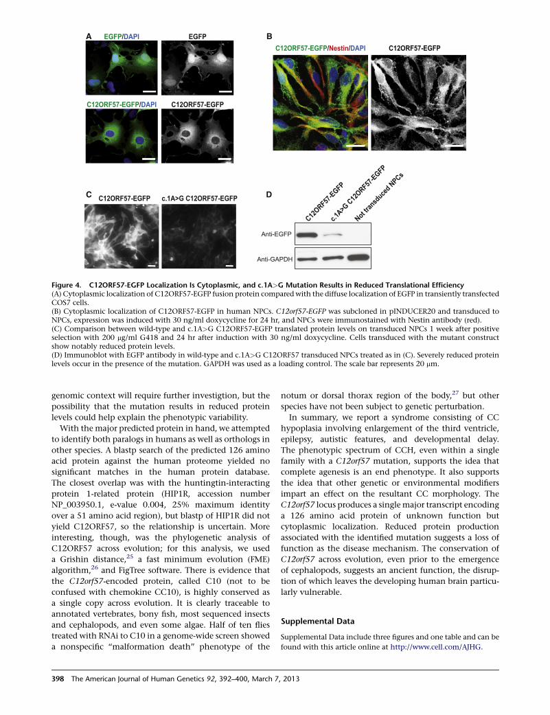
genomic context will require further investigtion, but thepossibility that the mutation results in reduced proteinlevels could help explain the phenotypic variability.With the major predicted protein in hand, we attempted
to identify both paralogs in humans as well as orthologs inother species. A blastp search of the predicted 126 aminoacid protein against the human proteome yielded nosignificant matches in the human protein database.The closest overlap was with the huntingtin-interactingprotein 1-related protein (HIP1R, accession numberNP_003950.1, e-value 0.004, 25% maximum identityover a 51 amino acid region), but blastp of HIP1R did notyield C12ORF57, so the relationship is uncertain. Moreinteresting, though, was the phylogenetic analysis ofC12ORF57 across evolution; for this analysis, we useda Grishin distance,25 a fast minimum evolution (FME)algorithm,26 and FigTree software. There is evidence thatthe C12orf57-encoded protein, called C10 (not to beconfused with chemokine CC10), is highly conserved asa single copy across evolution. It is clearly traceable toannotated vertebrates, bony fish, most sequenced insectsand cephalopods, and even some algae. Half of ten fliestreated with RNAi to C10 in a genome-wide screen showeda nonspecific ‘‘malformation death’’ phenotype of the
notum or dorsal thorax region of the body,27 but otherspecies have not been subject to genetic perturbation.In summary, we report a syndrome consisting of CC
hypoplasia involving enlargement of the third ventricle,epilepsy, autistic features, and developmental delay.The phenotypic spectrum of CCH, even within a singlefamily with a C12orf57 mutation, supports the idea thatcomplete agenesis is an end phenotype. It also supportsthe idea that other genetic or environmental modifiersimpart an effect on the resultant CC morphology. TheC12orf57 locus produces a singlemajor transcript encodinga 126 amino acid protein of unknown function butcytoplasmic localization. Reduced protein productionassociated with the identified mutation suggests a loss offunction as the disease mechanism. The conservation ofC12orf57 across evolution, even prior to the emergenceof cephalopods, suggests an ancient function, the disrup-tion of which leaves the developing human brain particu-larly vulnerable.
Supplemental Data
Supplemental Data include three figures and one table and can be
found with this article online at http://www.cell.com/AJHG.
A B
C D
Figure 4. C12ORF57-EGFP Localization Is Cytoplasmic, and c.1A>G Mutation Results in Reduced Translational Efficiency(A) Cytoplasmic localization of C12ORF57-EGFP fusion protein compared with the diffuse localization of EGFP in transiently transfectedCOS7 cells.(B) Cytoplasmic localization of C12ORF57-EGFP in human NPCs. C12orf57-EGFP was subcloned in pINDUCER20 and transduced toNPCs, expression was induced with 30 ng/ml doxycycline for 24 hr, and NPCs were immunostained with Nestin antibody (red).(C) Comparison between wild-type and c.1A>G C12ORF57-EGFP translated protein levels on transduced NPCs 1 week after positiveselection with 200 mg/ml G418 and 24 hr after induction with 30 ng/ml doxycycline. Cells transduced with the mutant constructshow notably reduced protein levels.(D) Immunoblot with EGFP antibody in wild-type and c.1A>G C12ORF57 transduced NPCs treated as in (C). Severely reduced proteinlevels occur in the presence of the mutation. GAPDH was used as a loading control. The scale bar represents 20 mm.
398 The American Journal of Human Genetics 92, 392–400, March 7, 2013

Acknowledgments
We thank the Center for Inherited Disease Research and UCLA
Microarray Core (supported by the National Institutes of Health
(NIH) and the NIH Heart, Lung, and Blood Institute) for genotyp-
ing support, and the Broad Institute (U54HG003067 to Eric
Lander) for sequencing support and analysis.We thank S.J. Elledge
for pINDUCER20 vector. We also thank the Wold lab (Caltech),
Myers Lab (HudsonAlpha Institute for Biotechnology), and
Sandstrom lab (UW ENCODE) for, respectively, RNAseq, RNApol
ChIP, and H3K4me3 ChIP data, deposited on the UCSC Genome
Browser. This work was supported by NIH grants R01NS048453,
R01NS052455, P01HD070494 (to J.G.G.), and R01NS058721 (to
E.H.S.), the Simons Foundation Autism Research Initiative, the
Howard Hughes Medical Institute (J.G.G.), and a California Insti-
tute for Regenerative Medicine training grant (N.A).
Received: September 15, 2012
Revised: December 3, 2012
Accepted: February 11, 2013
Published: February 28, 2013
Web Resources
The URLs for data presented herein are as follows:
BLAST, http://blast.ncbi.nlm.nih.gov/Blast.cgi
Online Mendelian Inheritance in Man (OMIM), http://www.
omim.org
SeattleSeq Annotation, http://snp.gs.washington.edu/
SeattleSeqAnnotation134
Exome Variant Server, http://evs.gs.washington.edu/EVS/
dbSNP Annotation, www.ncbi.nlm.nih.gov/projects/SNP/
UCSC Genome Browser, http://www.genome.ucsc.edu/
FigTree, http://tree.bio.ed.ac.uk/software/figtree/
References
1. Paul, L.K. (2011). Developmental malformation of the corpus
callosum: a review of typical callosal development and exam-
ples of developmental disorders with callosal involvement. J.
Neurodev. Disord. 3, 3–27.
2. Scamvougeras, A., Kigar, D.L., Jones, D., Weinberger, D.R., and
Witelson, S.F. (2003). Size of the human corpus callosum is
genetically determined: an MRI study in mono and dizygotic
twins. Neurosci. Lett. 338, 91–94.
3. Hanna, R.M., Marsh, S.E., Swistun, D., Al-Gazali, L., Zaki, M.S.,
Abdel-Salam, G.M., Al-Tawari, A., Bastaki, L., Kayserili, H.,
Rajab, A., et al. (2011). Distinguishing 3 classes of corpus
callosal abnormalities in consanguineous families. Neurology
76, 373–382.
4. Sztriha, L. (2005). Spectrum of corpus callosum agenesis.
Pediatr. Neurol. 32, 94–101.
5. Murray, S.S., Oliphant, A., Shen, R., McBride, C., Steeke, R.J.,
Shannon, S.G., Rubano, T., Kermani, B.G., Fan, J.B., Chee,
M.S., and Hansen, M.S. (2004). A highly informative SNP
linkage panel for human genetic studies. Nat. Methods 1,
113–117.
6. Hoffmann, K., and Lindner, T.H. (2005). easyLINKAGE-Plus—
automated linkage analyses using large-scale SNP data. Bio-
informatics 21, 3565–3567.
7. DePristo, M.A., Banks, E., Poplin, R., Garimella, K.V., Maguire,
J.R., Hartl, C., Philippakis, A.A., del Angel, G., Rivas, M.A.,
Hanna, M., et al. (2011). A framework for variation discovery
and genotyping using next-generation DNA sequencing
data. Nat. Genet. 43, 491–498.
8. Ramensky, V., Bork, P., and Sunyaev, S. (2002). Human non-
synonymous SNPs: server and survey. Nucleic Acids Res. 30,
3894–3900.
9. Grantham, R. (1974). Amino acid difference formula to help
explain protein evolution. Science 185, 862–864.
10. Siepel, A., Bejerano, G., Pedersen, J.S., Hinrichs, A.S., Hou, M.,
Rosenbloom, K., Clawson, H., Spieth, J., Hillier, L.W.,
Richards, S., et al. (2005). Evolutionarily conserved elements
in vertebrate, insect, worm, and yeast genomes. Genome
Res. 15, 1034–1050.
11. Cooper, G.M., Stone, E.A., Asimenos, G., Green, E.D., Batzo-
glou, S., and Sidow, A.; NISC Comparative Sequencing
Program. (2005). Distribution and intensity of constraint in
mammalian genomic sequence. Genome Res. 15, 901–913.
12. Derbent, M., Agras, P.I., Gedik, S., Oto, S., Alehan, F., and
Saatci, U. (2004). Congenital cataract, microphthalmia, hypo-
plasia of corpus callosum and hypogenitalism: report and
review of Micro syndrome. Am. J. Med. Genet. A. 128A,
232–234.
13. Aligianis, I.A., Johnson, C.A., Gissen, P., Chen, D., Hampshire,
D., Hoffmann, K., Maina, E.N., Morgan, N.V., Tee, L., Morton,
J., et al. (2005). Mutations of the catalytic subunit of RAB3GAP
cause Warburg Micro syndrome. Nat. Genet. 37, 221–223.
14. Carstea, E.D., Morris, J.A., Coleman, K.G., Loftus, S.K., Zhang,
D., Cummings, C., Gu, J., Rosenfeld, M.A., Pavan, W.J.,
Krizman, D.B., et al. (1997). Niemann-Pick C1 disease gene:
homology to mediators of cholesterol homeostasis. Science
277, 228–231.
15. Walterfang, M., Fahey, M., Abel, L., Fietz, M., Wood, A.,
Bowman, E., Reutens, D., and Velakoulis, D. (2011). Size and
shape of the corpus callosum in adult Niemann-Pick type C
reflects state and trait illness variables. AJNR Am. J. Neurora-
diol. 32, 1340–1346.
16. Trouard, T.P., Heidenreich, R.A., Seeger, J.F., and Erickson, R.P.
(2005). Diffusion tensor imaging in Niemann-Pick Type C
disease. Pediatr. Neurol. 33, 325–330.
17. Palmeri, S., Battisti, C., Federico, A., and Guazzi, G.C. (1994).
Hypoplasia of the corpus callosum in Niemann-Pick type C
disease. Neuroradiology 36, 20–22.
18. Li, J., Shivakumar, S., Wakahiro, M., Mukherjee, P., Barkovich,
A.J., Slavotinek, A., and Sherr, E.H. (2007). Agenesis of the
corpus callosum, optic coloboma, intractable seizures, cranio-
facial and skeletal dysmorphisms: an autosomal recessive
disorder similar to Temtamy syndrome. Am. J. Med. Genet.
A. 143A, 1900–1905.
19. Najmabadi, H., Hu, H., Garshasbi, M., Zemojtel, T., Abedini,
S.S., Chen, W., Hosseini, M., Behjati, F., Haas, S., Jamali, P.,
et al. (2011). Deep sequencing reveals 50 novel genes for reces-
sive cognitive disorders. Nature 478, 57–63.
20. Ansari-Lari, M.A., Shen, Y., Muzny, D.M., Lee, W., and Gibbs,
R.A. (1997). Large-scale sequencing in human chromosome
12p13: experimental and computational gene structure deter-
mination. Genome Res. 7, 268–280.
21. ENCODE Project Consortium. (2011). A user’s guide to the
encyclopedia of DNA elements (ENCODE). PLoS Biol. 9,
e1001046.
The American Journal of Human Genetics 92, 392–400, March 7, 2013 399

22. Rosenbloom, K.R., Sloan, C.A., Malladi, V.S., Dreszer, T.R.,
Learned, K., Kirkup, V.M., Wong, M.C., Maddren, M., Fang, R.,
Heitner, S.G., et al. (2012). ENCODE data in the UCSCGenome
Browser: year 5 update. Nucleic Acids Res. 41, D56–D63.
23. Meerbrey, K.L., Hu, G., Kessler, J.D., Roarty, K., Li, M.Z., Fang,
J.E., Herschkowitz, J.I., Burrows, A.E., Ciccia, A., Sun, T., et al.
(2011). The pINDUCER lentiviral toolkit for inducible RNA
interference in vitro and in vivo. Proc. Natl. Acad. Sci. USA
108, 3665–3670.
24. Kozak, M. (1989). Context effects and inefficient initiation at
non-AUG codons in eucaryotic cell-free translation systems.
Mol. Cell. Biol. 9, 5073–5080.
25. Grishin, N.V. (1999). A novel approach to phylogeny
reconstruction from protein sequences. J. Mol. Evol. 48,
264–273.
26. Desper, R., andGascuel,O. (2002). Fast andaccuratephylogeny
reconstruction algorithms based on the minimum-evolution
principle. J. Comput. Biol. 9, 687–705.
27. Mummery-Widmer, J.L., Yamazaki, M., Stoeger, T., Novatch-
kova, M., Bhalerao, S., Chen, D., Dietzl, G., Dickson, B.J.,
and Knoblich, J.A. (2009). Genome-wide analysis of Notch
signalling in Drosophila by transgenic RNAi. Nature 458,
987–992.
400 The American Journal of Human Genetics 92, 392–400, March 7, 2013
Copyright © 2022 FDOKUMEN