Whole-Exome Sequencing Links a Variant in DHDDS to Retinitis Pigmentosa
6
REPORT Whole-Exome Sequencing Links a Variant in DHDDS to Retinitis Pigmentosa Stephan Zu ¨chner, 1,2 Julia Dallman, 6 Rong Wen, 3 Gary Beecham, 1,2 Adam Naj, 1,2 Amjad Farooq, 4,5 Martin A. Kohli, 1,2 Patrice L. Whitehead, 1,2 William Hulme, 1,2 Ioanna Konidari, 1,2 Yvonne J.K. Edwards, 1,2 Guiqing Cai, 7 Inga Peter, 8 David Seo, 1,2 Joseph D. Buxbaum, 7,8 Jonathan L. Haines, 9 Susan Blanton, 1,2 Juan Young, 1,2 Eduardo Alfonso, 3 Jeffery M. Vance, 1,2 Byron L. Lam, 3, * and Margaret A. Peri cak-Vance 1,2, * Increasingly, mutations in genes causing Mendelian disease will be supported by individual and small families only; however, exome sequencing studies have thus far focused on syndromic phenotypes characterized by low locus heterogeneity. In contrast, retinitis pig- mentosa (RP) is caused by >50 known genes, which still explain only half of the clinical cases. In a single, one-generation, nonsyn- dromic RP family, we have identified a gene, dehydrodolichol diphosphate synthase (DHDDS), demonstrating the power of combining whole-exome sequencing with rapid in vivo studies. DHDDS is a highly conserved essential enzyme for dolichol synthesis, permitting global N-linked glycosylation. Zebrafish studies showed virtually identical photoreceptor defects as observed with N-linked glycosyla- tion-interfering mutations in the light-sensing protein rhodopsin. The identified Lys42Glu variant likely arose from an ancestral founder, because eight of the nine identified alleles in 27,174 control chromosomes were of confirmed Ashkenazi Jewish ethnicity. These findings demonstrate the power of exome sequencing linked to functional studies when faced with challenging study designs and, importantly, link RP to the pathways of N-linked glycosylation, which promise new avenues for therapeutic interventions. Retinitis pigmentosa (RP) refers to a large group of geneti- cally heterogeneous retinal degenerative disorders charac- terized by early rod photoreceptor dysfunction followed by progressive rod and cone photoreceptor dysfunction and photoreceptor death (MIM 268000). Impaired night vision followed by impaired peripheral vision generally starts in adolescence to young adulthood, with subsequent impaired central vision in later life. 1 The prevalence of RP is approximately 1 in 3000–4500 persons. 2 Autosomal- recessive, autosomal-dominant, and X-linked recessive forms of RP are all found. Mutations in over 50 genes have been identified that cause RP. These include muta- tions in genes that encode proteins in the phototransduc- tion cascade, vitamin A metabolism, cellular or cytoskel- etal structure, cell-to-cell signaling or synaptic interaction, RNA intron-splicing factors, and intracellular protein trafficking. 3 Genetic testing of genes known to cause RP fails to detect the disease-causing mutations in 50% of cases. 3 We studied a family of Ashkenazi Jewish (AJ) origin in which three out of four siblings (two females and one male) were diagnosed with RP in their teenage years (Figure 1A). Early symptoms consisted of impaired night and peripheral vision. Clinical examination of the affected individuals revealed pigmentary retinal degeneration (see Figure S4 available online), and the diagnosis of retinitis pigmentosa was confirmed by impaired rod and cone responses on electroretinograms. In addition to ophthalmic evaluations, neurologic examination, X-ray bone body survey, bone density scan, echocardiogram, brain MRI with and without contrast, serum fasting choles- terol and lipid profile, serum thyroid function studies (TSH, free T4, free T3, TBG), serum insulin-like growth factor (IGF) binding protein 1, serum IGF binding protein 2, serum clotting factors (II, V, VII, VIII, IX, X, XI, XII), and antithrombin III were performed. All studies gave normal results, but individuals II:1 and II:2 had a history of lytic bone disease first diagnosed over 15 years ago. Informed consent was obtained from all individuals, and the Institu- tional Review Board at the University of Miami Miller School of Medicine approved the study. To identify the genetic cause of this likely recessive subtype of RP, we screened all genes known to harbor RP mutations and found that they were negative for muta- tions. Classic linkage approaches were not applicable because of the size of the nonconsanguineous family, so we performed whole-exome sequencing in the three affected siblings and one unaffected sibling (Whole Human Exome Capture kit, Roche). We produced approxi- mately 10 gigabases (Gb) of paired-end 75 bp sequence reads per individual on the Illumina GAII platform. To test the overall quality of the sequence data, we compared the genotypes of variants found in the sequence data to variants derived from genotyping via a genome-wide SNP 1 John P. Hussman Institute for Human Genomics, University of Miami Miller School of Medicine, Miami, FL 33136, USA; 2 Dr. John T. Macdonald Depart- ment of Human Genetics, University of Miami Miller School of Medicine, Miami, FL 33136, USA; 3 Bascom Palmer Eye Institute, University of Miami Miller School of Medicine, Miami, FL 33136, USA; 4 Department of Biochemistry and Molecular Biology, University of Miami Miller School of Medicine, Miami, FL 33136, USA; 5 Braman Family Breast Cancer Institute, Sylvester Comprehensive Cancer Center, University of Miami, Miami, FL 33146, USA; 6 Department of Biology, University of Miami, Miami, FL 33146, USA; 7 Department of Psychiatry, Mount Sinai School of Medicine, New York, NY 10029, USA; 8 Department of Genetics and Genomic Sciences, Mount Sinai School of Medicine, New York, NY 10029, USA; 9 Center for Human Genetics Research, Vanderbilt Univer- sity School of Medicine, Nashville, TN 37232, USA *Correspondence: [email protected] (B.L.L.), [email protected] (M.A.P.-V.) DOI 10.1016/j.ajhg.2011.01.001. Ó2011 by The American Society of Human Genetics. All rights reserved. The American Journal of Human Genetics 88, 201–206, February 11, 2011 201
-
Upload
independent -
Category
Documents
-
view
3 -
download
0
Transcript of Whole-Exome Sequencing Links a Variant in DHDDS to Retinitis Pigmentosa

REPORT
Whole-Exome Sequencing Links a Variantin DHDDS to Retinitis Pigmentosa
Stephan Zuchner,1,2 Julia Dallman,6 Rong Wen,3 Gary Beecham,1,2 Adam Naj,1,2 Amjad Farooq,4,5
Martin A. Kohli,1,2 Patrice L. Whitehead,1,2 William Hulme,1,2 Ioanna Konidari,1,2
Yvonne J.K. Edwards,1,2 Guiqing Cai,7 Inga Peter,8 David Seo,1,2 Joseph D. Buxbaum,7,8
Jonathan L. Haines,9 Susan Blanton,1,2 Juan Young,1,2 Eduardo Alfonso,3 Jeffery M. Vance,1,2
Byron L. Lam,3,* and Margaret A. Peri�cak-Vance1,2,*
Increasingly, mutations in genes causing Mendelian disease will be supported by individual and small families only; however, exome
sequencing studies have thus far focused on syndromic phenotypes characterized by low locus heterogeneity. In contrast, retinitis pig-
mentosa (RP) is caused by >50 known genes, which still explain only half of the clinical cases. In a single, one-generation, nonsyn-
dromic RP family, we have identified a gene, dehydrodolichol diphosphate synthase (DHDDS), demonstrating the power of combining
whole-exome sequencing with rapid in vivo studies. DHDDS is a highly conserved essential enzyme for dolichol synthesis, permitting
global N-linked glycosylation. Zebrafish studies showed virtually identical photoreceptor defects as observed with N-linked glycosyla-
tion-interfering mutations in the light-sensing protein rhodopsin. The identified Lys42Glu variant likely arose from an ancestral
founder, because eight of the nine identified alleles in 27,174 control chromosomes were of confirmed Ashkenazi Jewish ethnicity. These
findings demonstrate the power of exome sequencing linked to functional studies when faced with challenging study designs and,
importantly, link RP to the pathways of N-linked glycosylation, which promise new avenues for therapeutic interventions.
Retinitis pigmentosa (RP) refers to a large group of geneti-
cally heterogeneous retinal degenerative disorders charac-
terized by early rod photoreceptor dysfunction followed
by progressive rod and cone photoreceptor dysfunction
and photoreceptor death (MIM 268000). Impaired night
vision followed by impaired peripheral vision generally
starts in adolescence to young adulthood, with subsequent
impaired central vision in later life.1 The prevalence of RP
is approximately 1 in 3000–4500 persons.2 Autosomal-
recessive, autosomal-dominant, and X-linked recessive
forms of RP are all found. Mutations in over 50 genes
have been identified that cause RP. These include muta-
tions in genes that encode proteins in the phototransduc-
tion cascade, vitamin A metabolism, cellular or cytoskel-
etal structure, cell-to-cell signaling or synaptic
interaction, RNA intron-splicing factors, and intracellular
protein trafficking.3 Genetic testing of genes known to
cause RP fails to detect the disease-causing mutations in
50% of cases.3
We studied a family of Ashkenazi Jewish (AJ) origin in
which three out of four siblings (two females and one
male) were diagnosed with RP in their teenage years
(Figure 1A). Early symptoms consisted of impaired night
and peripheral vision. Clinical examination of the affected
individuals revealed pigmentary retinal degeneration (see
Figure S4 available online), and the diagnosis of retinitis
pigmentosa was confirmed by impaired rod and cone
1John P. Hussman Institute for Human Genomics, University of Miami Miller S
ment of Human Genetics, University of Miami Miller School of Medicine, Miam
School of Medicine, Miami, FL 33136, USA; 4Department of Biochemistry andM
33136, USA; 5Braman Family Breast Cancer Institute, Sylvester Comprehensive
Biology, University of Miami, Miami, FL 33146, USA; 7Department of Psychiatr
of Genetics and Genomic Sciences, Mount Sinai School of Medicine, New York
sity School of Medicine, Nashville, TN 37232, USA
*Correspondence: [email protected] (B.L.L.), [email protected] (
DOI 10.1016/j.ajhg.2011.01.001. �2011 by The American Society of Human
The America
responses on electroretinograms. In addition to
ophthalmic evaluations, neurologic examination, X-ray
bone body survey, bone density scan, echocardiogram,
brainMRI with andwithout contrast, serum fasting choles-
terol and lipid profile, serum thyroid function studies
(TSH, free T4, free T3, TBG), serum insulin-like growth
factor (IGF) binding protein 1, serum IGF binding protein
2, serum clotting factors (II, V, VII, VIII, IX, X, XI, XII), and
antithrombin III were performed. All studies gave normal
results, but individuals II:1 and II:2 had a history of lytic
bone disease first diagnosed over 15 years ago. Informed
consent was obtained from all individuals, and the Institu-
tional Review Board at the University of Miami Miller
School of Medicine approved the study.
To identify the genetic cause of this likely recessive
subtype of RP, we screened all genes known to harbor RP
mutations and found that they were negative for muta-
tions. Classic linkage approaches were not applicable
because of the size of the nonconsanguineous family, so
we performed whole-exome sequencing in the three
affected siblings and one unaffected sibling (Whole
Human Exome Capture kit, Roche). We produced approxi-
mately 10 gigabases (Gb) of paired-end 75 bp sequence
reads per individual on the Illumina GAII platform. To
test the overall quality of the sequence data, we compared
the genotypes of variants found in the sequence data to
variants derived from genotyping via a genome-wide SNP
chool of Medicine, Miami, FL 33136, USA; 2Dr. John T. Macdonald Depart-
i, FL 33136, USA; 3Bascom Palmer Eye Institute, University of Miami Miller
olecular Biology, University of MiamiMiller School of Medicine, Miami, FL
Cancer Center, University of Miami, Miami, FL 33146, USA; 6Department of
y, Mount Sinai School of Medicine, New York, NY 10029, USA; 8Department
, NY 10029, USA; 9Center for Human Genetics Research, Vanderbilt Univer-
M.A.P.-V.)
Genetics. All rights reserved.
n Journal of Human Genetics 88, 201–206, February 11, 2011 201
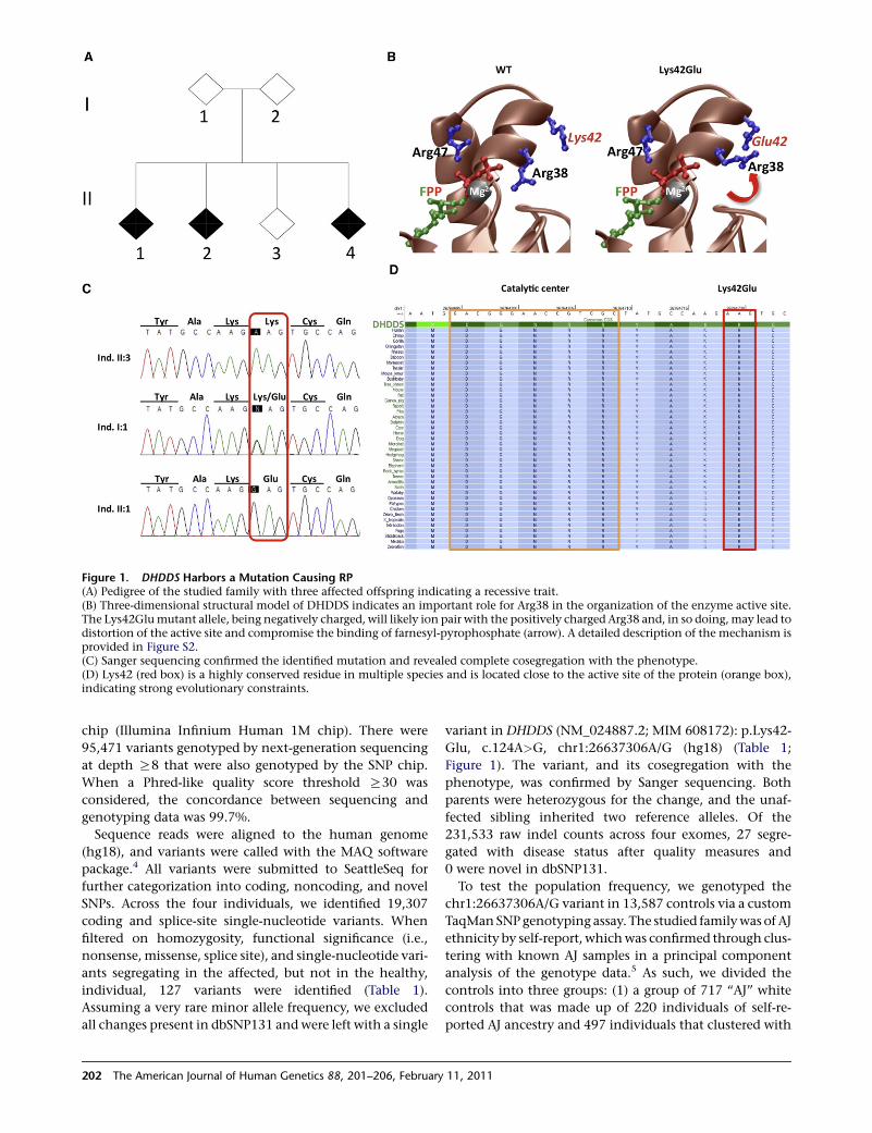
Figure 1. DHDDS Harbors a Mutation Causing RP(A) Pedigree of the studied family with three affected offspring indicating a recessive trait.(B) Three-dimensional structural model of DHDDS indicates an important role for Arg38 in the organization of the enzyme active site.The Lys42Glumutant allele, being negatively charged, will likely ion pair with the positively charged Arg38 and, in so doing,may lead todistortion of the active site and compromise the binding of farnesyl-pyrophosphate (arrow). A detailed description of the mechanism isprovided in Figure S2.(C) Sanger sequencing confirmed the identified mutation and revealed complete cosegregation with the phenotype.(D) Lys42 (red box) is a highly conserved residue in multiple species and is located close to the active site of the protein (orange box),indicating strong evolutionary constraints.
chip (Illumina Infinium Human 1M chip). There were
95,471 variants genotyped by next-generation sequencing
at depth R8 that were also genotyped by the SNP chip.
When a Phred-like quality score threshold R30 was
considered, the concordance between sequencing and
genotyping data was 99.7%.
Sequence reads were aligned to the human genome
(hg18), and variants were called with the MAQ software
package.4 All variants were submitted to SeattleSeq for
further categorization into coding, noncoding, and novel
SNPs. Across the four individuals, we identified 19,307
coding and splice-site single-nucleotide variants. When
filtered on homozygosity, functional significance (i.e.,
nonsense, missense, splice site), and single-nucleotide vari-
ants segregating in the affected, but not in the healthy,
individual, 127 variants were identified (Table 1).
Assuming a very rare minor allele frequency, we excluded
all changes present in dbSNP131 andwere left with a single
202 The American Journal of Human Genetics 88, 201–206, February
variant in DHDDS (NM_024887.2; MIM 608172): p.Lys42-
Glu, c.124A>G, chr1:26637306A/G (hg18) (Table 1;
Figure 1). The variant, and its cosegregation with the
phenotype, was confirmed by Sanger sequencing. Both
parents were heterozygous for the change, and the unaf-
fected sibling inherited two reference alleles. Of the
231,533 raw indel counts across four exomes, 27 segre-
gated with disease status after quality measures and
0 were novel in dbSNP131.
To test the population frequency, we genotyped the
chr1:26637306A/G variant in 13,587 controls via a custom
TaqManSNPgenotyping assay. The studied familywas of AJ
ethnicity by self-report, whichwas confirmed through clus-
tering with known AJ samples in a principal component
analysis of the genotype data.5 As such, we divided the
controls into three groups: (1) a group of 717 ‘‘AJ’’ white
controls that was made up of 220 individuals of self-re-
ported AJ ancestry and 497 individuals that clustered with
11, 2011

Table 1. Number of Identified Single-Nucleotide Variants Compared to the Reference Genome hg18 and Their Categorization andFiltering
AffectedSibling 1
AffectedSibling 2
AffectedSibling 3
Shared betweenAffectedSiblings 1 and 2
Shared betweenAffectedSiblings 1, 2, and 3
þ NOT inUnaffectedSibling 4
Missense, nonsense, splice-sitevariants
8712 8716 8752 5289 4507 ND
þ Homozygous state 2516 2489 2649 1411 981 127
þ Not reported in dbSNP131 11 18 27 5 4 1
ND denotes not determined.
known AJ in a principal component analysis based on
genome-wide genotype data, (2) a group of 6977 ‘‘non-AJ’’
white controls that did not cluster with the known AJ
samples, and (3) a group of 5893 white controls without
genome-wide genotype data. The chr1:26637306A/G
variant was observed only eight times among the 1434
chromosomes from the AJ controls. Each time, the variant
was observed as a heterozygote (minor allele frequency
[MAF] ¼ 0.0056). This confirms that the variant is indeed
rare among the AJ population and that homozygotes
should be very rare (frequency ¼ 3 3 10�5, or 3 in
100,000, assuming Hardy-Weinberg equilibrium). The
variant is extremely rare in non-AJ populations; it was not
observed in any of the confirmed non-AJ controls (0 out
of 13,954 chromosomes;MAF< 0.00008) andwas observed
only once in controls without genome-wide genotype data
(1 out of 11,786 chromosomes; MAF< 0.00009) (Table S1).
The array-based genotype data were also utilized post
hoc to attest for consistency with the hypothesis that
this mutation is causal for RP. Two different, though
related, methods were applied. The first was to search for
runs of homozygosity present among the affected siblings
but not present in the remaining familymembers. This was
accomplished with the PLINK software.6 To determine
large blocks of homozygosity, we required ‘‘homozygous’’
segments to be at least 500 kb long and have at least
95% homozygosity. There were 91 such regions that were
homozygous among all three affected siblings and were
not homozygous among the parents and unaffected
sibling (Table S3). One of these regions on chr1:25.7Mb–
chr1:29Mb contained DDHDS. The second method to
prioritize variants was to examine estimated identity by
descent (IBD) to determine regions for which all affected
siblings share the same maternal and paternal haplotypes
(regardless of homozygosity or heterozygosity). There
were 33 regions estimated to be completely shared by all
three affected siblings.DHDDS falls in one of these regions.
We identified four known RP genes within the homozy-
gosity and IBD sharing regions (PDE6B [MIM 180072],
USH1G/SANS [MIM 706696], PRCD [MIM 610598], and
CA4 [MIM 114760]).7 Mutations in these genes were
excluded by Sanger sequencing (PDE6B, USH1G/SANS,
CA4) and/or exome sequencing (PRCD).
DHDDS is a key enzyme in the final steps of dolichol-
pyrophosphate synthesis.8 As shown in Figure 1D, the
The America
Lys42 residue is highly conserved in different species
and is located close to the catalytic center and the
substrate binding site for farnesyl pyrophosphate phos-
phate (FPP) of the enzyme.9 The observed Lys>Glu
amino acid change at position 42 is predicted to be
‘‘damaging’’ by Polyphen10 and received a high 5.25
genomic evolutionary rate profiling conservation score.11
To further understand the physiological significance of
the Lys42Glu mutation, we applied the MODELER soft-
ware package12 to generate 3D atomic models of human
DHDDS in complex with FPP/Mg2þ on the basis of
amino acid sequence ortholog of E. coli dhdds, for which
there is a known crystal structure in complex with FPP/
Mg2þ13 (Figure 1B and Figure S1). Within the enzyme
active site, the farnesyl tail of FPP inserts deep into
a hydrophobic cavity stabilized by numerous intermolec-
ular van der Waals contacts, with the basic residues Arg38
and Arg47 playing a key role (Figure 1). Residue Lys42 is
sterically close to residue Arg38 and facilitates its optimal
orientation for ion pairing with the pyrophosphate group
of FPP via charge-charge repulsive forces. Remarkably, the
mutant residue Glu42 will compete with the pyrophos-
phate group of FPP for ion pairing with Arg38, resulting
in the misorientation of guanidine moiety of Arg38.
This suggests a loss-of-function mechanism in effectively
binding the substrate FPP.
To determine whether insufficient DHDDS activity
induces photoreceptor degeneration, we knocked down
the expression of the highly conserved dhdds
(NM_213187.1) in zebrafish by injecting a splice-junction
blocking morpholino (MO) into fertilized eggs. MO oligo-
nucleotides (GeneTools LLC) designed against exon1/
intron1 of the zebrafish dhdds were injected into one-
cell embryos (Figure S2). Embryos were allowed to
develop for 4 days. At this age, they reliably respond to
abrupt changes in light intensity with bursts of swim-
ming (Movies S1–S3). Dhddsmorphants exhibited a range
of phenotypes typical of such knockdown experiments.
In functional assays, MO-injected fish failed to react to
light on-off switches in contrast to controls that reacted
to light on-off switches with typical escape responses
(see Figure S5 and Movies S1–S3). Upon microscopic
examination, the gross pathology of the retina was intact
in both the wild-type and the MO-treated animals
(Figure 2). However, the photoreceptor outer segments
n Journal of Human Genetics 88, 201–206, February 11, 2011 203

Figure 2. Photoreceptor Degeneration Induced by Suppression of DHDDS Expression and Illustration of the Involved Pathways of N-Linked Glycosylation(A–F) Microphotographs in (A) and (D) show representative retinal sections of control and fish treated with MO. The areas in red rect-angles are shown at higher magnification in (B) and (E). The layers of the retina are indicated by the white bars in (A), including theretinal pigment epithelium (1), the outer nuclear layer (2), the inner nuclear layer (3), the inner plexiform layer (4), and the retinalganglion cell layer (5). In the retinas treated with MO, the outer segments of photoreceptors are very short, or completely missing(E, arrowhead), compared to the controls (B, arrowhead). Staining of cone outer segments with peanut agglutinin (PNA) highlightsthat cone outer segments in MO-treated zebrafish (F, arrowhead), but not in controls (C, arrowhead), are missing. Semithin plasticsections (A, B, D, and E) were stained with toluidine blue. Cryosections (C and F) were stained with PNA (conjugated with Alexa Fluor488). Scale bars represent 25 mm for (A) and (D), 10 mm for (B) and (E), and 20 mm for (C) and (F).(G) A simplified schematic of essential N-linked glycosylation pathways (steps 1–4). An emphasis is given to key processes upstream anddownstream of dolichol-PP synthesis (DHDDS) that are also related to RP phenotypes. (1) The cytoplasmic mevalonate pathwayproduces isoprenoide units, which are (2) chained to ER membrane-bound dolichol species, dolichol-pyrophosphate (dolichol-PP).(3) Dolichol-PP serves as an intermediate lipid to bind a common core of carbohydrates, en bloc, forming the lipid-linked oligosaccharide(LLO). The LLO is ‘‘flipped’’ within the ER membrane to face the ER lumen. (4) Finally, the common core of carbohydrates is transferredto proteins, such as rhodopsin. Along this pathway, mevalonate kinase deficiency is associated with RP,14 mutations in PMM2 (phospho-mannomutase 2; MIM 601785) lead to congenital disorder of glycosylation Ia (CDGIa), often involving RP, and, finally, glycosylationdefects in rhodopsin cause RP. N denotes asparagine.
were very short or completely missing in the retinas
treated with dhdds morpholinos (Figure 2; Figure S3). In
addition to a reduced lights-off response and reduced or
absent photoreceptor outer segments, one-third of mor-
phants had smaller eyes, with a slight ventral flexion to
the body axis; this indicates that, depending on degree
of dhdds knockdown, a broader set of tissues are affected,
with the most sensitized being photoreceptors. These
results are consistent with dhdds being a key protein for
the generation and continuous renewal of photoreceptor
outer segments.
What is the functional role of DHDDS? About 65% of all
mammalianproteins contain at least oneN-linked glycosyl-
ation site (Asn-X-Ser(Thr)). In generating N-linked glyco-
204 The American Journal of Human Genetics 88, 201–206, February
proteins, first a common core of carbohydrate is attached
to a lipid intermediate that resides in the endoplasmic retic-
ulum membrane: dolichol-pyrophosphate (dolichol-PP).
Only after this lipid-linked oligosaccharide structure has
formed can the transfer of oligosaccharides to proteins
occur to form glycoproteins. DHDDS catalyzes the forma-
tionof dolichol-PP by chaining 17–21 isoprene units. These
isoprene units are a product of the mevalonate pathway.
Thus, DHDDS connects the upstreammevalonate pathway
to themembrane-boundN-linked glycosylationmachinery
supportedby amultitudeof enzymes and cofactors. As illus-
trated in Figure 2G, along these pathways, several clinically
complex and heterogeneous genetic conditions involve RP
as a symptom: (1) mevalonate kinase deficiency (MIM
11, 2011

610377) can present with RP,14 (2) congenital disorder of
glycosylation Ia (MIM 212065) is frequently associated
with RP,15 and (3) mutations in glycosylation sites of the
light-activated G protein-coupled receptor rhodopsin
(MIM 180380), in itself a cause for RP,16 lead to shortening
of rodouter segments in the retina ofX. laevis.17 These latter
changes are virtually identical to those we observed in ze-
brafish (Figure 2). Rhodopsin is responsible for light capture
and resides in the outer segment of photoreceptor cells.
Given that the outer segment, together with the rhodopsin
it contains, is continuously being renewed,18 it is conceiv-
able that this highly specialized structure has a pronounced
susceptibility to defects ofN-linked glycosylation, as would
be caused by less-efficient production of dolichols. We
therefore suggest that the identified mutation causes
reduced, rather than complete, loss of enzymatic function
of DHDDS. Notably, two of the affected subjects were diag-
nosed with lytic bones, potentially indicating additional
subclinical consequences of the DHDDS defect, although
the literature does not support a clear connection of lytic
bones with diseases of glycosylation. A reduced enzymatic
activity ofDHDDS, due to insufficient substrate interaction,
might eventually provide an opportunity for a therapeutic
intervention.
In summary, we have shown that even under most con-
strained preconditions, such as a single nuclear family with
only three affected siblings suffering from a genetically
highly heterogeneous ‘‘pure’’ phenotype, a causative
variant linked to RP can be identified by combining
human genomic sequencing approaches with rapid animal
modeling and in silico prediction of protein function. It
thus appears to be possible to overcome some of the inter-
pretation challenges that increasingly confront Mendelian
genetics and genomic medicine.
Supplemental Data
Supplemental Data include five figures, three tables, and three
movies and can be found with this article online at http://www.
cell.com/AJHG/.
Acknowledgments
We are thankful to the family members studied and to their
support for our research. We also thank Olaf Bodamer for helpful
discussions and Yiwen Li, Deqiang Huang, and Zhengying Wang
for technical assistance on the development of this manuscript.
This study was supported by grants from the Department of
Defense (W81XWH-09-1-0674), National Institutes of Health
(P30-EY14801 core grant, R01-EY018586 to R.W., R01-EY012118
to M.A.P.-V., R01-GM083897 to A.F., and U54-NS065712 to S.Z.),
Hope for Vision, an unrestricted grant from Research to Prevent
Blindness, and a grant from the Florida Office of Tourism, Trade
and Economic Development.
Received: October 14, 2010
Revised: January 4, 2011
Accepted: January 10, 2011
Published online: February 3, 2011
The America
Web Resources
The URLs for data presented herein are as follows:
Online Mendelian Inheritance in Man (OMIM), http://www.ncbi.
nlm.nih.gov/Omim/
SeattleSeq Annotation, http://gvs.gs.washington.edu/SeattleSeq
Annotation/
References
1. Heckenlively, J.R., Yoser, S.L., Friedman, L.H., and Oversier, J.J.
(1988). Clinical findings and common symptoms in retinitis
pigmentosa. Am. J. Ophthalmol. 105, 504–511.
2. Pagon, R.A. (1988). Retinitis pigmentosa. Surv. Ophthalmol.
33, 137–177.
3. Hartong, D.T., Berson, E.L., and Dryja, T.P. (2006). Retinitis
pigmentosa. Lancet 368, 1795–1809.
4. Li, H., Ruan, J., and Durbin, R. (2008). Mapping short DNA
sequencing reads and calling variants using mapping quality
scores. Genome Res. 18, 1851–1858.
5. Price, A.L., Patterson, N.J., Plenge, R.M., Weinblatt, M.E.,
Shadick, N.A., and Reich, D. (2006). Principal components
analysis corrects for stratification in genome-wide association
studies. Nat. Genet. 38, 904–909.
6. Purcell, S., Neale, B., Todd-Brown, K., Thomas, L., Ferreira,
M.A., Bender, D., Maller, J., Sklar, P., de Bakker, P.I., Daly,
M.J., and Sham, P.C. (2007). PLINK: A tool set for whole-
genome association and population-based linkage analyses.
Am. J. Hum. Genet. 81, 559–575.
7. Goodwin, P. (2008). Hereditary retinal disease. Curr. Opin.
Ophthalmol. 19, 255–262.
8. Endo, S., Zhang, Y.W., Takahashi, S., and Koyama, T. (2003).
Identification of human dehydrodolichyl diphosphate syn-
thase gene. Biochim. Biophys. Acta 1625, 291–295.
9. Fujihashi, M., Zhang, Y.W., Higuchi, Y., Li, X.Y., Koyama, T.,
and Miki, K. (2001). Crystal structure of cis-prenyl chain elon-
gating enzyme, undecaprenyl diphosphate synthase. Proc.
Natl. Acad. Sci. USA 98, 4337–4342.
10. Adzhubei, I.A., Schmidt, S., Peshkin, L., Ramensky, V.E., Gera-
simova, A., Bork, P., Kondrashov, A.S., and Sunyaev, S.R.
(2010). A method and server for predicting damaging
missense mutations. Nat. Methods 7, 248–249.
11. Cooper, G.M., Stone, E.A., Asimenos, G., Green, E.D., Batzo-
glou, S., and Sidow, A.; NISC Comparative Sequencing
Program. (2005). Distribution and intensity of constraint in
mammalian genomic sequence. Genome Res. 15, 901–913.
12. Martı-Renom, M.A., Stuart, A.C., Fiser, A., Sanchez, R., Melo,
F., and Sali, A. (2000). Comparative protein structure
modeling of genes and genomes. Annu. Rev. Biophys. Biomol.
Struct. 29, 291–325.
13. Guo, R.T., Ko, T.P., Chen, A.P., Kuo, C.J., Wang, A.H., and
Liang, P.H. (2005). Crystal structures of undecaprenyl pyro-
phosphate synthase in complex with magnesium, isopen-
tenyl pyrophosphate, and farnesyl thiopyrophosphate: Roles
of the metal ion and conserved residues in catalysis. J. Biol.
Chem. 280, 20762–20774.
14. Balgobind, B., Wittebol-Post, D., and Frenkel, J. (2005). Reti-
nitis pigmentosa in mevalonate kinase deficiency. J. Inherit.
Metab. Dis. 28, 1143–1145.
15. Grunewald, S. (2009). The clinical spectrum of phosphoman-
nomutase 2 deficiency (CDG-Ia). Biochim. Biophys. Acta
1792, 827–834.
n Journal of Human Genetics 88, 201–206, February 11, 2011 205

16. Sullivan, L.S., Heckenlively, J.R., Bowne, S.J., Zuo, J., Hide,
W.A., Gal, A., Denton, M., Inglehearn, C.F., Blanton, S.H.,
and Daiger, S.P. (1999). Mutations in a novel retina-specific
gene cause autosomal dominant retinitis pigmentosa. Nat.
Genet. 22, 255–259.
206 The American Journal of Human Genetics 88, 201–206, February
17. Tam, B.M., and Moritz, O.L. (2009). The role of rhodopsin
glycosylation in protein folding, trafficking, and light-sensi-
tive retinal degeneration. J. Neurosci. 29, 15145–15154.
18. Young, R.W. (1967). The renewal of photoreceptor cell outer
segments. J. Cell Biol. 33, 61–72.
11, 2011