
Dental Topography of Platyrrhines and Prosimians: Convergence and Contrasts
Upload
mpibpc-mpgCategory
view
3download
0
www.elsevier.com/locate/ynimg
NeuroImage 32 (2006) 989–994Topography of the human corpus callosum revisited—Comprehensivefiber tractography using diffusion tensor magnetic resonance imaging
Sabine Hofer a,b,⁎ and Jens Frahma
aBiomedizinische NMR Forschungs GmbH am Max-Planck-Institut für biophysikalische Chemie, 37070 Göttingen, GermanybBernstein Center for Computational Neuroscience, 37073 Göttingen, Germany
Received 21 March 2006; revised 19 May 2006; accepted 23 May 2006Available online 18 July 2006
Several tracing studies have established a topographical distribution offiber connections to the cortex in midsagittal cross-sections of the corpuscallosum (CC). The most prominent example is Witelson's scheme,which defines five vertical partitions mainly based on primate data.Conventional MRI of the human CC does not reveal morphologicallydiscernable structures, although microscopy techniques identifiedmyelinated axons with a relatively small diameter in the anterior andposterior third of the CC as opposed to thick fibers in the midbody andposterior splenium. Here, we applied diffusion tensor imaging (DTI) inconjunction with a tract-tracing algorithm to gain cortical connectivityinformation of the CC in individual subjects. With DTI-basedtractography, we distinguished five vertical segments of the CC,containing fibers projecting into prefrontal, premotor (and supplemen-tary motor), primary motor, and primary sensory areas as well as intoparietal, temporal, and occipital cortical areas. Striking differences toWitelson's classification were recognized in the midbody and anteriorthird of the CC. In particular, callosal motor fiber bundles were found tocross the CC in a much more posterior location than previouslyindicated. Differences in water mobility were found to be in qualitativeagreement with differences in the microstructure of transcallosal fibersyielding the highest anisotropy in posterior regions of the CC. The lowestanisotropy was observed in compartments assigned to motor andsensory cortical areas. In conclusion, DTI-based fiber tractography ofhealthy human subjects suggests a modification of the widely acceptedWitelson scheme and a new classification of vertical CC partitions.© 2006 Elsevier Inc. All rights reserved.
Keywords: DTI; Human brain; Primary motor cortex; Fractional anisotropy
Introduction
The corpus callosum (CC) is by far the largest fiber bundle in thehuman brain interconnecting the two cerebral hemispheres withmore than 300 million fibers. Surgical transection of the CC in
⁎ Corresponding author. Fax: +49 551 201 1307.E-mail address: [email protected] (S. Hofer).Available online on ScienceDirect (www.sciencedirect.com).
1053-8119/$ - see front matter © 2006 Elsevier Inc. All rights reserved.doi:10.1016/j.neuroimage.2006.05.044
humans provides evidence of its important role in interhemisphericfunctional integration communicating perceptual, cognitive, learned,and volitional information. Most of the fibers serve homotopicinterconnections between the hemispheres, but a number ofheterotopic fibers also asymmetrically link functionally differentcortical areas (Clarke and Zaidel, 1994; De Lacoste et al., 1985;Witelson, 1989). The CC is one of the few white matter tracts thatcan be discretely identified by conventional MRI. It has been thetarget of extensive studies (e.g., see Thompson et al., 2003)indicating that its morphology may be related to dyslexia (vonPlessen et al., 2002), Tourette's syndrome (Plessen et al., 2004),Down's syndrome (Teipel et al., 2003), depression (Lacerda et al.,2005), schizophrenia (Narr et al., 2002), and HIV/AIDS (Thompsonet al., 2006).
Due to the fact that there are no macroscopic anatomicallandmarks that clearly delimit distinct callosal areas in amidsagittal cross-section, several geometric partitioning schemeshave been designed to subdivide the CC (Duara et al., 1991;Larsen et al., 1992; Weis et al., 1993; Rajapakse et al., 1996). Infact, most studies rely on Witelson's classification, although theunderlying data predominantly originates from non-human pri-mates (Witelson, 1989). This scheme defines five vertical callosalsegments based on specific arithmetic fractions of the maximumanterior–posterior extent. In particular, the CC is subdivided intoregions comprising the anterior third, the anterior and posteriormidbody, the posterior third, and the posterior one-fifth. Compart-ments of the anterior third, including the rostrum, genu, and rostralbody, are assigned to prefrontal, premotor, and supplementarymotor cortical areas. Fibers originating in the motor cortex areassumed to cross the CC through the anterior midbody, whereassomaesthetic and posterior parietal fiber bundles cross the CCthrough the posterior midbody. Compartments of the posteriorthird, including the isthmus and splenium, are assigned totemporal, parietal, and occipital cortical regions. It should benoted, however, that neither Witelson's classification nor othergeometric partitioning schemes exactly mirror the texture of theCC at the cellular level.
Light microscopic examinations of the fiber composition in thehuman CC (Aboitz et al., 1992) revealed a well-defined micro-
990 S. Hofer, J. Frahm / NeuroImage 32 (2006) 989–994
structure with a consistent pattern of regional differentiation. Thedensity of fibers with a small diameter is most pronounced in theanterior CC (genu), decreases to a minimum in the posteriormidbody, and increases again toward the posterior CC (splenium). Incontrast, fibers with a large diameter show a diametrically opposedpattern with a peak density in the posterior midbody. Absolute fiberdensity decreases from the genu to the posterior midbody, increaseswhen entering the splenium, and is diminished again in the mostposterior splenial region.
The development of magnetic resonance diffusion tensorimaging (DTI) in conjunction with fiber tractography has openednew possibilities in the research of white matter anatomy. Theapproach provides unique access to in vivo information about thetopography of major fiber tracts and even their maturation duringontogeny (Huang et al., 2005; Dougherty et al., 2005; Ramnani et al.,2004). The method is based on the fact that the MRI-detectablediffusivity of water molecules depends on the principal orienta-tion of the fiber tracts within white matter. Diffusion-weightedMRI sequences probe such mobility along multiple directions inorder to fully characterize its orientational distribution within animage voxel. Under the assumption that this distribution may bemathematically represented by a tensor, the principle axis of thecorresponding diffusion ellipsoid coincides with the direction ofthe greatest diffusion coefficient, which then can be identifiedwith the orientation of the underlying fiber bundle. Anisotropymeasures reflect the degree to which diffusion is preferred alongthis direction relative to other directions. Here, fractional ani-sotropy (FA) values have been used as scalar indicators of thedirectionality and coherence of fiber tracts (Basser and Pierpaoli,1996).
The goal of this study was to complement structural assess-ments of the CC by cortical connectivity information in individualsubjects by means of a geometrically undistorted DTI techniquewithout sensitivity to susceptibility differences (Nolte et al., 2000;Rieseberg et al., 2005) in conjunction with a simple algorithm fortract tracing (Mori et al., 1999). A region-of-interest (ROI) to ROItractography method was employed to identify all trajectories thatcross individual pixels within the entire CC (first ROI) andpenetrate selective cortex areas (second ROI). This strategyresulted in a detailed separation of transcallosal fiber tracts withrespect to their specific cortical projections. In addition, theapproach allowed for an assignment of the distribution of FAvalues to functionally distinct CC regions.
Methods
Participants
A total of eight right-handed human subjects (4 female, 4 male;age range 21−47 years) participated in the study. None of them hadany history of neurological abnormality. Handedness was assessedin accordance to the hand preference score reported by Oldfield(1971). All subjects gave written informed consent before eachMRI examination.
Data acquisition
MRI studies were conducted at 2.9 T (Siemens Trio, Erlangen,Germany) using an 8-channel phased-array head coil. Acquisitionswere performed at 2 mm isotropic resolution using diffusion-weighted single-shot stimulated echo acquisition mode (STEAM)
MRI sequences and 5/8 partial Fourier encoding in combinationwith a projection onto convex subjects (POCS) reconstructionalgorithm (Rieseberg et al., 2005). The protocol comprised 24independent diffusion gradient directions and b values of 0 and1000 s/mm2. A total of 50 transverse sections (2 mm thickness)covered the CC and the upper part of the brain. The acquisition timeper data set was approximately 9min. To increase the signal-to-noiseratio, the acquisition was repeated three times. Anatomic imageswere based on a T1-weighted 3D fast low angle shot (FLASH) MRIsequence (repetition time TR = 11 ms, echo time TE = 4.9 ms, flipangle 15°).
Fiber tractography
Before calculation of the diffusion tensor the diffusion-weightedimages were smoothed with a 3D Gaussian filter (σ = 2 mm).Estimates of axonal projections were computed by the fiberassignment by continuous tracking (FACT) algorithm (Mori et al.,1999). Tracking terminated when the FA value was lower than 0.15or the main diffusion directions in consecutive steps differed bymore than 40° (empirically optimized thresholds). Axonal projec-tions were traced in both anterograde and retrograde directions. AllROIs were manually defined on color-coded maps of the maindiffusion direction. The first ROI was placed in a midsagittal planeto define the entire CC. The second target ROI was placed close tothe cortex in a transverse section to separate the transcallosal pro-jections into prefrontal, premotor (and supplementary motor),primary motor, primary sensory, parietal, temporal, and occipitalregions. The DTI analysis relied on software developed in-house(M. Küntzel).
Parcellation of the corpus callosum
We arrived at a parcellation of the corpus callosum byintroducing vertical subdivisions – similar to the Witelson clas-sification – but with respect to the outcome of the fiber tractography.In the midsagittal section of the CC, a geometric baseline wasdefined by connecting the most anterior and posterior points of theCC. After fiber tracking, the CC regions crossed by fibers belongingto defined cortical areas provided a natural segmentation. Themaximum anterior and posterior extent of a segment were used asborder lines and projected onto the baseline. The callosal parietal,temporal, and occipital fiber bundles overlapped and could not beseparated by vertical lines. They constitute the most posterior regionof the CC.
Fractional anisotropy
Quantitative measures of diffusion anisotropy were evaluated inthe midsagittal and the two directly neighboring CC sections. Inevery subject, we determined the mean FA values averaged acrossthe three sections for each cortical region as defined by fibertractography. To account for interindividual differences in absoluteanisotropy and to facilitate group comparisons, the data werenormalized to the mean of all FA values in the CC of the respectivesubject. We then calculated the mean and standard deviation of thenormalized FA values of every CC region across subjects. Regionaldifferences were tested for significance (Superior PerformingSoftware System, SPSS Inc.) using ANOVA combined with a posthoc test (Scheffe's test for multiple comparisons) at a threshold ofP < 0.05.
991S. Hofer, J. Frahm / NeuroImage 32 (2006) 989–994
Results and discussion
Topography of callosal fiber tracts
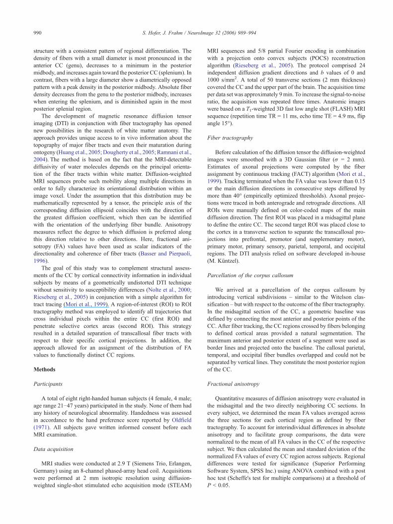
DTI-based tractography allowed us to reconstruct comprehen-sive sets of callosal fiber bundles projecting to the corticalhemispheres in all subjects. By classifying tracts according to theircortical projections, we could distinguish between fibers associatedwith prefrontal, premotor (combined with supplementary motor),primary motor, primary sensory, parietal, temporal and occipitalcortical regions. The resulting topology of fiber bundles in the CCis demonstrated in Figs. 1A–C for a single subject using 3D viewsof respective tracts as well as in Fig. 2 for all subjects by outliningthe regional distribution of fibers from all discernable projectionareas that cross the midsagittal CC.
The mapping of the callosal radiation, by which correspondingcortical areas of opposite hemispheres are connected, was achievedin much more detail than described before (Huang et al., 2005)
Fig. 1. Transcallosal fiber tracts from a single male subject overlaid onto individualreconstruction of all callosal fibers comprising bundles projecting into the prefrontaprimary motor cortex (dark blue), primary sensory cortex (red), parietal lobe (orangoblique views of callosal fiber tracts that project into the primary motor cortex. Thcolor code in the lower right of the figure: A, anterior; I, inferior; L, left; P, poste
and, for example, accounts for a clear separation of callosalprimary motor and sensory fiber bundles. Most importantly,however, fibers originating in the motor cortex were found to crossthe CC through its posterior half. This is shown in more detail inFigs. 1D and E again using 3D views of complete fiber tracks(single subject) and in Fig. 2 by selectively marking the callosalmotor fibers in the midsagittal plane (all subjects). The region ofthe CC associated with the frontal cortex appears enlarged up to thetwo-thirds when compared to Witelson's classification. Fibersoriginating in cortical areas posterior to the motor cortex pass theCC through the remaining posterior one third.
One of the most striking differences compared with theWitelson scheme is that callosal motor and sensory fiber bundlescross the CC in a much more posterior location. Thesediscrepancies between the DTI-based fiber topology of the humanCC and Witelson's classification as the standard segmentationstimulated us to suggest a new scheme. As shown in Fig. 3, theproposed arrangement of subdivisons still represents a rather
anatomical reference images. (A–C) Sagittal, top, and oblique views of a 3Dl lobe (coded in green), premotor and supplementary motor areas (light blue),e), occipital lobe (yellow), and temporal lobe (violet). (D and E) Sagittal ande colors correspond to the local mean diffusion direction as indicated by therior; R, right; S, superior.
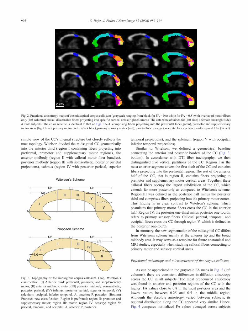
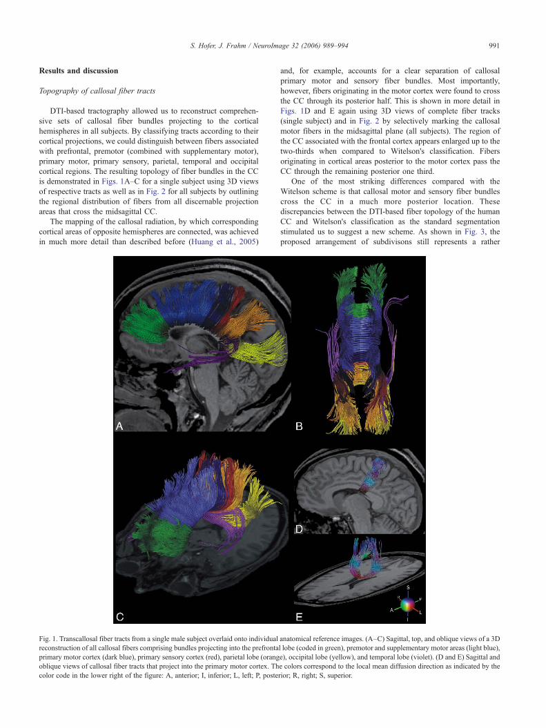
Fig. 2. Fractional anisotropy maps of the midsagittal corpus callosum (grayscale ranging from black for FA = 0 to white for FA = 0.8) with overlay of motor fibersonly (left columns) and all discernable fibers projecting into specific cortical areas (right columns). The data were obtained for (left side) 4 female and (right side)4 male subjects. The color scheme is identical to that of Figs. 1A–C comprising fibers projecting into the prefrontal lobe (green), premotor and supplementarymotor areas (light blue), primary motor cortex (dark blue), primary sensory cortex (red), parietal lobe (orange), occipital lobe (yellow), and temporal lobe (violet).
992 S. Hofer, J. Frahm / NeuroImage 32 (2006) 989–994
simple view of the CC's internal structure but closely reflects thetract topology. Witelson divided the midsagittal CC geometricallyinto the anterior third (region I containing fibers projecting intoprefrontal, premotor and supplementary motor regions), theanterior midbody (region II with callosal motor fiber bundles),posterior midbody (region III with somaesthetic, posterior parietalprojections), isthmus (region IV with posterior parietal, superior
Fig. 3. Topography of the midsagittal corpus callosum. (Top) Witelson'sclassification. (I) Anterior third: prefrontal, premotor, and supplementarymotor; (II) anterior midbody: motor; (III) posterior midbody: somaesthetic,posterior parietal; (IV) isthmus: posterior parietal, superior temporal; (V)splenium: occipital, inferior temporal. A, anterior; P, posterior. (Bottom)Proposed new classification. Region I: prefrontal; region II: premotor andsupplementary motor; region III: motor; region IV: sensory; region V:parietal, temporal, and occipital. A, anterior; P, posterior.
temporal projections), and the splenium (region V with occipital,inferior temporal projections).
Similar to Witelson, we defined a geometrical baselineconnecting the anterior and posterior borders of the CC (Fig. 3,bottom). In accordance with DTI fiber tractography, we thendistinguished five vertical partitions of the CC. Region I as themost anterior segment covers the first sixth of the CC and containsfibers projecting into the prefrontal region. The rest of the anteriorhalf of the CC, that is region II, contains fibers projecting topremotor and supplementary motor cortical areas. Together, thesecallosal fibers occupy the largest subdivision of the CC, whichextends far more posteriorly as compared to Witelson's scheme.Region III was defined as the posterior half minus the posteriorthird and comprises fibers projecting into the primary motor cortex.This finding is in clear contrast to Witelson's scheme, whichpostulates that primary motor fibers cross the CC in the anteriorhalf. Region IV, the posterior one-third minus posterior one-fourth,refers to primary sensory fibers. Callosal parietal, temporal, andoccipital fibers cross the CC through region V, which is defined asthe posterior one-fourth.
In summary, the new segmentation of the midsagittal CC differsfrom Witelson's scheme mainly at the anterior tip and the broadmidbody area. It may serve as a template for future anatomical andMRI studies, especially when studying callosal fibers connecting toprimary motor and sensory cortical areas.
Fractional anisotropy and microstructure of the corpus callosum
As can be appreciated in the grayscale FA maps in Fig. 2 (leftcolumns), there are consistent differences in diffusion anisotropyacross the CC in all subjects. The most pronounced anisotropywas found in anterior and posterior regions of the CC with thehighest FA values close to 0.8 in the most posterior area and thelowest values between 0.25 and 0.5 in the middle region.Although the absolute anisotropy varied between subjects, itsregional distribution along the CC appeared very similar. Hence,Fig. 4 compares normalized FA values averaged across subjects
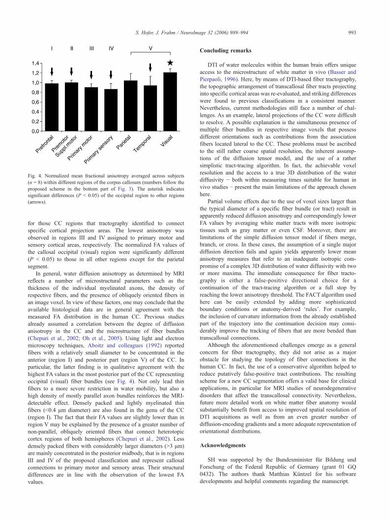
Fig. 4. Normalized mean fractional anisotropy averaged across subjects(n = 8) within different regions of the corpus callosum (numbers follow theproposed scheme in the bottom part of Fig. 3). The asterisk indicatessignificant differences (P < 0.05) of the occipital region to other regions(arrows).
993S. Hofer, J. Frahm / NeuroImage 32 (2006) 989–994
for those CC regions that tractography identified to connectspecific cortical projection areas. The lowest anisotropy wasobserved in regions III and IV assigned to primary motor andsensory cortical areas, respectively. The normalized FA values ofthe callosal occipital (visual) region were significantly different(P < 0.05) to those in all other regions except for the parietalsegment.
In general, water diffusion anisotropy as determined by MRIreflects a number of microstructural parameters such as thethickness of the individual myelinated axons, the density ofrespective fibers, and the presence of obliquely oriented fibers inan image voxel. In view of these factors, one may conclude that theavailable histological data are in general agreement with themeasured FA distribution in the human CC. Previous studiesalready assumed a correlation between the degree of diffusionanisotropy in the CC and the microstructure of fiber bundles(Chepuri et al., 2002; Oh et al., 2005). Using light and electronmicroscopy techniques, Aboitz and colleagues (1992) reportedfibers with a relatively small diameter to be concentrated in theanterior (region I) and posterior part (region V) of the CC. Inparticular, the latter finding is in qualitative agreement with thehighest FA values in the most posterior part of the CC representingoccipital (visual) fiber bundles (see Fig. 4). Not only lead thinfibers to a more severe restriction in water mobility, but also ahigh density of mostly parallel axon bundles reinforces the MRI-detectable effect. Densely packed and lightly myelinated thinfibers (<0.4 μm diameter) are also found in the genu of the CC(region I). The fact that their FA values are slightly lower than inregion V may be explained by the presence of a greater number ofnon-parallel, obliquely oriented fibers that connect heterotopiccortex regions of both hemispheres (Chepuri et al., 2002). Lessdensely packed fibers with considerably larger diameters (>3 μm)are mainly concentrated in the posterior midbody, that is in regionsIII and IV of the proposed classification and represent callosalconnections to primary motor and sensory areas. Their structuraldifferences are in line with the observation of the lowest FAvalues.
Concluding remarks
DTI of water molecules within the human brain offers uniqueaccess to the microstructure of white matter in vivo (Basser andPierpaoli, 1996). Here, by means of DTI-based fiber tractography,the topographic arrangement of transcallosal fiber tracts projectinginto specific cortical areas was re-evaluated, and striking differenceswere found to previous classifications in a consistent manner.Nevertheless, current methodologies still face a number of chal-lenges. As an example, lateral projections of the CC were difficultto resolve. A possible explanation is the simultaneous presence ofmultiple fiber bundles in respective image voxels that possessdifferent orientations such as contributions from the associationfibers located lateral to the CC. These problems must be ascribedto the still rather coarse spatial resolution, the inherent assump-tions of the diffusion tensor model, and the use of a rathersimplistic tract-tracing algorithm. In fact, the achievable voxelresolution and the access to a true 3D distribution of the waterdiffusivity − both within measuring times suitable for human invivo studies − present the main limitations of the approach chosenhere.
Partial volume effects due to the use of voxel sizes larger thanthe typical diameter of a specific fiber bundle (or tract) result inapparently reduced diffusion anisotropy and correspondingly lowerFA values by averaging white matter tracts with more isotropictissues such as gray matter or even CSF. Moreover, there arelimitations of the simple diffusion tensor model if fibers merge,branch, or cross. In these cases, the assumption of a single majordiffusion direction fails and again yields apparently lower meananisotropy measures that refer to an inadequate isotropic com-promise of a complex 3D distribution of water diffusivity with twoor more maxima. The immediate consequence for fiber tracto-graphy is either a false-positive directional choice for acontinuation of the tract-tracing algorithm or a full stop byreaching the lower anisotropy threshold. The FACT algorithm usedhere can be easily extended by adding more sophisticatedboundary conditions or anatomy-derived ‘rules’. For example,the inclusion of curvature information from the already establishedpart of the trajectory into the continuation decision may consi-derably improve the tracking of fibers that are more bended thantranscallosal connections.
Although the aforementioned challenges emerge as a generalconcern for fiber tractography, they did not arise as a majorobstacle for studying the topology of fiber connections in thehuman CC. In fact, the use of a conservative algorithm helped toreduce putatively false-positive tract contributions. The resultingscheme for a new CC segmentation offers a valid base for clinicalapplications, in particular for MRI studies of neurodegenerativedisorders that affect the transcallosal connectivity. Nevertheless,future more detailed work on white matter fiber anatomy wouldsubstantially benefit from access to improved spatial resolution ofDTI acquisitions as well as from an even greater number ofdiffusion-encoding gradients and a more adequate representation oforientational distributions.
Acknowledgments
SH was supported by the Bundesminister für Bildung undForschung of the Federal Republic of Germany (grant 01 GQ0432). The authors thank Matthias Küntzel for his softwaredevelopments and helpful comments regarding the manuscript.
994 S. Hofer, J. Frahm / NeuroImage 32 (2006) 989–994
References
Aboitz, F., Scheibel, A.B., Fisher, R.S., Zaidel, E., 1992. Fiber compositionof the human corpus callosum. Brain Res. 598, 143–153.
Basser, P.J., Pierpaoli, C., 1996. Microstructural and physiological featuresof tissues elucidated by quantitative diffusion-tensor MRI. J. Magn.Reson., B 111, 209–219.
Chepuri, N.B., Yen, Y.F., Burdette, J.H., Li, H., Moody, D.M., Maldjian,J.A., 2002. Diffusion anisotropy in the corpus callosum. Am. J.Neuroradiol. 23, 803–808.
Clarke, J.M., Zaidel, E., 1994. Anatomical–behavioral relationships: corpuscallosum morphometry and hemispheric specialization. Behav. Brain.Res. 64, 185–202.
De Lacoste, M.C., Kirkpatrick, J.B., Ross, E.D., 1985. Topography of thehuman corpus callosum. J. Neuropathol. Exp. Neurol. 44, 578–591.
Dougherty, R.F., Ben-Shachar, M., Bammer, R., Brewer, A.A., Wandell,B.A., 2005. Functional organization of human occipital–callosal fibertracts. Proc. Natl. Acad. Sci. U. S. A. 102, 7350–7355.
Duara, R., Kushch, A., Gross-Glenn, K., Barker, W.W., Jallad, B., Pascal, S.,Loewenstein, D.A., Sheldon, J., Rabin, M., Levin, B., 1991. Neuroana-tomic differences between dyslexic and normal readers on magneticresonance imaging scans. Arch. Neurol. 48, 410–416.
Huang, H., Zhang, J., Jiang, H., Wakana, S., Poetscher, L., Miller, M.I.,van Zijl, P.C., Hillis, A.E., Wytik, R., Mori, S., 2005. DTI tractographybased parcellation of white matter: application to the mid-sagittalmorphology of corpus callosum. NeuroImage 15, 195–205.
Lacerda, A.L., Brambilla, P., Sassi, R.B., Nicoletti, M.A., Mallinger, A.G.,Frank, E., Kupfer, D.J., Keshavan, M.S., Soares, J.C., 2005. AnatomicalMRI study of corpus callosum in unipolar depression. J. Psychiatr. Res.39, 347–354.
Larsen, J.P., Høien, T., Ödegaard, H., 1992. Magnetic resonance imaging ofthe corpus callosum in developmental dyslexia. Cogn. Neuropsychol. 9,123–134.
Mori, S., Crain, B.J., Chacko, V.P., van Zijl, P.C., 1999. Three-dimensionaltracking of axonal projections in the brain by magnetic resonanceimaging. Ann. Neurol. 45, 265–269.
Narr, K.L., Cannon, T.D., Woods, R.P., Thompson, P.M., Kim, S.,Asunction, D., van Erp, T.G., Poutanen, V.P., Huttunen, M., Lonnqvist,J., Standerksjold-Nordenstam, C.G., Kaprio, J., Mazziotta, J.C., Toga,A.W., 2002. Genetic contributions to altered callosal morphology inschizophrenia. J. Neurosci. 22, 3720–3729.
Nolte, U.G., Finsterbusch, J., Frahm, J., 2000. Rapid isotropic diffusionmapping without susceptibility artifacts. Whole brain studies using
diffusion-weighted single-shot STEAM MR imaging. Magn. Reson.Med. 44, 731–736.
Oh, J.S., Suk Park, K., Chan Song, I., Ju Kim, S., Hwang, J., Chung, A.,Kyoon Lyoo, I., 2005. Fractional anisotropy-based divisions ofmidsagittal corpus callosum. NeuroReport 16, 317–320.
Oldfield, R.C., 1971. The assessment and analysis of handedness: theEdinburgh Inventory. Neuropsychologia 9, 97–113.
Plessen, K.J., Wentzel-Larsen, T., Hugdahl, K., Feineigle, P., Klein, J., Staib,L.H., Leckman, J.F., Bansal, R., Peterson, B.S., 2004. Alteredinterhemispheric connectivity in individuals with Tourette's disorder.Am. J. Psychiatry 161, 2028–2037.
Rajapakse, J.C., Giedd, J.N., Rumsey, J.M., Vaituzis, A.C., Hamburger,S.D., Rapoport, J.L., 1996. Regional MRI measurements of the corpuscallosum: a methodological and developmental study. Brain Dev. 18,379–388.
Ramnani, N., Behrens, T.E., Penny, W., Matthews, P.M., 2004. Newapproaches for exploring anatomical and functional connectivity in thehuman brain. Biol. Psychiatry 56, 613–619.
Rieseberg, S., Merboldt, K.D., Küntzel, M., Frahm, J., 2005. Diffusiontensor imaging using partial Fourier STEAM MRI with projection ontoconvex subsets reconstruction. Magn. Reson. Med. 54, 486–490.
Teipel, S.J., Schapiro, M.B., Alexander, G.E., Krasuski, J.S., Horwitz, B.,Hoehne, C., Moller, H.J., Rapoport, S.I., Hampel, H., 2003. Relation ofcorpus callosum and hippocampal size to age in nondemented adultswith Down's syndrome. Am. J. Psychiatry 160, 1870–1878.
Thompson, P.M., Narr, K.L., Blanton, R.E., Toga, A.W., 2003. Mappingstructural alterations of the corpus callosum during brain developmentand degeneration. In: Zaidel, E., Iacoboni, M. (Eds.), The Parallel Brain:The Cognitive Neuroscience of the Corpus Callosum, pp. 93–130.
Thompson, P.M., Dutton, R.A., Hayashi, K.M., Lu, A., Lee, S.E., Lee, J.Y.,Lopez, O.L., Aizenstein, H.J., Toga, A.W., Becker, J.T., 2006. 3Dmapping of ventricular and corpus callosum abnormalities in HIV/AIDS.NeuroImage 13, 12–23.
von Plessen, K., Lundervold, A., Duta, N., Heiervang, E., Klauschen, F.,Smievoll, A.I., Ersland, L., Hugdahl, K., 2002. Less developed corpuscallosum in dyslexic subjects-a structural MRI study. Neuropsychologia40, 1035–1044.
Weis, S., Kimbacher, M., Wegner, E., 1993. Morphometric analysis of thecorpus callosum using MR: correlation of measurements with aging inhealthy individuals. Am. J. Neuroradiol. 14, 637–645.
Witelson, S.F., 1989. Hand and sex differences in the isthmus and genu ofthe human corpus callosum. A postmortem morphological study. Brain112, 799–835.
Copyright © 2022 FDOKUMEN