From diffusion tractography to quantitative white matter tract measures: a reproducibility study
Fiber Tractography Reveals Disruption of Temporal Lobe WhiteMatter Tracts in Schizophrenia
Owen R. Phillipsa, Keith H. Nuechterleinb, Kristi A. Clarkc, Liberty S. Hamiltona, Robert F.Asarnowb, Nathan S. Hagemana, Arthur W. Togaa, and Katherine L. NarraaLaboratory of Neuro Imaging, Department of Neurology Geffen School of Medicine at UCLA
bDepartment of Psychiatry and Biobehavioral Sciences Geffen School of Medicine at UCLA
cAhmanson-Lovelace Brain Mapping Center, Department of Neurology Geffen School of Medicine at UCLA
AbstractDiffusion tensor imaging (DTI) studies have demonstrated abnormal anisotropic diffusion inschizophrenia. However, examining data with low spatial resolution and/or a low number of gradientdirections and limitations associated with analysis approaches sensitive to registration confoundsmay have contributed to mixed findings concerning the regional specificity and direction of results.This study examined three major white matter tracts connecting lateral and medial temporal loberegions with neocortical association regions widely implicated in systems-level functional andstructural disturbances in schizophrenia. Using DTIstudio, a previously validated regions of interesttractography method was applied to 30 direction diffusion weighted imaging data collected fromdemographically similar schizophrenia (n=23) and healthy control subjects (n=22). The diffusiontensor was computed at each voxel after intra-subject registration of diffusion-weighted images.Three-dimensional tract reconstruction was performed using the Fiber Assignment by ContinuousTracking (FACT) algorithm. Tractography results showed reduced fractional anisotropy (FA) of thearcuate fasciculi (AF) and inferior longitudinal fasciculi (ILF) in patients compared to controls. FAchanges within the right ILF were negatively correlated with measures of thought disturbance.Reduced volume of the left AF was also observed in patients. These results, which avoid registrationissues associated with voxel-based analyses of DTI data, support that fiber pathways connectinglateral and medial temporal lobe regions with neocortical regions are compromised in schizophrenia.Disruptions of connectivity within these pathways may potentially contribute to the disturbances ofmemory, language, and social cognitive processing that characterize the disorder.
Correspondence should be addressed to: Dr. Katherine L. Narr, David Geffen School of Medicine at UCLA, Laboratory of Neuro Imaging,Neuroscience Research Building 1, Rm 225, 635 Charles Young Drive South, Los Angeles, CA 90095-7334, Email: [email protected] of Interest: None of the authors have any conflicts of interest to report.Contributors: Owen R. Phillips was responsible for data acquisition and analysis and manuscript preparation. Keith H. Nuechterleinand Robert F. Asarnow provided the clinical expertise for this study. Liberty S. Hamilton assisted in preparation of the manuscript anddata acquisition. Kristi A. Clark was responsible for software development and expertise regarding registration algorithms. Nathan S.Hageman provided expertise regarding diffusion measurements. Arthur W. Toga and Katherine L. Narr contributed to the conceptionand overall design of the study. Substantial scientific contributions were made by all authors of this study.Publisher's Disclaimer: This is a PDF file of an unedited manuscript that has been accepted for publication. As a service to our customerswe are providing this early version of the manuscript. The manuscript will undergo copyediting, typesetting, and review of the resultingproof before it is published in its final citable form. Please note that during the production process errors may be discovered which couldaffect the content, and all legal disclaimers that apply to the journal pertain.
NIH Public AccessAuthor ManuscriptSchizophr Res. Author manuscript; available in PMC 2010 January 1.
Published in final edited form as:Schizophr Res. 2009 January ; 107(1): 30–38. doi:10.1016/j.schres.2008.10.019.
NIH
-PA Author Manuscript
NIH
-PA Author Manuscript
NIH
-PA Author Manuscript

Keywordsdiffusion tensor imaging; white matter; uncinate fasciculus; inferior longitudinal fasciculus; arcuatefasciculus; fractional anisotropy
1. IntroductionSome brain systems appear selectively vulnerable to disease processes in schizophrenia.Structural imaging studies have repeatedly demonstrated reductions of gray matter (Shentonet al., 2001; Narr et al., 2005b; Whitford et al., 2006) that appear most reproducible in lateraland medial temporal cortices, particularly the hippocampus (Narr et al., 2004; Goldman et al.,2008) and the superior temporal gyrus (Shenton et al., 2001; Honea et al., 2008). Disturbancesof lateral and medial temporal lobe function including deficits in auditory sensory (Rabinowiczet al., 2000), language (DeLisi, 2001) memory (Saykin et al., 1994; Bilder et al., 2000; Harrisonand Fowler, 2004; Harrison, 2004) and social cognitive processing (Green et al., 2005; Yamadaet al., 2007) are also widely documented in schizophrenia. These behavioral disturbances maybe associated with neuropathology in discrete brain regions and/or from disruptions ofconnectivity within functional networks (Frith et al., 1995; Walterfang et al., 2005). However,the effects of illness on the physiological connections linking fronto-limbic and other temporo-neocortical association areas are not fully understood.
With Diffusion tensor imaging (DTI) it is possible to address whether disturbances of whitematter connectivity occur within particular cortical networks. That is, because the diffusiontensor approximating local diffusion is determined by the local tissue architecture, it can beused to generate an in vivo visualization of white matter tracts (Conturo et al., 1999; Ashburnerand Friston, 2000; Catani et al., 2002a; Wakana et al., 2004). Tractography methods assemblethe local diffusion tensor data into tracts where scalar metrics, such as fractional anisotropy(FA), along these tracts then allows for the precise localization of white matter abnormalities.Although some doubt exists that tractography may generate sufficiently reproducibleneuroanatomical detail for use in quantitative analyses, growing evidence supports thattracking results agree well with postmortem definitions (Catani et al., 2002b; Jellison et al.,2004; Wakana et al., 2007). To investigate possible white matter abnormalities inschizophrenia, we examined three major white matter tracts: the arcuate fasciculus (AF), theinferior longitudinal fasciculus (ILF), and the uncinate fasciculus (UF). These tracts have beenclassically defined using postmortem studies and are easily identifiable using a multiple regionof interest (ROI) approach in DTI data.
The AF (or temporal component of the superior longitudinal fasciculus [SLF]) connects thesuperior temporal gyrus with dorsal prefrontal cortex (Brodmann areas 8 and 46) (Petrides andPandya, 1984; Makris et al., 2005). By linking frontal and temporal cortices, the AF mayprovide a means by which the prefrontal cortex receives and modulates auditory andaudiospatial information (Leinonen et al., 1980; Petrides M, 2002). Several prior investigationshave implicated fronto-temporal processing disruptions in schizophrenia (Frith et al., 1995;Liddle, 1996; Andreasen, 2000). Further, some prior voxel-based DTI studies suggest thatwhite matter abnormalities occur in the vicinity of the AF (Burns et al., 2003; Kubicki et al.,2005; Douaud et al., 2007) and may be associated with the experience of hallucinations in theauditory modality (Hubl et al., 2004). To our knowledge, however, no prior studies haveexamined the AF using DTI tractography methods, which may provide a more powerful meansto examine changes in the physiology of this tract in schizophrenia.
The ILF has been demonstrated by postmortem and DTI studies to be a major associativeconnection between the anterior temporal and occipital lobe and plays an important role in
Phillips et al. Page 2
Schizophr Res. Author manuscript; available in PMC 2010 January 1.
NIH
-PA Author Manuscript
NIH
-PA Author Manuscript
NIH
-PA Author Manuscript

visual memory (Bauer and Trobe, 1984; Shinoura et al., 2007). It also connects theparahippocampal gyrus and amygdala (Catani et al., 2002a), regions implicated in the structuraland functional neuropathology of schizophrenia (Benes and Berretta, 2000; Narr et al., 2001;Harrison, 2004; Surguladze et al., 2006). At least one prior schizophrenia DTI tractographystudy has reported reduced FA for the left ILF (Ashtari et al., 2007a) where negativeassociations between FA and visual hallucinations were further observed.
The UF links the temporal lobe with ventral, medial, and orbital frontal cortices (Ebeling andvon Cramon, 1992). This tract plays an important role in the formation and retrieval ofmemories (Levine et al., 1998; Squire et al., 2004). Though patients with schizophrenia exhibitgeneralized cognitive impairment, declarative memory deficits appear particularly pronounced(Bilder et al., 2000). However, while some DTI studies have observed lower FA in the UF inschizophrenia (Nestor et al., 2008), other reports have been mixed (Jones et al., 2006).
In spite of the evidence suggesting disturbances of fiber connectivity in schizophrenia, theregional specificity of results remains unclear. Discrepancies in prior findings may be at leastpartially attributable to the methodological limitations associated with examining data with alow number of gradient directions and low spatial resolution in small cohorts, using voxel-based methods sensitive to registration confounds (Alexander et al., 2001), and the paucity ofstudies using methods that allow the precise mapping of tract anatomy within subjects (Kubickiet al., 2007). We thus examined the connectivity of the AF, ILF, and UF in 23 schizophreniaand 22 healthy subjects using 30 direction DTI data and sensitive fiber tracking methods thatdo not rely on registration procedures to align imaging data across subjects. We predicted thateach of these fiber pathways would be disturbed in schizophrenia. We further explored whetherpositive and negative symptom cluster scores are associated with fiber tract abnormalitieswithin medial and lateral temporal lobe systems in schizophrenia patients.
2. Methods2.1 Subjects
23 patients with schizophrenia and 22 healthy control subjects participated in this study.Diagnostic groups did not differ significantly in age or sex (Table 1). All participants providedinformed consent as approved by the University of California, Los Angeles (UCLA)Institutional Review Board. Schizophrenia patients were recruited from the UCLA AftercareResearch Program through the UCLA Family Study protocol. Control subjects were recruitedthrough direct calls using a survey sampling methodology as well as community outreach viaadvertisement in newspapers and fliers. Exclusion criteria for all subjects included mentalretardation, neurological disorder (e.g., temporal lobe epilepsy), and recent or past history ofsignificant and habitual drug abuse or alcoholism. Control subjects were screened by clinicalinterview using the SCID-NP (First et al., 1994) to exclude schizophrenia and schizoaffectivedisorder and potential schizophrenia spectrum disorders. A radiologist reviewed any suspectedabnormalities in the high-resolution T1-weighted data. However, using this sequence only, itwas not possible to rule out the presence of all possible brain abnormalities. No incidental brainlesions were observed in the current sample, and no subjects were excluded from analysis.Schizophrenia diagnosis was confirmed by consensus of specially trained diagnosticians(Ventura et al., 1998) as determined by DSM-IV criteria using the Structured Clinical Interviewfor DSM-IV (SCID-I/P) (First et al., 1994;First et al., 1996;First et al., 2001) and by informantinformation. Clinical symptoms were assessed using the expanded 24-item Brief PsychiatricRating Scale (BPRS; (Ventura et al., 1993;Ventura et al., 2000) and clustered into withdrawaland thinking disorder scores (Burger et al., 1997). Withdrawal (negative symptoms factor)scores were calculated as the mean of BPRS scores for emotional withdrawal, motorretardation, blunted affect, disorientation, and self-neglect. Thinking disorder (positivesymptoms factor) scores were calculated as the mean of BPRS scores for conceptual
Phillips et al. Page 3
Schizophr Res. Author manuscript; available in PMC 2010 January 1.
NIH
-PA Author Manuscript
NIH
-PA Author Manuscript
NIH
-PA Author Manuscript
disorganization, grandiosity, hallucinatory behavior, unusual thought content, and bizarrebehavior. Handedness was determined based on a modified version of the Edinburgh Inventory.(Oldfield, 1971)
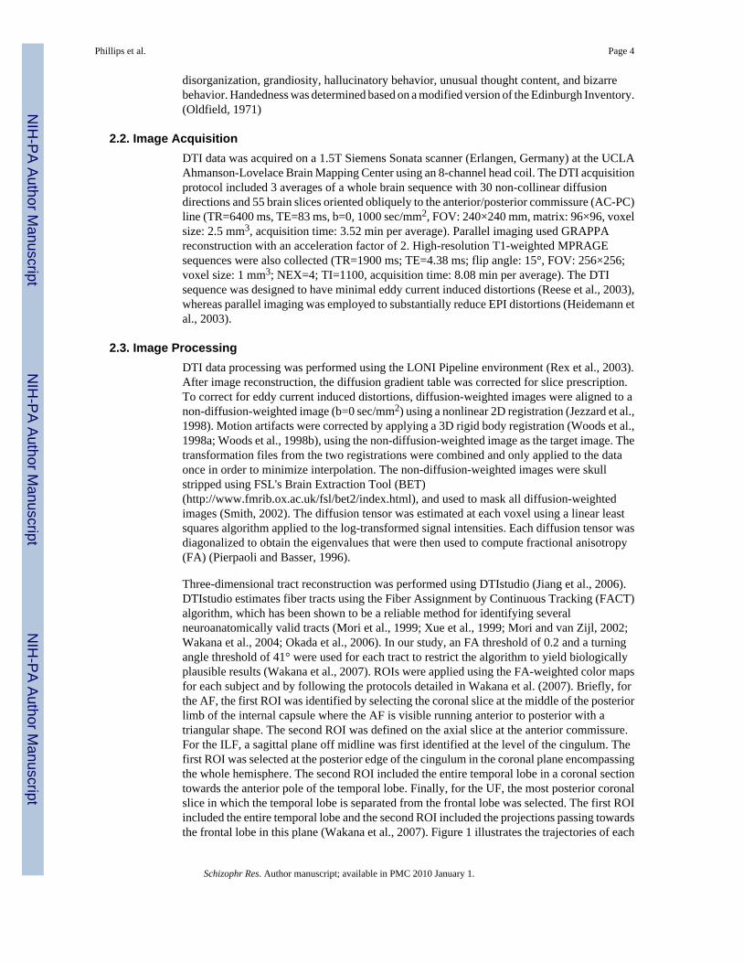
2.2. Image AcquisitionDTI data was acquired on a 1.5T Siemens Sonata scanner (Erlangen, Germany) at the UCLAAhmanson-Lovelace Brain Mapping Center using an 8-channel head coil. The DTI acquisitionprotocol included 3 averages of a whole brain sequence with 30 non-collinear diffusiondirections and 55 brain slices oriented obliquely to the anterior/posterior commissure (AC-PC)line (TR=6400 ms, TE=83 ms, b=0, 1000 sec/mm2, FOV: 240×240 mm, matrix: 96×96, voxelsize: 2.5 mm3, acquisition time: 3.52 min per average). Parallel imaging used GRAPPAreconstruction with an acceleration factor of 2. High-resolution T1-weighted MPRAGEsequences were also collected (TR=1900 ms; TE=4.38 ms; flip angle: 15°, FOV: 256×256;voxel size: 1 mm3; NEX=4; TI=1100, acquisition time: 8.08 min per average). The DTIsequence was designed to have minimal eddy current induced distortions (Reese et al., 2003),whereas parallel imaging was employed to substantially reduce EPI distortions (Heidemann etal., 2003).
2.3. Image ProcessingDTI data processing was performed using the LONI Pipeline environment (Rex et al., 2003).After image reconstruction, the diffusion gradient table was corrected for slice prescription.To correct for eddy current induced distortions, diffusion-weighted images were aligned to anon-diffusion-weighted image (b=0 sec/mm2) using a nonlinear 2D registration (Jezzard et al.,1998). Motion artifacts were corrected by applying a 3D rigid body registration (Woods et al.,1998a; Woods et al., 1998b), using the non-diffusion-weighted image as the target image. Thetransformation files from the two registrations were combined and only applied to the dataonce in order to minimize interpolation. The non-diffusion-weighted images were skullstripped using FSL's Brain Extraction Tool (BET)(http://www.fmrib.ox.ac.uk/fsl/bet2/index.html), and used to mask all diffusion-weightedimages (Smith, 2002). The diffusion tensor was estimated at each voxel using a linear leastsquares algorithm applied to the log-transformed signal intensities. Each diffusion tensor wasdiagonalized to obtain the eigenvalues that were then used to compute fractional anisotropy(FA) (Pierpaoli and Basser, 1996).
Three-dimensional tract reconstruction was performed using DTIstudio (Jiang et al., 2006).DTIstudio estimates fiber tracts using the Fiber Assignment by Continuous Tracking (FACT)algorithm, which has been shown to be a reliable method for identifying severalneuroanatomically valid tracts (Mori et al., 1999; Xue et al., 1999; Mori and van Zijl, 2002;Wakana et al., 2004; Okada et al., 2006). In our study, an FA threshold of 0.2 and a turningangle threshold of 41° were used for each tract to restrict the algorithm to yield biologicallyplausible results (Wakana et al., 2007). ROIs were applied using the FA-weighted color mapsfor each subject and by following the protocols detailed in Wakana et al. (2007). Briefly, forthe AF, the first ROI was identified by selecting the coronal slice at the middle of the posteriorlimb of the internal capsule where the AF is visible running anterior to posterior with atriangular shape. The second ROI was defined on the axial slice at the anterior commissure.For the ILF, a sagittal plane off midline was first identified at the level of the cingulum. Thefirst ROI was selected at the posterior edge of the cingulum in the coronal plane encompassingthe whole hemisphere. The second ROI included the entire temporal lobe in a coronal sectiontowards the anterior pole of the temporal lobe. Finally, for the UF, the most posterior coronalslice in which the temporal lobe is separated from the frontal lobe was selected. The first ROIincluded the entire temporal lobe and the second ROI included the projections passing towardsthe frontal lobe in this plane (Wakana et al., 2007). Figure 1 illustrates the trajectories of each
Phillips et al. Page 4
Schizophr Res. Author manuscript; available in PMC 2010 January 1.
NIH
-PA Author Manuscript
NIH
-PA Author Manuscript
NIH
-PA Author Manuscript

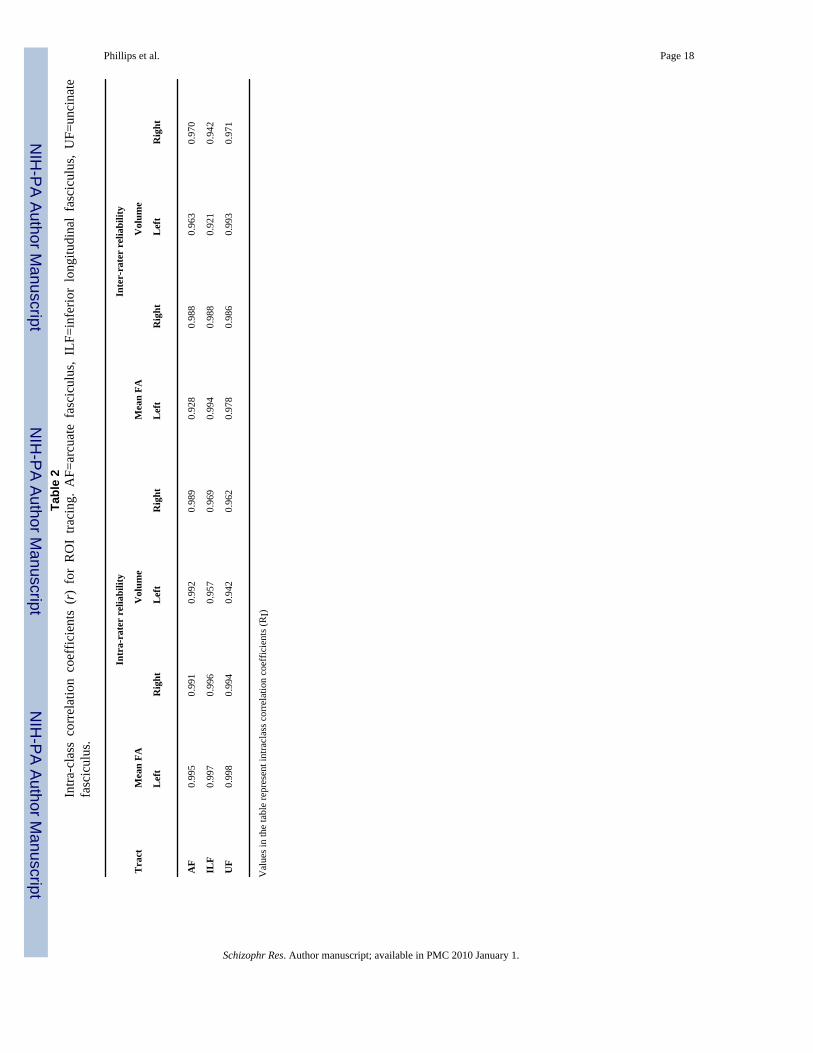
fiber pathway obtained using the ROIs described above. The locations of ROI placement foreach tract are illustrated in supplementary Figures 1-3. If a tract showed a fiber that was clearlyanatomically incorrect, the NOT function was used to remove the fiber from the bundle. Intra-rater reliability was estimated by identifying each of the tracts using ROI placement in 8randomly chosen volumes duplicated in the dataset. To determine inter-rater reliability, fibertracks were identified in 9 randomly chosen brain volumes by two independent raters. Excellentintra- and inter-rater reliability was achieved for ROI placement as determined by computingthe inter-class correlation coefficients for tract volume, the volume for the number of voxelsthrough which the tract passes, and mean FA, a value representing the average intra-voxel FAacross the tract of interest in each subject (Table 2).
To obtain measures of intracranial brain volume and volumes of segmented brain tissuecompartments, the T1-weighted MPRAGE images were processed as previously detailed (Narret al., 2005b; Narr et al., 2005a). Briefly, image volumes were corrected for head tilt andalignment using six-parameter rigid-body transformations. To scalp edit the T1-weighted data,FSL's BET was used to generate a temporary mask that was then manually edited on a slice-by-slice basis to correct any small errors using MNI Display(http://www.bic.mni.mcgill.ca/software/Display/Display.html), (scalp editing inter-raterreliability, rI= .99). Edited brain volumes were then corrected for signal intensityinhomogeneities (Sled et al., 1998), and segmented into gray matter, white matter, and CSFusing a partial volume classification method (Shattuck et al., 2001) after which whole brainand brain tissue volumes were computed.
2.4. Statistical AnalysisMean FA and tract volume (the number of voxels the tract traverses multiplied by the 2.5mm3 voxel size) were estimated for each fiber tract within subjects. A repeated measuresanalysis of variance (ANOVA) examined group differences in left and right mean FA, whichwere included as dependent variables in order to test fewer hypotheses and avoid potentialType 1 error while simultaneously assessing the presence of hemisphere effects for the AF,ILF, and UF. Diagnosis and sex were included as between-subject factors. To reduce potentialbias due to less precise estimates in subjects contributing fewer voxels, mean FA measureswere corrected for overall tract volume. That is, since hemisphere was a within-subject variable,left and right mean FA values were residualized for left and right tract volume respectively.Repeated measures ANOVAs were followed-up by univariate tests when appropriate.
As noted above, we controlled for tract volume when examining changes in mean FA betweengroups. However, to determine whether this measure itself was different across groups, thesame statistical model was used to examine diagnostic group differences in fiber tract volume.For these comparisons, total brain white matter volume was included as a covariate to controlbias in the overall number of white matter voxels expected in individuals with larger braintissue volumes. Finally, correlations between mean FA and positive and negative symptomcluster scores were examined. Two patients were excluded from these analyses because BPRSdata was unavailable. Demographic differences were assessed using chi-square or independentsample t-tests as appropriate.
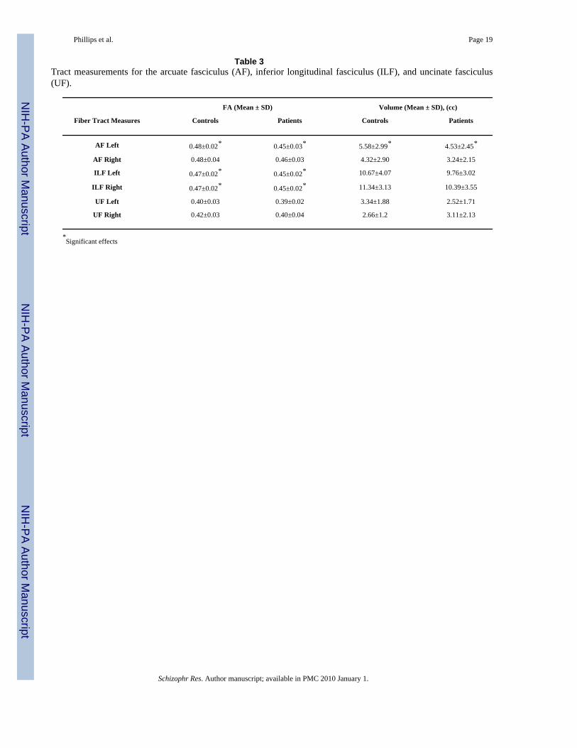
3. ResultsPatient and control groups did not significantly differ in age or gender distribution or in totalbrain or white matter volume (Table 1). The repeated measures ANOVA showed significantlylower FA values in patients compared to controls in the ILF [F(1,41)=6.55, p=0.014] and AF[F(1, 41)=4.78, p=0.035], but not in the UF, [F(1,41)=0.09, p=0.758]. There were no sex orasymmetry effects or interactions between diagnosis and sex or hemisphere. Follow-upunivariate tests revealed significant effects of diagnosis for the Left ILF [F(1,44)=4.84, p=0.03]
Phillips et al. Page 5
Schizophr Res. Author manuscript; available in PMC 2010 January 1.
NIH
-PA Author Manuscript
NIH
-PA Author Manuscript
NIH
-PA Author Manuscript

and Right ILF [F(1,44)=6.06, p=0.018] and the Left AF [F(1,45)=5.01, p=0.03]. Table 3 listsmeans and standard deviations (SDs) for mean FA values, and Figure 2 shows the 95%confidence intervals of the means within diagnostic groups. Follow-up tests performed afterexcluding the one left-handed control subject from the analysis produced similarly significantresults.
Analyses of fiber volume showed a significant reduction in left AF volume only [F(1,45)=7.725, p=0.008]. Means and SDs for AF, ILF, and UF volume are provided in Table 3.Spearman's correlation analyses between mean FA and positive (thinking disorder) andnegative symptom (withdrawal) cluster scores revealed a significant negative association(r=-0.439) between thinking disorder (positive symptoms) and mean FA within the right ILF(p=0.046) (Figure 3).
4. DiscussionUsing a validated ROI approach for identifying major fiber tracts (Wakana et al., 2007), wewere able to examine highly localized changes of white matter integrity in brain systems widelyimplicated in the pathophysiology of schizophrenia. Specifically, we examined three majorwhite matter tracts that form integral connections between lateral and medial temporal loberegions with neocortical association areas. Results revealed significant reductions of mean FAwithin the ILF bilaterally and the left AF in schizophrenia patients compared to healthycontrols. A significant negative association between thinking disorder and mean FA of the rightILF was further observed. Although the neurobiological mechanisms underlying altered FAare not known, prior human and animal studies of neural development and of relateddisturbances in brain function support that an increase in FA is related to an increase in theconnectivity of white matter bundles (Dong et al., 2004). Our results are consistent with severalprevious diffusion imaging findings despite differences in patient populations (e.g. early onset,first episode, and chronic schizophrenia) and analysis approaches. However, our findingsprovide more detailed information concerning the localization of fiber tract abnormalities inthe disorder that may be of particular functional relevance.
4.1. Inferior Longitudinal FasciculusOur observations of significantly reduced mean FA in patients in bilateral ILF support priorfindings in adolescent and never-medicated first-episode schizophrenia patients (Ashtari et al.,2007b; Cheung et al., 2007) identified using tractography and voxel-based methodsrespectively. Our findings are also consistent with those of (Friedman et al., 2008) showingreduced FA in the left ILF in chronic patients and trend level FA reductions in first episodeschizophrenia. Although the functions of the ILF are not fully characterized, this tract appearsto mediate the fast transfer of visual signals to anterior temporal regions and neuromodulatoryback-projections from the amygdala to early visual areas (Catani et al., 2003). The ILF, whichis the major occipital-temporal white matter connection, is suggested to play a role in visualmemory and in processing the emotional valence of a visual stimulus (Catani et al., 2003).Disturbances in the structural integrity of this tract may thus relate to observations thatschizophrenia patients are impaired in their ability to recognize and respond appropriately toemotional facial expressions (Mueser et al., 1996; Penn et al., 1996; Brune, 2005) andcontribute to developmental differences in social cognition as well as visual-spatial integration(Barnea-Goraly et al., 2005). Our finding of an association between mean FA in the right ILFand thinking disorder (positive symptoms) cluster scores, which includes a visual hallucinationrating, is partially consistent with a report of a negative association between mean FA of theleft ILF in early onset patients with a history of visual hallucinations (Ashtari et al., 2007a),although individual BPRS factor scores were not examined in our study.
Phillips et al. Page 6
Schizophr Res. Author manuscript; available in PMC 2010 January 1.
NIH
-PA Author Manuscript
NIH
-PA Author Manuscript
NIH
-PA Author Manuscript

4.2. Arcuate FasciculusSome diffusion studies have reported reduced FA in the vicinity of the left AF in adolescentonset (Douaud et al., 2007) and in adult schizophrenia patients (Burns et al., 2003; Kubicki etal., 2005; Douaud et al., 2007), consistent with our results. At least two prior studies usingvoxel-based methods further report positive associations between FA values and the symptomof auditory hallucinations, particularly in the left AF (Hubl et al., 2004; Shergill et al., 2007).Specifically, one study reported significantly greater FA values compared to controls inpatients with current and past history of hallucinations (Hubl et al., 2004). However, a secondstudy observing positive associations between FA and hallucination ratings still showed lowerFA values in patients with a propensity for current or past hallucinations compared to healthysubjects (Shergill et al., 2007). A third study failed to observe AF associations, althoughpositive relationships between the severity of auditory and tactile hallucinations and the inferiorfronto-occipital fasciculus FA were detected (Szeszko et al., 2008). Finally, Seok et al.,(2007) reported positive associations between auditory hallucinations and FA values in thefrontal as opposed to the temporal component of the SLF. Unfortunately, we were not ableclarify these relationships in the current investigation, since patients were not acutely ill at thetime of scan, and symptom scores for auditory hallucinations were low and narrow in range.Regardless, our results suggest that reduced FA within the AF persist during periods of relativesymptom stability. Our investigation also revealed a reduction in the overall size of the left AFin patients compared to controls. This finding may suggest that the length of AF fibers are lessextensive in patients or indicate tract shape differences. Although tractography is sensitive toanatomical variations in fiber pathways (Wakana et al., 2007), it is possible that the extent ofthis fiber tract was not fully resolved with our choice of ROIs, which were devised based onthe fiber anatomy of healthy individuals (Wakana et al., 2007).
4.3. Uncinate FasciculusThe UF connects parahippocampal and prefrontal regions and is involved in declarativememory processing, for which patients exhibit pronounced deficits (Bilder et al., 2000). Atleast one investigation supports relationships between UF FA and memory functioning inschizophrenia specifically (Nestor et al., 2008). In this study, however, we failed to detectsignificant differences of mean UF FA in schizophrenia patients compared to controls. Theseresults contrast with one prior tractography study (Price et al., 2008) and with some voxel-based investigations. Specifically, reduced FA of the UF has been documented in chronicschizophrenia (Seal et al., 2008) and in first episode patients bilaterally (Szeszko et al., 2008)and in the left UF exclusively (Szeszko et al., 2005), and at trend-level in a cluster at the borderof the UF and the ILF (Burns et al., 2003). Still, several studies, like the present investigation,have failed to detect significant diagnostic group differences of FA within the UF (Jones et al.,2006). Some studies, however, have revealed altered FA asymmetries in schizophrenia(Kubicki et al., 2002; Park et al., 2004), although hemisphere by diagnosis effects in UF cross-sectional area and fiber numbers were not detected in post mortem data (Highley et al.,2002). In the absence of diagnostic group effects, another group reported a trend level age bygroup interaction for FA within the UF (Rosenberger et al., 2008). Inconsistencies in findingsmay be partially attributable to clinical heterogeneity amongst patient samples and tomethodological differences, where it is worth noting that reported cluster coordinates locationsare highly variable between voxel-based studies. Measurements for the UF may also becomplicated by the tightly curved shape and smaller trajectory of this fiber bundle in relationto other larger tracts and/or could indicate that disturbances of UF connectivity in schizophreniaare subtler than those of other tracts.
Phillips et al. Page 7
Schizophr Res. Author manuscript; available in PMC 2010 January 1.
NIH
-PA Author Manuscript
NIH
-PA Author Manuscript
NIH
-PA Author Manuscript
4.4. Potential LimitationsA possible limitation to the study is the influence of patient medications on white matterconnectivity and/or myelination. A positive association between FA and antipsychotictreatment has been observed in at least one prior study (Kuroki et al., 2006), although FAchanges are also reported in never-medicated patients (Cheung et al., 2007). Whether otherfactors including intelligence, social economic status and past history of drug and alcohol use,which is shown to affect FA in non-schizophrenia samples e.g., (Pfefferbaum et al., 2006),influence fiber integrity warrants further study. However, some these factors may be moredifficult to dissociate from other disease-related characteristics. In spite of excellent reliabilityfor ROI placements, it is possible that the anatomic definitions imposed for ROI placementmay have influenced results. Still, our method, which focused on well documented tracts fromprevious studies using anatomical constraints (multiple ROIs), made it possible to isolate andexamine tracts based on the individual's morphology that do not appear influenced by ROIplacement. Furthermore, this method does not require perfect anatomical registration acrosssubjects and is thus not sensitive to registration-related confounds that continue to be an issuefor the analysis of DTI data. Finally, it is possible that other diffusion measures such as radial,axial and mean diffusivity may reveal subtler cytoarchitectonic disturbances in schizophrenia.Future studies including larger sample sizes may address broader hypotheses relating to thesediffusion measures.
4.5. ConclusionOur observations of reduced mean FA in the AF and ILF support the hypothesis that abnormalwhite matter connectivity influences systems-level processing deficits involving fronto-limbicand temporo-neocortical networks in schizophrenia. Abnormal connectivity of the AF maycontribute to language and auditory deficits in patients. Disturbances of ILF integrity maycontribute to visual memory impairments and to deficits in social cognition. Furthermore, IFLconnectivity may be influenced by clinical state as suggested by our observations of negativeassociations with positive symptom ratings. It remains unclear whether white matterabnormalities serve as primary or complicating mechanisms for impairments in specific brainsystems in schizophrenia or whether these abnormalities are secondary to changes in brainmorphology, particularly of gray matter. Future studies may clarify these questions and addresswhether FA changes are the result of disturbed white matter developmental trajectories.
Supplementary MaterialRefer to Web version on PubMed Central for supplementary material.
AcknowledgementsRole of Funding Source: This work was supported by grants from the National Center for Research Resources (P41RR13642), the National Institute of Mental Health (RO1 MH60374), the NIH Roadmap Initiative (P20 RR020750),the National Library of Medicine (R01 LM05639), the NIH Roadmap for Medical Research, Grant U54 RR021813entitled Center for Computational Biology (CCB), and a Career Development Award (KO1 MH073990, to KLN).The NIMH grants MH066286 and MH037705 allowed recruitment of the subjects and MH049716 supported the MRIs.This study utilized the LONI Pipeline environment (http://pipeline.loni.ucla.edu), which was developed by theLaboratory of Neuro Imaging and partially funded by NIH grants P41 RR013642, R01 MH71940 and U54 RR021813.The above funding agencies had no further contributions to the study design, data collect, analysis, manuscriptpreparation, and in the decision to submit the paper for publication.
ReferencesAlexander DC, Pierpaoli C, Basser PJ, Gee JC. Spatial transformations of diffusion tensor magnetic
resonance images. IEEE Trans Med Imaging 2001;20:1131–1139. [PubMed: 11700739]
Phillips et al. Page 8
Schizophr Res. Author manuscript; available in PMC 2010 January 1.
NIH
-PA Author Manuscript
NIH
-PA Author Manuscript
NIH
-PA Author Manuscript
Andreasen NC. Schizophrenia: the fundamental questions. Brain Res Brain Res Rev 2000;31:106–112.[PubMed: 10719138]
Ashburner J, Friston KJ. Voxel-based morphometry--the methods. Neuroimage 2000;11:805–821.[PubMed: 10860804]
Ashtari M, Cottone J, Ardekani BA, Cervellione K, Szeszko PR, Wu J, Chen S, Kumra S. Disruption ofwhite matter integrity in the inferior longitudinal fasciculus in adolescents with schizophrenia asrevealed by fiber tractography. Arch Gen Psychiatry 2007a;64:1270–1280. [PubMed: 17984396]
Ashtari M, Cervellione KL, Hasan KM, Wu J, McIlree C, Kester H, Ardekani BA, Roofeh D, SzeszkoPR, Kumra S. White matter development during late adolescence in healthy males: a cross-sectionaldiffusion tensor imaging study. Neuroimage 2007b;35:501–510. [PubMed: 17258911]
Barnea-Goraly N, Menon V, Eckert M, Tamm L, Bammer R, Karchemskiy A, Dant CC, Reiss AL. Whitematter development during childhood and adolescence: a cross-sectional diffusion tensor imagingstudy. Cereb Cortex 2005;15:1848–1854. [PubMed: 15758200]
Bauer RM, Trobe JD. Visual memory and perceptual impairments in prosopagnosia. J ClinNeuroophthalmol 1984;4:39–46. [PubMed: 6233309]
Benes FM, Berretta S. Amygdalo-entorhinal inputs to the hippocampal formation in relation toschizophrenia. Ann N Y Acad Sci 2000;911:293–304. [PubMed: 10911881]
Bilder RM, Goldman RS, Robinson D, Reiter G, Bell L, Bates JA, Pappadopulos E, Willson DF, AlvirJM, Woerner MG, Geisler S, Kane JM, Lieberman JA. Neuropsychology of first-episodeschizophrenia: initial characterization and clinical correlates. Am J Psychiatry 2000;157:549–559.[PubMed: 10739413]
Brune M. Emotion recognition, ‘theory of mind,’ and social behavior in schizophrenia. Psychiatry Res2005;133:135–147. [PubMed: 15740990]
Burger GK, Calsyn RJ, Morse GA, Klinkenberg WD, Trusty ML. Factor structure of the expanded BriefPsychiatric Rating Scale. J Clin Psychol 1997;53:451–454. [PubMed: 9257222]
Burns J, Job D, Bastin ME, Whalley H, Macgillivray T, Johnstone EC, Lawrie SM. Structuraldisconnectivity in schizophrenia: a diffusion tensor magnetic resonance imaging study. Br JPsychiatry 2003;182:439–443. [PubMed: 12724248]
Catani M, Howard RJ, Pajevic S, Jones DK. Virtual in vivo interactive dissection of white matter fasciculiin the human brain. Neuroimage 2002a;17:77–94. [PubMed: 12482069]
Catani M, Jones DK, Donato R, Ffytche DH. Occipito-temporal connections in the human brain. Brain2003;126:2093–2107. [PubMed: 12821517]
Catani M, Mecocci P, Tarducci R, Howard R, Pelliccioli GP, Mariani E, Metastasio A, Benedetti C, SeninU, Cherubini A. Proton magnetic resonance spectroscopy reveals similar white matter biochemicalchanges in patients with chronic hypertension and early Alzheimer's disease. J Am Geriatr Soc 2002b;50:1707–1710. [PubMed: 12366626]
Cheung V, Cheung C, McAlonan GM, Deng Y, Wong JG, Yip L, Tai KS, Khong PL, Sham P, Chua SE.A diffusion tensor imaging study of structural dysconnectivity in never-medicated, first-episodeschizophrenia. Psychol Med 2007:1–9.
Conturo TE, Lori NF, Cull TS, Akbudak E, Snyder AZ, Shimony JS, McKinstry RC, Burton H, RaichleME. Tracking neuronal fiber pathways in the living human brain. Proc Natl Acad Sci U S A1999;96:10422–10427. [PubMed: 10468624]
DeLisi LE. Speech disorder in schizophrenia: review of the literature and exploration of its relation tothe uniquely human capacity for language. Schizophr Bull 2001;27:481–496. [PubMed: 11596849]
Dong Q, Welsh RC, Chenevert TL, Carlos RC, Maly-Sundgren P, Gomez-Hassan DM, Mukherji SK.Clinical applications of diffusion tensor imaging. J Magn Reson Imaging 2004;19:6–18. [PubMed:14696215]
Douaud G, Smith S, Jenkinson M, Behrens T, Johansen-Berg H, Vickers J, James S, Voets N, WatkinsK, Matthews PM, James A. Anatomically related grey and white matter abnormalities in adolescent-onset schizophrenia. Brain 2007;130:2375–2386. [PubMed: 17698497]
Ebeling U, von Cramon D. Topography of the uncinate fascicle and adjacent temporal fiber tracts. ActaNeurochir (Wien) 1992;115:143–148. [PubMed: 1605083]
Phillips et al. Page 9
Schizophr Res. Author manuscript; available in PMC 2010 January 1.
NIH
-PA Author Manuscript
NIH
-PA Author Manuscript
NIH
-PA Author Manuscript

First, M.; Spitzer, R.; Gibbon, M.; Williams, J. Structured Clinical Interview for DSM-IV Axis I Disorders- Patient Edition (SCID-I/P). New York: Biometrics Research Department, New York StatePsychiatric Institute; 2001.
First, MB.; Gibbon, M.; Spitzer, RL.; Williams, JBW. Structured Clinical Interview for DSM-IV Axis IDisorders. Vol. Patient. New York: Biometrics Research; 1996.
First MB, Frances AJ, Pincus HA, Vettorello N, Davis WW. DSM-IV in progress. Changes in substance-related, schizophrenic, and other primarily adult disorders. Hosp Community Psychiatry 1994;45:18–20. [PubMed: 8125454]
Friedman JI, Tang C, Carpenter D, Buchsbaum M, Schmeidler J, Flanagan L, Golembo S, KanellopoulouI, Ng J, Hof PR, Harvey PD, Tsopelas ND, Stewart D, Davis KL. Diffusion tensor imaging findingsin first-episode and chronic schizophrenia patients. Am J Psychiatry 2008;165:1024–1032. [PubMed:18558643]
Frith CD, Friston KJ, Herold S, Silbersweig D, Fletcher P, Cahill C, Dolan RJ, Frackowiak RS, LiddlePF. Regional brain activity in chronic schizophrenic patients during the performance of a verbalfluency task. Br J Psychiatry 1995;167:343–349. [PubMed: 7496643]
Goldman AL, Pezawas L, Mattay VS, Fischl B, Verchinski BA, Zoltick B, Weinberger DR, Meyer-Lindenberg A. Heritability of brain morphology related to schizophrenia: a large-scale automatedmagnetic resonance imaging segmentation study. Biol Psychiatry 2008;63:475–483. [PubMed:17727823]
Green MF, Olivier B, Crawley JN, Penn DL, Silverstein S. Social cognition in schizophrenia:recommendations from the measurement and treatment research to improve cognition inschizophrenia new approaches conference. Schizophr Bull 2005;31:882–887. [PubMed: 16135561]
Harrison CL, Fowler D. Negative symptoms, trauma, and autobiographical memory: an investigation ofindividuals recovering from psychosis. J Nerv Ment Dis 2004;192:745–753. [PubMed: 15505518]
Harrison PJ. The hippocampus in schizophrenia: a review of the neuropathological evidence and itspathophysiological implications. Psychopharmacology (Berl) 2004;174:151–162. [PubMed:15205886]
Heidemann RM, Ozsarlak O, Parizel PM, Michiels J, Kiefer B, Jellus V, Muller M, Breuer F, BlaimerM, Griswold MA, Jakob PM. A brief review of parallel magnetic resonance imaging. Eur Radiol2003;13:2323–2337. [PubMed: 12942278]
Highley JR, Walker MA, Esiri MM, Crow TJ, Harrison PJ. Asymmetry of the uncinate fasciculus: a post-mortem study of normal subjects and patients with schizophrenia. Cereb Cortex 2002;12:1218–1224.[PubMed: 12379610]
Honea RA, Meyer-Lindenberg A, Hobbs KB, Pezawas L, Mattay VS, Egan MF, Verchinski B,Passingham RE, Weinberger DR, Callicott JH. Is gray matter volume an intermediate phenotype forschizophrenia? A voxel-based morphometry study of patients with schizophrenia and their healthysiblings. Biol Psychiatry 2008;63:465–474. [PubMed: 17689500]
Hubl D, Koenig T, Strik W, Federspiel A, Kreis R, Boesch C, Maier SE, Schroth G, Lovblad K, DierksT. Pathways that make voices: white matter changes in auditory hallucinations. Arch Gen Psychiatry2004;61:658–668. [PubMed: 15237078]
Jellison BJ, Field AS, Medow J, Lazar M, Salamat MS, Alexander AL. Diffusion tensor imaging ofcerebral white matter: a pictorial review of physics, fiber tract anatomy, and tumor imaging patterns.AJNR Am J Neuroradiol 2004;25:356–369. [PubMed: 15037456]
Jezzard P, Barnett AS, Pierpaoli C. Characterization of and correction for eddy current artifacts in echoplanar diffusion imaging. Magn Reson Med 1998;39:801–812. [PubMed: 9581612]
Jiang H, van Zijl PC, Kim J, Pearlson GD, Mori S. DtiStudio: resource program for diffusion tensorcomputation and fiber bundle tracking. Comput Methods Programs Biomed 2006;81:106–116.[PubMed: 16413083]
Jones DK, Catani M, Pierpaoli C, Reeves SJ, Shergill SS, O'sullivan M, Golesworthy P, McGuire P,Horsfield MA, Simmons A, Williams SC, Howard RJ. Age effects on diffusion tensor magneticresonance imaging tractography measures of frontal cortex connections in schizophrenia. Hum BrainMapp 2006;27:230–238. [PubMed: 16082656]
Phillips et al. Page 10
Schizophr Res. Author manuscript; available in PMC 2010 January 1.
NIH
-PA Author Manuscript
NIH
-PA Author Manuscript
NIH
-PA Author Manuscript

Kubicki M, McCarley R, Westin CF, Park HJ, Maier S, Kikinis R, Jolesz FA, Shenton ME. A review ofdiffusion tensor imaging studies in schizophrenia. J Psychiatr Res 2007;41:15–30. [PubMed:16023676]
Kubicki M, Westin CF, Maier SE, Frumin M, Nestor PG, Salisbury DF, Kikinis R, Jolesz FA, McCarleyRW, Shenton ME. Uncinate fasciculus findings in schizophrenia: a magnetic resonance diffusiontensor imaging study. Am J Psychiatry 2002;159:813–820. [PubMed: 11986136]
Kubicki M, Park H, Westin CF, Nestor PG, Mulkern RV, Maier SE, Niznikiewicz M, Connor EE, LevittJJ, Frumin M, Kikinis R, Jolesz FA, McCarley RW, Shenton ME. DTI and MTR abnormalities inschizophrenia: analysis of white matter integrity. Neuroimage 2005;26:1109–1118. [PubMed:15878290]
Kuroki N, Kubicki M, Nestor PG, Salisbury DF, Park HJ, Levitt JJ, Woolston S, Frumin M, NiznikiewiczM, Westin CF, Maier SE, McCarley RW, Shenton ME. Fornix integrity and hippocampal volume inmale schizophrenic patients. Biol Psychiatry 2006;60:22–31. [PubMed: 16406249]
Leinonen L, Hyvarinen J, Sovijarvi AR. Functional properties of neurons in the temporo-parietalassociation cortex of awake monkey. Exp Brain Res 1980;39:203–215. [PubMed: 6772459]
Levine B, Black SE, Cabeza R, Sinden M, McIntosh AR, Toth JP, Tulving E, Stuss DT. Episodic memoryand the self in a case of isolated retrograde amnesia. Brain 1998;121(Pt 10):1951–1973. [PubMed:9798749]
Liddle PF. Functional imaging--schizophrenia. Br Med Bull 1996;52:486–494. [PubMed: 8949252]Makris N, Kennedy DN, McInerney S, Sorensen AG, Wang R, Caviness VS Jr, Pandya DN. Segmentation
of subcomponents within the superior longitudinal fascicle in humans: a quantitative, in vivo, DT-MRI study. Cereb Cortex 2005;15:854–869. [PubMed: 15590909]
Mori S, van Zijl PC. Fiber tracking: principles and strategies - a technical review. NMR Biomed2002;15:468–480. [PubMed: 12489096]
Mori S, Crain BJ, Chacko VP, van Zijl PC. Three-dimensional tracking of axonal projections in the brainby magnetic resonance imaging. Ann Neurol 1999;45:265–269. [PubMed: 9989633]
Mueser KT, Doonan R, Penn DL, Blanchard JJ, Bellack AS, Nishith P, DeLeon J. Emotion recognitionand social competence in chronic schizophrenia. J Abnorm Psychol 1996;105:271–275. [PubMed:8723008]
Narr KL, Toga AW, Szeszko P, Thompson PM, Woods RP, Robinson D, Sevy S, Wang Y, Schrock K,Bilder RM. Cortical thinning in cingulate and occipital cortices in first episode schizophrenia. BiolPsychiatry 2005a;58:32–40. [PubMed: 15992520]
Narr KL, Thompson PM, Sharma T, Moussai J, Blanton R, Anvar B, Edris A, Krupp R, Rayman J,Khaledy M, Toga AW. Three-dimensional mapping of temporo-limbic regions and the lateralventricles in schizophrenia: gender effects. Biol Psychiatry 2001;50:84–97. [PubMed: 11526999]
Narr KL, Thompson PM, Szeszko P, Robinson D, Jang S, Woods RP, Kim S, Hayashi KM, AsunctionD, Toga AW, Bilder RM. Regional specificity of hippocampal volume reductions in first-episodeschizophrenia. Neuroimage 2004;21:1563–1575. [PubMed: 15050580]
Narr KL, Bilder RM, Toga AW, Woods RP, Rex DE, Szeszko PR, Robinson D, Sevy S, Gunduz-BruceH, Wang YP, DeLuca H, Thompson PM. Mapping cortical thickness and gray matter concentrationin first episode schizophrenia. Cereb Cortex 2005b;15:708–719. [PubMed: 15371291]
Nestor PG, Kubicki M, Niznikiewicz M, Gurrera RJ, McCarley RW, Shenton ME. Neuropsychologicaldisturbance in schizophrenia: a diffusion tensor imaging study. Neuropsychology 2008;22:246–254.[PubMed: 18331167]
Okada T, Miki Y, Fushimi Y, Hanakawa T, Kanagaki M, Yamamoto A, Urayama S, Fukuyama H,Hiraoka M, Togashi K. Diffusion-tensor fiber tractography: intraindividual comparison of 3.0-T and1.5-T MR imaging. Radiology 2006;238:668–678. [PubMed: 16396839]
Oldfield RC. The assessment and analysis of handedness: the Edinburgh inventory. Neuropsychologia1971;9:97–113. [PubMed: 5146491]
Park HJ, Westin CF, Kubicki M, Maier SE, Niznikiewicz M, Baer A, Frumin M, Kikinis R, Jolesz FA,McCarley RW, Shenton ME. White matter hemisphere asymmetries in healthy subjects and inschizophrenia: a diffusion tensor MRI study. Neuroimage 2004;23:213–223. [PubMed: 15325368]
Penn DL, Spaulding W, Reed D, Sullivan M. The relationship of social cognition to ward behavior inchronic schizophrenia. Schizophr Res 1996;20:327–335. [PubMed: 8827860]
Phillips et al. Page 11
Schizophr Res. Author manuscript; available in PMC 2010 January 1.
NIH
-PA Author Manuscript
NIH
-PA Author Manuscript
NIH
-PA Author Manuscript
Petrides M, Pandya DN. Projections to the frontal cortex from the posterior parietal region in the rhesusmonkey. J Comp Neurol 1984;228:105–116. [PubMed: 6480903]
Petrides, M.; P, D. Association pathways of the prefrontal cortex and functional observations. In: Struss,DT.; Knight, RT., editors. Principles of frontal lobe functions. 2002. p. 31-50.
Pfefferbaum A, Adalsteinsson E, Sullivan EV. Supratentorial profile of white matter microstructuralintegrity in recovering alcoholic men and women. Biol Psychiatry 2006;59:364–372. [PubMed:16125148]
Pierpaoli C, Basser PJ. Toward a quantitative assessment of diffusion anisotropy. Magn Reson Med1996;36:893–906. [PubMed: 8946355]
Price G, Cercignani M, Parker GJ, Altmann DR, Barnes TR, Barker GJ, Joyce EM, Ron MA. Whitematter tracts in first-episode psychosis: A DTI tractography study of the uncinate fasciculus.Neuroimage 2008;39:949–955. [PubMed: 17988894]
Rabinowicz EF, Silipo G, Goldman R, Javitt DC. Auditory sensory dysfunction in schizophrenia:imprecision or distractibility? Arch Gen Psychiatry 2000;57:1149–1155. [PubMed: 11115328]
Reese TG, Heid O, Weisskoff RM, Wedeen VJ. Reduction of eddy-current-induced distortion in diffusionMRI using a twice-refocused spin echo. Magn Reson Med 2003;49:177–182. [PubMed: 12509835]
Rex DE, Ma JQ, Toga AW. The LONI Pipeline Processing Environment. Neuroimage 2003;19:1033–1048. [PubMed: 12880830]
Rosenberger G, Kubicki M, Nestor PG, Connor E, Bushell GB, Markant D, Niznikiewicz M, Westin CF,Kikinis R, A JS, McCarley RW, Shenton ME. Age-related deficits in fronto-temporal connectionsin schizophrenia: a diffusion tensor imaging study. Schizophr Res 2008;102:181–188. [PubMed:18504117]
Saykin AJ, Shtasel DL, Gur RE, Kester DB, Mozley LH, Stafiniak P, Gur RC. Neuropsychologicaldeficits in neuroleptic naive patients with first-episode schizophrenia. Arch Gen Psychiatry1994;51:124–131. [PubMed: 7905258]
Seal ML, Yucel M, Fornito A, Wood SJ, Harrison BJ, Walterfang M, Pell GS, Pantelis C. Abnormalwhite matter microstructure in schizophrenia: a voxelwise analysis of axial and radial diffusivity.Schizophr Res 2008;101:106–110. [PubMed: 18262770]
Seok JH, Park HJ, Chun JW, Lee SK, Cho HS, Kwon JS, Kim JJ. White matter abnormalities associatedwith auditory hallucinations in schizophrenia: a combined study of voxel-based analyses of diffusiontensor imaging and structural magnetic resonance imaging. Psychiatry Res 2007;156:93–104.[PubMed: 17884391]
Shattuck DW, Sandor-Leahy SR, Schaper KA, Rottenberg DA, Leahy RM. Magnetic resonance imagetissue classification using a partial volume model. Neuroimage 2001;13:856–876. [PubMed:11304082]
Shenton ME, Dickey CC, Frumin M, McCarley RW. A review of MRI findings in schizophrenia.Schizophr Res 2001;49:1–52. [PubMed: 11343862]
Shergill SS, Kanaan RA, Chitnis XA, O'Daly O, Jones DK, Frangou S, Williams SC, Howard RJ, BarkerGJ, Murray RM, McGuire P. A diffusion tensor imaging study of fasciculi in schizophrenia. Am JPsychiatry 2007;164:467–473. [PubMed: 17329472]
Shinoura N, Suzuki Y, Tsukada M, Katsuki S, Yamada R, Tabei Y, Saito K, Yagi K. Impairment ofinferior longitudinal fasciculus plays a role in visual memory disturbance. Neurocase 2007;13:127–130. [PubMed: 17566944]
Sled JG, Zijdenbos AP, Evans AC. A nonparametric method for automatic correction of intensitynonuniformity in MRI data. IEEE Trans Med Imaging 1998;17:87–97. [PubMed: 9617910]
Smith SM. Fast robust automated brain extraction. Hum Brain Mapp 2002;17:143–155. [PubMed:12391568]
Squire LR, Stark CE, Clark RE. The medial temporal lobe. Annu Rev Neurosci 2004;27:279–306.[PubMed: 15217334]
Surguladze S, Russell T, Kucharska-Pietura K, Travis MJ, Giampietro V, David AS, Phillips ML. Areversal of the normal pattern of parahippocampal response to neutral and fearful faces is associatedwith reality distortion in schizophrenia. Biol Psychiatry 2006;60:423–431. [PubMed: 16487943]
Szeszko PR, Ardekani BA, Ashtari M, Kumra S, Robinson DG, Sevy S, Gunduz-Bruce H, Malhotra AK,Kane JM, Bilder RM, Lim KO. White matter abnormalities in first-episode schizophrenia or
Phillips et al. Page 12
Schizophr Res. Author manuscript; available in PMC 2010 January 1.
NIH
-PA Author Manuscript
NIH
-PA Author Manuscript
NIH
-PA Author Manuscript

schizoaffective disorder: a diffusion tensor imaging study. Am J Psychiatry 2005;162:602–605.[PubMed: 15741480]
Szeszko PR, Robinson DG, Ashtari M, Vogel J, Betensky J, Sevy S, Ardekani BA, Lencz T, MalhotraAK, McCormack J, Miller R, Lim KO, Gunduz-Bruce H, Kane JM, Bilder RM. Clinical andNeuropsychological Correlates of White Matter Abnormalities in Recent Onset Schizophrenia.Neuropsychopharmacology 2008;33:976–984. [PubMed: 17581532]
Ventura J, Liberman RP, Green MF, Shaner A, Mintz J. Training and quality assurance with the StructuredClinical Interview for DSM-IV(SCID-I/P). Psychiatry Research 1998;79:163–173. [PubMed:9705054]
Ventura J, Nuechterlein KH, Subotnik KL, Gutkind D, Gilbert EA. Symptom dimensions in recent-onsetschizophrenia and mania: a principal components analysis of the 24-item Brief Psychiatric RatingScale. Psychiatry Res 2000;97:129–135. [PubMed: 11166085]
Ventura J, Lukoff D, Nuechterlein KH, Liberman RP, Green MF, Shaner A. Brief Psychiatric RatingScale (BPRS) expanded version: Scales, anchor points, and administration manual. InternationalJournal of Methods in Psychiatric Research 1993;3:227–243.
Wakana S, Jiang H, Nagae-Poetscher LM, van Zijl PC, Mori S. Fiber tract-based atlas of human whitematter anatomy. Radiology 2004;230:77–87. [PubMed: 14645885]
Wakana S, Caprihan A, Panzenboeck MM, Fallon JH, Perry M, Gollub RL, Hua K, Zhang J, Jiang H,Dubey P, Blitz A, van Zijl P, Mori S. Reproducibility of quantitative tractography methods appliedto cerebral white matter. Neuroimage 2007;36:630–644. [PubMed: 17481925]
Walterfang M, Wood SJ, Velakoulis D, Copolov D, Pantelis C. Diseases of white matter andschizophrenia-like psychosis. Aust N Z J Psychiatry 2005;39:746–756. [PubMed: 16168032]
Whitford TJ, Grieve SM, Farrow TF, Gomes L, Brennan J, Harris AW, Gordon E, Williams LM.Progressive grey matter atrophy over the first 2-3 years of illness in first-episode schizophrenia: atensor-based morphometry study. Neuroimage 2006;32:511–519. [PubMed: 16677830]
Woods RP, Grafton ST, Holmes CJ, Cherry SR, Mazziotta JC. Automated image registration: I. Generalmethods and intrasubject, intramodality validation. J Comput Assist Tomogr 1998a;22:139–152.[PubMed: 9448779]
Woods RP, Grafton ST, Watson JD, Sicotte NL, Mazziotta JC. Automated image registration: II.Intersubject validation of linear and nonlinear models. J Comput Assist Tomogr 1998b;22:153–165.[PubMed: 9448780]
Xue R, van Zijl PC, Crain BJ, Solaiyappan M, Mori S. In vivo three-dimensional reconstruction of ratbrain axonal projections by diffusion tensor imaging. Magn Reson Med 1999;42:1123–1127.[PubMed: 10571934]
Yamada M, Hirao K, Namiki C, Hanakawa T, Fukuyama H, Hayashi T, Murai T. Social cognition andfrontal lobe pathology in schizophrenia: a voxel-based morphometric study. Neuroimage2007;35:292–298. [PubMed: 17240165]
Phillips et al. Page 13
Schizophr Res. Author manuscript; available in PMC 2010 January 1.
NIH
-PA Author Manuscript
NIH
-PA Author Manuscript
NIH
-PA Author Manuscript

Figure 1.Fiber tracts including the (a) arcuate fasciculus (AF), (b) inferior longitudinal fasciculus (ILF),and (c) uncinate fasciculus (UF) are shown for a representative control subject. Fiber pathwayswere obtained using multiple region of interest approach and tract statistics were collectedacross the whole tract. Colors represent individual fibers within the greater body of the tract.
Phillips et al. Page 14
Schizophr Res. Author manuscript; available in PMC 2010 January 1.
NIH
-PA Author Manuscript
NIH
-PA Author Manuscript
NIH
-PA Author Manuscript

Figure 2.Means and the 95% confidence intervals for mean FA measures obtained from each of thethree fiber tracts in patients with schizophrenia (blue) and control subjects (red).
Phillips et al. Page 15
Schizophr Res. Author manuscript; available in PMC 2010 January 1.
NIH
-PA Author Manuscript
NIH
-PA Author Manuscript
NIH
-PA Author Manuscript
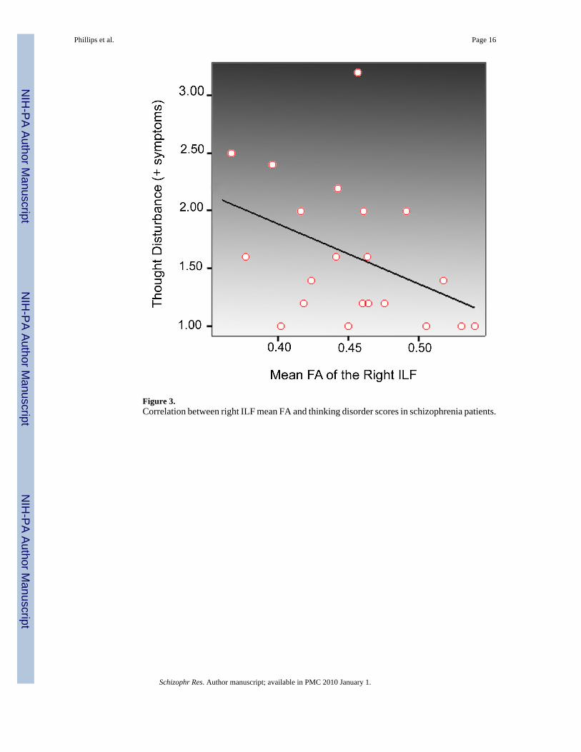
Figure 3.Correlation between right ILF mean FA and thinking disorder scores in schizophrenia patients.
Phillips et al. Page 16
Schizophr Res. Author manuscript; available in PMC 2010 January 1.
NIH
-PA Author Manuscript
NIH
-PA Author Manuscript
NIH
-PA Author Manuscript

NIH
-PA Author Manuscript
NIH
-PA Author Manuscript
NIH
-PA Author Manuscript
Phillips et al. Page 17
Table 1Subject demographic Information.
Schizophrenia Controls Statistic P Value
Age (mean ± SD) 34.65±9.17 30.87±9.52 t=-1.36 0.18
Gender (male/female) 17/6 17/5 χ2 =0.07 0.79
Handedness (dextral/non-dextral) 23/0 21/1 χ2 = 1.07 0.30
Years of Education 14.17±1.85 15.31±2.05 t=1.96 0.06
Duration of Illness 10.14±7.94 - - -
Age of Onset 21.93±4.22 - - -
Total Brain Volume (cc) 1442.6 1431.4 t=-0.29 0.77
White Matter Volume (cc) 566.5 556.3 t=0.61 0.54
Anergia (mean ± SD)* 1.69±0.65 - - -
Thought Disturbance (mean ± SD)* 1.63+0.60 - - -
*Missing data for 2 patients.
Schizophr Res. Author manuscript; available in PMC 2010 January 1.
NIH
-PA Author Manuscript
NIH
-PA Author Manuscript
NIH
-PA Author Manuscript
Phillips et al. Page 18Ta
ble
2In
tra-c
lass
cor
rela
tion
coef
ficie
nts
(r)
for
RO
I tra
cing
. A
F=ar
cuat
e fa
scic
ulus
, IL
F=in
ferio
r lo
ngitu
dina
l fa
scic
ulus
, U
F=un
cina
tefa
scic
ulus
.
Intr
a-ra
ter
relia
bilit
yIn
ter-
rate
r re
liabi
lity
Tra
ctM
ean
FAV
olum
eM
ean
FAV
olum
e
Lef
tR
ight
Lef
tR
ight
Lef
tR
ight
Lef
tR
ight
AF
0.99
50.
991
0.99
20.
989
0.92
80.
988
0.96
30.
970
ILF
0.99
70.
996
0.95
70.
969
0.99
40.
988
0.92
10.
942
UF
0.99
80.
994
0.94
20.
962
0.97
80.
986
0.99
30.
971
Val
ues i
n th
e ta
ble
repr
esen
t int
racl
ass c
orre
latio
n co
effic
ient
s (R
I)
Schizophr Res. Author manuscript; available in PMC 2010 January 1.
NIH
-PA Author Manuscript
NIH
-PA Author Manuscript
NIH
-PA Author Manuscript
Phillips et al. Page 19
Table 3Tract measurements for the arcuate fasciculus (AF), inferior longitudinal fasciculus (ILF), and uncinate fasciculus(UF).
FA (Mean ± SD) Volume (Mean ± SD), (cc)
Fiber Tract Measures Controls Patients Controls Patients
AF Left 0.48±0.02* 0.45±0.03* 5.58±2.99* 4.53±2.45*
AF Right 0.48±0.04 0.46±0.03 4.32±2.90 3.24±2.15
ILF Left 0.47±0.02* 0.45±0.02* 10.67±4.07 9.76±3.02
ILF Right 0.47±0.02* 0.45±0.02* 11.34±3.13 10.39±3.55
UF Left 0.40±0.03 0.39±0.02 3.34±1.88 2.52±1.71
UF Right 0.42±0.03 0.40±0.04 2.66±1.2 3.11±2.13
*Significant effects
Schizophr Res. Author manuscript; available in PMC 2010 January 1.
Copyright © 2022 FDOKUMEN