Left inferior parietal dominance in gesture imitation: an fMRI study
Motion Standstill Leads to Activation ofInferior Parietal Lobe
Andrea Federspiel,1,2* Umberto Volpe,2,3 Helge Horn,1 Thomas Dierks,1,2
Anders Franck,2,4 Patrizia Vannini,2 Lars-Olof Wahlund,2
Silvana Galderisi,3 and Mario Maj3
1Department of Psychiatric Neurophysiology, University Hospital of Clinical Psychiatry,Bern, Switzerland
2Neurotec Department, Division of Clinical Geriatrics, Karolinska Institutet, Karolinska UniversityHospital, Stockholm, Sweden
3Department of Psychiatry, School of Medicine, University of Naples SUN, Naples, Italy4Department of Hospital Physics, Karolinska Institutet, Karolinska University Hospital,
Stockholm, Sweden
� �
Abstract: Previous studies on motion perception revealed motion-processing brain areas sensitive to changesin luminance and texture (low-level) and changes in salience (high-level). The present functional magneticresonance imaging (fMRI) study focused on motion standstill. This phenomenon, occurring at fast presentationfrequencies of visual moving objects that are perceived as static, has not been previously explored byneuroimaging techniques. Thirteen subjects were investigated while perceiving apparent motion at 4 Hz, at 30Hz (motion standstill), isoluminant static and flickering stimuli, fixation cross, and blank screen, presentedrandomly and balanced for rapid event-related fMRI design. Blood oxygenation level-dependent (BOLD)signal in the occipito-temporal brain region MT/V5 increased during apparent motion perception. Here wecould demonstrate that brain areas like the posterior part of the right inferior parietal lobule (IPL) demon-strated higher BOLD-signal during motion standstill. These findings suggest that the activation of higher-ordermotion areas is elicited by apparent motion at high presentation rates (motion standstill). We interpret thisobservation as a manifestation of an orienting reaction in IPL towards stimulus motion that might be detectedbut not resolved by other motion-processing areas (i.e., MT/V5). Hum Brain Mapp 27:340–349, 2006.© 2005 Wiley-Liss, Inc.
Key words: motion standstill; apparent motion; event-related functional magnetic resonance imaging;inferior parietal lobe; motion perception
� �
INTRODUCTION
The ability to perceive objects in motion, regardless ofwhether the motion is real or illusionary, is one of the majorproperties shown by the human brain in daily life. “Motionstandstill” is a phenomenon, first described by Julesz andPayne [1968], which occurs when motion is so fast that themoving visual stimuli are perceived as static. Previous stud-ies have attempted to explore the mechanism of the percep-tion of this motion illusion. Results from psychophysicalvisual investigations with isoluminant chromatic motionstimuli and rotating luminance gratings suggest a distinc-tion between three different-motion processing mechanisms[Cavanagh 1992; Lu et al., 1999a; Lu and Sperling 2001]: (1)
Contract grant sponsors: Dargut and Milena Kemali Foundation forBasic and Clinical Neurosciences, Naples, Italy (to U.V.); KarolinskaInstitutets Research Funds; Alzheimerfonden, Stockholm, Sweden.*Correspondence to: Andrea Federspiel, PhD, Department of Psy-chiatric Neurophysiology, University Hospital of Clinical Psychia-try, Bollingenstrasse 111, CH-3000, Bern 60, Switzerland.E-mail: [email protected] for publication 29 October 2004; Accepted 27 June 2005DOI: 10.1002/hbm.20189Published online 17 August 2005 in Wiley InterScience (www.interscience.wiley.com).
� Human Brain Mapping 27:340–349(2006) �
© 2005 Wiley-Liss, Inc.
luminance-based process (motion process responsive tomoving luminance patterns); (2) a feature-based process(motion process responsive to moving modulations of fea-ture type); and (3) a salience-based mechanism. The low-level visual processing includes luminance- and feature-based motion. A higher-level, salience-based mechanismallows the processing of visual stimuli by computing a neu-ral representation of visual space even when no substantialdifference in luminance exists.
Brain imaging studies revealed several motion-sensitiveareas: recent functional magnetic resonance imaging (fMRI)studies identified as motion-sensitive areas the so-called“human motion complex” (or middle temporal complex,MT/V5) [Zeki et al., 1991], the extrastriate visual area V3A[Tootell et al., 1995; Tootell and Taylor 1995], the superiortemporal sulcus (STS) [Noguchi et al., 2005; Sunaert et al.,1999], and the intraparietal sulci (IPS) [Bremmer et al., 2001;Corbetta and Shulman 2002]. In particular, activation inMT/V5 has been reported to be induced even by a simplemovement of a dot pattern when the load on attention forperforming a relevant linguistic task was low [Rees et al.,1997]. This finding suggests that all these areas might beresponsible for a low-level motion perception.
Other fMRI studies, dealing with illusionary movement,revealed that the MT/V5 constituted an optimal candidateregion for conscious perception of both real and illusionarymotion [Seghier et al., 2000; Tootell et al., 1995; Zeki et al.,1991, 1993]. However, recent brain imaging studies castsome doubt on a possible specific role of MT/V5 areas inmotion detection. For example, Orban et al. [1998] proposedthat lingual and cuneal regions are sensitive to real motion,whereas MT/V5, as reported also in other studies [Chawlaet al., 1998; Sunaert et al., 2000], could be merely involved inmotion speed discrimination. In conclusion, a definitivemodel about the role of MT/V5 as a motion- and/or speed-sensitive brain region has not been achieved yet and itssupposed specificity for human motion perception is stillcontroversial.
Furthermore, several studies have indicated that MT/V5cannot be regarded as a “stand-alone” motion perceptivearea, since the perception of moving objects seems to involveboth low-level and high-level brain areas sensitive to mo-tion. Recently, it was shown that activation in the tem-poroparietal junction (TPJ), including the inferior parietallobe (IPL), is evoked by salient stimuli and during the de-tection of low-frequency stimuli at an attended location[Kirino et al., 2000]. The issue of the representation of motionperception has been more specifically addressed in a recentfMRI study [Claeys et al., 2003] in which the authors pro-posed that high-level motion processing engages the IPL.The authors reported activation of IPL for isoluminant colorgrating motion and for apparent motion with a temporalfrequency of 7 Hz. However, IPL did not respond to 2-Hzapparent motion. Motion processing in IPL might thereforebe driven by both salience-based and luminance-based ap-parent motion. Furthermore, the authors reported an activa-tion due to lower-level, luminance-based features in the
traditional motion-sensitive regions (i.e., MT/V5, V3a, STS).This suggests that salience, as well as presentation rate, exertsome influence on the activation of higher-order motionbrain areas.
An increase of the presentation rate, far above the fre-quency used in the study by Claeys et al. [2003], leads to theabove-described phenomenon called motion standstill. Sofar, motion standstill has been investigated using chromaticdisplay [Lu et al., 1999a,b]. The authors suggested that theisoluminant color motion was only perceived by a salience-based motion system. In the present study we used onlyblack and white stimuli to produce the effect of motionstandstill. To our knowledge, it is the first investigation ofthe phenomenon of motion standstill with fMRI. The goalwas twofold: clarifying whether motion standstill is pro-cessed in traditional motion-processing areas only (i.e., MT/V5, V3A, STS, IPS) or if additional brain areas are alsoinvolved in this process. Furthermore, we addressed thequestion of whether motion standstill differs from simpleperception of flicker. These issues were investigated with thefMRI method, which offers good spatial resolution.
SUBJECTS AND METHODS
Subjects
Thirteen volunteers (8 women, 5 men) participated in thefist experiment. Age (mean � SD) was 29.7 � 4.6 years. Allwere staff members or students at the Karolinska Institutetof Stockholm (Sweden). All subjects were right-handed, asassessed by the Edinburgh Inventory [Oldfield, 1971]. Visionwas normal or corrected-to-normal in all subjects, and nonehad a history of major medical, neurological, or psychiatricdiseases, nor did they receive psychotropic medication, asascertained by a 1-hour nonstructured clinical interview. Allparticipants had normal structural MRI, as confirmed by theevaluation of an expert radiologist (M.K.W.). The study wasapproved by the local ethical committee and written in-formed consent was obtained from all subjects.
An additional four subjects (3 females, ages 27.4, 27.11,and 23.8 years, and 1 male, age 33.1 years) were investigatedin a second fMRI experiment.
Experimental Paradigm
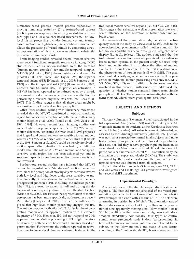
A schematic view of the stimulation paradigm is shown inFigure 1. The first experiment consisted of the visual pre-sentation against a black background of nine white dots (Ø0.9°) arranged in a circle with a radius of 10°. The dots werealternating in position by a 20° shift. The alternation rate ofthese 9 dots was set either to 4 Hz (resulting in the percep-tion of nine apparently moving dots: “slow motion”), or to30 Hz (resulting in the perception of eighteen static dots:“motion standstill”). Additionally, four types of controlstimuli were presented: static 9 dots (corresponding, interms of luminance and visual information delivered to thesubject, to the “slow motion”) and static 18 dots (corre-sponding to the “motion standstill”), blank screen, and fix-
� fMRI of Motion Standstill �
� 341 �
ation cross. Stimuli were presented to the subjects by agoggle system (VisuaStim XGA; Resonance Technology,Northridge, CA). The parameters of this system were: fieldof view � 30°, refresh rate � 60 Hz, resolution � 1024 � 768.The alternation rate of 30 Hz was measured with photo-diodes directly placed in front of the goggle system andlinked to a digital storage oscilloscope. Stimuli were pre-sented for 3,000 ms, followed by a blank screen (interstimu-lus interval: 3,000 ms). Each stimulus category was pre-sented 40 times: the resulting 240 stimuli were randomizedand the stimulus categories were balanced. Subjects wereasked to give a motor response by pressing a button with theright index finger whenever they perceived either the im-pression of a “motion standstill” or a “slow motion” pattern.Response times and omission rate to each stimulus wasrecorded by an MRI-compatible optical keypress devicewhich was interfaced to an external personal computer run-ning the experimental paradigm outside the scanner room.
A second experiment was conducted with the same stim-ulation paradigm as described above but with different con-trol stimuli. Here the 4 Hz “slow motion” category wascontrasted with 9 dots presented at a flicker frequency of 4Hz. The 30 Hz “motion standstill” category was contrastedwith 9 dots flickering at 30 Hz (the isoluminant controlcategory) and by 18 dots flickering at 30 Hz (a controlcategory with two times higher luminance). Stimulus dis-play time, interstimulus interval, total number of stimuli,and number of stimuli for each category were unchanged.
fMRI Acquisition
All images were acquired using a 1.5 T whole-body MRIsystem (Siemens Vision, Erlangen, Germany) equipped witha standard RF head coil. Head motion was minimizedwithin the coil using a vacuum deflatable pillow and anopen-cell polyurethane foam. The fMRI session included theacquisition of a set of 3-D T1-weighted (magnetization pre-pared rapid acquisition gradient echo, MP-RAGE) images,providing 170 sagittal slices with 1.0 mm thickness, 256� 256 mm field of view (FOV), and a matrix size of 256� 256. Further scan parameters were: 11.4 ms repetition time
Figure 1.Visual stimuli presented. A: Nine white dots (Ø 0.9°) arranged ina circle with radius of 10° on black background. Dots werealternating in position by a 20° shift. Illusionary stimuli used in thefirst and second experiments originated by an alternation rate ofthe 9 dots corresponding to 4 Hz (the illusionary perception is“slow motion”), or to 30 Hz (the illusionary perception is that of18 dots “motion standstill”). B: Control categories in the firstexperiment were 9 static dots and 18 static dots. C: Controlcategories in the second experiment were 9 dots presented atflicker frequency of 4 Hz and 18 dots flickering at 30 Hz. Notshown in this schematic view are two categories that are displayedin each experiment: blank screen and fixation cross. Each stimuluscategory was presented 40 times: the resulting 240 stimuli wererandomized and the stimulus categories were balanced.
� Federspiel et al. �
� 342 �
(TR), 4.4 ms echo time (TE), and a flip angle of 15° (FA).Functional images were acquired using a multislice single-shot T2*-weighted echo planar imaging sequence, with 16interleaved axial oblique slices, positioned in-line with thebicommissural axis; 720 dynamic scans were collected ineach subject (TR � 2,000 ms, TE � 40 ms, FA � 90°, slicethickness � 4 mm, gap thickness � 1 mm, matrix size � 64� 64, FOV � 240 � 240 mm).
fMRI Analysis
Analysis of the fMRI data was performed using BrainVoy-ager 4.9 (Brain Innovation, Maastricht, The Netherlands).Preprocessing of the images included the removal of low-frequency drifts, 3-D motion detection and correction, andspatial smoothing with 4 mm FWHM. Coregistration of the2-D functional to the 3-D structural images was performedusing the scanner’s slice position parameters of the T2*-weighted measurements and the T1-weighted anatomicalmeasurements. Spatial resolution of the functional datasetwas interpolated to isovoxel 1 mm3. Finally, the anatomicaland functional datasets were transformed into the normal-ized Talairach space [Talairach and Tournoux, 1988].
All voxels with t-test value � 7.6 were defined as regionsof interest (ROIs). The reconstruction of the hemodynamicresponse function within these ROIs was performed using ageneral linear model (GLM) to estimate the finite impulseresponse (FIR) associated with each stimulus category. Foreach stimulus type the convolution of the discrete timepoints with the event sequence was built (i.e., a “1” wasplaced in the row corresponding to the time at which eachimage was acquired and in the column corresponding to theappropriate point of the hemodynamic response). For theextraction of the event-related responses we used 10 FIRtime bins (with TR � 2 s; this corresponds to the timeinterval 0–20 s after stimulus onset). The hemodynamicresponse function can substantially vary between subjects[Aguirre et al., 1998], and therefore we decided to define thepeak values of the event-related response in each individualand for each stimulus, a method repeatedly used in previousfMRI studies [Huk et al., 2001; Noguchi et al., 2005]. Peakvalues of the event-related data were chosen as the averageof the three highest consecutive values in the BOLD event-related data for each subject and for each category in thestatistical analysis [Lehmann et al., 2004].
In addition to the conventional normalization of individ-ual functional time series in the standard Talairach space, animproved correspondence of cortical regions across subjectswas obtained by using a cortex-based intersubject alignment[Fischl et al., 1999].
Statistical Analysis
An analysis of variance (ANOVA) with subjects as therandom factor and category as fixed factor was calculated.Furthermore, an ANOVA for repeated measures was per-formed with the average of three highest BOLD peak valuesof the corresponding hemodynamic response function inorder to test the effect of stimulus for each resulting brain
area (factor category). Post-hoc pairwise comparison wasperformed using Scheffe’s tests. The software system Statis-tica v. 6 (StatSoft, Tulsa, OK, 2001) was used.
RESULTS
Test Performance
The mean (�SD) motor reaction time for the perception ofslow motion was 512.1 (�281.5) ms and for the perception ofmotion standstill was 528.1 (�254.6) ms (this difference inreaction time was not statistically significant). Subjects re-sponded to 4 Hz and 30 Hz but not to the flicker controlconditions. Due to the simple nature of the task the subjectsresponded to all presented stimuli and there were no signif-icant differences between omission rates for the two stimu-lus categories in which a button press was required.
fMRI Results
The ANOVA with subjects as the random factor showedsignificant category effect (F(4,48) � 38.44; P � 0.0001). TheGLM analysis showed that the contrast between apparentmotion stimuli and static stimuli categories vs. the fixationcategory yielded a significant activation in the early visualareas (calcarine cortex), as illustrated in Figure 2A. TheTalairach coordinates corresponding to the center of gravityfor each identified ROI and the results of the repeated mea-sure ANOVA for within-subject effects, as well as the aver-age and SDs of the three highest peak values, time param-eters, and maximum peak values for each category aresummarized in Table I. The comparison between apparentmotion and static stimuli revealed activation of the right andleft MT/V5, as depicted in Figure 2B. The contrast betweenslow motion and static 9 dots stimulus categories, as illus-trated in Figure 2C, yielded significant activation of the rightMT/V5. Finally, a comparison between motion standstillwith static 18 dots stimulus categories yielded significantactivation of the left MT/V5, left inferior frontal gyrus (IFG)corresponding to BA 45, right IFG corresponding to BA 44,and bilateral IPL corresponding to BA 40, as illustrated inFigure 2D.
The averaged event-related responses for early visual areaare shown in Figure 3 and were obtained from the regiondisplayed in Figure 2A. The averaged event-related re-sponses for all other identified regions are shown in Figure4a–f.
The results of the post-hoc pairwise comparison are sum-marized in Table II. The post-hoc comparison between mo-tion standstill and slow motion revealed a statistical ten-dency to significance (t(1,12) � 3.3; P � 0.06) only for the rightIPL (r-IPL posterior).
BOLD responses, as depicted in Figure 4, are higher forthe apparent motion stimulus category rather than for staticstimuli. Generally, the right and left MT/V5 regions ap-peared to be more involved in the processing of slow motionthan for motion standstill stimuli. In contrast, the right IPLregion (r-IPL posterior) appeared more active for motion
� fMRI of Motion Standstill �
� 343 �

standstill than for slow motion stimuli. In all identifiedregions but early visual areas the activation for “static 9dots” was higher than for “static 18 dots” stimuli but thedifference was not statistically significant.
The cortex-based intersubject alignment revealed a sec-ond region in right IPL (Talairach coordinates: x � 60, y� –31, z � 32; t-value: 5.5; P � 0.0001) for the contrastbetween motion standstill and slow motion (r-IPL ante-rior). However, no significant differences were obtainedfor the contrast between motion standstill and static 18dots. Instead, using the contrast between motion standstillstimulus as compared to all other categories, there wasalso significant activation in the anterior cingulum cortex(ACC) (Talairach coordinates: x � 1, y � 23, z � 29;t-value: 4.0; P � 0.0001).
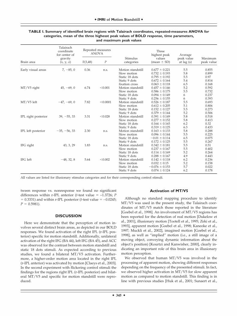
The results of the second experiment are shown in Figure5. In right IPL (r-IPL posterior) the highest activation wasobserved by motion standstill. Activation due to slow mo-tion and due to the flickering control categories was lower.Activation of early visual areas, bilateral MT/V5, and r-IPLposterior due to motion standstill and slow motion are com-parable to the results of the first experiment. No statisticaldifferences were observed in the ACC (t-value: 0.96; P� n.s.) for the comparison between motion standstill andflickering control categories.
Finally, we pooled the fMRI data and compared the BOLDsignals that contained the motion response with the BOLDsignals without motor response. For the comparison be-
Figure 3.BOLD signal time course in cortical early visual areas averagedover 13 subjects. The region is shown in Figure 2A.
Figure 2.Group GLM contrast analysis detects regionsin which: (A) activity is higher during percep-tion of visual stimuli than during fixation cross.Activation in the human early visual areas (Ta-lairach coordinates: 7-85 0); (B) activity ishigher during perception of illusionary stimuli(both motion standstill and slow motion) thanduring “static” stimulation. Activation is ob-served bilateral in human motion complex MT/V5; (C) activity is higher during the perceptionof slow motion stimulus (4 Hz) as compared tostatic 9 dots stimulus in right human motioncomplex MT/V5; (D) activity is higher duringthe perception of motion standstill stimulus(30 Hz) as compared to static 18 dots stimulus.These regions corresponds to left MT/V5, leftBA45 (gyrus frontalis inferior), right BA 44(gyrus frontalis inferior), and bilaterally in theposterior part of BA 40 (inferior parietal lobe;gyrus supramarginal). Right top corner: colorscale represents values of t-test; significancelevel indicated (P(corrected) � 0.00001).
� Federspiel et al. �
� 344 �
tween response vs. nonresponse we found no significantdifferences within r-IPL anterior (t-test value � –1.3726; P� 0.3331) and within r-IPL posterior (t-test value � –0.0245;P � 0.5981).
DISCUSSION
Here we demonstrate that the perception of motion in-volves several distinct brain areas, as depicted in our BOLDresponses. We found activation of the right IPL (r-IPL pos-terior) specific for motion standstill. Additionally, unilateralactivation of the right IFG (BA 44), left IFG (BA 45), and ACCwas observed for the contrast between motion standstill andstatic 18 dots stimuli. As expected according to previousstudies, we found a bilateral MT/V5 activation. Further-more, a higher-order motion area located in the right IPL(r-IPL anterior) was activated by motion [Claeys et al., 2003].In the second experiment with flickering control stimuli thefindings for the regions right IPL (r-IPL posterior) and bilat-eral MT/V5 and specific for motion standstill were repro-duced.
Activation of MT/V5
Although no standard mapping procedure to identifyMT/V5 was used in the present study, the Talairach coor-dinates of MT/V5 match those reported in the literature[Goebel et al., 1998]. An involvement of MT/V5 regions hasbeen reported for the detection of real motion [Dukelow etal., 2001], illusionary motion [Tootell et al., 1995; Zeki et al.,1993], apparent motion [Goebel et al., 1998; Kaneoke et al.,1997; Muckli et al., 2002], imagined motion [Goebel et al.,1998], as well as “implied” motion (i.e., a still image of amoving object, conveying dynamic information about theobject’s position) [Kourtzi and Kanwisher, 2000], clearly in-dicating an important role of this brain area in illusionarymotion perception.
We observed that human MT/V5 was involved in theprocessing of apparent motion, showing different responsesdepending on the frequency of the presented stimuli. In fact,we observed higher activation in MT/V5 for slow apparentmotion as compared to motion standstill. This finding is inline with previous studies [Huk et al., 2001; Sunaert et al.,
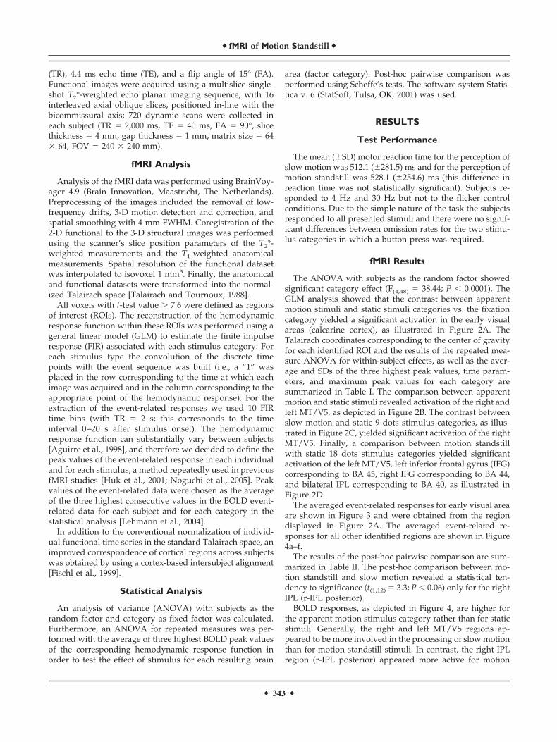
TABLE I. Summary of identified brain regions with Talairach coordinates, repeated-measures ANOVA forcategories, mean of the three highesst peak values of BOLD response, time parameters,
and maximum peak values
Brain area
Talairachcoordinate
for center ofgravity(x, y, z)
Repeated measuresANOVA
Stimuluscategories
Threehighest peak
values(mean � SD)
Averagepeak valueat lag (s)
Maximumpeak valueF(3,48) P
Early visual areas 7, �85, 0 0.36 n.s. Motion standstill 0.677 � 0.221 5.5 0.855Slow motion 0.732 � 0.193 5.8 0.899Static 18 dots 0.795 � 0.192 5.5 0.97Static 9 dots 0.672 � 0.164 5.4 0.814fixation cross 0.063 � 0.118 6.5 0.168
MT/V5 right 45, �69, 0 6.74 �0.001 Motion standstill 0.457 � 0.146 5.2 0.592Slow motion 0.586 � 0.175 5.5 0.732Static 18 dots 0.094 � 0.149 6 0.237Static 9 dots 0.256 � 0.155 6 0.393
MT/V5 left �47, �69, 0 7.82 �0.0001 Motion standstill 0.526 � 0.187 5.5 0.693Slow motion 0.612 � 0.205 5.1 0.806Static 18 dots 0.152 � 0.127 5.5 0.273Static 9 dots 0.379 � 0.144 5.2 0.506
IPL right posterior 39, �55, 33 3.31 �0.028 Motion standstill 0.391 � 0.149 5.8 0.518Slow motion 0.277 � 0.152 5.8 0.413Static 18 dots 0.164 � 0.165 6.2 0.32Static 9 dots 0.319 � 0.125 6.3 0.433
IPL left posterior �35, �56, 33 2.30 n.s. Motion standstill 0.163 � 0.133 5.8 0.288Slow motion 0.096 � 0.144 5.5 0.225Static 18 dots �0.01 � 0.114 6.5 0.075Static 9 dots 0.172 � 0.125 5.5 0.267
IFG right 43, 3, 29 1.83 n.s. Motion standstill 0.342 � 0.181 5.5 0.51Slow motion 0.237 � 0.167 5.5 0.402Static 18 dots 0.114 � 0.169 5.2 0.264Static 9 dots 0.288 � 0.167 4.9 0.443
IFG left �48, 32, 8 5.64 �0.002 Motion standstill 0.142 � 0.118 6.2 0.236Slow motion 0.032 � 0.15 5.2 0.158Static 18 dots �0.076 � 0.153 5.7 0.022Static 9 dots 0.074 � 0.124 6.2 0.178
All values are listed for illusionary stimulus categories and for their corresponding control stimuli.
� fMRI of Motion Standstill �
� 345 �

2000] and especially with the inverted “U” dependency ofMT/V5 on speed reported by Chawla et al. [1998]. Theyused dots moving at constant speed (the velocities used are
well below the velocities used in our study for motionstandstill), radially from the center of the screen, as stimulusmaterial to demonstrate the dependency on speed of MT/V5
Figure 4.BOLD signal time course in cor-tical areas obtained as contrastbetween illusionary stimuli and“static” stimulation (a,b), and ascontrast between motion stand-still stimulus (30 Hz) and static18 dots stimulus (c–f). Verticalscale: averaged percentage signalchange; horizontal scale: stimulusonset time in seconds. Legendsas well as scales are equal in allplots and only shown in a.
TABLE II. Summary of post-hoc pairwise comparison between categories performed using Scheffe’s tests withinthe identified brain regions with Talairach coordinates for center of gravity
Brain area
Talairach coordinatefor center of gravity
(x, y, z)
Motion standstill vs. static18-dot stimuli
Slow motion vs. static 9-dotstimuli
t(1,12) P t(1,12) P
MT/V5 right 45, �69, 0 8.9 �0.0001 3.9; p �0.0001MT/V5 left �47, �69, 0 4.7 �0.0001 4.3 �0.0001IPL right posterior 39, �55, 33 3.3 �0.006 0.4; n.s.IPL left posterior �35, �56, 33 5.3 �0.0001 0.7; n.s.IFG right 43, 3, 29 5.2; p �0.0002 0.9; n.s.IFG left �48, 32, 8 1.5; n.s. 3.5 �0.004
� Federspiel et al. �
� 346 �
and V3a activation. This result was obtained in a wholebrain fMRI measurement and was subsequently replicatedby the same group [Chawla et al., 1999].
It has been reported that motion standstill can occur whenthe spatiotemporal resolution of the shape and color systemsexceeds that of the motion system: at higher presentationfrequencies (�8 Hz) the motion system for this salience-based stimulus breaks down [Lu et al., 1999a]. In our study,the effect of motion standstill was produced with high al-ternation frequency of nine white dots displaced by 20°(velocity above 100° visual angle/s). Is it possible that mo-tion standstill can also occur with luminance-based objectswhen the temporal resolution of the physical stimulus ex-ceeds the resolution of the motion systems in MT/V5? Inthis case, motion standstill would be the result of a speedeffect of the first-order luminance-based apparent motion.
The response in MT/V5 for real and apparent motion issimilar as long as the speed of the apparent motion does notexceed the temporal resolution of the human visual system.The upper limit for temporal resolution has been reported to bein the 4–8 Hz range [Verstraten et al., 2000]. Above this rate,perception of the sequence of changes in the positions of dotsis no longer possible. In the present experiment the motionstandstill stimulus clearly differs from the static 18 dots stim-ulus in terms of physical properties. However, both stimuli areperceived as 18 dots. In fact, all the subjects could distinguishbetween both types of stimuli. This is also reflected by theobserved BOLD response in MT/V5, which was found to bemore pronounced for the motion standstill stimulus, indicatingthat the process of motion analysis had already started.
However, in MT/V5 we also observed significant differ-ences in the control categories between 9 dots and 18 dots(both experiments, static and flicker). Surprisingly, the mag-nitude of BOLD signal for the 9 dot control stimuli inMT/V5 exceeded the magnitude of the 18 dots control stim-uli. This phenomenon is contrary to the activation in early
visual areas, which shows stronger activation in the 18 dotscontrol category. The results in early visual areas can beattributed to luminance effects. Baseline problems couldexplain this discrepancy, although the averaged time coursein the early visual region shows a clear distinction betweenfixation cross and all other stimulus categories. An alterna-tive explanation could be an “expectancy of movement” forthe 9 dot control stimuli. In fact, for the first 250 ms the twocategories (slow motion and 9 dot control stimuli) are iden-tical. Since the first 9 dot stimuli presented to the subjects aremoving, an anticipation of motion may be evoked duringthe 9 dot control stimuli. On the contrary, based on thepresented stimuli, the 18 dot control stimuli are not expectedto move.
If this explanation for the control stimuli holds, then thedifferences between motion standstill and slow motion inMT/V5 might originate from a presentation frequencywhich exceeds the temporal resolution of MT/V5. In thiscase, the motion stimuli that cannot be allocated to a cleardirection in space may provoke a pronounced shift of atten-tion in order to achieve a consistent explanation. At the timewe are not aware of other explanations for the surprisingeffect of the control stimuli in MT/V5. Future investigationswith an appropriate design will have to provide furtherinsights.
Activation of the Anterior Part of theRight IPL (r-IPL Anterior)
In addition to the activations in MT/V5, we observedactivation in the right IPL region for the motion stimuli.Activation in this region due to slow motion and due to thecontrol stimuli (both experiments, static and flicker) waslower. The involvement of this region in motion processinghas been described once before by Claeys et al. [2003], whocharacterized IPL as a potential higher-order motion region.
Figure 5.Second experiment: BOLD peakvalues (with standard deviation)for illusionary motion stimuli(motion standstill (black bars)and slow motion (dark gray bars)and for control stimuli (18 dotspresented at flicker frequency of30 Hz (white bars) and 9 dotspresented at flicker frequency of4 Hz (light gray bars). Verticalscale: averaged percentage signalchange; horizontal scale: brainregions right IPL posterior,MT/V5 right and left and earlyvisual areas.
� fMRI of Motion Standstill �
� 347 �
In their study, right IPL responded to isoluminance colorgratings stimuli when the saliency of one color was in-creased. IPL also responded to apparent motion stimulationat a 7 Hz rate, but not at a 2-Hz rate. This finding is in linewith our observation that the 30-Hz stimulus (but not the4-Hz stimulus) evokes a response in the IPL, although thefrequencies used in both studies substantially differ. A directcomparison is hence difficult.
Activation of the Posterior Part of theRight IPL (r-IPL Posterior)
In the posterior part of the right IPL we observed activa-tion by motion standstill only. Presently, we are not aware ofother studies showing activation of the r-IPL posterior orig-inated by motion. Generally, the IPL region is known toshow activations in multiple paradigms in all sensory mo-dalities. Downar et al. [2000] recently reported that tem-poroparietal junction (including IPL) was also activatedwhen visual and other sensory stimuli were suddenlychanged; this finding was interpreted as a consequence of areorienting attention-demanding task. In our study the acti-vation of the r-IPL posterior was only present for the motionstandstill stimulus. Other contrasts, including the reorienta-tion of attention (i.e., slow motion vs. fixation cross), did notevoke an activation in the r-IPL posterior. Therefore, reori-entation of attention to a sudden change in stimulation alonecannot fully explain activation in the r-IPL posterior. None-theless, modulation of the r-IPL posterior by visual attentioncannot be ruled out. It seems reasonable to hypothesize thatthe activation in this area could be due to a special kind ofarousal. This view is supported by the observed activation inright IFG (BA 44), left IFG (BA 45), and ACC also only bymotion standstill. Taken together, by motion standstill weobserved activation of a typical attention network [see Cor-betta and Shulman, 2002, for a detailed review] (r-IPL pos-terior [BA 40], ACC, right IFG [BA 44] and left IFG [BA4 5]).Furthermore, this network is also activated by visual [Kirinoet al., 2000; Linden et al., 1999] and acoustic [Horn et al.,2003] target detection paradigms. Therefore, the r-IPL pos-terior can be regarded as a multimodal area, an area acti-vated together with the anterior attentional network by ori-enting reaction towards unresolved or target stimuli.
In conclusion, our study confirms the previously reportedactivation of lower-order motion region MT/V5 in an ap-parent motion paradigm [Goebel et al., 1998; Muckli et al.,2002; Tootell et al., 1995; Zeki et al., 1993]. In line with thereported speed-dependency of these regions [Chawla et al.,1998, 1999] (the velocities used in these studies are wellbelow the velocities used in our study), our observed re-sponse to slow motion is higher, if compared to motionstandstill. Additionally, in the present study the activationof higher-order motion regions in the right IPL (r-IPL ante-rior) was observed with all motion categories. This result isin line with the work of Claeys et al. [2003]. In addition tothis known higher-order motion region, we observed a spe-cific activation by motion standstill in the r-IPL posteriorand in an anterior attention network.
These findings support the hypothesis that motion stand-still exceeding the temporal resolution of MT/V5 evokes aresponse in an attention network. This response can becaused by an orienting reaction with a stimulus-relatedarousal.
ACKNOWLEDGMENTS
We thank Drs. Armida Mucci (Naples), Tomas Jonsson(Stockholm), Thomas J. Muller, and Christoph Lehmann(Bern) for valuable discussions, all the members of the neu-roradiology staff of the Karolinska University Hospital,Stockholm (particularly, Reza Ghaderi and Kerstin Ericsson)for their support in recording the MR studies, and Dr. MariaKristoffersen-Wiberg for help in the evaluation of the struc-tural MRIs. Andrea Federspiel, Umberto Volpe, and HelgeHorn contributed equally to this work.
REFERENCES
Aguirre GK, Zarahn E, D’Esposito M (1998): The variability ofhuman, BOLD hemodynamic responses. Neuroimage 8:360–369.
Bremmer F, Schlack A, Shah NJ, Zafiris O, Kubischik M, HoffmannK, Zilles K, Fink GR (2001): Polymodal motion processing inposterior parietal and premotor cortex: a human fMRI studystrongly implies equivalencies between humans and monkeys.Neuron 29:287–296.
Cavanagh P (1992): Attention-based motion perception. Science 257:1563–1565.
Chawla D, Phillips J, Buechel C, Edwards R, Friston KJ (1998):Speed-dependent motion-sensitive responses in V5: an fMRIstudy. Neuroimage 7:86–96.
Chawla D, Buechel C, Edwards R, Howseman A, Josephs O, Ash-burner J, Friston KJ (1999): Speed-dependent responses in V5: areplication study. Neuroimage 9:508–515.
Claeys KG, Lindsey DT, De Schutter E, Orban GA (2003): A higherorder motion region in human inferior parietal lobule: evidencefrom fMRI. Neuron 40:631–642.
Corbetta M,Shulman GL (2002): Control of goal-directed and stim-ulus-driven attention in the brain. Nat Rev Neurosci 3:201–215.
Downar J, Crawley AP, Mikulis DJ, Davis KD (2000): A multimodalcortical network for the detection of changes in the sensoryenvironment. Nat Neurosci 3:277–283.
Dukelow SP, DeSouza JF, Culham JC, van den Berg AV, Menon RS,Vilis T (2001): Distinguishing subregions of the human MT�complex using visual fields and pursuit eye movements. J Neu-rophysiol 86:1991–2000.
Fischl B, Sereno MI, Tootell RB, Dale AM (1999): High-resolutionintersubject averaging and a coordinate system for the corticalsurface. Hum Brain Mapp 8:272–284.
Goebel R, Khorram-Sefat D, Muckli L, Hacker H, Singer W (1998):The constructive nature of vision: direct evidence from func-tional magnetic resonance imaging studies of apparent motionand motion imagery. Eur J Neurosci 10:1563–1573.
Horn H, Syed N, Lanfermann H, Maurer K, Dierks T (2003): Cere-bral networks linked to the event-related potential P300. EurArch Psychiatry Clin Neurosci 253:154–159.
Huk AC, Ress D, Heeger DJ (2001): Neuronal basis of the motionaftereffect reconsidered. Neuron 32:161–172.
Julesz B,Payne RA (1968): Differences between monocular and bin-ocular stroboscopic movement perception. Vis Res 8:433–444.
� Federspiel et al. �
� 348 �
Kaneoke Y, Bundou M, Koyama S, Suzuki H, Kakigi R (1997):Human cortical area responding to stimuli in apparent motion.Neuroreport 8:677–682.
Kirino E, Belger A, Goldman-Rakic P, McCarthy G (2000): Prefrontalactivation evoked by infrequent target and novel stimuli in avisual target detection task: an event-related functional magneticresonance imaging study. J Neurosci 20:6612–6618.
Kourtzi Z, Kanwisher N (2000): Activation in human MT/MST bystatic images with implied motion. J Cogn Neurosci 12:48–55.
Lehmann C, Mueller T, Federspiel A, Hubl D, Schroth G, Huber O,Strik W, Dierks T (2004): Dissociation between overt and uncon-scious face processing in fusiform face area. Neuroimage 21:75–83.
Linden DE, Prvulovic D, Formisano E, Vollinger M, Zanella FE,Goebel R, Dierks T (1999): The functional neuroanatomy oftarget detection: an fMRI study of visual and auditory oddballtasks. Cereb Cortex 9:815–823.
Lu ZL, Lesmes LA, Sperling G (1999a): Perceptual motion standstillin rapidly moving chromatic displays. Proc Natl Acad Sci U S A96:15374–15379.
Lu ZL, Lesmes LA, Sperling G (1999b): The mechanism of isolumi-nant chromatic motion perception. Proc Natl Acad Sci U S A96:8289–8294.
Lu ZL, Sperling G (2001): Three-systems theory of human visualmotion perception: review and update. J Opt Soc Am A OptImage Sci Vis 18:2331–2370.
Muckli L, Kriegeskorte N, Lanfermann H, Zanella FE, Singer W,Goebel R (2002): Apparent motion: event-related functionalmagnetic resonance imaging of perceptual switches and states.J Neurosci 22:RC219 1–5.
Noguchi Y, Kaneoke Y, Kakigi R, Tanabe HC, Sadato N (2005): Roleof the superior temporal region in human visual motion percep-tion. Cereb Cortex (in press).
Oldfield RC (1971): The assessment and analysis of handedness: theEdinburgh inventory. Neuropsychologia 9:97–113.
Orban GA, Dupont P, De Bruyn B, Vandenberghe R, Rosier A,Mortelmans L (1998): Human brain activity related to speeddiscrimination tasks. Exp Brain Res 122:9–22.
Rees G, Frith CD, Lavie N (1997): Modulating irrelevant motionperception by varying attentional load in an unrelated task.Science 278:1616–1619.
Seghier M, Dojat M, Delon-Martin C, Rubin C, Warnking J, Seg-ebarth C, Bullier J (2000): Moving illusory contours activateprimary visual cortex: an fMRI study. Cereb Cortex 10:663–670.
Sunaert S, Van Hecke P, Marchal G, Orban GA (1999): Motion-respon-sive regions of the human brain. Exp Brain Res 127:355–370.
Sunaert S, Van Hecke P, Marchal G, Orban GA (2000): Attention tospeed of motion, speed discrimination, and task difficulty: anfMRI study. Neuroimage 11:612–623.
Talairach J, Tournoux P (1988): Co-planar stereotaxic atlas of thehuman brain: 3-dimensional proportional system: an approachto cerebral imaging. New York: Thieme.
Tootell RB, Taylor JB (1995): Anatomical evidence for MT andadditional cortical visual areas in humans. Cereb Cortex 5:39 –55.
Tootell RB, Reppas JB, Kwong KK, Malach R, Born RT, Brady TJ,Rosen BR, Belliveau JW (1995): Functional analysis of human MTand related visual cortical areas using magnetic resonance im-aging. J Neurosci 15:3215–3230.
Verstraten FA, Cavanagh P, Labianca AT (2000): Limits of attentivetracking reveal temporal properties of attention. Vis Res 40:3651–3664.
Zeki S, Watson JD, Lueck CJ, Friston KJ, Kennard C, Frackowiak RS(1991): A direct demonstration of functional specialization inhuman visual cortex. J Neurosci 11:641–649.
Zeki S, Watson JD, Frackowiak RS (1993): Going beyond the infor-mation given: the relation of illusory visual motion to brainactivity. Proc R Soc Lond B Biol Sci 252:215–222.
� fMRI of Motion Standstill �
� 349 �
Copyright © 2022 FDOKUMEN