A Placebo-controlled Study of Inferior Turbinate Surgery - Trepo
142
TEEMU HARJU A Placebo-controlled Study of Inferior Turbinate Surgery Acta Universitatis Tamperensis 2415
-
Upload
khangminh22 -
Category
Documents
-
view
0 -
download
0
Transcript of A Placebo-controlled Study of Inferior Turbinate Surgery - Trepo

TEEMU HARJU
A Placebo-controlled Study of Inferior Turbinate Surgery
Acta Universitatis Tamperensis 2415
TEEMU
HAR
JU A Placebo-controlled Study of Inferior Turbinate Surgery
AUT 2415
TEEMU HARJU
A Placebo-controlled Study of Inferior Turbinate Surgery
ACADEMIC DISSERTATIONTo be presented, with the permission of
the Faculty Council of the Faculty of Medicine and Life Sciences of the University of Tampere,
for public discussion in the auditorium F114 of the Arvo building, Arvo Ylpön katu 34, Tampere,
on 28 September 2018, at 12 o’clock.
UNIVERSITY OF TAMPERE
TEEMU HARJU
A Placebo-controlled Study of Inferior Turbinate Surgery
Acta Universi tati s Tamperensi s 2415Tampere Universi ty Pres s
Tampere 2018

Reviewed by Docent Leif BäckUniversity of HelsinkiFinlandDocent Tuomo PuhakkaUniversity of TurkuFinland
Supervised by Professor Markus RautiainenUniversity of TampereFinland
Acta Universitatis Tamperensis 2415 Acta Electronica Universitatis Tamperensis 1925ISBN 978-952-03-0838-4 (print) ISBN 978-952-03-0839-1 (pdf )ISSN-L 1455-1616 ISSN 1456-954XISSN 1455-1616 http://tampub.uta.fi
Suomen Yliopistopaino Oy – Juvenes PrintTampere 2018
441 729Painotuote
The originality of this thesis has been checked using the Turnitin OriginalityCheck service in accordance with the quality management system of the University of Tampere.
ACADEMIC DISSERTATIONUniversity of Tampere, Faculty of Medicine and Life Sciences Tampere University Hospital, Department of Otorhinolaryngology Finland
Copyright ©2018 Tampere University Press and the author
Cover design byMikko Reinikka

To my dear wife Pauliina and my three sweet daughters Elli, Iida, and Aliisa whohave tolerated me and laughed at my even more than usual absent-mindednessduring the months I spent writing this doctoral thesis.

ABSTRACT
Inferior turbinate enlargement is one of the main causes of chronic nasalobstruction. There are various techniques available for the surgical treatment ofinferior turbinate enlargement. Only three of the previous studies of inferiorturbinate surgery have been placebo-controlled and, based on them, the placeboeffect seems have a role in the results of the surgery. The relationship betweenEustachian tube dysfunction (ETD) and inferior turbinate enlargement or inferiorturbinate surgery has never been studied before. Furthermore, most of the studiesthat have evaluated the effect of inferior turbinate surgery on ciliated epitheliumhave only been descriptive and lacked statistical analysis. This thesis deals withinferior turbinate enlargement and its surgical treatment paying attention to all theabove-mentioned aspects.
A total of 104 inferior turbinate enlargement patients were consecutivelyblinded and randomized into placebo, radiofrequency ablation (RFA), diode laseror microdebrider-assisted turbinoplasty (MAIT) groups in a ratio of 1:2:2:2. Priorto the operation, 6 patients withdrew from the study leaving a total of 98 patientswho underwent one of the four alternative procedures. All the patients wereevaluated prior to operation and three months subsequent to the operation.
The results of all 98 treated patients were assessed when the effect of inferiorturbinate surgery techniques on nasal obstruction was evaluated. At the end of thethree-month follow-up, all the procedures, including placebo, decreased the VisualAnalog Scale (VAS) score of the severity of nasal obstruction significantly.However, all three active treatments decreased the symptom score of severity ofnasal obstruction significantly more than the placebo procedure.
For the first time, we evaluated the relationship between inferior turbinateenlargement and ETD-related symptoms using the Eustachian Tube DysfunctionQuestionnaire (ETDQ-7) as an assessment method. We compared the first 40consecutive patients aged < 45 years from the total 104 patients with inferiorturbinate enlargement with 40 healthy controls and found that the ETDQ-7 scorewas significantly higher in the inferior turbinate enlargement group.
The first 72 consecutive patients out of a total of 98 were evaluated for the effectof inferior turbinate surgery techniques on ETD-related symptoms and examinedwith the ETDQ-7. All the active treatments decreased the total ETDQ-7 scoresignificantly, but there were no significant differences in the results between theplacebo procedure and active treatments.
Samples for scanning electron microcopy (SEM) were taken in the form ofmucosal biopsies from 66 consecutive patients in the RFA, diode laser and MAITgroups preoperatively and at the control visit. After SEM, image series of 44patients were of a technically acceptable quality to be further evaluated. Thenumber of cilia was found to increase significantly after RFA and MAIT treatmentsand the amount of squamous metaplasia increased significantly after diode lasertreatment.
From the above-mentioned findings, we conclude that 1) the placebo effect hasa large role in the overall reduction in the severity of nasal obstruction in inferiorturbinate surgery. However, all three examined techniques provide a statisticallysignificant additional reduction in the severity of nasal obstruction compared withthe placebo procedure. 2) Patients with inferior turbinate enlargement have moresymptoms related to ETD than healthy controls. 3) The improvement of ETD-related symptoms due to inferior turbinate surgery as a sole procedure is equal toplacebo. 4) RFA and MAIT are more mucosal preserving techniques than diodelaser, which seems to increase squamous metaplasia. The number of cilia seems toeven increase after RFA and MAIT procedures.
TIIVISTELMÄ
Nenän alakuorikoiden liikakasvu on yksi yleisimmistä kroonisen nenäntukkoisuuden aiheuttajista. Sen kirurgiseen hoitoon on tarjolla useita erilaisiamenetelmiä. Alakuorikkokirurgiaa on tähän mennessä tutkittu kolmessalumekontrolloidussa tutkimuksessa ja niiden perusteella lumeella on vaikutustakirurgian tuloksiin. Korvatorven toimintahäiriön ja alakuorikoiden liikakasvun taialakuorikkokirurgian välistä yhteyttä ei ole aiemmin tutkittu. Alakuorikkokirurgianvaikutuksia nenän limakalvojen värekarvalliseen epiteeliin on aiemmin arvioitu,mutta suurin osa tutkimuksista on ollut luonteeltaan kuvailevia eivätkä olesisältäneet tilastollisia analyyseja. Tämä väitöskirja keskittyy käsittelemäänalakuorikkojen liikakasvua ja sen kirurgisia hoitomenetelmiä pyrkien täydentämäänaiempaa tutkimustietoa asiasta.
Yhteensä 104 potilasta sokkoutettiin ja satunnaistettiin suhteessa 1:2:2:2 neljääneri ryhmään, jotka olivat lumetoimenpide, radiotaajuushoito (RFA), diodilaserhoitoja imuleikkuriavusteinen turbinoplastia (MAIT). Potilaat tutkittiin korva-, nenä- jakurkkutautien poliklinikalla ennen toimenpidettä ja jälkikontrollissa kolmekuukautta toimenpiteen jälkeen.
Toimenpide toteutui 98 potilaalle kuuden potilaan keskeytettyä tutkimuksenennen toimenpiteen ajankohtaa. Tutkimustulosten perusteella arvioitiin erialakuorikkokirurgisten menetelmien vaikutusta potilaan kokemaan nenäntukkoisuuteen. Kaikkien toimenpiteiden, myös lumetoimenpiteen, havaittiinlaskevan tilastollisesti merkitsevästi visual analog scale-mittarilla (VAS) arvioituanenän tukkoisuuden voimakkuutta. Kaikki kirurgiset hoitomuodot vähensivätoiretta kuitenkin tilastollisesti merkitsevästi lumetoimenpidettä enemmän.
Alakuorikoiden liikakasvun ja korvatorven toimintahäiriön suhdetta arvioitiintutkimuksessa ensimmäistä kertaa käyttäen korvatorven toimintahäiriökyselyä(ETDQ-7). Tätä tutkimuksen osaa varten valittiin 40 ensimmäistä iältään alle 45-vuotiasta alakuorikoiden liikakasvupotilasta ja heitä verrattiin 40 terveeseenverrokkiin. Oirekyselyn pistemäärä oli tilastollisesti merkitsevästi korkeampialakuorikoiden liikakasvu-ryhmässä. Korvatorven toimintahäiriön oireita arvioitiin
ETDQ-7-oirekyselyllä 72 ensimmäisen hoidetun potilaan osalta myösalakuorikkojen liikakasvun hoitotoimenpiteen jälkeen. Oirepisteet laskivattilastollisesti merkitsevästi kaikissa kolmessa kirurgisesti hoidetussa ryhmässä, muttamillään ryhmällä ero verrattuna lumetoimenpiteeseen ei ollut tilastollisestimerkitsevä.
Kirurgisesti hoidettujen ryhmien 66 ensimmäiseltä potilaalta otettiinlimakalvonäytteet alakuorikosta ennen toimenpidettä ja jälkikontrollin yhteydessäkolme kuukautta toimenpiteen jälkeen. Näytteet tutkittiinpyyhkäisyelektronimikroskooppitutkimuksella (SEM) ja 44 potilaan näytteidenhavaittiin olevan teknisesti onnistuneita arviointia varten. Jälkikontrollin yhteydessäotetuissa näytteissä havaittiin värekarvojen lisääntymistä verrattuna toimenpidettäedeltävään määrään RFA- ja MAIT-ryhmissä. Diodilaser-ryhmässä vastaavaalisääntymistä ei havaittu, mutta levyepiteelimetaplasian todettiin lisääntyneen.
Väitöskirjan yhteenvetona todetaan: 1) Lumevaikutuksella on suuri roolialakuorikkokirurgian jälkeisessä nenän tukkoisuusoireen paranemisessa. Kaikkikolme tutkittua kirurgista tekniikkaa vähentävät tukkoisuutta kuitenkinmerkitsevästi lumetoimenpidettä tehokkaammin. 2) Alakuorikoiden liikakasvustakärsivillä potilailla on enemmän korvatorven toimintahäiriön oireita kuin terveilläverrokeilla. 3) Alakuorikoiden liikakasvupotilailla alakuorikkokirurgia ei parannakorvatorven toimintahäiriön oireita lumekirurgiaa enemmän. 4) RFA ja MAIT ovatalakuorikkokirurgiassa limakalvoja säästävämpiä toimenpiteitä kuin diodilaserhoito.
CONTENTS
Abstract ......................................................................................................................................... 5
Tiivistelmä .................................................................................................................................... 7
Contents ........................................................................................................................................ 9
List of original communications ............................................................................................... 13
Abbreviations ............................................................................................................................. 14
1 Introduction ............................................................................................................................ 17
2 Review of the literature .......................................................................................................... 192.1 Nasal cavity and nasal airflow ................................................................................. 19
2.1.1 Nasal cavity ................................................................................................ 192.1.2 Functions of the nose ............................................................................... 192.1.3 Inferior turbinate ....................................................................................... 202.1.4 Nasal cycle ................................................................................................. 202.1.5 Internal nasal valve .................................................................................... 212.1.6 Nasal airflow .............................................................................................. 212.1.7 Sensation of nasal airflow ......................................................................... 22
2.2 Chronic nasal obstruction ....................................................................................... 232.2.1 Chronic allergic and non-allergic rhinitis ................................................ 23
2.2.1.1 Allergic rhinitis............................................................................ 242.2.1.2 Non-allergic rhinitis.................................................................... 242.2.1.3 Conservative treatment of allergic rhinitis ............................... 252.2.1.4 Conservative treatment of non-allergic rhinitis ....................... 25
2.2.2 Inferior turbinate enlargement ................................................................. 262.3 Examination of chronic nasal obstruction ............................................................. 27
2.3.1 History and clinical examination .............................................................. 272.3.2 Allergy testing ............................................................................................ 282.3.3 Measurement of nasal obstruction........................................................... 29
2.3.3.1 Subjective methods .................................................................... 292.3.3.2 Visual Analog Scale .................................................................... 292.3.3.3 Objective methods ..................................................................... 302.3.3.4 Peak nasal expiratory and inspiratory flow............................... 302.3.3.5 Rhinomanometry ........................................................................ 312.3.3.6 Acoustic rhinometry ................................................................... 312.3.3.7 Imaging ........................................................................................ 32
2.4 Principles of inferior turbinate surgery .................................................................. 33
2.4.1 Indications for inferior turbinate surgery ...........................................332.4.2 Role of vasoconstriction test ...............................................................332.4.3 Site of the surgery and surgical extension ..........................................34
2.5 Inferior turbinate surgery techniques ................................................................35 2.5.1 Partial turbinectomy .............................................................................35 2.5.2 Total turbinectomy...............................................................................36
2.5.1.1 Atrophic rhinitis and ‘empty nose syndrome’ .....................36 2.5.3 Outfracture ...........................................................................................37 2.5.4 Electrocautery .......................................................................................37 2.5.5 Cryotherapy ..........................................................................................38 2.5.6 Lasers.....................................................................................................38
2.5.6.1 Diode laser .............................................................................39 2.5.7 Submucosal resection...........................................................................39
2.5.7.1 Microdebrider-assisted inferior turbinoplasty .....................402.5.8 Radiofrequency ablation ......................................................................412.5.9 Comparative studies of inferior turbinate surgerytechniques ......................................................................................................422.5.10 Placebo-controlled studies of inferior turbinate surgery ................43
2.6 Nasal epithelium and cilia...................................................................................442.6.1 Nasal epithelium ...................................................................................44
2.6.1.1 Basal cells ...............................................................................442.6.1.2 Ciliated and non-ciliated columnar cells ..............................452.6.1.3 Goblet cells ............................................................................45
2.6.2 Respiratory cilia and mucociliary function .........................................462.6.2.1 Ciliary ultrastructure ..............................................................462.6.2.2 Mucociliary function .............................................................462.6.2.3 Ciliary activity ........................................................................472.6.2.4 Mucus .....................................................................................48
2.7 Methods for studying cilia and mucociliary function .......................................482.7.1 Ciliary beat frequency ..........................................................................482.7.2 Mucociliary transport ...........................................................................482.7.3 Electron microscopy ............................................................................49
2.7.3.1 Scanning electron microscopy..............................................492.7.3.2 Sample preparation for scanning electronmicroscopy .........................................................................................502.7.3.3 Findings of respiratory epithelium in scanningelectron microscopy ..........................................................................502.7.3.4 Epithelial metaplasia .............................................................502.7.3.5 Ciliary disorientation .............................................................51
2.8 Various factors affecting cilia and mucociliary function .................................512.9 The effect of inferior turbinate surgery on cilia and mucociliaryfunction .....................................................................................................................53
2.9.1 The effect of inferior turbinate surgery on ciliatedepithelium.......................................................................................................532.9.2 The effect of inferior turbinate surgery on mucociliaryfunction ..........................................................................................................54
2.10 Eustachian tube .................................................................................................562.11 Eustachian tube dysfunction ............................................................................56
2.12 Examination of Eustachian tube dysfunction ..................................................... 572.12.1 Tympanometry ........................................................................................ 572.12.2 Eustachian Tube Dysfunction Questionnaire ...................................... 58
2.13 The role of sinonasal factors in Eustachian tube dysfunction ........................... 582.14 Placebo effect ......................................................................................................... 602.15 A placebo-controlled study ................................................................................... 61
3 Aims of the study .................................................................................................................... 63
4 Materials and methods ........................................................................................................... 644.1 Patient selection ....................................................................................................... 644.2 Eustachian tube dysfunction related symptoms in chronic nasalobstruction caused by inferior turbinate enlargement (I) ........................................... 654.3 Randomization (II, III, IV) ..................................................................................... 654.4 Surgical procedures (II, III, IV) .............................................................................. 65
4.4.1 Radiofrequency ablation ........................................................................... 664.4.2 Diode laser ................................................................................................. 664.4.3 Microdebrider-assisted inferior turbinoplasty ......................................... 674.4.4 Placebo ....................................................................................................... 67
4.5 The effect of inferior turbinate surgery on ear symptoms (II)............................. 674.6 A prospective, randomized, placebo-controlled study of inferiorturbinate surgery (III) .................................................................................................... 684.7 The effect of inferior turbinate surgery on ciliated epithelium andmucociliary function (IV) .............................................................................................. 694.8 Statistical analysis ..................................................................................................... 704.9 Ethical considerations.............................................................................................. 70
5 Results ................................................................................................................................... 715.1 Eustachian tube dysfunction-related symptoms in chronic nasalobstruction caused by inferior turbinate enlargement (I) ........................................... 715.2 The effect of inferior turbinate surgery on ear symptoms (II)............................. 725.3 A prospective, randomized, placebo-controlled study of inferiorturbinate surgery (III) .................................................................................................... 735.4 The effect of inferior turbinate surgery on ciliated epithelium andmucociliary function (IV) .............................................................................................. 76
6 Discussion ............................................................................................................................... 796.1 Inferior turbinate enlargement and Eustachian tube dysfunction-relatedsymptoms ........................................................................................................................ 79
6.1.1 The evaluation of ear symptoms using Eustachian TubeDysfunction Questionnaire .............................................................................. 796.1.2 Possible mechanisms behind the Eustachian tube dysfunctionrelated symptoms................................................................................................ 806.1.3 Eustachian tube dysfunction related symptoms and objectivefindings ................................................................................................................ 806.1.4 The effect of inferior turbinate surgery on Eustachian tubedysfunction-related symptoms .......................................................................... 81
6.2 Placebo-controlled comparison of inferior turbinate surgery techniques ........... 82
6.2.1 The operations and side-effects ..........................................................82 6.2.2 The effect of inferior turbinate surgery procedures on the
severity of nasal obstruction .........................................................................83 6.2.3 The effect of inferior turbinate surgery procedures on nasal
discharge and crusting ...................................................................................84 6.2.4 Objective examination .........................................................................84 6.2.5 Economical aspects ..............................................................................85
6.3 The effect of inferior turbinate surgery techniques on mucosalepithelim and mucociliary function .........................................................................86
6.3.1 The meaning of the biopsy site ...........................................................866.3.2 The effect of inferior turbinate surgery techniques onmucosal epithelium........................................................................................876.3.3 The effect of inferior turbinate surgery techniques onmucociliary function......................................................................................88
6.4 The significance of study setting in the results .................................................896.4.1 Randomization .....................................................................................896.4.2 Blinding .................................................................................................896.4.3 Placebo-control ....................................................................................916.4.4 Internal quality of the study and generalizability of theresults..............................................................................................................916.4.5 The significance of the placebo effect in clinical practice ................92
6.5 Future aspects......................................................................................................92
7 Conclusions ........................................................................................................................94
Acknowledgements ...............................................................................................................95
References..............................................................................................................................97
Original communications .................................................................................................. 115
13
LIST OF ORIGINAL COMMUNICATIONS
I Harju T, Kivekäs I, Numminen J, Rautiainen M. 2017. Eustachian tubedysfunction related symptoms in chronic nasal obstruction caused byinferior turbinate enlargement. Ann Otol Rhinol Laryngol 126(12):798-803.
II Harju T, Kivekäs I, Numminen J, Rautiainen M. 2018. The effect ofinferior turbinate surgery on ear symptoms. Laryngoscope 128(3):568-572.
III Harju T, Kivekäs I, Numminen J, Rautiainen M. 2018. A Prospective,randomized, placebo-controlled study of inferior turbinate surgery.Laryngoscope doi: 10.1002/lary.27103. [Epub ahead of print]
IV Harju T, Honkanen M, Vippola M, Kivekäs I, Rautiainen M. 2018. Theeffect of inferior turbinate surgery on ciliated epithelium: A randomized,blinded study. Laryngoscope doi: 10.1002/lary.27409. [Epub ahead ofprint]
14
ABBREVIATIONS
ANCOVA analysis of covarianceANOVA analysis of varianceAR allergic rhinitisATP adenosine triphosphateATPase adenosine triphosphatasecAMP cyclic adenosine monophosphataseCBCT cone beam computed tomographyCBF ciliary beat frequencyCFD computational fluid dynamicscGMP cyclic guanosine monophosphataseCI confidence intervalCO2 carbon dioxideCRS chronic rhinosinusitisCT computed tomographyENS empty nose syndromeET Eustachian tubeETD Eustachian tube dysfunctionETDQ-7 Eustachian Tube Dysfunction QuestionnaireHMDS hexamethyldisilazaneHo:YAG holmium:yttrium-aluminum garnetIgE immunoglobulin EIQR interquartile rangeKTP potassium titanyl phosphateMAIT microdebrider-assisted inferior turbinoplastyNAR non-allergic rhinitisNd:YAG neodymium:yttrium-aluminum garnetIR idiopathic rhinitisMCA minimal cross-sectional areaMRI magnetic resonance imagingNOSE Nasal Obstruction Symptoms Evaluation scaleOME otitis media with effusion
15
PCD primary ciliary dyskinesiaPNEF peak nasal expiratory flowPNIF peak nasal inspiratory flowRCT randomized controlled trialRFA radiofrequency ablationSEM scanning electron microcopySTT saccharin transit timeTEM transmission electron microscopyTPP tympanometric peak pressureTRPM8 transient receptor potential melastin family member 8VAS Visual Analog ScaleV2-5 cm anterior nasal cavity volume

16
17
1 INTRODUCTION
Chronic nasal obstruction is a common nasal symptom that has many adverseeffects, including mouth breathing, dryness of the oropharynx, nasal speech,disordered sleep, restlessness, malaise, reduced lung volume, and overall descendedquality of life. In chronic year-round allergic (AR) or non-allergic rhinitis (NAR),long standing swelling may become irreversible leading to inferior turbinateenlargement, which is one of the main causes of chronic nasal obstruction (Willatt2009). Conservative treatment of inferior turbinate enlargement is based onintranasal corticosteroids, and medical management is considered mandatorybefore surgery can be undertaken (Rice et al. 2003). If the topical treatment fails,inferior turbinate surgery can be considered (Jackson and Koch 1999; Chang andRies 2004).
Various surgical techniques have been described for the reduction of enlargedinferior turbinates. Microdebrider-assisted inferior turbinoplasty (MAIT) andradiofrequency ablation (RFA) are called mucosal sparing techniques that arewidely used and the most commonly studied in the recent literature (Bhandarkarand Smith 2010). Diode laser treatment has also gained popularity during recentyears due to its ease of use in an office setting (Janda et al. 2000). Previous studiesthat have compared RFA and the diode laser (Rhee et al. 2001; Kisser et al. 2014)and MAIT and the diode laser (Kassab et al. 2012) have not found significantdifferences between the techniques in the treatment of chronic nasal obstruction.A systematic review and meta-analysis by Acavedo et al. compared RFA andMAIT. They concluded that inferior turbinate reduction produces a significantsubjective and objective improvement in nasal airflow in the short term that is notrelated to the technique used (Acavedo et al. 2015). No previous studies havecompared all three techniques together.
Thus far, three placebo-controlled trials have been published regarding RFA.These studies have evaluated the influence of the treatment on the subjectivescores of nasal obstruction, and they have found that the placebo effect seems tohave a role in the results of inferior turbinate surgery (Powell et al. 2001; Nease andKrempl 2004; Bran et al. 2012).
18
Eustachian tube dysfunction (ETD) usually refers to a problem with the ventilatoryfunction of the Eustachian tube (ET) and is defined by symptoms and signs ofpressure dysregulation in the middle ear (Schilder et al. 2015). It has been presentedthat sinonasal problems, such as viral upper respiratory infections (Doyle et al.2000), allergic rhinitis (Ohrui el al. 2005), and chronic rhinosinusitis (Stammberger1986; Stoikes and Dutton 2005), can cause mucosal inflammation leading todilatory ETD. However, the relationship between chronic nasal obstruction causedby inferior turbinate enlargement and ETD and the effect of turbinate surgery onETD have not previously been evaluated in the literature.
The beating cilia are the driving force of mucociliary clearance, which is theprimary means by which the airway clears pathogens, allergens, debris, and toxins(Gudis et al. 2012b). Various factors can affect cilia and mucociliary function.Mucociliary disorders can be primary, caused by an inherited defect resulting inabnormal cilia structure or, more commonly, secondary, caused by environmental,infectious, or inflammatory factors, such as chronic rhinosinusitis (Toskala et al.1995a; Gudis 2012b), AR and NAR (Watanabe an Kiuna; 1998, Gindros et al.2009), and infections (Rautiainen et al. 1992; Gudis 2012b). Most of the previousstudies that have evaluated the effect of inferior turbinate surgery on ciliatedepithelium have only been descriptive and lacked statistical analysis with relativelysmall numbers of specimens examined.
This thesis summarizes the main aspects of chronic nasal obstruction andinferior turbinate surgery, and also goes though the basics of ETD as well as ciliaand mucociliary disorders. Above all, the thesis describes a prospective randomizedplacebo-controlled study, the main objective of which was to compare RFA, diodelaser, and MAIT techniques in the treatment of chronic nasal obstruction causedby inferior turbinate enlargement, and to compare these techniques with a placeboprocedure (III). In addition, the study also evaluated the relationship betweenchronic nasal obstruction caused by inferior turbinate enlargement and ETD-related symptoms and the effect of RFA, diode laser, MAIT and placeboprocedures on ETD-related symptoms (I, II). Furthermore, the study alsocompared the effects of RFA, diode laser, and MAIT techniques on ciliatedepithelium and mucociliary function (IV).
19
2 REVIEW OF THE LITERATURE
2.1 Nasal cavity and nasal airflow
2.1.1 Nasal cavity
The nasal cavity is an epithelial lined passageway bounded by the paranasal sinuses,oral cavity, and intracranial vault (Moche and Palmer 2012). The external nosesurrounds the nostrils and one-third of the nasal cavity, which in its entiretyconsists of a 5 cm high and 10 cm long dual chamber. The total surface area ofboth nasal cavities is about 150 cm2 and the total volume is about 15 ml (Dahl andMygind 1998). The nasal septum separates the right and left nasal airway, beginningat the columella and extending to the nasopharynx. The septum has both ananterior cartilaginous component and a posterior bony component covered withmucosa. The entrance area to the nose on both sides is called the nasal vestibule.Posterior to the vestibule, the lateral wall of the nasal cavity has three bonyprojections known as the inferior, middle, and superior turbinates. Each turbinateextends the length of the nasal cavity and is covered with a submucosal layer andrespiratory mucosa (Moche and Palmer 2012). Inferior and lateral to each turbinateare passages called the inferior, middle, and superior meatus. The inferior meatusreceives the openings of the nasolacrimal duct and the middle meatus the openingsof the paranasal sinuses. The middle meatus is an important anatomic area in thepathophysiology of sinus disease. It has a complex anatomy of bones and mucosalfolds and is referred to as the osteomeatal complex. The olfactory region is locatedin the upper part of the nasal cavity (Dahl and Mygind 1998).
2.1.2 Functions of the nose
The main functions of the nose are the filtering, warming, and humidifying ofinspired air, the condensation of exhaled water, directing airflow up into theolfactory area, and protecting the host with mucosal IgA and mucociliary clearance
20
(Dahl and Mygind 1998; Willatt 2009). The nose is well suited to its air-conditioning function. The shape of the nasal cavity assures close contact betweenthe inhaled air and the mucous membranes. The width of the cavity can adaptquickly to changing needs by alteration in mucosal swelling. Heat exchange isfacilitated by the large amount of arterial blood flowing in arteriovenousanastomoses. The nasal mucosa also has a high secretory capacity (Dahl andMygind 1998).
2.1.3 Inferior turbinate
The inferior turbinate is the largest of the three turbinates. It runs parallel to thefloor of the nose (Moche and Palmer 2012). The normal turbinate is composed ofan outer layer of respiratory mucosa, a submucosal layer, an inner periosteal layer,and the turbinate bone. The submucosal layer is largely venous sinusoids, capableof engorgement with blood, that cause swelling of the nasal mucosa and largelyregulate the width of the nasal cavity. The distension of the venous sinusoids iscontrolled mainly by the sympathetic nervous system. The inferior turbinate plays amajor role in the main functions of the nose (Dahl and Mygind 1998; Berger et al.2006; Willatt 2009).
2.1.4 Nasal cycle
The physiologic alternating of vascular congestion and decongestion of theturbinates and nasal mucosa is referred to as the nasal cycle. It is a physiologicprocess caused by selective autonomic innervation by the hypothalamus. Thealternating swelling of the inferior turbinates is found in 80% of the normalpopulation. The turbinates on one side are filled with blood, whereas the oppositeturbinates are decongested, with total nasal resistance remaining constant (Mocheand Palmer 2012). The duration of this cycle is 2 to 4 hours (Dahl and Mygind1998). It has also been observed that when a person is in the lateral decubitusposition, the turbinates in the same nasal cavity side are filled with blood. It hasbeen thought that the purpose of this alternating positional obstruction is to causea person to turn from one side to the other while sleeping (Moche and Palmer2012).
21
2.1.5 Internal nasal valve
The internal nasal valve, which is traditionally referred to as the nasal valve, is thetwo-dimensional opening formed by the caudal edge of the upper lateral cartilage,the nasal septum, and the anterior head of the inferior turbinate. The nasal valve isthe narrowest part of the nasal cavity, and its angle should ideally be between 15and 20 degrees. It comprises up to 50% of the total airway resistance (Moche andPalmer 2012).
2.1.6 Nasal airflow
During recent years, the mechanics of nasal airflow have been studied withexperimental measurements, such as digital particle image velocimetry and withnumerical simulations using computational fluid dynamics (CFD) (Doorly et al.2008). In particle image velocimetry, a geometric model, based on a computedtomography (CT) scan or magnetic resonance imaging (MRI) image of a patient'snasal cavity, is created. Then a fluid is circulated inside it with aim of measuring thelocal and instantaneous fluid velocity. This is achieved by illuminating a plane ofinterest by a high-energy pulsed laser sheet (Quadrio et al. 2014). In CFD, a CTscan or MRI image of the actual nasal cavity is reconstructed into a three-dimensional computer model. Then a three dimensional mesh of the inner surfaceof the nasal cavity is constructed using a variety of software. The simulation, whichusually takes days or weeks, is carried out in the resultant model. The results of thesimulation are derived from the complex calculations of the Navier-Stokesequation (Leong et al. 2014).
The aerodynamics of the airflow alters significantly from the relatively laminarprofile at the nasal vestibule to the high turbulent kinetic energy at the nasal valveregion (Leong et al. 2010). Furthermore, > 50% of the total nasal airflow pressure-drop happens at the nasal valve region. Nasal resistance and wall shear stress arealso highest at the nasal valve region. After the nasal valve, the turbulent energydecreases to zero. Nasal resistance and wall shear stress also decrease as the nasalairway expands (Zhao and Jiang 2014).
It is generally agreed that nasal airflow at a restful breathing rate (< 200 ml/s) ispredominantly laminar, although strong sniffing can cause turbulent flow (Zhaoand Jiang 2014). The shape of the flow downstream from the nasal valve varies
22
markedly (Doorly et. al. 2008). The most obvious variation is the formation of ananterior dorsal vortex immediately after the nasal valve. The forming of the vortexlikely depends on the angle of the nasal valve and the abrupt volume increase afterthe nasal valve. The formation of such vortices is quite common, especially innarrower and taller noses. The relevance of these vortices is unclear (Zhao andJiang 2014). In sniffing, however, the gradual growth of the vortex has been foundto entrain and mix the inspired air before releasing it into the jet to the olfactorycleft (Doorly et al. 2008).
There is considerable variation across individuals in the main nasal flowpathways posterior to nasal valve region. They appear in the inferior meatus, themiddle meatus and the common meatus (between the turbinates and septum), butnever in the superior meatus (Zhao and Jiang 2014). In recent CFD studies, healthyindividuals have been found to have significantly higher middle airflow (level of themiddle turbinate), which correlates with the sensation of nasal patency (Zhao andJiang 2014; Casey et al. 2016).
Areas of high temperature increase, such as the nasal valve and the head of theinferior turbinate, are associated with turbulent airflow with vortices of lowvelocity that indicate increased contact between the air and the mucosa. Heating ofthe respiratory air depends on both heating the air during inspiration and heatrecovery during expiration. During inspiration, the greatest increase in airtemperature occurs at the nasal valve region. Smaller temperature increments occurdistally (Leong et al. 2010).
2.1.7 Sensation of nasal airflow
The mechanism of the perception of airflow is not fully understood. In particular,the presence of airway receptors in the mucosa has been the subject of controversy(Willatt 2009). Earlier studies suggested that the nasal vestibule is the primary areafor sensing nasal airflow (Jones et al. 1987; Jones et al. 1989). However, morerecent literature suggests that menthol-sensitive cold reseptors are uniformlydistributed throughout the nasal cavity (Meusel et al. 2010). Based on the currentliterature, the primary physiological mechanism that produces the sensation ofample nasal airflow is the activation of trigeminal cool thermoreceptors, specificallytransient receptor potential melastin family member 8 (TRPM8), by nasal mucosal
23
cooling. The dynamic change in temperature is ultimately sensed. Nasal mucosalcooling is therefore the result of conductive heat loss, driven by temperaturegradient, and evaporative heat loss, driven by humidity gradient (Sozansky andHouser 2014). In a study by Zhao et al., peak nasal mucosal heat loss occurredposterior to the nasal vestibule and correlated with nasal patency (Zhao et al. 2014).Sullivan et al. concluded that the sensation of nasal patency was due to thestimulation of cold receptors throughout the nasal mucosa rather than a single sitewhere heat flux is maximal (Sullivan et al. 2014). Casey et al., in turn, concludedthat reduced middle airflow correlates with the sensation of nasal obstruction,possibly due to a reduction in mucosal cooling in this region (Casey et al. 2016).
2.2 Chronic nasal obstruction
Chronic nasal obstruction is a common nasal symptom that has many adverseeffects, including mouth breathing, dryness of the oropharynx, nasal speech,disordered sleep, restlessness, malaise, reduced lung volume, and an overallreduction in quality of life (Willatt 2009). Chronic nasal obstruction has severaldifferent anatomic and physiologic causes. For example, AR and NAR, inferiorturbinate enlargement, chronic rhinosinusitis (CRS), septum deviation, septal bodyhypertrophy, and internal/external valve collapse or stenosis can all lead to nasalobstruction (Willatt 2009; Moche and Palmer 2012; Ye and Zhou 2015).
On the other hand, the sensations of nasal obstruction and patency are complexparameters. Although nasal obstruction is commonly associated with increasednasal airway resistance, the objective measurements do not always correlate withthe subjective degree of nasal obstruction. Damaged or by-passed trigeminal nerveendings can create the sensation of nasal obstruction without an objective increasein nasal airway resistance. Furthermore, stimulation of the menthol receptors cancause a subjective sensation of nasal patency without decreasing airway resistance(Willatt 2009).
2.2.1 Chronic allergic and non-allergic rhinitis
The prevalence of chronic rhinitis is estimated to be as high as 30% of the totalpopulation. Chronic rhinitis is defined as a symptomatic inflammation of the inner
24
lining of the nose, leading to nasal obstruction, rhinorrhea, sneezing, ornasal/ocular itch. To define chronic rhinitis, two of the abovementioned nasalsymptoms should be present for at least one hour daily for a minimum of 12 weeksper year. Patients with occasional or physiological nasal symptoms and those withrhinosinusitis are excluded from this definition. It is important, however, to realizethat rhinitis symptoms are also present in those individuals with CRS (Hellings etal. 2017). The two major classifications of chronic rhinitis are AR and NAR(Greiner et al. 2011; Hellings et al. 2017).
2.2.1.1 Allergic rhinitis
AR, an inflammatory condition of the nasal mucosa mediated by animmunoglobulin E (IgE)-associated response to environmental allergens, hastraditionally been classified as being seasonal or perennial, depending on whetheran individual is sensitized to cyclic pollens or year-round allergens. In most parts ofthe world, AR is mainly triggered by inhalant allergens, such as grass and treepollens and house dust mite. Allergen-specific IgE and eosinophilic nasalinflammation are typical features of AR (Greiner et al. 2011). The diagnosis of ARis based on the correspondence between the history of induction of symptoms byallergen contact and the positive results of a skin prick test or allergen-specific IgEin the blood.
Local allergic rhinitis or entopy is a subgroup of AR, where an allergic reactionis confined to the nasal mucosa with negative results in the above-mentioned tests.The pathophysiology is characterized by local production of IgE and a Th2cytokine pattern of mucosal cell infiltration. The diagnosis of local AR can beconfirmed by the detection of nasal IgE, a positive nasal provocation test, or both(Hellings et al. 2017).
2.2.1.2 Non-allergic rhinitis
There are many patients suffering from persistent rhinitis who are defined as NARpatients. NAR is defined as an inflammation of the nasal mucosa with the presenceof a minimum of two nasal symptoms, such as nasal obstruction, rhinorrhea,sneezing, and/or itchy nose, without clinical evidence of endonasal infection andwithout systemic signs of sensitization to inhalant allergens. The symptoms
25
of NAR may have a wide range of severity and be either continuously presentand/or induced by exposure to unspecific triggers, also called nasalhyperresponsiveness. Nasal hyperresponsiveness represents a clinical feature ofboth AR and NAR patients. The inflammatory pathway is found in a subgroup ofNAR patients. However, several patients with NAR do not have an influx ofinflammatory cells in the nasal mucosa, and a neurogenic mechanism is believed tobe involved (Hellings et al. 2017). NAR comprises different subgroups: drug-induced rhinitis, (non-allergic) occupational rhinitis, hormonal rhinitis (includingpregnancy rhinitis), gustatory rhinitis, senile rhinitis, non-allergic rhinitis witheosinophilia, and idiopathic rhinitis (IR). Up to 50% of patients are included in theIR subgroup. NAR and local allergic rhinitis should be distinguished from eachother (Tran et al. 2011; Hellings et al. 2017).
2.2.1.3 Conservative treatment of allergic rhinitis
The conservative treatment of AR includes allergen avoidance, antihistamines (oralor intranasal), intranasal corticosteroids, intranasal chromones, leukotriene receptorantagonists, and immunotherapy. Occasional systemic corticosteroids anddecongestants (oral and topical) are also used. Intranasal corticosteroids are themost effective medications for improving all allergic rhinitis symptoms. Their onsetof action is from 3 to 12 hours. Their use on an as needed basis is not as effectiveas continual use (Tran et al. 2011). The clinical effects of intranasal corticosteroidsare based on a broad mechanism of action that leads to a reduction ofinflammation in the nasal mucosa (Greiner et al. 2011).
2.2.1.4 Conservative treatment of non-allergic rhinitis
In the conservative treatment of NAR, avoidance of environmental triggers, suchas strong odors and air pollutants that are respiratory irritants, is recommended inthose who find such triggers worsen their rhinitis symptoms. Intranasalcorticosteroids have been found to be effective for some non-allergic rhinitis andshould be considered as a first-line therapy. Topical antihistamines (e.g., azelastine)have been found to be effective for the overall treatment of NAR and should alsobe considered as a first-line therapy (Tran et al. 2011). Ipratropium bromide is ananticholinergic drug that is effective in reducing the severity of rhinorrhea in senile
26
rhinitis. There have also been studies published showing that the repeatedadministration of nasal capsaicin leads to a significant long-term reduction insymptoms in patients with IR (Hellings et al. 2017). Decongestants may be used inthe treatment of NAR for symptomatic improvement as occasional adjunctivetherapy (Tran et al. 2011).
2.2.2 Inferior turbinate enlargement
Inferior turbinate enlargement has not been very accurately defined in the previousliterature. Enlargement can be unilateral or bilateral (Willatt 2009). Unilateralenlargement occurs in association with a congenital or acquired anatomicaldeviation of the septum into the contralateral nasal passage. In unilateralenlargement, there is some increase in the thickness of the mucosa and a significantdoubling in the thickness of the conchal bone (Berger et al. 2000). Bilateralenlargement is due to AR or NAR and is thought to be one of the main causes ofchronic nasal obstruction. In chronic perennial rhinitis, long standing swelling maybecome irreversible. This may be the result of dilated submucosal venous sinusesbecoming atonic and varicose and losing their ability to vasoconstrict withsympathetic nervous system stimulation or medical treatment or because of fibrosis(Willatt 2009).
The pathologically hypertrophied inferior turbinate is quantitatively significantlywider, with a medial mucosal layer doubling in width and making the greatestcontribution to the hypertrophy of the inferior turbinate, the bone changing verylittle in size. The increase in the width of the mucosa is predominantly due to anincrease in the thickness of the lamina propria, which houses inflammatory cells,venous sinuses, and submucosal glands. The hypertrophied inferior turbinateshows dilated, engorged thin-walled venous sinusoids, marked subepithelialinflammatory cell infiltrate beneath the basement membrane, and fibrosis of thelamina propria, suggesting a progressive and irreversible course and representingthe end point of inflammation. At this point, supportive treatment usually fails(Berger et al. 2006; Willatt 2009).
The aerodynamic patterns of the nasal cavity are changed in inferior turbinateenlargement. The streamlines of the airflow are redirected into the upper part ofthe nasal cavity. Both total negative pressure and maximum shear stress are
27
increased by more than three and two times compared with healthy noses. Theintensity of turbulent airflow is increased and moved upward into the upper andsuperior nasal cavity (Chen et al. 2010).
2.3 Examination of chronic nasal obstruction
2.3.1 History and clinical examination
Patient history and clinical examination are important aspects in the assessment ofthe significance and the etiology of the nasal obstruction. Patients often complainof a long-standing inability to breathe either through one or both nostrils. Theymay also describe other symptoms, such as rhinorrhea, postnasal drip, sneezing,and iching. Provocative factors, such as allergens and irritating factors, should beidentified. Patients may describe an alternating congestion occurring every severalhours, which refers to the nasal cycle. It is therefore important to separate what islikely a normal physiologic process from disease. Patients should be asked aboutprior nasal surgeries, traumas, and their use of medications, such as nasalcorticosteroids and topical decongestants. An abundant use of topicaldecongestants may refer to rhinitis medicamentosa. In addition, a history of traumamay refer to a fracture or displacement of the nasal septum, and symptoms ofpurulent nasal drainage or facial pain may refer to sinusitis. Patients should beasked about unilateral or bilateral epistaxis. Unilateral nasal bleeding withobstruction may be a sign of a nasal or sinus mass. Patients should also be askedabout loss of smell or taste and any inflammatory or collagen diseases they or theirfamily members have (Moche and Palmer 2012; Ottaviano and Fokkens 2016).
On inspection, a patient with severe nasal obstruction often shows mouth openposturing. A patient with chronic rhinitis may have an irritated upper lip and nasalvestibule due to repetitive nasal blowing and an irritated junction of the nasal tipand dorsum due to repetitive wiping. Deviations along the nasal dorsum anddepressions of the lateral sidewalls at rest and during inspiration should berecorded. Patients with longer noses often have an extended cartilaginous middlevault and, as a result, are more predisposed to internal valve collapse. In such cases,a narrow nasal base and nostril may limit nasal airflow. A depression above thenasal alae may indicate external valve collapse (Moche and Palmer 2012).
28
In anterior rhinoscopy, the caudal internal valve should be noted, and the anglebetween the nasal septum and upper lateral cartilage should be evaluated. A Cottlemaneuver can be performed uniraterally by occluding first the other nostril andwith the opposite hand extending the facial soft tissues laterally to open thecontralateral internal and external valve. A modified Cottle maneuver may also helpidentify internal nasal valve collapse by placing a head of a cotton tip applicator inthe nasal valve between the upper lateral cartilage and and septum. If collapse ineither is present, the patient responds to a dramatic improvement in airflow. Septaldeviations, spurs, and perforations should be identified. The swelling of theinferior turbinates should also be evaluated. The inferior turbinate should generallynot be in contact with the nasal septum. The addition of a topical decongestant,such as oxymetatsoline, helps remove any swelling and allows a large open view ofthe nasal anatomy. It also helps in assessing whether the cause of nasal obstructionis anatomical or mucosal. The region of the middle turbinate should be examinedfor any signs of purulence or nasal polyps (Moche and Palmer 2012).
Nasal endoscopy allows inspection of the full nasal cavity, with a wider range ofview and increased detail in comparison with anterior rhinoscopy. Rigid endoscopyis, however, considered to be more patient-friendly and provides a better imagethan a flexible endoscope (Ottaviano and Fokkens 2016).
2.3.2 Allergy testing
If AR is suspected, allergy testing is recommended to identify those patients withallergic sentisization. In most cases, this is carried out with skin prick testing ordetermination of allergen-specific IgE in the blood (Jutel et al. 2014). Skin testingprovides results within 15 minutes of doing the test, whereas results of theallergen-specific IgE can take several days to arrive, and the test can therefore beless cost effective than skin prick testing. Test results must be interpreted alongwith the patient´s history, since both false-positive (sentisization without clinicaldisease) and false negative results can arise (Greiner et al. 2011).
An allergen provocation test is most commonly performed when occupationalallergy is suspected. It can be performed in the form of a placebo-controlled nasalprovocation test or an allergen challenge chamber test (Numminen 2017).Immunospot and microcell are special tests that are not routinely used in theallergy diagnostics. Immunospot shows allergen IgE against the patient's ownallergen samples or laboratory allergens in the patient's serum. Microcell is an
29
allergen component IgE test, which can test sensitization to over 100 allergencomponents at the same time. (Numminen 2017; HUS 2018).
2.3.3 Measurement of nasal obstruction
The measurement of nasal obstruction is essential for both patient selection forsurgery and for the assessment of surgical techniques in research (Leong andEccles 2010). Objective and subjective evaluation give different information thattogether optimizes the diagnosis and treatment of the patients (Ottaviano andFokkens 2016).
2.3.3.1 Subjective methods
In previous studies, the most commonly used instrument in the subjectiveassessment of the outcomes of turbinate surgery has been the Visual Analog Scale(VAS) (Batra et al. 2009). The Nasal Obstruction Symptom Evaluation (NOSE)scale was developed by Stewart et al. to measure the burden of nasal obstruction. Itis a disease-specific questionnaire for the assessment of nasal obstruction that hasbeen validated solely in septoplasty patients (Stewart et al. 2004).
2.3.3.2 Visual Analog Scale
VAS is a measurement instrument that tries to measure a characteristic or attitudethat is believed to range across a continuum of values and cannot easily be directlymeasured. For example, the severity of nasal obstruction that a patient feels rangesacross a continuum from none to an extreme severity of nasal obstruction. From apatient's perspective, this spectrum appears continuous - their nasal obstructiondoes not take discrete jumps, as a categorization of none, mild, moderate, andsevere would suggest. Operationally, a VAS is usually a horizontal line, 100 mm inlength, that is anchored by word descriptors at each end. The patient marks on theline the point that they feel represents their perception of their current state. TheVAS score is determined by measuring in millimeters from the left-hand end of theline to the point that the patient marks. The validity of the 10 cm VAS has beenestablished, and there is evidence that the 10 cm scale graded into one cm intervals
30
with small markings on the horizontal line is more reliable (Bradley et al. 1989;Wewers and Lowe 1990).
2.3.3.3 Objective methods
Commonly used methods in the objective assessment of nasal obstruction are peaknasal expiratory flow (PNEF), peak nasal inspiratory flow (PNIF),rhinomanometry, and acoustic rhinometry. All these methods provide valuesbefore and after decongestion. The differences in these values can be attributed tonasal mucosal congestion. The values obtained after nasal decongestion allow theevaluation of anatomical factors (Grymer 2000; Ottaviano and Fokkens 2016).
Based on the literature, it seems that objective methods roughly correlate witheach other and that all of them can be alternatively used in research as well as inclinical practice (Numminen et al. 2003; Ottaviano and Fokkens 2016).However, the subjective sense of nasal patency and the outcomes found withobjective methods do not always correlate. It seems that the chance of acorrelation is greater when each nasal passage is assessed individually and whenobstructive symptoms are present (André et al. 2009). One possible explanation forthe poor correlation is that the nasal valve region primarily determines nasalresistance, while the sensation of nasal obstruction may be related to thecongestion in other areas of the upper airway (Ottaviano and Fokkens 2016).
2.3.3.4 Peak nasal expiratory and inspiratory flow
Peak nasal flows can be measured during expiration (PNEF) and inspiration(PNIF) by means of flow meters. Both of the measurements have been shown tobe valid for the measurement of nasal airflow and are well tolerated. PNIFcorrelates better with nasal resistances than PNEF. PNIF is also an inexpensive,fast, and simple technique with good reproducibility. As an inspiratory maneuver,PNIF can cause alar collapse and when the nose is completely blocked, it is notpossible to obtain a PNIF measurement (Ottaviano and Fokkens 2016).
31
2.3.3.5 Rhinomanometry
Rhinomanometry involves the measurement of nasal airflow and the pressuregradient required to achieve a flow from which nasal airway resistance can then becalculated. Active anterior rhinomanometry is the most commonly used method ofrhinomanometry. Nasal airway resistance is normally determined at a fixed pressuregradient of 150 Pa. The reference interval for normal total nasal airway resistanceunder congested nasal mucosal conditions is found to be at 0.25 Pa/cm3/s (95%RI 0.10 - 0.40 Pa/cm3/s) for adults (Ottaviano and Fokkens 2016).
2.3.3.6 Acoustic rhinometry
Acoustic rhinometry allows the determination of the cross-sectional area andvolume of the nasal cavity as a function of the distance into the nasal cavity. Themethod is based on a comparison/analysis of the amplitude (representative for thearea) of sound waves as reflections by the nasal cavity of an incident sound wave,and this as a function of time (representative for the distance into the nasal cavity(Hilberg et al. 1989; Hilberg and Pedersen 2000; Hilberg 2002). The methodprovides values before and after decongestion, which allows evaluating whether thecause of the nasal obstruction is mainly mucosal or anatomical. The majority of thepublications dealing with acoustic rhinometry in nasal surgery are related toseptoplasty and inferior turbinate surgery (Grymer 2000).
In acoustic rhinometry, the narrowest part of the nasal cavity is usually situatedwithin a distance of 3 cm from the nares and moves anteriorly during decongestion(Grymer et al. 1991). Two minima have been described in this region. The firstdeflection reflects the nasal valve (I-notch representing the Isthmus nasi), thesecond the anterior end of the inferior turbinate (C-notch representing the head ofthe inferior turbinate) (Lenders and Pirsig 1990). The third deflection reflects themiddle - posterior portion of the middle turbinate (Corey 2006). One of the twofirst minimum areas is the absolute minimum of the curve. In some subjects, theminimal cross-sectional area (MCA) corresponds to the nasal valve, while in othersit corresponds with the head of the inferior turbinate (Hilberg and Pedersen 2000).For mucosal changes, the anterior nasal cavity volume (V2-5 cm) is an importantvalue. Measurement of V2-5 cm rather than MCA is the most sensitive measure forchanges in mucosal swelling during decongestion (Straszek et al. 2007).
32
Acoustic rhinometry will in most instances give a valid result at least for the first 5cm to 6 cm into the nasal cavity (Hilberg 2002). The accuracy of the techniquedecreases as the distance from the beginning of the nasal cavity increases. Areasmeasured in the posterior part of the nasal cavity may be affected by the openingsto the paranasal sinuses, especially the maxillary sinuses (Hilberg and Pedersen2000). When evaluating nasal patency, it is important to have an estimate of thenormal variation in nasal mucosal congestion. The nasal cycle may contribute tothe variation, but in the case of classical nasal cycle, the total congestion sum of thetwo sides is usually constant (Hilberg 2002). For the nose, it is difficult to definenormal values that can separate normal from pathological conditions. In somepatients, the nose may be almost totally occluded without subjective notice. Thenostril is generally considered obstructed if the MCA value is below 0.35 cm2
(Hilberg and Pedersen 2000).Acoustic rhinometry is a simple technique that requires minimal patient
cooperation (Ottaviano and Fokkens 2016). Most of the variability in themeasurements is usually caused by the unsatisfactory coupling of the nose to thenosepiece of the equipment. In order to avoid error, the operator of themeasurement should be well trained and follow a standard procedure.Environmental conditions (temperature, noise), which may also affect the results,should be controlled (Hilberg Pedersen 2000).
2.3.3.7 Imaging
CT scans and MRI images can be used in the measurement of the cross-sectionalarea of the nasal passage as well as in the measurement of nasal cavity volume. Inprevious studies, the imaging techniques have been used for assessing the effects ofdifferent treatment options and for validitating measurement methods, such asacoustic rhinometry (Ottaviano and Fokkens 2016). Recently, CT scans and to alesser extent, MRI images have been used in CFD studies in the modeling ofairway boundaries (Doorly et al. 2008). In clinical practice, CT scans and ConeBeam Computed Tomography (CBCT), the radiation intensity of which is notablylower than in normal CT (Hodez et al. 2011), are normally used in the evaluationof the paranasal sinuses. MRI, in turn, is commonly used in sinonasal tumordiagnostics.
33
2.4. Principles of inferior turbinate surgery
2.4.1 Indications for inferior turbinate surgery
Medical treatment, which is based on intranasal corticosteroids, is considered to bemandatory before inferior turbinate surgery can be undertaken (Rice et al. 2003). Iftopical treatment fails, inferior turbinate surgery can be considered (Jackson andKoch 1999; Chang and Ries 2004). However, the duration and exact nature of themedical treatment is not well defined. There is a need for studies to define whatsufficient medical treatment is and how long it should be administered beforesurgery is considered (Willatt 2009). Surgery is also often preferred to avoid theside effects of the long-term use of medicines and because patients prefer not tohave long-term use of medicines.
The feeling of nasal patency is an individual and complex parameter thatinfluences the decisions about treatment. Single variables of objective methods donot provide enough information for the diagnosis of nasal obstruction. Forexample, in the single individual, it is not known which change in the internaldimension of the nasal cavity is necessary to change the feeling of nasal patency.Therefore, when a decision about the treatment is made, the results of theobjective measurements should be interpreted together with rhinoscopy findingsand subjective complaints (Grymer et al. 1991).
2.4.2 Role of the vasoconstriction test
Chronic bilateral mucosal swelling can be identified with the vasoconstriction test.The effect of topical vasoconstrictor use can be evaluated subjectively using VASor objectively using rhinometric methods (Jones et al. 1989; Yilmaz 2006; Volk etal. 2010). In normal subjects, the increase in nasal cavity volume aftervasoconstriction has been reported to be between 30% and 55%. Studies ofrhinomanometry, in turn, have shown decreases in nasal resistance afterdecongestion of around 25% to 40% (Fisher 1997). These values should be takeninto account when defining how much the nasal cavity volume must increase orthe resistance decrease due to the shrinking of the turbinates after vasoconstrictionin order to diagnose chronic bilateral inferior turbinate enlargement.
34
There have been studies that show that response to topical vasoconstriction ispredictive of a favorable response to various inferior turbinate surgery techniques(Jones et al 1989; Yilmaz et al. 2006; Volk et al. 2010). In a study by Yilmaz et al.,an improvement due to surgical treatment seemed to depend more on how muchthe patients’ turbinates responded to topical vasoconstrictor than how much theycomplained of nasal obstruction (Yilmaz et al. 2006).
2.4.3 Site of the surgery and surgical extension
Based on their findings, Berger et al. suggest the targets for inferior turbinatesurgery are the medial and inferior mucosal layers. The medial layer shows thegreatest thickening and obstructs the airway. The inferior layer contributes less tothe increase in width but is rich in sinusoids, poor in glandular elements, and lacksmajor arteries. Hence, surgical reduction of the inferior layer will help congestion,will not increase the risk of nasal dryness nor increase the risk of perioperativehemorrhage. The lateral mucosal layer should be spared because it does notencroach on the airway and plays an important role in humidifying the inspired airand maintaining mucociliary clearance (Berger et al. 2006).
The nasal valve is the narrowest part of the nasal cavity. The anterior portion ofthe inferior turbinate is the predominant structure in this part of the nose andtherefore plays a major role in nasal obstruction. The main strategy of turbinatesurgery is to reduce the volume of the inferior turbinate, especially in its anteriorportion (Apaydin 2011; Moche and Palmer 2012). In a study by Civalek et al.,treatment applied to the anterior 1/3 of the inferior turbinate was equally aseffective in decreasing obstruction as treatment to the whole turbinate (Civalek etal. 2010). In a CFD simulation study by Lee et al. based on the total pressure fielddistribution and the need to maintain olfaction, the 1/3 resection seemed to bebetter than total turbinectomy and front-end surgery (Lee et al. 2013). However, intheir recent review, Ye and Zhou suggest that the resection range of the inferiorturbinate should be based on the severity of the nasal obstruction (Ye and Zhou2015).
Recent studies have reported that mucosal cooling correlates with subjectivefeeling of nasal patency. This finding should also be taken into account whenperforming inferior turbinate surgery (Sullivan et al. 2014; Zhao et al. 2014; Casey
35
et al. 2016). A too wide nasal passage with the bulk of the airstream having littlecontact with the mucosal wall may produce a weak mucosal cooling and thesensation of congestion. A slightly narrower nasal valve, in turn, would benefitpatency by creating strong regional mucosal cooling (Zhao and Jiang 2014).
2.5 Inferior turbinate surgery techniques
Various surgical techniques have been described for the reduction of enlargedinferior turbinates, such as total or partial turbinectomy, submucous resection,various lasers, cryosurgery, RFA, and outfracture (Willatt 2009; Sinno et al. 2016).No clear consensus exists in the literature, however, on the exact role of thesurgery or the most optimal method for surgical treatment (Batra et al. 2009).Generally, techniques that remove the turbinate most, such as total or partialturbinectomy and submucosal resection, have the greatest and longest lastingeffects, but they are also accompanied by a higher likelihood of adverse effects,most commonly bleeding, postoperative pain, crusting, and synechia (Passali et al.2003, Willatt 2009; Sinno et al. 2016). In addition, treatments such as cryotherapy,electrocautery, and some laser types have failed to provide long-term results(Passali et al. 2003; Sinno et al. 2016).
In recent years, surgical procedures have concentrated on minimal disturbancetowards the nasal mucosa. The aim of inferior turbinate surgery is to carry outreduction of the turbinate with an improvement in nasal obstruction whilemaintaining nasal function and minimizing complications (Bhandarkar and Smith2010). MAIT, which is a powered subtype of submucosal resection (Sinno et al.2016), and RFA are called mucosal sparing techniques that are widely used andmost commonly studied in the recent literature (Bhandarkar and Smith 2010). Thediode laser has also gained popularity in recent years for its ease of use in the officesetting (Janda et al. 2000).
2.5.1 Partial turbinectomy
Partial turbinectomy means selective trimming of the inferior turbinate (Sinno et al.2016). Any enlarged tissue of the inferior turbinate can be excised with turbinatescissors. The resection may include mucosa, submucosal tissue, and a small
36
resection of bone. The extent of the resection depends on the degree ofenlargement. The procedure can be carried out under general or local anesthesiaand short nasal packing is indicated (Romano et al. 2015). Crusting has beenreported to be the most common adverse effect (Sinno et al. 2016). Bleeding riskhas been found to be higher compared with submucosal techniques (Romano et al.2015). Partial turbinectomy has been found to be effective in treating inferiorturbinate enlargement compared with other old and modern techniques (Sapci etal. 2003; Salzano et al. 2009; Romano et al. 2015). However, trimming remains acontroversial procedure regarding the preservation of nasal mucosa and physiology(Willatt 2009; Romano et al. 2015; Garzaro et al. 2012).
2.5.2 Total turbinectomy
Total turbinectomy involves the removal of the entire inferior turbinate. Previousstudies have shown a decrease in nasal resistance and an improvement in nasalobstruction in about 80% of patients (Sinno et al. 2016). However, 20% of patientshave been found to complain of recurrent nasal obstruction two years after theprocedure (Wight et al. 1990). Total turbinectomy is also associated with higherrates of crusting, postoperative pain, and bleeding compared with the othertechniques. Moreover, the huge postoperative increase in nasal airflow correspondsto a decrease in the humidifying activity of the nasal mucosa resulting in excessivedrying and crust formation (Passali et al. 2003).
2.5.2.1 Atrophic rhinitis and ‘empty nose’ syndrome
Traditionally, there has been a concern that total removal of the turbinates mayresult in rhinitis sicca, atrofic rhinitis, and/or ozaena. These processes have beenattributed to excessive drying from the loss of mucosa, the destruction of ciliasecondary to scarring, atrophy, and endstage infection (Willatt 2009). However, theprevious literature regarding the role of excessive turbinate resection as an etiologyfor secondary atrophic rhinitis has been controversial (Moore and Kern 2001).
Some patients may have symptoms of nasal obstruction after turbinectomy inthe absence of anatomic obstruction. This has been termed the "empty nosesyndrome" (ENS), which has been associated with the patient's inability to

37
recognize the normal nasal sensation of breathing. An extreme lack of nasalresistance may paradoxically be perceived as nasal obstruction (Willatt 2009). ENSpatients have been found to have a significantly smaller inferior turbinate volumethan healthy controls (Hong and Jang 2016). The precise pathogenesis of ENS has,however, remained unclear. Various factors, such as nasal aerodynamics andsensorineural dysfunction, have been suspected. In a recent study, CFD analysisshowed that, paradoxically for all ENS patients, inferior turbinate reduction did notdraw more airflow to the airway surrounding the inferior turbinates but ratherresulted in nasal airflow forming into a narrow jet toward the middle meatusregion, leaving the airway surrounding the inferior turbinate with significantlyreduced airflow intensity and air-mucosal interactions. ENS patients also hadsignificantly impaired trigeminal sensorineural sensitivity compared with healthycontrols (Li et al. 2017).
For the reasons mentioned above, total turbinectomy is no longerrecommended. Nowadays, otorhinolaryngologists believe that turbinate reductionshould be performed as conservatively as possible (Rice et al. 2003).
2.5.3 Outfracture
Outfracture is performed by medially infracturing the turbinate, crushing it with aflat bladed instrument, then forcing it laterally and holding it in position with nasalpacking (Willatt 2009). The procedure is recommended in combination with tissue-reduction surgery (Sinno et al. 2016) because it has been reported to improve theresults (Passali et al. 2003).
2.5.4 Electrocautery
Electrocautery can be performed with either linear mucosal or submucosal contact.For surface cautery, a wire or needle electrode can be used to streak the turbinatemucosal surface. Submucosal cautery can be performed with either a unipolar orbipolar electrode to induce fibrosis and wound contracture and resultant volumereduction. High tissue temperatures (up to 800 °C) are achieved (Salzano et al.2009). Electrocautery has the highest percentage of patients that suffer frompostoperative crusting compared with other techniques (Passali et al. 2003; Sinno

38
et al. 2016). Also, more synechiae have been reported after electrocauterycompared with other older techniques (Passali et al. 2003). In addition,electrocautery has not been shown to provide long-lasting results (Passali et al.2003; Sinno et al. 2016).
2.5.5 Cryotherapy
Cryotherapy is a minimally invasive procedure used to treat lesions by causingnecrosis from the freezing and thawing of cells (Sinno et al. 2016). The treatment iswell tolerated and is not associated with significant adverse effects (Willatt 2009).However, the procedure has been shown to provide only short-term benefit in thetreatment of inferior turbinate enlargement (Passali et al. 2003; Sinno et al. 2016).
2.5.6 Lasers
Since the early 1980s, various types of lasers have been used for the reduction ofenlarged inferior turbinates. Laser-assisted turbinate surgery causes limitedsubmucosal scarring and obliteration of the venous sinusoids shrinking theturbinate and relieving nasal obstruction (Cakli et al. 2012). Laser surgery is aminimally invasive procedure with a controllable ablation of tissue causing fewcomplications and almost never requires nasal packing. It is also easy to perform asan outpatient procedure (Janda et al. 2001). Lasers that have been used to treatenlarged inferior turbinates include Nd:YAG (neodymium:yttrium-aluminumgarnet), Ho:YAG (holmium:yttrium-aluminum garnet), argon, CO2 (carbondioxide), KTP (potassium titanyl phosphate), and diode lasers (Janda et al. 2001;Huttenbrink 2005). The basic differences between these laser types depend on theemitted laser wavelength, output power, whether the laser emission is applied inpulsed or continuous wave mode, and whether it is a contact or non-contactapplication (Janda et al. 2001). Various contact techniques have been described forthe lasers including linear vaporization, where parallel strips are coagulated alongthe length of the inferior turbinate, cross-hatching of the mucosa, contact cauteryalong the free inferior edge of the turbinate, diffuse application using non-contacttechnique, and spot application on the head of the turbinate (Huttenbrink 2005).
39
With the exception of the CO2-laser, the other lasers can be delivered using aflexible quartz fiber in contact or non-contact mode (Cakli et al. 2012).
After follow-ups of up to one year, laser treatment seems to be as or even moreeffective than most of the conventional techniques such as electrocautery (Janda etal. 2001) or even as effective as newer techniques such as RFA (Rhee et al. 2001;Sapci et al. 2003). More invasive techniques, such as submucosal resection, havebeen found to achieve better results in most cases, especially in the long-term(Janda et al. 2001; Passali et al. 2003). There are, however, also some previousstudies where high success rates using various lasers have been reported in longerfollow-ups (Sroka et al. 2007; Iwasaki et al. 2010; Raja et al. 2017). In a comparativereview by Janda et al., the authors concluded that due to the variety of applicationmodes with very different results in several studies, it is neither meaningful tocompare laser systems, nor to determine the best system for turbinate surgery.Depending on the parameters chosen and the knowledge and experience of thephysician, it is possible to induce very similar tissue effects with laser emissions ofdifferent wavelengths (Janda et al. 2001).
2.5.6.1 Diode laser
Today, the diode laser is the most portable and least expensive of the lasersavailable for rhinologic applications. The laser has gained increasing popularity forits ease of use under local anesthesia in the office setting (Janda 2000 et al; Volk2010 et al; Caffier et al. 2011). The used wavelengths are 940 nm, 980 nm, and1470 nm (Janda et al. 2000; Cakli et al. 2012; Havel et al. 2011). In previous studies,the diode laser has been described to provide significant improvement in nasalairflow subjectively and also in objective measurements at one-year (Janda et al.2000; Cakli et al. 2012), two-year (Caffier et al. 2011), and three-year (Sroka et al.2007) follow-ups. The most common adverse effects are nasal edema and crusting,which usually resolve in 2 to 4 weeks (Janda et al. 2000; Sroka et al. 2007).
2.5.7 Submucosal resection
The goal of submucosal resection is to reduce bulky submucosal tissue, whilemaximally preserving the overlying mucosa, and thereby maintaining ciliaryfunction (Chabra and Houser 2012). Submucosal resection can be accomplished
40
with either manual or powered instrumentation. Inferior turbinoplasty is a classicalform of submucosal resection. In this method, a vertical part of the conchal boneand a wedge of soft tissue attached to the lateral and inferior aspects are resected.The remaining flap is rolled up to form a neoturbinate (Mabry 1982; Willatt 2009).Passali et al. indicated that submucosal resection was superior to various forms ofsurgery in relieving nasal obstruction, rhinorrhea, sneezing, and postnasal drippingpostoperatively (Passali et al. 1999), and that it resulted in the highest long-termdegree of nasal patency and mucociliary clearance (Passali et al. 2003). Joniau et al.described extraturbinal turbinoplasty in which a powered microdebrider withoscillating blade is used to remove the tissue as in inferior turbinoplasty. Using thistechnique, they demonstrated the long-term subjective and objective relief of nasalobstruction in five-year follow-up (Joniau et al. 2006).
2.5.7.1 Microdebrider assisted inferior turbinoplasty
Intraturbinal power-assisted submucosal turbinate reduction can be carried outwith small (2.0 mm or 2.9 mm) microdebrider blades with an elevator. Under localanaesthesia, a microdebrider tip is pushed into the turbinate, and a submucosalpocket is created by tunneling the elevator tip in a sweeping motion on the medialside of the bony turbinate. Debridement of the submucosal tissue from the inferiorturbinate is performed in the medial submucosal plane at speeds of up to 3000rpm. No postoperative packing is necessary (Willatt 2009), and postoperativecomplications are considered minor. Nasal congestion can occur up to one month(Yanez and Mora 2008). However, notable intraoperative bleeding (Kassab et al.2012) or postoperative bleeding during the early days after MAIT (Cingi et al.2010) has been reported. Chen et al. showed that the intraturbinal microdebridertechnique is at least as effective as traditional submucous resection in reducingsubjective complaints and improving nasal airflow. Also, operation time and bloodloss are significantly less with MAIT compared with traditional submucousresection (Chen et al. 2008). Huang and Cheng found a significant decrease in nasalresistance and an increase in quality of life at one year postoperatively. Theysuggested that the rhinorrhea, sneezing, and postnasal discharge in allergic patientswere the result of the removal of the large number of inflammatory cells in themedial submucosal tissue (Huang and Cheng 2006). In addition, submucosalturbinectomy cuts the branches of the postnasal nerve arising from thesphenopalatine foramen that may have a role in decreasing sneezing andhypersecretion (Mori et al. 2002). Yanez and Mora treated 350 non-allergic patients

41
with MAIT in study with 10-year follow-up. At 10 years postoperatively, 91% ofpatients were completely symptom free and rhinomanometry revealed also long-term improvement (Yañez and Mora 2008).
2.5.8 Radiofrequency ablation
Since described by Li et al. in 1998 (Li et al. 1998), RFA has probably become themost used inferior turbinate reduction technique. In general, RFA is anelectrosurgical medical procedure in which the operated tissue is ablated using theheat generated from low to medium frequency alternating current (in the range of100 to 500 kHz) (Bäck 2002; Courtney and Townsend 2012). RFA utilizes a probeor an electrode inserted directly into the inferior turbinate that is used to deliverionic energy. The procedure creates a submucosal lesion, inducing tissue volumereduction. The submucosal lesion results from friction between ions that rapidlychange direction because of the alternating current produced by the electrode. Atemperature of less than 85 °C is generated as opposed to the 800 °C temperaturebrought about by electrocautery. The lesion is therefore controlled, and theoverlaying mucosa and the underlying turbinate bone are preserved. Postoperativewound contracture and fibrosis induces tissue volume reduction (Willatt 2009).There are two RFA techniques, monopolar and bipolar, which have been reportedto be equally efficient (Cavaliere et al. 2007). Moreover, RFA is reported to be asafe procedure, and complications are rare and minor. The most common adverseeffects are pain during the procedure, bleeding, crusting, and postoperative edemacausing blockage of the nose during the first weeks after the procedure (Hytönenet al. 2009). The procedure can be performed on an outpatient basis under localaneshesia without nasal packing, and it can be repeated if necessary (Bäck et al.2002). In a systematic review by Hytönen et al., most of the studies reported animprovement in subjective symptoms after treatment and the number of seriousadverse effects was small. However, most of the published studies wereobservational and had a relatively short follow-up (Hytönen et al. 2009). Theevidence of the long-term effect of RFA is controversial. Liu et al. treated patientswith allergic rhinitis and enlarged inferior turbinates. They found an improvementin VAS and rhinomanometry at six months that was only sustained to one yearwith a gradual return to baseline from two to three years postoperatively (Liu et al.

42
2009). There are other studies, however, where the clinical benefit of the procedurehas persisted for up to five years (Lin et al. 2010; Cukurova et al. 2011).
2.5.9 Comparative studies of inferior turbinate surgery techniques
During recent years, comparative studies involving diode laser, MAIT, and RFAhave been published. Kizilkaya et al. conducted a prospective trial with 30 patientswho underwent simultaneous RFA and MAIT on consecutive sides. Bothprocedures resulted in improvements in both VAS score of nasal obstruction andacoustic rhinometry results, with no significant difference in six-month follow-up(Kizilkaya et al. 2008). Liu et al. prospectively studied 120 patients with enlargedinferior turbinates caused by persistent allergic rhinitis who received MAIT orRFA. The outcome measures were VAS score of nasal obstruction andrhinomanometry. MAIT resulted in an improvement of all parameters at sixmonths with sustained improvement to three years. RFA also resulted inimprovement at six months but was sustained only to one year with a return tobaseline from two to three years (Liu et al. 2009). Cingi et al. compared MAIT andRFA in a prospective study with 268 patients. The severity of nasal obstructiongrades improved significantly in the microdebrider group in the first week, the firstmonth and the third month post-operatively, compared with the radiofrequencygroup. The severity of nasal discharge, headache, and hyposmia grades improvedsignificantly in the first week of the operation both in the microdebrider and theradiofrequency group and persisted to the third month of the operation. At threemonths, the rhinomanometric measurements were also significantly lower in theMAIT group (Cingi et al. 2010). A systematic review and meta-analysis by Acavedoet al. compared RFA and MAIT. The median follow-up of the included studies was6 months. There was no difference in outcome by technique, allergic rhinitis, orquality score. They concluded that inferior turbinate reduction produces asignificant subjective and objective improvement in nasal airflow in the short term,which is not related to the technique used (Acavedo et al. 2015).
Rhee et al. compared RFA and diode laser in a prospective randomized studywith 24 patients. At 8 weeks, the severity of nasal obstruction as well as nasalvolume and resistance improved significantly in both groups. The significantsubjective improvement occurred within two to three days after RFA and within
43
four weeks after diode laser (Rhee et al. 2001). In their prospective randomizedsingle-blinded clinical trial with 26 patients, Kisser et al. compared RFA and diodelaser (940 nm wavelength). In the trial, one side of the nose was treated with RFAand the other with diode laser. After three months, a significant reduction in nasalobstruction was observed for both groups with no significant difference betweenthem. Objective parameters did not improve significantly (Kisser et al.2014). Kassab et al. compared MAIT and diode laser (980 nm wavelength) in aprospective randomized study with 40 patients. After six months, 90% of theMAIT group patients and 85% of the diode laser group patients were completelyrelieved of their nasal obstruction. There were no significant differences betweenthe groups regarding success rate, acoustic rhinometry results, complications, oroperation time (Kassab et al. 2012). Veit et al. compared anterior turbinoplasty(submucosal resection), RFA, and diode laser (1470 nm), each in combination withstandard septoplasty, in a prospective single-blinded randomized trial with 60patients. A significant subjective improvement in nasal breathing and nasal volumewas observed at three months, one year, and two years in the anterior turbinoplastyand RFA groups. In the diode laser group, a significant improvement in nasalbreathing was only observed at three months. Furthermore, the diode laser groupalso failed to reach significant improvement in nasal volume at all follow-up visits(Veit et al. 2015). To date, no studies have compared the diode laser, MAIT, andRFA techniques together.
2.5.10 Placebo-controlled studies of inferior turbinate surgery
So far, there have only been three placebo-controlled trials evaluating the influenceof treatment on the subjective scores of nasal obstruction. In a double-blindedplacebo-controlled trial by Powell et al., 22 patients were randomly assigned toeither an RFA (n = 17) or placebo (n = 5) group. The beneficial effect of treatmenton nasal obstruction with RFA was demonstrated, but it did not reach statisticalsignificance, probably due to the small sample size (Powell et al. 2001). A single-blinded placebo-controlled trial with a crossover option by Nease and Kremplincluded 32 patients who were randomized to either an RFA (n = 16) or placebo (n= 16) group. Improvement was shown in the frequency of nasal obstruction,severity of nasal obstruction, and overall ability to breathe for both the placebo andRFA treatment arms at 8 weeks. However, the effects of RFA were significantly
44
better than those of placebo in the severity of nasal obstruction and the overallability to breathe (Nease and Krempl 2004). In a single-blinded placebo-controlledcrossover trial by Bran et al. with 22 patients, the first group received RFA firstfollowed by a placebo treatment 6 to 8 weeks later. In the second group, the orderwas reversed. Nasal obstruction only decreased significantly after RFA in bothgroups. There was a tendency of improvement after placebo in both treatmentarms. However, the observed trends of a placebo effect were not statisticallysignificant (Bran et al. 2013).
2.6 Nasal epithelium and cilia
2.6.1 Nasal epithelium
The vestibulum of the nose is covered by a squamous epithelium that containssebaceous glands, sweat glands, vibrissae, and finer hair. The upper part of thecavity is covered by an olfactory epithelium. The remaining portion of the nasalcavity as well as the paranasal sinuses, Eustachian tube, and middle ear arecovered by a typical respiratory epithelium, which is ciliated, pseudostratified andcolumnar. The upper part of the nasopharynx is covered by ciliated epithelium andthe lower part by squamous epithelium. Respiratory epithelium is composed offour major cell types: basal cells, ciliated and non-ciliated columnar cells, and gobletcells (Mygind et al.1982; Dahl and Mygind 1998; Gudis et al. 2012a).
2.6.1.1 Basal cells
Basal cells, which are progenitors of the other cells types, lie on the basementmembrane and do not reach the airway lumen. The basal cells may develop intocolumnar cells with or without cilia or goblet cells via intermediate cells. Theepithelium rests upon a layer of collagen fibrils, which in the light microscope isreferred to as 'basement membrane'. Currently, basal cells are also believed to helpin the adhesion of columnar cells to the basement membrane (Mygind et al. 1982;Dahl and Mygind 1998).
45
2.6.1.2 Ciliated and non-ciliated columnar cells
Columnar cells are approximately 25 µm long and 7 µm wide, tapering from 2 µmto 4 µm at the base. They are connected to neighboring cells by tight junctionsapically and in the uppermost part by interdigitations of the membrane. All thecells are in contact with the basement membrane, but only part of the cells reachthe surface of the epithelium (Mygind et al.1982; Dahl and Mygind 1998). All thecolumnar cells, with or without cilia, are covered with 300 to 400 microvilli. Theseare finger-like expansions of the cell membrane up to 2 µm long and about 0.1 µmwide that increase the surface area of the epithelium and probably retain themoisture essential for ciliary function (Mygind et al.1982; Petruson et al. 1984).
A ciliated cell contains about 50 to 200 cilia (Gudis et al. 2012a). The anteriorone-third of the nasal cavity is non-ciliated. On the lateral wall, the density of theciliated cells increases in the antero-posterior direction. Cilia start occurring justbehind the front edge of the inferior turbinate. More posteriorly, the density of theciliated cells ranges from 50% in the 1/3 middle to 100% in the posterior part ofthe inferior turbinate. Similar density patterns are present on the septum side andon the medial turbinate. In all the paranasal sinuses, the density is very high. Thedistribution pattern of ciliated cells corresponds well with a map of nasal airflow,indicating that the density of ciliated cells is inversely proportional to the linearvelocity of inspiratory air in the nasal cavity. It is also assumed that other physicalproperties of the nasal airflow, such as low temperature, low humidity, andcontamination, contribute to the reduced number of ciliated cells (Halama et al.1990). Understandably, the anterior part of the nasal cavity hit by strong currentsof cold, dry, and polluted air has a lower density of ciliated cells (Dahl and Mygind1998).
2.6.1.3 Goblet cells
Goblet cells contain mucous granules that give the cells their goblet shape. Thesurface of the goblet cells is covered with microvilli. The goblet cells constitutebetween 5% and 15% of the mucosal cells in the nasal inferior turbinate (Petrusonet al. 1984). The goblet cell contribution to the volume of nasal secretion isprobably small when compared to that of the submucosal glands. The releasemechanism from goblet cells is not under the control of the parasympathetic

46
nervous system. Goblet cells probably respond to the physical and chemicalirritants in the microenvironments (Dahl and Mygind 1998).
2.6.2 Respiratory cilia and mucociliary function
2.6.2.1 Ciliary ultrastructure
The human respiratory cilium is an extension of the columnar cell. Cilia are 5 µmto 6 µm long and about 0.2 µm wide (Mygind 1975; Rautiainen et al. 1984). Thelongitudinal fibrils of the ciliary shaft form a tubular complex called the axoneme.The axoneme is composed of two central microtubules in the middle of the ciliumand nine peripheral microtubule doublets around the central tubules. Theperipheral double tubules are arranged regularly near the ciliary membrane. Eachdoublet comprises one complete A microtubule and one partial B microtubule thatis attached laterally to the A microtubule (Warner 1974; Sleigh 1977).
There are inner and outer adenosine triphosphatase (ATPase) containing dyneinarms projecting from microtubule A of each doublet toward microtubule B of theadjacent doublet. The outer one is longer and has an extra segment at its tip. Theperipheral doublets are connected to each other by nexin links. The radial spokes,ending in a spoke head, connect microtubule A to the central sheet around thecentral tubules (Sleigh 1977).
2.6.2.2 Mucociliary function
Mucociliary function describes the process by which cilia transport the mucusoverlying the respiratory mucosa to the gastrointestinal track for ingestion.Mucociliary function is the primary means by which the airway clears pathogens,allergens, debris, and toxins (Gudis et al. 2012b). Effective mucociliary function isbased on an interaction between the three components: moving cilia, periciliaryfluid, and mucus.
47
2.6.2.3 Ciliary activity
The beating cilia are the driving force of mucociliary transport. Ciliary beat usuallybegins with a recovery phase, where the cilium swings about 180 degrees backwardbeing in periciliary fluid. Then, the cilium is streched and goes directly through itseffective stroke. During the effective phase, the tip of the streched cilia moves themucus layer towards the larynx (Satir 1974; Sanderson and Sleigh 1981). Themovement of cilia is highly syncronized, producing metachronical waves that passover the epithelial surface. The mechanism by which the metachronal waves arecoordinated seems to be based on mechanical impulses between adjecent cilia: onecilium swinging backwards from its resting state stimulates adjecent cilia to go intothe recovery phase (Sanderson and Sleigh 1981). In humans, the spontaneousbeating of the cilia can range from ~ 9 Hz to 15 Hz (Gudis et al. 2012a).
According to the sliding filament theory, the ciliary movement is based on thesliding of the axonemal microtubules without there being any change in theirlength (Satir 1974). The sliding of the doublet microtubules is accomplished bysuccessive detachment - reattachment of the ATPase containing dynein arm to theadjecent microtubule B containing adenosine triphosphate (ATP). Eachdetachment requires binding of new ATP, which is then hydrolyzed at some pointin the subsequent cycle, providing energy for directional force generation (Satir1980).
CBF changes in response to several chemical, thermal, mechanical, andhormonal stimuli (Gudis et al. 2012a). The stimulation of beating is mediated andcontrolled by an intricate signaling network that relies on interplay between calciumand cyclic nucleotide pathways (Cohen 2006; Braiman and Priel 2008). The ciliarymachinery can work in at least two different modes: a low rate of beating thatrequires only ATP and a high rate of beating involving not only ATP but anacceletory modification regulated by secondary messengers, such as cAMP, cGMP,and Ca++ (Braiman and Priel 2008). Extracellular nucleotides as well as adrenergic,cholinergic, and peptidergic stimulation have also been demonstrated to stimulateciliary motility (Gudis et al. 2012b).
48
2.6.2.4 Mucus
Airway mucus is composed of water (95%), glycoproteins (2%), other proteiiniaincluding albumin, immunoglobulons, lysozyme and lactoferin (1%), inorganic salts(1%), and lipids (<1%). The submucosal glands, and to a lesser degree the gobletcells, are the main source of mucus glycoproteins that provide mucus with itsviscoelastic properties (Kaliner et al. 1984). The mucus lining is about 5micrometers thick and comprises two layers. The periciliary layer (sol) is about 4micrometers thick and the overlying layer (gel) from 1 to 2 µm thick. The upperlayer is composed of mucus of high viscosity and the lower layer of fluid with lowviscosity (Dudley and Cherry 1980). In the effective phase of the ciliary beat, thetips of the cilia penetrate into the upper gel layer and propel the mucus forward. Inthe recovery phase, the cilia sweep back without touching the upper layer (Sleigh1983).
2.7 Methods for studying cilia and mucociliary function
2.7.1 Ciliary beat frequency
The sample is typically obtained via brushing of the nasal epithelium and thenobserved under a microscope. Ciliary beat frequency (CBF) has been analyzedpreviously in ex vivo specimens using a variety of methods including photodiodedetectors and conventional video recording (Schipor et al. 2006). More recently,high-speed digital video analysis has become increasingly popular in the evaluationof ciliary motion and CBF (Chilvers and O'Callaghan 2000).
2.7.2 Mucociliary transport
Mucociliary transport can be evaluated with the radioisotopic method (Kärjä et al.1982) or with the saccharin transit time (STT) test (Anderson and Lundqvist 1977).
In the STT test, a particle of saccharin is placed just behind the anterior end ofthe inferior turbinate and the patient is asked to sit quietly with their head tiltedforward and not to sniff, sneeze, eat, or drink. The time taken to the firstperception of the sweet taste is the STT. The average STT for an adult free from
49
nasal disease would be 7 to 15 min. Patients who have a saccharin clearance timegreater than 20 min have disturbed nasal mucociliary transport (Anderson andLundqvist 1977; Lale et al. 1998). Also, a blue dye can be included in the saccharin.When the patient tastes the saccharin, the test is stopped, and the transfer can beconfirmed by direct viewing of the blue color on the pharyngeal wall. This is calleda saccharin-dye test (Dahl and Mygind 1998).
2.7.3 Electron microscopy
Transmission electron microscopy (TEM) is widely used for studying theultrastructure of cilia. Tubulus anomalies, compound cilia, dynein defects, radialspoke defects, and disorientation of the cilia can all be investigated using TEM.Much larger populations of cilia can be studied with scanning electronmicroscopy (SEM). SEM reveals a three-dimensional impression of mucosalsurfaces, and it is a suitable method when ciliary orientations or lengths areevaluated (Toskala et al. 1994).
2.7.3.1 Scanning electron microscopy
Two major components of SEM are the electron column and the control console.The electron column comprises an electron gun and electron lenses that influencethe path of the electrons traveling down an evacuated tube. The control consolecomprises a cathode ray tube, a viewing screen, knobs, and a computerkeyboard. (Goldstein et al. 2003).
The electron gun accelerates electrons to energy in the range of 0.1 keV to 30keV. Electron lenses are used to demagnify the spot size of the electron beam toget better image resolution. The beam emerges from the final lens into thespecimen chamber and interacts with the specimen generating signals used to forman image. The deflection system, which comprises two pairs of electromagneticscan coils, causes the beam to move to a series of discrete locations along the lineand then another line below, and so on, until a rectangular raster is generated onthe specimen. Simultaneously, the same scan generator creates a similar raster onthe viewing screen. The magnification is also controlled by the deflection system.The electron detector collects the signals and converts them to point-to-point

50
intensity changes on the viewing screen and produces an image (Goldstein et al.2003).
2.7.3.2 Sample preparation for scanning electron microscopy
Samples from the nasal mucosa are first taken in the form of mucosal biopsies withforceps and then washed. The fixation of the samples is usually carried out usingsolutions that contain glutaraldehyde (Karnovsky 1965). The water in the biologicalsamples must be removed before SEM. The removal of water must be achievedwith minimal extraction (difficult) and minimal distortion of the specimen(possible). There are several ways to dehydrate the specimens (Goldstein et al.2003). The most common method of dehydrating specimens for SEM is criticalpoint drying (Cohen 1979). A highly volatile liquid, hexamethyldisilazane (HMDS),is a time-saving and inexpensive alternative to critical point drying. Tissuesprepared with HMDS are fixed in 1% glutaraldehyde, dehydrated through a gradedethanol series, immersed in HMDS for 5 to 15 minutes, and then air dried (Nation1983). The specimens are then attached on supporting specimen stubs, for transferto and examination in SEM, using various glues, such as carbon glue. The stubsmust be conductive and are made of metal. It is important to mark the stub with acode to identify the sample. The stub is then dried over night. Finally, the specimenis coated in a sputter coater with a thin layer of conductive material, such as gold,to avoid sample charging during SEM (Goldstein et al. 2003).
2.7.3.3 Findings of respiratory epithelium in scanning electron microscopy
Epithelial metaplasia, loss of ciliated cells, ciliary disorientation, microvilli, andshort cilia are all possible epithelial findings in SEM. These findings have all beenfound to correlate with poor mucociliary transport rate (Toskala et al. 1995b).
2.7.3.4 Epithelial metaplasia
The epithelium in the nasal mucosa can change as a reaction to external stimuli(Hilding 1932). Among these stimuli are the flow of inspired air, its humidity andtemperature, the inhalation of dust particles, chemical products, and trauma.Pseudostratified epithelium is replaced by fully developed
51
stratified squamous epithelium through a sequence of epithelial alterations (Boysen1982). Metaplasia is mainly found in those parts of the nasal mucosa exposed toexternal agents. The substitution of one type of epithelium for another is anindication that one type is better adapted to certain circumstances (Augusto et al.2001). Squamous metaplasia starts at an early age and becomes more prominentwith increasing years. The changes in surface features during metaplastictransformation include an increase in the number and shortening of the microvilli,the appearance of microridges in squamous epithelium, and alterations in mucussecretion (Boysen 1982).
2.7.3.5 Ciliary disorientation
The parallel beat direction of cilia is important for effective mucociliaryfunction. Internationally accepted normal values for ciliary disorientation are < or= 20 degrees; values of 20 to 35 degrees indicate increased disorientation; andvalues > 35 degrees represent a random orientation (Jorissen and Willems2004). Ciliary disorientation is common in primary ciliary dyskinesia (PCD)(Rautiainen et al. 1990), but it can also be secondary due to inflammation (Rayneret al. 1995).
2.8 Various factors affecting cilia and mucociliary function
PCD, or immobile cilia syndrome, is an inherited disorder of dysfunctional ciliathat appears as severely impaired muciliary clearance. In most cases, the inheriteddefect of the disorder is in the dynein arms of the axonemal microtubules, whichmay result in the absence of the outer dynein arms, the inner dynein arms, or both.PCD patients typically have chronic airway and recurrent middle ear infections, andapproximately 50% of PCD patients have situs inversus. Many PCD patients, bothmale and female, also suffer from infertility. Kartagener syndrome is a subgroup ofPCD marked by the triad of chronic sinusitis, situs inversus, and bronchiectasis(Gudis et al. 2012a).
The nasal mucosa of the patients with CRS shows loss of ciliated cells, ciliarydisorientation, and epithelial metaplasia (Toskala et al. 1995a). Mucociliaryclearance is decreased in CRS resulting in stasis of sinonasal secretions andsubsequent chronic infection and/or persistent inflammation (Chen et al. 2006).
52
The literature is contradictory regarding mucus viscosity and basal ciliary beatfrequency in CRS. (Gudis et al. 2012b) For example, recent studies have suggestedthat a subset of patients with CRS have a blunted ciliary response to environmentalstimuli (Chen et al. 2006).
There have been previous studies that show the loss of cilia is more common inboth AR and NAR compared with healthy controls (Watanabe and Kiuna 1998;Gindros et al. 2009). Also, abnormalities of ciliary ultrastructure have beenobserved in both conditions (Maurizi et al. 1984). Patients with septal deviationhave decreased mucociliary activity on both sides of the deviation, the least activitybeing on the side opposite the deviation (Kamani et al. 2006). More severe lossof cilia has also been reported on the concave side (Jang et al. 2002).
Common bacterial pathogens, such as pseudomonas aeruginosa, haemophilusinfluenza, streptococcus pneumoniae, and staphylococcus aureus, produce specifictoxins that impair ciliary motion and destroy cilia and ciliated cells (Gudis et al.2012b). Viral infections can cause a reduction in mucociliary transport rate thatlasts for weeks. This reduction is due to the loss of cilia and ciliated cells, ratherthan to ultrastructural anomalies in the cilia (Wilson et al. 1987; Rautiainen et al.1992). Studies of cilia from the airways of smokers have consistently showed adecreased number of cilia (Gudis et al. 2012b). Nasal mucociliary clearance has alsobeen shown to be reduced in smokers (Stanley et al. 1986).
Topical corticosteroids have been found to reduce ciliary beat frequency invitro, but short-term use of these drugs does not affect mucociliary transport invivo (Stanley et al. 1985; Duchateau et al. 1986). The clinical concentration of thetopical nasal decongestant oxymetazoline, 0.05%, has no obvious inhibitory effecton human nasal CBF in vitro, but higher concentrations induce a significantlylower CBF (Zhang et al. 2008). Prolonged application of nasal vasoconstrictorscauses rhinitis medicamentosa, which causes epithelial damage and loss of ciliatedcells (Knipping et al. 2007).
In cystic fibrosis, ciliary activity and ultrastructure are normal, but abnormalviscous mucus seriously affects mucociliary clearance and results in chronicinfections (Rossman et al. 1984). In Sjögren's syndrome, the drying of mucousmembranes results in poor mucociliary function (Takeuchi et al. 1989).
53
2.9 The effect of inferior turbinate surgery on cilia andmucociliary function
2.9.1. The effect of inferior turbinate surgery on ciliated epithelium
There have been some previous electron and light microscopy studies that haveevaluated the effect of inferior turbinate surgery on ciliated epithelium. Most ofthem have been only descriptive lacking statistical analysis with relatively smallnumbers of specimens examined.
Talaat et al. treated non-allergic hypertrophic rhinitis patients with submucosalelectrocautery and found restoration of normality for the epithelium withreappearance of cilia in small areas of the epithelium one month postoperatively(Talaat et al. 1987). Elwany et al. treated non-allergic patients with hypertrophicrhinitis with bipolar submucosal diathermy using a radiofrequency unit. In TEMexamination one year postoperatively, the specimens showed intact ciliatedepithelium with goblet cells (Elwany et al. 1999). Inoye et al. investigated patientswith perennial allergic rhinitis. In SEM examination before the KTP laseroperation, they found a decrease of ciliated cells, a shortening of cilia, and anincrease of goblet cells. After 4 to 10 weeks postoperatively, the mucosas had nociliated cells or goblet cells and were covered with squamous epithelium. Findingssuggestive of regeneration of squamous epithelium were also recognized (Inoye etal. 1999). Bergler et al., in turn, examined the effect of argon plasma lasercoagulation. After three months of treatment, electron microscopic findingsshowed a totally regenerated ciliary epithelium, which was already presentpreoperatively (Bergler et al. 2001). In a study by Coste et al., ciliated cells were themost abundant epithelial cell type before and after RFA surgery, although in fivecases, moderate numbers of squamous cells were detected on either day 7 or day60 (Coste et al. 2001). Sargon et al. found no ultrastructural pathology in thenumber and morphology of the cilia in SEM and TEM examinations eitherpreoperatively or 2 months after the RFA treatment (Sargon et al. 2009). Recently,Neri et al. treated 18 patients carrying out a total removal of the mucosa of themedial and inferior portions of the inferior turbinate with the microdebrider. Acomplete re-epithelisation of nasal mucosa with well differentiated columnarciliated epithelium was detected at 4 months postoperatively (Neri 2016).

54
Cassano et al. compared laser-assisted turbinoplasty and endoscopicturbinoplasty. At both one and three-month postoperative visits, the number ofaltered ciliated cells had increased in the laser-assisted turbinoplasty-treated groupbut had decreased in the endoscopic turbinoplasty-treated group (Cassano et al.2010). Garzaro et al. compared RFA and partial inferior turbinectomy. Inpreoperative samples, findings by TEM showed a normal appearance of theepithelium with a preserved number of cilia. Six months postoperatively, however,ultrastructural analysis revealed a loss of ciliated cells in the partial inferiorturbinectomy group, whereas normal ciliated epithelium was found in the RFAgroup (Garzaro et al. 2012). Gindros et al. compared ultrasound turbinatereduction, submucosal monopolar electrocautery, and RFA and their effects onmucosa. In preoperative TEM examination, they found severe metaplasia of thenasal epithelium, evident stratification in the epithelium, loss of cilia, and anincreased number of goblet cells. After electrocautery and RFA, metaplasticepithelium was observed in most specimens. Only after the use of ultrasound didthe specimens show islands with ciliated cells at six months postoperatively. Theyconcluded that epithelial changes due to chronic hypertrophic rhinitis do notsignificantly improve after turbinate reduction, and that only after ultrasoundprocedure does some regeneration of ciliated epithelium occur (Gindros et al.2008).
2.9.2 The effect of inferior turbinate surgery on mucociliary function
There have been many previous inferior turbinate surgery studies where the effectof the techniques on mucociliary transport has been evaluated with STT. Theresults of the studies in up to 12 months follow-up are presented in Table 1.
There have also been some previous comparative studies and studies withlonger follow-ups that are not shown in the table. Passali et al., for example,compared the older techniques of turbinectomy, CO2-laser, cryotherapy,electrocautery, submucosal resection, and submucosal resection with lateraldisplacement over a six-year follow-up period. Only the patients with submucosalresection with or without lateral displacement achieved normalized STT (Passali etal. 2003). Chen et al. evaluated the long-term efficacy of MAIT with lateralizationcompared with submucosal resection. In both groups, STT was significantlydecreased compared with preoperative values at 1, 2, and 3 years after surgery(Chen et al. 2008). Kizilkaya et al. compared RFA and MAIT. In their

55
study, STT showed no significant post-treatment variation between the groupsthree and six months postoperatively. Preoperative values were not reported in thestudy (Kizilkaya et al. 2008). Liu et al. compared MAIT and RFA over a three-yearfollow-up period. Compared with preoperative values, the STT for the MAITgroup significantly decreased from six months to three years after surgery. In theRFA group, the STT significantly improved from six months to one yearpostoperatively compared with the preoperative levels. However, in the RFAgroup, no improvement in the STT from two to three years was noted (Liu et al.2009). In a study by Neri et al. where the inferior and medial portions of theinferior turbinate above the periostium were shaved using the microdebrider, STTwas found to decrease compared with preoperative values at 4 months and 4 yearsafter the procedure (Neri et al. 2016).
Table 1. Postoperative changes in STT in the previous studies in up to 12 months follow-up.Author Technique Follow-up time (months)
1 2 3 6 12Sapci 2003 Partial turbinectomy +/-Salzano 2009 Partial turbinectomy +/-Garzaro 2012 Partial turbinectomy +Romano 2015 Partial turbinectomy +/-Cavaliere 2005 Submucosal resection +/-Veit 2016 Submucosal resection - -Salzano 2009 Electrocautery +Bergler 2001 Argon plasma coagulation +/-Sapci 2003 CO2-laser +Vijayakumar 2016 KTP-laser + +/-Min 1996 Diode laser +/-Janda 2000 Diode laser +/-Parida 2013 Diode laser + +/-Veit 2016 Diode laser + -Coste 2001 RFA -Rhee 2001 RFA +/-Sapci 2003 RFA +/-Cavaliere 2005 RFA +/-Salzano 2009 RFA +Garzaro 2012 RFA +Rosato 2016 RFA +/- +/- +/-Veit 2016 RFA - -Romano 2015 MAIT +/-STT = saccharin transit time; +/- = preserved; + = prolonged; - = decreased.
56
2.10 Eustachian tube
ET is responsible for maintaining aeration, clearing secretions, and preventinggastroesophageal and sound reflux within the middle-ear cavity. The ET coursesfrom the middle-ear cavity (proximally) to the nasopharynx (distally). The distaltwo thirds comprise a cartilaginous skeleton, whereas the proximal one third has anosseous skeleton. The osseous portion is normally patent and does not open andclose dynamically as does the cartilaginous portion. The cartilaginous portion ofthe ET remains closed in its resting state and only opens for brief periods of time.When the ET is closed, gases diffuse out of the middle-ear into the mucosal liningand capillaries forming negative pressure that continues to build until the ETperiodically opens. The ET remains closed until the peritubular musculature dilatesthe tube open during deglutition or yawning. There are four peritubular muscles.The most important muscle for the opening of the ET is the tensor veli palatini.When the ET opens, nasopharyngeal air, generally at atmospheric pressures, isexchanged with the middle-ear gases allowing for an equalization of pressure onboth sides of the tympanic membrane. The closure of the cartilaginous tubal lumenproceeds progressively toward the nasopharyngeal orifice. When the tensor velipalatine muscle relaxes it forms a muscular pumping mechanism that, together withmucociliary function, removes the secretions from the middle-ear (Kivekäs et al.2015). Small amounts of liquid placed into the middle ear are transported to thenasopharynx by mucociliary function, whereas large amounts are transported bothby the mucociliary system and muscular function (Seppä 1993).
2.11 Eustachian tube dysfunction
ETD usually refers to a problem with the ventilatory function of the ET and isdefined by symptoms and signs of pressure dysregulation in the middle ear(Schilder et al. 2015). The prevalence of chronic ETD in Western countries hasbeen evaluated to be 1% in the adult population (Browning and Gatehouse1992). A recent consensus statement by Schilder et al. defined three subtypes ofETD: dilatory ETD, baro-challenge-induced ETD, and patulous ETD (Schilder etal. 2015). In most cases, the problem is dilatory ETD caused by mucosalinflammation and edema in the ET lumen (Poe et al. 2001). It has been reportedthat viral upper respiratory infections (Doyle et al. 2000), allergic rhinitis (Ohrui et
57
al. 2005), chronic sinusitis (Stammberger 1986; Stoikes and Dutton 2005), andupper gastroesofageal reflux disease (Brunworth at al. 2014) can cause mucosalinflammation leading to dilatory ETD. Dilatory ETD can also be caused bymuscular problems leading to dynamic obstruction or result from anatomicalobstruction caused by adenoid hypertrophy, nasopharyngeal cysts, and malignantmasses (Kivekäs et al. 2015; Schilder et al. 2015). Typical symptoms of ETDinclude ‘aural fullness’ or ‘popping’ or discomfort/pain. Patients may also reportpressure, a clogged or ‘under water’ sensation, crackling, ringing, autophony, andmuffled hearing. In baro-challenge-induced ETD, symptoms of aural fullness,popping, or discomfort/pain occur or are initiated under conditions of alterationto the ambient pressure. Patients are typically asymptomatic once they return toground level. Patulous ETD presents with symptoms of aural fullness andautophony and is thought to be caused by an abnormally patent ET (Schilder et al.2015). In dilatory ETD, the negative middle-ear pressure may also lead to othersequelae, such as retraction pocket formation, tympanic-membrane perforation,atelectasis, cholesteatoma, serous otitis media, and chronic otitis media (Kivekäs etal. 2015).
2.12 Examination of Eustachian tube dysfunction
Many subjective and objective methods have been reported in the assessment ofET function that include otoscopic appearance, Valsalva and Toynbee tests,tympanometry, endoscopic examination of the nasopharyngeal orifice, the forcedresponse test, the inflation-deflation test (Kivekäs et al. 2015; Schilder et al. 2015),tubomanometry (Liu et al. 2016), and sonotubometry (van der Avoort et al.2005). Due to the complexity of the functional anatomy and physiology of the ET,however, there is still no diagnostic “gold standard” for ETD (McCoul et al. 2012;Smith and Tysome 2015).
2.12.1 Tympanometry
In tympanometry, middle ear pressure is measured with an electroacousticimpedance meter that allows the precise evaluation of eardrum mobility and helpsto assess ET function. The tympanometric curves are traditionally classified asfollows (Liden 1969; Jerger 1970): type A, normal ET function; type B, flat

58
tympanogram characteristic of the presence of fluid in the middle ear or eardrumperforation; or type C, peak at very negative pressure, typically 150 dPa, which maybe indicative of eardrum retraction and ETD. Tympanometric peak pressure (TPP)is the peak admittance in the tympanogram and an indirect measurement of middleear pressure.
2.12.2 Eustachian Tube Dysfunction Questionnaire
McCoul et al. have developed the Eustachian Tube Dysfunction Questionnaire(ETDQ-7), which is an instrument for the assessment of symptoms related to theobstructive dysfunction of the ET and treatment outcome. The ETDQ-7comprises 7 questions and a 7-item Likert-type scale, with a response of 1indicating no problem and 7 indicating a severe problem. The total score is dividedby 7 to give an overall score ranging from 1.0 to 7.0 (Table 2). The ETDQ-7 is theonly patient-reported outcomes tool to have undergone initial validation studies. Itcan provide a more precise estimate of a disease burden and may yield informationnot readily identified by traditional clinical methods (McCoul et al. 2012).
2.13 The role of sinonasal factors in Eustachian tube dysfunction
In a prospective series of 167 patients with adult-onset otitis media with effusion(OME), Finkelstein et al. found evidence of CRS, particularly in the ethmoids, in66% of cases (Finkelstein et al. 1994). In a retrospective study by Stoikes etal., symptoms of ETD were found to improve or resolve in the majority of patientsundergoing endoscopic sinus surgery (Stoikes et al. 2005).
Low and Willatt evaluated the relationship between middle ear pressure and adeviated nasal septum. They found that middle ear pressure ipsilateral to theobstructed nasal passage was negatively correlated with the degree of asymmetry ofthe patencies of the two nasal passages. Middle ear pressure improved in both earsafter septal surgery. They postulated that the postnasal airflow turbulenceassociated with a deviated nasal septum may lead to ETD (Low and Willatt 1993).Deron et al., in turn, found that septum deviation has as much influence on thepassive opening pressure of the ET on the deviated side as on the non-deviatedside (Deron et al. 1995).
59
In AR, nasal mucosal oedema can affect the ET by direct expansion to thenasopharyngeal ET orifice or through nasal obstruction. Nasal obstruction canthen lead to an increase in negative pressure in the nasopharynx, resultingsecondarily in decreased ET patency, disturbance of its ciliary epithelium function,and obstruction of the ET, with a subsequent increase in the negative pressure ofthe middle ear cavity (Pelikan 2008). There have been many studies that supportthe role of AR in the development of ETD and OME, whereas there have onlybeen a few reports on the role of NAR (Quaranta et al. 2014). Most of thesestudies deal with children and evaluate the relationship between AR and OMEamong them (Kreiner-Möller et al. 2012, Roditi et al. 2016). In a recent largeAmerican retrospective analysis of cross-sectional national databases, the presenceof AR was found to significantly increase the odds of OME, ETD, or tympanicmembrane retraction (odds ratio = 4.2) in children aged 6 years or older (Roditi etal. 2016). In a prospective Mexican study, both children and adults with mostlyperennial AR had more negative values of tympanometric peak pressure than thecontrols, indicating a higher risk of ETD (Lazo-Saenz et al. 2005). Ohrui etal. explored whether there is an association between AR and ear pain experiencedin hypobaric chamber training in the Japanese Air Self-Defense Force. Theyhypothesized that the increased incidence of ear pain may be due to the increasedincidence of ETD associated with AR. Trainees with AR complained of ear painsignificantly more often during the allergy season than trainees without AR (Ohruiet al. 2005).
There have been a few previous studies that have compared the incidence ofETD between patients with an allergic cause of nasal obstruction and patients witha non-allergic cause of nasal obstruction. In a Danish birth cohort study of 291children in the sixth year of life, OME was associated with AR (odds ratio = 3.4),but not with NAR or mucosal swelling (Kreiner-Möller et al. 2012). Bakhshaee etal. investigated the ET function of patients with nasal obstruction due to nasalpolyposis. The ET function in the patients with polyposis was disturbed. However,regression analysis revealed that the infection and allergic inflammation associatedwith polyposis have more important roles in ET function than the obstructivenature of the disease (Bakhshaee et al. 2016).
In their randomized prospective placebo-controlled trial, Gluth et al. evaluatedthe efficacy of nasal steroid spray in treating the tympanometric signs andsymptoms of ETD. They found no statistically significant difference in the
60
normalization of abnormal tympanometric signs or in the overall post studysymptom score between the active treatment arm and placebo (Gluth et al. 2011).
The relationship between the chronic nasal obstruction caused by inferiorturbinate enlargement and ETD and the effect of inferior turbinate surgery onETD have not been evaluated in the previous literature.
2.14 Placebo effect
Based on recent studies of the neurobiology of placebo and nocebo effects,the psychosocial context around the patient and the therapy, which represents theritual of the therapeutic act, may change the biochemistry and the neuronalcircuitry of the patient’s brain. This context includes the physical properties of thetreatment, the characteristics of the healthcare setting, the sight of healthprofessionals (words, attitudes, and behaviours), medical instruments, and theinteraction between patient and physicians. The nocebo effect is the opposite tothe placebo effect because it involves the pathogenic consequences of placeboadministration within a negative psychosocial context (Frisaldi et al. 2015).
The placebo effect can be triggered by a variety of psychological mechanisms,such as expectation, conditioning, and reward, which can, in turn, be modulated byother factors, such as anxiety, desire, motivation, memory, self-efficacy, andreinforcing feedback. During medical treatment, the patient has differentexpectations about the therapeutic outcome. The patient also anticipates thepossible positive or negative effects of the therapy (Frisaldi et al. 2015). Thisanticipatory process, in turn, triggers internal changes that result in suchexperiences as placebo analgesia, where the mere belief that one is receiving aneffective analgesic treatment can reduce pain in itself or enhance the analgeticbenefit of a real treatment. In contrast, negative treatment expectancy can abolishthe analgesic effect (Bingel et al. 2011; Frisaldi et al. 2015). Put simply, a positiveexpectation creates a positive response and vice versa.
Placebo analgesia is based on the activation of the endogenous opioid system(Benedetti and Amanzio 2013). It has been shown in many neuroimaging studiesthat placebo analgesia activates the same brain region as opioids (Petrovic et al.2002; Bingel et al. 2011). Cholecystokinin has been found to have an inhibitoryeffect on opioids, and thus inhibiting placebo analgesia. Cholecystokinin is also
61
involved in nocebo hyperalgesia (Frisaldi et al. 2015). In addition to pain, theplacebo has also been studied in Parkinson's disease where it induces the release ofdopamine in the striatum (Benedetti and Amanzio 2013).
Taking into account the whole ritual of the therapeutic act associated withsurgical procedures, it can be thought that surgery has the potential to producepowerful placebo effects, which has also been proven in previous studies (O'Malleyet al. 2002; Sihvonen et al. 2013).
Overall, the placebo effect highlights the important role of therapeutic rituals aswell as the expectations and experiences of patients in the overall therapeuticoutcome.
2.15 A placebo-controlled study
Randomized controlled trials (RCT) are scientific studies that try to reduce biaswhen testing a treatment (Zwarenstein et al. 2008). RCTs are considered to be thebest way to evaluate healthcare interventions. The subjects in the RCT arerandomly allocated to either the group receiving the treatment under examinationor to a control group. The subjects then receive either a previously tested treatment(positive-control study) or a placebo treatment (placebo-controlled study).
The majority of RCTs are parallel group trials, where each participant israndomized to one of the intervention arms. Crossover trials, where eachparticipant is exposed to each intervention in a random sequence, are the secondmost common trial design (Hopewell et al. 2010). RCTs can also be classified as"explanatory" or "pragmatic." Explanatory RCTs test efficacy in a research settingwith highly selected participants and under highly controlled conditions. As aresult, explanatory RCTs have a high internal quality. In contrast, pragmatic RCTstest effectiveness under usual practice circumstances, and therefore they havehigh generalizability or external validity (Zwarenstein et al. 2008).
The use of placebos is a standard control component of most clinical trials. Inplacebo-controlled studies, the placebo is used to find out the true treatment effectof the examined treatment. The true treatment effect is the difference inimprovement between the intervention group and the control group (placeboeffect) (Powell et al. 2001). Common examples of inert placebo treatments area pill containing only sugar and sham surgery where no efficacious surgical
62
procedure is really done. In a single-blinded study (Bran et al. 2013), the subjectsdo not know whether they are treated actively or not. In a double-blinded study, inaddition to patients, the physicians are also unaware of which treatment is given tothe patients (Powell et al. 2001). This prevents the physicians from affecting theexpectations of the patients, or inadvertently informing the patients of their groupwith their own behaviour (Bran et al. 2013).
The placebo effect is very important in surgery. During recent years, inortopaedics for example, placebo-controlled trials have been published that haveshown that placebo/sham surgery is as effective as certain common arthroscopicprocedures, and have therefore questioned the use of such procedures in clinicalpractice (O'Malley et al. 200; Sihvonen et al. 2013). The presence of the placeboeffect has already been shown in inferior turbinate surgery (Powell et al. 2001;Nease and Krempl 2004; Bran et al. 2012). These facts highlight the further needfor randomized, placebo-controlled studies to adequately examine the truetreatment effects of inferior turbinate surgery techniques.

63
3 AIMS OF THE STUDY
1. To evaluate the relationship between chronic nasal obstruction caused byinferior turbinate enlargement and ETD-related symptoms using theETDQ-7 as an assessment method (I).
2. To evaluate the effects of RFA, diode laser, MAIT, and placeboprocedures on ETD-related symptoms using the ETDQ-7 as anassessment method (II).
3. To compare RFA, diode laser, and MAIT techniques in the treatment ofchronic nasal obstruction caused by inferior turbinate enlargement, and tocompare these techniques with a placebo procedure (III).
4. To evaluate the effects of RFA, diode laser, and MAIT techniques onciliated epithelium and mucociliary function (IV).

64
4 MATERIALS AND METHODS
4.1 Patient selection
The study was carried out at Tampere University Hospital, Tampere, Finland,between February 2014 and September 2017. A total of 104 consecutive adultpatients with enlarged inferior turbinates due to persistent year-round rhinitis wereenrolled in this study. An additional 40 subjects were enrolled for healthy controlsin Paper I. The patients with enlarged inferior turbinates presented symptoms ofbilateral nasal obstruction related to inferior turbinate congestion that had notresponded to a three-month trial of appropriate treatment with intranasalcorticosteroids. Patients with significant nasal septum deviation affecting the nasalvalve region, internal/external valve collapse/stenosis, chronic rhinosinusitis withor without polyposis, previous nasal surgery, sinonasal tumor, severe systemicdisorder, severe obesity, or malignancy were excluded.
CBCT imaging (Planmeca Max, Planmeca, Helsinki, Finland) was used toexclude patients with chronic rhinosinusitis from the study. Serum-specific IgElevel measurements were used to identify patients with allergic sensitization.Allergic sensitization was defined as a specific IgE > 0.35 for any commonairborne allergen (cat, dog, horse, birch, grass, mugwort, D. pteronyssinus, andmolds).
The definition of inferior turbinate enlargement was based on persistentbilateral symptoms, a finding of bilateral swelling of the inferior turbinate in nasalendoscopy, and the evident shrinking of both turbinates in a decongestion test.The nasal response to the topical vasoconstrictor 0.5% xylometazolinehydrochloride (Nasolin, Orion, Finland) in both nasal cavities 15 minutes beforeobtaining the second measurement was evaluated objectively using acousticrhinometry (Acoustic rhinometer A1, GM instruments Ltd, Kilwinning, UK). Animprovement of less than 30% in anterior nasal cavity volume (V2–5 cm) in one orboth nasal cavities was considered normal and those patients were excluded from
65
the study. The limit value of 30% was chosen according to the previous literature(Grymer et al. 1991; Tomkinson and Eccles 1996; Fisher 1997).
4.2 Eustachian tube dysfunction-related symptoms in chronicnasal obstruction caused by inferior turbinate enlargement (I)
The first 40 consecutive patients aged < 45 years from the total 104 patients withinferior turbinate enlargement formed Group 1 (inferior turbinate enlargement).Group 2 (controls) were 40 healthy subjects without any clinical signs or history ofpersistent nasal obstruction or other nasal problems. The group comprisedvolunteer medical students and health care personnel. The appearance of theirinferior turbinates was normal in nasal examination. The subjects in Group 2 didnot undergo rhinometric evaluation or CBCT. Otherwise, the clinical exclusioncriteria were the same as in Group 1. The groups were matched by age and sex.After signing the informed consent, all the subjects underwent anotorhinolaryngologic examination with tympanometry included and then filled outthe ETDQ-7.
4.3 Randomization (II, III, IV)
Patients were consecutively blinded and randomized into placebo, RFA, diodelaser, and MAIT groups in a ratio of 1:2:2:2 using Minim, a free MS-DOS programthat randomizes patients to treatment groups by the method of minimization.Proportional numbers of patients with allergic sensitization were kept similar ineach group. Age and sex distributions were also kept similar for each group.
4.4 Surgical procedures (II, III, IV)
Before surgery, 6 patients withdrew from the study, and so a total of 98 patientsunderwent the placebo, RFA, diode laser, or MAIT procedure. All procedures wereperformed in similar circumstances at the day surgery department of the hospital'sENT clinic. All the procedures were performed by the same surgeon (T.H). Thestaff of the day surgery department was instructed to avoid all communication
66
before and during the operation, as well as during post-operative care, that mightreveal the group of the patient. All the alternative surgical devices were available inthe operation room. The procedures were carried out under local anesthesia withthe patient's eyes covered.
First, the inferior turbinate was topically anesthetized using cotton strips with amixture of lidocaine 40 mg/ml (Lidocain, Orion, Finland) and 2 to 3 drops ofepinephrine 0.1 % in 5 to 10 ml of lidocaine. The local anesthetic (Lidocain 10mg/ml c. adrenalin 10 µg/ml, Orion, Finland) was then applied to the medialportions of both inferior turbinates. Before the procedures, small (2 mm to 3 mmin diameter) nasal mucosal biopsies were taken from the anterior medial portionsof the inferior turbinates of all the patients in the placebo group and from the first66 patients in the treatment groups, who formed a study population in Paper IV.All the procedures were performed under the direct vision of a straight, 4mm-diameter, 0-degree endoscope (Karl Storz, Germany). In all the groups withevery technique, the treatment was given to the medial sides of the anterior halvesof the inferior turbinates. Short-term (until next morning) nasal packing (Ivalon,Fabco, New London, Connecticut) was applied only if active bleeding developedduring the surgery.
4.4.1 Radiofrequency ablation
The RFA treatment was carried out with a radiofrequency generator (Sutter RFgenerator BM-780 II). A “Binner” bipolar needle electrode was inserted into themedial submucosal tissue of the inferior turbinate. The upper and lower parts ofthe medial side of the anterior half of the inferior turbinate were treated for 6 s at10 W output power in three areas.
4.4.2 Diode laser
The diode laser treatment was performed with a FOX Laser (A.R.C. LASERGmbH, Nuremberg, Germany). The settings were as follows: wavelength of 980nm, output power of 6 W in continuous-wave mode, and laser delivery by a 600µm fiber using “contact” mode. Four parallel stripes were made on the mucosa by
67
drawing the fiber from the posterior to the anterior direction along the medial edgeof the anterior half of the inferior turbinate.
4.4.3 Microdebrider assisted inferior turbinoplasty
In the MAIT treatment, a 2.9 mm-diameter rotatable microdebrider tip (MedtronicXomed, Jacksonville, Florida) was firmly pushed toward the turbinate bone until itpierced the mucosa of the anterior face of the inferior turbinate. Next, asubmucosal pocket was dissected by tunneling the elevator tip in ananterior-to-posterior and superior-to-inferior sweeping motion. Once an adequatepocket had been created, resection of the stromal tissue was carried out by movingthe blade back and forth in a sweeping motion, with the system set at 3000 rpmusing suction irrigation.
4.4.4 Placebo
In the placebo procedure, small (2 mm to 3 mm in diameter) nasal mucosalbiopsies were first taken from the anterior medial portions of the inferiorturbinates, causing minor bleeding. Next, a radiofrequency tissue ablation devicewas turned on repeatedly near the patient, but without the needle electrodes of thedevice touching the patient; the patient could only hear the acoustic tone of thedevice. During this sound deception, a suction tube and a nasal endoscope weremoved lightly in both sides of the nose for a couple of minutes in order toconvince the patients that they had undergone surgery.
4.5 The effect of inferior turbinate surgery on ear symptoms (II)
Of the 98 patients who underwent one of the 4 alternative procedures, 72 patientsformed the population in Paper II, which evaluated the effect of various inferiorturbinate surgery techniques on ETD-related symptoms. All the patients wereevaluated prior to surgery and three months subsequent to the surgery. All clinicalexaminations were performed by the same examiner (T.H.), who was also theoperator and not blinded to the patients’ groups. The patients filled out the
68
ETDQ-7 and tympanometry was also performed both before the procedure andduring the control visit at three months. During the control visit, the patients filledout the questionnaire before meeting the examiner.
4.6 A prospective, randomized, placebo-controlled study ofinferior turbinate surgery (III)
All 98 patients who underwent one of the 4 alternative procedures formed thepopulation in Paper III, which compared RFA, diode laser, and MAIT techniquesin the treatment of chronic nasal obstruction caused by inferior turbinateenlargement and compared these techniques with a placebo procedure. After theoperation, none of the patients were given medical treatment, such as analgesics,nasal steroids, and nasal decongestants. The patients were given a 100 mm visualanalogue scale (VAS) questionnaire that had first been filled out during thepreoperative visit. They were asked to mark on the line the point that they feltrepresented their perception of their current state regarding post-operative nasalsymptoms that included severity of nasal obstruction (not obstructed – veryobstructed), discharge (no discharge – much discharge), crusting (no crust – muchcrust), and pain (no pain – much pain). The questionnaire was to be filled out dailyfor the first week, and then after two, three, and four weeks post-operatively. Theywere instructed to return the questionnaire by post one month after the operation.
All the patients were evaluated prior to surgery and three months subsequent tothe surgery. All clinical examinations were performed by the same examiner (T.H)who was also the operator and not blinded to the patients’ group. The patientsfilled out the VAS questionnaire regarding nasal symptoms during both visits. Theywere asked to mark on the line the point that they felt represented their perceptionof their current state. Acoustic rhinometry was also performed both before thesurgery and during the control visit at three months. During the control visit, thepatients filled out the questionnaire before meeting the examiner.

69
4.7 The effect of inferior turbinate surgery techniques on ciliatedepithelium and mucociliary function (IV)
Before the procedures, preoperative samples for SEM were taken in the form ofsmall (2 mm to 3 mm in diameter) nasal mucosal biopsies from the anterior medialportion of the left inferior turbinate of the first 66 consecutive patients in the RFA,Diode laser, and MAIT groups. These patients formed a study population in PaperIV that evaluated the effects of the three examined techniques on ciliatedepithelium and mucociliary function. All the patients were evaluated prior tosurgery and three months subsequent to the surgery. During both visits, nasalmucociliary transport was evaluated with STT by placing a saccharine particle onthe anterior portion of the inferior turbinate, and the time until the patient tastedsweetness was measured. Postoperative samples for SEM were taken under localanesthesia from the anterior medial portion of the left inferior turbinate of everypatient at the control visit.
The samples for SEM were first immersed in 1% glutaraldehyde for fixation.Then, the samples were dehydrated in a graded alcohol series. Finally, the sampleswere immersed in hexamethyldisilazane for 15 min at room temperature and air-dried overnight. The samples were glued on the SEM specimen stubs with carbonglue. To avoid sample charging during the SEM studies, the specimens were coatedfor 3 min with gold using Edwards S150 sputter coater in an argon atmosphere. Intotal, 4 to 8 randomly selected fields per sample were viewed and imaged with aPhilips XL30 SEM at primary magnification of x2000.
After SEM, the quality of both the pre- and postoperative SEM image series of44 patients, out of 66, were of a technically acceptable quality to be furtherevaluated. The main reasons for sample rejection were fibrin or other impuritycovering the surface, glue covering the surface, broken sample and sample wrongside up, and a charged sample. The pre- and postoperative image series of everypatient were put into separate folders. Then, the folders were coded and evaluatedby five examiners (T.H., I.K., M.H., M.V., M.R.), who did not know whichtechnique had been used or whether the pictures were pre- or postoperative andwere therefore blinded. The evaluated parameters were the number and amount ofcilia, non-ciliated pseudostratified columnar cells, squamous metaplasia, microvilli,goblet cells, and disorientation of the cilia. The parameters were graded as follows:

70
0 = no; 1 = a little; 2 moderately; 3 = a lot. The mean of the grades given to animage series by the five examiners was used as a score of one image series.
4.8 Statistical analysis
IBM SPSS Statistics 22.0 was used for the statistical analyses. Non-parametric datawas statistically processed using the Wilcoxon signed-rank test, Mann-whitney Utest, and Kruskal-Wallis test. In the cases of parametric data, the analysis of thedifferences between the groups was carried out by a one-way ANOVA and a three-way ANCOVA. Chi-square test was used to calculate the associations betweenvarious categorical variables. Correlations were evaluated using Pearson’scorrelation and Spearman’s rho.
4.9 Ethical considerations
The study protocol (R13144) was approved by the Ethics Committee of PirkanmaaHospital District. All subjects provided written informed consent. The work wascarried out according to the Helsinki declaration.
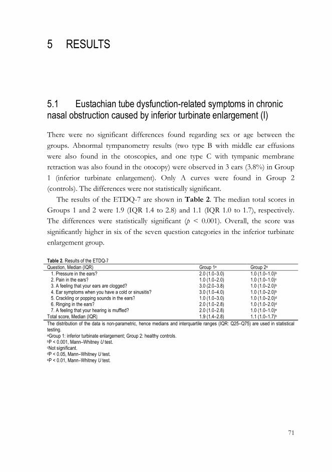
71
5 RESULTS
5.1 Eustachian tube dysfunction-related symptoms in chronicnasal obstruction caused by inferior turbinate enlargement (I)
There were no significant differences found regarding sex or age between thegroups. Abnormal tympanometry results (two type B with middle ear effusionswere also found in the otoscopies, and one type C with tympanic membraneretraction was also found in the otocopy) were observed in 3 ears (3.8%) in Group1 (inferior turbinate enlargement). Only A curves were found in Group 2(controls). The differences were not statistically significant.
The results of the ETDQ-7 are shown in Table 2. The median total scores inGroups 1 and 2 were 1.9 (IQR 1.4 to 2.8) and 1.1 (IQR 1.0 to 1.7), respectively.The differences were statistically significant (p < 0.001). Overall, the score wassignificantly higher in six of the seven question categories in the inferior turbinateenlargement group.
Table 2. Results of the ETDQ-7Question, Median (IQR) Group 1a Group 2a
1. Pressure in the ears? 2.0 (1.0–3.0) 1.0 (1.0–1.0)b
2. Pain in the ears? 1.0 (1.0–2.0) 1.0 (1.0–1.0)c
3. A feeling that your ears are clogged? 3.0 (2.0–3.8) 1.0 (1.0–2.0)b
4. Ear symptoms when you have a cold or sinusitis? 3.0 (1.0–4.0) 1.0 (1.0–2.0)b
5. Crackling or popping sounds in the ears? 1.0 (1.0–3.0) 1.0 (1.0–2.0)d
6. Ringing in the ears? 2.0 (1.0–2.8) 1.0 (1.0–2.0)d
7. A feeling that your hearing is muffled? 2.0 (1.0–2.8) 1.0 (1.0–1.0)e
Total score, Median (IQR) 1.9 (1.4–2.8) 1.1 (1.0–1.7)b
The distribution of the data is non-parametric, hence medians and interquartile ranges (IQR: Q25–Q75) are used in statisticaltesting.aGroup 1: inferior turbinate enlargement; Group 2: healthy controls.bP < 0.001, Mann–Whitney U test.cNot significant.dP < 0.05, Mann–Whitney U test.eP < 0.01, Mann–Whitney U test.
72
Of the subjects in Group 1, 23% had a total score of 3.0 or more. The maximumscore in Group 1 was 4.9. Of the subjects in Group 2, 88% had a total score of lessthan 2.0. The maximum score in Group 2 was 2.4.
Some 48% of the subjects in Group 1 had positive values in serum- specific IgElevel measurements for common airborne allergens, but there was no significantdifference in the ETDQ-7 total score between the subjects with allergicsensitization and other subjects. There was no significant correlation between theETDQ-7 total score and the improvement in V2–5 cm after vasoconstriction inGroup 1.
5.2 The effect of inferior turbinate surgery on ear symptoms (II)
In the active treatment groups, there were abnormal tympanometry curves in six(five type B and one type C) ears preoperatively and seven (six type B and one typeC) ears postoperatively. The difference was not statistically significant. In theplacebo group, there was an abnormal tympanometry curve in only one ear (typeC) preoperatively, and there were no abnormal findings postoperatively. Thisdifference was not significant either. There were no significant changes inpreoperative and postoperative TPP values among either the actively treatedpatients or the placebo group patients.
The results of the ETDQ-7 are shown in Table 3. Some 45% of the patientshad a preoperative ETDQ-7 total score of 1.0 to 1.9, 33% had a score of 2.0 to 2.9,and 22% had a score of 3.0 or more (range 3.1 to 4.4). In the evaluation of allpatients, RFA, MAIT, and placebo procedures decreased the total scoresignificantly. When only patients with a preoperative score of 2.0 or more wereevaluated, all the active treatment procedures decreased the total score significantly.
A three-way ANCOVA was run on the sample of 72 patients to examine theeffect of sex, allergy status, and procedure group on the ETDQ-7 score change.Age was added to the model as a continuous covariate. In the evaluation of allpatients, none of the factors had a significant individual main effect on the changein ETDQ-7 total score. There were no significant differences in the change inETDQ-7 total score between the procedure groups, nor were there significantdifferences in the results between the placebo and any of the active treatmentprocedures. Allergic sensitization, sex, and age had no effect on the results either.The analysis was repeated for the patients with a preoperative total score of 2.0 ormore, and the same results were found.
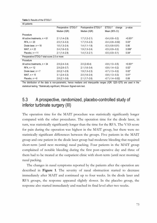
73
5.3 A prospective, randomized, placebo-controlled study ofinferior turbinate surgery (III)
The operation time for the MAIT procedure was statistically significantly longercompared with the other procedures. The operation time for the diode laser, inturn, was statistically significantly longer than the time for the RFA. The VAS scorefor pain during the operation was highest in the MAIT group, but there were nostatistically significant differences between the groups. Five patients in the MAITgroup and one patient in the diode laser group had moderate bleeding that requiredshort-term (until next morning) nasal packing. Four patients in the MAIT groupcomplained of notable bleeding during the first post-operative day and three ofthem had to be treated at the outpatient clinic with short-term (until next morning)nasal packing.
The changes in nasal symptoms reported by the patients after the operation aredescribed in Figure 1. The severity of nasal obstruction started to decreaseimmediately after MAIT and continued up to four weeks. In the diode laser andRFA groups, the response appeared slightly slower. In the placebo group, theresponse also started immediately and reached its final level after two weeks.
Table 3. Results of the ETDQ-7.All patients
Preoperative ETDQ-7Median (IQR)
Postoperative ETDQ-7Median (IQR)
ETDQ-7 changeMean (95% CI)
p-value
ProcedureAll active treatments, n = 61 2.1 (1.4–2.9) 1.7 (1.2–2.1) -0.4 (-0.6–-0.2) <0.001*
RFA, n = 20 2.5 (1.3–3.3) 1.7 (1.5–2.3) -0.4 (-0.8–-0.02) 0.03*Diode laser, n = 18 1.6 (1.3–2.4) 1.4 (1.1–1.9) -0.3 (-0.6–0.01) 0.06MAIT, n = 23 2.4 (1.6–3.3) 1.9 (1.3–2.4) -0.5 (-0.9–-0.2) 0.006*Placebo, n = 11 2.1 (1.4–2.9) 1.4 (1.3–2.1) -0.5 (-0.9–-0.1) 0.04*
Preoperative ETDQ-7 total score 2.0 or moreProcedureAll active treatments, n = 34 2.9 (2.4–3.4) 2.0 (2.43.4) -0.8 (-1.0–-0.5) <0.001*
RFA, n = 12 2.9 (2.6–3.7) 2.1 (1.6–3.4) -0.8 (-1.4–-0.2) 0.02*Diode laser, n = 7 2.6 (2.1–2.9) 1.9 (1.3–2.3) -0.7 (-1.3–-0.2) 0.02*MAIT, n = 15 3.1 (2.4–3.3) 2.0 (1.6–2.4) -0.8 (-1.3–-0.3) 0.01*Placebo, n = 6 2.8 (2.1–3.6) 2.1 (1.7–2.6) -0.7 (-1.4–-0.02) 0.08
The distribution of the data is non-parametric, hence medians and interquartile ranges (IQR: Q25–Q75) are used in thestatistical testing. *Statistically significant, Wilcoxon Signed-rank test.
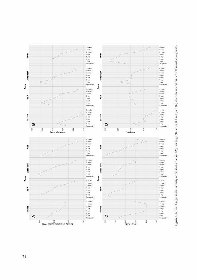
74
Figu
re1.
Mea
nch
ange
sin
the
seve
rity
ofna
salo
bstru
ctio
n(A
),di
scha
rge
(B),
crus
t(C)
and
pain
(D)a
ftert
heop
erat
ion;
VA
S=
visu
alan
alog
scale
.
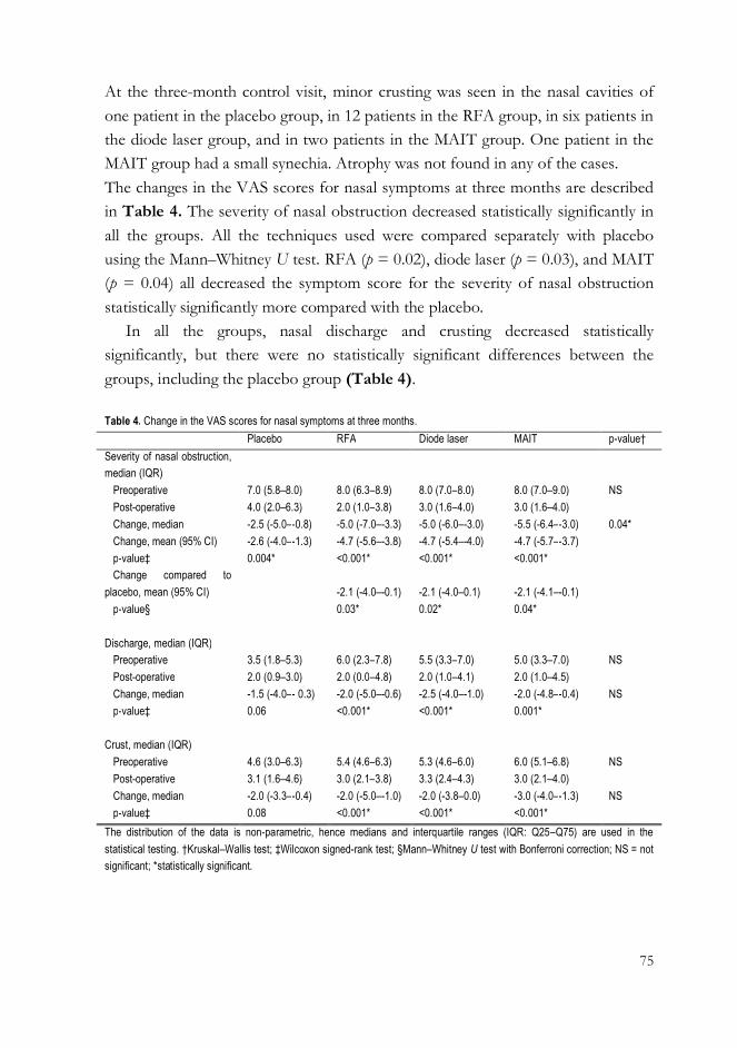
75
At the three-month control visit, minor crusting was seen in the nasal cavities ofone patient in the placebo group, in 12 patients in the RFA group, in six patients inthe diode laser group, and in two patients in the MAIT group. One patient in theMAIT group had a small synechia. Atrophy was not found in any of the cases.The changes in the VAS scores for nasal symptoms at three months are describedin Table 4. The severity of nasal obstruction decreased statistically significantly inall the groups. All the techniques used were compared separately with placebousing the Mann–Whitney U test. RFA (p = 0.02), diode laser (p = 0.03), and MAIT(p = 0.04) all decreased the symptom score for the severity of nasal obstructionstatistically significantly more compared with the placebo.
In all the groups, nasal discharge and crusting decreased statisticallysignificantly, but there were no statistically significant differences between thegroups, including the placebo group (Table 4).
Table 4. Change in the VAS scores for nasal symptoms at three months.Placebo RFA Diode laser MAIT p-value†
Severity of nasal obstruction,median (IQR)
Preoperative 7.0 (5.8–8.0) 8.0 (6.3–8.9) 8.0 (7.0–8.0) 8.0 (7.0–9.0) NSPost-operative 4.0 (2.0–6.3) 2.0 (1.0–3.8) 3.0 (1.6–4.0) 3.0 (1.6–4.0)Change, median -2.5 (-5.0–-0.8) -5.0 (-7.0–-3.3) -5.0 (-6.0–-3.0) -5.5 (-6.4–-3.0) 0.04*Change, mean (95% CI) -2.6 (-4.0–-1.3) -4.7 (-5.6–-3.8) -4.7 (-5.4–-4.0) -4.7 (-5.7–-3.7)p-value‡ 0.004* <0.001* <0.001* <0.001*Change compared to
placebo, mean (95% CI) -2.1 (-4.0–-0.1) -2.1 (-4.0–0.1) -2.1 (-4.1–-0.1)p-value§ 0.03* 0.02* 0.04*
Discharge, median (IQR)Preoperative 3.5 (1.8–5.3) 6.0 (2.3–7.8) 5.5 (3.3–7.0) 5.0 (3.3–7.0) NSPost-operative 2.0 (0.9–3.0) 2.0 (0.0–4.8) 2.0 (1.0–4.1) 2.0 (1.0–4.5)Change, median -1.5 (-4.0–- 0.3) -2.0 (-5.0–-0.6) -2.5 (-4.0–-1.0) -2.0 (-4.8–-0.4) NSp-value‡ 0.06 <0.001* <0.001* 0.001*
Crust, median (IQR)Preoperative 4.6 (3.0–6.3) 5.4 (4.6–6.3) 5.3 (4.6–6.0) 6.0 (5.1–6.8) NSPost-operative 3.1 (1.6–4.6) 3.0 (2.1–3.8) 3.3 (2.4–4.3) 3.0 (2.1–4.0)Change, median -2.0 (-3.3–-0.4) -2.0 (-5.0–-1.0) -2.0 (-3.8–0.0) -3.0 (-4.0–-1.3) NSp-value‡ 0.08 <0.001* <0.001* <0.001*
The distribution of the data is non-parametric, hence medians and interquartile ranges (IQR: Q25–Q75) are used in thestatistical testing. †Kruskal–Wallis test; ‡Wilcoxon signed-rank test; §Mann–Whitney U test with Bonferroni correction; NS = notsignificant; *statistically significant.
76
All three techniques improved the V2–5 cm acoustic rhinometry valuessignificantly, but there were no statistically significant differences between thegroups, including placebo (Table 5).
5.4 The effect of inferior turbinate surgery techniques on ciliatedepithelium and mucociliary function (IV)
The total loss of cilia was detected in 52% and squamous metaplasia in 61% of thepreoperative samples. The median preoperative scores of the number and amountof cilia and squamous metaplasia of all the patients were 0.0 (IQR 0.0 to 1.0) and0.2 (IQR 0.0 to 0.8), respectively (Table 6). There were no significant differencesin the preoperative scores of the number and amount of cilia and squamousmetaplasia between the patients with and without allergic sensitization. Thepreoperative scores of the number and amount of cilia and squamous metaplasiadid not have significant correlations with age either.
In the RFA group, the pre- and postoperative numbers of cases with the totalloss of cilia were 10 and 2, respectively. The difference was statistically significant(p = 0.01). In the MAIT group, a decrease in the number of cases was alsodetected. The result was at the borderline regarding statistical significance (p =0.05). In the diode laser group, there was no significant difference between the pre-and postoperative numbers of the total loss of cilia cases. In the diode laser group,the pre- and postoperative numbers of the cases where the squamous metaplasiawas detected were 4 and 12, respectively. The difference was statistically significant(p = 0.004). There were no significant differences between the pre- and
Table 5. Change in V2–5 cm acoustic rhinometry values (one side of the nasal cavity).Placebo RFA Diode laser MAIT p-value†
V2–5 cm (cm3)Preoperative
Median (IQR) 3.60 (2.82–4.94) 4.39 (3.02–5.65) 3.47 (2.71 4.36) 3.57 (2.52–4.58) NSPost-operative
Median (IQR) 3.94 (2.82–5.33) 5.39 (4.29–6.48) 4.37 (3.56–5.60) 4.27 (3.37–5.41Change
Median (IQR) 0.39 (-0.21–0.96) 1.12 (-0.38–2.39) 0.84 (-0.41–1.66) 0.64 (-0.27–1.56) NSMean (95% CI) 0.13 (-0.51–0.77) 1.14 (0.51–1.78) 0.85 (0.43–1.26) 0.86 (0.30–1.41)p-value‡ NS <0.001* 0.001* 0.003*
The distribution of the data is non-parametric, hence medians and interquartile ranges (IQR: Q25–Q75) are used in thestatistical testing. † Kruskal–Wallis test; ‡ Wilcoxon signed-rank test; NS = not significant; * statistically significant
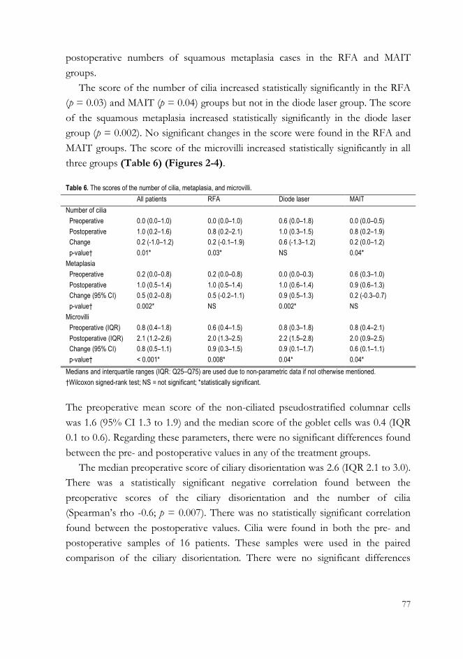
77
postoperative numbers of squamous metaplasia cases in the RFA and MAITgroups.
The score of the number of cilia increased statistically significantly in the RFA(p = 0.03) and MAIT (p = 0.04) groups but not in the diode laser group. The scoreof the squamous metaplasia increased statistically significantly in the diode lasergroup (p = 0.002). No significant changes in the score were found in the RFA andMAIT groups. The score of the microvilli increased statistically significantly in allthree groups (Table 6) (Figures 2-4).
The preoperative mean score of the non-ciliated pseudostratified columnar cellswas 1.6 (95% CI 1.3 to 1.9) and the median score of the goblet cells was 0.4 (IQR0.1 to 0.6). Regarding these parameters, there were no significant differences foundbetween the pre- and postoperative values in any of the treatment groups.
The median preoperative score of ciliary disorientation was 2.6 (IQR 2.1 to 3.0).There was a statistically significant negative correlation found between thepreoperative scores of the ciliary disorientation and the number of cilia(Spearman’s rho -0.6; p = 0.007). There was no statistically significant correlationfound between the postoperative values. Cilia were found in both the pre- andpostoperative samples of 16 patients. These samples were used in the pairedcomparison of the ciliary disorientation. There were no significant differences
Table 6. The scores of the number of cilia, metaplasia, and microvilli.All patients RFA Diode laser MAIT
Number of ciliaPreoperative 0.0 (0.0–1.0) 0.0 (0.0–1.0) 0.6 (0.0–1.8) 0.0 (0.0–0.5)Postoperative 1.0 (0.2–1.6) 0.8 (0.2–2.1) 1.0 (0.3–1.5) 0.8 (0.2–1.9)Change 0.2 (-1.0–1.2) 0.2 (-0.1–1.9) 0.6 (-1.3–1.2) 0.2 (0.0–1.2)p-value† 0.01* 0.03* NS 0.04*
MetaplasiaPreoperative 0.2 (0.0–0.8) 0.2 (0.0–0.8) 0.0 (0.0–0.3) 0.6 (0.3–1.0)Postoperative 1.0 (0.5–1.4) 1.0 (0.5–1.4) 1.0 (0.6–1.4) 0.9 (0.6–1.3)Change (95% CI) 0.5 (0.2–0.8) 0.5 (-0.2–1.1) 0.9 (0.5–1.3) 0.2 (-0.3–0.7)p-value† 0.002* NS 0.002* NS
MicrovilliPreoperative (IQR) 0.8 (0.4–1.8) 0.6 (0.4–1.5) 0.8 (0.3–1.8) 0.8 (0.4–2.1)Postoperative (IQR) 2.1 (1.2–2.6) 2.0 (1.3–2.5) 2.2 (1.5–2.8) 2.0 (0.9–2.5)Change (95% CI) 0.8 (0.5–1.1) 0.9 (0.3–1.5) 0.9 (0.1–1.7) 0.6 (0.1–1.1)p-value† < 0.001* 0.008* 0.04* 0.04*
Medians and interquartile ranges (IQR: Q25–Q75) are used due to non-parametric data if not otherwise mentioned.†Wilcoxon signed-rank test; NS = not significant; *statistically significant.
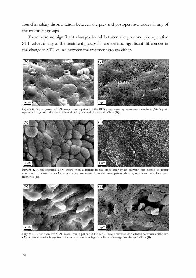
78
found in ciliary disorientation between the pre- and postoperative values in any ofthe treatment groups.
There were no significant changes found between the pre- and postoperativeSTT values in any of the treatment groups. There were no significant differences inthe change in STT values between the treatment groups either.
Figure 2. A pre-operative SEM image from a patient in the RFA group showing squamous metaplasia (A). A post-operative image from the same patient showing oriented ciliated epithelium (B).
Figure 3. A pre-operative SEM image from a patient in the diode laser group showing non-ciliated columnarepithelium with microvilli (A). A post-operative image from the same patient shoving squamous metaplasia withmicrovilli (B).
Figure 4. A pre-operative SEM image from a patient in the MAIT group showing non-ciliated columnar epithelium(A). A post-operative image from the same patient showing that cilia have emerged on the epithelium (B).
79
6 DISCUSSION
6.1 Inferior turbinate enlargement and Eustachian tubedysfunction-related symptoms
6.1.1 The evaluation of ear symptoms using Eustachian Tube DysfunctionQuestionnaire
In the present study, patients with nasal obstruction due to inferior turbinateenlargement were found to have significantly more symptoms related to ETDcompared with the healthy controls (I). In previous studies, the relationshipbetween ETD and nasal factors has been evaluated using different methods,including symptom questions (Ohrui et al. 2005; Stoikes and Dutton 2005),tympanometry (Lazo-Sáenz et al. 2005), and Toynbee and Valsalva tests(Bakhshaee et al. 2016). In the study, the relationship between ETD-relatedsymptoms and nasal obstruction was evaluated for the first time using the ETDQ-7. The main problem for the patients in the inferior turbinate enlargement groupwas nasal obstruction; ear symptoms were not their major concern in the firstplace. Their median ETDQ-7 score was higher than healthy subjects but not veryhigh. Only three abnormal tympanograms were found among them, and thedifference was not significant compared with the healthy subjects. Based on thefindings of our study, the ETDQ-7 may be a useful tool in detecting patients with amild level of ETD without clear objective findings, such as tympanic membraneretraction or middle ear effusion (I).
In the study by McCoul et al., the ETDQ-7 was able to discriminate betweenpatients with ETD and those without. The mean ETDQ-7 total score for the ETDgroup was 4.0 (SD 1.1). In our study, taking into account the relatively low medianof the ETDQ-7 total score in the inferior turbinate enlargement group, the lack ofsignificant difference in the number of abnormal tympanometry results betweenthe groups may cause a doubt as to whether the ear symptoms of the patients were
80
just symptomology related to the nasal obstruction itself but not ETD. However,almost one fourth of the subjects in the inferior turbinate enlargement group hadan ETDQ-7 total score of 3.0 or more (range 3.0 to 4.9) (I), representing the sameseverity of symptoms as the patients with ETD in the study by McCoul et al.(McCoul et al 2012).
The weakness of ETDQ-7 is that it cannot discriminate between the threesubtypes of ETD (van Royen et al. 2015). In cases of patulous Eustachian tube,patients often complain about similar symptoms as patients with dilatory ETD, butthere is also otoscopic or tympanometric evidence of excursion of the tympanicmembrane with breathing, and patients often report autophony (Schildrel et al.2015).
6.1.2 Possible mechanisms behind the Eustachian tube dysfunction-relatedsymptoms
Almost half of the patients in the inferior turbinate enlargement group had apositive result in allergy testing. However, there was no significant difference inETD-related symptoms between the subjects with allergic sensitization and othersubjects in the group. This finding supports the idea that nasal obstruction in itselfmay have a predisposing role in ETD. It is also possible that in some cases ofchronic inferior turbinate enlargement, the nasal mucosal oedema – which canoriginally be caused by environmental allergens or non-allergic and non-specifictriggers – can expand to the nasopharyngeal ET orifice, disturbing ET function.The degree of mucosal oedema in the anterior part of the nose does not seem,however, to correlate directly with the degree of disturbances in the ET which issupported by the finding that there was no correlation between the ETDQ-7 totalscore and improvement in V2–5 cm after vasoconstriction (I).
6.1.3 Eustachian tube dysfunction-related symptoms and objective findings
The consensus statement by Schildrel et al. agrees that to diagnose dilatory ETDpatient-reported symptoms should go together with evidence of negative pressurein the middle ear as assessed by clinical assessment (Schildrel et al. 2015). In ourstudy, tympanometry was the objective examination method for detecting possible
81
dilatory ETD. The finding that there was no significant difference in the numberof abnormal tympanometry results between the groups can be partly explained bythe inadequate power of the study. 40 subjects in both of the groups wereestimated to be enough for the evaluation of the differences in the ETDQ-7 score.Regarding the tympanometry results, a larger sample size might have been needed.Overall, the added value of tympanometry in this study was low. In any case, that iswhy we are not able to definitely say that dilatory ETD and not just ETD-relatedsymptoms is more common among inferior turbinate enlargement patients (I). TheETDQ-7 cannot be used as a sole criterion for the diagnosis of dilatory ETDbecause it cannot discriminate between the subtypes of ETD (van Royen et al.2015). It is, however, sometimes very difficult to detect the signs of negativepressure by clinical methods. In mild or fluctuating cases, the patients may havesymptoms suggestive of ETD, but the clinical tests can be within normallimits, whereas in more severe cases the tympanic membrane findings are usuallyabnormal (I). For example, in cases of baro-challenge-induced Eustachian tubedysfunction tympanometry may be normal (Schildrel et al. 2015).
6.1.4 The effect of inferior turbinate surgery on Eustachian tubedysfunction-related symptoms
In the present study, the ETD-related symptoms were found to improve due toinferior turbinate procedures. However, the placebo procedure improved thesymptom score as effectively as real surgery. Furthermore, in the subgroup withmore preoperative ear symptoms, there were no significant differences in theETDQ-7 total score changes between the active treatments and the placeboprocedure (II). There were no significant changes in the pre- and postoperativenumbers of abnormal tympanometry curves or in the pre- and postoperative TPPvalues among actively treated patients and the placebo patients. Possible changes inTPP values may have indicated minor changes in middle ear pressure as a result ofthe operation (II).
Based on the findings of our study, the surgical treatment of the anterior part ofthe inferior turbinate did not have any real effect on the nasopharyngeal ET orificeand possible ETD (II). The posterior part of the turbinate, which is anatomicallylocated very close to ET orifice, was not operated on in the present study. The

82
main aim of the larger study setting was to compare the efficacy of various inferiorturbinate surgery procedures in the treatment of chronic nasal obstruction, whichwas the patients’ main problem. Surgical treatment of the anterior part of theinferior turbinate, in turn, has been found to be effective in decreasing nasalobstruction (Civelek et al. 2010). In addition, to make the procedures ascomparable as possible, we treated the same length of the turbinate with everytechnique. The easiest way to guarantee that was to limit the operation to theanterior part of the inferior turbinate because in some cases it can be difficult tooperate the posterior part of the turbinate under local anesthesia.
More than half of the patients had an ETDQ-7 score of 2.0 or more, and morethan one fifth of the patients had a score of 3.0 or more (II). This represents thesame severity of symptoms as patients found to have ETD in the study byMcCoul et al. (McCoul et al. 2012). However, fewer than 10% of the enrolledpatients had an abnormal tympanogram (type B or C) confirming the diagnosis ofETD. As a result, we are not able to generalize the results of our study to ETD.Therefore, we can only talk about the effect of inferior turbinate procedure onpatient symptoms suggestive of ETD (II).
6.2 Placebo-controlled comparison of inferior turbinate surgerytechniques
6.2.1 The operations and side-effects
Some studies have reported notable intraoperative bleeding (Kassab et al. 2012) orpost-operative bleeding in the early days after MAIT (Cingi et al. 2010), and thiswas also the case in our study. During the operation, the mean VAS score for painwas the highest in the MAIT group, but there was no statistical difference betweenthe groups. Post-operatively, there was an increase in the VAS score for pain in theMAIT group that lasted up to one week. This can possibly be explained by thedirect surgical tissue trauma caused by the operation. Short-term nasal packing,which was most common in the MAIT group, may also partly explain the increaseof pain during the first days of the follow-up. Increased pain was also reportedduring the first two days after diode laser treatment. This can possibly be explained

83
by the higher temperature and therefore the more severe tissue damage caused bythe laser compared with the RFA (III).
In the present study, a temporary increase in the symptom of nasal dischargeand a minor increase in the symptom of crusting during the first post-operativedays after RFA was noted (III). A similar finding has been reported in previousstudies (Coste et al. 2001; Bäck et al. 2002). Some patients treated with RFAcomplained of crust formation for as long as three months (III). In a study byJanda et al., patients showed moderate-to-severe nasal obstruction, crusting, andnasal secretion in the first four weeks following diode laser treatment (Janda et al.2000). In our study, however, patients in the diode laser group on average reporteda decrease in crusting immediately after the operation and an increase in nasaldischarge only during the first two days. In some cases, however, crust formationcontinued up to three months (III).
6.2.2 The effect of inferior turbinate surgery procedures on the severity ofnasal obstruction
In recent years, studies have been published that compare RFA and MAIT in thetreatment of nasal obstruction. There are studies where the results of MAIT havebeen better in the short follow-up (Cingi et al. 2010) and also in the longer follow-up (Liu et al. 2009). There are also studies where the techniques have been foundequal in efficacy (Kizilkaya et al. 2008). In their systematic review and meta-analysiscomparing RFA and MAIT, Acavedo et al. concluded that inferior turbinatereduction produces a significant subjective and objective improvement in nasalairflow in the short term, which is not related to the technique used (Acavedo et al.2015). There are also studies that have compared RFA and the diode laser (Rhee etal. 2001; Kisser et al. 2014) and MAIT and the diode laser (Kassab et al. 2012) butno significant differences between the techniques have been found in any of thesestudies. Furthermore, no previous studies have compared all three techniquestogether.
In our study, the decrease in the VAS score for severity of nasal obstructionstarted faster after MAIT (III). In MAIT, the submucosa of the turbinate isemptied immediately during the operation, whereas in the RFA and diode laserprocedures the heat caused by the device creates a submucosal lesion, inducing
84
scarring and tissue volume reduction during the following weeks (Willatt 2009;Cakli et al. 2012). The post-operative nasal packing, which was most common inthe MAIT group, squeezes the mucosa of the emptied turbinate against the boneand may therefore contribute to the faster response. Based on this conclusion andthe higher rate of post-operative bleeding in the MAIT group, nasal packing can berecommended in the MAIT procedures (III).
At the end of the three-month follow-up, all three techniques decreased theVAS score for the severity of nasal obstruction and V2–5 cm values statisticallysignificantly compared with the preoperative values, and they were equal in efficacyregarding both parameters (III).
There have been no previous placebo-controlled studies involving the threedifferent techniques, or studies where the diode laser and MAIT have beencompared with placebo treatment. In the present study, RFA, diode laser, andMAIT all improved the VAS score for the severity of nasal obstruction statisticallysignificantly better than placebo (III). This finding is in line with the previous studyby Nease and Krempl regarding RFA (Nease and Krempl 2004).
6.2.3 The effect of inferior turbinate surgery procedures on nasal dischargeand crusting
In the present study, all the techniques decreased the symptom scores of nasaldischarge and crusting statistically significantly compared to the preoperative valuesat the end of the three-month follow-up, but none of them statistically significantlycompared with placebo. In addition, minor crusting was still found in some nosesat three months, especially in the RFA and diode laser groups. Therefore, most ofthe improvement in these symptoms can be explained by the placebo effect (III).
6.2.4 Objective examination
V2–5 cm in acoustic rhinometry was used as a parameter for objectiveexamination. It represents well the operated anterior half of the inferior turbinateand describes the possible nasal cavity volume changes due to the operation at thatregion. In the three-month follow-up, RFA, diode laser, and MAIT techniques allimproved the V2–5 cm values statistically significantly compared with the
85
preoperative values. Due to the lack of a real operation, there was understandablyno statistically significant change in the V2-5 cm values in the placebo group. Thissupports the conclusion that the improvement in the VAS score for severity ofnasal obstruction in the placebo group was due to placebo effect. However, noneof the techniques improved the volume statistically significantly compared withplacebo. This finding can be partly explained by the relatively small number ofpatients in the placebo group. A larger number might have led to statisticalsignificance (III). In addition, environmental conditions and technical difficulties inmeasurement may have an influence on the volume values (Hilberg and Pedersen2001). Furthermore, the symptom of nasal obstruction and objective parametersdo not always correlate very well (André et al. 2009). On the other hand, it is wiseto remember that the increase in the nasal cavity volume should be optimal insteadof too large in order to avoid making the nose too wide, which might lead toproblems, such as nasal dryness, crusting, atrophy, worsening of the symptom ofnasal obstruction, and "empty nose syndrome" (Willatt 2009; Hong 2016).
6.2.5 Economical aspects
One aspect that should be noted when comparing these techniques is cost-efficiency. The cost-efficiency depends on, for example, whether the equipment ismulti-use or disposable, and whether the procedure is carried out in the officesetting or in the operation room setting. There are also many devices availablefrom different manufacturers and their prices vary. The RFA electrode (purchaseprice 419 euros) that was used in the present study is a multi-use (instrumentcleaning price 1.5 euros) instrument. RFA treatment is a fast procedure, which wasalso found in the present study (III). It is also usually carried out in the officesetting (Nease and Krempl 2004; Cavaliere et al. 2005). Therefore, it can beconsidered cost-effective. The diode laser fiber (purchase price 122 euros) that weused is disposable (III). The diode laser treatment can also be easily carried out inthe office setting (Sroka et al. 2007), which increases its cost-efficiency. Themicrodebrider blade (purchase price 179 euros) that we used is disposable, whichdecreases its cost efficiency (III). It is commonly used in the operation roomsetting, although the treatment can be carried out in the office setting as well. The

86
final conclusion about cost-efficiency should be based on the long-term effect ofthe treatments, and for that our study cannot give answers.
6.3 The effect of inferior turbinate surgery techniques onmucosal epithelium and mucociliary function
6.3.1 The meaning of the biopsy site
In previous light and electron microcopy studies of inferior turbinate surgery, thepreoperative findings regarding cilia have been controversial. There have beenstudies reporting preoperative findings of epithelial metaplasia and loss of ciliatedcells (Inoye et al. 1999; Gindros et al. 2009; Neri et al. 2016). On the other hand,there have also been studies that have shown normal appearance of the epitheliumwith a preserved number of cilia (Sargon et al. 2008; Garzaro et al. 2012). In thepresent study, there were squamous metaplasia and total loss of ciliated cells foundin the majority of the preoperative samples (IV). This may partly be explained bythe chronic rhinitis behind the inferior turbinate enlargement taking into accountprevious studies showing that the loss of cilia is more common in both allergic andnon-allergic rhinitis compared with healthy controls (Watanabe et al. 1998; Gindroset al. 2009). In our study, however, the biopsies were taken from the anteriorportion of the inferior turbinate approximately 1 cm behind the anterior edge, andthis is the most likely explanation for the high incidence of squamous metaplasiaand total loss of ciliated cells in the preoperative samples (IV). The density ofciliated cells increases in the nasal cavity in the antero-posterior direction (Halamaet al. 1990). Augusto et al. reported that ciliated epithelium with goblet cells(normal respiratory epithelium) was found in only half of the samples taken fromthe anterior portions of the inferior turbinates of normal noses and various degreesof epithelial metaplasia was found in half of the samples taken from the samesites. The anterior part of the inferior turbinate is more exposed to externalelements, such as strong air flow, and therefore prone to metaplatic transformation(Augusto et al. 2001).
87
6.3.2 The effect of inferior turbinated surgery techniques on mucosalepithelium
There have been some previous light and electron microscopy studies that haveevaluated the effect of inferior turbinate surgery on ciliated epithelium. Based onthe previous studies, RFA (Coste 2001, Gindros 2008, Sargon 2009, Garzaro 2012)and submucosal electrocautery (Talaat 1987, Elwany 1999, Gindros 2008) do nothave a significant effect on ciliated epithelium. After endoscopic turbinoplasty,some improvement has been described (Cassano 2010). Regarding lasers, however,the findings have been controversial (Inoye 1999, Bergler 2001, Cassano 2010).Ultrasound reduction, in turn, may have an improving effect on ciliated epithelium(Gindros 2008), whereas a worsening effect has been previously described forpartial inferior turbinectomy (Garzaro 2012). Interestingly, in a recent study byNeri et al., a complete re-epithelisation of nasal mucosa with columnar ciliatedepithelium was detected 4 months after the total removal of the mucosa of themedial and inferior portions of the inferior turbinate with the microdebrider (Neri2016).
In the present study, the effect of various inferior turbinate surgery techniqueson ciliated epithelium was for the first time evaluated with a statistical analysisusing a score based on the mean of the grades given to the image series by theblinded examiners. Based on the score, the number of cilia increased significantlyafter RFA and MAIT treatments. The number of cases with total loss of cilia wasalso found to decrease after these treatments. No similar changes were found afterdiode laser treatment. On the other hand, the score of metaplasia seemed toincrease significantly after diode laser treatment as well as the overall number ofcases where metaplasia was detected. No similar findings were found after RFA orMAIT. Regarding the diode laser, the findings can be explained by the damage thelaser causes to the mucosal surface during the treatment. RFA and MAIT are moreclearly submucosal techniques that do not notably harm the mucosal surface. Insome cases, however, mucosal surface changes can also occur after thesesubmucosal procedures due, for example, to vascular alterations of the turbinate.Inferior turbinate surgery creates more airspace at the level of the anterior part ofthe inferior turbinate, which decreases the nasal airway resistance, causes changesin the airflow patterns, and likely decreases the shear stress on the mucosa of the
88
anterior part of the inferior turbinate. This could be a possible explanation for theincrease in the number of cilia after RFA and MAIT treatments (IV).
In human nasal epithelial cells, the number of microvilli has been found toincrease before the ciliogenesis starts (Jorissen et al. 1990). In chronicrhinosinusitis, an increased number of microvilli and short cilia are thought toindicate the regeneration of epithelium and ciliated cells (Toskala and Rautiainen2003). In the present study, the number of microvilli increased significantly in allthree groups. This finding can be interpreted to be related to a regeneration of themucosal epithelium after the damage caused by the surgery (IV).
The degree of ciliary disorientation was relatively high both pre- andpostoperatively in all three groups and there were no significant differences indisorientation between the pre- and postoperative samples (IV). The disorientationcan be explained mainly by the inflammation caused by chronic rhinitis (Rayner etal. 1995) and the shear stress caused by the airflow (Augusto et al. 2001).Preoperatively, there was also a negative correlation found between the scores ofthe ciliary disorientation and the number of cilia indicating that the smaller thenumber of cilia is the more disorientated they are (IV). This finding is in line with aprevious study by Rautiainen, where a few cilia or small groups of cilia were foundin most fields that differed dramatically from the main orientation (Rautiainen1988).
6.3.3 The effect of inferior turbinate surgery techniques on mucociliaryfunction
The effect of various techniques of inferior turbinate surgery on mucociliaryfunction has been examined in several studies using STT. Based on the previousstudies, the diode laser technique may worsen the mucociliary transport during thefirst three months after operation, but in longer follow-up it returns to normal(Parida et al. 2013, Veit et al. 2016). In most of the studies regarding RFA, the STThas either been preserved (Sapci et al. 2003; Cavaliere et al. 2005; Rosato et al.2016) or improved (Coste et al. 2001; Veit et al. 2016) after the operation in one- tothree-month follow-ups. However, there are also studies where STT has beenprolonged at two (Salzano et al. 2009) and six (Garzaro et al. 2012) months afterthe operation. There is also one study where the STT significantly improved from
89
six months to one year postoperatively compared with the preoperative levels.However, no improvement in the STT from two to three years was noted (Liu etal. 2009). Regarding MAIT, there are studies where STT has been preserved afterthree months (Romano et al. 2015) or decreased in a longer, up to three years,follow-up (Liu et al. 2009).
In the present study, there were no significant changes found between the pre-and postoperative STT values in any of the treatment groups. There were also nosignificant differences in the change of STT values between the treatment groups.The findings are in line with the previous studies on RFA and MAIT. Although theamount of squamous metaplasia was found to increase in the diode laser group, theSTT was not found to increase. One possible explanation for this could be thecontact technique used, where parallel stripes were made on the mucosa along theturbinate leaving lanes on the mucosal surface with preserved mucociliary functionbetween them. Moreover, STT itself might also be too rough a measurement tofind out slight differences (IV).
6.4 The significance of the study setting in the results
6.4.1 Randomization
Possible biases caused by confounding factors, such as patient biases, naturalcourse of the disease, and regression to the mean (Benedetti et al. 2013) can becontrolled with proper randomization. In our study, the randomization was carriedout just before the procedure in consecutive order by the method of minimization.The only parameters that were inputted to the program were age, sex, and allergicsentisization. The distributions of these parameters were kept similar in each groupto prevent the possible confounding effect of the parameters on the results.
6.4.2 Blinding
In order to increase feasibility, all clinical examinations and operations in thissingle-blinded placebo-controlled study were performed by the same physician,who was not blinded to the patient's groups. Unblinding of the examining
90
physician could introduce unintentional bias by causing misinterpretation of data.Blinding of the treating physicians is recommended, even if they are not assessingoutcomes, because they may inadvertently affect the expectations of the patients orinform the patients of their treatment group, thus spoiling the placebo control(Powell et al. 2001). However, the examination parameters used in this study werenot based on the subjective evaluation of the examining physician. Therefore, fromthe examining physician’s point of view, the study data were objective. In ourstudy, during the preoperative visit, the examining physician did not yet know thepatients´ group, and during the control visit the patients filled out thequestionnaires regarding nasal symptoms before meeting the physician, whichmakes the accidental revelation impossible. If the operation is carried out underlocal anaesthesia, as in our study, there is always a small risk that the operationroom staff or the surgeon may accidentally reveal the treatment group no matter ifthe setting is single- or double-blinded. Taking into account the quality of theinferior turbinate reduction operation, it would have been exaggerated to treat allthese adult patients under general anaesthesia.
In the evaluation of the effect of inferior turbinate surgery on the ciliatedepithelium, the five examiners who evaluated the SEM images did not know whichtechnique had been used or whether the images were pre- or postoperative.Blinding of the evaluators was extremely important in order to prevent anypossible expectations and attitudes of the examiners towards the treatments fromaffecting their evaluations (IV).
Crossover trials, such as two (Nease and Krempl 2004; Bran et al. 2012) of thethree (Powell et al. 2001; Nease and Krempl 2004; Bran et al. 2012) previousplacebo-controlled RFA studies, are prone to biases. These include the periodiceffect, where the order in which treatments are given may affect the outcome, andthe carryover effect, where the effect of the first treatment extends into the timeinterval between the periods of intervention and has an impact on the outcome ofthe second treatment (Bran et al. 2013). For these reasons, we did not use thecrossover model in our study.

91
6.4.3 Placebo-control
Inferior turbinate surgery procedures are very common in otorhinolaryngologist'sclinical practice. Occasionally, clinicians may be preoccupied by the questionwhether the procedures are genuinely useful or not. In times like these, when thereis a lot of conversation about how to increase effectiveness in the public healthcare, there is also a general need for the studies, which evaluate the trueeffectiveness of the given surgical treatment.
In placebo-controlled studies, the placebo is used to find out the true treatmenteffect of the examined treatment. The true treatment effect is the difference inimprovement between the intervention group and the control group (placeboeffect) (Powell et al. 2001). In our placebo-controlled comparison of inferiorturbinate techniques, the mean true treatment effect in the VAS score change ofthe severity of nasal obstruction was found to be -2.1 for all three techniques. Thisindicates that RFA, diode laser, and MAIT are all genuinely effective and usefultechniques in decreasing the severity of nasal obstruction in inferior turbinateenlargement. The finding justifies their use in the clinical practice, although, theplacebo effect accounted for a large part of the total improvement seen in theintervention groups (III). We also found that the improvement of ETD-relatedsymptoms due to surgery of the anterior half of the inferior turbinate was equal toplacebo (II), and that most of the improvements in symptom scores of nasaldischarge and crusting can be explained by the placebo effect (III). Without theplacebo control, we would have falsely found a greater improvement andsignificance due to treatment.
6.4.4 Internal quality of the study and generalizability of the results
For the above-mentioned reasons, and despite the mainly single-blinded studysetting, the internal quality of our study can be considered high. When we thinkabout the generalizability of the results regarding the treatment of nasalobstruction, we have to take a look at the exclusion criteria of the study. Weexcluded those patients with obvious anatomical abnormalities, such as severeseptum deviation, and patients with chronic sinusitis with or without polyposis.For these patients, inferior turbinate surgery can be carried out in clinical practice.

92
However, inferior turbinate reduction, as a sole procedure, does not necessarilylead to a satisfactory outcome in these cases, and additional procedures, such asseptoplasty or middle meatal antrostomy, are required at the same time. If we thinkabout the indications of inferior turbinate surgery as a sole procedure, thegeneralizability of this study can be considered to be good.
6.4.5 The significance of the placebo effect in clinical practice
Physicians in general quite often deal with the placebo effect when treating theirpatients. There are several situations where it is difficult to recognize whether theeffect of the treatment is due to the treatment itself or due to the placebo effect.Taking into account how complex the sensations of nasal obstruction and patencyare, it is easy to understand the role of the placebo effect in the treatment of nasalobstruction. Probably, physicians most commonly use placebo in their practicewhen they explain the positive effects of the treatments, which are known andproven to be effective, to their patients. This increases the patients’ positiveexpectations towards the treatment, which is one of the main things behind theplacebo effect. When the treatment is proven to be effective, as now seems to bethe case with the three inferior turbinate surgery techniques in treating chronicnasal obstruction in short-term follow-up, it is justifiable to make use of all theadditional benefits of the placebo effect to increase the improvement of thesymptoms that the patients suffer. Regarding inferior turbinate enlargement, theplacebo effect can therefore be called an otorhinolaryngologist’s best helper.
6.5 Future aspects
In general, there will be need for this kind of placebo-controlled studies, whichevaluate the true effectiveness of the given surgical treatment, and in their part,guide the use of economical resources in the health care.
In future, more studies comparing inferior turbinate surgery techniques withlong (several years) follow-ups are needed to find out the possible differences intheir long-term efficacy in treating the chronic nasal obstruction related to inferiorturbinate enlargement. To date, the three-month follow-up period in our study isthe longest study of inferior turbinate surgery with a placebo control group. It
93
would also be recommendable to carry out placebo-controlled studies with an evenlonger follow-up in future to evaluate the true long-term treatment effect ofinferior turbinate surgery. However, taking into account ethical aspects, etc., itcould be very challenging to carry out a placebo-controlled study with several yearsfollow-up. A placebo-controlled study with one-year follow-up period wouldprobably be the next step.
In the present study, some of the patients in all treatment groups benefittedconsiderably from the inferior turbinate reduction in the treatment of their chronicnasal obstruction. On the other hand, however, there were also patients who didnot benefit from the treatment at all. In future studies, special attention should bepaid to the possible factors in the preoperative diagnostics that predict positive ornegative outcomes. The use of CFD models in the evaluation of the most optimalnasal airflow concerning the feeling of nasal patency could be a useful in futurestudies. The definition of inferior turbinate enlargement and the criteria for inferiorturbinate surgery also require clarification. This would help otorhinolaryngologiststo operate on the patients who, as a result, are likely to benefit from the inferiorturbinate surgery.
Conservative treatment of the chronic rhinitis behind the inferior turbinateenlargement is essential before surgical treatment. Therefore, it would berecommendable to carry out controlled trials of medical treatment, where factorssuch as patient compliance, for example, are taken into account.
It would also be interesting to examine the usefulness of combining inferiorturbinate surgery with traditional functional endoscopic sinus surgery in thetreatment of those patients with CRS who also have signs of inferior turbinateenlargement.

94
7 CONCLUSIONS
The present study provides data to support the following conclusions andsummary:
After three-month follow-up, there were no statistically significant differencesbetween the RFA, diode laser, and MAIT techniques in their efficacy in treatingnasal obstruction. RFA, diode laser, and MAIT were all genuinely effective intreating the chronic nasal obstruction caused by inferior turbinate enlargement.The placebo effect plays a large role in the overall reduction of the severity of nasalobstruction in inferior turbinate surgery, but all three examined techniquesprovided a statistically significant additional reduction in the severity of nasalobstruction compared with the placebo procedure.
Patients with chronic nasal obstruction caused by inferior turbinate enlargementhave more symptoms related to ETD than healthy controls. ETD-relatedsymptoms among the patients are not very severe in most cases and findings, suchas abnormal tympanometry, are not common in clinical tests. Whether the patienthas allergic sensitization or not does not seem to cause a difference in thesymptoms related to ETD.
The improvement of ETD-related symptoms due to surgery of the anterior half ofthe inferior turbinate as a sole procedure was found to be equal to placebo. Thefindings of this study do not support the use of reduction of the anterior half ofthe inferior turbinate as a sole procedure intended to treat the ear symptomsassessed by the ETDQ-7 questionnaire.
RFA and MAIT are more mucosal preserving techniques than diode laser, whichwas found to increase the amount of squamous metaplasia at three-month follow-up. The number of cilia seemed to even increase after RFA and MAIT procedures,but not after diode laser. Nevertheless, the mucociliary transport was equallypreserved in all three groups.
95
ACKNOWLEDGEMENTS
First of all, I want to say “RESPECT!” to all the patients who participated in thisstudy knowing the placebo-controlled setting of the study.
I would like to thank my supervisor, Professor Markus Rautiainen, MD, PhD.You motivated me to start doing research, and you have made it possible for me towork and carry out research in our clinic. I also thank you for your expert adviceand comments during the research and writing process, especially regarding theexamination of the cilia samples.
I express my gratitude to Docent Jura Numminen, MD, PhD. The original ideaof starting this research project came from you. You helped me a lot in the verybeginning of this research project in the practical planning of the study setting.Throughout the whole project, I have been able to solve many of the practicalproblems regarding surgical techniques for inferior turbinate surgery andrhinometric measurements, for example, with your help and advice.
I also wish to express my special thanks to Docent Ilkka Kivekäs, MD, PhD.The idea of examining the relationship between inferior turbinate surgery andEustachian tube dysfunction came from you during one of our so-called“VALVIRA” trips, and I am still very greatful for that. Already before this researchproject, the cooperation with you had opened my eyes to the idea of doingresearch. You have shown me the way how clinical research is carried outefficiently with an open-minded and self-confident attitude.
I would like to thank our study nurse Marja-Leena “Piitu” Oksanen. It wouldhave been impossible to carry out this research project without your valuablepractical work with the study patients. Your tireless and energetic way of workingwith this and other research projects carried out in our clinic is admirable.
I also wish to express my thanks to Mari Honkanen, PhD, at TampereUniversity of Technoloqy. Scanning electron microscopy was somethingcompletely new for me. Imaging of the cilia samples was very interesting, and Ireally liked spending those long days at the Hervanta campus. It was very nice towork with you. I appreciate your expertise in electron microscopy, and it was greatthat you also participated in the evaluation of the cilia samples.

96
I would also like to thank Professor Minnamari Vippola, PhD, at TampereUniversity of Technology who also took part in the evaluation of the cilia samples.
I wish to express special thanks to Docent Leif Bäck, MD, PhD, and DocentTuomo Puhakka, MD, PhD, who officially reviewed the thesis and gave meconstructive comments.
I express my thanks to Docent Juha-Pekka Vasama, MD, PhD, for arrangingmy placements at the outpatient clinic and at the day surgery department so that Iwas able to carry out this study.
I would like to thank my colleque Saara Markkanen for helping me revising theoriginally clumsy version of the Finnish abstract of my thesis.
I also wish to express my thanks to Peter Heath for the official language editionof my thesis.
I would like to thank all the personnel at our day surgery departments both inTAYS and Valkeakoski for their positive and enthusiastic attitude towards myresearch project. Unfortunately, the curtain has fallen and Teemu’s“näytelmäkerho” is now over.
I would also like to thank all my colleques at the Department ofOtorhinolaryngology for just being my colleques during the past years. You aregreat.
This study received financial support from Korvatautien Tutkimussäätiö.
97
REFERENCES
Acevedo JL, Camacho M, Brietzke SE. 2015. Radiofrequency Ablation Turbinoplasty versusMicrodebrider-Assisted Turbinoplasty: A Systematic Review and Meta-analysis. OtolaryngolHead Neck Surg 153(6):951-6.
Anderson I, Lundqvist G. 1977. Nasal mucociliary function in humans. In: Lenfant C. LungBiology in Health and Disease. New York: Dekker, 427–452.
André RF, Vuyk HD, Ahmed A, Graamans K, Nolst Trenité GJ. 2009. Correlation betweensubjective and objective evaluation of the nasal airway. A systematic review of the highest levelof evidence. Clin Otolaryngol 34(6):518-25.
Apaydin F. 2011. Nasal valve surgery. Facial Plast Surg 27(2):179-91.
Batra PS, Seiden AM, Smith TL. 2009. Surgical management of adult inferior turbinatehypertrophy: a systematic review of the evidence. Laryngoscope. 2009 Sep;119(9):1819-27.
Augusto AG, Bussolotti Filho I, Dolci JE, König Júnior B. 2001. Structural and ultrastructuralstudy of the anterior portion of the nasal septum and inferior nasal concha. Ear Nose Throat J.80(5):325-7,333-8.
Bakhshaee M, Ardakani HP, Ghazizadeh AH, Movahed R, Jarahi L, Rajati M. 2016. Middle earfunction in sinonasal polyposis. Eur Arch Otorhinolaryngol 273(10):2911-6.
Benedetti F, Amanzio M. 2013. Mechanisms of the placebo response. Pulm Pharmacol Ther26(5):520-3.
Berger G, Hammel I, Berger R, Avraham S, Ophir D. 2000. Histopathology of the inferiorturbinate with compensatory hypertrophy in patients with deviated nasal septum. Laryngoscope110(12):2100-5.
Berger G, Gass S, Ophir D. 2006. The histopathology of the hypertrophic inferior turbinate.Arch Otolaryngol Head Neck Surg 132(6):588-94.
98
Bergler WF, Sadick H, Hammerschmitt N, Oulmi J, Hörmann K. 2001. Long-term results ofinferior turbinate reduction with argon plasma coagulation. Laryngoscope 111(9):1593-8.
Bhandarkar ND, Smith TL. 2010. Outcomes of surgery for inferior turbinate hypertrophy. CurrOpin Otolaryngol Head Neck Surg 18(1):49-53.
Bingel U, Wanigasekera V, Wiech K, Ni Mhuircheartaigh R, Lee MC, Ploner M, Tracey I. 2011.The effect of treatment expectation on drug efficacy: imaging the analgesic benefit of theopioid remifentanil. Sci Transl Med 3(70):70ra14.
Boysen M. 1982. The surface structure of the human nasal mucosa. I. Ciliated and metaplasticepithelium in normal individuals. A correlated study by scanning/transmission electron andlight microscopy. Virchows Arch B Cell Pathol Incl Mol Pathol. 40(3):279-94.
Bradley LA. 1989. Psychological testing. In: Tollison CD. Handbook of Chronic PainManagement. Baltimore: Williams and Wilkins.
Braiman A, Priel Z. 2008. Efficient mucociliary transport relies on efficient regulation of ciliarybeating. Respir Physiol Neurobiol 163(1-3):202-7.
Bran GM, Hünnebeck S, Herr RM, Hörmann K, Stuck BA. 2013. Bipolar radiofrequencyvolumetric tissue reduction of the inferior turbinates: evaluation of short-term efficacy in aprospective, randomized, single-blinded, placebo-controlled crossover trial. Eur ArchOtorhinolaryngol 270(2):595-601.
Browning GG, Gatehouse S. 1992. The prevalence of middle ear disease in the adult Britishpopulation. Clin Otolaryngol Allied Sci 17(4):317-21.
Brunworth JD, Mahboubi H, Garg R, Johnson B, Brandon B, Djalilian HR. Nasopharyngealacid reflux and Eustachian tube dysfunction in adults. Ann Otol Rhinol Laryngol2014;123(6):415-9.
Bäck LJ, Hytönen ML, Malmberg HO, Ylikoski JS. 2002. Submucosal bipolar radiofrequencythermal ablation of inferior turbinates: a long-term follow-up with subjective and objectiveassessment. Laryngoscope 112(10):1806-12.
Caffier PP, Scherer H, Neumann K, Lück S, Enzmann H, Haisch A. 2011. Diode lasertreatment in therapy-resistant allergic rhinitis: impact on nasal obstruction and associatedsymptoms. Lasers Med Sci 26(1):57-67.
99
Cakli H, Cingi C, Güven E, Gurbuz MK, Kaya E. 2012. Diode laser treatment of hypertrophicinferior turbinates and evaluation of the results with acoustic rhinometry. Eur ArchOtorhinolaryngol 269(12):2511-7.
Casey KP, Borojeni AA, Koenig LJ, Rhee JS, Garcia GJ. 2016. Correlation betweenSubjective Nasal Patency and Intranasal Airflow Distribution. Otolaryngol Head Neck Surg156(4):741-750.
Cassano M, Granieri C, Del Giudice AM, Mora F, Fiocca-Matthews E, Cassano P. 2010.Restoration of nasal cytology after endoscopic turbinoplasty versus laser-assisted turbinoplasty.Am J Rhinol Allergy 24(4):310-4.
Cavaliere M, Mottola G, Iemma M. 2005. Comparison of the effectiveness and safetyof radiofrequency turbinoplasty and traditional surgical technique in treatment of inferiorturbinate hypertrophy. Otolaryngol Head Neck Surg 133(6):972-8.
Cavaliere M, Mottola G, Iemma M. 2007. Monopolar and bipolar radiofrequency thermalablation of inferior turbinates: 20-month follow-up. Otolaryngol Head Neck Surg 137(2):256-63.
Chhabra N, Houser SM. 2012. Surgical options for the allergic rhinitis patient. Curr OpinOtolaryngol Head Neck Surg 20(3):199-204.
Chang CW, Ries WR. 2004. Surgical treatment of the inferior turbinate: new techniques. CurrOpin Otolaryngol Head Neck Surg 12(1):53-7.
Chen B, Shaari J, Claire SE, Palmer JN, Chiu AG, Kennedy DW, Cohen NA. 2006. Alteredsinonasal ciliary dynamics in chronic rhinosinusitis. Am J Rhinol 20(3):325-9.
Chen YL, Tan CT, Huang HM. 2008. Long-term efficacy of microdebrider-assisted inferiorturbinoplasty with lateralization for hypertrophic inferior turbinates in patients with perennialallergic rhinitis. Laryngoscope 118(7):1270-4.
Chen XB, Lee HP, Chong VF, Wang de Y. 2010. Impact of inferior turbinate hypertrophy onthe aerodynamic pattern and physiological functions of the turbulent airflow - a CFDsimulation model. Rhinology 48(2):163-8.
Chilvers MA, O'Callaghan C. 2000. Analysis of ciliary beat pattern and beat frequency usingdigital high speed imaging: comparison with the photomultiplier and photodiode methods.Thorax. 55(4):314-7.

100
Cingi C, Ure B, Cakli H, Ozudogru E. 2010. Microdebrider-assisted versus radiofrequency-assisted inferior turbinoplasty: a prospective study with objective and subjective outcomemeasures. Acta Otorhinolaryngol Ital 30(3):138-43.
Civelek S, Ozçelik M, Emre IE, Cakir BO, Turgut S. 2010. Comparison of radiofrequencyapplied to the total inferior choncha with application to its anterior third. Auris Nasus Larynx37(5):589-93.
Cohen AL. 1979. Critical point drying - principles and procedures. Scan Electr Microsc, II:303–23.
Cohen NA. 2006. Sinonasal mucociliary clearance in health and disease. Ann Otol RhinolLaryngol Suppl 196:20-6.
Corey JP. 2006. Acoustic rhinometry: should we be using it? Curr Opin Otolaryngol HeadNeck Surg 14(1):29-34.
Coste A, Yona L, Blumen M, Louis B, Zerah F, Rugina M, Peynègre R, Harf A, Escudier E.2001. Radiofrequency is a safe and effective treatment of turbinate hypertrophy. Laryngoscope111(5):894-9.
Courtney M. Townsend. 2012. Sabiston textbook of surgery: the biological basis of modernsurgical practice, 19th edition. Philadelphia, PA: Elsevier Saunders. 236.
Cukurova I, Demirhan E, Cetinkaya EA, Yigitbasi OG. 2011. Long-term clinical results ofradiofrequency tissue volume reduction for inferior turbinate hypertrophy. J Laryngol Otol125(11):1148-51.
Dahl R, Mygind N. 1998. Anatomy, physiology and function of the nasal cavities in health anddisease. Adv Drug Deliv Rev 29(1-2):3-12.
Deron P, Clement PA, Derde MP. 1995. Septal surgery and tubal function: early and lateresults. Rhinology 33(1):7-9.
Doorly DJ, Taylor DJ, Schroter RC. 2008. Mechanics of airflow in the human nasal airways.Respir Physiol Neurobiol 163(1-3):100-10.
Doyle WJ, Seroky JT, Angelini BL, Gulhan M, Skoner DP, Fireman P. 2000. Abnormal middleear pressures during experimental influenza A virus infection--role of Eustachian tube function.Auris Nasus Larynx 27(4):323-6.
101
Duchateau GS, Zuidema J, Merkus FW. 1986. The in vitro and in vivo effect of a new non-halogenated corticosteroid - budesonide - aerosol on human ciliary epithelial function. Allergy41(4):260-5.
Dudley JP, Cherry JD. 1980. Scanning electron microscopic demonstration of goblet celldischarge and mucous layer on nasal ciliated respiratory epithelium. Otolaryngol Head NeckSurg 88(4):439-41.
Elwany S, Gaimaee R, Fattah HA. 1999. Radiofrequency bipolar submucosal diathermy of theinferior turbinates. Am J Rhinol 13(2):145-9.
Finkelstein Y, Ophir D, Talmi YP, Shabtai A, Strauss M, Zohar Y. 1994. Adult-onset otitismedia with effusion. Arch Otolaryngol Head Neck Surg 120(5):517-27.
Fisher EW. 1997. Acoustic rhinometry. Clin Otolaryngol Allied Sci 22(4):307-17.
Frisaldi E, Piedimonte A, Benedetti F. 2015. Placebo and nocebo effects: a complex interplaybetween psychological factors and neurochemical networks. Am J Clin Hypn 57(3):267-84.
Garzaro M, Landolfo V, Pezzoli M, Defilippi S, Campisi P, Giordano C, Pecorari G. 2012.Radiofrequency volume turbinate reduction versus partial turbinectomy: clinical and histologicalfeatures. Am J Rhinol Allergy. 26(4):321-5.
Gindros G, Kantas I, Balatsouras DG, Kandiloros D, Manthos AK, Kaidoglou A. 2009.Mucosal changes in chronic hypertrophic rhinitis after surgical turbinate reduction. Eur ArchOtorhinolaryngol 266(9):1409-16.
Gluth MB, McDonald DR, Weaver AL, Bauch CD, Beatty CW, Orvidas LJ. 2011. Managementof eustachian tube dysfunction with nasal steroid spray: a prospective, randomized, placebo-controlled trial. Arch Otolaryngol Head Neck Surg 137(5):449-55.
Goldstein J, Newbury D.E, Joy D.C, Lyman C.E, Echlin P, Lifshin E, Sawyer L, Michael J.R.2003. Scanning Electron Microscopy and X-ray Microanalysis, 3rd edition. New York: Springer-Verlag, 21-60,591-618,647-673.
Greiner AN, Hellings PW, Rotiroti G, Scadding GK. 2011. Allergic rhinitis. Lancet378(9809):2112-22.
Grymer LF, Hilberg O, Pedersen OF, Rasmussen TR. 1991. Acoustic rhinometry: values fromadults with subjective normal nasal patency. Rhinology 29(1):35-47.
102
Grymer LF. 2000. Clinical applications of acoustic rhinometry. Rhinol Suppl 16:35-43.
Gudis DA, Woodworth BA, Cohen NA. 2012a. Sinonasal Physiology. In: Kennedy DW,Hwang PH. Rhinology: Diseases of the Nose, Sinuses and Scull Base. New York: Thieme.
Gudis DA, Zhao KQ, Cohen NA. 2012b. Acquired cilia dysfunction in chronic rhinosinusitis.Am J Rhinol Allergy 26(1):1-6.
Halama AR, Decreton S, Bijloos JM, Clement PA. 1990. Density of epithelial cells in thenormal human nose and the paranasal sinus mucosa. A scanning electron microscopic study.Rhinology 28(1):25-32.
Havel M, Sroka R, Leunig A, Patel P, Betz CS. 2011. A double-blind, randomized, intra-individual controlled feasibility trial comparing the use of 1,470 and 940 nm diode laser for thetreatment of hyperplastic inferior nasal turbinates. Lasers Surg Med 43(9):881-6.
Hellings PW, 23 others. 2017. Non-allergic rhinitis: Position paper of the European Academyof Allergy and Clinical Immunology. Allergy 72(11):1657-1665.
Helsingin ja Uudenmaan sairaanhoitopiiri (HUS). 2018. Allergian erikoistutkimukset.http://www.hus.fi/ammattilaiselle/allergiatutkimukset/allergian-erikoistutkimukset/Sivut/defaut.aspx. Referred 21.7.2018.
Hilberg O, Jackson AC, Swift DL, Pedersen OF. 1989. Acoustic rhinometry: evaluation of nasalcavity geometry by acoustic reflection. J Appl Physiol 66(1):295-303.
Hilberg O, Pedersen OF. 2000. Acoustic rhinometry: recommendations for technicalspecifications and standard operating procedures. Rhinol Suppl 16:3-17.
Hilberg O. 2002. Objective measurement of nasal airway dimensions using acoustic rhinometry:methodological and clinical aspects. Allergy 57 Suppl 70:5-39.
Hilding AC. 1932. Experimental surgery of the nose and sinuses. Arch Otolaryngol 16:9-18
Hodez C, Griffaton-Taillandier C, Bensimon I. 2011. Cone-beam imaging: applications inENT. Eur Ann Otorhinolaryngol Head Neck Dis 128(2):65-78.
Hong HR, Jang YJ. 2016. Correlation between remnant inferior turbinate volume and symptomseverity of empty nosesyndrome. Laryngoscope 126(6):1290-5.
103
Hopewell S, Dutton S, Yu LM, Chan AW, Altman DG. 2010. The quality of reports ofrandomised trials in 2000 and 2006: comparative study of articles indexed in PubMed. BMJ340:c723.
Huang TW, Cheng PW. 2006. Changes in nasal resistance and quality of life after endoscopicmicrodebrider-assisted inferior turbinoplasty in patients with perennial allergic rhinitis. ArchOtolaryngol Head Neck Surg 132(9):990-3.
Huttenbrink KB. 2005. Current topics in otolaryngology-head and neck surgery: Lasers inotorhinolaryngology. In: Lippert BM. Lasers in rhinology. New York: Thieme, 53–71.
Hytönen ML, Bäck LJ, Malmivaara AV, Roine RP. 2009. Radiofrequency thermal ablation forpatients with nasal symptoms: a systematic review of effectiveness and complications. Eur ArchOtorhinolaryngol 266(8):1257-66.
Inouye T, Tanabe T, Nakanoboh M, Ogura M. 1999. Laser surgery for allergic andhypertrophic rhinitis. Ann Otol Rhinol Laryngol Suppl 180:3-19.
Iwasaki A, Tokano H, Kamiyama R, Suzuki Y, Kitamura K. 2010. A 24-month-follow-up studyof argon plasma coagulation of the inferior turbinate in patients with perennial nasal allergy. JMed Dent Sci. 57(1):11-5.
Jackson LE, Koch RJ. 1999. Controversies in the management of inferior turbinatehypertrophy: a comprehensive review. Plast Reconstr Surg 103(1):300-12.
Janda P, Sroka R, Tauber S, Baumgartner R, Grevers G, Leunig A. 2000. Diode laser treatmentof hyperplastic inferior nasal turbinates. Lasers Surg Med 27(2):129-39.
Janda P, Sroka R, Baumgartner R, Grevers G, Leunig A. 2001. Laser treatment of hyperplasticinferior nasal turbinates: a review. Lasers Surg Med 28(5):404-13.
Jang YJ, Myong NH, Park KH, Koo TW, Kim HG. 2002. Mucociliary transport and histologiccharacteristics of the mucosa of deviated nasal septum. Arch Otolaryngol Head Neck Surg128(4):421-4.
Jerger J. 1970. Clinical experience with impedance audiometry. Arch Otolaryngol 92(4):311-24.
Jones AS, Crosher R, Wight RG, Lancer JM, Beckingham E. 1987. The effect of localanaesthesia of the nasal vestibule on nasal sensation of airflow and nasalresistance. ClinOtolaryngol Allied Sci 12(6):461-4.
104
Jones AS, Wight RG, Crosher R, Durham LH. 1989. Nasal sensation of airflow followingblockade of the nasal trigeminal afferents. Clin Otolaryngol Allied Sci 14(4):285-9.
Jones AS, Wight RG, Kabil Y, Beckingham E. 1989. Predicting the outcome of submucosaldiathermy to the inferior turbinates. Clin Otolaryngol Allied Sci 14(1):41-4.
Joniau S, Wong I, Rajapaksa S, Carney SA, Wormald PJ. 2006. Long-term comparison betweensubmucosal cauterization and powered reduction of the inferior turbinates. Laryngoscope116(9):1612-6.
Jorissen M, Willems T. 2004. The secondary nature of ciliary (dis)orientation in secondary andprimary ciliary dyskinesia. Acta Otolaryngol 124(4):527-31.
Jutel M, Papadopoulos NG, Gronlund H, Hoffman HJ, Bohle B, Hellings P, Braunstahl GJ,Muraro A, Schmid-Grendelmeier P, Zuberbier T, Agache I. 2014. Recommendations for theallergy management in the primary care. Allergy 69(6):708-18.
Kaliner M, Marom Z, Patow C, Shelhamer J. 1984. Human respiratory mucus. J Allergy ClinImmunol 73(3):318-23.
Kamani T, Yilmaz T, Surucu S, Turan E, Brent KA. 2006. Scanning electron microscopy ofciliae and saccharine test for ciliary function in septal deviations. Laryngoscope 116(4):586-90.
Karatas A, Salviz M, Dikmen B, Yüce T, Acar G. 2015. The effects ofdifferent radiofrequency energy magnitudes on mucociliary clearance in cases of turbinatehypertrophy. Rhinology 53(2):171-5.
Karnovsky MJ. 1965. A Formaldehyde-Glutaraldehyde Fixative of High Osmolality for Use inElectron Microscopy. J Cell Biol 27: 137-8a.
Kassab AN, Rifaat M, Madian Y. 2012. Comparative study of management of inferior turbinatehypertrophy using turbinoplasty assisted by microdebrider or 980 nm diode laser. J LaryngolOtol. 126(12):1231-7.
Kisser U, Stelter K, Gürkov R, Patscheider M, Schrötzlmair F, Bytyci R, Adderson-Kisser C, Berghaus A, Olzowy B. 2014. Diode laser versus radiofrequency treatment of theinferior turbinate - a randomized clinical trial. Rhinology 52(4):424-30.
Kivekäs I, Poe D. 2014. Eustachian Tube. In: Pensak M, Choo D. Clinical Otology, 4th edition.New York: Thieme.

105
Kizilkaya Z, Ceylan K, Emir H, Yavanoglu A, Unlu I, Samim E, Akagün MC. 2008.Comparison of radiofrequency tissue volume reduction and submucosal resection withmicrodebrider in inferior turbinate hypertrophy. Otolaryngol Head Neck Surg 138(2):176-81.
Knipping S, Holzhausen HJ, Goetze G, Riederer A, Bloching MB. 2007.Rhinitis medicamentosa: electron microscopic changes of human nasal mucosa. OtolaryngolHead Neck Surg 136(1):57-61.
Kreiner-Møller E, Chawes BL, Caye-Thomasen P, Bønnelykke K, Bisgaard H. 2012. Allergicrhinitis is associated with otitis media with effusion: a birth cohort study. Clin Exp Allergy42(11):1615-20.
Kärjä J, Nuutinen J, Karjalainen P. 1982. Radioisotopic method for measurement of nasalmucociliary activity. Arch Otolaryngol 108(2):99-101.
Lale AM, Mason JD, Jones NS. 1998. Mucociliary transport and its assessment: a review. ClinOtolaryngol Allied Sci 23(5):388-96.
Lazo-Sáenz JG, Galván-Aguilera AA, Martínez-Ordaz VA, Velasco-Rodríguez VM, Nieves-Rentería A, Rincón-Castañeda C. 2005. Eustachian tube dysfunction in allergic rhinitis.Otolaryngol Head Neck Surg 132(4):626-9.
Lee HP, Garlapati RR, Chong VF, Wang de Y. 2013. Comparison between effects of variouspartial inferior turbinectomy options on nasal airflow: a computer simulation study. ComputMethods Biomech Biomed Engin 16(1):112-8.
Lenders H, Pirsig W. 1990. Diagnostic value of acoustic rhinometry: patients with allergic andvasomotor rhinitis compared with normal controls. Rhinology 28(1):5-16.
Leong SC, Chen XB, Lee HP, Wang DY. 2010. A review of the implicationsof computational fluid dynamic studies on nasal airflow and physiology. Rhinology 48(2):139-45.
Leong SC, Eccles R. 2010. Inferior turbinate surgery and nasal airflow: evidence-basedmanagement. Curr Opin Otolaryngol Head Neck Surg. 18(1):54-9.
Li C, Farag AA, Leach J, Deshpande B, Jacobowitz A, Kim K, Otto BA, Zhao K. 2017.Computational fluid dynamics and trigeminal sensory examinations of empty nose syndromepatients. Laryngoscope 127(6):e176-e184.
106
Li KK, Powell NB, Riley RW, Troell RJ, Guilleminault C. 1998. Radiofrequency volumetrictissue reduction for treatment of turbinate hypertrophy: a pilot study. Otolaryngol Head NeckSurg 119(6):569-73.
Lidén G. 1969. The scope and application of current audiometric tests. J Laryngol Otol83(5):507-20.
Lin HC, Lin PW, Friedman M, Chang HW, Su YY, Chen YJ, Pulver TM. 2010. Long-termresults of radiofrequency turbinoplasty for allergic rhinitis refractory to medical therapy. ArchOtolaryngol Head Neck Surg 136(9):892-5.
Liu CM, Tan CD, Lee FP, Lin KN, Huang HM. 2009. Microdebrider-assisted versusradiofrequency-assisted inferior turbinoplasty. Laryngoscope 119(2):414-8.
Liu P, Su K, Zhu B, Wu Y, Shi H, Yin S. 2016. Detection of Eustachian tube openingsby tubomanometry in adult otitis media with effusion. Eur Arch Otorhinolaryngol273(10):3109-15.
Low WK, Willatt DJ. 1993. The relationship between middle ear pressure and deviated nasalseptum. Clin Otolaryngol Allied Sci 18(4):308-10.
Mabry RL. 1988. Inferior turbinoplasty: patient selection, technique, and long-termconsequences. Otolaryngol Head Neck Surg 98(1):60-6.
Maurizi M, Paludetti G, Todisco T, Almadori G, Ottaviani F, Zappone C. 1984. Ciliaryultrastructure and nasal mucociliary clearance in chronic and allergic rhinitis. Rhinology22(4):233-40.
McCoul ED, Anand VK, Christos PJ. 2012. Validating the clinical assessment of eustachiantube dysfunction: The Eustachian Tube Dysfunction Questionnaire (ETDQ-7). Laryngoscope122(5):1137-41.
Meusel T, Negoias S, Scheibe M, Hummel T. 2010. Topographical differences in distributionand responsiveness of trigeminal sensitivity within the human nasal mucosa. Pain. 2010151(2):516-21.
Min YG, Kim HS, Yun YS, Kim CS, Jang YJ, Jung TG. 1996. Contact laser turbinate surgeryfor the treatment of idiopathic rhinitis. Clin Otolaryngol Allied Sci 21(6):533-6.
107
Moche JA, Palmer O. 2012. Surgical management of nasal obstruction. Oral Maxillofac SurgClin North Am 24(2):229-37.
Moore EJ, Kern EB. 2001. Atrophic rhinitis: a review of 242 cases. Am J Rhinol 15(6):355-61.
Mori S, Fujieda S, Yamada T, Kimura Y, Takahashi N, Saito H. 2002. Long-term effect ofsubmucous turbinectomy in patients with perennial allergic rhinitis. Laryngoscope 112(5):865-9.
Moseley JB, O'Malley K, Petersen NJ, Menke TJ, Brody BA, Kuykendall DH, HollingsworthJC, Ashton CM, Wray NP. 2002. A controlled trial of arthroscopic surgery for osteoarthritis ofthe knee. N Engl J Med 347(2):81-8.
Mygind N. 1975. Scanning electron microscopy of the human nasal mucosa. Rhinology13(2):57-75.
Mygind N, Pedersen M, Nielsen M. 1982. Morphology of the upper airway epithelium. In:Proctor DF, Ardersen IB. The Nose: Upper airway physiology and the athmosphericenvironment. Amsterdam: Elsvier Biochemical press, 71-97.
Nation JL. 1983. A new method using hexamethyldisilazane for preparation of soft insecttissues for scanning electron microscopy. Stain Technol 58(6):347-51.
Nease CJ, Krempl GA. 2004. Radiofrequency treatment of turbinate hypertrophy: arandomized, blinded, placebo-controlled clinical trial. Otolaryngol Head Neck Surg 130(3):291-9.
Neri G, Cazzato F, Mastronardi V, Pugliese M, Centurione MA, Di Pietro R, Centurione L.2016. Ultrastructural regenerating features of nasal mucosa following microdebrider-assistedturbinoplasty are related to clinical recovery. Transl Med. 14(1):164.
Numminen J, Ahtinen M, Huhtala H, Rautiainen M. 2003. Comparison of rhinometricmeasurements methods in intranasal pathology. Rhinology 41(2):65-8.
Numminen J. 2017. Allergic rhinitis. Duodecim 133(5):473-8.
Ohrui N, Takeuchi A, Tong A, Iwata M, Nakamura A, Ohashi K. 2005. Allergic rhinitis and earpain in flight. Ann Allergy Asthma Immunol 95(4):350-3.
Ottaviano G, Fokkens WJ. 2016. Measurements of nasal airflow and patency: a critical reviewwith emphasis on the use of peak nasal inspiratory flow in daily practice. Allergy 71(2):162-74.
108
Parida PK, Surianarayanan G, Alexander A, Saxena SK, Santhosh K. 2013. Diodelaser turbinate reduction in the treatment of symptomatic inferior turbinate hypertrophy. IndianJ Otolaryngol Head Neck Surg 65(Suppl 2):350-5.
Passàli D, Lauriello M, Anselmi M, Bellussi L. 1999. Treatment of hypertrophy of the inferiorturbinate: long-term results in 382 patients randomly assigned to therapy. Ann Otol RhinolLaryngol 108(6):569-75.
Passàli D, Passàli FM, Damiani V, Passàli GC, Bellussi L. 2003. Treatment of inferior turbinatehypertrophy: a randomized clinical trial. Ann Otol Rhinol Laryngol 112(8):683-8.
Pelikan Z. 2009. Role of nasal allergy in chronic secretory otitis media. Curr Allergy AsthmaRep 9(2):107-13.
Petrovic P, Kalso E, Petersson KM, Ingvar M. 2002. Placebo and opioid analgesia-- imaging ashared neuronal network. Science 295(5560):1737-40.
Petruson B, Hansson HA, Karlsson G. 1984. Structural and functional aspects of cells in thenasal mucociliary system. Arch Otolaryngol 110(9):576-81.
Poe DS, Abou-Halawa A, Abdel-Razek O. 2001. Analysis of the dysfunctional Eustachiantube by video endoscopy. Otol Neurotol 22(5):590-5.
Powell NB, Zonato AI, Weaver EM, Li K, Troell R, Riley RW, Guilleminault C. 2001.Radiofrequency treatment of turbinate hypertrophy in subjects using continuous positive airwaypressure: a randomized, double-blind, placebo-controlled clinical pilot trial. Laryngoscope111(10):1783-90.
Quadrio M, Pipolo C, Corti S, Lenzi R, Messina F, Pesci C, Felisati G. 2014. Review ofcomputational fluid dynamics in the assessment of nasal air flow and analysis of its limitations.Eur Arch Otorhinolaryngol 271(9):2349-54.
Quaranta N, Iannuzzi L, Gelardi M. 2014. Does the type of rhinitis influence development ofotitis media with effusion in children? Curr Allergy Asthma Rep 14(11):472.
Raja H, Mitchell S, Barrett G, Sharma A, Skinner DW. 2017. Long-term follow-up ofKTP laser turbinate reduction for the treatment of obstructive rhinopathy. Ear Nose Throat J.96(4-5):170-182.
109
Rautiainen M, Collan Y, Nuutinen J, Kärjä J. 1984. Ultrastructure of human respiratory cilia: astudy based on serial sections. Ultrastruct Pathol 6(4):331-9.
Rautiainen M. 1988. Orientation of human respiratory cilia. Eur Respir J 1(3):257-61.
Rautiainen M, Collan Y, Nuutinen J, Afzelius BA. 1990. Ciliary orientation in the"immotile cilia" syndrome. Eur Arch Otorhinolaryngol 247(2):100-3.
Rautiainen M, Nuutinen J, Kiukaanniemi H, Collan Y. 1992. Ultrastructural changes in humannasal cilia caused by the common cold and recovery of ciliated epithelium. Ann Otol RhinolLaryngol 101(12):982-7.
Rayner CF, Rutman A, Dewar A, Cole PJ, Wilson R. 1995. Ciliary disorientation in patientswith chronic upper respiratory tract inflammation. Am J Respir Crit Care Med 151(3 Pt 1):800-4.
Rhee CS, Kim DY, Won TB, Lee HJ, Park SW, Kwon TY, Lee CH, Min YG. 2001. Changes ofnasal function after temperature-controlled radiofrequency tissue volume reduction for theturbinate. Laryngoscope 111(1):153-8.
Rice DH, Kern EB, Marple BF, Mabry RL, Friedman WH. 2003. The turbinates in nasal andsinus surgery: a consensus statement. Ear Nose Throat J 82(2):82-4.
Roditi RE, Veling M, Shin JJ. 2016. Age: An effect modifier of the association between allergicrhinitis and Otitis media with effusion. Laryngoscope 126(7):1687-92.
Romano A, Orabona GD, Salzano G, Abbate V, Iaconetta G, Califano L. 2015. Comparativestudy between partial inferior turbinotomy and microdebrider-assisted inferiorturbinoplasty. JCraniofac Surg 26(3):e235-8.
Rosato C, Pagliuca G, Martellucci S, de Vincentiis M, Greco A, Fusconi M, De Virgilio A,Gallipoli C, Simonelli M, Gallo A. 2016. Effect of Radiofrequency Thermal Ablation Treatmenton Nasal Ciliary Motility: A Study with Phase-Contrast Microscopy. Otolaryngol Head NeckSurg 154(4):754-8.
Rossman CM, Lee RM, Forrest JB, Newhouse MT. 1984. Nasal ciliary ultrastructure andfunction in patients with primary ciliary dyskinesia compared with that in normal subjects andin subjects with various respiratory diseases. Am Rev Respir Dis 129(1):161-7.

110
Salzano FA, Mora R, Dellepiane M, Zannis I, Salzano G, Moran E, Salami A. 2009.Radiofrequency, high-frequency, and electrocautery treatments vs partial inferior turbinotomy:microscopic and macroscopic effects on nasal mucosa. Arch Otolaryngol Head Neck Surg135(8):752-8.
Sanderson MJ, Sleigh MA. 1981. Ciliary activity of cultured rabbit tracheal epithelium: beatpattern and metachrony. J Cell Sci 47:331-47.
Sapçi T, Sahin B, Karavus A, Akbulut UG. 2003. Comparison of the effects of radiofrequencytissue ablation, CO2 laser ablation, and partial turbinectomy applications on nasal mucociliaryfunctions. Laryngoscope 113(3):514-9.
Sargon MF, Celik HH, Uslu SS, Yücel OT, Denk CC, Ceylan A. 2009. Histopathologicalexamination of the effects of radiofrequency treatment on mucosa in patients with inferiornasal concha hypertrophy. Eur Arch Otorhinolaryngol 266(2):231-5.
Satir P. 1974. How cilia move. Sci Am 231(4):44-52.
Satir P. 1980. Structural basis of ciliary movement. Environ Health Perspect 35:77-82.
Schilder AG, Bhutta MF, Butler CC, Holy C, Levine LH, Kvaerner KJ, Norman G, PenningsRJ, Poe D, Silvola JT, Sudhoff H, Lund VJ. 2015. Eustachian tube dysfunction: consensusstatement on definition, types, clinical presentation and diagnosis. Clin Otolaryngol 40(5):407-11.
Schipor I, Palmer JN, Cohen AS, Cohen NA. 2006. Quantification of ciliary beat frequency insinonasal epithelial cells using differential interference contrast microscopy and high-speeddigital video imaging. Am J Rhinol 20(1):124-7.
Seppä J. 1993. Mucociliary Transport of the Eustachian Tube in Chronic Otitis Media. ClinicalStudy with Reference to Nasal Mucociliary Transport and Operative Results. Kuopio: KuopioUniversity Publications D. Medical Sciences 22.
Sihvonen R, Paavola M, Malmivaara A, Itälä A, Joukainen A, Nurmi H, Kalske J, Järvinen TL;Finnish Degenerative Meniscal Lesion Study (FIDELITY) Group. 2013. Arthroscopic partialmeniscectomy versus sham surgery for a degenerative meniscal tear. N Engl J Med369(26):2515-24.
111
Sinno S, Mehta K, Lee ZH, Kidwai S, Saadeh PB, Lee MR. 2016. Inferior TurbinateHypertrophy in Rhinoplasty: Systematic Review of Surgical Techniques. Plast Reconstr Surg138(3):419e-29e.
Sleigh MA. 1977. The nature and action of respiratory tract cilia. In: Brain JD, Proctor DF, ReidLM. Respiratory defence mechanisms. New York: Marcel Dekker, 247-288.
Sleigh MA. 1983. Ciliary function in transport of mucus. Eur J Respir Dis Suppl 128 (Pt 1):287-92.
Smith ME, Tysome JR. 2015. Tests of Eustachian tube function: a review. Clin Otolaryngol40(4):300-11.
Sozansky J, Houser SM. 2014. The physiological mechanism for sensing nasal airflow: aliterature review. Int Forum Allergy Rhinol 4(10):834-8.
Sroka R, Janda P, Killian T, Vaz F, Betz CS, Leunig A. 2007. Comparison of long term resultsafter Ho:YAG and diode laser treatment of hyperplastic inferior nasal turbinates. Lasers SurgMed 39(4):324-31.
Stammberger H. 1986. An endoscopic study of tubal function and the diseased ethmoid sinus.Arch Otorhinolaryngol 243(4):254-9.
Stanley PJ, Griffin WM, Wilson R, Greenstone MA, Mackay IS, Cole PJ. 1985. Effect ofbetamethasone and betamethasone with neomycin nasal drops on human nasal mucociliaryclearance and ciliary beat frequency. Thorax 40(8):607-12.
Stanley PJ, Wilson R, Greenstone MA, MacWilliam L, Cole PJ. 1986. Effect ofcigarette smoking on nasal mucociliary clearance and ciliary beat frequency. Thorax 41(7):519-23.
Stewart MG, Witsell DL, Smith TL, Weaver EM, Yueh B, Hannley MT. 2004. Developmentand validation of the Nasal Obstruction Symptom Evaluation (NOSE) scale. Otolaryngol HeadNeck Surg 130(2):157-63.
Stoikes NF, Dutton JM. 2005. The effect of endoscopic sinus surgery on symptomsof Eustachian tube dysfunction. Am J Rhinol 19(2):199-202.
112
Straszek SP, Schlünssen V, Sigsgaard T, Pedersen OF. 2007. Reference values for acousticrhinometry in decongested school children and adults: the most sensitive measurement forchange in nasal patency. Rhinology 45(1):36-9.
Sullivan CD, Garcia GJ, Frank-Ito DO, Kimbell JS, Rhee JS. 2014. Perception of better nasalpatency correlates with increased mucosal cooling after surgery for nasal obstruction.Otolaryngol Head Neck Surg 150(1):139-47.
Takeuchi K, Sakakura Y, Murai S, Majima Y. 1989. Nasal mucociliary clearance inSjögren's syndrome. Dissociation in flow between sol and gel layers. Acta Otolaryngol 108(1-2):126-9.
Talaat M, el-Sabawy E, Baky FA, Raheem AA. 1987. Submucous diathermy of the inferiorturbinates in chronic hypertrophic rhinitis. J Laryngol Otol 101(5):452-60.
Tomkinson A, Eccles R. 1996. Comparison of the relative abilities of acoustic rhinometry,rhinomanometry, and the visual analogue scale in detecting chance in the nasal cavity in ahealthy adult population. Am J Rhinol 10(3):161-165.
Toskala E, Rautiainen M, Nuutinen J. 1994. Scanning and transmission electron microscopicfindings in cilia from human nasal turbinate and sinus mucosa following respiratory infection.Eur Arch Otorhinolaryngol 251(2):76-9.
Toskala E, Nuutinen J, Rautiainen M. 1995a. Scanning electron microscopy findings of humanrespiratory cilia in chronic sinusitis and in recurrent respiratory infections. Acta Otolaryngol115(1):61-5.
Toskala E, Nuutinen J, Rautiainen M, Torkkeli T. 1995b. The correlation of mucociliarytransport and scanning electron microscopy of nasal mucosa. Acta Otolaryngol 115(1):61-5.
Toskala E, Rautiainen M. 2003. Electron microscopy assessment of the recovery of sinusmucosa after sinus surgery. Acta Otolaryngol 123(8):954-9.
Tran NP, Vickery J, Blaiss MS. 2011. Management of Rhinitis: Allergic and Non-Allergic.Allergy Asthma Immunol Res 3(3):148–156.
van der Avoort SJ, van Heerbeek N, Zielhuis GA, Cremers CW. 2005. Sonotubometry:eustachian tube ventilatory function test: a state-of-the-art review. Otol Neurotol 26(3):538-43.
113
Veit JA, Nordmann M, Dietz B, Sommer F, Lindemann J, Rotter N, Greve J, von BomhardA, Hoffmann TK, Riepl R, Scheithauer MO. 2017.Three different turbinoplasty techniquescombined with septoplasty: Prospective randomized trial. Laryngoscope. 127(2):303-308.
Vijayakumar S, Divakaran S, Parida PK, Gopalakrishnan S. 2016. Potassium titanyl phosphatelaser turbinate reduction in the management of allergic inferior turbinate hypertrophy: Ourexperience. Allergy Rhinol (Providence). 7(1):29-33.
Volk GF, Pantel M, Guntinas-Lichius O, Wittekindt C. 2010. Prognostic value of anteriorrhinomanometry in diode laser turbinoplasty. Arch Otolaryngol Head Neck Surg 136(10):1015-9.
Warner FD. 1974. The fine structure of the ciliary and flagellary axoneme. In: Sleigh MA. Ciliaand flagella. London, New York: Academic press, 11-37.
Watanabe K, Kiuna C. 1998. Epithelial damage of nasal mucosa in nasal allergy. Ann OtolRhinol Laryngol 107(7):564-70.
Wewers ME, Lowe NK. 1990. A critical review of visual analogue scales in the measurement ofclinical phenomena. Research in Nursing and Health 13(4):227-236.
Wight RG, Jones AS, Beckingham E. 1990. Trimming of the inferior turbinates: a prospectivelong-term study. Clin Otolaryngol Allied Sci 15(4):347-50.
Willatt D. 2009. The evidence for reducing inferior turbinates. Rhinology 47(3):227-36.
Wilson R, Alton E, Rutman A, Higgins P, Al Nakib W, Geddes DM, Tyrrell DA, Cole PJ. 1987.Upper respiratory tract viral infection and mucociliary clearance. Eur J Respir Dis 70(5):272-9.
Yañez C, Mora N. 2008. Inferior turbinate debriding technique: ten-year results. OtolaryngolHead Neck Surg 138(2):170-5.
Ye T, Zhou B. 2015. Update on surgical management of adult inferior turbinate hypertrophy.Curr Opin Otolaryngol Head Neck Surg 23(1):29-33.
Yilmaz M, Kemalo lu YK, Baysal E, Tutar H. 2006. Radiofrequency for inferior turbinatehypertrophy: could its long-term effect be predicted with a preoperative topical vasoconstrictordrop test? Am J Rhinol 20(1):32-5.

114
Zhang L, Han D, Song X, Wang K, Wang H. 2008. Effect of oxymetazoline on healthy humannasal ciliary beat frequency measured with high-speed digital microscopy and mucociliarytransport time. Ann Otol Rhinol Laryngol 117(2):127-33.
Zhao K, Jiang J. 2014. What is normal nasal airflow? A computational study of 22 healthyadults. Int Forum Allergy Rhinol 4(6):435-46.
Zhao K, Jiang J, Blacker K, Lyman B, Dalton P, Cowart BJ, Pribitkin EA. 2014. Regionalpeak mucosal cooling predicts the perception of nasal patency. Laryngoscope. 124(3):589-95.
Zwarenstein M, Treweek S, Gagnier JJ, Altman DG, Tunis S, Haynes B, Oxman AD, Moher D;CONSORT group; Pragmatic Trials in Healthcare (Practihc) group. 2008. Improving thereporting of pragmatic trials: an extension of the CONSORT statement. BMJ 337:a2390.

115
ORIGINAL COMMUNICATIONS
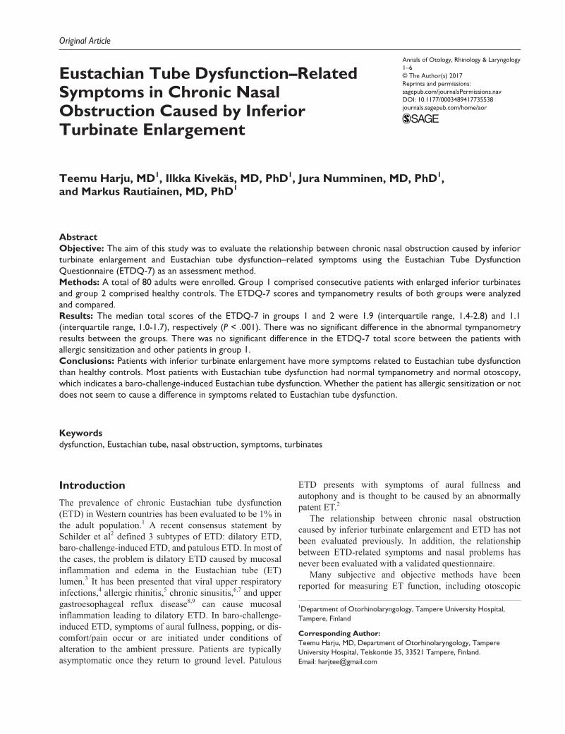
https://doi.org/10.1177/0003489417735538
Annals of Otology, Rhinology & Laryngology 1 –6© The Author(s) 2017Reprints and permissions: sagepub.com/journalsPermissions.navDOI: 10.1177/0003489417735538journals.sagepub.com/home/aor
Original Article
Introduction
The prevalence of chronic Eustachian tube dysfunction (ETD) in Western countries has been evaluated to be 1% in the adult population.1 A recent consensus statement by Schilder et al2 defined 3 subtypes of ETD: dilatory ETD, baro-challenge-induced ETD, and patulous ETD. In most of the cases, the problem is dilatory ETD caused by mucosal inflammation and edema in the Eustachian tube (ET) lumen.3 It has been presented that viral upper respiratory infections,4 allergic rhinitis,5 chronic sinusitis,6,7 and upper gastroesophageal reflux disease8,9 can cause mucosal inflammation leading to dilatory ETD. In baro-challenge-induced ETD, symptoms of aural fullness, popping, or dis-comfort/pain occur or are initiated under conditions of alteration to the ambient pressure. Patients are typically asymptomatic once they return to ground level. Patulous
ETD presents with symptoms of aural fullness and autophony and is thought to be caused by an abnormally patent ET.2
The relationship between chronic nasal obstruction caused by inferior turbinate enlargement and ETD has not been evaluated previously. In addition, the relationship between ETD-related symptoms and nasal problems has never been evaluated with a validated questionnaire.
Many subjective and objective methods have been reported for measuring ET function, including otoscopic
735538 AORXXX10.1177/0003489417735538Annals of Otology, Rhinology & LaryngologyHarju et alresearch-article2017
1Department of Otorhinolaryngology, Tampere University Hospital, Tampere, Finland
Corresponding Author:Teemu Harju, MD, Department of Otorhinolaryngology, Tampere University Hospital, Teiskontie 35, 33521 Tampere, Finland. Email: [email protected]
Eustachian Tube Dysfunction–Related Symptoms in Chronic Nasal Obstruction Caused by Inferior Turbinate Enlargement
Teemu Harju, MD1, Ilkka Kivekäs, MD, PhD1, Jura Numminen, MD, PhD1, and Markus Rautiainen, MD, PhD1
AbstractObjective: The aim of this study was to evaluate the relationship between chronic nasal obstruction caused by inferior turbinate enlargement and Eustachian tube dysfunction–related symptoms using the Eustachian Tube Dysfunction Questionnaire (ETDQ-7) as an assessment method.Methods: A total of 80 adults were enrolled. Group 1 comprised consecutive patients with enlarged inferior turbinates and group 2 comprised healthy controls. The ETDQ-7 scores and tympanometry results of both groups were analyzed and compared.Results: The median total scores of the ETDQ-7 in groups 1 and 2 were 1.9 (interquartile range, 1.4-2.8) and 1.1 (interquartile range, 1.0-1.7), respectively (P < .001). There was no significant difference in the abnormal tympanometry results between the groups. There was no significant difference in the ETDQ-7 total score between the patients with allergic sensitization and other patients in group 1.Conclusions: Patients with inferior turbinate enlargement have more symptoms related to Eustachian tube dysfunction than healthy controls. Most patients with Eustachian tube dysfunction had normal tympanometry and normal otoscopy, which indicates a baro-challenge-induced Eustachian tube dysfunction. Whether the patient has allergic sensitization or not does not seem to cause a difference in symptoms related to Eustachian tube dysfunction.
Keywordsdysfunction, Eustachian tube, nasal obstruction, symptoms, turbinates
2 Annals of Otology, Rhinology & Laryngology 00(0)
appearance, Valsalva and Toynbee tests, tympanometry, tubomanometry,10 and sonotubometry.11 However, due to the complexity of the functional anatomy and physiology of the ET, there is still no diagnostic “gold standard” for ETD.12,13 McCoul et al12 have developed the Eustachian Tube Dysfunction Questionnaire (ETDQ-7). Its usefulness has been shown in recent studies.10,14,15 Thus far, it has not been used in the evaluation of the relationship between ETD-related symptoms and nasal problems.
The aim of this study was to evaluate the relationship between chronic nasal obstruction caused by inferior turbi-nate enlargement and ETD-related symptoms using the ETDQ-7 as an assessment method.
Methods
This case-control study was carried out at Tampere University Hospital, Tampere, Finland, between February 2014 and August 2016. The institutional review board approved the study design (R13144). A total of 80 adults were enrolled and divided into 2 groups matched by age and sex.
Group 1 (cases) were consecutive patients with enlarged inferior turbinates due to persistent rhinitis. The patients pre-sented with year-round symptoms of bilateral nasal obstruc-tion related to congested turbinate mucosa that had not responded to a 3-month trial of appropriate treatment with topical corticosteroids. When the study was performed, the patients were no longer allowed to use topical corticosteroids or antihistamines. Patients with significant nasal septum deviation affecting the nasal valve region, internal/external valve collapse/stenosis, chronic rhinosinusitis with or with-out polyposis, previous nasal surgery, sinonasal tumor, Ménière’s disease, previous ear surgery, severe systemic dis-order, severe obesity, and malignancy were excluded.
All of the patients in group 1 underwent cone beam com-puted tomography (CBCT) (Planmeca Max; Planmeca, Helsinki, Finland) to exclude patients with chronic rhinosi-nusitis from the study. Serum-specific IgE level measure-ments were used to identify the patients with an allergic sensitization, which was defined as a specific IgE > 0.35 for any common airborne allergen.
The definition of inferior turbinate enlargement was based on the persistent bilateral symptom of nasal obstruc-tion, the finding of bilateral swelling of the inferior turbi-nates in nasal endoscopy, and evident shrinking of both of the turbinates in a decongestion test. The nasal response to the topical vasoconstrictor 0.5% xylometazoline hydrochlo-ride (Nasolin, Orion, Finland) in both nasal cavities 15 min-utes before obtaining the second measurement was evaluated objectively using acoustic rhinometry (Acoustic Rhinometer A1; GM Instruments Ltd, Kilwinning, UK). An improvement of less than 30% in anterior nasal cavity vol-ume (V2–5 cm)—the most sensitive measure of change in mucosal swell during decongestion16—in 1 or both nasal
cavities was considered normal, and those patients were excluded from the study. The limit value of 30% was cho-sen according to previous literature.17-19
Group 2 (controls) were healthy adults without any clini-cal signs or history of persistent nasal obstruction or other nasal problems. The group consisted of volunteer medical students and health care personnel. The appearance of their inferior turbinates was normal in nasal examination. The participants in group 2 did not undergo rhinometric evalua-tion or CBCT. Otherwise, the clinical exclusion criteria were the same as group 1.
After signing the informed consent, all participants underwent a complete otorhinolaryngologic examination with tympanometry included. The tympanometric curves were classified following the classification by Liden20 and Jerger21: type A, normal ET function; type B, flat tympano-gram characteristic of the presence of fluid in the middle ear or eardrum perforation; or type C, peak at very negative pressure, typically 150 dPa, which may be indicative of ear-drum retraction and ETD. Next, the participants completed the ETDQ-7, an instrument for the assessment of symptoms related to obstructive dysfunction of the ET and treatment outcome. The ETDQ-7 consists of 7 questions and a 7-item Likert-type scale, with a response of 1 indicating no problem and 7 indicating a severe problem. The total score is divided by 7 to give an overall score ranging from 1.0 to 7.0.12
IBM SPSS Statistics 22.0 was used for the statistical analyses. All the nonparametric data were statistically pro-cessed using the Mann–Whitney U test. The chi-square test was used in the evaluation of sex distribution and tym-panometry results. Correlations were evaluated using Spearman rho.
Results
There were no significant differences found between the groups regarding sex or age. Abnormal tympanometry results (2 type B with middle ear effusions found also in the otoscopies and 1 type C with tympanic membrane retraction found also in the otoscopy) were observed in 3 ears (3.8%) in group 1. Only A curves were found in group 2. The differ-ence was not statistically significant (Table 1).
The results of the ETDQ-7 are shown in Table 2. The median total scores in groups 1 and 2 were 1.9 (interquartile range, 1.4-2.8) and 1.1 (interquartile range, 1.0-1.7), respec-tively. The difference was statistically significant (P < .001). Overall, the score was significantly higher in 6 of the 7 question categories in the inferior turbinate enlargement group.
The distribution of the ETDQ-7 total score is described in Table 3. Of the participants in group 1, 23% had a total score of 3.0 or more. The maximum score in group 1 was 4.9. Of the participants in group 2, 88% had a total score of less than 2.0. The maximum score in group 2 was 2.4 (Table 3).
Harju et al 3
Some 48% of the participants in group 1 had positive val-ues in serum-specific IgE level measurements for common airborne allergens, but there was no significant difference in
the ETDQ-7 total score between the participants with aller-gic sensitization and other participants (Table 4).
There was no significant correlation between the ETDQ-7 total score and improvement in V2–5 cm after vasoconstric-tion in group 1.
Discussion
Inferior turbinate enlargement can be unilateral or bilateral. Unilateral inferior turbinate enlargement is associated with septum deviation, and bilateral inferior turbinate enlarge-ment is caused by allergic or nonallergic rhinitis. In chronic rhinitis, long-standing swelling may become irreversible. This may be due to dilated submucosal venous sinuses becoming varicose and unresponsive to sympathetic ner-vous system stimulation or medical treatment, or because of fibrosis.22 In the present study, the relationship between ETD-related symptoms and inferior turbinate enlargement was evaluated for the first time. Patients with nasal obstruc-tion due to inferior turbinate enlargement had significantly more symptoms related to ETD compared to the healthy controls.
In previous studies, the relationship between ETD and nasal factors has been evaluated using different methods, including symptom questions,5,7 tympanometry,23 and Toynbee and Valsalva tests.24 In the present study, the rela-tionship between ETD-related symptoms and nasal obstruc-tion was evaluated for the first time using the ETDQ-7, which is the only patient-reported outcomes tool to have undergone initial validation studies. It can provide a more precise estimate of a disease burden and may yield informa-tion not readily identified by traditional clinical methods.12 In the present study, the main problem of the patients in the inferior turbinate enlargement group was nasal obstruction; ear symptoms were not their major concern. Their median ETDQ-7 score was higher than healthy participants but not very high. Only 3 abnormal tympanograms were found among them, and the difference was not significant com-pared to the healthy participants. Based on the present study, the ETDQ-7 may be a useful tool in detecting patients with a mild level of ETD without clear objective findings,
Table 1. Patients’ Characteristics and Tympanometry Results.a
Group 1 (N = 40)
Group 2 (N = 40) P Value
Age Median (range), y 32 (24-45) 28 (24-42) .2b
Sex Male, No. (%) 23 (57) 22 (55) .8c
Female, No. (%) 17 (43) 18 (45) Tympanometry (160 ears) Type A, No. (%) 77 (96) 80 (100) .08c
Type B or C, No. (%) 3 (4) 0 (0)
aGroup 1: inferior turbinate enlargement; group 2: healthy controls.bMann–Whitney U test.cChi-square test.
Table 2. Results of the Eustachian Tube Dysfunction Questionnaire.a
Group 1 Group 2
Question1. Pressure in the ears? 2.0 (1.0-3.0) 1.0 (1.0-1.0)b
2. Pain in the ears? 1.0 (1.0-2.0) 1.0 (1.0-1.0)c
3. A feeling that your ears are clogged?
3.0 (2.0-3.8) 1.0 (1.0-2.0)b
4. Ear symptoms when you have a cold or sinusitis?
3.0 (1.0-4.0) 1.0 (1.0-2.0)b
5. Crackling or popping sounds in the ears?
1.0 (1.0-3.0) 1.0 (1.0-2.0)d
6. Ringing in the ears? 2.0 (1.0-2.8) 1.0 (1.0-2.0)d
7. A feeling that your hearing is muffled?
2.0 (1.0-2.8) 1.0 (1.0-1.0)e
Total score 1.9 (1.4-2.8) 1.1 (1.0-1.7)b
aGroup 1: inferior turbinate enlargement; group 2: healthy controls. Data are presented as median (interquartile range, Q25-Q75).bP < .001 (Mann–Whitney U test).cNot significant.dP < .05 (Mann–Whitney U test).eP < .01 (Mann–Whitney U test).
Table 3. Distribution of the Eustachian Tube Dysfunction Questionnaire Total Score.a
Total Score Group 1 Group 2
1.0-1.9 20 (50) 35 (88)b
2.0-2.9 11 (27) 5 (12)c
3.0 or more 9 (23) 0 (0)b
aGroup 1: inferior turbinate enlargement; group 2: healthy controls. Data are presented as No. (%).bP < .001 (chi-square test).cNot significant.
Table 4. Effect of Allergic Sensitization on the Eustachian Tube Dysfunction Questionnaire (ETDQ-7) Total Score in Group 1 (Inferior Turbinate Enlargement).
Allergic sensitization Yes, No. (%) 19 (48) No, No. (%) 21 (53)ETDQ-7 Allergic sensitization, median (IQR) 2.1 (1.4-2.7)a
No allergic sensitization, median (IQR) 1.9 (1.4-3.0)
Abbreviation: IQR, interquartile range.aNot significant (Mann–Whitney U test).
4 Annals of Otology, Rhinology & Laryngology 00(0)
such as tympanic membrane retraction or middle ear effusion.
In the study by McCoul et al, the ETDQ-7 was able to discriminate between patients with ETD and those without. In their study, the mean (standard deviation) ETDQ-7 total score for the ETD group was 4.0 (1.1). In the present study, taking into account the relatively low median of the ETDQ-7 total score in the inferior turbinate enlargement group, the lack of significant difference in the amount of abnormal tympanometry results between the groups may cast doubt on whether the ear symptoms of the patients were just sympto-mology related to nasal obstruction itself and not ETD. In the present study, however, almost one fourth of the partici-pants in the inferior turbinate enlargement group had an ETDQ-7 total score of 3.0 or more (range, 3.0-4.9), repre-senting the same severity of symptoms as the patients with ETD in the study by McCoul et al.12
In the present study, patients with chronic rhinosinusitis and significant nasal septum deviation were excluded. However, the relationship between these nasal factors and ETD has been documented in the previous literature. In a prospective series of 167 patients with adult-onset otitis media with effusion (OME), Finkelstein et al25 found evi-dence of paranasal sinus disease, particularly in the eth-moids, in 66% of cases. In a retrospective study by Stoikes et al,7 symptoms of ET dysfunction were found to improve or resolve in the majority of patients undergoing endoscopic sinus surgery. Low and Willatt26 evaluated the relationship between middle ear pressure and a deviated nasal septum. They found that middle ear pressure ipsilateral to the obstructed nasal passage was negatively correlated with the degree of asymmetry of the patencies of the 2 nasal pas-sages. Middle ear pressure improved in both ears after sep-tal surgery. They postulated that postnasal airflow turbulence associated with a deviated nasal septum may lead to ETD. Deron et al,27 in turn, found that septum deviation has as much influence on the passive opening pressure of the ET on the deviated side as on the nondeviated side.
In allergic rhinitis, nasal mucosal edema can affect the ET by direct expansion to the nasopharyngeal ET orifice or through nasal obstruction. Nasal obstruction can then lead to an increase in negative pressure in the nasopharynx, resulting secondarily in decreased ET patency, disturbance of its ciliary epithelium function, and obstruction of the ET, with a subsequent increase in the negative pressure of the ME cavity.28 There are many studies that support the role of allergic rhinitis in the development of ETD and OME, whereas there are only a few reports on the role of nonal-lergic rhinitis.29 Most of these studies deal with children and evaluate the relationship between allergic rhinitis and OME among them.30,31 In a recent large American retro-spective analysis of cross-sectional national databases, the presence of allergic rhinitis was found to significantly increase the odds of OME, ETD, or tympanic membrane
retraction (odds ratio = 4.2) in children age 6 years or older.31 In a prospective Mexican study, both children and adults with mostly perennial allergic rhinitis had more neg-ative values of tympanometric peak pressure than the con-trols, indicating a higher risk of ETD.23 Ohrui et al5 explored whether there is an association between allergic rhinitis and ear pain experienced in hypobaric chamber training in the Japan Air Self-Defense Force. They hypoth-esized that the increased incidence of ear pain may be due to the increased incidence of ETD associated with allergic rhinitis. Trainees with allergic rhinitis complained of ear pain significantly more often during the allergy season than trainees without allergic rhinitis.
There are few previous studies that have compared the incidence of ETD between patients with an allergic cause of nasal obstruction and patients with a nonallergic cause of nasal obstruction. In a Danish birth cohort study of 291 chil-dren in the sixth year of life, OME was associated with allergic rhinitis (odds ratio = 3.4) but not with nonallergic rhinitis or mucosal swelling.30 Bakhshaee et al24 investi-gated the ET function of patients with nasal obstruction due to nasal polyposis. The ET function in the patients with pol-yposis was disturbed. However, regression analysis revealed that infection and allergic inflammation associated with polyposis have more important roles in ET function than the obstructive nature of the disease. In the present study, almost half of the patients in the inferior turbinate enlarge-ment group (group 1) had a positive result in allergy testing. However, there was no significant difference in ETD-related symptoms between the participants with allergic sensitization and other participants in the group. This find-ing supports the idea that nasal obstruction in itself may have a predisposing role in ETD. It is also possible that in some cases of chronic inferior turbinate enlargement, the nasal mucosal edema, which can originally be caused by environmental allergens or nonallergic and nonspecific trig-gers, can expand to the nasopharyngeal ET orifice, disturb-ing ET function. However, the degree of mucosal edema in the anterior part of the nose does not seem to correlate directly with the degree of disturbances in the ET, which is supported by the finding that there was no correlation between the ETDQ-7 total score and improvement in V2–5 cm after vasoconstriction.
The consensus statement by Schilder et al2 shows agree-ment that to diagnose dilatory ETD, patient-reported symp-toms should be combined with evidence of negative pressure in the middle ear as assessed by clinical assess-ment. Tympanometry was the objective examination method in detecting the possible dilatory ETD in the present study. The finding that there was no significant difference in the amount of abnormal tympanometry results between the groups can partly be explained by the inadequate power of the study. A larger number of participants might have led to significant differences. In any case, that is why we are not
Harju et al 5
able to definitively say that dilatory ETD, and not just ETD-related symptoms, is more common among patients with inferior turbinate enlargement. However, it is sometimes very difficult to detect the signs of negative pressure by clinical methods. In mild or fluctuating cases, patients may have symptoms suggestive of ETD but the clinical tests are within normal limits, whereas in more severe cases, the tympanic membrane findings are usually abnormal. For example, in cases of baro-challenge-induced ETD, tympa-nometry may be normal.2 Overall, the added value of tym-panometry in this study is low. The weakness of the ETDQ-7, in turn, is that it cannot discriminate between the 3 subtypes of ETD.16 Therefore, it cannot be used as a sole criterion for the diagnosis of dilatory ETD. In cases of patu-lous ET, patients often complain of symptoms that are simi-lar to those of patients with dilatory ETD, but there is also otoscopic or tympanometric evidence of excursion of the tympanic membrane with breathing, and patients often report autophony.2
Conclusion
Patients with chronic nasal obstruction caused by inferior turbinate enlargement have more symptoms related to ETD than healthy controls. Most patients with ETD had normal tympanometry and normal otoscopy, which indicates a baro-challenge-induced ETD. Whether the patient has aller-gic sensitization or not does not seem to cause a difference in symptoms related to ETD.
Declaration of Conflicting Interests
The author(s) declared no potential conflicts of interest with respect to the research, authorship, and/or publication of this article.
Funding
The author(s) received no financial support for the research, authorship, and/or publication of this article.
References
1. Browning GG, Gatehouse S. The prevalence of middle ear disease in the adult British population. Clin Otolaryngol Allied Sci. 1992;17:317-321.
2. Schilder AG, Bhutta MF, Butler CC, et al. Eustachian tube dysfunction: consensus statement on definition, types, clinical presentation and diagnosis. Clin Otolaryngol. 2015;40:407-411.
3. Poe DS, Abou-Halawa A, Abdel-Razek O. Analysis of the dysfunctional Eustachian tube by video endoscopy. Otol Neurotol. 2001;22:590-595.
4. Doyle WJ, Seroky JT, Angelini BL, Gulhan M, Skoner DP, Fireman P. Abnormal middle ear pressures during experimen-tal influenza A virus infection—role of Eustachian tube func-tion. Auris Nasus Larynx. 2000;27:323-326.
5. Ohrui N, Takeuchi A, Tong A, Iwata M, Nakamura A, Ohashi K. Allergic rhinitis and ear pain in flight. Ann Allergy Asthma Immunol. 2005;95:350-353.
6. Stammberger H. An endoscopic study of tubal function and the diseased ethmoid sinus. Arch Otorhinolaryngol. 1986;243:254-259.
7. Stoikes NF, Dutton JM. The effect of endoscopic sinus sur-gery on symptoms of Eustachian tube dysfunction. Am J Rhinol. 2005;19:199-202.
8. Poelmans J, Tack J, Feenstra L. Prospective study on the inci-dence of chronic ear complaints related to gastroesophageal reflux and on the outcome of antireflux therapy. Ann Otol Rhinol Laryngol. 2002;111:933-938.
9. Brunworth JD, Mahboubi H, Garg R, Johnson B, Brandon B, Djalilian HR. Nasopharyngeal acid reflux and Eustachian tube dysfunction in adults. Ann Otol Rhinol Laryngol. 2014;123:415-419.
10. Liu P, Su K, Zhu B, Wu Y, Shi H, Yin S. Detection of Eustachian tube openings by tubomanometry in adult otitis media with effusion. Eur Arch Otorhinolaryngol. 2016;273:3109-3115.
11. van der Avoort SJ, van Heerbeek N, Zielhuis GA, Cremers CW. Sonotubometry: Eustachian tube ventilatory function test: a state-of-the-art review. Otol Neurotol. 2005;26:538-543.
12. McCoul ED, Anand VK, Christos PJ. Validating the clinical assessment of Eustachian tube dysfunction: the Eustachian Tube Dysfunction Questionnaire (ETDQ-7). Laryngoscope. 2012;122:1137-1141.
13. Smith ME, Tysome JR. Tests of Eustachian tube function: a review. Clin Otolaryngol. 2015;40:300-311.
14. Van Roeyen S, Van de Heyning P, Van Rompaey V. Value and discriminative power of the seven-item Eustachian Tube Dysfunction Questionnaire. Laryngoscope. 2015;125:2553-2556.
15. Van Roeyen S, Van de Heyning P, Van Rompaey V. Responsiveness of the 7-item Eustachian Tube Dysfunction Questionnaire. J Int Adv Otol. 2016;12:106-108.
16. Straszek SP, Schlünssen V, Sigsgaard T, Pedersen OF. Reference values for acoustic rhinometry in decongested school children and adults: the most sensitive measurement for change in nasal patency. Rhinology. 2007;45:36-39.
17. Grymer LF, Hilberg O, Pedersen OF, Rasmussen TR. Acoustic rhinometry: values from adults with subjective nor-mal nasal patency. Rhinology. 1991;29:35-47.
18. Tomkinson A, Eccles R. Comparison of the relative abilities of acoustic rhinometry, rhinomanometry, and the visual ana-logue scale in detecting chance in the nasal cavity in a healthy adult population. Am J Rhinol. 1996;10:161-165.
19. Fisher EW. Acoustic rhinometry. Clin Otolaryngol Allied Sci. 1997;22:307-317.
20. Liden G. The scope and application of current audiometric tests. J Laryngol Otol. 1969;83:507-520.
21. Jerger J. Clinical experience with impedance audiometry. Arch Otolaryngol. 1970;92:311-324.
22. Willatt D. The evidence for reducing inferior turbinates. Rhinology. 2009;47:227-236.
23. Lazo-Sáenz JG, Galván-Aguilera AA, Martínez-Ordaz VA, Velasco-Rodríguez VM, Nieves-Rentería A, Rincón-Castañeda C. Eustachian tube dysfunction in allergic rhinitis. Otolaryngol Head Neck Surg. 2005;132:626-629.
6 Annals of Otology, Rhinology & Laryngology 00(0)
24. Bakhshaee M, Ardakani HP, Ghazizadeh AH, Movahed R, Jarahi L, Rajati M. Middle ear function in sinonasal polyposis. Eur Arch Otorhinolaryngol. 2016;273:2911-2916.
25. Finkelstein Y, Ophir D, Talmi YP, Shabtai A, Strauss M, Zohar Y. Adult-onset otitis media with effusion. Arch Otolaryngol Head Neck Surg. 1994;120:517-527.
26. Low WK, Willatt DJ. The relationship between middle ear pressure and deviated nasal septum. Clin Otolaryngol Allied Sci. 1993;18:308-310.
27. Deron P, Clement PA, Derde MP. Septal surgery and tubal function: early and late results. Rhinology. 1995;33:7-9.
28. Pelikan Z. Role of nasal allergy in chronic secretory otitis media. Curr Allergy Asthma Rep. 2009;9:107-113.
29. Quaranta N, Iannuzzi L, Gelardi M. Does the type of rhinitis influence development of otitis media with effusion in chil-dren? Curr Allergy Asthma Rep. 2014;14:472.
30. Kreiner-Møller E, Chawes BL, Caye-Thomasen P, Bønnelykke K, Bisgaard H. Allergic rhinitis is associated with otitis media with effusion: a birth cohort study. Clin Exp Allergy. 2012;42:1615-1620.
31. Roditi RE, Veling M, Shin JJ. Age: an effect modifier of the association between allergic rhinitis and otitis media with effusion. Laryngoscope. 2016;126:1687-1692.
The LaryngoscopeVC 2017 The American Laryngological,Rhinological and Otological Society, Inc.
The Effect of Inferior Turbinate Surgery on Ear Symptoms
Teemu Harju, MD ; Ilkka Kivek€as, MD, PhD; Jura Numminen, MD, PhD; Markus Rautiainen, MD, PhD
Objective: The aim of this placebo-controlled study was to evaluate the effect of various inferior turbinate surgery tech-niques on Eustachian tube dysfunction-related symptoms.
Study Design: Outcomes were evaluated using the Eustachian Tube Dysfunction Questionnaire (ETDQ-7) and tympan-ometry results.
Methods: A total of 72 consecutively blinded and randomized adult patients with enlarged inferior turbinates due to persis-tent year-round rhinitis underwent either a radiofrequency ablation, diode laser, microdebrider-assisted inferior turbinoplasty, orsham surgery procedure. Assessments were conducted prior to surgery and 3 months subsequent to the surgery.
Results: In the evaluation of all patients, radiofrequency ablation, microdebrider-assisted inferior turbinoplasty, andsham surgery procedures decreased the ETDQ-7 total score significantly. In a three-way analysis of covariance, there were nosignificant differences in the results between sham surgery and any of the active treatment procedures. Allergic sensitization,sex, and age also had no effect on the results. There were no significant changes in the pre- and postoperative amounts ofabnormal tympanometry curves or in the pre- and postoperative tympanometric peak pressure values in the actively treatedpatients or in the sham surgery group.
Conclusion: The improvement of Eustachian tube dysfunction-related symptoms due to surgery of the anterior half of theinferior turbinate was found to be equal to placebo. The findings of this study do not support the use of reduction of the anteriorhalf of the inferior turbinate as a sole procedure intended to treat the ear symptoms assessed by the ETDQ-7 questionnaire.
Key Words: Eustachian tube dysfunction, symptoms, nasal obstruction, inferior turbinate surgery, placebo-controlled,randomized.
Level of Evidence: 1b.Laryngoscope, 128:568–572, 2018
INTRODUCTIONInferior turbinate enlargement is one of the main
causes of nasal obstruction. Bilateral inferior turbinateswelling is due to allergic or nonallergic rhinitis. Intra-nasal corticosteroids are the most important medicinegroup in the treatment of rhinitis. When medical ther-apy for enlarged inferior turbinates fails, turbinate sur-gery can be performed.1,2
Eustachian tube dysfunction (ETD) is a common con-dition in otorhinolaryngology practice. Symptoms includefullness or clogging of the ears, pain or discomfort, hearingloss, tinnitus, dizziness, and an inability to rapidly equili-brate middle ear pressure.3,4 ETD is in most cases causedby mucosal inflammation and edema in the Eustachiantube (ET) lumen.5 It has been presented that sinonasaldiseases, such as nasal viral upper-respiratory tract infec-tions,6 allergic rhinitis,7 and chronic rhinosinusitis8 cancause mucosal inflammation that leads to ETD. However,the relationship between chronic nasal obstruction causedby inferior turbinate enlargement and ETD and the effect
of turbinate surgery on ETD have not been evaluated pre-viously in the literature.
Many subjective and objective methods have beenreported for measuring ET function, including otoscopicappearance, the Valsalva and Toynbee tests, tympanom-etry, tubomanometry,9 and sonotubometry.10 However,due the complexity of the functional anatomy and physi-ology of the ET, there still is a lack of a diagnostic gold-standard for obstructive ET dysfunction.4,11
McCoul et al. have developed the Eustachian TubeDysfunction Questionnaire (ETDQ-7), which is a vali-dated, organ-specific tool for the assessment of symp-toms in ETD.4 Its usefulness has been shown in recentstudies.9,12,13 Thus far, it has not been used in the evalu-ation of the relationship between ETD and functionalproblems of the nose.
The aim of this placebo-controlled study was toevaluate the effect of various inferior turbinate surgerytechniques, such as radiofrequency ablation (RFA), diodelaser, and microdebrider-assisted inferior turbinoplasty(MAIT), on ETD-related symptoms using the ETDQ-7 asan assessment method.
MATERIALS AND METHODSThe study was carried out at Tampere University Hospi-
tal, Tampere, Finland, between February 2014 and November2016. The institutional review board approved the study design(R13144), and all patients provided written, informed consent.A total of 76 consecutive adult patients with enlarged inferiorturbinates due to persistent year-round rhinitis were enrolledin this study. The patients presented symptoms of bilateral
From the Department of Otorhinolaryngology, Tampere UniversityHospital (T.H., I.K., J.N., M.R.), Tampere, Finland.
Editor’s Note: This Manuscript was accepted for publication July5, 2017.
The authors have no funding, financial relationships, or conflictsof interest to disclose.
Send correspondence to Teemu Harju, MD, Department ofOtorhinolaryngology, Tampere University Hospital, Teiskontie 35, 33521Tampere, Finland. E-mail: [email protected]
DOI: 10.1002/lary.26823
Laryngoscope 128: March 2018 Harju et al.: Turbinate Surgery on Ear Symptoms
568

nasal obstruction related to inferior turbinate congestion thathad not responded to a 3-month trial of appropriate treatmentwith topical corticosteroids. Patients with significant nasal sep-tum deviation affecting the nasal valve region, internal/externalvalve collapse/stenosis, chronic rhinosinusitis with or withoutpolyposis, previous nasal surgery, sinonasal tumor, M�eniere’sdisease, previous ear surgery, severe systemic disorder, severeobesity, and malignancy were excluded.
Cone-beam computed tomography (Planmeca Max, Plan-meca, Helsinki, Finland) was used to exclude patients withchronic rhinosinusitis from the study. Serum-specific IgE levelmeasurements were used to identify the patients with an aller-
gic sensitization. Allergic sensitization was defined as a specificIgE > 0.35 for any common airborne allergen (cat, dog, horse,birch, grass, mugwort, D. pteronyssinus, and molds).
The definition of inferior turbinate enlargement was basedon persistent bilateral symptoms, a finding of bilateral swellingof the inferior turbinates in nasal endoscopy, and the evident
shrinking of both turbinates in a decongestion test. The nasalresponse to the topical vasoconstrictor 0.5% xylometazolinehydrochloride (Nasolin, Orion, Finland) in both nasal cavities
15 minutes before obtaining the second measurement was eval-uated objectively using acoustic rhinometry (Acoustic Rhinome-ter A1, GM instruments Ltd, Kilwinning, UK). An improvementof less than 30% in anterior nasal cavity volume (V2–5 cm)—
the most sensitive measure of change in mucosal swelling duringdecongestion14—in one or both nasal cavities was considered nor-mal, and those patients were excluded from the study. The limit
value of 30% was chosen according to previous literature.15–17
Patients were consecutively randomized into active treat-ment groups (RFA, diode laser, MAIT) and a sham surgery group
using the Minim program, which is a free MS-DOS program thatrandomizes patients into treatment groups by the method ofminimization.
The surgical treatment was performed in similar circum-stances for all groups in the day surgery department of the hos-pital’s ear, nose, and throat clinic. All surgical procedures were
performed by the same surgeon (T.H.). The staff of the day sur-gery section was instructed to avoid any kind of communica-tion—before and during the operation, as well as during the
postoperative care—that might reveal the group of the patient.All of the alternative surgical devices were available in theoperation room. The procedures were carried out under localanesthesia with patients’ eyes covered.
First, the inferior turbinates were topically anesthetizedusing cotton strips with a mixture of lidocaine 40 mg/mL (Lidocain,
Orion, Finland) and two to three drops of epinephrine 0.1% in 5 to10 mL of lidocaine. The local anesthetic (Lidocain 10 mg/mL c.adrenalin, Orion, Finland) was then applied to the medial portionsof the inferior turbinates. All the procedures were performed under
the direct vision of a straight, 4 mm-diameter, 0-degree endoscope(Karl Storz, Germany). In all the groups with different techniques,the treatment was given to the medial side of the anterior half of
the inferior turbinate. Nasal packing (Ivalon, Fabco, New London,CT, U.S.A) was applied only if active bleeding developed duringthe surgery.
The RFA treatment was carried out with a radiofrequencygenerator (Sutter RF generator BM-780 II, Freiburg, Germany).The Binner bipolar needle electrode was inserted into the
medial submucosal tissue of the inferior turbinate. The upperand lower parts of the anterior half of the inferior turbinatewere treated for 6 seconds at 10-watt (W) output power in threeareas.
The diode laser treatment was given with a diode laser(FOX Laser, A.R.C. LASER GmbH, Nuremberg, Germany). The
settings were as follows: wavelength of 980 nm, output power of
6 W in continuous-wave mode, and laser delivery by a 600 lmfiber using contact mode. Four parallel stripes were made onthe mucosa by drawing the fiber from the posterior to the ante-rior direction along the medial edge of the inferior turbinate.
In the MAIT treatment, a 2.9 mm-diameter rotatablemicrodebrider tip (Medtronic Xomed, Jacksonville, FL, U.S.A.)was firmly pushed toward the turbinate bone until it piercedthe mucosa of the anterior face of the inferior turbinate. Next, asubmucosal pocket was dissected by tunneling the elevator tipin an anterior-to-posterior and superior-to-inferior sweepingmotion. Once an adequate pocket had been created, resection ofthe stromal tissue was carried out by moving the blade backand forth in a sweeping motion, with the system set at 3,000rpm using suction irrigation.
In the sham surgery procedure, small nasal mucosal biop-sies for histological and cilia analysis were first taken from theanterior medial portions of the inferior turbinates, causingminor bleeding. Next, the radiofrequency tissue ablation devicewas turned on repeatedly near the patient but without the nee-dle electrodes of the device touching the patient; the patientcould only hear the acoustic tone of the device. During thissound deception, a suction tube and a nasal endoscope weremoved lightly in both sides of the nose for a couple of minutesto convince the patient that they had undergone surgery.
All the patients were evaluated prior to surgery and 3months subsequent to the surgery. All clinical examinations wereperformed by the same examiner (T.H.), who also was the operatorand not blinded to the patients’ groups. The patients filled theETDQ-7. During the control visits, the patients filled the question-naire before meeting the examiner. The ETDQ-7 consists of sevenquestions with a seven-item Likert scale, with a response of 1indicating no problem and 7 indicating a severe problem. The totalscore is divided by 7 to give an overall score ranging from 1.0 to7.0.4 The questions comprising the ETDQ-7 are shown in Table I.Tympanometry also was performed. The tympanometry curveswere classified as follows: type A, normal Eustachian tube func-tion; type B, flat tympanogram, characteristic of the presence offluid in the middle ear or eardrum perforation; and type C, peakat very negative pressure (< 2150 dPa), which may be indicativeof eardrum retraction. Tympanometric peak pressure (TPP)—thatis, the peak admittance in the tympanogram and an indirect mea-surement of middle ear pressure—also was documented.
IBM SPSS Statistics 22.0 (IBM Corp., Armonk, NY) wasused for the statistical analyses. All the nonparametric datawas statistically processed using the Wilcoxon signed rank test.The chi-squared test was used in the evaluation of tympanome-try curves. The effects of multiple factors were evaluated usinga three-way analysis of covariance (ANCOVA).
RESULTSOf the 76 patients enrolled in the study, four with-
drew from the study before the operation took place.
TABLE I.Eustachian Tube Dysfunction Questionnaire-7 (ETDQ-7)
Questions.
1. Pressure in the ears?
2. Pain in the ears?
3. A feeling that your ears are clogged?
4. Ear symptoms when you have a cold or sinusitis?
5. Crackling or popping sounds in the ears?
6. Ringing in the ears?
7. A feeling that your hearing is muffled?
Laryngoscope 128: March 2018 Harju et al.: Turbinate Surgery on Ear Symptoms
569

A total of 72 patients underwent surgery and finallywere included in the study. The patients’ characteristicsare described in Table II. Some 45% of the patients hada preoperative ETDQ-7 total score of 1.0 to 1.9; 33% hada score of 2.0 to 2.9; and 22% had a score of 3.0 or more(range 3.1–4.4).
In the active treatment groups, there were abnormaltympanometry curves in six (five type B and one type C)ears preoperatively and seven (six type B and one type C)ears postoperatively. The difference was not statisticallysignificant. In the sham surgery group, there was anabnormal tympanometry curve in only one ear (type C)preoperatively, and there were no abnormal findings post-operatively. This difference was not significant either.There were no significant changes in preoperative andpostoperative TPP values among either the activelytreated patients or the sham surgery patients (Table III).
The results of the ETDQ-7 are shown in Table IV.In the evaluation of all patients, RFA, MAIT, and shamsurgery procedures decreased the total score signifi-cantly. When only patients with a preoperative score 2.0or more were evaluated, all the active treatment proce-dures decreased the total score significantly.
A three-way ANCOVA was run on the sample of 72patients to examine the effect of sex, allergy status, andprocedure group on the ETDQ-7 score change. Age wasadded to the model as a continuous covariate. In theevaluation of all patients, none of the factors had a sig-nificant individual main effect on the change in ETDQ-7total score. There were no significant differences in thechange in ETDQ-7 total score between the proceduregroups, nor were there significant differences in theresults between the sham surgery and any of the activetreatment procedures. Allergic sensitization, sex, andage had no effect on the results either. The analysis wasrepeated for the patients with a preoperative total score2.0 or more, and the same results were found.
DISCUSSIONIn previous studies, the relationship between ETD
and sinonasal factors has been evaluated using variousmethods, including symptom questions,7,18 tympanome-try,19,20 and the Toynbee and Valsalva tests.21 To carryout a placebo-controlled study, it is suitable to use a vali-dated questionnaire, if possible. In the present study,the relationship between nasal obstruction and ETD-related symptoms was evaluated using the ETDQ-7,which is the only patient-reported outcomes tool to haveundergone initial outcome studies. It is an organ-specificinstrument for the assessment of symptoms, which canprovide a more precise estimate of a disease burden andmay yield information not readily identified by tradi-tional clinical methods.4
The effect of sinonasal procedures and therapies onET function has been evaluated in a few previous stud-ies. Low and Willatt evaluated the relationship betweenmiddle ear pressure and a deviated nasal septum. Theyfound out that middle ear pressure ipsilateral to theobstructed nasal passage was negatively correlated tothe degree of asymmetry of the patencies of the twonasal passages. Middle ear pressure improved in bothears after septal surgery. They postulated that postnasalairflow turbulence associated with a deviated nasalseptum may lead to Eustachian tube dysfunction.19
Deron et al. in turn found out that correction of theseptal deviation improved the passive opening pressureof the Eustachian tube on the deviated side similarly tothe nondeviated side.22 In a retrospective study by
TABLE II.Patients’ Characteristics.
Age (years), Mean (range) 44 (23–69)
Sex
Male, No. (%) 42 (58)
Female, No. (%) 30 (42)
Allergic sensitization
Yes, No. (%) 28 (39)
No, No. (%) 44 (61)
Technique
RFA, No. (%) 20 (28)
Diode laser, No. (%) 18 (25)
MAIT, No. (%) 23 (32)
Sham surgery, No. (%) 11 (15)
Preoperative ETDQ-7 total score
1.0–1.9, No. (%) 32 (45)
2.0–2.9, No (%) 24 (33)
3.0 or more, No. (%) 16 (22)
ETDQ-7 Eustachian Tube Dysfunction Questionnaire-7; MAIT 5
microdebrider-assisted inferior turbinoplasty; RFA 5 radiofrequency ablation.
TABLE III.Tympanometry Results (N 5 144 ears).
Preoperative Postoperative
Abnormal tympanometry (B or C)
Active treatment groups (N 5 122), No. (%) 6 (5) 7 (6)*
Sham surgery group (N 5 22), No. (%) 1 (5) 0 (0)*
Tympanometric peak pressure (dPa)
Active treatment groups, median (IQR) 24.0 (215.0 to 14.0) 24.0 (215.0 to 2.0)†
Sham surgery group, median (IQR) 28.0 (216.5 to 16.3) 25.0 (216.5 to 14.5)†
*Not significant, chi-squared test.†Not significant, Wilcoxon signed rank test.IQR 5 interquartile range (Q25–Q75)
Laryngoscope 128: March 2018 Harju et al.: Turbinate Surgery on Ear Symptoms
570
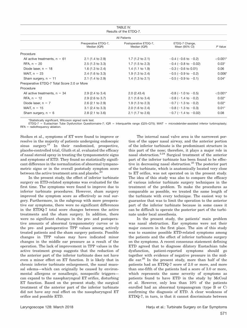
Stoikes et al., symptoms of ET were found to improve orresolve in the majority of patients undergoing endoscopicsinus surgery.18 In their randomized, prospective,placebo-controlled trial, Gluth et al. evaluated the efficacyof nasal steroid spray in treating the tympanometric signsand symptoms of ETD. They found no statistically signifi-cant difference in the normalization of abnormal tympano-metric signs or in the overall poststudy symptom scorebetween the active treatment arm and placebo.23
In the present study, the effect of inferior turbinatesurgery on ETD-related symptoms was evaluated for thefirst time. The symptoms were found to improve due toinferior turbinate procedures. However, sham surgeryimproved the symptom score as effectively as real sur-gery. Furthermore, in the subgroup with more preopera-tive ear symptoms, there were no significant differencesin the ETDQ-7 total score changes between the activetreatments and the sham surgery. In addition, therewere no significant changes in the pre- and postopera-tive amounts of abnormal tympanometry curves or inthe pre- and postoperative TPP values among activelytreated patients and the sham surgery patients. Possiblechanges in TPP values may have indicated minorchanges in the middle ear pressure as a result of theoperation. The lack of improvement in TPP values in theactive treatment group suggests that the reduction ofthe anterior part of the inferior turbinate does not haveeven a minor effect on ET function. It is likely that inchronic inferior turbinate enlargement, the nasal muco-sal edema—which can originally be caused by environ-mental allergens or nonallergic, nonspecific triggers—can expand to the nasopharyngeal ET orifice, disturbingET function. Based on the present study, the surgicaltreatment of the anterior part of the inferior turbinatedid not have any real effect on the nasopharyngeal ETorifice and possible ETD.
The internal nasal valve area is the narrowest por-tion of the upper nasal airway, and the anterior portionof the inferior turbinate is the predominant structure inthis part of the nose; therefore, it plays a major role innasal obstruction.2,24 Surgical treatment of the anteriorpart of the inferior turbinate has been found to be effec-tive in decreasing nasal obstruction.25 The posterior partof the turbinate, which is anatomically located very closeto ET orifice, was not operated on in the present study.The idea of this study was also to compare the efficacyof various inferior turbinate surgery techniques in thetreatment of the problem. To make the procedures ascomparable as possible, we treated the same length ofthe turbinate with every technique. The easiest way toguarantee that was to limit the operation to the anteriorpart of the inferior turbinate because in some cases itcan be difficult to operate the posterior part of the turbi-nate under local anesthesia.
In the present study, the patients’ main problemwas nasal obstruction. Ear symptoms were not theirmajor concern in the first place. The aim of this studywas to examine possible ETD-related symptoms amongthe patients and the effect of inferior turbinate surgeryon the symptoms. A recent consensus statement definingETD agreed that to diagnose dilatory Eustachian tubedysfunction, patient-reported symptoms should gotogether with evidence of negative pressure in the mid-dle ear.26 In the present study, more than half of thepatients had an ETDQ-7 score of 2.0 or more, and morethan one-fifth of the patients had a score of 3.0 or more,which represents the same severity of symptoms aspatients found to have ETD in the study by McCoulet al. However, only less than 10% of the patientsenrolled had an abnormal tympanogram (type B or C)confirming the diagnosis of ETD. A clear weakness ofETDQ-7, in turn, is that it cannot discriminate between
TABLE IV.Results of the ETDQ-7.
All Patients
Preoperative ETDQ-7,Median (IQR)
Postoperative ETDQ-7,Median (IQR)
ETDQ-7 Change,Mean (95% CI) P Value
Procedure
All active treatments, n 5 61 2.1 (1.4 to 2.9) 1.7 (1.2 to 2.1) 20.4 (20.6 to 20.2) <0.001*
RFA, n 5 20 2.5 (1.3 to 3.3) 1.7 (1.5 to 2.3) 20.4 (20.8 to 20.02) 0.03*
Diode laser, n 5 18 1.6 (1.3 to 2.4) 1.4 (1.1 to 1.9) 20.3 (20.6 to 0.01) 0.06
MAIT, n 5 23 2.4 (1.6 to 3.3) 1.9 (1.3 to 2.4) 20.5 (20.9 to 20.2) 0.006*
Sham surgery, n 5 11 2.1 (1.4 to 2.9) 1.4 (1.3 to 2.1) 20.5 (20.9 to 20.1) 0.04*
Preoperative ETDQ-7 Total Score 2.0 or More
Procedure
All active treatments, n 5 34 2.9 (2.4 to 3.4) 2.0 (2.43.4) -0.8 (21.0 to 20.5) <0.001*
RFA, n 5 12 2.9 (2.6 to 3.7) 2.1 (1.6 to 3.4) 20.8 (21.4 to 20.2) 0.02*
Diode laser, n 5 7 2.6 (2.1 to 2.9) 1.9 (1.3 to 2.3) 20.7 (21.3 to 20.2) 0.02*
MAIT, n 5 15 3.1 (2.4 to 3.3) 2.0 (1.6 to 2.4) 20.8 (21.3 to 20.3) 0.01*
Sham surgery, n 5 6 2.8 (2.1 to 3.6) 2.1 (1.7 to 2.6) 20.7 (21.4 to 20.02) 0.08
*Statistically significant, Wilcoxon signed rank test.ETDQ-7 5 Eustachian Tube Dysfunction Questionnaire-7; IQR 5 Interquartile range (Q25–Q75); MAIT 5 microdebrider-assisted inferior turbinoplasty;
RFA 5 radiofrequency ablation.
Laryngoscope 128: March 2018 Harju et al.: Turbinate Surgery on Ear Symptoms
571
patients with obstructive ET dysfunction and patulousET. Therefore, it cannot be used as a sole criterion forthe diagnosis of ETD. For these reasons, we are not ableto generalize the results of this study to ETD. We canonly talk about the effect of inferior turbinate procedureon patients’ symptoms suggestive of ETD.
All clinical examinations and operations were per-formed by the same physician, who was not blinded to thepatients’ groups. Unblinding of the examining physiciancould introduce unintentional bias by causing misinter-pretation of data. However, the examination parametersused in this study were not based on the subjective evalua-tion of the examining physician. Parameters of tympan-ometry, unlike otoscopic appearance or Valsalva andToynbee tests, are not based on the subjective evaluationof the examiner. That is why tympanometry was chosen tobe the only objective examination method in this study.However, it sometimes is very challenging to make a con-firmed diagnosis of ETD based on both patient-reportedsymptoms and signs of negative pressure in the middleear in the form of abnormal tympanometry curves. In mildor fluctuating cases, the patients may have symptomssuggestive of ETD, but their tympanometry curves can bewithin normal limits.
CONCLUSIONThe improvement of ETD-related symptoms due to
surgery of the anterior half of the inferior turbinate wasfound to be equal to placebo. The findings of this study donot support the use of reduction of the anterior half of theinferior turbinate as a sole procedure intended to treat theear symptoms assessed by the ETDQ-7 questionnaire.
BIBLIOGRAPHY
1. Berger G, Gass S, Ophir D. The histopathology of the hypertrophic inferiorturbinate. Arch Otolaryngol Head Neck Surg 2006;132:588–594.
2. Willatt D. The evidence for reducing inferior turbinates. Rhinology 2009;47:227–236.
3. Seibert JW, Danner CJ. Eustachian tube function and the middle ear. Oto-laryngol Clin North Am 2006;39:1221–1235.
4. McCoul ED, Anand VK, Christos PJ. Validating the clinical assessment ofeustachian tube dysfunction: the Eustachian Tube Dysfunction Ques-tionnaire (ETDQ-7). Laryngoscope 2012;122:1137–1141.
5. Poe DS, Abou-Halawa A, Abdel-Razek O. Analysis of the dysfunctionalEustachian tube by video endoscopy. Otol Neurotol 2001;22:590–595.
6. Doyle WJ, Seroky JT, Angelini BL, Gulhan M, Skoner DP, Fireman P.Abnormal middle ear pressures during experimental influenza A virusinfection—role of Eustachian tube function. Auris Nasus Larynx 2000;27:323–326.
7. Ohrui N, Takeuchi A, Tong A, Iwata M, Nakamura A, Ohashi K. Allergicrhinitis and ear pain in flight. Ann Allergy Asthma Immunol 2005;95:350–353.
8. Stammberger H. An endoscopic study of tubal function and the diseasedethmoid sinus. Arch Otorhinolaryngol 1986;243:254–259.
9. Liu P, Su K, Zhu B, Wu Y, Shi H, Yin S. Detection of Eustachian tubeopenings by tubomanometry in adult otitis media with effusion. EurArch Otorhinolaryngol 2016;273:3109–3115.
10. van der Avoort SJ, van Heerbeek N, Zielhuis GA, Cremers CW. Sonotub-ometry: Eustachian tube ventilatory function test: a state-of-the-artreview. Otol Neurotol 2005;26:538–543.
11. Smith ME, Tysome JR. Tests of Eustachian tube function: a review. ClinOtolaryngol 2015;40:300–311.
12. Van Roeyen S, Van de Heyning P, Van Rompaey V. Value and discrimina-tive power of the seven-item Eustachian Tube Dysfunction Question-naire. Laryngoscope 2015;125:2553–2556.
13. Van Roeyen S, Van de Heyning P, Van Rompaey V. Responsiveness of the7-item Eustachian Tube Dysfunction Questionnaire. J Int Adv Otol2016;12:106–108.
14. Straszek SP, Schlunssen V, Sigsgaard T, Pedersen OF. Reference valuesfor acoustic rhinometry in decongested school children and adults: themost sensitive measurement for change in nasal patency. Rhinology2007;45:36–39.
15. Grymer LF, Hilberg O, Pedersen OF, Rasmussen TR. Acoustic rhinometry:values from adults with subjective normal nasal patency. Rhinology1991;29:35–47.
16. Tomkinson A, Eccles R. Comparison of the relative abilities of acousticrhinometry, rhinomanometry, and the visual analogue scale in detectingchance in the nasal cavity in a healthy adult population. Am J Rhinol1996;10:161–165.
17. Fisher EW. Acoustic rhinometry. Clin Otolaryngol Allied Sci 1997;22:307–317.
18. Stoikes NF, Dutton JM. The effect of endoscopic sinus surgery on symp-toms of Eustachian tube dysfunction. Am J Rhinol 2005;19:199–202.
19. Low WK, Willatt DJ. The relationship between middle ear pressure anddeviated nasal septum. Clin Otolaryngol Allied Sci 1993;18:308–310.
20. Lazo-Saenz JG, Galvan-Aguilera AA, Martinez-Ordaz VA, Velasco-Rodriguez VM, Nieves-Renteria A, Rincon-Castaneda C. Eustachiantube dysfunction in allergic rhinitis. Otolaryngol Head Neck Surg 2005;132:626–629.
21. Bakhshaee M, Ardakani HP, Ghazizadeh AH, Movahed R, Jarahi L, RajatiM. Middle ear function in sinonasal polyposis. Eur Arch Otorhinolar-yngol 2016;273:2911–2916.
22. Deron P, Clement PA, Derde MP. Septal surgery and tubal function: earlyand late results. Rhinology 1995;33:7–9.
23. Gluth MB, McDonald DR, Weaver AL, Bauch CD, Beatty CW, Orvidas LJ.Management of Eustachian tube dysfunction with nasal steroid spray: aprospective, randomized, placebo-controlled trial. Arch OtolaryngolHead Neck Surg 2011;137:449–455.
24. Moche JA, Palmer O. Surgical management of nasal obstruction. OralMaxillofac Surg Clin North Am 2012;24:229–237.
25. Civelek S, Ozcelik M, Emre IE, Cakir BO, Turgut S. Comparison of radio-frequency applied to the total inferior choncha with application to itsanterior third. Auris Nasus Larynx 2010;37:589–593.
26. Schilder AG, Bhutta MF, Butler CC, et al. Eustachian tube dysfunction:consensus statement on definition, types, clinical presentation and diag-nosis. Clin Otolaryngol 2015;40:407–411.
Laryngoscope 128: March 2018 Harju et al.: Turbinate Surgery on Ear Symptoms
572
The LaryngoscopeVC 2018 The American Laryngological,Rhinological and Otological Society, Inc.
A Prospective, Randomized, Placebo-Controlled Study of Inferior
Turbinate Surgery
Teemu Harju, MD ; Jura Numminen, MD, PhD; Ilkka Kivek€as, MD, PhD; Markus Rautiainen, MD, PhD
Objectives/Hypothesis: The purpose of this study was to compare radiofrequency ablation, diode laser, andmicrodebrider-assisted inferior turbinoplasty techniques in the treatment of chronic nasal obstruction caused by inferior tur-binate enlargement, and to compare these techniques with a placebo procedure.
Study Design: Prospective, randomized, single-blinded, placebo-controlled study.Methods: A total of 98 consecutive patients with enlarged inferior turbinates due to persistent year-round rhinitis were
randomized into a placebo, radiofrequency ablation, diode laser, and microdebrider-assisted inferior turbinoplasty groups in aratio of 1:2:2:2. All the procedures were carried out under local anesthesia with the patients’ eyes covered. Assessmentswere conducted prior to surgery and 3 months subsequent to the surgery.
Results: The severity of nasal obstruction measured by visual analog scale score decreased statistically significantly inall the groups, including placebo. Radiofrequency ablation (P5.03), diode laser (P5.02), and microdebrider-assisted inferiorturbinoplasty (P5.04) all decreased the symptom score of the severity of nasal obstruction statistically significantly morecompared to the placebo procedure.
Conclusions: The placebo effect had a large role in the overall improvement of the severity of nasal obstruction afterthe inferior turbinate surgery. However, all three techniques provided a statistically significant additional reduction of theseverity of nasal obstruction compared to the placebo procedure.
Key Words: Nasal obstruction, inferior turbinate surgery, placebo controlled, randomized.Level of Evidence: 1b.
Laryngoscope, 00:000–000, 2018
INTRODUCTIONInferior turbinate enlargement due to persistent
year-round allergic or nonallergic rhinitis is one of themain causes of chronic nasal obstruction.1 Intranasalcorticosteroids are the most important medicine group inthe treatment of rhinitis and hypertrophied inferior tur-binates. When medical therapy for enlarged inferior tur-binates fails, turbinate surgery can be considered.2
Various surgical techniques have been described forthe reduction of hyperplastic inferior turbinates. How-ever, no clear consensus exists in the literature on theexact role of surgery or the most optimal method for sur-gical treatment.3 In recent years, surgical procedureshave concentrated on the minimal disturbance of thenasal mucosa. The aim of inferior turbinate surgery hasbeen to maximize the volumetric reduction of the turbi-nate to decrease the nasal obstruction while maintainingnasal function and minimizing complications.4
Microdebrider-assisted inferior turbinoplasty(MAIT)—a powered subtype of submucosal resection5—
and radiofrequency ablation (RFA) are referred to asmucosal sparing techniques; they are widely used andthe most commonly studied techniques in the recent lit-erature.4 The diode laser technique has also gained inpopularity due to its ease of use in the office setting.6
Thus far, three placebo-controlled trials have beenpublished regarding RFA. These studies have evaluatedthe influence of the treatment on subjective scores ofnasal obstruction, and they found that the placebo effectseems to have a role in the results of turbinatesurgery.7–9
The purpose of this prospective, randomized, single-blinded, placebo-controlled study was to compare RFA,diode laser, and MAIT techniques in the treatment ofchronic nasal obstruction caused by inferior turbinateenlargement, and to compare these techniques with aplacebo treatment.
MATERIALS AND METHODSThis prospective randomized study was carried out at
Tampere University Hospital, Tampere, Finland, between Feb-ruary 2014 and September 2017. The institutional review boardapproved the study design (R13144), and all patients providedwritten, informed consent. A total of 98 consecutive adultpatients with enlarged inferior turbinates due to persistentyear-round rhinitis were enrolled in this study. The patientspresented symptoms of bilateral nasal obstruction relatedto inferior turbinate congestion that had not responded to a3-month trial of appropriate treatment with intranasal cortico-steroids. Patients with significant nasal septum deviation
From the Department of Otorhinolaryngology, Tampere UniversityHospital, Tampere, Finland.
Editor’s Note: This Manuscript was accepted for publicationDecember 29, 2017.
The authors have no funding, financial relationships, or conflictsof interest to disclose.
Send correspondence to Teemu Harju, MD, Department of Otorhi-nolaryngology, Tampere University Hospital, Teiskontie 35, 33521 Tam-pere, Finland. E-mail: [email protected]
DOI: 10.1002/lary.27103
Laryngoscope 00: Month 2018 Harju et al.: Placebo-Controlled Study of Turbinate Surgery
1
affecting the nasal valve region, internal/external valve col-lapse/stenosis, chronic rhinosinusitis with or without polyposis,previous nasal surgery, sinonasal tumor, severe systemic disor-der, severe obesity, or malignancy were excluded.
Cone beam computed tomography (Planmeca Max; Plan-
meca, Helsinki, Finland) was used to exclude patients with
chronic rhinosinusitis from the study. Serum-specific immuno-
globulin E (IgE) level measurements were used to identify the
patients with an allergic sensitization. Allergic sensitization
was defined as a specific IgE>0.35 for any common airborne
allergen (cat, dog, horse, birch, grass, mugwort, Dermatopha-
goides pteronyssinus, and molds).
The definition of inferior turbinate enlargement was based
on persistent bilateral symptoms, a finding of bilateral swelling
of the inferior turbinate in nasal endoscopy, and the evident
shrinking of both turbinates in a decongestion test, which could
not be applied to either bony turbinate enlargement of soft tis-
sue hypertrophy unresponsive to topical decongestant. The
nasal response to the topical vasoconstrictor 0.5% xylometazo-
line hydrochloride (Nasolin; Orion, Espoo, Finland) in both
nasal cavities 15 minutes before obtaining the second measure-
ment was evaluated objectively using acoustic rhinometry
(Acoustic Rhinometer A1; GM Instruments Ltd., Kilwinning,
United Kingdom). An improvement of less than 30% in anterior
nasal cavity volume (V2–5 cm) in one or both nasal cavities was
considered normal, and those patients were excluded from the
study. The limit value of 30% was chosen according to previous
literature.10–12
Patients were consecutively randomized into placebo, RFA,
diode laser, and MAIT groups in a ratio of 1:2:2:2 using Minim,
a free MS-DOS program that randomizes patients to treatment
groups by the method of minimization. Proportional amounts of
patients with allergic sensitization were kept similar for each
group. Age and sex distributions were also kept similar for each
group.
The surgical treatment was performed in similar circum-
stances at the day surgery department of the hospital’s ear,
nose, and throat clinic. All surgical procedures were performed
by the same surgeon (T.H). The staff of the day surgery depart-
ment was instructed to avoid all communication before and dur-
ing the operation, as well as during the postoperative care, that
might reveal the group of the patient. All the alternative surgi-
cal devices were available in the operation room. The proce-
dures were carried out under local anesthesia with the patient’s
eyes covered.
First, the inferior turbinate was topically anesthetized
using cotton strips with a mixture of lidocaine 40 mg/mL (Lido-
cain; Orion) and 2 to 3 drops of epinephrine 0.1% in 5 to 10 mL
of lidocaine. Next, 1.5 mL of local anesthetic (Lidocain 10 mg/mL
c. Adrenalin 10mg/mL; Orion) was then applied to the medial
portions of both inferior turbinates. All the procedures were
performed under the direct vision of a straight, 4-mm-diameter,
0 8 endoscope (Karl Storz, Tuttlingen, Germany). In all the
groups with every technique, the treatment was given to the
medial side of the anterior half of the inferior turbinate. Short-
term (until the next morning) nasal packing with a nonabsorb-
able packing material (Ivalon; Fabco, New London, CT) was
applied only if active bleeding developed during the surgery.
The RFA treatment was carried out with a radiofrequency
generator (Sutter RF generator BM-780 II; Sutter, Freiburg,
Germany). A Binner bipolar needle electrode was inserted into
the medial submucosal tissue of the inferior turbinate. The
upper and lower parts of the anterior half of the inferior turbi-
nate were treated for 6 seconds at 10 W output power in three
areas.
The diode laser treatment was given with a FOX Laser(A.R.C. Laser GmbH, Nuremberg, Germany). The settings were
as follows: wavelength of 980 nm, output power of 6 W incontinuous-wave mode, and laser delivery by a 600-mm fiber
using contact mode. Four parallel stripes were made on themucosa by drawing the fiber from the posterior to the anterior
direction along the medial edge of the anterior half of the infe-rior turbinate.
In the MAIT treatment, a 2.9-mm-diameter rotatablemicrodebrider tip (Medtronic Xomed, Jacksonville, Florida) was
firmly pushed toward the turbinate bone until it pierced themucosa of the anterior face of the inferior turbinate. Next, asubmucosal pocket was dissected by tunneling the elevator tip
in an anterior-to-posterior and superior-to-inferior sweepingmotion. Once an adequate pocket had been created, resection of
the stromal tissue was carried out by moving the blade backand forth in a sweeping motion, with the system set at
3,000 rpm using suction irrigation.
In the placebo procedure, small (2–3 mm in diameter)
nasal mucosal biopsies were first taken from the anteriormedial portions of the inferior turbinates, causing minor bleed-
ing. Next, a radiofrequency tissue ablation device was turnedon repeatedly near the patient, but without the needle electro-des of the device touching the patient; the patient could only
hear the acoustic tone of the device. During this sound decep-tion, a suction tube and a nasal endoscope were moved lightly
in both sides of the nose for a couple of minutes to convince thepatients that they had undergone surgery.
After the operation, none of the patients were given medi-cal treatment, including analgesics, nasal steroids, and nasal
decongestants. The patients were given a 100-mm visual ana-logue scale (VAS) questionnaire, which had been filled for the
first time during the preoperative visit. They were asked tomark on the line the point that they feel represents their per-ception of their current state regarding postoperative nasal
symptoms, including severity of nasal obstruction (notobstructed–very obstructed), discharge (no discharge–much dis-
charge), crusting (no crust–much crust), and pain (no pain–much pain). The questionnaire was to be filled daily for the first
week, and then after 2, 3, and 4 weeks postoperatively. Theywere instructed to return the questionnaire by mail 1 monthafter the operation.
All of the patients were evaluated prior to surgery and 3
months following the surgery. All clinical examinations wereperformed by the same examiner (T.H), who was also the opera-tor and not blinded to the patients’ group. The patients filled
the VAS questionnaire regarding nasal symptoms during bothvisits. They were asked to mark on the line the point that they
feel represents their perception of their current state. Acousticrhinometry was also performed both before the surgery and
during the control visit at 3 months. During the control visit,the patients filled the questionnaire before meeting theexaminer.
IBM SPSS Statistics 22.0 (IBM, Armonk, NY) was usedfor the statistical analyses. All nonparametric data were statis-
tically processed using the Wilcoxon signed rank test, Mann-Whitney U test, and Kruskal-Wallis test. In the cases with
parametric data, the comparison between the groups was car-ried out by one-way analysis of variance.
RESULTSThe patients’ characteristics are described in
Table I and the data of the operations in Table II. Theoperation time for the MAIT procedure was statisticallysignificantly longer compared to the other procedures.
Laryngoscope 00: Month 2018 Harju et al.: Placebo-Controlled Study of Turbinate Surgery
2
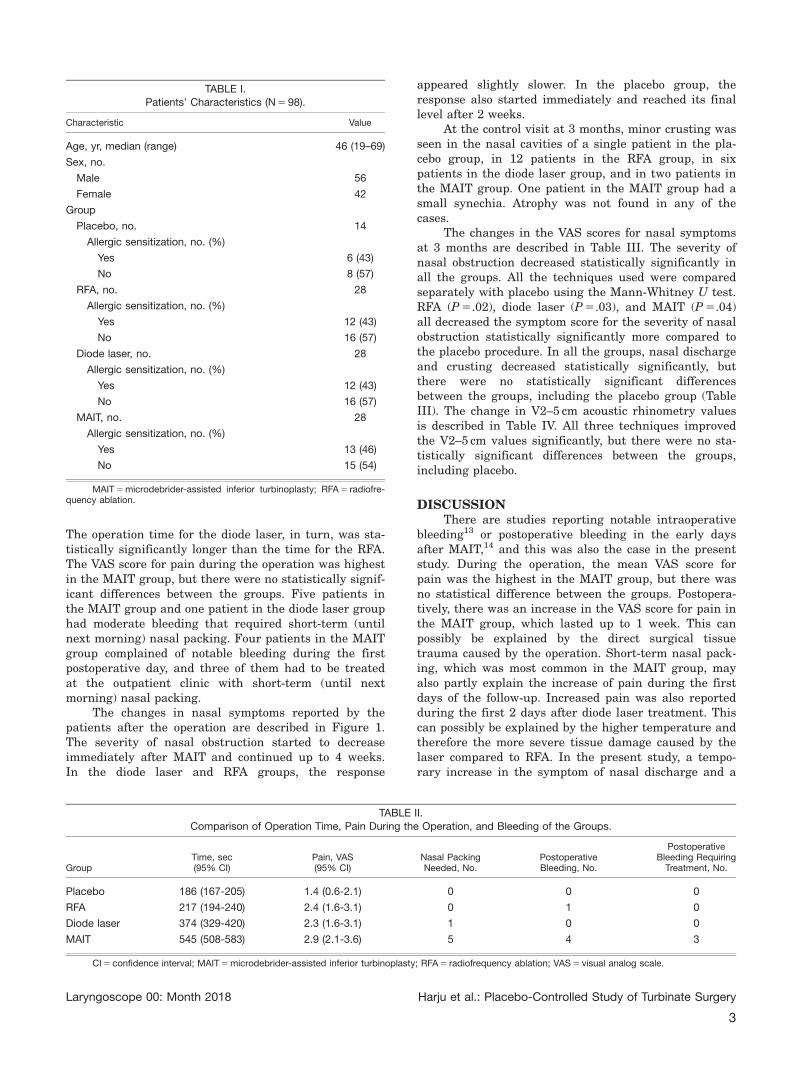
The operation time for the diode laser, in turn, was sta-tistically significantly longer than the time for the RFA.The VAS score for pain during the operation was highestin the MAIT group, but there were no statistically signif-icant differences between the groups. Five patients inthe MAIT group and one patient in the diode laser grouphad moderate bleeding that required short-term (untilnext morning) nasal packing. Four patients in the MAITgroup complained of notable bleeding during the firstpostoperative day, and three of them had to be treatedat the outpatient clinic with short-term (until nextmorning) nasal packing.
The changes in nasal symptoms reported by thepatients after the operation are described in Figure 1.The severity of nasal obstruction started to decreaseimmediately after MAIT and continued up to 4 weeks.In the diode laser and RFA groups, the response
appeared slightly slower. In the placebo group, theresponse also started immediately and reached its finallevel after 2 weeks.
At the control visit at 3 months, minor crusting wasseen in the nasal cavities of a single patient in the pla-cebo group, in 12 patients in the RFA group, in sixpatients in the diode laser group, and in two patients inthe MAIT group. One patient in the MAIT group had asmall synechia. Atrophy was not found in any of thecases.
The changes in the VAS scores for nasal symptomsat 3 months are described in Table III. The severity ofnasal obstruction decreased statistically significantly inall the groups. All the techniques used were comparedseparately with placebo using the Mann-Whitney U test.RFA (P 5.02), diode laser (P 5.03), and MAIT (P 5.04)all decreased the symptom score for the severity of nasalobstruction statistically significantly more compared tothe placebo procedure. In all the groups, nasal dischargeand crusting decreased statistically significantly, butthere were no statistically significant differencesbetween the groups, including the placebo group (TableIII). The change in V2–5 cm acoustic rhinometry valuesis described in Table IV. All three techniques improvedthe V2–5 cm values significantly, but there were no sta-tistically significant differences between the groups,including placebo.
DISCUSSIONThere are studies reporting notable intraoperative
bleeding13 or postoperative bleeding in the early daysafter MAIT,14 and this was also the case in the presentstudy. During the operation, the mean VAS score forpain was the highest in the MAIT group, but there wasno statistical difference between the groups. Postopera-tively, there was an increase in the VAS score for pain inthe MAIT group, which lasted up to 1 week. This canpossibly be explained by the direct surgical tissuetrauma caused by the operation. Short-term nasal pack-ing, which was most common in the MAIT group, mayalso partly explain the increase of pain during the firstdays of the follow-up. Increased pain was also reportedduring the first 2 days after diode laser treatment. Thiscan possibly be explained by the higher temperature andtherefore the more severe tissue damage caused by thelaser compared to RFA. In the present study, a tempo-rary increase in the symptom of nasal discharge and a
TABLE I.Patients’ Characteristics (N 5 98).
Characteristic Value
Age, yr, median (range) 46 (19–69)
Sex, no.
Male 56
Female 42
Group
Placebo, no. 14
Allergic sensitization, no. (%)
Yes 6 (43)
No 8 (57)
RFA, no. 28
Allergic sensitization, no. (%)
Yes 12 (43)
No 16 (57)
Diode laser, no. 28
Allergic sensitization, no. (%)
Yes 12 (43)
No 16 (57)
MAIT, no. 28
Allergic sensitization, no. (%)
Yes 13 (46)
No 15 (54)
MAIT 5 microdebrider-assisted inferior turbinoplasty; RFA 5 radiofre-quency ablation.
TABLE II.
Comparison of Operation Time, Pain During the Operation, and Bleeding of the Groups.
GroupTime, sec(95% CI)
Pain, VAS(95% CI)
Nasal PackingNeeded, No.
PostoperativeBleeding, No.
PostoperativeBleeding Requiring
Treatment, No.
Placebo 186 (167-205) 1.4 (0.6-2.1) 0 0 0
RFA 217 (194-240) 2.4 (1.6-3.1) 0 1 0
Diode laser 374 (329-420) 2.3 (1.6-3.1) 1 0 0
MAIT 545 (508-583) 2.9 (2.1-3.6) 5 4 3
CI 5 confidence interval; MAIT 5 microdebrider-assisted inferior turbinoplasty; RFA 5 radiofrequency ablation; VAS 5 visual analog scale.
Laryngoscope 00: Month 2018 Harju et al.: Placebo-Controlled Study of Turbinate Surgery
3
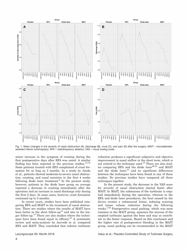
minor increase in the symptom of crusting during thefirst postoperative days after RFA was noted. A similarfinding has been reported in the previous studies.15,16
Some patients treated with RFA complained of crust for-mation for as long as 3 months. In a study by Jandaet al., patients showed moderate-to-severe nasal obstruc-tion, crusting, and nasal secretion in the first 4 weeksfollowing diode laser treatment.6 In the present study,however, patients in the diode laser group on averagereported a decrease in crusting immediately after theoperation and an increase in nasal discharge only duringthe first 2 days. In some cases, however, crust formationcontinued up to 3 months.
In recent years, studies have been published com-paring RFA and MAIT in the treatment of nasal obstruc-tion. There are studies where the results of MAIT havebeen better in the short follow-up14 and also in the lon-ger follow-up.17 There are also studies where the techni-ques have been found equal in efficacy.18 A systematicreview and meta-analysis by Acevedo et al. comparedRFA and MAIT. They concluded that inferior turbinate
reduction produces a significant subjective and objectiveimprovement in nasal airflow in the short term, which isnot related to the technique used.19 There are also stud-ies comparing RFA and the diode laser20,21 and MAITand the diode laser,13 and no significant differencesbetween the techniques have been found in any of thesestudies. No previous studies have compared all threetechniques together.
In the present study, the decrease in the VAS scorefor severity of nasal obstruction started faster afterMAIT. In MAIT, the submucosa of the turbinate is emp-tied immediately during the operation, whereas in theRFA and diode laser procedures, the heat caused by thedevice creates a submucosal lesion, inducing scarringand tissue volume reduction during the followingweeks.1,22 Postoperative nasal packing, which was mostcommon in the MAIT group, squeezes the mucosa of theemptied turbinate against the bone and may so contrib-ute to the faster response. Based on this conclusion andthe higher rate of postoperative bleeding in the MAITgroup, nasal packing can be recommended in the MAIT
Fig. 1. Mean changes in the severity of nasal obstruction (A), discharge (B), crust (C), and pain (D) after the surgery. MAIT 5 microdebrider-assisted inferior turbinoplasty; RFA 5 radiofrequency ablation; VAS 5 visual analog scale.
Laryngoscope 00: Month 2018 Harju et al.: Placebo-Controlled Study of Turbinate Surgery
4
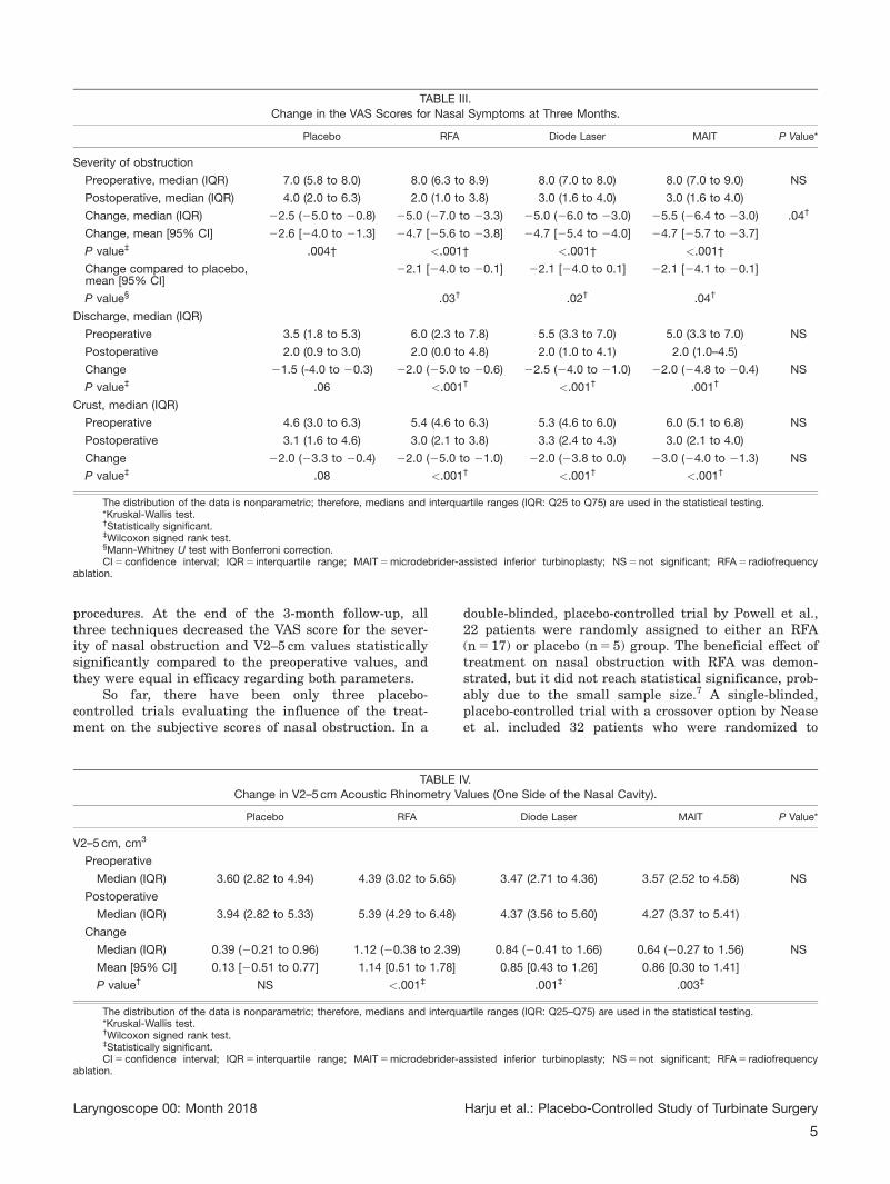
procedures. At the end of the 3-month follow-up, allthree techniques decreased the VAS score for the sever-ity of nasal obstruction and V2–5 cm values statisticallysignificantly compared to the preoperative values, andthey were equal in efficacy regarding both parameters.
So far, there have been only three placebo-controlled trials evaluating the influence of the treat-ment on the subjective scores of nasal obstruction. In a
double-blinded, placebo-controlled trial by Powell et al.,22 patients were randomly assigned to either an RFA(n 5 17) or placebo (n 5 5) group. The beneficial effect oftreatment on nasal obstruction with RFA was demon-strated, but it did not reach statistical significance, prob-ably due to the small sample size.7 A single-blinded,placebo-controlled trial with a crossover option by Neaseet al. included 32 patients who were randomized to
TABLE III.Change in the VAS Scores for Nasal Symptoms at Three Months.
Placebo RFA Diode Laser MAIT P Value*
Severity of obstruction
Preoperative, median (IQR) 7.0 (5.8 to 8.0) 8.0 (6.3 to 8.9) 8.0 (7.0 to 8.0) 8.0 (7.0 to 9.0) NS
Postoperative, median (IQR) 4.0 (2.0 to 6.3) 2.0 (1.0 to 3.8) 3.0 (1.6 to 4.0) 3.0 (1.6 to 4.0)
Change, median (IQR) 22.5 (25.0 to 20.8) 25.0 (27.0 to 23.3) 25.0 (26.0 to 23.0) 25.5 (26.4 to 23.0) .04†
Change, mean [95% CI] 22.6 [24.0 to 21.3] 24.7 [25.6 to 23.8] 24.7 [25.4 to 24.0] 24.7 [25.7 to 23.7]
P value‡ .004† <.001† <.001† <.001†
Change compared to placebo,mean [95% CI]
22.1 [24.0 to 20.1] 22.1 [24.0 to 0.1] 22.1 [24.1 to 20.1]
P value§ .03† .02† .04†
Discharge, median (IQR)
Preoperative 3.5 (1.8 to 5.3) 6.0 (2.3 to 7.8) 5.5 (3.3 to 7.0) 5.0 (3.3 to 7.0) NS
Postoperative 2.0 (0.9 to 3.0) 2.0 (0.0 to 4.8) 2.0 (1.0 to 4.1) 2.0 (1.0–4.5)
Change 21.5 (-4.0 to 20.3) 22.0 (25.0 to 20.6) 22.5 (24.0 to 21.0) 22.0 (24.8 to 20.4) NS
P value‡ .06 <.001† <.001† .001†
Crust, median (IQR)
Preoperative 4.6 (3.0 to 6.3) 5.4 (4.6 to 6.3) 5.3 (4.6 to 6.0) 6.0 (5.1 to 6.8) NS
Postoperative 3.1 (1.6 to 4.6) 3.0 (2.1 to 3.8) 3.3 (2.4 to 4.3) 3.0 (2.1 to 4.0)
Change 22.0 (23.3 to 20.4) 22.0 (25.0 to 21.0) 22.0 (23.8 to 0.0) 23.0 (24.0 to 21.3) NS
P value‡ .08 <.001† <.001† <.001†
The distribution of the data is nonparametric; therefore, medians and interquartile ranges (IQR: Q25 to Q75) are used in the statistical testing.*Kruskal-Wallis test.†Statistically significant.‡Wilcoxon signed rank test.§Mann-Whitney U test with Bonferroni correction.CI 5 confidence interval; IQR 5 interquartile range; MAIT 5 microdebrider-assisted inferior turbinoplasty; NS 5 not significant; RFA 5 radiofrequency
ablation.
TABLE IV.Change in V2–5 cm Acoustic Rhinometry Values (One Side of the Nasal Cavity).
Placebo RFA Diode Laser MAIT P Value*
V2–5 cm, cm3
Preoperative
Median (IQR) 3.60 (2.82 to 4.94) 4.39 (3.02 to 5.65) 3.47 (2.71 to 4.36) 3.57 (2.52 to 4.58) NS
Postoperative
Median (IQR) 3.94 (2.82 to 5.33) 5.39 (4.29 to 6.48) 4.37 (3.56 to 5.60) 4.27 (3.37 to 5.41)
Change
Median (IQR) 0.39 (20.21 to 0.96) 1.12 (20.38 to 2.39) 0.84 (20.41 to 1.66) 0.64 (20.27 to 1.56) NS
Mean [95% CI] 0.13 [20.51 to 0.77] 1.14 [0.51 to 1.78] 0.85 [0.43 to 1.26] 0.86 [0.30 to 1.41]
P value† NS <.001‡ .001‡ .003‡
The distribution of the data is nonparametric; therefore, medians and interquartile ranges (IQR: Q25–Q75) are used in the statistical testing.*Kruskal-Wallis test.†Wilcoxon signed rank test.‡Statistically significant.CI 5 confidence interval; IQR 5 interquartile range; MAIT 5 microdebrider-assisted inferior turbinoplasty; NS 5 not significant; RFA 5 radiofrequency
ablation.
Laryngoscope 00: Month 2018 Harju et al.: Placebo-Controlled Study of Turbinate Surgery
5
either an RFA (n 5 16) or placebo (n 5 16) group.Improvement was shown in the frequency of nasalobstruction, severity of nasal obstruction, and overallability to breathe for both the placebo and RFA treat-ment arms at 8 weeks. However, the effects of RFA weresignificantly better than those of placebo in the severityof nasal obstruction and overall ability to breathe.8 In asingle-blinded, placebo-controlled, crossover trial byBran et al. with 22 patients, the first group receivedRFA first followed by a placebo treatment 6 to 8 weekslater. In the second group, the order was reversed. Nasalobstruction decreased significantly only after RFA inboth groups. There was a tendency of improvement afterplacebo in both treatment arms. However, the observedtrends of a placebo effect were not statisticallysignificant.9
Our study is the first placebo-controlled studyinvolving three different techniques, and also the firststudy where the diode laser and MAIT have been com-pared with placebo treatment. The follow-up time of3 months with the placebo group is also the longest sofar. RFA, diode laser, and MAIT all improved the VASscore for the severity of nasal obstruction statisticallysignificantly better than placebo. The finding is in linewith the study by Nease et al. regarding RFA.8 The truetreatment effect is the difference in improvementbetween the intervention group and the control group(placebo effect).7 In the present study, the mean truetreatment effect in the VAS score change was found tobe 22.1 for all three techniques. The placebo effectaccounted for a large part of the total improvement seenin the intervention groups. Without a placebo control,we would have falsely found a greater improvement andsignificance due to treatment.
In the present study, all the techniques decreasedthe symptom scores of nasal discharge and crusting sta-tistically significantly compared to the preoperative val-ues at the end of the 3-month follow-up, but none ofthem statistically significantly compared to the placebo.In addition, minor crusting was still found in some nosesat 3 months, especially in the RFA and diode lasergroups. Therefore, the major part of the improvement ofthese symptoms can be explained by the placebo effect.
Anterior nasal cavity volume (V2–5 cm) in acousticrhinometry was used as a parameter for objective exami-nation. It represents well the operated anterior half ofthe inferior turbinate. Acoustic rhinometry will give avalid result in most instances, at least for the first 5 to6 cm of the nasal cavity.23 Measurement of V2–5 cm isthe most sensitive measurement for change in mucosalswelling during decongestion,24 and that is why theparameter also had an important role in the preopera-tive decongestion test in defining inferior turbinateenlargement. In the 3-month follow-up, RFA, diode laser,and MAIT techniques all improved the V2–5 cm valuesstatistically significantly compared to the preoperativevalues. Due to lack of a real surgical procedure, therewas understandably no statistically significant change inthe V2-5 cm values in the placebo group. This supportsthe conclusion that the improvement in the VAS scorefor severity of nasal obstruction in the placebo group
was due to a placebo effect. However, none of the techni-ques improved the volume statistically significantly com-pared to placebo. This finding can be partly explained bythe relatively small number of patients in the placebogroup. A larger number might have led to a statisticalsignificance. In addition, environmental conditions andtechnical difficulties in measurement may have an influ-ence in the volume values.25 Furthermore, the symptomof nasal obstruction and objective parameters do notalways correlate very well.26 On the other hand, it iswise to remember that the increase in the nasal cavityvolume should be optimal instead of too large to avoidmaking the nose too wide, which might lead to problemssuch as nasal dryness, crusting, atrophy, worsening ofthe symptom of nasal obstruction, and empty nosesyndrome.1,27
CONCLUSIONAfter 3 months follow-up, there were no statistically
significant differences between the examined techniquesin their efficacy in treating nasal obstruction. Possibledifferences in their efficacy may emerge in the longterm. Therefore, more studies comparing these techni-ques with longer follow-ups are needed. RFA, diodelaser, and MAIT were all genuinely effective in treatingchronic nasal obstruction caused by inferior turbinateenlargement. The placebo effect had a large role in theoverall reduction of the severity of nasal obstructionafter the inferior turbinate surgery, but all three techni-ques provided a statistically significant additional reduc-tion in the severity of nasal obstruction compared to theplacebo procedure.
BIBLIOGRAPHY
1. Willatt D. The evidence for reducing inferior turbinates. Rhinology 2009;47:227–236.
2. Rice DH, Kern EB, Marple BF, Mabry RL, Friedman WH. The turbinatesin nasal and sinus surgery: a consensus statement. Ear Nose Throat J2003;82:82–84.
3. Batra PS, Seiden AM, Smith TL. Surgical management of adult inferiorturbinate hypertrophy: a systematic review of the evidence. Laryngo-scope 2009;119:1819–1827.
4. Bhandarkar ND, Smith TL. Outcomes of surgery for inferior turbinatehypertrophy. Curr Opin Otolaryngol Head Neck Surg 2010;18:49–53.
5. Sinno S, Mehta K, Lee ZH, Kidwai S, Saadeh PB, Lee MR. Inferior turbi-nate hypertrophy in rhinoplasty: Systematic review of surgical techni-ques. Plast Reconstr Surg 2016;138:419e–429e.
6. Janda P, Sroka R, Tauber S, Baumgartner R, Grevers G, Leunig A. Diodelaser treatment of hyperplastic inferior nasal turbinates. Lasers SurgMed 2000;27:129-139.
7. Powell NB, Zonato AI, Weaver EM, et al. Radiofrequency treatment of tur-binate hypertrophy in subjects using continuous positive airway pres-sure: a randomized, double-blind, placebo-controlled clinical pilot trial.Laryngoscope 2001;111:1783–1790.
8. Nease CJ, Krempl GA. Radiofrequency treatment of turbinate hypertro-phy: a randomized, blinded, placebo-controlled clinical trial. OtolaryngolHead Neck Surg 2004;130:291–299.
9. Bran GM, H€unnebeck S, Herr RM, H€ormann K, Stuck BA. Bipolar radio-frequency volumetric tissue reduction of the inferior turbinates: evalua-tion of short-term efficacy in a prospective, randomized, single-blinded,placebo-controlled crossover trial. Eur Arch Otorhinolaryngol 2013;270:595–601.
10. Grymer LF, Hilberg O, Pedersen OF, Rasmussen TR. Acoustic rhinometry:values from adults with subjective normal nasal patency. Rhinology1991;29:35–47.
11. Tomkinson A, Eccles R. Comparison of the relative abilities of acousticrhinometry, rhinomanometry, and the visual analogue scale in detectingchance in the nasal cavity in a healthy adult population. Am J Rhinol1996;10:161–165.
12. Fisher EW. Acoustic rhinometry. Clin Otolaryngol Allied Sci 1997;22:307–317.
Laryngoscope 00: Month 2018 Harju et al.: Placebo-Controlled Study of Turbinate Surgery
6
13. Kassab AN, Rifaat M, Madian Y. Comparative study of management ofinferior turbinate hypertrophy using turbinoplasty assisted by microde-brider or 980 nm diode laser. J Laryngol Otol 2012;126:1231–1237.
14. Cingi C, Ure B, Cakli H, Ozudogru E. Microdebrider-assisted versusradiofrequency-assisted inferior turbinoplasty: a prospective study withobjective and subjective outcome measures. Acta Otorhinolaryngol Ital2010;30:138–143.
15. Coste A, Yona L, Blumen M, et al. Radiofrequency is a safe and effectivetreatment of turbinate hypertrophy. Laryngoscope 2001;111:894–899.
16. B€ack LJ, Hyt€onen ML, Malmberg HO, Ylikoski JS. Submucosal bipolar radio-frequency thermal ablation of inferior turbinates: a long-term follow-up withsubjective and objective assessment. Laryngoscope 2002;112:1806–1812.
17. Liu CM, Tan CD, Lee FP, Lin KN, Huang HM. Microdebrider-assisted ver-sus radiofrequency-assisted inferior turbinoplasty. Laryngoscope 2009;119:414–418.
18. Kizilkaya Z, Ceylan K, Emir H, et al. Comparison of radiofrequency tissuevolume reduction and submucosal resection with microdebrider in inferiorturbinate hypertrophy. Otolaryngol Head Neck Surg 2008;138:176–181.
19. Acevedo JL, Camacho M, Brietzke SE. Radiofrequency ablation turbino-plasty versus microdebrider-assisted turbinoplasty: a systematic reviewand meta-analysis. Otolaryngol Head Neck Surg 2015;153:951–956.
20. Rhee CS, Kim DY, Won TB, et al. Changes of nasal function aftertemperature-controlled radiofrequency tissue volume reduction for theturbinate. Laryngoscope 2001;111:153–158.
21. Kisser U, Stelter K, G€urkov R, et al. Diode laser versus radiofrequencytreatment of the inferior turbinate—a randomized clinical trial. Rhinol-ogy 2014;52:424–430.
22. Cakli H, Cingi C, G€uven E, Gurbuz MK, Kaya E. Diode laser treatment ofhypertrophic inferior turbinates and evaluation of the results withacoustic rhinometry. Eur Arch Otorhinolaryngol 2012;269:2511–2517.
23. Hilberg O. Objective measurement of nasal airway dimensions usingacoustic rhinometry: methodological and clinical aspects. Allergy 2002;57(suppl 70):5–39.
24. Straszek SP, Schl€unssen V, Sigsgaard T, Pedersen OF. Reference valuesfor acoustic rhinometry in decongested school children and adults: themost sensitive measurement for change in nasal patency. Rhinology2007;45:36–39.
25. Hilberg O, Pedersen OF. Acoustic rhinometry: recommendations fortechnical specifications and standard operating procedures [publishedcorrection appears in Rhinology 2001;39:119]. Rhinol Suppl 2000;16:3–17.
26. Andr�e RF, Vuyk HD, Ahmed A, Graamans K, Nolst Trenit�e GJ. Correla-tion between subjective and objective evaluation of the nasal airway. Asystematic review of the highest level of evidence. Clin Otolaryngol2009;34:518–525.
27. Hong HR, Jang YJ. Correlation between remnant inferior turbinate vol-ume and symptom severity of empty nose syndrome. Laryngoscope 2016;126:1290–1295.
Laryngoscope 00: Month 2018 Harju et al.: Placebo-Controlled Study of Turbinate Surgery
7
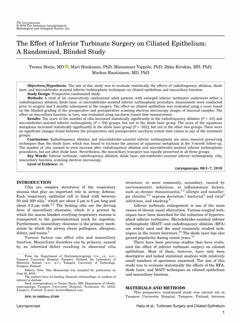
The Effect of Inferior Turbinate Surgery on Ciliated Epithelium:
A Randomized, Blinded Study
Teemu Harju, MD ; Mari Honkanen, PhD; Minnamari Vippola, PhD; Ilkka Kivekäs, MD, PhD;
Markus Rautiainen, MD, PhD
Objectives/Hypothesis: The aim of this study was to evaluate statistically the effects of radiofrequency ablation, diodelaser, and microdebrider-assisted inferior turbinoplasty techniques on ciliated epithelium and mucociliary function.
Study Design: Prospective randomized study.Methods: A total of 66 consecutively randomized adult patients with enlarged inferior turbinates underwent either a
radiofrequency ablation, diode laser, or microdebrider-assisted inferior turbinoplasty procedure. Assessments were conductedprior to surgery and 3 months subsequent to the surgery. The effect on ciliated epithelium was evaluated using a score basedon the blinded grading of the preoperative and postoperative scanning electron microscopy images of mucosal samples. Theeffect on mucociliary function, in turn, was evaluated using saccharin transit time measurement.
Results: The score of the number of cilia increased statistically significantly in the radiofrequency ablation (P = .03) andmicrodebrider-assisted inferior turbinoplasty (P = .04) groups, but not in the diode laser group. The score of the squamousmetaplasia increased statistically significantly in the diode laser group (P = .002), but not in the other two groups. There wereno significant changes found between the preoperative and postoperative saccharin transit time values in any of the treatmentgroups.
Conclusions: Radiofrequency ablation and microdebrider-assisted inferior turbinoplasty are more mucosal preservingtechniques than the diode laser, which was found to increase the amount of squamous metaplasia at the 3-month follow-up.The number of cilia seemed to even increase after radiofrequency ablation and microdebrider-assisted inferior turbinoplastyprocedures, but not after diode laser. Nevertheless, the mucociliary transport was equally preserved in all three groups.
Key Words: Inferior turbinate, radiofrequency ablation, diode laser, microdebrider-assisted inferior turbinoplasty, cilia,mucociliary function, scanning electron microscopy.
Level of Evidence: 1bLaryngoscope, 00:1–7, 2018
INTRODUCTIONCilia are complex structures of the respiratory
mucosa that play an important role in airway defense.
Each respiratory epithelial cell is lined with between
50 and 200 cilia,1 which are about 5 μm to 6 μm long and
about 0.2 μm wide.2,3 The beating cilia are the driving
force of mucociliary clearance, which is a process by
which the mucus blanket overlying respiratory mucosa is
transported to the gastrointestinal track for ingestion.
Furthermore, mucociliary clearance is the primary mech-
anism by which the airway clears pathogens, allergens,
debris, and toxins.1
Various factors can affect cilia and mucociliary
function. Mucociliary disorders can be primary, caused
by an inherited defect resulting in abnormal cilia
structure, or more commonly, secondary, caused by
environmental, infectious, or inflammatory factors,
such as chronic rhinosinusitis,1,4 allergic and nonaller-
gic rhinitis,5,6 septum deviation,7 bacterial1 and viral8
infections, and smoking.9
Inferior turbinate enlargement is one of the main
causes of chronic nasal obstruction. Various surgical tech-
niques have been described for the reduction of hypertro-
phied inferior turbinates. Microdebrider-assisted inferior
turbinoplasty (MAIT) and radiofrequency ablation (RFA)
are widely used and the most commonly studied tech-
niques in the recent literature.10 The diode laser has also
gained popularity during recent years.11
There have been previous studies that have evalu-
ated the effect of inferior turbinate surgery on ciliated
epithelium. Most of them, however, have only been
descriptive and lacked statistical analysis with relatively
small numbers of specimens examined. The aim of this
study was to evaluate statistically the effects of the RFA,
diode laser, and MAIT techniques on ciliated epithelium
and mucociliary function.
MATERIALS AND METHODSThis prospective randomized study was carried out at
Tampere University Hospital, Tampere, Finland, between
From the Department of Otorhinolaryngology (T.H., I.K., M.R.),Tampere University Hospital, Tampere, Finland; the Laboratory ofMaterials Science (M.H., M.V.), Tampere University of Technology,Tampere, Finland
Editor’s Note: This Manuscript was accepted for publication onJune 05, 2018.
The authors have no funding, financial relationships, or conflicts ofinterest to disclose.
Send correspondence to Teemu Harju, MD, Department of Otorhi-nolaryngology, Tampere University Hospital, Teiskontie 35, 33521Tampere, Finland. E-mail: [email protected]
DOI: 10.1002/lary.27409
Laryngoscope Harju et al.: Turbinate Surgery and Ciliated Epithelium
1
The Laryngoscope© 2018 The American Laryngological,Rhinological and Otological Society, Inc.

February 2014 and September 2017. The institutional review
board approved the study design (R13144), and all patients provided
written informed consent. A total of 66 consecutive adult patients
presenting with enlarged inferior turbinates due to persistent year-
round rhinitis were enrolled in this study. The patients presented
symptoms of bilateral nasal obstruction related to inferior turbinate
congestion that had not responded to a 3-month trial of appropriate
treatment with intranasal corticosteroids. Patients with significant
nasal septum deviation affecting the nasal valve region, internal/
external valve collapse/stenosis, chronic rhinosinusitis with or with-
out polyposis, previous nasal surgery, sinonasal tumor, severe sys-
temic disorder, severe obesity, or malignancy were excluded from
the study.
Cone beam computed tomography (Planmeca Max; Planmeca,
Helsinki, Finland) was used to exclude patients with chronic rhinosi-
nusitis from the study. Serum-specific immunoglobulin (Ig)E level
measurements were used to identify those patients with allergic sen-
sitization. Allergic sensitization was defined as a specific IgE > 0.35
for any common airborne allergen (cat, dog, horse, birch, grass, mug-
wort, Dermatophagoides pteronyssinus, and molds).
The definition of inferior turbinate enlargement was based
on persistent bilateral symptoms, a finding of bilateral swelling
of the inferior turbinate in nasal endoscopy, and the evident
shrinking of both turbinates in a decongestion test. The nasal
response to the topical vasoconstrictor 0.5% xylometazoline
hydrochloride (Nasolin; Orion, Espoo Finland) in both nasal cavi-
ties 15 minutes before obtaining the second measurement was
evaluated objectively using acoustic rhinometry (Acoustic rhin-
ometer A1; GM Instruments Ltd, Kilwinning, United Kingdom).
An improvement of less than 30% in anterior nasal cavity volume
(V2–5 cm) in one or both nasal cavities was considered normal,
and those patients were excluded from the study. The limit value
of 30% was chosen according to the previous literature.12–14
Patients were consecutively randomized into RFA, diode
laser, and MAIT groups. The surgical treatment was performed
under local anesthesia at the day surgery department of the hos-
pital’s ear, nose, and throat clinic. All the procedures were per-
formed under the direct vision of a straight, 4-mm-diameter, 0 �
endoscope (Karl Storz, Tuttlingen, Germany). First, the inferior
turbinates were topically anesthetized using cotton strips with a
mixture of lidocaine (Orion) 40 mg/mL and two to three drops of
epinephrine 0.1% in 5 mL to 10 mL of lidocaine. The local anes-
thetic (lidocain 10 mg/mL cum adrenalin) was then applied to
the medial portions of the inferior turbinates. Before the treat-
ment, preoperative samples for scanning electron microcopy
(SEM) were taken from each patient in the form of small
(2–3 mm in diameter) nasal mucosal biopsies from the anterior
medial portion of the left inferior turbinate. In all of the groups
with every technique, the treatment was given to the medial side
of the anterior half of the inferior turbinates.
The RFA treatment was carried out with a radiofrequency
generator (Sutter RF generator BM-780 II; Sutter, Freiburg, Ger-
many). A Binner bipolar needle electrode was inserted into the
medial submucosal tissue of the inferior turbinate. The upper
and lower parts of the anterior half of the inferior turbinate were
treated for 6 seconds at 10 W output power in three areas.
The diode laser treatment was given with a FOX Laser (A.R.C.
Laser GmbH, Nuremberg, Germany). The settings were as follows:
wavelength of 980 nm, output power of 6 W in continuous-wave
mode, and laser delivery by a 600 μm fiber using contact mode. Four
parallel stripes were made on the mucosa by drawing the fiber from
the posterior to the anterior direction along the medial edge of the
inferior turbinate.
In the MAIT treatment, a 2.9-mm-diameter rotatable
microdebrider tip (Medtronic Xomed, Jacksonville, FL) was
firmly pushed toward the turbinate bone until it pierced the
mucosa of the anterior face of the inferior turbinate. Next, a
submucosal pocket was dissected by tunneling the elevator tip in
an anterior-to-posterior and superior-to-inferior sweeping
motion. Once an adequate pocket had been created, resection of
the stromal tissue was carried out by moving the blade back and
forth in a sweeping motion, with the system set at 3,000 rpm
using suction irrigation.
All the patients were evaluated prior to surgery and 3 months
subsequent to the surgery. During both visits, nasal mucociliary
transport was evaluated with saccharin transit time (STT) by plac-
ing a saccharine particle on the anterior portion of the left inferior
turbinate, and the time until the patient tasted sweetness was
measured. Postoperative samples for SEM were taken under local
anesthesia from the anterior medial portion of the left inferior tur-
binate of every patient at the control visit. The SEM samples were
first immersed in 1% glutaraldehyde for fixation. Then, the sam-
ples were dehydrated in a graded alcohol series. Finally, the sam-
ples were immersed in hexamethyldisilazane for 15 minutes at
room temperature and air dried overnight. The samples were
glued on the SEM specimen stubs with carbon glue. To avoid sam-
ple charging during the SEM studies, the specimens were coated
for 3 minutes with gold using Edwards S150 sputter coater in an
argon atmosphere. Then, four to eight randomly selected fields
were viewed and imaged with a Philips XL30 SEM at primary
magnification of 2,000×.
After SEM, the quality of both the preoperative and postopera-
tive SEM image series of 44 patients out of 66 was of a technically
acceptable quality to be further evaluated. The main reasons for
sample rejection were fibrin or other impurity covering the surface,
glue covering the surface, broken sample, and sample wrong side
up. The preoperative and postoperative image series of each patient
were put into separate folders. Then, the folders were coded and
evaluated by five examiners (T.H., I.K., M.H., M.V., M.R.), who did not
know which technique had been used or whether the images were
preoperative or postoperative, and were therefore blinded. The eval-
uated parameters were the number or amount of cilia, nonciliated
pseudostratified columnar cells, squamous metaplasia, microvilli,
goblet cells, and disorientation of the cilia. The parameters were
graded as follows: 0 = no, 1 = a little, 2 moderately, 3 = a lot. The
mean of the grades given to an image series by the five examiners
was used as a score of one image series.
IBM SPSS statistics 22.0 (IBM, Armonk, NY) was used for
the statistical analyses. All nonparametric data were statistically
processed using the Wilcoxon signed rank test. In cases with
parametric data, the analysis was carried out by paired samples
t test and one-way analysis of variance. A χ2 test was used in the
evaluation of the total loss of cilia and the detected metaplasia.
Correlations were evaluated using Spearman’s ρ.
TABLE I.
Characteristics of the Patients Whose Samples Were Evaluated
All Patients,N = 44
RFA,N = 17
Diode Laser,N = 13
MAIT,N = 14
Age, yr,mean (range)
43 (19–69) 43 (24–68) 41 (25–65) 45 (19–69)
Sex, no. (%)
Male 27 (61) 11 (65) 7 (54) 9 (64)
Female 17 (39) 6 (35) 6 (46) 5 (36)
Allergicsensitization,no. (%)
Yes 16 (36) 6 (35) 3 (23) 7 (50)
No 28 (64) 11 (65) 10 (77) 7 (50)
MAIT = microdebrider-assisted inferior turbinoplasty; RFA = radio-frequency ablation.
Laryngoscope Harju et al.: Turbinate Surgery and Ciliated Epithelium
2
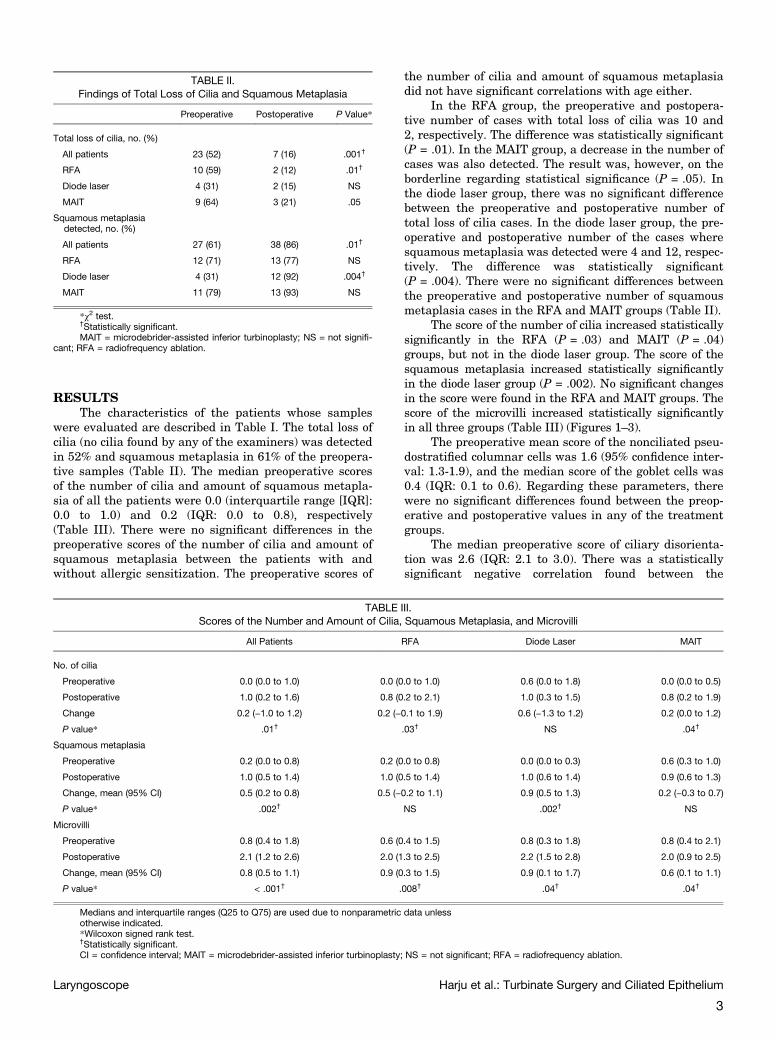
RESULTSThe characteristics of the patients whose samples
were evaluated are described in Table I. The total loss of
cilia (no cilia found by any of the examiners) was detected
in 52% and squamous metaplasia in 61% of the preopera-
tive samples (Table II). The median preoperative scores
of the number of cilia and amount of squamous metapla-
sia of all the patients were 0.0 (interquartile range [IQR]:
0.0 to 1.0) and 0.2 (IQR: 0.0 to 0.8), respectively
(Table III). There were no significant differences in the
preoperative scores of the number of cilia and amount of
squamous metaplasia between the patients with and
without allergic sensitization. The preoperative scores of
the number of cilia and amount of squamous metaplasia
did not have significant correlations with age either.
In the RFA group, the preoperative and postopera-
tive number of cases with total loss of cilia was 10 and
2, respectively. The difference was statistically significant
(P = .01). In the MAIT group, a decrease in the number of
cases was also detected. The result was, however, on the
borderline regarding statistical significance (P = .05). In
the diode laser group, there was no significant difference
between the preoperative and postoperative number of
total loss of cilia cases. In the diode laser group, the pre-
operative and postoperative number of the cases where
squamous metaplasia was detected were 4 and 12, respec-
tively. The difference was statistically significant
(P = .004). There were no significant differences between
the preoperative and postoperative number of squamous
metaplasia cases in the RFA and MAIT groups (Table II).
The score of the number of cilia increased statistically
significantly in the RFA (P = .03) and MAIT (P = .04)
groups, but not in the diode laser group. The score of the
squamous metaplasia increased statistically significantly
in the diode laser group (P = .002). No significant changes
in the score were found in the RFA and MAIT groups. The
score of the microvilli increased statistically significantly
in all three groups (Table III) (Figures 1–3).
The preoperative mean score of the nonciliated pseu-
dostratified columnar cells was 1.6 (95% confidence inter-
val: 1.3-1.9), and the median score of the goblet cells was
0.4 (IQR: 0.1 to 0.6). Regarding these parameters, there
were no significant differences found between the preop-
erative and postoperative values in any of the treatment
groups.
The median preoperative score of ciliary disorienta-
tion was 2.6 (IQR: 2.1 to 3.0). There was a statistically
significant negative correlation found between the
TABLE III.
Scores of the Number and Amount of Cilia, Squamous Metaplasia, and Microvilli
All Patients RFA Diode Laser MAIT
No. of cilia
Preoperative 0.0 (0.0 to 1.0) 0.0 (0.0 to 1.0) 0.6 (0.0 to 1.8) 0.0 (0.0 to 0.5)
Postoperative 1.0 (0.2 to 1.6) 0.8 (0.2 to 2.1) 1.0 (0.3 to 1.5) 0.8 (0.2 to 1.9)
Change 0.2 (−1.0 to 1.2) 0.2 (−0.1 to 1.9) 0.6 (−1.3 to 1.2) 0.2 (0.0 to 1.2)
P value* .01† .03† NS .04†
Squamous metaplasia
Preoperative 0.2 (0.0 to 0.8) 0.2 (0.0 to 0.8) 0.0 (0.0 to 0.3) 0.6 (0.3 to 1.0)
Postoperative 1.0 (0.5 to 1.4) 1.0 (0.5 to 1.4) 1.0 (0.6 to 1.4) 0.9 (0.6 to 1.3)
Change, mean (95% CI) 0.5 (0.2 to 0.8) 0.5 (−0.2 to 1.1) 0.9 (0.5 to 1.3) 0.2 (−0.3 to 0.7)
P value* .002† NS .002† NS
Microvilli
Preoperative 0.8 (0.4 to 1.8) 0.6 (0.4 to 1.5) 0.8 (0.3 to 1.8) 0.8 (0.4 to 2.1)
Postoperative 2.1 (1.2 to 2.6) 2.0 (1.3 to 2.5) 2.2 (1.5 to 2.8) 2.0 (0.9 to 2.5)
Change, mean (95% CI) 0.8 (0.5 to 1.1) 0.9 (0.3 to 1.5) 0.9 (0.1 to 1.7) 0.6 (0.1 to 1.1)
P value* < .001† .008† .04† .04†
Medians and interquartile ranges (Q25 to Q75) are used due to nonparametric data unlessotherwise indicated.*Wilcoxon signed rank test.†Statistically significant.CI = confidence interval; MAIT = microdebrider-assisted inferior turbinoplasty; NS = not significant; RFA = radiofrequency ablation.
TABLE II.
Findings of Total Loss of Cilia and Squamous Metaplasia
Preoperative Postoperative P Value*
Total loss of cilia, no. (%)
All patients 23 (52) 7 (16) .001†
RFA 10 (59) 2 (12) .01†
Diode laser 4 (31) 2 (15) NS
MAIT 9 (64) 3 (21) .05
Squamous metaplasiadetected, no. (%)
All patients 27 (61) 38 (86) .01†
RFA 12 (71) 13 (77) NS
Diode laser 4 (31) 12 (92) .004†
MAIT 11 (79) 13 (93) NS
*χ2 test.
†Statistically significant.MAIT = microdebrider-assisted inferior turbinoplasty; NS = not signifi-
cant; RFA = radiofrequency ablation.
Laryngoscope Harju et al.: Turbinate Surgery and Ciliated Epithelium
3
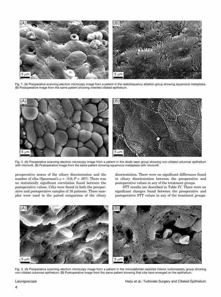
preoperative scores of the ciliary disorientation and the
number of cilia (Spearman’s ρ = −0.6; P = .007). There was
no statistically significant correlation found between the
postoperative values. Cilia were found in both the preoper-
ative and postoperative samples of 16 patients. These sam-
ples were used in the paired comparison of the ciliary
disorientation. There were no significant differences found
in ciliary disorientation between the preoperative and
postoperative values in any of the treatment groups.
STT results are described in Table IV. There were no
significant changes found between the preoperative and
postoperative STT values in any of the treatment groups.
Fig. 1. (A) Preoperative scanning electron microcopy image from a patient in the radiofrequency ablation group showing squamous metaplasia.(B) Postoperative image from the same patient showing oriented ciliated epithelium.
Fig. 2. (A) Preoperative scanning electron microcopy image from a patient in the diode laser group showing non-ciliated columnar epitheliumwith microvilli. (B) Postoperative image from the same patient showing squamous metaplasia with microvilli.
Fig. 3. (A) Preoperative scanning electron microcopy image from a patient in the microdebrider-assisted inferior turbinoplasty group showingnon-ciliated columnar epithelium. (B) Postoperative image from the same patient showing that cilia have emerged on the epithelium.
Laryngoscope Harju et al.: Turbinate Surgery and Ciliated Epithelium
4

There were no significant differences in the change in STT
values between the treatment groups either.
DISCUSSIONIn the present study, the five examiners who evaluated
the SEM images did not know which technique had been
used or whether the images were preoperative or postopera-
tive. Blinding of the evaluators was extremely important to
prevent possible expectations and attitudes of the examiners
toward the treatments from affecting their evaluations.
In previous light and electron microcopy studies of
inferior turbinate surgery, the preoperative findings
regarding cilia have been controversial. There have been
studies that have reported preoperative findings of epi-
thelial metaplasia and loss of ciliated cells.6,15,16 On the
other hand, there are studies that have shown a normal
appearance of the epithelium with a preserved number of
cilia.17,18 In the present study, squamous metaplasia and
total loss of ciliated cells were found in the majority of
the preoperative samples. This may partly be explained
by the chronic rhinitis associated with inferior turbinate
enlargement. Furthermore, previous studies have shown
that a loss of cilia is more common in both allergic and
nonallergic rhinitis compared with healthy controls.5,6
In the present study, however, the most likely expla-
nation for the high incidence of squamous metaplasia and
total loss of ciliated cells in the preoperative samples is
that the biopsies were taken from the anterior portion of
the inferior turbinate, approximately 1 cm behind the
anterior edge. Halama et al. examined 214 biopsies taken
from various parts of the nose and paranasal sinuses
postmortem. They found that the density of ciliated cells
was increased in the nasal cavity in the anteroposterior
direction. Cilia start occurring just behind the front edge
of the inferior turbinate. More posteriorly, the density of
ciliated cells ranges from 50% in the one-third middle to
100% in the posterior part of the inferior turbinate. The
distribution pattern of ciliated cells corresponds well with
a map of nasal airflow, indicating that the density of cili-
ated cells is inversely proportional to the linear velocity
of inspiratory air in the nasal cavity. Furthermore, it is
also assumed that other physical properties of the nasal
airflow, such as low temperature, low humidity, and con-
tamination contribute to the reduced number of ciliated
cells.19 In the study by Augusto et al., ciliated epithelium
with goblet cells (normal respiratory epithelium) was
found in only half of the samples taken from the anterior
portions of the inferior turbinates of normal noses, and
various degrees of epithelial metaplasia was found in half
of the samples taken from the same sites. The anterior
part of the inferior turbinate is more exposed to external
elements, such as strong air flow, and is therefore prone
to metaplastic transformation.20
There have been some previous light and electron
microscopy studies that have evaluated the effect of infe-
rior turbinate surgery on ciliated epithelium. Based on
the findings of previous studies, RFA6,17,18,21 and submu-
cosal electrocautery6,22,23 do not have a significant effect
on ciliated epithelium. After endoscopic turbinoplasty,
some improvement has been described.24 The findings
have, however, been controversial regarding lasers.15,24,25
Ultrasound reduction, in turn, may also have an improving
effect on ciliated epithelium.6 A worsening has previously
been described regarding partial inferior turbinectomy.18
In a recent study by Neri et al. with 18 patients, however,
a complete reepithelization of nasal mucosa with well dif-
ferentiated columnar ciliated epithelium was detected
4 months after the total removal of the mucosa of the
medial and inferior portions of the inferior turbinate with
the microdebrider.16
In our study, the effect of various inferior turbinate
surgery techniques on ciliated epithelium has for the first
time been evaluated with a statistical analysis using a
score based on the mean of the grades given to the image
series by the blinded examiners. Based on our score, the
number of cilia increased significantly after RFA and
MAIT treatments. The number of cases with total loss of
cilia was also found to have decreased after these treat-
ments. No similar changes were found after diode laser
treatment. On the other hand, the score of metaplasia
seemed to increase significantly after diode laser treat-
ment as well as the overall number of cases where meta-
plasia was detected. No similar findings were found after
RFA or MAIT. Regarding the diode laser, the findings can
be explained by the damage the laser causes to the muco-
sal surface during the treatment of submucosa. RFA and
MAIT are more clearly submucosal techniques that do
not notably harm the mucosal surface. Inferior turbinate
surgery creates more airspace at the level of the anterior
part of the inferior turbinate, which decreases nasal air-
way resistance, causes changes in the airflow patterns,
and likely decreases the shear stress on the mucosa of the
anterior part of the inferior turbinate. This could be a
possible explanation for the increase in the number of
cilia after RFA and MAIT treatments.
In human nasal epithelial cells, the number of micro-
villi has been found to increase before the ciliogenesis
starts.26 In chronic rhinosinusitis, an increased number
of microvilli and short cilia are thought to indicate the
regeneration of epithelium and ciliated cells.27 In the pre-
sent study, the number of microvilli increased signifi-
cantly in all three groups, which can be interpreted to be
related to a regeneration of the mucosal epithelium.
The degree of ciliary disorientation was relatively
high both preoperatively and postoperatively in all three
groups, and there were no significant differences in dis-
orientation between the preoperative and postoperative
TABLE IV.
Saccharin Transit Time Results (n = 66)
Preoperative,sec (95% CI)
Postoperative,sec (95% CI)
Change,sec (95% CI)
RFA, n = 23 588 (471 to 704) 602 (482 to 721) 14 (−148 to 176) NS
Diode laser,n = 20
609 (487 to 732) 547 (464 to 630) −62 (−205 to 81) NS
MAIT, n = 23 608 (485 to 731) 583 (478 to 689) −24 (−162 to 113) NS
CI = confidence interval; MAIT = microdebrider-assisted inferior turbi-noplasty; NS = not significant (paired samples t test); RFA = radiofrequencyablation.
Laryngoscope Harju et al.: Turbinate Surgery and Ciliated Epithelium
5
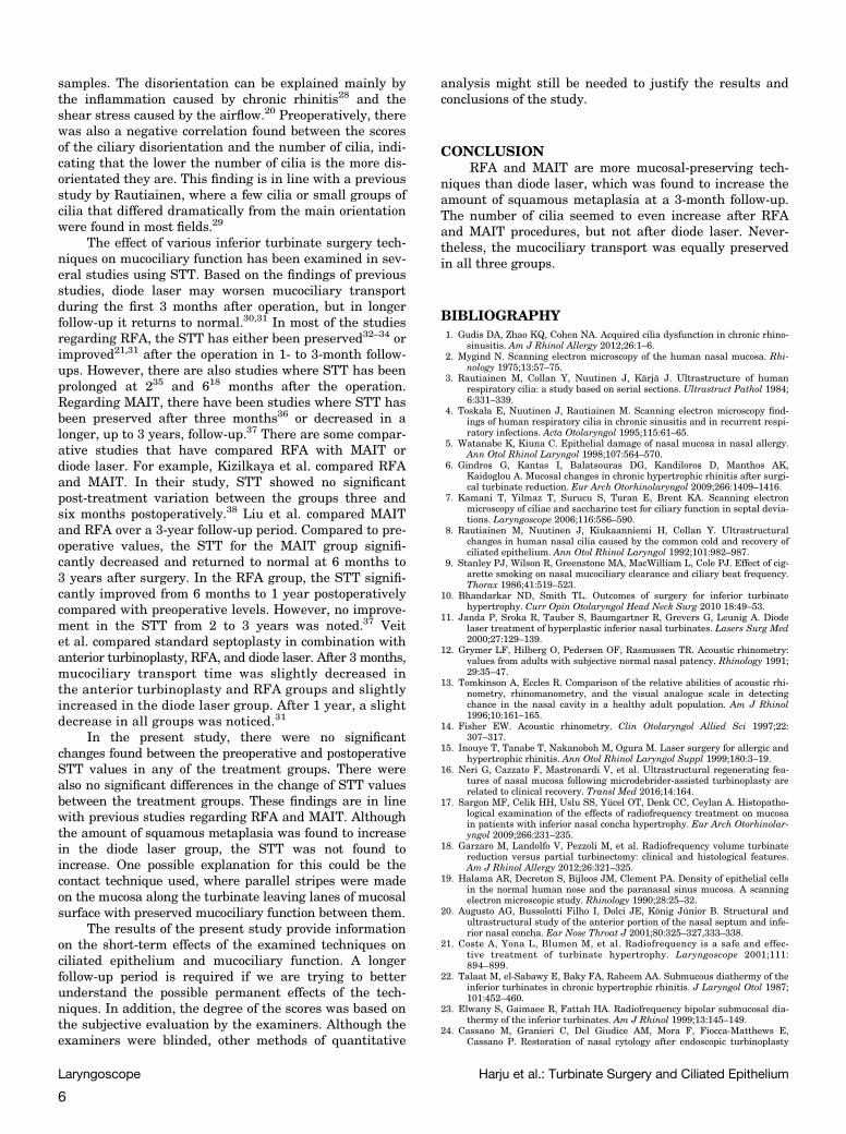
samples. The disorientation can be explained mainly by
the inflammation caused by chronic rhinitis28 and the
shear stress caused by the airflow.20 Preoperatively, there
was also a negative correlation found between the scores
of the ciliary disorientation and the number of cilia, indi-
cating that the lower the number of cilia is the more dis-
orientated they are. This finding is in line with a previous
study by Rautiainen, where a few cilia or small groups of
cilia that differed dramatically from the main orientation
were found in most fields.29
The effect of various inferior turbinate surgery tech-
niques on mucociliary function has been examined in sev-
eral studies using STT. Based on the findings of previous
studies, diode laser may worsen mucociliary transport
during the first 3 months after operation, but in longer
follow-up it returns to normal.30,31 In most of the studies
regarding RFA, the STT has either been preserved32–34 or
improved21,31 after the operation in 1- to 3-month follow-
ups. However, there are also studies where STT has been
prolonged at 235 and 618 months after the operation.
Regarding MAIT, there have been studies where STT has
been preserved after three months36 or decreased in a
longer, up to 3 years, follow-up.37 There are some compar-
ative studies that have compared RFA with MAIT or
diode laser. For example, Kizilkaya et al. compared RFA
and MAIT. In their study, STT showed no significant
post-treatment variation between the groups three and
six months postoperatively.38 Liu et al. compared MAIT
and RFA over a 3-year follow-up period. Compared to pre-
operative values, the STT for the MAIT group signifi-
cantly decreased and returned to normal at 6 months to
3 years after surgery. In the RFA group, the STT signifi-
cantly improved from 6 months to 1 year postoperatively
compared with preoperative levels. However, no improve-
ment in the STT from 2 to 3 years was noted.37 Veit
et al. compared standard septoplasty in combination with
anterior turbinoplasty, RFA, and diode laser. After 3 months,
mucociliary transport time was slightly decreased in
the anterior turbinoplasty and RFA groups and slightly
increased in the diode laser group. After 1 year, a slight
decrease in all groups was noticed.31
In the present study, there were no significant
changes found between the preoperative and postoperative
STT values in any of the treatment groups. There were
also no significant differences in the change of STT values
between the treatment groups. These findings are in line
with previous studies regarding RFA and MAIT. Although
the amount of squamous metaplasia was found to increase
in the diode laser group, the STT was not found to
increase. One possible explanation for this could be the
contact technique used, where parallel stripes were made
on the mucosa along the turbinate leaving lanes of mucosal
surface with preserved mucociliary function between them.
The results of the present study provide information
on the short-term effects of the examined techniques on
ciliated epithelium and mucociliary function. A longer
follow-up period is required if we are trying to better
understand the possible permanent effects of the tech-
niques. In addition, the degree of the scores was based on
the subjective evaluation by the examiners. Although the
examiners were blinded, other methods of quantitative
analysis might still be needed to justify the results and
conclusions of the study.
CONCLUSIONRFA and MAIT are more mucosal-preserving tech-
niques than diode laser, which was found to increase the
amount of squamous metaplasia at a 3-month follow-up.
The number of cilia seemed to even increase after RFA
and MAIT procedures, but not after diode laser. Never-
theless, the mucociliary transport was equally preserved
in all three groups.
BIBLIOGRAPHY
1. Gudis DA, Zhao KQ, Cohen NA. Acquired cilia dysfunction in chronic rhino-sinusitis. Am J Rhinol Allergy 2012;26:1–6.
2. Mygind N. Scanning electron microscopy of the human nasal mucosa. Rhi-nology 1975;13:57–75.
3. Rautiainen M, Collan Y, Nuutinen J, Kärjä J. Ultrastructure of humanrespiratory cilia: a study based on serial sections. Ultrastruct Pathol 1984;6:331–339.
4. Toskala E, Nuutinen J, Rautiainen M. Scanning electron microscopy find-ings of human respiratory cilia in chronic sinusitis and in recurrent respi-ratory infections. Acta Otolaryngol 1995;115:61–65.
5. Watanabe K, Kiuna C. Epithelial damage of nasal mucosa in nasal allergy.Ann Otol Rhinol Laryngol 1998;107:564–570.
6. Gindros G, Kantas I, Balatsouras DG, Kandiloros D, Manthos AK,Kaidoglou A. Mucosal changes in chronic hypertrophic rhinitis after surgi-cal turbinate reduction. Eur Arch Otorhinolaryngol 2009;266:1409–1416.
7. Kamani T, Yilmaz T, Surucu S, Turan E, Brent KA. Scanning electronmicroscopy of ciliae and saccharine test for ciliary function in septal devia-tions. Laryngoscope 2006;116:586–590.
8. Rautiainen M, Nuutinen J, Kiukaanniemi H, Collan Y. Ultrastructuralchanges in human nasal cilia caused by the common cold and recovery ofciliated epithelium. Ann Otol Rhinol Laryngol 1992;101:982–987.
9. Stanley PJ, Wilson R, Greenstone MA, MacWilliam L, Cole PJ. Effect of cig-arette smoking on nasal mucociliary clearance and ciliary beat frequency.Thorax 1986;41:519–523.
10. Bhandarkar ND, Smith TL. Outcomes of surgery for inferior turbinatehypertrophy. Curr Opin Otolaryngol Head Neck Surg 2010 18:49–53.
11. Janda P, Sroka R, Tauber S, Baumgartner R, Grevers G, Leunig A. Diodelaser treatment of hyperplastic inferior nasal turbinates. Lasers Surg Med2000;27:129–139.
12. Grymer LF, Hilberg O, Pedersen OF, Rasmussen TR. Acoustic rhinometry:values from adults with subjective normal nasal patency. Rhinology 1991;29:35–47.
13. Tomkinson A, Eccles R. Comparison of the relative abilities of acoustic rhi-nometry, rhinomanometry, and the visual analogue scale in detectingchance in the nasal cavity in a healthy adult population. Am J Rhinol1996;10:161–165.
14. Fisher EW. Acoustic rhinometry. Clin Otolaryngol Allied Sci 1997;22:307–317.
15. Inouye T, Tanabe T, Nakanoboh M, Ogura M. Laser surgery for allergic andhypertrophic rhinitis. Ann Otol Rhinol Laryngol Suppl 1999;180:3–19.
16. Neri G, Cazzato F, Mastronardi V, et al. Ultrastructural regenerating fea-tures of nasal mucosa following microdebrider-assisted turbinoplasty arerelated to clinical recovery. Transl Med 2016;14:164.
17. Sargon MF, Celik HH, Uslu SS, Yücel OT, Denk CC, Ceylan A. Histopatho-logical examination of the effects of radiofrequency treatment on mucosain patients with inferior nasal concha hypertrophy. Eur Arch Otorhinolar-yngol 2009;266:231–235.
18. Garzaro M, Landolfo V, Pezzoli M, et al. Radiofrequency volume turbinatereduction versus partial turbinectomy: clinical and histological features.Am J Rhinol Allergy 2012;26:321–325.
19. Halama AR, Decreton S, Bijloos JM, Clement PA. Density of epithelial cellsin the normal human nose and the paranasal sinus mucosa. A scanningelectron microscopic study. Rhinology 1990;28:25–32.
20. Augusto AG, Bussolotti Filho I, Dolci JE, König Júnior B. Structural andultrastructural study of the anterior portion of the nasal septum and infe-rior nasal concha. Ear Nose Throat J 2001;80:325–327,333–338.
21. Coste A, Yona L, Blumen M, et al. Radiofrequency is a safe and effec-tive treatment of turbinate hypertrophy. Laryngoscope 2001;111:894–899.
22. Talaat M, el-Sabawy E, Baky FA, Raheem AA. Submucous diathermy of theinferior turbinates in chronic hypertrophic rhinitis. J Laryngol Otol 1987;101:452–460.
23. Elwany S, Gaimaee R, Fattah HA. Radiofrequency bipolar submucosal dia-thermy of the inferior turbinates. Am J Rhinol 1999;13:145–149.
24. Cassano M, Granieri C, Del Giudice AM, Mora F, Fiocca-Matthews E,Cassano P. Restoration of nasal cytology after endoscopic turbinoplasty
Laryngoscope Harju et al.: Turbinate Surgery and Ciliated Epithelium
6

versus laser-assisted turbinoplasty. Am J Rhinol Allergy 2010;24:310–314.
25. Bergler WF, Sadick H, Hammerschmitt N, Oulmi J, Hörmann K. Long-termresults of inferior turbinate reduction with argon plasma coagulation.Laryngoscope 2001;111:1593–1598.
26. Jorissen M, Willems T. The secondary nature of ciliary (dis)orientation in sec-ondary and primary ciliary dyskinesia. Acta Otolaryngol 2004;124:527–531.
27. Toskala E, Rautiainen M. Electron microscopy assessment of the recoveryof sinus mucosa after sinus surgery. Acta Otolaryngol 2003;123:954–959.
28. Rayner CF, Rutman A, Dewar A, Cole PJ, Wilson R. Ciliary disorientationin patients with chronic upper respiratory tract inflammation. Am JRespir Crit Care Med 1995;151:800–804.
29. Rautiainen M. Orientation of human respiratory cilia. Eur Respir J 1988;1:257–261.
30. Parida PK, Surianarayanan G, Alexander A, Saxena SK, Santhosh K. Diodelaser turbinate reduction in the treatment of symptomatic inferior turbinatehypertrophy. Indian J Otolaryngol Head Neck Surg 2013;65:350–355.
31. Veit JA, Nordmann M, Dietz B, et al. Three different turbinoplasty tech-niques combined with septoplasty: prospective randomized trial. Laryngo-scope 2017;127:303–308.
32. Sapçi T, Sahin B, Karavus A, Akbulut UG. Comparison of the effects ofradiofrequency tissue ablation, CO2 laser ablation, and partial
turbinectomy applications on nasal mucociliary functions. Laryngoscope2003;113:514–519.
33. Cavaliere M, Mottola G, Iemma M. Comparison of the effectiveness andsafety of radiofrequency turbinoplasty and traditional surgical techniquein treatment of inferior turbinate hypertrophy. Otolaryngol Head NeckSurg 2005;133:972–978.
34. Rosato C, Pagliuca G, Martellucci S, et al. Effect of radiofrequency thermalablation treatment on nasal ciliary motility: a study with phase-contrastmicroscopy. Otolaryngol Head Neck Surg 2016;154:754–758.
35. Salzano FA, Mora R, Dellepiane M, et al. Radiofrequency, high-frequency,and electrocautery treatments vs partial inferior turbinotomy: microscopicand macroscopic effects on nasal mucosa. Arch Otolaryngol Head NeckSurg 2009;135:752–758.
36. Romano A, Orabona GD, Salzano G, Abbate V, Iaconetta G, Califano L. Compar-ative study between partial inferior turbinotomy and microdebrider-assistedinferior turbinoplasty. J Craniofac Surg 2015;26:e235–e238.
37. Liu CM, Tan CD, Lee FP, Lin KN, Huang HM. Microdebrider-assisted ver-sus radiofrequency-assisted inferior turbinoplasty. Laryngoscope 2009;119:414–418.
38. Kizilkaya Z, Ceylan K, Emir H, et al. Comparison of radiofrequency tissuevolume reduction and submucosal resection with microdebrider in inferiorturbinate hypertrophy. Otolaryngol Head Neck Surg 2008;138:176–181.
Laryngoscope Harju et al.: Turbinate Surgery and Ciliated Epithelium
7