ELP4 in rolandic epilepsy and BRD2 in juvenile myoclonic epilepsy
Upload
independentCategory
view
3download
0
Electroencephalography (EEG) remains the single mostimportant physiologic test of the cerebral cortex1,2. In temporallobe epilepsy (TLE), focal interictal and ictal epileptiformdischarges help in identifying the involved cortical areas3.Several nonepileptiform EEG abnormalities can also be seen inTLE. Scalp and sometimes invasive EEG recordings are neededfor accurate localization of the seizure onset. The developmentof video-EEG monitoring has allowed careful correlation ofclinical semiology with simultaneous EEG recordings4. Thispaper will review the EEG characteristics of TLE.
EEG RECORDINGI. Scalp EEG recordingThe 10-20 electrode system is an internationally accepted
standard method of measurement and application of EEG scalpelectrodes5. The standard electrodes can detect only up to 58% of
ABSTRACT: Electroencephalography (EEG) is an important tool for diagnosing, lateralizing andlocalizing temporal lobe seizures. In this paper, we review the EEG characteristics of temporal lobeepilepsy (TLE). Several “non-standard” electrodes may be needed to further evaluate the EEGlocalization. Ictal EEG recording is a major component of preoperative protocols for surgicalconsideration. Various ictal rhythms have been described including background attenuation, start-stop-start phenomenon, irregular 2-5 Hz lateralized activity, and 5-10 Hz sinusoidal waves or repetitiveepileptiform discharges. The postictal EEG can also provide valuable lateralizing information. Postictaldelta can be lateralized in 60% of patients with TLE and is concordant with the side of seizure onset inmost patients. When patients are being considered for resective surgery, invasive EEG recordings maybe needed. Accurate localization of the seizure onset in these patients is required for successful surgicalmanagement.
RÉSUMÉ: Caractéristiques électroencéphalographiques de l'épilepsie temporale. L'électroencéphalographie(EEG) est un outil important pour le diagnostic, la latéralisation et la localisation de l'épilepsie temporale (ET). Danscet article, nous revoyons les caractéristiques électroencéphalographiques de l'épilepsie temporale. Plusieursélectrodes « non-standard » peuvent être nécessaires pour une évaluation plus poussée de la localisation EEG.L'enregistrement EEG ictal est une composante importante des protocoles préopératoires pour des motifschirurgicaux. Différents rythmes ictaux ont été décrits, dont l'atténuation du rythme de fond, le phénomène départ-arrêt-départ, une activité latéralisée irrégulière à 2-5 Hz et des ondes sinusoïdales à 5-10 Hz ou des déchargesépileptiformes répétitives. L'EEG postictal peut également fournir de l'information précieuse pour la latéralisation.L'activité delta postictale peut être latéralisée chez 60% des patients atteints d'ET et concorde avec le côté du débutde la crise chez la plupart des patients. Quand les patients sont évalués en vue d'une résection chirurgicale, desenregistrements EEG effractifs peuvent s'avérer nécessaires. Le succès de la chirurgie repose sur la localisationexacte du début des crises chez ces patients.
Can. J. Neurol. Sci. 2010; 37: 439-448
THE CANADIAN JOURNAL OF NEUROLOGICAL SCIENCES 439
Electroencephalographic Features ofTemporal Lobe EpilepsyMohammed M. Jan, Mark Sadler, Susan R. Rahey
From the Department of Pediatrics (MMJ), Faculty of Medicine, King AbdulazizUniversity, Jeddah, Saudi Arabia; Division of Neurology and Dalhousie UniversityMedical School (MS); Neuroelectrodiagnostic Unit (SRR), Capital District HealthAuthority, Halifax, Nova Scotia, Canada.
RECEIVED FEBRUARY 1, 2010. FINAL REVISIONS SUBMITTED APRIL 12, 2010.Correspondence to: Mohammed M.S. Jan, Department of Pediatrics, Faculty ofMedicine, King AbdulAziz University, Box 80215, Jeddah 21589, Kingdom of SaudiArabia.
REVIEW ARTICLE
all spikes6. Therefore, additional measured and more closelyspaced scalp electrodes placed midway between the standardelectrodes of the 10-20 system can provide further localizationof epileptiform discharges in TLE7. Several “non-standard”electrodes can be used to further evaluate the EEG abnormalitiesincluding sphenoidal, nasopharyngeal, anterior temporal

(T1/T2), and surface electrodes applied over the mandibularnotch (MN) or zygoma7-9.Sphenoidal electrodes are inserted bilaterally through the skin
below the zygomatic arch, 2-3 cm anterior to the tragus anddirected posterosuperiorly in the direction of the foramenovale10. This procedure should be performed with localanesthesia by a qualified physician. Flexible wire electrodes,insulated and bared at the tip, are placed about 3 cm deep byattaching them to the external shaft of a 22 gage special (lumbarpuncture like) needle. The needle is then removed leaving therecording electrode in place; the external wire is coiled to relievetension and taped to the skin.Nasopharyngeal electrodes are uncomfortable and have not
consistently demonstrated superior sensitivity for spike detectionwhen compared to the other non-standard electrodes11,12.Therefore, nasopharyngeal electrodes are no longer inwidespread use. Sphenoidal, T1/T2, and MN electrodes canrecord spikes not detected by standard electrodes (Figure 1).Sphenoidal spikes are almost always detected by T1/T2 or MNelectrodes, although with slightly lower amplitudes13. Recordingwith multiple non-standard electrodes simultaneously to obtain amore comprehensive evaluation of the electrical field has beenrecommended7.Few studies have evaluated the ictal (as opposed to interictal)
merits of these electrodes or have addressed the issue of montagedesign for interictal and ictal TLE abnormalities14. The ability toperform off-line montage reformatting with digital EEGtechnology diminishes the importance of initial montage
selection. Ives et al studied ictal EEGs in patients withsphenoidal electrodes by using a coronal sphenoidal montage ascompared to an anterior-posterior temporal montage14. Thesphenoidal montage was superior in the detection of ictal EEGchanges.
II. Invasive EEG recordingInvasive intracranial techniques using subdural or
intracerebral depth electrodes can be used to improve diagnosticaccuracy if surface EEG recording yields inconclusive orambiguous results15. Recent shifts in surgical candidacy towardyounger age groups resulted in increased utility of invasivemonitoring especially in patients with atypical clinicalsemiology or normal imaging studies16. Subdural electrodes areinserted surgically to record over the cerebral cortex. Electrodegrids are square or rectangular in shape with small platinum orstainless steel disks embedded into soft silastic with severalcontact points. Different sizes and shapes can be selecteddependant on the anatomic area to be covered and require acraniotomy for insertion. Electrode strips, of various sizes,consist of a row of contacts and are usually inserted through aburr hole.Intracerebral or depth electrodes are used to record from
within the temporal lobe using rigid or flexible probes. Multiple-lead intracerebral electrodes can be implanted stereotactically(stereoelectroencephalography) to allow sampling from multiplemedial (amygdala, hippocampus, entorhinal cortex, insularcortex) and lateral (temporal pole, gyri, parietotemporal and
THE CANADIAN JOURNAL OF NEUROLOGICAL SCIENCES
440
Figure 1: Anterior temporal spike with phase reversal at F7 and shown in three montages,(A) antero-posterior, (B) common average reference (CAR), and (C) coronal withmandibular notch (MN) electrodes. Notice that the spike is better illustrated when MNelectrode was used. (A = low frequency filter [LFF] 0.3 sec, high frequency filter [HFF]70 Hz, Sensitivity 7.5 µV/mm / B&C = LFF 0.3 sec, HFF 70 Hz, Sensitivity 10 µV/mm).

occipitotemporal junctions) temporal lobe structures17. It isimportant to note that scalp electrodes detect activity generatedfrom a large cortical surface whereas subdural or depthelectrodes will detect potential changes which occur over only afew millimeters of cortex. Interictal spikes not otherwise seen onscalp EEG can be easily identified with invasive electrodes(Figure 2). Invasive electrodes are more sensitive in detectingspikes and seizures from a localized area but may missepileptiform activity which occurs in adjacent regions only ashort distance away.Indications for invasive recording include lack of seizure
lateralization and/or localization, seizure onset discrepant withother data (clinical, EEG, and/or imaging), and relation ofseizure localization to eloquent cortex. Advantages of invasivemonitoring in TLE include improved spatial resolution andincreased sensitivity (early signal), no attenuation from scalp and
skull, reduced ictal muscle and movement artifact, and utility fordirect electrical stimulation of cortex15. Disadvantages includelimited cortical sampling or risk for sampling error (tunnelvision), and risk of significant complications (2-3%)15. Thesecomplications include aseptic meningitis (subdural more thandepth electrodes), cerebrospinal fluid leak, cerebral edema, braincontusion, scalp infection, and bleeding (depth more thansubdural electrodes)15.Foramen ovale electrodes are occasionally used to record
from mesial temporal structures without requiring penetration ofthe skull18. A multi-contact flexible electrode is placed in theambient cistern with the aid of a needle inserted through theforamen ovale. These electrodes are not as close to hippocampalstructures as intracerebral electrodes and do not allow as large arecording field as grids and strips but detect mesial temporalEEG discharges better than sphenoidal and scalp electrodes18.
LE JOURNAL CANADIEN DES SCIENCES NEUROLOGIQUES
Volume 37, No. 4 – July 2010 441
Figure 2: Right anterior temporal spikes on subdural electrodes (top channels) using referential montage. Simultaneousscalp recording (bottom 2 channels) did not reveal the interictal spikes. (low frequency filter [LFF] 0.1 sec, highfrequency filter [HFF] 70 Hz, Sensitivity 100 µV/mm for subdural channels / LFF 0.3 sec, HFF 70 Hz, Sensitivity 20µV/mm for scalp channels).
Figure 3: Benign epileptiform transients of sleep (BETS) maximally expressed on the right temporal region on antero-posterior (AP) bipolarmontage (A). Their low voltage and wide field is best seen on common average reference (CAR) (B). Notice the central spindles of sleep seen inboth figures. (A&B = LFF 0.3 sec, HFF 70 Hz, Sensitivity 3 µV/mm).

When extracranial recordings are equivocal, foramen ovaleelectrodes offer a less invasive alternative to a more completeintracranial evaluation or can be used in association with gridsand strips.
ARTEFACTSThe differentiation of artefacts from EEG abnormalities can
be problematic on video-EEG recordings. Unusual artefacts notseen in standard laboratory EEG recording include mechanicalartefacts from altered electrode/scalp contact, intermittent leadwire disconnection, body movement, or movement of the
electrode cable3,16. Artefacts produced by rubbing or scratchingof the scalp and other rhythmic movements of head or extremitycan result in patterns that must be differentiated from ictaldischarges. Some artefacts are characteristic of the oro-alimentary automatisms of TLE (e.g. rhythmic chewing andswallowing). Rhythmic slow waves can also be due to chippedelectrode coating, instability of the electrode scalp interface, andelectrode wire movement. A conservative interpretation ofunusual or equivocal EEG events is mandatory, particularlywhen the patient’s activity cannot be verified by video review.Interictal sharp waves noted only during active wakefulnessshould be interpreted with caution. Ictal rhythms should have arecognizable field with typical ictal progression (evolution ofrate and voltage), spread to other regions, or postictal slowing3,16.Intracranial recordings tend to have fewer physiologic artefacts,such as chewing and eye blinks.
NORMAL EEG VARIANTSThe differentiation of some normal or benign EEG transients
from EEG abnormalities poses a problem in EEG interpretationand its clinical correlation19. Normal EEG variants refer towaveforms that mimic epileptiform abnormalities. In TLE, threeimportant normal variants needs to be recognized to avoidconfusion; small sharp spikes of sleep, wicket spikes, andpsychomotor variant.Small sharp spikes of sleep, also known as benign
epileptiform transients of sleep, are sharp, small waves occurringunilaterally or bilaterally (often asynchronously), especially inthe temporal and frontal regions (Figure 3). Benign epileptiformtransients of sleep have large, generally sloping fields in contrastto the restricted field of temporal lobe spikes. They are mostoften noted in adults during light sleep and have no associationwith TLE.Wicket spikes have no pathological significance. They occur
almost exclusively in adults and are thought to be due toharmonics of two or more waveforms that combine to formpseudospikes. They have a typical arciform, sharply contouredappearance (Figure 4). When temporal wicket spikes have largeamplitude, they may appear similar to pathological spikes.
THE CANADIAN JOURNAL OF NEUROLOGICAL SCIENCES
442
Figure 4: Wicket spikes involving T4 on AP bipolar montage. Notice thatthere is no after coming slow wave or disruption in the backgroundrhythm. (LFF 0.3 sec, HFF 70 Hz, Sensitivity 5 µV/mm).
Figure 5: Rhythmic mid-temporal discharge shown on AP bipolar montage. Notice the asymmetry with onset on the right mid temporal region (A), andoffset on the other side (B). (A&B = LFF 0.3 sec, HFF 70 Hz, Sensitivity 7.5 µV/mm).
However, they can be easily distinguished by their morphology,lack of after coming slow wave, and superimposition on normalbackground rhythms.Psychomotor variant (rhythmic mid-temporal discharge) can
be confused with an ictal rhythm. This benign rhythm occurs asasymmetrical runs of theta activity primarily in the temporalregions, lasting for a few seconds or as long as 45 seconds(Figure 5). This waveform is a harmonic of two or more rhythms.The waves often have a bifid appearance, start suddenly on oneside and last for several seconds before terminating abruptly. Therhythm does not evolve in frequency or amplitude and has noassociated clinical symptoms, all of which help in differentiatingit from an ictal rhythm19.
INTERICTAL EEG ABNORMALITIESI. Background abnormalitiesFocal and regional background slowing (theta and delta) is anonspecific finding that can be seen in a variety of centraldisorders, including epilepsy. Persistent focal or regionalslowing usually suggests an underlying "structural" abnormalitywhile intermittent slowing is a "functional" abnormality that mayindicate an underlying epileptic zone. Lateralized arrhythmic(irregular) delta activity may be found in up to 66% of patientswith TLE and is highly concordant with temporal spiking20.These authors concluded that their lateralizing value is similar tothat of temporal spiking. Temporal intermittent rhythmic deltaactivity (TIRDA) is a more specific and accurate interictalindicator of temporal lobe seizures21. Trains of TIRDA werefound in up to 90% of patients with MRI demonstratedhippocampal atrophy and TLE22. Others confirmed a significantassociation with mesial rather than lateral TLE23. The deltalateralized to the side of atrophy with accuracy equal to spikesand was never discordant from spikes24. These authorsconcluded that focal TIRDA is a reliable indicator of the side ofthe epileptogenic focus and is considered an EEG marker of anepileptogenesis that involves the mesial temporal structures.Therefore, focal or regional polymorphic delta in patients withestablished TLE should not be ignored when assessing the sideof ictal onset.
II. Epileptiform dischargesThe classic interictal EEG abnormality of temporal lobe
seizures is a spike (or sharp wave) that has an electronegativepeak over the anterior temporal region (F7/F8) as shown inFigure 125. Anterior temporal spikes are seen in most (94%)patients with medial TLE26. Computer field mapping show thatthe peak electronegative field of these spikes is anterior andinferior to the standard 10-20 electrode positions (F7/F8); thisfield accounts for the improved sensitivity of the additional non-standard electrodes described in the EEG recording section27.Most patients with sphenoidal maximum spikes havehippocampal originating seizures28. Sphenoidal spikes are alsoseen in temporal neocortical and orbital frontal onset seizures28.Thus, while sphenoidal maximum spikes are sensitive to mesialTLE, they are not highly specific. Mid temporal (T3/T4) andposterior temporal (T5/T6) spikes are more likely to originatefrom the temporal neocortex.Sleep increases the spike frequency in TLE where spike foci
unapparent during wakefulness may be found29. In youngpatients, a natural sleep recording is always superior to a druginduced sleep as spike activation may occur mainly in the lighterstages of sleep30. Sleep deprivation is therefore recommended toachieve this goal, however, this procedure should not berequested routinely because of the possible risk of seizureprovocation.It seems intuitively obvious that patients with a unifocal or
very strongly unilaterally predominant interictal temporal spikeshave a strong likelihood of ictal onset from the same temporalregion31. Several studies have demonstrated a significantcorrelation between the side of ictal onset and the side with apreponderance of interictal spikes32,33. This concordance is bestif unilateral hippocampal atrophy is associated33. Although manypublications support the value of the site of maximum interictalspike frequency for predicting the site of ictal onset, apredominant lateralized surface interictal finding may stillincorrectly predict the temporal lobe of ictal origin. Ictalrecording is therefore recommended before surgicalmanagement is considered34. Spikes associated with TLE are notalways confined to one temporal lobe. Up to 30% of patients
LE JOURNAL CANADIEN DES SCIENCES NEUROLOGIQUES
Volume 37, No. 4 – July 2010 443
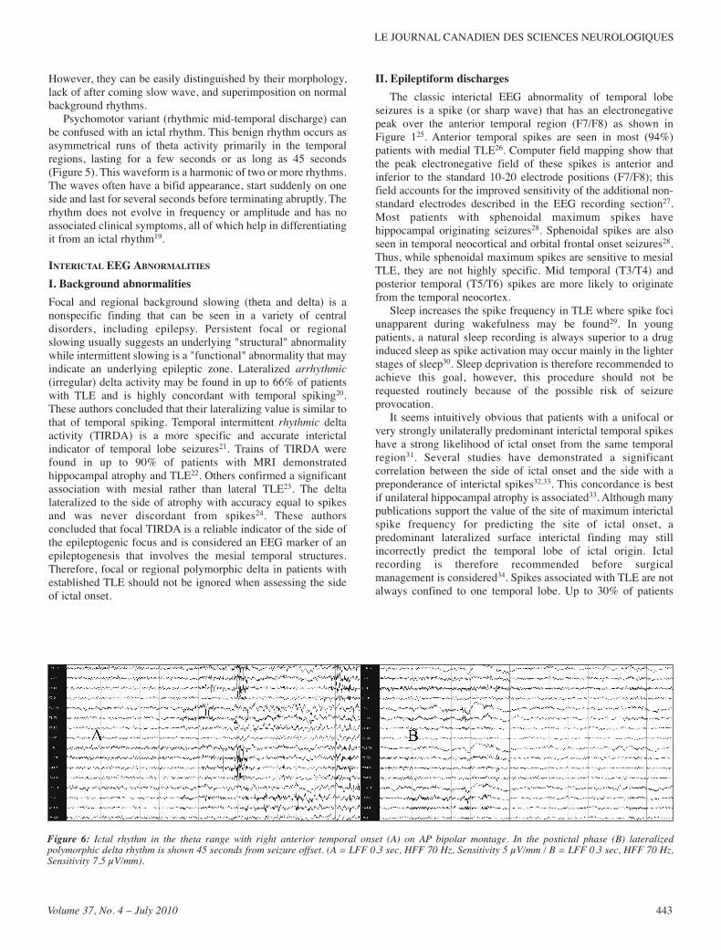
Figure 6: Ictal rhythm in the theta range with right anterior temporal onset (A) on AP bipolar montage. In the postictal phase (B) lateralizedpolymorphic delta rhythm is shown 45 seconds from seizure offset. (A = LFF 0.3 sec, HFF 70 Hz, Sensitivity 5 µV/mm / B = LFF 0.3 sec, HFF 70 Hz,Sensitivity 7.5 µV/mm).
with TLE demonstrate independent bitemporal discharges,particularly when prolonged telemetry recording is used3. Thesepatients could still have unilateral pathology and subsequentsuccessful surgical outcome, particularly when the neuroimagingabnormality and ictal events were concordant. Bisynchronousspike waves are occasionally encountered in patients with focaltemporal EEG abnormalities35,36. The mechanism of productionof these diffuse waves is unclear; however, their presence shouldnot deter the search for a surgically resectable focus36.
ICTAL EEG ABNORMALITIESI. Scalp recorded rhythmsIctal EEG recording is a major component of presurgical
evaluation37. Ictal rhythms include background attenuation, start-stop-start phenomenon, irregular 2-5 Hz lateralized activity, 5-10Hz sinusoidal waves or repetitive epileptiform potentials(Figures 6,7). As well, no or minimal EEG change may be noted(see later section on invasive EEG recording). The multiple scalpEEG expressions of temporal lobe originating seizures are likelya consequence of several integrated factors including thelocation of ictal onset (mesial vs. lateral), the underlyingpathology (hippocampal sclerosis or atrophy vs. neoplasm),routes and speed of seizure propagation38. Factors that can alterseizure propagation and therefore the EEG expression includethe state of the patient (wake vs. sleep), antiepileptic drug use,and whether the patient had unifocal vs. multifocal epilepsy.Focal or regional background attenuation as the initial EEG
change of scalp-recorded temporal lobe seizure is seen in up to25% of seizures39. In a smaller percentage (13%), the seizuremay appear to start, stop, and restart “start-stop-startphenomenon"40. The initial "start" typically has a more restrictedfield than the restart. If the initial start is overlooked, the restartmay be misinterpreted as the actual seizure onset and thereforeconsidered non-localizing, while in fact the initial start istypically focal and localizing.Other initial ictal EEG features of TLE include sinusoidal
waves and repetitive epileptiform potentials (47% of cases). Asseizures evolve, there is a change in frequency (usually anincrease) with spread to other EEG channels as a result of seizurepropagation. Only in the final stages of the seizures is there a
decrease in wave frequency39. A lateralized ictal change ofirregular polymorphic 2-5 Hz temporal rhythm is most oftenassociated with TLE originating from the neocortex41. Alateralized ictal change of rhythmic 5-10 Hz sharp activity,maximally at F7/F8 or sphenoidal electrode position is mostcommonly seen in hippocampal onset TLE (Figures 6,7). Thisrhythm usually occurs within 30 seconds of seizure onset and hashigh specificity for hippocampal onset41-43. However, thisrhythm is not directly caused by seizures in the hippocampus(see later section on invasive EEG recording).Another abnormality of possible significance is the increased
heart rate just before the onset of ictal EEG discharges44. Thisphenomenon is associated with EEG pattern of TLE originatingfrom the mesial structures, suggesting activation of the neuronalcircuits involved in sympathetic regulation.There are no prospective studies to answer the question of
how many ictal recordings should be obtained in a given patientfor definite lateralization and localization of TLE. Patients withunilateral interictal spikes were much more likely to have allseizures “concordant” than patients with bilateral interictalspikes45. Among the group of patients with “conflicting”seizures, all patients demonstrated the conflicting seizures by thefourth recorded seizure. These results were similar to those ofBlum who determined, using a statistical modeling techniquethat five recorded seizures were required to have a 95% chanceof avoiding a conflicting seizure and only four seizures wereneeded if all interictal spikes were unilateral46.Potential difficulties with scalp recordings include an
inability to interpret the EEG because of excessive muscle andmovement artifacts, a paucity of unequivocal changes, inter-observer disagreement, and false lateralization. The overallaccuracy of scalp lateralization ranges between 60 to 83%47.Other causes of such errors include temporal 'plus' epilepsy andextratemporal originating seizure. Temporal 'plus' epilepsy ischaracterized by temporal lobe seizures that also involve theneighboring structures such as the orbitofrontal cortex, insula,frontal and parietal opercula, and temporo-parieto-occipitaljunctions. These are currently most accurately identified byinvasive EEG monitoring but scalp EEG can be useful indifferentiating TLE from temporal 'plus' epilepsy47. In temporal'plus' epilepsy, the interictal EEG more frequently exhibited
THE CANADIAN JOURNAL OF NEUROLOGICAL SCIENCES
444
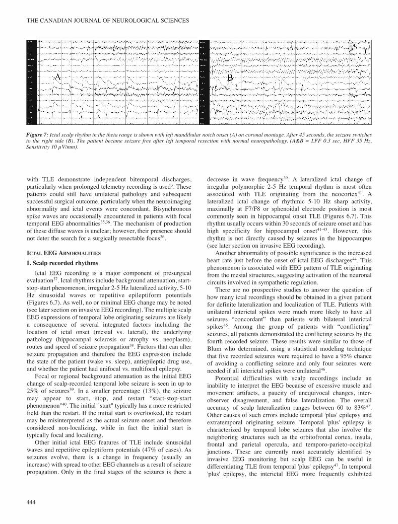
Figure 7: Ictal scalp rhythm in the theta range is shown with left mandibular notch onset (A) on coronal montage. After 45 seconds, the seizure switchesto the right side (B). The patient became seizure free after left temporal resection with normal neuropathology. (A&B = LFF 0.3 sec, HFF 35 Hz,Sensitivity 10 µV/mm).

LE JOURNAL CANADIEN DES SCIENCES NEUROLOGIQUES
Volume 37, No. 4 – July 2010 445
Figu
re8:
1)Ictalscalp
EEGshowing
nochangesatseizureonset(A)oncoronalmo
ntage.A
fter60seconds,anictalrhythm
isseenon
theleftside
(B),falselylateralizingtheseizurebecause
of"burntout"righthip
pocamp
us(seealso
fig8.2
).(A&B
=LFF0.3
sec,HF
F70
Hz,Sensitivity10
µV/mm).
2)IctalsubduralEEGofthe
same
patientinFigure8.1
confirming
thattheictal
onsetwasind
eedfromthe
rightside(A&
B).Astheseizureterminates,propagationisseentotheleftside
(C).Thisiswhenthe
falselylateralizingscalprhythm(Fig8.1
)wasseen.(A=LFF0.1
sec,HF
F70
Hz,Sensitivity75
µV/mm/B&C
=LFF0.1
sec,HF
F70
Hz,Sensitivity100µ
V/mm
).
8.1
8.2

bilateral or precentral abnormalities, while the ictal EEG morefrequently suggested early involvement of the anterior frontal,temporo-parietal and central regions.Occasionally, a seizure starts on one side but terminates
ipsilaterally as the seizure propagates to the other temporal lobe(Figure 7). False lateralization of ictal scalp EEG can also berarely secondary to the so called "burnt out hippocampus"48.Severe advanced hippocampal atrophy and sclerosis results infailure of ipsilateral neocortical propagation and recognition onscalp EEG ipsilaterally49. Several seconds after seizure onset, theictal discharge appears on the contralateral side resulting in falselateralization (Figure 8). Therefore, the ictal rhythm cannot beused in isolation for definite ictal localization.
II. Intracranial recorded rhythmsScalp EEG incompletely records many aspects of intracranial
activity50. The sensitivity, specificity, and interobserverreliability of scalp ictal EEG for localization of the epileptogeniczone have been disputed. If presurgical surface EEG recording,with additional non-standard EEG electrodes (e.g. sphenoidal ormandibular notch electrodes) cannot provide an adequateanswer, invasive intracranial techniques (primarily subdural orintracerebral electrode recordings) can be used to improvelocalization of the epileptogenic zone51. Intracranial EEGdirectly records paroxysmal activity with high sensitivity,relatively artefact free recording, and excellent temporalresolution in the order of milliseconds52,53. In the pre-MRI erathe need for such intracranial recording in the determination ofan epileptogenic focus was emphasized at some centers whereinvasive procedures were required in most patients prior toepilepsy surgery. It is now recognized that in most cases ofmedial temporal lobe epilepsy, standard EEG combined withsphenoidal electrodes and evidence of mesial sclerosis on MRI,will suffice to define the surgical focus.In addition to the ictal rhythms described in the scalp EEG
section, a low voltage fast (>20 Hz) rhythm can be seen at theonset of intracranial recorded TLE53. It has been demonstratedusing a combination of surface and depth electrodes in patientswith TLE that a significant proportion of spatially restrictedseizures seen with depth recordings produce no visible changesin surface EEG recordings (Figure 8)54. Only 19% of auras and10% of subclinical seizures are accompanied by scalp EEGchanges and occasionally, seizures produce bilateral scalpabnormalities which are not indicative of the side from whichdepth recorded seizure activity was initiated54. A comprehensivesystematic study of scalp-intracranial ictal EEG findings in TLEwas reported by Pacia and Ebersole55. Simultaneous scalpelectrodes, mesial temporal depth probes, subdural stripelectrodes, and subdural grids were used. Patients with seizuresbeginning at the hippocampal contacts of the depth electrode hadictal rhythms recorded from the mesial subdural strip electrodes.Simultaneous scalp recordings showed either no change or adiffuse disruption of background. Only when the seizure spreadto the basal and inferolateral subdural contacts did a 6-7 Hzrhythm appear in the inferior and standard scalp electrodes. Theauthors concluded that a scalp recorded 5-9 Hz rhythm is highlyassociated with hippocampal onset seizures but is not a directmanifestation of hippocampal ictal activity. This characteristicrhythm appears to reflect recruitment of adjacent temporal cortex
and cortico-hippocampal interactions as seizures initiated in theneocortex did not demonstrate this scalp pattern until thehippocampus became involved. Seizures confined to the mesial-basal cortex produce a different scalp rhythm that is manifest onthe scalp as a 5 Hz rhythm maximally expressed, perhapsunexpectedly, at the vertex. This rhythm is of opposite polarityfrom that seen with the intracranial basal electrodes and althoughseemingly unlateralized, is in fact quite localizing because of theparticular cortical orientation required for its generation55. Theintracranial correlate of this seizure type was a focal or regionalfast rhythm (beta frequency or higher). Such high frequencychanges were never seen with scalp recordings in this study. Thecorresponding scalp recordings consisted of a high voltage,irregular, delta frequency pattern. A further study showed that thelocation of seizure onset in the temporal lobe was related to thedegree of hippocampal pathology56. Marked hippocampalatrophy and high grade hippocampal sclerosis were bothassociated with initial ictal discharge restricted to thehippocampus on depth recording56.Other authors studied the ictal onset of TLE secondary to
other pathologies, such as temporal dysembryoplasticneuroepithelial tumor, using invasive subdural EEG recording57.These patients frequently had more than one ictal onset zone,which were detected more frequently in the tissue adjacent to thetumor rather than within the tumor or in the mesial temporalstructures. Such accurate localization of the ictal onset is criticalin dysembryoplastic neuroepithelial tumor patients as thesurgical outcome was better when all ictal onset zones werecompletely resected57.
POSTICTAL EEG ABNORMALITIESExcess muscle and movement artefacts may obscure the EEG
features of scalp recordings resulting in difficulties in identifyingthe seizure onset. Several postictal features are described in TLEincluding polymorphic delta activity, regional attenuation, oractivation of focal spikes. Of these, regional delta was mostfrequently observed by Kaibara et al in 57% of patients withscalp EEG recorded seizures58. Other authors identifiedlateralized delta in up to 69% of postictal EEGs26. Postictalchange, whatever its nature appeared principally or exclusivelyipsilateral to the side of seizure onset in all cases (Figure 6B). Inthe Williamson et al study of patients rendered seizure-free aftera temporal lobectomy, 67% of patients had lateralized postictalslowing that was concordant to the side of seizure origin in 100%of instances26. Similarly, Walczak et al found that when postictalchanges were present and could be lateralized (50% of patients),these findings were concordant with the side of seizure onset in96-100% of patients who were at least two years seizure-freeafter surgery43. The postictal changes may be situated principallyover the region most intensely involved in the seizure, whichmay not always coincide with the region of seizure onset. Weevaluated the relationship of postictal delta to the side of seizureonset in patients with TLE by reviewing postictal EEGs with theEEGers blind to clinical and EEG data59. Lateralized postictaldelta was present in 64% of all ictal EEGs and in at least onerecord from 22 patients (76%). The two EEGers agreed on thepostictal EEG findings in 73 of the 80 seizures (kappa=0.88).Lateralized postictal delta, when present, was concordant withthe side of surgery in 96% of the EEGs. The results confirmed
THE CANADIAN JOURNAL OF NEUROLOGICAL SCIENCES
446

that lateralized postictal delta strongly predicts the side ofseizure onset in TLE. In another analysis, the postictal EEGfeatures of simple partial (SP) seizures were compared to thoseof complex partial (CP) and secondary generalized (SG) seizuresin patients with documented TLE60. Postictal delta was identifiedin 55%, 85%, and 100% of postictal EEG segments of SP, CP,and SG seizures respectively. Unilateral postictal delta wasidentified in 36%, 76%, and 44% of postictal EEG segments ofSP, CP, and SG seizures respectively. The EEGs of the partialseizures (SP and CP) were seven times more likely to revealunilateral PDA when compared to the EEGs of SG seizures,however, unilateral postictal delta was more likely seen in the CPgroup. Unilateral postictal delta was ipsilateral to the side oftemporal lobectomy in 94% of EEGs. These findings confirm thepotentially important role of postictal EEG in seizurelateralization, particularly if the ictal rhythm was missed orobscured. If polymorphic delta activity emerges in the EEG, itshould raise the suspicion of a preceding ictal event.
SUMMARY AND CONCLUSIONSScalp EEG is needed for accurate lateralization and
localization of the seizure onset in TLE. Important EEGbackground abnormalities include TIRDA, which is a reliableindicator of epileptogenesis that involves the mesial temporalstructures. Standard EEG can detect up to 58% of all interictalspikes, therefore, additional “non-standard” electrodes can beused to further evaluate the EEG abnormalities. Ictal rhythms inTLE includes background attenuation, start-stop-startphenomenon, irregular 2-5 Hz lateralized activity, and 5-10 Hzsinusoidal waves or repetitive epileptiform potentials. Postictaldelta can be lateralized in 60% of patients with TLE and isconcordant with the side of seizure onset in most patients.Invasive EEG is needed in patients with atypical clinicalsemiology or normal imaging studies. Invasive electrodes can beimplanted on the surface of the brain or stereotactically to allowsampling from multiple medial and lateral temporal lobestructures. Electroencephalography abnormalities should becorrelated with the clinical and imaging data as accurateidentification of the seizure origin is more likely to be achievedif focal EEG onset and MRI findings were concordant with theclinical semiology.
ACKNOWLEDGMENTSThe authors thank Ms. Meredith Sadler for her preparation of
illustrations.
REFERENCES1. Sundaram M, Sadler RM, Young GB, Pillay N. EEG in epilepsy:
current perspectives. Can J Neurol Sci. 1999;26:255-62.2. Jan MM. Assessment of the utility of pediatric electro-
encephalography. Seizure. 2002;11(2):99-103.3. Sadler M, Desbiens R. Scalp EEG in temporal lobe epilepsy
surgery. Can J Neurol Sci. 2000;27:l:22-8.4. Jan MM, Girvin J. Seizure semiology: value in identifying seizure
origin. Can J Neurol Sci. 2008;35:22-30.5. Jasper H. The ten-twenty electrode system of the International
Federation. Electroencephalogr Clin Neurophysiol. 1958;10:371-5.
6. Sadler RM, Goodwin J. Multiple electrodes for detecting spikes inpartial complex seizures. Can J Neurol Sci. 1989;16:326-9.
7. Morris HH, Luders HL, Lesser RP, Dinner DS, Wyllie E. Value ofclosely spaced electrodes in the localization of epileptiform foci:a study of 26 patients with complex partial seizures.Electroencephalogr Clin Neurophysiol. 1987;63:107-11.
8. Nuwer MR. Recording electrode site nomenclature. J ClinNeurophysiol. 1987;4:121-33.
9. Silverman D. The anterior temporal electrode and the ten-twentysystem. Electroencephalogr Clin Neurophysiol. 1960;12:735-7.
10. Kanner AM, Ramirez L, Jones JC. The utility of placing sphenoidalelectrodes under the foramen ovale with fluoroscopic guidance.J Clin Neurophysiol. 1995;12:72-81.
11. Sperling MR, Engel J. EEG from the temporal lobes: a comparisonof ear, anterior temporal, and nasopharyngeal electrodes. AnnNeurol. 1985;17:510-3.
12. Sperling MR, Mendius JR, Engel J. Mesial temporal spikes: asimultaneous comparison of sphenoidal, nasopharyngeal, and earelectrodes. Epilepsia. 1986;27:81-6.
13. Binnie CD, Marston D, Polkey CE, et al. Distribution of temporalspikes in relation to the sphenoidal electrode. Electro-encephalogr Clin Neurophysiol. 1989;73:403-9.
14. Ives JR, Drislane FW, Schacter SC, et al. Comparison of coronalsphenoidal versus anteroposterior temporal montage in the EEGrecording of temporal lobe seizures. Electroencephalogr ClinNeurophysiol. 1996;98:417-21.
15. Dubeau F, McLachlan RS. Invasive electrographic recordingtechniques in temporal lobe epilepsy. Can J Neurol Sci. 2000;27(1):29-34.
16. Noachtar S, Rémi J. The role of EEG in epilepsy: a critical review.Epilepsy Behav. 2009;15(1):22-33.
17. Maillard L, Vigal JP, Gavaret M, et al. Semiologic and electro-physiologic correlations in temporal lobe seizure subtypes.Epilepsia. 2004;45(12):1590-9.
18. Nilsson D, Fohlen M, Jalin C, Dorfmuller G, Bulteau C, DelalandeO. Foramen ovale electrodes in the preoperative evaluation oftemporal lobe epilepsy in children. Epilepsia. 2009;50(9):2085-96.
19. Santoshkumar B, Chong JJ, Blume WT, et al. Prevalence of benignepileptiform variants. Clin Neurophysiol. 2009;20:856-61.
20. Koutroumanidis M, Martin-Migel C, Hennessy MJ, et al. Interictaltemporal delta activity in temporal lobe epilepsy: correlationswith pathology and outcome. Epilepsia. 2004;45(11):11351-67.
21. Geyer JD, Bilir E, Faught RE, Kuzniecky R, Gilliam F. Significanceof interictal temporal lobe delta activity for localization of theprimary epileptogenic region. Neurology. 1999;52:202-5.
22. Gambardella A, Gotman J, Cendes F, Andermann F. Focalintermittent delta activity in patients with mesiotemporalatrophy: a reliable marker of the epileptogenic focus. Epilepsia.1995;36:122-9.
23. Di Gennaro G, Quarato PP, Onorati P, et al. Localizing significanceof temporal intermittent rhythmic delta activity (TIRDA) indrug-resistant focal epilepsy. Clin Neurophysiol. 2003;114:70-8.
24. Blume WT, Borghesi JL, Lemieux JF. Interictal indices of temporallobe origin. Ann Neurol. 1993;34:703-9.
25. Klass D. Electroencephalographic manifestations of complexpartial seizures. In: Penry JK, Daly DD, editors. Advances inneurology. New York: Raven Press; 1975. p. 113-40.
26. Williamson PD, French JA, Thadani VM. Characteristics of medialtemporal lobe epilepsy: II. Interictal and ictal scalpelectroencephalography, neuropsychological testing, neuro-imaging, surgical results, and pathology. Ann Neurol. 1993;34:781-7.
27. Sadler RM, Lemieux JF, Blume WT. Potential fields of anteriortemporal spikes. Electroencephalogr Clin Neurophysiol. 1984;58:47-8.
28. Marks DA, Kratz A, Booke J, et al. Comparison and correlation ofsurface and sphenoidal electrodes with simultaneous intracranialrecording: an interictal study. Electroencephalogr ClinNeurophysiol. 1992;82:23-9.
29. Malow BA, Kushwaha R, Lin X, et al. Relationship of interictalepileptiform discharges to sleep depth in partial epilepsy.Electroencephalogr Clin Neurophysiol. 1997;102:20-6.
30. Jan MM, Aquino MF. The use of chloral hydrate in pediatricelectroencephalography. Neurosciences. 2001;6(2):99-102.
LE JOURNAL CANADIEN DES SCIENCES NEUROLOGIQUES
Volume 37, No. 4 – July 2010 447

31. Quesney LF, Risinger MW, Shewmon DA. Extracranial EEGevaluation. In: Engel J, editor. Surgical treatment of theepilepsies. New York: Raven Press; 1993. p. 173-95.
32. Holmes MD, Dodrill CB, Wilensky AJ, et al. Unilateral focalpreponderance of interictal epileptiform discharges as a predictorof seizure origin. Arch Neurol. 1996;53:228-32.
33. Cendes F, Li LM, Watson C, Andermann F, Dubeau F, Arnold DL.Is ictal recording mandatory in temporal lobe epilepsy? ArchNeurol. 2000;57:497-500.
34. Engel J, Wiebe S, French J, et al. Practice parameter: temporal lobeand localized neocortical resections for epilepsy. Neurology.2003;60:538-47.
35. Gabor A, Ajmone-Marsan C. Co-existence of focal and bilateraldiffuse paroxysmal discharges in epileptics: a clinicalelectrographic study. Epilepsia. 1969;10:453-72.
36. Sadler RM, BlumeWT. Significance of bisynchronous spike-wavesin patients with temporal lobe spikes. Epilepsia. 1989;30: 143-6.
37. Engel J. Update on surgical treatment of the epilepsies. Neurology.1993;43:1612-7.
38. Lieb J, Dasheiff R, Engel J. Role of the frontal lobes in thepropagation of mesial temporal lobe seizures. Epilepsia. 1991;32:822-37.
39. Blume WT, Young GB, Lemieux JF. EEG morphology of partialepileptic seizures. Electroencephalogr Clin Neurophysiol. 1984;57:295-302.
40. Atalla N, Abou-Khalil B, Fakhoury T. The start-stop-startphenomenon in scalp-sphenoidal recordings. ElectroencephalogrClin Neurophysiol. 1996;98:9-13.
41. Ebersole JS, Pacia SV. Localization of temporal lobe foci by ictalEEG patterns. Epilepsia. 1996;37(4):386-99.
42. Vossler DG, Abson Kraemer DL, Knowlton RC, et al. Temporalictal electroencephalographic frequency correlates withhippocampal atrophy and sclerosis. Ann Neurol. 1998;43:756-62.
43. Walczak T, Radtke R, Lewis D. Accuracy and interobserverreliability of scalp ictal EEG. Neurology. 1992;42:2279-85.
44. Di Gennaro G, Quarato PP, Sebastiano F, et al. Ictal heart rateincrease precedes EEG discharge in drug-resistant mesialtemporal lobe seizures. Clin Neurophysiol. 2004;115:1169-77.
45. Sirven JI, Liporace JD, French JA, et al. Seizures in temporal lobeepilepsy. I. Reliability of scalp sphenoidal ictal recording.Neurology. 1997;48:1041-6.
46. Blum D. Prevalence of bilateral partial seizure foci and implicationsfor electroencephalographic telemetry monitoring and epilepsysurgery. Electroencephalogr Clin Neurophysiol. 1994;91:329-36.
47. Bara C, Barbati G, Minotti L, Hoffman D, Kahane P. Ictal clinicaland scalp-EEG findings differentiating temporal lobe epilepsiesfrom temporal 'plus' epilepsies. Brain. 2007;130:1957-67.
48. Mintzer S, Cendes F, Soss J, et al. Hippocampal sclerosis withcontralateral temporal scalp ictal onset. epilepsia. 2004;45(7):792-802.
49. Castro LH, Serpa MH, Valerio RM, et al. Good surgical outcome indiscordant ictal EEG-MRI unilateral mesial temporal sclerosispatients. Epilepsia. 2008;49(8):1324-32.
50. Cooper R, Winter AL, Crow HJ, et al. Comparison of subcortical,cortical, and scalp activity using chronic indwelling electrodes inman. Electroencephalogr Clin Neurophysiol. 1965;18:217-28.
51. King D, Spencer S. Invasive electroencephalography in mesialtemporal lobe epilepsy. J Clin Neurophysiol. 1995;12:32-45.
52. Wendling F, Hernandez A, Bellanger J, Chauvel P, Bartlomei F.Interictal to ictal transition in human temporal lobe epilepsy:insights from a computational model of intracerebral EEG. J ClinNeurophysiol. 2005;22(5):343-56.
53. Jung WY, Pacia SV, Devinsky O. Neocortical temporal lobeepilepsy: intracranial EEG features and surgical outcome. J ClinNeurophysiol. 1999;16(5):419-26.
54. Lieb JP, Walsh GO, Babb TL, et al. A comparison of EEG seizurepatterns recorded with surface and depth electrodes in patientswith temporal lobe epilepsy. Epilepsia. 1976;17:137-60.
55. Pacia SV, Ebersole JS. Intracranial EEG substrates of scalp ictalpatterns from temporal lobe foci. Epilepsia. 1997;38:642-54.
56. Vossler DG, Kraemer DL, Haltiner AM, et al. Intracranial EEG intemporal lobe epilepsy: location of seizure onset relates todegree of hippocampal pathology. Epilepsia. 2004(45(5):497-503.
57. Soe DW, Hong SB. Epileptogenic foci on subdural recording inintractable epilepsy patients with temporal dysembryoplasticneuroepithelial tumor. J Korean Med Sci. 2003;18:559-65.
58. Kaibara M, Blume WT. The postictal electroencephalogram.Electroencephalogr Clin Neurophysiol. 1988;70:99-104.
59. Jan MM, Sadler M, Rahey SR. Lateralized postictal EEG deltapredicts the side of seizure surgery in temporal lobe epilepsy.Epilepsia. 2001;42:402-5.
60. Jan MM. The value of postictal electroencephalogram in temporallobe seizures. Ann Saudi Med. 1999;19:550-3.
THE CANADIAN JOURNAL OF NEUROLOGICAL SCIENCES
448
Copyright © 2022 FDOKUMEN