Statistical Mechanical Equilibrium Theory of Selective Ion Channels
Upload
independentCategory
view
0download
0
American Journal of Medical Genetics (Semin. Med. Genet.) 106:146±159 (2001)
A R T I C L E
Ion Channels and EpilepsyHOLGER LERCHE, KARIN JURKAT-ROTT, AND FRANK LEHMANN-HORN*
Ion channels provide the basis for the regulation of excitability in the central nervous system and in other excitabletissues such as skeletal and heart muscle. Consequently, mutations in ion channel encoding genes are found in avariety of inherited diseases associated with hyper- or hypoexcitability of the affected tissue, the so-called`channelopathies.' An increasing number of epileptic syndromes belongs to this group of rare disorders: Autosomaldominant nocturnal frontal lobe epilepsy is caused by mutations in a neuronal nicotinic acetylcholine receptor (affectedgenes: CHRNA4, CHRNB2), benign familial neonatal convulsions by mutations in potassium channels constituting theM-current (KCNQ2, KCNQ3), generalized epilepsy with febrile seizures plus by mutations in subunits of the voltage-gated sodium channel or the GABAA receptor (SCN1B, SCN1A, GABRG2), and episodic ataxia type 1Ðwhich isassociated with epilepsy in a few patientsÐby mutations within another voltage-gated potassium channel (KCNA1).These rare disorders provide interesting models to study the etiology and pathophysiology of disturbed excitability inmolecular detail. On the basis of genetic and electrophysiologic studies of the channelopathies, novel therapeuticstrategies can be developed, as has been shown recently for the antiepileptic drug retigabine activating neuronalKCNQ potassium channels. ß 2001 Wiley-Liss, Inc.
KEY WORDS: ion channel; epilepsy; genetics; electrophysiology; patch clamp
INTRODUCTION
Epileptic seizures are induced by abnor-
mal focal or generalized synchronized
electrical discharges within the central
nervous system (CNS). The equilibrium
in communication between neurons is
regulated by a network of excitatory and
inhibitory circuits. Both enhancement
of excitatory and impairment of inhibi-
tory mechanisms will disturb this equili-
brium, which may result in epileptic
discharges. There are two basic mechan-
isms underlying the electrophysiological
excitability of and the communication
between neurons: Axonal conduction is
mediated by action potentials and signal
transduction from cell to cell by synaptic
transmission. Since ion channels provide
the basis for these processes, any muta-
tion-induced channel malfunction may
directly alter brain excitability and can
induce epileptic seizures.
Ion channels are membrane-span-
ning proteins forming selective pores for
Na�, K�, Clÿ, or Ca2� ions. During
action potentials a precise control of ion
channel gating is mediated by membrane
voltage, during synaptic transmission
by the binding of speci®c neurotrans-
mitters, such as acetylcholine (ACh).
With regard to these basic principles,
two distinct and structurally conserved
classes of ion channels emerged during
evolution, the voltage-gated and the
ligand-gated channels [Hille, 1992].
Ion channels are
membrane-spanning
proteins forming selective
pores for Na�, K�,
Clÿ, or Ca2� ions.
Since these two are the only channel
types that so far have been shown to
be affected by mutations causing epi-
lepsy, other classes of ionic channels,
e.g., those regulated by intracellular ions
such as Ca2�, by nucleotides, or by cell
volume, will not be considered in this
article.
Over the last 10±15 years, the
combination of electrophysiological and
genetic studies has revealed an increasing
number of inherited diseases associated
with mutations in ion channel encoding
genes. The ®rst of these so-called ion
channel disorders or `channelopathies'
Holger Lerche is a neurophysiologist and clinical neurologist in the Departments of AppliedPhysiology and Neurology, University of Ulm, Germany. Main research interests are the genetics,pathophysiology, and therapy of inherited neurological diseases; in particular, inherited forms ofepilepsy and the relationship to molecular mechanisms of ion channel gating.
Karin Jurkat-Rott is in the Department of Applied Physiology, University of Ulm, Germany.Research focus: Physiology and pathophysiology of cellular excitation and muscle excitation-contraction coupling; genetics and pathogenesis of hereditary muscle and channel diseases withrespect to skeletal muscle and the central nervous system; data bases on diagnostic criteria.
Frank Lohmann-Horn is in the Department of Applied Physiology, University of Ulm, Germany.Research focus: Physio(patho)logy of cellular excitation, particularly structure±function relation-ships of ligand- and voltage-dependent ion channels, etiology and pathogenesis of hereditary ionchannel diseases in neurology.
Grant sponsor: the Deutsche Forschungsgemeinschaft; Grant number: DFG Le1030/5-1; Grantsponsor: the Bundesministerium fuÈ r Bildung und Forschung (BMBF) / InterdisziplinaÈ res Zentrum fuÈ rKlinische Forschung (IZKF) Ulm, projects B1 and B8.
*Correspondence to: Frank Lehmann-Horn, Department of Applied Physiology, University ofUlm, D-89069 Ulm, Germany. E-mail: [email protected]
DOI 10.1002/ajmg.1582
ß 2001 Wiley-Liss, Inc.

were found in skeletal muscle, the
myotonias and periodic paralyses, caused
by mutations in voltage-gated Na�, Clÿ,
or Ca2� channels. Subsequently, several
disorders of the CNS, the episodic
ataxias, familial hemiplegic migraine,
spinocerebellar ataxia type 6, startle
disease, and several epileptic syndromes,
were identi®ed as belonging to the
growing family of channelopathies
[Lehmann-Horn and Jurkat-Rott,
1999, 2000; Ptacek, 1999; Cannon,
2000]. The current review will
focus on the pathophysiological mecha-
nisms of the epileptic channelopathies
in man. We will start with a short
overview of the structure and function
of voltage- and ligand-gated ion chan-
nels, then summarize the clinical,
genetic, and pathophysiological con-
cepts of the known epileptic channel
syndromes and ®nally discuss the impli-
cations of the general contribution of
mutated ion channels to the genetics and
therapy of the more common forms of
epilepsy.
STRUCTURE ANDFUNCTION OFVOLTAGE-GATEDCATION CHANNELS
Voltage-gated K�, Na�, and Ca2�
channels consist of several subunits, a
main a-subunit constituting both the
gating and permeation machinery of the
channel and one or more smaller sub-
units with modifying functions, called b,
g, or d. The a-subunits have a common
tetrameric structure of homologous
domains (I±IV) each with six transmem-
brane segments (S1±S6). Whereas K�
channels are constituted by four identical
domains, the about fourfold longer
genes of Na�- and Ca2�-channel a-
subunits encode four homologous but
distinct domains. In all voltage-gated
cation channels, the S4 segments contain
four to eight positively charged residues
conferring voltage dependence to the
channel protein and the S5±S6 loops
form the major part of the ionic pore
with the selectivity ®lter (Figs. 3, 5, 7).
There are three main conforma-
tional states of voltage-gated channels, a
closed, an open, and an inactivated state.
At the resting potential the channels are
in the closed and activatable state. Upon
membrane depolarization, the voltage
sensors move outward opening the
`activation gate' of the channel on a
time-scale of milliseconds by a yet-
unknown mechanism, and with sus-
tained depolarization the channels inac-
tivate spontaneously by closing of a
different, `inactivation' gate. Upon
membrane repolarization, inactivated
channels remain refractory to further
openings for a certain period deter-
mined by the time needed for recovery
from inactivation (Fig. 1). Typical mod-
ifying properties of the smaller b-, g-, or
Figure 1. The three main conformational states of voltage-gated ion channels. From a closed state at the hyperpolarized restingmembrane potential, channels open upon depolarization during an action potential via outward movement of the voltage sensors thatopen the activation gate. Some channels, such as the voltage-gated Na� channel, inactivate spontaneously via closing of a different,inactivation gate, when depolarization is maintained. From the inactivated state they can only recover upon repolarization of the cellmembrane before they are ready for another opening.
ARTICLE AMERICAN JOURNAL OF MEDICAL GENETICS (SEMIN. MED. GENET.) 147

d-subunits are the regulation of the
amount of functional protein in the
membrane or minor alterations of the
kinetics or voltage dependence of chan-
nel gating [Lehmann-Horn and Jurkat-
Rott, 1999; Catterall, 2000; Siegelbaum
and Koester, 2000].
The time course of depolarization
and repolarization during an action
potential is conveyed by the gating of
voltage-dependent Na� and K� chan-
nels: Activation of the Na� inward
current mediates the steep depolarizing
phase, whereas fast inactivation of Na�
channels and activation of the outward
K� current are responsible for mem-
brane repolarization. Consequently, dis-
ruption of fast Na� channel inactivation
or a decrease in K� conductance leads to
slowed or incomplete repolarization of
the cell membrane, resulting in hyper-
excitability and spontaneous series of
action potentials. Both are the most
common disease-causing mechanisms in
the channelopathies. Main functions of
neuronal voltage-gated Ca2� channels
are the regulation of transmitter release
in presynaptic nerve-terminals.
The different subunits, in particular
the channel a-subunits, are expressed
tissue speci®cally. For example, there are
several genes encoding different Na�
channel a-subunits (SCN1A-SCN11A)
that are expressed in skeletal muscle
(SCN4A), heart muscle (SCN5A) or
neuronal tissue; four of these subunits
(SCN1A, SCN2A, SCN3A, and
SCN8A) are considered to be respon-
sible for the sodium current in brain
[Goldin et al., 2000]. The tissue speci-
®city explains why there are Na�
channel disorders with symptoms
restricted to skeletal or heart muscle
(myotonia or cardiac arrhythmia), or to
the CNS (febrile and afebrile seizures).
Whereas the relatively few different Na�
channels are structurally and function-
ally highly conserved among each other,
a large variety of different voltage-gated
K� channel types with distinct electro-
physiological properties is known
[Chandy and Gutman, 1995; Leh-
mann-Horn and Jurkat-Rott, 1999].
For example, there are inactivating
(e.g., KCNA1) and noninactivating
(e.g., KCNQ1-5) K� channels and large
differences in the kinetics of activation
and inactivation have been described.
STRUCTURE ANDFUNCTION OF LIGAND-GATED ION CHANNELS
Ligand-gated channels are a group of ion
channels activated by different neuro-
transmitters such as acetylcholine
(ACh), g-amino-butyric-acid (GABA),
glycine, glutamate, or nucleotides. They
are also composed of several subunits,
usually four or ®ve. In contrast to the
voltage-gated cation channels, all sub-
units have a similar structure, with two to
four transmembrane segments (M1±4,
Fig. 2). They form a channel complex
with each subunit contributing equally
Figure 2. Proposed structure of the nicotinic acetylcholine receptor with mutations found in patients with autosomal dominantnocturnal frontal lobe epilepsy (ADNFLE, see text). Each of the ®ve subunits contains four transmembrane regions (M1±M4), the M2segments line the channel pore and the long extracellular N-terminal part of the a-subunit contains the binding site for acetylcholine.
148 AMERICAN JOURNAL OF MEDICAL GENETICS (SEMIN. MED. GENET.) ARTICLE

to the ion conducting central pore for-
med by the M2 segments (Fig. 2). The
pore is not as selective as in the voltage-
gated channels and permeable either to
cations, as in excitatory ACh or gluta-
mate receptors, or to anions, such as in
inhibitory GABA or glycine receptors.
Ligand-gated channels are
a group of ion channels
activated by different
neurotransmitters such as
acetylcholine (ACh),
c-amino-butyric-acid
(GABA), glycine,
glutamate, or nucleotides.
The binding sites for transmitters are
located in long extracellular loops.
Similar to the voltage-gated chan-
nels, there are three main con forma-
tional states of the ligand-gated channels:
closed, open, and desensitized. Binding
of the transmitter opens the channel
from the closed state and during constant
presence of the transmitter desensiti-
zation will occur. Only after removal
of the transmitter can the channel
recover from desensitization and sub-
sequently be available for another
opening [Kandel and Siegelbaum,
2000].
Neuronal nicotinic ACh receptors
(nAChR) have a pentameric structure of
two a- and three b-subunits (Fig. 2).
Eight a-(a2±9) and three b-(b2±4) sub-
unit isoforms are known to be expressed
differentially in brain. Most abundantly
found in all brain areas are the a4- and
b2-subunits encoded by the genes
CHRNA4 and CHRNB2, which are
both affected in autosomal dominant
nocturnal frontal lobe epilepsy [Bertrand
and Changeux, 1999]. GABA receptors
belong to the same family of ligand-
gated channels having the same penta-
meric structure. There are several
different subunit classes of GABAA
receptors (a1±6, b1±3, g1±3, d, E, p,
r1±3). The subunit composition most
abundantly found in brain is probably
2a12b21g2 [Mehta and Ticku, 1999;
Sieghart et al., 1999].
AUTOSOMAL DOMINANTNOCTURNAL FRONTALLOBE EPILEPSY (ADNFLE)
This disorder is characterized by fre-
quent brief seizures with hyperkinetic or
tonic manifestations occurring typically
in clusters at night. Ictal video-electro-
encephalographic studies revealed
partial seizures originating from the
frontal lobe. The onset is usually in
childhood, inheritance is autosomal
dominant, and the penetrance ap-
proximately 70±80% [Scheffer et al.,
1995; Picard et al., 2000]. ADNFLE
has often been misdiagnosed as par-
oxysmal nocturnal dyskinesia, sleep
disorders such as night terrors or night-
mares, or hysteria [Scheffer et al., 1994].
In a large Australian family, linkage was
found to chromosome 20q13.2 [Phillips
et al., 1995] and subsequently a mutation
was identi®ed in the gene CHRNA4
encoding the a4-subunit of a neuronal
nicotinic acetylcholine receptor
(nAChR), being the ®rst ion channel
mutation in an inherited form of
epilepsy [Steinlein et al., 1995]. Two
more mutations were found in
CHRNA4 [Steinlein et al., 1997a;
Hirose et al., 1999; Phillips et al., 2000]
and recently, two groups identi®ed
mutations in CHRNB2 [De Fusco
et al., 2000; Phillips et al., 2001], the
gene encoding the b2-subunit of neuro-
nal nAChR, located on chromosome 1.
All mutations described so far reside in
one of the M2 transmembrane segments
lining the ion conducting pore of the
ligand-gated channel (Fig. 2).
Functional expression of some of
the known mutations in Xenopus oocytes
or human embryonic kidney (HEK)
cells revealed different effects on gating
of heteromeric a4b2 channels. The ®rst
two studies of the S248F mutation
postulated a decrease of the overall
channel activity by enhanced desensiti-
zation, slowed recovery from desensiti-
zation, reduced single channel
conductance, and reduced permeability
for Ca2� ions [Weiland et al., 1996;
Kuryatov et al., 1997]. Further studies of
S248F and the 776ins3 mutations also
revealed mechanisms that increase the
activity of the channel. A use-dependent
potentiation to repetitive ACh-exposi-
tions, absent in wild-type receptors, was
found for both mutations, and the
776ins3 mutation revealed a 10-fold
increase in ACh-sensitivity [Steinlein
et al., 1997a; Bertrand et al., 1998;
Figl et al., 1998]. On the other hand,
Ca2� permeability was reduced for
both receptors [Bertrand et al., 1998].
First studies of the mutations in the
b2-subunit revealed only functional
alterations that enhance channel activity.
The V287L mutation showed a pro-
found slowing of desensitization kine-
tics [De Fusco et al., 2000] and V287M
showed a 10-fold increase in ACh-
sensitivity [Phillips et al., 2001]. Ca2�
permeability for V287L was normal.
Thus, pathomechanisms that enhance
the activity of the nAChR seem to
predominate. A disease-causing hyper-
activity of the channel is also sup-
ported by a study showing a threefold
increase in sensitivity to block by
carbamazepine of mutant nAChR, sug-
gesting that the good therapeutic
response of ADNFLE patients to this
drug is at least in part due to carbama-
zepine block of the mutant channel
[Picard et al., 1999].
ADNFLE has often been
misdiagnosed as paroxysmal
nocturnal dyskinesia, sleep
disorders such as night terrors
or nightmares, or hysteria.
How these changes in the electro-
physiological properties of the nAChR
induce frontal lobe seizures remains to
be elucidated. Both the a4- and b2-
subunits are expressed abundantly in
nearly all brain tissues without speci®city
to the frontal lobe or to projections
into this region [Bertrand and Chan-
geux, 1999]. Also, the nocturnal occur-
rence of the seizures is dif®cult to
explain. Transgenic mice generated with
either a knock-out or knock-in of the
ARTICLE AMERICAN JOURNAL OF MEDICAL GENETICS (SEMIN. MED. GENET.) 149

a4-subunit were not reported to develop
seizures [Ross et al., 2000; Labarca et al.,
2001].
BENIGN FAMILIALNEONATAL CONVULSIONS(BFNC)
BFNC is a rare dominantly inherited
epileptic syndrome characterized by
frequent brief seizures within the ®rst
days of life that typically disappear
spontaneously after weeks to months.
Neurological examination, interictal
EEG, and development of these children
are usually normal. The risk of recurring
seizures later in life is about 15%. The
penetrance is as high as 85% [Ronen
et al., 1993; Plouin, 1994]. The disease
was ®rst mapped to the long arm of
chromosome 20 [Leppert et al., 1989]
and a second locus on chromosome 8
has been described [Lewis et al., 1993].
BFNC is a rare dominantly
inherited epileptic syndrome
characterized by frequent
brief seizures within the
®rst days of life that typically
disappear spontaneously
after weeks to months.
Subsequently, mutations in two novel
voltage-gated potassium channel genes,
KCNQ2 (20q13.3) [Biervert et al.,
1998; Singh et al., 1998; Biervert and
Steinlein, 1999; Lerche et al., 1999;
Miraglia del Giudice et al., 2000] and
KCNQ3 (8q24) [Charlier et al., 1998;
Hirose et al., 2000], have been identi®ed
(Fig. 3).
The KCNQ gene family encodes
delayed recti®er K� channels that are
mainly expressed in heart muscle
(KCNQ1), in the CNS (KCNQ2±5),
the inner ear (KCNQ4), and skeletal
muscle (KCNQ5) [reviewed by Jentsch,
2000]. They are activated upon depolar-
ization of the cell membrane and con-
tribute to the repolarizing phase of the
action potential. Mutations in four of the
®ve genes identi®ed cause inherited
diseases. KCNQ1 mutations cause car-
diac arrhythmia in the long QT syn-
drome [Wang et al., 1996], KCNQ2
and KCNQ3 mutations cause epileptic
seizures in BFNC (see above), and
Figure 3. Proposed structure of the voltage-gated K� channels KCNQ2 and KCNQ3 containing mutations causing benign familialneonatal convulsions (BFNC, see text). The channels are built of six transmembrane segments (S1±S6), the S4 segments containingpositively charged residues conferring voltage-dependence to the channel protein and the P-loops between S5 and S6 forming the ionconducting pore. The long cytoplasmic C-terminus is a particular feature of all KCNQ K� channels and most probably mediates theformation of heteromeric KCNQ2/3 channels (see text). Mutations are located within two hot spots: in the pore and in the C-terminus.
150 AMERICAN JOURNAL OF MEDICAL GENETICS (SEMIN. MED. GENET.) ARTICLE
KCNQ4 mutations cause congenital
deafness [Kubisch et al., 1999]. KCNQ5
is the only channel in which disease-
causing mutations have not been found
thus far [Lerche et al., 2000a; SchroÈder
et al., 2000]. Functional expression of
the known mutations revealed a con-
sistent reduction of the resulting potas-
sium current in KCNQ1±4 [Chouabe
et al., 1997; Wollnik et al., 1997;
Biervert et al., 1998; SchroÈder et al.,
1998; Kubisch et al., 1999; Lerche et al.,
1999] (Fig. 4). This leads to an impair-
ment of membrane repolarization,
explaining the occurrence of hyperex-
citability in the affected tissues.
However, the effects on current
reduction were quite different for chan-
nels expressed in heart muscle and outer
hair cells compared to those expressed
exclusively in brain. Whereas KCNQ1
and KCNQ4 mutations exhibited strong
dominant negative effects on WT chan-
nels [Chouabe et al., 1997; Wollnik et al.,
1997; Kubisch et al., 1999], KCNQ2
and KCNQ3 mutations did not. The
latter cause a dominant disease by
haploinsuf®ciency [Biervert et al.,
1998; SchroÈder et al., 1998; Lerche
et al., 1999]. Hence, the brain seems to
be more sensitive to changes in K�
conductance inducing hyperexcitability
than heart muscle ®bers, a fact that
apparently applies similarly to the Na�
channel disorders in muscle and brain
(see below).
After the discovery of the neuron-
speci®c KCNQ2 and KCNQ3 channels,
it was shown that both interact with each
other, since the current size of KCNQ2
is enhanced by about 10-fold upon
coexpression with KCNQ3, which
exhibits only very small currents when
expressed alone [Yang et al., 1998]. Both
channels most probably constitute the
so-called `M-current,' a neuronal K�
current known for several decades to
play an important role in the regulation
of the ®ring rate of neurons [Wang et al.,
1998; Shapiro et al., 2000]. When the in
vivo situation for dominant KCNQ2
and KCNQ3 mutations was mimicked
in vitro by coexpressing, for example,
WT and mutant KCNQ2 with WT
KCNQ3 channels in a 1:1:2 ratio in
Xenopus oocytes, the reduction in cur-
rent size was only 20±25% compared to
WT KCNQ2 combined with KCNQ3
[SchroÈder et al., 1998]. Thus, as stated
above, relatively small changes of the M-
current seem to be suf®cient to cause
epileptic seizures.
Disease-causing mutations in
KCNQ channels are clustered in two
regions of the protein, in the P-loop
between segments S5 and S6 constitut-
ing the pore region and in the long C-
terminus, which is speci®c for this family
of K� channels (Fig. 3). The pore muta-
tions should reduce K� current by
affecting ionic conductance, whereas
the C-terminus is most probably respon-
sible for assembly to heteromeric chan-
nels. Although the stoichiometry of
KCNQ channels has not been examined
so far, it is well known from other
voltage-gated K� channels that they
assemble to form tetramers. A mutation
in the C-terminal part of KCNQ1
causing Jervell and Lange-Nielson syn-
drome disrupt assembly of KCNQ1
channels [Schmitt et al., 2000] and
experiments using chimeras between
KCNQ1, KCNQ2, and KCNQ3 chan-
nels show that the interaction of
KCNQ2 and KCNQ3 channels is
indeed mediated by this region [Lerche
et al., 2000b; Maljevic et al., 2001].
Hence, C-terminal mutations probably
reduce current size by inhibiting the
formation of functional heteromers
inserting into the cell membrane. This
hypothesis corresponds well to a reduced
surface expression of a KCNQ2 mutant
truncating the C-terminus. In con-
trast, pore mutations in KCNQ2 and
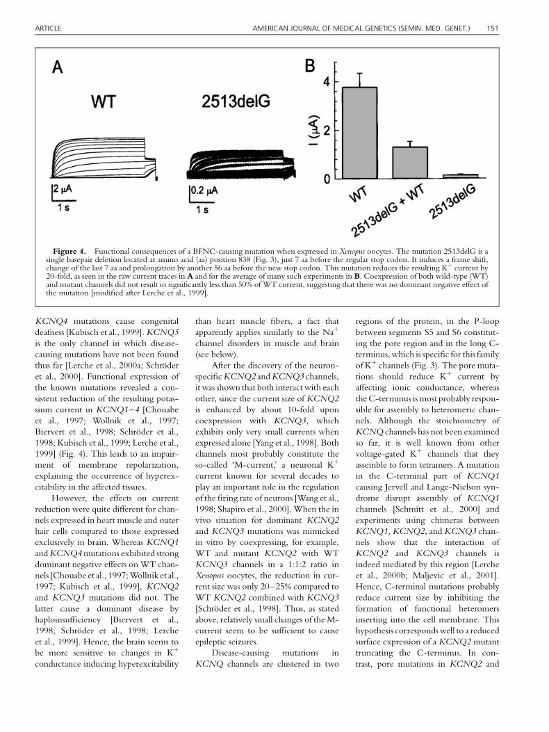
Figure 4. Functional consequences of a BFNC-causing mutation when expressed in Xenopus oocytes. The mutation 2513delG is asingle basepair deletion located at amino acid (aa) position 838 (Fig. 3), just 7 aa before the regular stop codon. It induces a frame shift,change of the last 7 aa and prolongation by another 56 aa before the new stop codon. This mutation reduces the resulting K� current by20-fold, as seen in the raw current traces in A and for the average of many such experiments in B. Coexpression of both wild-type (WT)and mutant channels did not result in signi®cantly less than 50% of WT current, suggesting that there was no dominant negative effect ofthe mutation [modi®ed after Lerche et al., 1999].
ARTICLE AMERICAN JOURNAL OF MEDICAL GENETICS (SEMIN. MED. GENET.) 151

KCNQ3 did not affect surface expres-
sion [Schwake et al., 2000].
The question remains why the
reduced KCNQ2/KCNQ3 K� current
results in seizures preferentially during
the neonatal period. One possibility
could be that the brain is generally more
likely to develop seizures in this pre-
mature state than later in life [Swann
et al., 1993]. Another explanation might
be the differential expression of potas-
sium channels during maturation, which
may attribute a dominant role to KCNQ
channels in central neurons within the
®rst days to weeks of life. Either potas-
sium channels of the KCNQ family
could be upregulated during this period
or other voltage-gated potassium chan-
nels could still not have reached their full
expression level. Differential expression
with reduced expression of KCNQ3
during the ®rst days of life [Tinel et al.,
1998] and expression of a shorter splice
variant of KCNQ2 in fetal brain that
attenuates KCNQ2 and KCNQ3 chan-
nels upon coexpression [Smith et al.,
2001] have been reported. However, it
remains unclear how these ®ndings
contribute to the neonatal seizure phe-
notype.
GENERALIZED EPILEPSYWITH FEBRILE SEIZURESPLUS (GEFS�) AND SEVEREMYOCLONIC EPILEPSY OFINFANCY (SMEI)
GEFS� was ®rst described in 1997 and
1999 by the group of Scheffer, Berkovic
and colleagues [Scheffer and Berkovic,
1997; Singh et al., 1999] as a childhood-
onset syndrome featuring febrile con-
vulsions and avarietyof afebrile epileptic
seizure types within the same pedigree
with autosomal dominant inheritance.
Most common was the febrile convul-
sion syndrome (FS), often with febrile
seizures persisting after the sixth year of
life or in combination with afebrile
generalized tonic-clonic seizures (called
`FS�'). The phenotypes FS and FS�
were found in about two-thirds of
affected individuals. According to the
additional seizure types occurring in the
remaining third of the patients, pheno-
types such as `FS� with absences,' `FS�
with myoclonic seizures,' or `FS� with
atonic seizures' were described. The
most severe phenotype was myoclonic
astatic epilepsy (MAE). Also, partial
epilepsies occurred in rare cases (`FS�
with temporal lobe epilepsy'). The
penetrance was about 60%.
Severe myoclonic epilepsy of
infancy as ®rst described by Dravet
[1978] is characterized by clonic and
tonic-clonic seizures in the ®rst year of
life that are often prolonged and asso-
ciated with fever. Later, patients have
afebrile generalized seizures such as my-
oclonic, absence, or tonic-clonic, and
also simple and complex partial seizures
Figure 5. Proposed structure of the voltage-gated Na� channel containing mutations causing generalized epilepsy with febrileseizures plus (GEFS�) or severe myoclonic epilepsy of infancy (SMEI) in the a-subunit encoded by the gene SCN1A or the b1-subunitencoded by SCN1B. Na� channels are built similar to K� channels, as shown in Figure 3. They have four highly homologous repeats (I±IV) with six transmembrane segments each (S1±S6) forming a central pore (lower right). The a-subunit mutations that have beenfunctionally characterized so far (Fig. 6) are located in the highly conserved voltage sensors in repeat II and IV, respectively (T875M,R1648H). The b1-subunit mutation disrupts a disul®de bridge between two cysteine residues in the extracellular loop that is essential forinteraction with the a-subunit (see text).
152 AMERICAN JOURNAL OF MEDICAL GENETICS (SEMIN. MED. GENET.) ARTICLE

occur. Developmental stagnation with
dementia occurs in early childhood. In
contrast to GEFS�, the syndrome is
usually resistant to pharmacotherapy.
The ®rst genetic defect in GEFS�
was found by Wallace et al. [1998]. The
authors described linkage to chromo-
some 19q13 and identi®ed a point
mutation within the gene SCN1B
encoding the b1-subunit of the vol-
tage-gated Na� channel. The mutation
predicts substitution of tryptophan for a
cysteine residue at position 121 disrupt-
ing a disul®de bridge and changing the
secondary structure of the b1-subunit
extracellular loop (Fig. 5). This leads to a
loss of b-subunit function resulting
electrophysiologically in a slight slowing
of the inactivation time course of the
resulting Na� current [Wallace et al.,
1998]. Although the b1-subunit is also
expressed in skeletal muscle, interest-
ingly, these patients were not reported
to suffer from myotonia like others
carrying mutations within the skeletal
muscle Na� channel a-subunit gene
SCN4A. Hence, the brain seems to be
more sensitive to such changes of
excitability than skeletal muscle ®bers,
or, alternatively, there are different
disease-causing mechanisms for both
diseases, which is discussed in more
detail below.
Subsequently, several groups found
linkage to a cluster of genes encoding
neuronal Na� channel a-subunits on
chromosome 2q21-33 and the ®rst two
point mutations were detected in
SCN1A predicting amino acid changes
within the voltage sensors (S4 segments)
of domains II and IV [Escayg et al.,
2000a] (Fig. 5). Recently, several more
SCN1A mutations have been described
[Escayg et al., 2001; Wallace et al.,
2001a] (Fig. 5) and there is evidence
for further genetic as well as clinical
heterogeneity [Lerche et al., 2001].
Heterologous functional expression
of the ®rst two SCN1A mutations in
segments II/S4 and IV/S4 using the
highly conserved gene SCN4A, human
embryonic kidney cells (tsA201), and
the whole-cell patch clamp technique
revealed only subtle changes in sodium
channel fast inactivation and activation
and no persistent current [Alekov et al.,
2000, 2001] (Fig. 6). The most obvious
alteration of the IV/S4 mutant
(R1460H) was a threefold acceleration
of recovery from inactivation (Fig. 6B),
which was also reported in a preliminary
study with expression of the mutation in
the SCN1A gene using Xenopus oocytes
and two-microelectrode voltage clamp-
ing [Escayg et al., 2000b]. In addition,
we found little acceleration of the
activation time course at potentials
more negative than ÿ20 mV for both
mutations compared to wild-type chan-
nels (Fig. 6C). By shortening the
refractory period after an action poten-
tial and the time of depolarization
needed to elicit an action potential,
these alterations would increase mem-
brane excitability.
However, the most obvious differ-
ence in gating for the II/S4 mutation
(T685M) in comparison to the wild type
was an enhancement of both fast and
slow inactivation of the channel. The
steady-state fast and slow inactivation
curves were shifted by ÿ10 or ÿ20 mV,
respectively, entry into slow inactivation
was accelerated and recovery from slow
inactivation signi®cantly slowed. These
alterations were also found for the IV/S4
mutation, although less pronounced
(Fig. 6D,E) [Alekov et al., 2000, 2001].
Figure 6. Functional consequences of two SCN1A GEFS�-mutations in II/S4and IV/S4 (T875M, R1648H; Fig. 5). The mutations were introduced in the highlyhomologous skeletal muscle a-subunit gene SCN4A (corresponding mutationsT685M, R1460H) and studied in human embryonic kidney cells (tsA201) using thewhole-cell patch-clamp technique. A: Families of raw current traces show only barelymeasurable differences in the time course of inactivation and no persistent Na� currentat the end of the depolarizing test pulses for the mutations compared to wild type (WT)channels. B: A strong acceleration of the time course of recovery from inactivation wasfound for R1460H (shown at ÿ100 mV). C: Another signi®cant difference was anacceleration of the time course of activation at potentials more negative than ÿ20 mV,shown as a shortening of the rise time of the whole-cell Na� current for bothmutations. D,E: Steady-state fast (E) and slow (F) inactivation curves. For bothmutations inactivation is enhanced resulting in a loss-of-function and decrease ofmembrane excitability in contrast to the gain-of-function mechanisms shown in B,C[modi®ed after Alekov et al., 2000, 2001].
ARTICLE AMERICAN JOURNAL OF MEDICAL GENETICS (SEMIN. MED. GENET.) 153

Hence, the disease-causing mechanism
of sodium channel mutations found in
GEFS� might be a loss-of-function by
enhanced inactivation of the channel.
Hence, the disease-causing
mechanism of sodium
channel mutations found
in GEFS� might be a
loss-of-function by
enhanced inactivation
of the channel.
The increase of excitability due to
acceleration of recovery from fast inac-
tivation or of the activation time
courseÐwhich would have to exert its
effect on excitatory neurons to explain
the occurrence of epileptic seizuresÐ
may be to small, in particular for the II/
S4 mutation. In contrast, enhancement
of both fast and slow inactivation would
decrease membrane excitability by redu-
cing the number of available sodium
channels. When acting on inhibitory
neurons, this effect could be responsible
for the occurrence of synchronous
activity in neuronal circuits causing
epileptic seizures.
These ®ndings are in contrast to the
gain-of-function mechanism by a failure
of inactivation that has been shown for
SCN4A mutations causing sodium
channel disorders of skeletal muscle, like
myotonia and periodic paralysis [Leh-
mann-Horn and Jurkat-Rott, 1999;
Cannon, 2000; Mitrovic and Lerche,
2000]. Nonetheless, such a gain-of-
function has also been shown to induce
epileptic seizures in a transgenic mouse
model in which an SCN2A mutation
with slowing of the inactivation time
course and increased persistent current
was introduced. Only 25% of the animals
survived beyond 6 months of age; death
occurred due to severe status epilepticus
[Kearneyet al., 2001]. The gating defects
of inactivation were much less pro-
nounced than those found for SCN4A
mutations causing myotonia, suggesting
that the CNS reacts much more sensi-
tively to such alterations of excitability
than muscle ®bers, which seems to apply
similarly for K� channel defects (see
above).
Two recent advances in the genetics
of idiopathic epilepsies support the
hypothesis that a decrease of excitability
of inhibitory neurons is the most
important disease-causing mechanism
for GEFS�-causing sodium channel
mutations. First, only recently mutations
in two GEFS� families were found in the
g2-subunit of GABAA receptors. One of
these families presented with a typical
GEFS� phenotype (FS and FS�) [Baulac
et al., 2001], the other with a frequent
combination of FS and absence seizures
besides other syndromes described in
GEFS� [Wallace et al., 2001b]. The two
mutations are located in different regions
of the channel, one in the benzodiaze-
pine binding domain in the N-terminal
extracellular loop (R43Q) [Wallaceet al.,
2001b] and the other in the loop
connecting transmembrane segments
M2 and M3 (K289M) [Baulac et al.,
2001]. Functional expression of the
mutant receptor g2-subunits together
with a1- and b2-subunits revealed two
distinct gating defects. Whereas muta-
tion K289M reduced GABA-activated
currents 10-fold, R43Q revealed nor-
mal GABA-activated currents, but abol-
ished the sensitivity to benzodiazepines
such that activation by diazepam was no
longer present. Thus, both mutations
lead to a loss-of-function of GABAA
receptors, although for R43Q it has to
be postulated that `endozepines'do exist
and can prevent the development of
epileptic seizures in vivo [Wallace et al.,
2001b]. For these mutations, undoubt-
edly a decrease of excitability in inhibi-
tory neurons is the pathophysiological
mechanism causing seizures, as it could
similarly be explained by enhanced
inactivation of sodium channels.
Second, novel mutations were id-
enti®ed in SCN1A causing a more
severe phenotype than GEFS�, that is
SMEI [Claes et al., 2001]. Most of these
mutations predict an early stop codon
and a truncated protein without func-
tion (Fig. 5), with regard to all we know
about structure±function relationships
of voltage-gated sodium channels [Cat-
terall, 2000]. Therefore, SMEI is a loss-
of-function sodium channel disorder
caused by haploinsuf®ciency and, from
a genetic point of view, a severe allelic
variant of GEFS�.
Finally, the loss of b1-subunit func-
tion by the SCN1B mutation could also
induce a loss-of-function of the sodium
channel, since one of the major effects of
the b1-subunit upon coexpression with
the a-subunit is to increase the current
amplitude [Catterall, 2000]. Altogether,
loss-of-function mechanisms (in inhibi-
tory neurons) seem to predominate and
are common to all mutations causing
GEFS� or SMEI.
EPISODIC ATAXIA TYPE 1WITH MYOKYMIA (ANDPARTIAL EPILEPSY)
Another ion channel disorder with
disturbed excitability of the CNS is
episodic ataxia type 1 with myokymia
(EA-1).
Another ion channel disorder
with disturbed excitability of
the CNS is episodic ataxia type
1 with myokymia.
Dysfunction occurs predominantly in
the cerebellum. Patients suffer from brief
kinesiogenic attacks of gait and limb
ataxia or cerebellar dysarthria. Interic-
tally they experience myokymia. In four
families, partial epileptic seizures were
also reported, occurring in some family
members affected by ataxia or myokymia
[van Dyke et al., 1975; Brunt and
van Weerden, 1990; Zuberi et al., 1999;
Eunson et al., 2000]. Zuberi et al. [1999]
estimated a 10-fold increased risk to de-
velop epilepsy when affected by EA-1.
Genetic analyses in EA-1 revealed
linkage to chromosome 12p13 and
mutations within the Shaker homolo-
gous gene KCNA1 encoding the K�
channel Kv1.1 [Browne et al., 1994; Litt
et al., 1994] (Fig. 7). Functional expres-
sion in Xenopus oocytes or mammalian
cells resulted in a reduction of the K�
currents either by diminished expression
154 AMERICAN JOURNAL OF MEDICAL GENETICS (SEMIN. MED. GENET.) ARTICLE

or shifts in voltage-dependence. Ac-
cording to these studies, both dominant
negative effects on WT channels and
haploinsuf®ciency can cause EA-1
[Adelman et al., 1995; Zerr et al.,
1998; Bretschneider et al., 1999; Zuberi
et al., 1999; Eunson et al., 2000]. A
speci®c defect for those mutations going
along with an epilepsy phenotype could
not be found [Zuberi et al., 1999;
Eunson et al., 2000]. In support of the
hypothesis that KCNA1 mutations can
induce epileptic seizures, a knock-out
mouse model for Kv1.1 presented with
an epileptic phenotype [Smart et al.,
1998].
ASSOCIATION OF IONCHANNEL DEFECTS WITHCOMMON FORMS OFIDIOPATHIC EPILEPSY
Genetic linkage studies in a few large
families with a presumably monogenic
trait of idiopathic generalized epilepsy
(IGE) revealed loci on chromosomes 6p
and 15q14 for juvenile myoclonic epi-
lepsy [JME; 6p: Greenberg et al., 1988;
Serratosa et al., 1996; Sander et al., 1997;
15q14: Elmslie et al., 1997] and on 8q24
for childhood absence epilepsy [CAE,
Fong et al., 1998; Sugimoto et al., 2000].
In two linkage studies using a large
number of smaller IGE families, the
8q24 locus was also found, while the 6p
locus could not be veri®ed [Zara et al.,
1995]; other potential loci were
described on 2q36, 3q26, and 14q23;
15q14 was con®rmed [Sander et al.,
2000]. Signi®cant linkage to the JME
locus on chromosome 15 was recently
also described for Rolando epilepsy
[Neubauer et al., 1998]. Until now,
mutations in genes at these locations
have not been identi®ed but there
are several ion channel or transporter
encoding genes that are strong candi-
dates: on chromosome 2q36 the
chloride-bicarbonate exchanger gene
SLC4A3, on 3q26 the voltage-gated
K� channel b-subunit gene KCNA1B
[Schultz et al., 1996], and the Clÿ
channel gene CLCN2 [Cid et al.,
1995], on 14q24 the Na�/Ca2�-
exchanger gene SLC8A2 [Li et al.,
1994] and on 15q14 the a7-subunit gene
of the neuronal nAChR CHRNA7
[Chini et al., 1994].
Several ion channel encoding genes
were tested in association studies and
mutation screenings if the play a role in
the genetics of IGE. For KCNQ2
[Steinlein et al., 1999], KCNQ3 [Haug
et al., 2000a], KCNJ3 and KCNJ6
[Girk1 and Girk2: Hallmann et al.,
2000], KCNN3 [hKCa3: Sander et al.,
1999], CACNA1A [Sander et al., 1998],
and SCN1B [Haug et al., 2000b], no
association could be found. A possible
association of a benign polymorphism
in CHRNA4 with IGE [Steinlein et al.,
1997b] could not be con®rmed in
another study [Chioza et al., 2000].
Recently, a mutation was discov-
ered in a patient with juvenile myoclonic
epilepsy in the gene CACNB4, encod-
ing the b4-subunit of the high voltage-
gated L-type Ca2� channel, and func-
tional studies revealed differences in
channel gating for this mutation com-
Figure 7. Proposed transmembrane structure of the voltage-gated potassiumchannel Kv1.1, the human homolog of the Shaker K� channel, encoded by the geneKCNA1. Mutations cause episodic ataxia type 1 with myokymia, two mutations areassociated with partial epilepsy, and one mutation causes isolated myokymia.
ARTICLE AMERICAN JOURNAL OF MEDICAL GENETICS (SEMIN. MED. GENET.) 155
pared to the WT [Escayg et al., 2000c].
Naturally occurring mutations in differ-
ent subunits of the same channel com-
plex cause epilepsy with generalized
spike and wave discharges in the EEG
in several mouse models (Noebels,
accompanying article). Mutations in
CACNA1A, encoding the a-subunit,
cause episodic ataxia type II, familial
hemiplegic migraine, or spinocerebellar
ataxia type 6 in man [Ophoff et al., 1996;
Zhuchenko et al., 1997]. Interestingly,
the same mutation in CACNB4 causing
JME caused EA-2 in a Canadian family
[Escayg et al., 2000c]. It remains to be
proven in further studies if CACNB4 is
an `epilepsy gene' involved in a larger
number of IGE families.
IMPLICATIONS FORTHERAPY
The discovery of genetic defects and, in
particular, the electrophysiological char-
acterization of mutant ion channels in
hereditary forms of epilepsy elucidates
pathophysiological concepts of hyper-
excitability in the CNS. This knowledge
enables new therapeutic strategies by
antagonizing the epilepsy-causing
mechanisms using the defective proteins
as pharmacological targets. In the case of
BFNC, a completely novel approach in
the treatment of epilepsies emerged from
identifying retigabine as an activator of
M-currents conducted by KCNQ2 and
KCNQ3 K� channels. Retigabine shifts
the voltage dependence of steady-state
activation of these channels by about 20
mV in the negative direction so that they
are active at the resting membrane
potential. This stabilizes the cell mem-
brane via hyperpolarization towards the
K� equilibrium potential [Rundtfeld
and Netzer, 2000; Main et al., 2000a;
Wickenden et al., 2000].
It has been shown that for openers
of ATP-dependent K� channels (KATP
channels) that they reduce hyperexcit-
ability and can reverse paralysis of
biopsied skeletal muscle ®bers from
patients with myotonia or periodic
paralysis in vitro by hyperpolarization
of the cell membrane [Grafe et al., 1990;
Quasthoff et al., 1990; Lerche et al.,
1996]. Attempts to treat such patients
with KATP channel openers failed due to
intolerable cardiovascular side effects,
since KATP channels are expressed abun-
dantly in heart, smooth muscle cells, and
other tissues [Lawson, 2000]. In con-
trast, KCNQ2 and KCNQ3 channels
are expressed speci®cally in neurons and,
therefore, side effects of retigabine
should be diminished, since it has no
effect on KCNQ1 channels expressed in
the heart [Main et al., 2000b]. Clinical
trials are currently under way.
REFERENCES
Adelman JP, Bond CT, Pessia M, Maylie J. 1995.Episodic ataxia results from voltage-depen-dent potassium channels with altered func-tions. Neuron 15:1449±1454.
Alekov AK, Rahman MM, Mitrovic N, Leh-mann-Horn F, Lerche H. 2000. A sodiumchannel mutation causing epilepsy in manexhibits subtle defects in fast inactivation andactivation in vitro. J Physiol 529:533±539.
Alekov AK, Rahman MM, Mitrovic N, Leh-mann-Horn F, Lerche H. 2001. Enhancedinactivation and acceleration of activation ofthe sodium channel associated with epilepsyin man. Eur J Neurosci 13:2171±2176.
Baulac S, Gour®nkel-An I, Picard F, Rosenberg-Bourgin M, Prud'homme J-F, Baulac M,Brice A, LeGuern E. 1999. A second locusfor familial generalized epilepsy with febrileseizures plus maps to chsomosome 2q21-q33. Am J Hum Genet 65:1078±1085.
Baulac S, Huberfeld G, Gour®nkel-An I, Mitro-poulou G, Beranger A, Prud'homme JF,Baulac M, Brice A, Bruzzone R, LeGuernE. 2001. First genetic evidence of GABA-Areceptor dysfunction in epilepsy: a mutationin the g2-subunit gene. Nat Genet 28:46±48.
Bertrand D, Changeux JP. 1999. Nicotinicreceptor: a prototype of allosteric ligand-gated ion channels and its possible implica-tions in epilepsy. Adv Neurol 79:171±188.
Bertrand S, Weiland S, Berkovic SF, Steinlein OK,Bertrand D. 1998. Properties of neuronalnicotinic acetylcholine receptor mutantsfrom human suffering from autosomaldominant nocturnal frontal lobe epilepsy.Br J Pharmacol 125:751±760.
Biervert C, Steinlein OK. 1999. Structural andmutational analysis of KCNQ2, the majorgene locus for benign familial neonatalconvulsions. Hum Genet 104:234±240.
Biervert C, Schroeder BC, Kubisch C, BerkovicSF, Propping P, Jentsch TJ, Steinlein OK.1998. A potassium channel mutation inneonatal human epilepsy. Science 279:403±406.
Bretschneider F, Wrisch A, Lehmann-Horn F,Grissmer S. 1999. Electrophysiological char-acterization of two mutant Kv1.1 potassiumchannels causing episodic ataxia type 1 inmammalian cells. Eur J Neurosci 11:2403±2412.
Browne DL, Gancher ST, Nutt JG, Brunt ERP,Smith EA, Kramer P, Litt M. 1994. Episodic
ataxia/myokymia syndrome is associatedwith point mutations in the human potas-sium channel gene, KCNA1. Nat Genet8:136±140.
Brunt ER, van Weerden TW. 1990. Familialparoxysmal kinesigenic ataxia and contin-uous myokymia. Brain 113:1361±1382.
Cannon SC. 2000. Spectrum of sodium channeldisturbances in the nondystrophic myoto-nias and periodic paralyses. Kidney Int57:772±779.
Catterall WA. 2000. From ionic currents tomolecular mechanisms: the structure andfunction of voltage-gated sodium channels.Neuron 26:13±25.
Chandy KG, Gutman GA. 1995. Voltage-gatedK� channel genes. In: North RA, editor.Handbook of receptors and channels.Ligand- and voltage-gated ion channels.Boca Raton, FL: CRC Press. p 1±71.
Charlier C, Singh NA, Ryan SG, Lewis TB, ReusBE, Leach RJ, Leppert M. 1998. A poremutation in a novel KQT-like potassiumchannel gene in an idiopathic epilepsyfamily. Nat Genet 18:53±55.
Chini B, Raimond E, Elgoyhen AB, Moralli D,Balzaretti M, Heinemann S. 1994. Mole-cular cloning and chromosomal localizationof the human alpha 7-nicotinic receptorsubunit gene. Genomics 19:379±381.
Chioza B, Goodwin H, Blower J, McCormick D,Nashef L, Asherson P, Makoff AJ. 2000.Failure to replicate association between thegene for the neuronal nicotinic acetylcho-line receptor alpha 4 subunit (CHRNA4)and IGE. Am J Med Genet 96:814±816.
Chouabe C, Neyroud N, Guicheney P, LazdunskiM, Romey G, Barhanin J. 1997. Propertiesof KvLQT1 K� channel mutations inRomano-Ward and Jervell and Lange-Niel-sen inherited cardiac arrhythmias. EMBO J16:5472±5479.
Cid L, Montrose-Ra®zadeh C, Smith DI, Gug-gino WB, Cutting GR. 1995. Cloning of aputative human voltage-gated chloridechannel (CLC-2) cDNA widely expressedin human tissues. Hum Mol Genet 4:407±413.
Claes L, Del-Favero J, Ceulemans B, Lagae L, VanBroeckhoven C, De Jonghe P. 2001. Denovo mutations in the sodium-channel genescn1a cause severe myoclonic epilepsy ofinfancy. Am J Hum Genet 68:1327±1332.
De Fusco M, Becchetti A, Patrignani A, AnnesiG, Gambardella A, Quattrone A, Ballabio A,Wanke E, Casari G. 2000. The nicotinicreceptor b2 subunit is mutant in nocturnalfrontal lobe epilepsy. Nat Genet 26:275±276.
Dravet C. 1978. Les eÂplepsies graves de l'enfant.Vie Med 8:543±548.
Elmslie FV, Rees M, Williamson MP, Kerr M,Kjeldsen MJ, An Pang K, Sundqvist A, FriisML, Chadwick D, Richens A, Covanis A,Santos M, Arzimanoglou A, Panayiotopou-los CP, Curtis D, Whitehouse WP, GardinerRM. 1997. Genetic mapping of a majorsusceptibility locus for juvenile myoclonicepilepsy on chromosome 15q. Hum MolGenet 6:1329±1334.
Escayg AP, MacDonald BT, Meisler MH, BaulacS, Huberfeld G, An-Gour®nkel I, Brice A,LeGuern E, Moulard B, Chaigne D, BuresiC, Malafosse A. 2000a. Mutations of
156 AMERICAN JOURNAL OF MEDICAL GENETICS (SEMIN. MED. GENET.) ARTICLE

SCN1A, encoding a neuronal sodiumchannel, in two families with GEFS�2.Nat Genet 24:343±345.
Escayg AP, MacDonald BT, Spmpanato J, GoldinAL, Meisler MH. 2000b. Coding andnoncoding variation in the neuronal sodiumchannel SCN1A in patients with epilepsy.Soc Neurosci Abstr 26:222.
Escayg AP, De Waard M, Lee DD, Bichet D, WolfP, Mayer T, Johnston J, Baloh R, Sander T,Meisler MH. 2000c. Coding and noncodingvariation of the human calcium-channel b4-subunit gene CACNB4 in patients withidiopathic generalized epilepsy and episodicataxia. Am J Hum Genet 66:1531±1539.
Escayg A, Heils A, MacDonald BT, Haug K,Sander T, Meisler MH. 2001. A novelSCN1A mutation associated with general-ized epilepsy with febrile seizures plus andprevalence of variants in patients withepilepsy. Am J Hum Genet 68:866±873.
Eunson EL, Rea R, Zuberi SM, Youroukos S,Panayiotopoulos CP, Liguori R, Avoni P,McWilliam RC, Stephenson JBP, HannaMG, Kullmann DM, Spauschus A. 2000.Clinical, genetic, and expression studies ofmutations in the potassium channel geneKCNA1 reveal new phenotypic variability.Ann Neurol 48:647±656.
Figl A, Viseshakul N, Shafaee N, Forsayeth J,Cohen BN. 1998. Two mutations linked tonocturnal frontal lobe epilepsy cause use-dependent potentiation of the nicotinic Achresponse. J Physiol 513:655±670.
Fong GCY, Shah PU, Gee MN, Serratosa JM,Castroviejo IP, Khan S, Ravat SH, Mani J,Huang Y, Zhao HZ, Medina MT, TreimanLJ, Pineda G, Delgado-Escueta AV. 1998.Childhood absence epilepsy with tonic-clonic seizures and electroencephalogram3-4-Hz spike and multispike-slow wavecomplexes: linkage to chromosome 8q24.Am J Hum Genet 63:1117±1129.
Goldin AL, Barchi RL, Caldwell JH, Hofmann F,Howe JR, Hunter JC, Kallen RG, MandelG, Meisler MH, Netter YB, Noda M,Tamkun MM, Waxman SG, Wood JN,Catterall WA. 2000. Nomenclature ofvoltage-gated sodium channels. Neuron28:368.
Grafe P, Quasthoff S, Strupp M, Lehmann-HornF. 1990. Enhancement of K� conductanceimproves in vitro the contraction force ofskeletal muscle in hypokalemic periodicparalysis. Muscle Nerve 13:451±457.
Greenberg DA, Delgado-Escueta AV, Widelitz H,Sparkes RS, Treiman L, Maldonado HM,Park MS, Terasaki PI. 1988. Juvenilemyoclonic epilepsy (JME) may be linkedto the BF and HLA loci on humanchromosome 6. Am J Med Genet 31:185±192.
Hallmann K, Durner M, Sander T, Steinlein OK.2000. Mutation analysis of the inwardlyrectifying K(�) channels KCNJ6 (GIRK2)and KCNJ3 (GIRK1) in juvenile myoclonicepilepsy. Am J Med Genet 96:8±11.
Haug K, Hallmann K, Horvath S, Sander T,Kubisch C, Rau B, Dullinger J, BeyenburgS, Elger CE, Propping P, Heils A. 2000a. Noevidence for association between theKCNQ3 gene and susceptibility to idio-pathic generalized epilepsy. Epilepsy Res42:57±62.
Haug K, Sander T, Hallmann K, Rau B, DullingerJS, Elger CE, Propping P, Heils A. 2000b.The voltage-gated sodium channel beta2-subunit gene and idiopathic generalizedepilepsy. Neuroreport 11:2687±2689.
Hille B. 1992. Ionic channels of excitablemembranes. Sunderland: Sinauer.
Hirose S, Iwata H, Akiyoshi H, Kobayashi K, ItoM, Wada K, Kaneko S, Mitsudome A. 1999.A novel mutation of CHRNA4 responsiblefor autosomal dominant nocturnal frontallobe epilepsy. Neurology 53:1749±1753.
Hirose S, Zenri F, Akiyoshi H, Fukuma G, IwataH, Inoue T, Yonetani M, Tsutsumi M,Muranaka H, Kurokawa T, Hanai T, WadaK, Kaneko S, Mitsudome A. 2000. A novelmutation of KCNQ3 (c.925T!C) in aJapanese family with benign familial neona-tal convulsions. Ann Neurol 47:822±826.
Jentsch TJ. 2000. Neuronal KCNQ potassiumchannels: physiology and role in disease. NatNeurosci Rev 1:21±30.
Kandel ER, Siegelbaum SA. 2000. Synapticintegration. In: Kandel ER, Schwartz JH,Jessel MT, editors. Principles of neuralscience. New York: McGraw Hill. p 207±228.
Kearney JA, Plummer NW, Smith MR, Kapur J,Cummins TR, Waxman SG, Goldin AL,Meisler MH. 2001. A gain-of-functionmutation in the sodium channel gene Scn2aresults in seizures and behavioral abnormal-ities. Neuroscience 102:307±317.
Kubisch C, Schroeder BC, Friedrich T, LuÈtjo-hann B, El-Amraoui A, Martin S, Petit C,Jentsch TJ. 1999. KCNQ4, a novel potas-sium channel expressed in sensory outer haircells, is mutated in dominant deafness. Cell96:437±446.
Kuryatov A, Gerzanich V, Nelson M, Olale F,Lindstrom J. 1997. Mutation causing auto-somal dominant nocturnal frontal lobeepilepsy alters Ca2� permeability, conduc-tance, and gating of human alpha4beta2nicotinic acetylcholine receptors. J Neurosci17:9035±9047.
Labarca C, Schwarz J, Deshpande P, Schwarz S,Nowak MW, Fonck C, Nashmi R, Kofuji P,Dang H, Shi W, Fidan M, Khakh BS, ChenZ, Bowers BJ, Boulter J, Wehner JM, LesterHA. 2001. Point mutant mice with hyper-sensitive alpha 4 nicotinic receptors showdopaminergic de®cits and increased anxiety.Proc Natl Acad Sci USA 98:2786±2791.
Lawson K. 2000. Potassium channel openers aspotential therapeutic weapons in ion chan-nel disease. Kidney Int 57:838±845.
Lehmann-Horn F, Jurkat-Rott K. 1999. Voltage-gated ion channels and hereditary disease.Physiol Rev 79:1317±1372.
Lehmann-Horn F, Jurkat-Rott K. 2000. Chan-nelopathies: common mechanisms inaura, arrhythmia and alkalosis. Amsterdam:Elsevier.
Leppert M, Anderson VE, Quattlebaum T,Stauffer D, O'Connell P, Nakamura Y,Lalouel JM, White R. 1989. Benign familialneonatal convulsions linked to geneticmarkers on chromosome 20. Nature 337:647±648.
Lerche H, Mitrovic N, Dubowitz V, Lehmann-Horn F. 1996. Paramyotonia congenita: theR1448P sodium channel mutation in adult
human skeletal muscle. Ann Neurol39:599±608.
Lerche H, Biervert C, Alekov AK, Schleithoff L,Lindner M, Klingler W, Bretschneider F,Mitrovic N, Jurkat-Rott K, Bode H,Lehmann-Horn F, Steinlein OK. 1999. Areduced K� current due to a novel mutationin KCNQ2 causes neonatal convulsions.Ann Neurol 46:305±312.
Lerche C, Scherer CR, Seebohm G, Derst C, WeiAD, Busch AE, Steinmeyer K. 2000a.Molecular cloning and functional expressionof KCNQ5, a potassium channel subunitthat may contribute to neuronal M-currentdiversity. J Biol Chem 275:22395±22400.
Lerche C, Seebohm G, Schiebe M, Busch AE,Lerche H. 2000b. Evidence for assembly ofKCNQ2 and KCNQ3 K� channels via theC-terminus. Soc Neurosci Abstr 26:1909.
Lerche H, Weber YG, Baier H, Jurkat-Rott K,Kraus de Camargo O, Ludolph AC, BodeH, Lehmann-Horn F. 2001. Generalizedepilepsy with febrile seizures plus: furtherheterogeneity in a large family. Neurology(in press).
Lewis TB, Leach RJ, Ward K, O'Connell P, RyanSG. 1993. Genetic heterogeneity in benignfamilial neonatal convulsions: identi®cationof a new locus on chromosome 8q. Am JHum Genet 53:670±675.
Li Z, Matsuoka S, Hryshko LV, Nicoll DA,Bersohn MM, Burke EP, Lifton RP, Phi-lipson KD. 1994. Cloning of the NCX2isoform of the plasma membrane Na�/Ca2�
exchanger. J Biol Chem 269:17434±17439.Litt M, Kramer P, Browne D, Gancher S, Brunt
ERP, Root D, Phromchotikul T, Dubay CJ,Nutt J. 1994. A gene for episodic ataxia/myokymia maps to chromosome 12p13. AmJ Hum Genet 55:702±709.
Lopes-Cendes I, Scheffer IE, Berkovic SF,Rousseau M, Andermann E, Rouleau GA.2000. A new locus for generalized epilepsywith febrile seizures plus maps to chromo-some 2. Am J Hum Genet 66:698±701.
Main MJ, Cryan JE, Dupere JRB, Cox B, Clare JJ,Burbidge SA. 2000a. Modulation ofKCNQ2/3 potassium channels by the novelanticonvulsant retigabine. Mol Pharm58:253±262.
Main MJ, Tatulian L, Cryan JE, Selyanko A,Brown D, Clare JJ, Hayes A, Trezise DJ,Burbidge SA. 2000b. Modulation of KCNQpotassium channels by retigabine. Soc Neu-rosci Abs 26:1908.
Maljevic S, Lerche C, Seebohm G, Wuttke T,Alekov A, Busch AE, Lerche H. 2001.Evidence for assembly of KCNQ2 andKCNQ3 K� channels via the C-terminus.P¯uÈgers Arch Eur J Physiol 441:R143.
Mehta AK, Ticku MK. 1999. An update onGABA-A receptors. Brain Res Rev29:196±217.
Miraglia del Giudice E, Coppola G, ScuccimarraG, Cirillo G, Bellini G, Pascotto A. 2000.Benign familial neonatal convulsions(BFNC) resulting from mutation of theKCNQ2 voltage sensor. Eur J Med Genet8:994±997.
Mitrovic N, Lerche H. 2000. Sodium and calciumchannelopathies of sarcolemma: periodicparalyses, paramyotonia congenita andpotassium-aggravated myotonia. In: Leh-mann-Horn F, Jurkat-Rott K, editors.
ARTICLE AMERICAN JOURNAL OF MEDICAL GENETICS (SEMIN. MED. GENET.) 157

ChannelopathiesÐcommon mechanisms inaura, arrhythmia and alkalosis. Amsterdam:Elsevier-Science. p 3±32.
Moulard B, Guipponi M, Chaigne D, MouthonD, Buresi C, Malafosse A. 1999. Identi®ca-tion of a new locus for generalized epilepsywith febrile seizures plus (GEFS�) onchromosome 2q24-q33. Am J Hum Genet65:1396±1400.
Neubauer BA, Fiedler B, Himmelein B, KaÈmpferF, LaÈbker U, Schwabe G, Spanier I, Tams D,Bretscher O, Moldenhauer K, KurlemannG, Weise S, Tedroff K, Eeg-Olofsson O,Wadelius C, Stephani U. 1998. Centrotem-poral spikes in families with rolandicepilepsy. Linkage to chromosome 15q14.Neurology 51:1608±1612.
Ophoff RA, Terwindt GM, Vergouwe MN, vanEijk R, Oefner PJ, Hoffman SMG, Lamer-din JE, Mohrenweiser HW, Bulman DE,Ferrari M, Haan J, Lindhout D, van OmmenGJB, Hofker MH, Ferrari MD, Frants RR.1996. Familial hemiplegic migraine andepisodic ataxia type-2 are caused by muta-tions in the Ca2� channel geneCACNL1A4. Cell 87:543±552.
Pfeiffer A, Thompson J, Charlier C, Otterud B,Varvil T, Pappas C, Barnitz C, Gruenthal K,Kuhn R, Leppert M. 1999. A locus forfebrile seizures (FEB3) maps to chromo-some 2q23-24. Ann Neurol 46:671±678.
Phillips HA, Scheffer IE, Berkovic SF, HollwayGE, Sutherland GR, Mulley JC. 1995.Localization of a gene for autosomal domi-nant nocturnal frontal lobe epilepsy tochromosome 20q 13.2. Nat Genet 10:117±118.
Phillips HA, Marini C, Scheffer IE, SutherlandGR, Mulley JC, Berkovic SF. 2000. A denovo mutation in sporadic nocturnal frontallobe epilepsy. Ann Neurol 48:264±267.
Phillips HA, Favre I, Kirkpatrick M, Zuberi SM,Goudie D, Heron SE, Scheffer IE, Suther-land GR, Berkovic SF, Bertrand D, MulleyJC. 2001. CHRNB2 is the second acetyl-choline receptor subunit associated withautosomal dominant nocturnal frontallobe epilepsy. Am J Hum Genet 68:225±231.
Picard F, Bertrand S, Steinlein OK, Bertrand D.1999. Mutated nicotinic receptors respon-sible for autosomal dominant nocturnalfrontal lobe epilepsy are more sensitive tocarbamazepine. Epilepsia 40:1198±1209.
Picard F, Baulac S, Kahane P, Hirsch E, Sebastia-nelli R, Thomas P, Vigevano F, Genton P,Guerrini R, Gericke CA, An I, Rudolf G,Herman A, Brice A, Marescaux C, LeGuernE. 2000. Dominant partial epilepsies. Aclinical, electrophysiological and geneticstudy of 19 European families. Brain123:1247±1262.
Plouin P. 1994. Benign idiopathic neonatalconvulsions (familial and non-familial):open questions about these syndromes. In:Wolf P, editor. Epileptic seizures andsyndromes. London: John Libbey & Co. p193±201.
Ptacek LJ. 1999. Ion channel diseases: episodicdisorders of the nervous system. SeminNeurol 19:363±369.
Quasthoff S, Spuler A, Spittelmeister W, Leh-mann-Horn F, Grafe P. 1990. K� channelopeners suppress myotonic activity of
human skeletal muscle in vitro. Eur JPharmacol 186:125±128.
Ronen GM, Rosales TO, Connolly M, AndersonVE, Leppert M. 1993. Seizure character-istics in chromosome 20 benign familialneonatal convulsions. Neurology 43:1355±1360.
Ross SA, Wong JY, Clifford JJ, Kinsella A,Massalas JS, Horne MK, Scheffer IE, KolaI, Waddington JL, Berkovic SF, Drago J.2000. Phenotypic characterization of analpha 4 neuronal nicotinic acetylcholinereceptor subunit knock-out mouse. J Neu-rosci 20:6431±6441.
Rundtfeld C, Netzer R. 2000. The novel antic-onvulsant retigabine activates M-currents inChinese hamster ovary-cells transfected withhuman KCNQ2/3 subunits. Neurosci Lett282:73±76.
Sander T, Bockenkamp B, Hildmann T, BlasczykR, Kretz R, Wienker TF, Volz A, SchmitzB, Beck-Mannagetta G, Rieb O, Epplen JT,Janz D, Ziegler A. 1997. Re®ned mappingof the epilepsy susceptibility locus EJM1 onchromosome 6. Neurology 49:842±847.
Sander T, Peters C, Janz D, Bianchi A, Bauer G,Wienker TF, Hildmann T, Epplen JT, RiessO. 1998. The gene encoding the alpha1A-voltage-dependent calcium channel(CACN1A4) is not a candidate for causingcommon subtypes of idiopathic generalizedepilepsy. Epilepsy Res 29:115±122.
Sander T, Scholz L, Janz D, Epplen JT, Riess O.1999. Length variation of a polyglutaminearray in the gene encoding a small-con-ductance, calcium-activated potassiumchannel (hKCa3) and susceptibility to idio-pathic generalized epilepsy. Epilepsy Res33:227±233.
Sander T, Schulz H, Saar K, Gennaro E, RiggioMC, Bianchi A, Zara F, Luna D, Bulteau C,Kaminska A, Ville D, Cieuta C, Picard F,Prud'homme JF, Bate L, Sundquist A,Gardiner RM, Janssen GA, Haan GJ,Kasteleijn-Nolst-Trenite DG, Bader A,Lindhout D, Riess O, Wienker TF, Janz D,Reis A. 2000. Genome search for suscept-ibility loci of common idiopathic general-ised epilepsies. Hum Mol Genet 9:1465±1472.
Scheffer IE, Berkovic SF. 1997. Generalizedepilepsy with febrile seizures plus. A geneticdisorder with heterogeneous clinical phe-notypes. Brain 120:479±490.
Scheffer IE, Bhatia KP, Lopes-Cendes I, et al.1994. Autosomal dominant frontal epilepsymisdiagnosed as sleep disorder. Lancet343:515±517.
Scheffer IE, Bhatia KP, Lopes-Cendes I, et al.1995. Autosomal dominant nocturnal fron-tal lobe epilepsy: a distinct clinical disorder.Brain 118:61±73.
Schmitt N, Schwarz M, Peretz A, Abitbol I, AttaliB, Pongs O. 2000. A recessive C-terminalJervell and Lange-Nielsen mutation of theKCNQ1 channel impairs subunit assembly.EMBO J 19:332±340.
Schroeder BC, Kubisch C, Stein V, Jentsch TJ.1998. Moderate loss of function of cyclic-AMP-modulated KCNQ2/KCNQ3 K�
channels causes epilepsy. Nature 396:687±690.
Schroeder BC, Hechenberger M, Weinreich F,Kubisch C, Jentsch TJ. 2000. KCNQ5, a
novel potassium channel broadly expressedin brain, mediates M-type currents. J BiolChem 275:24089±24095.
Schultz D, Litt M, Smith L, Thayer M, McCor-mick K. 1996. Localization of two potas-sium channel beta subunit genes, KCNA1Band KCNA2B. Genomics 31:389±391.
Schwake M, Pusch M, Kharkovets T, Jentsch TJ.2000. Surface expression and single channelproperties of KCNQ2/KCNQ3, M-typeK� channels involved in epilepsy. J BiolChem 275:13343±13348.
Serratosa JM, Delgado-Escueta AV, Medina MT,Zhang Q, Iranmanesh R, Sparkes RS. 1996.Clinical and genetic analysis of a largepedigree with juvenile myoclonic epilepsy.Ann Neurol 39:187±195.
Shapiro MS, Roche JP, Kaftan EJ, Cruzblanca H,Mackie K, Hille B. 2000. Reconstitution ofmuscarinic modulation of the KCNQ2/KCNQ3 K� channels that underlie theneuronal M current. J Neurosci 20:1710±1721.
Siegelbaum SA, Koester J. 2000. Ion channels. In:Kandel ER, Schwartz JH, Jessel MT, editors.Principles of neural science. New York:McGraw Hill. p 105±124.
Sieghart W, Fuchs K, Tretter V, Ebert V, JechlingerM, Hoger H, Adamiker D. 1999. Structureand subunit composition of GABA(A)receptors. Neurochem Int 34:379±385.
Singh NA, Charlier C, Stauffer D, DuPont BR,Leach RJ, Melis R, Ronen GM, Bjerre I,Quattlebaum T, Murphy JV, McHarg ML,Gagnon D, Rosales TO, Peiffer A, AndersonE, Leppert M. 1998. A novel potassiumchannel gene, KCNQ2, is mutated in aninherited epilepsy of newborns. Nat Genet18:25±29.
Singh R, Scheffer IE, Crossland K, Berkovic SF.1999. Generalized epilepsy with febrileseizures plus: a common childhood-onsetgenetic epilepsy syndrome. Ann Neurol45:75±81.
Smart SL, Lopantsev V, Zhang CL, Robbins CA,Wang H, Chiu SY, Schwartzkroin PA,Messing A, Tempel BL. 1998. Deletion ofthe Kv1.1 potassium channel causes epilepsyin mice. Neuron 20:809±819.
Smith JS, Iannotti CA, Dargis P, Christian EP,Aiyar J. 2001. Differential expression ofKCNQ2 splice variants: implications to Mcurrent function during neuronal develop-ment. J Neurosci 21:1096±1103.
Steinlein OK, Mulley JC, Propping P, WallaceRH, Phillips HA, Sutherland GR, SchefferIE, Berkovic SF. 1995. A missense mutationin the neuronal nicotinic acetylcholinereceptor a4 subunit is associated withautosomal dominant nocturnal frontal lobeepilepsy. Nat Genet 11:201±203.
Steinlein OK, Magnusson A, Stoodt J, Bertrand S,Weiland S, Berkovic SF, Nakken KO,Propping P, Bertrand D. 1997a. An insertionmutation of the CHRNA4 gene in a familywith autosomal dominant nocturnal frontallobe epilepsy. Hum Mol Genet 6:943±947.
Steinlein OK, Sander T, Stoodt J, Kretz R, Janz D,Propping P. 1997b. Possible association of asilent polymorphism in the neuronal nico-tinic acetylcholine receptor subunit alpha4with common idiopathic generalized epi-lepsies. Am J Med Genet 74:445±449.
158 AMERICAN JOURNAL OF MEDICAL GENETICS (SEMIN. MED. GENET.) ARTICLE

Steinlein OK, Stoodt J, Biervert C, Janz D, SanderT. 1999. The voltage gated potassiumchannel KCNQ2 and idiopathic generalizedepilepsy. Neuroreport 10:1163±1166.
Sugimoto Y, Morita R, Amano K, Fong CY, ShahPU, Castroviejo IP, Khan S, Delgado-Escueta AV, Yamakawa K. 2000. Childhoodabsence epilepsy in 8q24: re®nement ofcandidate region and construction of phy-sical map. Genomics 68:264±272.
Swann JW, Smith KL, Brady RJ, Pierson MG.1993. Neurophysiological studies of altera-tions in seizure susceptibility during braindevelopment. In: Schwartzkroin PA, editor.Epilepsy: models, mechanisms and concepts.Cambridge: Cambridge University Press. p209±243.
Tinel N, Lauritzen I, Chouabe C, Lazdunski M,Borsotto M. 1998. The KCNQ2 potassiumchannel: splice variants, functional anddevelopmental expression. Brain localiza-tion and comparison with KCNQ3. FEBSLett 438:171±176.
Van Dyke DH, Griggs RC, Murphy MJ, Gold-stein MN. 1975. Hereditary myokymia andperiodic ataxia. J Neurol 25:109±118.
Wallace RH, Wang DW, Singh R, Scheffer IE,George AL Jr, Phillips HA, Saar K, Reis A,Johnson EW, Sutherland GR, Berkovic SF,Mulley JC. 1998. Febrile seizures andgeneralized epilepsy associated with a muta-tion in the Na� channel b1 subunit geneSCN1B. Nat Genet 19:366±370.
Wallace RH, Scheffer IE, Barnett S, Richards M,Dibbens L, Desai RR, Lerman-Sagie T, LevD, Mazarib A, Brand N, Ben-Zeev B,Goikhman I, Singh R, Kremmidiotis G,
Gardner A, Sutherland GR, George AL Jr,Mulley JC, Berkovic SF. 2001a. Neuronalsodium-channel alpha1-subunit mutationsin generalized epilepsy with febrile seizuresplus. Am J Hum Genet 68:859±865.
Wallace RH, Marini C, Petrou S, Harkin LA,Bowser DN, Panchal RG, Williams DA,Sutherland GR, Mulley JC, Scheffer IE,Berkovic SF. 2001b. Mutant GABA-Areceptor g2-subunit in childhood absenceepilepsy and febrile seizures. Nat Genet28:49±52.
Wang Q, Curran ME, Splawski I, Burn TC,Millholland JM, VanRaay TJ, Shen J,Timothy KW, Vincent GM, de Jager T,Schwartz PJ, Towbin JA, Moss AJ, AtkinsonDL, Landes GM, Connors TD, Keating MT.1996. Positional cloning of a novel potas-sium channel gene: KVLQT1 mutationscause cardiac arrhythmias. Nat Genet12:17±23.
Wang HS, Pan Z, Shi W, Brown BS, Wymore RS,Cohen IS, Dixon JE, McKinnon D. 1998.KCNQ2 and KCNQ3 potassium channelsubunits: molecular correlates of the M-channel. Science 282:1890±1893.
Weiland S, Witzemann V, Villarroel A, ProppingP, Steinlein O. 1996. An amino acidexchange in the second transmembranesegment of a neuronal nicotinic receptorcauses partial epilepsy by altering ist desen-sitization kinetics. FEBS Lett 398:91±96.
Wickenden AD, Yu W, Zou A, Jegla T, WagonerPK. 2000. Retigabine, a novel anti-con-vulsant, enhances activation of KCNQ2/3potassium channels. Mol Pharmacol58:591±600.
Wollnik B, Schroeder BC, Kubisch C, EspererHD, Wieacker P, Jentsch TJ. 1997. Patho-physiological mechanisms of dominant andrecessive KVLQT1 K� channel mutationsfound in inherited cardiac arrhythmias.Hum Mol Gen 6:1943±1949.
Yang W-P, Levesque PC, Little WA, Conder ML,Ramakrishnan P, Neubauer MG, BlanarMA. 1998. Functional expression of twoKvLQT1-related potassium channels re-sponsible for an inherited idiopathic epi-lepsy. J Biol Chem 273:19419±19423.
Zara F, Bianchi A, Avanzini G, Di Donato S,Castellotti B, Patel PI, Pandolfo M. 1995.Mapping of genes predisposing to idiopathicgeneralized epilepsy. Hum Mol Genet4:1201±1207.
Zerr P, Adelman JP, Maylie J. 1998. Characteriza-tion of three episodic ataxia mutations in thehuman Kv1.1 potassium channel. FEBS Lett431:461±464.
Zhuchenko O, Bailey J, Bonnen P, Ashizawa T,Stockton DW, Amos C, Dobyns WB,Subramony SH, Zoghbi HY, Lee CC.1997. Autosomal dominant cerebellar ataxia(SCA6) associated with small polyglutamineexpansions in the a1A-voltage-dependentcalcium channel. Nat Genet 15:62±69.
Zuberi SM, Eunson LH, Spauschus A, De SilvaR, Tolmie J, Wood NW, McWilliam RC,Stephenson JPB, Kullmann DM, HannaMG. 1999. A novel mutation in the humanvoltage-gated potassium channel gene(Kv1.1) associates with episodic ataxia type1 and sometimes with partial epilepsy. Brain122:817±825.
ARTICLE AMERICAN JOURNAL OF MEDICAL GENETICS (SEMIN. MED. GENET.) 159
Copyright © 2022 FDOKUMEN