Human Non-Small Cell Lung Cancer Cells Express a Type 2 Cytokine Pattern1

Functional ion channels in pulmonary alveolar type Icells support a role for type I cells in lungion transportMeshell D. Johnson*†, Hui-Fang Bao‡§, My N. Helms‡§, Xi-Juan Chen‡¶, Zac Tigue�, Lucky Jain‡§¶, Leland G. Dobbs*�**,and Douglas C. Eaton‡§¶
Departments of *Medicine, **Pediatrics, and �Cardiovascular Research Institute, University of California, San Francisco, CA 94143; and Departmentsof ‡Physiology and ¶Pediatrics, and §Center for Cell and Molecular Signalling, Emory University School of Medicine, Atlanta, GA 30322
Communicated by John A. Clements, University of California, San Francisco, CA, February 2, 2006 (received for review June 3, 2005)
Efficient gas exchange in the lungs depends on regulation of theamount of fluid in the thin (average 0.2 �m) liquid layer lining thealveolar epithelium. Fluid fluxes are regulated by ion transportacross the alveolar epithelium, which is composed of alveolar typeI (TI) and type II (TII) cells. The accepted paradigm has been that TIIcells, which cover <5% of the internal surface area of the lung,transport Na� and Cl� and that TI cells, which cover >95% of thesurface area, provide a route for water absorption. Here wepresent data that TI cells contain functional epithelial Na� channels(ENaC), pimozide-sensitive cation channels, K� channels, and thecystic fibrosis transmembrane regulator. TII cells contain ENaC andcystic fibrosis transmembrane regulator, but few pimozide-sensi-tive cation channels. These findings lead to a revised paradigm ofion and water transport in the lung in which (i) Na� and Cl�
transport occurs across the entire alveolar epithelium (TI and TIIcells) rather than only across TII cells; and (ii) by virtue of their verylarge surface area, TI cells are responsible for the bulk of transep-ithelial Na� transport in the lung.
Shortly before birth, the fetal lung converts from fluid secre-tion to fluid reabsorption. After birth, efficient gas exchange
depends on regulation of the amount of fluid in the thin(average, 0.2 �m) liquid layer lining the alveolar epithelium (1).Alveolar flooding resulting from cardiogenic pulmonary edemaor acute lung injury impairs gas diffusion across the air�bloodbarrier; an increase in alveolar fluid clearance restores a normalair�blood barrier. Alveolar fluid transport from alveolar tointerstitial spaces, driven by active Na� transport across thealveolar epithelium (2), can be inhibited either by the additionof amiloride, a Na� channel inhibitor, to the alveolar space, orouabain, a Na�,K�-ATPase inhibitor, to the vascular bed (3),suggesting that the alveolar epithelium is the major site of Na�
transport and fluid absorption in the adult lung.The alveolar epithelium, which covers �99% of the large
internal surface area of the lung (4), is composed of two celltypes, alveolar type I (TI) and type II (TII) cells. TII cells, whichcover 2–5% of the internal surface area of the lung, are cuboidalcells that synthesize and secrete pulmonary surfactant. TII cellscontain ion channels, including the amiloride-sensitive epithelialNa� channel (ENaC) (5), Na�,K�-ATPase (3) and the cysticfibrosis transmembrane regulator (CFTR) (6). TI cells are largesquamous cells whose thin cytoplasmic extensions cover �95%of the internal surface area of the lung (7). TI cells expressaquaporin 5, a water channel (8), and have the highest knownosmotic water permeability of any mammalian cell type (9). Theobservations that TII cells contain ion channels and TI cellsexpress aquaporins led to the paradigm that TII cells governalveolar fluid balance by regulating Na� transport in the lungs,whereas TI cells merely provide a route for passive waterabsorption (2). Recent immunohistochemical studies demon-strating that TI cells contain ENaC and Na�,K�-ATPase (10, 11)and radionucleotide uptake studies showing that TI cells cantransport Na� and K� (10) have raised questions about whether this
paradigm adequately describes lung ion and fluid transport. Al-though these newer observations are consistent with the hypothesisthat TI cells play a role in active alveolar ion transport, the lack ofdirect electrophysiologic evidence of specific functional ion chan-nels in TI cells has precluded general acceptance of such a concept(12). Here, we present evidence that TI cells contain functional ionchannels, and compare the electrophysiological characteristics ofspecific ion channels in TI and TII cells. These data support theconcept that TI cells play an important role in the regulation of ionand fluid balance in the lung.
ResultsTI Cells Contain Highly Selective Cation (HSC) and Nonselective Cation(NSC) Channels. Patch clamp analysis of TI cells demonstrated thepresence of both 4- to 6-picoSiemen (pS) HSC and 19- to 21-pSNSC channel activities (Fig. 1 A and B and Table 1). HSC channelshad a positive membrane reversal potential and were inhibited byamiloride (K0.5 � 50 nM), characteristics consistent with those ofHSC channels composed of �-, �-, and �ENaC subunits (13). NSCchannels had a unit conductance of 19–21 pS, were equally selectiveto Na� and K�, and inhibited by amiloride (K0.5 � 1 mM, data notshown), characteristics consistent with NSC channels composed of�ENaC alone (14, 15). The results of patch clamp studies of TI cellsare shown in Table 1. Values for conductances and reversalpotentials (ER) can be found in Table 3, which is published assupporting information on the PNAS web site.
TI Cells Contain Cyclic Nucleotide-Gated (CNG) and K� Channels. CNGchannel activity was suggested by the existence of channels witha unit conductance of 2.9 pS, an ER close to zero, equalselectivity of Na� and K�, insensitivity to amiloride, inhibitionwith pimozide (300 nM), and an apparent increase in conduc-tance in Ca2�-free bath and pipette solutions (Fig. 1D). Theactivity of these channels in Ca2�-free solutions was also in-creased by the application of membrane permeable analogues ofcGMP (chloro-phenyl-thio-cGMP or 8-bromo-cGMP). K� chan-nel activity was suggested by a unit conductance of 15–16 pS, anegative ER, and barium sensitivity (Fig. 1C).
TI Cells Contain CFTR Channels. CFTR is an anion channel that, inairway cells, has a conductance of �4 pS (6) and is stimulatedby cAMP. After stimulating TI cells with forskolin and isobutyl-methyl-xanthine, we could detect functional CFTR in TI cells(Fig. 1E and Table 1) with a conductance of �4 pS and an ERconsistent with the chloride reversal potential [ER is not altered
Conflict of interest statement: No conflicts declared.
Abbreviations: TI, alveolar type I; TII, alveolar type 2; ENaC, epithelial Na� channel; CTFR,cystic fibrosis transmembrane regulator; HSC, highly selective cation; NSC, nonselectivecation; pS, picoSiemen; CNG, cyclic nucleotide-gated; Q-PCR, quantitative PCR.
†To whom correspondence should be addressed. E-mail: [email protected].
© 2006 by The National Academy of Sciences of the USA
4964–4969 � PNAS � March 28, 2006 � vol. 103 � no. 13 www.pnas.org�cgi�doi�10.1073�pnas.0600855103

when all univalent cations in the pipette are replaced with animpermeant cation, such as N-methyl-D-glucamine (NMDG)].The channel is not affected when 10 �M amiloride is in thepipette, but can be blocked by a CFTR blocker, NPPB [5-nitro-2-(3-phenylpropylamino)-benzoate] (Fig. 1E). In the absence ofstimulation, we could not detect functional CFTR.
TI Cells Contain mRNAs for All Three ENaC Subunits and for CFTR.Northern blotting of TI and TII cell RNA demonstrated mRNAsfor �-, �-, and �ENaC, �1- and �1- Na�,K�-ATPase (Fig. 2A).
Alveolar macrophages were used as a negative control for ENaC(2). TI cells contain more ENaC transcript (normalized to 18S)than do TII cells. The relative quantities of transcripts for all 3ENaC subunits and CFTR were calculated by quantitative (Q)-PCR (Fig. 2B). The ratios of TI�TII mRNA content (TI cells, n �5; TII cells, n � 4) for each ENaC subunit were: �ENaC, 3.0;�ENaC, 2.2; �ENaC, 5.0 (Fig. 2B), consistent with the results foundin our Northern blot analysis. CFTR mRNA was detected in TI cellsvia Q-PCR (n � 6, each cell type), although at lower levels than inTII cells; the ratio of TI�TII CFTR mRNA content was 0.2.
Fig. 1. Representative single channel activity in cell-attached patches on adult rat alveolar TI cells. TI cells were isolated and cell-attached patches formed onthe apical surface of the cells as described in Materials and Methods. Typical records and current-voltage relationships obtained from cells with the frequencygiven in Table 1 are shown. (A) A 4- to 5-pS channel whose ER, channel kinetics, and block by amiloride are consistent with an HSC channel (ENaC). (B) A 19- to21-pS channel whose ER and channel kinetics are consistent with an NSC channel. (C) A 5- to 6-pS channel whose ER and the observation that it is completelyblocked by 5 mM Ba2� in the bath suggests that it is a K� channel. (D) A 7-pS nonselective cation channel blocked by 300 nM pimozide and observed in the absenceof bath Ca2� and with 10 �M amiloride in the pipette is consistent with a CNG channel. (E) A 4-pS anion channel that can be observed with either 10 �M amilorideor an impermeable cation NMDG (N-methyl-D-glucamine) in the pipette and can be blocked by 10 mM NPPB [5-nitro-2-(3-phenlproplyamino)benzoate], whichis consistent with CFTR.
Johnson et al. PNAS � March 28, 2006 � vol. 103 � no. 13 � 4965
CELL
BIO
LOG
Y
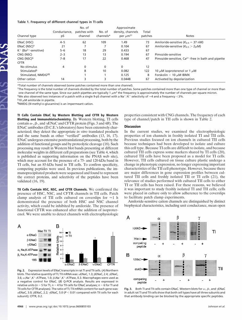
TI Cells Contain ENaC by Western Blotting and CFTR by WesternBlotting and Immunohistochemistry. By Western blotting, TI cellscontain �-, �-, and �ENaC and CFTR protein (Figs. 3 and 4B). TheENaC antibodies (D.C.E.’s laboratory) have been extensively char-acterized; they detect the appropriate in vitro translated productsand the same bands as other ‘‘verified’’ antibodies (13, 16, 17).ENaC undergoes extensive posttranslational processing, both by theaddition of functional groups and by proteolytic cleavage (18). Suchprocessing may result in Western blot bands presenting at differentmolecular weights in different cell preparations (see Table 4, whichis published as supporting information on the PNAS web site),which may account for the presence of a 75- and 120-kDa band inTI cells, but an 85-kDa band in TII cells. To confirm specificity,competing peptides were used. In previous publications, the im-munoprecipitated products were sequenced and found to representthe correct proteins, and selectivity of the peptides have beenvalidated (16, 19).
TII Cells Contain HSC, NSC, and CFTR Channels. We confirmed thepresence of HSC, NSC, and CFTR channels in TII cells. Patchclamp analysis of TII cells cultured on coverslips for �1 hdemonstrated the presence of both HSC and NSC channelactivity, which could be inhibited by amiloride. The presence offunctional CFTR was enhanced after the addition of isoproter-enol. We were unable to detect channels with electrophysiologic
properties consistent with CNG channels. The frequency of eachtype of channel�patch in TII cells is shown in Table 2.
DiscussionIn the current studies, we examined the electrophysiologicproperties of ion channels in freshly isolated TI and TII cells.Previous studies focused on ion channels in cultured TII cellsbecause techniques had been developed to isolate and culturethis cell type. Because TI cells are difficult to isolate, and becausecultured TII cells express some markers shared by TI cells (20),cultured TII cells have been proposed as a model for TI cells.However, TII cells cultured on tissue culture plastic undergo achange in phenotypic expression, no longer expressing importantcharacteristics of the TII cell phenotype. However, because thereare major differences in gene expression profiles between cul-tured TII cells and freshly isolated TII or TI cells (21), therelevance of studies performed with cultured TII cells to eitherTI or TII cells has been raised. For these reasons, we believedit was important to study freshly isolated TI and TII cells; cellswere placed in culture only to allow adherence to the coverslipsto facilitate patch clamp experiments.
Amiloride-sensitive cation channels are distinguished by distinctbiophysical characteristics, including unit conductance, mean open
Table 1. Frequency of different channel types in TI cells
Channel typeConductance,
pS
No. ofpatches with
channelNo. of
channels*
Approximatedensity, channels
per �m2†
Totalpatches Notes
ENaC (HSC) 4–5 62 109 1.49 73 Amiloride-sensitive (K0.5 � 37 nM)ENaC (NSC)‡ 21 7 7 0.104 67 Amiloride-sensitive (K0.5 � 2�M)K� (Ba2�-sensitive) 5–6 18 29 0.433 67CNG (NSC) 2–3 13 13 0.194 67 Pimozide-sensitiveCNG (NSC)§ 7–8 17 22 0.468 47 Pimozide-sensitive, Ca2�-free in bath and pipetteCFTR
No stimulus 4 0 0 0 12Stimulated§ 8 10 0.082 122 10 �M isoproterenol or 1 �MStimulated, NMDG§¶ 1 1 0.125 8 Forskolin � 10 �M IBMX
Other cation 14 3 3 0.0448 67 Activated by depolarization
*Total number of channels observed (some patches contained more than one channel).†The frequency is the total number of channels divided by the total number of patches. Some patches contained more than one type of channel or more thanone channel of the same type. Since our patch pipettes are typically 1 �m2 the frequency is approximately the number of channels per square micron.
‡We also observed two instances of a patch with a single 9-pS channel with a Na��K� selectivity of �4 and a frequency �3%.§10 �M amiloride in pipette.¶NMDG (N-methyl-D-glucamine) is an impermeant cation.
Fig. 2. Expression levels of ENaC transcripts in rat TI and TII cells. (A) Northernblots. The relative quantity of TI�TII mRNA was: �ENaC, 1.3; �ENaC, 2.4; �ENaC,3.6; �1Na�,K�-ATPase, 1.0; �1Na�,K�-ATPase, 0.3. Macrophages were used asa negative control for ENaC. (B) Q-PCR analysis. Results are expressed inrelative units (n � 5 for TI, n � 4 for TII cells for ENaC analyses; n � 6 for TI andTII cells for CFTR analyses). The ratio of TI�TII mRNA content for each gene was:�ENaC, 3.0; �ENaC, 2.2; �ENaC, 5.0 (P � 0.01 compared with TII cells for eachsubunit); CFTR, 0.2.
Fig. 3. Both TI and TII cells contain ENaC. Western blots for �-, �-, and �ENaCin adult rat TI and TII cells show that both cell types have all three subunits andthat antibody binding can be blocked by the appropriate specific peptides.
4966 � www.pnas.org�cgi�doi�10.1073�pnas.0600855103 Johnson et al.

and closed times, cationic selectivity, and amiloride sensitivity. Thecloning of an epithelial Na� channel consisting of three homolo-gous subunits (�, �, and �) from rat colon provided a moleculardefinition of one class of Na� channel (ENaC) (14, 22). Expressionstudies of various combinations of the different ENaC subunits inheterologous expression systems showed that HSC channels werecomposed of �, �, and �ENaC subunits, and that NSC channelswere composed of � subunits alone (14). The predominant amilo-ride-sensitive channels previously described in TII cells were NSCchannels with a unit conductance of 19–24 pS and Na��K�
selectivity of �1.5, and HSC channels with a unit conductance of4–5 pS and Na��K� selectivity of �40 (23).
To determine whether TI cells contained functional ENaC,isolated TI cells were identified by their typical morphology (9) andpatched in the cell-attached mode. TI cell-specific antibodies wereused to confirm the identity of TI cells after patch clamping. Thepatches contained both 4- to 6-pS HSC channels and 19- to 21-pSNSC channels (Fig. 1 A and B and Table 1). The HSC channelswere inhibited by amiloride with a K0.5 � 50 nM and had an ER thatimplied a very high selectivity of the channel for Na� over K�,consistent with the properties of ENaC channels composed of �, �,and � subunits. TI cells also contained a second type of Na�
channel, with a unit conductance of 19–21 pS, equal selectivity toNa� and K�, and inhibitable by amiloride, although only at higherconcentrations (K0.5 � 1 mM). These characteristics are consistentwith NSC channels (14, 15).
TII cells examined under the same conditions also containedboth HSC and NSC channels (Table 2). Culture conditions canalter the types of Na� channels found in TII cells. NSC channelsare predominant in TII cells cultured for 12–96 h on tissueculture plastic (24), and HSC channels are predominant in TIIcells cultured with an apical air interface in the presence of FBS(16), which preserves some of the characteristics of the TII
phenotype, such as surfactant protein expression (20). In thecurrent study, TII cells cultured for brief (�24 h) periods of timecontain a similar frequency of HSC channels to that observed inTI cells (Tables 1 and 2). Because culture conditions can affectENaC expression, one might hypothesize that changes in cellularmicroenvironments caused by acute lung injury or pulmonaryedema could alter the composition and types of functioning ionchannels in alveolar epithelial cells.
TI cells contain the molecular components of the HSC andNSC channels. By Northern blotting, Q-PCR, and Westernblotting, TI cells contain all three ENaC subunits. By bothNorthern blot (Fig. 2 A) analysis and Q-PCR (Fig. 2B), TI cellscontain more of all ENaC subunit transcripts than do TII cells.
As much as 60% of alveolar fluid clearance (AFC) is amiloride-insensitive (25). In a previous study, amiloride inhibited Na� uptakeby �30% in TI and TII cells (10), suggesting that the majority of theNa� uptake by both cell types is amiloride-insensitive. It has beenproposed that CNG channels are responsible for the amiloride-insensitive fraction of AFC because CNG mRNA can be found inthe alveolar epithelium by in situ hybridization (26). CNG channelsshow no preference for Na� over K� and are activated in theabsence of Ca2�; they are amiloride-insensitive but are inhibited bypimozide (27). Properties of single CNG channels are difficult topredict because channels form heteromultimers of different sub-units and unit conductance is affected by the extracellular concen-trations of various ions (28). In our studies, the pimozide-sensitivechannels observed in TI cells are consistent with some CNGchannel isoforms. We frequently observed a channel with a unitconductance of 2.5–3.0 pS and an ER near 0 that was inhibited bypimozide (Fig. 1D), characteristics suggestive of a CNG channel.The unit conductance of these channels increased to 7 pS in aCa2�-free bath solution, and the open probability was increased bymembrane-permeable analogues of cGMP, both consistent withCNG channels. Although we did not observe similar channels in TIIcells, we cannot exclude the possibility that TII cells contain CNGchannels because the expression of alternative CNG channel iso-forms and�or different cellular microenvironments might makedetection of these channels difficult. A previous report that TII cellscontained CNG channels (29) used TII cells cultured for 2 days,consistent with other observations that culture conditions may alterchannel expression. The sensitivity of lung ion transport to pimo-zide (25) seems likely to be due to CNG channels in TI cells. Aportion of ‘‘amiloride-insensitive’’ AFC may also be due to a cationchannel other than CNG, such as NSC channels, which are muchless sensitive to amiloride (K0.5 � 2 mM) than HSC channels, andtherefore may not have been completely inhibited by amilorideinstilled in the upper airways.
We found similar numbers of HSC and NSC channels andmany more CNG channels�patch in TI cell membranes than inTII cell membranes. In the Sprague–Dawley rat, the cell surface
Fig. 4. Immunohistochemistry and Western blot demonstrating that CFTR ispresent in TI cells. (A) (Upper) Human lung tissue showing immunofluores-cence (green) for HTI56 (integral membrane protein specific to human TI cells;ref. 42) showing typical apical membrane localization. (Lower) Immunofluo-rescence for CFTR (red) showing similar TI cell apical membrane. (B) Westernblots for CFTR show protein expression in both TI and TII cells.
Table 2. Frequency of different channel types in TII cells
Channel typeConductance,
pS
No. ofpatches with
channelNo. of
channels*Approximate density,
channels per �m2†
Totalpatches Notes
ENaC (HSC) 4–5 185 407 1.63 249 Amiloride-sensitive (K0.5 � 39 nM)ENaC (NSC)‡ 21 34 34 0.167 204 Amiloride-sensitive (K0.5 � 2.2 �M)K� (Ba2�-sensitive) 5–6 0 0 0 409 None observedCNG 2–3 0 0 0 283 None observedCFTR
No cAMP 8 11 16 0.127 126 NPPB-sensitiveAfter cAMP§ 18 55 0.982 56
*Total number of channels observed (some patches contained more than one channel).†Frequency and density calculated as in Table 1.‡We occasionally observed instances of a 9 pS channel with a Na��K� selectivity of 4, an amiloride K0.5 � 800 nM, and a frequency �4%.§cAMP was increased in cells by addition of 300 nM isoproterenol.
Johnson et al. PNAS � March 28, 2006 � vol. 103 � no. 13 � 4967
CELL
BIO
LOG
Y

area ratio of TI:TII cells is � 43:1 (9). Taken together, these datasuggest that there are many more Na� channels in TI cells thanin TII cells; therefore, TI cells may be responsible for the bulkof basal Na� transport in the lung.
K� channels in alveolar epithelial cells are important in prevent-ing cell depolarization during times of Cl� efflux and Na� influx(30). Multiple types of K� channels (�15) have been detected inalveolar epithelial cells via RT-PCR, but corresponding proteinshave been detected for only �1�3 of these (30). K� channels arepresent in the apical membrane of cultured TII cells (31); there isa recent report that K� channels are present in TI cells in situ.†† Wefound channels in TI cells with a unit conductance of 5–6 pS, an ERat very negative intracellular voltages, and barium sensitivity (Fig.1C), all characteristics of K� channels. We were unable to detect K�
channels in freshly isolated TII cells. Previous studies of TII cellscultured for 5–7 days found evidence for K� channels (31), dem-onstrating again that culture conditions can alter channel expres-sion. In addition, the � subunit of the voltage-gated K� channel(Kv) subfamily Kv9 can electrically silence other � subunits of theKv2 and Kv3 subfamilies, but not the currents mediated by the �subunits of the Kv1 or Kv4 subfamilies (32), which may also explainthe difficulty in detecting K� channels by patch clamping in TIIcells.
In utero, the alveolar epithelium actively secretes Cl� into thedeveloping airspace, promoting fluid secretion, which in turnmodulates lung growth (33); fluid in the fetal lung is cleared in theperipartum period. The importance of Cl� transport in the adultlung is uncertain. CFTR is a cAMP-dependent Cl� channel thatregulates epithelial Cl� and fluid secretion. It has been controver-sial whether CFTR is present in alveolar epithelia and whetherCFTR could modulate bidirectional Cl� flux (i.e., from the alveolaror airway space into the cell). Studies have suggested a role forCFTR in alveolar fluid transport in the adult lung (34), but onlyrecently has there been direct evidence that functional CFTRchannels are present in TII cells (6). We were able to detect bothCFTR mRNA (Fig. 2B) and protein in TI cells, the latter by bothimmunohistochemistry and Western blotting (Fig. 4). The patternof CFTR immunostaining in human lung is comparable withlocalization to the apical plasma membrane. We also found func-tional evidence of CFTR channels in TI cells (Fig. 1E). Isoproter-enol increased CFTR channel activity in TII cell membranepatches; observation of functional CFTR in TI cells requiredstimulation with both forskolin and isobutyl-methyl-xanthine.CFTR undergoes rapid recycling between the plasma membraneand intracellular compartments. It is possible that different rates ofCFTR recycling between these two compartments may determinethe differences in observed CFTR plasma membrane activitybetween TI and TII cells.
TI cells contain several types of cation channels: HSC, NSC,CNG, and K� channels, and at least one type of anion channel:CFTR. TII cells contain HSC, NSC, and CFTR; we did not findevidence for functional K� or CNG channels in TII cells. Takentogether, these findings suggest a revised paradigm for ion andwater transport in the lung in which Na� transport occurs acrossthe entire alveolar epithelium (TI and TII cells), rather than onlyat specialized anatomic locations (i.e., across TII cells). Fig. 5depicts a proposed model of ion and water transport across thealveolar epithelium based on both the current results and theobservations of other investigators. In this model, Na� is ab-sorbed from the apical surface of both TI and TII cells via ENaCand CNG channels and transported into the interstitial space byNa�-, K�-ATPase. K� may be transported via K� channelslocated on the apical surface of TI cells or through basolateralK� channels in TII cells. If net ion transport is from the apical
surface to the interstitium, an osmotic gradient would be createdthat would direct water transport either through aquaporins orby diffusion. Electroneutrality is conserved with Cl� movementvia CFTR in both TI and TII cells, and�or paracellularly throughtight junctions or possibly via an as yet unidentified apicalCl�–HCO3
� exchanger. Because most eukaryotic cells extrudeH� to regulate intracellular pH (35), the net effect of uptake viaa Cl�-HCO3
� exchange would be to absorb Cl� and extrudeHCO3
�, which, when combined with H� in the alveolar lumen,would produce CO2, which is exhaled. Extensive experimentscharacterizing the various channels and the mechanisms bywhich they are regulated will be necessary to understand thepathways by which anions are transported in the alveolus.
The data presented in this article lead to a revised paradigm ofion transport in the lung, in which ion transport occurs across theentire alveolar surface, rather than being limited to TII cells. Basedon the ion channel characteristics of TI and TII cells and the largedifferences in surface area of the two cell types, it appears likely thatTI cells may be more instrumental in driving bulk Na� transportthan are TII cells. Further insight into the characterization andfunction of ion channels in TI cells specifically, and ion transport inthe lung as a whole, will allow clarification of the pathways by whichion and water movement occur across the alveolar epithelium.Better understanding of these pathways will help delineate themechanisms that regulate alveolar fluid balance and may providethe basis for developing strategies to prevent or treat the respiratorycompromise associated with alveolar flooding.
Materials and MethodsPreparation of Rat Alveolar TI and TII Cells. The isolation anddetermination of cell purity of TI and TII cells from adultSprague–Dawley rat lungs were performed as described in ref.10. Preparations of TI cells containing �80% TI cells or with�1% TII cells were discarded, as were preparations of TII cellscontaining �85% TII cells or with �1% TI cells. Viabilities forboth cell types were �95% (Live�Dead kit, Molecular Probes).Both cell types were plated on glass coverslips coated withfibronectin or on permeable supports (Millipore CM mem-branes) at a density of 1 � 106 cells per coverslip.
Patch Clamp Studies. TI cells were identified by their distinctivemorphologic appearance (i.e., large size, extensive cytoplasmicextensions) before patch clamping: after patch clamping, identitywas confirmed by immunostaining. We used the cell-attachedconfiguration for these studies. Cells were studied both 1 and24 h after plating cells on coverslips or permeable supports withan air interface. The 24-h time point was chosen for practicalexperimental reasons, because each cell isolation took 5 h andeach patch recording took �1 h. Results obtained at the two timepoints were indistinguishable. Gigaseal patch clamp methodswere the same as those previously described (16) and aredetailed again in Supporting Text, which is published as support-ing information on the PNAS web site.
††Bourke, S., Helms, M. N., Kim, K. J., Crandall, E. D., Borok, Z. & Kemp, P. J. (2004) FASEBJ. 18, A723 (abstr.).
Fig. 5. Proposed paradigm of channels and transporters involved in alveolarepithelial ion and fluid transport. TI cells (green), TII cells (yellow); ion channelsand transporters are labeled as they appear in the text (e.g., HSC, highlyselective Na� channel). See text for details.
4968 � www.pnas.org�cgi�doi�10.1073�pnas.0600855103 Johnson et al.

Northern Blot Analysis. Total RNA was extracted from freshlyisolated TI cells, TII cells, or alveolar macrophages using anRNEasy kit (Qiagen), fractionated on formaldehyde–agarose gels(1%), blotted on nylon membranes, and hybridized with 32P-labeledfull-length cDNA probes for the �, �, or � subunits of ENaC (kindgifts of Pascal Barbry, Centre National de la Recherche Scienti-fique, Paris) and for the �1 or �1 subunits of Na�,K�-ATPase (kindgifts of Jerry Lingrel, University of Cincinnati, Cincinnati). Radio-activity was quantified by PhosphorImager analysis (Storm 840,Molecular Dynamics). Results were normalized to 18S total RNA.
Real-Time Q-PCR. A real-time Q-PCR protocol (TaqMan, AppliedBiosystems) was used to evaluate relative RNA expression of ENaCsubunits and CFTR in TI and TII cells using the ABI Prism 7700Sequence Detection System. Primer–probe combinations wereas follows: �ENaC forward 5�-GGCGCCTTCTCCTTGGA-3�,reverse 5�-CACTACAAGGCTTCCGACACTT-3�, probe 5�-CTGGGCTGTTTCTCC-3�; �ENaC forward 5�-GTGACTACA-ACACGACCTATTCCA-3�, reverse 5�-CAGTTATGGATC-ATGTGGTCTTGGA-3�, probe 5�-CCTGCCTTCATTCCTG-3�;�ENaC forward 5�-GGCACAATGGCCGTCTGA-3�, reverse 5�-CTTTGGTCCCAGGTGAGAACATT-3�, probe 5�-CAGC-AACCATTTCTCG-3�; CFTR forward 5�-AAGCTTGCCAAC-TACAGGAGGA-3�, reverse 5�-TCTTGCACGTTGACCTC-CACT-3�, probe 5�-TGACCAAGTTTGCAGAACAAGACAA-CACAGT-3�. Standard curves for each ENaC subunit were ob-tained from full-length �-, �-, or �ENaC cDNAs (gifts of PascalBarbry) of known concentration. Lung cDNA was used to generatea standard curve for CFTR. Relative amounts of mRNA were thencalculated by using the comparative threshold method as recom-mended by Applied Biosystems with 18S total RNA as the internalcontrol.
Western Blot Analysis. Western blot analysis for �-, �-, and �ENaCin TI and TII cells was performed with antibodies that have beencharacterized and shown to be subunit-specific (13, 16, 17) bothin the absence and in the presence of specific competing peptidesas described (16). For detection of CFTR, 30 �g of both TI andTII cells were added to sample buffer and heated at 55°C for 10min. Protein bands were resolved on a 4–12% NuPage Bis-Trisgel (Invitrogen), transferred to a nitrocellulose membrane,blocked with 5% powdered milk, and then incubated with CFTRNBD1 antibody (kind gift of David Bedwell, University ofAlbama, Birmingham); goat anti-rabbit IgG-HRP (Vector Lab-oratories) and SuperSignal West Pico chemiluminescent sub-strate (Pierce) were then added before autoradiography.
Immunohistochemistry. Two-micrometer cryostat sections of hu-man lung were subjected to antigen retrieval (10) and incubationwith PBS, 0.1% BSA, 0.3% Triton X-100, and 10% normal goatserum before being sequentially incubated with the followingantibodies: CFTR H-182 (Santa Cruz Biotechnology); goatanti-rabbit IgG Alexa 594 (Molecular Probes); HTI56, a mousemonoclonal antibody directed against an integral membraneprotein specific in human lung to TI cells (37); goat anti-rabbitIgG Alexa 488 (Molecular Probes). Images were captured witha Leica DC500 camera on a Leica Orthoplan microscope.
We thank Dr. Pascal Barbry for his gift of ENaC cDNA probes,Dr. Jerry Lingrel for his gift of Na�,K�-ATPase cDNA probes, andDr. David Bedwell for his gift of CFTR NBD1 antibody. Our work wassupported by grants from the National Institutes of Health (to L.J.,L.G.D., and D.C.E.) and the Robert Wood Johnson Amos MedicalFaculty Development Program (to M.D.J.).
1. Bastacky, J., Lee, C. Y., Goerke, J., Koushafar, H., Yager, D., Kenaga, L.,Speed, T. P., Chen, Y. & Clements, J. A. (1995) J. Appl. Physiol. 79, 1615–1628.
2. Matalon, S. & O’Brodovich, H. (1999) Annu. Rev. Physiol. 61, 627–661.3. Olivera, W., Ridge, K., Wood, L. D. & Sznajder, J. I. (1994) Am. J. Physiol. 266,
L577–L584.4. Crapo, J. D., Young, S. L., Fram, E. K., Pinkerton, K. E., Barry, B. E. & Crapo,
R. O. (1983) Am. Rev. Respir. Dis. 128, S42–S46.5. Voilley, N., Lingueglia, E., Champigny, G., Mattei, M. G., Waldmann, R.,
Lazdunski, M. & Barbry, P. (1994) Proc. Natl. Acad. Sci. USA 91, 247–251.6. Brochiero, E., Dagenais, A., Prive, A., Berthiaume, Y. & Grygorczyk, R. (2004)
Am. J. Physiol. 287, L382–L392.7. Stone, K. C., Mercer, R. R., Freeman, B. A., Chang, L. Y. & Crapo, J. D. (1992)
Am. Rev. Respir. Dis. 146, 454–456.8. Nielsen, S., King, L. S., Christensen, B. M. & Agre, P. (1997) Am. J. Physiol.
273, C1549–C1561.9. Dobbs, L. G., Gonzalez, R., Matthay, M. A., Carter, E. P., Allen, L. &
Verkman, A. S. (1998) Proc. Natl. Acad. Sci. USA 95, 2991–2996.10. Johnson, M. D., Widdicombe, J. H., Allen, L., Barbry, P. & Dobbs, L. G. (2002)
Proc. Natl. Acad. Sci. USA 99, 1966–1971.11. Ridge, K., Olivera, W., Saldı́as, F. J., Azzam, Z., Horowitz, S., Rutschman,
D. H., Dumasius, V., Factor, P. & Sznajder, J. I. (2003) Circ. Res. 92, 453–460.12. Kemp, P. J. & Kim, K. J. (2004) Am. J. Physiol. 287, L460–L464.13. Chen, X. J., Eaton, D. C. & Jain, L. (2002) Am. J. Physiol. 282, L609–L620.14. Canessa, C. M., Schild, L., Buell, G., Thorens, B., Gautschi, I., Horisberger,
J. D. & Rossier, B. C. (1994) Nature 367, 463–467.15. Jain, L., Chen, X. J., Malik, B., Al-Khalili, O. & Eaton, D. C. (1999) Am. J.
Physiol. 276, L1046–L1051.16. Jain, L., Chen, X. J., Ramosevac, S., Brown, L. A. & Eaton, D. C. (2001) Am. J.
Physiol. 280, L646–L658.17. Malik, B., Yue, Q., Yue, G., Chen, X. J., Price, S. R., Mitch, W. E. & Eaton,
D. C. (2005) Am. J. Physiol. 289, F107–F116.18. Hughey, R. P., Mueller, G. M., Bruns, J. B., Kinlough, C. L., Poland, P. A.,
Harkleroad, K. L., Carattino, M. D. & Kleyman, T. R. (2003) J. Biol. Chem. 278,37073–37082.
19. Hardiman, K. M., McNicholas-Bevensee, C. M., Fortenberry, J., Myles, C. T.,Malik, B., Eaton, D. C. & Matalon, S. (2004) Am. J. Respir. Cell Mol. Biol. 30,720–728.
20. Dobbs, L. G., Pian, M. S., Maglio, M., Dumars, S. & Allen, L. (1997) Am. J.Physiol. 273, L347–L354.
21. Gonzalez, R., Yang, Y. H., Griffin, C., Allen, L., Tigue, Z. & Dobbs, L. (2004)Am. J. Physiol. 288, L179–L189.
22. Lingueglia, E., Renard, S., Waldmann, R., Voilley, N., Champigny, G., Plass,H., Lazdunski, M. & Barbry, P. (1994) J. Biol. Chem. 269, 13736–13739.
23. Matalon, S., Lazrak, A., Jain, L. & Eaton, D. C. (2002) J. Appl. Physiol. 93,1852–1859.
24. Feng, Z. P., Clark, R. B. & Berthiaume, Y. (1993) Am. J. Respir. Cell Mol. Biol.9, 248–254.
25. Norlin, A., Lu, L. N., Guggino, S. E., Matthay, M. A. & Folkesson, H. G. (2001)J. Appl. Physiol. 90, 1489–1496.
26. Ding, C., Potter, E. D., Qiu, W., Coon, S. L., Levine, M. A. & Guggino, S. E.(1997) Am. J. Physiol. 272, C1335–C1344.
27. Junor, R. W., Benjamin, A. R., Alexandrou, D., Guggino, S. E. & Walters, D. V.(1999) J. Physiol. 520, 255–260.
28. Flynn, G. E., Johnson, J. P., Jr., & Zagotta, W. N. (2001) Nat. Rev. Neurosci.2, 643–651.
29. Kemp, P. J., Kim, K. J., Borok, Z. & Crandall, E. D. (2001) J. Physiol. 536,693–701.
30. O’Grady, S. M. & Lee, S. Y. (2003) Am. J. Physiol. 284, L689–L700.31. Lee, S. Y., Maniak, P. J., Ingbar, D. H. & O’Grady, S. M. (2003) Am. J. Physiol.
284, C1614–C1624.32. Stocker, M., Hellwig, M. & Kerschensteiner, D. (1999) J. Neurochem. 72,
1725–1734.33. Kitterman, J. A. (1984) J. Dev. Physiol. 6, 67–82.34. Fang, X., Fukuda, N., Barbry, P., Sartori, C., Verkman, A. S. & Matthay, M.
(2002) J. Gen. Physiol. 119, 199–207.35. Lubman, R. L. & Crandall, E. D. (1992) Am. J. Physiol. 262, L1–L14.36. Dobbs, L. G., Gonzalez, R. F., Allen, L. & Froh, D. K. (1999) J. Histochem.
Cytochem. 47, 129–137.
Johnson et al. PNAS � March 28, 2006 � vol. 103 � no. 13 � 4969
CELL
BIO
LOG
Y
Copyright © 2022 FDOKUMEN