Does the plasma membrane of the rod outer segment contain more than one type of ion channel?
11
Neuroscience Research, Suppl. 2 (1985) $89-$99 $89 Elsevier Scientific Publishers Ireland Ltd. DOES THE PLASMA MEMBRANE OF THE ROD OUTER SEGMENT CONTAIN MORE THAN ONE TYPE OF ION CHANNEL? W.A. SATHER, R.D. BODOIA and P.B. DETWILER University of Washington, Department of Physiology and Biophysics, Seattle, Washington 98195 USA INTRODUCTION The current that flows in response to a change in membrane voltage is given by the sum of ionic and capacitative components. Baylor and Lamb s found that when the light-sensitive conductance of the rod is fully shut off by bright light, the plasma membrane of the outer segment responds to a voltage change with a purely capacitative current. This implies that the only functional ion channels in the outer segment surface membrane are those that are closed by light. This short paper describes preliminary observations made with suction electrode and patch clamp recording that support speculation about the existence of other types of outer segment channels. METHODS Animals and Preparation. Experiments were performed on frog (Rana pipiens) rods. Under infrared illumination eyes were removed from the pithed head of a dark-adapted (> 12 hours) animal and hemisected at the level of the ora serrata. The posterior half of each eye was cut into 2 to 4 pieces and stored in iced Ringer under a stream of 100% oxygen. Stored tissue remained viable for 4 to 5 hours. Eye cup pieces were taken as needed and bathed in oxygenated Ringer where fine forceps were used to separate the retina from the pigment epithelium. Frog rods were mechanically dissociated from pieces of isolated retina by gently stroking the vertical surface with a forcep. A significant fraction of the rods obtained in this way retained their ellipsoid, but usually the synaptic process of the inner segment was lost. No attempt was made to distinguish between "red" and "green" rods. Dissociated rods were transferred in 200 ~I of frog Ringer (containing mM: NaCI, I00; KCI, 1,0; MgS04, 1.2; CaC12, 1.0; dextrose, 10; Hepes, 2.8; adjusted with NaOH to pH 7.55) to a chamber consisting of a 3 mm space between a glass microscope slide and a narrow #i cover slip (20 mm x 3 mm x 150 ~m) that was supported at one end. Surface tension held the fluid under the cover slip and formed vertical meniscuses on three sides. A narrow channel in the end support connected the chamber with a larger fluid reservoir which compensated for evaporative losses. 0168-0102/85/$03.30 © 1985 Elsevier Scientific Publishers Ireland Ltd.
-
Upload
independent -
Category
Documents
-
view
2 -
download
0
Transcript of Does the plasma membrane of the rod outer segment contain more than one type of ion channel?

Neuroscience Research, Suppl. 2 (1985) $89-$99 $89 Elsevier Scientific Publishers Ireland Ltd.
DOES THE PLASMA MEMBRANE OF THE ROD OUTER SEGMENT CONTAIN MORE THAN ONE
TYPE OF ION CHANNEL?
W.A. SATHER, R.D. BODOIA and P.B. DETWILER
Universi ty of Washington, Department of Physiology and Biophysics, Seat t le ,
Washington 98195 USA
INTRODUCTION
The current that flows in response to a change in membrane voltage is
given by the sum of ionic and capac i ta t ive components. Baylor and Lamb s
found that when the l i g h t - s e n s i t i v e conductance of the rod is f u l l y shut
o f f by br ight l i g h t , the plasma membrane of the outer segment responds to a
voltage change with a purely capac i ta t ive current . This impl ies that the
only funct ional ion channels in the outer segment surface membrane are
those that are closed by l i g h t . This short paper describes pre l iminary
observations made with suction electrode and patch clamp recording that
support speculation about the existence of other types of outer segment
channels.
METHODS
Animals and Preparat ion. Experiments were performed on frog (Rana pipiens) rods. Under in f rared i l l um ina t i on eyes were removed from the
pithed head of a dark-adapted (> 12 hours) animal and hemisected at the
level of the ora ser ra ta . The pos ter io r hal f of each eye was cut in to 2 to
4 pieces and stored in iced Ringer under a stream of 100% oxygen. Stored
t issue remained v iable for 4 to 5 hours. Eye cup pieces were taken as
needed and bathed in oxygenated Ringer where f ine forceps were used to
separate the re t ina from the pigment ep i the l ium. Frog rods were
mechanically d issociated from pieces of iso la ted re t ina by gently stroking
the ver t i ca l surface with a forcep. A s i gn i f i can t f rac t ion of the rods
obtained in th is way retained t h e i r e l l i p s o i d , but usual ly the synaptic
process of the inner segment was l o s t . No attempt was made to d is t ingu ish
between "red" and "green" rods. Dissociated rods were t ransferred in 200
~I of frog Ringer (containing mM: NaCI, I00; KCI, 1,0; MgS04, 1.2; CaC12,
1.0; dextrose, 10; Hepes, 2.8; adjusted with NaOH to pH 7.55) to a chamber
consist ing of a 3 mm space between a glass microscope s l ide and a narrow #i
cover s l i p (20 mm x 3 mm x 150 ~m) that was supported at one end. Surface
tension held the f l u i d under the cover s l i p and formed ver t i ca l meniscuses
on three sides. A narrow channel in the end support connected the chamber
with a la rger f l u i d reservo i r which compensated for evaporative losses.
0168-0102/85/$03.30 © 1985 Elsevier Scientific Publishers Ireland Ltd.
$90
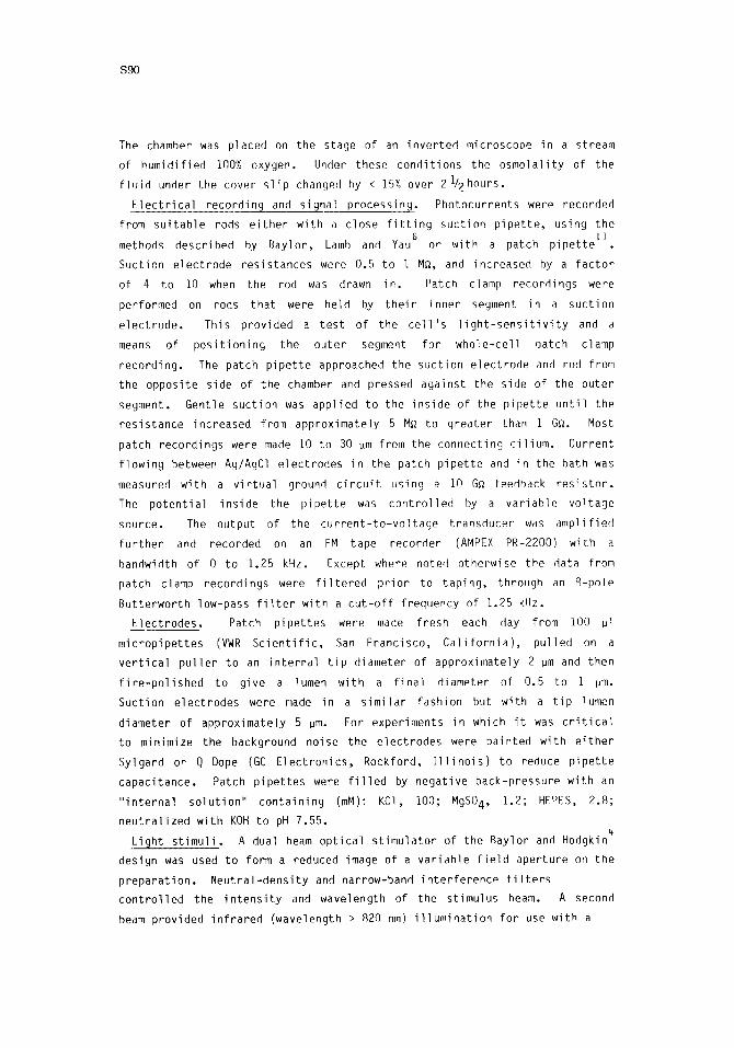
The chamber was placed on the stage of an i nve r t ed microscope in a stream
of humid i f i ed IQO% oxygen. Under these cond i t i ons the o s m o l a l i t y of the
f l u i d under the cover s l i p changed by < 15% over 21/2 hours.
E l e c t r i c a l record ing and s ignal p rocess ing. Photocurrents were recorded
from s u i t a b l e rods e i t h e r w i th a c lose f i t t i n g suc t ion p i p e t t e , using the 6 I i
methods descr ibed hy Bay lo r , Lamb and Yau or w i th a patch p i p e t t e
Suct ion e l ec t r ode res is tances were 0.5 to i M~, and increased by a f a c t o r
of 4 to I0 when the rod was drawn i n . Patch clamp record ings were
performed on rods t ha t were held by t h e i r inner segment in a suc t ion
e l e c t r o d e . This prov ided a t es t of the c e l l ' s l i g h t - s e n s i t i v i t y and a
means of p o s i t i o n i n g the ou te r segment f o r who le - ce l l patch clamp
reco rd ing . The patch p i p e t t e approached the suc t ion e l ec t r ode and rod from
the oppos i te side of the chamber and pressed aga ins t the side of the ou te r
segment. Gent le suc t ion was app l ied to the i ns ide of the p i p e t t e u n t i l the
res is tance increased from approx imate ly 5 M~ to g rea te r than I GQ. Most
patch record ings were made I0 to 30 ~m from the connect ing c i l i u m . Current
f l ow ing between Ag/AgCl e lec t rodes in the patch p i p e t t e and in the bath was
measured w i th a v i r t u a l ground c i r c u i t using a IN G~ feedback r e s i s t o r .
The p o t e n t i a l i ns ide the p i p e t t e was c o n t r o l l e d by a v a r i a b l e vo l t age
source. The output of the c u r r e n t - t o - v o l t a g e t ransducer was amp l i f i ed
f u r t h e r and recorded on an FM tape recorder (AMPEX PR-2200) w i th a
bandwidth of 0 to 1.25 kHz. Except where noted o therwise the data from
patch clamp record ings were f i l t e r e d p r i o r to t ap ing , through an 8-po le
Bu t te rwor th low-pass f i l t e r w i th a c u t - o f f f requency of 1.25 kHz.
E lec t rodes . Patch p i pe t t es were made f resh each day from i00 pl
m i c rop i pe t t es (VWR S c i e n t i f i c , San Franc isco, C a l i f o r n i a ) , pu l led on a
v e r t i c a l p u l l e r to an i n t e r n a l t i p d iameter of approx imate ly 2 ~m and then
f i r e - p o l i s h e d to g ive a lumen w i th a f i n a l d iameter of 0.5 to i pm.
Suct ion e lec t rodes were made in a s i m i l a r fash ion but w i th a t i p lumen
d iameter of approx imate ly 5 pm. For exper iments in which i t was c r i t i c a l
to minimize the background noise the e lec t rodes were pa in ted wi th e i t h e r
Sylgard or Q Dope (GC E l e c t r o n i c s , Rockford, I l l i n o i s ) to reduce p i p e t t e
capac i tance . Patch p i pe t t es were f i l l e d by nega t i ve back-pressure w i th an
" i n t e r n a l s o l u t i o n " con ta in i ng (mM): KCI, I00; MgS04, 1.2; HEPES, 2.8;
n e u t r a l i z e d w i th KOH to pH 7.55.
L igh t s t i m u l i . A dual beam o p t i c a l s t i m u l a t o r of the Bay lor and Hodgkin
design was used to form a reduced image of a v a r i a b l e f i e l d aper tu re on the
p r e p a r a t i o n . N e u t r a l - d e n s i t y and narrow-band i n t e r f e r e n c e f i l t e r s
c o n t r o l l e d the i n t e n s i t y and wavelength of the s t imulus beam. A second
beam prov ided i n f r a r e d (wavelength > 820 nm) i l l u m i n a t i o n f o r use wi th a

S91
s i l i con vidicon to view the preparation and electrode manipulations on a TV
monitor. Light in tens i ty was measured with a Quantum photometer (model
II40A, Princeton Applied Research Corp., Princeton, New Jersey) and
referenced to a cal ibrated s i l i con photodiode (United Detector Technology,
Calver City, Ca l i f o rn ia ) .
RESULTS
Suction electrode recordin 9. The shape of the d is ta l t i p of the outer
segment of dissociated frog rods varied. In most cases i t appeared to end
abruptly with a f l a t p r o f i l e . In other cases the t i p appeared to be
beveled or conical (Fig. I ) . Photocurrents recorded from f l a t - t i pped outer
Fig. I . Photomicrographs of dissociated frog rods with beveled and f l a t - t ipped outer segments. This is not an a r t i f ac t of d issociat ion since s imi lar p ro f i les are observed in intact ret ina .

S92
segments were in the outward d i rec t i on , consistent with the suppression of
a standing inward membrane dark current 6. Suction electrode recordings
from the t i p of bevel-shaped outer segments, however, often showed
d i s t i n c t l y d i f f e ren t photocurrents. Responses typ ica l of approximately 20%
of the rods with beveled outer segments is shown in Fig. 2. F u l l - f i e l d
i l l umina t ion of the rod evoked photocurrents that were inward when recorded
from only the beveled t i p of the outer segment but were outward when
recorded from the whole outer segment. Recordings from intermediate
amounts of outer segment were markedly biphasic. Inward photocurrents were
recorded from the t i p in response to local i l l um ina t ion of other regions of
the outer segment. The smallest responses were evoked by flashes of a s l i t
of l i g h t posit ioned on or near the v i c i n i t y of the t i p . Scattered l i g h t
Recording ~ t i o n s Full Field Illumination Proximal Local Illunina1~n I I
pA
( ' ~ -10
-
'I_/-- -10
I I I I 0 1 2 3
Fig. 2. Inverted l i g h t responses at the t i p of beveled outer segments. The photocurrents evoked by moderate in tens i t y 20 ms flashes of 520 nm l i g h t are shown in 4 d i f f e ren t recording condi t ions. Both f u l l - f i e l d and local i l l um ina t ion of the proximal hal f of the rod evoked an inverted (inward) photocurrent when only the beveled t i p of the rod was recorded. When larger amounts of the outer segment were drawn into the recording p ipet te the photocurrent became biphasic and f i n a l l y monophasic and outward when the whole outer segment current was recorded. The bottom trace shows that a normal po la r i t y photocurrent was recorded from the inner segment of the same c e l l . Standard Ringer's bathing so lu t ion; bandwidth DC to 16 Hz.

S93
makes i t d i f f i c u l t to know whether or not the t i p was l i gh t - sens i t i ve .
I f a l l the ion channels in the plasma membrane of the beveled t i p were
permanently closed then the change in membrane potent ia l due to
i l luminat ion of proximal regions of the outer segment would generate a
capaci tat ive current proport ional to the f i r s t der iva t ive of the potent ia l
change. The expected current would then be biphasic and rapid. In
contrast, the photocurrent recorded from the beveled t i p has a r e l a t i ve l y
slow time course and is monophasic. This result is consistent with the
presence of a passive (light-insensitive) conductance in the membrane of
the beveled t i p . Inward current through the passive conductance would
increase during the hyperpolarizing l i gh t response of the rod. The net
d i rect ion and waveform of the photocurrent would depend on the ra t io of
passive to l i gh t - sens i t i ve conductance in the membrane recorded by the
suction p ipet te,
Patch clamp record!ng. Current jumps resembling single-channel events
were observed most consistent ly in whole-cell and excised patch recordings
CELL ATTACHED R E ~ N G
I I I I I 0 1 2 3 4
SECONDS
Fig. 3. Current steps recorded from an intact patch of outer segment membrane. Downward def lect ion corresponds to inward membrane current. Lower trace is a continuation of the upper trace. Recording in darkness at a bandwidth of DC to 160 Hz. Pipette f i l l i n g solut ion was 0 . i mM Ca ++ Ringer's; cel l bathed in standard Ringer's.

$94
but were rarely observed in cel l -at tached recordings. They generally
developed in bursts af ter several minutes of gigaseal recording and were
not obviously influenced by l i g h t . At the resting potent ial the events
were inward steps that ranged from 0.5 to 5 pA in cel l -at tached recording
(Fig. 3) and I to 6 pA in whole-cell recording (Fig. 4). The amplitude of
+30 mV
PAflz
0 mV
pA
-20 mV
~AI[
I I I I I 0 2 4
8ECCN~
Fig. 4. Single-channel currents in whole-cell recording from a dissociated frog rod. Samples of the membrane current recorded in darkness at -20, O, and +30 mV holding potent ia ls ; bandwidth DC to 160 Hz. An inward change in membrane current is shown as an upward def lec t ion. Patch pipette f i l l e d with " internal so lu t ion" ; cel l bathed in standard Ringer's.
the events changed with holding potent ial in a manner consistent with a
reversal voltage more posi t ive than the resting potent ia l . Recordings of

$95
channel act ivi ty varied greatly over time and from one cell to another. A
single recording often included events of different discrete amplitudes.
Step durations were typical ly 10 to 50 msec but longer and briefer events
were also recorded. The longer duration currents often appeared to
rapidly f l icker between open and closed states. The incidence and
properties of single-channel events did not appear to depend on the
location of the recording pipette along the outer segment. Single-channel
currents similar to what we have observed have been reported by other 13 14 15 1
investigators ' ' ' Although i t is certain that the current steps
observed in cell-attached records originated in the patch of recorded outer
segment membrane the origin of the events observed in whole-cell records is
uncertain. In whole-cell recording single-channel currents could be
attributed to gating of channels located in either outer or inner segment
membrane.
Excised patches. After establishment of a gigaseal recording the patch
pipette could be pulled off the outer segment without a significant change
F---- ~---- ~ ~,~ r ] - - - -
mV_ 6oL
Fig. 5. Activation and inactivation of channel events in an excised patch of outer segment membrane. Each column shows four examples of the current evoked by stepping the pipette voltage to either +50, +30 or -60 mV (4 sec steps). The arrow points to a smaller class of channel events. Current flowing out of the pipette corresponds to an upward deflection. Recording in darkness at a bandwidth of DC to 200 Hz. Cell bathed in standard Ringer's. Patch pipette f i l l ed with internal solution containing 10 mM BaCl 2 (similar results have been obtained without BaCl2).

$96
in seal resistance. When th is is done fo l lowing a whole-cel l recording the
excised patch is expected to be plasma membrane oriented with i t s outside i i
surface facing the bath solut ion The true or ien ta t ion of the excised
patch, however, can not be determined d i r e c t l y nor can the p o s s i b i l i t y of
inclusion of disk membrane be ignored. Excised membrane t y p i c a l l y
separated a p ipet te solut ion containing " in terna l solut ion" (see Methods)
from a bath solut ion containing standard Ringer 's. Under these condi t ions,
imposing a voltage gradient resulted in current jumps that resembled
single-channel a c t i v i t y in approximately 15% of the experiments. Current
events were always inward (current f low into the p ipet te) for negative
p ipet te potent ia ls with a reversal potent ia l in the v i c i n i t y of 0 mV. Two
types of channels were i d e n t i f i e d . Both had unit conductances in the range
of 350 to 400 pS. One type of channel inact ivated at constant voltages and
the other did not. The marked ac t iva t ion and inac t i va t ion of the former
type of channel is shown in Fig. 5. The traces show four samples of the
pA
24-
18- 12- 6~
-60 -50 -40 -30 -20 -I0 L I I L I J
' - 6
• t_12 -18
-24
mV b r I T I 20 30 40 50
Fig. 6. Current-voltage re la t ion of an open channel. The amplitude of the large current events in Fig. 5 is p lo t ted against the holding po ten t i a l . Stra ight l ine drawn by eye has a slope of 360 pS.
$97
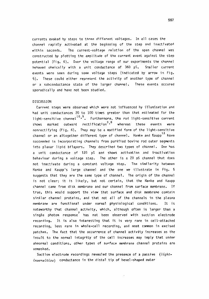
currents evoked by steps to three d i f fe ren t voltages. In a l l cases the
channel rapid ly activated at the beginning of the step and inact ivated
within seconds. The current-voltage re la t ion of the open channel was
constructed by p lo t t ing the amplitude of the current event against the step
potent ia l (Fig. 6). Over the voltage range of our experiments the channel
behaved ohmically with a unit conductance of 360 pS. Smaller current
events were seen during some voltage steps ( indicated by arrow in Fig.
5). These could e i ther represent the ac t i v i t y of another type of channel
or a subconductance state of the larger channel. These events occured
sporadical ly and have not been studied.
DISCUSSION
Current steps were observed which were not influenced by i l luminat ion and
had unit conductances 20 to i00 times greater than that estimated for the
l i gh t - sens i t i ve channel I ° ,9 . Furthermore, the rod l i gh t - sens i t i ve current
shows marked outward r e c t i f i c a t i o n 3'B whereas these events were
nonrect i fy ing (Fig. 6). They may be a modified form of the l i gh t - sens i t i ve
channel or an al together d i f fe ren t type of channel. Hanke and Kaupp 12 have
succeeded in incorporat ing channels from pur i f ied bovine rod outer segments
into planar l i p i d b i layers . They described two types of channel. One has
a unit conductance of 120 pS and shows act ivat ion and inac t iva t ion
behaviour during a voltage step. The other is a 20 pS channel that does
not inact ivate during a constant voltage step, The s im i l a r i t y between
Hanke and Kaupp's large channel and the one we i l l u s t r a t e in Fig. 5
suggests that they are the same type of channel. The or ig in of the channel
is not c lear; i t is l i k e l y , but not cer ta in, that the Hanke and Kaupp
channel came from disk membrane and our channel from surface membrane. I f
t rue, th is would support the view that surface and disk membrane contain
s imi lar channel proteins, and that not a l l of the channels in the plasma
membrane are functional under normal physiological condit ions. I t is
noteworthy that channel a c t i v i t y , which, although often is larger than a ?
single photon response has not been observed with suction electrode
recording. I t is also in terest ing that i t is very rare in cel l -at tached
recording, less rare in whole-cell recording, and most common in excised
patches. The fact that the occurrence of channel a c t i v i t y increases as the
insu l t to the normal i n teg r i t y of the cel l increases may imply that under
abnormal condit ions, other types of surface membrane channel proteins are
unmasked.
Suction electrode recordings revealed the presence of a passive (light- insensitive) conductance in the distal t ip of bevel-shaped outer

S98
segments. The or ig in of the conductance is not c lear. The two most l i k e l y
poss ib i l i t i es are that i t is a hole produced by damage caused by the
suction electrode or that i t is a l i gh t - i nsens i t i ve protein channel. We
argue against electrode damage being the cause of the passive conductance
for the fol lowing reasons. I) Suction electrode recording did not produce
a passive conductance in f la t - t i pped outer segments. 2) Repeatedly drawing
a beveled-t ip outer segment in and out of the suction electrode did not
augment the passive conductance or produce any sign of in jury . 3) I t is
possible to obtain stable, long-term (> 40 min) suction electrode
recordings from beveled-t ip outer segments.
I f the passive conductance is a l i gh t - i nsens i t i ve ion channel i t might
represent normally l i gh t -sens i t i ve channels that do not receive or respond
to internal t ransmit ter . The beveled p ro f i l e of the outer segment t i p
could be due to the curl ing of d ista l disks during an early stage of
phagosome formation in preparation for disk shedding 2. I t is possible that
curled disks are non-functional or are too far away from the surface
membrane to e f fec t i ve l y communicate with the l i gh t -sens i t i ve channel.
Another poss ib i l i t y is that the passive conductance is expressed through
channels that are unrelated to the l i gh t -sens i t i ve conductance. The
ce l l u la r signal for i n i t i a t i o n of shedding may involve act ivat ion of
channels that normally exist in the surface membrane in a nonfunctional
state. This explanation is supported to some extent by the existence of
outer segment single-channel events that d i f f e r , in both the i r conductance
and rec t i fy ing propert ies, from the l i gh t - sens i t i ve channel.
ACKNOWLEDGMENTS
The authors thank Professor Ber t i l H i l l e for his continued interest and
advice and Karen Chin for secretar ial help. Supported by U.S.P.H.S. Grant
EY-02048 from the National Eye Ins t i tu te .
REFERENCES
I . Attwell D, Gray P (1984) Patch-clamp recording from rods of the salamander ret ina. J Physiol 351:9p
2. Anderson DH, Fisher SK (1976) The photoreceptors of diurnal squir re ls: outer segment structure, disk shutt ing, and protein renewal. J Ul t rastruct Res 55:119-141
3. Bader CR, MacLeish PR, Schwartz EA (1978) Responses to l i gh t of so l i ta ry rod photoreceptors isolated from t ige r salamander ret ina. Proc Natl Acad Sci USA 75:3507-3511

$99
4. Baylor DA, Hodgkin AL (1973) Detection and resolut ion of visual st imul i by t u r t l e photoreceptors. J Physiol 234:163-198
5. Baylor DA, Lamb TD (1982) Local effects of bleaching ad ret ina l rods of the toad. J Physiol 328:49-71
6. 8aylor DA, Lamb TD, Yau K-W (1979a) The membrane current of single outer segments. J Physiol 288:589-611
7. Baylor DA, Lamb TD, Yau K-W (1979b) Responses of ret inal rods to single photons. J Physiol 288:613-634
8. Baylor DA, Nunn B (1983) Voltage-dependence of the l i gh t - sens i t i ve conductance of salamander ret inal rods. Biophys J 41:125a
9. Bodoia RD, Detwiler PB (1985) Patch clamp recordings of the l i gh t - sensi t ive dark noise in ret inal rods from l izard and frog. (submitted)
i0. Detwiler PB, Conner JD, Bodoia RD (1982) Gigaseal patch clamp recording from outer segments of in tact ret inal rods. Nature 300:59- 61
i i . Hamill OP, Marty A, Neher E, Sakmann B, Sigworth FJ (1981) Improved patch-clamp techniques for high-resolut ion current recording from ce l ls and ce l l - f ree membrane patches. Pflugers Arch 391:85-100
12. Hanke W, Kaupp UB (1984) Incorporation of ion channels from bovine rod outer segments into planar l i p i d b i layers. Biophys J 46:587-595
13. Noell G, Neher E, Baumann C (1982) Patch electrode recordings from the plasma membrane of rod outer segments of the frog. Pflugers Arch 394:suppl R45
14. Robinson PR, MacLeish PR, Lisman J (1983) Internal d ia lys is of so l i t a ry salamander rod photoreceptors. Soc Neurosci Abst 9:165
15. Robinson PR, MacLeish PR, Lisman J (1984) Whole-cell recording of so l i t a ry salamander rod photoreceptors. Biophys J 45:295a