Type I Interferon Production of Plasmacytoid Dendritic Cells ...
47
International Journal of Molecular Sciences Review Type I Interferon Production of Plasmacytoid Dendritic Cells under Control Dóra Bencze 1,2,† , Tünde Fekete 1,† and Kitti Pázmándi 1, * Citation: Bencze, D.; Fekete, T.; Pázmándi, K. Type I Interferon Production of Plasmacytoid Dendritic Cells under Control. Int. J. Mol. Sci. 2021, 22, 4190. https://doi.org/ 10.3390/ijms22084190 Academic Editor: Marcello Chieppa Received: 11 March 2021 Accepted: 12 April 2021 Published: 18 April 2021 Publisher’s Note: MDPI stays neutral with regard to jurisdictional claims in published maps and institutional affil- iations. Copyright: © 2021 by the authors. Licensee MDPI, Basel, Switzerland. This article is an open access article distributed under the terms and conditions of the Creative Commons Attribution (CC BY) license (https:// creativecommons.org/licenses/by/ 4.0/). 1 Department of Immunology, Faculty of Medicine, University of Debrecen, 1 Egyetem Square, H-4032 Debrecen, Hungary; [email protected] (D.B.); [email protected] (T.F.) 2 Doctoral School of Molecular Cell and Immune Biology, University of Debrecen, 1 Egyetem Square, H-4032 Debrecen, Hungary * Correspondence: [email protected]; Tel./Fax: +36-52-417-159 † These authors have contributed equally to this work and share first authorship. Abstract: One of the most powerful and multifaceted cytokines produced by immune cells are type I interferons (IFNs), the basal secretion of which contributes to the maintenance of immune homeostasis, while their activation-induced production is essential to effective immune responses. Although, each cell is capable of producing type I IFNs, plasmacytoid dendritic cells (pDCs) possess a unique ability to rapidly produce large amounts of them. Importantly, type I IFNs have a prominent role in the pathomechanism of various pDC-associated diseases. Deficiency in type I IFN production increases the risk of more severe viral infections and the development of certain allergic reactions, and supports tumor resistance; nevertheless, its overproduction promotes autoimmune reactions. Therefore, the tight regulation of type I IFN responses of pDCs is essential to maintain an adequate level of immune response without causing adverse effects. Here, our goal was to summarize those endogenous factors that can influence the type I IFN responses of pDCs, and thus might serve as possible therapeutic targets in pDC-associated diseases. Furthermore, we briefly discuss the current therapeutic approaches targeting the pDC-type I IFN axis in viral infections, cancer, autoimmunity, and allergy, together with their limitations defined by the Janus-faced nature of pDC-derived type I IFNs. Keywords: plasmacytoid dendritic cells; type I interferon; regulation; antiviral response; viral infection; cancer; autoimmunity; allergy; IFN gene signature; therapy 1. Introduction Plasmacytoid dendritic cells (pDCs) are a specialized subset of dendritic cells (DCs), which despite their low frequency in the blood, play a crucial role in antiviral immunity and participate in the pathomechanism of several human diseases. PDCs represent a very heterogeneous and plastic cell population [1], which were initially described as a subset of cells with plasma cell-like morphology in lymph nodes in 1958, hence, the name plasmacytoid [2]. Later, in vitro studies showed that these cells share the developmental and functional features of DCs [3], and eventually were identified as professional type I interferon (IFN) producing cells (IPCs) due to their potential to produce large quantities of IFNα in response to viral stimuli [4]. Under physiological conditions, pDCs circulate in the blood or reside in secondary lymphoid organs but can hardly be found in peripheral non-immune tissues [5,6]. Never- theless, under pathological conditions such as microbial infection, chronic inflammation, or cancer, pDCs leave the circulation and accumulate in the inflamed tissues by following the route marked by different chemotactic factors [7]. PDCs infiltrate the mucosa or skin during viral infections [8,9], and their number is also increased in tissue lesions of patients suffering from different autoimmune diseases [10]. In addition, they are present in the nasal mucosa of allergic patients, and they are also associated with different tumor tissues [10]. Int. J. Mol. Sci. 2021, 22, 4190. https://doi.org/10.3390/ijms22084190 https://www.mdpi.com/journal/ijms
-
Upload
khangminh22 -
Category
Documents
-
view
0 -
download
0
Transcript of Type I Interferon Production of Plasmacytoid Dendritic Cells ...
International Journal of
Molecular Sciences
Review
Type I Interferon Production of Plasmacytoid Dendritic Cellsunder Control
Dóra Bencze 1,2,†, Tünde Fekete 1,† and Kitti Pázmándi 1,*
�����������������
Citation: Bencze, D.; Fekete, T.;
Pázmándi, K. Type I Interferon
Production of Plasmacytoid Dendritic
Cells under Control. Int. J. Mol. Sci.
2021, 22, 4190. https://doi.org/
10.3390/ijms22084190
Academic Editor: Marcello Chieppa
Received: 11 March 2021
Accepted: 12 April 2021
Published: 18 April 2021
Publisher’s Note: MDPI stays neutral
with regard to jurisdictional claims in
published maps and institutional affil-
iations.
Copyright: © 2021 by the authors.
Licensee MDPI, Basel, Switzerland.
This article is an open access article
distributed under the terms and
conditions of the Creative Commons
Attribution (CC BY) license (https://
creativecommons.org/licenses/by/
4.0/).
1 Department of Immunology, Faculty of Medicine, University of Debrecen, 1 Egyetem Square,H-4032 Debrecen, Hungary; [email protected] (D.B.); [email protected] (T.F.)
2 Doctoral School of Molecular Cell and Immune Biology, University of Debrecen, 1 Egyetem Square,H-4032 Debrecen, Hungary
* Correspondence: [email protected]; Tel./Fax: +36-52-417-159† These authors have contributed equally to this work and share first authorship.
Abstract: One of the most powerful and multifaceted cytokines produced by immune cells aretype I interferons (IFNs), the basal secretion of which contributes to the maintenance of immunehomeostasis, while their activation-induced production is essential to effective immune responses.Although, each cell is capable of producing type I IFNs, plasmacytoid dendritic cells (pDCs) possess aunique ability to rapidly produce large amounts of them. Importantly, type I IFNs have a prominentrole in the pathomechanism of various pDC-associated diseases. Deficiency in type I IFN productionincreases the risk of more severe viral infections and the development of certain allergic reactions,and supports tumor resistance; nevertheless, its overproduction promotes autoimmune reactions.Therefore, the tight regulation of type I IFN responses of pDCs is essential to maintain an adequatelevel of immune response without causing adverse effects. Here, our goal was to summarizethose endogenous factors that can influence the type I IFN responses of pDCs, and thus mightserve as possible therapeutic targets in pDC-associated diseases. Furthermore, we briefly discussthe current therapeutic approaches targeting the pDC-type I IFN axis in viral infections, cancer,autoimmunity, and allergy, together with their limitations defined by the Janus-faced nature ofpDC-derived type I IFNs.
Keywords: plasmacytoid dendritic cells; type I interferon; regulation; antiviral response; viralinfection; cancer; autoimmunity; allergy; IFN gene signature; therapy
1. Introduction
Plasmacytoid dendritic cells (pDCs) are a specialized subset of dendritic cells (DCs),which despite their low frequency in the blood, play a crucial role in antiviral immunityand participate in the pathomechanism of several human diseases. PDCs represent avery heterogeneous and plastic cell population [1], which were initially described as asubset of cells with plasma cell-like morphology in lymph nodes in 1958, hence, the nameplasmacytoid [2]. Later, in vitro studies showed that these cells share the developmentaland functional features of DCs [3], and eventually were identified as professional type Iinterferon (IFN) producing cells (IPCs) due to their potential to produce large quantities ofIFNα in response to viral stimuli [4].
Under physiological conditions, pDCs circulate in the blood or reside in secondarylymphoid organs but can hardly be found in peripheral non-immune tissues [5,6]. Never-theless, under pathological conditions such as microbial infection, chronic inflammation,or cancer, pDCs leave the circulation and accumulate in the inflamed tissues by followingthe route marked by different chemotactic factors [7]. PDCs infiltrate the mucosa or skinduring viral infections [8,9], and their number is also increased in tissue lesions of patientssuffering from different autoimmune diseases [10]. In addition, they are present in the nasalmucosa of allergic patients, and they are also associated with different tumor tissues [10].
Int. J. Mol. Sci. 2021, 22, 4190. https://doi.org/10.3390/ijms22084190 https://www.mdpi.com/journal/ijms

Int. J. Mol. Sci. 2021, 22, 4190 2 of 47
Under these pathological conditions, pDCs act as a double-edged sword in regulatingimmune responses. On the one hand, pDCs as professional IPCs are indispensable elementsof antiviral immune responses, while on the other hand they can exacerbate inflammatoryresponses or symptoms of autoimmune diseases by the excessive production of type I IFNs,which are powerful cytokines with pleiotropic effects.
Proteins of the type I IFN family have a common helical structure composed of severallong α-helices and are encoded by genes clustered on chromosome 9 in humans [11]. Inhumans, the multi-gene cytokine family of type I IFNs includes 13 subtypes of IFNα, onlyone subtype of IFNβ and single subtypes of the poorly defined IFNε, IFNκ and IFNω [12].Human pDCs mainly express the IFNα and IFNβ subtypes, which act in an autocrine andparacrine manner to initiate cellular and intercellular processes to prevent the spread ofviruses and promote the elimination of virus-infected cells [13]. Almost all cell types in thebody can produce type I IFNs, mainly IFNβ, in response to viral infection, although to amuch lower extent than pDCs. In addition, various microbial products and a diverse arrayof host factors such as cytokines and growth factors can trigger the production of type IIFNs in many cells [14].
Once secreted, type I IFNs signal through the heterodimeric transmembrane IFNαreceptor (IFNAR), which is composed of the IFNAR1 and IFNAR2 subunits. The engage-ment of the receptor activates the tyrosine kinases Janus kinase 1 (JAK1) and tyrosinekinase 2 (TYK2), which phosphorylate the signal transducer and activator of transcription1 (STAT1) and STAT2 transcription factors. Following that, STAT1 and STAT2 moleculesdimerize and translocate to the nucleus to form the so-called IFN-stimulated gene factor3 (ISGF3) trimolecular complex upon assembly with interferon regulatory factor (IRF) 9.ISGF3 then binds to IFN-stimulated response elements (ISREs) and results in the transcrip-tion of several hundreds of IFN-stimulated genes (ISGs). ISG-encoded proteins inducethe establishment of an antiviral state in infected and neighboring cells to prevent viralreplication and the dissemination of the pathogen, thus type I IFNs are a powerful tool totackle viral infections [14,15]. Among the IFN-induced proteins, many enzymes such as theRNA-dependent protein kinase (PKR), the 2′,5′-oligoadenylate (Oligo A) synthetase (OAS),the ribonuclease L (RNase L) and the myxovirus resistance guanosine triphosphatases(Mx GTPases) are upregulated and implicated in the protection against viral infection. Inparticular, PKR changes the translational pattern of the host cell by phosphorylating the αsubunit of eukaryotic initiation factor 2 (eIF2α), which leads to the transient suppression ofprotein synthesis and the consequent prevention of viral replication [16]. Upon binding todsRNA, OAS starts to synthesize Oligo A, which as a second messenger activates RNase Lto degrade viral RNA [17]. Moreover, Mx protein GTPases self-assemble into oligomersand block the intracellular transport of viral nucleocapsid or nucleocapsid-like structures,thus these viral components become trapped and unavailable for the generation of newvirus particles [18].
In addition to eliciting an antiviral state, type I IFNs as potent pleiotropic cytokinesfine-tune innate immune responses by promoting antigen presentation, supporting nat-ural killer (NK) cell functions while limiting the excess production of inflammatory cy-tokines [14]. In particular, IFNα promotes the recruitment of monocytes into the inflamedtissues and their differentiation into effective antigen presenting cells (APCs) [19,20]. More-over, type I IFNs induce DC maturation and activation [21]. At later stages of infection,type I IFNs activate adaptive T and B cell responses and promote the development ofimmunological memory. Via supporting the production of B lymphocyte stimulators suchas B cell activating factor (BAFF) and A proliferation-inducing ligand (APRIL) by DCs andmacrophages, type I IFNs have also been reported to improve B cell survival, maturation,differentiation, and class-switch recombination [22]. Furthermore, upon acute infections,type I IFNs support the activation and expansion of antigen-specific CD4+ helper T (Th)cells and CD8+ cytotoxic T cells, and contribute to the differentiation of follicular Th cells,which are critical to the induction of B cell responses [23,24].
Int. J. Mol. Sci. 2021, 22, 4190 3 of 47
Besides regulating innate and adaptive immune cell activation, survival, and differen-tiation, type I IFNs also contribute to the control of various physiological processes such asthe regulation of hematopoietic stem cell niche function, bone remodeling, the maintenanceof synaptic plasticity and cognitive function of the healthy central nervous system, and themaintenance of immune homeostasis [25,26]. Furthermore, commensal microbiota-driventonic levels of IFN signals at mucosal surfaces adjust the activation threshold of the innateimmune system, which is essential for ideal antiviral responses [27–29] (Figure 1).
Int. J. Mol. Sci. 2021, 22, 4190 3 of 49
Besides regulating innate and adaptive immune cell activation, survival, and differ-entiation, type I IFNs also contribute to the control of various physiological processes such as the regulation of hematopoietic stem cell niche function, bone remodeling, the mainte-nance of synaptic plasticity and cognitive function of the healthy central nervous system, and the maintenance of immune homeostasis [25,26]. Furthermore, commensal microbi-ota-driven tonic levels of IFN signals at mucosal surfaces adjust the activation threshold of the innate immune system, which is essential for ideal antiviral responses [27–29] (Fig-ure 1).
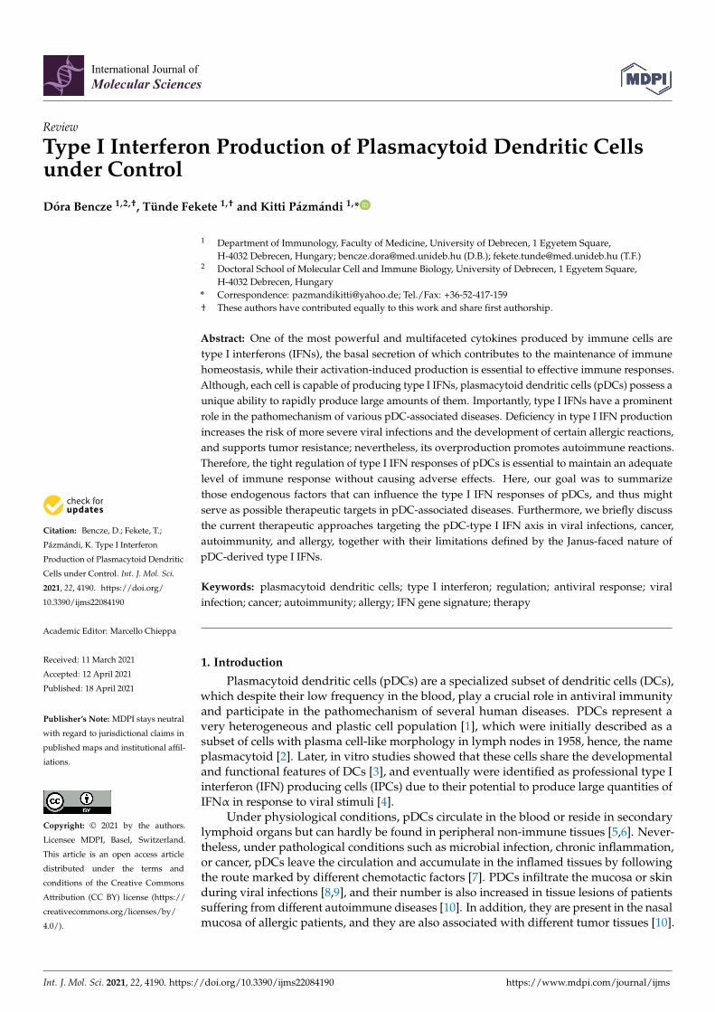
Figure 1. The pleiotropic effects of type I interferons (IFNs). Continuous baseline production of type I IFNs by various tissues and cells fine-tunes a wide variety of physiological processes including hematopoietic stem cell functions, synaptic plasticity, bone remodeling and immune homeostasis. In addition, the microbiota-induced basal IFN-signature prepares stromal and immune cells for upcoming infections (upper left panel). Upon viral infection, type I IFN signaling induces antiviral state in all nucleated cells via the upregulation of IFN-stimulated genes that inhibit the replication and spreading of viruses (upper right panel). Type I IFNs also control the cells of innate (lower left panel) as well as adaptive (lower right panel) immune system by shaping the activation, differentiation, effector functions and trafficking of these cells. eIF2α: eukaryotic initiation factor 2α; IFN: interferon; IFNAR: interferon-alpha/beta receptor; ISRE: IFN-stimulated response ele-ment; Mx GTPase: myxovirus resistance guanosine triphosphatase; NK: natural killer; OAS: 2′-5′ oligoadenylate synthetase; Oligo A: 2′-5′-oligoadenylate; PKR: protein kinase R; Rnase L: ribonuclease L; Th: T helper.
Under physiological conditions, in the absence of acute infection, type I IFNs are con-stitutively secreted at a baseline level in many tissues, and it appears that lower or higher amounts than that might have pathologic consequences [25]. Despite their beneficial ef-fects, type I IFNs are detrimental to the host when their expression is dysregulated. While the acute and transient production of type I IFNs promote antiviral responses, their sus-tained and chronic secretion drives various autoimmune and non-autoimmune inflam-matory diseases. These diseases are characterized by the so-called IFN gene signature (IGS), which refers to the upregulation of IFN inducible genes in peripheral blood cells. IGS was found to be correlated with disease severity in patients with autoimmune dis-eases such as systemic lupus erythematosus (SLE), rheumatoid arthritis (RA) or derma-tomyositis [30].
Owing to their potential to secrete large quantities of type I IFNs, pDCs have been identified as major players in a number of type I IFN-mediated inflammatory conditions.
Figure 1. The pleiotropic effects of type I interferons (IFNs). Continuous baseline production of type I IFNs by varioustissues and cells fine-tunes a wide variety of physiological processes including hematopoietic stem cell functions, synapticplasticity, bone remodeling and immune homeostasis. In addition, the microbiota-induced basal IFN-signature preparesstromal and immune cells for upcoming infections (upper left panel). Upon viral infection, type I IFN signaling inducesantiviral state in all nucleated cells via the upregulation of IFN-stimulated genes that inhibit the replication and spreadingof viruses (upper right panel). Type I IFNs also control the cells of innate (lower left panel) as well as adaptive (lowerright panel) immune system by shaping the activation, differentiation, effector functions and trafficking of these cells.eIF2α: eukaryotic initiation factor 2α; IFN: interferon; IFNAR: interferon-alpha/beta receptor; ISRE: IFN-stimulated response element;Mx GTPase: myxovirus resistance guanosine triphosphatase; NK: natural killer; OAS: 2′-5′ oligoadenylate synthetase; Oligo A:2′-5′-oligoadenylate; PKR: protein kinase R; Rnase L: ribonuclease L; Th: T helper.
Under physiological conditions, in the absence of acute infection, type I IFNs areconstitutively secreted at a baseline level in many tissues, and it appears that lower orhigher amounts than that might have pathologic consequences [25]. Despite their beneficialeffects, type I IFNs are detrimental to the host when their expression is dysregulated.While the acute and transient production of type I IFNs promote antiviral responses,their sustained and chronic secretion drives various autoimmune and non-autoimmuneinflammatory diseases. These diseases are characterized by the so-called IFN gene signature(IGS), which refers to the upregulation of IFN inducible genes in peripheral blood cells. IGSwas found to be correlated with disease severity in patients with autoimmune diseases suchas systemic lupus erythematosus (SLE), rheumatoid arthritis (RA) or dermatomyositis [30].
Owing to their potential to secrete large quantities of type I IFNs, pDCs have been iden-tified as major players in a number of type I IFN-mediated inflammatory conditions. Sincethe discovery of pDCs, several regulatory factors and mechanisms have been identified,
Int. J. Mol. Sci. 2021, 22, 4190 4 of 47
which affect their type I IFN production and might serve as possible therapeutic targets.In the present review, first we briefly introduce the molecular basis of the unique type IIFN producing capacity of pDCs. Following that, we summarize all those endogenousfactors, which can modulate type I IFN production specifically in pDCs and outline therole of pDC-derived type I IFNs in antiviral response, cancer, autoimmunity, and allergy,and shortly discuss the potential therapeutic approaches in these diseases.
2. Professionalism of pDCs in the Production of Type I IFNs
Despite being discovered in the mid-20th century, pDCs were explicitly characterizedmany decades later due to their low frequencies in peripheral blood and rapid apoptosisunder in vitro culture conditions. After the ability of these cells to secrete extreme amountsof type I IFNs in a relatively short time was recognized, they were defined as naturalIPCs. According to the current model, IPCs are precursors of pDCs, and therefore theIFN-producing form of pDCs is regarded as plasmacytoid pre-DCs. Plasmacytoid pre-DCsare small, round cells with a plasma cell-like morphology and a well-developed endoplas-mic reticulum (ER), which enables the production of large quantities of proteins. Upondifferentiation into mature pDCs, they lose their exceptional type I IFN-producing ability,take up DC-like morphology, express high levels of major histocompatibility complex(MHC) and costimulatory molecules, and become professional APCs being capable ofstimulating naive T cells [31].
Plasmacytoid DCs have emerged as “the virus experts” of our body owing to theirstriking capacity to produce large amounts of type I IFNs. Within 6 h of viral exposure,pDCs devote 50% of their induced transcriptional activity to initiate type I IFN gene ex-pression [32]. Human pDCs express a wide repertoire of type I IFNs including 13 subtypesof IFNα and single subtypes of IFNβ, IFNω, and IFNτ [33]. In response to viral infection,pDCs are responsible for 95% of type I IFN production by mononuclear cells, as they areable to produce 200–1000 times more type I IFNs than any other white blood cell aftermicrobial exposure [4]. Quantitatively, one single pDC can produce 3–10 pg of IFNα inresponse to a strong stimulus. While secreting high amount of IFNα, pDCs produce muchlower levels of IFNβ and additional type I IFNs [34]. All these data raise the question ofhow pDCs are capable of such a peak performance. In contrast to conventional DCs (cDC),pDCs selectively express endosomal Toll-like receptor (TLR) 7 and 9, which recognize viralRNA and DNA, respectively, and their activation is associated with a high productionof type I IFNs by pDCs [35]. In contrast to other cell types, pDCs show a high degree ofresistance to viral infections, and do not need to be infected with live viruses to inducethe production of type I IFNs [36], since inactivated viruses can also stimulate pDCs if theviral envelope remains intact [37]. The internalized virus particles are degraded withinendocytic vesicles and are sensed through TLR7 and TLR9, the engagement of whichinduces signaling through the adaptor protein MyD88. Upon association with downstreamsignaling components, MyD88 leads to the phosphorylation and nuclear translocationof the IRF7 transcription factor, which initiates the transcriptional activation of type IIFN genes. In contrast to cDC, in which IRF7 is inducible and requires prior stimulation,pDCs constitutively express IRF7, possibly due to the lack of the translational repressoreukaryotic translation initiation factor 4E (eIF4E)-binding protein (4E-BP), which allows therapid onset of type I IFN production in pDCs [38]. However, constitutive IRF7 expressionalone would not be sufficient to induce the production of large amounts of type I IFNsin pDCs. Honda et al. described a unique spatiotemporal regulation of the TLR9-MyD88pathway in pDCs in response to a specific TLR9 ligand, CpG-A. Following recognition,CpG-A oligonucleotides form large multimeric aggregates, which are retained in the earlyendosomes of pDCs for about 30 min that allows prolonged IRF7 induction, whereas incDCs, those are rapidly transferred to lysosomal compartments. Thus, CpG-A stimulationefficiently enhances the production of type I IFNs in pDCs via IRF7, whereas barely affectsnuclear factor-κ B (NF-κB) activity and thus the maturation of these cells. However, itis important to note that this effect highly depends on the structural properties of the
Int. J. Mol. Sci. 2021, 22, 4190 5 of 47
TLR ligand. Other subtypes of oligonucleotides such as the monomeric CpG-B is rapidlytransported to late endosomes, where recognition through TLR9 leads to the activation ofthe NF-κB pathway, which induces the synthesis of inflammatory cytokines, chemokines,and costimulatory molecules; therefore, CpG-B is much less effective in the initiation oftype I IFN production compared to CpG-A [39]. In terms of kinetic, pDCs secrete highamounts of IFNα within the first 12 h of exposure to CpG-A or live viruses, and thenin the next 48 h, pDCs are able to produce only a small fraction of this quantity uponre-stimulation [33].
Previously, our research group proposed a model, in which type I IFN secretionby pDCs occurs in two waves under the coordinated regulation by different subtypes ofpattern recognition receptors (PRRs) [40]. In the early phase of viral infection, pDCs are ableto detect debris from virus-infected cells in the lymph nodes through constantly expressingTLR7 and TLR9 receptors that results in the secretion of high levels of type I IFNs andsubsequent induction of a systemic antiviral state. The systemic effect is facilitated by theunique localization of pDCs, in which aspect they highly differ from cDCs that are generallylocated in peripheral tissues, i.e., at the sites of viral entry, whereas pDCs are found inthe blood or in lymphoid tissues such as lymph nodes. Therefore, pDCs are initially notinfected, but can detect virus-infected cell debris delivered to lymph nodes and producea large amount of antiviral cytokines, which might reach every cell in the body throughthe blood or lymphoid circulation, and thus contributes to the development of a systemicantiviral state. In the later stage of viral infection, TLR-activated pDCs migrate to the siteof virus entry, where due to the high viral load, they can also get infected. However, it isimportant to note that previous TLR stimulation induces the expression of cytosolic retinoicacid-inducible gene I (RIG-I)-like receptors (RLRs) in pDCs, the expression of which isonly marginal in these cells without prior activation. Therefore, pDCs gain the ability torecognize replicating viruses even in the cytoplasm after TLR stimulation. Upon ligandengagement, RLRs recruit the mitochondrial antiviral signaling adapter protein (MAVS),which leads to the activation of MAVS-dependent IRF3/7 pathways and ultimately resultsin a second/late wave of type I IFN production. The RLR-mediated type I IFN productionis lower in amounts than the TLR-induced secretion by pDCs; however, it still effectivelysupports potent local antiviral responses [40].
Based on single-cell genomic profiling several models have recently been proposedon the fate and functional plasticity of pDC [1,36,41]. According to these models, pDCshighly differ in their degree of differentiation and consequently in their capacity to producetype I IFNs or present antigens. A current study demonstrated that the same individualpDC first produces type I IFNs and then acquires the ability to present antigens to T cellsduring in vivo viral infections suggesting that pDCs exert different functions orchestratedin a spatiotemporal manner [42]. Other models suggest that only a small fraction of pDCscan produce type I IFNs, regardless of the stimulus. It is also plausible that while somepDCs get infected, others recognize the infected pDCs and start to produce the type I IFNsas a response [1]. The above data indicate that further studies are required to explain theobserved discrepancies and clarify whether the functional differences of pDCs are due toterminal functional specialization or are subsequent stages of pDC maturation.
In conclusion, pDCs represent the number one source of type I IFNs, which cytokinesimpact an array of cellular events, physiological processes and both innate and adaptiveimmune responses. Therefore, fine-tuning type I IFN responses of pDCs by differentendogenous factors or regulatory mechanisms is essential to keep a balance betweenprotection and unwanted pathological events.
3. Regulation of Type I IFN Production at the Transcriptional and Posttranscriptional Level3.1. Transcription Factors
The induction of type I IFN secretion is primarily controlled at the transcriptionallevel, where IRFs play an essential role. In pDCs, IRF7 and IRF3 are the master regulators oftype I IFN responses [40,43]. As we previously described, the MyD88-dependent TLR7/9
Int. J. Mol. Sci. 2021, 22, 4190 6 of 47
signaling cascade activates IRF7 to induce robust production of type I IFNs, whereas theMAVS-dependent RLR signaling pathway is able to activate both IRF3 and IRF7 to promotethe secretion of type I IFNs in pDCs [40,44]. In addition, other members of the IRF familyhave been implicated in the regulation of type I IFN production [45].
Several studies demonstrated that IRF5 and IRF8 also contribute to the regulationof type I IFN production. In mouse pDCs, IRF5 was found to only partially affect theinduction of IFNα, whereas it seems to be critical for IFNβ gene induction [46,47]. Inthe human CAL-1 pDC cell line, IRF5 silencing resulted in an 80% reduction in CpG-B-triggered gene activation as compared with controls [48]. Interestingly, the same studyidentified IRF8 as a negative regulator, since silencing of IRF8 led to a 60% increase ingene activation upon CpG stimulation [48]. Moreover, the authors also found that IRF5and IFR8, which colocalize within the cytoplasm of resting pDCs, rapidly translocate tothe nucleus after CpG triggering, and thus hypothesize that IRF8 interacts with IRF5 tocontrol TLR9 signaling in human pDCs [48]. By contrast, another study demonstratedthat the depletion of IRF8 decreased the IFNα secreting capacity of mouse pDCs uponCpG stimulation indicating its importance in mouse pDCs [49]. Thus, the above datasuggest that IRF8 controls the magnitude of IFN responses and exerts positive or negativeregulatory effects depending on the origin of the cell.
Besides IRFs, several other transcription factors were identified as regulators of pDCfunctions. Runt-related transcription factor 2 (RUNX2) is essential for the optimal produc-tion of type I IFNs through the modulation of IRF7 expression in mice. In the absence ofRUNX, both resting and CpG-activated mouse bone marrow (BM)-derived pDCs showeddecreased IRF7 expression, which resulted in a significantly reduced production of IFNαand IFNβ [50]. Further, mouse pDCs highly express the Ets family transcription factor,Spi-B, which can transactivate the promoters of type I IFN in synergy with IRF7 [51].BM-derived and splenic pDCs from Spi-B-deficient mice showed defective induction ofIFNα genes following CpG-B, polyuridylic acid (polyU) and vesicular stomatitis virus(VSV) stimulation. The authors also concluded that constitutive high expression of Spi-Bcontributes to the ability of pDCs to produce high amounts of type I IFNs in response toTLR7 and TLR9 ligands [51]. The nuclear factor of activated T cells C 3 (NFATC3) was alsofound to enhance IRF7-mediated IFN release by both mouse and human pDCs [52]. InBM-derived pDCs from NFATC3-deficient mice TLR7/9-mediated IFNα production wasgreatly reduced compared to wild type mice [52]. In the Gen2.2 human pDC cell line, theknockdown of NFATC3 also led to a substantial reduction of IFNα production in responseto CpG-A. Furthermore, it was demonstrated that NFATC3 forms a complex with IRF7and binds to IFN promoters to augment maximal production of type I IFNs upon TLR9stimulation in GEN2.2 cells [52].
In contrast to the above mentioned transcription factors, the pleiotropic transcriptionfactor MYC negatively regulates the TLR-mediated antiviral response of human pDCs [53].The knockdown of MYC increased the CpG-B triggered induction of IFN-stimulated genesin the human GEN2.2 pDC cell line. In particular, MYC is shown to interact and forma complex with the nuclear receptor co-repressor 2 (NCOR2) and histone deacetylase 3(HDAC3) to occupy, and thus repress the promoter region of IRF7. These data implythat MYC suppresses IRF7 promoter activity to ensure the optimal levels of type I IFNproduction and prevent the development of autoimmune diseases [53].
Another critical regulator of various signaling pathways, the CXXC type zink fingerprotein 5 (CXXC5) is suggested to act as a transcription factor as well as an epigeneticmodifier [54]. It is highly expressed in mouse and human pDCs, where, as an epigeneticregulator it controls DNA methylation and histone modifications [55]. Following stimu-lation with herpes simplex virus-1 (HSV-1) or CpG-A, pDCs from CXXC5-deficient miceexpressed lower levels of IRF7 and produced much less type I IFN compared to controlpDCs. Mechanistically, CXXC5 recruits the Tet2 DNA demethylase, which maintains hy-pomethylation of CpG-island containing genes such as IRF7, and thus contributes to therapid and robust type I IFN production of mouse pDCs [55]. Similarly, the knockdown of

Int. J. Mol. Sci. 2021, 22, 4190 7 of 47
CXXC5 in the human GEN2.2 pDC cell line led to reduced IRF7 expression and decreasedmRNA levels of IFNα and IFNβ upon exposure to the TLR7 ligand R848 and HSV-1 [55].Furthermore, E2-2 is also a specific regulator of mouse as well as human pDCs, since itcan directly activate the expression of multiple genes involved in pDC-mediated type IIFN responses, namely the TLR7 and TLR9 receptors as well as the IRF7, IRF8 and Spi-Btranscription factors [56]. The deletion of E2-2 in mouse BM-derived pDCs abolished type IIFN release in response to CpG-A that could be explained by the reduced expression of theaforementioned TLR pathway components [56]. Similarly, E2-2 knockdown in the humanGEN2.2 cell line downregulated the expression of pDC signature genes and diminishedIFNα production in response to CpG-B. Interestingly, E2-2 silencing in GEN2.2 cells upreg-ulated a set of cDC specific genes, including the anti-inflammatory TLR10 and inhibitorySiglec-6 receptors that could explain the abrogated IFNα production in response to TLR9stimulation [57].
The above data indicate that several transcription factors act in concert to coordinatethe optimal expression of type I IFNs in both human and mouse pDCs (Table 1).
Table 1. Regulation of type I IFN production at the transcriptional and posttranscriptional level.
Transcription Factors
RegulatingFactor
Type ofRegulation Mechanism of Regulation Model Ref.
IRF5 positive induces the expression of type IIFN genes mouse [46,47]
IRF5 positive induces the expression of type IIFN genes human [48]
IRF8 negative inhibits IRF5 human [48]
IRF8 positive - mouse [49]
RUNX2 positive induces IRF7 expression human [50]
Spi-B positive transactivates the promoters oftype I IFNs mouse [51]
NFATC3 positive
binds to type I IFN promoters insynergy with IRF7
(mechanism demonstrated onhuman pDCs)
mouse/human [52]
MYC negative represses IRF7 promoter activity human [53]
CXXC5 positive
maintains constitutivetranscription of IRF7
(mechanism demonstrated onmouse pDCs)
mouse/human [55]
E2-2 positive supports the expression of TLR7,TLR9, IRF7, IRF8 and Spi-B mouse [56]
E2-2 positive downregulates the expression ofTLR10 and Siglec-6 human [57]
Adaptor Proteins and Other Intracellular Regulators
RegulatingFactor
Type ofRegulation Mechanism of Regulation Model Ref.
Opn-i positive supports the nuclear translocationof IRF7 mouse [58]
PACSIN1 positive - mouse/human [59]

Int. J. Mol. Sci. 2021, 22, 4190 8 of 47
Table 1. Cont.
Adaptor Proteins and Other Intracellular Regulators
RegulatingFactor
Type ofRegulation Mechanism of Regulation Model Ref.
TRIM8 positiveprevents phosphorylated IRF7from proteasomal degradation(demonstrated on HEK293T)
human [60]
PLSCR1 positive
supports TLR9 trafficking to theearly endosomes
(mechanism demonstrated onhuman pDCs)
mouse/human [61].
SphK1 positive
regulates the nuclear transport ofIRF7 and uptake of CpG
(mechanism demonstrated onhuman pDCs)
mouse/human [62]
SCARB2 positive mediates TLR9 trafficking and thenuclear translocation of IRF7 human [63]
mTOR positivesupports TLR-mediated IRF7phosphorylation and nuclear
translocation
mouse/human
[64]
mTOR positive supports RLR-mediated TBK1phosphorylation human [65]
mtROS negativesuppresses TLR9-triggered type I
IFN production through inhibitingIRF7 phosphorylation
human [66]
mtROS positivesupports RLR-triggered type I IFN
production through IRF3phosphorylation
human [66]
ROS negative inhibits TLR7-mediated type I IFNs human [67]
MicroRNAs
RegulatingFactor
Type ofRegulation Mechanism of Regulation Model Ref.
miR-155 negative represses TAB2 human [68]
miR-155 * positive suppresses IRAK-M human [68]
miR-146a negative targets IRAK-1 human [69]
miR-618 positive - human [70]
miR-21 positive suppresses PTEN mouse [71]
miR-126 positive targets TSC1 mouse [72]Abbreviations: CXXC5: CXXC-type zinc finger protein 5; IFN: interferon; IRAK: interleukin 1 receptor associated kinase 1;IRF: interferon regulatory factor; miR: microRNA; mtDNA: mitochondrial DNA; mTOR: mammalian target of rapamycin;mtROS: mitochondrial ROS;NFATC3: nuclear factor of activated T cells 3; Opn-i: intracellular osteopontin; Ox-mtDNA:oxidized mitochondrial DNA; PACSIN1: protein kinase C and casein kinase substrate in neurons 1; PLSCR1: phospholipidscramblase 1; PTEN: phosphatase and tensin homolog; RLR: RIG-I-like receptor; RUNX2: Runt-related transcription factor2; SCARB2: scavenger receptor class B member 2; SphK1: sphingosine kinase 1; TAB2: TGFβ activated kinase 1 bindingprotein 2; TBK1: TANK-binding kinase 1; TLR: toll-like receptor; TRIM8: tripartite motif containing protein 8; TSC1:tuberous sclerosis complex 1.
3.2. Adaptor Proteins and Other Intracellular Regulators
Besides the common downstream signaling components of TLR and RLR pathways,numerous adaptor proteins and intracellular molecules act as positive or negative regula-tors of type I IFN secretion by pDCs.
Among them, the glycoprotein osteopontin (Opn) seems to be essential to the typeI IFN responses in pDCs. Many cell types are able to express Opn, which is involved in
Int. J. Mol. Sci. 2021, 22, 4190 9 of 47
various pathophysiological events when secreted [73]. However, novel studies found thata portion of newly synthesized Opn is retained in the cytoplasm (referred as Opn-i), whereacting as an adaptor protein controls signal transduction pathways downstream of innateimmune receptors [73]. In splenic mouse pDCs, Opn deficiency significantly reduced theproduction of IFNα in response to CpG-A or CpG-B stimulation [58]. Opn-i colocalizes withMyD88 and TLR9 that is essential to the nuclear translocation of IRF7, and thus the optimalinduction of IFNα gene expression [58]. Furthermore, the cytoplasmic phosphoproteinsProtein kinase C and casein kinase substrate in neurons 1 (PACSIN1) was also identified asa pDC-specific adaptor molecule critical for the TLR7/9-mediated type I IFN responses inboth human and mouse pDCs [59]. The knockdown of PACSIN1 in human GEN2.2 cellsinhibited IFNα responses to CpG-A stimulation. In addition, the depletion of PACSIN1 inmouse BM-derived pDCs considerably reduced the levels of IFNα production in responseto CpG-A, influenza virus (Flu), VSV and HSV-1. Since Flu and VSV are recognized viaTLR7, whereas HSV-1 and CpG are sensed by TLR9, the authors concluded that PACSIN1affects IFNα production by both TLR7 and TLR9 signaling pathways in pDCs [59].
Several members of the tripartite motif (TRIM)-containing proteins have also emergedas important modulators of innate immune signaling cascades including the type I IFNpathway of pDCs. In primary human pDCs, TRIM20, TRIM22, TRIM28, and TRIM36were identified as negative regulators of type I IFN responses [60]. On the contrary,TRIM8, which is constitutively expressed in resting pDCs, was revealed as a strong positiveregulator of type I responses of pDCs and therefore its regulatory mechanisms werefurther investigated [60]. The silencing of TRIM8 in pDCs led to a profound decreasein IRF7 phosphorylation and abolished type I IFN production in response to HIV or Fluvirus infection. The authors also showed with mechanistic studies on HEK293T cells thatTRIM8 prevents the proteasomal degradation of phosphorylated IRF7 through inhibitingits recognition by the peptidyl-prolyl isomerase Pin1 [60].
Another positive regulator is the phospholipid scramblase 1 (PLSCR1) protein, whichinteracts directly with several plasma membrane receptors, and acting as a scramblase, isinvolved in multiple biological processes [74]. Silencing of PLSCR1 in human GEN2.2 cellsled to a significant reduction of IFNα responses following CpG-A or CpG-B stimulation [61].Similar results were also obtained with BM-derived pDCs from PLSCR1-deficient micewhen stimulated with CpG-A, Flu virus and HSV-1. In one human pDC cell line, PLSCR1plays an important role in TLR9 trafficking from the ER to the early endosomes, and thussupports its engagement with synthetic or viral ligand [61].
Recently, it was reported that the lipid converting enzyme Sphingosine kinase 1(SphK1), which catalyzes the formation of the lipid signaling molecule sphingosine-1-phosphate (S1P), plays a critical role in pDC functions as well [62]. The depletion ofShpK1 in mouse pDCs or pre-treatment of human CAL-1 cells and mouse splenic pDCswith ShpK1-specific inhibitors significantly impaired TLR7/9-mediated type I IFN produc-tion [62]. Mechanistically, ShpK1 was found to regulate the nuclear transport of IRF7 aswell as the uptake and trafficking of CpG to endosomes, where TLR9 activation occurs.Further, pharmacological inhibition of SphK1 or its genetic deletion in a mouse model ofSLE decreased pDC activation and ISGs expression [62].
In addition, the lysosomal membrane protein Scavenger receptor class B member 2(SCARB2) is highly expressed in resting pDCs, and its expression can be further upregulatedby CpG stimulation [63]. Interestingly, the silencing of SCARB2 in human GEN2.2 cellssignificantly reduced the CpG-B-mediated IFNα production, whereas did not affect it uponCpG-A triggering. Further studies revealed that SCARB2 localizes in the late endosome,where it mediates the endosomal transport of TLR9 as well as nuclear translocation of IRF7,and thus regulates the expression of IFNα in pDCs [63].
Besides the aforementioned regulatory factors, the major drivers of immunometabolicchanges can also impact type I IFN production of pDCs. Immunometabolism is one ofthe hottest topics and a dynamically growing field of immunology, which can providenew therapeutic approaches for the treatment of immune-related diseases. Immune cell
Int. J. Mol. Sci. 2021, 22, 4190 10 of 47
functions can be shaped both by intracellular metabolic processes and external metabolitesderived from pathogens, microbes, or tumor cells [75]. Most importantly, several papersreported that alterations in energy and lipid metabolism modulate the type I IFN producingability of pDCs [76–78]. The mammalian target of rapamycin (mTOR) has emerged as themaster regulator of cellular metabolism by controlling a myriad of cellular functions inresponse to environmental factors and intracellular signals. It was revealed first in 2008that the phosphatidylinositol 3-kinase (PI3K)-Akt-mTOR signaling pathway is crucial forthe TLR-mediated type I IFN responses of pDCs [64]. The inhibition of mTOR complex1 by rapamycin significantly decreased the TLR9-triggered production of IFNα both inmouse and human pDCs. Moreover, the inhibition of the PI3K upstream molecule orp70S6 kinase downstream target also substantially suppressed the CpG-A-induced IFNαsecretion by mouse pDC. Furthermore, the TLR7-induced IFNα secretion was also impairedin murine splenic pDCs. The findings also indicated that blockade of mTOR signalingresults in the disruption of MyD88-TLR9 complex and impairment of IRF7 phosphorylationand nuclear translocation [64]. Another group further confirmed these data by showingthat suppressing the signaling components of the PI3K-Akt-mTOR pathway by variousapproaches significantly reduced IFNα secretion in primary human pDCs [79]. A differentstudy also demonstrated that rapamycin potently inhibits TLR7/9-induced IFNα secretionin peripheral blood pDCs [80]. In addition, our research group demonstrated for the firsttime that mTOR signaling is also essential to the RLR-triggered antiviral immune responsesof pDCs. We described that mTOR blockade by rapamycin or the dual kinase inhibitorAZD8055 inhibited the type I IFN production in primary human pDCs and GEN2.2 cellsupon RLR stimulation [65]. Further, we found that mTOR blockade decreased the RLR-triggered phosphorylation of Tank-binding kinase 1 (TBK1), which based on literaturedata is a requirement for IRF3/7 activation [81]. Thus, we hypothesize that mTOR mightsupport the RLR-initiated type I IFN production of pDCs via regulating at the level orupstream of TBK1.
It has long been recognized that several metabolic processes are linked to the enhancedgeneration of mitochondrial reactive oxygen species (mtROS), which are also essentialmediators of immune responses. MtROS are constitutively generated during physiologicalconditions and their production can be further increased under pathological states. Asimportant signaling molecules, mtROS participate in various processes of immune cellfunctions such as activation or inflammatory cytokine production [82]. Interestingly, BM-derived pDCs of aged mice show defective IRF7 upregulation upon TLR9 activation ascompared with cells from younger counterparts [83]. Both resting and TLR9 activated agedpDCs displayed elevated levels of reactive oxygen species (ROS), the reduction of which byantioxidant pre-treatment restored IFNα production during TLR9 activation. These resultssuggest that age-induced oxidative stress impairs the antiviral capacity of pDCs in responseto TLR9 stimulation, which might explain the increased susceptibility of older individualsto viral infections. In addition, our research group described that mtROS can influence thetype I IFN producing capacity of human pDCs [66]. The TLR9 agonist CpG-A triggeredthe type I IFN production of human GEN2.2 cells was markedly reduced by high mtROSlevels, which inhibited the phosphorylation of IRF7. On the contrary, elevated mtROSincreased RIG-I-stimulated type I IFN expression and augmented the phosphorylationof Akt and IRF3, which are essential components of RLR signaling [66]. Interestingly, ina separate study, we demonstrated that exogenous ROS also have a negative effect onthe TLR7-dependent activation of pDCs [67]. Thus, our data suggest that the effects ofmtROS on pDCs depend on which viral sensing pathways are stimulated. Namely, theearly TLR7/9-mediated type I IFN response is abrogated, whereas the late RLR-mediatedproduction of type I IFNs is supported by elevated mtROS levels. (Table 1)
Although, the above listed regulatory molecules have multifunctional roles in theregulation of various physiological cellular functions, those are also important in fine-tuning the extent of type I IFN responses by pDCs as well. It is also noteworthy that through
Int. J. Mol. Sci. 2021, 22, 4190 11 of 47
feedback mechanisms secreted type I IFNs might also act on diverse cellular functionsincluding metabolic processes, which highly influence the activation of immune cells.
3.3. MicroRNAs
MicroRNAs (miRs) are short, non-coding RNAs, which upon binding to target mRNAslead to their degradation or translational suppression. By controlling gene expression atthe posttranscriptional level in various cell types, miRs have emerged as key coordinatorsof both innate and adaptive immune responses [84], and several of them have also beenlinked to the regulation of type I IFN responses in pDCs [85].
MiRs profiling of human pDCs revealed that TLR7 stimulation highly induced theexpression of both the guide (miR) and passenger strands (miR*) of 19 different miRNAs,among which miR-155 and miR-155* were the most highly induced ones. Interestingly, miR-155 inhibited the TLR7-triggered production of IFNα and IFNβ expression by targetingTGFβ activated kinase 1 binding Protein 2 (TAB2), whereas miR-155* augmented it bysuppressing interleukin-1 receptor-associated kinase (IRAK)-M [68].
Similarly, miR-146a expression was also induced upon TLR7/9 stimulation in primaryhuman pDCs [69]. However, the silencing of miR-146a increased the percentage of IFNα ex-pressing pDCs upon stimulation with CpG-A, thus it was identified as a negative regulatorof type I IFN production [69]. Further studies with the CAL-1 pDC cell line also revealedthat miR-146a targets IRAK-1, which is an essential element for IRF7 activation [69].
Interestingly, increasing evidence has revealed that the dysregulation of miR ex-pression can contribute to the development and maintenance of various autoimmunediseases [86]. So far, two studies revealed association between miR expression and type IIFN production of pDCs in patients with IFN signature. One study found that pDCs ofsystemic sclerosis patients upregulated miR-618, the overexpression of which in primaryhuman pDCs resulted in a higher secretion of IFNα in response to the TLR9 stimulationcompared to pDCs derived from healthy individuals [70]. Another study reported thatpDCs of SLE and antiphospholipid syndrome (APS) patients with high IFN signaturedisplayed reduced expression of miR-361-5p, miR-128-3p and miR-181a-2-3 p compared topDCs from patients without an IFN signature or healthy controls [87]. Circulating pDCsfrom Sjögren’s patients with an IFN signature also showed decreased expression of severalmiRs, however, no strong correlation was found between the miR profile of pDCs and IFNsignature of patients [88].
The role of miRs in the regulation of type I IFN responses by pDCs was also proven inmouse models. In mouse BM-derived pDCs, both TLR7 and TLR9 stimulation increasedthe expression of different miRs such as miR-21, which was found to be an essentialpositive regulator for IFNα. The phosphatase and tensin homolog (PTEN) was identifiedas the target of miR-21. By suppressing PTEN, miR-21 promotes the TLR7/9-mediatedactivation of PI3K-Akt-mTOR signaling, which is essential for IRF7 protein expression andsubsequent IFNα production [71]. Furthermore, miR-126, which is mostly associated withthe regulation of vasculogenesis and angiogenesis, is also able to control IFNα and IFNβproduction of mouse pDCs. MiR-126 deficient mice produced less IFNα than wild-typemice in response to TLR7 (R848) and TLR9 (CpG-A) agonists [72]. Splenic and lymphnode-derived pDCs from miR-126-/- mice were also defective in their TLR9-mediatedIFNα producing capacity compared to wild type pDCs [72]. Further data indicate thattuberous sclerosis complex 1 (TSC1), which is a negative regulator of mTOR, is the target formiR-126 in pDCs [72]. It was also hypothesized that miR-126 acts through the upregulationof vascular endothelial growth factor receptor 2 (VEGFR2), which also negatively regulatesTSC1, thus increases mTOR activity and type I IFN production in pDCs [72] (Table 1).
Altogether, miRs can impact the type I IFN release of pDCs both in a positive ornegative manner and might represent potential biomarkers for the diagnosis and prognosisof certain type I IFN-mediated autoimmune diseases.
Int. J. Mol. Sci. 2021, 22, 4190 12 of 47
4. Regulation of Type I IFN Production by Receptor Interactions4.1. Activating Receptors of Type I IFN Production
Receptor-mediated regulatory mechanisms influence various events in a cell’s life.PDCs express a wide range of cell-surface and intracellular receptors, which upon in-teraction, might affect the outcome of pDC activation [1]. Interestingly, only a few ofthese immunoregulatory receptors have the ability to support pDC functions, and morespecifically their type I IFN producing capacity.
The first identified positive regulator of type I IFN responses of pDCs is representedby the immunoregulatory CD300a/c molecule. It was found that the cross-linking ofCD300a/c increased IRF7 expression and IFNα secretion by primary human pDCs af-ter TLR7/9 activation [89]. Later, it was reported that pDC specific triggering receptorexpressed on myeloid cells (PDC-TREM), which expression requires TLR7/9 stimula-tion, also positively affects the type I IFN production of pDCs. Upon stimulation ofmouse BM-derived pDCs with CpG-A, PDC-TREM forms a complex with endogenousPlexin-A1 and its endogenous ligand Sema6D, which leads to the phosphorylation of PI3K,extracellular-signal-regulated kinase 1/2 (ERK1/2) and inhibitory kappa B kinase α (IKKα),and eventually results in a robust production of type I IFNs by pDCs [90].
Interestingly, despite the presence of the inhibitory cytoplasmic immunoreceptortyrosine-based inhibition motif (ITIM), Ly49Q is a positive regulator of type I IFN re-sponses in mouse pDCs as well. In mice, the interaction of Ly49Q receptor with its ligand,the classical MHC-I molecule, was found to be required for IFNα secretion by pDCs, sincesplenic pDCs from Ly49Q-deficient mice displayed lower levels of IFNα upon CpG-Achallenge [91]. Moreover, Ly49Q-/- mice were defective in systemic IFNα production.Blockade of either Ly49Q or its ligand by monoclonal antibodies (mAB) almost com-pletely abrogated the IFNα secretion by pDCs. These findings suggest that Ly49Q andMHC-I linkage positively regulates TLR9 mediated type I IFN production in mice [91].As a mechanism, the research group demonstrated that Ly49Q controls the intracellulartrafficking of TLR9/CpG-A containing vesicular compartments [92]. These data weresupported by another study showing that Ly49Q deficient mouse BM-derived pDCs pro-duced lower amounts of IFNα and IFNβ relative to control pDCs in response to CpG-Band Sendai virus [93]. As another mechanism, later it was revealed that Ly49Q enhancesTLR9-mediated IRF7 nuclear translocation, and thus type I IFN gene expression in anITIM-dependent manner in mouse pDCs as well [94].
In addition, the signaling lymphocyte activation molecule family 9 (SLAMF9) re-ceptor is highly expressed on pDCs and plays and important role in their differentiationand function. In mice, SLAMF9 deficiency results in the accumulation of pDCs in thelymph nodes, where they exhibit reduced costimulatory potential, and show a decreasedcapacity to secrete IFNα in steady-state condition and during experimental autoimmuneencephalomyelitis (EAE). Moreover, SLAMF9-/- pDCs derived from EAE mice and CpG-Astimulated mice show a reduction in IFNα levels compared with the wild type controlssuggesting that SLAMF9 might impact pDC functionality under different inflammatoryconditions. A gene expression analysis of SLAMF9 deficient pDCs further revealed that thelevels of the SpiB transcription factor was strongly downregulated that might contribute tothe impaired functionality of pDCs in the periphery [95].
Furthermore, pDCs express the multifunctional receptor for advanced glycation end-products (RAGE), which is able to bind multiple ligands via recognizing a common struc-tural motif in them, and thus it is regarded as a PRR [96]. An important ligand for RAGE isthe nuclear protein high mobility group box 1 protein (HMGB1), which when released bydying cells acts as a proinflammatory factor. In 2005, it was demonstrated that purified hu-man pDCs express RAGE, and upon TLR9 stimulation secrete HMGB1, which, acting in anautocrine manner, supports the maturation and type I IFN secretion of pDCs [97]. Anotherstudy demonstrated that HMGB1 released by pDCs and NK cells triggers IFNα secretion ofpDC in the context of HIV infection in humans [98]. By supporting IFN secretion, HMGB1contributes to the upregulation of tumor necrosis factor-related apoptosis-inducing ligand
Int. J. Mol. Sci. 2021, 22, 4190 13 of 47
(TRAIL) on pDCs, which makes them able to kill death receptor 5 (DR5) expressing CD4+ Tcells. Moreover, HMGB1 released from necrotic cells can bind to DNA containing immunecomplexes, which then positively regulate IFNα secretion by pDCs. HMGB1 in complexwith CpG-A oligonucleotides significantly increased the IFNα production in mouse BM-derived pDCs compared to CpG-A alone through inducing the association of RAGE withMyD88-TLR9 [99]. In obese individuals, by transporting extracellular DNA through RAGEto TLR9, adipose tissue derived HMGB1 triggers pDCs to produce IFNα, which thendrives the proinflammatory polarization of resident macrophages and contributes to thedevelopment of systemic insulin resistance [100] (Table 2).
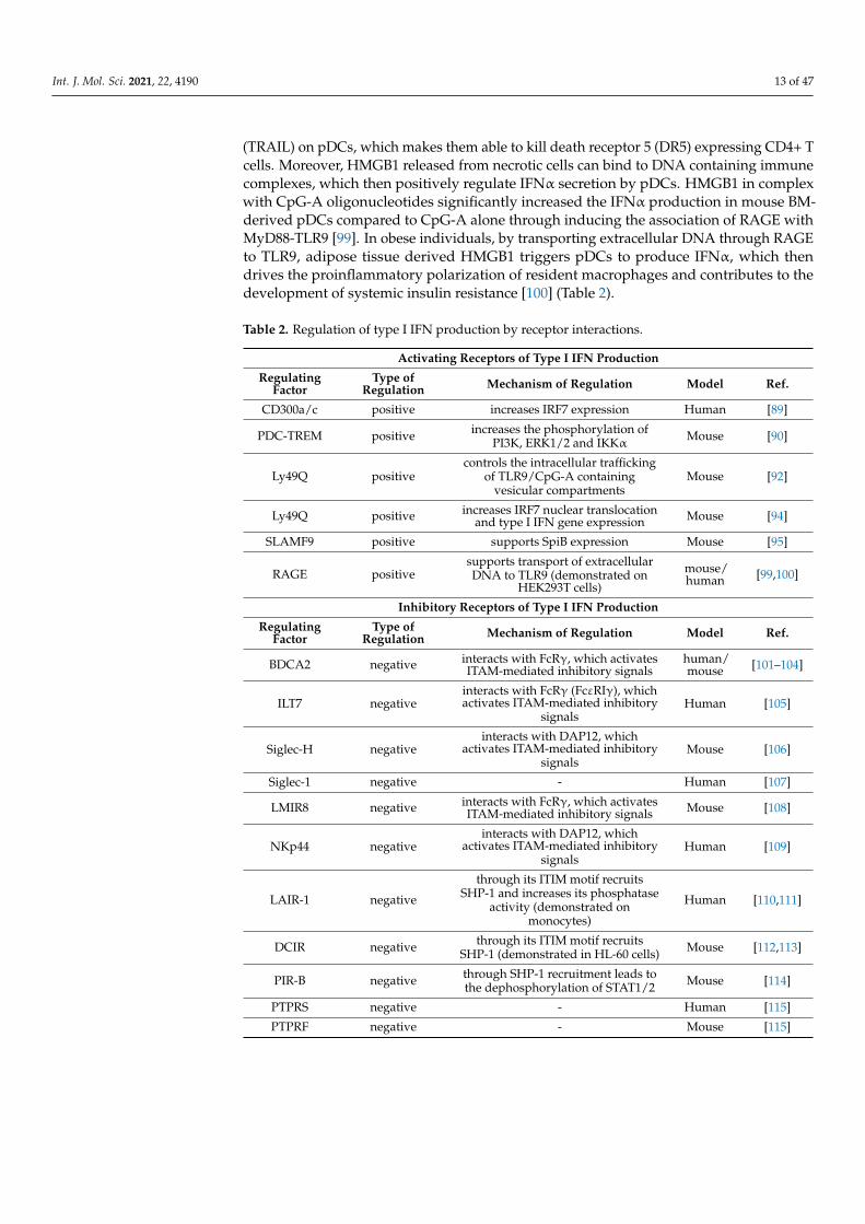
Table 2. Regulation of type I IFN production by receptor interactions.
Activating Receptors of Type I IFN Production
RegulatingFactor
Type ofRegulation Mechanism of Regulation Model Ref.
CD300a/c positive increases IRF7 expression Human [89]
PDC-TREM positive increases the phosphorylation ofPI3K, ERK1/2 and IKKα Mouse [90]
Ly49Q positivecontrols the intracellular trafficking
of TLR9/CpG-A containingvesicular compartments
Mouse [92]
Ly49Q positive increases IRF7 nuclear translocationand type I IFN gene expression Mouse [94]
SLAMF9 positive supports SpiB expression Mouse [95]
RAGE positivesupports transport of extracellularDNA to TLR9 (demonstrated on
HEK293T cells)
mouse/human [99,100]
Inhibitory Receptors of Type I IFN Production
RegulatingFactor
Type ofRegulation Mechanism of Regulation Model Ref.
BDCA2 negative interacts with FcRγ, which activatesITAM-mediated inhibitory signals
human/mouse [101–104]
ILT7 negativeinteracts with FcRγ (FcεRIγ), whichactivates ITAM-mediated inhibitory
signalsHuman [105]
Siglec-H negativeinteracts with DAP12, which
activates ITAM-mediated inhibitorysignals
Mouse [106]
Siglec-1 negative - Human [107]
LMIR8 negative interacts with FcRγ, which activatesITAM-mediated inhibitory signals Mouse [108]
NKp44 negativeinteracts with DAP12, which
activates ITAM-mediated inhibitorysignals
Human [109]
LAIR-1 negative
through its ITIM motif recruitsSHP-1 and increases its phosphatase
activity (demonstrated onmonocytes)
Human [110,111]
DCIR negative through its ITIM motif recruitsSHP-1 (demonstrated in HL-60 cells) Mouse [112,113]
PIR-B negative through SHP-1 recruitment leads tothe dephosphorylation of STAT1/2 Mouse [114]
PTPRS negative - Human [115]
PTPRF negative - Mouse [115]

Int. J. Mol. Sci. 2021, 22, 4190 14 of 47
Table 2. Cont.
Inhibitory Receptors of Type I IFN Production
RegulatingFactor
Type ofRegulation Mechanism of Regulation Model Ref.
EBI2 negative through Gαi subunit of the G proteininhibits type I IFN responses Mouse [116]
CD28 negative - Mouse [117]
TIM-3 negativeinhibits the trafficking of nucleic
acids into endosomes (demonstratedon BM-DCs)
Mouse [118]
Receptors with Distinct Regulatory Roles: Fc Receptors
RegulatingFactor
Type ofRegulation Mechanism of Regulation Model Ref.
FcγRIIα+ IgGcontainingimmunecomplex
positive supports TLR9 trafficking Human [119]
FcεRI+ IgEcontainingimmunecomplex
positive promotes the delivery of DNA toTLR9 Human [120]
FcεRI+ freeIgE negative triggers TNF-α, which reduces TLR9
expression Human [121,122]
Interactions of Pattern Recognition Receptors
RegulatingFactor
Type ofRegulation Mechanism of Regulation Model Ref.
TLR7-TLR9 negativeTLR7 activation inhibits
TLR9-triggered IRF7 expressionanddownregulates TLR9
human/mouse [123,124]
MR-TLR9 positive - Mouse [125]
TLR7-RLR positive TLR7 activation induces RLRexpression Human [40]
TLR9-RLR positive TLR9 activation induces RLRexpression Human [40]
NLRX1-RLR negative - Human [126]
NLRC5-RLR negative - Human [126]
TLR9-cGAS/STING negative cGAS/STING stimulation
upregulates SOCS1 and SOCS3 Human [127]
Adhesion Receptors
RegulatingFactor
Type ofRegulation Mechanism of Regulation Model Ref.
LFA-1 positive induces TLR7 trafficking fromendosomes to lysosomes Mouse [128]
Abbreviations: BDCA2: blood dendritic cells antigen 2; BM-DC: bone marrow-derived dendritic cell; CD: cluster ofdifferentiation; cGAS: cyclic GMP-AMP synthase; DAP12: DNAX activating protein of 12 kDa; DCIR: dendritic cellimmunoreceptor; EBI2: Epstein-Barr virus-induced G-protein-coupled receptor 2; ERK1/2: extracellular signal-regulatedkinase 1/2; FcRγ: γ subunit of the Fc receptor; FcγRIIα: Fc gamma receptor II alpha; FcεRI: Fc epsilon receptor I; FcεRIγ: γsubunit of the Fc epsilon receptor; Gαi: Gi alpha subunit; IFN: interferon; IgE: immunoglobulin E; IKKα: IκB kinase (IKK)complex α; ILT7: immunoglobulin-like transcript 7; IRF: interferon regulatory factor; ITAM: immunoreceptor tyrosine-basedactivation motif; ITIM: immunoreceptor tyrosine-based inhibition motif; LAIR-1: leukocyte-associated immunoglobulin-likereceptor 1; LFA-1: lymphocyte function-associated antigen 1; LMIR8: leukocyte mono-immunoglobulin-like receptor 8;MR: mannose receptor; mTOR: mammalian target of rapamycin; NKp44: natural killer cell p44-related protein; NLRC5:NOD-like receptor family CARD domain containing 5; NLRX1: nucleotide-binding domain and leucine-rich repeat–containing protein X1; p70S6K: P70 S6 kinase; PDC-TREM: plasmacytoid dendritic cell—triggering receptor expressed onmyeloid cells; PI3K: phosphatidylinositol 3-kinase; PIR-B: paired immunoglobulin-like receptor B; PTPRF: Protein tyrosinephosphatase receptor type F; PTPRS: Protein tyrosine phosphatase receptor type S; RAGE: receptor for advanced glycationendproducts; RLR: RIG-I-like receptor; SHP-1: Src homology 2 domain-containing protein tyrosine phosphatase 1; Siglec:sialic acid-binding immunoglobulin-type lectin; SLAMF9: signaling lymphocytic-activating molecule family 9; SOCS:suppressor of cytokine signaling; STAT: signal transducer and activator of transcription; STING: stimulator of IFN genes;TIM-3: T cell immunoglobulin and mucin domain-containing protein 3; TLR: toll-like receptor; TNF: tumor necrosis factor.
Int. J. Mol. Sci. 2021, 22, 4190 15 of 47
4.2. Inhibitory Receptors of Type I IFN Production
Many studies have reported that the cross-linking of regulatory cell surface receptorson pDCs efficiently suppresses their ability to produce type I IFNs. Several of these regu-latory receptors associate with immunoreceptor tyrosine-based activation motif (ITAM)containing adapter proteins such as DNAX activation protein 12 (DAP12) and Fc receptor(FcR) γ-chain (FcRγ) or contain ITIM motifs themselves, which mediate inhibitory sig-nals [129]. FcRγ is often referred to as FcεRIγ since it was first discovered as the thirdsubunit of FcεRI [130]. Later, it was revealed that it is a common subunit of various FcRsand is also able to associate with a number of immune receptors such as blood dendriticcell antigen 2 (BDCA2) and immunoglobulin-like transcript 7 (ILT7) [131].
BDCA2 (also known as CD303) is a type II C-type lectin receptor, which is exclusivelyexpressed by human pDCs. In 2001, it was demonstrated that the ligation of BDCA2 witha specific antibody suppresses the ability of peripheral blood-derived human pDCs toproduce IFNα in response to CpG-A [101]. Later, co-immunoprecipitation experimentsrevealed that BDCA2 forms a complex with the transmembrane adapter FcRγ, whichinterferes with the TLR7/9-mediated type I IFN producing ability of purified humanpDCs [102]. A separate study with primary human pDCs revealed further details of BDCA2and FcRγ interaction by showing that upon association with FcRγ BDCA2 signals througha B cell receptor (BCR) signalosome like complex consisting of Lyn, spleen tyrosine kinase(Syk), Bruton tyrosine kinase (Btk), Src homology 2 (SH-2) domain-containing leukocyteprotein of 65 kDa (Slp65) and phospholipase C-gamma 2 (PLCγ2) [103]. Moreover, asubsequent study with GEN2.2 cells and primary human pDCs declared that the BCR-like signaling activates the mitogen-activated protein kinase (MAPK) kinase (MEK)1/2-ERK pathway, which then upregulates c-Fos, and thus results in the inhibition of CpG-Amediated type I IFN production [132]. Further data indicate that the CD2-associatedadaptor protein (CD2AP), which is specifically expressed by pDCs, forms a complexwith SH-2 domain-containing inositol-5-phosphatase 1 (SHIP1) and inhibits the Casitas Bcell lymphoma (Cbl)-mediated ubiquitination and degradation of FcRγ [104]. Thus, viasupporting BDCA2/FcRγ receptor signaling, CD2AP negatively controls TLR9-inducedtype I IFN responses in pDCs [104]. Interestingly, some studies identified a few possiblevirus-derived ligands for BDCA2, in particular, hepatitis B virus surface antigen (HBsAg)and HIV-1 glycoprotein 120 (gp120). The binding of HBsAg [133] or gp-120 [134] to BDCA2on the surface of human pDCs contributed to the suppression of IFNα secretion in responseto TLR9.
The above data indicate that targeting BDCA2 by blocking or depleting antibodiesmight be a promising approach to manage type I IFN-mediated pathologies includingautoimmune diseases [135–137]. Importantly, certain mAbs are already under clinicaltesting for the treatment of SLE and cutaneous lupus erythematosus (CLE) [138].
Similar to BDCA2, the ILT7 (also known as LILRA4) protein, which is exclusivelyexpressed by pDCs, can also form a complex with FcRγ, and thereby leads to the inhibitionof pDC functions. The cross-linking of ILT7 inhibited the production of IFNα in bothCpG- and Flu-activated pDCs. The rapid phosphorylation of Src family kinases and Sykindicates that ILT7 cross-linking leads to the activation of ITAM mediated inhibitory signalsin pDCs [105]. The bone marrow stromal cell antigen 2 (BST2), which is expressed onlyin low amounts on the surface of human pDCs, was identified as the biological ligand forILT7. Following incubation with recombinant BST2, human pDCs were impaired in theirability to express type I IFNs upon challenge with CpG-A or Flu [139]. Interestingly, theVpu HIV-1 protein hijacks this interaction by acting as an antagonist of BST2 and inhibitingTLR7-mediated type I IFN production by pDC. Vpu downregulates BST2 to enable virionassembly, while relocates remaining BST2 molecules to the surface of infected cells tomaintain sufficient inhibitory signals for pDCs [140]. The ILT7-mediated negative feedbackon type I IFN production is also hijacked by human cancer cells. Many human cancer celllines constitutively express ILT7 ligands, thus inhibit TLR9-triggered IFNα productionby human peripheral blood pDCs [141]. Interestingly, a study demonstrated that in vitro
Int. J. Mol. Sci. 2021, 22, 4190 16 of 47
culturing of human peripheral blood mononuclear cells (PBMCs) leads to a spontaneousloss of ILT7 on the surface of pDCs. Consequently, the blocking of BST2 by mABs had noeffect on the IFNα production of TLR7/9-triggered pDCs. Therefore, the authors proposethat BST2-mediated ILT7 as a homeostatic regulator limits the activity of immature pDCsand does not serve as a negative feedback mechanism to restrict the functions of maturepDCs [142].
Another modulator of pDC functions is the transmembrane sialic acid binding im-munoglobulin type lectins H (Siglec-H) receptor, which was identified as a specific markerof mouse pDCs. First it was demonstrated that cross-linking of Siglec-H with specificantibodies reduced TLR9-mediated type I IFN production of pDCs through the DAP12adaptor protein [106]. In mice infected with murine cytomegalovirus (MCMV), Siglec-Hdeficiency induced elevated serum IFNα levels compared with wild type mice, whereasviral clearance was not affected [143]. Further, it was revealed that Siglec-H protects micefrom developing MCMV virus-triggered lupus-like syndrome by preventing the inductionof type I IFN signature [144].
According to a recent study, the Siglec-1 positive pDCs might represent the humancounterpart of the Siglec-H positive pDCs in mice [107]. In humans, Siglec-1 is expressedin a subset of blood pDCs, which display a semi-mature phenotype, express lower levelsof BDCA2 and interleukin (IL)-3 receptor α, and do not respond to TLR7/9 engagement.In vitro, its expression is inducible in Siglec-1 negative pDCs upon exposure of wholeblood to Flu. Interestingly, the proportion of Siglec-1 expressing pDCs is higher in SLEpatient and correlates with disease severity compared to healthy individuals; nevertheless,their functional role in SLE needs further investigations [107].
Another FcRγ-coupled regulator is the leukocyte mono-immunoglobulin-like receptor8 (LMIR8), which is selectively expressed by mouse pDCs residing in the BM, spleen, orlymph nodes. It was found that LMIR8 cross-linking with mABs attenuates the CpG-A-mediated production of IFNα in BM-derived pDCs through the ITAM-containing adaptorprotein FcRγ [108]. An additional pDC inhibitory receptor is NKp44, the expressionof which is inducible on the surface of human tonsil and blood-derived pDCs uponculture with IL-3. It was reported that cross-linking of NKp44 inhibits IFNα secretionin CpG-activated pDCs through association with the ITAM-containing adaptor moleculeDAP12 [109]. In contrast to NKp44, the expression of leukocyte-associated Ig-like receptor-1 (LAIR-1) is high on resting human pDCs, whereas it is downregulated in the presenceof IL-3. The cross-linking of the inhibitory ITIM-containing LAIR-1 receptor impairsIFNα production by pDCs in response to TLR stimulation, and thus acts synergisticallywith Nkp44 to inhibit IFNα release [110]. Human pDCs also express the inhibitory C-type lectin receptor DC immunoreceptor (DCIR), which contains one ITIM motif. Thecross-linking of DCIR inhibits TLR9-induced IFNα production, while it promotes antigenuptake and efficient antigen presentation by pDCs [112]. In mice, the ITIM-bearing pairedimmunoglobulin-like receptor B (PIR-B) is an inhibitory MHC-I receptor, which suppressesCpG-A triggered type I IFN production in mouse pDCs. In particular, PIR-B recruits the SH-2 domain-containing phosphatase 1 (SHP-1), which then leads to the dephosphorylation ofSTAT1/STAT2, and thereby prevents the autocrine type I IFN-mediated positive feedbackloop [114].
In contrast to the aforementioned inhibitory receptors the following ones do not con-tain ITAM or associate with ITAM-containing adaptors, and the specific mechanism bywhich they regulate type I IFN responses of pDCs remained largely unknown. Proteintyrosine phosphatase receptor type S (PTPRS) is an evolutionarily conserved pDC specificinhibitory molecule, which is expressed by both murine and human pDCs, whereas proteintyrosine phosphatase receptor type F (PTPRF) is detectable only in mice. PTPRS cross-linking decreased type I IFN production in primary human pDCs and GEN2.2 cells uponCpG-A stimulation. In mice, quiescent pDCs co-express PTPRS and PTPRF, the knockdownof which enhanced TLR9-induced pDC activation. These receptors are downregulatedon activated pDCs that seems to be a requirement for efficient type I IFN production. In
Int. J. Mol. Sci. 2021, 22, 4190 17 of 47
addition, PTPRS and PTPRF deficiency was associated with enhanced pDC activation,increased leukocyte infiltration, and the development of spontaneous colitis in mice sug-gesting the importance of these receptors in maintaining immune homeostasis [115]. Inaddition, the Epstein-Barr virus induced receptor 2 (EBI2) functions as a negative regulatorof type I IFN responses in pDCs as well. In mice, EBI2 is a chemotactic G protein-coupledreceptor, which drives the migration of B cells and DCs through interaction with its lig-and, 7α, 25-dihydroxycholesterol. In addition, it was found that EBI2 inhibits type I IFNresponses of pDC upon stimulation with CpG-A, polyU or lymphocytic choriomeningitisvirus (LCMV) through mechanisms depending on the Gαi subunit of the G protein [116].
Interestingly, CD28, the well-known costimulatory receptor of T cells, is also highlyexpressed on murine pDCs and acts as a negative regulator of type I IFN production.However, CD28 is undetectable in human blood circulating pDCs, it is expressed only bylymph node-derived human pDCs. BM-derived pDCs from CD28-deficient mice producesignificantly higher levels of type I IFN compared to pDCs from wild type mice uponstimulation with TLR9 ligand, CpG-A or TLR7 ligand, Loxoribin. During MCMV or LCMVinfection, systemic type I IFN levels were higher in CD28 knockout mice than in their wildtype counterparts suggesting that CD28 controls TLR7- and TLR9-triggered IFN responses.Moreover, in a mouse wound healing model it was demonstrated that CD28 restricts IFNsignature during non-viral innate immune responses as well [117].
In the previous section, we have established that in the course of inflammation HMGB1binds to RAGE on the surface of pDCs and supports their type I IFN response. Paradox-ically, in the tumor microenvironment, HMGB1 directly secreted by tumor cells renderspDCs tolerogenic. HMGB1 produced by neoplastic keratinocytes decreased IFNα secre-tion by human cord blood-derived pDCs following TLR9 stimulation. Moreover, pDCsco-cultured with neoplastic keratinocytes promoted the differentiation of naïve CD4+ Tcells into regulatory T cells that could be reverted by the addition of the HMGB1 specificantibody [145]. One possible explanation for the inhibitory effect of HMGB1 in the tumormilieu might be that tumor-infiltrating pDCs highly express the T cell immunoglobulin andmucin domain-containing protein 3 (TIM-3), which binds HMGB1 with an affinity similarto that of RAGE and inhibits nucleic acid trafficking into endosomes leading to impairedTLR-mediated responses of mouse pDCs [118]. Specifically, treatment of tumor-associatedmouse pDCs with mABs to TIM-3 resulted in a higher expression of IFNβ in responseto CpG-A stimulation. Following stimulation with poly(dA:dT) dsDNA and HMGB1,it was observed that DNA uptake was much lower in the endosomal vesicles of TIM-3expressing BM-derived mouse DCs compared to those from TIM-3 deficient mice [118].These findings suggest that contrary to RAGE, TIM-3 serves as a negative regulator ofendosomal TLR-mediated immune responses of DCs in the tumor microenvironment in aHMGB1 dependent manner (Table 2).
4.3. Receptors with Distinct Regulatory Roles: Fc Receptors
As we have described in the previous chapter, a number of ligand-binding regulatoryreceptors use the common γ subunit of FcRs as an interacting partner to manipulate theoutcome of pDC responses. In addition, pDCs also express functional Fc receptors, whichupon binding to their actual ligands, namely immune complexes or immunoglobulins, cansend out activating or inhibitory signals, respectively [146].
PDCs have a predominant role in the pathogenesis of SLE, where immune complexesformed by self-nucleic acid and autoantibodies trigger pDC activation. It was recognizedin the early 2000s that apoptotic cells in combination with serum IgG obtained from SLEpatients can induce IFNα secretion by pDCs in PBMCs [147]. In a subsequent study, theauthors also revealed that FcγRIIα (CD32), which is expressed on the surface of pDCs, isresponsible for the increased IFNα secretion triggered by the combination of apoptotic cellsand autoantibodies [148]. Later, it was also demonstrated that both necrotic and apoptoticcells release materials, namely RNA and DNA, which, in the presence of autoantibodies,induce the production of IFNα by human pDCs [149]. Thereafter, it was described that
Int. J. Mol. Sci. 2021, 22, 4190 18 of 47
mouse pDCs lacking the common γ-chain of FcγR do not produce IFNα in response to DNAcontaining immune complexes. Further, it was found that FcγR-mediated internalizationof DNA-autoantibody complexes is necessary to induce the non-canonical LC3-associatedphagocytosis (LAP) pathway, which is required for TLR9 trafficking to phagosomes andinitiation of IRF7-dependent IFNα secretion in mouse pDCs [119].
Similar to IgG, IgE in complex with self-DNA enhances type I IFN responses ofpDCs as well. Following ligation, FcεRI, which is the major receptor mediating allergicinflammatory signals in mast cells, promotes the delivery of DNA to TLR9 and in thismanner works in synergy with the FcγRIIα to activate pDCs. Intriguingly, IgE is muchstronger in its ability to enhance the phagocytic potential of pDCs, probably due to itsstrong interaction with the high affinity FcεRI. By facilitating type I IFN secretion, self-reactive IgE exacerbates self-destructive autoimmune reactions, thus serum concentrationsof DNA-IgE complexes correlate with disease severity in SLE [120].
In contrast, when not in complex with DNA, IgE suppresses pDCs in their ability tosecrete IFNα in response to TLR9 triggering. A study showed that IgE cross-linking onpDCs with mABs reduces TLR9 expression and CpG-A-mediated secretion of IFNα byhuman pDCs [121]. Additionally, the authors later demonstrated that upon stimulationwith anti-IgE antibody, pDCs secrete large amounts of tumor necrosis factor α (TNFα),which acts in an autocrine manner to reduce TLR9 on pDCs, and thus abrogates their type IIFN secretion [122]. Interestingly, a study showed that in allergic asthma patients, elevatedserum IgE concentrations and increased surface FcεRI expression on pDCs correlate withtheir impaired antiviral response, which might contribute to disease exacerbation uponviral respiratory infections [150]. Nevertheless, it was recognized that omalizumab, the IgEneutralizing antibody used for the treatment of allergic asthma, restores the virus-inducedIFNα responses of pDCs in allergic patients upon rhinovirus infection [151] implyingfurther that free IgE has a negative impact on the type I IFN production of pDCs (Table 2).
4.4. Interactions of PRRs
As the first line of defense against infectious agents, the mammalian immune systemexpresses a myriad of PRRs, which recognize different conserved molecular structuresknown as pathogen-associated molecular patterns (PAMPs) and damage-associated molec-ular patterns (DAMPs). These receptors might interact with each other in either an in-hibitory or a synergistic manner that might result in the modulation of host innate immuneresponses [152].
PDCs selectively express the endosomal TLR7 and TLR9, which might interact witheach other during their activation. Interestingly, the presence of the TLR7 ligand R848significantly decreased TLR9-mediated IFNα secretion in purified human pDCs by twodifferent mechanisms [123]. On the one hand, R848 prevented the TLR9-mediated up-regulation of IRF7 expression, and on the other hand it initiated TLR7 upregulation andTLR9 downregulation in pDCs. Both TLR7 and TLR9 are strong inducers of IFNα, thusthe negative regulatory effect of TLR7 on TLR9-mediated signaling might have evolvedas a compensatory mechanism to prevent the overproduction of type I IFNs that mightbe detrimental to the host [123]. Almost at the same time, another research group demon-strated that various natural and synthetic TLR7 ligands, even applied at substimulatoryconcentrations, significantly inhibited the TLR9-induced IFNα secretion in both humanand mouse pDCs [124]. On the contrary, structurally different TLR7 ligands added simulta-neously have a synergistic effect on pDC activity. In the human pDC cell line CAL-1, theco-administration of the single-stranded 9.2s RNA and the adenine analog CL264 markedlyincreased the secretion of IFNβ and pro-inflammatory cytokines through upregulatingthe expression of numerous immune-related genes. In addition, 9.2s RNA increased thecell surface binding, and thus probably the uptake of CL264 that might contribute to thesynergistic effect of distinct TLR7 agonists on pDCs as well [153].
Besides TLRs, pDCs also express cell surface C-type lectin receptors, which recognizespecific carbohydrate residues on bacterial and fungal cell surfaces. Specifically, the C-type
Int. J. Mol. Sci. 2021, 22, 4190 19 of 47
lectin mannose receptor (MR) was found to synergize with the TLR9 receptor on pDCs. Itwas demonstrated that CpG-A in combination with the Cryptococcus neoformans-derivedmannoprotein induced significantly more IFNα than CpG-A alone in mouse BM-derivedpDCs. Furthermore, MR was found to synergize with other TLRs in conventional DCs aswell, thus the study suggests that TLR ligands might be used as adjuvants in mannoprotein-based vaccines [125].
In addition, our research group observed that under steady-state conditions humanpDCs express only marginal levels of cytoplasmic RIG-I receptor, an RLR family member,the expression of which can be greatly upregulated by TLR7/9 stimulation [40]. Later,we also demonstrated that melanoma differentiation-associated gene-5 (MDA5), anothermember of the RLR family, is also inducible upon the CpG-A treatment of human GEN2.2pDC cells [126]. As we have described earlier in Chapter 2, RLR-mediated signalingcontributes to the second wave of type I IFN production by pDCs, which shows thecollaborative action of TLR and RLR families in the antiviral immune responses providedby pDCs [40].
Our research group recently demonstrated that two members of the nucleotide-binding domain leucine-rich repeat containing receptor (NLR) family, namely NLRX1and NLRC5 inhibit the RIG-I and MDA5-mediated type I IFN responses of human pDCs.Silencing of NLRX1 and NLRC5 in GEN2.2 cells significantly increased the productionof IFNα and IFNβ upon stimulation with synthetic RLR ligands or following infectionwith VSV. Thus, our findings suggest that these negative regulatory NLRs control antiviralsignaling of pDCs to avoid aberrant IFN production and prevent the host from unwantedinflammation [126].
Lastly, it was demonstrated that stimulation of the DNA-sensing cyclic GMP-AMPsynthase (cGAS)-stimulator of IFN gene (STING) pathway also interferes with the TLR9-mediated IFN production of human pDCs as well [127]. Both cGAS and STING areconstitutively expressed by primary human pDCs and compared to TLR9 use a differentcellular signaling machinery to induce type I IFN secretion. Interestingly, TLR9 and cGAS–STING signaling do not act synergistically, since pre-stimulation of the cGAS-STING axismarkedly inhibited the type I IFN production of pDCs in response to CpG-A or HSV-1.The authors further revealed that cGAS–STING stimulation upregulated the expressionof suppressor of cytokine signaling (SOCS) 1 and SOCS3, which might contribute to thedownregulation of TLR9 signaling in pDCs [127] (Table 2).
The low frequency of primary pDCs in human blood sets a limit to the number ofexperiments, thus several receptors such as other cytosolic sensors wait for functionalcharacterization, which could extend our knowledge regarding the receptor interactionsregulating pDC type I IFN responses.
4.5. Adhesion Receptors
It has been long observed that pDCs form clusters upon in vitro culturing as wellas in vivo in the T cell rich area of lymphoid tissues that seems to be critical for theproduction of optimal amounts of type I IFNs. At higher cell density, pDCs form clustersand spontaneously produce low amounts of type I IFNs, which then acts in a feedforwardmechanism to amplify TLR-triggered type I IFN secretion [154].
Later, it was revealed that cell adhesion is required for TLR7 trafficking, since the lackof lymphocyte functional antigen 1 (LFA-1, also termed CD11a/CD18 or αLβ2) integrindecreased the ability of mouse pDCs to produce IFNα in response to TLR7 activation.Furthermore, it was found that LFA-1 activation induces microtubule polymerization thatleads to TLR7 trafficking from endosomes to lysosomes, which enables the interactionwith downstream signaling components such as TNF receptor associated factor 3 (TRAF)3, IKKα, and mTOR required for IFNα/β induction. Moreover, the results suggest thatcell adhesion-dependent type I IFN secretion boosts paracrine IFNα induction in pDCclusters [128].
Int. J. Mol. Sci. 2021, 22, 4190 20 of 47
Besides the above described homotypic cell adhesion, pDCs also interact with othercell types such as NK cells through LFA-1. NK cells promote the IFNα secretion of humanpDCs stimulated with RNA-containing immune complexes through LFA-1 mediated cell–cell contact and macrophage inflammatory protein-1β (MIP-1β) production [155].
Furthermore, recent studies suggest that pDCs establish contact with infected cellsthrough LFA1-dependent adhesion, which leads to the robust production of type I IFNs.Under in vivo conditions, the loss of the LFA-1 encoding gene impaired the type I IFNproduction of mouse pDCs upon CpG-A challenge or MCMV infection. Further, it wasdemonstrated that pDCs interact with infected cells through LFA-1 in the marginal zone ofthe spleen during MCMV infection that facilitates their type I IFN production [156]. Thesefindings were further supported by another study showing that human pDCs interactwith infected cells through the so-called interferogenic synapse, which enables the transferof viral RNA to pDCs resulting in the local secretion of type I IFNs. In particular, initialencounter with virus infected cells activates TLR7 signaling in pDCs, which promotesstructural reorganization including polarized endocytosis to facilitate the transfer of PAMPsto pDCs and sustained cell-cell contact through LFA-1, which further amplifies TLR7mediated local secretion of type I IFN responses [157] (Table 2).
In summary, several cell surface and intracellular receptors control the type I IFNsecretion of pDCs by sending out positive or negative feedback signals to fine-tune theamplitude of type I IFN response by pDCs. In the course of infection or inflammation amultitude of stimuli can trigger cells due to the distinct molecular structure of pathogensand release of multiple inflammatory factors that leads to the simultaneous activation ofdifferent receptors, and thus to the formation of a complex regulatory system.
5. Regulation of Type I IFN Production by Extracellular Soluble Factors5.1. Tumor-Derived Factors
Tumors have the ability to modulate their microenvironment by secreting solublefactors, which can impair the capacity of tumor-infiltrating pDCs to secrete type I IFNs andeventually endows them with immunosuppressive properties [158].
The non-canonical wingless-related integration site 5a (Wnt5a) is a homolog of Wing-less proteins in Drosophila species, which is secreted by cancer cells such as melanomacells. Recombinant human Wnt significantly inhibited CpG-B triggered IFNα productionby human pDCs, probably via inhibiting the cytoskeletal rearrangement required for cellactivation [159]. In mouse splenic pDCs Wnt5a was found to upregulate the surface ex-pression of indoleamine 2,3-dioxygenase-1 (IDO), which plays a significant role in DCtolerogenesis in melanoma [160].
The pleiotropic neuropeptide vasoactive intestinal peptide (VIP) is majorly released byneurons and immune cells [161]; however, a special type of neuroendocrine tumor is alsoable to secrete massive amounts of it [162]. A research group discovered that recombinantVIP decreases type I IFN production of primary human pDCs upon stimulation withCpG-A and CpG-B [163]. In a separate study, the authors further demonstrated that VIPeffectively reduced TLR9-triggered IFNα secretion of pDCs from healthy individuals andfrom SLE patients as well [164].
Tumor cells can also secrete transforming growth factor β (TGFβ), which acting aloneor synergistically with other cytokines also makes pDCs tolerogenic. The treatment of CAL-1 cells with tumor supernatant or combination of TGFβ and TNFα prevented TLR9-inducedproduction of type I IFNs. On the one hand, TGFβ exposure sustains the expression oflysosome-associated membrane protein (LAMP5), which promotes TLR9 transport to lateendosomes, and increases its degradation, thus limits type IFN secretion in pDCs. Onthe other hand, it is known that TNF promotes pDC maturation, and thus inhibits IFNαrelease [165]. Moreover, pDCs from breast tumors show a strongly upregulated expressionof LAMP5 compared to pDCs isolated from the peripheral blood of the same patients [166].A separate study also described that human tumor-associated pDCs purified from breastcancer samples secrete less IFNα compared to tonsil-derived pDCs from healthy controls

Int. J. Mol. Sci. 2021, 22, 4190 21 of 47
in response to Flu and CpG-A [167]. Furthermore, tumor-derived TGFβ acts in synergywith TNFα to inhibit the ability of pDCs to produce IFNα via blocking IRF7 expressionand nuclear translocation [168]. Similarly, pDCs purified from tumor samples of ovariancancer patients also secrete lower amounts of IFNα compared to blood pDCs upon Flu andCpG-A stimulation due to the presence of TGFβ and TNFα [169]. TGFβ also suppressesCpG-induced IFNα production by purified tumor-associated pDCs in mouse models oflung cancer and melanoma [170]. Furthermore, pDCs from oral squamous cell carcinomapatients express lower levels of TLR9 and secrete less IFNα upon CpG-B stimulation.The oral cancer cell supernatant was observed to reduce TLR9 mRNA expression andinhibit TLR9-mediated IFNα production in human peripheral blood pDCs through a TGFβand IL-10 dependent mechanism [171]. Another study showed that recombinant IL-10can boost the suppressive effect exerted by the supernatants of different head and necksquamous cell carcinoma (HNSCC) cell lines on the IFNα secretion ability of pDCs [172].Nevertheless, a recent paper reported that TNF rather than IL-10 is the major inhibitorof IFNα production by pDCs in HPV negative HNSCC, since neutralization of TNF inHNSCC supernatants almost completely restored the ability of pDCs to produce IFNα inresponse to CpG-A [173].
Moreover, TGFβ is also able to synergize with tumor-derived prostaglandin E 2(PGE2) to abrogate IFNα production by purified human pDCs in response to differentTLR7/9 stimuli [174]. TGFβ suppresses TLR-driven IFNα secretion, probably via inducingsmad, the main signal transducers of TGFβ receptor, since blocking of smad abolishedthe inhibitory effect of TGFβ. In addition, PGE2 seems to exert its effect via the G protein-coupled PGE2 receptors, which upon ligand binding activate adenylate cyclase, and therebyincrease the levels of cyclic adenosine monophosphate (cAMP), which is known to interferewith TLR-mediated signaling events [175]. Almost at the same time, another researchgroup showed that prostaglandin analogs inhibited the IFNα secretion of TLR9-activatedhuman pDCs with similar efficiency. Moreover, data suggest that PGE2 exerts its effect atthe transcriptional level, since PGE2 significantly suppresses IRF7 mRNA expression inTLR9-stimulated PDCs [164].
Many tumor cells are also able to secret HMGB1, which, as an important componentof the tumor milieu, plays a central role in cancer and metastasis development [176]. Inthe previous chapter, we mentioned that tumor-infiltrating pDCs start to express TIM-3,which upon interaction with HMBG1 interferes with the TLR-mediated immune responsesof pDCs, and thus negatively influences their type I IFN secretion [118].
Another binding partner for TIM-3 is the conserved s-type lectin galectin-9 (Gal-9),which is a pleiotropic immune modulator. Besides TIM-3, Gal-9 has numerous bindingpartners and can be secreted by cancer cells via various mechanisms [177]. It has beenrecently recognized that Gal-9 inhibits the production of IFNα by mouse and human pDCsstimulated with various TLR7/9 ligands. Interestingly, it was found that the inhibitoryeffect of Gal-9 on the type I IFN secretion of pDCs is independent of TIM-3, and instead it ismediated through interaction with CD44 that leads to the disruption of the p70S6K/mTORsignaling pathway. Further, it was observed that Gal-9 administration suppressed thedevelopment of different autoimmune conditions in mice. In particular, it reduced inflam-matory processes such as lymphocyte migration and alleviated disease symptoms such assplenomegaly in lupus prone mice [178] (Table 3).

Int. J. Mol. Sci. 2021, 22, 4190 22 of 47
Table 3. Regulation of type I IFN production by extracellular soluble factors.
Tumor-Derived Factors
RegulatingFactor
Type ofRegulation Mechanism of Regulation Model Ref.
Wnt5a negative inhibits cytoskeletal rearrangementrequired for cell activation human [159]
Wnt5a negative upregulates the surface expressionof IDO mouse [160]
VIP negative - human [163,164]
TGFβ negativeinhibits TLR9 transport to lateendosomes and increases its
degradationhuman [165]
TGFβ in synergywith TNFα negative blocks IRF7 expression and nuclear
translocation human [167,168]
TGFβ in synergywith IL-10 negative reduces TLR9 mRNA expression human [171]
IL-10 negative - human [172]
PGE2 in synergywith TGFβ negative TGFβ induces smad, whereas
PGE2 increases the levels of cAMP human [174]
PGE2 negative suppresses IRF7 mRNA expression human [164]
HMGB1 negative interacts with TIM-3 mouse [118]
Gal-9 negativethrough interacting with CD44
disrupts the p70S6K/mTORsignalling
human/mouse [178]
Biogenic Amines and Steroid Hormones
RegulatingFactor
Type ofRegulation Mechanism of Regulation Model Ref.
histamine negative acts through H2 and H4 receptors human [179,180]
histamine,dopamine,serotonin
negative binds to CXCR4 and induces itsinternalization human [181]
17β-estradiol positive - mouse [182,183]
17β-estradiol positive increases the expression of IRF5 human [184]
progesterone negativeinhibits the TLR9-induced nuclear
accumulation of IRF7(mechanism demonstrated on
mouse pDCs)
human/mouse [185]
DMPA negative - mouse [185]
MPA negative - human [186]
DHT negative - human [187]Abbreviations: cAMP: cyclic adenosine monophosphate; CXCR4: C-X-C motif chemokine receptor 4; DHT: dihydrotestos-terone; DMPA: depot-medroxyprogesterone acetate; Gal-9: galectin-9; H2: histamine receptor 2; H4: histamine receptor 4;HMGB1: high mobility group box 1 protein; IDO: indoleamine 2,3-dioxygenase; IL: interleukin; IRF: interferon regulatoryfactor; MPA: medroxyprogesterone acetate; PGE2: prostaglandin E2; TGF-β: transforming growth factor beta; TIM-3: Tcell immunoglobulin and mucin domain-containing protein 3; TLR: toll-like receptor; TNF: tumor necrosis factor; VIP:vasoactive intestinal peptide; Wnt5a: wingless-related integration site 5a.
5.2. Biogenic Amines and Steroid Hormones
Besides fulfilling their essential functions in the signaling events of the central and pe-ripheral nervous system, biogenic amine neurotransmitters are also involved in the regula-tion of immune responses including the control of type I IFN production of pDCs [188,189].
Histamine, a major mediator of allergic diseases, was found to block IFNα releaseof human pDCs stimulated with CpG or Flu through the histamine receptor 2 (H2) [179].Later, it was described that pDCs isolated from peripheral blood and from skin lesions ofpsoriasis patients express another histamine receptor subtype, termed H4, the expressionof which can be further stimulated with CpG treatment [180]. Moreover, it was found thatstimulation with H4 receptor agonists reduced the CpG-induced IFNα secretion similar
Int. J. Mol. Sci. 2021, 22, 4190 23 of 47
to H2 receptor agonists. Interestingly, pDCs from psoriasis patients express higher H4receptor levels, and thus show a stronger downregulation of IFNα secretion in response toH4 receptor agonists than pDCs from healthy individuals [180]. Another study reportedthat histamine can also efficiently block the IFNα secretion of HIV-1 activated purifiedhuman pDCs similar to other amines including dopamine and serotonin [181]. Further,the authors demonstrated that these amines exert their inhibitory effects on the antiviralcapacity of pDCs through CXCR4 chemokine receptor engagement. In particular, naturalamines do not act as ligands of CXCR4 but can interact with the receptor through theirammonium moiety and induce its internalization, which process seems to be essential forthe inhibitory effects of amines [181].
Steroid hormones, due to their lipophilic nature, easily pass through plasma mem-branes, and by binding to soluble nuclear receptors are able to exert various effects ondifferent immune cell types including pDCs [190]. A high number of findings indicatethat estrogen plays an important role in the regulation of TLR-mediated responses ofhuman and mouse pDCs as well. In mice, 17β-estradiol (E2) was described to signifi-cantly increase the IFNα production of CpG-B stimulated splenic pDCs [182]. In line withthis, E2 treatment of postmenopausal women markedly elevated the TLR7/9-triggeredIFNα production of primary pDCs. The authors further demonstrated that E2 directlytargets pDCs, since deletion of estrogen receptor-α (ERα) abolishes the boosting effect ofE2 treatment on the TLR induced IFNα production of mouse pDCs [183]. Furthermore, theimpairment of estrogen receptor signaling significantly decreased the TLR7-induced IFNαexpression in human pDCs generated from umbilical cord blood. [191]. Another studysuggested that ERα signaling supports the IFNα secreting potential of TLR7-stimulatedmouse pDCs by increasing the expression of IRF5, the level of which positively associateswith the percentage of IFNα secreting pDCs. In addition, human pDCs overexpressingIRF5 exhibited increased IFNα secretion in response to TLR7 stimulation compared withcontrol pDCs [184].
In contrast with estrogen, progesterone and its synthetic analogs have well docu-mented immunosuppressive effects on innate immunity, and were observed to negativelyregulate type I IFN secretion by human pDCs [192]. Under in vitro conditions, proges-terone substantially inhibited the TLR9-mediated IFNα secretion of human and mousepDCs. Moreover, the treatment of mice with depot medroxyprogesterone acetate (DMPA),an artificial progesterone significantly reduced the ability of isolated splenic pDCs to pro-duce IFNα in response to CpG-A. Upon in vivo VSV infection, DMPA-treated mice hadsignificantly lower serum IFNα levels compared with control mice. The authors furtherdemonstrated that progesterone treatment inhibited the TLR9-induced nuclear accumula-tion of IRF7 in mouse BM-derived pDCs [185]. Another study showed that the syntheticprogestin medroxyprogesterone acetate (MPA) is also able to impair the TLR7/9-stimulatedIFNα production in human pDCs [186].
To date, only one study has investigated the effect of androgens on pDC function.The androgen hormone dihydrotestosterone (DHT) was demonstrated to decrease theTLR7-stimulated IFNα production of pDCs purified from the blood of healthy women. Theauthors further found that pDCs from male infants produced less IFNα in response to TLR7stimulation compared to female infants that might be explained by the early postnatalandrogen surge in male infants at 1–6 months of age [187]. In conclusion, while estrogenpositively regulates the type I IFN release by pDCs, progesterone and testosterone affectit negatively.
The above data also indicate that gender differences greatly influence the individualimmune responses to viral infections. Indeed, it is now widely accepted that gender-based immunological differences affect various aspects of immune responses includingthe susceptibility to infections, the prevalence of autoimmune diseases, and the efficacyof vaccination [193]. Moreover, pDCs are highly sensitive to their environment and themyriad of soluble factors released from different cell types can highly impact the intensityof their type I IFN responses (Table 3).
Int. J. Mol. Sci. 2021, 22, 4190 24 of 47
6. Targeting Type I IFNs in pDC-Related Pathological Conditions6.1. Viral Infections
The rapid and robust type I IFN production of pDCs is essential to control the earlyphase of viral infections by directly inhibiting viral replication, preventing virus-triggeredtissue damage, and activating a wide repertoire of cellular and humoral innate immunesignaling elements. The important role of pDCs in overcoming viral infections has alreadybeen highlighted in different mouse models upon mouse hepatitis virus (MHV), HSV,dengue virus (DENV), chikungunya virus (CHIKV), LCMV and respiratory syncytial virus(RSV) infections (reviewed in [1]).
However, if the immune system is not able to effectively clear the virus, it leads to thedevelopment of chronic infections associated with persistent inflammation and immuneactivation, in which the adverse effects of type IFNs are manifested. Furthermore, chronicviral exposure negatively affects pDC functions. For instance, HIV positive patients withhigh viral load are characterized by functionally exhausted pDCs with decreased type IIFN secretion and increased rate of apoptosis, which leads to alterations in pDC populationdynamics [194]. Nevertheless, type I IFNs released by pDCs play a versatile role in thecourse of HIV-1 infection. At the early phase of infection, the secreted large amounts of typeI IFNs have beneficial effects, whereas during the late phase, chronic activation of pDCsresults in type I IFN-driven pathologies such as T cell exhaustion, apoptosis of uninfectedCD4+ T cells through an increased expression of programmed cell death 1 (PD-1) receptoras well as generation of regulatory T cells via upregulating tolerogenic mediators such asIL-10 or IDO [195–198]. Based on these data, the clinical usage of type I IFN therapy inHIV-1 infection is quite controversial, since it seems to be beneficial in the acute phase,whereas it is not advised in the chronic phase of infection [199,200]. In spite of this, it isimportant to note that recombinant human IFNα2b is indicated for the treatment of varioushuman chronic viral infections associated with malignant conditions such as acquiredimmune deficiency syndrome (AIDS)-related Kaposi’s sarcoma, human papilloma virus(HPV)-caused condyloma acuminata, and chronic hepatitis C (HCV) or hepatitis B (HBV)infections [201–204].
The novel coronavirus pandemic caused by the severe acute respiratory syndromecoronavirus 2 (SARS-CoV-2) strain, further emphasizes the importance of the timing oftype I IFN therapy in the treatment of viral infections. So far, a rapidly growing numberof publications have focused on the association between type I IFNs and coronavirusdisease 2019 (COVID-19) [205–209]. The progression of COVID-19 can be divided into 3stages [210]. The first phase of the disease is asymptomatic or characterized by mild flu-likesymptoms, which is followed by the second pulmonary phase designated by viral pneu-monitis. In some individuals, the infection culminates further in the last and mostly lethalhyperinflammation phase, which results in shock or acute respiratory distress syndrome(ARDS) due to the dysregulated antiviral and pro-inflammatory response, known as a“cytokine storm” [210]. It is worth noting that SARS-CoV-2 uses distinct strategies to bypassthe early type I IFN response of the host, and due to the delayed antiviral response theviral load gradually increases that eventually leads to the induction of cytokine storm andrecruitment of inflammatory immune cells to the lung [205]. Thus, in COVID-19 patients,decreased production of antiviral type I IFNs and large amounts of pro-inflammatorymediators, primarily TNFα, ILs (e.g., IL-1β, IL-2, IL-6, IL-7, IL-10, IL -12, IL-18, IL-33) andchemokines can be observed [211]. It was also found that factors influencing type I IFNproduction, such as sex, age, genetic defects in IFN signaling, or autoantibodies againsttype I IFNs, predispose some to more severe disease outcomes highlighting the protectiverole of these antiviral cytokines in the early phase of infection [68,208,212]. Although pDCsdo not express the angiotensin converting enzyme 2 (ACE-2) entry receptor of the virus andare resistant to active infection, it seems that they can be fully activated when challengedwith SARS-CoV-2 strains. However, the data so far implicate that pDCs are not the culpritbehind the fatal cytokine storm developing in some patient with severe COVID-19 [213].

Int. J. Mol. Sci. 2021, 22, 4190 25 of 47
Thus, the above data arise the question, whether type I IFN administration mighthave therapeutic benefits in the treatment of COVID-19 patients. Previous experimentswith SARS-CoV or Middle East respiratory syndrome coronavirus (MERS-CoV) showedthat IFN therapy is only effective when it is used as prophylaxis or in the initial phase ofthe infection, whereas at later stages, type I IFNs can be ineffective or even detrimental tothe host [205,207]. Intranasally administered type I IFNs can be used as a prophylaxis toprevent SARS-CoV-2-triggered symptoms and exogenous type I IFNs might also help toclear the infection after SARS-CoV-2 reached the lung. Nevertheless, in order to avoid theexaggeration of inflammation type I IFN administration is not recommended in the thirdstage of the disease [206,207,211,214]. Currently, a number of possible immunotherapeuticapproaches including the targeting of type I IFN responses are under evaluation for thetreatment of COVID-19, which are extensively reviewed in reference [215].
Overall, in viral infections, type I IFN therapies might be indicated at the early stageof infection to avoid uncontrolled viral replication and the cytopathic effects of the viruses;however, due to their strong ability to activate immune cells, exogenous type I IFNs are nolonger recommended for the treatment of infections associated with extensive inflammation(Figure 2).
Int. J. Mol. Sci. 2021, 22, 4190 26 of 49
Figure 2. The Janus-faced role of pDCs in viral infections. Via producing large amounts of type I IFNs, pDCs are essential to initiate an effective antiviral response. Recombinant type I IFN therapy is suggested for the treatment of acute viral respiratory tract infections or as a prophylaxis (upper panel). On the contrary, persistent and uncontrolled activation of pDCs leads to type I IFN-driven pathologies upon chronic viral infections. Therefore, the use of type I IFNs is not recom-mended for the treatment of chronic viral infections as it may exacerbate pre-existing inflammation (lower panel). CHIKV: chikungunya virus; DENV: dengue virus; HBV: hepatitis B virus; HCV: hepatitis C virus; HHV8: human herpesvirus 8; HIV: human immunodeficiency virus; HPV: human papillomavirus; IDO: indoleamine-2,3-dioxygenase; IFN: interferon; IL: interleukin; LCMV: lymphocytic choriomeningitis virus; NK: natural killer; PD-1: programmed cell death protein 1; pDC: plasmacytoid dendritic cell; RSV: respiratory syncytial virus; SARS-COV-2: severe acute respiratory syndrome coronavirus 2; VSV: vesicular stomatitis virus.
6.2. Cancer Besides viral infections, type I IFNs might be promising therapeutic targets in cancer,
since type I IFNs possess direct as well as indirect antitumor effects owing to their ability to control the activation of innate and adaptive immune cells, protein synthesis, autoph-agy, apoptosis, and angiogenesis. [216]. It is noteworthy that the first cancer immunother-apy approved by the Food and Drug Administration (FDA) was recombinant IFNα ther-apy; however, the development of severe systemic autoimmune reactions was observed in certain individuals as a side effect [217]. Although, nowadays many other drugs with less severe side effects are available on the market, type I IFNs might be back in the game, since many on-going clinical trials combine type I IFN-based strategies with other treat-ment protocols to develop more efficient therapies for cancer treatment [218].
The infiltration of pDCs has been reported in the microenvironment of many differ-ent types of tumors such as in the stroma of melanoma, ovarian carcinoma, breast cancer, glioma, head and neck tumors, colorectal carcinoma, lung cancer, and hepatocellular car-cinoma [219–221], where they usually display a tolerogenic phenotype and support tumor growth. Thereby their accumulation is associated with poor prognosis in breast cancer, ovarian cancer, oral squamous cell carcinoma and melanoma [169,222–227]. In the tumor niche, a large number of tumor-derived factors contribute to the reprogramming of infil-trating pDCs, which develop an immature, tolerogenic phenotype with impaired IFNα production, and are involved in the maintenance of an immunosuppressive tumor micro-environment. Therefore, the re-activation of tumor-associated pDCs would be essential to
Figure 2. The Janus-faced role of pDCs in viral infections. Via producing large amounts of type I IFNs, pDCs are essentialto initiate an effective antiviral response. Recombinant type I IFN therapy is suggested for the treatment of acute viralrespiratory tract infections or as a prophylaxis (upper panel). On the contrary, persistent and uncontrolled activationof pDCs leads to type I IFN-driven pathologies upon chronic viral infections. Therefore, the use of type I IFNs is notrecommended for the treatment of chronic viral infections as it may exacerbate pre-existing inflammation (lower panel).CHIKV: chikungunya virus; DENV: dengue virus; HBV: hepatitis B virus; HCV: hepatitis C virus; HHV8: human herpesvirus 8; HIV:human immunodeficiency virus; HPV: human papillomavirus; IDO: indoleamine-2,3-dioxygenase; IFN: interferon; IL: interleukin;LCMV: lymphocytic choriomeningitis virus; NK: natural killer; PD-1: programmed cell death protein 1; pDC: plasmacytoid dendriticcell; RSV: respiratory syncytial virus; SARS-COV-2: severe acute respiratory syndrome coronavirus 2; VSV: vesicular stomatitis virus.
Int. J. Mol. Sci. 2021, 22, 4190 26 of 47
6.2. Cancer
Besides viral infections, type I IFNs might be promising therapeutic targets in cancer,since type I IFNs possess direct as well as indirect antitumor effects owing to their abilityto control the activation of innate and adaptive immune cells, protein synthesis, autophagy,apoptosis, and angiogenesis. [216]. It is noteworthy that the first cancer immunotherapyapproved by the Food and Drug Administration (FDA) was recombinant IFNα therapy;however, the development of severe systemic autoimmune reactions was observed incertain individuals as a side effect [217]. Although, nowadays many other drugs with lesssevere side effects are available on the market, type I IFNs might be back in the game, sincemany on-going clinical trials combine type I IFN-based strategies with other treatmentprotocols to develop more efficient therapies for cancer treatment [218].
The infiltration of pDCs has been reported in the microenvironment of many differenttypes of tumors such as in the stroma of melanoma, ovarian carcinoma, breast cancer,glioma, head and neck tumors, colorectal carcinoma, lung cancer, and hepatocellularcarcinoma [219–221], where they usually display a tolerogenic phenotype and supporttumor growth. Thereby their accumulation is associated with poor prognosis in breastcancer, ovarian cancer, oral squamous cell carcinoma and melanoma [169,222–227]. In thetumor niche, a large number of tumor-derived factors contribute to the reprogrammingof infiltrating pDCs, which develop an immature, tolerogenic phenotype with impairedIFNα production, and are involved in the maintenance of an immunosuppressive tumormicroenvironment. Therefore, the re-activation of tumor-associated pDCs would be essen-tial to the development of an effective anti-tumor response, which makes pDCs promisingtargets in anti-tumor therapies. Activated pDCs have direct cytotoxic effects by inducinggranzyme- and TRAIL-dependent apoptosis of tumor cells [228–230]. In addition, they canalso exert indirect anti-tumor effects by producing IFNα, which effectively activates theanti-tumor response of NK cells and CD8+ T cells [231]. Thus, in cancer treatment one ofthe major therapeutic goal is the re-activation of tumor-associated pDCs that could help torevive their beneficial direct and indirect anti-tumor activities.
Among the different types of cancer therapies targeting the pDC-type I IFN axishas potential benefits in the treatment of metastatic melanomas. To boost pDC-mediatedanti-tumor responses, specific TLR7 and TLR9 agonists can be applied as a monotherapy orin combination with other anti-tumor agents. For example, topically applied imiquimod incombination with monobenzone resulted in the local regression of cutaneous metastases inhalf of the melanoma patients [232]. Imiquimod has also been used as a vaccine adjuvantfor the immunization of malignant melanoma patients [233]. In addition, TLR9 agonistsas monotherapy or in combination with checkpoint inhibitors also represent possibletherapeutic options [234–236]. Furthermore, pDC-based vaccines have also been testedto utilize the cross-presentation capacity of activated pDCs. In a study, autologous pDCsisolated from the peripheral blood of melanoma patients were activated and loaded withtumor-associated peptides, then injected into the lymph node of the patients [237]. Thevaccine was able to elicit the systemic secretion of type I IFNs and induction of tumorantigen-specific CD8+ T cells [237]. Importantly, HLA matched allogeneic pDCs loadedwith melanoma-derived antigens are also able to effectively activate tumor-specific T cellsthat opens up new avenues for their usage in adoptive cellular immunotherapy [238,239].The main approaches targeting pDCs in melanoma including clinical trials are summarizedin detail in a recent review [220].
The potential of targeting the pDC-type I IFN axis was also explored in other ma-lignancies. The efficiency of IFNα with combined checkpoint inhibition was tested inmetastatic melanoma and renal cell carcinoma as well; however, the combination therapywas discontinued due to the poor tolerability and low antitumor activity [240]. In a phase1/2 study, intratumoral CpG injection in combination with radiotherapy elicited a clinicallymeaningful response in patients with low-grade B cell lymphoma [241] and mycosis fun-goides [242]. Furthermore, a vaccination of pDCs and/or myeloid DCs loaded with peptideantigens evoked functional antigen-specific T cell responses in patients with chemo-naive
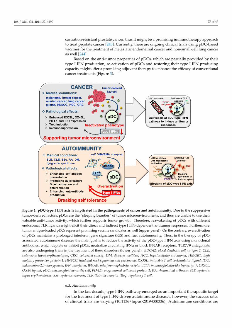
Int. J. Mol. Sci. 2021, 22, 4190 27 of 47
castration-resistant prostate cancer, thus it might be a promising immunotherapy approachto treat prostate cancer [243]. Currently, there are ongoing clinical trials using pDC-basedvaccines for the treatment of metastatic endometrial cancer and non-small-cell lung canceras well [244].
Based on the anti-tumor properties of pDCs, which are partially provided by theirtype I IFN production, re-activation of pDCs and restoring their type I IFN producingcapacity might offer a promising adjuvant therapy to enhance the efficacy of conventionalcancer treatments (Figure 3).
Int. J. Mol. Sci. 2021, 22, 4190 28 of 49
Figure 3. pDC-type I IFN axis is implicated in the pathogenesis of cancer and autoimmunity. Due to the suppressive tumor-derived factors, pDCs are the “sleeping beauties” of tumor microenvironments, and thus are unable to use their valuable anti-tumor activity, which further supports tumor growth. Therefore, reawakening of pDCs with different endo-somal TLR ligands might elicit their direct and indirect type I IFN-dependent antitumor responses. Furthermore, tumor antigen-loaded pDCs represent promising vaccine candidates as well (upper panel). On the contrary, overactivation of pDCs maintains a prolonged interferon gene signature (IGS) and fuel autoimmunity. Thus, in the therapy of pDC-associ-ated autoimmune diseases the main goal is to reduce the activity of the pDC-type I IFN axis using monoclonal antibodies, which deplete or inhibit pDCs, neutralize circulating IFNα or block IFNAR receptors. TLR7/9 antagonists are also under-going trials in the treatment of these disorders (lower panel). BDCA2: blood dendritic cell antigen 2; CLE: cutaneous lupus erythematosus; CRC: colorectal cancer; DM: diabetes mellitus; HCC: hepatocellular carcinoma; HMGB1: high mobility group box protein 1; HNSCC: head and neck squamous cell carcinoma; ICOSL: inducible T cell costimulator ligand; IDO: indoleamine-2,3- dioxygenase; IFN: interferon; IFNAR: interferon-alpha/beta receptor; ILT7: immunoglobulin-like transcript 7; OX40L: OX40 ligand; pDC: plasmacytoid dendritic cell; PD-L1: programmed cell death protein 1; RA: rheumatoid arthritis; SLE: systemic lupus erythema-tosus; SSc: systemic sclerosis; TLR: Toll-like receptor; Treg: regulatory T cell.
6.3. Autoimmunity In the last decade, type I IFN pathway emerged as an important therapeutic target
for the treatment of type I IFN-driven autoimmune diseases; however, the success rates of clinical trials are varying (10.1136/lupus-2019-000336). Autoimmune conditions are commonly accompanied by IGS, which refers to the increased expression of genes regu-lated by type I IFNs. IGS can be observed in the blood and/or in the affected inflamed tissues of patients with CLE, SLE, dermatomyositis, RA, systemic sclerosis (SSc) and Sjögren’s syndrome [10,245–247]. Autoimmune reactions can be associated with over-ac-tivated pDCs, which are rapidly recruited to inflamed tissues, where they actively pro-duce type I IFNs and interact with other immune cells to exacerbate inflammation. For instance, it was described that pDCs promote plasmablast differentiation and autoanti-body production in SLE through the release of IFNα and CD40 engagement [248]. Since a clear association exists between infiltrated pDC numbers and type I IFN overproduction, it seems feasible that the selective targeting of pDCs or their signaling molecules would
Figure 3. pDC-type I IFN axis is implicated in the pathogenesis of cancer and autoimmunity. Due to the suppressivetumor-derived factors, pDCs are the “sleeping beauties” of tumor microenvironments, and thus are unable to use theirvaluable anti-tumor activity, which further supports tumor growth. Therefore, reawakening of pDCs with differentendosomal TLR ligands might elicit their direct and indirect type I IFN-dependent antitumor responses. Furthermore,tumor antigen-loaded pDCs represent promising vaccine candidates as well (upper panel). On the contrary, overactivationof pDCs maintains a prolonged interferon gene signature (IGS) and fuel autoimmunity. Thus, in the therapy of pDC-associated autoimmune diseases the main goal is to reduce the activity of the pDC-type I IFN axis using monoclonalantibodies, which deplete or inhibit pDCs, neutralize circulating IFNα or block IFNAR receptors. TLR7/9 antagonistsare also undergoing trials in the treatment of these disorders (lower panel). BDCA2: blood dendritic cell antigen 2; CLE:cutaneous lupus erythematosus; CRC: colorectal cancer; DM: diabetes mellitus; HCC: hepatocellular carcinoma; HMGB1: highmobility group box protein 1; HNSCC: head and neck squamous cell carcinoma; ICOSL: inducible T cell costimulator ligand; IDO:indoleamine-2,3- dioxygenase; IFN: interferon; IFNAR: interferon-alpha/beta receptor; ILT7: immunoglobulin-like transcript 7; OX40L:OX40 ligand; pDC: plasmacytoid dendritic cell; PD-L1: programmed cell death protein 1; RA: rheumatoid arthritis; SLE: systemiclupus erythematosus; SSc: systemic sclerosis; TLR: Toll-like receptor; Treg: regulatory T cell.
6.3. Autoimmunity
In the last decade, type I IFN pathway emerged as an important therapeutic targetfor the treatment of type I IFN-driven autoimmune diseases; however, the success ratesof clinical trials are varying (10.1136/lupus-2019-000336). Autoimmune conditions are
Int. J. Mol. Sci. 2021, 22, 4190 28 of 47
commonly accompanied by IGS, which refers to the increased expression of genes regulatedby type I IFNs. IGS can be observed in the blood and/or in the affected inflamed tissuesof patients with CLE, SLE, dermatomyositis, RA, systemic sclerosis (SSc) and Sjögren’ssyndrome [10,245–247]. Autoimmune reactions can be associated with over-activatedpDCs, which are rapidly recruited to inflamed tissues, where they actively produce type IIFNs and interact with other immune cells to exacerbate inflammation. For instance, it wasdescribed that pDCs promote plasmablast differentiation and autoantibody productionin SLE through the release of IFNα and CD40 engagement [248]. Since a clear associationexists between infiltrated pDC numbers and type I IFN overproduction, it seems feasiblethat the selective targeting of pDCs or their signaling molecules would be a more potentapproach to control excessive type I IFN production in these disorders [249].
Autoimmune diseases are currently incurable conditions, and most therapies arebased on non-specific immunosuppression using glucocorticoids or cytostatic agents toreduce the severity of symptoms [249]. These therapeutic agents also act on pDC functions,for example, steroid administration decreases pDC numbers and their ability to producetype I IFNs in SLE patients. However, after discontinuing glucocorticoids, both the numberof pDCs and the level of IFNα recovered rapidly [250,251]. Hydroxychloroquine (HCQ)can also diminish type I IFN production by TLR7 or TLR9 activated pDCs from SLEpatients [252]. Another study found that clinically relevant high serum HCQ levels reducedTLR9 but not TLR7/8 induced type I IFN production in pDCs from CLE patients [136].Furthermore, mycophenolic acid, the active form of mycophenolate mofetil used to treatlupus nephritis is also able to dose-dependently suppress CpG-induced type I IFN secretionfrom pDCs of SLE patients via inhibiting nuclear translocation of IRF7 [253]. Similarly,clinically relevant doses of arsenic trioxide, which is used to treat SSc, could also diminishtype I IFN release by CpG-A activated pDCs through downregulating the expression aswell as the phosphorylation of IRF7 [254].
A more specific approach to treat autoimmune diseases is using monoclonal antibodiesto block the actions of over-activated cells or to reduce the level of factors associated withdisease exacerbation [249]. With regard to pDCs, clinical trials mainly target IFN signalingdownstream of pDCs by using monoclonal antibodies, which neutralize IFNα or blocktype I IFN receptors [255,256]. In addition, novel approaches are focusing on the depletionor functional inhibitions of pDCs. For example, the ILT7 targeting antibody MEDI7734(alternative name: VIB7734) is in a phase I clinical trial for the treatment of various typeI IFN-mediated autoimmune diseases, whereas the BDCA2 targeting BIIB059 is in phaseII clinical trial for the treatment of SLE and CLE [136,137,257]. Moreover, inhibitorymolecules blocking the endosomal TLR signaling pathway such as the IRAK-4 inhibitorPF-06650833 are also in clinical trials for the treatment of rheumatic and autoimmunediseases [258]. A promising approach for the treatment of IGS-associated diseases isactive immunization with IFNα-kinoid, a therapeutic vaccine consisting of inactivatedrecombinant human IFNα2b and a carrier protein, the keyhole limpet haemocyanin, whichcan induce a strong polyclonal antibody response against IFNα and thus reduce IGS inpatients with SLE [259,260]. Clinical trials focusing on the pDC-type I IFN axis are reviewedin more details in references [196,249].
Overall, as opposed to anti-tumor therapies, the goal of non-specific and antigen-specific immunotherapies in the treatment of IGS-associated autoimmune conditions isto reduce excessive type I IFN production, which can improve the life quality of patients(Figure 3).
6.4. Allergy
Besides the aforementioned pathological conditions, pDCs and their type I IFN pro-duction have also been implicated in the pathogenesis of allergic diseases. Studies foundthat pDCs are present in the nasal mucosa of allergic patients [261,262], and after allergeninhalation their number increases in the airways [263,264]. However, in allergic skin lesions,
Int. J. Mol. Sci. 2021, 22, 4190 29 of 47
the number of pDCs may vary depending on the type of hypersensitivity reaction andassociated cytokine milieu, which can affect their viability in the lesional skin [265,266].
Among allergic diseases, the role of pDCs has been most extensively studied in asthma,a chronic inflammatory disease of the airways, and it seems that due to their tolerogenicfunctions, pDCs help to relieve clinical symptoms and have a protective effect on diseaseprogression. In allergy, type I IFN production could account for the beneficial effects ofpDCs, since type I IFNs have the ability to inhibit the development of Th2 cells, the functionsof innate lymphoid cells 2 (ILC2), the secretion of Th2 cytokines, and the isotype switchingto IgE. Besides, pDCs drive regulatory T cell expansion and thus contribute to the inductionof tolerance in allergic diseases [267]. However, asthmatic patients are characterized byattenuated type I IFN production and uncontrolled Th2 response that makes them moresusceptible to viral infections and infection-induced asthma exacerbations. The impairedtype I IFN production might be the result of FcεRI cross-linking, which negatively effectsthe type I IFN pathway [268–270].
Multiple studies have shown the tolerogenic potential of pDCs in different mousemodels of allergic airway inflammation [271–274]. In a recent study, it was shown thatneonatal mice are more susceptible to severe allergic airway inflammation compared toadult mice that could be associated with the lower percentage of pDCs and thus reducedlevel of IFNα in neonates. Nevertheless, adoptive transfer of pDCs or addition of IFNα toneonatal mice inhibited the development of allergic inflammation. Depletion of pDCs inadult mice resulted in more severe allergic inflammation as well. The authors also reportedthat the protective effects of pDCs are mediated through their IFNα release, which inhibitsthe secretion of epithelial cell-derived chemotactic factors and thus the recruitment of Th2promoting cDCs and ILC2 cells. In line with that, the percentage of pDCs and levels ofIFNα were lower in the sputum of asthmatic children than in adult patients that mightrender children more susceptible to viral infection [275]. Indeed, reports suggest that lowerfrequencies of circulating pDCs correlate with the risk of lower respiratory tract infectionsin infants and young children up to the age of 5 years [276,277].
Despite their beneficial effects in preventing allergy development at early ages, studieswith experimental animal models suggest that pDCs drive virus-induced asthma exacerba-tion when the disease is already established. After rhinovirus infection of mice with allergicairway disease pDCs were recruited to the lung then migrated to the draining lymph nodesto trigger Th2-mediated allergic responses, which could be abrogated by pDC depletion.Consistent with that, higher pDC levels were detected in the sputum of asthmatic patientsduring acute exacerbations, and their numbers correlated with the severity of inflammationand frequency of asthmatic attacks [278].
The above data indicate that due to their tolerogenic properties pDCs contributeto the maintenance of a healthy Th1/Th2 balance, thus adequate pDC numbers andfunctions have a protective effect on the development of allergic diseases. Nevertheless,when the allergic disease is already established, acute viral infections might trigger theinflammatory properties of pDCs, which negatively affects the allergic condition and causesthe exacerbation of symptoms. However, it is worth to mention that while the protectiverole of the pDC-type I IFN axis is well established in infants and children with asthma,there is controversy regarding the role of pDCs in adult asthmatic patients. Therefore,further studies are needed to define the exact function of pDCs in the context of asthmaand to resolve these contradictions [279].
Inhaled corticosteroids are commonly used for the long-term treatment of asthmaticpatients to resolve symptoms. However, it is important to note that these drugs cansuppress type I IFN production and contribute to the onset of a Th2-dominant phenotypethat makes patients more vulnerable to respiratory tract infections [270]. Therefore, type IIFN therapy could be utilized for the treatment of patients with allergic asthma and otherchronic atopic diseases with Th2 dominance, in which type I IFNs could counterbalance Th2cell-mediated inflammation. Preclinical and clinical trials using type I IFN-based therapyfor the treatment of atopic diseases are summarized in references [268,270] (Figure 4).
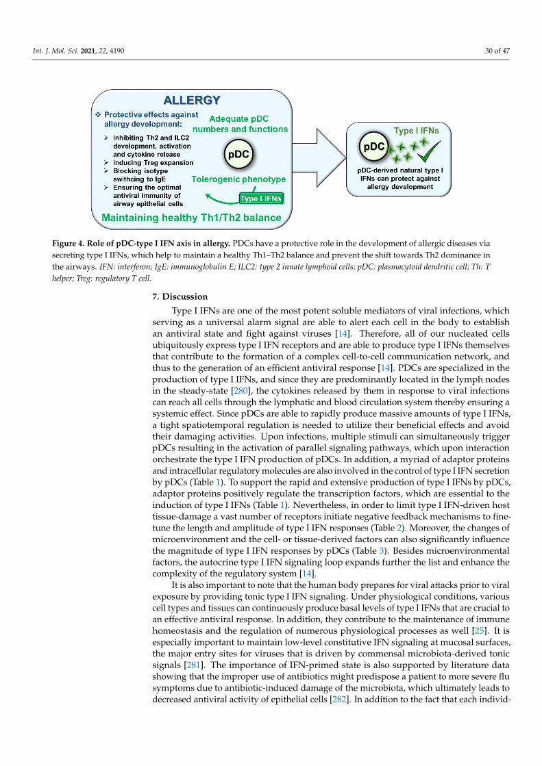
Int. J. Mol. Sci. 2021, 22, 4190 30 of 47Int. J. Mol. Sci. 2021, 22, 4190 31 of 49
Figure 4. Role of pDC-type I IFN axis in allergy. PDCs have a protective role in the development of allergic diseases via secreting type I IFNs, which help to maintain a healthy Th1–Th2 balance and prevent the shift towards Th2 dominance in the airways. IFN: interferon; IgE: immunoglobulin E; ILC2: type 2 innate lymphoid cells; pDC: plasmacytoid dendritic cell; Th: T helper; Treg: regulatory T cell.
7. Discussion Type I IFNs are one of the most potent soluble mediators of viral infections, which
serving as a universal alarm signal are able to alert each cell in the body to establish an antiviral state and fight against viruses [14]. Therefore, all of our nucleated cells ubiqui-tously express type I IFN receptors and are able to produce type I IFNs themselves that contribute to the formation of a complex cell-to-cell communication network, and thus to the generation of an efficient antiviral response [14]. PDCs are specialized in the produc-tion of type I IFNs, and since they are predominantly located in the lymph nodes in the steady-state [280], the cytokines released by them in response to viral infections can reach all cells through the lymphatic and blood circulation system thereby ensuring a systemic effect. Since pDCs are able to rapidly produce massive amounts of type I IFNs, a tight spatiotemporal regulation is needed to utilize their beneficial effects and avoid their dam-aging activities. Upon infections, multiple stimuli can simultaneously trigger pDCs result-ing in the activation of parallel signaling pathways, which upon interaction orchestrate the type I IFN production of pDCs. In addition, a myriad of adaptor proteins and intra-cellular regulatory molecules are also involved in the control of type I IFN secretion by pDCs (Table 1). To support the rapid and extensive production of type I IFNs by pDCs, adaptor proteins positively regulate the transcription factors, which are essential to the induction of type I IFNs (Table 1). Nevertheless, in order to limit type I IFN-driven host tissue-damage a vast number of receptors initiate negative feedback mechanisms to fine-tune the length and amplitude of type I IFN responses (Table 2). Moreover, the changes of microenvironment and the cell- or tissue-derived factors can also significantly influence the magnitude of type I IFN responses by pDCs (Table 3). Besides microenvironmental factors, the autocrine type I IFN signaling loop expands further the list and enhance the complexity of the regulatory system [14].
It is also important to note that the human body prepares for viral attacks prior to viral exposure by providing tonic type I IFN signaling. Under physiological conditions, various cell types and tissues can continuously produce basal levels of type I IFNs that are crucial to an effective antiviral response. In addition, they contribute to the mainte-nance of immune homeostasis and the regulation of numerous physiological processes as well [25]. It is especially important to maintain low-level constitutive IFN signaling at mu-cosal surfaces, the major entry sites for viruses that is driven by commensal microbiota-derived tonic signals [281]. The importance of IFN-primed state is also supported by lit-erature data showing that the improper use of antibiotics might predispose a patient to more severe flu symptoms due to antibiotic-induced damage of the microbiota, which ultimately leads to decreased antiviral activity of epithelial cells [282]. In addition to the
Figure 4. Role of pDC-type I IFN axis in allergy. PDCs have a protective role in the development of allergic diseases viasecreting type I IFNs, which help to maintain a healthy Th1–Th2 balance and prevent the shift towards Th2 dominance inthe airways. IFN: interferon; IgE: immunoglobulin E; ILC2: type 2 innate lymphoid cells; pDC: plasmacytoid dendritic cell; Th: Thelper; Treg: regulatory T cell.
7. Discussion
Type I IFNs are one of the most potent soluble mediators of viral infections, whichserving as a universal alarm signal are able to alert each cell in the body to establishan antiviral state and fight against viruses [14]. Therefore, all of our nucleated cellsubiquitously express type I IFN receptors and are able to produce type I IFNs themselvesthat contribute to the formation of a complex cell-to-cell communication network, andthus to the generation of an efficient antiviral response [14]. PDCs are specialized in theproduction of type I IFNs, and since they are predominantly located in the lymph nodesin the steady-state [280], the cytokines released by them in response to viral infectionscan reach all cells through the lymphatic and blood circulation system thereby ensuring asystemic effect. Since pDCs are able to rapidly produce massive amounts of type I IFNs,a tight spatiotemporal regulation is needed to utilize their beneficial effects and avoidtheir damaging activities. Upon infections, multiple stimuli can simultaneously triggerpDCs resulting in the activation of parallel signaling pathways, which upon interactionorchestrate the type I IFN production of pDCs. In addition, a myriad of adaptor proteinsand intracellular regulatory molecules are also involved in the control of type I IFN secretionby pDCs (Table 1). To support the rapid and extensive production of type I IFNs by pDCs,adaptor proteins positively regulate the transcription factors, which are essential to theinduction of type I IFNs (Table 1). Nevertheless, in order to limit type I IFN-driven hosttissue-damage a vast number of receptors initiate negative feedback mechanisms to fine-tune the length and amplitude of type I IFN responses (Table 2). Moreover, the changes ofmicroenvironment and the cell- or tissue-derived factors can also significantly influencethe magnitude of type I IFN responses by pDCs (Table 3). Besides microenvironmentalfactors, the autocrine type I IFN signaling loop expands further the list and enhance thecomplexity of the regulatory system [14].
It is also important to note that the human body prepares for viral attacks prior to viralexposure by providing tonic type I IFN signaling. Under physiological conditions, variouscell types and tissues can continuously produce basal levels of type I IFNs that are crucial toan effective antiviral response. In addition, they contribute to the maintenance of immunehomeostasis and the regulation of numerous physiological processes as well [25]. It isespecially important to maintain low-level constitutive IFN signaling at mucosal surfaces,the major entry sites for viruses that is driven by commensal microbiota-derived tonicsignals [281]. The importance of IFN-primed state is also supported by literature datashowing that the improper use of antibiotics might predispose a patient to more severe flusymptoms due to antibiotic-induced damage of the microbiota, which ultimately leads todecreased antiviral activity of epithelial cells [282]. In addition to the fact that each individ-

Int. J. Mol. Sci. 2021, 22, 4190 31 of 47
ual has a unique microbiome, several other factors such as gender related differences inimmune responses, genetic defects in type I IFN signaling, various pathological conditions,drugs, or biological therapies can also influence the individual’s baseline IFN signatureand antiviral capacity. Thus, the severity of viral infections might vary greatly amongindividuals even though they are infected by the same virus [193,283].
For instance, females mount a stronger and more effective humoral and cellularimmune response than males due to the positive stimulatory effect of the female sexhormone, estrogen, which also supports the type I IFN production of pDCs [183,190]. Inaddition to sex hormones, X chromosome dosage can also influence the IFN response ofpDCs. In a humanized mouse model, where CD34+ human progenitor cells (HPC) weretransplanted into female or male mice, pDCs developed from female HPCs producedhigher amounts of type I IFNs in response to TLR7 stimulation as compared with pDCsfrom male donors regardless of the sex of the recipient mice. These data imply that thedouble X chromosome in females has an immunological advantage by contributing to anenhanced immune response against infections. [191]. A similar study also investigated theeffect of X chromosome number and sex hormones on the TLR7-mediated IFNα productionby primary human pDCs from healthy females, males, transgender volunteers on hormonetherapy, and females with Turner syndrome. The authors found that the expression ofthe type I IFN-inducible antiviral membrane protein tetherin and the TLR7-induced IFNαproduction are higher in females that correlates with the number of X chromosomes but notwith sex hormone serum levels [284]. It has been long proposed that several genes involvedin TLR signaling might escape from X inactivation and account for the strong immuneresponses in females [285]. Indeed, single cell analyses of TLR7 allelic expression in pDCs,B cells and monocytes from females and Klinefelter (XXY) individuals revealed that TLR7escapes from X chromosome silencing. A recent study further demonstrated that femalepDCs with biallelic TLR7 expression have higher type I IFN production than monoallelicTLR7 expressing pDCs, presumably due to the IFNAR mediated type I IFN positivefeedback loop [286]. This molecular mechanism might also provide a plausible explanationfor the higher COVID-19 mortality rates among males compared to females [287,288]. Theabove data suggest that female pDCs with biallelic TLR7 expression respond more rapidlyto SARS-CoV-2 infection by producing large amounts of type IFNs that might account forthe better control of SARS-CoV-2 infection in women compared to men [286]. This idea isalso supported by a recent report showing that loss-of-function mutation of X-chromosomalTLR7 is associated with impaired type I IFN responses of young male patients with severeCOVID-19 that further emphasizes the importance of intact TLR7-mediated type I IFNresponses in the pathogenesis of the disease [289]. Moreover, pDC-derived type I IFNsmediate B cell activation and differentiation into plasma cells, thus are essential to elicit anoptimal antibody response to viral infection including SARS-CoV-2 infection as well [290].
All the above data highlight the critical importance of type I IFNs in mounting aneffective antiviral response. Notably, immunosuppressive drugs such as corticosteroids,which are commonly used in the therapy of autoimmune and allergic diseases, inhibit typeI IFN signaling, and thus enhance the susceptibility of patients to viral infections [251,291].Furthermore, it should be noted that type I IFN therapies might have undesired outcomeswhen the timing and dosage is not adjusted properly. For instance, type I IFN therapy canbe used as prophylaxis to reduce the severity of acute viral infections; however, its usageis not recommended during chronic viral infections, since it would exacerbate diseasesymptoms [292]. Furthermore, type I IFNs are used for the treatment of cancer due to theirtumor suppressive activity; nevertheless, they might induce robust autoimmune adverseevents [218].
Due to their high plasticity, pDCs are able to rapidly switch their phenotype andfunction in response to microenvironmental changes, and acquire either immunogenic ortolerogenic properties; thus, different disease-causing phenotypes might arise dependingon the associated pathological condition [293]. PDCs are unique in many of their propertiesamong immune cells and differ from their conventional counterparts in multiple aspects
Int. J. Mol. Sci. 2021, 22, 4190 32 of 47
including their receptor pattern, phenotypic and metabolic profile as well as their effectorfunctions [294]. Therefore, regulatory mechanisms, which have been widely exploredin cDCs cannot be extended to pDCs. Without doubt, pDCs are professional in type IIFN production, and thus play a prominent role in antiviral responses; however, theirinadequate or excessive type I IFN production might play a key role in the pathogenesisand pathomechanism of several human diseases. Although, currently, several ongoingclinical trials target the pDC-type I IFN axis for treating pDC-associated diseases, furtherinvestigations are needed to fully explore the mechanisms regulating the type I IFN re-sponses by pDCs that might help to better understand the complex regulation of type IIFN production, and thus might improve the efficacy of these therapies.
Author Contributions: All authors have a substantial contribution to the literature search andmanuscript writing, and all authors approved the submitted version of the article. All authors haveread and agreed to the published version of the manuscript.
Funding: This work was supported by the National Research, Development and Innovation Office(NKFIH FK 128294 to K.P., NKFIH PD 135193 to T.F.). The work was also supported by GINOP-2.3.2-15-2016-00050 project. The project is co-financed by the European Union and the EuropeanRegional Development Fund. K.P. was also supported by ÚNKP-20-05-DE-3 New National ExcellenceProgram of the Ministry for Innovation and Technology from the source of the National Research,Development and Innovation Fund and the János Bolyai Research Scholarship from the HungarianAcademy of Sciences.
Institutional Review Board Statement: Not applicable.
Informed Consent Statement: Not applicable.
Data Availability Statement: Not applicable.
Conflicts of Interest: The authors declare no conflict of interest.
Abbreviations
4E-BP Eukaryotic translation initiation factor 4E (eIF4E)-binding proteinACE-2 angiotensin converting enzyme 2AIDS Acquired Immune Deficiency SyndromeAPC antigen presenting cellAPRIL A proliferation-inducing ligandAPS antiphospholipid syndromeARDS acute respiratory distress syndromeBAFF B cell activating factorBCR B cell receptorBDCA2 blood dendritic cell antigen 2BM bone marrowBST2 bone marrow stromal cell antigen 2Btk Bruton tyrosine kinasecAMP cyclic adenosine monophosphateCbl Casitas B cell lymphomaCD cluster of differentiationCD2AP CD2-associated adaptor proteincDC conventional dendritic cellcGAS DNA-sensing cyclic GMP-AMP synthaseCHIKV chikungunya virusCLE cutaneous lupus erythematosusCOVID-19 coronavirus disease 2019CXXC5 CXXC type zink finger protein 5DAMP damage-associated molecular patternsDAP12 DNAX activation protein 12DC dendritic cell
Int. J. Mol. Sci. 2021, 22, 4190 33 of 47
DCIR DC immunoreceptorDENV dengue virusDHT DihydrotestosteroneDMPA depot medroxyprogesterone acetateDR5 death receptor 5E2 17β-estradiolEAE experimental autoimmune encephalomyelitisEBI2 Epstein-Barr virus induced receptor 2eIF2α eukaryotic initiation factor 2αER endoplasmic reticulumERK extracellular-signal-regulated kinaseERα estrogen receptor-αFcR Fc receptorFDA Food and Drug AdministrationFlu influenza virusGal-9 galectin-9gp120 glycoprotein 120H2 histamine receptor 2H4 histamine receptor 4HBsAg hepatitis B virus surface antigenHBV hepatitis B virusHCQ HydroxychloroquineHCV hepatitis C virusHDAC3 histone deacetylase 3HMGB1 high mobility group box 1 proteinHPC human progenitor cellsHPV human papilloma virusHSV-1 herpes simplex viruses-1IDO indoleamine 2,3-dioxygenaseIFN InterferonIFNAR interferon α receptorIGS interferon gene signatureIKK-α inhibitory kappa B kinase αIL InterleukinILC2 innate lymphoid cells 2ILT7 immunoglobulin-like transcript 7IPC type I interferon producing cellIRAK interleukin-1 receptor-associated kinaseIRF interferon regulatory factorISG interferon stimulated geneISGF3 interferon-stimulated gene factor 3ISRE interferon-stimulated response elementITAM immunoreceptor tyrosine-based activation motifITIM immunoreceptor tyrosine-based inhibition motifJAK1 tyrosine kinases Janus kinase 1LAIR-1 leukocyte-associated Ig-like receptor-1LAMP5 lysosome-associated membrane protein 5LAP LC3-associated phagocytosisLCMV lymphocytic choriomeningitis virusLFA-1 lymphocyte functional antigen 1LMIR8 leukocyte mono-immunoglobulin-like receptor 8mAB monoclonal antibodiesMAPK mitogen-activated protein kinaseMAVS mitochondrial antiviral signaling adapter proteinMCMV murine cytomegalovirus
Int. J. Mol. Sci. 2021, 22, 4190 34 of 47
MDA5 melanoma differentiation-associated gene-5MEK mitogen-activated protein kinase kinaseMERS-CoV Middle East respiratory syndrome coronavirusMHC major histocompatibility complexMHV mouse hepatitis virusMIP-1β macrophage inflammatory protein-1βmiRs MicroRNAsMR mannose receptormTOR mammalian target of rapamycinmtROS mitochondrial reactive oxygen speciesMx GTPAse myxovirus resistance guanosine triphosphataseMxA myxovirus resistance protein 1NCOR2 nuclear receptor co-repressor 2NFATC3 nuclear factor of activated T cells C 3NF-κB Nuclear factor κ BNK natural killerNLR nucleotide-binding domain leucine-rich repeat containing receptorOAS Oligo A synthetaseOligo A 2′,5′-oligoadenylateOpn OsteopontinPACSIN1 Protein kinase C and casein kinase substrate in neurons 1PAMP pathogen-associated molecular patternsPBMC peripheral blood mononuclear cellsPD-1 programmed cell death 1pDC plasmacytoid dendritic cellPDC-TREM plasmacytoid dendritic cell—triggering receptor expressed on myeloid cellPGE2 prostaglandin E 2PI3K phosphatidylinositol 3-kinasePIR-B paired immunoglobulin-like receptor BPKR protein kinasePLCγ2 phospholipase C-gamma 2PLSCR1 phospholipid scramblase 1polyU polyuridylic acidPRR pattern recognition receptorsPTEN phosphatase and tensin homologPTPRF Protein tyrosine phosphatase receptor type FPTPRS Protein tyrosine phosphatase receptor type SRA Rheumatoid arthritisRAGE receptor for advanced glycation endproductsRIG-I retinoic acid-inducible gene IRLR RIG-I-like receptorRNase L ribonuclease LROS reactive oxygen speciesRSV respiratory syncytial virusRUNX2 Runt-related transcription factor 2S1P sphingosine-1-phosphateSARS-CoV-2 severe acute respiratory syndrome coronavirus 2SCARB2 Scavenger receptor class B, member 2SH2 Src homology region 2SHIP1 SH-2 domain-containing inositol-5-phosphatase 1SHP-1 SH-2 domain-containing phosphatase 1Siglec-H sialic acid binding immunoglobulin type lectins HSLAMF signaling lymphocyte activation molecule familySLE Systemic lupus erythematosusSlp65 Src homology 2 domain-containing leukocyte protein of 65 kDaSOCS suppressor of cytokine signaling
Int. J. Mol. Sci. 2021, 22, 4190 35 of 47
SphK1 Sphingosine kinase 1SSc systemic sclerosisSTAT signal transducer and activator of transcriptionSTING stimulator of interferon geneSyk spleen tyrosine kinaseTAB2 TGFβ activated kinase 1 binding Protein 2TBK1 Tank-binding kinase 1TGFβ transforming growth factor-βTh T helperTIM-3 T cell immunoglobulin and mucin domain-containing protein 3TLR Toll-like receptorTNF tumor necrosis factorTRAF TNF Receptor Associated FactorTRAIL tumor necrosis factor-related apoptosis-inducing ligandTRIM tripartite motifTSC1 tuberous sclerosis complex 1TYK2 tyrosine kinase 2VEGFR2 vascular endothelial growth factor receptor 2VIP vasoactive intestinal peptideVSV Vesicular stomatitis virusWnt5a wingless-related integration site 5a
References1. Reizis, B. Plasmacytoid Dendritic Cells: Development, Regulation, and Function. Immunity 2019, 50, 37–50. [CrossRef]2. Lennert, K.; Remmele, W. Karyometric research on lymph node cells in man. I. Germinoblasts, lymphoblasts & lymphocytes.
Acta Haematol. 1958, 19, 99–113. [CrossRef]3. Grouard, G.; Rissoan, M.C.; Filgueira, L.; Durand, I.; Banchereau, J.; Liu, Y.J. The enigmatic plasmacytoid T cells develop into
dendritic cells with interleukin (IL)-3 and CD40-ligand. J. Exp. Med. 1997, 185, 1101–1111. [CrossRef] [PubMed]4. Siegal, F.P.; Kadowaki, N.; Shodell, M.; Fitzgerald-Bocarsly, P.A.; Shah, K.; Ho, S.; Antonenko, S.; Liu, Y.J. The nature of the
principal type 1 interferon-producing cells in human blood. Science 1999, 284, 1835–1837. [CrossRef] [PubMed]5. Merad, M.; Manz, M.G. Dendritic cell homeostasis. Blood 2009, 113, 3418–3427. [CrossRef]6. Tel, J.; van der Leun, A.M.; Figdor, C.G.; Torensma, R.; de Vries, I.J. Harnessing human plasmacytoid dendritic cells as professional
APCs. Cancer Immunol. Immunother. 2012, 61, 1279–1288. [CrossRef] [PubMed]7. Vanbervliet, B.; Bendriss-Vermare, N.; Massacrier, C.; Homey, B.; de Bouteiller, O.; Briere, F.; Trinchieri, G.; Caux, C. The inducible
CXCR3 ligands control plasmacytoid dendritic cell responsiveness to the constitutive chemokine stromal cell-derived factor 1(SDF-1)/CXCL12. J. Exp. Med. 2003, 198, 823–830. [CrossRef] [PubMed]
8. Huch, J.H.; Cunningham, A.L.; Arvin, A.M.; Nasr, N.; Santegoets, S.J.; Slobedman, E.; Slobedman, B.; Abendroth, A. Impact ofvaricella-zoster virus on dendritic cell subsets in human skin during natural infection. J. Virol. 2010, 84, 4060–4072. [CrossRef][PubMed]
9. Baranek, T.; Zucchini, N.; Dalod, M. Plasmacytoid dendritic cells and the control of herpesvirus infections. Viruses 2009, 1,383–419. [CrossRef]
10. Lande, R.; Gilliet, M. Plasmacytoid dendritic cells: Key players in the initiation and regulation of immune responses. Ann. N. Y.Acad. Sci. 2010, 1183, 89–103. [CrossRef]
11. Walter, M.R. The Role of Structure in the Biology of Interferon Signaling. Front. Immunol. 2020, 11, 606489. [CrossRef]12. Ali, S.; Mann-Nuttel, R.; Schulze, A.; Richter, L.; Alferink, J.; Scheu, S. Sources of Type I Interferons in Infectious Immunity:
Plasmacytoid Dendritic Cells Not Always in the Driver’s Seat. Front. Immunol. 2019, 10, 778. [CrossRef]13. Stetson, D.B.; Medzhitov, R. Type I interferons in host defense. Immunity 2006, 25, 373–381. [CrossRef] [PubMed]14. Ivashkiv, L.B.; Donlin, L.T. Regulation of type I interferon responses. Nat. Rev. Immunol. 2014, 14, 36–49. [CrossRef] [PubMed]15. Samuel, C.E. Antiviral actions of interferons. Clin. Microbiol. Rev. 2001, 14, 778–809, table of contents. [CrossRef]16. Gal-Ben-Ari, S.; Barrera, I.; Ehrlich, M.; Rosenblum, K. PKR: A Kinase to Remember. Front. Mol. Neurosci. 2018, 11, 480. [CrossRef]
[PubMed]17. Ibsen, M.S.; Gad, H.H.; Thavachelvam, K.; Boesen, T.; Despres, P.; Hartmann, R. The 2’-5’-oligoadenylate synthetase 3 enzyme
potently synthesizes the 2’-5’-oligoadenylates required for RNase L activation. J. Virol. 2014, 88, 14222–14231. [CrossRef]18. Haller, O.; Kochs, G. Interferon-induced mx proteins: Dynamin-like GTPases with antiviral activity. Traffic 2002, 3, 710–717.
[CrossRef] [PubMed]19. Gabriele, L.; Borghi, P.; Rozera, C.; Sestili, P.; Andreotti, M.; Guarini, A.; Montefusco, E.; Foa, R.; Belardelli, F. IFN-alpha promotes
the rapid differentiation of monocytes from patients with chronic myeloid leukemia into activated dendritic cells tuned toundergo full maturation after LPS treatment. Blood 2004, 103, 980–987. [CrossRef]
20. Lee, A.J.; Ashkar, A.A. The Dual Nature of Type I and Type II Interferons. Front. Immunol. 2018, 9, 2061. [CrossRef]
Int. J. Mol. Sci. 2021, 22, 4190 36 of 47
21. Simmons, D.P.; Wearsch, P.A.; Canaday, D.H.; Meyerson, H.J.; Liu, Y.C.; Wang, Y.; Boom, W.H.; Harding, C.V. Type I IFN drives adistinctive dendritic cell maturation phenotype that allows continued class II MHC synthesis and antigen processing. J. Immunol.2012, 188, 3116–3126. [CrossRef] [PubMed]
22. Kiefer, K.; Oropallo, M.A.; Cancro, M.P.; Marshak-Rothstein, A. Role of type I interferons in the activation of autoreactive B cells.Immunol. Cell Biol. 2012, 90, 498–504. [CrossRef] [PubMed]
23. Kuka, M.; De Giovanni, M.; Iannacone, M. The role of type I interferons in CD4(+) T cell differentiation. Immunol. Lett. 2019, 215,19–23. [CrossRef]
24. Kolumam, G.A.; Thomas, S.; Thompson, L.J.; Sprent, J.; Murali-Krishna, K. Type I interferons act directly on CD8 T cells to allowclonal expansion and memory formation in response to viral infection. J. Exp. Med. 2005, 202, 637–650. [CrossRef]
25. Gough, D.J.; Messina, N.L.; Clarke, C.J.; Johnstone, R.W.; Levy, D.E. Constitutive type I interferon modulates homeostatic balancethrough tonic signaling. Immunity 2012, 36, 166–174. [CrossRef]
26. Hosseini, S.; Michaelsen-Preusse, K.; Grigoryan, G.; Chhatbar, C.; Kalinke, U.; Korte, M. Type I Interferon Receptor Signaling inAstrocytes Regulates Hippocampal Synaptic Plasticity and Cognitive Function of the Healthy CNS. Cell Rep. 2020, 31, 107666.[CrossRef] [PubMed]
27. Kawashima, T.; Kosaka, A.; Yan, H.; Guo, Z.; Uchiyama, R.; Fukui, R.; Kaneko, D.; Kumagai, Y.; You, D.J.; Carreras, J.; et al.Double-stranded RNA of intestinal commensal but not pathogenic bacteria triggers production of protective interferon-beta.Immunity 2013, 38, 1187–1197. [CrossRef] [PubMed]
28. Bradley, K.C.; Finsterbusch, K.; Schnepf, D.; Crotta, S.; Llorian, M.; Davidson, S.; Fuchs, S.Y.; Staeheli, P.; Wack, A. Microbiota-Driven Tonic Interferon Signals in Lung Stromal Cells Protect from Influenza Virus Infection. Cell Rep. 2019, 28, 245–256.e4.[CrossRef] [PubMed]
29. Abt, M.C.; Osborne, L.C.; Monticelli, L.A.; Doering, T.A.; Alenghat, T.; Sonnenberg, G.F.; Paley, M.A.; Antenus, M.; Williams, K.L.;Erikson, J.; et al. Commensal bacteria calibrate the activation threshold of innate antiviral immunity. Immunity 2012, 37, 158–170.[CrossRef]
30. Banchereau, J.; Pascual, V. Type I interferon in systemic lupus erythematosus and other autoimmune diseases. Immunity 2006, 25,383–392. [CrossRef]
31. Liu, Y.J. IPC: Professional type 1 interferon-producing cells and plasmacytoid dendritic cell precursors. Annu. Rev. Immunol. 2005,23, 275–306. [CrossRef]
32. Haeryfar, S.M. The importance of being a pDC in antiviral immunity: The IFN mission versus Ag presentation? Trends Immunol.2005, 26, 311–317. [CrossRef]
33. Ito, T.; Kanzler, H.; Duramad, O.; Cao, W.; Liu, Y.J. Specialization, kinetics, and repertoire of type 1 interferon responses by humanplasmacytoid predendritic cells. Blood 2006, 107, 2423–2431. [CrossRef]
34. Fitzgerald-Bocarsly, P.; Dai, J.; Singh, S. Plasmacytoid dendritic cells and type I IFN: 50 years of convergent history. CytokineGrowth Factor Rev. 2008, 19, 3–19. [CrossRef] [PubMed]
35. Bao, M.; Liu, Y.J. Regulation of TLR7/9 signaling in plasmacytoid dendritic cells. Protein Cell 2013, 4, 40–52. [CrossRef] [PubMed]36. Swiecki, M.; Colonna, M. The multifaceted biology of plasmacytoid dendritic cells. Nat. Rev. Immunol. 2015, 15, 471–485.
[CrossRef]37. McKenna, K.; Beignon, A.S.; Bhardwaj, N. Plasmacytoid dendritic cells: Linking innate and adaptive immunity. J. Virol. 2005, 79,
17–27. [CrossRef] [PubMed]38. Colina, R.; Costa-Mattioli, M.; Dowling, R.J.; Jaramillo, M.; Tai, L.H.; Breitbach, C.J.; Martineau, Y.; Larsson, O.; Rong, L.;
Svitkin, Y.V.; et al. Translational control of the innate immune response through IRF-7. Nature 2008, 452, 323–328. [CrossRef]39. Honda, K.; Ohba, Y.; Yanai, H.; Negishi, H.; Mizutani, T.; Takaoka, A.; Taya, C.; Taniguchi, T. Spatiotemporal regulation of
MyD88-IRF-7 signalling for robust type-I interferon induction. Nature 2005, 434, 1035–1040. [CrossRef]40. Szabo, A.; Magyarics, Z.; Pazmandi, K.; Gopcsa, L.; Rajnavolgyi, E.; Bacsi, A. TLR ligands upregulate RIG-I expression in human
plasmacytoid dendritic cells in a type I IFN-independent manner. Immunol. Cell Biol. 2014, 92, 671–678. [CrossRef]41. Musumeci, A.; Lutz, K.; Winheim, E.; Krug, A.B. What Makes a pDC: Recent Advances in Understanding Plasmacytoid DC
Development and Heterogeneity. Front. Immunol. 2019, 10, 1222. [CrossRef]42. Abbas, A.; Vu Manh, T.P.; Valente, M.; Collinet, N.; Attaf, N.; Dong, C.; Naciri, K.; Chelbi, R.; Brelurut, G.; Cervera-Marzal, I.; et al.
The activation trajectory of plasmacytoid dendritic cells in vivo during a viral infection. Nat. Immunol. 2020, 21, 983–997.[CrossRef]
43. Ning, S.; Pagano, J.S.; Barber, G.N. IRF7: Activation, regulation, modification and function. Genes Immun. 2011, 12, 399–414.[CrossRef]
44. Honda, K.; Yanai, H.; Negishi, H.; Asagiri, M.; Sato, M.; Mizutani, T.; Shimada, N.; Ohba, Y.; Takaoka, A.; Yoshida, N.; et al. IRF-7is the master regulator of type-I interferon-dependent immune responses. Nature 2005, 434, 772–777. [CrossRef]
45. Pelka, K.; Latz, E. IRF5, IRF8, and IRF7 in human pDCs—the good, the bad, and the insignificant? Eur. J. Immunol. 2013, 43,1693–1697. [CrossRef]
46. Dai, P.; Cao, H.; Merghoub, T.; Avogadri, F.; Wang, W.; Parikh, T.; Fang, C.M.; Pitha, P.M.; Fitzgerald, K.A.; Rahman, M.M.; et al.Myxoma virus induces type I interferon production in murine plasmacytoid dendritic cells via a TLR9/MyD88-, IRF5/IRF7-, andIFNAR-dependent pathway. J. Virol. 2011, 85, 10814–10825. [CrossRef]

Int. J. Mol. Sci. 2021, 22, 4190 37 of 47
47. Yasuda, K.; Nundel, K.; Watkins, A.A.; Dhawan, T.; Bonegio, R.G.; Ubellacker, J.M.; Marshak-Rothstein, A.; Rifkin, I.R. Phenotypeand function of B cells and dendritic cells from interferon regulatory factor 5-deficient mice with and without a mutation inDOCK2. Int. Immunol. 2013, 25, 295–306. [CrossRef]
48. Steinhagen, F.; Rodriguez, L.G.; Tross, D.; Tewary, P.; Bode, C.; Klinman, D.M. IRF5 and IRF8 modulate the CAL-1 humanplasmacytoid dendritic cell line response following TLR9 ligation. Eur. J. Immunol. 2016, 46, 647–655. [CrossRef]
49. Sichien, D.; Scott, C.L.; Martens, L.; Vanderkerken, M.; Van Gassen, S.; Plantinga, M.; Joeris, T.; De Prijck, S.; Vanhoutte, L.;Vanheerswynghels, M.; et al. IRF8 Transcription Factor Controls Survival and Function of Terminally Differentiated Conventionaland Plasmacytoid Dendritic Cells, Respectively. Immunity 2016, 45, 626–640. [CrossRef]
50. Chopin, M.; Preston, S.P.; Lun, A.T.L.; Tellier, J.; Smyth, G.K.; Pellegrini, M.; Belz, G.T.; Corcoran, L.M.; Visvader, J.E.; Wu, L.; et al.RUNX2 Mediates Plasmacytoid Dendritic Cell Egress from the Bone Marrow and Controls Viral Immunity. Cell Rep. 2016, 15,866–878. [CrossRef]
51. Sasaki, I.; Hoshino, K.; Sugiyama, T.; Yamazaki, C.; Yano, T.; Iizuka, A.; Hemmi, H.; Tanaka, T.; Saito, M.; Sugiyama, M.; et al.Spi-B is critical for plasmacytoid dendritic cell function and development. Blood 2012, 120, 4733–4743. [CrossRef]
52. Bao, M.; Wang, Y.; Liu, Y.; Shi, P.; Lu, H.; Sha, W.; Weng, L.; Hanabuchi, S.; Qin, J.; Plumas, J.; et al. NFATC3 promotes IRF7transcriptional activity in plasmacy–toid dendritic cells. J. Exp. Med. 2016, 213, 2383–2398. [CrossRef]
53. Kim, T.W.; Hong, S.; Lin, Y.; Murat, E.; Joo, H.; Kim, T.; Pascual, V.; Liu, Y.J. Transcriptional Repression of IFN RegulatoryFactor 7 by MYC Is Critical for Type I IFN Production in Human Plasmacytoid Dendritic Cells. J. Immunol. 2016, 197, 3348–3359.[CrossRef]
54. Ayaz, G.; Razizadeh, N.; Yasar, P.; Kars, G.; Kahraman, D.C.; Saatci, O.; Sahin, O.; Cetin-Atalay, R.; Muyan, M. CXXC5 as anunmethylated CpG dinucleotide binding protein contributes to estrogen-mediated cellular proliferation. Sci. Rep. 2020, 10, 5971.[CrossRef] [PubMed]
55. Ma, S.; Wan, X.; Deng, Z.; Shi, L.; Hao, C.; Zhou, Z.; Zhou, C.; Fang, Y.; Liu, J.; Yang, J.; et al. Epigenetic regulator CXXC5 recruitsDNA demethylase Tet2 to regulate TLR7/9-elicited IFN response in pDCs. J. Exp. Med. 2017, 214, 1471–1491. [CrossRef]
56. Cisse, B.; Caton, M.L.; Lehner, M.; Maeda, T.; Scheu, S.; Locksley, R.; Holmberg, D.; Zweier, C.; den Hollander, N.S.;Kant, S.G.; et al. Transcription factor E2-2 is an essential and specific regulator of plasmacytoid dendritic cell development.Cell 2008, 135, 37–48. [CrossRef] [PubMed]
57. Cheng, M.; Zhang, X.; Yu, H.; Du, P.; Plumas, J.; Chaperot, L.; Su, L.; Zhang, L. Characterization of species-specific genes regulatedby E2-2 in human plasmacytoid dendritic cells. Sci. Rep. 2015, 5, 10752. [CrossRef]
58. Shinohara, M.L.; Lu, L.; Bu, J.; Werneck, M.B.; Kobayashi, K.S.; Glimcher, L.H.; Cantor, H. Osteopontin expression is essential forinterferon-alpha production by plasmacytoid dendritic cells. Nat. Immunol. 2006, 7, 498–506. [CrossRef]
59. Esashi, E.; Bao, M.; Wang, Y.H.; Cao, W.; Liu, Y.J. PACSIN1 regulates the TLR7/9-mediated type I interferon response inplasmacytoid dendritic cells. Eur. J. Immunol. 2012, 42, 573–579. [CrossRef]
60. Maarifi, G.; Smith, N.; Maillet, S.; Moncorge, O.; Chamontin, C.; Edouard, J.; Sohm, F.; Blanchet, F.P.; Herbeuval, J.P.;Lutfalla, G.; et al. TRIM8 is required for virus-induced IFN response in human plasmacytoid dendritic cells. Sci. Adv. 2019, 5,eaax3511. [CrossRef]
61. Talukder, A.H.; Bao, M.; Kim, T.W.; Facchinetti, V.; Hanabuchi, S.; Bover, L.; Zal, T.; Liu, Y.J. Phospholipid scramblase 1 regulatesToll-like receptor 9-mediated type I interferon production in plasmacytoid dendritic cells. Cell Res. 2012, 22, 1129–1139. [CrossRef]
62. Mohammed, S.; Vineetha, N.S.; James, S.; Aparna, J.S.; Babu Lankadasari, M.; Maeda, T.; Ghosh, A.; Saha, S.; Li, Q.Z.;Spiegel, S.; et al. Regulatory role of SphK1 in TLR7/9-dependent type I interferon response and autoimmunity. FASEB J. 2020, 34,4329–4347. [CrossRef]
63. Guo, H.; Zhang, J.; Zhang, X.; Wang, Y.; Yu, H.; Yin, X.; Li, J.; Du, P.; Plumas, J.; Chaperot, L.; et al. SCARB2/LIMP-2 RegulatesIFN Production of Plasmacytoid Dendritic Cells by Mediating Endosomal Translocation of TLR9 and Nuclear Translocation ofIRF7. J. Immunol. 2015, 194, 4737–4749. [CrossRef]
64. Cao, W.; Manicassamy, S.; Tang, H.; Kasturi, S.P.; Pirani, A.; Murthy, N.; Pulendran, B. Toll-like receptor-mediated induction oftype I interferon in plasmacytoid dendritic cells requires the rapamycin-sensitive PI(3)K-mTOR-p70S6K pathway. Nat. Immunol.2008, 9, 1157–1164. [CrossRef] [PubMed]
65. Fekete, T.; Agics, B.; Bencze, D.; Bene, K.; Szanto, A.; Tarr, T.; Vereb, Z.; Bacsi, A.; Pazmandi, K. Regulation of RLR-MediatedAntiviral Responses of Human Dendritic Cells by mTOR. Front. Immunol. 2020, 11, 572960. [CrossRef]
66. Agod, Z.; Fekete, T.; Budai, M.M.; Varga, A.; Szabo, A.; Moon, H.; Boldogh, I.; Biro, T.; Lanyi, A.; Bacsi, A.; et al. Regulation oftype I interferon responses by mitochondria-derived reactive oxygen species in plasmacytoid dendritic cells. Redox Biol. 2017, 13,633–645. [CrossRef] [PubMed]
67. Pazmandi, K.; Magyarics, Z.; Boldogh, I.; Csillag, A.; Rajnavolgyi, E.; Bacsi, A. Modulatory effects of low-dose hydrogen peroxideon the function of human plasmacytoid dendritic cells. Free Radic Biol. Med. 2012, 52, 635–645. [CrossRef]
68. Zhou, H.; Huang, X.; Cui, H.; Luo, X.; Tang, Y.; Chen, S.; Wu, L.; Shen, N. miR-155 and its star-form partner miR-155* cooperativelyregulate type I interferon production by human plasmacytoid dendritic cells. Blood 2010, 116, 5885–5894. [CrossRef]
69. Karrich, J.J.; Jachimowski, L.C.; Libouban, M.; Iyer, A.; Brandwijk, K.; Taanman-Kueter, E.W.; Nagasawa, M.; de Jong, E.C.;Uittenbogaart, C.H.; Blom, B. MicroRNA-146a regulates survival and maturation of human plasmacytoid dendritic cells. Blood2013, 122, 3001–3009. [CrossRef] [PubMed]

Int. J. Mol. Sci. 2021, 22, 4190 38 of 47
70. Rossato, M.; Affandi, A.J.; Thordardottir, S.; Wichers, C.G.K.; Cossu, M.; Broen, J.C.A.; Moret, F.M.; Bossini-Castillo, L.; Chouri, E.;van Bon, L.; et al. Association of MicroRNA-618 Expression With Altered Frequency and Activation of Plasmacytoid DendriticCells in Patients With Systemic Sclerosis. Arthritis Rheumatol. 2017, 69, 1891–1902. [CrossRef]
71. Liu, F.; Liu, C.; Hu, X.; Shang, Y.; Wu, L. MicroRNA-21: A Positive Regulator for Optimal Production of Type I and Type IIIInterferon by Plasmacytoid Dendritic Cells. Front. Immunol. 2017, 8, 947. [CrossRef]
72. Agudo, J.; Ruzo, A.; Tung, N.; Salmon, H.; Leboeuf, M.; Hashimoto, D.; Becker, C.; Garrett-Sinha, L.A.; Baccarini, A.;Merad, M.; et al. The miR-126-VEGFR2 axis controls the innate response to pathogen-associated nucleic acids. Nat. Immunol.2014, 15, 54–62. [CrossRef]
73. Inoue, M.; Shinohara, M.L. Intracellular osteopontin (iOPN) and immunity. Immunol. Res. 2011, 49, 160–172. [CrossRef] [PubMed]74. Kodigepalli, K.M.; Bowers, K.; Sharp, A.; Nanjundan, M. Roles and regulation of phospholipid scramblases. FEBS Lett. 2015, 589,
3–14. [CrossRef] [PubMed]75. O’Neill, L.A.; Kishton, R.J.; Rathmell, J. A guide to immunometabolism for immunologists. Nat. Rev. Immunol. 2016, 16, 553–565.
[CrossRef] [PubMed]76. Bajwa, G.; DeBerardinis, R.J.; Shao, B.; Hall, B.; Farrar, J.D.; Gill, M.A. Cutting Edge: Critical Role of Glycolysis in Human
Plasmacytoid Dendritic Cell Antiviral Responses. J. Immunol. 2016, 196, 2004–2009. [CrossRef] [PubMed]77. Fekete, T.; Suto, M.I.; Bencze, D.; Mazlo, A.; Szabo, A.; Biro, T.; Bacsi, A.; Pazmandi, K. Human Plasmacytoid and Monocyte-
Derived Dendritic Cells Display Distinct Metabolic Profile Upon RIG-I Activation. Front. Immunol. 2018, 9, 3070. [CrossRef]78. Wu, D.; Sanin, D.E.; Everts, B.; Chen, Q.; Qiu, J.; Buck, M.D.; Patterson, A.; Smith, A.M.; Chang, C.H.; Liu, Z.; et al. Type 1
Interferons Induce Changes in Core Metabolism that Are Critical for Immune Function. Immunity 2016, 44, 1325–1336. [CrossRef]79. Van de Laar, L.; van den Bosch, A.; Boonstra, A.; Binda, R.S.; Buitenhuis, M.; Janssen, H.L.; Coffer, P.J.; Woltman, A.M. PI3K-PKB
hyperactivation augments human plasmacytoid dendritic cell development and function. Blood 2012, 120, 4982–4991. [CrossRef][PubMed]
80. Boor, P.P.; Metselaar, H.J.; Mancham, S.; van der Laan, L.J.; Kwekkeboom, J. Rapamycin has suppressive and stimulatory effectson human plasmacytoid dendritic cell functions. Clin. Exp. Immunol. 2013, 174, 389–401. [CrossRef]
81. Chen, H.; Jiang, Z. The essential adaptors of innate immune signaling. Protein Cell 2013, 4, 27–39. [CrossRef]82. Forrester, S.J.; Kikuchi, D.S.; Hernandes, M.S.; Xu, Q.; Griendling, K.K. Reactive Oxygen Species in Metabolic and Inflammatory
Signaling. Circ. Res. 2018, 122, 877–902. [CrossRef]83. Stout-Delgado, H.W.; Yang, X.; Walker, W.E.; Tesar, B.M.; Goldstein, D.R. Aging impairs IFN regulatory factor 7 up-regulation in
plasmacytoid dendritic cells during TLR9 activation. J. Immunol. 2008, 181, 6747–6756. [CrossRef]84. Chandan, K.; Gupta, M.; Sarwat, M. Role of Host and Pathogen-Derived MicroRNAs in Immune Regulation During Infectious
and Inflammatory Diseases. Front. Immunol. 2019, 10, 3081. [CrossRef] [PubMed]85. Smyth, L.A.; Boardman, D.A.; Tung, S.L.; Lechler, R.; Lombardi, G. MicroRNAs affect dendritic cell function and phenotype.
Immunology 2015, 144, 197–205. [CrossRef] [PubMed]86. Jin, F.; Hu, H.; Xu, M.; Zhan, S.; Wang, Y.; Zhang, H.; Chen, X. Serum microRNA Profiles Serve as Novel Biomarkers for
Autoimmune Diseases. Front. Immunol. 2018, 9, 2381. [CrossRef] [PubMed]87. Van den Hoogen, L.L.; Rossato, M.; Lopes, A.P.; Pandit, A.; Bekker, C.P.J.; Fritsch-Stork, R.D.E.; van Roon, J.A.G.; Radstake, T.
microRNA downregulation in plasmacytoid dendritic cells in interferon-positive systemic lupus erythematosus and antiphos-pholipid syndrome. Rheumatology 2018, 57, 1669–1674. [CrossRef] [PubMed]
88. Hillen, M.R.; Chouri, E.; Wang, M.; Blokland, S.L.M.; Hartgring, S.A.Y.; Concepcion, A.N.; Kruize, A.A.; Burgering, B.M.T.;Rossato, M.; van Roon, J.A.G.; et al. Dysregulated miRNome of plasmacytoid dendritic cells from patients with Sjogren’ssyndrome is associated with processes at the centre of their function. Rheumatology 2019, 58, 2305–2314. [CrossRef]
89. Ju, X.; Zenke, M.; Hart, D.N.; Clark, G.J. CD300a/c regulate type I interferon and TNF-alpha secretion by human plasmacytoiddendritic cells stimulated with TLR7 and TLR9 ligands. Blood 2008, 112, 1184–1194. [CrossRef]
90. Watarai, H.; Sekine, E.; Inoue, S.; Nakagawa, R.; Kaisho, T.; Taniguchi, M. PDC-TREM, a plasmacytoid dendritic cell-specificreceptor, is responsible for augmented production of type I interferon. Proc. Natl. Acad. Sci. USA 2008, 105, 2993–2998. [CrossRef]
91. Tai, L.H.; Goulet, M.L.; Belanger, S.; Toyama-Sorimachi, N.; Fodil-Cornu, N.; Vidal, S.M.; Troke, A.D.; McVicar, D.W.;Makrigiannis, A.P. Positive regulation of plasmacytoid dendritic cell function via Ly49Q recognition of class I MHC. J. Exp. Med.2008, 205, 3187–3199. [CrossRef]
92. Yoshizaki, M.; Tazawa, A.; Kasumi, E.; Sasawatari, S.; Itoh, K.; Dohi, T.; Sasazuki, T.; Inaba, K.; Makrigiannis, A.P.;Toyama-Sorimachi, N. Spatiotemporal regulation of intracellular trafficking of Toll-like receptor 9 by an inhibitory receptor,Ly49Q. Blood 2009, 114, 1518–1527. [CrossRef]
93. Omatsu, Y.; Iyoda, T.; Kimura, Y.; Maki, A.; Ishimori, M.; Toyama-Sorimachi, N.; Inaba, K. Development of murine plasmacytoiddendritic cells defined by increased expression of an inhibitory NK receptor, Ly49Q. J. Immunol. 2005, 174, 6657–6662. [CrossRef]
94. Rahim, M.M.; Tai, L.H.; Troke, A.D.; Mahmoud, A.B.; Abou-Samra, E.; Roy, J.G.; Mottashed, A.; Ault, N.; Corbeil, C.;Goulet, M.L.; et al. Ly49Q positively regulates type I IFN production by plasmacytoid dendritic cells in an immunoreceptortyrosine-based inhibitory motif-dependent manner. J. Immunol. 2013, 190, 3994–4004. [CrossRef] [PubMed]
95. Sever, L.; Radomir, L.; Stirm, K.; Wiener, A.; Schottlender, N.; Lewinsky, H.; Barak, A.F.; Friedlander, G.; Ben-Dor, S.;Becker-Herman, S.; et al. SLAMF9 regulates pDC homeostasis and function in health and disease. Proc. Natl. Acad. Sci.USA 2019, 116, 16489–16496. [CrossRef]
Int. J. Mol. Sci. 2021, 22, 4190 39 of 47
96. Kierdorf, K.; Fritz, G. RAGE regulation and signaling in inflammation and beyond. J. Leukoc. Biol. 2013, 94, 55–68. [CrossRef][PubMed]
97. Dumitriu, I.E.; Baruah, P.; Bianchi, M.E.; Manfredi, A.A.; Rovere-Querini, P. Requirement of HMGB1 and RAGE for the maturationof human plasmacytoid dendritic cells. Eur. J. Immunol. 2005, 35, 2184–2190. [CrossRef]
98. Saidi, H.; Bras, M.; Formaglio, P.; Melki, M.T.; Charbit, B.; Herbeuval, J.P.; Gougeon, M.L. HMGB1 Is Involved in IFN-alphaProduction and TRAIL Expression by HIV-1-Exposed Plasmacytoid Dendritic Cells: Impact of the Crosstalk with NK Cells. PLoSPathog. 2016, 12, e1005407. [CrossRef]
99. Tian, J.; Avalos, A.M.; Mao, S.Y.; Chen, B.; Senthil, K.; Wu, H.; Parroche, P.; Drabic, S.; Golenbock, D.; Sirois, C.; et al. Toll-likereceptor 9-dependent activation by DNA-containing immune complexes is mediated by HMGB1 and RAGE. Nat. Immunol. 2007,8, 487–496. [CrossRef]
100. Ghosh, A.R.; Bhattacharya, R.; Bhattacharya, S.; Nargis, T.; Rahaman, O.; Duttagupta, P.; Raychaudhuri, D.; Liu, C.S.; Roy, S.;Ghosh, P.; et al. Adipose Recruitment and Activation of Plasmacytoid Dendritic Cells Fuel Metaflammation. Diabetes 2016, 65,3440–3452. [CrossRef]
101. Dzionek, A.; Sohma, Y.; Nagafune, J.; Cella, M.; Colonna, M.; Facchetti, F.; Gunther, G.; Johnston, I.; Lanzavecchia, A.;Nagasaka, T.; et al. BDCA-2, a novel plasmacytoid dendritic cell-specific type II C-type lectin, mediates antigen capture and is apotent inhibitor of interferon alpha/beta induction. J. Exp. Med. 2001, 194, 1823–1834. [CrossRef]
102. Cao, W.; Zhang, L.; Rosen, D.B.; Bover, L.; Watanabe, G.; Bao, M.; Lanier, L.L.; Liu, Y.J. BDCA2/Fc epsilon RI gamma complexsignals through a novel BCR-like pathway in human plasmacytoid dendritic cells. PLoS Biol. 2007, 5, e248. [CrossRef]
103. Rock, J.; Schneider, E.; Grun, J.R.; Grutzkau, A.; Kuppers, R.; Schmitz, J.; Winkels, G. CD303 (BDCA-2) signals in plasmacytoiddendritic cells via a BCR-like signalosome involving Syk, Slp65 and PLCgamma2. Eur. J. Immunol. 2007, 37, 3564–3575. [CrossRef][PubMed]
104. Bao, M.; Hanabuchi, S.; Facchinetti, V.; Du, Q.; Bover, L.; Plumas, J.; Chaperot, L.; Cao, W.; Qin, J.; Sun, S.C.; et al. CD2AP/SHIP1complex positively regulates plasmacytoid dendritic cell receptor signaling by inhibiting the E3 ubiquitin ligase Cbl. J. Immunol.2012, 189, 786–792. [CrossRef] [PubMed]
105. Cao, W.; Rosen, D.B.; Ito, T.; Bover, L.; Bao, M.; Watanabe, G.; Yao, Z.; Zhang, L.; Lanier, L.L.; Liu, Y.J. Plasmacytoid dendriticcell-specific receptor ILT7-Fc epsilonRI gamma inhibits Toll-like receptor-induced interferon production. J. Exp. Med. 2006, 203,1399–1405. [CrossRef]
106. Blasius, A.L.; Cella, M.; Maldonado, J.; Takai, T.; Colonna, M. Siglec-H is an IPC-specific receptor that modulates type I IFNsecretion through DAP12. Blood 2006, 107, 2474–2476. [CrossRef] [PubMed]
107. Wilhelm, T.R.; Taddeo, A.; Winter, O.; Schulz, A.R.; Malzer, J.N.; Domingo, C.; Biesen, R.; Alexander, T.; Thiel, A.;Radbruch, A.; et al. Siglec-1-positive plasmacytoid dendritic cells (pDCs) in human peripheral blood: A semi-mature andmyeloid-like subset imbalanced during protective and autoimmune responses. Clin. Immunol. 2016, 163, 42–51. [CrossRef]
108. Kaitani, A.; Izawa, K.; Maehara, A.; Isobe, M.; Takamori, A.; Matsukawa, T.; Takahashi, M.; Yamanishi, Y.; Oki, T.; Yamada, H.; et al.Leukocyte mono-immunoglobulin-like receptor 8 (LMIR8)/CLM-6 is an FcRgamma-coupled receptor selectively expressed inmouse tissue plasmacytoid dendritic cells. Sci. Rep. 2018, 8, 8259. [CrossRef]
109. Fuchs, A.; Cella, M.; Kondo, T.; Colonna, M. Paradoxic inhibition of human natural interferon-producing cells by the activatingreceptor NKp44. Blood 2005, 106, 2076–2082. [CrossRef] [PubMed]
110. Bonaccorsi, I.; Cantoni, C.; Carrega, P.; Oliveri, D.; Lui, G.; Conte, R.; Navarra, M.; Cavaliere, R.; Traggiai, E.; Gattorno, M.; et al.The immune inhibitory receptor LAIR-1 is highly expressed by plasmacytoid dendritic cells and acts complementary with NKp44to control IFNalpha production. PLoS ONE 2010, 5, e15080. [CrossRef] [PubMed]
111. Son, M.; Diamond, B. C1q-mediated repression of human monocytes is regulated by leukocyte-associated Ig-like receptor 1(LAIR-1). Mol. Med. 2015, 20, 559–568. [CrossRef]
112. Meyer-Wentrup, F.; Benitez-Ribas, D.; Tacken, P.J.; Punt, C.J.; Figdor, C.G.; de Vries, I.J.; Adema, G.J. Targeting DCIR on humanplasmacytoid dendritic cells results in antigen presentation and inhibits IFN-alpha production. Blood 2008, 111, 4245–4253.[CrossRef]
113. Huang, X.; Yuan, Z.; Chen, G.; Zhang, M.; Zhang, W.; Yu, Y.; Cao, X. Cloning and characterization of a novel ITIM containinglectin-like immunoreceptor LLIR and its two transmembrane region deletion variants. Biochem. Biophys. Res. Commun. 2001, 281,131–140. [CrossRef]
114. Mitsuhashi, Y.; Nakamura, A.; Endo, S.; Takeda, K.; Yabe-Wada, T.; Nukiwa, T.; Takai, T. Regulation of plasmacytoid dendritic cellresponses by PIR-B. Blood 2012, 120, 3256–3259. [CrossRef]
115. Bunin, A.; Sisirak, V.; Ghosh, H.S.; Grajkowska, L.T.; Hou, Z.E.; Miron, M.; Yang, C.; Ceribelli, M.; Uetani, N.; Chaperot, L.; et al.Protein Tyrosine Phosphatase PTPRS Is an Inhibitory Receptor on Human and Murine Plasmacytoid Dendritic Cells. Immunity2015, 43, 277–288. [CrossRef]
116. Chiang, E.Y.; Johnston, R.J.; Grogan, J.L. EBI2 is a negative regulator of type I interferons in plasmacytoid and myeloid dendriticcells. PLoS ONE 2013, 8, e83457. [CrossRef]
117. Macal, M.; Tam, M.A.; Hesser, C.; Di Domizio, J.; Leger, P.; Gilliet, M.; Zuniga, E.I. CD28 Deficiency Enhances Type I IFNProduction by Murine Plasmacytoid Dendritic Cells. J. Immunol. 2016, 196, 1900–1909. [CrossRef] [PubMed]
Int. J. Mol. Sci. 2021, 22, 4190 40 of 47
118. Chiba, S.; Baghdadi, M.; Akiba, H.; Yoshiyama, H.; Kinoshita, I.; Dosaka-Akita, H.; Fujioka, Y.; Ohba, Y.; Gorman, J.V.;Colgan, J.D.; et al. Tumor-infiltrating DCs suppress nucleic acid-mediated innate immune responses through interactions betweenthe receptor TIM-3 and the alarmin HMGB1. Nat. Immunol. 2012, 13, 832–842. [CrossRef]
119. Henault, J.; Martinez, J.; Riggs, J.M.; Tian, J.; Mehta, P.; Clarke, L.; Sasai, M.; Latz, E.; Brinkmann, M.M.; Iwasaki, A.; et al.Noncanonical autophagy is required for type I interferon secretion in response to DNA-immune complexes. Immunity 2012, 37,986–997. [CrossRef] [PubMed]
120. Henault, J.; Riggs, J.M.; Karnell, J.L.; Liarski, V.M.; Li, J.; Shirinian, L.; Xu, L.; Casey, K.A.; Smith, M.A.; Khatry, D.B.; et al.Self-reactive IgE exacerbates interferon responses associated with autoimmunity. Nat. Immunol. 2016, 17, 196–203. [CrossRef][PubMed]
121. Schroeder, J.T.; Bieneman, A.P.; Xiao, H.; Chichester, K.L.; Vasagar, K.; Saini, S.; Liu, M.C. TLR9- and FcepsilonRI-mediatedresponses oppose one another in plasmacytoid dendritic cells by down-regulating receptor expression. J. Immunol. 2005, 175,5724–5731. [CrossRef]
122. Schroeder, J.T.; Chichester, K.L.; Bieneman, A.P. Toll-like receptor 9 suppression in plasmacytoid dendritic cells after IgE-dependent activation is mediated by autocrine TNF-alpha. J. Allergy Clin. Immunol. 2008, 121, 486–491. [CrossRef]
123. Marshall, J.D.; Heeke, D.S.; Gesner, M.L.; Livingston, B.; Van Nest, G. Negative regulation of TLR9-mediated IFN-alpha inductionby a small-molecule, synthetic TLR7 ligand. J. Leukoc. Biol. 2007, 82, 497–508. [CrossRef]
124. Berghofer, B.; Haley, G.; Frommer, T.; Bein, G.; Hackstein, H. Natural and synthetic TLR7 ligands inhibit CpG-A- and CpG-C-oligodeoxynucleotide-induced IFN-alpha production. J. Immunol. 2007, 178, 4072–4079. [CrossRef] [PubMed]
125. Dan, J.M.; Wang, J.P.; Lee, C.K.; Levitz, S.M. Cooperative stimulation of dendritic cells by Cryptococcus neoformans mannopro-teins and CpG oligodeoxynucleotides. PLoS ONE 2008, 3, e2046. [CrossRef] [PubMed]
126. Fekete, T.; Bencze, D.; Szabo, A.; Csoma, E.; Biro, T.; Bacsi, A.; Pazmandi, K. Regulatory NLRs Control the RLR-Mediated Type IInterferon and Inflammatory Responses in Human Dendritic Cells. Front. Immunol. 2018, 9, 2314. [CrossRef]
127. Deb, P.; Dai, J.; Singh, S.; Kalyoussef, E.; Fitzgerald-Bocarsly, P. Triggering of the cGAS-STING Pathway in Human PlasmacytoidDendritic Cells Inhibits TLR9-Mediated IFN Production. J. Immunol. 2020, 205, 223–236. [CrossRef] [PubMed]
128. Saitoh, S.I.; Abe, F.; Kanno, A.; Tanimura, N.; Mori Saitoh, Y.; Fukui, R.; Shibata, T.; Sato, K.; Ichinohe, T.; Hayashi, M.; et al. TLR7mediated viral recognition results in focal type I interferon secretion by dendritic cells. Nat. Commun. 2017, 8, 1592. [CrossRef][PubMed]
129. Hamerman, J.A.; Ni, M.; Killebrew, J.R.; Chu, C.L.; Lowell, C.A. The expanding roles of ITAM adapters FcRgamma and DAP12 inmyeloid cells. Immunol. Rev. 2009, 232, 42–58. [CrossRef]
130. Kuster, H.; Thompson, H.; Kinet, J.P. Characterization and expression of the gene for the human Fc receptor gamma subunit.Definition of a new gene family. J. Biol. Chem. 1990, 265, 6448–6452. [CrossRef]
131. Hamdan, T.A.; Lang, P.A.; Lang, K.S. The Diverse Functions of the Ubiquitous Fcgamma Receptors and Their Unique Constituent,FcRgamma Subunit. Pathogens 2020, 9, 140. [CrossRef]
132. Janovec, V.; Aouar, B.; Font-Haro, A.; Hofman, T.; Trejbalova, K.; Weber, J.; Chaperot, L.; Plumas, J.; Olive, D.; Dubreuil, P.; et al.The MEK1/2-ERK Pathway Inhibits Type I IFN Production in Plasmacytoid Dendritic Cells. Front. Immunol. 2018, 9, 364.[CrossRef]
133. Xu, Y.; Hu, Y.; Shi, B.; Zhang, X.; Wang, J.; Zhang, Z.; Shen, F.; Zhang, Q.; Sun, S.; Yuan, Z. HBsAg inhibits TLR9-mediatedactivation and IFN-alpha production in plasmacytoid dendritic cells. Mol. Immunol. 2009, 46, 2640–2646. [CrossRef]
134. Martinelli, E.; Cicala, C.; Van Ryk, D.; Goode, D.J.; Macleod, K.; Arthos, J.; Fauci, A.S. HIV-1 gp120 inhibits TLR9-mediatedactivation and IFN-{alpha} secretion in plasmacytoid dendritic cells. Proc. Natl. Acad. Sci. USA 2007, 104, 3396–3401. [CrossRef]
135. Pellerin, A.; Otero, K.; Czerkowicz, J.M.; Kerns, H.M.; Shapiro, R.I.; Ranger, A.M.; Otipoby, K.L.; Taylor, F.R.; Cameron, T.O.;Viney, J.L.; et al. Anti-BDCA2 monoclonal antibody inhibits plasmacytoid dendritic cell activation through Fc-dependent andFc-independent mechanisms. EMBO Mol. Med. 2015, 7, 464–476. [CrossRef]
136. Gardet, A.; Pellerin, A.; McCarl, C.A.; Diwanji, R.; Wang, W.; Donaldson, D.; Franchimont, N.; Werth, V.P.; Rabah, D. Effect ofin vivo Hydroxychloroquine and ex vivo Anti-BDCA2 mAb Treatment on pDC IFNalpha Production From Patients AffectedWith Cutaneous Lupus Erythematosus. Front. Immunol. 2019, 10, 275. [CrossRef]
137. Ross, R.L.; Corinaldesi, C.; Migneco, G.; Carr, I.M.; Antanaviciute, A.; Wasson, C.W.; Carriero, A.; Distler, J.H.W.; Holmes, S.;El-Sherbiny, Y.M.; et al. Targeting human plasmacytoid dendritic cells through BDCA2 prevents skin inflammation and fibrosis ina novel xenotransplant mouse model of scleroderma. Ann. Rheum. Dis. 2021. [CrossRef]
138. Hartmann, S.; Biliouris, K.; Naik, H.; Rabah, D.; Stevenson, L.; Shen, C.; Nestorov, I.A.; Lesko, L.J.; Trame, M.N. A clinicalpopulation pharmacokinetic/pharmacodynamic model for BIIB059, a monoclonal antibody for the treatment of systemic andcutaneous lupus erythematosus. J. Pharm. Pharm. 2020, 47, 255–266. [CrossRef]
139. Cao, W.; Bover, L.; Cho, M.; Wen, X.; Hanabuchi, S.; Bao, M.; Rosen, D.B.; Wang, Y.H.; Shaw, J.L.; Du, Q.; et al. Regulation ofTLR7/9 responses in plasmacytoid dendritic cells by BST2 and ILT7 receptor interaction. J. Exp. Med. 2009, 206, 1603–1614.[CrossRef]
140. Bego, M.G.; Cote, E.; Aschman, N.; Mercier, J.; Weissenhorn, W.; Cohen, E.A. Vpu Exploits the Cross-Talk between BST2 and theILT7 Receptor to Suppress Anti-HIV-1 Responses by Plasmacytoid Dendritic Cells. PLoS Pathog. 2015, 11, e1005024. [CrossRef]
Int. J. Mol. Sci. 2021, 22, 4190 41 of 47
141. Tsukamoto, N.; Okada, S.; Onami, Y.; Sasaki, Y.; Umezawa, K.; Kawakami, Y. Impairment of plasmacytoid dendritic cells forIFN production by the ligand for immunoglobulin-like transcript 7 expressed on human cancer cells. Clin. Cancer Res. 2009, 15,5733–5743. [CrossRef]
142. Tavano, B.; Galao, R.P.; Graham, D.R.; Neil, S.J.; Aquino, V.N.; Fuchs, D.; Boasso, A. Ig-like transcript 7, but not bone marrowstromal cell antigen 2 (also known as HM1.24, tetherin, or CD317), modulates plasmacytoid dendritic cell function in primaryhuman blood leukocytes. J. Immunol. 2013, 190, 2622–2630. [CrossRef]
143. Puttur, F.; Arnold-Schrauf, C.; Lahl, K.; Solmaz, G.; Lindenberg, M.; Mayer, C.T.; Gohmert, M.; Swallow, M.; van Helt, C.;Schmitt, H.; et al. Absence of Siglec-H in MCMV infection elevates interferon alpha production but does not enhance viralclearance. PLoS Pathog. 2013, 9, e1003648. [CrossRef] [PubMed]
144. Schmitt, H.; Sell, S.; Koch, J.; Seefried, M.; Sonnewald, S.; Daniel, C.; Winkler, T.H.; Nitschke, L. Siglec-H protects fromvirus-triggered severe systemic autoimmunity. J. Exp. Med. 2016, 213, 1627–1644. [CrossRef] [PubMed]
145. Demoulin, S.; Herfs, M.; Somja, J.; Roncarati, P.; Delvenne, P.; Hubert, P. HMGB1 secretion during cervical carcinogenesispromotes the acquisition of a tolerogenic functionality by plasmacytoid dendritic cells. Int. J. Cancer 2015, 137, 345–358. [CrossRef][PubMed]
146. Ben Mkaddem, S.; Benhamou, M.; Monteiro, R.C. Understanding Fc Receptor Involvement in Inflammatory Diseases: FromMechanisms to New Therapeutic Tools. Front. Immunol. 2019, 10, 811. [CrossRef]
147. Bave, U.; Vallin, H.; Alm, G.V.; Ronnblom, L. Activation of natural interferon-alpha producing cells by apoptotic U937 cellscombined with lupus IgG and its regulation by cytokines. J. Autoimmun. 2001, 17, 71–80. [CrossRef]
148. Bave, U.; Magnusson, M.; Eloranta, M.L.; Perers, A.; Alm, G.V.; Ronnblom, L. Fc gamma RIIa is expressed on natural IFN-alpha-producing cells (plasmacytoid dendritic cells) and is required for the IFN-alpha production induced by apoptotic cells combinedwith lupus IgG. J. Immunol. 2003, 171, 3296–3302. [CrossRef] [PubMed]
149. Lovgren, T.; Eloranta, M.L.; Bave, U.; Alm, G.V.; Ronnblom, L. Induction of interferon-alpha production in plasmacytoid dendriticcells by immune complexes containing nucleic acid released by necrotic or late apoptotic cells and lupus IgG. Arthritis Rheum2004, 50, 1861–1872. [CrossRef]
150. Gill, M.A.; Bajwa, G.; George, T.A.; Dong, C.C.; Dougherty, I.I.; Jiang, N.; Gan, V.N.; Gruchalla, R.S. Counterregulation betweenthe FcepsilonRI pathway and antiviral responses in human plasmacytoid dendritic cells. J. Immunol. 2010, 184, 5999–6006.[CrossRef]
151. Gill, M.A.; Liu, A.H.; Calatroni, A.; Krouse, R.Z.; Shao, B.; Schiltz, A.; Gern, J.E.; Togias, A.; Busse, W.W. Enhanced plasmacytoiddendritic cell antiviral responses after omalizumab. J. Allergy Clin. Immunol. 2018, 141, 1735–1743.e9. [CrossRef]
152. Timmermans, K.; Plantinga, T.S.; Kox, M.; Vaneker, M.; Scheffer, G.J.; Adema, G.J.; Joosten, L.A.; Netea, M.G. Blueprints ofsignaling interactions between pattern recognition receptors: Implications for the design of vaccine adjuvants. Clin. VaccineImmunol. 2013, 20, 427–432. [CrossRef]
153. Hilbert, T.; Steinhagen, F.; Weisheit, C.; Baumgarten, G.; Hoeft, A.; Klaschik, S. Synergistic Stimulation with Different TLR7Ligands Modulates Gene Expression Patterns in the Human Plasmacytoid Dendritic Cell Line CAL-1. Mediat. Inflamm. 2015,2015, 948540. [CrossRef]
154. Kim, S.; Kaiser, V.; Beier, E.; Bechheim, M.; Guenthner-Biller, M.; Ablasser, A.; Berger, M.; Endres, S.; Hartmann, G.; Hornung,V. Self-priming determines high type I IFN production by plasmacytoid dendritic cells. Eur. J. Immunol. 2014, 44, 807–818.[CrossRef]
155. Hagberg, N.; Berggren, O.; Leonard, D.; Weber, G.; Bryceson, Y.T.; Alm, G.V.; Eloranta, M.L.; Ronnblom, L. IFN-alpha productionby plasmacytoid dendritic cells stimulated with RNA-containing immune complexes is promoted by NK cells via MIP-1beta andLFA-1. J. Immunol. 2011, 186, 5085–5094. [CrossRef]
156. Tomasello, E.; Naciri, K.; Chelbi, R.; Bessou, G.; Fries, A.; Gressier, E.; Abbas, A.; Pollet, E.; Pierre, P.; Lawrence, T.; et al. Moleculardissection of plasmacytoid dendritic cell activation in vivo during a viral infection. EMBO J. 2018, 37. [CrossRef]
157. Assil, S.; Coleon, S.; Dong, C.; Decembre, E.; Sherry, L.; Allatif, O.; Webster, B.; Dreux, M. Plasmacytoid Dendritic Cells andInfected Cells Form an Interferogenic Synapse Required for Antiviral Responses. Cell Host Microbe 2019, 25, 730–745.e6. [CrossRef][PubMed]
158. Koucky, V.; Boucek, J.; Fialova, A. Immunology of Plasmacytoid Dendritic Cells in Solid Tumors: A Brief Review. Cancers 2019,11, 470. [CrossRef]
159. Hack, K.; Reilly, L.; Proby, C.; Fleming, C.; Leigh, I.; Foerster, J. Wnt5a inhibits the CpG oligodeoxynucleotide-triggered activationof human plasmacytoid dendritic cells. Clin. Exp. Derm. 2012, 37, 557–561. [CrossRef]
160. Holtzhausen, A.; Zhao, F.; Evans, K.S.; Tsutsui, M.; Orabona, C.; Tyler, D.S.; Hanks, B.A. Melanoma-Derived Wnt5a PromotesLocal Dendritic-Cell Expression of IDO and Immunotolerance: Opportunities for Pharmacologic Enhancement of Immunotherapy.Cancer Immunol. Res. 2015, 3, 1082–1095. [CrossRef]
161. Delgado, M.; Ganea, D. Vasoactive intestinal peptide: A neuropeptide with pleiotropic immune functions. Amino Acids 2013, 45,25–39. [CrossRef] [PubMed]
162. Sastry, K.S.; Chouchane, A.I.; Wang, E.; Kulik, G.; Marincola, F.M.; Chouchane, L. Cytoprotective effect of neuropeptides oncancer stem cells: Vasoactive intestinal peptide-induced antiapoptotic signaling. Cell Death Dis. 2017, 8, e2844. [CrossRef]
Int. J. Mol. Sci. 2021, 22, 4190 42 of 47
163. Fabricius, D.; O’Dorisio, M.S.; Blackwell, S.; Jahrsdorfer, B. Human plasmacytoid dendritic cell function: Inhibition of IFN-alphasecretion and modulation of immune phenotype by vasoactive intestinal peptide. J. Immunol. 2006, 177, 5920–5927. [CrossRef][PubMed]
164. Fabricius, D.; Neubauer, M.; Mandel, B.; Schutz, C.; Viardot, A.; Vollmer, A.; Jahrsdorfer, B.; Debatin, K.M. Prostaglandin E2inhibits IFN-alpha secretion and Th1 costimulation by human plasmacytoid dendritic cells via E-prostanoid 2 and E-prostanoid 4receptor engagement. J. Immunol. 2010, 184, 677–684. [CrossRef]
165. Palucka, A.K.; Blanck, J.P.; Bennett, L.; Pascual, V.; Banchereau, J. Cross-regulation of TNF and IFN-alpha in autoimmune diseases.Proc. Natl. Acad. Sci. USA 2005, 102, 3372–3377. [CrossRef]
166. Combes, A.; Camosseto, V.; N’Guessan, P.; Arguello, R.J.; Mussard, J.; Caux, C.; Bendriss-Vermare, N.; Pierre, P.; Gatti, E.BAD-LAMP controls TLR9 trafficking and signalling in human plasmacytoid dendritic cells. Nat. Commun. 2017, 8, 913.[CrossRef]
167. Sisirak, V.; Faget, J.; Gobert, M.; Goutagny, N.; Vey, N.; Treilleux, I.; Renaudineau, S.; Poyet, G.; Labidi-Galy, S.I.;Goddard-Leon, S.; et al. Impaired IFN-alpha production by plasmacytoid dendritic cells favors regulatory T-cell expan-sion that may contribute to breast cancer progression. Cancer Res. 2012, 72, 5188–5197. [CrossRef]
168. Sisirak, V.; Vey, N.; Goutagny, N.; Renaudineau, S.; Malfroy, M.; Thys, S.; Treilleux, I.; Labidi-Galy, S.I.; Bachelot, T.; Dezutter-Dambuyant, C.; et al. Breast cancer-derived transforming growth factor-beta and tumor necrosis factor-alpha compromiseinterferon-alpha production by tumor-associated plasmacytoid dendritic cells. Int. J. Cancer 2013, 133, 771–778. [CrossRef]
169. Labidi-Galy, S.I.; Sisirak, V.; Meeus, P.; Gobert, M.; Treilleux, I.; Bajard, A.; Combes, J.D.; Faget, J.; Mithieux, F.; Cassignol, A.; et al.Quantitative and functional alterations of plasmacytoid dendritic cells contribute to immune tolerance in ovarian cancer. CancerRes. 2011, 71, 5423–5434. [CrossRef]
170. Terra, M.; Oberkampf, M.; Fayolle, C.; Rosenbaum, P.; Guillerey, C.; Dadaglio, G.; Leclerc, C. Tumor-Derived TGFbeta Alters theAbility of Plasmacytoid Dendritic Cells to Respond to Innate Immune Signaling. Cancer Res. 2018, 78, 3014–3026. [CrossRef]
171. Han, N.; Zhang, Z.; Jv, H.; Hu, J.; Ruan, M.; Zhang, C. Culture supernatants of oral cancer cells induce impaired IFN-alphaproduction of pDCs partly through the down-regulation of TLR-9 expression. Arch. Oral. Biol. 2018, 93, 141–148. [CrossRef][PubMed]
172. Bruchhage, K.L.; Heinrichs, S.; Wollenberg, B.; Pries, R. IL-10 in the microenvironment of HNSCC inhibits the CpG ODN inducedIFN-alpha secretion of pDCs. Oncol. Lett. 2018, 15, 3985–3990. [CrossRef]
173. Koucky, V.; Hladikova, K.; Taborska, E.; Boucek, J.; Grega, M.; Spisek, R.; Fialova, A. The cytokine milieu compromises functionalcapacity of tumor-infiltrating plasmacytoid dendritic cells in HPV-negative but not in HPV-positive HNSCC. Cancer Immunol.Immunother. 2021. [CrossRef] [PubMed]
174. Bekeredjian-Ding, I.; Schafer, M.; Hartmann, E.; Pries, R.; Parcina, M.; Schneider, P.; Giese, T.; Endres, S.; Wollenberg, B.;Hartmann, G. Tumour-derived prostaglandin E and transforming growth factor-beta synergize to inhibit plasmacytoid dendriticcell-derived interferon-alpha. Immunology 2009, 128, 439–450. [CrossRef] [PubMed]
175. Power Coombs, M.R.; Belderbos, M.E.; Gallington, L.C.; Bont, L.; Levy, O. Adenosine modulates Toll-like receptor function: Basicmechanisms and translational opportunities. Expert Rev. Anti. Infect. Ther. 2011, 9, 261–269. [CrossRef]
176. Tang, D.; Kang, R.; Zeh, H.J., 3rd; Lotze, M.T. High-mobility group box 1 and cancer. Biochim. Biophys. Acta 2010, 1799, 131–140.[CrossRef]
177. Sideras, K.; de Man, R.A.; Harrington, S.M.; Polak, W.G.; Zhou, G.; Schutz, H.M.; Pedroza-Gonzalez, A.; Biermann, K.;Mancham, S.; Hansen, B.E.; et al. Circulating levels of PD-L1 and Galectin-9 are associated with patient survival in surgically treatedHepatocellular Carcinoma independent of their intra-tumoral expression levels. Sci. Rep. 2019, 9, 10677. [CrossRef] [PubMed]
178. Panda, S.K.; Facchinetti, V.; Voynova, E.; Hanabuchi, S.; Karnell, J.L.; Hanna, R.N.; Kolbeck, R.; Sanjuan, M.A.; Ettinger, R.; Liu, Y.J.Galectin-9 inhibits TLR7-mediated autoimmunity in murine lupus models. J. Clin. Investig. 2018, 128, 1873–1887. [CrossRef]
179. Mazzoni, A.; Leifer, C.A.; Mullen, G.E.; Kennedy, M.N.; Klinman, D.M.; Segal, D.M. Cutting edge: Histamine inhibits IFN-alpharelease from plasmacytoid dendritic cells. J. Immunol. 2003, 170, 2269–2273. [CrossRef]
180. Gschwandtner, M.; Mommert, S.; Kother, B.; Werfel, T.; Gutzmer, R. The histamine H4 receptor is highly expressed on plasmacy-toid dendritic cells in psoriasis and histamine regulates their cytokine production and migration. J. Investig. Derm. 2011, 131,1668–1676. [CrossRef]
181. Smith, N.; Pietrancosta, N.; Davidson, S.; Dutrieux, J.; Chauveau, L.; Cutolo, P.; Dy, M.; Scott-Algara, D.; Manoury, B.;Zirafi, O.; et al. Natural amines inhibit activation of human plasmacytoid dendritic cells through CXCR4 engagement. Nat.Commun. 2017, 8, 14253. [CrossRef]
182. Li, X.; Xu, Y.; Ma, L.; Sun, L.; Fu, G.; Hou, Y. 17beta-estradiol enhances the response of plasmacytoid dendritic cell to CpG. PLoSONE 2009, 4, e8412. [CrossRef]
183. Seillet, C.; Laffont, S.; Tremollieres, F.; Rouquie, N.; Ribot, C.; Arnal, J.F.; Douin-Echinard, V.; Gourdy, P.; Guery, J.C. TheTLR-mediated response of plasmacytoid dendritic cells is positively regulated by estradiol in vivo through cell-intrinsic estrogenreceptor alpha signaling. Blood 2012, 119, 454–464. [CrossRef] [PubMed]
184. Griesbeck, M.; Ziegler, S.; Laffont, S.; Smith, N.; Chauveau, L.; Tomezsko, P.; Sharei, A.; Kourjian, G.; Porichis, F.; Hart, M.; et al.Sex Differences in Plasmacytoid Dendritic Cell Levels of IRF5 Drive Higher IFN-alpha Production in Women. J. Immunol. 2015,195, 5327–5336. [CrossRef] [PubMed]
Int. J. Mol. Sci. 2021, 22, 4190 43 of 47
185. Hughes, G.C.; Thomas, S.; Li, C.; Kaja, M.K.; Clark, E.A. Cutting edge: Progesterone regulates IFN-alpha production byplasmacytoid dendritic cells. J. Immunol. 2008, 180, 2029–2033. [CrossRef]
186. Huijbregts, R.P.; Helton, E.S.; Michel, K.G.; Sabbaj, S.; Richter, H.E.; Goepfert, P.A.; Hel, Z. Hormonal contraception and HIV-1infection: Medroxyprogesterone acetate suppresses innate and adaptive immune mechanisms. Endocrinology 2013, 154, 1282–1295.[CrossRef] [PubMed]
187. Wang, J.P.; Zhang, L.; Madera, R.F.; Woda, M.; Libraty, D.H. Plasmacytoid dendritic cell interferon-alpha production to R-848stimulation is decreased in male infants. BMC Immunol. 2012, 13, 35. [CrossRef]
188. Branco, A.; Yoshikawa, F.S.Y.; Pietrobon, A.J.; Sato, M.N. Role of Histamine in Modulating the Immune Response and Inflamma-tion. Mediat. Inflamm. 2018, 2018, 9524075. [CrossRef]
189. Franco, R.; Pacheco, R.; Lluis, C.; Ahern, G.P.; O’Connell, P.J. The emergence of neurotransmitters as immune modulators. TrendsImmunol. 2007, 28, 400–407. [CrossRef]
190. Bereshchenko, O.; Bruscoli, S.; Riccardi, C. Glucocorticoids, Sex Hormones, and Immunity. Front. Immunol. 2018, 9, 1332.[CrossRef]
191. Laffont, S.; Rouquie, N.; Azar, P.; Seillet, C.; Plumas, J.; Aspord, C.; Guery, J.C. X-Chromosome complement and estrogen receptorsignaling independently contribute to the enhanced TLR7-mediated IFN-alpha production of plasmacytoid dendritic cells fromwomen. J. Immunol. 2014, 193, 5444–5452. [CrossRef] [PubMed]
192. Mackern-Oberti, J.P.; Jara, E.L.; Riedel, C.A.; Kalergis, A.M. Hormonal Modulation of Dendritic Cells Differentiation, Maturationand Function: Implications for the Initiation and Progress of Systemic Autoimmunity. Arch. Immunol. Ther. Exp. 2017, 65, 123–136.[CrossRef] [PubMed]
193. Klein, S.L.; Flanagan, K.L. Sex differences in immune responses. Nat. Rev. Immunol. 2016, 16, 626–638. [CrossRef] [PubMed]194. Greene, T.T.; Jo, Y.R.; Zuniga, E.I. Infection and cancer suppress pDC derived IFN-I. Curr. Opin. Immunol. 2020, 66, 114–122.
[CrossRef]195. Fitzgerald-Bocarsly, P.; Jacobs, E.S. Plasmacytoid dendritic cells in HIV infection: Striking a delicate balance. J. Leukoc. Biol. 2010,
87, 609–620. [CrossRef] [PubMed]196. Barrat, F.J.; Su, L. A pathogenic role of plasmacytoid dendritic cells in autoimmunity and chronic viral infection. J. Exp. Med.
2019, 216, 1974–1985. [CrossRef] [PubMed]197. Aiello, A.; Giannessi, F.; Percario, Z.A.; Affabris, E. The involvement of plasmacytoid cells in HIV infection and pathogenesis.
Cytokine Growth Factor Rev. 2018, 40, 77–89. [CrossRef] [PubMed]198. O’Brien, M.; Manches, O.; Bhardwaj, N. Plasmacytoid dendritic cells in HIV infection. Adv. Exp. Med. Biol. 2013, 762, 71–107.
[CrossRef]199. He, B.; Tran, J.T.; Sanchez, D.J. Manipulation of Type I Interferon Signaling by HIV and AIDS-Associated Viruses. J. Immunol. Res.
Ther. 2019, 2019, 8685312. [CrossRef]200. Rivero-Juarez, A.; Frias, M.; Rivero, A. Current views on interferon therapy for HIV. Expert Opin. Biol. 2016, 16, 1135–1142.
[CrossRef]201. Yang, J.; Pu, Y.G.; Zeng, Z.M.; Yu, Z.J.; Huang, N.; Deng, Q.W. Interferon for the treatment of genital warts: A systematic review.
BMC Infect. Dis. 2009, 9, 156. [CrossRef]202. Huang, M.; Jiang, J.D.; Peng, Z. Recent advances in the anti-HCV mechanisms of interferon. Acta Pharm. Sin. B 2014, 4, 241–247.
[CrossRef]203. Woo, A.S.J.; Kwok, R.; Ahmed, T. Alpha-interferon treatment in hepatitis B. Ann. Transl. Med. 2017, 5, 159. [CrossRef]204. Krown, S.E.; Lee, J.Y.; Lin, L.; Fischl, M.A.; Ambinder, R.; Von Roenn, J.H. Interferon-alpha2b with protease inhibitor-based
antiretroviral therapy in patients with AIDS-associated Kaposi sarcoma: An AIDS malignancy consortium phase I trial. J. Acquir.Immune Defic. Syndr. 2006, 41, 149–153. [CrossRef] [PubMed]
205. Sa Ribero, M.; Jouvenet, N.; Dreux, M.; Nisole, S. Interplay between SARS-CoV-2 and the type I interferon response. PLoS Pathog.2020, 16, e1008737. [CrossRef] [PubMed]
206. Schreiber, G. The Role of Type I Interferons in the Pathogenesis and Treatment of COVID-19. Front. Immunol. 2020, 11, 595739.[CrossRef]
207. Park, A.; Iwasaki, A. Type I and Type III Interferons—Induction, Signaling, Evasion, and Application to Combat COVID-19. CellHost Microbe 2020, 27, 870–878. [CrossRef] [PubMed]
208. Lopez, L.; Sang, P.C.; Tian, Y.; Sang, Y. Dysregulated Interferon Response Underlying Severe COVID-19. Viruses 2020, 12, 1433.[CrossRef] [PubMed]
209. Zhang, Q.; Bastard, P.; Liu, Z.; Le Pen, J.; Moncada-Velez, M.; Chen, J.; Ogishi, M.; Sabli, I.K.D.; Hodeib, S.; Korol, C.; et al. Inbornerrors of type I IFN immunity in patients with life-threatening COVID-19. Science 2020, 370. [CrossRef]
210. Shi, Y.; Wang, Y.; Shao, C.; Huang, J.; Gan, J.; Huang, X.; Bucci, E.; Piacentini, M.; Ippolito, G.; Melino, G. COVID-19 infection: Theperspectives on immune responses. Cell Death Differ. 2020, 27, 1451–1454. [CrossRef]
211. Nile, S.H.; Nile, A.; Qiu, J.; Li, L.; Jia, X.; Kai, G. COVID-19: Pathogenesis, cytokine storm and therapeutic potential of interferons.Cytokine Growth Factor Rev. 2020, 53, 66–70. [CrossRef]
212. Zhang, Q.; Bastard, P.; Bolze, A.; Jouanguy, E.; Zhang, S.Y.; Effort, C.H.G.; Cobat, A.; Notarangelo, L.D.; Su, H.C.; Abel, L.; et al.Life-Threatening COVID-19: Defective Interferons Unleash Excessive Inflammation. Med 2020, 1, 14–20. [CrossRef]
Int. J. Mol. Sci. 2021, 22, 4190 44 of 47
213. Onodi, F.; Bonnet-Madin, L.; Meertens, L.; Karpf, L.; Poirot, J.; Zhang, S.Y.; Picard, C.; Puel, A.; Jouanguy, E.; Zhang, Q.; et al.SARS-CoV-2 induces human plasmacytoid pre-dendritic cell diversification via UNC93B and IRAK4. bioRxiv 2021. [CrossRef]
214. Lee, J.S.; Shin, E.C. The type I interferon response in COVID-19: Implications for treatment. Nat. Rev. Immunol. 2020, 20, 585–586.[CrossRef] [PubMed]
215. Bonam, S.R.; Kaveri, S.V.; Sakuntabhai, A.; Gilardin, L.; Bayry, J. Adjunct Immunotherapies for the Management of Severely IllCOVID-19 Patients. Cell Rep. Med. 2020, 1, 100016. [CrossRef] [PubMed]
216. Medrano, R.F.V.; Hunger, A.; Mendonca, S.A.; Barbuto, J.A.M.; Strauss, B.E. Immunomodulatory and antitumor effects of type Iinterferons and their application in cancer therapy. Oncotarget 2017, 8, 71249–71284. [CrossRef] [PubMed]
217. Niewold, T.B. Interferon alpha-induced lupus: Proof of principle. J. Clin. Rheumatol. 2008, 14, 131–132. [CrossRef] [PubMed]218. Arico, E.; Castiello, L.; Capone, I.; Gabriele, L.; Belardelli, F. Type I Interferons and Cancer: An Evolving Story Demanding Novel
Clinical Applications. Cancers 2019, 11, 1943. [CrossRef]219. Li, S.; Wu, J.; Zhu, S.; Liu, Y.J.; Chen, J. Disease-Associated Plasmacytoid Dendritic Cells. Front. Immunol. 2017, 8, 1268. [CrossRef]220. Monti, M.; Consoli, F.; Vescovi, R.; Bugatti, M.; Vermi, W. Human Plasmacytoid Dendritic Cells and Cutaneous Melanoma. Cells
2020, 9, 417. [CrossRef]221. Mitchell, D.; Chintala, S.; Dey, M. Plasmacytoid dendritic cell in immunity and cancer. J. NeuroImmunol. 2018, 322, 63–73.
[CrossRef] [PubMed]222. Treilleux, I.; Blay, J.Y.; Bendriss-Vermare, N.; Ray-Coquard, I.; Bachelot, T.; Guastalla, J.P.; Bremond, A.; Goddard, S.; Pin, J.J.;
Barthelemy-Dubois, C.; et al. Dendritic cell infiltration and prognosis of early stage breast cancer. Clin. Cancer Res. 2004, 10,7466–7474. [CrossRef] [PubMed]
223. Labidi-Galy, S.I.; Treilleux, I.; Goddard-Leon, S.; Combes, J.D.; Blay, J.Y.; Ray-Coquard, I.; Caux, C.; Bendriss-Vermare, N.Plasmacytoid dendritic cells infiltrating ovarian cancer are associated with poor prognosis. Oncoimmunology 2012, 1, 380–382.[CrossRef] [PubMed]
224. Han, N.; Zhang, Z.; Liu, S.; Ow, A.; Ruan, M.; Yang, W.; Zhang, C. Increased tumor-infiltrating plasmacytoid dendritic cellspredicts poor prognosis in oral squamous cell carcinoma. Arch. Oral Biol. 2017, 78, 129–134. [CrossRef] [PubMed]
225. Jensen, T.O.; Schmidt, H.; Moller, H.J.; Donskov, F.; Hoyer, M.; Sjoegren, P.; Christensen, I.J.; Steiniche, T. Intratumoral neutrophilsand plasmacytoid dendritic cells indicate poor prognosis and are associated with pSTAT3 expression in AJCC stage I/II melanoma.Cancer 2012, 118, 2476–2485. [CrossRef] [PubMed]
226. Aspord, C.; Leccia, M.T.; Charles, J.; Plumas, J. Plasmacytoid dendritic cells support melanoma progression by promoting Th2and regulatory immunity through OX40L and ICOSL. Cancer Immunol. Res. 2013, 1, 402–415. [CrossRef] [PubMed]
227. Chevolet, I.; Speeckaert, R.; Schreuer, M.; Neyns, B.; Krysko, O.; Bachert, C.; Van Gele, M.; van Geel, N.; Brochez, L. Clinicalsignificance of plasmacytoid dendritic cells and myeloid-derived suppressor cells in melanoma. J. Transl. Med. 2015, 13, 9.[CrossRef]
228. Bratke, K.; Nielsen, J.; Manig, F.; Klein, C.; Kuepper, M.; Geyer, S.; Julius, P.; Lommatzsch, M.; Virchow, J.C. Functional expressionof granzyme B in human plasmacytoid dendritic cells: A role in allergic inflammation. Clin. Exp. Allergy 2010, 40, 1015–1024.[CrossRef]
229. Kalb, M.L.; Glaser, A.; Stary, G.; Koszik, F.; Stingl, G. TRAIL(+) human plasmacytoid dendritic cells kill tumor cells in vitro:Mechanisms of imiquimod- and IFN-alpha-mediated antitumor reactivity. J. Immunol. 2012, 188, 1583–1591. [CrossRef]
230. Stary, G.; Bangert, C.; Tauber, M.; Strohal, R.; Kopp, T.; Stingl, G. Tumoricidal activity of TLR7/8-activated inflammatory dendriticcells. J. Exp. Med. 2007, 204, 1441–1451. [CrossRef]
231. Kim, A.; Noh, Y.W.; Kim, K.D.; Jang, Y.S.; Choe, Y.K.; Lim, J.S. Activated natural killer cell-mediated immunity is required for theinhibition of tumor metastasis by dendritic cell vaccination. Exp. Mol. Med. 2004, 36, 428–443. [CrossRef] [PubMed]
232. Teulings, H.E.; Tjin, E.P.M.; Willemsen, K.J.; van der Kleij, S.; Ter Meulen, S.; Kemp, E.H.; Krebbers, G.; van Noesel, C.J.M.;Franken, C.; Drijfhout, J.W.; et al. Anti-Melanoma immunity and local regression of cutaneous metastases in melanoma patientstreated with monobenzone and imiquimod; a phase 2 a trial. Oncoimmunology 2018, 7, e1419113. [CrossRef] [PubMed]
233. Adams, S.; O’Neill, D.W.; Nonaka, D.; Hardin, E.; Chiriboga, L.; Siu, K.; Cruz, C.M.; Angiulli, A.; Angiulli, F.; Ritter, E.; et al.Immunization of malignant melanoma patients with full-length NY-ESO-1 protein using TLR7 agonist imiquimod as vaccineadjuvant. J. Immunol. 2008, 181, 776–784. [CrossRef] [PubMed]
234. Hofmann, M.A.; Kors, C.; Audring, H.; Walden, P.; Sterry, W.; Trefzer, U. Phase 1 evaluation of intralesionally injected TLR9-agonist PF-3512676 in patients with basal cell carcinoma or metastatic melanoma. J. Immunother. 2008, 31, 520–527. [CrossRef]
235. Pashenkov, M.; Goess, G.; Wagner, C.; Hormann, M.; Jandl, T.; Moser, A.; Britten, C.M.; Smolle, J.; Koller, S.; Mauch, C.; et al.Phase II trial of a toll-like receptor 9-activating oligonucleotide in patients with metastatic melanoma. J. Clin. Oncol. 2006, 24,5716–5724. [CrossRef]
236. Ribas, A.; Medina, T.; Kummar, S.; Amin, A.; Kalbasi, A.; Drabick, J.J.; Barve, M.; Daniels, G.A.; Wong, D.J.; Schmidt, E.V.; et al.SD-101 in Combination with Pembrolizumab in Advanced Melanoma: Results of a Phase Ib, Multicenter Study. Cancer Discov.2018, 8, 1250–1257. [CrossRef]
237. Tel, J.; Aarntzen, E.H.; Baba, T.; Schreibelt, G.; Schulte, B.M.; Benitez-Ribas, D.; Boerman, O.C.; Croockewit, S.; Oyen, W.J.;van Rossum, M.; et al. Natural human plasmacytoid dendritic cells induce antigen-specific T-cell responses in melanoma patients.Cancer Res. 2013, 73, 1063–1075. [CrossRef]

Int. J. Mol. Sci. 2021, 22, 4190 45 of 47
238. Aspord, C.; Charles, J.; Leccia, M.T.; Laurin, D.; Richard, M.J.; Chaperot, L.; Plumas, J. A novel cancer vaccine strategy based onHLA-A*0201 matched allogeneic plasmacytoid dendritic cells. PLoS ONE 2010, 5, e10458. [CrossRef]
239. Aspord, C.; Leccia, M.T.; Salameire, D.; Laurin, D.; Chaperot, L.; Charles, J.; Plumas, J. HLA-A(*)0201(+) plasmacytoid dendriticcells provide a cell-based immunotherapy for melanoma patients. J. Investig. Derm. 2012, 132, 2395–2406. [CrossRef]
240. Atkins, M.B.; Hodi, F.S.; Thompson, J.A.; McDermott, D.F.; Hwu, W.J.; Lawrence, D.P.; Dawson, N.A.; Wong, D.J.; Bhatia, S.;James, M.; et al. Pembrolizumab Plus Pegylated Interferon alfa-2b or Ipilimumab for Advanced Melanoma or Renal Cell Carci-noma: Dose-Finding Results from the Phase Ib KEYNOTE-029 Study. Clin. Cancer Res. 2018, 24, 1805–1815. [CrossRef]
241. Brody, J.D.; Ai, W.Z.; Czerwinski, D.K.; Torchia, J.A.; Levy, M.; Advani, R.H.; Kim, Y.H.; Hoppe, R.T.; Knox, S.J.; Shin, L.K.; et al.In situ vaccination with a TLR9 agonist induces systemic lymphoma regression: A phase I/II study. J. Clin. Oncol. 2010, 28,4324–4332. [CrossRef]
242. Kim, Y.H.; Gratzinger, D.; Harrison, C.; Brody, J.D.; Czerwinski, D.K.; Ai, W.Z.; Morales, A.; Abdulla, F.; Xing, L.; Navi, D.; et al.In situ vaccination against mycosis fungoides by intratumoral injection of a TLR9 agonist combined with radiation: A phase 1/2study. Blood 2012, 119, 355–363. [CrossRef]
243. Westdorp, H.; Creemers, J.H.A.; van Oort, I.M.; Schreibelt, G.; Gorris, M.A.J.; Mehra, N.; Simons, M.; de Goede, A.L.;van Rossum, M.M.; Croockewit, A.J.; et al. Blood-derived dendritic cell vaccinations induce immune responses that corre-late with clinical outcome in patients with chemo-naive castration-resistant prostate cancer. J. Immunother. Cancer 2019, 7, 302.[CrossRef]
244. Van der Sluis, R.M.; Egedal, J.H.; Jakobsen, M.R. Plasmacytoid Dendritic Cells as Cell-Based Therapeutics: A Novel Immunother-apy to Treat Human Immunodeficiency Virus Infection? Front. Cell Infect. MicroBiol. 2020, 10, 249. [CrossRef]
245. Crow, M.K.; Olferiev, M.; Kirou, K.A. Type I Interferons in Autoimmune Disease. Annu. Rev. Pathol. 2019, 14, 369–393. [CrossRef]246. Psarras, A.; Emery, P.; Vital, E.M. Type I interferon-mediated autoimmune diseases: Pathogenesis, diagnosis and targeted therapy.
Rheumatology 2017, 56, 1662–1675. [CrossRef] [PubMed]247. Ronnblom, L.; Eloranta, M.L. The interferon signature in autoimmune diseases. Curr. Opin. Rheumatol. 2013, 25, 248–253.
[CrossRef] [PubMed]248. Menon, M.; Blair, P.A.; Isenberg, D.A.; Mauri, C. A Regulatory Feedback between Plasmacytoid Dendritic Cells and Regulatory B
Cells Is Aberrant in Systemic Lupus Erythematosus. Immunity 2016, 44, 683–697. [CrossRef] [PubMed]249. Panda, S.K.; Kolbeck, R.; Sanjuan, M.A. Plasmacytoid dendritic cells in autoimmunity. Curr. Opin. Immunol. 2017, 44, 20–25.
[CrossRef]250. Shodell, M.; Shah, K.; Siegal, F.P. Circulating human plasmacytoid dendritic cells are highly sensitive to corticosteroid administra-
tion. Lupus 2003, 12, 222–230. [CrossRef]251. Shodell, M.; Siegal, F.P. Corticosteroids depress IFN-alpha-producing plasmacytoid dendritic cells in human blood. J. Allergy Clin.
Immunol. 2001, 108, 446–448. [CrossRef]252. Sacre, K.; Criswell, L.A.; McCune, J.M. Hydroxychloroquine is associated with impaired interferon-alpha and tumor necrosis
factor-alpha production by plasmacytoid dendritic cells in systemic lupus erythematosus. Arthritis Res. Ther. 2012, 14, R155.[CrossRef]
253. Shigesaka, M.; Ito, T.; Inaba, M.; Imai, K.; Yamanaka, H.; Azuma, Y.; Tanaka, A.; Amuro, H.; Nishizawa, T.; Son, Y.; et al.Mycophenolic acid, the active form of mycophenolate mofetil, interferes with IRF7 nuclear translocation and type I IFNproduction by plasmacytoid dendritic cells. Arthritis Res. Ther. 2020, 22, 264. [CrossRef]
254. Ye, Y.; Ricard, L.; Siblany, L.; Stocker, N.; De Vassoigne, F.; Brissot, E.; Lamarthee, B.; Mekinian, A.; Mohty, M.; Gaugler, B.; et al.Arsenic trioxide induces regulatory functions of plasmacytoid dendritic cells through interferon- alpha inhibition. Acta Pharm.Sin. B 2020, 10, 1061–1072. [CrossRef]
255. Furie, R.; Khamashta, M.; Merrill, J.T.; Werth, V.P.; Kalunian, K.; Brohawn, P.; Illei, G.G.; Drappa, J.; Wang, L.; Yoo, S.; et al.Anifrolumab, an Anti-Interferon-alpha Receptor Monoclonal Antibody, in Moderate-to-Severe Systemic Lupus Erythematosus.Arthritis Rheumatol. 2017, 69, 376–386. [CrossRef]
256. Morand, E.F.; Furie, R.; Tanaka, Y.; Bruce, I.N.; Askanase, A.D.; Richez, C.; Bae, S.C.; Brohawn, P.Z.; Pineda, L.; Berglind, A.; et al.Trial of Anifrolumab in Active Systemic Lupus Erythematosus. N. Engl. J. Med. 2020, 382, 211–221. [CrossRef]
257. Furie, R.; Werth, V.P.; Merola, J.F.; Stevenson, L.; Reynolds, T.L.; Naik, H.; Wang, W.; Christmann, R.; Gardet, A.; Pellerin, A.; et al.Monoclonal antibody targeting BDCA2 ameliorates skin lesions in systemic lupus erythematosus. J. Clin. Investig. 2019, 129,1359–1371. [CrossRef] [PubMed]
258. Danto, S.I.; Shojaee, N.; Singh, R.S.P.; Li, C.; Gilbert, S.A.; Manukyan, Z.; Kilty, I. Safety, tolerability, pharmacokinetics, andpharmacodynamics of PF-06650833, a selective interleukin-1 receptor-associated kinase 4 (IRAK4) inhibitor, in single and multipleascending dose randomized phase 1 studies in healthy subjects. Arthritis Res. Ther. 2019, 21, 269. [CrossRef] [PubMed]
259. Lauwerys, B.R.; Hachulla, E.; Spertini, F.; Lazaro, E.; Jorgensen, C.; Mariette, X.; Haelterman, E.; Grouard-Vogel, G.; Fanget, B.;Dhellin, O.; et al. Down-regulation of interferon signature in systemic lupus erythematosus patients by active immunization withinterferon alpha-kinoid. Arthritis Rheum. 2013, 65, 447–456. [CrossRef]
260. Houssiau, F.A.; Thanou, A.; Mazur, M.; Ramiterre, E.; Gomez Mora, D.A.; Misterska-Skora, M.; Perich-Campos, R.A.;Smakotina, S.A.; Cerpa Cruz, S.; Louzir, B.; et al. IFN-alpha kinoid in systemic lupus erythematosus: Results from a phase IIb,randomised, placebo-controlled study. Ann. Rheum. Dis. 2020, 79, 347–355. [CrossRef] [PubMed]
Int. J. Mol. Sci. 2021, 22, 4190 46 of 47
261. Jahnsen, F.L.; Lund-Johansen, F.; Dunne, J.F.; Farkas, L.; Haye, R.; Brandtzaeg, P. Experimentally induced recruitment ofplasmacytoid (CD123high) dendritic cells in human nasal allergy. J. Immunol. 2000, 165, 4062–4068. [CrossRef] [PubMed]
262. Reinartz, S.M.; van Tongeren, J.; van Egmond, D.; de Groot, E.J.; Fokkens, W.J.; van Drunen, C.M. Dendritic Cell Subsets in OralMucosa of Allergic and Healthy Subjects. PLoS ONE 2016, 11, e0154409. [CrossRef] [PubMed]
263. Bratke, K.; Lommatzsch, M.; Julius, P.; Kuepper, M.; Kleine, H.D.; Luttmann, W.; Christian Virchow, J. Dendritic cell subsets inhuman bronchoalveolar lavage fluid after segmental allergen challenge. Thorax 2007, 62, 168–175. [CrossRef] [PubMed]
264. Dua, B.; Watson, R.M.; Gauvreau, G.M.; O’Byrne, P.M. Myeloid and plasmacytoid dendritic cells in induced sputum after allergeninhalation in subjects with asthma. J. Allergy Clin. Immunol. 2010, 126, 133–139. [CrossRef]
265. Bangert, C.; Friedl, J.; Stary, G.; Stingl, G.; Kopp, T. Immunopathologic features of allergic contact dermatitis in humans:Participation of plasmacytoid dendritic cells in the pathogenesis of the disease? J. Investig. Derm. 2003, 121, 1409–1418. [CrossRef]
266. Wollenberg, A.; Wagner, M.; Gunther, S.; Towarowski, A.; Tuma, E.; Moderer, M.; Rothenfusser, S.; Wetzel, S.; Endres, S.;Hartmann, G. Plasmacytoid dendritic cells: A new cutaneous dendritic cell subset with distinct role in inflammatory skin diseases.J. Investig. Derm. 2002, 119, 1096–1102. [CrossRef]
267. Lynch, J.P.; Werder, R.B.; Loh, Z.; Sikder, M.A.A.; Curren, B.; Zhang, V.; Rogers, M.J.; Lane, K.; Simpson, J.; Mazzone, S.B.; et al.Plasmacytoid dendritic cells protect from viral bronchiolitis and asthma through semaphorin 4a-mediated T reg expansion. J.Exp. Med. 2018, 215, 537–557. [CrossRef] [PubMed]
268. Gonzales-van Horn, S.R.; Farrar, J.D. Interferon at the crossroads of allergy and viral infections. J. Leukoc. Biol. 2015, 98, 185–194.[CrossRef]
269. Edwards, M.R.; Strong, K.; Cameron, A.; Walton, R.P.; Jackson, D.J.; Johnston, S.L. Viral infections in allergy and immunology:How allergic inflammation influences viral infections and illness. J. Allergy Clin. Immunol. 2017, 140, 909–920. [CrossRef]
270. Rich, H.E.; Antos, D.; Melton, N.R.; Alcorn, J.F.; Manni, M.L. Insights Into Type I and III Interferons in Asthma and Exacerbations.Front. Immunol. 2020, 11, 574027. [CrossRef]
271. De Heer, H.J.; Hammad, H.; Soullie, T.; Hijdra, D.; Vos, N.; Willart, M.A.; Hoogsteden, H.C.; Lambrecht, B.N. Essential role oflung plasmacytoid dendritic cells in preventing asthmatic reactions to harmless inhaled antigen. J. Exp. Med. 2004, 200, 89–98.[CrossRef] [PubMed]
272. Lewkowich, I.P.; Herman, N.S.; Schleifer, K.W.; Dance, M.P.; Chen, B.L.; Dienger, K.M.; Sproles, A.A.; Shah, J.S.; Kohl, J.;Belkaid, Y.; et al. CD4+CD25+ T cells protect against experimentally induced asthma and alter pulmonary dendritic cell phenotypeand function. J. Exp. Med. 2005, 202, 1549–1561. [CrossRef] [PubMed]
273. Oriss, T.B.; Ostroukhova, M.; Seguin-Devaux, C.; Dixon-McCarthy, B.; Stolz, D.B.; Watkins, S.C.; Pillemer, B.; Ray, P.; Ray, A.Dynamics of dendritic cell phenotype and interactions with CD4+ T cells in airway inflammation and tolerance. J. Immunol. 2005,174, 854–863. [CrossRef] [PubMed]
274. Kool, M.; van Nimwegen, M.; Willart, M.A.; Muskens, F.; Boon, L.; Smit, J.J.; Coyle, A.; Clausen, B.E.; Hoogsteden, H.C.;Lambrecht, B.N.; et al. An anti-inflammatory role for plasmacytoid dendritic cells in allergic airway inflammation. J. Immunol.2009, 183, 1074–1082. [CrossRef] [PubMed]
275. Wu, M.; Gao, L.; He, M.; Liu, H.; Jiang, H.; Shi, K.; Shang, R.; Liu, B.; Gao, S.; Chen, H.; et al. Plasmacytoid dendritic cell deficiencyin neonates enhances allergic airway inflammation via reduced production of IFN-alpha. Cell. Mol. Immunol. 2020, 17, 519–532.[CrossRef]
276. Silver, E.; Yin-DeClue, H.; Schechtman, K.B.; Grayson, M.H.; Bacharier, L.B.; Castro, M. Lower levels of plasmacytoid dendriticcells in peripheral blood are associated with a diagnosis of asthma 6 yr after severe respiratory syncytial virus bronchiolitis.Pediatr. Allergy Immunol. 2009, 20, 471–476. [CrossRef]
277. Upham, J.W.; Zhang, G.; Rate, A.; Yerkovich, S.T.; Kusel, M.; Sly, P.D.; Holt, P.G. Plasmacytoid dendritic cells during infancy areinversely associated with childhood respiratory tract infections and wheezing. J. Allergy Clin. Immunol. 2009, 124, 707–713.e2.[CrossRef]
278. Chairakaki, A.D.; Saridaki, M.I.; Pyrillou, K.; Mouratis, M.A.; Koltsida, O.; Walton, R.P.; Bartlett, N.W.; Stavropoulos, A.; Boon, L.;Rovina, N.; et al. Plasmacytoid dendritic cells drive acute asthma exacerbations. J. Allergy Clin. Immunol. 2018, 142, 542–556.e12.[CrossRef]
279. Xi, Y.; Upham, J.W. Plasmacytoid dendritic cells and asthma: A review of current knowledge. Expert Rev. Respir. Med. 2020, 14,1095–1106. [CrossRef]
280. Jegalian, A.G.; Facchetti, F.; Jaffe, E.S. Plasmacytoid dendritic cells: Physiologic roles and pathologic states. Adv. Anat. Pathol.2009, 16, 392–404. [CrossRef]
281. Schaupp, L.; Muth, S.; Rogell, L.; Kofoed-Branzk, M.; Melchior, F.; Lienenklaus, S.; Ganal-Vonarburg, S.C.; Klein, M.; Guendel, F.;Hain, T.; et al. Microbiota-Induced Type I Interferons Instruct a Poised Basal State of Dendritic Cells. Cell 2020, 181, 1080–1096.e19.[CrossRef] [PubMed]
282. Becattini, S.; Taur, Y.; Pamer, E.G. Antibiotic-Induced Changes in the Intestinal Microbiota and Disease. Trends Mol. Med. 2016, 22,458–478. [CrossRef] [PubMed]
283. Ter Horst, R.; Jaeger, M.; Smeekens, S.P.; Oosting, M.; Swertz, M.A.; Li, Y.; Kumar, V.; Diavatopoulos, D.A.; Jansen, A.F.M.;Lemmers, H.; et al. Host and Environmental Factors Influencing Individual Human Cytokine Responses. Cell 2016, 167,1111–1124.e13. [CrossRef] [PubMed]

Int. J. Mol. Sci. 2021, 22, 4190 47 of 47
284. Webb, K.; Peckham, H.; Radziszewska, A.; Menon, M.; Oliveri, P.; Simpson, F.; Deakin, C.T.; Lee, S.; Ciurtin, C.; Butler, G.; et al.Sex and Pubertal Differences in the Type 1 Interferon Pathway Associate With Both X Chromosome Number and Serum SexHormone Concentration. Front. Immunol. 2018, 9, 3167. [CrossRef]
285. Souyris, M.; Cenac, C.; Azar, P.; Daviaud, D.; Canivet, A.; Grunenwald, S.; Pienkowski, C.; Chaumeil, J.; Mejia, J.E.; Guery, J.C.TLR7 escapes X chromosome inactivation in immune cells. Sci. Immunol. 2018, 3. [CrossRef]
286. Hagen, S.H.; Henseling, F.; Hennesen, J.; Savel, H.; Delahaye, S.; Richert, L.; Ziegler, S.M.; Altfeld, M. Heterogeneous Escape fromX Chromosome Inactivation Results in Sex Differences in Type I IFN Responses at the Single Human pDC Level. Cell Rep. 2020,33, 108485. [CrossRef]
287. Gebhard, C.; Regitz-Zagrosek, V.; Neuhauser, H.K.; Morgan, R.; Klein, S.L. Impact of sex and gender on COVID-19 outcomes inEurope. Biol. Sex Differ. 2020, 11, 29. [CrossRef]
288. Yanez, N.D.; Weiss, N.S.; Romand, J.A.; Treggiari, M.M. COVID-19 mortality risk for older men and women. BMC Public Health2020, 20, 1742. [CrossRef] [PubMed]
289. Van der Made, C.I.; Simons, A.; Schuurs-Hoeijmakers, J.; van den Heuvel, G.; Mantere, T.; Kersten, S.; van Deuren, R.C.;Steehouwer, M.; van Reijmersdal, S.V.; Jaeger, M.; et al. Presence of Genetic Variants Among Young Men With Severe COVID-19.JAMA 2020. [CrossRef]
290. Gabriele, L.; Fragale, A.; Romagnoli, G.; Parlato, S.; Lapenta, C.; Santini, S.M.; Ozato, K.; Capone, I. Type I IFN-dependentantibody response at the basis of sex dimorphism in the outcome of COVID-19. Cytokine Growth Factor Rev. 2020. [CrossRef][PubMed]
291. Thomas, B.J.; Porritt, R.A.; Hertzog, P.J.; Bardin, P.G.; Tate, M.D. Glucocorticosteroids enhance replication of respiratory viruses:Effect of adjuvant interferon. Sci. Rep. 2014, 4, 7176. [CrossRef] [PubMed]
292. Murira, A.; Lamarre, A. Type-I Interferon Responses: From Friend to Foe in the Battle against Chronic Viral Infection. Front.Immunol. 2016, 7, 609. [CrossRef] [PubMed]
293. Leylek, R.; Idoyaga, J. The versatile plasmacytoid dendritic cell: Function, heterogeneity, and plasticity. Int. Rev. Cell Mol. Biol.2019, 349, 177–211. [CrossRef] [PubMed]
294. Collin, M.; Bigley, V. Human dendritic cell subsets: An update. Immunology 2018, 154, 3–20. [CrossRef] [PubMed]