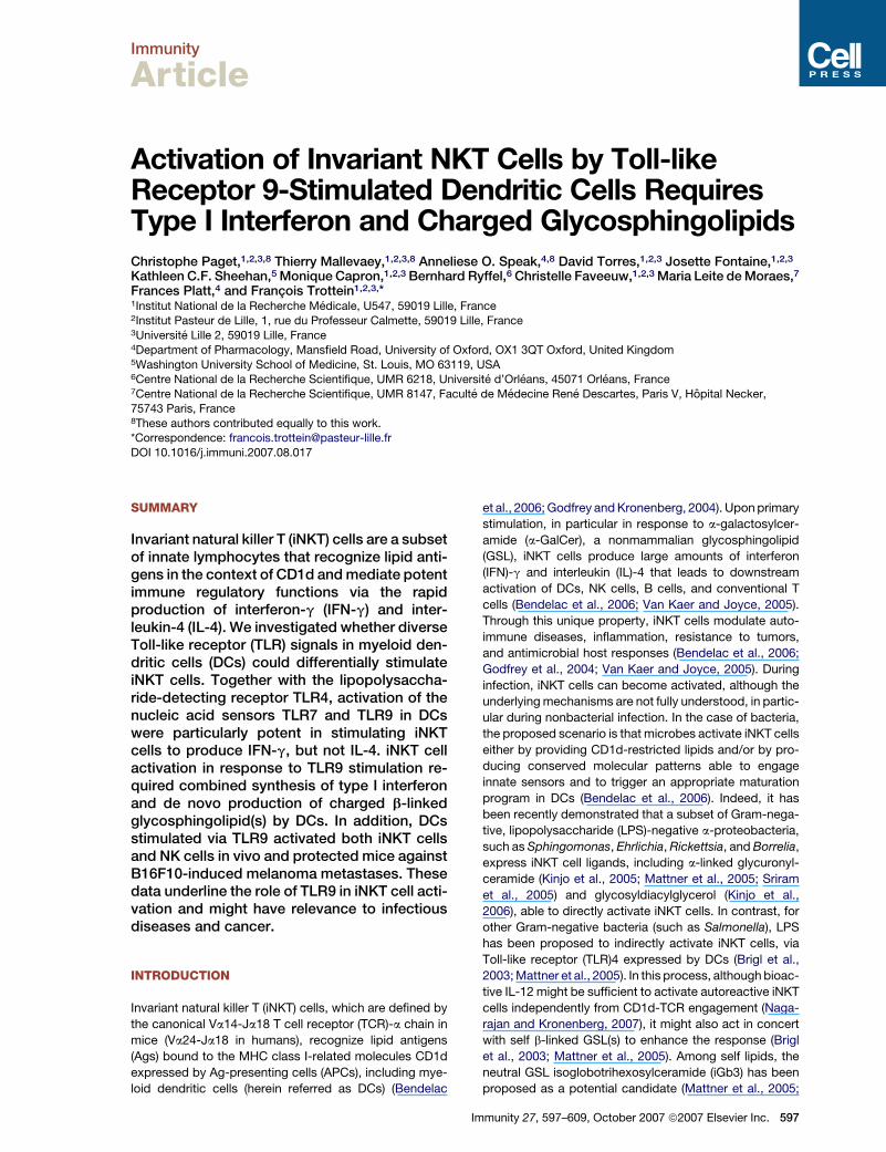
Activation of Invariant NKT Cells by Toll-like Receptor 9Stimulated Dendritic Cells Requires Type I...
13
Immunity Article Activation of Invariant NKT Cells by Toll-like Receptor 9-Stimulated Dendritic Cells Requires Type I Interferon and Charged Glycosphingolipids Christophe Paget, 1,2,3,8 Thierry Mallevaey, 1,2,3,8 Anneliese O. Speak, 4,8 David Torres, 1,2,3 Josette Fontaine, 1,2,3 Kathleen C.F. Sheehan, 5 Monique Capron, 1,2,3 Bernhard Ryffel, 6 Christelle Faveeuw, 1,2,3 Maria Leite de Moraes, 7 Frances Platt, 4 and Franc ¸ ois Trottein 1,2,3, * 1 Institut National de la Recherche Me ´ dicale, U547, 59019 Lille, France 2 Institut Pasteur de Lille, 1, rue du Professeur Calmette, 59019 Lille, France 3 Universite ´ Lille 2, 59019 Lille, France 4 Department of Pharmacology, Mansfield Road, University of Oxford, OX1 3QT Oxford, United Kingdom 5 Washington University School of Medicine, St. Louis, MO 63119, USA 6 Centre National de la Recherche Scientifique, UMR 6218, Universite ´ d’Orle ´ ans, 45071 Orle ´ ans, France 7 Centre National de la Recherche Scientifique, UMR 8147, Faculte ´ de Me ´ decine Rene ´ Descartes, Paris V, Ho ˆ pital Necker, 75743 Paris, France 8 These authors contributed equally to this work. *Correspondence: [email protected] DOI 10.1016/j.immuni.2007.08.017 SUMMARY Invariant natural killer T (iNKT) cells are a subset of innate lymphocytes that recognize lipid anti- gens in the context of CD1d and mediate potent immune regulatory functions via the rapid production of interferon-g (IFN-g) and inter- leukin-4 (IL-4). We investigated whether diverse Toll-like receptor (TLR) signals in myeloid den- dritic cells (DCs) could differentially stimulate iNKT cells. Together with the lipopolysaccha- ride-detecting receptor TLR4, activation of the nucleic acid sensors TLR7 and TLR9 in DCs were particularly potent in stimulating iNKT cells to produce IFN-g, but not IL-4. iNKT cell activation in response to TLR9 stimulation re- quired combined synthesis of type I interferon and de novo production of charged b-linked glycosphingolipid(s) by DCs. In addition, DCs stimulated via TLR9 activated both iNKT cells and NK cells in vivo and protected mice against B16F10-induced melanoma metastases. These data underline the role of TLR9 in iNKT cell acti- vation and might have relevance to infectious diseases and cancer. INTRODUCTION Invariant natural killer T (iNKT) cells, which are defined by the canonical Va14-Ja18 T cell receptor (TCR)-a chain in mice (Va24-Ja18 in humans), recognize lipid antigens (Ags) bound to the MHC class I-related molecules CD1d expressed by Ag-presenting cells (APCs), including mye- loid dendritic cells (herein referred as DCs) (Bendelac et al., 2006; Godfrey and Kronenberg, 2004). Upon primary stimulation, in particular in response to a-galactosylcer- amide (a-GalCer), a nonmammalian glycosphingolipid (GSL), iNKT cells produce large amounts of interferon (IFN)-g and interleukin (IL)-4 that leads to downstream activation of DCs, NK cells, B cells, and conventional T cells (Bendelac et al., 2006; Van Kaer and Joyce, 2005). Through this unique property, iNKT cells modulate auto- immune diseases, inflammation, resistance to tumors, and antimicrobial host responses (Bendelac et al., 2006; Godfrey et al., 2004; Van Kaer and Joyce, 2005). During infection, iNKT cells can become activated, although the underlying mechanisms are not fully understood, in partic- ular during nonbacterial infection. In the case of bacteria, the proposed scenario is that microbes activate iNKT cells either by providing CD1d-restricted lipids and/or by pro- ducing conserved molecular patterns able to engage innate sensors and to trigger an appropriate maturation program in DCs (Bendelac et al., 2006). Indeed, it has been recently demonstrated that a subset of Gram-nega- tive, lipopolysaccharide (LPS)-negative a-proteobacteria, such as Sphingomonas, Ehrlichia, Rickettsia, and Borrelia, express iNKT cell ligands, including a-linked glycuronyl- ceramide (Kinjo et al., 2005; Mattner et al., 2005; Sriram et al., 2005) and glycosyldiacylglycerol (Kinjo et al., 2006), able to directly activate iNKT cells. In contrast, for other Gram-negative bacteria (such as Salmonella), LPS has been proposed to indirectly activate iNKT cells, via Toll-like receptor (TLR)4 expressed by DCs (Brigl et al., 2003; Mattner et al., 2005). In this process, although bioac- tive IL-12 might be sufficient to activate autoreactive iNKT cells independently from CD1d-TCR engagement (Naga- rajan and Kronenberg, 2007), it might also act in concert with self b-linked GSL(s) to enhance the response (Brigl et al., 2003; Mattner et al., 2005). Among self lipids, the neutral GSL isoglobotrihexosylceramide (iGb3) has been proposed as a potential candidate (Mattner et al., 2005; Immunity 27, 597–609, October 2007 ª2007 Elsevier Inc. 597
-
Upload
independent -
Category
Documents
-
view
0 -
download
0
Transcript of Activation of Invariant NKT Cells by Toll-like Receptor 9Stimulated Dendritic Cells Requires Type I...
Immunity
Article
Activation of Invariant NKT Cells by Toll-likeReceptor 9-Stimulated Dendritic Cells RequiresType I Interferon and Charged GlycosphingolipidsChristophe Paget,1,2,3,8 Thierry Mallevaey,1,2,3,8 Anneliese O. Speak,4,8 David Torres,1,2,3 Josette Fontaine,1,2,3
Kathleen C.F. Sheehan,5 Monique Capron,1,2,3 Bernhard Ryffel,6 Christelle Faveeuw,1,2,3 Maria Leite de Moraes,7
Frances Platt,4 and Francois Trottein1,2,3,*1Institut National de la Recherche Medicale, U547, 59019 Lille, France2Institut Pasteur de Lille, 1, rue du Professeur Calmette, 59019 Lille, France3Universite Lille 2, 59019 Lille, France4Department of Pharmacology, Mansfield Road, University of Oxford, OX1 3QT Oxford, United Kingdom5Washington University School of Medicine, St. Louis, MO 63119, USA6Centre National de la Recherche Scientifique, UMR 6218, Universite d’Orleans, 45071 Orleans, France7Centre National de la Recherche Scientifique, UMR 8147, Faculte de Medecine Rene Descartes, Paris V, Hopital Necker,
75743 Paris, France8These authors contributed equally to this work.*Correspondence: [email protected]
DOI 10.1016/j.immuni.2007.08.017
SUMMARY
Invariant natural killer T (iNKT) cells are a subsetof innate lymphocytes that recognize lipid anti-gens in the context of CD1d and mediate potentimmune regulatory functions via the rapidproduction of interferon-g (IFN-g) and inter-leukin-4 (IL-4). We investigated whether diverseToll-like receptor (TLR) signals in myeloid den-dritic cells (DCs) could differentially stimulateiNKT cells. Together with the lipopolysaccha-ride-detecting receptor TLR4, activation of thenucleic acid sensors TLR7 and TLR9 in DCswere particularly potent in stimulating iNKTcells to produce IFN-g, but not IL-4. iNKT cellactivation in response to TLR9 stimulation re-quired combined synthesis of type I interferonand de novo production of charged b-linkedglycosphingolipid(s) by DCs. In addition, DCsstimulated via TLR9 activated both iNKT cellsand NK cells in vivo and protected mice againstB16F10-induced melanoma metastases. Thesedata underline the role of TLR9 in iNKT cell acti-vation and might have relevance to infectiousdiseases and cancer.
INTRODUCTION
Invariant natural killer T (iNKT) cells, which are defined by
the canonical Va14-Ja18 T cell receptor (TCR)-a chain in
mice (Va24-Ja18 in humans), recognize lipid antigens
(Ags) bound to the MHC class I-related molecules CD1d
expressed by Ag-presenting cells (APCs), including mye-
loid dendritic cells (herein referred as DCs) (Bendelac
et al., 2006; Godfrey and Kronenberg, 2004). Upon primary
stimulation, in particular in response to a-galactosylcer-
amide (a-GalCer), a nonmammalian glycosphingolipid
(GSL), iNKT cells produce large amounts of interferon
(IFN)-g and interleukin (IL)-4 that leads to downstream
activation of DCs, NK cells, B cells, and conventional T
cells (Bendelac et al., 2006; Van Kaer and Joyce, 2005).
Through this unique property, iNKT cells modulate auto-
immune diseases, inflammation, resistance to tumors,
and antimicrobial host responses (Bendelac et al., 2006;
Godfrey et al., 2004; Van Kaer and Joyce, 2005). During
infection, iNKT cells can become activated, although the
underlying mechanisms are not fully understood, in partic-
ular during nonbacterial infection. In the case of bacteria,
the proposed scenario is that microbes activate iNKT cells
either by providing CD1d-restricted lipids and/or by pro-
ducing conserved molecular patterns able to engage
innate sensors and to trigger an appropriate maturation
program in DCs (Bendelac et al., 2006). Indeed, it has
been recently demonstrated that a subset of Gram-nega-
tive, lipopolysaccharide (LPS)-negative a-proteobacteria,
such as Sphingomonas, Ehrlichia, Rickettsia, and Borrelia,
express iNKT cell ligands, including a-linked glycuronyl-
ceramide (Kinjo et al., 2005; Mattner et al., 2005; Sriram
et al., 2005) and glycosyldiacylglycerol (Kinjo et al.,
2006), able to directly activate iNKT cells. In contrast, for
other Gram-negative bacteria (such as Salmonella), LPS
has been proposed to indirectly activate iNKT cells, via
Toll-like receptor (TLR)4 expressed by DCs (Brigl et al.,
2003; Mattner et al., 2005). In this process, although bioac-
tive IL-12 might be sufficient to activate autoreactive iNKT
cells independently from CD1d-TCR engagement (Naga-
rajan and Kronenberg, 2007), it might also act in concert
with self b-linked GSL(s) to enhance the response (Brigl
et al., 2003; Mattner et al., 2005). Among self lipids, the
neutral GSL isoglobotrihexosylceramide (iGb3) has been
proposed as a potential candidate (Mattner et al., 2005;
Immunity 27, 597–609, October 2007 ª2007 Elsevier Inc. 597
Immunity
TLR9-Activated DCs Stimulate iNKT Cells
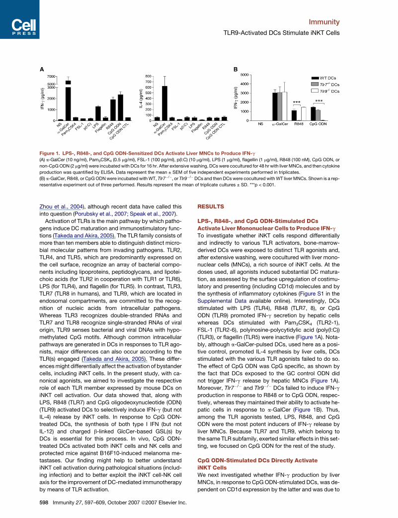
Figure 1. LPS-, R848-, and CpG ODN-Sensitized DCs Activate Liver MNCs to Produce IFN-g
(A) a-GalCer (10 ng/ml), Pam3CSK4 (0.5 mg/ml), FSL-1 (100 pg/ml), p(I:C) (10 mg/ml), LPS (1 mg/ml), flagellin (1 mg/ml), R848 (100 nM), CpG ODN, or
non-CpG ODN (2 mg/ml) were incubated with DCs for 16 hr. After extensive washing, DCs were cocultured for 48 hr with liver MNCs, and then cytokine
production was quantified by ELISA. Data represent the mean ± SEM of five independent experiments performed in triplicates.
(B) a-GalCer, R848, or CpG ODN were incubated with WT, Tlr7�/�, or Tlr9�/�DCs and then DCs were cocultured with WT liver MNCs. Shown is a rep-
resentative experiment out of three performed. Results represent the mean of triplicate cultures ± SD. ***p < 0.001.
Zhou et al., 2004), although recent data have called this
into question (Porubsky et al., 2007; Speak et al., 2007).
Activation of TLRs is the main pathway by which patho-
gens induce DC maturation and immunostimulatory func-
tions (Takeda and Akira, 2005). The TLR family consists of
more than ten members able to distinguish distinct micro-
bial molecular patterns from invading pathogens. TLR2,
TLR4, and TLR5, which are predominantly expressed on
the cell surface, recognize an array of bacterial compo-
nents including lipoproteins, peptidoglycans, and lipotei-
choic acids (for TLR2 in cooperation with TLR1 or TLR6),
LPS (for TLR4), and flagellin (for TLR5). In contrast, TLR3,
TLR7 (TLR8 in humans), and TLR9, which are located in
endosomal compartments, are committed to the recog-
nition of nucleic acids from intracellular pathogens.
Whereas TLR3 recognizes double-stranded RNAs and
TLR7 and TLR8 recognize single-stranded RNAs of viral
origin, TLR9 senses bacterial and viral DNAs with hypo-
methylated CpG motifs. Although common intracellular
pathways are generated in DCs in responses to TLR ago-
nists, major differences can also occur according to the
TLR(s) engaged (Takeda and Akira, 2005). These differ-
ences might differentially affect the activation of bystander
cells, including iNKT cells. In the present study, with ca-
nonical agonists, we aimed to investigate the respective
role of each TLR member expressed by mouse DCs on
iNKT cell activation. Our data showed that, along with
LPS, R848 (TLR7) and CpG oligodeoxynucleotide (ODN)
(TLR9) activated DCs to selectively induce IFN-g (but not
IL-4) release by iNKT cells. In response to CpG ODN-
treated DCs, the synthesis of both type I IFN (but not
IL-12) and charged b-linked GlcCer-based GSL(s) by
DCs is essential for this process. In vivo, CpG ODN-
treated DCs activated both iNKT cells and NK cells and
protected mice against B16F10-induced melanoma me-
tastases. Our finding might help to better understand
iNKT cell activation during pathological situations (includ-
ing infection) and to better exploit the iNKT cell-NK cell
axis for the improvement of DC-mediated immunotherapy
by means of TLR activation.
598 Immunity 27, 597–609, October 2007 ª2007 Elsevier Inc.
RESULTS
LPS-, R848-, and CpG ODN-Stimulated DCsActivate Liver Mononuclear Cells to Produce IFN-g
To investigate whether iNKT cells respond differentially
and indirectly to various TLR activators, bone-marrow-
derived DCs were exposed to distinct TLR agonists and,
after extensive washing, were cocultured with liver mono-
nuclear cells (MNCs), a rich source of iNKT cells. At the
doses used, all agonists induced substantial DC matura-
tion, as assessed by the surface upregulation of costimu-
latory and presenting (including CD1d) molecules and by
the synthesis of inflammatory cytokines (Figure S1 in the
Supplemental Data available online). Interestingly, DCs
stimulated with LPS (TLR4), R848 (TLR7, 8), or CpG
ODN (TLR9) promoted IFN-g secretion by hepatic cells
whereas DCs stimulated with Pam3CSK4 (TLR2-1),
FSL-1 (TLR2-6), polyinosine-polycytidylic acid (poly(I:C))
(TLR3), or flagellin (TLR5) were inactive (Figure 1A). Nota-
bly, although a-GalCer-pulsed DCs, used here as a posi-
tive control, promoted IL-4 synthesis by liver cells, DCs
stimulated with the various TLR agonists failed to do so.
The effect of CpG ODN was CpG specific, as shown by
the fact that DCs exposed to the GC control ODN did
not trigger IFN-g release by hepatic MNCs (Figure 1A).
Moreover, Tlr7�/� and Tlr9�/� DCs failed to induce IFN-g
production in response to R848 or to CpG ODN, respec-
tively, whereas they maintained their ability to activate he-
patic cells in response to a-GalCer (Figure 1B). Thus,
among the TLR agonists tested, LPS, R848, and CpG
ODN were the most potent inducers of IFN-g release by
liver MNCs. Because TLR7 and TLR9, which belong to
the same TLR subfamily, exerted similar effects in this set-
ting, we focused on CpG ODN for the rest of the study.
CpG ODN-Stimulated DCs Directly ActivateiNKT CellsWe next investigated whether IFN-g production by liver
MNCs, in response to CpG ODN-stimulated DCs, was de-
pendent on CD1d expression by the latter and was due to

Immunity
TLR9-Activated DCs Stimulate iNKT Cells
Figure 2. CpG ODN-Stimulated DCs Directly Activate iNKT Cells
(A) a-GalCer or CpG ODN was incubated with WT or CD1d1�/� DCs and then cocultured with liver MNCs from WT mice for 2 days.
(B) Stimulated DCs were cocultured with liver MNCs isolated from WT or Ja18-deficient mice.
(C) Intracellular FACS staining of iNKT cells after stimulation with a-GalCer- or CpG ODN-activated DCs. Stimulated DCs were cocultured with liver
cells for 12 hr, and afterwards brefeldin A was added for another 4 hr. Cells were labeled with TCRb mAb and TT, fixed, and permeabilized for intra-
cellular cytokine staining. Cells were analyzed by flow cytometry, and TT+ TCRb+ cells were gated and screened for intracellular IFN-g production.
Gates were set based on the isotype control. The percentages of TT+ TCRb+ cells positive for IFN-g are represented. One representative experiment
out of three is shown.
(D) a-GalCer- or CpG ODN-stimulated DCs were cocultured with purified liver-derived CD5+ NK1.1+ or CD5+ NK1.1� cells (left) or with TT+ TCRb+ cells
(right) for 2 days. Of note, exposure of purified CD5+ NK1.1+ or TT+ TCRb+ cells with CpG ODN alone did not result in IFN-g production (not shown).
(A, B, D) Cytokine production was measured by ELISA. Shown is a representative experiment of four (A, B) or three (D) performed. Results represent
the mean of triplicate cultures ± SD. ***p < 0.001; **p < 0.01.
iNKT cells. CD1d deficiency in DCs resulted in a complete
abrogation of IFN-g secretion in response to a-GalCer-
sensitized DCs. These DCs also showed a �70% de-
crease (average of four independent experiments) in the
secretion of IFN-g in response to CpG ODN-treated DCs
(Figure 2A). This was confirmed by the neutralizing
CD1d antibody (Ab) 1B1, added during the DC-liver MNC
coculture (not shown). In addition, IFN-g release was
strongly reduced (by �80%) when CpG ODN-treated DCs
were cocultured with hepatic MNCs isolated from iNKT
Immunity 27, 597–609, October 2007 ª2007 Elsevier Inc. 599
Immunity
TLR9-Activated DCs Stimulate iNKT Cells
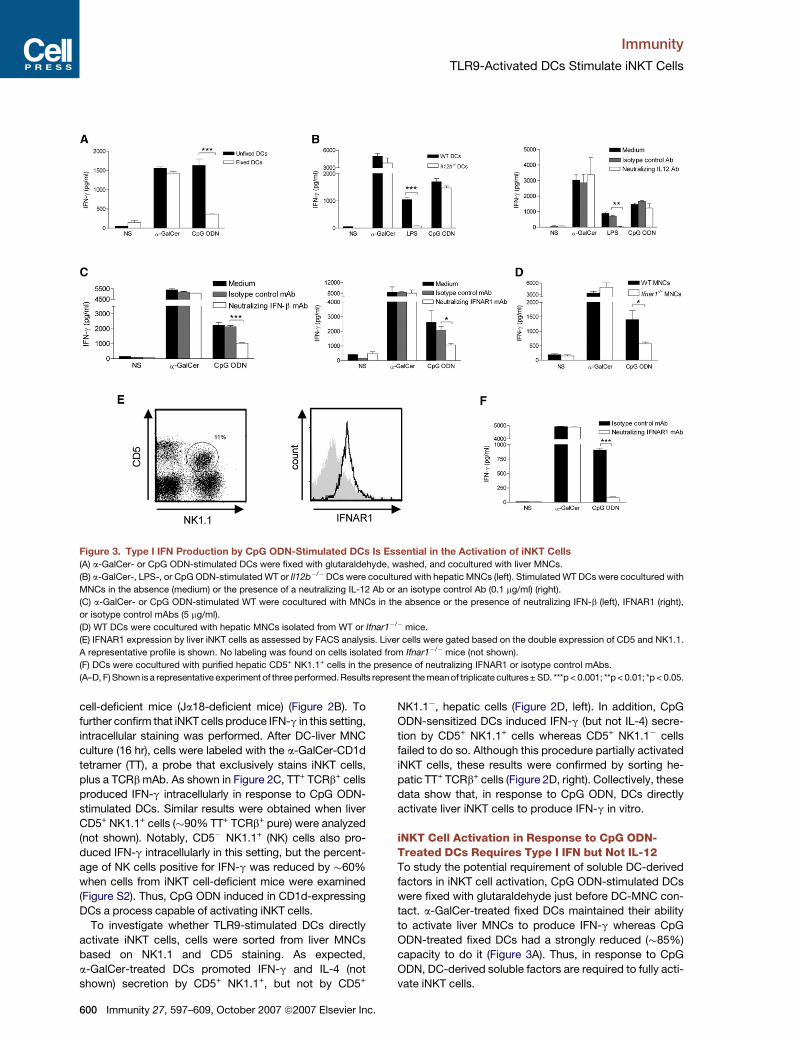
Figure 3. Type I IFN Production by CpG ODN-Stimulated DCs Is Essential in the Activation of iNKT Cells
(A) a-GalCer- or CpG ODN-stimulated DCs were fixed with glutaraldehyde, washed, and cocultured with liver MNCs.
(B) a-GalCer-, LPS-, or CpG ODN-stimulated WT or Il12b�/�DCs were cocultured with hepatic MNCs (left). Stimulated WT DCs were cocultured with
MNCs in the absence (medium) or the presence of a neutralizing IL-12 Ab or an isotype control Ab (0.1 mg/ml) (right).
(C) a-GalCer- or CpG ODN-stimulated WT were cocultured with MNCs in the absence or the presence of neutralizing IFN-b (left), IFNAR1 (right),
or isotype control mAbs (5 mg/ml).
(D) WT DCs were cocultured with hepatic MNCs isolated from WT or Ifnar1�/� mice.
(E) IFNAR1 expression by liver iNKT cells as assessed by FACS analysis. Liver cells were gated based on the double expression of CD5 and NK1.1.
A representative profile is shown. No labeling was found on cells isolated from Ifnar1�/� mice (not shown).
(F) DCs were cocultured with purified hepatic CD5+ NK1.1+ cells in the presence of neutralizing IFNAR1 or isotype control mAbs.
(A–D, F) Shown is a representative experiment of three performed. Results represent the mean of triplicate cultures± SD. ***p < 0.001; **p < 0.01; *p < 0.05.
cell-deficient mice (Ja18-deficient mice) (Figure 2B). To
further confirm that iNKT cells produce IFN-g in this setting,
intracellular staining was performed. After DC-liver MNC
culture (16 hr), cells were labeled with the a-GalCer-CD1d
tetramer (TT), a probe that exclusively stains iNKT cells,
plus a TCRb mAb. As shown in Figure 2C, TT+ TCRb+ cells
produced IFN-g intracellularly in response to CpG ODN-
stimulated DCs. Similar results were obtained when liver
CD5+ NK1.1+ cells (�90% TT+ TCRb+ pure) were analyzed
(not shown). Notably, CD5� NK1.1+ (NK) cells also pro-
duced IFN-g intracellularly in this setting, but the percent-
age of NK cells positive for IFN-g was reduced by �60%
when cells from iNKT cell-deficient mice were examined
(Figure S2). Thus, CpG ODN induced in CD1d-expressing
DCs a process capable of activating iNKT cells.
To investigate whether TLR9-stimulated DCs directly
activate iNKT cells, cells were sorted from liver MNCs
based on NK1.1 and CD5 staining. As expected,
a-GalCer-treated DCs promoted IFN-g and IL-4 (not
shown) secretion by CD5+ NK1.1+, but not by CD5+
600 Immunity 27, 597–609, October 2007 ª2007 Elsevier Inc.
NK1.1�, hepatic cells (Figure 2D, left). In addition, CpG
ODN-sensitized DCs induced IFN-g (but not IL-4) secre-
tion by CD5+ NK1.1+ cells whereas CD5+ NK1.1� cells
failed to do so. Although this procedure partially activated
iNKT cells, these results were confirmed by sorting he-
patic TT+ TCRb+ cells (Figure 2D, right). Collectively, these
data show that, in response to CpG ODN, DCs directly
activate liver iNKT cells to produce IFN-g in vitro.
iNKT Cell Activation in Response to CpG ODN-Treated DCs Requires Type I IFN but Not IL-12To study the potential requirement of soluble DC-derived
factors in iNKT cell activation, CpG ODN-stimulated DCs
were fixed with glutaraldehyde just before DC-MNC con-
tact. a-GalCer-treated fixed DCs maintained their ability
to activate liver MNCs to produce IFN-g whereas CpG
ODN-treated fixed DCs had a strongly reduced (�85%)
capacity to do it (Figure 3A). Thus, in response to CpG
ODN, DC-derived soluble factors are required to fully acti-
vate iNKT cells.
Immunity
TLR9-Activated DCs Stimulate iNKT Cells
IL-12 and type I IFN, two major cytokines produced by
DCs after TLR9 triggering (Figure S1), have recently been
shown to cooperate with the CD1d-TCR pathway to acti-
vate iNKT cells (Brigl et al., 2003; Marschner et al., 2005;
Mattner et al., 2005; Montoya et al., 2006). In response to
CpG ODN, DCs deficient in IL-12p40 maintained their abil-
ity to activate liver cells (Figure 3B, left), and this result was
confirmed via a neutralizing IL-12 Ab, added during the
DC-MNC coculture (Figure 3B, right). In contrast, in agree-
ment with previous studies (Brigl et al., 2003; Mattner et al.,
2005; Nagarajan and Kronenberg, 2007), IL-12 was essen-
tial in the ability of LPS-stimulated DCs to promote IFN-g
release by liver MNCs. To investigate the contribution of
IFN-b, which is released by CpG ODN-stimulated DCs
(Figure S1), a neutralizing Ab against it was utilized. Block-
ade of IFN-b reduced by �50% the CpG ODN-induced
production of IFN-g by hepatic MNCs (Figure 3C, left). To
confirm this finding, a neutralizing Ab directed against
the IFNAR1 subunit of the type I IFN receptor or liver
MNCs isolated from Ifnar1�/�mice were used. Compared
to controls, this resulted in a strong reduction of IFN-g pro-
duction by liver cells (�60% and 70% reduction, respec-
tively) (Figure 3C, right, and Figure 3D). In contrast, when
IFNAR1 was blocked or absent in liver cells, the degree
of iNKT cell activation after exposure to a-GalCer-treated
DCs (Figures 3C and 3D) or to LPS-stimulated DCs (not
shown) was not reduced. FACS analysis showed that liver
CD5+ NK1.1+ expressed IFNAR1 (Figure 3E). Interestingly,
blockade of IFNAR1 on sorted iNKT cells resulted in a total
abrogation of IFN-g production by these cells (Figure 3F).
These data argue that, in response to CpG ODN-
stimulated DCs, iNKT cell activation fully requires type I
IFN but not IL-12.
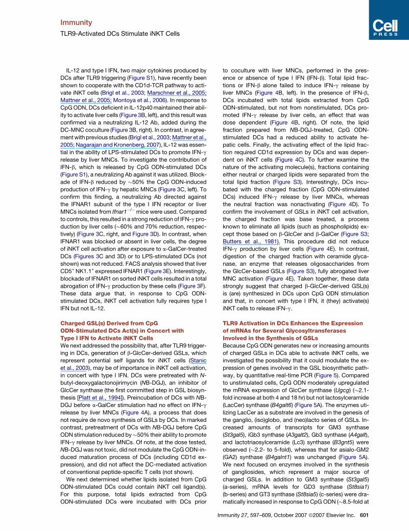
Charged GSL(s) Derived from CpGODN-Stimulated DCs Act(s) in Concert withType I IFN to Activate iNKT CellsWe next addressed the possibility that, after TLR9 trigger-
ing in DCs, generation of b-GlcCer-derived GSLs, which
represent potential self ligands for iNKT cells (Stanic
et al., 2003), may be of importance in iNKT cell activation,
in concert with type I IFN. DCs were pretreated with N-
butyl-deoxygalactonojirimycin (NB-DGJ), an inhibitor of
GlcCer synthase (the first committed step in GSL biosyn-
thesis [Platt et al., 1994]). Preincubation of DCs with NB-
DGJ before a-GalCer stimulation had no effect on IFN-g
release by liver MNCs (Figure 4A), a process that does
not require de novo synthesis of GSLs by DCs. In marked
contrast, pretreatment of DCs with NB-DGJ before CpG
ODN stimulation reduced by�50% their ability to promote
IFN-g release by liver MNCs. Of note, at the dose tested,
NB-DGJ was not toxic, did not modulate the CpG ODN-in-
duced maturation process of DCs (including CD1d ex-
pression), and did not affect the DC-mediated activation
of conventional peptide-specific T cells (not shown).
We next determined whether lipids isolated from CpG
ODN-stimulated DCs could contain iNKT cell ligand(s).
For this purpose, total lipids extracted from CpG
ODN-stimulated DCs were incubated with DCs prior
to coculture with liver MNCs, performed in the pres-
ence or absence of type I IFN (IFN-b). Total lipid frac-
tions or IFN-b alone failed to induce IFN-g release by
liver MNCs (Figure 4B, left). In the presence of IFN-b,
DCs incubated with total lipids extracted from CpG
ODN-stimulated, but not from nonstimulated, DCs pro-
moted IFN-g release by liver cells, an effect that was
dose dependent (Figure 4B, right). Of note, the lipid
fraction prepared from NB-DGJ-treated, CpG ODN-
stimulated DCs had a reduced ability to activate he-
patic cells. Finally, the activating effect of the lipid frac-
tion required CD1d expression by DCs and was depen-
dent on iNKT cells (Figure 4C). To further examine the
nature of the activating molecule(s), fractions containing
either neutral or charged lipids were separated from the
total lipid fraction (Figure S3). Interestingly, DCs incu-
bated with the charged fraction (CpG ODN-stimulated
DCs) induced IFN-g release by liver MNCs, whereas
the neutral fraction was nonactivating (Figure 4D). To
confirm the involvement of GSLs in iNKT cell activation,
the charged fraction was base treated, a process
known to eliminate all lipids (such as phospholipids) ex-
cept those based on b-GlcCer and b-GalCer (Figure S3;
Butters et al., 1981). This procedure did not reduce
IFN-g production by liver cells (Figure 4E). In contrast,
digestion of the charged fraction with ceramide glyca-
nase, an enzyme that releases oligosaccharides from
the GlcCer-based GSLs (Figure S3), fully abrogated liver
MNC activation (Figure 4E). Taken together, these data
strongly suggest that charged b-GlcCer-derived GSL(s)
is (are) synthesized in DCs upon CpG ODN stimulation
and that, in concert with type I IFN, it (they) activate(s)
iNKT cells to release IFN-g.
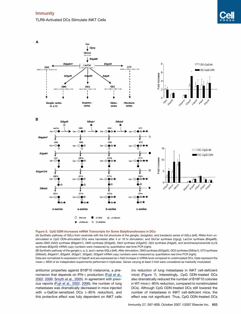
TLR9 Activation in DCs Enhances the Expressionof mRNAs for Several GlycosyltransferasesInvolved in the Synthesis of GSLsBecause CpG ODN generates new or increasing amounts
of charged GSLs in DCs able to activate iNKT cells, we
investigated the possibility that it could modulate the ex-
pression of genes involved in the GSL biosynthetic path-
way, by quantitative real-time PCR (Figure 5). Compared
to unstimulated cells, CpG ODN moderately upregulated
the mRNA expression of GlcCer synthase (Ugcg) (�2.1-
fold increase at both 4 and 18 hr) but not lactosylceramide
(LacCer) synthase (B4galt6) (Figure 5A). The enzymes uti-
lizing LacCer as a substrate are involved in the genesis of
the ganglio, (iso)globo, and (neo)lacto series of GSLs. In-
creased amounts of transcripts for GM3 synthase
(St3gal5), iGb3 synthase (A3galt2), Gb3 synthase (A4galt),
and lactotriaosylceramide (Lc3) synthase (B3gnt5) were
observed (�2.2- to 5-fold), whereas that for asialo-GM2
(GA2) synthase (B4galnt1) was unchanged (Figure 5A).
We next focused on enzymes involved in the synthesis
of gangliosides, which represent a major source of
charged GSLs. In addition to GM3 synthase (St3gal5)
(a-series), mRNA levels for GD3 synthase (St8sia1)
(b-series) and GT3 synthase (St8sia5) (c-series) were dra-
matically increased in response to CpG ODN (�8.5-fold at
Immunity 27, 597–609, October 2007 ª2007 Elsevier Inc. 601
Immunity
TLR9-Activated DCs Stimulate iNKT Cells
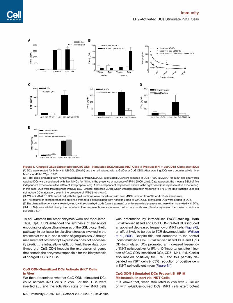
Figure 4. Charged GSLs Extracted from CpG ODN-Stimulated DCs Activate iNKT Cells to Produce IFN-g, via CD1d-Competent DCs
(A) DCs were treated for 24 hr with NB-DGJ (50 mM) and then stimulated with a-GalCer or CpG ODN. After washing, DCs were cocultured with liver
MNCs for 48 hr. ***p < 0.001.
(B) Total lipids extracted from nonstimulated (NS) or from CpG ODN-stimulated DCs were exposed to DCs (1/500 in DMSO) for 16 hr, and afterwards
washed DCs were cocultured with liver MNCs for 48 hr, in the presence or absence of IFN-b (1000 U/ml). Data represent the mean ± SEM of five
independent experiments (five different lipid preparations). A dose-dependent response is shown in the right panel (one representative experiment).
In this case, DCs were treated or not with NB-DGJ. Of note, excepted CD1d, which was upregulated in response to IFN-b, the lipid fractions used did
not induce DC maturation, even in the presence of IFN-b (not shown).
(C) WT or Cd1d1�/� DCs sensitized with the lipid fractions were cocultured with liver MNCs isolated from WT or Ja18-deficient mice.
(D) The neutral or charged fractions obtained from total lipids isolated from nonstimulated or CpG ODN-stimulated DCs were added to DCs.
(E) The charged fractions were treated, or not, with sodium hydroxide (base treatment) or with ceramide glycanase and were then incubated with DCs.
(C–E) IFN-b was added during the coculture. One representative experiment out of four is shown. Results represent the mean of triplicate
cultures ± SD.
18 hr), whereas the other enzymes were not modulated.
Thus, CpG ODN enhanced the synthesis of transcripts
encoding for glycosyltransferases of the GSL biosynthetic
pathway, in particular for sialyltransferases involved in the
first step of the a, b, and c series of gangliosides. Although
measurement of transcript expression does not necessar-
ily predict the intracellular GSL content, these data con-
firmed that CpG ODN impacts the expression of genes
that encode the enzymes responsible for the biosynthesis
of charged GSLs in DCs.
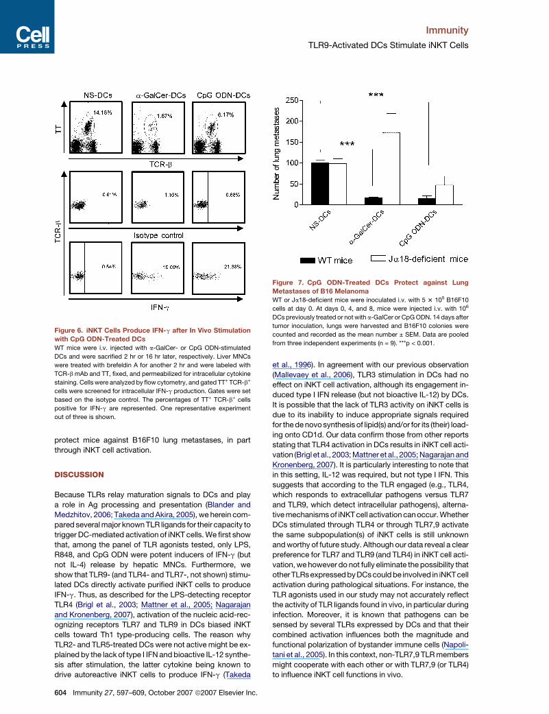
CpG ODN-Sensitized DCs Activate iNKT CellsIn VivoWe then determined whether CpG ODN-stimulated DCs
could activate iNKT cells in vivo. For this, DCs were
injected i.v., and the activation state of liver iNKT cells
602 Immunity 27, 597–609, October 2007 ª2007 Elsevier Inc.
was determined by intracellular FACS staining. Both
a-GalCer-sensitized and CpG ODN-treated DCs induced
an apparent decreased frequency of iNKT cells (Figure 6),
an effect likely to be due to TCR downmodulation (Wilson
et al., 2003). Despite this, and compared to the control
(nonstimulated DCs), a-GalCer-sensitized DCs and CpG
ODN-stimulated DCs promoted an increased frequency
of iNKT cells positive for IFN-g. Of importance, after injec-
tion of CpG ODN-sensitized DCs, CD5� NK1.1+ (NK cells)
also labeled positively for IFN-g and this partially de-
pended on iNKT cells (�65% reduction of positive cells
in iNKT cell-deficient mice) (Figure S4).
CpG ODN-Stimulated DCs Prevent B16F10Metastasis, in part via iNKT CellsIt is known that, when stimulated in vivo with a-GalCer
or with a-GalCer-pulsed DCs, iNKT cells exert potent
Immunity
TLR9-Activated DCs Stimulate iNKT Cells
Figure 5. CpG ODN Increases mRNA Transcripts for Some Sialyltransferases in DCs
(A) Synthetic pathway of GSLs from ceramide with the full structures of the ganglio, (iso)globo, and (neo)lacto series of GSLs (left). RNAs from un-
stimulated or CpG ODN-stimulated DCs were harvested after 4 or 18 hr stimulation, and GlcCer synthase (Ugcg), LacCer synthase (B4galt6),
asialo-GM2 (GA2) synthase (B4galnt1), GM3 synthase (St3gal5), iGb3 synthase (A3galt2), Gb3 synthase (A4galt), and lactotriaosylceramide (Lc3)
synthase (B3gnt5) mRNA copy numbers were measured by quantitative real-time PCR (right).
(B) Synthetic pathway of the ganglio o, a, b, and c series GSLs (left). After stimulation, GM3 synthase (St3gal5), GD3 synthase (St8sia1), GT3 synthase
(St8sia5), B4galnt1, B3galt4, St3gal1, St3gal2, St3gal4 mRNA copy numbers were measured by quantitative real-time PCR (right).
Data are normalized to expression of Gapdh and are expressed as n-fold increase in mRNA level compared to unstimulated DCs. Data represent the
mean ± SEM of six independent experiments performed in triplicates. Genes varying at least 2-fold were considered as markedly modulated.
antitumor properties against B16F10 melanoma, a phe-
nomenon that depends on IFN-g production (Fujii et al.,
2002, 2006; Smyth et al., 2005). In agreement with previ-
ous reports (Fujii et al., 2002, 2006), the number of lung
metastases was dramatically decreased in mice injected
with a-GalCer-sensitized DCs (�85% reduction), and
this protective effect was fully dependent on iNKT cells
(no reduction of lung metastases in iNKT cell-deficient
mice) (Figure 7). Interestingly, CpG ODN-treated DCs
also dramatically reduced the number of B16F10 colonies
in WT mice (�85% reduction, compared to nonstimulated
DCs). Although CpG ODN-treated DCs still lowered the
number of metastases in iNKT cell-deficient mice, the
effect was not significant. Thus, CpG ODN-treated DCs
Immunity 27, 597–609, October 2007 ª2007 Elsevier Inc. 603
Immunity
TLR9-Activated DCs Stimulate iNKT Cells
protect mice against B16F10 lung metastases, in part
through iNKT cell activation.
DISCUSSION
Because TLRs relay maturation signals to DCs and play
a role in Ag processing and presentation (Blander and
Medzhitov, 2006; Takeda and Akira, 2005), we herein com-
pared several major known TLR ligands for their capacity to
trigger DC-mediated activation of iNKT cells. We first show
that, among the panel of TLR agonists tested, only LPS,
R848, and CpG ODN were potent inducers of IFN-g (but
not IL-4) release by hepatic MNCs. Furthermore, we
show that TLR9- (and TLR4- and TLR7-, not shown) stimu-
lated DCs directly activate purified iNKT cells to produce
IFN-g. Thus, as described for the LPS-detecting receptor
TLR4 (Brigl et al., 2003; Mattner et al., 2005; Nagarajan
and Kronenberg, 2007), activation of the nucleic acid-rec-
ognizing receptors TLR7 and TLR9 in DCs biased iNKT
cells toward Th1 type-producing cells. The reason why
TLR2- and TLR5-treated DCs were not active might be ex-
plained by the lack of type I IFN and bioactive IL-12 synthe-
sis after stimulation, the latter cytokine being known to
drive autoreactive iNKT cells to produce IFN-g (Takeda
Figure 6. iNKT Cells Produce IFN-g after In Vivo Stimulation
with CpG ODN-Treated DCs
WT mice were i.v. injected with a-GalCer- or CpG ODN-stimulated
DCs and were sacrified 2 hr or 16 hr later, respectively. Liver MNCs
were treated with brefeldin A for another 2 hr and were labeled with
TCR-b mAb and TT, fixed, and permeabilized for intracellular cytokine
staining. Cells were analyzed by flow cytometry, and gated TT+ TCR-b+
cells were screened for intracellular IFN-g production. Gates were set
based on the isotype control. The percentages of TT+ TCR-b+ cells
positive for IFN-g are represented. One representative experiment
out of three is shown.
604 Immunity 27, 597–609, October 2007 ª2007 Elsevier Inc.
et al., 1996). In agreement with our previous observation
(Mallevaey et al., 2006), TLR3 stimulation in DCs had no
effect on iNKT cell activation, although its engagement in-
duced type I IFN release (but not bioactive IL-12) by DCs.
It is possible that the lack of TLR3 activity on iNKT cells is
due to its inability to induce appropriate signals required
for the de novo synthesis of lipid(s) and/or for its (their) load-
ing onto CD1d. Our data confirm those from other reports
stating that TLR4 activation in DCs results in iNKT cell acti-
vation (Brigl et al., 2003; Mattner et al., 2005; Nagarajan and
Kronenberg, 2007). It is particularly interesting to note that
in this setting, IL-12 was required, but not type I IFN. This
suggests that according to the TLR engaged (e.g., TLR4,
which responds to extracellular pathogens versus TLR7
and TLR9, which detect intracellular pathogens), alterna-
tive mechanisms of iNKT cell activation can occur. Whether
DCs stimulated through TLR4 or through TLR7,9 activate
the same subpopulation(s) of iNKT cells is still unknown
and worthy of future study. Although our data reveal a clear
preference for TLR7 and TLR9 (and TLR4) in iNKT cell acti-
vation, we however do not fully eliminate the possibility that
other TLRsexpressed byDCs could be involved in iNKTcell
activation during pathological situations. For instance, the
TLR agonists used in our study may not accurately reflect
the activity of TLR ligands found in vivo, in particular during
infection. Moreover, it is known that pathogens can be
sensed by several TLRs expressed by DCs and that their
combined activation influences both the magnitude and
functional polarization of bystander immune cells (Napoli-
tani et al., 2005). In this context, non-TLR7,9 TLR members
might cooperate with each other or with TLR7,9 (or TLR4)
to influence iNKT cell functions in vivo.
Figure 7. CpG ODN-Treated DCs Protect against Lung
Metastases of B16 MelanomaWT or Ja18-deficient mice were inoculated i.v. with 5 3 105 B16F10
cells at day 0. At days 0, 4, and 8, mice were injected i.v. with 106
DCs previously treated or not with a-GalCer or CpG ODN. 14 days after
tumor inoculation, lungs were harvested and B16F10 colonies were
counted and recorded as the mean number ± SEM. Data are pooled
from three independent experiments (n = 9). ***p < 0.001.
Immunity
TLR9-Activated DCs Stimulate iNKT Cells
The use of genetically deficient mouse cells and neutral-
izing Abs indicated that type I IFN (probably IFN-b) is es-
sential in iNKT cell activation induced by CpG ODN- (as
well as R848-, not shown) stimulated DCs. This finding is
in line with a recent study reporting that IFN-a, produced
by CpG ODN-stimulated plasmacytoid DCs, potentiates
a-GalCer-induced IFN-g production by iNKT cells, at least
in the human system (Marschner et al., 2005). Further-
more, a recent study showed that IFN-b induces a higher
expression of CD1d on DCs and that, through this mech-
anism, it could contribute to activate iNKT cells during
infection (Raghuraman et al., 2006). In contrast, it was re-
ported that exogenous IFN-b decreased the release of
IFN-g by mouse iNKT cells cocultured with a-GalCer-
pulsed DCs (Trobonjaca et al., 2002). In this phenomenon,
it appeared that IFN-b targeted DCs to reduce IL-12-
dependent responses of iNKT cells. Thus, to our best
knowledge, our report is the first that shows that type I
IFN, a key cytokine implicated in antiviral defense and
tumor immunity (Dunn et al., 2006; Stetson and Medzhi-
tov, 2006), cooperates with self Ag(s) to promote iNKT
cell cytokine release. Notably, IFN-b alone did not trigger
IFN-g production by iNKT cells cocultured with nonstimu-
lated DCs, indicating that the inherent autoreactivity of
iNKT cells toward self Ag is not amplified by this factor.
We hypothesized that, along with type I IFN, CpG ODN
could induce the generation of new or increasing amounts
of self lipids in DCs. Indeed, stress-inducible lipid Ags (in-
cluding sulfatide and GM1 ganglioside) are increasingly
generated in human APCs, in response to some bacteria
(Bacillus subtilis, Staphylococcus aureus) or to LPS, to
activate CD1(a, b)-restricted cells (De Libero et al.,
2005). Two approaches were employed to investigate
this hypothesis in our system. First, pretreatment of DCs
with NB-DGJ, an inhibitor of GlcCer synthase, profoundly
affected the capacity of CpG ODN-stimulated DCs (or
lipids extracted from these cells) to activate iNKT cells.
Second, DCs incubated with total lipids extracted from
CpG ODN- (as well as from R848-, not shown) exposed
DCs exerted a stimulatory, CD1d-dependent, effect on
iNKT cells, whereas lipids extracted from nonstimulated
cells failed to do so. As discussed previously, iNKT cell ac-
tivation required the presence of type I IFN in this setting.
Interestingly, charged, but not neutral, lipids activated
iNKT cells via DCs. Moreover, although treatment of the
lipids with sodium hydroxide (to eliminate all lipids except
b-linked GSLs) had no impact on iNKT cell activation, di-
gestion with ceramide glycanase (cleaves the glycan
from ceramide) abrogated it. These data strongly suggest
that charged b-GlcCer-derived GSL(s) is (are) generated in
DCs in response to CpG ODN to stimulate iNKT cells, in
concert with type I IFN. Although unlikely, we cannot fully
rule out the possibility that the active charged GSL(s), to-
gether with IFN-b, might have an effect on CD1d traffick-
ing and/or loading of a different self Ag, perhaps by stim-
ulating lipid exchange. Of interest, we also found that the
charged fraction activated, via DCs, the iNKT cell hybrid-
oma 2C12 to produce IL-2 (not shown). Our results rule out
iGb3 as a potential candidate because this GSL is not
charged and also because we did not detect its presence
in stimulated DCs (not shown). Attempts are now under-
way to characterize the stimulatory compound(s), the
search of which represents an intense area of investiga-
tion at the moment. Among potential candidates is GD3,
a charged (melanoma-derived) GSL able to activate a sub-
set of iNKT cells (Wu et al., 2003). The consequences of
TLR activation on the synthesis of enzymes involved in
the genesis of GSLs are presently unknown. We show
that, in response to CpG ODN, some transcripts encoding
enzymes responsible for GSL synthesis were enhanced in
DCs, albeit at a moderate amount. Interestingly, the most
marked increase in transcript expression corresponded to
three GSL-specific sialyltransferases involved in the pro-
duction of a, b, and c series of gangliosides, namely
St3gal5, St8sia1, and St8sia5. Together, these results
confirmed that CpG ODN modulates GSL biosynthesis
in DCs.
Suzuki and coworkers recently reported that CpG ODN
encapsulated in cationic liposomes activates splenic
CD3+ NK1.1+ cells to produce IFN-g in vivo (Suzuki
et al., 2004). Although IFN-g production by iNKT cells
was not firmly shown, and despite the fact that CpG
ODN could also act via other cells than myeloid DCs (for
instance plasmacytoid DCs), our data are in accordance
with and extend those from this report. Indeed, adminis-
tration of ex vivo CpG ODN-stimulated DCs to naive
mice activated liver iNKT cells to produce IFN-g. As ob-
served in vitro, CpG ODN-stimulated DCs also activated
NK cells to produce IFN-g intracellularly, an effect that
partially depends on iNKT cells. It is likely that the residual
NK cell activation in iNKT cell-deficient mice is due, at
least in part, to direct action of CpG ODN-stimulated
DCs on NK cells (Zanoni et al., 2005) (data not shown). It
was described that the iNKT cell-NK cell pathway can
promote potent cell-mediated antitumor responses (e.g.,
rejection of B16F10 melanoma) when stimulated with
a-GalCer (Smyth et al., 2005) or a-GalCer-sensitized
DCs (Fujii et al., 2002, 2006), a phenomenon that depends
on IFN-g. In addition, it has been widely reported that CpG
ODN or CpG ODN-stimulated DCs can exert highly effec-
tive antitumor immune responses in vivo (Hafner et al.,
2001; Suzuki et al., 2004). However, although suspected,
the role of iNKT cells in this setting has not yet been for-
mally demonstrated. Here, we show that DCs stimulated
through TLR9 can protect against B16F10 melanoma
and that it depends, at least in part, on iNKT cells. The
lack of total iNKT cell dependency might be explained
by the (in)direct effects of CpG ODN-stimulated DCs on
NK cells (Zanoni et al., 2005) as well as on other effector
cells.
The finding that engagement of TLR7 or TLR9 in DCs
activates iNKT cells might be relevant in other pathological
situations, such as infection. In the mouse system, these
receptors are not only expressed in myeloid DCs but
also in plasmacytoid DCs, a cell population participating
in innate responses to several different types of viruses,
via type I IFN production. Thus, TLR7 and TLR9 might
serve a special function in iNKT cell activation during
Immunity 27, 597–609, October 2007 ª2007 Elsevier Inc. 605
Immunity
TLR9-Activated DCs Stimulate iNKT Cells
infection, in particular for subgroups of intracellular patho-
gens. Indeed, early after mouse cytomegalovirus infec-
tion, splenic NK1.1+ TCRb+ (NKT) cells as well as NK1.1+
TCRb� (NK) cells produce IFN-g intracellularly and that it
is largely dependent on TLR9 (Tabeta et al., 2004).
Whether myeloid DCs and/or plasmacytoid DCs partici-
pate in this process is still unknown and deserves further
investigation. Moreover, although it is known that infected
DCs can directly activate NK cells to produce IFN-g and/
or to promote their cytotoxic functions (Degli-Esposti and
Smyth, 2005), it would be interesting to assess the contri-
bution of TLR-based DC-iNKT cell crosstalk on NK cell
functions during infection. In certain circumstances,
TLR7 and TLR9 can also be activated by self components
(small ribonucleoproteins and auto-DNAs, respectively) to
promote autoimmune diseases (Deane and Bolland,
2006). Here, too, whether TLR-stimulated DCs contribute
to the pathology by impacting iNKT cell functions is un-
known and worthy of further investigation. Along with its
therapeutic potential to improve resistance to cancer
and infection, CpG ODN (as well as TLR7/8 ligands) can
also protect against some inflammatory diseases, includ-
ing asthma (Klinman, 2004; Krieg, 2006). It is possible that
the mechanisms by which activators of TLR7 or TLR9 pro-
vide therapeutic benefit in these pathological situations is
dependent on the iNKT cell-NK cell axis.
In conclusion, we have described a mechanism of iNKT
cell activation and provided evidence that TLR9 (as well as
TLR7) triggering in myeloid DCs is important in the primary
activation of iNKT cells. We also report that type I IFN can
drive iNKT cells to produce IFN-g in concert with TLR-
inducible lipid(s) expressed by DCs. Our results may
lead to the identification of new CD1d-restricted Ag(s)
involved in the peripheral activation of iNKT cells.
EXPERIMENTAL PROCEDURES
Reagents and Abs
Pam3CSK4 and FSL-1 were purchased from EMC Microcollections
(Tuebingen, Germany) and p(I:C), ultrapure LPS (from Escherichia
coli serotype 0111:B4), resiquimod (R848), and type B CpG ODN
(ODN 1826) were from Cayla (Toulouse, France). Non-CpG ODN
(Cayla), in which the CG sequence was changed to GC, was used as
a control. Flagellin was kindly provided by J.C. Sirard (Institut Pasteur
de Lille). a-GalCer was purchased from Axxora Life Sciences (Coger
S.A., Paris, France), and NB-DGJ was from Calbiochem (San Diego,
CA). Monoclonal Abs against mouse CD5 (APC-conjugated), NK1.1
(PE- or PerCP-conjugated), IFN-g (PE-conjugated), isotype controls,
and PE-conjugated streptavidin were purchased from BD PharMingen
(Le Pont de Claix, France). APC-conjugated TT was from ProImmune
(Oxford, UK). The neutralizing goat IgGs directed against mouse
IFNAR1 and IL-12 was from R&D Systems (Abingdon, UK), and rat
monoclonal anti-mouse IFN-b Ab was from PBL Biomedical Laborato-
ries (Piscataway, NJ). Isotype control Abs were from Sigma (Lyon,
France). The biotin-conjugated IFNAR1 mAb (MAR1-5A3) and the
isotype control mAb (GIR-208) used for FACS staining have been
described (Sheehan et al., 2006). Recombinant IFN-b was from PBL
Biomedical Laboratories (Piscataway, NJ).
Mice
6- to 8-week-old female wild-type C57BL/6 mice were purchased from
Janvier (Le Genest-St-Isle, France). The generation of Cd1d1�/� and
606 Immunity 27, 597–609, October 2007 ª2007 Elsevier Inc.
iNKT cell (Ja18)-deficient C57BL/6 mice has been already described
(Kawano et al., 1997; Mendiratta et al., 1997). The generation of
Tlr7�/�, Tlr9�/�, and Ifnar1�/�mice C57BL/6 mice has been described
earlier (Hemmi et al., 2000, 2002; Muller et al., 1994). Il12b�/� mice
were from Jackson Laboratories (Bar Harbor, ME). Mice were bred in
our own facility in pathogen-free conditions. All animal work con-
formed to the Pasteur Institute animal care and use committee guide-
lines.
Preparation of Liver MNCs
Perfused livers from naive mice were harvested and homogenized with
a 90 mm pore filter. After extensive washes, liver homogenates were re-
suspended in a 33% Percoll gradient, and after centrifugation, the cells
in the pellet were recovered. Red blood cells were removed by lysis in
155 mM NH4Cl (pH 7.4) containing 10 mM NaHCO3 and 0.1 mM EDTA.
Generation of DCs and Coculture
In brief, BM-derived cells were cultured in IMDM medium supple-
mented with 10% FCS and 1% of supernatant from a granulocyte-
macrophage colony-stimulating factor (GM-CSF)-expressing cell line
(J558-GM-CSF) (Mallevaey et al., 2006) (20–30 ng GM-CSF/ml). DCs
were used on day 14 of culture. DCs (1 3 106 cells/ml) were stimulated
with a-GalCer (10 ng/ml) or with TLR agonists for 16 hr, extensively
washed with PBS, and cultured for 48 hr with liver MNCs (105 DCs +
5 3 105 MNCs/well) or sorted iNKT cells (4 3 104 DCs + 4 3 104
iNKTs/well) in round bottom 96-well plates in RPMI supplemented
with 5% FCS. In some cases, neutralizing or control Abs were added
during the coculture. To fix DCs, CpG ODN-stimulated cells were ex-
posed to glutaraldehyde (0.05% in PBS) for 3 min, to 0.1 M lysine,
and then extensively washed. To investigate the activity of lipids ex-
tracted from TLR-stimulated DCs, cells (105 cells/well, 96-well plate)
were exposed to lipid fractions or vehicle alone for 16 hr, washed,
and then cocultured with liver MNCs (5 3 105) in the presence or ab-
sence of recombinant IFN-b (1000 U/ml). Coculture supernatants
were collected, and IL-4 and IFN-g concentrations were measured
by ELISA (R&D Systems). Cytokines present in DC supernatants
were quantified by ELISAs with commercial kits distributed by R&D
Systems and PBL Laboratories (IFN-aA and IFN-b).
Preparation of Sorted iNKT Cells
For sorting of iNKT cells, liver MNCs were labeled with APC-conju-
gated anti-CD5 and PE-conjugated anti-NK1.1 Abs. After cell-surface
labeling, cells were sorted with a FACSVantage (BD PharmMingen).
Sorted CD5+ NK1.1+ populations were 90% (TT+ TCRb+) pure. In
some case, iNKT cells were sorted from livers with APC-conjugated
TT and FITC-conjugated anti-TCRb Ab (�97% pure).
GSL Extraction and Treatment
Nonstimulated or CpG ODN-stimulated DCs (5 3 107 DCs) were exten-
sively washed, resuspended in 1 ml water (18.2 MU), and subjected to
three freeze-thaw cycles to lyse the cells. Chloroform: methanol (2:1,
v/v) (4 ml) was added to the cell mixture and left to mix gently overnight
at 4�C. Lipids were purified over C18 columns (Waters, Elstree, Herts,
UK) as previously described (Neville et al., 2004). These purified lipids
represent the total fraction. Neutral and charged lipids were then sep-
arated with DEAE-sephadex A25 (Sigma Aldrich, Poole, Dorset, UK)
column chromatography. DEAE-sephadex in the acetate form was
placed in a disposable column (1 ml bed volume) and washed with
chloroform:methanol:water (30:60:8, v/v/v). The total fraction was
dried under nitrogen and resuspended in chloroform:methanol:water
(30:60:8, v/v/v) and applied to the column. The neutral lipid fraction
was obtained after several passages of chloroform:methanol:water
(30:60:8, v/v/v), and charged lipids were eluted from the column with
chloroform:methanol:1 M sodium acetate (30:60:8, v/v/v). To eliminate
all lipids except b-linked GSLs, lipid extracts were dried under nitrogen
and resuspended in 500 ml chloroform:methanol (1:1, v/v), and 83 ml of
0.35 M sodium hydroxide in methanol was added for 16 hr at room
temperature. A phase split was achieved by the addition of 83 ml
Immunity
TLR9-Activated DCs Stimulate iNKT Cells
water:methanol (9:1, v/v), 166.5 ml water, and 416 ml chloroform. The
upper phase was removed and the lower phase was dried under nitro-
gen. The lower phase was resuspended in 5 ml chloroform:methanol
(1:3, v/v) prior to recombining with the upper phase to be desalted
over C18 columns as above. To remove the oligosaccharide from
b-GlcCer-based GSLs, ceramide glycanase from Macrobella decora
(EC 3.2.1.123) was used as recommended (Merck Biosciences, Not-
tingham, UK). All lipid fractions were desalted over C18 columns, dried
under nitrogen, and resuspended in DMSO (the equivalent of 1 3 106
DCs/ml).
RNA Extraction, cDNA Synthesis, and Real-Time PCR
Total RNA from resting or CpG ODN-treated DCs were isolated with
the Trizol reagent (Life Technologies, Grand Island, NY), and cDNA
were synthesized from 1 mg of total RNA with random hexamer primers
and Superscript reverse transcriptase (Invitrogen, Cergy Pontoise,
France) according to standard procedures. cDNAs were used as tem-
plates for PCR amplification with the SYBR Green PCR Master Mix
(Molecular Probes, Leiden, The Netherlands) and the ABI PRISM
7700 Sequence Detector (Applied Biosystems, Foster City, CA).
Primers, which are listed in Table S1, were designed by the Primer
Express Program (Applied Biosystems) and used for amplification in
triplicate assays. PCR amplification of Gapdh was performed to con-
trol for sample loading and to allow normalization between samples.
DCt values were obtained by deducting the raw cycle threshold
(Ct values) obtained for Gapdh mRNA, the internal standard, from
the Ct values obtained for investigated genes. For graphical represen-
tation, data are expressed as n-fold increase in mRNA level compared
to the expression level in nonstimulated cells.
Analysis of iNKT Cell Activation In Vivo
Mice were injected i.v. with nonstimulated, a-GalCer-sensitized, or
CpG ODN-stimulated DCs (106 DCs/mouse) in 200 ml PBS. Saline-
perfused livers were harvested 16 hr later (2 hr for a-GalCer-treated
DCs), and liver MNCs were prepared as described above. Cell suspen-
sions were incubated with appropriate dilutions of APC-conjugated TT
and FITC-labeled anti-TCRb for 30 min in PBS containing 2% FCS and
0.01% NaN3. Cells were then fixed in PBS 1% paraformaldehyde for
10 min, resuspended in PBS plus 2% FCS and 0.1% saponin (perme-
abilization buffer), and incubated with PE-conjugated mAb against
IFN-g or control IgG mAb in permeabilization buffer. Cells were ac-
quired and analyzed on a FACSCalibur (Becton Dickinson, Rungis,
France) cytometer by the CellQuest software.
B16F10 Lung Metastasis Model
B16F10 melanoma cells were maintained as described previously
(Smyth et al., 2005). WT or Ja18-deficient mice received 5 3 105
B16F10 cells by way of i.v. injection. 3 hr later, mice were injected
i.v with immature DCs, a-GalCer-sensitized DCs, or CpG ODN-stimu-
lated DCs (106 DCs/mouse) in 200 ml PBS. On days 4 and 8, mice were
reinjected with DCs, having been treated equally than the first day.
Mice were killed on day 14, and surface lung metastases were counted
with the aid of a microscope.
Statistical Analysis
Results are expressed as the mean ± SD or ± SEM. The statistical sig-
nificance of differences between experimental groups was calculated
by an ANOVA1 with a Bonferroni post test or an unpaired Student’s
t test (GraphPad Prism 4 Software, San Diego, CA). Results with a
p value of less than 0.05 were considered significant. The percentages
of inhibition are given as averages of at least three independent exper-
iments and are indicated in the text.
Supplemental Data
Four figures are available at http://www.immunity.com/cgi/content/
full/27/4/597/DC1/.
ACKNOWLEDGMENTS
This study was supported by Inserm, Institut Pasteur de Lille, Univer-
site de Lille 2, and l’Agence Nationale de la Recherche (program
MIME, grant APV05103ESA). C.P. and T.M. were recipients of a doc-
toral fellowship from the Conseil Regional Nord Pas de Calais/Inserm
and from the Ministere de l’Education Nationale de la Recherche et
Technique, respectively, and D.T. was a recipient of a postdoctoral fel-
lowship from the Conseil Regional Nord Pas de Calais/Inserm. A.O.S.
was supported by a scholarship from the Glycobiology Institute, Uni-
versity of Oxford. C.F. is supported by Inserm and F.T., B.R., and
M.L. de M. by the CNRS.
We gratefully acknowledge T. Nakayama and M. Taniguchi (Chiba
University, Japan) and L. Van Kaer (Vanderbilt University, Nashville,
TN) for the respective gifts of Ja18-deficient and CD1d1�/� C57BL/6
mice. We also thank S. Akira (Osaka University, Japan) and M. Aguet
for the generous gift of Tlr7�/�, Tlr9�/�, and Ifnar1�/� C57BL/6 mice.
The authors would like to express their gratitude to P. Delannoy
(CNRS 8576, Villeneuve d’ascq, France), L. Brossay (Brown University,
Providence, RI), and L. Gapin (University of Colorado Health Science
Center, Denver, CO) for critical reading of this manuscript. M. Salio
and V. Cerundolo (Oxford University, UK) are acknowledged for the
helpful discussion and C. Vendeville for the technical assistance. The
authors declare that they have no competing financial interests.
Received: April 10, 2007
Revised: July 5, 2007
Accepted: August 21, 2007
Published online: October 18, 2007
REFERENCES
Bendelac, A., Savage, P.B., and Teyton, L. (2006). The biology of NKT
cells. Annu. Rev. Immunol. 25, 297–336.
Blander, J.M., and Medzhitov, R. (2006). Toll-dependent selection of
microbial antigens for presentation by dendritic cells. Nature 440,
808–812.
Brigl, M., Bry, L., Kent, S.C., Gumperz, J.E., and Brenner, M.B. (2003).
Mechanism of CD1d-restricted natural killer T cell activation during
microbial infection. Nat. Immunol. 4, 1230–1237.
Butters, T.D., Hughes, R.C., and Vischer, P. (1981). Steps in the bio-
synthesis of mosquito cell membrane glycoproteins and the effects
of tunicamycin. Biochim. Biophys. Acta 640, 672–686.
De Libero, G., Moran, A.P., Gober, H.J., Rossy, E., Shamshiev, A.,
Chelnokova, O., Mazorra, Z., Vendetti, S., Sacchi, A., Prendergast,
M.M., et al. (2005). Bacterial infections promote T cell recognition of
self-glycolipids. Immunity 22, 763–772.
Deane, J.A., and Bolland, S. (2006). Nucleic acid-sensing TLRs as
modifiers of autoimmunity. J. Immunol. 177, 6573–6578.
Degli-Esposti, M.A., and Smyth, M.J. (2005). Close encounters of dif-
ferent kinds: dendritic cells and NK cells take centre stage. Nat. Rev.
Immunol. 5, 112–124.
Dunn, G.P., Koebel, C.M., and Schreiber, R.D. (2006). Interferons, im-
munity and cancer immunoediting. Nat. Rev. Immunol. 6, 836–848.
Fujii, S., Shimizu, K., Kronenberg, M., and Steinman, R.M. (2002). Pro-
longed IFN-gamma-producing NKT response induced with alpha-
galactosylceramide-loaded DCs. Nat. Immunol. 3, 867–874.
Fujii, S., Shimizu, K., Hemmi, H., Fukui, M., Bonito, A.J., Chen, G.,
Franck, R.W., Tsuji, M., and Steinman, R.M. (2006). Glycolipid alpha-
C-galactosylceramide is a distinct inducer of dendritic cell function
during innate and adaptive immune responses of mice. Proc. Natl.
Acad. Sci. USA 103, 11252–11257.
Godfrey, D.I., and Kronenberg, M. (2004). Going both ways: immune
regulation via CD1d-dependent NKT cells. J. Clin. Invest. 114, 1379–
1388.
Immunity 27, 597–609, October 2007 ª2007 Elsevier Inc. 607
Immunity
TLR9-Activated DCs Stimulate iNKT Cells
Godfrey, D.I., MacDonald, H.R., Kronenberg, M., Smyth, M.J., and Van
Kaer, L. (2004). NKT cells: what’s in a name? Nat. Rev. Immunol. 4,
231–237.
Hafner, M., Zawatzky, R., Hirtreiter, C., Buurman, W.A., Echtenacher,
B., Hehlgans, T., and Mannel, D.N. (2001). Antimetastatic effect of CpG
DNA mediated by type I IFN. Cancer Res. 61, 5523–5528.
Hemmi, H., Takeuchi, O., Kawai, T., Kaisho, T., Sato, S., Sanjo, H.,
Matsumoto, M., Hoshino, K., Wagner, H., Takeda, K., and Akira, S.
(2000). A Toll-like receptor recognizes bacterial DNA. Nature 408,
740–745.
Hemmi, H., Kaisho, T., Takeuchi, O., Sato, S., Sanjo, H., Hoshino, K.,
Horiuchi, T., Tomizawa, H., Takeda, K., and Akira, S. (2002). Small
anti-viral compounds activate immune cells via the TLR7 MyD88-
dependent signaling pathway. Nat. Immunol. 3, 196–200.
Kawano, T., Cui, J., Koezuka, Y., Toura, I., Kaneko, Y., Motoki, K.,
Ueno, H., Nakagawa, R., Sato, H., Kondo, E., et al. (1997). CD1d-
restricted and TCR-mediated activation of valpha14 NKT cells by
glycosylceramides. Science 278, 1626–1629.
Kinjo, Y., Wu, D., Kim, G., Xing, G.W., Poles, M.A., Ho, D.D., Tsuji, M.,
Kawahara, K., Wong, C.H., and Kronenberg, M. (2005). Recognition of
bacterial glycosphingolipids by natural killer T cells. Nature 434, 520–
525.
Kinjo, Y., Tupin, E., Wu, D., Fujio, M., Garcia-Navarro, R., Benhnia,
M.R., Zajonc, D.M., Ben-Menachem, G., Ainge, G.D., Painter, G.F.,
et al. (2006). Natural killer T cells recognize diacylglycerol antigens
from pathogenic bacteria. Nat. Immunol. 7, 978–986.
Klinman, D.M. (2004). Immunotherapeutic uses of CpG oligodeoxynu-
cleotides. Nat. Rev. Immunol. 4, 249–258.
Krieg, A.M. (2006). Therapeutic potential of Toll-like receptor 9 activa-
tion. Nat. Rev. Drug Discov. 5, 471–484.
Mallevaey, T., Zanetta, J.P., Faveeuw, C., Fontaine, J., Maes, E., Platt,
F., Capron, M., de-Moraes, M.L., and Trottein, F. (2006). Activation of
invariant NKT cells by the helminth parasite schistosoma mansoni.
J. Immunol. 176, 2476–2485.
Marschner, A., Rothenfusser, S., Hornung, V., Prell, D., Krug, A.,
Kerkmann, M., Wellisch, D., Poeck, H., Greinacher, A., Giese, T.,
et al. (2005). CpG ODN enhance antigen-specific NKT cell activation
via plasmacytoid dendritic cells. Eur. J. Immunol. 35, 2347–2357.
Mattner, J., Debord, K.L., Ismail, N., Goff, R.D., Cantu, C., 3rd, Zhou,
D., Saint-Mezard, P., Wang, V., Gao, Y., Yin, N., et al. (2005). Exoge-
nous and endogenous glycolipid antigens activate NKT cells during
microbial infections. Nature 434, 525–529.
Mendiratta, S.K., Martin, W.D., Hong, S., Boesteanu, A., Joyce, S., and
Van Kaer, L. (1997). CD1d1 mutant mice are deficient in natural T cells
that promptly produce IL-4. Immunity 6, 469–477.
Montoya, C.J., Jie, H.B., Al-Harthi, L., Mulder, C., Patino, P.J.,
Rugeles, M.T., Krieg, A.M., Landay, A.L., and Wilson, S.B. (2006).
Activation of plasmacytoid dendritic cells with TLR9 agonists initiates
invariant NKT cell-mediated cross-talk with myeloid dendritic cells.
J. Immunol. 177, 1028–1039.
Muller, U., Steinhoff, U., Reis, L.F., Hemmi, S., Pavlovic, J., Zinkerna-
gel, R.M., and Aguet, M. (1994). Functional role of type I and type II
interferons in antiviral defense. Science 264, 1918–1921.
Nagarajan, N.A., and Kronenberg, M. (2007). Invariant NKT cells am-
plify the innate immune response to lipopolysaccharide. J. Immunol.
178, 2706–2713.
Napolitani, G., Rinaldi, A., Bertoni, F., Sallusto, F., and Lanzavecchia,
A. (2005). Selected Toll-like receptor agonist combinations synergisti-
cally trigger a T helper type 1-polarizing program in dendritic cells. Nat.
Immunol. 6, 769–776.
Neville, D.C., Coquard, V., Priestman, D.A., te Vruchte, D.J., Sillence,
D.J., Dwek, R.A., Platt, F.M., and Butters, T.D. (2004). Analysis of
fluorescently labeled glycosphingolipid-derived oligosaccharides fol-
608 Immunity 27, 597–609, October 2007 ª2007 Elsevier Inc.
lowing ceramide glycanase digestion and anthranilic acid labeling.
Anal. Biochem. 331, 275–282.
Platt, F.M., Neises, G.R., Karlsson, G.B., Dwek, R.A., and Butters, T.D.
(1994). N-butyldeoxygalactonojirimycin inhibits glycolipid biosynthesis
but does not affect N-linked oligosaccharide processing. J. Biol.
Chem. 269, 27108–27114.
Porubsky, S., Speak, A.O., Luckow, B., Cerundolo, V., Platt, F.M.,
and Grone, H.J. (2007). From the cover: normal development and
function of invariant natural killer T cells in mice with isoglobotrihex-
osylceramide (iGb3) deficiency. Proc. Natl. Acad. Sci. USA 104,
5977–5982.
Raghuraman, G., Geng, Y., and Wang, C.R. (2006). IFN-beta-mediated
up-regulation of CD1d in bacteria-infected APCs. J. Immunol. 177,
7841–7848.
Sheehan, K.C., Lai, K.S., Dunn, G.P., Bruce, A.T., Diamond, M.S., Heu-
tel, J.D., Dungo-Arthur, C., Carrero, J.A., White, J.M., Hertzog, P.J.,
and Schreiber, R.D. (2006). Blocking monoclonal antibodies specific
for mouse IFN-alpha/beta receptor subunit 1 (IFNAR-1) from mice im-
munized by in vivo hydrodynamic transfection. J. Interferon Cytokine
Res. 26, 804–819.
Smyth, M.J., Wallace, M.E., Nutt, S.L., Yagita, H., Godfrey, D.I., and
Hayakawa, Y. (2005). Sequential activation of NKT cells and NK
cells provides effective innate immunotherapy of cancer. J. Exp.
Med. 201, 1973–1985.
Speak, A.O., Salio, M., Neville, D.C., Fontaine, J., Priestman, D.A.,
Platt, N., Heare, T., Butters, T.D., Dwek, R.A., Trottein, F., et al.
(2007). From the cover: implications for invariant natural killer T cell li-
gands due to the restricted presence of isoglobotrihexosylceramide
in mammals. Proc. Natl. Acad. Sci. USA 104, 5971–5976.
Sriram, V., Du, W., Gervay-Hague, J., and Brutkiewicz, R.R. (2005). Cell
wall glycosphingolipids of Sphingomonas paucimobilis are CD1d-
specific ligands for NKT cells. Eur. J. Immunol. 35, 1692–1701.
Stanic, A.K., De Silva, A.D., Park, J.J., Sriram, V., Ichikawa, S., Hir-
abyashi, Y., Hayakawa, K., Van Kaer, L., Brutkiewicz, R.R., and
Joyce, S. (2003). Defective presentation of the CD1d1-restricted
natural Va14Ja18 NKT lymphocyte antigen caused by beta-D-gluco-
sylceramide synthase deficiency. Proc. Natl. Acad. Sci. USA 100,
1849–1854.
Stetson, D.B., and Medzhitov, R. (2006). Type I interferons in host de-
fense. Immunity 25, 373–381.
Suzuki, Y., Wakita, D., Chamoto, K., Narita, Y., Tsuji, T., Takeshima,
T., Gyobu, H., Kawarada, Y., Kondo, S., Akira, S., et al. (2004). Li-
posome-encapsulated CpG oligodeoxynucleotides as a potent ad-
juvant for inducing type 1 innate immunity. Cancer Res. 64,
8754–8760.
Tabeta, K., Georgel, P., Janssen, E., Du, X., Hoebe, K., Crozat, K.,
Mudd, S., Shamel, L., Sovath, S., Goode, J., et al. (2004). Toll-like
receptors 9 and 3 as essential components of innate immune defense
against mouse cytomegalovirus infection. Proc. Natl. Acad. Sci. USA
101, 3516–3521.
Takeda, K., and Akira, S. (2005). Toll-like receptors in innate immunity.
Int. Immunol. 17, 1–14.
Takeda, K., Seki, S., Ogasawara, K., Anzai, R., Hashimoto, W.,
Sugiura, K., Takahashi, M., Satoh, M., and Kumagai, K. (1996). Liver
NK1.1+ CD4+ alpha beta T cells activated by IL-12 as a major effector
in inhibition of experimental tumor metastasis. J. Immunol. 156, 3366–
3373.
Trobonjaca, Z., Kroger, A., Stober, D., Leithauser, F., Moller, P.,
Hauser, H., Schirmbeck, R., and Reimann, J. (2002). Activating immu-
nity in the liver. II. IFN-beta attenuates NK cell-dependent liver injury
triggered by liver NKT cell activation. J. Immunol. 168, 3763–3770.
Van Kaer, L., and Joyce, S. (2005). Innate immunity: NKT cells in the
spotlight. Curr. Biol. 15, R429–R431.
Immunity
TLR9-Activated DCs Stimulate iNKT Cells
Wilson, M.T., Johansson, C., Olivares-Villagomez, D., Singh, A.K.,
Stanic, A.K., Wang, C.R., Joyce, S., Wick, M.J., and Van Kaer, L.
(2003). The response of natural killer T cells to glycolipid antigens is
characterized by surface receptor down-modulation and expansion.
Proc. Natl. Acad. Sci. USA 100, 10913–10918.
Wu, D.Y., Segal, N.H., Sidobre, S., Kronenberg, M., and Chapman,
P.B. (2003). Cross-presentation of disialoganglioside GD3 to natural
killer T cells. J. Exp. Med. 198, 173–181.
Zanoni, I., Foti, M., Ricciardi-Castagnoli, P., and Granucci, F. (2005).
TLR-dependent activation stimuli associated with Th1 responses con-
fer NK cell stimulatory capacity to mouse dendritic cells. J. Immunol.
175, 286–292.
Zhou, D., Mattner, J., Cantu, C., 3rd, Schrantz, N., Yin, N., Gao, Y.,
Sagiv, Y., Hudspeth, K., Wu, Y.P., Yamashita, T., et al. (2004). Lyso-
somal glycosphingolipid recognition by NKT cells. Science 306,
1786–1789.
Immunity 27, 597–609, October 2007 ª2007 Elsevier Inc. 609