Neuronal expression of cAMP-specific phosphodiesterase 7B mRNA in the rat brain
Upload
independentCategory
view
1download
0
Molecular Biology of the CellVol. 14, 1392–1404, April 2003
IGF-I–induced Differentiation of L6 Myogenic CellsRequires the Activity of cAMP-PhosphodiesteraseVania De Arcangelis,* Dario Coletti,* Marco Conti,† Michel Lagarde,‡Mario Molinaro,* Sergio Adamo,* Georges Nemoz,‡§ and Fabio Naro,*§¶
*Dipartimento di Istologia ed Embriologia Medica, Universita di Roma La Sapienza, 00161 Roma,Italia; ‡INSERM U352, Laboratoire de Biochimie et Pharmacologie, INSA de Lyon, 69621,Villeurbanne France; and †Division of Reproductive Biology, Stanford University School of Medicine,Stanford, California 94305-5317
Submitted March 19, 2002; Revised November 13, 2002; Accepted December 26, 2002Monitoring Editor: Carl-Henrik Heldin
Inhibition of type 4 cAMP-specific phosphodiesterase (PDE4) activity in L6-C5 and L6-E9 abol-ished myogenic differentiation induced by low-serum medium and IGF-I. L6-C5 cells cultured inlow-serum medium displayed a PDE4 activity higher than cells cultured in serum-free medium,a condition not sufficient to induce differentiation. In the presence of serum, PDE4D3, the majorisoform natively expressed in L6-C5 cells, translocated to a Triton-insoluble fraction, whichincreased the PDE specific activity of the fraction, and exhibited a Mr shift typical of phosphor-ylation of this isoform. Furthermore, serum promoted the localization of PDE4D3 to a vesicularsubcellular compartment. In L6-C5 cells, IGF-I is a stronger inducer of myogenic differentiation inthe presence than in absence of serum. Its ability to trigger differentiation in the absence of serumwas restored by overexpressing wild-type PDE4D3, but not a phosphorylation-insensitive mutant.This finding was confirmed in single cells overexpressing a GFP-PDE4D3 fusion protein byassessing nuclear accumulation of myogenin in both L6-C5 and L6-E9. Overexpression of otherPDE isoforms was less efficient, confirming that PDE4D3 is the physiologically relevant phospho-diesterase isoform in the control of myogenesis. These results show that downregulation of cAMPsignaling through cAMP-phosphodiesterase stimulation is a prerequisite for induction of myo-genesis.
INTRODUCTION
Myogenic differentiation is a complex phenomenon that in-volves morphological, biochemical, and molecular modifi-cations resulting in the formation of multinucleated postmi-totic myotubes expressing an array of muscle-specificproteins such as sarcomeric myosin and creatine kinase(CK). This model of differentiation has been extensivelyinvestigated, especially since the discovery of a family ofmyogenic regulatory factors: Myf5, MyoD, myogenin, andMRF4, belonging to the basic helix-loop-helix (bHLH) tran-scription factors superfamily (Edmondson and Olson, 1993).
Several extracellular factors, such as hormones and growthfactors, can positively or negatively modulate myogenic dif-ferentiation. Among the few factors that have been shown topromote the myogenic program, one class of peptide growthfactors, the insulin-like growth factors (IGFs) I and II, po-tently stimulate myogenic cells to differentiate in vitro (Flo-rini et al., 1996) and their presence is required in vivo for thedevelopment of skeletal muscle (Liu et al., 1993). L6 ratmuscle cells are widely used as a model for studying theeffects of IGFs on myogenic differentiation because theyproduce very low amounts of IGF compared with othermyogenic cell lines (Florini et al., 1991b). In myogenic celllines, IGFs can induce either differentiation or proliferation(Florini et al., 1996), suggesting that other factors influencemyoblast response. Both responses are elicited throughbinding to the same type 1 IGF tyrosine protein kinasereceptor (Florini et al., 1996). How a single receptor can elicittwo opposite responses is not clear. To address this issue,the IGF-I signal transduction pathways in L6 myogenic cellshave been extensively dissected. IGF-I activates both theMAP-kinase pathway, which is mainly involved in the mi-togenic response (Coolican et al., 1997; Samuel et al., 1999),
Article published online ahead of print. Mol. Biol. Cell 10.1091/mbc.E02–03–0156. Article and publication date are at www.molbi-olcell.org/cgi/doi/10.1091/mbc.E02–03–0156.
¶ Corresponding author. E-mail address: [email protected].§ Joint senior authors.
Abbreviations used: AVP, Arg8-vasopressin; CK, creatine ki-nase; FBS, fetal bovine serum; IGF-I, insulin like growth factor 1;PDE, cAMP-phosphodiesterase; PKA, protein kinase A; GFP,green fluorescent protein.
1392 © 2003 by The American Society for Cell Biology

and PI3-kinase, which notably mediates the differentiativesignals (Coolican et al., 1997; Kaliman et al., 1999; Samuel etal., 1999). In particular, IGF-I stimulation of PI3K activitygenerates phospatidylinositol 3,4,5-trisphosphate and in-duces the activation of p70S6K and Akt kinases (Kaliman etal., 1999); Akt is involved in the PI3K effect on myogenicdifferentiation (Jiang et al., 1999) possibly by increasing myo-genin expression in L6 cells (Xu and Wu, 2000) .
Because several studies have shown that high intracellularlevels of cAMP or high PKA activity can totally suppress thedifferentiation of myogenic cells (Li et al., 1992; Winter et al.,1993), we have been interested in the involvement of cAMP-phosphodiesterases (PDE) in the myogenic differentiationprocess. These enzymes are indeed able to determine thelocal concentrations of cAMP in discrete cell compartmentsand thus regulate various cell functions (Soderling andBeavo, 2000). We have previously observed that the hor-mone arg8-vasopressin (AVP) potently induces in vitro dif-ferentiation in various myogenic primary cells and cell lines(Nervi et al., 1995; Minotti et al., 1998) and that this effectinvolves the stimulation of the type 4 cAMP-phosphodies-terase isoform PDE4D3 (Naro et al., 1999), suggesting that adownregulation of the cAMP pathway is required for myo-genesis. In this article, we address the more general issue ofthe role of PDE activity in myogenic differentiation achievedeither by reducing serum content in culture medium or bytreatment of L6 cells by the physiological effector IGF-I. Asmentioned above, IGF-I has various effects on myoblasts,and we hypothesized that the expression of myoblast differ-entiative response to IGF-I is conditioned by their type 4PDE activity, which could vary according to culture condi-tions and the resulting cell status. To investigate the role ofPDE4, we used myoblasts of the L6-C5 subclone, which havebeen thoroughly characterized in our laboratory. These cellsare unable to differentiate as far as they are maintainedquiescent in the absence of serum and, in these conditions,weakly respond to IGF-I. The ability of IGF-I to inducedifferentiation is restored by factors present in serum or bythe addition of AVP. This cell model enabled us to investi-gate whether culture conditions allowing IGF-I differentiat-ing effects to develop affect PDE4 activity, and whether inturn overexpression of PDE could restore IGF-I effects. Themajor findings were verified by using another widely usedL6 clone, the L6-E9 line.
Furthermore, we investigated the role and the specificityof the PDE4D3 isoform in the control of myogenin translo-cation from the cytoplasm to the nucleus, which could rep-resent a novel checkpoint in the differentiation program ofL6 cells.
MATERIALS AND METHODS
Cell CultureTwo clones of L6 cells were used: the subclone C5 (L6-C5; Teti et al.,1993; Minotti et al., 1998), and the subclone E9 (L6-E9), widely usedas a cell model for studying the factors involved muscle cell differ-entiation (Nadal-Ginard, 1978). The cells were seeded at the densityof 10,000/cm2 in DMEM, supplemented with 100 U/ml penicillin,100 �g/ml streptomycin, and 10% heat-inactivated FBS. Twenty-four hours after plating, cultures were washed with PBS and eithershifted to serum-free medium consisting of DMEM supplementedwith 1% (wt/vol) fatty acid–free BSA (Roche Molecular Biochemi-cals, Mannheim, Germany), or to 1% FBS-containing DMEM (dif-
ferentiative medium), with or without IGF-I addition. IGF-I waspurchased from Chemicon (Temecula, CA). Terminal myogenicdifferentiation was morphologically evaluated after 6 d by assessingthe presence of multinucleated myotubes in May-Grunwald-Giemsa–stained cultures. Cell viability was assessed using a color-imetric assay based on the cleavage of a tetrazolium salt by mito-chondrial dehydrogenase in viable cells (Cell Proliferation ReagentWST-1, Roche Molecular Biochemicals).
Transfections ExperimentsTransient transfections were performed by using Fugene-6 accord-ing to manufacturer’s instructions (Roche Molecular Biochemicals).Briefly, 1 �g of each PDE construct (Sette and Conti, 1996; Jin et al.,1998; Verde et al., 2001) was mixed with diluted Fugene reagent (97�l of serum-free medium � 3 �l of Fugene) and incubated for 15min. The DNA-Fugene mix was then added dropwise to 200,000cells/ml in suspension. The transfected cells were seeded and incu-bated for an additional 24 h in 10% FBS-containing DMEM beforebeing treated.
PDE4D Immunoblotting AnalysisThe cell soluble and particulate fractions were prepared as previ-ously reported (Jin et al., 1998). Briefly, L6 cells were harvested inhypotonic buffer (20 mM Tris-HCl, pH 8, 1 mM EDTA, 0.2 mMEGTA, 50 mM NaF, 10 mM sodium orthovanadate, and 50 mMbenzamidine, 0.5 �g/ml leupeptin, 0.7 �g/ml pepstatin, 4 �g/mlaprotinin, and 2 mM phenylmethylsulphonyl fluoride). The homog-enate obtained with an all-glass Dounce homogenizer was centri-fuged at 20,000 � g for 15 min. The supernatant was collected as thesoluble fraction. The pellet was washed twice with the same hypo-tonic buffer and extracted with Triton X-100 for 30 min at 4°C. Aftercentrifugation at 20,000 � g the supernatant fraction was consideredas the Triton X-100–soluble fraction and the pellet as the Triton-insoluble fraction. All the fractions were separated on 10% acryl-amide gel, after loading equal amounts of proteins. Proteins weretransferred onto a nitrocellulose membrane and blotted as previ-ously reported (Jin et al., 1998). The first antibody incubation wascarried out with the PDE4D-specific mAb M3S1 diluted 1:50 andincubated overnight. A second antibody incubation was performedwith 1:10,000 dilution of anti-mouse immunoglobulin G antibodyconjugated to HRP (Amersham Biosciences Limited, AmershamPlace, England).
PDE4D ImmunofluorescenceOne day after transfection with pCMV5-PDE4D3 expression vector(Sette et al., 1994), L6-C5 cells were shifted to either DMEM � 1%BSA or DMEM � 1% FBS for 24 h. Cells were fixed with 4%PFA/PBS for 20 min at RT and permeabilized with 0.2% TritonX-100/PBS for 10 min. After blocking, overexpressed PDE4D3 wasdetected using the PDE4D specific mAb M3S1 diluted 1:50 in 5%goat serum/PBS for 1 h, followed by incubation with a 1:500 dilu-tion of AlexaFluor-568 goat anti-mouse IgG (Molecular Probes, Eu-gene, OR) in 5% goat serum/PBS for 1 h. After extensive washingand mounting in antifade medium the samples were analyzed usinga Leica TCS-SP (UV) confocal microscope (Chantilly, VA). In theabsence of M3S1 antibody or with untransfected cells no stainingcould be detected.
Preparation of GFP-PDE4D3 ConstructThe green fluorescent protein plasmid pEGFP-N1 was obtainedfrom Clontech (Palo Alto, CA). It was amplified using the maxi-prepkit from Qiagen (Valencia, VA) and DH5� bacterial cells from LifeTechnologies (Gaithersburg, MD). PDE4D3 was amplified by PCRwith Pfu DNA polymerase from Stratagene (La Jolla, CA) and wasinserted into the pEGFP-N1 vector with DNA ligase (Roche Diag-nostics, Milan, Italy). Stop codon was removed before subcloning to
Myogenic Differentiation and PDE4 Activity
Vol. 14, April 2003 1393
insure continuous translation of the GFP located after the PDE4D3C terminus. The completed construct was then checked by restric-tion digests and sequencing.
Myogenin ExpressionCells were fixed in 4% paraformaldehyde in PBS for 30 min at 4°Cand permeabilized in 0.2% Triton X-100 in PBS for 30 min. Cellswere washed with 1% BSA in PBS and incubated overnight at RTwith the undiluted supernatant of F5D hybridoma cells (developedby Dr. W.E. Wright, University of Texas, Dallas, obtained from theDevelopmental Studies Hybridoma Bank maintained by the Uni-versity of Iowa, Department of Biological Sciences, Iowa City, IA).After washing with 1% BSA in PBS, the cells were incubated for 1 hat RT with fluorescein- or rhodamine-conjugated goat anti-mouseIgG (Cappel Laboratories, West Chester, PA). In some experiments,the cytoskeleton was visualized by using rhodamin-labeled phalloi-din, which binds to F-actin, in the conditions indicated by themanufacturer (Molecular Probes, Eugene, OR).
Creatine Kinase AssayAfter 6 d of culture, cells were washed with PBS, homogenized in 30mM Tris, 1 mM EDTA, pH 7.2. The 20,000 � g supernatant was usedto measure CK activity as previously described (Nervi et al., 1995).
Myosin Expression and QuantificationmAb MF20, directed against the myosin heavy chain (kindly givenby Dr. D. Fischman, Cornell University Medical College, New York,NY; Bader et al., 1982) was used to quantify myosin by indirect
ELISA, as described in Naro et al. (1999) or by Western blotting, asin Minotti et al. (1998).
cAMP PDE AssayAt the end of the treatment period, the cells were washed with coldPBS and scraped into 300 �l of homogenization buffer (20 mMTris/HCl, pH 8.0, 1 mM EDTA, 0.2 mM EGTA, 1.25 mM 2-�-mercaptoethanol, 50 mM benzamidine, 0.5 �g/ml leupeptin, 0.7�g/ml pepstatin, 4 �g/ml aprotinin, and 2 mM phenylmethylsul-phonyl fluoride). Cells were homogenized and immediately as-sayed for PDE activity using 1 �M cAMP as substrate, as previouslydescribed (Naro et al., 1999). Alternatively, subcellular fractionsprepared as described above were assayed in the same conditions.In some experiments, rolipram (4-[3-(cyclopentyloxy)-4-methoxy-phenyl]-2-pyrrolidinone; Calbiochem, La Jolla, CA), a specific inhib-itor of the type 4 cAMP-PDEs (Schwabe et al., 1976), was added tothe incubation mixture at a final concentration of 10 �M.
RESULTS
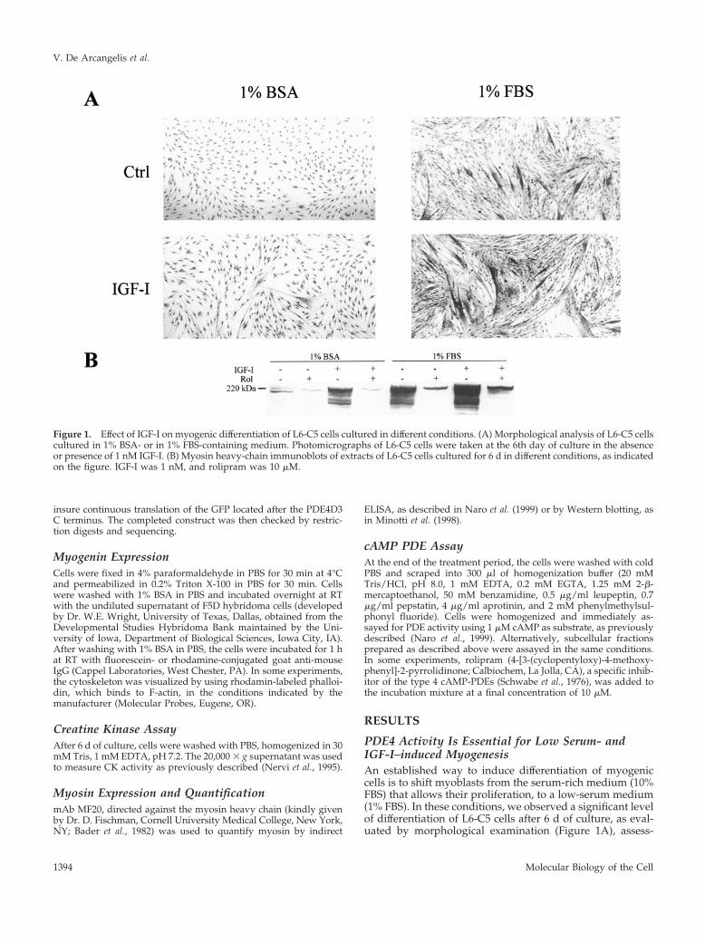
PDE4 Activity Is Essential for Low Serum- andIGF-I–induced MyogenesisAn established way to induce differentiation of myogeniccells is to shift myoblasts from the serum-rich medium (10%FBS) that allows their proliferation, to a low-serum medium(1% FBS). In these conditions, we observed a significant levelof differentiation of L6-C5 cells after 6 d of culture, as eval-uated by morphological examination (Figure 1A), assess-
Figure 1. Effect of IGF-I on myogenic differentiation of L6-C5 cells cultured in different conditions. (A) Morphological analysis of L6-C5 cellscultured in 1% BSA- or in 1% FBS-containing medium. Photomicrographs of L6-C5 cells were taken at the 6th day of culture in the absenceor presence of 1 nM IGF-I. (B) Myosin heavy-chain immunoblots of extracts of L6-C5 cells cultured for 6 d in different conditions, as indicatedon the figure. IGF-I was 1 nM, and rolipram was 10 �M.
V. De Arcangelis et al.
Molecular Biology of the Cell1394
ment of the differentiation marker myosin heavy chain byimmunoblotting (Figure 1B), and measurement of CK activ-ity (Table 1). Supplementation of 1% FBS-containing me-dium with 10�9 M IGF-I led to further stimulation of cellfusion with the formation of large multinucleated myotubes(Figure 1A), a strong increase in myosin expression (Figure1B), and of CK activity (Table 1). By contrast, in the absenceof serum (myoblast shifted to 1% BSA-containing medium),the cells were maintained quiescent and no differentiationwas observed. In these culture conditions, IGF-I had verylittle effect (Table 1 and Figure 1, A and B).
Addition of the selective type 4 PDE inhibitor rolipram tothe culture medium efficiently blocked myogenic differenti-ation (Table 1 and Figure 1B) in all conditions tested, sug-gesting that PDE4 activity is essential for L6-C5 differentia-tion regardless of induction conditions. The sensitivity torolipram is not a distinctive feature of L6-C5 clone, becauserolipram could as well inhibit the differentiation of thewidely used L6-E9 clone, achieved in various conditions.
L6-C5 Cells Cultured in the Presence of SerumDisplay a Higher cAMP-PDE Activity than CellsCultured in Serum-free MediumAs shown above, cells cultured in low serum mediumachieved a much higher degree of differentiation comparedwith cells cultured in the absence of serum. If PDE4 activityis required for differentiation, this difference in behaviorcould be related to a different level of PDE4 activity in cellsundergoing differentiation compared with quiescent cells.
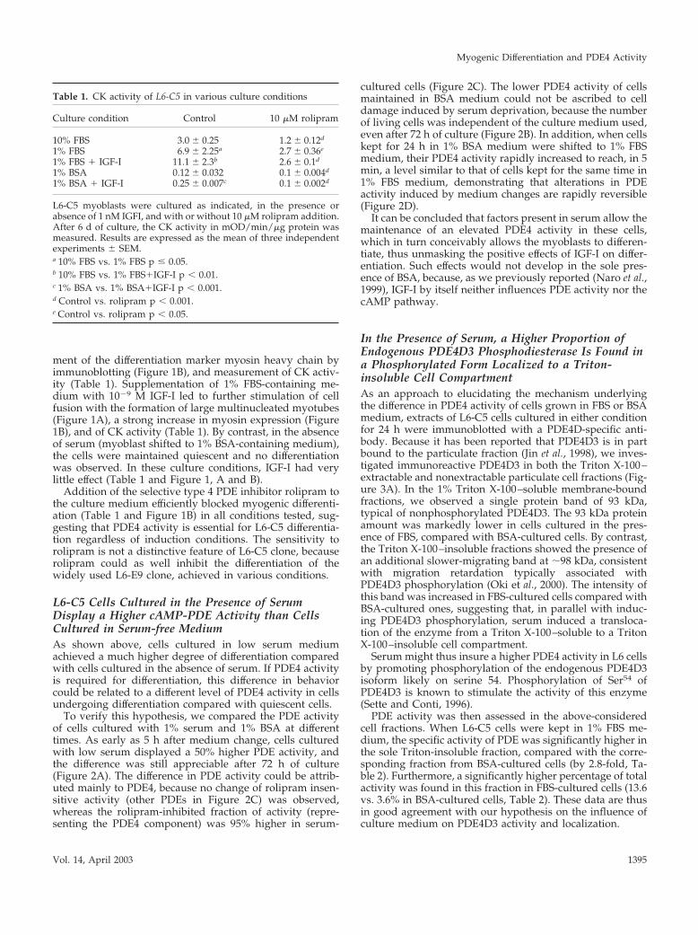
To verify this hypothesis, we compared the PDE activityof cells cultured with 1% serum and 1% BSA at differenttimes. As early as 5 h after medium change, cells culturedwith low serum displayed a 50% higher PDE activity, andthe difference was still appreciable after 72 h of culture(Figure 2A). The difference in PDE activity could be attrib-uted mainly to PDE4, because no change of rolipram insen-sitive activity (other PDEs in Figure 2C) was observed,whereas the rolipram-inhibited fraction of activity (repre-senting the PDE4 component) was 95% higher in serum-
cultured cells (Figure 2C). The lower PDE4 activity of cellsmaintained in BSA medium could not be ascribed to celldamage induced by serum deprivation, because the numberof living cells was independent of the culture medium used,even after 72 h of culture (Figure 2B). In addition, when cellskept for 24 h in 1% BSA medium were shifted to 1% FBSmedium, their PDE4 activity rapidly increased to reach, in 5min, a level similar to that of cells kept for the same time in1% FBS medium, demonstrating that alterations in PDEactivity induced by medium changes are rapidly reversible(Figure 2D).
It can be concluded that factors present in serum allow themaintenance of an elevated PDE4 activity in these cells,which in turn conceivably allows the myoblasts to differen-tiate, thus unmasking the positive effects of IGF-I on differ-entiation. Such effects would not develop in the sole pres-ence of BSA, because, as we previously reported (Naro et al.,1999), IGF-I by itself neither influences PDE activity nor thecAMP pathway.
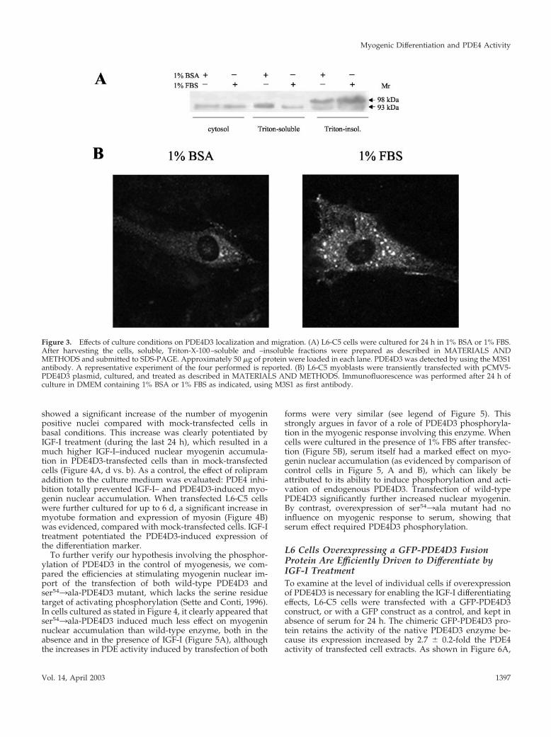
In the Presence of Serum, a Higher Proportion ofEndogenous PDE4D3 Phosphodiesterase Is Found ina Phosphorylated Form Localized to a Triton-insoluble Cell CompartmentAs an approach to elucidating the mechanism underlyingthe difference in PDE4 activity of cells grown in FBS or BSAmedium, extracts of L6-C5 cells cultured in either conditionfor 24 h were immunoblotted with a PDE4D-specific anti-body. Because it has been reported that PDE4D3 is in partbound to the particulate fraction (Jin et al., 1998), we inves-tigated immunoreactive PDE4D3 in both the Triton X-100–extractable and nonextractable particulate cell fractions (Fig-ure 3A). In the 1% Triton X-100–soluble membrane-boundfractions, we observed a single protein band of 93 kDa,typical of nonphosphorylated PDE4D3. The 93 kDa proteinamount was markedly lower in cells cultured in the pres-ence of FBS, compared with BSA-cultured cells. By contrast,the Triton X-100–insoluble fractions showed the presence ofan additional slower-migrating band at �98 kDa, consistentwith migration retardation typically associated withPDE4D3 phosphorylation (Oki et al., 2000). The intensity ofthis band was increased in FBS-cultured cells compared withBSA-cultured ones, suggesting that, in parallel with induc-ing PDE4D3 phosphorylation, serum induced a transloca-tion of the enzyme from a Triton X-100–soluble to a TritonX-100–insoluble cell compartment.
Serum might thus insure a higher PDE4 activity in L6 cellsby promoting phosphorylation of the endogenous PDE4D3isoform likely on serine 54. Phosphorylation of Ser54 ofPDE4D3 is known to stimulate the activity of this enzyme(Sette and Conti, 1996).
PDE activity was then assessed in the above-consideredcell fractions. When L6-C5 cells were kept in 1% FBS me-dium, the specific activity of PDE was significantly higher inthe sole Triton-insoluble fraction, compared with the corre-sponding fraction from BSA-cultured cells (by 2.8-fold, Ta-ble 2). Furthermore, a significantly higher percentage of totalactivity was found in this fraction in FBS-cultured cells (13.6vs. 3.6% in BSA-cultured cells, Table 2). These data are thusin good agreement with our hypothesis on the influence ofculture medium on PDE4D3 activity and localization.
Table 1. CK activity of L6-C5 in various culture conditions
Culture condition Control 10 �M rolipram
10% FBS 3.0 � 0.25 1.2 � 0.12d
1% FBS 6.9 � 2.25a 2.7 � 0.36e
1% FBS � IGF-I 11.1 � 2.3b 2.6 � 0.1d
1% BSA 0.12 � 0.032 0.1 � 0.004d
1% BSA � IGF-I 0.25 � 0.007c 0.1 � 0.002d
L6-C5 myoblasts were cultured as indicated, in the presence orabsence of 1 nM IGFI, and with or without 10 �M rolipram addition.After 6 d of culture, the CK activity in mOD/min/�g protein wasmeasured. Results are expressed as the mean of three independentexperiments � SEM.a 10% FBS vs. 1% FBS p � 0.05.b 10% FBS vs. 1% FBS�IGF-I p � 0.01.c 1% BSA vs. 1% BSA�IGF-I p � 0.001.d Control vs. rolipram p � 0.001.e Control vs. rolipram p � 0.05.
Myogenic Differentiation and PDE4 Activity
Vol. 14, April 2003 1395
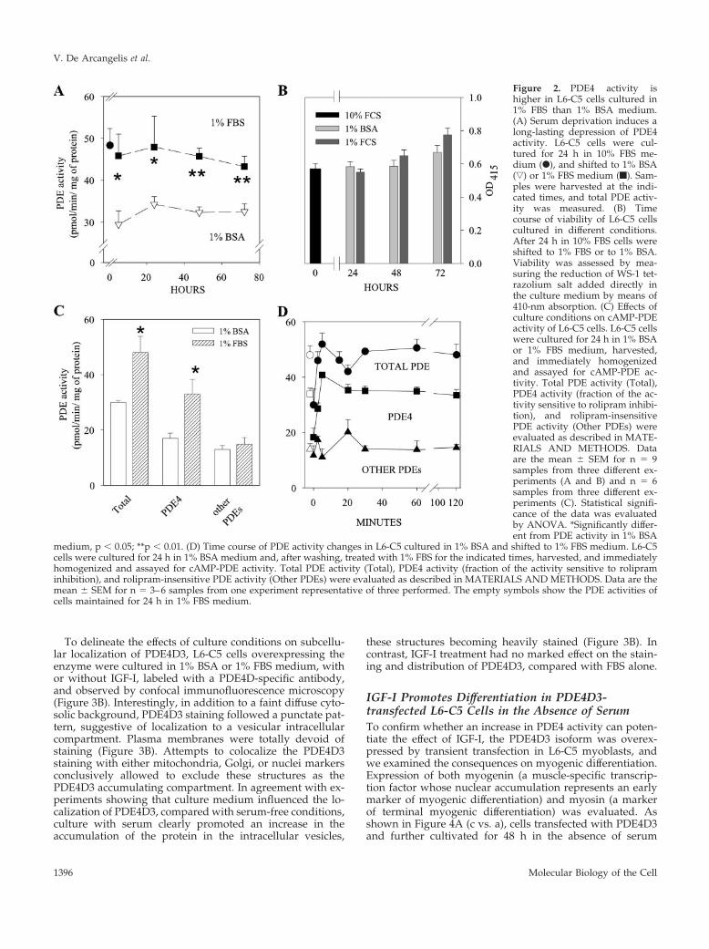
To delineate the effects of culture conditions on subcellu-lar localization of PDE4D3, L6-C5 cells overexpressing theenzyme were cultured in 1% BSA or 1% FBS medium, withor without IGF-I, labeled with a PDE4D-specific antibody,and observed by confocal immunofluorescence microscopy(Figure 3B). Interestingly, in addition to a faint diffuse cyto-solic background, PDE4D3 staining followed a punctate pat-tern, suggestive of localization to a vesicular intracellularcompartment. Plasma membranes were totally devoid ofstaining (Figure 3B). Attempts to colocalize the PDE4D3staining with either mitochondria, Golgi, or nuclei markersconclusively allowed to exclude these structures as thePDE4D3 accumulating compartment. In agreement with ex-periments showing that culture medium influenced the lo-calization of PDE4D3, compared with serum-free conditions,culture with serum clearly promoted an increase in theaccumulation of the protein in the intracellular vesicles,
these structures becoming heavily stained (Figure 3B). Incontrast, IGF-I treatment had no marked effect on the stain-ing and distribution of PDE4D3, compared with FBS alone.
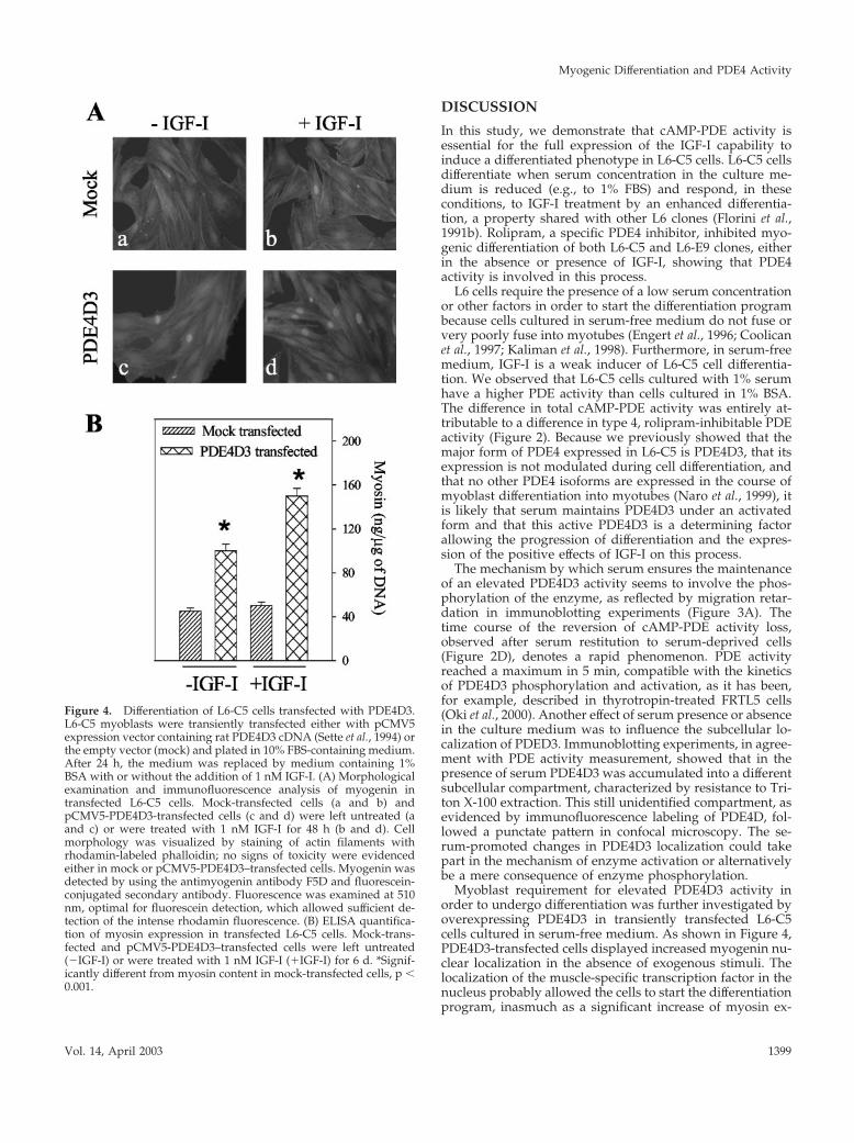
IGF-I Promotes Differentiation in PDE4D3-transfected L6-C5 Cells in the Absence of SerumTo confirm whether an increase in PDE4 activity can poten-tiate the effect of IGF-I, the PDE4D3 isoform was overex-pressed by transient transfection in L6-C5 myoblasts, andwe examined the consequences on myogenic differentiation.Expression of both myogenin (a muscle-specific transcrip-tion factor whose nuclear accumulation represents an earlymarker of myogenic differentiation) and myosin (a markerof terminal myogenic differentiation) was evaluated. Asshown in Figure 4A (c vs. a), cells transfected with PDE4D3and further cultivated for 48 h in the absence of serum
Figure 2. PDE4 activity ishigher in L6-C5 cells cultured in1% FBS than 1% BSA medium.(A) Serum deprivation induces along-lasting depression of PDE4activity. L6-C5 cells were cul-tured for 24 h in 10% FBS me-dium (F), and shifted to 1% BSA(ƒ) or 1% FBS medium (f). Sam-ples were harvested at the indi-cated times, and total PDE activ-ity was measured. (B) Timecourse of viability of L6-C5 cellscultured in different conditions.After 24 h in 10% FBS cells wereshifted to 1% FBS or to 1% BSA.Viability was assessed by mea-suring the reduction of WS-1 tet-razolium salt added directly inthe culture medium by means of410-nm absorption. (C) Effects ofculture conditions on cAMP-PDEactivity of L6-C5 cells. L6-C5 cellswere cultured for 24 h in 1% BSAor 1% FBS medium, harvested,and immediately homogenizedand assayed for cAMP-PDE ac-tivity. Total PDE activity (Total),PDE4 activity (fraction of the ac-tivity sensitive to rolipram inhibi-tion), and rolipram-insensitivePDE activity (Other PDEs) wereevaluated as described in MATE-RIALS AND METHODS. Dataare the mean � SEM for n � 9samples from three different ex-periments (A and B) and n � 6samples from three different ex-periments (C). Statistical signifi-cance of the data was evaluatedby ANOVA. *Significantly differ-ent from PDE activity in 1% BSA
medium, p � 0.05; **p � 0.01. (D) Time course of PDE activity changes in L6-C5 cultured in 1% BSA and shifted to 1% FBS medium. L6-C5cells were cultured for 24 h in 1% BSA medium and, after washing, treated with 1% FBS for the indicated times, harvested, and immediatelyhomogenized and assayed for cAMP-PDE activity. Total PDE activity (Total), PDE4 activity (fraction of the activity sensitive to rolipraminhibition), and rolipram-insensitive PDE activity (Other PDEs) were evaluated as described in MATERIALS AND METHODS. Data are themean � SEM for n � 3–6 samples from one experiment representative of three performed. The empty symbols show the PDE activities ofcells maintained for 24 h in 1% FBS medium.
V. De Arcangelis et al.
Molecular Biology of the Cell1396
showed a significant increase of the number of myogeninpositive nuclei compared with mock-transfected cells inbasal conditions. This increase was clearly potentiated byIGF-I treatment (during the last 24 h), which resulted in amuch higher IGF-I–induced nuclear myogenin accumula-tion in PDE4D3-transfected cells than in mock-transfectedcells (Figure 4A, d vs. b). As a control, the effect of rolipramaddition to the culture medium was evaluated: PDE4 inhi-bition totally prevented IGF-I– and PDE4D3-induced myo-genin nuclear accumulation. When transfected L6-C5 cellswere further cultured for up to 6 d, a significant increase inmyotube formation and expression of myosin (Figure 4B)was evidenced, compared with mock-transfected cells. IGF-Itreatment potentiated the PDE4D3-induced expression ofthe differentiation marker.
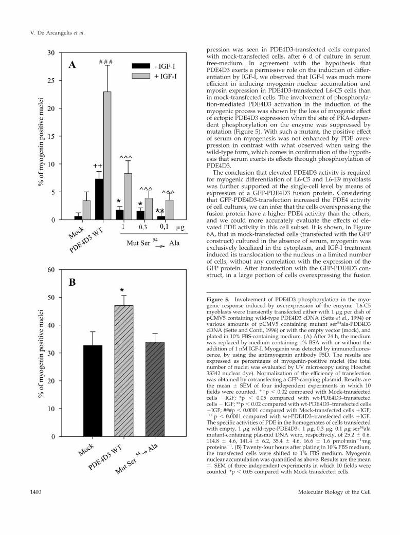
To further verify our hypothesis involving the phosphor-ylation of PDE4D3 in the control of myogenesis, we com-pared the efficiencies at stimulating myogenin nuclear im-port of the transfection of both wild-type PDE4D3 andser543ala-PDE4D3 mutant, which lacks the serine residuetarget of activating phosphorylation (Sette and Conti, 1996).In cells cultured as stated in Figure 4, it clearly appeared thatser543ala-PDE4D3 induced much less effect on myogeninnuclear accumulation than wild-type enzyme, both in theabsence and in the presence of IGF-I (Figure 5A), althoughthe increases in PDE activity induced by transfection of both
forms were very similar (see legend of Figure 5). Thisstrongly argues in favor of a role of PDE4D3 phosphoryla-tion in the myogenic response involving this enzyme. Whencells were cultured in the presence of 1% FBS after transfec-tion (Figure 5B), serum itself had a marked effect on myo-genin nuclear accumulation (as evidenced by comparison ofcontrol cells in Figure 5, A and B), which can likely beattributed to its ability to induce phosphorylation and acti-vation of endogenous PDE4D3. Transfection of wild-typePDE4D3 significantly further increased nuclear myogenin.By contrast, overexpression of ser543ala mutant had noinfluence on myogenic response to serum, showing thatserum effect required PDE4D3 phosphorylation.
L6 Cells Overexpressing a GFP-PDE4D3 FusionProtein Are Efficiently Driven to Differentiate byIGF-I TreatmentTo examine at the level of individual cells if overexpressionof PDE4D3 is necessary for enabling the IGF-I differentiatingeffects, L6-C5 cells were transfected with a GFP-PDE4D3construct, or with a GFP construct as a control, and kept inabsence of serum for 24 h. The chimeric GFP-PDE4D3 pro-tein retains the activity of the native PDE4D3 enzyme be-cause its expression increased by 2.7 � 0.2-fold the PDE4activity of transfected cell extracts. As shown in Figure 6A,
Figure 3. Effects of culture conditions on PDE4D3 localization and migration. (A) L6-C5 cells were cultured for 24 h in 1% BSA or 1% FBS.After harvesting the cells, soluble, Triton-X-100–soluble and –insoluble fractions were prepared as described in MATERIALS ANDMETHODS and submitted to SDS-PAGE. Approximately 50 �g of protein were loaded in each lane. PDE4D3 was detected by using the M3S1antibody. A representative experiment of the four performed is reported. (B) L6-C5 myoblasts were transiently transfected with pCMV5-PDE4D3 plasmid, cultured, and treated as described in MATERIALS AND METHODS. Immunofluorescence was performed after 24 h ofculture in DMEM containing 1% BSA or 1% FBS as indicated, using M3S1 as first antibody.
Myogenic Differentiation and PDE4 Activity
Vol. 14, April 2003 1397
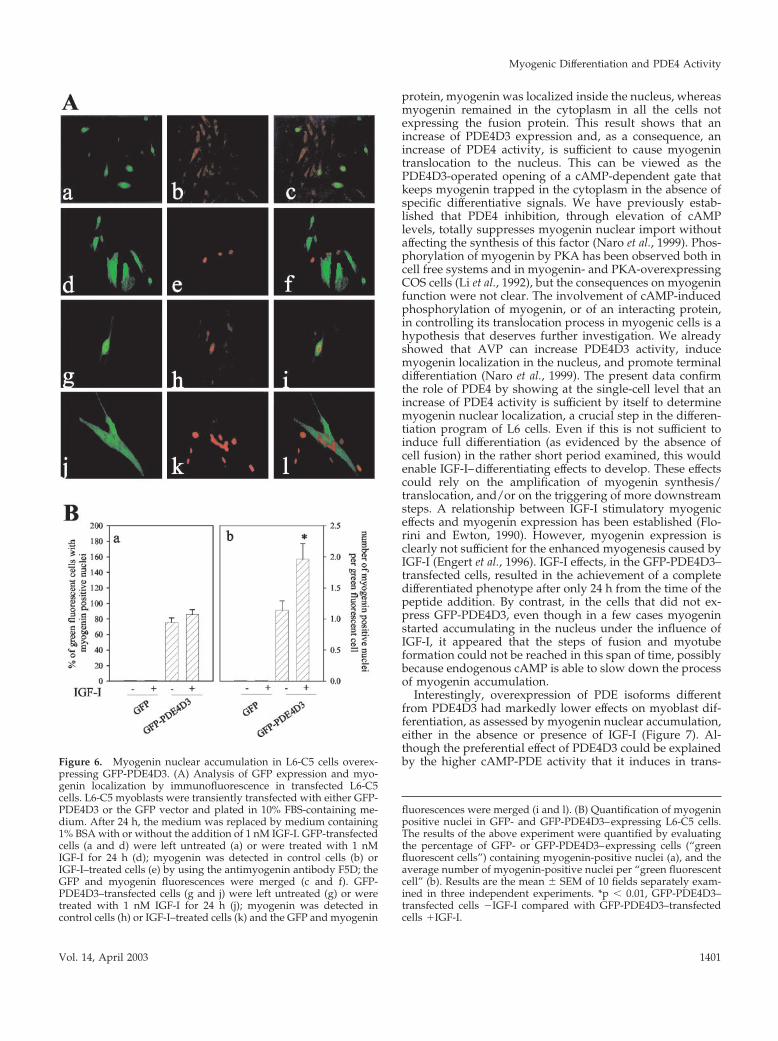
cells expressing GFP (a) displayed a cytoplasmic, not nu-clear, diffuse myogenin immunofluorescence (b and c). Inthe presence of IGF-I, GFP-transfected cells showed rare andscattered myogenin-positive nuclei without any superposi-tion with GFP expression (d–f). In cells transfected with theGFP-PDE4D3 construct, the fusion protein was efficientlyexpressed in part of the myoblasts, where intracellular greenfluorescence could be visualized (Figure 6A, g). In controlpreparations (which were not stimulated by IGF-I), myoge-nin was localized in the cytoplasm of cells expressing noGFP-PDE4D3 (green fluorescence negative), whereas myo-genin accumulated in the nuclei exclusively in the GFP-PDE4D3–overexpressing cells (h and i). This result stronglysupports the hypothesis that an increase of PDE4 activity iseffective by itself to translocate myogenin into the nucleus.Cells overexpressing GFP-PDE4D3 were challenged withIGF-I and appeared to fuse into large myotubes (Figure 6A,j), a differentiation level that was not achieved in the otherconditions used. The PDE-overexpressing myotubes con-tained clusters of myogenin-positive nuclei (Figure 6A, l),reemphasizing the correlation between myogenin nuclearimport and terminal myogenic differentiation. Interestingly,in the same cell cultures it could be noticed that IGF-Ipromoted myogenin accumulation in the nuclei of somecells that did not express GFP-PDE4D3, but these cells ap-peared to be mononucleated (the nuclei were scattered).Similar results were obtained with L6-E9 cells.
Quantification of the results of the above experiments isshown in Figure 6B. As it appears in panel a, IGF-I did notinfluence the proportion of GFP or GFP-PDE4D3–express-ing cells that contained one or more myogenin-positive nu-clei, whereas this proportion was much higher in GFP-PDE4D3–expressing cells compared with GFP-expressingcells. Panel b shows that IGF-I significantly increased thenumber of myogenin positive nuclei per cell, only in cellsoverexpressing GFP-PDE4D3. It thus clearly appeared thatoverexpression of PDE4D3 activity allowed myogenin accu-mulation in the cell nuclei and that IGF-I treatment furtherstimulated differentiation in the PDE4D3-overexpressing
cells by inducing cell fusion and formation of multinucle-ated myotubes.
PDE Isoforms Promote Myogenin NuclearAccumulation with Different EfficienciesWith regard to the multiplicity of isoforms of PDE ex-pressed in mammalian cells the question arose of thespecificity of the PDE4D3 isoform as a factor promotingIGF-I effects. To address this point various phosphodies-terase isoforms belonging either to the type 4 PDE family(PDE4D1 and PDE4D2, which are short splicing variantsderiving from the expression of gene PDE4D, which alsogives rise to the long variants PDE4D3 and PDE4D4;PDE4A5 and PDE4B2 expressed from other type 4 PDEgenes) or to other PDE families (PDE3A, PDE7A1,PDE11A2) were ectopically expressed in L6-C5 cells, andnuclear myogenin accumulation in the presence or ab-sence of IGF-I treatment was evaluated. As shown inFigure 7, several PDE isoforms (namely, PDE3A, PDE4D1,PDE4D3, PDE4D4, PDE7A1) were able to promote myo-genin nuclear accumulation in the presence of IGF-I, al-though with markedly different efficiencies. PDE4D3 wasthe only active isoform in the absence of IGF-I, and themost effective in the presence of this growth factor.PDE4D4, although it increased cAMP-PDE activity ofL6-C5 cells similarly to PDE4D3 (Figure 6, inset) hadmuch less effect (approximately threefold) on myogeninnuclear import. Transfection with other isoforms in-creased cAMP-PDE activity to variable extents (Figure 7,inset), without a correlation between the levels of cAMP-PDE activity and the effects on myogenin labeling: e.g.,PDE3A transfection, which induced a very modest in-crease in cAMP-PDE activity, significantly promotedmyogenin accumulation, whereas PDE4A5 or PDE4B2transfections, which markedly elevated cAMP-PDE activ-ity (by approximately fourfold), had no effect on the pro-portion of myogenin labeled nuclei.
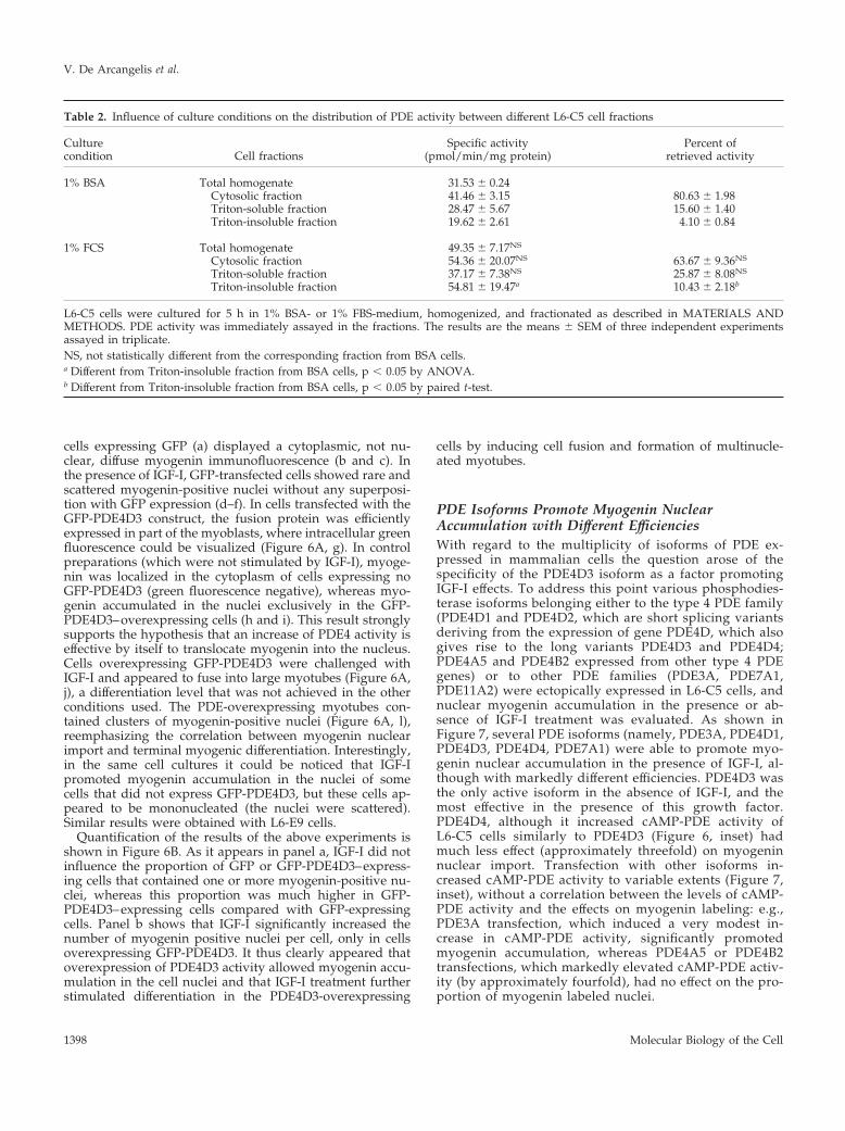
Table 2. Influence of culture conditions on the distribution of PDE activity between different L6-C5 cell fractions
Culturecondition Cell fractions
Specific activity(pmol/min/mg protein)
Percent ofretrieved activity
1% BSA Total homogenate 31.53 � 0.24Cytosolic fraction 41.46 � 3.15 80.63 � 1.98Triton-soluble fraction 28.47 � 5.67 15.60 � 1.40Triton-insoluble fraction 19.62 � 2.61 4.10 � 0.84
1% FCS Total homogenate 49.35 � 7.17NS
Cytosolic fraction 54.36 � 20.07NS 63.67 � 9.36NS
Triton-soluble fraction 37.17 � 7.38NS 25.87 � 8.08NS
Triton-insoluble fraction 54.81 � 19.47a 10.43 � 2.18b
L6-C5 cells were cultured for 5 h in 1% BSA- or 1% FBS-medium, homogenized, and fractionated as described in MATERIALS ANDMETHODS. PDE activity was immediately assayed in the fractions. The results are the means � SEM of three independent experimentsassayed in triplicate.NS, not statistically different from the corresponding fraction from BSA cells.a Different from Triton-insoluble fraction from BSA cells, p � 0.05 by ANOVA.b Different from Triton-insoluble fraction from BSA cells, p � 0.05 by paired t-test.
V. De Arcangelis et al.
Molecular Biology of the Cell1398
DISCUSSION
In this study, we demonstrate that cAMP-PDE activity isessential for the full expression of the IGF-I capability toinduce a differentiated phenotype in L6-C5 cells. L6-C5 cellsdifferentiate when serum concentration in the culture me-dium is reduced (e.g., to 1% FBS) and respond, in theseconditions, to IGF-I treatment by an enhanced differentia-tion, a property shared with other L6 clones (Florini et al.,1991b). Rolipram, a specific PDE4 inhibitor, inhibited myo-genic differentiation of both L6-C5 and L6-E9 clones, eitherin the absence or presence of IGF-I, showing that PDE4activity is involved in this process.
L6 cells require the presence of a low serum concentrationor other factors in order to start the differentiation programbecause cells cultured in serum-free medium do not fuse orvery poorly fuse into myotubes (Engert et al., 1996; Coolicanet al., 1997; Kaliman et al., 1998). Furthermore, in serum-freemedium, IGF-I is a weak inducer of L6-C5 cell differentia-tion. We observed that L6-C5 cells cultured with 1% serumhave a higher PDE activity than cells cultured in 1% BSA.The difference in total cAMP-PDE activity was entirely at-tributable to a difference in type 4, rolipram-inhibitable PDEactivity (Figure 2). Because we previously showed that themajor form of PDE4 expressed in L6-C5 is PDE4D3, that itsexpression is not modulated during cell differentiation, andthat no other PDE4 isoforms are expressed in the course ofmyoblast differentiation into myotubes (Naro et al., 1999), itis likely that serum maintains PDE4D3 under an activatedform and that this active PDE4D3 is a determining factorallowing the progression of differentiation and the expres-sion of the positive effects of IGF-I on this process.
The mechanism by which serum ensures the maintenanceof an elevated PDE4D3 activity seems to involve the phos-phorylation of the enzyme, as reflected by migration retar-dation in immunoblotting experiments (Figure 3A). Thetime course of the reversion of cAMP-PDE activity loss,observed after serum restitution to serum-deprived cells(Figure 2D), denotes a rapid phenomenon. PDE activityreached a maximum in 5 min, compatible with the kineticsof PDE4D3 phosphorylation and activation, as it has been,for example, described in thyrotropin-treated FRTL5 cells(Oki et al., 2000). Another effect of serum presence or absencein the culture medium was to influence the subcellular lo-calization of PDED3. Immunoblotting experiments, in agree-ment with PDE activity measurement, showed that in thepresence of serum PDE4D3 was accumulated into a differentsubcellular compartment, characterized by resistance to Tri-ton X-100 extraction. This still unidentified compartment, asevidenced by immunofluorescence labeling of PDE4D, fol-lowed a punctate pattern in confocal microscopy. The se-rum-promoted changes in PDE4D3 localization could takepart in the mechanism of enzyme activation or alternativelybe a mere consequence of enzyme phosphorylation.
Myoblast requirement for elevated PDE4D3 activity inorder to undergo differentiation was further investigated byoverexpressing PDE4D3 in transiently transfected L6-C5cells cultured in serum-free medium. As shown in Figure 4,PDE4D3-transfected cells displayed increased myogenin nu-clear localization in the absence of exogenous stimuli. Thelocalization of the muscle-specific transcription factor in thenucleus probably allowed the cells to start the differentiationprogram, inasmuch as a significant increase of myosin ex-
Figure 4. Differentiation of L6-C5 cells transfected with PDE4D3.L6-C5 myoblasts were transiently transfected either with pCMV5expression vector containing rat PDE4D3 cDNA (Sette et al., 1994) orthe empty vector (mock) and plated in 10% FBS-containing medium.After 24 h, the medium was replaced by medium containing 1%BSA with or without the addition of 1 nM IGF-I. (A) Morphologicalexamination and immunofluorescence analysis of myogenin intransfected L6-C5 cells. Mock-transfected cells (a and b) andpCMV5-PDE4D3-transfected cells (c and d) were left untreated (aand c) or were treated with 1 nM IGF-I for 48 h (b and d). Cellmorphology was visualized by staining of actin filaments withrhodamin-labeled phalloidin; no signs of toxicity were evidencedeither in mock or pCMV5-PDE4D3–transfected cells. Myogenin wasdetected by using the antimyogenin antibody F5D and fluorescein-conjugated secondary antibody. Fluorescence was examined at 510nm, optimal for fluorescein detection, which allowed sufficient de-tection of the intense rhodamin fluorescence. (B) ELISA quantifica-tion of myosin expression in transfected L6-C5 cells. Mock-trans-fected and pCMV5-PDE4D3–transfected cells were left untreated(�IGF-I) or were treated with 1 nM IGF-I (�IGF-I) for 6 d. *Signif-icantly different from myosin content in mock-transfected cells, p �0.001.
Myogenic Differentiation and PDE4 Activity
Vol. 14, April 2003 1399
pression was seen in PDE4D3-transfected cells comparedwith mock-transfected cells, after 6 d of culture in serumfree-medium. In agreement with the hypothesis thatPDE4D3 exerts a permissive role on the induction of differ-entiation by IGF-I, we observed that IGF-I was much moreefficient in inducing myogenin nuclear accumulation andmyosin expression in PDE4D3-transfected L6-C5 cells thanin mock-transfected cells. The involvement of phosphoryla-tion-mediated PDE4D3 activation in the induction of themyogenic process was shown by the loss of myogenic effectof ectopic PDE4D3 expression when the site of PKA-depen-dent phosphorylation on the enzyme was suppressed bymutation (Figure 5). With such a mutant, the positive effectof serum on myogenesis was not enhanced by PDE ovex-pression in contrast with what observed when using thewild-type form, which comes in confirmation of the hypoth-esis that serum exerts its effects through phosphorylation ofPDE4D3.
The conclusion that elevated PDE4D3 activity is requiredfor myogenic differentiation of L6-C5 and L6-E9 myoblastswas further supported at the single-cell level by means ofexpression of a GFP-PDE4D3 fusion protein. Consideringthat GFP-PDE4D3-transfection increased the PDE4 activityof cell cultures, we can infer that the cells overexpressing thefusion protein have a higher PDE4 activity than the others,and we could more accurately evaluate the effects of ele-vated PDE activity in this cell subset. It is shown, in Figure6A, that in mock-transfected cells (transfected with the GFPconstruct) cultured in the absence of serum, myogenin wasexclusively localized in the cytoplasm, and IGF-I treatmentinduced its translocation to the nucleus in a limited numberof cells, without any correlation with the expression of theGFP protein. After transfection with the GFP-PDE4D3 con-struct, in a large portion of cells overexpressing the fusion
Figure 5. Involvement of PDE4D3 phosphorylation in the myo-genic response induced by overexpression of the enzyme. L6-C5myoblasts were transiently transfected either with 1 �g per dish ofpCMV5 containing wild-type PDE4D3 cDNA (Sette et al., 1994) orvarious amounts of pCMV5 containing mutant ser54ala-PDE4D3cDNA (Sette and Conti, 1996) or with the empty vector (mock), andplated in 10% FBS-containing medium. (A) After 24 h, the mediumwas replaced by medium containing 1% BSA with or without theaddition of 1 nM IGF-I. Myogenin was detected by immunofluores-cence, by using the antimyogenin antibody F5D. The results areexpressed as percentages of myogenin-positive nuclei (the totalnumber of nuclei was evaluated by UV microscopy using Hoechst33342 nuclear dye). Normalization of the efficiency of transfectionwas obtained by cotransfecting a GFP-carrying plasmid. Results arethe mean � SEM of four independent experiments in which 10fields were counted. ��p � 0.02 compared with Mock-transfectedcells �IGF; *p � 0.05 compared with wt-PDE4D3–transfectedcells � IGF; **p � 0.02 compared with wt-PDE4D3–transfected cells�IGF; ###p � 0.0001 compared with Mock-transfected cells �IGF;∧∧∧ p � 0.0001 compared with wt-PDE4D3–transfected cells �IGF.The specific activities of PDE in the homogenates of cells transfectedwith empty, 1 �g wild-type-PDE4D3-, 1 �g, 0.3 �g, 0.1 �g ser54alamutant-containing plasmid DNA were, respectively, of 25.2 � 0.6,114.8 � 4.6, 141.4 � 6.2, 35.4 � 4.6, 16.6 � 1.6 pmol�min�1�mgproteins�1. (B) Twenty-four hours after plating in 10% FBS medium,the transfected cells were shifted to 1% FBS medium. Myogeninnuclear accumulation was quantified as above. Results are the mean�. SEM of three independent experiments in which 10 fields werecounted. *p � 0.05 compared with Mock-transfected cells.
V. De Arcangelis et al.
Molecular Biology of the Cell1400
protein, myogenin was localized inside the nucleus, whereasmyogenin remained in the cytoplasm in all the cells notexpressing the fusion protein. This result shows that anincrease of PDE4D3 expression and, as a consequence, anincrease of PDE4 activity, is sufficient to cause myogenintranslocation to the nucleus. This can be viewed as thePDE4D3-operated opening of a cAMP-dependent gate thatkeeps myogenin trapped in the cytoplasm in the absence ofspecific differentiative signals. We have previously estab-lished that PDE4 inhibition, through elevation of cAMPlevels, totally suppresses myogenin nuclear import withoutaffecting the synthesis of this factor (Naro et al., 1999). Phos-phorylation of myogenin by PKA has been observed both incell free systems and in myogenin- and PKA-overexpressingCOS cells (Li et al., 1992), but the consequences on myogeninfunction were not clear. The involvement of cAMP-inducedphosphorylation of myogenin, or of an interacting protein,in controlling its translocation process in myogenic cells is ahypothesis that deserves further investigation. We alreadyshowed that AVP can increase PDE4D3 activity, inducemyogenin localization in the nucleus, and promote terminaldifferentiation (Naro et al., 1999). The present data confirmthe role of PDE4 by showing at the single-cell level that anincrease of PDE4 activity is sufficient by itself to determinemyogenin nuclear localization, a crucial step in the differen-tiation program of L6 cells. Even if this is not sufficient toinduce full differentiation (as evidenced by the absence ofcell fusion) in the rather short period examined, this wouldenable IGF-I–differentiating effects to develop. These effectscould rely on the amplification of myogenin synthesis/translocation, and/or on the triggering of more downstreamsteps. A relationship between IGF-I stimulatory myogeniceffects and myogenin expression has been established (Flo-rini and Ewton, 1990). However, myogenin expression isclearly not sufficient for the enhanced myogenesis caused byIGF-I (Engert et al., 1996). IGF-I effects, in the GFP-PDE4D3–transfected cells, resulted in the achievement of a completedifferentiated phenotype after only 24 h from the time of thepeptide addition. By contrast, in the cells that did not ex-press GFP-PDE4D3, even though in a few cases myogeninstarted accumulating in the nucleus under the influence ofIGF-I, it appeared that the steps of fusion and myotubeformation could not be reached in this span of time, possiblybecause endogenous cAMP is able to slow down the processof myogenin accumulation.
Interestingly, overexpression of PDE isoforms differentfrom PDE4D3 had markedly lower effects on myoblast dif-ferentiation, as assessed by myogenin nuclear accumulation,either in the absence or presence of IGF-I (Figure 7). Al-though the preferential effect of PDE4D3 could be explainedby the higher cAMP-PDE activity that it induces in trans-Figure 6. Myogenin nuclear accumulation in L6-C5 cells overex-
pressing GFP-PDE4D3. (A) Analysis of GFP expression and myo-genin localization by immunofluorescence in transfected L6-C5cells. L6-C5 myoblasts were transiently transfected with either GFP-PDE4D3 or the GFP vector and plated in 10% FBS-containing me-dium. After 24 h, the medium was replaced by medium containing1% BSA with or without the addition of 1 nM IGF-I. GFP-transfectedcells (a and d) were left untreated (a) or were treated with 1 nMIGF-I for 24 h (d); myogenin was detected in control cells (b) orIGF-I–treated cells (e) by using the antimyogenin antibody F5D; theGFP and myogenin fluorescences were merged (c and f). GFP-PDE4D3–transfected cells (g and j) were left untreated (g) or weretreated with 1 nM IGF-I for 24 h (j); myogenin was detected incontrol cells (h) or IGF-I–treated cells (k) and the GFP and myogenin
fluorescences were merged (i and l). (B) Quantification of myogeninpositive nuclei in GFP- and GFP-PDE4D3–expressing L6-C5 cells.The results of the above experiment were quantified by evaluatingthe percentage of GFP- or GFP-PDE4D3–expressing cells (“greenfluorescent cells”) containing myogenin-positive nuclei (a), and theaverage number of myogenin-positive nuclei per “green fluorescentcell” (b). Results are the mean � SEM of 10 fields separately exam-ined in three independent experiments. *p � 0.01, GFP-PDE4D3–transfected cells �IGF-I compared with GFP-PDE4D3–transfectedcells �IGF-I.
Myogenic Differentiation and PDE4 Activity
Vol. 14, April 2003 1401
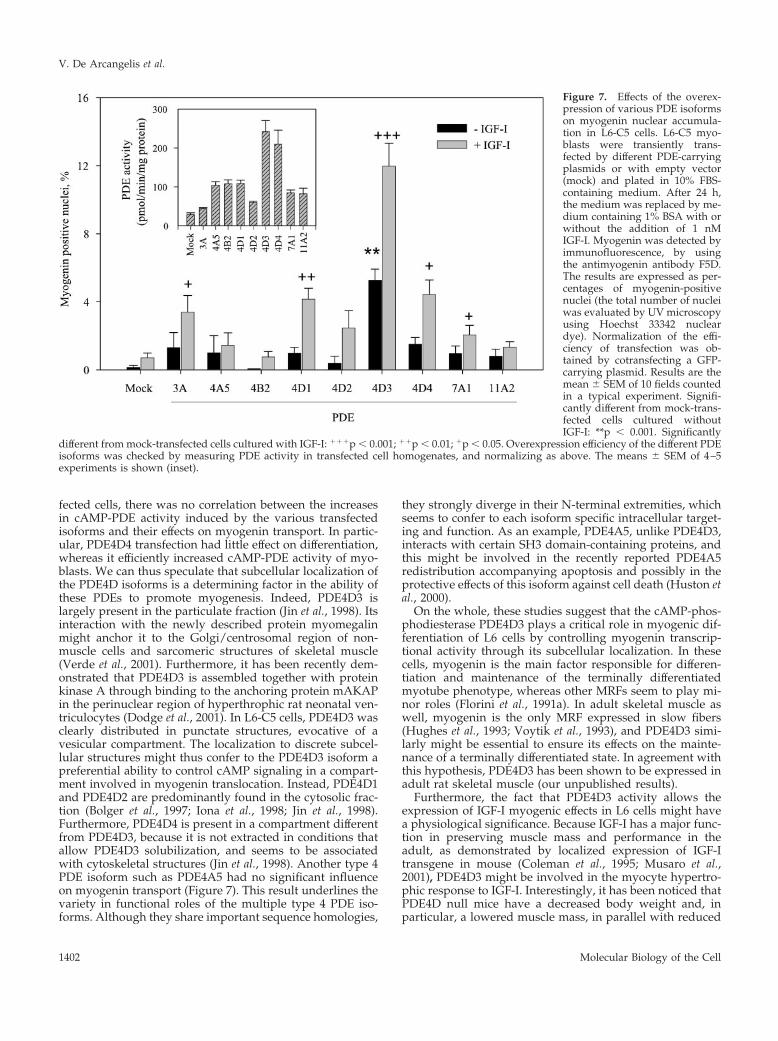
fected cells, there was no correlation between the increasesin cAMP-PDE activity induced by the various transfectedisoforms and their effects on myogenin transport. In partic-ular, PDE4D4 transfection had little effect on differentiation,whereas it efficiently increased cAMP-PDE activity of myo-blasts. We can thus speculate that subcellular localization ofthe PDE4D isoforms is a determining factor in the ability ofthese PDEs to promote myogenesis. Indeed, PDE4D3 islargely present in the particulate fraction (Jin et al., 1998). Itsinteraction with the newly described protein myomegalinmight anchor it to the Golgi/centrosomal region of non-muscle cells and sarcomeric structures of skeletal muscle(Verde et al., 2001). Furthermore, it has been recently dem-onstrated that PDE4D3 is assembled together with proteinkinase A through binding to the anchoring protein mAKAPin the perinuclear region of hyperthrophic rat neonatal ven-triculocytes (Dodge et al., 2001). In L6-C5 cells, PDE4D3 wasclearly distributed in punctate structures, evocative of avesicular compartment. The localization to discrete subcel-lular structures might thus confer to the PDE4D3 isoform apreferential ability to control cAMP signaling in a compart-ment involved in myogenin translocation. Instead, PDE4D1and PDE4D2 are predominantly found in the cytosolic frac-tion (Bolger et al., 1997; Iona et al., 1998; Jin et al., 1998).Furthermore, PDE4D4 is present in a compartment differentfrom PDE4D3, because it is not extracted in conditions thatallow PDE4D3 solubilization, and seems to be associatedwith cytoskeletal structures (Jin et al., 1998). Another type 4PDE isoform such as PDE4A5 had no significant influenceon myogenin transport (Figure 7). This result underlines thevariety in functional roles of the multiple type 4 PDE iso-forms. Although they share important sequence homologies,
they strongly diverge in their N-terminal extremities, whichseems to confer to each isoform specific intracellular target-ing and function. As an example, PDE4A5, unlike PDE4D3,interacts with certain SH3 domain-containing proteins, andthis might be involved in the recently reported PDE4A5redistribution accompanying apoptosis and possibly in theprotective effects of this isoform against cell death (Huston etal., 2000).
On the whole, these studies suggest that the cAMP-phos-phodiesterase PDE4D3 plays a critical role in myogenic dif-ferentiation of L6 cells by controlling myogenin transcrip-tional activity through its subcellular localization. In thesecells, myogenin is the main factor responsible for differen-tiation and maintenance of the terminally differentiatedmyotube phenotype, whereas other MRFs seem to play mi-nor roles (Florini et al., 1991a). In adult skeletal muscle aswell, myogenin is the only MRF expressed in slow fibers(Hughes et al., 1993; Voytik et al., 1993), and PDE4D3 simi-larly might be essential to ensure its effects on the mainte-nance of a terminally differentiated state. In agreement withthis hypothesis, PDE4D3 has been shown to be expressed inadult rat skeletal muscle (our unpublished results).
Furthermore, the fact that PDE4D3 activity allows theexpression of IGF-I myogenic effects in L6 cells might havea physiological significance. Because IGF-I has a major func-tion in preserving muscle mass and performance in theadult, as demonstrated by localized expression of IGF-Itransgene in mouse (Coleman et al., 1995; Musaro et al.,2001), PDE4D3 might be involved in the myocyte hypertro-phic response to IGF-I. Interestingly, it has been noticed thatPDE4D null mice have a decreased body weight and, inparticular, a lowered muscle mass, in parallel with reduced
Figure 7. Effects of the overex-pression of various PDE isoformson myogenin nuclear accumula-tion in L6-C5 cells. L6-C5 myo-blasts were transiently trans-fected by different PDE-carryingplasmids or with empty vector(mock) and plated in 10% FBS-containing medium. After 24 h,the medium was replaced by me-dium containing 1% BSA with orwithout the addition of 1 nMIGF-I. Myogenin was detected byimmunofluorescence, by usingthe antimyogenin antibody F5D.The results are expressed as per-centages of myogenin-positivenuclei (the total number of nucleiwas evaluated by UV microscopyusing Hoechst 33342 nucleardye). Normalization of the effi-ciency of transfection was ob-tained by cotransfecting a GFP-carrying plasmid. Results are themean � SEM of 10 fields countedin a typical experiment. Signifi-cantly different from mock-trans-fected cells cultured withoutIGF-I: **p � 0.001. Significantly
different from mock-transfected cells cultured with IGF-I: ���p � 0.001; ��p � 0.01; �p � 0.05. Overexpression efficiency of the different PDEisoforms was checked by measuring PDE activity in transfected cell homogenates, and normalizing as above. The means � SEM of 4–5experiments is shown (inset).
V. De Arcangelis et al.
Molecular Biology of the Cell1402
circulating IGF-I levels (Jin et al., 1999). These observationshighlight the integration between PDE4D activity and theIGF-I pathway during muscle development.
ACKNOWLEDGMENTS
We thank Prof. Joseph Beavo for kindly sharing the PDE7A1 andPDE11A2 plasmids, Dr. Antonio Musaro for the gift of L6-E9 clone,and Dr. Claudio Sette for helpful discussions. The exchanges be-tween the collaborating institutions were supported by a ConsiglioNazionale delle Ricerche–Institut National de la Sante et de laRecherche Medicale joint program grant (to G.N. and S.A.). Theresearch was supported by grants from MIUR (Ministero IstruzioneUniversita Ricerca) to S.A. and M.M., and from P.F. 83/2001 (Min-istero della Salute) to M.M.
REFERENCES
Bader, D., Masaki, T., and Fishman, D.A. (1982). Immunochemicalanalysis of myosin heavy chain during avian myogenesis in vivoand in vitro. J. Cell Biol. 95, 763–770.
Bolger, G.B., Erdogan, S., Jones, R.E., Loughney, K., Scotland, G.,Hoffmann, R., Wilkinson, I., Farrell, C., and Houslay, M.D. (1997).Characterization of five different proteins produced by alternativelyspliced mRNAs from the human cAMP-specific phosphodiesterasePDE4D gene. Biochem. J. 328, 539–548.
Coleman, M.E., DeMayo, F., Yin, K.C., Lee, H.M., Geske, R., Mont-gomery, C., and Schwartz, R.J. (1995). Myogenic vector expressionof insulin-like growth factor I stimulates muscle cell differentiationand myofiber hypertrophy in transgenic mice. J. Biol. Chem. 270,12109–12116.
Coolican, S.A., Samuel, D.S., Ewton, D.Z., McWade, F.J., and Florini,J.R. (1997). The mitogenic and myogenic actions of insulin-likegrowth factors utilize distinct signaling pathways. J. Biol. Chem.272, 6653–6662.
Dodge, K.L., Khouangsathiene, S., Kapiloff, M.S., Mouton, R., Hill,E.V., Houslay, M.D., Langeberg, L.K., and Scott, J.D. (2001). mAKAPassembles a protein kinase A/PDE4 phosphodiesterase cAMP sig-naling module. EMBO J. 20, 1921–1930.
Edmondson, D.G., and Olson, E.N. (1993). Helix-loop-helix proteinsas regulators of muscle-specific transcription. J. Biol. Chem. 268,755–758.
Engert, J.C., Berglund, E.B., and Rosenthal, N. (1996). Proliferationprecedes differentiation in IGF-I stimulated myogenesis. J. Cell Biol.135, 431–440.
Florini, J.R., and Ewton, D.Z. (1990). Highly specific inhibition ofIGF-1 stimulated differentiation by an antisense oligodeoxyribo-nucleotide to myogenin mRNA. No effects on other actions of IGF-1.J. Biol. Chem. 265, 13435–13437.
Florini, J.R., Ewton, D.Z., and Coolican, S.A. (1996). Growth hor-mone and the insulin-like growth factor system in myogenesis.Endocr. Rev. 17, 481–517.
Florini, J.R., Ewton, D.Z., and Roof, S.L. (1991a). Insulin-like growthfactor-1 stimulates terminal myogenic differentiation by inductionof myogenin gene expression. Mol. Endocrinol. 5, 718–724.
Florini, J.R., Magri, K.A., Ewton, D.Z., James, P.L., Grindstaff, K.,and Rotwein, P.S. (1991b). “Spontaneous” differentiation of skeletalmyoblasts is dependent upon autocrine secretion of insulin-likegrowth factor-II. J. Biol. Chem. 266, 15917–15923.
Hughes, S.M., Taylor, J.M., Tapscott, S.J., Gurley, C.M., Carter, W.J.,and Peterson, C.A. (1993). Selective accumulation of MyoD andmyogenin mRNAs in fast and slow adult skeletal muscle is con-trolled by innervation and hormones. Development 118, 1137–1147.
Huston, E., Beard, M., McCallum, F., Pyne, N.J., Vandenabeele, P.,Scotland, G., and Houslay, M.D. (2000). The cAMP-specific phos-phodiesterase PDE4A5 is cleaved downstream of its SH3 interactiondomain by caspase-3. Consequences for altered intracellular distri-bution. J. Biol. Chem. 275, 28063–28074.
Iona, S., Cuomo, M., Bushnik, T., Naro, F., Sette, C., Hess, M.,Shelton, E.R., and Conti, M. (1998). Characterization of the rolipram-sensitive, cAMP specific phosphodiesterases: identification and dif-ferential expression of immunologically distinct forms in the ratbrain. Mol. Pharmacol. 53, 23–32.
Jiang, B.H., Aoki, M., Zheng, J.Z., Li, J., and Vogt, P.K. (1999).Myogenic signaling of phosphatidylinositol 3-kinase requires theserine-threonine kinase Akt/protein kinase B. Proc. Natl. Acad. Sci.USA 96, 2077–2081.
Jin, S.L.C., Bushnik, T., Lan, L., and Conti, M. (1998). Subcellularlocalization of rolipram-sensitive, cAMP-specific phosphodiester-ases. J. Biol. Chem. 273, 19672–19678.
Jin, S.L., Richard, F.J., Kuo, W.P., D’Ercole, A.J., and Conti, M.(1999). Impaired growth and fertility of cAMP-specific phosphodi-esterase PDE4D-deficient mice. Proc. Natl. Acad. Sci. USA 96,11998–12003.
Kaliman, P., Canicio, J., Shepherd, P.R., Beeton, C.A., Testar, X.,Palacin, M., and Zorzano, A. (1998). Insulin-like growth factorsrequire phosphatidylinositol 3-kinase to signal myogenesis: domi-nant negative p85 expression blocks differentiation of L6E9 musclecells. Mol. Endocrinol. 12, 66–77.
Kaliman, P., Canicio, J., Testar, X., Palacin, M., and Zorzano, A.(1999). Insulin-like growth factor-II, phosphatidylinositol 3-kinase,nuclear factor-kappaB and inducible nitric-oxide synthase define acommon myogenic signaling pathway. J. Biol. Chem. 274, 17437–17444.
Li, L., Heller-Harrison, R., Czech, M., and Olson, E.N. (1992). CyclicAMP-dependent protein kinase activity inhibits the activity of myo-genic helix-loop-helix proteins. Mol. Cell. Biol. 12, 4478–4485.
Liu, J.P., Baker, J., Perkins, A.S., Robertson, E.J., and Efstratiadis, A.(1993). Mice carrying null mutations of the genes encoding insulin-like growth factor I (Igf-1) and type 1 IGF receptor (Igf1r). Cell 75,59–72.
Minotti, S., Scicchitano, B.M., Nervi, C., Scarpa, S., Lucarelli, M.,Molinaro, M., and Adamo, S. (1998). Vasopressin and insulin-likegrowth factors synergistically induce myogenesis in serum-free me-dium. Cell Growth Diff. 9, 155–163.
Musaro, A., McCullagh, K., Paul, A., Houghton, L., Dobrowolny, G.,Molinaro, M., Barton, E.R., Sweeney, H.L., and Rosenthal, N. (2001).Localized Igf-1 transgene expression sustains hypertrophy and re-generation in senescent skeletal muscle. Nat. Genet. 27, 195–200.
Nadal-Ginard, B. (1978). Commitment, fusion and biochemical dif-ferentiation of a myogenic cell line in the absence of DNA synthesis.Cell 15, 855–864.
Naro, F. et al. (1999). Involvement of type 4 cAMP-phosphodiester-ase in the myogenic differentiation of L6 cells. Mol. Biol. Cell 10,4355–4367.
Nervi, C., Benedetti, L., Minasi, A., Molinaro, M., and Adamo, S.(1995). Arginine-vasopressin induces differentiation of skeletal myo-genic cells and upregulation of myogenin and myf-5. Cell GrowthDiff. 6, 81–89.
Oki, N., Takahashi, S.I., Hidaka, H., and Conti, M. (2000). Short termfeedback regulation of cAMP in FRTL-5 thyroid cells. Role ofPDE4D3 phosphodiesterase activation. J. Biol. Chem. 275, 10831–10837.
Samuel, D.S., Ewton, D.Z., Coolican, S.A., Petley, T.D., McWade,F.J., and Florini, J.R. (1999). Raf-1 activation stimulates proliferation
Myogenic Differentiation and PDE4 Activity
Vol. 14, April 2003 1403
and inhibits IGF-stimulated differentiation in L6A1 myoblasts.Horm. Metab. Res. 31, 55–64.
Schwabe, U., Miyake, M., Ohga, Y., and Daly, J.W. (1976). 4-(-3-cyclopentyloxy-4-methoxyphenyl)-2-pyrrolidone (ZK 62711): a po-tent inhibitor of adenosine cyclic 3� 5�-mono-phosphate-phosphodi-esterases in homogenates and tissue slices from rat brain. Mol.Pharmacol. 12, 900–910.
Sette, C., Vicini, E., and Conti, M. (1994). The ratPDE3/IVd phos-phodiesterase gene codes for multiple proteins differentially acti-vated by cAMP-protein kinase. J. Biol. Chem. 269, 18271–18274.
Sette, C., and Conti, M. (1996). Phosphorylation and activation of acAMP-specific phosphodiesterase by the cAMP-dependent proteinkinase. Involvement of serine 54 in the enzyme activation. J. Biol.Chem. 271, 16526–16534.
Soderling, S.H., and Beavo, J.A. (2000). Regulation of cAMP andcGMP signaling: new phosphodiesterases and new functions. Curr.Opin. Cell Biol. 12, 174–179.
Teti, A., Naro, F., Molinaro, M., and Adamo, S. (1993). Transductionof the arginine-vasopressin signal in skeletal myogenic cells. Am. J.Physiol. 265(C34), C113–C121.Verde, I., Pahlke, G., Salanova, M., Zhang, G., Wang, S., Coletti, D.,Onuffer, J., Jin, S.L., and Conti, M. (2001). Myomegalin is a novelprotein of the golgi/centrosome that interacts with a cyclic nucleo-tide phosphodiesterase. J. Biol. Chem. 276, 11189–11198.Voytik, S.L., Przyborski, M., Badylak, S.F., and Konieczny, S.F.(1993). Differential expression of muscle regulatory factor genes innormal and denervated adult rat hindlimb muscles. Dev. Dyn. 198,214–224.Winter, B., Braun, T., and Arnold, H.H. (1993). cAMP-dependentprotein kinase represses myogenic differentiation and the activity ofthe muscle-specific helix-loop-helix transcription factors Myf-5 andMyoD. J. Biol. Chem. 268, 9869–9878.Xu, Q., and Wu, Z. (2000). The IGF-PI3K-Akt signaling pathwayregulates myogenin expression in normal myogenic cells but not inRhabdomyosarcoma derived RD cells. J. Biol. Chem. 275, 36750–36757.
V. De Arcangelis et al.
Molecular Biology of the Cell1404
Copyright © 2022 FDOKUMEN