Neuronal expression of cAMP-specific phosphodiesterase 7B mRNA in the rat brain
13
NEURONAL EXPRESSION OF cAMP-SPECIFIC PHOSPHODIESTERASE 7B mRNA IN THE RAT BRAIN E. REYES-IRISARRI, S. PÉREZ-TORRES AND G. MENGOD* Department of Neurochemistry, Institut d’Investigacions, Biomèdiques de Barcelona, Consejo Superior de Investigaciones Científicas, Institut d’Investigacions Biomèdiques August Pi i Sunyer (IIBB-CSIC, IDIBAPS), c/Rosselló 161, 6 a , E-08036 Barcelona, Spain Abstract—cAMP plays an important role as second messen- ger molecule controlling multiple cellular processes in the brain. cAMP levels depend critically on the phosphodiester- ases (PDE) activity, enzymes responsible for the clearance of intracellular cAMP. We have examined the regional distribu- tion and cellular localization of mRNA coding for the cAMP- specific phosphodiesterase 7B (PDE7B) in rat brain by in situ hybridization histochemistry. PDE7B mRNA is specifically distributed in rat brain, preferentially in neuronal cell popu- lations. The highest levels of hybridization are observed in olfactory tubercle, islands of Calleja, dentate gyrus, caudate- putamen and some thalamic nuclei. Positive hybridization signals are also detected in other areas, such as cerebral cortex, Purkinje cells of the cerebellum and area postrema. By double in situ hybridization histochemistry, we found that 74% and 79% of the cells expressing PDE7B mRNA in stria- tum and olfactory tubercle, respectively, were GABAergic cells (expressing glutamic acid decarboxylase mRNA), in contrast with the lack of expression in the few cholinergic cells (expressing choline acetyltransferase mRNA) present in those two areas (around 0.4% in olfactory tubercle). In the thalamic nuclei, a majority of cells containing PDE7B mRNA also expresses a glutamatergic marker (76.7% express vesic- ular glutamate transporter vGluT1 and 76% express vGluT2 mRNAs). Almost all PDE7B expressing cells in dentate gyrus (93%) were glutamatergic. These results offer a neuroanatomical and neurochemical base that will support the search for specific functions for cAMP dependent PDEs and for the development of specific PDE7 inhibitors. © 2005 IBRO. Published by Elsevier Ltd. All rights reserved. Key words: in situ hybridization, PDE, glutamatergic cells, GABAergic cells, cholinergic cells. cAMP and cGMP play a key role in signal transduction in a wide variety of cellular responses. In brain, cAMP has been implicated in sensory functions, synaptic plasticity, learning and memory. Thus, information about how the intracellular cAMP levels are regulated will help to under- stand the mechanisms underlying these functions. Intra- cellular levels of cAMP are controlled not only by its syn- thesis by the enzyme adenylyl cyclase (Houslay and Milligan, 1997), but also by its degradation through the action of cyclic nucleotide phosphodiesterases (PDE), which cata- lyze the hydrolysis of 3=,5=-cyclic nucleotides into 5=- nucleoside monophosphates (Beavo, 1995; Conti and Jin, 1999; Francis et al., 2002; Houslay, 1998). PDEs have been so far classified into 11 families (PDE1–PDE11). They have different regulatory properties and intracellular location, with particular isoforms being expressed in a cell-specific manner (Conti and Jin, 1999). Families 4, 7 and 8 specifically hydrolyze cAMP, with PDE7 and PDE8 having a higher affinity for this substrate. Two members of the PDE7 family have been cloned, PDE7A (Michaeli et al., 1993), and PDE7B (Hetman et al., 2000; Sasaki et al., 2000). The distribution of PDE7A transcripts in the CNS, was first determined in mouse brain by RNase protection assays (Bloom and Beavo, 1996). Later, PDE7A mRNA was visualized by in situ hybridiza- tion in a preliminary study in a few adult and embryonic rat brain regions (Hoffmann et al., 1998), in rat brain and peripheral organs (Miró et al., 2001), and in some areas of the human brain (Pérez-Torres et al., 2003). Recently, alterations on the mRNA levels of PDE7A in postmortem human brains with Alzheimer’s disease has been reported (Pérez-Torres et al., 2003). Different in vivo models have been developed suggest- ing an implication of cAMP in learning, memory processes and other brain functions. Treatment of aged mice with rolipram, a PDE4 inhibitor, ameliorated the physiological and the memory defects of the age-related spatial memory loss (Bach et al., 1999). Administration of rolipram, a se- lective PDE4 inhibitor, induced long-term potentiation in the CA1 field of mouse hippocampus (Barad et al., 1998). These findings suggest the involvement of cAMP in syn- aptic plasticity during learning and memory processes. Since PDE7 has high affinity for cAMP, this enzyme could play an important role in maintaining cAMP levels at the basal intracellular levels. It has been shown that PDE 7A plays a role in the activation and/or proliferation of T cells (Li et al., 1999) and is upregulated in human B-lymphocytes (Lee et al., 2002). A few PDE7 inhibitors have been de- scribed (Barnes et al., 2001; Castro et al., 2001; Martinez et al., 2000; Pitts et al., 2004). The cellular localization of the PDEs in brain is an important step forward for the understanding of their func- tion. Here we describe experiments aimed at the analysis of both regional and cellular expression of the mRNA coding for PDE7B in neuronal populations expressing spe- cific neurotransmitters of the adult rat brain. Our study *Corresponding author. Tel: 34-933-63-8323; fax: 34-933-63-8301. E-mail address: [email protected] (G. Mengod). Abbreviations: AC, adenylyl cyclase; ChAT, choline acetyltransferase GAD, glutamic acid decarboxylase; GFAP, glial fibrillary acidic protein; MBP, myelin binding protein; PBS, phosphate-buffered saline; PDE, phosphodiesterase; vGluT1, vesicular glutamate transporter 1; vGluT2, vesicular glutamate transporter 2. Neuroscience 132 (2005) 1173–1185 0306-4522/05$30.000.00 © 2005 IBRO. Published by Elsevier Ltd. All rights reserved. doi:10.1016/j.neuroscience.2005.01.050 1173
-
Upload
independent -
Category
Documents
-
view
3 -
download
0
Transcript of Neuronal expression of cAMP-specific phosphodiesterase 7B mRNA in the rat brain

N7
EG
DddI
AgbaitshdlopscB7tcccttaum(
bcPr
KG
cwblis
*EAGMpv
Neuroscience 132 (2005) 1173–1185
0d
EURONAL EXPRESSION OF cAMP-SPECIFIC PHOSPHODIESTERASE
B mRNA IN THE RAT BRAINct1cln1
(aeFPTP2tbLtbptah(
iaralltTaSpbpe(se
itoc
. REYES-IRISARRI, S. PÉREZ-TORRES AND. MENGOD*
epartment of Neurochemistry, Institut d’Investigacions, Biomèdiquese Barcelona, Consejo Superior de Investigaciones Científicas, Institut’Investigacions Biomèdiques August Pi i Sunyer (IIBB-CSIC,DIBAPS), c/Rosselló 161, 6a, E-08036 Barcelona, Spain
bstract—cAMP plays an important role as second messen-er molecule controlling multiple cellular processes in therain. cAMP levels depend critically on the phosphodiester-ses (PDE) activity, enzymes responsible for the clearance ofntracellular cAMP. We have examined the regional distribu-ion and cellular localization of mRNA coding for the cAMP-pecific phosphodiesterase 7B (PDE7B) in rat brain by in situybridization histochemistry. PDE7B mRNA is specificallyistributed in rat brain, preferentially in neuronal cell popu-
ations. The highest levels of hybridization are observed inlfactory tubercle, islands of Calleja, dentate gyrus, caudate-utamen and some thalamic nuclei. Positive hybridizationignals are also detected in other areas, such as cerebralortex, Purkinje cells of the cerebellum and area postrema.y double in situ hybridization histochemistry, we found that4% and 79% of the cells expressing PDE7B mRNA in stria-um and olfactory tubercle, respectively, were GABAergicells (expressing glutamic acid decarboxylase mRNA), inontrast with the lack of expression in the few cholinergicells (expressing choline acetyltransferase mRNA) present inhose two areas (around 0.4% in olfactory tubercle). In thehalamic nuclei, a majority of cells containing PDE7B mRNAlso expresses a glutamatergic marker (76.7% express vesic-lar glutamate transporter vGluT1 and 76% express vGluT2RNAs). Almost all PDE7B expressing cells in dentate gyrus
93%) were glutamatergic.These results offer a neuroanatomical and neurochemical
ase that will support the search for specific functions forAMP dependent PDEs and for the development of specificDE7 inhibitors. © 2005 IBRO. Published by Elsevier Ltd. Allights reserved.
ey words: in situ hybridization, PDE, glutamatergic cells,ABAergic cells, cholinergic cells.
AMP and cGMP play a key role in signal transduction in aide variety of cellular responses. In brain, cAMP haseen implicated in sensory functions, synaptic plasticity,
earning and memory. Thus, information about how thentracellular cAMP levels are regulated will help to under-tand the mechanisms underlying these functions. Intra-
Corresponding author. Tel: �34-933-63-8323; fax: �34-933-63-8301.-mail address: [email protected] (G. Mengod).bbreviations: AC, adenylyl cyclase; ChAT, choline acetyltransferaseAD, glutamic acid decarboxylase; GFAP, glial fibrillary acidic protein;BP, myelin binding protein; PBS, phosphate-buffered saline; PDE,
chosphodiesterase; vGluT1, vesicular glutamate transporter 1;GluT2, vesicular glutamate transporter 2.
306-4522/05$30.00�0.00 © 2005 IBRO. Published by Elsevier Ltd. All rights reseroi:10.1016/j.neuroscience.2005.01.050
1173
ellular levels of cAMP are controlled not only by its syn-hesis by the enzyme adenylyl cyclase (Houslay and Milligan,997), but also by its degradation through the action ofyclic nucleotide phosphodiesterases (PDE), which cata-
yze the hydrolysis of 3=,5=-cyclic nucleotides into 5=-ucleoside monophosphates (Beavo, 1995; Conti and Jin,999; Francis et al., 2002; Houslay, 1998).
PDEs have been so far classified into 11 familiesPDE1–PDE11). They have different regulatory propertiesnd intracellular location, with particular isoforms beingxpressed in a cell-specific manner (Conti and Jin, 1999).amilies 4, 7 and 8 specifically hydrolyze cAMP, withDE7 and PDE8 having a higher affinity for this substrate.wo members of the PDE7 family have been cloned,DE7A (Michaeli et al., 1993), and PDE7B (Hetman et al.,000; Sasaki et al., 2000). The distribution of PDE7Aranscripts in the CNS, was first determined in mouse brainy RNase protection assays (Bloom and Beavo, 1996).ater, PDE7A mRNA was visualized by in situ hybridiza-ion in a preliminary study in a few adult and embryonic ratrain regions (Hoffmann et al., 1998), in rat brain anderipheral organs (Miró et al., 2001), and in some areas ofhe human brain (Pérez-Torres et al., 2003). Recently,lterations on the mRNA levels of PDE7A in postmortemuman brains with Alzheimer’s disease has been reportedPérez-Torres et al., 2003).
Different in vivo models have been developed suggest-ng an implication of cAMP in learning, memory processesnd other brain functions. Treatment of aged mice witholipram, a PDE4 inhibitor, ameliorated the physiologicalnd the memory defects of the age-related spatial memory
oss (Bach et al., 1999). Administration of rolipram, a se-ective PDE4 inhibitor, induced long-term potentiation inhe CA1 field of mouse hippocampus (Barad et al., 1998).hese findings suggest the involvement of cAMP in syn-ptic plasticity during learning and memory processes.ince PDE7 has high affinity for cAMP, this enzyme couldlay an important role in maintaining cAMP levels at theasal intracellular levels. It has been shown that PDE 7Alays a role in the activation and/or proliferation of T cells (Lit al., 1999) and is upregulated in human B-lymphocytesLee et al., 2002). A few PDE7 inhibitors have been de-cribed (Barnes et al., 2001; Castro et al., 2001; Martinezt al., 2000; Pitts et al., 2004).
The cellular localization of the PDEs in brain is anmportant step forward for the understanding of their func-ion. Here we describe experiments aimed at the analysisf both regional and cellular expression of the mRNAoding for PDE7B in neuronal populations expressing spe-
ific neurotransmitters of the adult rat brain. Our studyved.

rtwe
T
ACtsubaiatS
H
Fn1wtw
mabv4Gsaoaawtts
o(sa1(w(qBsr
uMtumCaDd
I
Th(awp8wepcTmaiwStrpsl(bNiwpf
Dh
H2iMmamwaHan(eldKwwN
pie
A
Twts
W
E. Reyes-Irisarri et al. / Neuroscience 132 (2005) 1173–11851174
eveals that PDE7B mRNA has a restricted distribution inhe brain being especially abundant in forebrain structures,here it is expressed in different neuronal populations withxcitatory and inhibitory neurotransmitters.
EXPERIMENTAL PROCEDURES
issue preparation
dult male Wistar rats (n�5; 200–300 g) were purchased from Iffaredo (Lyon, France). Animal care followed the Spanish legisla-
ion on “Protection of animals used in experimental and othercientific purposes” in agreement with the European (E.E.C) reg-lations (O.J. of E.C. L358/1 18/12/1986). The animals were killedy decapitation. All efforts were made to minimize the number ofnimals used and their suffering. The brains were frozen on dry
ce and kept at �20 °C. Tissue sections, 14 �m thick, were cut onmicrotome-cryostat (Microm HM500 OM, Walldorf, Germany),
haw-mounted onto APTS (3-aminopropyltriethoxysilane; Sigma,t. Louis, MO, USA) -coated slides, and kept at �20 °C until used.
ybridization probes
or the detection of PDE7B mRNA two 45 base-oligodeoxyribo-ucleotides that were complementary to bases 556–600 and 1390–434 (GenBank acc. no. NM_080894) were used. These regionsere chosen because they share no similarity with other members of
he different PDE families. Most of the results shown in the presentork were done using the first PDE7B oligonucleotide.
Glutamatergic cells were recognized by the presence of theRNA coding for both vesicular glutamate transporters (vGluT1nd vGluT2): vGluT1 with two oligonucleotides complementary toases 127–172 and 1756–1800 (GenBank acc. no U07609) andGluT2 with two oligonucleotides complementary to the bases66–510 and 2156–2200 (GenBank acc. no AF271235).ABAergic cells were identified by the presence of the enzymeynthesizing GABA, glutamic acid decarboxylase (GAD) that indult brain exists as two major isoforms, GAD65 and GAD67. Twoligonucleotides for each isoform mRNA were made: bp 159–213nd 514–558 (GenBank acc. no NM_012563) and bp 191–235nd 1600–1653 (GenBank acc. no NM_017007). Cholinergic cellsere distinguished by the presence of the enzyme choline acetyl-
ransferase (ChAT) mRNA with two oligonucleotides complemen-ary to bases 571–618 and 1321–1368 of the rat ChAT cDNAequence (Ishii et al., 1990).
We also identified the non-neuronal population by the presencef the glial fibrillary acidic protein (GFAP) and myelin binding proteinMBP) mRNAs, markers for astrocytes and oligodendrocytes, re-pectively. Two oligonucleotides complementary to bases 225–274nd 1194–1242 were made for the GFAP mRNA (Brenner et al.,990) and one oligonucleotide complementary to bases 179–223GenBank acc. no M25889) for MBP mRNA. The oligonucleotidesere all synthesized and HPLC purified by Isogen Bioscience BV
Maarsden, The Netherlands). Evaluation of the oligonucleotide se-uences with basic local alignment search tool of EMBL and Gen-ank databases indicated that the probes do not show any significantimilarity with mRNAs other than their corresponding targets in theat.
Oligonucleotides for PDE7B mRNA were labeled at their 3=-endsing [�-33P]dATP (3000 Ci/mmol; New England Nuclear, Boston,A, USA) for the in situ hybridization histochemistry experiments and
erminal deoxynucleotidyltransferase (Oncogene Research Prod-cts, San Diego, CA, USA), purified using QIAquick Nucleotide Re-oval Kit (Qiagen GmbH, Hilden, Germany; Tomiyama et al., 1997).hAT, GAD and vGluT oligonucleotides (100 pmol) were non-radio-ctively labeled with the same enzyme and Dig-11-dUTP (Rocheiagnostics GmbH, Mannheim, Germany) according to a previously
escribed procedure (Schmitz et al., 1991). An situ hybridization histochemistry procedure
he protocols for single- and double-label in situ hybridizationistochemistry were based on previously described proceduresLandry et al., 2000; Tomiyama et al., 1997) and have beenlready published (Serrats et al., 2003). Frozen tissue sectionsere brought to room temperature, fixed for 20 min at 4 °C in 4%araformaldehyde in phosphate-buffered saline (PBS; 1� PBS:mM Na2HPO4, 1.4 mM KH2PO4, 136 mM NaCl, 2.6 mM KCl),ashed for 5 min in 3� PBS at room temperature, twice for 5 minach in 1� PBS, and incubated for 2 min at 21 °C in a solution ofredigested pronase (Calbiochem, San Diego, CA, USA) at a finaloncentration of 24 U/ml in 50 mM Tris–HCl pH 7.5, 5 mM EDTA.he enzymatic activity was stopped by immersion for 30 s in 2g/ml glycine in 1� PBS. Tissues were finally rinsed in 1� PBSnd dehydrated through a graded series of ethanol. For hybrid-
zation, radioactively labeled and non-radioactively labeled probesere diluted in a solution containing 50% formamide, 4� SSC (1�SC: 150 mM NaCl, 15 mM sodium citrate), 1� Denhardt’s solu-
ion (0.02% Ficoll, 0.02% polyvinylpyrrolidone, 0.02% bovine se-um albumin), 10% dextran sulfate, 1% sarkosyl, 20 mM phos-hate buffer pH 7.0, 250 �g/ml yeast tRNA and 500 �g/ml salmonperm DNA. The final concentrations of radioactive and Dig-abeled probes in the hybridization buffer were in the same rangeapproximately 1.5 nM). Tissue sections were covered with hy-ridization solution containing the labeled probe/s, overlaid withescofilm coverslips (Bando Chemical Inc., Kobe, Japan) and
ncubated overnight at 42 °C in humid boxes. Sections wereashed four times (15 min each) in 0.6 M NaCl, 10 mM Tris–HClH 7.5 at 60 °C, and once in the same buffer at room temperatureor 30 min.
evelopment of radioactive and non-radioactiveybridization signal
ybridized sections were treated as described by Landry et al.,000. Briefly, after washing, the slides were immersed for 30 min
n a buffer containing 0.1 M Tris–HCl pH 7.5, 1 M NaCl, 2 mMgCl2 and 0.5% bovine serum albumin (Sigma, Steinheim, Ger-any) and incubated overnight at 4 °C in the same solution withlkaline-phosphatase-conjugated anti-digoxigenin-F(ab) frag-ents (1:5000; Roche Diagnostics GmbH). Afterward, they wereashed three times (10 min each) in the same buffer (withoutntibody), and twice in an alkaline buffer containing 0.1 M Tris–Cl pH 9.5, 0.1 M NaCl, and 5 mM MgCl2. Alkaline phosphatasectivity was developed by incubating the sections with 3.3 mgitroblue tetrazolium and 1.65 mg bromochloroindolyl phosphateRoche Diagnostics GmbH) diluted in 10 ml of alkaline buffer. Thenzymatic reaction was blocked by extensive rinsing in the alka-
ine buffer containing 1 mM EDTA. The sections were then brieflyipped in 70% and 100% ethanol, air-dried and dipped into Ilford5 nuclear emulsion (Ilford, Mobberly, Chesire, UK) diluted 1:1ith distilled water. They were exposed in the dark at 4 °C for 6eeks, and finally developed in Kodak D19 (Kodak, Rochester,Y, USA) for 5 min, and fixed in Ilford Hypam fixer (Ilford).
For film autoradiography, some hybridized sections were ex-osed to Biomax-MR (Kodak) films for 2–4 weeks at �70 °C with
ntensifying screens. Consecutive sections were stained with Cr-syl Violet for anatomical reference.
nalysis of the results
he average densities of PDE7B mRNA in different brain regionsere evaluated semi quantitatively on film autoradiograms with
he aid of an image analysis system (MCID M4; Imaging Re-earch, St. Catherines, Ontario, Canada).
Tissue sections were examined in bright- and dark-field in aild 420 macroscope (Leica, Heerbrugg, Germany), in a Zeiss
xioplan microscope (Zeiss, Oberkochen, Germany) and in a
Nwwmai
teusoh
ummm
P
Ht
cAuUwt
C
Ti
F3 both thec espondin
AAAAAACCCCCdDDGHIIILLmM
E. Reyes-Irisarri et al. / Neuroscience 132 (2005) 1173–1185 1175
ikon Eclipse E1000 microscope (Nikon, Tokyo, Japan) equippedith bright- and dark-field condensers for transmitted light andith epi-illumination. A Darklite illuminator (Micro Video Instru-ents, Avon, MA, USA) was used to improve the visualization ofutoradiographic silver grains and capture of bright and dark-field
mages.Glutamatergic, GABAergic, cholinergic neurons, and also as-
rocytes and oligodendrocytes, were identified as cellular profilesxhibiting a dark precipitate (alkaline phosphatase reaction prod-ct) surrounding or covering the nucleus. PDE7B hybridizationignal was considered positive when accumulation of silver grainsver the stained cellular profiles was at least two- to three-foldigher than the background levels.
Cell counting was performed manually at the microscopesing the analySIS software (Soft Imaging System, Münster, Ger-any). Only cellular profiles showing abundance of both PDE7BRNA and the cell type identifier (vGluT1, vGluT2, GAD or ChATRNAs) were considered double labeled.
ig. 1. Specificity controls of the hybridization signal obtained with t3P-labeled oligonucleotide rPDE7B/1(A) or rPDE7B/2 (B), showingo-hybridizing each labeled oligonucleotide with an excess of the corr
Abbreviation
cc accumbens nucleusD anterodorsal thalamic nucleusO anterior olfactory nucleusP area postremarc arcuate nucleusVVL anteroventral thalamic nucleus ventrolateralA1 field CA1 of the hippocampusA3 field CA3 of the hippocampusB cell bridges of the ventral striatumg cingulate cortexpu caudate putamen (striatum)cl dense cell layerG dentate gyrusLG dorsal lateral geniculate nucleusr granular layer of the cerebellumil hilus of the dentate gyrus
AD interanterodorsal thalamic nucleusCj islands of CallejaCjM islands of Calleja major islandD laterodorsal thalamic nucleusPLR lateral posterior thalamic nucleus laterorostralfl multiform layer
GD medial geniculate nucleus dorsal partreparation on figures
ybridized tissue section images from film autoradiograms were digi-alized with an Agfa Duoscan scanner (Agfa Gevaert, Mortsel, Belgium).
Microphotography was performed using a Zeiss Axioplan mi-roscope equipped with a digital camera (DXM1200 F, Nikon) andCT-1 Nikon Software. Figures were prepared for publicationsing Adobe Photoshop software (Adobe Software, San Jose, CA,SA). Contrast and brightness of images were the only variablese adjusted digitally. For anatomical reference, sections close to
hose used were stained with Cresyl Violet.
RESULTS
ontrols for specificity of the probes
he specificity of the autoradiographic signal obtained in then situ hybridization histochemistry experiments was con-
beled oligonucleotides. Rat horizontal sections were hybridized withsame hybridization pattern. No hybridization signal remained afterg unlabeled oligonucleotide (A=, B=) Scale bar�2 mm.
the figures
M medial geniculate nucleus medial partV medial geniculate nucleus ventral partb medial habenular nucleusl molecular layer of olfactory tuberclel molecular layer of the cerebellum
Purkinje cell layer of the cerebellumperiventricular hypothalamic nucleusprefrontal cortexpiriform cortex
Co posteromedial cortical amygdaloid groupposterior thalamic nucleus groupretrosplenial cortexsubiculum
O subfornical organl nucleus of the solitary tract
superior thalamic radiationolfactory tubercle
HVL ventromedial nucleus ventrolateralL ventromedial-ventrolateral thalamic nucleus
ventroposterior thalamic nucleusdorsal motor nucleus of vagus
he two la
s used in
MGMGMHmoMoPCPePFPirPMPoRSSSFSoStrTuVMVMVP10

fiedeas1hosgtwnotwt
D
TPrs
bohutAflmr
C
Ic(ba
O
Tbof
Fh leotide. N( pocampa
E. Reyes-Irisarri et al. / Neuroscience 132 (2005) 1173–11851176
rmed by performing a series of routine controls (Pompeianot al., 1992). For each mRNA under study, at least twoifferent oligonucleotide probes complementary to differ-nt regions of the same mRNA were used independentlys hybridization probes in consecutive sections of theame animal showing identical hybridization patterns (Fig.A, B). For a given oligonucleotide probe, addition in theybridization solution of an excess of the same unlabeledligonucleotide resulted in the complete abolition of thepecific hybridization signal. The remaining autoradio-raphic signal was considered background (Fig. 1A=, B=). Ifhe unlabeled oligonucleotide included in the hybridizationas a different oligonucleotide, then the hybridization sig-al was not affected (data not shown). The thermal stabilityf the hybrids was examined by washing at increasingemperatures: a sharp decrease in the hybridization signalas observed at a temperature consistent with the Tm of
he hybrids (not shown).
istribution of PDE7B mRNA
he distribution by in situ hybridization histochemistry ofDE7B mRNA transcripts at various coronal levels of the
at brain, illustrated in Fig. 2, showed a selective expres-
ig. 2. Regional distribution of PDE7B mRNA in rat brain. Film autybridization pattern obtained with the 33P-labeled rPDE7B/2 oligonucTu), caudate-putamen (CPu), CA1 field and dentate gyrus of the hip
ion pattern in certain cellular layers and nuclei of the rat P
rain. Labeled brain nuclei were identified by comparisonf the film autoradiograms with Cresyl Violet staining of theybridized tissues. PDE7B mRNA transcripts were partic-larly enriched in the olfactory tubercle, striatum, somehalamic nuclei, and the molecular layer of dentate gyrus.dditionally, some layers of the neocortex, piriform cortex,
ew brainstem nuclei and the Purkinje cells of the cerebel-um also presented PDE7B mRNA. A semiquantitative
easurement of PDE7B mRNA content in several brainegions is summarized in Table 1.
ortex
n the prefrontal cortex, PDE7B mRNA (Fig. 2A) was lo-alized in external layers II–III. In other neocortical areasparietal, temporal and occipital cortices; Fig. 2B–H), hy-ridization signal was located in the external layers II–IIInd in deeper layers, V–VI.
lfactory system
he mRNA coding for PDE7B was weakly expressed inoth the olfactory bulb (Fig. 1A, B), and in the anteriorlfactory nucleus (Fig. 2A). The highest expression wasound in the olfactory tubercle (Figs. 2B, C, 3A), where
ms from rat rostral to caudal sections are presented, showing theote the intense hybridization signal observed in the olfactory tuberclel formation, and some thalamic nuclei. Scale bar�2 mm.
oradiogra
DE7B mRNA was present in many intensely labeled

csbi(msieaFcPiitst
tul(
B
Srmilcmm
cciu
Tt
B
C
O
B
L
T
B
T
B
C
C
1
22
irvd
E. Reyes-Irisarri et al. / Neuroscience 132 (2005) 1173–1185 1177
ells, both in the pyramidal dense cell layer and in cellscattered in the multiform layer (Figs. 2B, C, 3A). By dou-le in situ hybridization histochemistry, we have visualized,
n the olfactory tubercle, two neuronal populationsGABAergic and cholinergic cells) expressing PDE7BRNA. Most of the PDE7B mRNA containing cells (79%;
ee Table 2) of the pyramidal dense cell layer (white grainsn Fig. 3C), also co-expressed GAD65 and GAD 67 mRNA,nzyme isoforms responsible of GABA synthesis used asmarker for GABAergic cells (seen as dark precipitate in
ig. 3B, C). Approximately 87% of the cells of the piriformortex showing moderate to low levels of expression ofDE7B mRNA (Fig. 3A) were glutamatergic cells, visual-
zed as a dark precipitate in Fig. 3D, E due to the hybrid-zation with the oligonucleotide probe complementary tohe vesicular GluT1 mRNA. Cholinergic cells were foundcattered in the olfactory tubercle, and almost none ofhem expressed PDE7B mRNA (Fig. 3F, G).
Intermediate levels of PDE7B expression were de-ected in the compact clusters of granule cells that makep the islands of Calleja (Fig. 2C, 3A) in the multiform cell
ayer, with very high levels in the island of Calleja magnaFig. 2B).
asal ganglia and related areas
everal components of the basal ganglia are among theegions of the rat brain the most enriched in PDE7BRNA. The nucleus accumbens presented strong hybrid-
zation signal (Fig. 2B), with the shell being less intenselyabeled than the core. Ventral and lateral to the hybridizedells of the nucleus accumbens, stripes of labeled cells,ost likely the striatal cell bridges are observed toward theultiform cell layer of olfactory tubercle (Fig. 3A).
PDE7B mRNA hybridization signal was strong inaudate-putamen (Fig. 2B–F). We attempted to identify theell type expressing PDE7B mRNA by double in situ hybrid-zation histochemistry in this brain area. We have found that
able 1. Continued
rain area PDE7A1 PDE7B2
Inferior olive �/�� �
erebellumMolecular layer � �
Granular layer ��/��� �
Purkinje cell layer ��� ��
White matter � �
Cerebellar nuclei � �/�ircumventricular organsChoroid plexus ��/��� �
Subfornical organ � �
Area postrema � ��/���
Labeling intensities for PDE7A mRNA are taken from Miró et al.,001.mRNA data are expressed as semiquantitative estimates of hybrid-
zation intensity obtained by microdensitometric analysis of film auto-adiograms. The levels of hybridization signals are indicated by “���”ery strong; “��” moderate; “�” low; “�/�” very low and “�” notetected.
able 1. Estimated densities of PDE7A mRNA in different regions ofhe rat brain
rain area PDE7A1 PDE7B2
ortexParietal cortex �� ��
Frontal cortex �� ��
Cingulate cortex �� ��
Retrospenial cortex �/�� �/�Entorhinal cortex �� �/��
lfactory systemOlfactory bulb ��� �
Anterior olfactory nucleus �� �
Olfactory tubercle �� ����
Piriform cortex ��� ��
Islands of Calleja �/�� ��
Islands of Calleja, major island �/�� ���
asal ganglia and related areasCaudate-putamen �� ���
Accumbens � ��/���
Substantia nigra �� �
imbic areasAmmon’s horn
CA1 (pyramidal cell layer) �� �/��
CA2 (pyramidal cell layer) ��/��� �
CA3 (pyramidal cell layer) ��/��� �
Dentate gyrus ��� ���
Subiculum �� �/�Pre-,parasubiculum �� NDAmygdala �� �
Lateral septal nucleus � �
Medial septal nucleus � �
halamus and hypothalamusMedial habenular nucleus ��/��� ��
Lateral habenular nucleus �/�� �/�Anterodorsal thalamic nucleus ND ���
Ventroposterior thalamic nuclei �/�� ���
Laterodorsal thalamic nucleus �/�� ���
Mediodorsal thalamic nucleus �/�� ���
Posterior thalamic nucleus group � ���
Zona incerta �� �
Reticular thalamic nucleus � �
Ventromedial hypothalamus � �
Dorsomedial hypothalamic nucleus � �
Arcuate nucleus � �
Periventricular hypothalamicnucleus
� �
Medial geniculate nucleus �/�� ��/���
Internal capsule �/�� �
rainstemRed nucleus �� �
Superior colliculus �/�� �/�Pontine nucleus ��/��� �/�Ventral tegmental nucleus �� �
Accessory facial nucleus �� �
Facial nucleus � �
Dorsal cochlear nucleus �/�� �
Nucleus of the solitary tract �/�� �/��
Hypoglossal nucleus �� �
Prepositus hypoglossal nucleus �/�� �
Cuneate nucleus �� �
Dorsal motor nucleus of vagus � ��/���
Lateral reticular nucleus � �
Gracile nucleus �� �
p to 74% (Table 2) of PDE7B mRNA-expressing cells, were
FgsgEp
E. Reyes-Irisarri et al. / Neuroscience 132 (2005) 1173–11851178
ig. 3. High magnification dark-field digital photomicrograph of emulsion-dipped section of the ventral accumbens and olfactory tubercle. Autoradio-raphic grains are seen as bright points and show cells expressing PDE7B mRNA (A). Panels B–G are bright field photomicrographs illustrating theimultaneous detection of two species of mRNAs by double in situ hybridization histochemistry using 33P-labeled r7PDEB/2 oligonucleotide (silverrains) and different Dig-labeled oligonucleotides (dark precipitate) for GAD mRNA (B, C), for vGluT1 mRNA (D, E) and for ChAT mRNA (F, G). C,and G are higher magnification of B, D and F fields, respectively, taken with a Darklite illuminator. Note that photomicrographs were taken in the
lane of the autoradiographic grains and therefore cellular staining is slightly out of focus. Scale bars�200 �m A; B, D, F�100 �m; C, E, G�20 �m.

GGwcwComcvr
L
VcPcomFlc
T
Tms2Glp(p6a(ncv
Ppo2
Tc
B
TPCDT
1
omgcip in a total
FicS
E. Reyes-Irisarri et al. / Neuroscience 132 (2005) 1173–1185 1179
ABAergic cells, showing low levels of hybridization withAD 65 and GAD 67 mRNA probes (Fig. 4A, black andhite arrow heads). None of the darkest digoxigenin labeledells expressing GAD 65 and GAD 67 mRNA at high levelsas positive for PDE7B mRNA (Fig. 4A, white arrowheads).holinergic interneurons, characterized here by the presencef the mRNA coding for ChAT, did not express PDE7BRNA (Fig. 4B). No hybridization signal for PDE7B mRNA
ould be detected in cells of globus pallidus (Fig. 2D–F),entral pallidum (Fig. 2C) and substantia nigra, in both parseticulata and pars compacta (Fig. 2H).
imbic areas
ery strong hybridization signals were seen in the granuleell layer of the dentate gyrus (Fig. 2F–H; Fig. 5A), whereDE7B mRNA co-localized with most of the glutamatergicells (93%; see Table 2; Fig. 5B). The pyramidal cell layerf the CA3 and CA1 fields of the hippocampus presentedoderate levels of PDE7B mRNA expression (Fig. 2F–H;ig. 5A). The subiculum showed very low hybridization
evels for PDE7B mRNA (Fig. 2H). Low mRNA expressionould be detected in some amygdaloid nuclei (Fig. 2G).
able 2. Expression of PDE7B isozyme transcripts in glutamatergicholinergic (ChAT mRNA positive) cells
rain region vGluT1 mRNA vGluT2 mR
u — —ir 87.1�5.1 —Pu — —G 93.4�1.9 —h 76.7�11.3 76�6.7
Given the low number of GAD and ChAT cells found in those structf expression with respect to PDE7B mRNA expressing cells. Dataeasurements from several fields per area) and represent the pelutamatergic cells independently labeled with vGluT1 and vGluT2 oholinergic expressing the enzyme marker ChAT. The average numben a total of 11 fields (Tu), 57�16 cells per each field, in a total of 17 fier each field, in a total of 15 fields (DG), 29�6 cells per each field,
ig. 4. Cellular localization of PDE7B mRNA (labeled with 33P, white an A, black arrowheads) and with ChAT mRNA (dark precipitate in B, bla
ells expressing both mRNAs are indicated by double black and white arrowheadcale bar�20 �m.halamus and hypothalamus
he mRNA levels of PDE7B isozyme were very high inany thalamic nuclei. Remarkably, strong mRNA den-
ities were seen in anterodorsal thalamic nucleus (Fig.E), and in ventroposterior thalamic nucleus (Fig. 2F,), in both lateral and medial parts. High hybridization
evels could be observed in laterodorsal, mediodorsal,osterior, ventromedial and ventrolateral thalamic nucleiFig. 2E, F), in dorsolateral geniculate nucleus, lateralosterior thalamic nucleus laterorostral, (Fig. 2G, Fig.A, B) and in medial geniculate nucleus dorsal, medialnd ventral parts (Fig. 2H). In ventroposterior thalamicFig. 6C, E) and dorsolateral geniculate (Fig. 6D, F)uclei, similar proportions of PDE7B mRNA containingells (76%) are expressing either vGluT1 (Fig. 6C, D) orGluT2 (Fig. 6E, F).
In hypothalamus there were a few nuclei containingDE7B mRNA. Low expression was found in theeriventricular hypothalamic nucleus, ventrolateral partf the ventromedial nucleus, and arcuate nucleus (Fig.F).
and vGluT2 mRNA positive), GABAergic (GAD mRNA positive) and
GAD mRNA ChAT mRNA
79.3�6.5 0.31
0.51 —74.7�9 01
0.41 —— —
Dig positive cells were counted, in order to estimate the percentagens of three rats (each individual value is the mean of independentof the counted cells expressing the mRNA of PDE7B mRNA in
otide probes, GABAergic cells which were GAD mRNA-positive andcounted expressing PDE7B mRNA were: 83�11 cells per each field,, 43�16 cells per each field, in a total of 15 fields (CPu), 81�26 cellsof 19 fields (Th).
s) in the cells of the striatum expressing GAD mRNA (dark precipitateheads). Pictures were taken with a Darklite illuminator. Double-labeled
(vGluT1
NA
ures onlyare mearcentageligonucler of cellselds (Pir)
rrowheadck arrow
s. Cholinergic cells do not display PDE7B mRNA hybridization signal.

B
Tnsoh
C
Iflwl
F rk-field pg Scale ba
FBhon
E. Reyes-Irisarri et al. / Neuroscience 132 (2005) 1173–11851180
rainstem
he expression of PDE7B mRNA observed in the brainstemuclei was almost at background levels. Low hybridizationignal could be appreciated in the red nucleus (Fig. 2H). Webserved moderate hybridization levels in solitary tract andigh in the dorsal motor nucleus of the vagus (Fig. 2I).
ig. 5. The expression of PDE7B mRNA in the rat hippocampus (dalutamatergic cells as shown in the bright field photomicrograph in B.
ig. 6. Cellular distribution of PDE7B mRNA in rat thalamus. A is a dshows a higher magnification of panel A. C–F are bright field photom
istochemistry using 33P-labeled rPDE7B/2 oligonucleotide (dark silv
ligonucleotides in central thalamic nucleus (C, E) and lateral thalamic nucleuseurons express the mRNA coding for PDE7B isoenzyme. Scale bars�250 �merebellum
n cerebellum, (Fig. 2I) the strongest hybridization signalor PDE7B mRNA was found in the Purkinje cells, muchower levels in the granule cell layer of the cerebellum,hereas PDE7B mRNA was absent in the molecular cell
ayer (Fig. 7A–C).
hotomicrograph of emulsion-dipped section in A) is colocalized withrs�250 �m A; B�20 �m.
icrophotograph of emulsion-dipped section of some thalamic nuclei.s illustrating the simultaneous detection by double in situ hybridization) and Dig-labeled vGluT1 (C, D) or with Dig-labeled vGluT2 (E, F)
ark-field micrographer grains
(D, F). Note that both glutamatergic populations of the thalamic nucleiA; B�100 �m; C–F�20 �m.

C
Low
G
NtGm
WPmsbcrmd
Ptn
iTptPdt
Pcwntccne
Fs( high magb
E. Reyes-Irisarri et al. / Neuroscience 132 (2005) 1173–1185 1181
ircumventricular organs
ow hybridization intensities were observed in subfornicalrgan (Fig. 2D) whereas in area postrema PDE7B mRNAas expressed at high levels (Fig. 2I).
lial cell population
o hybridization signal for PDE7B mRNA could be de-ected either in glial cells (identified by the expression ofFAP mRNA) or in oligodendrocytes, expressing MBPRNA (not shown).
DISCUSSION
e have examined the regional and cellular distribution ofDE7B in the rat brain by using oligonucleotides comple-entary to the mRNA coding for this isozyme. Our results
how that PDE7B mRNA is selectively distributed in therain and co-expressed in different neurotransmitter-ontaining cells. To our knowledge, this is the first detailedeport of the regional and cellular distribution of PDE7BRNA in the rat. Previous studies have only partially ad-
ig. 7. The expression of PDE7B mRNA in the rat cerebellum. (A–Cections. White silver grains show expression of PDE7B mRNA in the PGr) and no labeling in molecular layer (Mol). White arrowheads in thears�250 �m A; B�30 �m.
ressed this issue. The regional and cellular distribution of v
DE7B is also different from that of other cAMP-PDEs inhe rat brain, suggesting multiple but cell-specific mecha-isms of cAMP regulation in the brain.
Some of the results described in the present work aren agreement with those reported by Sasaki et al. (2002).hese authors have similarly shown that PDE7B mRNAresented the higher levels of hybridization in the cells ofhe striatum (both by Northern and in situ hybridization).ositive hybridization signal in tissue sections was alsoescribed in other regions such as hippocampus, posteriorhalamic nuclei, and olfactory nucleus.
When comparing PDE7B mRNA localization to that ofDE7A mRNA (Miró et al., 2001), some overlapping oc-urs, as summarized in Table 1. PDE7A mRNA expressionas found in several cortical areas, in most of the compo-ents of the olfactory system, striatum, pyramidal cells ofhe hippocampus, dentate gyrus, and many thalamic nu-lei, regions were PDE7B mRNA was also expressed. Inontrast to the expression of PDE7A in many brainstemuclei, PDE7B mRNA was almost absent there, with thexception of the red nucleus, dorsal motor nucleus of the
shown are dark field photomicrographs of emulsion-dipped coronalell layer (PC). Moderate hybridization levels are seen in granular layernifications of the cerebellum in B and C, point to the PC bodies. Scale
) Picturesurkinje c
agus and nucleus solitarius. This parallel expression of

brsc
tope(gw(cifti1
Gai(ccmwatpspptoAbjsePttrnbrmGcHccts(pt
mec1Atm(bs
bbpe((hmwic4(1PsdTsPccc(aid
cimoafPpan1blc
pumo
E. Reyes-Irisarri et al. / Neuroscience 132 (2005) 1173–11851182
oth components of the PDE7 family in the same brainegion could indicate that both enzymes are playing theame function, acting on different regulation pathways inase they were expressed in the same cell.
The areas of the brain showing the highest hybridiza-ion levels for PDE7B were striatum, nucleus accumbens,lfactory tubercle and islands of Calleja. PDE7B was com-letely absent from substantia nigra, globus pallidus andntopeduncular nucleus, in agreement with other authorsSasaki et al., 2002). The presence of PDE7B mRNA in theranule cells of islands of Calleja could have a connectionith the expression of dopamine receptor D3 in these cells
Landwehrmeyer et al., 1993; Sokoloff et al., 1990). Itould be an indication of the participation of this isoenzymen the modulation of the D3 receptor activity and in theunction attributed to this group of cells as chemical recep-ors detecting some factors in the brain and communicat-ng the sensory data to other CNS regions (Millhouse,987).
In the striatum PDE7B mRNA was absent from theABAergic cell population characterized by high GAD65nd GAD67 mRNA content, that could correspond to both
nterneurons and to large striatonigral projection neuronsGerfen, 1992; Kita and Kitai, 1988) and also from theholinergic cells. A great proportion (74%) of the striatalells expressing PDE7B were GABAergic cells belongingost probably to the medium-spiny projection neuronsith intermediate to low hybridization intensities for GAD65nd GAD67 mRNA. This apparent compartmentalization ofhe expression of PDE7B mRNA in a defined striatal cellopulation deserves further studies. The striatal mediumpiny projection neurons are subdivided into two cellularopulations: one expressing substance P/dynorphin, do-amine D1 and adenosine A1 receptors, and projecting tohe substantia nigra and entopeduncular nucleus and thether expressing enkephalin, dopamine D2 and adenosine2A receptors projecting to the globus pallidus, reviewedy Gerfen (1992) and Graybiel (2000). These spiny pro-
ection neurons in the striatum are known to use chemicallyeparate channels for their intrastriatal signaling (Leet al., 1997). Analyzing the cell proportions, it is likely thatDE7B expression is excluded from a particular subset of
hese medium spiny projection neurons, thus suggestinghe possible participation of PDE7B in the regulation of theelease of GABA in projecting areas such as substantiaigra, entopeduncular nucleus and globus pallidus. It haseen proposed that in substantia nigra, presynaptic D1eceptors can regulate GABA release through a cAMP-ediated mechanism (Radnikow and Misgeld, 1998).iven the importance of the basal ganglia in the movementontrol and in motor diseases such as Parkinson anduntington’s chorea, it will be of interest to determine theoexpression of PDE7B in the striatal cells with adenylylyclase stimulating neurotransmitter receptors located inhe striatum such as dopamine D1 (Mengod et al., 1991),erotonin 5-HT4 (Vilaró et al., 1996) or adenosine A2A
Schiffmann et al., 1991) receptors and also with the do-amine and cAMP-regulated protein 32 kDa phosphopro-
ein DARPP-32 (Ouimet et al., 1984). The fact that dopa- vine promotes differentiation of striatal neurons in ratmbryonic cultures via stimulation of D1 receptors and theAMP/PKA signal transduction pathway (Schmidt et al.,998) underscores the importance of cAMP in striatum.ctually, a transcriptional activation of PDE7B1 through
he cAMP pathway by stimulation of D1 receptors in pri-ary striatal cell culture has been very recently described
Sasaki et al., 2004), further supporting the relationshipetween this isozyme and the dopaminergic signalingystem.
When analyzing the modulation of cAMP levels inrain, the presence of several cAMP-specific PDEs has toe taken into account. Thus, striatum presents high ex-ression levels of mRNAs coding for PDE10A (Seegert al., 2003), PDE4B (Pérez-Torres et al., 2000), PDE9Andreeva et al., 2001; van Staveren et al., 2002), PDE1BRepaske et al., 1993). A detailed study with double in situybridization histochemistry for PDE7B and the above-entioned striatal enzymes, neuropeptides and receptors,ould further contribute to the knowledge of the cAMP
nvolvement in the striatal function. When the Km values forAMP of the different PDEs are compared, PDE4 (0.5–�M), PDE7 (0.2 �M), PDE8 (0.15 �M), and PDE10A
0.05 �M for cAMP and 3 �M for cGMP) (Soderling et al.,999; Souness et al., 2000), the first conclusion is that theDE7 has the higher affinity for cAMP of the single-ubstrate PDEs, although it should be noticed that theual-substrate PDE10A has the highest affinity for cAMP.he abovementioned cellular compartmentalization ob-erved for PDE7B in this work, also described for otherDEs, such as, for example, PDE10A (Seeger et al., 2003)ould suggest that different isozymes localized in a singleell type could be involved in different physiological pro-esses. The actual affinity of each PDE for the substratecAMP), together with the relative abundance of each PDEnd the degree of interactions among the different
sozymes could be partially controlling the function of me-ium spiny neurons maintaining the basal levels of cAMP.
The high expression of PDE7B mRNA observed inomponents of the limbic system points to the possible
nvolvement of this enzyme in processes such as learning,emory, and synaptic plasticity. The pattern of expressionf PDE7B mRNA in the brain can be correlated in somereas with the restricted pattern of the expression of dif-erent adenylyl cyclases. Thus the high expression ofDE7B in dentate gyrus and also in cerebral cortex isaralleled by the presence of adenylyl cyclase AC1 (Xia etl., 1991), whereas the expression in some hypothalamicuclei coincides with the presence of AC8 (Cali et al.,994; Matsuoka et al., 1992), which could suggest thatoth enzymes will be regulating the intracellular cAMP
evels in those particular brain areas. Further studies onolocalization will be necessary.
Sensory relay nuclei of the thalamus such as lateralosterior thalamic, ventroposterior and dorsolateral genic-late nuclei presented a remarkable density of PDE7BRNA located in glutamatergic cells, expressing eitherne of the two vGluTs used to identify them (vGluT1 and
GluT2). Some thalamic neurons known to give rise to
tg1oc
icaSc1bbP2Tm1s1a
dPS2P(fTbhtietcsdtmorh
bilacest
Pett
ffteitbtm
ACIs
A
A
B
B
B
B
B
B
B
C
C
C
C
F
F
E. Reyes-Irisarri et al. / Neuroscience 132 (2005) 1173–1185 1183
halamocortical and thalamostriatal pathways are glutamater-ic (Fujiyama et al., 2001; Ottersen and Storm-Mathisen,984). Thus it cannot be ruled out the possible implicationf PDE7B in glutamatergic transmission in those brainircuits.
It is interesting to note the expression of PDE7B mRNAn two of the so-called circumventricular organs, subforni-al organ located at the rostral tip of the third ventricle andrea postrema located around the fourth brain ventricle.ince the subfornical organ is known to be involved in theontrol of the liquid ingestion responses in the rat (Simpson,981), a participation of PDE7B isoenzyme in the drinkingehavior could not be excluded. Other cAMP PDEs haveeen also described to be expressed in the area postrema:DE7A (Miró et al., 2001), PDE4B and PDE4D (Miró et al.,002; Pérez-Torres et al., 2000; Takahashi et al., 1999).his circumventral organ is of particular interest since itediates vomiting and nausea (Borinson and Wang,953). The fact that the emetic reflex is triggered in re-ponse to receptors acting through cAMP (Carpenter et al.,988) suggests that cAMP signaling modification in therea postrema could participate in the control of emesis.
There are reports in the literature on the expression ofifferent PDEs in cerebellar Purkinje cells. For example:DE1B1 (Polli and Kincaid, 1994), PDE5 (Kotera et al., 1997;himizu-Albergine et al., 2003), and PDE9 (Andreeva et al.,001) were described to be expressed in Purkinje cells,DE1B was detected in a subset of mouse Purkinje cells
Shimizu-Albergine et al., 2003), PDE4 isoenzymes wereound in granule cells but not in Purkinje cells (Pérez-orres et al., 2000) and PDE7A was located in both cere-ellar cell populations (Miró et al., 2001), although withigher densities in Purkinje cells. In this work we report onhe presence of PDE7B mRNA in both cell populations. Its worth to notice that most of the PDEs found to bexpressed in Purkinje cells are cGMP-dependent and thathese cells express high levels of soluble guanylate cy-lase (Matsuoka et al., 1992). Thus, the presence in theame Purkinje cell of cAMP and cGMP as well as cAMP-ependent PDEs and cGMP-dependent PDEs points tohe possibility of cross-talking between these two secondessenger systems. This has been already postulated toccur in the contraction of smooth muscle and recentlyeviewed (Abdel-Latif, 2001), thus supporting ourypothesis.
The knowledge of the protein localization in the brainy using specific antibodies against PDE7B will be an
mportant issue, since the determination of the cellularocalization of mRNA coding for this isozyme is not anbsolute indication of active enzyme. Determining the pre-ise cellular location of PDE7B and the regulation of itsxpression and enzymatic activity will be crucial in under-tanding the physiological role played by this isozyme inhe brain.
In summary, we have shown a restricted association ofDE7B mRNA in certain brain areas where this enzyme isxpressed in neuronal populations utilizing GABA or glu-
amate as neurotransmitters. These neurons participate in
he control of brain circuits involved in well-characterizedunctions from movement control (striatum), to high brainunctions such as learning, memory and sensory integra-ion (cortex and thalamus). Modulation of brain cAMP lev-ls through inhibition of PDE4 with rolipram has shown the
nvolvement of this second messenger in many brain func-ions. Because of the selective distribution of PDE7B in therain, the modulation of this enzyme by pharmacologicalools or others could provide new approaches for the treat-ent of brain diseases
cknowledgments—This work was supported, by grants fromICYT (SAF1999-0123, SAF2003-02083) and Red CIEN
DIBAPS-ISCIII RTIC C03/06. S.P.-T was a recipient of a fellow-hip from CIRIT (Generalitat de Catalunya).
REFERENCES
bdel-Latif AA (2001) Cross talk between cyclic nucleotides and poly-phosphoinositide hydrolysis, protein kinases, and contraction insmooth muscle. Exp Biol Med 226:153–163.
ndreeva SG, Dikkes P, Epstein PM, Rosenberg PA (2001) Expres-sion of cGMP-specific phosphodiesterase 9A mRNA in the ratbrain. J Neurosci 21:9068–9076.
ach ME, Barad M, Son H, Zhuo M, Lu YF, Shih R, Mansuy I, HawkinsRD, Kandel ER (1999) Age-related defects in spatial memory arecorrelated with defects in the late phase of hippocampal long-termpotentiation in vitro and are attenuated by drugs that enhance thecAMP signaling pathway. Proc Natl Acad Sci USA 96:5280–5285.
arad M, Bourtchouladze R, Winder DG, Golan H, Kandel E (1998)Rolipram, a type IV-specific phosphodiesterase inhibitor, facilitatesthe establishment of long-lasting long-term potentiation and im-proves memory. Proc Natl Acad Sci USA 95:15020–15025.
arnes MJ, Cooper N, Davenport RJ, Dyke HJ, Galleway FP, GalvinFC, Gowers L, Haughan AF, Lowe C, Meissner JW, Montana JG,Morgan T, Picken CL, Watson RJ (2001) Synthesis and structure-activity relationships of guanine analogues as phosphodiesterase7 (PDE7) inhibitors. Bioorg Med Chem Lett 11:1081–1083.
eavo JA (1995) Cyclic nucleotide phosphodiesterases: functionalimplications of multiple isoforms. Physiol Rev 75:725–748.
loom TJ, Beavo JA (1996) Identification and tissue-specific expres-sion of PDE7 phosphodiesterase splice variants. Proc Natl AcadSci USA 93:14188–14192.
orinson HL, Wang SC (1953) Physiology and pharmacology of vom-iting. Pharmacol Rev 5:193–230.
renner M, Lampel K, Nakatani Y, Mill J, Banner C, Mearow K,Dohadwala M, Lipsky R, Freese E (1990) Characterization ofhuman cDNA and genomic clones for glial fibrillary acidic protein.Mol Brain Res 7:277–286.
ali JJ, Zwaagstra JC, Mons N, Cooper DM, Krupinski J (1994) Type VIIIadenylyl cyclase: a Ca2�/calmodulin-stimulated enzyme expressed indiscrete regions of rat brain. J Biol Chem 269:12190–12195.
arpenter DO, Briggs DB, Knox AP, Strominger N (1988) Excitation ofarea postrema neurons by transmitters, peptides, and cyclic nu-cleotides. J Neurophysiol 59:358–369.
astro A, Abasolo MI, Gil C, Segarra V, Martínez A (2001) CoMFA ofbenzyl derivatives of 2,1,3-benzo and benzothieno[3,2-alpha]thia-diazine 2,2-dioxides: clues for the design of phosphodiesterase 7inhibitors. Eur J Med Chem 36:333–338.
onti M, Jin SL (1999) The molecular biology of cyclic nucleotidephosphodiesterases. Prog Nucleic Acid Res Mol Biol 63:1–38.
rancis SH, Turko IV, Corbin JD (2002) Cyclic nucleotide phos-phodiesterases: relating structure and function. Prog Nucleic AcidRes Mol Biol 65:1–52.
ujiyama F, Furuta T, Kaneko T (2001) Immunocytochemical localiza-tion of candidates for vesicular glutamate transporters in the rat
cerebral cortex. J Comp Neurol 435:379–387.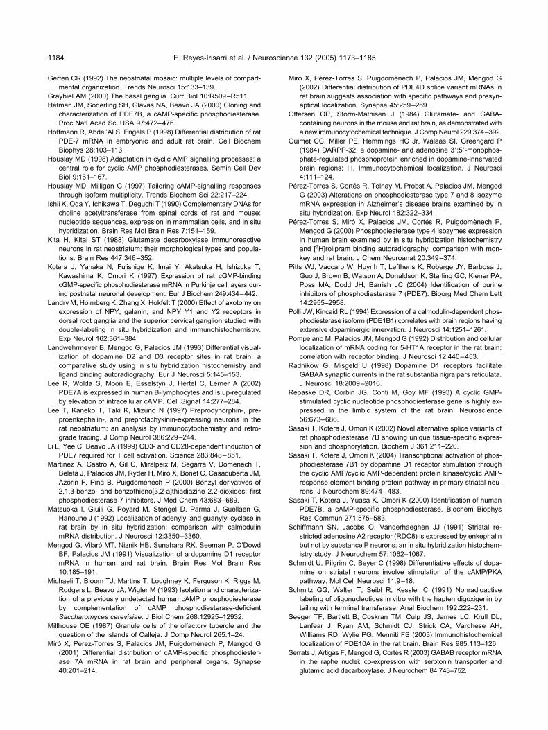
G
GH
H
H
H
I
K
K
L
L
L
L
L
M
M
M
M
M
M
M
O
O
P
P
P
P
P
R
R
S
S
S
S
S
S
S
S
E. Reyes-Irisarri et al. / Neuroscience 132 (2005) 1173–11851184
erfen CR (1992) The neostriatal mosaic: multiple levels of compart-mental organization. Trends Neurosci 15:133–139.
raybiel AM (2000) The basal ganglia. Curr Biol 10:R509–R511.etman JM, Soderling SH, Glavas NA, Beavo JA (2000) Cloning and
characterization of PDE7B, a cAMP-specific phosphodiesterase.Proc Natl Acad Sci USA 97:472–476.
offmann R, Abdel’Al S, Engels P (1998) Differential distribution of ratPDE-7 mRNA in embryonic and adult rat brain. Cell BiochemBiophys 28:103–113.
ouslay MD (1998) Adaptation in cyclic AMP signalling processes: acentral role for cyclic AMP phosphodiesterases. Semin Cell DevBiol 9:161–167.
ouslay MD, Milligan G (1997) Tailoring cAMP-signalling responsesthrough isoform multiplicity. Trends Biochem Sci 22:217–224.
shii K, Oda Y, Ichikawa T, Deguchi T (1990) Complementary DNAs forcholine acetyltransferase from spinal cords of rat and mouse:nucleotide sequences, expression in mammalian cells, and in situhybridization. Brain Res Mol Brain Res 7:151–159.
ita H, Kitai ST (1988) Glutamate decarboxylase immunoreactiveneurons in rat neostriatum: their morphological types and popula-tions. Brain Res 447:346–352.
otera J, Yanaka N, Fujishige K, Imai Y, Akatsuka H, Ishizuka T,Kawashima K, Omori K (1997) Expression of rat cGMP-bindingcGMP-specific phosphodiesterase mRNA in Purkinje cell layers dur-ing postnatal neuronal development. Eur J Biochem 249:434–442.
andry M, Holmberg K, Zhang X, Hokfelt T (2000) Effect of axotomy onexpression of NPY, galanin, and NPY Y1 and Y2 receptors indorsal root ganglia and the superior cervical ganglion studied withdouble-labeling in situ hybridization and immunohistochemistry.Exp Neurol 162:361–384.
andwehrmeyer B, Mengod G, Palacios JM (1993) Differential visual-ization of dopamine D2 and D3 receptor sites in rat brain: acomparative study using in situ hybridization histochemistry andligand binding autoradiography. Eur J Neurosci 5:145–153.
ee R, Wolda S, Moon E, Esselstyn J, Hertel C, Lerner A (2002)PDE7A is expressed in human B-lymphocytes and is up-regulatedby elevation of intracellular cAMP. Cell Signal 14:277–284.
ee T, Kaneko T, Taki K, Mizuno N (1997) Preprodynorphin-, pre-proenkephalin-, and preprotachykinin-expressing neurons in therat neostriatum: an analysis by immunocytochemistry and retro-grade tracing. J Comp Neurol 386:229–244.
i L, Yee C, Beavo JA (1999) CD3- and CD28-dependent induction ofPDE7 required for T cell activation. Science 283:848–851.
artinez A, Castro A, Gil C, Miralpeix M, Segarra V, Domenech T,Beleta J, Palacios JM, Ryder H, Miró X, Bonet C, Casacuberta JM,Azorin F, Pina B, Puigdomenech P (2000) Benzyl derivatives of2,1,3-benzo- and benzothieno[3,2-a]thiadiazine 2,2-dioxides: firstphosphodiesterase 7 inhibitors. J Med Chem 43:683–689.
atsuoka I, Giuili G, Poyard M, Stengel D, Parma J, Guellaen G,Hanoune J (1992) Localization of adenylyl and guanylyl cyclase inrat brain by in situ hybridization: comparison with calmodulinmRNA distribution. J Neurosci 12:3350–3360.
engod G, Vilaró MT, Niznik HB, Sunahara RK, Seeman P, O’DowdBF, Palacios JM (1991) Visualization of a dopamine D1 receptormRNA in human and rat brain. Brain Res Mol Brain Res10:185–191.
ichaeli T, Bloom TJ, Martins T, Loughney K, Ferguson K, Riggs M,Rodgers L, Beavo JA, Wigler M (1993) Isolation and characteriza-tion of a previously undetected human cAMP phosphodiesteraseby complementation of cAMP phosphodiesterase-deficientSaccharomyces cerevisiae. J Biol Chem 268:12925–12932.
illhouse OE (1987) Granule cells of the olfactory tubercle and thequestion of the islands of Calleja. J Comp Neurol 265:1–24.
iró X, Pérez-Torres S, Palacios JM, Puigdomènech P, Mengod G(2001) Differential distribution of cAMP-specific phosphodiester-ase 7A mRNA in rat brain and peripheral organs. Synapse
40:201–214.iró X, Pérez-Torres S, Puigdomènech P, Palacios JM, Mengod G(2002) Differential distribution of PDE4D splice variant mRNAs inrat brain suggests association with specific pathways and presyn-aptical localization. Synapse 45:259–269.
ttersen OP, Storm-Mathisen J (1984) Glutamate- and GABA-containing neurons in the mouse and rat brain, as demonstrated witha new immunocytochemical technique. J Comp Neurol 229:374–392.
uimet CC, Miller PE, Hemmings HC Jr, Walaas SI, Greengard P(1984) DARPP-32, a dopamine- and adenosine 3=:5=-monophos-phate-regulated phosphoprotein enriched in dopamine-innervatedbrain regions: III. Immunocytochemical localization. J Neurosci4:111–124.
érez-Torres S, Cortés R, Tolnay M, Probst A, Palacios JM, MengodG (2003) Alterations on phosphodiesterase type 7 and 8 isozymemRNA expression in Alzheimer’s disease brains examined by insitu hybridization. Exp Neurol 182:322–334.
érez-Torres S, Miró X, Palacios JM, Cortés R, Puigdomènech P,Mengod G (2000) Phosphodiesterase type 4 isozymes expressionin human brain examined by in situ hybridization histochemistryand [3H]rolipram binding autoradiography: comparison with mon-key and rat brain. J Chem Neuroanat 20:349–374.
itts WJ, Vaccaro W, Huynh T, Leftheris K, Roberge JY, Barbosa J,Guo J, Brown B, Watson A, Donaldson K, Starling GC, Kiener PA,Poss MA, Dodd JH, Barrish JC (2004) Identification of purineinhibitors of phosphodiesterase 7 (PDE7). Bioorg Med Chem Lett14:2955–2958.
olli JW, Kincaid RL (1994) Expression of a calmodulin-dependent phos-phodiesterase isoform (PDE1B1) correlates with brain regions havingextensive dopaminergic innervation. J Neurosci 14:1251–1261.
ompeiano M, Palacios JM, Mengod G (1992) Distribution and cellularlocalization of mRNA coding for 5-HT1A receptor in the rat brain:correlation with receptor binding. J Neurosci 12:440–453.
adnikow G, Misgeld U (1998) Dopamine D1 receptors facilitateGABAA synaptic currents in the rat substantia nigra pars reticulata.J Neurosci 18:2009–2016.
epaske DR, Corbin JG, Conti M, Goy MF (1993) A cyclic GMP-stimulated cyclic nucleotide phosphodiesterase gene is highly ex-pressed in the limbic system of the rat brain. Neuroscience56:673–686.
asaki T, Kotera J, Omori K (2002) Novel alternative splice variants ofrat phosphodiesterase 7B showing unique tissue-specific expres-sion and phosphorylation. Biochem J 361:211–220.
asaki T, Kotera J, Omori K (2004) Transcriptional activation of phos-phodiesterase 7B1 by dopamine D1 receptor stimulation throughthe cyclic AMP/cyclic AMP-dependent protein kinase/cyclic AMP-response element binding protein pathway in primary striatal neu-rons. J Neurochem 89:474–483.
asaki T, Kotera J, Yuasa K, Omori K (2000) Identification of humanPDE7B, a cAMP-specific phosphodiesterase. Biochem BiophysRes Commun 271:575–583.
chiffmann SN, Jacobs O, Vanderhaeghen JJ (1991) Striatal re-stricted adenosine A2 receptor (RDC8) is expressed by enkephalinbut not by substance P neurons: an in situ hybridization histochem-istry study. J Neurochem 57:1062–1067.
chmidt U, Pilgrim C, Beyer C (1998) Differentiative effects of dopa-mine on striatal neurons involve stimulation of the cAMP/PKApathway. Mol Cell Neurosci 11:9–18.
chmitz GG, Walter T, Seibl R, Kessler C (1991) Nonradioactivelabeling of oligonucleotides in vitro with the hapten digoxigenin bytailing with terminal transferase. Anal Biochem 192:222–231.
eeger TF, Bartlett B, Coskran TM, Culp JS, James LC, Krull DL,Lanfear J, Ryan AM, Schmidt CJ, Strick CA, Varghese AH,Williams RD, Wylie PG, Menniti FS (2003) Immunohistochemicallocalization of PDE10A in the rat brain. Brain Res 985:113–126.
errats J, Artigas F, Mengod G, Cortés R (2003) GABAB receptor mRNAin the raphe nuclei: co-expression with serotonin transporter and
glutamic acid decarboxylase. J Neurochem 84:743–752.
S
S
S
S
S
T
T
v
V
X
E. Reyes-Irisarri et al. / Neuroscience 132 (2005) 1173–1185 1185
himizu-Albergine M, Rybalkin SD, Rybalkina IG, Feil R, Wolfsgruber W,Hofmann F, Beavo JA (2003) Individual cerebellar Purkinje cellsexpress different cGMP phosphodiesterases (PDEs): in vivo phos-phorylation of cGMP-specific PDE (PDE5) as an indicator ofcGMP-dependent protein kinase (PKG) activation. J Neurosci23:6452–6459.
impson JB (1981) The circumventricular organs and the centralactions of angiotensin. Neuroendocrinology 32:248–256.
oderling SH, Bayuga SJ, Beavo JA (1999) Isolation and character-ization of a dual-substrate phosphodiesterase gene family:PDE10A. Proc Natl Acad Sci USA 96:7071–7076.
okoloff P, Giros B, Martres MP, Bouthenet ML, Schwartz JC (1990)Molecular cloning and characterization of a novel dopamine recep-tor (D3) as a target for neuroleptics. Nature 347:146–151.
ouness JE, Aldous D, Sargent C (2000) Immunosuppressive andanti-inflammatory effects of cyclic AMP phosphodiesterase (PDE)type 4 inhibitors. Immunopharmacology 47:127–162.
akahashi M, Terwilliger R, Lane C, Mezes PS, Conti M, Duman
RS (1999) Chronic antidepressant administration increases theexpression of cAMP-specific phosphodiesterase 4A and 4B iso-forms. J Neurosci 19:610–618.
omiyama M, Palacios JM, Cortés R, Vilaró MT, Mengod G (1997)Distribution of AMPA receptor subunit mRNAs in the humanbasal ganglia: an in situ hybridization study. Mol Brain Res46:281–289.
an Staveren WC, Glick J, Markerink-Van Ittersum M, Shimizu M,Beavo JA, Steinbusch HW, de Vente J (2002) Cloning and local-ization of the cGMP-specific phosphodiesterase type 9 in the ratbrain. J Neurocytol 31:729–741.
ilaró MT, Cortés R, Gerald C, Branchek TA, Palacios JM, Mengod G(1996) Localization of 5-HT4 receptor mRNA in rat brain by in situhybridization histochemistry. Brain Res Mol Brain Res 43:356–360.
ia ZG, Refsdal CD, Merchant KM, Dorsa DM, Storm DR (1991)Distribution of mRNA for the calmodulin-sensitive adenylate cy-clase in rat brain: expression in areas associated with learning and
memory. Neuron 6:431–443.(Accepted 28 January 2005)(Available online 11 April 2005)