Advancing Regenerative Cellular Therapies in Non-Scarring ...
Upload
tu-dresdenCategory
view
2download
0
Developmental Cell
Short Article
Regenerative Neurogenesisfrom Neural Progenitor CellsRequires Injury-Induced Expression of Gata3Caghan Kizil,1,2 Nikos Kyritsis,1,2 Stefanie Dudczig,1,2 Volker Kroehne,1,2 Dorian Freudenreich,1 Jan Kaslin,1,3
and Michael Brand1,2,*1Biotechnology Center, Technische Universitat Dresden, Tatzberg 47/49, 01307 Dresden, Germany2DFG-Center for Regenerative Therapies Dresden, Cluster of Excellence (CRTD), Fetscherstr. 105, 01307 Dresden, Germany3Present address: Australian Regenerative Medicine Institute (ARMI), Monash University, 3800 Victoria, Australia
*Correspondence: [email protected]
http://dx.doi.org/10.1016/j.devcel.2012.10.014
SUMMARY
The adult zebrafish brain, unlikemammalian counter-parts, can regenerate after injury owing to theneurogenic capacity of stem cells with radial glialcharacter. We hypothesized that injury-inducedregenerative programs might be turned on afterinjury in zebrafish brain and enable regenerative neu-rogenesis. Here we identify one such gene—the tran-scription factor gata3—which is expressed only afterinjury in different zebrafish organs. Gata3 is requiredfor reactive proliferation of radial glia cells, subse-quent regenerative neurogenesis, and migration ofthe newborn neurons. We found that these regenera-tion-specific roles of Gata3 are dependent on theinjury because Gata3 overexpression in the unle-sioned adult zebrafish brain is not sufficient to induceneurogenesis. Thus, gata3 acts as a specific injury-induced proregenerative factor that is essential forthe regenerative capacity in vertebrates.
INTRODUCTION
In mammalian brains, the constitutive production of new neurons
is limited to two subregions of the telencephalon and relies on
the presence of stem cells with glial character (Alvarez-Buylla
et al., 2002; Morrens et al., 2012). After injury, glial cells may
proliferate but form neurons only poorly and hence cannot
accomplish sufficient regenerative neurogenesis in most verte-
brates (Arvidsson et al., 2002; Yiu and He, 2006; Rolls et al.,
2009; Kempermann, 2010; Robel et al., 2011). In contrast, adult
zebrafish have a remarkable ability to regenerate the central
nervous system (Becker and Becker, 2008; Antos and Brand,
2010; Poss, 2010; Kizil et al., 2012a). Besides widespread adult
neurogenic activity (Grandel et al., 2006; Adolf et al., 2006; Cha-
pouton et al., 2007; Kaslin et al., 2008; Ganz et al., 2010; Kizil
et al., 2012a), zebrafish brains can also regenerate severe trau-
matic brain injuries, during which glial progenitors are stimulated
to divide and replenish lost neurons (Kroehne et al., 2011; Baum-
gart et al., 2012; Kishimoto et al., 2012). However, the molecular
1230 Developmental Cell 23, 1230–1237, December 11, 2012 ª2012
mechanisms involved in the initiation and maintenance of
the injury-induced regenerative response in the adult zebrafish
brain are unknown. Conceivably, the profound differences in
the regenerative capacity between the brains of zebrafish and
mammals (Dinsmore, 1991; Kempermann, 2010; Poss, 2010)
might be linked to these mechanisms. Thus, a comparison
between the regenerative mechanisms in mammals and zebra-
fish could reveal genes underlying the differential responses,
which could then be harnessed for therapeutic applications
in human neurological disorders and acute injuries. Here, we
sought to identify injury-induced genes in the adult zebrafish
brain using a traumatic brain injury paradigm to the telenceph-
alon that we established previously (Kroehne et al., 2011). We
find that the transcription factor gata3 is specifically associated
with the regenerative state of brain and several other zebrafish
tissues and that gata3 is required for telencephalon and fin
regeneration. Overall, we demonstrate that Gata3 is essential
to trigger a molecular program in radial glia cells after injury,
thus enabling the regenerative neurogenesis from these progen-
itor cells.
RESULTS AND DISCUSSION
Stab-Lesion Injury Induces gata3 Expression in theAdult Zebrafish TelencephalonTo identify regeneration-associated factors in the adult zebrafish
brain, we compared the transciptomes of uninjured and injured
telencephalons, coupled to a random-pick secondary in situ
hybridization (ISH) screen on cross-sections (C.K. and M.B.,
unpublished data; Figure 1A). The zinc-finger transcription factor
gata3 is not expressed in embryonic (based on quantitative real-
time PCR and ISH; data not shown) and adult zebrafish telen-
cephalon (Figure 1B) but was induced in the telencephalon after
injury (Figure 1C). Immunohistochemistry (IHC) confirmed the
injury-dependent induction of Gata3 (Figures 1D and 1E) along
the entire ventricular region (Figures 1E1–1E4), which contains
the radial glia cells (RGCs) as neurogenic progenitors (Kroehne
et al., 2011; Rothenaigner et al., 2011). Gata3 is induced in prolif-
erating and nonproliferating RGCs (marked by her4.1:GFP)
following the injury (Figures 1F and 1G; Figure S1A available
online). gata3 expression is first observed at 12 hr after the lesion
(hpl) in ventricular cells that are mainly nonproliferative (Fig-
ures 1G, S1B, and S1B0). The number of gata3-positive RGCs
Elsevier Inc.
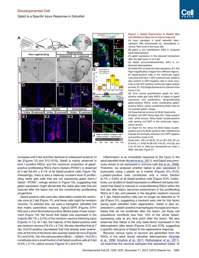
Figure 1. Gata3 Expression in Radial Glia
and Newborn Neurons Is Injury Induced
(A) Injury paradigm in adult zebrafish telen-
cephalon. DM, dorsomedial; DL, dorsolateral; V,
ventral. Red circle is the injury site.
(B) gata3 in situ hybridization (ISH) in uninjured
adult telencephalon.
(C) gata3 expression in the lesioned hemisphere
after the stab lesion (*) at 3 dpl.
(D) Gata3 immunohistochemistry (IHC) in un-
lesioned telencephalon.
(E) Gata3 IHC on lesioned telencephalons. (E1–E4)
High-magnification images from different regions.
(F) Gata3-positive cells in the ventricular region
colocalize with her4.1:GFP (white arrows). Gata3 is
also present in GFP-negative cells in close prox-
imity to the GFP-positive ventricular region (yellow
arrows). (F1–F3) Single fluorescence channel of the
frame in (F).
(G) Time course quantification graph for her4-
positive radial glial cells (RGC) in regard to gata3
expression and proliferation. Nonproliferating
gata3-positive RGCs, white; proliferating gata3-
positive RGCs, yellow; proliferating RGCs that do
not express gata3, orange.
(H) Experimental scheme for BrdU treatments.
(I) Gata3, huC:GFP, BrdU triple IHC. Triple-positive
cells, white arrows. BrdU-Gata3 double-positive
cells lacking huC:GFP in the ventricular region,
yellow arrows.
(J) Graph for the ratios for BrdU-Gata3 double-
positive and only BrdU-positive cells. Dashed lines
indicate the boundary between huC:GFP-negative
and positive zones (I–I3).
Scale bars: 100 mm (B–E); 50 mm (E1–E4); 20 mm
(F and I). n = 8 fish for (B–E4); 4 for (F), 4 for (G), and
5 for (H and J). Data are represented as mean ±
SEM. See also Figure S1.
Developmental Cell
Gata3 Is a Specific Injury Response in Zebrafish
increases until 3 dpl and then declines to unlesioned levels at 14
dpl (Figures 1G and S1C–S1G). Gata3 is mainly observed in
her4.1-positive RGCs, and the maximum proportion of gata3-
positive proliferating RGCs (her4+/Gata3+/PCNA+) is observed
at 3 dpl (54.6% ± 6.1% of all Gata3-positive cells; Figure 1G).
Interestingly, there is also a relatively constant level of prolifer-
ating radial glial cells that are not expressing gata3 (her4+/
Gata3�/PCNA+; orange section in Figure 1G), suggesting that
gata3 expression might demarcate the radial glial cells that are
induced after the lesion but not the constitutively proliferating
progenitors.
Gata3-positive cells were also detectable outside the ventric-
ular zone at 3 dpl (Figure 1F), and these cells might be newborn
neurons. To address this, we used a transgenic zebrafish line
that marks postmitotic neurons, Tg(huC:GFP) (Figures S1H–
SI3) and a short Bromodeoxyuridine (BrdU) pulse-chase experi-
ment (Figure 1H). We found that Gata3 was expressed in the
majority (85.7% ± 6.2%) of the newborn neurons following injury
(Figures 1I–1J). At 7 dpl, the majority of the Gata3-positive cells
are newborn neurons (79.4%± 4.7%).We also identified that at 7
dpl, HuC/D-positive neuroblasts that had already been postmi-
totic at the time of the lesion also express Gata3 de novo (Figures
S1J and S1K). Yet, this cell population (BrdU�, Gata3+, HuC/D+)
constitutes only a small fraction of all Gata3-positive cells at 3 dpl
(4.6% ± 2.1%; yellow arrows; Figures S1J and S1K).
Developmenta
Inflammation is an immediate response to the injury in the
adult zebrafish brain (Kroehne et al., 2011), and Gata3 was previ-
ously shown to be expressed in immune cells (Ho et al., 2009).
Therefore, we analyzed whether Gata3 is also expressed in
leukocytes using L-plastin as a marker (Figures S1L–S1O).
L-plastin-positive cells constituted only a minor fraction
(5.1% ± 0.8%) of all Gata3-positive cells (Figure S1P). Collec-
tively, our studies onGata3 expression in different cell types indi-
cated that Gata3 is induced in nonproliferating RGCs within the
first day after lesion, becomes predominant in the proliferating
RGCs at 3 dpl, and persists in the daughter newborn neurons
at 7 dpl. Gata3-positive cells can no longer be detected at 14
dpl (Figure S1), suggesting a transient early role for this factor
during adult zebrafish brain regeneration. Gata3 is also ex-
pressed in L-plastin-positive macrophages/microglia and neuro-
blasts that do not proliferate after the lesion, yet these cell
populations constitute less than 10% of the whole Gata3-
expressing cells at any time point after the lesion. We also
observed that Gata3 is the only Gata-factor expressed in the
telencephalon after lesion (Figures S1Q and S1R), suggesting
a specific relevance of Gata3 to the regenerative response.
Because various types of neurons are generated from the
RGCs in the adult dorsal zebrafish telencephalon (Grandel
et al., 2006; Kroehne et al., 2011; Rothenaigner et al., 2011),
we examined the neuronal subtypes that expressed Gata3. At
l Cell 23, 1230–1237, December 11, 2012 ª2012 Elsevier Inc. 1231

Figure 2. Gata3 Is Required for Reactive Cell Proliferation, Reactive
Neurogenesis, and Tissue Distribution of Newborn Neurons
(A) Morpholino injection paradigm.
(B) Gata3 and BrdU IHC for control morpholino-injected brains.
(C) High magnification of the framed region in (B).
(D) Gata3 and BrdU IHC for Gata3-antisense morpholino-injected brains.
(E) High magnification of the framed region in (D).
(F) Quantification graph for BrdU-positive cells in the ventricular region of
unlesioned and 3 dpl telencephalic hemispheres.
Developmental Cell
Gata3 Is a Specific Injury Response in Zebrafish
1232 Developmental Cell 23, 1230–1237, December 11, 2012 ª2012
7 days postlesion (dpl), newborn Gata3-positive cells expressed
various neuronal or glial markers (Figures S1S–S1X), suggesting
that gata3 partakes in regeneration of most or all neuronal and
nonneuronal subtypes in the telencephalon.
Gata3 Is Required for Reactive Proliferation of theProgenitors, Reactive Neurogenesis, and Migration ofNewborn NeuronsExpression of Gata3 in the adult zebrafish telencephalon after
injury was observed also in proliferating progenitors (Figures
1F, 1I, and S2A–S2D00). We thus hypothesized that Gata3 might
be required for injury-induced proliferation of radial glial cells
(RGCs). To test this, we knocked down Gata3 after the injury in
periventricular cells by cerebroventricular microinjection (CVMI)
of functional gata3 morpholinos (Figures S2E–S2M), a method
that we recently developed to allow knockdown of gene function
in ventricular zone cells (Kizil and Brand, 2011). After CVMI, we
treated the fish with BrdU at 3 dpl and analyzed the BrdU and
Gata3 immunoreactivity in the unlesioned control and 3 dpl
experimental brains (Figure 2A). We found that at 3 dpl, Gata3
and BrdU-positive cells are present throughout the ventricular
regions in the control morpholino-injected (ctrl-MO) telencepha-
lons (Figures 2B and 2C), whereas gata3-morphants (AS-MO)
lack Gata3 protein along the ventricle (Figures 2D and 2E).
Interestingly, knocking down gata3 reduces the number of
injury-stimulated proliferation at 3 dpl significantly, to the level
characteristic for unlesioned telencephalon (Figure 2F). PCNA
immunostainings confirmed these results further, showing that
knockdown of Gata3 production prevented injury-induced prolif-
eration after injury in the adult zebrafish telencephalon (Figures
S2N–S2O).
Injury-induced (reactive) proliferation in lesioned adult zebra-
fish telencephalon leads to production of neurons from RGCs
(Kroehne et al., 2011). Because knocking down Gata3 strongly
reduced reactive cell proliferation of the RGCs, we examined
(G) BrdU pulse-chase experiment.
(H) BrdU IHC on control morpholino-injected brains.
(I) BrdU IHC on Gata3 antisense morpholino-injected brains.
(J) Quantification graph. Scale bars, 50 mm. n = 7 fish for (B–F) and 4 for (G–J).
Data are represented as mean ± SEM.
(K) Morpholino-injection and BrdU pulse-chase schemes for reactive neuro-
genesis assay. (K1–K3) HuC/D-BrdU double IHC on sham/uninjected, control
morpholino-injected, and gata3 morpholino-injected telencephalons.
(L) Quantification graph.
(M) Morpholino-injection and BrdU pulse-chase schemes for reactive neuro-
genesis assay for PVA neuronal subtype. (M1–M3) PVA-BrdU double IHC on
sham/uninjected, control morpholino-injected, and gata3morpholino-injected
telencephalons.
(N) Quantification graph.
(O) Morpholino-injection and BrdU pulse-chase schemes to assay for migra-
tion of newborn neurons. (O1 and O2) HuC/D-BrdU double IHC on control
morpholino-injected and gata3 morpholino-injected telencephalons. (O3 and
O4) High-magnification images of BrdU staining in framed regions of (E1 and
E2). Dashed line is the ventricular surface.
(P) Histogram and bar graph depicting the relative distance distribution of
HuC/D-BrdU positive newborn neurons from the ventricle and the average
distance migrated by a newborn neuron in control- and antisense morpholino-
injected telencephalons. Scale bars: 20 mm (K1–K3, insets of M1–M3, O3, and
O4) and 50 mm (M1–M3, O1, and O2). n = 8 fish for (K–L), 6 for (M–N), and 4 for
(O–P). Data are represented as mean ± SEM.
See also Figures S2 and S3, and Tables S1 and S2.
Elsevier Inc.

Developmental Cell
Gata3 Is a Specific Injury Response in Zebrafish
whether this also reduces the injury-stimulated production of
new cells. We observed that Gata3 knockdown significantly
reduces the number of BrdU-labeled cells at 15 dpl (72.2% ±
6.4%) (Figures 2G–2J). To test whether production of newborn
neurons (reactive neurogenesis) is also reduced, we performed
double immunostaining for the early neuronal marker HuC/D
and BrdU at 15 dpl after BrdU administration (Figures 2K–2K3).
We observed that upon Gata3 knockdown, the number of
newborn neurons is significantly reduced (80.1% ± 9.5%) to
the level characteristic for uninjured brains (Figure 2L).
The pronounced reactive cell proliferation of the RGCs after
a lesion was shown to peak at 3 days postlesion (dpl) and
to persist in a gradually decreasing manner until 14 dpl, when
the ventricular cell proliferation returns to constitutive levels
(Kroehne et al., 2011). Even if the initial burst of reactive prolifer-
ation of the RGCs is blocked by Gata3 knockdown, the CVMI
knockdown is likely to be transient, estimated to be most effec-
tive until about 5 days postinjection (Kizil and Brand, 2011). Thus,
the remaining constitutive neurogenesis could conceivably
compensate for the lack of regenerative neurogenesis.We there-
fore asked if neurons can be regenerated in the long-term by any
other secondary mechanism in the brains, where injury-induced
reactive proliferation was reduced by knocking down gata3. We
analyzed the number of newborn Parvalbumin (PVA+/BrdU+)
neurons of the dorsal telencephalon at 6 weeks postlesion
(Figures 2M–2M3). We found that compared to sham controls,
injured telencephalon shows enhanced production of PVA neu-
rons but that knocking down gata3 still reduces production of
newly born PVA neurons significantly (by 67.1% ± 10.1%) (Fig-
ure 2N). These results indicate that the initial burst of injury-
induced proliferation of the RGCs is indeed critical for successful
regenerative neurogenesis of the adult zebrafish telencephalon
and cannot be efficiently compensated by ongoing or later con-
stitutive neurogenesis within the tested time period.
Following injury, Gata3 is also expressed in maturing neurons
within the periventricular zone of the telencephalon (yellow
arrows marking the GFP-negative cells in close proximity to
the GFP-positive RGCs at the ventricular region in Figure 1F).
This suggests that Gata3 might have other roles in addition to
controlling proliferation after brain injury. Therefore, we investi-
gated possible functions of gata3 in survival and/or migration
during neuronal differentiation. TUNEL stainings at 1 and 3 dpl
for control and gata3 morpholino-injected brains showed no
significant difference in the number of apoptotic cells at these
time points (Figures S2P–S2S). These results indicate that
gata3 is not required for cell survival. In order to analyze the
distribution of newborn neurons relative to their place of birth
in the ventricular zone, we treated the lesioned brains with
BrdU at 3 dpl, injected the control and Gata3 morpholinos at 5
dpl, and counted the BrdU-positive neurons at 15 dpl (Figures
2O–2O4). This experiment would label the reactively proliferating
RGCs at 3 dpl, and until the morpholino injection at 5 dpl, some
of the proliferating RGCs would become postmitotic and differ-
entiate into neurons that would start migrating toward the lesion
site, as described before (Kroehne et al., 2011). The Gata3
knockdown at 5 dpl would then block further the production of
neurons and allow visualization of how far the postmitotic neu-
rons have migrated. As a result, we found that compared to
the control morphant brains, knocking down Gata3 led to sig-
Developmenta
nificantly altered tissue distribution and average migration
distance of the newborn neurons (Figure 2P). We also observed
that the total number of BrdU-positive neurons did not change
upon Gata3 knockdown in comparison to the control morpholino
injection (data not shown), confirming our previous results that
gata3 is not required for survival. In summary, our results suggest
that gata3 is required for neuronal regeneration of the lesioned
adult zebrafish telencephalon, specifically for injury-induced
proliferation of radial glial cells (RGCs), reactive neurogenesis,
and proper distribution of newborn neurons.
By using amodified version of the CVMImethod (Figures S3A–
S3G) and transfecting the ventricular cells of the telencephalon
with plasmids expressing Gata3 and GFP (Figures S3H–S3O),
we overexpressed Gata3 and found that Gata3 alone is not
sufficient to increase the ventricular cell proliferation and regen-
erative neurogenesis in unlesioned brains. This suggests that
Gata3 requires the injury context and acts synergistically with
those cues. Additionally, we found that increased Gata3 ex-
pression after a lesion does not further increase the prolifera-
tion levels at the ventricle (Figures S3P–S3R), suggesting that
additional cells cannot be recruited and/or that the proliferation
response is at its maximum level and cannot be enhanced
further.
gata3 Expression Is Induced in Regenerating ZebrafishOrgans after InjuryTo investigate whether gata3 might be involved in regeneration
of other adult organs outside the central nervous system, we
performed ISH on control and amputated fish heart and caudal
fin (Figure 3). In contrast to sham-operated hearts (Figures 3A–
3A0), gata3 is expressed in hearts at 3 days postamputation
(dpa) (Figure 3B–3B0). Sense probe control indicates the speci-
ficity of the in situ staining (Figures 3B01–B02). Similarly, whereas
gata3 expression levels are undetectable in the control fins (Fig-
ure 3C), fin amputation induces gata3 expression based on ISH
(Figures 3D–3G) and quantitative real-time PCR analyses (Fig-
ure 3H). The induction of gata3 expression can first be seen at
1 dpa during the wound healing phase (Figure 3D). As regenera-
tion proceeds in the caudal fin, gata3 expression is observed in
the regenerating portion of the fin and overlapping to the blas-
tema and the interray epithelium (Figures 3E and 3F). At 7 dpa
when the proximal structures of the fin regenerates, gata3
expression levels reduce significantly (Figure 3G), suggesting
a transient early role of gata3 in the caudal fin regeneration.
When we used a morpholino-knockdown-based regenerative
outgrowth assay (Figure 3I; Kizil et al., 2009), we found that
gata3 knockdown significantly reduced regenerative outgrowth
of the fin by 58.3% ± 8.9% (Figures 3I0 and 3J), indicating that
gata3 expression is necessary for regeneration of the caudal
fin. We observed that gata3 knockdown reduces cell prolifera-
tion in the blastema and the interray tissue significantly by
64.2% ± 14.3% (Figures 3K–3M), comparable to the reduction
in the extent of regenerative outgrowth (Figure 3J). In control
antibody stainings for Gata3, we observed that antisense mor-
pholinos efficiently reduce the specific Gata3 signal in the epi-
thelial and interray tissue and reduce the regenerative outgrowth
of the fin (Figures 3N–3O0). We also found that gata3 expression
was induced after injury in other adult zebrafish tissues, such as
cerebellum, optic tectum, spinal cord, and liver (data not shown).
l Cell 23, 1230–1237, December 11, 2012 ª2012 Elsevier Inc. 1233

Figure 3. Gata3 Expression Is Injury Induced in Regenerating Heart and Caudal Fin and Is Required for Caudal Fin Regeneration
(A) gata3 ISH on sham-operated zebrafish heart. (A0) High-magnification image.
(B) gata3 ISH on amputated heart. Amputation plane, black arrows. (B0) High-magnification image. Dashed lines, amputation plane. (B01) gata3 ISHwith the sense
probe control on amputated heart. (B02) High-magnification image from the amputation margin of the heart in (B01).(C–G) Time course gata3 ISH on adult zebrafish caudal fin: unamputated (C), 1 dpa (D), 2 dpa (E), 3 dpa (F), and 7 dpa (G).
(H) Quantitative real-time PCR analyses for gata3 expression on crude lysates of the unamputated, 1 dpa, and 3 dpa caudal fins. Bars represent ± SEM.
(I) Experimental scheme for caudal fin outgrowth assay. (I0 ) A 4 dpa caudal fin. One lobe is injected with control morpholino (Ctrl-MO) and the other with gata3
antisensemorpholino (AS-MO) at 2 dpa. A1, Area regenerated between 0–2 dpa in the control lobe before injection. A2, Area regenerated between 0–2 dpa in the
AS-MO lobe before injection. A3, Area regenerated between 2–4 dpa after Ctrl-MO injection. A4, Area regenerated between 2–4 dpa after AS-MO injection.
(J) Quantification graph for the relative regenerative outgrowth between 2 and 4 dpa compared to uninjected regenerates. Auninj, Area regenerated between 2–4
dpa without injection.
(K) BrdU IHC on control MO-injected lobe. Parenthesis indicate the blastema. Inset is a high magnification for the blastema of one fin ray. Bars represent ± SEM.
(L) BrdU IHC on gata3 MO-injected lobe. Parenthesis indicate the blastema. Inset is a high magnification for the blastema of one fin ray.
(M) Quantification graph for the relative number of BrdU-positive cells in ctrl-MO and AS-MO-injected fins. Scale bars 100 mm; data are represented as
mean ± SEM.
(N) BrdU-Gata3 double immunostaining shown on DIC image of the fin in (K). (N0) Gata3 staining alone is shown.
(O) BrdU-Gata3 double immunostaining shown on DIC image of the fin in (L). (O0) Gata3 staining alone is shown. n = 3 fish for (A–B0), 4 for every set for (C–G), 3 for
(H), 6 for (I–J), 4 for (K–M), and 4 for (N–O0).
Developmental Cell
Gata3 Is a Specific Injury Response in Zebrafish
Overall, these results indicate that the injury-dependent activa-
tion of and requirement for gata3 is not limited to the central
nervous system but also prevails in other zebrafish organs, sug-
gesting a general involvement of gata3 in regeneration programs
of zebrafish tissues.
Fgf Signaling Is Required for gata3 Expression after theInjuryThe Fgf signaling pathway was previously shown to be an inte-
gral regulator of the regeneration of heart and caudal fin tissue
(Lepilina et al., 2006; Wills et al., 2008). In adult zebrafish brain,
progenitor cells of the adult cerebellum and telencephalon also
require Fgf signaling for proliferation and maintenance (Kaslin
et al., 2009; Ganz et al., 2010). Therefore, we examined whether
Fgf signaling is required to induce gata3 expression after the
injury, by using heterozygous heat-shock-inducible dominant
negative Fgfr1-EGFP transgenic fish (Lee et al., 2005) (Fig-
ure 4A). Compared to control (nontransgenic, lesioned, and
heat-shocked) brains (Figure 4B), transgenic animals (lesioned
and heat shocked) showed a dramatic reduction in the number
of Gata3-positive cells in the ventricular region after injury (Fig-
ure 4C). In the medial region the reduction was pronounced,
and no Gata3-positive cells remained in the ventricular region
after blocking Fgf signaling (Figures 4D–4E). ISH for gata3 indi-
1234 Developmental Cell 23, 1230–1237, December 11, 2012 ª2012
cated that the reduction in the Gata3 protein levels upon Fgf-
blockage is due to abolished gata3 gene expression (Figures
4F–4G). Using the same transgenic line, we also observed that
gata3 expression in the regenerating caudal fin is dependent
on Fgf signaling (Figures 4H–4H00). Blocking Fgf signaling during
the whole regeneration process until 3 dpa blocked gata3 ex-
pression (Figures 4H–4H00). Similarly, a short pulse of heat shock
leads to abolishment of gata3 expression as detected by Gata3
immunohistochemistry (Figures 4I–4I00). These results suggest
that Fgf signaling is required for injury-induced gata3 expres-
sion in regenerating zebrafish tissues and that Fgf signaling
regulates Gata3 expression. In order to address whether or not
the epistatic interaction between the Fgf signaling and gata3
expression was an injury-specific phenomenon, we analyzed
the expression of gata3 in zebrafish embryos upon blocking
Fgf signaling (Figures 4J–4M0). We found that upon blocking
Fgf signaling, the expression of Fgf-responsive genes (such as
spry4) was lost in zebrafish embryos (Figures 4J–4K0), whereas
gata3 expression was not affected (Figures 4L–4M0). These
results indicate that Fgf signaling regulates gata3 expression in
an injury-dependent context.
We found that continuous blockage of Fgf signaling from
shortly before the injury until 3 dpl abolished gata3 expression
in the adult zebrafish telencephalon (Figure 4); however, this
Elsevier Inc.

Figure 4. Fgf Signaling Is Required for Injury-Induced Gata3
Expression
(A) Heat-shock scheme.
(B) Gata3 IHC on 3 dpl heat-shocked sham control fish telencephalon.
(C) Gata3 IHC on 3 dpl heat-shocked Tg(hsp:dnfgfr1-gfp) fish telencephalon.
Inset: double IHC for GFP and Gata3, showing uniform dnFgfr1-GFP.
(D) High magnification from dorsomedial region of (B) with DAPI.
(E) High magnification from dorsomedial region of (C) with DAPI.
(F) gata3 ISH in heat-shocked control telencephalon at 3 dpl.
(G) gata3 ISH in heat-shocked Tg(hsp:dnfgfr1-gfp) telencephalon at 3 dpl.
Scale bars 20 mm. n = 4 fish for (B), 6 for (C), 6 for (D and E), and 5 for (F and G).
Data are represented as mean ± SEM.
(H) Heat-shock and amputation scheme for Tg(hsp70l:dnfgfr1) fish. (H0 ) gata3ISH on 2 dpa nontransgenic siblings. (H00) gata3 ISH on 2 dpa Tg(hsp70:
dnfgfr1) fish.
(I) Transient and brief heat-shock and amputation scheme for Tg(hsp70l:
dnfgfr1) fish. (I0) Gata3 and GFP immunostaining on nontransgenic siblings
shown for single fin ray. Dashed lines, amputation margin. (I01–I03) Individualfluorescence channel for Gata3, DAPI, and merged image. (I00) Gata3 and GFP
immunostaining on Tg(hsp:dnfgfr1-egfp) animals shown for single fin ray.
Dashed lines, amputation margin.
(J) spry4 expression at 24 hr postfertilization (hpf) embryo.
(K) Blocking Fgf signaling abrogates spry4 expression.
(L) gata3 expression at 24 hpf.
(M) Blocking Fgf signaling does not alter the expression of gata3. (J0–M0) Dorsalview of animals in (J, K, L, and M), respectively. n = 5 for (H–H00), 3 for (I–I00), andmore than 50 embryos for (J–M0). Scale bars 200 mm.
See also Figure S4.
Developmental Cell
Gata3 Is a Specific Injury Response in Zebrafish
Developmenta
effect might be indirect because of the extended blockage time,
and Fgf signaling might act via intermediate signaling pathways
to regulate gata3 expression. To address this issue in the brain,
we blocked Fgf signaling transiently by a single heat shock at
2 dpl and analyzed the expression of gata3 at 3 dpl (Figures
S4A–S4A00). We observed that a single heat shock to express
dominant negative fgfr1 reduced but did not abrogate the
injury-induced expression of gata3 (Figures S4A–S4A00). Com-
bined with the data on fin regeneration (Figures 4H–4I00), theseresults suggest that Fgf signaling has a direct effect on expres-
sion of gata3 after injury in zebrafish tissues and that this interac-
tion represents an injury-dependent mechanism.
We found that blocking gata3 activity in the ventricular region
does not affect wound closure and resolution of blood clotting at
the lesion site in the adult zebrafish brain (Figure S4B). Addition-
ally, generalized stress conditions or incision wounding does
not induce gata3 expression (Figure S4C). These results suggest
a specific requirement of gata3 in regeneration programs of
zebrafish and that gata3 expression upon injury correlates with
the regenerative ability, which might be controlled by genetic
programs that are fundamental to the initiation and maintenance
of the regenerative response. A better understanding of these
programs might allow us to devise ways to unlock the regenera-
tive potential in humans. Overall, we propose that zebrafish acti-
vates molecular programs that may link the nonphysiological
conditions of an injury, which are often detrimental for regenera-
tion in mammals, to the initiation of redevelopment of the lost
structures, and that these programs are distinct from the ones
involved in constitutive stem-cell maintenance and physiology.
Specifically, gata3 may represent an integral component of a
molecular program that is used universally for regeneration in
zebrafish. Thus, studies on activating injury-associated signaling
pathways involving Gata3 in mammalian tissues might be a
promising approach in regenerative medicine.
EXPERIMENTAL PROCEDURES
Fish Maintenance and Husbandry
All animal experiments and procedures were carried out in accordance with
the recommendations and official guidelines for animal handling and research
of the Regierungsprasidium Dresden (permit numbers: AZ 24D-9168.11-1/
2008-2, 4, and 14). All surgery was performed under anesthesia, and all efforts
were made to minimize suffering. Fish were maintained as described (Brand
et al., 2002).
Transgenic Animals
Tg(huC:GFP) (Park et al., 2002), Tg(olig2:GFP) (Shin et al., 2003), Tg(her4.1:
GFP) (Yeo et al., 2007), and Tg(her4.1:mCherry) (Kroehne et al., 2011) were
used as reporters. For blocking Fgf signaling, Tg(hsp70l:dnfgfr1-GFP) (Lee
et al., 2005) was used. Animals were heat shocked after lesion until 3 days
postlesion (dpl) on a daily basis for 1.5 hr in a 37�C water bath.
Stab Lesions, Cell Sorting, and Quantitative Real-Time PCR
Injuries to telencephalon were performed as described (Kroehne et al., 2011)
on 6-month-old zebrafish. Total RNA was isolated from (Tg(her4:GFP))-sorted
cells using Trizol (Invitrogen, Carlsbad, CA, USA). Quantitative real-time PCR
was performed on zebrafish tissues as described (Kizil et al., 2009).
Tissue Preparation, In Situ Hybridization, and
Immunohistochemistry
Tissues were prepared as described (Kizil et al., 2009, 2012b). Fish brains and
hearts were cryosectioned at 12 mm thickness. gata3 (NCBI accession number
l Cell 23, 1230–1237, December 11, 2012 ª2012 Elsevier Inc. 1235

Developmental Cell
Gata3 Is a Specific Injury Response in Zebrafish
NM_131211) was isolated from 2 dpf zebrafish total cDNA (Supplemental
Experimental Procedures). mRNA probes were generated using in vitro tran-
scription kit (Roche, Indianapolis, IN, USA). In situ hybridization on sections
was performed using VSi In Situ Robot (Intavis, Koeln, Germany). Immunohis-
tochemistry was performed as described (Grandel et al., 2006; Kizil et al.,
2009; Ganz et al., 2010) (Supplemental Experimental Procedures). Incisions
and hematoxylin and eosin staining was performed as described (Kizil et al.,
2009).
Injections of Vivo Morpholinos
Cerebroventricular microinjection was performed as described (Kizil and
Brand, 2011). Splice- (this paper) and translation-blocking (Yang et al., 2010)
and control vivomorpholino oligonucleotides were used (Supplemental Exper-
imental Procedures).
Transient Overexpression by Plasmid Transduction in the Adult
Zebrafish Brain
We suspended 0.25 fmol of desired plasmid in 0.4% Lipofectamine (Invitro-
gen) and Dulbecco’s modified Eagle’s medium. Fish were injected as
described (Kizil and Brand, 2011). Analysis is performed at 2 days after
injection.
Image Acquisition and Processing
Images were taken using a structured illumination microscope (Apotome,
Zeiss, Oberkochen, Germany) and a confocal microscope (LSM780, Zeiss).
Single-stack images were acquired. ImageJA v.1.45b, Adobe Photoshop
7.0, and Corel Draw X5 were used to generate figures.
Quantifications and Statistical Analyses
Tissue sections (12 mm) between the olfactory bulb and the caudal telenceph-
alon were used for statistical analyses (8–12 sections). Quantifications from
one brain (multiple sections) were analyzed for normal distribution using the
NORMDIST function of Excel software (MS Office). All data sets conformed
to normal distribution (NORMDIST value < 0.005). Paired t test was used for
p value and significance (equal number of data points) to compare two data
sets. Quantifications for same time points from different brains or different
time points for same type of treatment were analyzed using two-way
ANOVA with posttest (alpha: 0.05; for comparisons of more than two data
sets; Analysis ToolPak, MS Excel, and GraphPad Prism) and heteroscedastic
t test (for comparisons of two data sets with different number of data points).
Significance is denoted with asterisks: *p < 0.05, **p < 0.01, ***p < 0.005. Bars
in graphs represent SEM.
SUPPLEMENTAL INFORMATION
Supplemental information includes four figures and Supplemental Experi-
mental Procedures and can be found with this article online at http://dx.doi.
org/10.1016/j.devcel.2012.10.014.
ACKNOWLEDGMENTS
We would like to thank K. Echeverri, G. Kempermann, G. Ozhan-Kizil, C.L.
Antos, and S. Hans for comments. We gratefully acknowledge support for
this work by the Deutsche Forschungsgemeinschaft (SFB-655-A3 and
CRTD), the European Union (ZF-Health), and the TU Dresden. We would like
to dedicate this paper to Christiane Nusslein-Volhard on the occasion of her
70th birthday.
Received: February 4, 2012
Revised: October 7, 2012
Accepted: October 16, 2012
Published online: November 15, 2012
REFERENCES
Adolf, B., Chapouton, P., Lam, C.S., Topp, S., Tannhauser, B., Strahle, U.,
Gotz, M., and Bally-Cuif, L. (2006). Conserved and acquired features of adult
neurogenesis in the zebrafish telencephalon. Dev. Biol. 295, 278–293.
1236 Developmental Cell 23, 1230–1237, December 11, 2012 ª2012
Alvarez-Buylla, A., Seri, B., andDoetsch, F. (2002). Identification of neural stem
cells in the adult vertebrate brain. Brain Res. Bull. 57, 751–758.
Antos, C.L., and Brand, M. (2010). Regeneration of organs and appendages in
zebrafish: a window into underlying control mechanisms. In Encyclopedia of
Life Sciences (Chichester, England: John Wiley & Sons, Ltd.).
Arvidsson, A., Collin, T., Kirik, D., Kokaia, Z., and Lindvall, O. (2002). Neuronal
replacement from endogenous precursors in the adult brain after stroke. Nat.
Med. 8, 963–970.
Baumgart, E.V., Barbosa, J.S., Bally-Cuif, L., Gotz, M., and Ninkovic, J. (2012).
Stab wound injury of the zebrafish telencephalon: a model for comparative
analysis of reactive gliosis. Glia 60, 343–357.
Becker, C.G., and Becker, T. (2008). Adult zebrafish as a model for successful
central nervous system regeneration. Restor. Neurol. Neurosci. 26, 71–80.
Brand, M., Granato, M., and Nusslein-Volhard, C. (2002). Keeping and raising
zebrafish. In Zebrafish: A Practical Approach, C. Nusslein-Volhard and R.
Dahm, eds. (Oxford: Oxford University Press).
Chapouton, P., Jagasia, R., and Bally-Cuif, L. (2007). Adult neurogenesis in
non-mammalian vertebrates. Bioessays 29, 745–757.
Dinsmore, C. (1991). A History of Regeneration Research: Milestones in the
Evolution of a Science (New York: Cambridge University Press).
Ganz, J., Kaslin, J., Hochmann, S., Freudenreich, D., and Brand, M. (2010).
Heterogeneity and Fgf dependence of adult neural progenitors in the zebrafish
telencephalon. Glia 58, 1345–1363.
Grandel, H., Kaslin, J., Ganz, J., Wenzel, I., and Brand, M. (2006). Neural stem
cells and neurogenesis in the adult zebrafish brain: origin, proliferation
dynamics, migration and cell fate. Dev. Biol. 295, 263–277.
Ho, I.C., Tai, T.S., and Pai, S.Y. (2009). GATA3 and the T-cell lineage: essential
functions before and after T-helper-2-cell differentiation. Nat. Rev. Immunol. 9,
125–135.
Kaslin, J., Ganz, J., and Brand, M. (2008). Proliferation, neurogenesis and
regeneration in the non-mammalian vertebrate brain. Philos. Trans. R. Soc.
Lond. B Biol. Sci. 363, 101–122.
Kaslin, J., Ganz, J., Geffarth, M., Grandel, H., Hans, S., and Brand, M. (2009).
Stem cells in the adult zebrafish cerebellum: initiation and maintenance of
a novel stem cell niche. J. Neurosci. 29, 6142–6153.
Kempermann, G. (2010). Adult Neurogenesis 2: Stem Cells and Neuronal
Development in the Adult Brain, Second Edition (Oxford: Oxford University
Press).
Kishimoto, N., Shimizu, K., and Sawamoto, K. (2012). Neuronal regeneration in
a zebrafish model of adult brain injury. Dis. Model. Mech. 5, 200–209.
Kizil, C., and Brand, M. (2011). Cerebroventricular microinjection (CVMI) into
adult zebrafish brain is an efficient misexpressionmethod for forebrain ventric-
ular cells. PLoS ONE 6, e27395.
Kizil, C., Otto, G.W., Geisler, R., Nusslein-Volhard, C., and Antos, C.L. (2009).
Simplet controls cell proliferation and gene transcription during zebrafish
caudal fin regeneration. Dev. Biol. 325, 329–340.
Kizil, C., Kaslin, J., Kroehne, V., and Brand,M. (2012a). Adult neurogenesis and
brain regeneration in zebrafish. Dev. Neurobiol. 72, 429–461.
Kizil, C., Dudczig, S., Kyritsis, N., Machate, A., Blaesche, J., Kroehne, V., and
Brand, M. (2012b). The chemokine receptor cxcr5 regulates the regenerative
neurogenesis response in the adult zebrafish brain. Neural Dev. 7, 27.
Kroehne, V., Freudenreich, D., Hans, S., Kaslin, J., and Brand, M. (2011).
Regeneration of the adult zebrafish brain from neurogenic radial glia-type
progenitors. Development 138, 4831–4841.
Lee, Y., Grill, S., Sanchez, A., Murphy-Ryan, M., and Poss, K.D. (2005). Fgf
signaling instructs position-dependent growth rate during zebrafish fin regen-
eration. Development 132, 5173–5183.
Lepilina, A., Coon, A.N., Kikuchi, K., Holdway, J.E., Roberts, R.W., Burns,
C.G., and Poss, K.D. (2006). A dynamic epicardial injury response supports
progenitor cell activity during zebrafish heart regeneration. Cell 127, 607–619.
Morrens, J., Van Den Broeck, W., and Kempermann, G. (2012). Glial cells in
adult neurogenesis. Glia 60, 159–174.
Elsevier Inc.

Developmental Cell
Gata3 Is a Specific Injury Response in Zebrafish
Park, H.C., Mehta, A., Richardson, J.S., and Appel, B. (2002). olig2 is required
for zebrafish primary motor neuron and oligodendrocyte development. Dev.
Biol. 248, 356–368.
Poss, K.D. (2010). Advances in understanding tissue regenerative capacity
and mechanisms in animals. Nat. Rev. Genet. 11, 710–722.
Robel, S., Berninger, B., and Gotz, M. (2011). The stem cell potential of glia:
lessons from reactive gliosis. Nat. Rev. Neurosci. 12, 88–104.
Rolls, A., Shechter, R., and Schwartz, M. (2009). The bright side of the glial scar
in CNS repair. Nat. Rev. Neurosci. 10, 235–241.
Rothenaigner, I., Krecsmarik, M., Hayes, J.A., Bahn, B., Lepier, A., Fortin, G.,
Gotz, M., Jagasia, R., and Bally-Cuif, L. (2011). Clonal analysis by distinct viral
vectors identifies bona fide neural stem cells in the adult zebrafish telenceph-
alon and characterizes their division properties and fate. Development 138,
1459–1469.
Developmenta
Shin, J., Park, H.C., Topczewska, J.M., Mawdsley, D.J., and Appel, B. (2003).
Neural cell fate analysis in zebrafish using olig2 BAC transgenics. Methods Cell
Sci. 25, 7–14.
Wills, A.A., Kidd, A.R., 3rd, Lepilina, A., and Poss, K.D. (2008). Fgfs control
homeostatic regeneration in adult zebrafish fins. Development 135, 3063–
3070.
Yang, L., Rastegar, S., and Strahle, U. (2010). Regulatory interactions speci-
fying Kolmer-Agduhr interneurons. Development 137, 2713–2722.
Yeo, S.Y., Kim, M., Kim, H.S., Huh, T.L., and Chitnis, A.B. (2007). Fluorescent
protein expression driven by her4 regulatory elements reveals the spatiotem-
poral pattern of Notch signaling in the nervous system of zebrafish embryos.
Dev. Biol. 301, 555–567.
Yiu, G., and He, Z. (2006). Glial inhibition of CNS axon regeneration. Nat. Rev.
Neurosci. 7, 617–627.
l Cell 23, 1230–1237, December 11, 2012 ª2012 Elsevier Inc. 1237
Copyright © 2022 FDOKUMEN