When neurogenesis encounters aging and disease
11
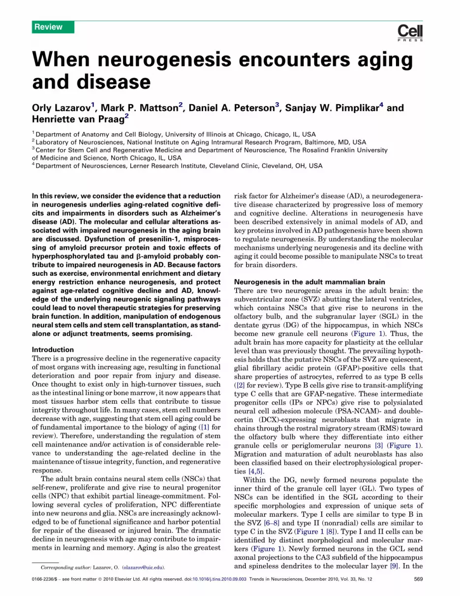
When neurogenesis encounters aging and disease Orly Lazarov 1 , Mark P. Mattson 2 , Daniel A. Peterson 3 , Sanjay W. Pimplikar 4 and Henriette van Praag 2 1 Department of Anatomy and Cell Biology, University of Illinois at Chicago, Chicago, IL, USA 2 Laboratory of Neurosciences, National Institute on Aging Intramural Research Program, Baltimore, MD, USA 3 Center for Stem Cell and Regenerative Medicine and Department of Neuroscience, The Rosalind Franklin University of Medicine and Science, North Chicago, IL, USA 4 Department of Neurosciences, Lerner Research Institute, Cleveland Clinic, Cleveland, OH, USA In this review, we consider the evidence that a reduction in neurogenesis underlies aging-related cognitive defi- cits and impairments in disorders such as Alzheimer’s disease (AD). The molecular and cellular alterations as- sociated with impaired neurogenesis in the aging brain are discussed. Dysfunction of presenilin-1, misproces- sing of amyloid precursor protein and toxic effects of hyperphosphorylated tau and b-amyloid probably con- tribute to impaired neurogenesis in AD. Because factors such as exercise, environmental enrichment and dietary energy restriction enhance neurogenesis, and protect against age-related cognitive decline and AD, knowl- edge of the underlying neurogenic signaling pathways could lead to novel therapeutic strategies for preserving brain function. In addition, manipulation of endogenous neural stem cells and stem cell transplantation, as stand- alone or adjunct treatments, seems promising. Introduction There is a progressive decline in the regenerative capacity of most organs with increasing age, resulting in functional deterioration and poor repair from injury and disease. Once thought to exist only in high-turnover tissues, such as the intestinal lining or bone marrow, it now appears that most tissues harbor stem cells that contribute to tissue integrity throughout life. In many cases, stem cell numbers decrease with age, suggesting that stem cell aging could be of fundamental importance to the biology of aging ([1] for review). Therefore, understanding the regulation of stem cell maintenance and/or activation is of considerable rele- vance to understanding the age-related decline in the maintenance of tissue integrity, function, and regenerative response. The adult brain contains neural stem cells (NSCs) that self-renew, proliferate and give rise to neural progenitor cells (NPC) that exhibit partial lineage-commitment. Fol- lowing several cycles of proliferation, NPC differentiate into new neurons and glia. NSCs are increasingly acknowl- edged to be of functional significance and harbor potential for repair of the diseased or injured brain. The dramatic decline in neurogenesis with age may contribute to impair- ments in learning and memory. Aging is also the greatest risk factor for Alzheimer’s disease (AD), a neurodegenera- tive disease characterized by progressive loss of memory and cognitive decline. Alterations in neurogenesis have been described extensively in animal models of AD, and key proteins involved in AD pathogenesis have been shown to regulate neurogenesis. By understanding the molecular mechanisms underlying neurogenesis and its decline with aging it could become possible to manipulate NSCs to treat for brain disorders. Neurogenesis in the adult mammalian brain There are two neurogenic areas in the adult brain: the subventricular zone (SVZ) abutting the lateral ventricles, which contains NSCs that give rise to neurons in the olfactory bulb, and the subgranular layer (SGL) in the dentate gyrus (DG) of the hippocampus, in which NSCs become new granule cell neurons (Figure 1). Thus, the adult brain has more capacity for plasticity at the cellular level than was previously thought. The prevailing hypoth- esis holds that the putative NSCs of the SVZ are quiescent, glial fibrillary acidic protein (GFAP)-positive cells that share properties of astrocytes, referred to as type B cells ([2] for review). Type B cells give rise to transit-amplifying type C cells that are GFAP-negative. These intermediate progenitor cells (IPs or NPCs) give rise to polysialated neural cell adhesion molecule (PSA-NCAM)- and double- cortin (DCX)-expressing neuroblasts that migrate in chains through the rostral migratory stream (RMS) toward the olfactory bulb where they differentiate into either granule cells or periglomerular neurons [3] (Figure 1). Migration and maturation of adult neuroblasts has also been classified based on their electrophysiological proper- ties [4,5]. Within the DG, newly formed neurons populate the inner third of the granule cell layer (GL). Two types of NSCs can be identified in the SGL according to their specific morphologies and expression of unique sets of molecular markers. Type I cells are similar to type B in the SVZ [6–8] and type II (nonradial) cells are similar to type C in the SVZ (Figure 1 [8]). Type I and II cells can be identified by distinct morphological and molecular mar- kers (Figure 1). Newly formed neurons in the GCL send axonal projections to the CA3 subfield of the hippocampus and spineless dendrites to the molecular layer [9]. In the Review Corresponding author: Lazarov, O. ([email protected]). 0166-2236/$ – see front matter ß 2010 Elsevier Ltd. All rights reserved. doi:10.1016/j.tins.2010.09.003 Trends in Neurosciences, December 2010, Vol. 33, No. 12 569
-
Upload
independent -
Category
Documents
-
view
0 -
download
0
Transcript of When neurogenesis encounters aging and disease
When neurogenesis encounters agingand diseaseOrly Lazarov1, Mark P. Mattson2, Daniel A. Peterson3, Sanjay W. Pimplikar4 andHenriette van Praag2
1 Department of Anatomy and Cell Biology, University of Illinois at Chicago, Chicago, IL, USA2 Laboratory of Neurosciences, National Institute on Aging Intramural Research Program, Baltimore, MD, USA3 Center for Stem Cell and Regenerative Medicine and Department of Neuroscience, The Rosalind Franklin University
of Medicine and Science, North Chicago, IL, USA4 Department of Neurosciences, Lerner Research Institute, Cleveland Clinic, Cleveland, OH, USA
Review
In this review, we consider the evidence that a reductionin neurogenesis underlies aging-related cognitive defi-cits and impairments in disorders such as Alzheimer’sdisease (AD). The molecular and cellular alterations as-sociated with impaired neurogenesis in the aging brainare discussed. Dysfunction of presenilin-1, misproces-sing of amyloid precursor protein and toxic effects ofhyperphosphorylated tau and b-amyloid probably con-tribute to impaired neurogenesis in AD. Because factorssuch as exercise, environmental enrichment and dietaryenergy restriction enhance neurogenesis, and protectagainst age-related cognitive decline and AD, knowl-edge of the underlying neurogenic signaling pathwayscould lead to novel therapeutic strategies for preservingbrain function. In addition, manipulation of endogenousneural stem cells and stem cell transplantation, as stand-alone or adjunct treatments, seems promising.
IntroductionThere is a progressive decline in the regenerative capacityof most organs with increasing age, resulting in functionaldeterioration and poor repair from injury and disease.Once thought to exist only in high-turnover tissues, suchas the intestinal lining or bonemarrow, it now appears thatmost tissues harbor stem cells that contribute to tissueintegrity throughout life. Inmany cases, stem cell numbersdecrease with age, suggesting that stem cell aging could beof fundamental importance to the biology of aging ([1] forreview). Therefore, understanding the regulation of stemcell maintenance and/or activation is of considerable rele-vance to understanding the age-related decline in themaintenance of tissue integrity, function, and regenerativeresponse.
The adult brain contains neural stem cells (NSCs) thatself-renew, proliferate and give rise to neural progenitorcells (NPC) that exhibit partial lineage-commitment. Fol-lowing several cycles of proliferation, NPC differentiateinto new neurons and glia. NSCs are increasingly acknowl-edged to be of functional significance and harbor potentialfor repair of the diseased or injured brain. The dramaticdecline in neurogenesis with age may contribute to impair-ments in learning and memory. Aging is also the greatest
Corresponding author: Lazarov, O. ([email protected]).
0166-2236/$ – see front matter � 2010 Elsevier Ltd. All rights reserved. doi:10.1016/j.tins.2010.
risk factor for Alzheimer’s disease (AD), a neurodegenera-tive disease characterized by progressive loss of memoryand cognitive decline. Alterations in neurogenesis havebeen described extensively in animal models of AD, andkey proteins involved in AD pathogenesis have been shownto regulate neurogenesis. By understanding the molecularmechanisms underlying neurogenesis and its decline withaging it could become possible to manipulate NSCs to treatfor brain disorders.
Neurogenesis in the adult mammalian brainThere are two neurogenic areas in the adult brain: thesubventricular zone (SVZ) abutting the lateral ventricles,which contains NSCs that give rise to neurons in theolfactory bulb, and the subgranular layer (SGL) in thedentate gyrus (DG) of the hippocampus, in which NSCsbecome new granule cell neurons (Figure 1). Thus, theadult brain has more capacity for plasticity at the cellularlevel than was previously thought. The prevailing hypoth-esis holds that the putative NSCs of the SVZ are quiescent,glial fibrillary acidic protein (GFAP)-positive cells thatshare properties of astrocytes, referred to as type B cells([2] for review). Type B cells give rise to transit-amplifyingtype C cells that are GFAP-negative. These intermediateprogenitor cells (IPs or NPCs) give rise to polysialatedneural cell adhesion molecule (PSA-NCAM)- and double-cortin (DCX)-expressing neuroblasts that migrate inchains through the rostral migratory stream (RMS) towardthe olfactory bulb where they differentiate into eithergranule cells or periglomerular neurons [3] (Figure 1).Migration and maturation of adult neuroblasts has alsobeen classified based on their electrophysiological proper-ties [4,5].
Within the DG, newly formed neurons populate theinner third of the granule cell layer (GL). Two types ofNSCs can be identified in the SGL according to theirspecific morphologies and expression of unique sets ofmolecular markers. Type I cells are similar to type B inthe SVZ [6–8] and type II (nonradial) cells are similar totype C in the SVZ (Figure 1 [8]). Type I and II cells can beidentified by distinct morphological and molecular mar-kers (Figure 1). Newly formed neurons in the GCL sendaxonal projections to the CA3 subfield of the hippocampusand spineless dendrites to the molecular layer [9]. In the
09.003 Trends in Neurosciences, December 2010, Vol. 33, No. 12 569

[()TD$FIG]
Figure 1. Neurogenesis in the adult rodent brain. Sagittal section of mouse brain showing the neurogenic microenvironments in the adult brain: the subventricular zone
(SVZ) and the subgranular layer (SGL) of the dentate gyrus (DG). Stages of morphological and physiological development of neural stem cells (NSCs) in the SVZ (left) and
SGL (right) are illustrated in inserts. Specifically, the SGL contains type I NSCs, and type II and type III progenitor cells which can be identified by distinct morphological and
molecular markers. Type I have radial processes extending into the inner molecular layer. These cells express nestin, glial fibrillary acidic protein (GFAP), mammalian hairy
and enhancer-of-split homologs (Hes5), brain lipid-binding protein (BLBP) and sex-determining region Y-box 1,2 (Sox1 and Sox2). This pool of NSCs stays relatively stable
throughout life. Type II [neural progenitor cells (NPCs) or intermediate progenitors (IP)] have only short processes if any, and do not express GFAP. Type II cells could arise
from type I cells. Type II cells can be divided to two subpopulations: (i) type IIa, which express Mash1 (Ascl1) as well as retaining expression of NSC markers such as Sox2,
and (ii) type IIb cells, which are early-committed neuronal progenitors expressing the transcription factors prospero homeobox protein 1 (Prox1), neurogenic differentiation
1 (NeuroD1), as well as doublecortin (Dcx). They continue to proliferate and can give rise to type III cells, which are migratory neuroblasts that express DCX and polysialated
neural cell-adhesion molecule (PSA-NCAM). After a limited number of cell divisions, type III cells exit the cell cycle and become mature granule neurons. Multiple signals in
the local niche determine the fate of NSCs. These signals include: soluble factors (purple ovals) such as brain-derived neurotrophic factor (BDNF), cell-surface signals
(yellow squares) such as Notch1, and extracellular matrix (ECM) factors such as laminin (pink triangles). Endothelial cells (EC) and astrocytes (AS) are thought to be
supporting cells in the neurogenic niche, providing signals that are important for maintaining and mobilizing the NSC populations [2,74,119]. In the SVZ, type B cells
resemble SGL type I cells. They express GFAP, nestin, Sox1, Sox2, BLBP and the astrocyte-specific L-glutamate L-aspartate transporter (GLAST). They give rise to GFAP-
negative transit-amplifying type C cells, which then give rise to type A cells. Type A are PSA-NCAM- and DCX-expressing neuroblasts that migrate radially on ‘glial tubes’ in
the rostral migratory stream (RMS) to layers in the olfactory bulb (OB) before terminal differentiation [108]. Abbreviations: cc, corpus callosum; hipp, hippocampus.
Review Trends in Neurosciences Vol.33 No.12
SGL and the SVZ, newly formed neurons have distinctphysiological characteristics that could uniquely contrib-ute to brain and behavioral plasticity [10–12].
Neurogenesis and learning and memoryNewly formed neurons are thought to play a role in brainfunction. In particular, the role of neurogenesis in olfactionand in hippocampal-dependent learning and memoryseems to be multifaceted. Several approaches have beentaken to elucidate the role of hippocampal neurogenesis inlearning and memory (Box 1 for a summary of the methodsused). It is crucial for the interpretation of the data
570
obtained in these studies to consider the method of inter-vention (chemical, genetic, environmental), the type ofneurogenic pathway involved, and its outcome for memoryfunction (Box 2, Table S1 in the supplementary materialonline). For example, genetic manipulation of neurotro-phin-3 affects spatial memory [13], whereas manipulationof presenilin-1 (PS1) affects contextual fear-conditioning[14]. Environmental enrichment [15] and running [16]produce increases in neurogenesis and enhanced perform-ance on spatial memory tasks in multiple strains of mice[17]. Conversely, stress paradigms [18], irradiation [19–21]and the DNA methylating agent methylazoxy-methanol

Box 1. Neurogenesis and learning and memory: how do we
make the connection?
Attempts to examine the relationship between neurogenesis and
learning and memory have so far included:
(a) Anti-mitotic drugs. These would destroy proliferating neural
progenitor cells, and any other proliferating cells in the brain
(e.g. astrocytes). These drugs could affect quiescent NSCs to a
lesser extent, as well as having potential secondary toxic effects
on the parenchymal environment and its cellular residents (e.g.
Ref. [22]).
(b) Irradiation. Low-dose irradiation would eliminate proliferating
cells, but affect quiescent NSCs to a lesser extent (e.g. [24]).
Thus, days or weeks after the irradiation, NPC populations
would be replenished. Irradiation causes an inflammatory
reaction that lasts for several weeks. This can cause secondary
damage to neuronal populations and could affect the neuro-
genic microenvironments.
(c) Expression of toxins driven by specific promoters. Toxins driven
by the promoters of genes that are active in NSC or neural
progenitor cells (NPCs) would enable selective elimination of a
subpopulation of these cells. Examples of such gene promoters
are nestin, Sox2 and GFAP (e.g. Ref. [114]).
(d) Expression of different genes driven by specific promoters. This
would affect specific signaling pathways regulating NSC biology
and could change the course of neurogenesis (e.g. Ref. [115]).
(e) Manipulation of the environment. Includes physical exercise
(running) and an enriched environment containing a running
wheel (e.g. Ref. [69]).
(f) Tailoring of learning and memory tests to the potential specific
function of the dentate gyrus within the tri-synaptic hippocam-
pal circuitry (e.g. Box 2, Refs. [73,111]).
(g) Tracing alterations in neurogenesis following behavioral train-
ing. Nucleotide analogs or retrovirus-expressed fluorescent
proteins can be used to label and trace NSCs, NPCs and/or their
subpopulations (e.g. Ref. [116]).
Review Trends in Neurosciences Vol.33 No.12
acetate (MAM) [22] all decrease neurogenesis and impairdifferent aspects of hippocampal-dependent memory(Box 2; Table S1 in the supplementary material online;see Ref. [23] for a review).
Although the role of neurogenesis in the SVZ is lessclear, pioneering studies suggest that SVZ neurogenesisregulates synaptic plasticity in the olfactory bulb [24] andplays a functional role in olfaction ([25] for review). Forexample, a recent study suggests that both hippocampaland SVZ neurogenesis play a role in offspring recognition.Newly generated paternal olfactory interneurons werepreferentially activated by offspring odors through prolac-tin signaling [26]. Likewise, male pheromone signaturesinduce neurogenesis in the SVZ and hippocampus of fe-male mice [27]. Reinforcing an experience-based role forSVZ-neurogenesis in olfaction, another study suggests thatexposure to male-soiled bedding, but not to its volatilecompounds, increases the number of new neurons in theaccessory olfactory bulb in mice [28]. Infusion of the anti-mitotic drug cytosine arabinoside (AraC) to the lateralventricle abolished the arrival of newly born neurons intothe olfactory bulb, resulting in decreased synchronizedactivity of mitral cells and impaired short-term olfactorymemory [29]. In addition, long-term olfactory memory wasfound to be affected by focal irradiation of the SVZ, whichdecreased the rate of production of new olfactory bulbneurons [30]. Another study found that preventing celldeath in the olfactory bulb (using a broad-spectrum cas-pase inhibitor) resulted in impaired odorant exploration
and discrimination [34]), thus revealing an important rolefor neuronal turnover in the olfactory bulb.
Neurogenesis and agingBothgerminal centers, theSVZand theSGL, exhibit anage-related decline in the production of new neurons [31]. Theage-related decline in cell proliferation and new neurons inthe SVZ has been linked to functional decline in olfaction inmice [32], and in the SGL is associated with decline inhippocampal-dependent spatialmemory [31,33,34]. Despitean age-related reduction in the formation of new hippocam-pal neurons, theneurons that are addedappear functionallyequivalent to those in young brain [35,36]. This observationsuggests thatneurogenesis in theagedbrain isnotaberrant,but simply downregulated. In support, reduced cell prolif-eration in the DG and retarded neuronal maturation in theaging SGL were observed [37]. An interesting insight isprovided by another study suggesting that the number ofNSCs does not decline with aging, but that these cellsexhibit increased quiescence [74] that could be due to de-creased volume of the vascular niche [38]. Further supportfor the notion that changes in the neurogenic milieu takeplace in aging is providedby the observation that the level ofkey neurotrophic factors such as fibroblast growth factor-2(FGF-2), insulin growth factor-1 (IGF-1) and vascular endo-thelial growth factor (VEGF) are reduced in the aginghippocampus [39].
Maintenance of stem cell activity appears to be tightlycontrolled to prevent unregulated growth that could lead tocancer. Keeping stem cells out of the active cell-cycle phaseand minimizing the risk of DNA damage could be especial-ly important in aging and has led to a stem cell hypothesisof aging [1,40]. Tumor suppressor proteins, particularlyp16INK4a, have been shown to suppress stem cell activity inmany aging stem cell populations [41–43]. Curiously,p16INK4a regulates stem cell activation in the SVZ, butnot in the hippocampus [42], suggesting a fundamentallydifferent regulatory mechanism is involved in the latterpopulation. At this point it is not known whether a differ-ent stem cell autonomous regulatorymechanism is at workor whether control of activation is mediated through sig-nals in the neurogenic niche within the aged hippocampus.An important question about the age-related decline inneurogenesis that remains unanswered concerns whetherthere is intrinsic suppression of NSC proliferation andmaturation, or whether the decline is due to lack of trophicsupport and deficits in the neurogenic niche. Resolvingwhich of these two regulatory mechanisms is involved, orwhether there is a relative contribution of both elements,will be necessary for advancing therapeutic stimulation ofaging NSCs.
Neurogenesis impairments in ADProgressive memory loss and cognitive decline are thefundamental characteristics of AD. In addition, individualsafflicted with the disease experience difficulties in learn-ing, speed of performance, recall accuracy and problemsolving (see Ref. [44] for review). Impaired olfactory func-tion (deficits in olfactory sensitivity, odor discrimination,and odor identification) appears to be one of the earliestdetectable functional alterations in AD, and olfactory sen-
571

Box 2. Insight into the functional significance of adult hippocampal neurogenesis
It has long been known that the hippocampus is important for the
acquisition of new memories [109]. However, the concept that
different hippocampal subfields [dentate gyrus (DG), area CA1, area
CA3] could make specific contributions to memory formation has
been investigated only more recently [110]. The DG is considered
important for spatial pattern separation, the ability to make fine
spatial distinctions. Adult neurogenesis could be closely linked to
this proposed function of the DG [73,111]. Using either a physiolo-
gical model of newly born cell ablation, the natural decline of
neurogenesis to very low levels in old mice [73], or elimination by X-
irradiation in young mice, deficits in distinguishing between
narrowly separated stimuli have been observed [73,111]. Figure I
illustrates the role of neurogenesis elicited by physical exercise in
promoting pattern separation.[()TD$FIG]
Figure I. Increased neurogenesis is associated with improved discrimination between the locations of two adjacent identical stimuli. (a–c) A mouse learns to choose
between two similar objects when only one of the objects is reinforced with a food reward. (a) During the training phase mice learn to touch a screen for a food pellet
reward. (b) Thereafter, mice were housed with or without a running wheel and learned to differentiate between a rewarded and non-rewarded icon. (c) The mouse
chooses the correct (reinforced) icon and is rewarded with a food pellet. (d) Subsequently, sedentary and running mice were tested with either a large or small distance
between two identical touchscreen icons. Mice were trained to reach a criterion of seven correct choices of the food-pellet rewarded trials out of a total of eight trials.
Mice that were housed with a running wheel in their cage (run) performed better than controls (con) in the acquisition of the small separation (*, P < 0.05), but not the
big separation condition (P > 0.24). (e) A trend towards a correlation between new neuron density in the DG and performance in the acquisition of the small separation
was observed (P = 0.13). Photomicrographs of bromodeoxyuridine (BrdU)-positive cells in the DG of (f) control and (g) exercising mice ten weeks after the last injection
of BrdU. BrdU is commonly used as a marker for dividing cells because it incorporates into the newly synthesized DNA of dividing cells during S phase, substituting for
thymidine. Scale bar, 50 mm. Reproduced, with permission, from Ref. [73].
Review Trends in Neurosciences Vol.33 No.12
sitivity and olfactory discrimination could prove to beuseful as predictors of cognitive decline [45,46].
Numerous studies employing different paradigms sug-gest that familial Alzheimer’s disease (FAD)-linked trans-genic mice exhibit impairments in learning and memory(see Ref. [47] for a review). These deficits include impair-ments in spatial reversal learning, acquisition of social
572
recognition memory, acquisition of long-term spatial mem-ory, utilization of spatial working memory, object recogni-tion memory and contextual fear-conditioning. Deficits inolfaction in mouse models of AD have also been described(e.g. Refs. [48–50]) One working hypothesis is that im-paired adult neurogenesis exacerbates memory deficitsand impairs olfactory sensory perception in AD, possibly

Review Trends in Neurosciences Vol.33 No.12
by impairing hippocampal and olfactory neural circuitsthat support spatial memory and olfactory processing,respectively (see Ref. [51] for a review).
Theneuropathological hallmarks ofAD – senile plaquesand neurofibrillary tangles – appear throughout the hip-pocampal formation and some regions of the cerebralcortex, and include both the SGL and SVZ neurogenicareas. Senile plaques are extracellular aggregates of am-yloid b-peptide (Ab) surrounded by dystrophic neurites.Ab is liberated from a larger integral membrane proteinAPP (amyloid precursor protein) by the concerted action ofb- and g-secretases (see Ref. [44] for review). The vastmajority of AD cases are the sporadic form of the disease.While the genetic cause forAD is not known, homozygosityfor apolipoprotein E4 (APOE4) is the greatest risk factorafter aging. Rare, familial, early-onset autosomal domi-nant FAD is caused by mutations in genes encoding APP,presenilins (PS1/PSEN1 and PS2/PSEN2). Presenilins,as components of the aspartyl protease g-secretase com-plex, cleave numerous membrane proteins within theirmembrane-spanning domains. Transgenic animals har-boring mutant forms of APP and PS1 exhibit impairedneurogenesis in both SVZ and SGL early in life, and thisbegins prior to Ab deposition and memory impairment[52]. In addition to Ab, hyperphosphorylation and aggre-gation of tau within neurons could underlie some of theseimpairments [52]. Decreased neurogenesis is also ob-served in older FADmice (e.g. Refs. [53,54]). Nevertheless,the extent of neurogenesis can either increase or decrease
[()TD$FIG]
Figure 2. Regulation of cell proliferation and neurogenesis in the hippocampus by ex
dentate gyrus (DG) of adult mice 1 day after the last of a series of 12 daily BrdU injection
than sedentary mice (a), showing that running increases cell proliferation. (c,d) Confo
NeuN (green; an indicator of neuronal phenotype), and S100b (blue; selective for glia
orange. These images demonstrate that cell survival and neuronal differentiation is enh
mice. (e,f) Labeling using a retrovirus (which only integrates into dividing cells) was us
(GFP) was injected into the DG of sedentary (e) and running (f) mice. Confocal images sh
after virus injection. The dashed lines represent the boundaries of the granule cell laye
during the progression of symptoms as a response to theneurodegenerative process [55]. Studies examining neu-rogenesis in transgenic mice expressing single mutantvariants of APP or PS1 have yielded conflicting observa-tions (see below), possibly due to the different roles thatAPP and PS1 play in the regulation of adult neurogenesis(see Ref. [51] for review).
Physiological roles of APP and PS1 in neurogenesisAlthough several studies have shown that transgenic miceexpressing mutant APP or PS1 show impaired adult neu-rogenesis, little is known of the physiological role of eitherprotein in neurogenesis. For example, APP is processed intothree main fragments and all three proteolytic productshave been shown to modulate neurogenesis differently. Asoluble fragment of APP generated by a-secretase (sAPPa),exerts proliferative effects on embryonic NSC and alsostimulates proliferation of progenitor cells in the adultSVZ [56]. The APP intracellular domain (AICD) generatedby g-secretase cleavage negatively modulates embryonicneurogenesis (possibly via binding to the Fe65 adapterprotein) [57]. AICD also inhibits adult neurogenesis [58]by inducing proinflammatory changes in AICD-overexpres-sing transgenic mice [59]. Studies on the effects of Ab, thethird fragment produced by APP, on adult neurogenesishave produced conflicting results. Some studies found thatAb negatively modulates neurogenesis, whereas otherinvestigations reported that Ab42 (or oligomeric Ab) stimu-lates adult SVZ neurogenesis in young but not old animals
ercise. (a,b) Photomicrographs of bromodeoxyuridine (BrdU)-positive cells in the
s (50 mg/kg per day). Mice housed with a running wheel (b) have more BrdU+ cells
cal images of sections that were immunofluorescent-triple-labeled for BrdU (red),
l phenotype). Neurons double-labeled for BrdU+ (red) and NeuN+ (green) appear
anced four weeks after the last BrdU injection in runners (d) relative to control (c)
ed to identify new neurons [112]. Retrovirus expressing green fluorescent protein
ow more GFP+ new neurons in running mice compared to sedentary mice 4 weeks
r of the DG. Reproduced, with permission, from Ref. [100].
573

[()TD$FIG]
Figure 3. Healthy and unhealthy lifestyles may differentially affect hippocampal plasticity and cognitive aging. (a) A working model illustrating mechanisms by which a
moderation of dietary energy intake, exercise and a cognitively challenging lifestyle can enhance hippocampal plasticity and sustain cognitive performance into late life.
Exercise, dietary energy restriction and enrichment are considered to increase the activation of excitatory input to dendrites of granule neurons (green neurons) in the
dentate gyrus. This synaptic activity induces the expression of neurotrophic factors such as brain-derived neurotrophic factor (BDNF) and fibroblast growth factor 2 (FGF-2),
which have multiple actions on neural stem cells (NSCs) and differentiated neurons that enhance hippocampal functional capability. FGF-2 enhances NSC proliferation.
BDNF signaling increases the strength of potentiated synapses and also acts on NSCs to promote their survival and differentiation. By increasing neuronal network activity,
healthy lifestyles can reduce levels of reactive oxygen species (ROS), hence reducing oxidative stress, and bolstering energy metabolism in hippocampal neurons and
NSCs, thereby counteracting the aging process. (b) Mechanisms by which excessive energy intake and low levels of energy expenditure adversely affect neurogenesis and
Review Trends in Neurosciences Vol.33 No.12
574

Review Trends in Neurosciences Vol.33 No.12
([51] for detailed discussion). Recent studies suggest that animbalance between inhibitory g-aminobutyric (GABAergic)and excitatory glutamatergic neurotransmission could dif-ferentially impair neurogenesis in animal models of AD.Thus, in APOE4-knockin mice, presynaptic GABAergic in-put-mediatedmaturation of newbornneurons is diminishedandpotentiation ofGABAergic signaling restores neurogen-esis in these mice to normal levels [60]. In mice expressingmutant formsofAPP (hAPP-J20), early inhibition ofGABAA
receptors to suppressGABAergic signaling or late inhibitionof calcineurin for the enhancement of glutamatergic signal-ing normalizes neurogenesis [61].
The mechanisms underlying impaired neurogenesis inFAD mice expressing PS1 variants remain unclear. Forexample, conditional knockout of PS1 in the forebrains ofadult mice resulted in decreased hippocampal neurogen-esis in mice that were housed in an enriched environment[14]. This also caused subtle deficits in the performance ofspatial memory behavioral tasks, such as the water maze[62]. Other studies confirmed that PS1 regulates the pro-liferation in adult neurogenesis that is triggered byenriched environment [63], possibly via a mechanism in-volving microglia [64]. Overexpression of wild-type PS1was shown to promote adult neurogenesis, but FAD mu-tant PS1 failed to do so [65]. Similarly, mutant PS1knockin (PS1-KI) mice exhibit decreased neurogenesisand impaired associative learning compared to wild-typemice [66]. By contrast, PS1-KO mice rescued with mutantPS1 exhibited increased proliferation of newly-born cells inthe DG compared to mice rescued with wild-type PS1 [67].This discrepancy might be attributed to the different ge-netic background of these mouse models. Nevertheless,these models are not fully adequate for examining the roleof PS1 in adult neurogenesis because PS1 is eithermutatedor knocked down in large populations of neurons through-out the brain and not just in NPC populations. Suchexperimental limitations raise concerns about the rele-vance of these observations for neurogenesis specifically,and call for further examination of the role of PS1 in adultneurogenesis.
Modulation of neurogenesis by the environment inaging and in ADHippocampal neurogenesis can be regulated by environ-mental factors. In particular, environmental enrichmenthas been shown to be a positive regulator of adult neurogen-esis [15]. Subsequent research revealed that the main neu-rogenic component of the enriched environment is physicalactivity (see Refs. [68,69] and Figure 2). Exercise-inducedneurogenesis is correlated with improved learning andmemory [16,68], possibly by modulation of bone morphoge-netic protein (BMP) signaling [70,117]. Age-dependent re-duction in neurogenesis can be partially prevented whenanimals are housed with a running wheel over a six-monthperiod [71]. In addition, the decline in neurogenesis andcognition associatedwith normal aging can also be partially
cognitive function. Overeating/gluttony, physical inactivity and a cognitively impoveri
neuronal network activity in turn results in reduced levels of neurotrophic factors such
and antioxidants. Consequently, NSC proliferation and differentiation into dentate granu
degenerate. Elevated levels of oxidative and metabolic stress contribute to impaired sy
reversedbyexercise (usinga runningwheel) that is initiatedin 18-month-oldmice [72], but not when initiated in very oldmice (22months old) [73], possibly due to loss of plasticity ofrapid-amplifying type II progenitor cells [74].
Although the benefits of exercise and enrichment onneurogenesis and learning are apparent in youngmice, thedata in mouse models of AD are not as clear. Environmen-tal enrichment has been reported to improve cognition inmousemodels of AD [51], and to enhance neurogenesis andameliorate neuropathology in some mouse models of AD[75]. However, environmental enrichment did not enhanceneurogenesis in transgenic mice harboring FAD-linkedPS1 variants [64] or in mice with conditional ablation ofPS1 in the forebrain [14]. Subsequent research suggeststhat soluble factors released from microglia could be re-sponsible for the lack of an effect of enrichment in FAD-linked PS1 mutant mice [64]. In other AD mouse models,such as APOE4 transgenic mice, enrichment reduced neu-rogenesis [76], whereas in the APP23 mouse (whichexpresses a human mutation in the APP gene; hAPP751)it enhanced neurogenesis [77]. Taken together, these stud-ies indicate that effects of environmental enrichment onadult neurogenesis vary greatly between the differentmouse models of AD. This may depend in part on theopportunity for physical activity within the enriched envi-ronment. Indeed, several studies have reported beneficialeffects of exercise on cognition, neurogenesis and amyloid-osis in AD mouse models. In TgCRND8 mice (whichexpress APP695swe,Ind), exercise improved spatial memoryand reduced extracellular Ab plaque load [78]. Exercisewas also found to be beneficial in Tg2576 mice (whichexpress APP695swe), even after the onset of pathology[79]. Similar to human studies on exercise, cognition,and APOE genotype [80], transgenic mice expressingAPOE4 have been shown to benefit from regular physicalactivity [81]. Future experiments should determine theeffect of exercise on FAD animal models exhibiting differ-ent aspects of AD pathology.
Aswith increased exercise, moderation in dietary energyintake can lead to improved energymetabolism, the promo-tion of neurogenesis and the protection of the brain againstthe adversities of aging (Figure 3a). Overeating, and theobesity and diabetes that result from excessive energyintake, are amajor cause of morbidity and premature deaththat are rapidly spreading throughout the industrializedworld. Epidemiological and clinical studies have providedevidence that individuals who overeat are at increased riskfor cognitive impairment,particularlywhentheyreach their5th and 6th decades of life [82]. Excessive dietary energy(calorie) intake can impair hippocampal neurogenesis in ratand mouse models [83], whereas dietary energy restriction(DR) enhances hippocampal neurogenesis [84]. A high-en-ergy diet could adversely affect neurogenesis and cognitivefunction by increasing levels of systemic stress (hyperacti-vation of the hypothalamic–pituitary–adrenal axis) andintrinsic oxidative and inflammatory stress in neurons,
shed lifestyle may result in suboptimal levels of input to the hippocampus. Low
as BDNF and FGF-2, as well as lower levels of neuroprotective protein chaperones
le neurons could be suppressed, and synapses can become dysfunctional and even
naptic plasticity and cognitive impairment. Diagram adapted from Ref. [113].
575

Review Trends in Neurosciences Vol.33 No.12
and by reducing the production of brain-derived neuro-trophic factor (BDNF), protein chaperones and antioxidantenzymes [85,86] (Figure 3b). These adverse effects of a high-energy diet could be counteracted, at least in part, by exer-cise, which has been demonstrated to stimulate productionof BDNF [85,118].
Modulation of neurogenesis as a therapeutic approach:minding the neurogenic nicheThe studies described above imply that modulation of self-renewal, proliferation, migration and differentiation ofendogenous NPCs could hold great promise for the main-tenance of brain plasticity, the preservation of learningand memory capabilities, the prevention of aging-linkeddecline in neurogenesis, and for the repair of the diseasedbrain. A prerequisite for the modulation of neurogenesis isthe identification of molecular targets regulating theseprocesses. The neurogenic niche is thought to be a special-ized microenvironment within the adult brain which hasthe capacity to sustain self-renewal of multipotent NSCsand promote their migration, as well as their differentia-tion into neurons and glia [87]. Adult NPCs derived fromnon-neurogenic areas exhibit self-renewal andmultipoten-tiality once transplanted into a neurogenic brain area, andcan differentiate in a region-specific context, suggestingthat the microenvironment has a crucial role in providingand regulating fate-determining cues in the adult brain[88].
What makes the SVZ and SGL special in supporting theproliferation and differentiation of multipotent NPCs is anarea of intensive investigation. It is postulated that endo-thelial cells and some specialized astrocytes provide aunique neurogenic niche and have the capability to pro-mote proliferation and neuronal fate determination[89,90]. By contrast, astrocytes from non-neurogenicregions (e.g. the adult spinal cord) do not promote neuronaldifferentiation [90]. In vivo hot-spots of cell proliferation inthe SGL are found to be in close proximity to capillariesand astrocytes [91,92]. It is thought that astrocytes in theneurogenic niche have a broad diversity of functions. Forexample, some behave like stem cells [92,93] and someprovide neurogenic signals [90,94]. The neurogenic niche isbelieved to play a regulatory role in all steps of NPCmaturation [95]. It is thought that the niche is comprisedof soluble, membrane-tethered and extracellular matrixsignaling molecules expressed by endothelial cells, GFAP-expressing NSC, NPCs, as well as by ependymal cells inthe SVZ niche [96]. Progenitor cells actively interact withtheir microenvironment and have the capability to regu-late it [90,97]. Numerous signaling pathways, some ofwhich are developmental signals, are implicated in theregulation of adult neurogenesis. For example, epidermalgrowth factor (EGF) and FGF-2 have been shown to play amajor role in the proliferation of progenitor cells. Wnt3(wingless-type MMTV integration site family, member 3),TLX (protein tailless homolog, also known as NR2E1), Shh(sonic hedgehog), BMP antagonists, Notch, leukemia in-hibitory factor, transforming growth factor-alpha and cyto-kines have also been shown to play a role in NPCproliferation and maintenance. Additional neurotrophicand growth factors such as BDNF, VEGF and the neuro-
576
transmitters GABA, glutamate and serotonin contributesignificantly to the proliferation, differentiation and inte-gration of new neurons into the existing circuitry (see Refs.[2,98] for reviews), whereas DCX and NCAM have beenspecifically implicated as being important factors involvedin neuroblast migration. It is noteworthy that extensivemigration capacity in amphibians and reptiles is thoughtto play a role in the high regenerative capacity of theseorganisms [99]. In the mammalian brain, migration ofprogenitors is widespread neonatally, and dramaticallyceases with adulthood. Understanding the molecular pro-cesses that support neurogenesis would enable the en-hancement of specific neurogenic processes and facilitatesuccessful transplantation of stem cells into the brain.
Stem cell therapy for the aging brainGiven the age-related increase in burden of neurologicaldiseases and injury, such as stroke, the idea of transplant-ing NPCs into the impaired aging brain has great appeal.As discussed above, despite the reduction in neurogenesisin the aged brain and the delayed maturation of the newlygenerated neurons, the aged hippocampus appears to re-tain sufficient environmental niche signals to support thenormalmaturation of new neurons. Indeed, there are a fewreports describing cognitive improvement following trans-plantation of NPC into the aged hippocampus in both ratsand humans [101,102]. Another relevant model is thetransplantation of NPCs into the injured, aged hippocam-pus. A successful differentiation of NPCs grafted into theaged hippocampus was achieved in an excitotoxic injurymodel in rats [103]. However, in a cautionary observationof the future challenges to therapeutic use of stem cells inthe aged brain, a quite low rate of efficiency in this processwas noted compared to experience with grafts trans-planted into the young hippocampus. It remains to bedetermined if the efficiency of neuronal differentiationcan be augmented by priming the environment to modu-late expression of some of the signaling factors discussedabove. Interestingly, grafting of NPCs appears to stimulateendogenous neurogenesis within the aged hippocampus[104]. Notably, recent studies show that transplantationof NPCs into a mouse model of FAD rescues cognitivedeficits in these mice [105] via BDNF signaling [106].
The goal of stem cell therapy would be to introduce newneurons that could contribute to functional enhancementor reconstruction of impaired neuronal circuitry. In thisregard, it might not be necessary to transplant exogenousstem cells to achieve this aim. An alternative therapeuticstrategy could be to enhance endogenous neurogenesis. Asdiscussed above, neurogenesis in the aged brain can bestimulated by a variety of factors and themodulation of theneurogenic niche could be accomplished by elevating levelsof the various proliferative and differentiation signalsmentioned above. Such a recruitment strategy can beenvisaged for the aged neurogenic regions within thehippocampus and olfactory bulb, but could prove moredifficult for the greater portion of the cortical and subcor-tical structures where neurodegeneration can also occurduring the aging process and in neurological disorders suchas AD and stroke. Recruitment of endogenous neural stem/progenitor cells would then rely upon directed migration

Box 3. Outstanding Questions
� What is the significance of adult neurogenesis in relation to
cognitive functions, and specifically in relation to the function of
the DG and hippocampus, in humans?
� What are the molecular signals underlying neurogenesis in the
adult brain?
� To what extent does deficient neurogenesis exacerbate cognitive
impairments in humans with AD?
� Can recruited or grafted progenitor cells functionally contribute to
circuitry in non-neurogenic regions of the brain, particularly on a
background of aging and neurodegeneration?
� Is the aging-linked decline in neurogenesis comparable to stem
cell decline in other organs?
� Is there a peripheral biomarker that could be used to detect
reduced neurogenesis?
Review Trends in Neurosciences Vol.33 No.12
from the neurogenic regions or mobilization of the rareprogenitor-like cells distributed throughout the brain pa-renchyma [107]. Given the overall reduction of neurogenicsignals in the aged brain, such recruitment strategies forthese other brain regions could be difficult to achieve.
Conclusion and future directionsThe existence of neurogenic niches in the adult mammali-an brain has initiated much hope for the use of NSCs forthe therapy of the aging and diseased brain. Enhancementof brain plasticity, learning and memory, improved cogni-tion and attenuation of neurodegeneration are only some ofthe high expectations of this therapy. Whether exogenousneural stem and/or progenitor cells are transplanted orendogenous cells are locally recruited, their successfulsurvival, differentiation, and functional integration willbe undertaken on a background of neurodegenerative pa-thology. Therapeutic stem cells will provide little benefit ifthey fall prey to the same pathology they are intended toreverse. Future experimental strategies will need to ascer-tain the extent of this risk and consider approaches toconditioning the environment into which the cells areplaced to protect the new cells. Alternatively, the thera-peutic cells might be themselves genetically modified toproduce the appropriate conditioning signals. Successfulimplementation of these strategies will require a morecomplete understanding of the environmental signals nec-essary to foster successful neuronal differentiation, func-tional integration into neuronal circuits and protectionagainst pathology (Box 3).
AcknowledgementsThe authors’ work was supported by the National Institutes of Healthgrants AG033570, AG036208Z (O.L.), AG20047 and AG22555 (D.A.P.)and AG026146 (S.W.P.); the Intramural Research Program of theNational Institute on Aging (M.M. and H.v.P.); Alzheimer’sAssociation Young Investigator Award, Alzheimer’s disease ResearchFund, the Illinois Department of Public Health, and the Brain ResearchFoundation (O.L.). The authors thank Archana Gadadhar, Yuan-shihHu and Michael Demars for producing images presented in thismanuscript. The authors thank KC Alexander and David J. Creer forfigure preparation.
Appendix A. Supplementary dataSupplementary data associated with this article can befound, in the online version, at doi:10.1016/j.tins.2010.09.003.
References1 Sharpless, N.E. and DePinho, R.A. (2007) How stem cells age and why
this makes us grow old. Nat. Rev. Mol. Cell Biol. 8, 703–7132 Suh, H. et al. (2009) Signaling in adult neurogenesis. Annu. Rev. Cell
Dev. Biol. 25, 253–2753 Petreanu, L. and Alvarez-Buylla, A. (2002) Maturation and death of
adult-born olfactory bulb granule neurons: role of olfaction. J.Neurosci. 22, 6106–6113
4 Carleton, A. et al. (2003) Becoming a new neuron in the adult olfactorybulb. Nat. Neurosci. 6, 507–518
5 Platel, J.C. et al. (2007) GABA and glutamate signaling: homeostaticcontrol of adult forebrain neurogenesis. J. Mol. Histol. 38, 602–610
6 Fukuda, S. et al. (2003) Two distinct subpopulations of nestin-positivecells in adult mouse dentate gyrus. J. Neurosci. 23, 9357–9366
7 Garcia, A.D. et al. (2004) GFAP-expressing progenitors are theprincipal source of constitutive neurogenesis in adult mouseforebrain. Nat. Neurosci. 7, 1233–1241
8 Suh, H. et al. (2007) In vivo fate analysis reveals the multipotent andself-renewal capacities of Sox2+ neural stem cells in the adulthippocampus. Cell Stem Cell 1, 515–528
9 Zhao, C. et al. (2006) Distinct morphological stages of dentate granuleneuron maturation in the adult mouse hippocampus. J. Neurosci. 26,3–11
10 Schmidt-Hieber, C. et al. (2004) Enhanced synaptic plasticity in newlygenerated granule cells of the adult hippocampus. Nature 429, 184–
18711 Ge, S. et al. (2007) GABA sets the tempo for activity-dependent adult
neurogenesis. Trends Neurosci. 30, 1–812 Kee, N. et al. (2007) Preferential incorporation of adult-generated
granule cells into spatial memory networks in the dentate gyrus.Nat.Neurosci. 10, 355–362
13 Shimazu, K. et al. (2006) NT-3 facilitates hippocampal plasticity andlearning and memory by regulating neurogenesis. Learn. Mem. 13,307–315
14 Feng, R. et al. (2001) Deficient neurogenesis in forebrain-specificpresenilin-1 knockout mice is associated with reduced clearance ofhippocampal memory traces. Neuron 32, 911–926
15 Kempermann, G. et al. (1997) More hippocampal neurons in adultmice living in an enriched environment. Nature 386, 493–495
16 van Praag, H. et al. (1999) Running enhances neurogenesis, learning,and long-term potentiation in mice. Proc. Natl. Acad. Sci. U. S. A. 96,13427–13431
17 Kempermann, G. and Gage, F.H. (2002) Genetic determinants ofadult hippocampal neurogenesis correlate with acquisition, but notprobe trial performance, in the water maze task. Eur J. Neurosci. 16,129–136
18 Lemaire, V. et al. (2000) Prenatal stress produces learning deficitsassociated with an inhibition of neurogenesis in the hippocampus.Proc. Natl. Acad. Sci. U. S. A. 97, 11032–11037
19 Rola, R. et al. (2004) Radiation-induced impairment of hippocampalneurogenesis is associated with cognitive deficits in young mice. Exp.Neurol. 188, 316–330
20 Madsen, T.M. et al. (2003) Arrested neuronal proliferation andimpaired hippocampal function following fractionated brainirradiation in the adult rat. Neuroscience 119, 635–642
21 Raber, J. et al. (2004) Radiation-induced cognitive impairments areassociated with changes in indicators of hippocampal neurogenesis.Radiat. Res. 162, 39–47
22 Shors, T.J. et al. (2002) Neurogenesis may relate to some but not alltypes of hippocampal-dependent learning. Hippocampus 12, 578–584
23 Deng,W. et al. (2010) Newneurons and newmemories: how does adulthippocampal neurogenesis affect learning and memory? Nat. Rev.Neurosci. 11, 339–350
24 Nissant, A. et al. (2009) Adult neurogenesis promotes synapticplasticity in the olfactory bulb. Nat. Neurosci. 12, 728–730
25 Doetsch, F. and Hen, R. (2005) Young and excitable: the function ofnew neurons in the adult mammalian brain. Curr. Opin. Neurobiol.15, 121–128
26 Mak, G.K. and Weiss, S. (2010) Paternal recognition of adult offspringmediated by newly generatedCNSneurons.Nat.Neurosci. 13, 753–758
27 Mak, G.K. et al. (2007) Male pheromone-stimulated neurogenesis inthe adult female brain: possible role inmating behavior.NatNeurosci.10, 1003–1011
577

Review Trends in Neurosciences Vol.33 No.12
28 Oboti, L. et al. (2009) Integration and sensory experience-dependentsurvival of newly-generated neurons in the accessory olfactory bulb offemale mice. Eur. J. Neurosci. 29, 679–692
29 Breton-Provencher, V. et al. (2009) Interneurons produced inadulthood are required for the normal functioning of the olfactorybulb network and for the execution of selected olfactory behaviors. J.Neurosci. 29, 15245–15257
30 Lazarini, F. et al. (2009) Cellular and behavioral effects of cranialirradiation of the subventricular zone in adult mice. PLoS One 4,e7017
31 Bernal, G.M. and Peterson, D.A. (2004) Neural stem cells astherapeutic agents for age-related brain repair. Aging Cell 3, 345–351
32 Enwere, E. et al. (2004) Aging results in reduced epidermal growthfactor receptor signaling, diminished olfactory neurogenesis, anddeficits in fine olfactory discrimination. J. Neurosci. 24, 8354–8365
33 Bizon, J.L. et al. (2004) Neurogenesis in a rat model of age-relatedcognitive decline. Aging Cell 3, 227–234
34 Dupret, D. et al. (2008) Spatial relational memory requireshippocampal adult neurogenesis. PLoS One 3, e1959
35 Morgenstern, N.A. et al. (2008) Newborn granule cells in the ageingdentate gyrus. J. Physiol. 586, 3751–3757
36 Toni, N. et al. (2008) Neurons born in the adult dentate gyrus formfunctional synapses with target cells. Nat. Neurosci. 11, 901–907
37 Rao, M.S. et al. (2005) Newly born cells in the ageing dentate gyrusdisplay normal migration, survival and neuronal fate choice butendure retarded early maturation. Eur. J. Neurosci. 21, 464–476
38 Hattiangady, B. and Shetty, A.K. (2008) Aging does not alter thenumber or phenotype of putative stem/progenitor cells in theneurogenic region of the hippocampus. Neurobiol. Aging 29, 129–147
39 Shetty, A.K. et al. (2005) Stem/progenitor cell proliferation factorsFGF-2, IGF-1, and VEGF exhibit early decline during the course ofaging in the hippocampus: role of astrocytes. Glia 51, 173–186
40 Beausejour, C.M. and Campisi, J. (2006) Ageing: balancingregeneration and cancer. Nature 443, 404–405
41 Janzen, V. et al. (2006) Stem-cell ageing modified by the cyclin-dependent kinase inhibitor p16INK4a. Nature 443, 421–426
42 Molofsky, A.V. et al. (2006) Increasing p16INK4a expressiondecreases forebrain progenitors and neurogenesis during ageing.Nature 443, 448–452
43 Nishino, J. et al. (2008) Hmga2 promotes neural stem cell self-renewalin young but not old mice by reducing p16Ink4a and p19ArfExpression. Cell 135, 227–239
44 Selkoe, D.J. (2001) Alzheimer’s disease: genes, proteins, and therapy.Physiol. Rev. 81, 741–766
45 Suzuki, Y. et al. (2004) Smell identification test as an indicator forcognitive impairment in Alzheimer’s disease. Int. J. Geriatr.Psychiatry 19, 727–733
46 Wilson, R.S. et al. (2007) The relationship between cerebralAlzheimer’s disease pathology and odour identification in old age.J. Neurol. Neurosurg. Psychiatry 78, 30–35
47 Ashe, K.H. (2001) Learning and memory in transgenic mice modelingAlzheimer’s disease. Learn. Mem. 8, 301–308
48 Young, J.W. et al. (2009) Progressive impairment in olfactory workingmemory in a mouse model of Mild Cognitive Impairment. Neurobiol.Aging 30, 1430–1443
49 Guerin, D. et al. (2009) Early locus coeruleus degeneration andolfactory dysfunctions in Tg2576 mice. Neurobiol. Aging 30, 272–283
50 Wesson, D.W. et al. (2010) Olfactory dysfunction correlates withamyloid-beta burden in an Alzheimer’s disease mouse model. J.Neurosci. 30, 505–514
51 Lazarov, O. and Marr, R.A. (2010) Neurogenesis and Alzheimer’sdisease: at the crossroads. Exp. Neurol. 223, 267–281
52 Demars, M. et al. (2010) Impaired neurogenesis is an early event inthe etiology of familial Alzheimer’s disease in transgenic mice. J.Neurosci. Res. 88, 2103–2117
53 Verret, L. et al. (2007) Alzheimer’s-type amyloidosis in transgenicmice impairs survival of newborn neurons derived from adulthippocampal neurogenesis. J. Neurosci. 27, 6771–6780
54 Zhang, C. et al. (2007) Long-lasting impairment in hippocampalneurogenesis associated with amyloid deposition in a knock-inmouse model of familial Alzheimer’s disease. Exp. Neurol. 204, 77–87
55 Chen, Q. et al. (2008) Adult neurogenesis is functionally associatedwith AD-like neurodegeneration. Neurobiol. Dis. 29, 316–326
578
56 Caille, I. et al. (2004) Soluble form of amyloid precursor proteinregulates proliferation of progenitors in the adult subventricularzone. Development 131, 2173–2181
57 Ma, Q.H. et al. (2008) A TAG1-APP signalling pathway through Fe65negatively modulates neurogenesis. Nat. Cell Biol. 10, 283–294
58 Ghosal, K. et al. (2010) APP intracellular domain impairs adultneurogenesis in transgenic mice by inducing neuroinflammation.PLoS One 5, e11866
59 Ghosal, K. et al. (2009) Alzheimer’s disease-like pathological featuresin transgenic mice expressing the APP intracellular domain. Proc.Natl. Acad. Sci. U. S. A. 106, 18367–18372
60 Li, G. et al. (2009) GABAergic interneuron dysfunction impairshippocampal neurogenesis in adult apolipoprotein E4 knockinmice. Cell Stem Cell 5, 634–645
61 Sun, B. et al. (2009) Imbalance betweenGABAergic and glutamatergictransmission impairs adult neurogenesis in an animal model ofAlzheimer’s disease. Cell Stem Cell 5, 624–633
62 Yu, H. et al. (2001) APP processing and synaptic plasticity inpresenilin-1 conditional knockout mice. Neuron 31, 713–726
63 Wen, P.H. et al. (2004) The presenilin-1 familial Alzheimer diseasemutant P117L impairs neurogenesis in the hippocampus of adultmice. Exp. Neurol. 188, 224–237
64 Choi, S.H. et al. (2008) Non-cell-autonomous effects of presenilin 1variants on enrichment-mediated hippocampal progenitor cellproliferation and differentiation. Neuron 59, 568–580
65 Wen, P.H. et al. (2002) Overexpression of wild type but not an FADmutant presenilin-1 promotes neurogenesis in the hippocampus ofadult mice. Neurobiol. Dis. 10, 8–19
66 Wang, R. et al. (2004) Presenilin 1 familial Alzheimer’s diseasemutation leads to defective associative learning and impaired adultneurogenesis. Neuroscience 126, 305–312
67 Chevallier, N.L. et al. (2005) Perturbed neurogenesis in the adulthippocampus associated with presenilin-1 A246E mutation. Am. J.Pathol. 167, 151–159
68 van Praag, H. (2008) Neurogenesis and exercise: past and futuredirections. Neuromolecular Med. 10, 128–140
69 van Praag, H. et al. (1999) Running increases cell proliferation andneurogenesis in the adult mouse dentate gyrus.Nat. Neurosci. 2, 266–
27070 Gobeske, K.T. et al. (2009) BMP signaling mediates effects of exercise
on hippocampal neurogenesis and cognition in mice. PLoS One 4,e7506
71 Kronenberg, G. et al. (2006) Physical exercise prevents age-relateddecline in precursor cell activity in the mouse dentate gyrus.Neurobiol. Aging 27, 1505–1513
72 van Praag, H. et al. (2005) Exercise enhances learning andhippocampal neurogenesis in aged mice. J. Neurosci. 25, 8680–8685
73 Creer, D.J. et al. (2010) Running enhances spatial pattern separationin mice. Proc. Natl. Acad. Sci. U. S. A. 107, 2367–2372
74 Lugert, S. et al. (2010) Quiescent and active hippocampal neural stemcells with distinct morphologies respond selectively to physiologicaland pathological stimuli and aging. Cell Stem Cell 6, 445–456
75 Hu, Y.S. et al. (2010) Complex environment experience rescuesimpaired neurogenesis, enhances synaptic plasticity, andattenuates neuropathology in familial Alzheimer’s disease-linkedAPPswe/PS1DE9 mice. FASEB J. 24, 1667–1681
76 Levi, O. and Michaelson, D.M. (2007) Environmental enrichmentstimulates neurogenesis in apolipoprotein E3 and neuronalapoptosis in apolipoprotein E4 transgenic mice. J. Neurochem. 100,202–210
77 Mirochnic, S. et al. (2009) Age effects on the regulation of adulthippocampal neurogenesis by physical activity and environmentalenrichment in the APP23 mouse model of Alzheimer disease.Hippocampus 19, 1008–1018
78 Adlard, P.A. et al. (2005) Voluntary exercise decreases amyloid loadin a transgenic model of Alzheimer’s disease. J. Neurosci. 25, 4217–
422179 Wolf, S.A. et al. (2006) Cognitive and physical activity differently
modulate disease progression in the amyloid precursor protein (APP)-23 model of Alzheimer’s disease. Biol. Psychiatry 60, 1314–1323
80 Etnier, J.L. et al. (2007) Cognitive performance in older womenrelative to ApoE-epsilon4 genotype and aerobic fitness. Med. Sci.Sports Exerc. 39, 199–207

Review Trends in Neurosciences Vol.33 No.12
81 Nichol, K. et al. (2009) Exercise improves cognition and hippocampalplasticity in APOE epsilon4 mice. Alzheimers Dement. 5, 287–294
82 Taylor, V.H. and MacQueen, G.M. (2007) Cognitive dysfunctionassociated with metabolic syndrome. Obes. Rev. 8, 409–418
83 Stranahan, A.M. and Mattson, M.P. (2008) Impact of energy intakeand expenditure on neuronal plasticity. Neuromolecular Med. 10,209–218
84 Lee, J. et al. (2002) Dietary restriction enhances neurotrophinexpression and neurogenesis in the hippocampus of adult mice. J.Neurochem. 80, 539–547
85 Stranahan, A.M. et al. (2009) Voluntary exercise and caloricrestriction enhance hippocampal dendritic spine density and BDNFlevels in diabetic mice. Hippocampus 19, 951–961
86 Arumugam, T.V. et al. (2010) Age and energy intake interact tomodifycell stress pathways and stroke outcome. Ann. Neurol. 67, 41–52
87 Ninkovic, J. andGotz, M. (2007) Signaling in adult neurogenesis: fromstem cell niche to neuronal networks. Curr. Opin. Neurobiol. 17, 338–
34488 Shihabuddin, L.S. et al. (2000) Adult spinal cord stem cells generate
neurons after transplantation in the adult dentate gyrus. J. Neurosci.20, 8727–8735
89 Doetsch, F. (2003) A niche for adult neural stem cells. Curr. Opin.Genet. Dev. 13, 543–550
90 Song, H. et al. (2002) Astroglia induce neurogenesis from adult neuralstem cells. Nature 417, 39–44
91 Palmer, T.D. et al. (2000) Vascular niche for adult hippocampalneurogenesis. J. Comp. Neurol. 425, 479–494
92 Seri, B. et al. (2001) Astrocytes give rise to new neurons in the adultmammalian hippocampus. J. Neurosci. 21, 7153–7160
93 Doetsch, F. et al. (1999) Subventricular zone astrocytes are neuralstem cells in the adult mammalian brain. Cell 97, 703–716
94 Lim, D.A. and Alvarez-Buylla, A. (1999) Interaction betweenastrocytes and adult subventricular zone precursors stimulatesneurogenesis. Proc. Natl. Acad. Sci. U. S. A. 96, 7526–7531
95 Seidenfaden, R. et al. (2006) Glial conversion of SVZ-derivedcommitted neuronal precursors after ectopic grafting into the adultbrain. Mol. Cell Neurosci. 32, 187–198
96 Lim, D.A. et al. (2007) The adult neural stem cell niche: lessons forfuture neural cell replacement strategies.Neurosurg. Clin. N. Am. 18,81–92 ix
97 Shen, Q. et al. (2004) Endothelial cells stimulate self-renewal andexpand neurogenesis of neural stem cells. Science 304, 1338–1340
98 Pathania, M. et al. (2010) A symphony of signals conducts early andlate stages of adult neurogenesis. Neuropharmacology 58, 865–876
99 Kaslin, J. et al. (2008) Proliferation, neurogenesis and regeneration inthe non-mammalian vertebrate brain. Philos. Trans. R. Soc. Lond. BBiol. Sci. 363, 101–122
100 Kobilo, T. et al. (2010) Neurogenesis and exercise. In Encyclopedia ofBehavioral Neuroscience (volume 3) (Koob, G.F. et al., eds), In pp.404–409, Oxford, Academic Press
101 Hodges, H. et al. (2000) Conditionally immortal neuroepithelial stemcell grafts reverse age-associated memory impairments in rats.Neuroscience 101, 945–955
102 Qu, T. et al. (2001) Human neural stem cells improve cognitivefunction of aged brain. Neuroreport 12, 1127–1132
103 Shetty, A.K. et al. (2008) Behavior of hippocampal stem/progenitorcells following grafting into the injured aged hippocampus. J.Neurosci. Res. 86, 3062–3074
104 Park, D.H. et al. (2010) Increased neuronal proliferation in thedentate gyrus of aged rats following neural stem cell implantation.Stem Cells Dev. 19, 175–180
105 Yamasaki, T.R. et al. (2007) Neural stem cells improve memory in aninducible mouse model of neuronal loss. J. Neurosci. 27, 11925–11933
106 Blurton-Jones,M. et al. (2009) Neural stem cells improve cognition viaBDNF in a transgenic model of Alzheimer disease. Proc. Natl. Acad.Sci. U. S. A. 106, 13594–13599
107 Hallbergson, A.F. et al. (2003) Neurogenesis and brain injury:managing a renewable resource for repair. J. Clin. Invest. 112,1128–1133
108 Kriegstein, A. and Alvarez-Buylla, A. (2009) The glial nature ofembryonic and adult neural stem cells. Annu. Rev. Neurosci. 32,149–184
109 Squire, L.R. et al. (2004) The medial temporal lobe. Annu. Rev.Neurosci. 27, 279–306
110 Gilbert, P.E. et al. (2001) Dissociating hippocampal subregions:double dissociation between dentate gyrus and CA1. Hippocampus11, 626–636
111 Clelland, C.D. et al. (2009) A functional role for adult hippocampalneurogenesis in spatial pattern separation. Science 325, 210–213
112 van Praag, H. et al. (2002) Functional neurogenesis in the adulthippocampus. Nature 415, 1030–1034
113 Aimone, J.B. et al. (2006) Potential role for adult neurogenesis in theencoding of time in new memories. Nat. Neurosci. 9, 723–727
114 Imayoshi, I. et al. (2008) Roles of continuous neurogenesis in thestructural and functional integrity of the adult forebrain. Nat.Neurosci. 11, 1153–1161
115 Zhang, C.L. et al. (2008) A role for adult TLX-positive neural stemcells in learning and behaviour. Nature 451, 1004–1007
116 Trouche, S. et al. (2009) Recruitment of adult-generated neurons intofunctional hippocampal networks contributes to updating andstrengthening of spatial memory. Proc. Natl. Acad. Sci. U. S. A.106, 5919–5924
117 Mira, H. et al. (2010) Signaling through BMPR-IA regulatesquiescence and long-term activity of neural stem cells in the adulthippocampus. Cell Stem Cell 7, 78–89
118 Neeper, S.A. et al. (1995) Exercise and brain neurotrophins. Nature373, 109
119 Platel, J.C. et al. (2010) NMDA receptors activated by subventricularzone astrocytic glutamate are critical for neuroblast survival prior toentering a synaptic network. Neuron 65, 859–872
579