Quantitative In Vivo Measurement of Gyrification in the Human Brain: Changes Associated with Aging

1
SpringerReferenceDr. Paul Luiten, Prof. Csaba Nyakas, Prof. Ulrich Eisel and Prof. Eddy van der ZeeAging of the Brain
19 Nov 2012 17:53http://www.springerreference.com/index/chapterdbid/332999
© Springer-Verlag Berlin Heidelberg 2012
Aging of the BrainAbstract
An increasing number of persons live for nine or more decades and enjoy the benefits of a well-functioning brain until theend of their life. In that respect, the cognitive performance in later life and the quality maintenance of the brain areamazing biological phenomena. Since most are generated during pregnancy and have to survive an activenerve cellslifetime, the brain has to be endowed with a maintenance machinery of surprising long-term quality. During successful,that is, non-pathological, aging in most brain regions, there is very little or no evidence for a decrease in numbers ofneurons. In some brain structures, a limited reduction of nerve cells may occur, but it is generally conceived that agingand aging-related cognitive impairments are not the result of massive cell loss but rather the result of synaptic changes,receptor dysfunction or signaling deficits, and metabolic decline. Besides, loss during may benerve cell normal agingcompensated by , dendritic branching, or in certain brain structures like by neurogenesissynaptogenesis dentate gyrusfrom progenitor stem cells. Yet most human individuals suffer from a mild but life-disturbing condition we callaging-related (AMI). In this chapter, some of the mechanisms will be shortly explored that arememory impairmentconsidered to be causal to non-pathological deterioration of cognitive faculties. In particular several cellular and molecularneuronal changes will be addressed that occur during aging, the consequences for interneuronal communication andmembrane potential, the blood supply to the brain and cerebrovascular condition, and some observations on theprotective neuroimmune system of the brain.AbbreviationAD Alzheimer's disease - Afterhyperpolarization - A-kinase-anchoring protein - Aging-related memoryAHP AKAP AMIimpairment - Alpha-amino-3-hydroxy-5-methylisoxazole-4-propionic acid - - AMPA ApoE Apolipoprotein E APP Amyloid
- Arachidonic acid - Activity-regulated cytoskeletal gene - - precursor protein ARA Arc ATP Adenosine triphosphate Aβ - Brain-derived neurotrophic factor - Blood-oxygen level dependent (imaging) - CornuAmyloid beta BDNF BOLD CA1
ammonis1 (2,3) - Calcium-calmodulin kinase - - Dopamine - CAMK CNS Central nervous system DA DHADocosahexaenoic acid - - Gamma-aminobutyric acid - fMRI Functional magnetic resonance imaging GABA GADGlutamic acid decarboxylase - - Interleukin-1 (4, 6, 10) - Inositol triphosphate - IEG Immediate early genes IL-1 IP3 LPSLipopolysaccharide - Long-term potentiation - - - LTP MWM Morris water maze NF-κB Nuclear factor kappa B NGF Nerve
- -methyl -aspartate - Prefrontal cortex - Phosphatidylinositol-biphosphate - growth factor NMDA N D PFC PIP2 PKB (Akt) - gamma - - Receptor for activatedProtein kinase B PKCγ Protein kinase C PUFA Polyunsaturated fatty acid RACK
C-kinase - - - Voltage-dependent calcium channelTIA Transient ischemic attack TNF Tumor necrosis factor VDCC
Brief History
An increasing number of persons live for nine or more decades and enjoy the benefits of a well-functioning brain until theend of their life. In that respect, the cognitive performance in later life and the quality maintenance of the brain areamazing biological phenomena. Since most are generated during pregnancy and have to survive an activenerve cellslifetime, the brain has to be endowed with a maintenance machinery of surprising long-term quality. During successful,that is, non-pathological, aging in most brain regions, there is very little or no evidence for a decrease in numbers ofneurons. In some brain structures, a limited reduction of nerve cells may occur, but it is generally conceived that agingand aging-related cognitive impairments are not the result of massive cell loss but rather the result of synaptic changes,receptor dysfunction or signaling deficits, and metabolic decline. Besides, loss during may benerve cell normal agingcompensated by , dendritic branching, or in certain brain structures like by neurogenesissynaptogenesis dentate gyrusfrom progenitor stem cells. Yet most human individuals suffer from a mild but life-disturbing condition we callaging-related (AMI). In this chapter, some of the mechanisms will be shortly explored that arememory impairmentconsidered to be causal to non-pathological deterioration of cognitive faculties. In particular several cellular and molecularneuronal changes will be addressed that occur during aging, the consequences for interneuronal communication andmembrane potential, the blood supply to the brain and cerebrovascular condition, and some observations on theprotective neuroimmune system of the brain.
Age-Related Memory Impairment Affects Mainly Effortful Processing but Spares
2
SpringerReferenceDr. Paul Luiten, Prof. Csaba Nyakas, Prof. Ulrich Eisel and Prof. Eddy van der ZeeAging of the Brain
19 Nov 2012 17:53http://www.springerreference.com/index/chapterdbid/332999
© Springer-Verlag Berlin Heidelberg 2012
Automatic Cognitive Processing Mechanisms
Aging is generally accompanied by a deterioration of many body systems and functions, including the brain and cognition.Aging-related memory impairment (AMI) is a well-recognized but relatively poorly defined phenomenon, partly becausethis decline has not one unitary cause. It is not characterized by global cognitive deterioration, but instead is ratherspecific. In general, aging spares but not effortful processing. AMI needs to be distinguished fromautomatic processingthe pathological forms of dementia. During non-pathological, normal aging-related decline of human cognition, the speedof slows down as compared to younger people. Typical memory hick-ups such as namescognitive processes forgetting(word-finding difficulties), the place you put your keys or in what magazine you read something interesting (source
failures), , and appointments made and learning new associations between items (such as a name withmemory promisesa face) typically increase in people over 40 and belong to the domain of . Many of these functions aredeclarative memoryfunctionally linked to the prefrontal cortex situated in the and the hippocampus situated in the (PFC) frontal lobe medial
. Although in itself not alarming, AMI usually is troublesome for the and negatively affectstemporal lobe affected individualthe . Moreover, the brain mechanisms underlying this non-pathological decline in cognition can set in motionquality of lifeor speed up developments toward pathological and eventually devastating cognitive conditions. For this reason, AMI is anincreasing biomedical concern worldwide, also since the vast majority of individuals aged over 75 experience AMI.It is a striking finding that some aged individuals show severe impairments in , whereas others of thecognitive functionssame age perform as well as young adults, even up to ages well over 100 (the successful centenarians). People in thisgroup age "successfully," and they, apparently, have some protection against AMI and beyond. This is referred to ashaving "cognitive brain reserve," which, to some extent, depends on specific genetic components. More importantly, thesepeople typically have led an active life, either physically or mentally (and usually both). Based on these observations, therole of and the underlying mechanisms and substrates in preventing physical exercise neurobiological cognitive declinereceives ample attention in . In particular the mechanisms involved are studied in mammalian animal models,gerontologymainly in rats, mice, rabbits, and monkeys.Next to a decline in declarative memory, age-related dysfunctions in navigational skills are well documented in humans.This usually occurs later in life than the mild declarative memory deficits mentioned above. Aged individuals are typicallyless flexible in adapting acquired behavior to novel situations: they behave more routine-like as compared to youngerpeople. This is referred to as loss of , characterized by more routine-like behavior mediated at large bybehavioral flexibilitythe striatum.Younger and older individuals perform rather similarly on simple cognition tasks requiring working memory (the temporarystorage and processing of information) depending on the PFC. However, when such as memoryexecutive functionsupdating are required, performance on working memory tasks is severely impaired in the elderly. In order to compensate,elderly recruit more PFC areas (which are often also more strongly activated, called PFC overactivation). Under the sameperformance level of an easy task, PFC activity is higher in old brain than in young brain. If the task is getting moredifficult, young people increase PFC activity much more than old people can do. Hence, level of performance drops in theelderly. Keeping out irrelevant information or deleting irrelevant information is more difficult at old age (less inhibitorycontrol). Next to working memory or short-term memory, aging can have great effects on long-term memory, mainly dueto reduced encoding (largely caused by inadequate PFC activation and hence a reduced neural representation neededfor proper information encoding). Older adults generally show weaker activation in the , andmedial temporal lobespecifically in the hippocampus, the brain region primarily involved in the generation of new (long-term) memories.More general observations made by brain imaging technologies revealed characteristic alterations in aged brainfunctioning related to cognitive performance. Age-related reductions in hemispheric asymmetry in older adults are oftenobserved. By this phenomenon, information processing is less specific due to loss of regional specialization. This is truetoo during overactivation, although initially this can be a successful compensation for cognition. In contrast to PFCPFCoveractivation, reduced activation in more posterior regions is often found, called the posterior-anterior shift in .brain agingSignal-to-noise ratios are decreased in the aging brain, which thus leads to less distinct neural representations. Finally,many MRI studies have shown that the aged human brain declines in white matter content. The loss of myelinated fibersmay contribute to reduced , reduced complexity, and cognitiveinformation processing speed neuronal networkdysfunction.
Cognitive Decline and Its Underlying Mechanisms Can Be Investigated Well in
3
SpringerReferenceDr. Paul Luiten, Prof. Csaba Nyakas, Prof. Ulrich Eisel and Prof. Eddy van der ZeeAging of the Brain
19 Nov 2012 17:53http://www.springerreference.com/index/chapterdbid/332999
© Springer-Verlag Berlin Heidelberg 2012
Mammals as Model for the Study of Human Cognitive Dysfunction
Age-related is also observed in a wide variety of , including rat, mouse, rabbit,memory impairment mammalian speciesand monkey. In these species and in humans, only a small subset of genes undergoes similar changes, serving as apotential common indicator of biological aging. Like in humans, particularly working memory/executive functions anddeclarative/spatial memory are affected. The brain regions predominantly involved in these two categories are the PFCand hippocampus, respectively, with (sub)region-specific vulnerability. The genetic changes in these areas point to agenetic underpinning for this region specificity.A decline in episodic memory (the "what, where, when" memory or the memory of autobiographical events or source
mediated by the PFC) is a hallmark of normal , and deficits increase with increasing age.memory cognitive agingAge-related working memory deficits due to PFC have been shown in rats too. As such, reduced PFChypo-activityfunctioning in aged animals largely the situation in aged humans.mimicsRodent spatial memory is best studied in spatial memory tasks such as the (MWM), the eight-arm Morris water maze
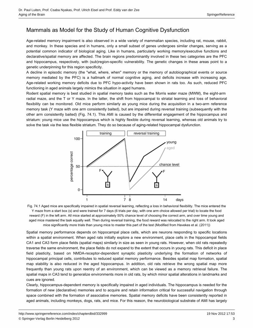
, and the T or Y maze. In the latter, the shift from hippocampal to striatal learning and loss of radial maze behavioral can be monitored. Old mice perform similarly as young mice during the acquisition in a two-arm referenceflexibility
memory task (Y maze with one arm consistently baited), but are impaired during reversal training (subsequently with theother arm consistently baited) (Fig. 74.1). This AMI is caused by the differential engagement of the hippocampus andstriatum: young mice use the hippocampus which is highly flexible during , whereas old animals try toreversal learningsolve the task via the less flexible striatum. They do so because of aging-related hippocampal dysfunction.
Fig. 74.1 Aged mice are specifically impaired in spatial reversal learning, reflecting a loss in behavioral flexibility. The mice entered theY maze from a start ( ) and were trained for 7 days (6 trials per day, with one arm choice allowed per trial) to locate the foodbox s
reward ( ) in the . All mice started at approximately 50% chance level of choosing the correct arm, and over time young andF left armaged mice mastered the task equally well. Then during reversal training, the food reward was relocated to the . It took agedright arm
mice significantly more trials than young mice to master this part of the test (Modified from Havekes et al. ( ))2011
Spatial memory performance depends on hippocampal , which are neurons responding to specific locationsplace cellswithin a spatial environment. When aged rats initially explore a new environment, place cells in the hippocampal fieldsCA1 and CA3 form (spatial maps) similarly in size as seen in young rats. However, when old rats repeatedlyplace fieldstraverse the same environment, the place fields do not expand to the extent that occurs in young rats. This deficit in placefield plasticity, based on NMDA-receptor-dependent synaptic plasticity underlying the formation of networks ofhippocampal principal cells, contributes to reduced spatial memory performance. Besides spatial map formation, spatialmap stability is also reduced in the aged hippocampus. In addition, old rats retrieve the wrong spatial map morefrequently than young rats upon reentry of an environment, which can be viewed as a failure. Thememory retrievalspatial maps in CA3 tend to generalize environments more in old rats, by which minor spatial alterations in landmarks andcues are ignored.Clearly, hippocampus-dependent memory is specifically impaired in aged individuals. The hippocampus is needed for theformation of new (declarative) memories and to acquire and retain information critical for successful navigation throughspace combined with the formation of associative memories. Spatial memory deficits have been consistently reported inaged animals, including monkeys, dogs, rats, and mice. For this reason, the substrate of AMI has largelyneurobiological

4
SpringerReferenceDr. Paul Luiten, Prof. Csaba Nyakas, Prof. Ulrich Eisel and Prof. Eddy van der ZeeAging of the Brain
19 Nov 2012 17:53http://www.springerreference.com/index/chapterdbid/332999
© Springer-Verlag Berlin Heidelberg 2012
focused on the hippocampus. The new memories have to be consolidated into long-term memory sites in the neocortex.The exact nature of the hippocampus-neocortex interactions is not well understood, but the hippocampus has to"reactivate" previous activity patterns necessary for long-term storage. This replay occurs during quiet periods and sleep.In aged rats, the correct sequence of firing of hippocampal neurons is less preserved, which may contribute to overall
in aged rats.memory impairmentWithin the rodent hippocampus, the is particularly vulnerable to the effects of aging, with a characteristicdentate gyrusreduction in rate of neurogenesis. Although neurogenesis declines with age, it has not been clearly demonstrated thatneurogenesis correlates with learning abilities in (aged) rodents. Some data indicate that a correlation betweenneurogenesis, aging, and learning does exist by which the aged impaired individuals have the least neurogenesis. Like inrodents, the generation of new is dramatically reduced in the aged human gyrus.nerve cells dentate
Mechanisms of Brain Aging
Many neuronal mechanisms remain more or less similar in young and aged brain. However, there are some specific,aging-related alterations in the old brain directly linked to impaired cognitive performance. Even in the course of normal
, the overall brain shows a certain degree of atrophy, which results in a decrease in brain weight and volume and aagingdecrease in the level of proteins. From 50 to 90 years of age, the loss of brain weight is approximately 2-3% per decade.An increase in ventricular volume and the total amount of cerebrospinal fluid indicates a reduction in the volume of braintissue during aging. With the aid of , the shrinkage of different brain areas can readilymagnetic resonance imaging (MRI)be followed. Especially the involution size of the hippocampus was repeatedly matched successfully to AMI in humans.The primary question is how neuronal atrophy affects the structure and function of different parts of the neuron, i.e., thesynapses, the neurites (axons and dendrites), and size and integrity of the cell bodies. These changes are thought tounderlie AMI and will be briefly discussed below.
During Aging, Neurons Rarely Die but Change at the Level of Cytoskeleton, Synapse, and SyntheticActivity Negatively Affecting Synaptic Plasticity
Gene transcription and protein synthesis are needed for the formation of long-term memory. Aberrant changes in genetranscription affect the of the aged brain: it can either be decreased or increased depending on the braincognitive abilitiesregion and the protein studied. Cognitive performance requires the transcription of (IEGs,immediate early genesincluding Arc (activity-regulated cytoskeletal gene)), (brain-derived neurotrophic factor), and the late gene zif268.BDNFIEGs are among the first genes switched on upon neuronal and synaptic stimulation, followed by switching on .late genesDecreased IEG expression has been shown in the aged brain. Moreover, BDNF and its receptors undergo significantage-associated downregulations. Hence, several genes known to be necessary for synaptic plasticity and normal memoryfunctioning are downregulated in the aged brain. Reductions in the amount of protein synthesis in the aged braincontribute to AMI. Along this line reduction in age-related protein synthesis is typically found in rat hippocampus (dentategyrus but not CA1), nucleus accumbens, and .locus coeruleusThe once generalized "neuronal loss theory of aging" has been disproved by novel cell counting methods based on morereliable mathematical techniques to quantify and estimate total neuron numbers. More classical studies reported someloss of neurons with aging in the neocortex, amygdala, spinal cord, nucleus basalis, corpus striatum/putamen,
, , and cerebellar , but more recently it was established that there is nosuprachiasmatic nucleus inferior olive Purkinje cellsovert loss of neurons during normal aging (except for the Purkinje cells). Hence, the impairment ofhippocampal-dependent memory in aged individuals is not due to massive cell death.As indicated above, if hippocampal volumetric loss occurs, it is primarily caused by neuronal atrophy (shrinkage) ratherthan by neuronal loss. The number of hippocampal neurons differs neither between young and aged individuals norbetween individuals with AMI and cognitively intact old subjects. Age-related functional and behavioral changes are theoutcome of other, more subtle, changes that occur in individual neurons or the communication between neurons.Aging has a profound effect on the integrity of the forebrain fiber patterns that find their origin in general modifyingtransmitter systems such as the serotonergic, noradrenergic, dopaminergic, and cholinergic cell groups in midbrain andbasal forebrain. In particular these midbrain and basal forebrain projection systems to the cortical mantle are prone to theformation of axonal pathologies like fiber swelling and profiles forming grape-like structures. Likewise, such axonalaberrations are also found for cytoskeletal proteins such as neurofilaments (Fig. 74.2). These aberrant fibers and fiberswelling become progressively pronounced in the course of normal . Thus, these anomalies of axon pathologybrain aging

5
SpringerReferenceDr. Paul Luiten, Prof. Csaba Nyakas, Prof. Ulrich Eisel and Prof. Eddy van der ZeeAging of the Brain
19 Nov 2012 17:53http://www.springerreference.com/index/chapterdbid/332999
© Springer-Verlag Berlin Heidelberg 2012
are rather ubiquitous in projecting neurons, and they appear to be causally related to hampered axonal transportprocesses. Since these axonal aberrations also occur in the aged animal brain where the specific neurodegenerativeprocesses that are known from human diseases are absent, these fiber anomalies should be considered as typical foraging per se. It is highly likely that fiber aberrations and neuritic dystrophies are indicators of a shift of balance fromsuccessful neuronal maintenance toward loss of restorative capacity of the brain progressively resulting in abundantaccumulation of dystrophic structures.
Fig. 74.2 Axonal aberrations (see in ( )); fibers stained for neurofilaments in rabbit hippocampus) are largearrows panel amalformations in thin fibers found throughout the aged brain. The number of axonal aberrations, here quantified for the hippocampus (
( )), gradually increases with age. Such fiber swellings are common for cortical projection systems that originate frompanel bsubcortical sources like cholinergic, serotonergic, and noradrenergic cell groups in basal forebrain and brain stem (Data taken from
Van der Zee et al. ( ))1997
Electrical properties remain relatively intact over the . Hence, firing properties do not differ at large between younglifespanand old neurons, but hippocampal connectivity is affected in a region-specific manner with accompanyingelectrophysiological changes. Reduced numbers of synaptic terminals have been reported in aged . Thedentate gyrushippocampal CA1 region is the most-studied brain area with regard to age-related changes in synapses. Notablyperforated synapses (synapses with multiple contact zones) are selectively reduced in size in aged rats with impairedspatial learning (Fig. 74.3).

6
SpringerReferenceDr. Paul Luiten, Prof. Csaba Nyakas, Prof. Ulrich Eisel and Prof. Eddy van der ZeeAging of the Brain
19 Nov 2012 17:53http://www.springerreference.com/index/chapterdbid/332999
© Springer-Verlag Berlin Heidelberg 2012
Fig. 74.3 Aging neurons are characterized by a reduced dendritic tree and reduced numbers of ( ( ) and ( ) fordendritic spines inset a cthe young and aged neuron, respectively). Few morphological changes are found at the synaptic level, although in some cases the
active zone is reduced with less synaptic vesicles and a reduced . Aged synapses can be relatively large, whichpostsynaptic densityis considered to be a compensation for the loss in number of synaptic contacts ( ( ) and ( ) show a synaptic contact of a younginset b d
and aged neuron, respectively; the altered morphology in ( ) is an exaggerated example) (The synaptic contact is modified fromdGeinisman et al. ( ). New figure adapted from own work Fig. 74.2 in J Neurosci. 2001, 21:103-116)2001
The number of perforated synapses correlates positively with spatial in aged animals. If the entirememory capacityhippocampus is considered, only minor loss of synapses is found. If the dorsal hippocampus is taken apart, a loss ofsynapses in the gyrus as a function of is found and even more so in rats with age-related decline indentate normal agingspatial memory. It should be noted that synapse number by itself does not predict cognitive status. It is more likely that Ca
-dependent cellular processes responsible for maintaining or altering synaptic strength are pivotal to AMI (see below).2+
Pre- and postsynaptic parameters remain relatively constant throughout the lifespan of rats. However,NMDA-receptor-mediated synaptic transmission is enhanced in very old rats, contributing to dysregulation of Ca2+
-dependent cellular processes. This age-related dysregulation of affects place cell function negatively,NMDA receptorsinfluencing spatial memory performance in aged individuals.Synaptic plasticity, notably long-term potentiation , is hampered in the aged brain, and deficits(LTP) learning and memoryfound in aged rodents parallel deficits in LTP (Fig. 74.4). LTP is considered the cellular basis of learning and memoryprocesses as information is stored via activity-dependent changes at the synapse. LTP is characterized by at least twodifferent phases: induction (early-phase LTP) and maintenance (late-phase LTP). There are no age-related deficits in LTPinduction if the stimulus parameters are strong enough. However, if the stimulus parameters are close to the threshold ofLTP induction, the aged hippocampus reveals an LTP-induction deficit. The decay rates of LTP do not differ muchbetween young and old rats in the first hours after LTP induction. At longer time intervals, however, significantmaintenance deficits do appear. At these time points, LTP decays twice as fast in aged rats than in young rats. Spatialmemory deficits in aged rats correlate with LTP (both within and between age groups). The rate of acquisition of spatialmemory correlates significantly with LTP induction, and rate of of spatial memory correlates significantly (butforgettingnegatively) with LTP maintenance.

7
SpringerReferenceDr. Paul Luiten, Prof. Csaba Nyakas, Prof. Ulrich Eisel and Prof. Eddy van der ZeeAging of the Brain
19 Nov 2012 17:53http://www.springerreference.com/index/chapterdbid/332999
© Springer-Verlag Berlin Heidelberg 2012
Fig. 74.4 Schematic representation of the abolishment of LTP in the aging hippocampus. At 0 (indicated by the ), atime point arrowtrain of 100-Hz stimuli is given to the hippocampus. LTP is rapidly induced, irrespective of age. While LTP is maintained in a stablefashion for a long period in the young hippocampus, it is not maintained in such a way in the aged hippocampus. Here, the EPSPs
drop back to baseline (and even slightly below) relatively quickly
Epigenetics refers to modification of and posttranslational modulation of by which patterns of geneDNA nuclear proteinsexpression are altered. Next to the age-related changes in , it is very likely that age-associatedgene transcriptiondisruption in and , two major processes in epigenetic mechanisms, leads toDNA methylation histone modificationage-specific memory disruption.In summary, to draw a clear distinction between normal and pathological neuronal aging is not yet possible, and thetransition from aging cell to a pathological state can be very gradual and not always easy to define unless explicitneurotoxic mechanisms can be identified. The AMI-related changes include shrinkage in size, loss or regression ofsomadendrites and , alterations in neurotransmitter receptors, and changes in electrophysiological properties.dendritic spinesThe integrative functional characteristics of dendrites are determined by several factors, including their morphology, thespatiotemporal patterning of , and the balance of inhibition and excitation. Thus, dendrites play a vital rolesynaptic inputsin the functional properties of neuronal circuits, and any structural changes can have profound and detrimental functionaleffects. Turnover of synapses, dendritic spines, and axonal nerve endings forms the basis of functional plasticity ofneurons. The recent findings in neuronal morphology during normal and abnormal support the view that thebrain agingsuboptimal plastic renewal of neuritic structures plays a key role in among the brain aging processes. Thecognitive agingstructural and functional aberrations of axons, synapses, and synaptic signaling appear to be the primary events duringaging which is enhanced in time and in magnitude in cognitive neurodegeneration processes as in Alzheimer's diseaseand other dementias.
Neuronal Membrane Structure and Composition Changes at Older Ages and Influences MembraneFluidity and Membrane Signaling. The Role of Cholesterol
Neuronal membranes support the larger part of cellular macromolecules (about 80%) and form functionally connectedmorphological structures including nuclear membrane, endoplasmic reticulum, , synaptic vesicles, andGolgi apparatussynaptic terminal membrane. The basic structure is the , which provides a matrix for proper membranelipid bilayerfunctioning. The major components of membranes are lipids, predominantly phospholipids and sterols (cholesterol), andproteins. The hydrophobic hydrocarbon chains of the polar lipids are located in the interior of the bilayer, whereas thehydrophilic head groups are exposed to the aqueous surroundings. The included proteins are membrane-spanning,anchored, and membrane-associated proteins, which can be receptors, ion channels, cytoskeletal proteins, and proteinsof the signaling machinery conveying information between the extra- and intracellular compartments. Physicochemicallythe lipid-protein interaction is one of the primary determinants of the quality of membrane function and, from this aspect,
merits consideration.membrane fluidityThe fluidity of neuronal membranes plays a pivotal role in brain aging and neurodegeneration. Membrane fluidity undernormal conditions supports lipid-protein interactions leading to adequate neurotransmitter functioning and transmembrane

8
SpringerReferenceDr. Paul Luiten, Prof. Csaba Nyakas, Prof. Ulrich Eisel and Prof. Eddy van der ZeeAging of the Brain
19 Nov 2012 17:53http://www.springerreference.com/index/chapterdbid/332999
© Springer-Verlag Berlin Heidelberg 2012
signal transfer, for example, in relaying transmitter signals to their machinery. The structure of fattysecond messengeracids influences membrane fluidity; the higher the number of double bonds, the higher the impact on fluidity. Aging isaccompanied by a decrement in long-chained (PUFAs, mainly arachidonic acid (ARA) andpolyunsaturated fatty acidsdocosahexaenoic acid ) in membrane phospholipids which results in decreased membrane fluidity, i.e., a more rigid(DHA)membrane is formed. Dietary omega-3 fatty acid intake improves the membrane function in the hippocampus and,consequently, especially by DHA intake from , spatial learning ability in old rats can be enhanced. It has beenfish oilwidely accepted that capability requires normal membrane fluidity.optimal learningPUFA's are not only of the membrane but also play a role in which isstructural components synapse formationdiminishing during aging. Brain phosphatide synthesis requires three circulating compounds: DHA, , and choline.uridineOral administration of these phosphatide precursors to experimental animals increases the level of phosphatides and
in the brain and the numbers of on hippocampal neurons.synaptic proteins dendritic spinesAs indicated above, is considered as a reliable physiological biomarker of learning capability. Aged rats exhibit anLTPimpaired ability to sustain LTP in the of the hippocampus which correlates with a decrease in ARAdentate gyrusconcentration. Dietary supplementation with arachidonic acid and its precursor, gamma-linolenic acid, reversed theimpairment in LTP in aged rats.Another key membrane function which can become affected during aging is the processing of amyloid precursor protein
especially with respect to AMI, and eventually to the development of Alzheimer's disease (AD) neuropathology.(APP)Gradual changes in steady-state levels of peptides (Ab peptides) in the brain are considered as the initialbeta amyloidstep in the amyloid cascade hypothesis of AD. DHA supports normal, physiological processing by increasingAPPα-secretase activity at the cell surface, since α-secretase leads to APP splicing products that are removed and brokendown by natural and nontoxic fragment processing. The alternative splicing of the APP molecule in the neuronal plasmamembrane by b-and g-secretases leads to the release of Ab peptides, of which the oligomeric forms are the most toxic forthe brain which is partly explained by the propensity of Aβ to aggregate initially to soluble and finally to insoluble polymersand fibrils.Cholesterol plays several structural and metabolic roles that are crucial to brain functioning. In membranes, it regulatesfluidity and events in cooperation with other lipids. Aβ-induced changes in could becell signaling membrane fluidityexplained by physicochemical interactions of the peptide with membrane components such as cholesterol, phospholipids,and , while reversely cholesterol stimulates Aβ production. Beside cholesterol, other lipids strongly modulatesphingolipidsAPP processing, whereas the APP cleavage products themselves regulate lipid, including cholesterol homeostasis,resulting in complex regulatory feedback cycles.High levels of cholesterol have been proposed as a risk factor for AD. Cholesterol inhibits α-secretase activity andfacilitates β- and γ-secretases, and in this way increases Aβ production. Therefore, it has been argued that cholesterollowering drugs such as statins could diminish the risk of AD. Indeed, there is evidence for a significantly reducedincidence of AD among people who have been using statins to curb hypercholesterolemia and its cardiovascular effects.There is now evidence for a diminished synthesis of cholesterol in the aging hippocampus, but without a noticeablechange in its concentration. In rat, the cholesterol levels at the age of 24 months appear to be increased in striatum,cerebellum, and brain stem, but not in the cerebral cortex or hippocampus.With respect to the cholesterol content in the nervous tissues in the various age phases, it became clear that regionaldifferences and subcellular partitioning of the cholesterol concentrations are characteristic for aging. An interesting viewon the role of cholesterol in the aged brain is the asymmetric distribution of cholesterol among different domains duringaging and that the cholesterol content of the exofacial leaflet of synaptic plasma membranes doubles with age (Fig. 74.5).This figure shows that the transbilayer distribution of cholesterol is associated with Aβ accumulation in the external layerof the synaptic membrane of aged mice. This accumulation of Aβ may disrupt other membrane such as lipidlipid domainsrafts, which are membrane microdomains particularly rich in ion channel and signaling proteins and actively involved insetting membrane potential and ionic signaling. This way cholesterol via amyloid may impinge on normal membranefunction in general. Note that , especially the abovementioned , and the main cholesterolpolyunsaturated fatty acids DHAtransporting neuronal lipoprotein are potent regulators of transbilayer cholesterol distribution.ApoE

9
SpringerReferenceDr. Paul Luiten, Prof. Csaba Nyakas, Prof. Ulrich Eisel and Prof. Eddy van der ZeeAging of the Brain
19 Nov 2012 17:53http://www.springerreference.com/index/chapterdbid/332999
© Springer-Verlag Berlin Heidelberg 2012
Fig. 74.5 Distribution of cholesterol in the synaptic plasma membrane outer and inner ( .) layers of young and aged.( )SPM ( .)Exo CytoThe SPM outer, exofacial leaflet of aged mice contains twice the amount of cholesterol than the younger mice. Partitioning of amyloidbeta-peptides ( ) into or out of the exofacial layer may be hindered by the accumulation of cholesterol ( ) inred structure yellow structurethe exofacial leaflet of aged individuals. This accumulation of Aß may disrupt membrane lipid domains and affect membrane functions
(Adapted with permission from Wood et al. ( ))2002
Neurotransmitter Systems Projecting to the Forebrain Appear Particularly Vulnerable to the AgingProcess
Many neurotransmitter systems to the cerebral cortex that have their origin in brain stem structures show aging-relateddownregulation in activity (e.g., acetylcholine, dopamine, serotonin, and noradrenaline). Usually specific changes occur atthe receptor level, contributing to a reduction in overall signal transduction. Several receptor types of these four ascendingsystems target on the same downstream intracellular pathways and molecular cascades. Nearly all signal transduction
involved in cognitive performance utilize the activation of kinases and phosphatases. Among the most robustlypathwaysdownregulated genes in aging humans are the "cognitive kinase" CAMKIV gene linked to the CREB-related pathway, andthe eukaryotic factors (eIFs) belonging to the translational machinery. Hence, protein synthesistranslation initiationneeded for is hampered in the aged brain.neuronal plasticityProtein modification through phosphorylation by kinases is a critical regulatory tool of enzyme activity levels (Fig. 74.6).One cognitive kinase required for memory acquisition is the Ca -dependent and brain-specific protein kinase Cγ (PKCγ).2+
This kinase translocates from the cytosol to the membrane (or specific intracellular targets) during encoding ofinformation, and remains attached to the membrane long after the initiating stimulus is gone. Here, it phosphorylateslearning- and memory-specific substrates. Various forms of age-related dysregulations of PKCγ have been reported,depending on the species studied. For example, upregulations in kinase activity levels (demonstrated in rabbit andSprague Dawley rat) or increased levels of membrane-bound PKCγ (demonstrated in Wistar rat) most likely reflectcompensatory attempts. However, like with overactivation, a negative effect of this type of compensation is loss ofPFCspecificity in PKCγ activity. Many dysregulations of kinase activities are due to changes in the scaffolding and anchoringproteins, for these enzymes orchestrate their subcellular location and regulate activity levels. Notably proteins likeRACK-1 and AKAP150 are dramatically reduced in the aged hippocampus (Fig. 74.6).

10
SpringerReferenceDr. Paul Luiten, Prof. Csaba Nyakas, Prof. Ulrich Eisel and Prof. Eddy van der ZeeAging of the Brain
19 Nov 2012 17:53http://www.springerreference.com/index/chapterdbid/332999
© Springer-Verlag Berlin Heidelberg 2012
Fig. 74.6 Dysregulations of intracellular phosphorylation events occur in aging neurons due to dramatic reductions in the expression ofscaffolding and anchoring proteins, for example, RACK-1 and AKAP150. Due to the loss of these , the delicateregulatory elements
balance between levels of phosphorylation and dephosphorylation of target proteins gets disturbed, as depicted schematically in the ( ) (young neuron) versus ( ) (aged neuron) for target protein 1 (normally heavily phosphorylated) and target protein 2upper panels a b
(normally non-phosphorylated). In the , examples are shown for the dramatic reduction in hippocampal AKAP150lower panelsexpression in aged as compared to young mice ( ( ) and ( ), respectively), and the increased activity of the calcium-dependentpanels e bkinase PKCγ as detected by an antibody specifically recognizing the activated form of PKCγ ( ) and ( ) for the young and aged rabbitc f
hippocampus, respectively). AKAP150 data taken from Ostroveanu et al. ( ), PKCγ data taken from Van der Zee et al. ( )2007 2004
The structural and anatomical axonal and neuritic dystrophies described above are paralleled by reductions in theenzymes that synthesize the neurotransmitters dopamine, norepinephrine, serotonin, and acetylcholine. This isaccompanied by partial shrinkage and functional decline of their subcortical cell bodies of origin in the midbrain substantianigra, , raphe, and . Therefore, not only neuronal loss in these subcorticallocus coeruleus nucleus basalis of Meynertnuclei but also the of the remaining or surviving neurons is essential for maintaining normal or adequate mentalvitalityfunctions.
Cognitive Dysfunction Parallels Degenerative Changes in Cortical Projection Transmitter Systems
Some of the cognitive dysfunctions in aging can well be linked to specific transmitter deficits. Converging evidence frompatient studies, animal research, pharmacological intervention, and indicates that dopamine ismolecular geneticscritically implicated in higher-order . Many and multiple markers of striatal andcognitive functioning cognitive functionsextrastriatal DA systems decline across adulthood and aging. Molecular imaging studies of dopaminergicneurotransmission measure biomarkers of dopamine, such as the dopamine transporter and dopamine receptor D1 andD2 indicate that individual differences in dopamine functions are linked to age-related decline in ,executive functioningepisodic memory, and perceptual speed.The neurotransmitter is paramount for regulation of mood, food intake, activity rhythms,serotonin (5-hydroxytryptamine)and sexual behavior, which are all behavioral expressions prone to changes during aging. Moreover, the serotonergicsystem is significantly involved in , in particular by interacting with the cholinergic, glutamatergic,learning and memorydopaminergic, or GABAergic systems. Its action is mediated via specific receptors located in crucial brain structuresinvolved in these functions, primarily the septo-hippocampal complex, the , and the .nucleus basalis magnocellularis PFC

11
SpringerReferenceDr. Paul Luiten, Prof. Csaba Nyakas, Prof. Ulrich Eisel and Prof. Eddy van der ZeeAging of the Brain
19 Nov 2012 17:53http://www.springerreference.com/index/chapterdbid/332999
© Springer-Verlag Berlin Heidelberg 2012
There is now ample evidence that the administration of a variety of serotonergic receptor targeting drugs can prevent and facilitate learning in situations involving a high cognitive demand.memory impairment
The human cholinergic basal forebrain system is comprised of within the septal/diagonal bandmagnocellular neuronscomplex and nucleus basalis of Meynert. During , the anatomical integrity of fiber pathways penetrating thenormal agingvarious cortical layers is compromised, a phenomenon that is not limited to the cortical regions but affects structures likethalamus and brain stem as well. Cholinergic neuronal cell loss was found predominantly in pathological aging, such asAlzheimer's disease or Parkinson's disease, while normal aging is accompanied by a gradual loss of cholinergic functioncaused by dendritic, synaptic, and as well as a decrease in trophic support by axonal degeneration nerve growth factor
. As a consequence, decrements in gene expression, impairments in intracellular signaling and cytoskeletal(NGF)transport, and weakening of trophic support may mediate cholinergic cell atrophy finally leading to the known age-relatedfunctional decline in the brain including AMI.As mentioned in section "During Aging, Neurons Rarely Die but Change at the Level of Cytoskeleton, Synapse, andSynthetic Activity Negatively Affecting Synaptic Plasticity", during aging, neurons evidently undergo a reduction in thecomplexity of dendritic arborization and dendritic length. numbers are also decreased, and since spinesDendritic spineare the major sites for excitatory synapses, changes in their numbers reflect a change in excitatory neurotransmission.This theory has been supported by the demonstration of a decrease in glutamate-receptor-mediated excitatoryresponses, as well as a decrease in the levels of alpha-amino-3-hydroxy-5-methylisoxazole-4-propionic acid and (AMPA)
-methyl- -aspartate expression during aging. The latter findings are of major importance forN D (NMDA) receptorunderstanding aging effects on cortical since these glutamate-driven ion channel receptors arecognitive functioningparamount in intercortical information processing and basic mechanisms such as . Glutamate is also an importantLTPplayer in the because of its role in both as well as in facilitation of excitotoxic neuronal injuryaging brain neuroprotectionand death. Glutamate, the brain's predominant excitatory neurotransmitter, facilitates release of brain-derivedneurotrophic factor and other neurotrophins and in this way indirectly modulates synaptic plasticity and(BDNF)activity-dependent neuronal survival which implicates a neuroprotective function of this neurotransmitter system.Neurotrophins comprise neuromodulatory functions and closely interact with neurotransmitters. Among neurotrophins,BDNF is the best characterized modulator of normal brain aging. It is a considerable modulator of molecular aging, as it isa neuronal activity-dependent secreted growth factor that declines steadily with age in the brain. It is neuroprotectiveagainst a variety of insults emerging in the course of aging and is required for changes in spine density underlying
systems that decline with age.learning and memoryThe aging process has also clear effects on the most common inhibiting neurotransmitter of the braingamma-aminobutyric acid . Its functional expressions such as GABA-A ion channel receptor-mediated inhibitory(GABA)responses and action potential firing rates are both significantly increased with age. Considering the mechanismsresponsible for age- and AD-related neuronal degeneration, little attention has been paid to the opposing relationshipsbetween the energy-rich phosphates, mainly the availability of the (ATP), and the activity of theadenosine triphosphateglutamic acid decarboxylase , the rate-limiting enzyme synthesizing the GABA. It has been postulated that in all(GAD)neuronal phenotypes, the declining ATP-mediated of GABA synthesis gradually declines and results innegative controlage-related increases of GABA synthesis.
Aging Has Serious Consequences for the Calcium Homeostasis of Neurons Which Has a Major Impacton Neuronal Physiology and Signaling
The regulation of intracellular free calcium levels is a crucial phenomenon for the cellular physiology of glial and neuronalcells of the nervous system and has attracted ample attention in aging research. Destruction or impairment of intracellularcalcium maintenance is not only essential for understanding breakdown of brain tissue in essentially all
, but also for brain aging in general. The reason being that loss of control of free calcium levelsneurodegenerative diseasehas profound consequences for a multitude of cellular neuronal functions. In young , free calcium homeostasisnerve cellsis very well maintained by balancing influx and intracellular release of calcium, temporary binding or storage of excesscalcium, and transport of excess out of the cell. Calcium concentrations in young neurons show a very low basal level, apeak-like increase upon a variety of activations followed by orchestrated stimulation of calcium targets necessary toperform the required , and finally a quick and efficient return to baseline levels (Fig. 74.7).physiological functions

12
SpringerReferenceDr. Paul Luiten, Prof. Csaba Nyakas, Prof. Ulrich Eisel and Prof. Eddy van der ZeeAging of the Brain
19 Nov 2012 17:53http://www.springerreference.com/index/chapterdbid/332999
© Springer-Verlag Berlin Heidelberg 2012
Fig. 74.7 Intracellular free calcium concentration in aged versus young nerve cells, baseline levels, and the calcium response uponstimulation of the neuron by the neurotransmitter glutamate or by potassium. Characteristically the baseline levels of free calcium areconsiderably higher in the aged neuron. Stimulation of the young results in a steep rise of calcium, which quickly returns tonerve cellbaseline level when the stimulus ends due to the homeostatic mechanisms. In the aged neuron, the rise of calcium during stimulationis much lower, and the return to baseline levels is very slow. Together this means probably an inadequate physiological response to a
temporary stimulus and a constantly high level of free calcium and its damaging consequences on activation of calcium-sensitiveenzymatic processes (Adapted with permission from Kirischuk and Verkhratsky (1996))
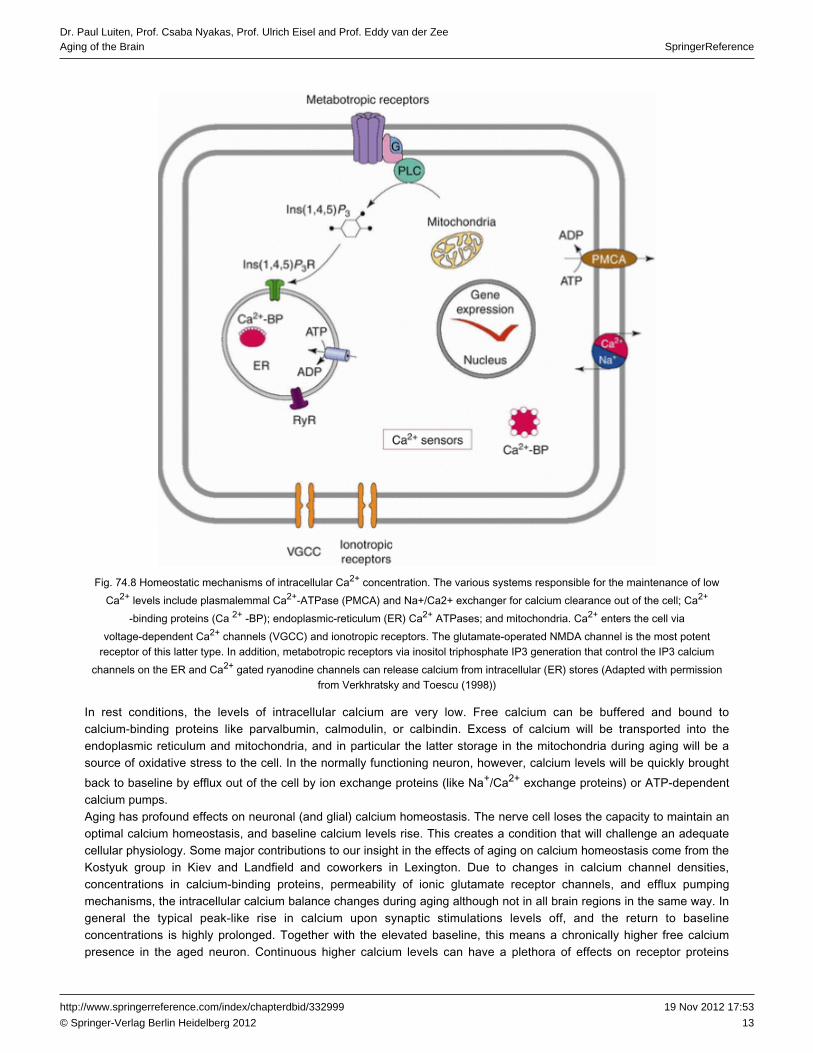
To appreciate the impact of aging, it is important to understand the basic mechanisms of intraneuronal calciumhomeostasis (Fig. 74.8). Levels of intracellular calcium increase upon activation of . Well-knownionotropic receptorsrepresentatives of these receptors are the ion channels that can open by the binding of such asexcitatory amino acidsglutamate of which the (N-methyl-D-aspartate) receptor has been extensively studied. Interestingly, the opening ofNMDAthe ion channel of the is voltage dependently blocked by Mg2+ ions. When glutamate binds to the NMDA receptor AMPA
due to sodium influx, the membrane potential of the neuron decreases from −70 to −35 mV upon which thereceptormagnesium block is removed followed by opening of the glutamate bound NMDA channel and the influx of calcium. Thisinflux of calcium is a strong ion flow which is potentiated by the very high concentration gradient over the neuronal cellmembrane between insite and outside of the cell since the extracellular concentration is 10 times the intracellular4
concentration. A second major calcium rise comes from opening of voltage-dependent calcium channels . These(VDCC)calcium channels vary in localization and function, and in the in the brain, the L-type and T-typepostsynaptic neuroncalcium channels play a role, but of these, the L-type channel is best characterized. L-type channels have a highactivation threshold but after opening become slowly inactivated. Upon depolarization, for example, by excitatory aminoacids like glutamate but also as a result of any source of depolarization, VDCCs open and allow a strong influx of calciumions. During aging, the density of L-type calcium channels increases in areas involved in like thecognitive functionshippocampus leading to enhanced inflow of calcium. A third source of increase of intracellular calcium levels comes fromactivation of which are linked to the phosphoinositol system that leads to mobilization ofG-protein-coupled receptorsphospholipase C and the formation of inositol triphosphate (IP3) as a splicing product of the membrane phospholipidphosphatidylinositol-biphosphate . The IP3 diffuses into the cytoplasm, binds to its receptors on(PIP2) second messengerthe membranes of the endoplasmic reticulum, and allows an outflow of calcium from the endoplasmic stores into thecytoplasm. Next to the IP3 channels, the endoplasmic reticulum is endowed with ryanodine-sensitive channels that aregated by the calcium itself and amplify the release of intracellular calcium.
13
SpringerReferenceDr. Paul Luiten, Prof. Csaba Nyakas, Prof. Ulrich Eisel and Prof. Eddy van der ZeeAging of the Brain
19 Nov 2012 17:53http://www.springerreference.com/index/chapterdbid/332999
© Springer-Verlag Berlin Heidelberg 2012
Fig. 74.8 Homeostatic mechanisms of intracellular Ca concentration. The various systems responsible for the maintenance of low2+
Ca levels include plasmalemmal and for calcium clearance out of the cell; Ca2+ Ca -ATPase2+ ( )PMCA Na+/Ca2+ exchanger 2+
-binding proteins ( ); endoplasmic-reticulum ( ) Ca ATPases; and mitochondria. Ca enters the cell viaCa 2+ -BP ER 2+ 2+
voltage-dependent Ca channels ( ) and ionotropic receptors. The glutamate-operated NMDA channel is the most potent2+ VGCCreceptor of this latter type. In addition, via inositol triphosphate IP3 generation that control the IP3 calciummetabotropic receptors
channels on the ER and Ca gated ryanodine channels can release calcium from intracellular ( ) stores (Adapted with permission2+ ERfrom Verkhratsky and Toescu ( ))1998
In rest conditions, the levels of intracellular calcium are very low. Free calcium can be buffered and bound tocalcium-binding proteins like , , or calbindin. Excess of calcium will be transported into theparvalbumin calmodulinendoplasmic reticulum and mitochondria, and in particular the latter storage in the mitochondria during aging will be asource of oxidative stress to the cell. In the normally functioning neuron, however, calcium levels will be quickly broughtback to baseline by efflux out of the cell by ion exchange proteins (like Na /Ca exchange proteins) or ATP-dependent+ 2+
calcium pumps.Aging has profound effects on neuronal (and glial) calcium homeostasis. The loses the capacity to maintain annerve celloptimal calcium homeostasis, and baseline calcium levels rise. This creates a condition that will challenge an adequatecellular physiology. Some major contributions to our insight in the effects of aging on calcium homeostasis come from theKostyuk group in Kiev and Landfield and coworkers in Lexington. Due to changes in calcium channel densities,concentrations in calcium-binding proteins, permeability of ionic , and efflux pumpingglutamate receptor channelsmechanisms, the intracellular calcium balance changes during aging although not in all brain regions in the same way. Ingeneral the typical peak-like rise in calcium upon synaptic stimulations levels off, and the return to baselineconcentrations is highly prolonged. Together with the elevated baseline, this means a chronically higher free calciumpresence in the aged neuron. Continuous higher calcium levels can have a plethora of effects on receptor proteins

14
SpringerReferenceDr. Paul Luiten, Prof. Csaba Nyakas, Prof. Ulrich Eisel and Prof. Eddy van der ZeeAging of the Brain
19 Nov 2012 17:53http://www.springerreference.com/index/chapterdbid/332999
© Springer-Verlag Berlin Heidelberg 2012
including ion lipids, kinases, proteases, and gene expression processes. Calcium-induced oxidativechannels, membranestress and a compromised mitochondrial energy production in the form of ATP as a consequence of the aging processwill constitute major hazards to cellular physiology underlying neuronal signaling that is basic to . Ascognitive functioningwe will see below, chronic high calcium levels in the aged neuron has been associated with enlarged polarization ofneurons at the end of the action potential.
Aged Neurons in the Hippocampus Show an Enlarged Afterhyperpolarization Which Parallels ImpairedLearning and Conditioning
Cognitive functions of the brain like attention rely for an important part on frontal and learning, memory, and temporal of the forebrain. Since these cortical regions in particular are subject to aging-related functional and structurallobes
decline, it appears that the neurons and glia of these brain areas are more than average sensitive to the aging process.The cognitive functions in the aged are subject to subtle changes. So the question remains which mechanisms may beresponsible for the physiological decline of neuronal function. One of the answers to this question can be found in theelectrophysiological properties of aging . Basic to all brain activity is the generation of action potentials by thenerve cellsindividual neuron. When the membrane potential decreases upon any source of excitatory activation beyond a criticalthreshold, an action potential occurs that results from massive opening of sodium channels and influx of sodium ions.After reaching the depolarization peak, sodium channels close, and potassium channels open that allow a fast outward K+
current. The potassium currents are activated by calcium, and since calcium is high during and shortly after thedepolarization phase of the action potential, the K current leads to an overshoot of the membrane potential which is+
called the . Since the baseline calcium concentrations are high in the aging , andafterhyperpolarization (AHP) nerve cellthe K current is calcium dependent, the is increased in the aged neuron. This phenomenon was first discovered by+ AHPLandfield and Pitler in and was confirmed and extended by others. The calcium-dependent enlarged AHP has1984serious consequences for the excitability of neurons in the , in particular in those brain regions involved in aging brain
.cognitive processesProlonged AHP is one of the mechanisms that make neurons more resistant to depolarization and recruitment forparticipation in activity during performance of a cognitive task. Interesting experiments that reveal theneuronal networkimpact of aging on AHP and a learning task were carried out by the group of John Disterhoft in Chicago. The learning taskthey used was trace eye blink conditioning in which animals (or humans) "learn" to associate a tone with an airpuffapplied later in time and that leads to an eye blink. Learning is successful when the eye blinks after the tone but beforethe airpuff. Aged animals perform much worse than young which was directly related to the much largercalcium-dependent AHP. Interestingly when they blocked the calcium influx in the neurons by means of the L-typecalcium antagonist nimodipine, the AHP was reduced, the excitability of the neuron increased, and the performance in theeye blink conditioning task greatly improved. Similar type of studies was reported more recently which described therelationship between the magnitude of the AHP in aged rats and the scores of these animals in the asMorris water mazea typical learning test. In these experiments, it could clearly be demonstrated that there is strong correlation between anenlarged AHP and poor memory performance in the water maze (see Fig. 74.9).

15
SpringerReferenceDr. Paul Luiten, Prof. Csaba Nyakas, Prof. Ulrich Eisel and Prof. Eddy van der ZeeAging of the Brain
19 Nov 2012 17:53http://www.springerreference.com/index/chapterdbid/332999
© Springer-Verlag Berlin Heidelberg 2012
Fig. 74.9 Relations between performance of aged rats in the Morris water maze ( ) and the afterhyperpolarization after electricalastimulation of neurons in the CA1 area of the hippocampus ( ). in ( ) is the rest membrane potential. The learning inb Dotted line b curve( ) is indicated by the distance that rats swim to escape to a hidden platform in a large water tank. During 5 days of training, it can beaappreciated that young and aged-unimpaired rats quickly learn to find the escape platform, whereas aged memory-impaired animalsperform much worse. The in ( ) show that the aged memory-impaired rats have a much larger AHP as compared to thewaveforms byoung and aged-unimpaired rats, which suggests that an enlarged AHP is a causal factor for impaired memory functions (Adapted
with permission from Tombaugh et al. (2005))
Sophisticated research on the consequences of these aging mechanisms was carried out by Carol Barnes and her teamon hippocampal neuronal networks during in which place cell assemblies respond to changes in spacespatial orientationaccording to dynamic activity patterns. Aging had profound effects on these pattern dynamics (as mentioned above) thatwere most likely the result of the deranged calcium homeostasis in the aged cell.In summary, aging does not so much affect the number or form of the neuron, but it does exert its influence on neuronal
at the level of action potential generation of the individual neuron and subsequently on the dynamics of neuronalplasticityactivity as they participate in neuronal networks during behavioral and cognitive tasks.
In the Aged Mammalian Brain, Blood Supply Is Decreased and the Vascular Condition Is Subject toDegenerative Mechanisms
Nervous Tissue Is Highly Dependent on Optimal Blood Flow for Its Energy Generation
The for performance of its many simultaneous functions is utterly dependent on an adequate andcentral nervous systemunhampered supply of nutrients for the generation of energy. In particular in an acute sense, the supply of glucose andoxygen is critical, and in a somewhat less acute way the availability of other feeding components such as amino acids forthe synthesis of neurotransmitters for neuronal signaling and proteins for neuronal . The importance ofhousekeepingreadily available glucose and oxygen can easily be observed when the blood supply to the central nervous system isobstructed when the larger arterial vessels to the brain become occluded as it occurs in ischemic stroke. Such anischemic insult in general is the result of long-term narrowing of the arterial lumen as a consequence of atherosclerosis ofthe vascular wall combined with the presence of a or thrombus in the brain circulation which results in ablood clotvascular occlusion. Notably when larger brain arteries are obstructed, this condition within seconds can lead to loss of
after onset of the occlusion and become the source of severe in the flow region of aconsciousness brain damageparticular vessel if this condition continues.One of the reasons why brain functions are so quickly influenced by a reduction of and thus of glucosecerebral blood flowand oxygen is the extreme dependence of readily available energy in the form of ATP. Each for performance ofnerve cellits signaling functions is dependent on maintaining the gradients of ions between the inside and the outside of itsmembranes: notably sodium, potassium, but also chloride and calcium. The control of the ionic concentration gradientsover the neuronal membrane is paramount to the balance of positive and negative charges between inside and outside ofthe nerve cell that is basic to the characteristic membrane potential of 65-70 mV in rest conditions. Without awell-controlled membrane potential, the communication between by way of action potentials is impossible andnerve cellssilence occurs. Nerve cells have only a very limited capacity to store glucose in the form of glycogen to provide sufficientglucose or the glucose metabolite pyruvate once the regular availability is hampered. If the regular supply route ofglucose is entirely blocked as in stroke, the production of ATP within seconds drops from 36 ATP molecules per glucosemolecule to 2 ATPs. The consequence of this rapid loss of ATP production will depend on the activity state of the affectedneuron, but if blood reperfusion does not occur, the sustained ATP depletion will result in loss of the capacity of

16
SpringerReferenceDr. Paul Luiten, Prof. Csaba Nyakas, Prof. Ulrich Eisel and Prof. Eddy van der ZeeAging of the Brain
19 Nov 2012 17:53http://www.springerreference.com/index/chapterdbid/332999
© Springer-Verlag Berlin Heidelberg 2012
ATP-dependent pumping mechanisms to restore the ionic balances over the neuronal membrane necessary to maintainthe membrane potential. Massive loss of membrane potential prohibits neuronal signaling which will lead to a variety ofbehavioral and cognitive dysfunctions dependent on the affected brain area. Loss of consciousness will occur whencortical structures are involved and the communication of the cortical mantle with the thalamic relay structures.
Cerebral Blood Flow Decreases with Climbing Age
Reduction of cerebral blood flow will not always be as dramatic and acute as is the case in pathological conditions likevascular occlusion in ischemic stroke or during the characteristic sudden but transient obstruction of arterial blood flowthat are known as transient ischemic attacks (TIAs). Ischemic stroke and TIAs, however, are conditions that commonly(but not exclusively) take place in the aging individual, which already indicates that vascular condition and vascularintegrity, combined with spontaneous , are associated with the aging process. As already noted reduction ofblood clottingblood flow in the brain in most cases is not dramatic but rather has the nature of a slowly developing condition in theaging period of life. Interestingly the reduction of blood flow is not equally affecting all brain divisions and regions in asimilar way. Recent progress of techniques for noninvasive measurement of cerebral blood flow by means of functional
and blood-oxygen-level-dependent signaling measurement has providedmagnetic resonance imaging (fMRI) (BOLD)detailed knowledge of the dynamics of general blood supply and neuronal activity assessment during the various phasesin life (Fig. 74.10).
Fig. 74.10 Blood-oxygen-level-dependent (BOLD) imaging of individuals during performance of a simple sensory-motor task, that ispressing a key when viewing a flickering . BOLD imaging is taken as a measure of brain activation. It can be clearlycheckerboard
seen that both motor and visual brain regions are activated, but the response in non-demented older adults ( ) and demented olderbadults ( ) is significantly less than in the young adults ( ) (Reproduced with permission from Buckner et al. ( ))c a 2000
There is now ample evidence that during , there is a significant reduction in and BOLDnormal aging cerebral blood flowneuronal signaling MRI in those cortical brain regions associated with cognitive function. This reduction in blood supply tothe brain and to the cortical mantle in particular has been established both for baseline resting state conditions, but alsowhen individuals perform behavioral and cognitive activities such as a simple sensory-motor task that require activation ofthe respective brain regions necessary to carry out the task. However, the reduction of blood flow is not equal for all brainregions, and sensory areas appear to react more sensitive to the aging process than motor regions. From suchexperimental findings, it can be concluded that cerebral blood supply tends to decrease with climbing age which has been

17
SpringerReferenceDr. Paul Luiten, Prof. Csaba Nyakas, Prof. Ulrich Eisel and Prof. Eddy van der ZeeAging of the Brain
19 Nov 2012 17:53http://www.springerreference.com/index/chapterdbid/332999
© Springer-Verlag Berlin Heidelberg 2012
correlated to cognitive performance, but also that individual variation within the aged population can be high. Apparentlysome individuals appear to be much more sensitive to the aging process than others. Besides genetic disposition anddevelopment of , other risk factors like feeding pattern, socioeconomic background, and chronichigh blood pressurestressful life circumstances are considered to play an important role.Several have been identified that can be linked to the age-related reduction in blood flow. Common incausal factorsespecially human aging is the occurrence of atherosclerotic mechanisms that lead to narrowing of major arterial channelsthat provide the brain with blood. An often measured parameter in that respect is the intima-media thickness of the carotidarteries, and several investigations have indicated a direct association between carotid artery atherosclerosis andcognitive function. For that reason, laser Doppler of carotid circulation is often considered to provide aflow measurementreliable record for threats of adequate cerebral blood supply.Apart from the changes in major arterial vessels to the brain, the entry of oxygen, glucose, and other nutrients to theneural tissues is for a very large part dependent on the functional integrity of the microvasculature and in particular of thecapillary systems of the brain (Fig. 74.11). There is the general notion that the arteries and arterioles regulate bloodpressure, for example, by modulating smooth muscle cell contractility but that the exchange of critical components suchas glucose is dependent on the blood-brain barrier structure and its transporter functions. In other words, the largervessels could be seen as the bulk suppliers of blood volumes, and the enormous number of as thebrain capillariesdomain of fine regulation for local nutrients and gas delivery and the of the blood-brain barrier. In that respect, it canseatbe noted that there is clear difference between brain capillary densities in white matter and gray matter, the latter beingthe domain of the metabolically much more active bodies. Along the same line there is a prominent correlationnerve cellbetween capillary density, local blood flow, and glucose utilization in a given brain area.
Fig. 74.11 Electron microscopic image from a brain capillary taken from the cingulate cortex of a young rat ( ) and a drawn version ofathis capillary with the various components of the microvessel. Note the thin layer of cytoplasm of the endothelial cell ( ), the locationepof mitochondria ( ), the characteristic ( ), the position of the ( ) embedded in a thin and regular layer of theem tight junction tj pericytes p
basement membrane ( ). Other abbreviations: astrocytic endfeet, endothelial nucleus, microvascular lumen (Farkas andbm a en lLuiten )2001
Moreover brain microdomains that are metabolically very active, such as regions with a high synaptic density, also havethe highest capillary densities. Obviously since the distance between vascular lumen and brain parenchyma is shortestbetween capillaries and neural tissue, and because of the transport systems over the vascular membrane structures, theaccess of nutrients to the metabolic active components of the brain is most direct in the capillary microdomain.Furthermore, new insights in blood flow regulation give a more prominent role to brain capillaries next to arteries andarterioles than previously considered. The classical concepts were based on the key importance of smooth muscle cellsin the arterial wall, but novel research data point to an essential role of the capillary pericytes that are endowed with highlevels of contractile proteins. The current concept is that blood circulation in the brain is for a large part oxygen dependentand subject to neuronal and glial regulation at the microvascular level.
Brain Microvessels Undergo a Progressive Structural Degenerative Decline During Aging

18
SpringerReferenceDr. Paul Luiten, Prof. Csaba Nyakas, Prof. Ulrich Eisel and Prof. Eddy van der ZeeAging of the Brain
19 Nov 2012 17:53http://www.springerreference.com/index/chapterdbid/332999
© Springer-Verlag Berlin Heidelberg 2012
The blood-brain barrier consists of specific structural and functional components including the endothelial cells with theirtight junctions where their membranes meet, an extracellular matrix of the basement membrane surrounding theendothelial cell, pericytes embedded in the basement membrane, and a layer of astrocytic endfeet. A major role in barrierfunction has been assigned to the endothelial cell with its characteristic continuous tight junction complexes, the lack orpaucity of vesicular transport, high density of mitochondria, and a range of transmembranous transporter proteins.Together this structural complex constitutes the blood-brain barrier which acts as the of entry but also removalgatekeeperof compounds into or out of the brain parenchyma. Its functional and anatomical integrity is critical for the maintenance ofthe homeostatic stability of the neural tissue.Study of the capillary ultrastructure in various brain regions including the cerebral cortex in a number of mammalian
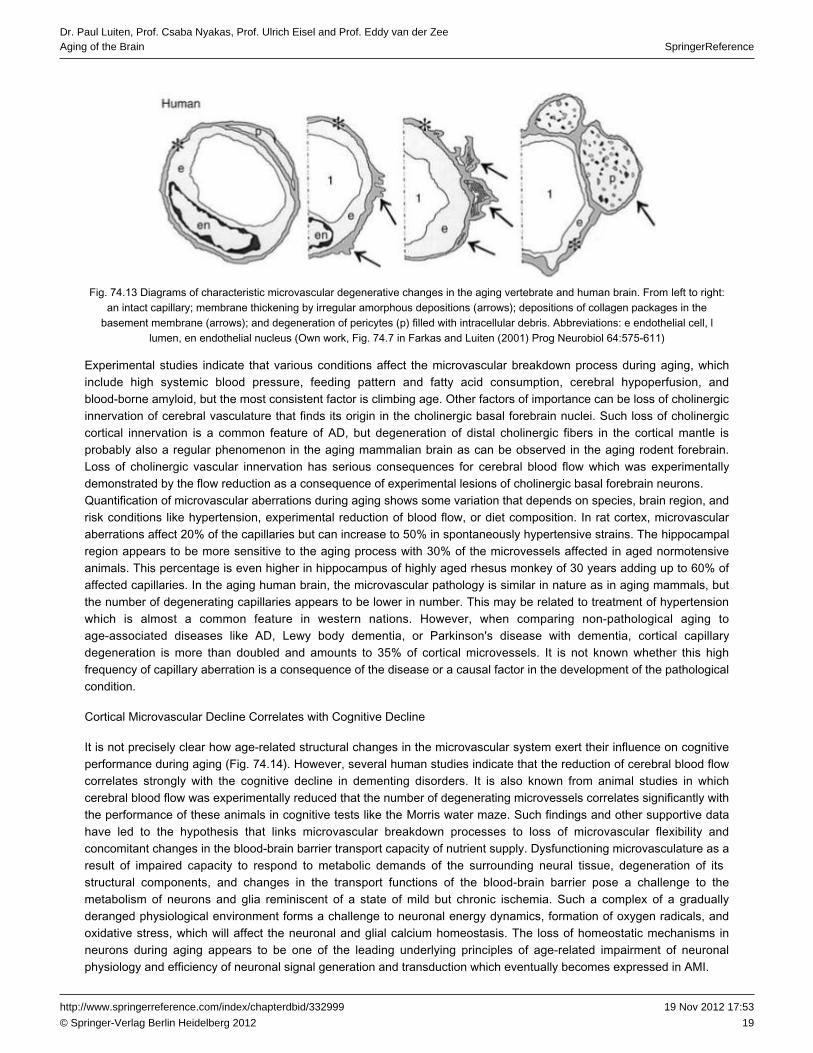
including man showed an almost linear decline of the cellular and subcellular condition of the microvascularspeciescomponents (Fig. 74.12). This decline can be summarized as the local increase of the thickness of the basementmembrane, distortion of the endothelial and its luminal surface, massive pericytic degeneration, and swellingcell structureof the surrounding astrocytic terminal endfeet embracing the capillary. The basement membrane thickening ischaracterized by deposition of various collagen components including packages of with its characteristiccollagen type IVperiodicity and of which the fibers can run both longitudinal to and circular to the capillary. Degenerating features ofmicrovascular consist of large amounts of intracellular membranous inclusions and swelling of the cytoplasm.pericytesPericytic degeneration has serious consequences since these cells are key players in the blood-brain barrier, controlcapillary integrity, and have a major function in microvascular blood flow (Fig. 74.13).
Fig. 74.12 Electron microscopic microphotographs of characteristic aging-dependent degenerative changes in the capillary wall ofmicrovessels in the cingulate cortex of senescent rats (30 month of age). The endothelial lining can often display an irregular shapeand surface ( in ( )), accompanied by an increase of mitochondria indicative for enhanced demand of energy probably forasterisks a
blood-brain barrier transport of nutrients and other components. The basement membrane undergoes considerable thickening by thedeposition of collagen of different compositions. The collagen in ( ) is clearly stratified by molecular periodicity, while in ( ) and ( ),b a c
the collagen became amorphous. Membrane thickening is accompanied by swelling of perivascular astrocytic endfeet ( inarrowheads( )) (Farkas and Luiten )c 2001
19
SpringerReferenceDr. Paul Luiten, Prof. Csaba Nyakas, Prof. Ulrich Eisel and Prof. Eddy van der ZeeAging of the Brain
19 Nov 2012 17:53http://www.springerreference.com/index/chapterdbid/332999
© Springer-Verlag Berlin Heidelberg 2012
Fig. 74.13 Diagrams of characteristic microvascular degenerative changes in the aging vertebrate and human brain. From to :left rightan intact capillary; membrane thickening by irregular amorphous depositions ( ); depositions of collagen packages in thearrows
basement membrane ( ); and degeneration of pericytes ( ) filled with intracellular debris. Abbreviations: endothelial cell, arrows p e llumen, endothelial nucleus (Own work, Fig. 74.7 in Farkas and Luiten ( ) Prog Neurobiol 64:575-611)en 2001
Experimental studies indicate that various conditions affect the microvascular breakdown process during aging, whichinclude high systemic blood pressure, feeding pattern and fatty acid consumption, cerebral hypoperfusion, andblood-borne amyloid, but the most consistent factor is climbing age. Other factors of importance can be loss of cholinergicinnervation of that finds its origin in the cholinergic basal forebrain nuclei. Such loss of cholinergiccerebral vasculaturecortical innervation is a common feature of AD, but degeneration of distal cholinergic fibers in the cortical mantle isprobably also a regular phenomenon in the aging mammalian brain as can be observed in the aging rodent forebrain.Loss of cholinergic vascular innervation has serious consequences for which was experimentallycerebral blood flowdemonstrated by the flow reduction as a consequence of experimental lesions of .cholinergic basal forebrain neuronsQuantification of microvascular aberrations during aging shows some variation that depends on species, brain region, andrisk conditions like hypertension, experimental reduction of blood flow, or diet composition. In rat cortex, microvascularaberrations affect 20% of the capillaries but can increase to 50% in spontaneously hypertensive strains. The hippocampal
appears to be more sensitive to the aging process with 30% of the microvessels affected in aged normotensiveregionanimals. This percentage is even higher in hippocampus of highly aged rhesus monkey of 30 years adding up to 60% ofaffected capillaries. In the aging human brain, the microvascular pathology is similar in nature as in aging mammals, butthe number of degenerating capillaries appears to be lower in number. This may be related to treatment of hypertensionwhich is almost a common feature in western nations. However, when comparing non-pathological aging toage-associated diseases like AD, , or Parkinson's disease with dementia, cortical capillaryLewy body dementiadegeneration is more than doubled and amounts to 35% of cortical microvessels. It is not known whether this highfrequency of capillary aberration is a consequence of the disease or a causal factor in the development of the pathologicalcondition.
Cortical Microvascular Decline Correlates with Cognitive Decline
It is not precisely clear how age-related structural changes in the microvascular system exert their influence on cognitiveperformance during aging (Fig. 74.14). However, several human studies indicate that the reduction of cerebral blood flowcorrelates strongly with the in dementing disorders. It is also known from animal studies in whichcognitive declinecerebral blood flow was experimentally reduced that the number of degenerating microvessels correlates significantly withthe performance of these animals in cognitive tests like the . Such findings and other supportive dataMorris water mazehave led to the hypothesis that links microvascular breakdown processes to loss of microvascular flexibility andconcomitant changes in the blood-brain barrier transport capacity of nutrient supply. Dysfunctioning microvasculature as aresult of impaired capacity to respond to metabolic demands of the surrounding neural tissue, degeneration of its
, and changes in the transport functions of the blood-brain barrier pose a challenge to thestructural componentsmetabolism of neurons and glia reminiscent of a state of mild but chronic ischemia. Such a complex of a graduallyderanged physiological environment forms a challenge to neuronal energy dynamics, formation of oxygen radicals, andoxidative stress, which will affect the neuronal and glial calcium homeostasis. The loss of homeostatic mechanisms inneurons during aging appears to be one of the leading underlying principles of age-related impairment of neuronalphysiology and efficiency of neuronal signal generation and transduction which eventually becomes expressed in AMI.

20
SpringerReferenceDr. Paul Luiten, Prof. Csaba Nyakas, Prof. Ulrich Eisel and Prof. Eddy van der ZeeAging of the Brain
19 Nov 2012 17:53http://www.springerreference.com/index/chapterdbid/332999
© Springer-Verlag Berlin Heidelberg 2012
Fig. 74.14 Diagram with hypothesis on interrelations between microvascular degenerative processes and cognitive dysfunctions.Conditions like high age, alone or in combination with hypertension, breakdown of the cholinergic innervation of forebrain vasculature,or blood-borne amyloid-β protein will lead to a gradual loss of vasoregulation processes of microvasculature accompanied by a variety
of degenerative changes of the microvascular wall. These microvascular aberrations will have their consequences for the transportmachinery of the blood-brain barrier of which the glucose transporter has a direct and immediate effect on supply of key nutrients. In achronic condition, this will affect the of the mitochondria that are highly dependent on unhampered availability of activity level energy
which has its consequences for ATP production and formation of . This, in turn, will exert its influencesources reactive oxygen specieson neuronal and glial physiology and the of the neuronal substrate underlying the cognitive brain ultimatelyphysiological functions
ending up in cognitive failure (Own work, Fig.19 in Farkas and Luiten ( ) Prog Neurobiol 64:575-611)2001
The Innate Inflammatory System of the Brain Can Both Damage and Protect the Brain AgainstBreakdown Processes
Challenging Conditions in the Brain Lead to Activation of Microglia
Microglia cells are cells of mesodermal origin and thus a group of cells that do not belong to the neuroectodermal line ofendogenous CNS cells like neurons, oligodendrocytes, and astrocytes. Microglia and their precursors invade the CNSduring early embryonic development and serve as CNS-specific tissue macrophages. The primary function of microgliaand astrocytes (macroglia) is the maintenance of tissue homeostasis including the active regulation and modulation of theneuronal circuit and the defense and repair upon cellular stress, infection, trauma, and other injuries. For these purposes,neurons, astrocytes, and microglia interact in a dynamic way. Resting microglia appears always in a ramified shape withfine cellular processes moving around and actively scanning their cellular environment. Upon sensing of any homeostaticdisturbance, microglia change their shape and can induce , . For thistissue remodeling inflammation, and neurogenesis

21
SpringerReferenceDr. Paul Luiten, Prof. Csaba Nyakas, Prof. Ulrich Eisel and Prof. Eddy van der ZeeAging of the Brain
19 Nov 2012 17:53http://www.springerreference.com/index/chapterdbid/332999
© Springer-Verlag Berlin Heidelberg 2012
purpose, microglia are able to induce a broad spectrum of molecules such as cytokines and chemokines, and can serveas antigen presenting cells. These possibilities enable microglia to trigger a cascade of responses inducing the attractionof peripheral immune cells through the blood-brain barrier which can finally end in a massive inflammatory responseincluding the induction of apoptosis, but they are also important in the finalizing of an , removing ofinflammatory reactioncell debris, and cell survival.
Neuroinflammatory Mechanisms and Release of Cytokines by Microglia Can Protect Against External Threats
The communication and interaction between microglia or the immune system and neurons - in close physical contact - isessential for the maintenance of brain function and homeostasis but also for the modulation of neurological functions anddysfunctions. The first line of defense is the . Innate immune cells such as microglia in the CNSinnate immune responsedetect potential disturbances in their environment via pattern-recognition receptors that recognize infections as well astissue damage. They respond by synthesizing and releasing cytokines such as , IL-6, and interleukin-1β (IL-1β) tumor
which mediate the immune response. These cytokines, however, can also lead to thenecrosis factor-α (TNF-α)breakdown of the blood-brain barrier and the infiltration of peripheral immune cells.Cytokines (also stemming from peripheral organs) can induce the so-called sickness behavior, a mixture of ,lethargydecreased activity, and retraction from social interactions, which goes together with other responses to infections likeincreased sleep, higher body temperature, decreased food intake, and activation of the hypothalamic-pituitary axis. Thisgeneral sickness response is thought to promote resistance to pathogens and to recovery from infection.Besides the response to intruding pathogens, microglia and other immune cells also monitor the state of tissue damageand can induce a transformatory process called tissue repair. This can also include signals to induce apoptosis of badlydamaged cells at the same time when other cells get trophic support.Microglia can activate the neuroinflammatory response by expressing proinflammatory cytokines such as TNF-α, ,IL-1αand ILβ or IL-6. Although the proinflammatory responses are often reported as part of a pathological process, mostinflammatory processes are actually protective in nature (Fig. 74.15). TNF-α, for example, can induce apoptosis via its
receptor-1, whereas TNF receptor-2 counteracts this effect and triggers a neuroprotective signaling cascade via TNF and . Interestingly this neuroprotective signal is enhanced byprotein kinase B (PKB/Akt) nuclear factor kappa B (NF-κB)
the ionotropic glutamate receptor and therefore reveals a direct interaction of neuronal and immune signals at theNMDAmolecular level.

22
SpringerReferenceDr. Paul Luiten, Prof. Csaba Nyakas, Prof. Ulrich Eisel and Prof. Eddy van der ZeeAging of the Brain
19 Nov 2012 17:53http://www.springerreference.com/index/chapterdbid/332999
© Springer-Verlag Berlin Heidelberg 2012
Fig. 74.15 TNF cell survival signaling in neurons. TNF-induced signaling via its two plays anreceptors. Tumor necrosis factorimportant role in guiding the cell through stress situations. The two receptors for TNF can have therefore extremely opposite
functions: with TNFR1-mediated apoptosis and TNFR2-mediated pro-survival pathways. Depending on the surrounding situation, theTNF system allows cells not only to respond by cellular restructuring, adaptation, and modulation of the surrounding immune
response by activating either pro- or anti-inflammatory responses. One key signaling mechanism in achieving this is the activation ofthe transcription factor NF-κB (p50/p65). Both TNFRs are able to activate this transcription factor by separate signaling mechanisms
and with different kinetics. Anti-apoptotic activation of NF-κB via in neurons is achieved by co-signaling with the ionotropicTNFR2glutamate receptor . Together they can activate protein kinase B/Akt which activates a long-lasting NF-κB activation whichNMDAR
leads to neuronal survival. Abbreviations: , cellular inhibitor of apoptosis, DD death domain cIAP FADD Fas-associated death domainprotein, inhibitor of kappa B, nuclear factor kappa B, protein kinase B/serine-threonine kinase, IκBα NFκB PKB/Akt PI3K
phosphoinositide 3-kinases, receptor-interacting protein, , TNFRIP TRADD TNF receptor-associated death domain TRAFreceptor-associated factor, receptor-1, tumor necrosis factorfactor, tumor necrosisTNF TNFR1 tumor necrosis factor TNFR2
receptor-2
The Dynamic Activation of Microglia Changes in Later Life
During , microglia become more sensitized, which means they react faster and stronger upon activation. Thisnormal agingeffect, also known as priming, results in a more rapid induction of activation and increased cytokine release. Primedmicroglia seem to stay in an intermediate activation state. Microglia priming was first described in chronic
but seems also to be a part of the aging process of the brain. Several indicators support thisneurodegenerative diseasesview: Microglial expression of MHCII, a marker for microglial activation, is increased in aged but healthy brains along withother cell surface markers and . In addition, it was shown that aged microglia expresses highercell adhesion moleculesamounts of , IL-6, and . As these cytokines are normally considered to be proinflammatory, it is of someIL-1β TNF-αimportance that at the same time such as IL-10 and IL-4 are decreased. These findings areanti-inflammatory cytokinesaccompanied by findings that during aging, genes involved in oxidative stress, inflammation, and glial activation areupregulated whereas those associated with synaptic, transport, and restorative functions decrease. This leads to a ratherpro-inflammatory and less flexible phenotype. Age-related priming of microglia leads to an exaggerated immune responsefollowing challenges. Prolonged and/or chronic increase of cytokines such as IL-1β, IL-6, and TNF-α leads to extendedsickness response, impaired cognition, and depressive behaviors.

23
SpringerReferenceDr. Paul Luiten, Prof. Csaba Nyakas, Prof. Ulrich Eisel and Prof. Eddy van der ZeeAging of the Brain
19 Nov 2012 17:53http://www.springerreference.com/index/chapterdbid/332999
© Springer-Verlag Berlin Heidelberg 2012
Neuroinflammatory Processes Affect Cognitive Functions During Aging
To estimate the grade of influence that the immune system has on brain functions, one should consider, for example, themood changes at the onset of a . So it is no wonder that a direct influence of components of the immunecommon coldsystem on basic neuronal functions can be observed. Proinflammatory cytokines such as IL-1b, IL-6, and TNF-α directlyinfluence . Glia actively participates in the homeostatic activity-dependent regulation of synaptic activity by the releaseLTPof TNF-α. TNF-α also increases the expression of and influences . In addition to theAMPA receptors synaptic scalingmodulatory function in neuronal networks, cytokines and microglia are also considered to be involved in the developmentof depression. As depressive and sickness behavior have a lot in common, a new concept considers a neuroinflammatoryresponse at the basis of major depression. In fact, peripheral as well as centrally applied lipopolysaccharides can(LPS)induce an inflammatory response in the brain and can induce in animal models depressive-like behavior. These effectscan be mimicked by various proinflammatory cytokines. It has been argued that these mechanisms come into play duringdevelopment of late-onset depression in the elderly. The modulatory function of cytokines is of great interest when weconsider their constitutive upregulation during but also in neurodegenerative diseases is considered. Withhealthy agingprimed microglia during aging in a more proinflammatory state, with more exaggerated responses to stimuli, themodulatory role of cytokines on and mood functions and depression, it is now becoming bettercognitive functionsunderstood which important role the immune response plays in AMI and why aged people are more prone to developdepressive symptoms.
Outlook
Aging of the brain is more relevant than ever given that life expectancy is nearly doubled in the twentieth century. Assome say, "the aging boom is upon us." Although the science of aging is developing rapidly, it still has to come of age.Many of the fundamental underpinnings of remain unclear and require continuous and extended researchbrain agingefforts of a multidisciplinary nature. , genetic, biochemical, and physiological processes need to beNeurobiologicalinterconnected to fully appreciate and understand the maintenance machinery to keep the brain going at a high level ofcognitive performance - even at an age of 85 and beyond as is anticipated to be the average life expectancy in the nearfuture. We expect that many more secrets of how to age healthy will be unraveled in the near future, and this chapterprovides some of key topics in which such discoveries may arise.There is no doubt that we will experience a burgeoning increase in scientific knowledge on the aging brain. Most, if not all,nations in the world are confronted with an , and since most health problems and problems of well-beingaging populationmanifest themselves in the later phases of human life, this is becoming a matter of global dimensions. Aging is aphenomenon with a large number of interacting and interdependent variables. When we can more fully understand thesefactors that determine the functioning of the aged individual, we could make predictions on how to adapt to lifestyle and(preferably noninvasive) treatments in order to achieve a better health condition, guarantee independent living, and delaythe onset of aging-associated disease. It is obvious from many scientific sources that the fate of the aged brain alreadystarts with the beginning of life with the genetic capital we are endowed with. Furthermore, complex factors like foodquality and food composition, education and upbringing, chronic stress levels, hygiene, available health care during thevarious life phases including pregnancy, exposure to disease in , and so on all exert their influence on the agingearly lifeprocess. No doubt that the collective information on all these determining factors will be leading to a better insight inhealthy aging conditions. For example, we know that perinatal stress makes the brain more sensitive to later life ischemicevents, we know that lipid composition in the daily diet affects brain vessel condition and blood pressure, we know thatcalcium inhibiting drugs can improve learning performance in aging, we know that high levels of stress hormonesenhance the formation of the neurotoxic amyloid proteins, and we know that regular motor activity promotes the formationof new neurons. These are just a few examples of how we could influence our aging brain in a positive way based onrecent scientific insights.
Further Reading
Barcelo-Coblijn G, Hogyes E, Kitajka K, Puskás LG, Zvara A, Hackler L Jr, Nyakas C, Penke Z, Farkas T (2003)Modification by docosahexaenoic acid of age-induced alterations in gene expression and molecular compositionof rat brain phospholipids. Proc Natl Acad Sci USA 100:11321-11326

24
SpringerReferenceDr. Paul Luiten, Prof. Csaba Nyakas, Prof. Ulrich Eisel and Prof. Eddy van der ZeeAging of the Brain
19 Nov 2012 17:53http://www.springerreference.com/index/chapterdbid/332999
© Springer-Verlag Berlin Heidelberg 2012
Bishop NA, Lu T, Yankner BA (2010) Neural mechanisms of ageing and cognitive decline. Nature 464:529-535Buckner RL, Snyder AZ, Sanders AL, Raichle ME, Morris JC (2000) Functional brain imaging of young,nondemented, and demented older adults. J Cogn Neurosc 12(Suppl 2):24-34Burke SN, Barnes CA (2006) Neural plasticity in the aged brain. Nat Rev Neurosci 7:30-40Dickstein DL, Kabaso D, Rocher AB, Luebke JI, Wearne SL, Hof PR (2007) Changes in the structural complexityof the aged brain. Aging Cell 6(3):275-284Disterhoft JF, Oh MM (2006) Learning, aging and intrinsic neuronal plasticity. Trends Neurosci 29:587-599Farkas E, Luiten PGM (2001) Cerebral microvascular pathology in aging and Alzheimer's disease. Prog Neurobiol64:575-611Fontaine V, Mohand-Said S, Hanoteau N, Fuchs C, Pfizenmaier K, Eisel U (2002) Neurodegenerative andneuroprotective effects of tumor necrosis factor (TNF) in retinal ischemia: opposite roles of TNF receptor 1 andTNF receptor 2. J Neurosci 22:RC216 (1)-RC216 (7)Gaykema RP, Nyakas C, Horvath E, Hersh LB, Majtenyi C, Luiten PG (1992) Cholinergic fiber aberrations innucleus basalis lesioned rat and Alzheimer's disease. Neurobiol Aging 13:441-448Geinisman Y, Berry RW, Disterhoft JF, Power JM, Van der Zee EA (2001) Associative learning elicits theformation of multiple-synapse boutons. J Neurosci 21:5568-5573Havekes R, Abel T, Van der Zee EA (2011) The cholinergic system and neostriatal memory functions. BehavBrain Res 221:412-423Kirischuk S, Verkhratsky A (1996) Calcium homeostasis in aged neurons. Life Sci 59:451-459Landfield PW, Pitler TA (1984) Prolonged Ca -dependent afterhyperpolarization in hippocampal neurons of2+
aged rats. Science 226:1089-1092Mattson MP (2007) Calcium and neurodegeneration. Aging Cell 6:337-350Nimmerjahn A, Kirchhoff F, Helmchen F (2005) Resting microglial cells are highly dynamic surveillants of brainparenchyma in vivo. Science 308:1314-1318Ostroveanu A, Van der Zee EA, Dolga AM, Luiten PG, Eisel UL, Nijholt IM (2007) A-kinase anchoring protein 150in the mouse brain is concentrated in areas involved in learning and memory. Brain Res 1145:97-107Stellwagen D, Malenka RC (2006) Synaptic scaling mediated by glial TNF-a. Nature 440:1054-1059Tombaugh GC, Rowe WB, Rose GM (2005) The slow afterhyperpolarization in hippocampal CA1 neuronscovaries in aged Fisher 344 rats. J Neurosci 25:2609-2616Van der Zee EA, Naber PA, Disterhoft JF (1997) Age-dependent changes in the immunoreactivity forneurofilaments in rabbit hippocampus. Neuroscience 79:103-116Van der Zee EA, Palm IF, O'Connor M, Maizels ET, Hunzicker-Dunn M, Disterhoft JF (2004) Aging-relatedalterations in the distribution of Ca(2+)-dependent PKC isoforms in rabbit hippocampus. Hippocampus 14:849-860Verkhratsky A, Toescu EC (1998) Calcium and neuronal aging. Trends Neurosci 21:2-7Wood WG, Schroeder F, Igbavboa U, Avdulov NA, Chochina SV (2002) Brain membrane cholesterol domains,aging and amyloid beta-peptides. Neurobiol Aging 23:685-694
25
SpringerReferenceDr. Paul Luiten, Prof. Csaba Nyakas, Prof. Ulrich Eisel and Prof. Eddy van der ZeeAging of the Brain
19 Nov 2012 17:53http://www.springerreference.com/index/chapterdbid/332999
© Springer-Verlag Berlin Heidelberg 2012
Aging of the Brain
Dr. Paul LuitenDepartment of Molecular Neurobiology, University of Groningen, Groningen,The Netherlands
Prof. CsabaNyakas
Department of Molecular Neurobiology, University of Groningen, Groningen,The Netherlands
Prof. Ulrich EiselDepartment of Molecular Neurobiology, University of Groningen, Groningen,The Netherlands
Prof. Eddy van derZee
Department of Molecular Neurobiology, University of Groningen, Groningen,The Netherlands
DOI: 10.1007/SpringerReference_332999
URL: http://www.springerreference.com/index/chapterdbid/332999
Part of: Neuroscience in the 21st Century
Editor: Donald Pfaff
PDF created on: November, 19, 2012 17:53
© Springer-Verlag Berlin Heidelberg 2012
Copyright © 2022 FDOKUMEN