Advancing Regenerative Cellular Therapies in Non-Scarring ...
25
Pharmaceutics 2022, 14, 612. https://doi.org/10.3390/pharmaceutics14030612 www.mdpi.com/journal/pharmaceutics Review Advancing Regenerative Cellular Therapies in Non‐Scarring Alopecia Talagavadi Channaiah Anudeep 1,2,3,4 , Madhan Jeyaraman 2,4,5 , Sathish Muthu 2,4,6 , Ramya Lakshmi Rajendran 7 , Prakash Gangadaran 7,8, *, Prabhu Chandra Mishra 4 , Shilpa Sharma 4,9 , Saurabh Kumar Jha 2,4 and Byeong‐Cheol Ahn 7,8, * 1 Department of Plastic Surgery, Topiwala National Medical College and BYL Nair Ch. Hospital, Mumbai 400008, India; [email protected] 2 Department of Biotechnology, School of Engineering and Technology, Sharda University, Greater Noida 201310, India; [email protected] (M.J.); [email protected] (S.M.); [email protected] (S.K.J.) 3 À La Mode Esthétique Studio, Mysuru 570011, India 4 International Association of Stem Cell and Regenerative Medicine (IASRM), New Delhi 110092, India; [email protected] (P.C.M.); [email protected] (S.S.) 5 Department of Orthopaedics, Faculty of Medicine—Sri Lalithambigai Medical College and Hospital, Dr MGR Educational and Research Institute, Chennai 600095, India 6 Department of Orthopaedics, Government Medical College and Hospital, Dindigul 624304, India 7 Department of Nuclear Medicine, School of Medicine, Kyungpook National University, Kyungpook National University Hospital, Daegu 41944, Korea; [email protected] 8 BK21 FOUR KNU Convergence Educational Program of Biomedical Sciences for Creative Future Talents, Department of Biomedical Sciences, School of Medicine, Kyungpook National University, Daegu 41944, Korea 9 Department of Paediatric Surgery, All India Institute of Medical Sciences, New Delhi 110029, India * Correspondence: [email protected] (P.G.); [email protected] (B.‐C.A.) Abstract: Alopecia or baldness is a common diagnosis in clinical practice. Alopecia can be scarring or non‐scarring, diffuse or patchy. The most prevalent type of alopecia is non‐scarring alopecia, with the majority of cases being androgenetic alopecia (AGA) or alopecia areata (AA). AGA is tra‐ ditionally treated with minoxidil and finasteride, while AA is treated with immune modulators; however, both treatments have significant downsides. These drawbacks compel us to explore re‐ generative therapies that are relatively devoid of adverse effects. A thorough literature review was conducted to explore the existing proven and experimental regenerative treatment modalities in non‐scarring alopecia. Multiple treatment options compelled us to classify them into growth factor‐ rich and stem cell‐rich. The growth factor‐rich group included platelet‐rich plasma, stem cell‐con‐ ditioned medium, exosomes and placental extract whereas adult stem cells (adipose‐derived stem cell‐nano fat and stromal vascular fraction; bone marrow stem cell and hair follicle stem cells) and perinatal stem cells (umbilical cord blood‐derived mesenchymal stem cells (hUCB‐MSCs), Wharton jelly‐derived MSCs (WJ‐MSCs), amniotic fluid‐derived MSCs (AF‐MSCs), and placental MSCs) were grouped into the stem cell‐rich group. Because of its regenerative and proliferative capabili‐ ties, MSC lies at the heart of regenerative cellular treatment for hair restoration. A literature review revealed that both adult and perinatal MSCs are successful as a mesotherapy for hair regrowth. However, there is a lack of standardization in terms of preparation, dose, and route of administra‐ tion. To better understand the source and mode of action of regenerative cellular therapies in hair restoration, we have proposed the “À La Mode Classification”. In addition, available evidence‐ based cellular treatments for hair regrowth have been thoroughly described. Keywords: alopecia; mesenchymal stem cells; regenerative therapy; cellular therapy Citation: Anudeep, T.C.; Muthu, S.; Jeyaraman, M.; Rajendran, R.L.; Gangadaran, P.; Mishra, P.C.; Sharma, S.; Jha, S.K.; Ahn, B.‐C. Advancing Regenerative Cellular Therapies in Non‐Scarring Alopecia. Pharmaceutics 2022, 14, 612. https://doi.org/10.3390/ pharmaceutics14030612 Academic Editors: Jianqing Gao and Bianca Vezzani Received: 27 January 2022 Accepted: 7 March 2022 Published: 10 March 2022 Publisher’s Note: MDPI stays neu‐ tral with regard to jurisdictional claims in published maps and institu‐ tional affiliations. Copyright: © 2022 by the authors. Licensee MDPI, Basel, Switzerland. This article is an open access article distributed under the terms and conditions of the Creative Commons Attribution (CC BY) license (https://creativecommons.org/license s/by/4.0/).
-
Upload
khangminh22 -
Category
Documents
-
view
0 -
download
0
Transcript of Advancing Regenerative Cellular Therapies in Non-Scarring ...

Pharmaceutics 2022, 14, 612. https://doi.org/10.3390/pharmaceutics14030612 www.mdpi.com/journal/pharmaceutics
Review
Advancing Regenerative Cellular Therapies
in Non‐Scarring Alopecia
Talagavadi Channaiah Anudeep 1,2,3,4, Madhan Jeyaraman 2,4,5, Sathish Muthu 2,4,6, Ramya Lakshmi Rajendran 7,
Prakash Gangadaran 7,8,*, Prabhu Chandra Mishra 4, Shilpa Sharma 4,9, Saurabh Kumar Jha 2,4
and Byeong‐Cheol Ahn 7,8,*
1 Department of Plastic Surgery, Topiwala National Medical College and BYL Nair Ch. Hospital,
Mumbai 400008, India; [email protected] 2 Department of Biotechnology, School of Engineering and Technology, Sharda University,
Greater Noida 201310, India; [email protected] (M.J.); [email protected] (S.M.);
[email protected] (S.K.J.) 3 À La Mode Esthétique Studio, Mysuru 570011, India 4 International Association of Stem Cell and Regenerative Medicine (IASRM),
New Delhi 110092, India; [email protected] (P.C.M.); [email protected] (S.S.) 5 Department of Orthopaedics, Faculty of Medicine—Sri Lalithambigai Medical College and Hospital,
Dr MGR Educational and Research Institute, Chennai 600095, India 6 Department of Orthopaedics, Government Medical College and Hospital, Dindigul 624304, India 7 Department of Nuclear Medicine, School of Medicine, Kyungpook National University,
Kyungpook National University Hospital, Daegu 41944, Korea; [email protected] 8 BK21 FOUR KNU Convergence Educational Program of Biomedical Sciences for Creative Future Talents,
Department of Biomedical Sciences, School of Medicine, Kyungpook National University,
Daegu 41944, Korea 9 Department of Paediatric Surgery, All India Institute of Medical Sciences, New Delhi 110029, India
* Correspondence: [email protected] (P.G.); [email protected] (B.‐C.A.)
Abstract: Alopecia or baldness is a common diagnosis in clinical practice. Alopecia can be scarring
or non‐scarring, diffuse or patchy. The most prevalent type of alopecia is non‐scarring alopecia,
with the majority of cases being androgenetic alopecia (AGA) or alopecia areata (AA). AGA is tra‐
ditionally treated with minoxidil and finasteride, while AA is treated with immune modulators;
however, both treatments have significant downsides. These drawbacks compel us to explore re‐
generative therapies that are relatively devoid of adverse effects. A thorough literature review was
conducted to explore the existing proven and experimental regenerative treatment modalities in
non‐scarring alopecia. Multiple treatment options compelled us to classify them into growth factor‐
rich and stem cell‐rich. The growth factor‐rich group included platelet‐rich plasma, stem cell‐con‐
ditioned medium, exosomes and placental extract whereas adult stem cells (adipose‐derived stem
cell‐nano fat and stromal vascular fraction; bone marrow stem cell and hair follicle stem cells) and
perinatal stem cells (umbilical cord blood‐derived mesenchymal stem cells (hUCB‐MSCs), Wharton
jelly‐derived MSCs (WJ‐MSCs), amniotic fluid‐derived MSCs (AF‐MSCs), and placental MSCs)
were grouped into the stem cell‐rich group. Because of its regenerative and proliferative capabili‐
ties, MSC lies at the heart of regenerative cellular treatment for hair restoration. A literature review
revealed that both adult and perinatal MSCs are successful as a mesotherapy for hair regrowth.
However, there is a lack of standardization in terms of preparation, dose, and route of administra‐
tion. To better understand the source and mode of action of regenerative cellular therapies in hair
restoration, we have proposed the “À La Mode Classification”. In addition, available evidence‐
based cellular treatments for hair regrowth have been thoroughly described.
Keywords: alopecia; mesenchymal stem cells; regenerative therapy; cellular therapy
Citation: Anudeep, T.C.; Muthu, S.;
Jeyaraman, M.; Rajendran, R.L.;
Gangadaran, P.; Mishra, P.C.;
Sharma, S.; Jha, S.K.; Ahn, B.‐C.
Advancing Regenerative Cellular
Therapies in Non‐Scarring Alopecia.
Pharmaceutics 2022, 14, 612.
https://doi.org/10.3390/
pharmaceutics14030612
Academic Editors: Jianqing Gao
and Bianca Vezzani
Received: 27 January 2022
Accepted: 7 March 2022
Published: 10 March 2022
Publisher’s Note: MDPI stays neu‐
tral with regard to jurisdictional
claims in published maps and institu‐
tional affiliations.
Copyright: © 2022 by the authors.
Licensee MDPI, Basel, Switzerland.
This article is an open access article
distributed under the terms and
conditions of the Creative Commons
Attribution (CC BY) license
(https://creativecommons.org/license
s/by/4.0/).

Pharmaceutics 2022, 14, 612 2 of 25
1. Introduction
Alopecia or baldness is a common diagnosis in clinical practice. A variety of causes,
including genetics, hormones, autoimmune, trauma, stress, and iatrogenic factors, all play
an important part in the pathophysiology of alopecia. Alopecia can be scarring or non‐
scarring, diffuse or patchy. The most prevalent type of alopecia is non‐scarring alopecia,
with the majority of cases being androgenetic alopecia (AGA) or alopecia areata (AA).
Androgenetic alopecia is a disorder of exaggerated response of hair follicles of the scalp
to systemic androgens leading to accelerated patterned hair loss [1,2]. AGA is a progres‐
sive condition with a hereditary propensity that is the most prevalent cause of baldness
in both men and women. It is characterized by hair follicle miniaturization and inflamma‐
tion [3–7]. Even though AGA is typically seen in patients in their 20’s and 30’s, disease
process begins with the onset of puberty and it progresses thereafter [3,8]. AGA warrants
immediate attention from a qualified trichologist because of its psychosocial impact on
patients, which in turn may lead to emotional distress and decreased quality of life [9–11].
Hair loss pattern in AGA varies between men and women. Male androgenetic alopecia is
characterized by bitemporal recession, frontal balding, and balding of the vertex of the
scalp, as opposed to female androgenetic alopecia, which is characterized by diffuse thin‐
ning of hair and sparing of the frontal hairline [12–14].
2. Hair Growth Cycle and Its Regulators
Hair is an appendage of the skin. The skin‐hair unit has been structurally divided
into three subunits, namely interfollicular epidermis (IFE), the hair follicle (HF), and the
sebaceous gland (SG). Just beneath the sebaceous gland, near the outer root sheath, we
can spot ‘the bulge’, which is a storehouse of stem cells. These stem cells have the innate
property of asymmetric self‐renewal to produce transit amplifying matrix cells, which en‐
cases the mesenchymal cells. These transits amplifying matrix cells move towards the sur‐
face to differentiate into epidermal keratinocytes and they move downwards towards the
hair follicle matrix to multiply and differentiate into the shaft of hair [15,16]. The adult HF
is always a part of a constant hair cycle which consists of three stages, namely (a) cat‐
agen—phase of degeneration, (b) telogen—resting phase and (c) anagen—growth phase.
The cradle of proliferation during the anagen phase is HF stem cells (HFSCs). The
matrix cells undergo apoptosis during the catagen phase which moves the dermal papilla
(DP) towards the epidermis below the hair germ, it is the early descendants of bulge stem
cells. DP maintains HFSCs in a dormant state and proficient for the next hair cycle [17].
SG and IFE are not maintained by HFSC, but they will help in the regeneration of the
epidermis and SG after wounding. Based on their location with respect to the basal lam‐
ina, HFSCs are divided into basal and suprabasal HFSCs [18]. The interaction between
epithelial and mesenchymal components is important to regulate HFSCs. Wnt signaling
helps in the proliferation by stabilizing β‐catenin, which translocate to the nucleus and
complexes with LEF1 to form a transcriptional activating complex leading to proliferation.
Hair follicles are maintained in the dormant state by Lef‐1/Tcf‐3 [19]. In the adult HF, the
accumulation of β‐catenin in the nucleus of HFSCs is associated with telogen to anagen
transition, this signifies the importance of Wnt signaling in the self‐renewal capacity of
the stem cells [20]. Wnt/β‐catenin signaling and LEF1 in the bulge are very important for
matrix cell differentiation towards the shaft [21]. Despite this, the source of the Wnt ligand
is not easy to elucidate [22]. The BMP pathway inhibits HF morphogenesis and adult
HFSC proliferation (Figure 1) [23,24]. BMP ligands and the antagonist noggin are balanced
by mesenchyme [19]. Heightened cycling of HFSCs is seen when the BMPR1a receptor is
inactivated in the HF and it impairs differentiation. The Hedgehog and Notch signaling
pathways are also implicated in the proliferation and differentiation of HF [25].

Pharmaceutics 2022, 14, 612 3 of 25
Figure 1. Structure of hair follicle and the molecular mechanisms involved in hair cycle. (BMP—
bone morphogenetic protein, TA—transit amplifying, ORS—outer root sheath, HFSCs—hair follic‐
ular stem cells). Created with BioRender.com (accessed on 27 January 2022).
3. Treatment of Non‐Scarring Alopecia and Adverse Effects
Conventionally, AGA treatments aim at lowering the levels of dihydroxy testos‐
terone by using 5 alpha‐reductase inhibitors, minoxidil and finasteride, a potassium chan‐
nel opener [26]. Minoxidil (topical 5% solution/foam) and finasteride (oral 1 mg/day) are
the first‐line therapies for AGA.
Finasteride is an inhibitor of 5 alpha‐reductase type 2, which decreases the concen‐
tration of DHT in serum and scalp and, hence, increases hair growth. It has to be taken for
at least 1 year to notice hair growth and has to be continued to maintain the hair growth.

Pharmaceutics 2022, 14, 612 4 of 25
The effect of regrowth will be lost if the drug is stopped for 6–9 months. Erectile and ejac‐
ulatory dysfunction are among the many negative effects of finasteride. Gynecomastia,
testicular pain, and depression are the rare adverse effects of finasteride [27,28]. Finaster‐
ide may interfere with the levels of prostatic specific antigen (PSA), it is shown that PSA
level decreases considerably in patients taking finasteride [29].
Minoxidil is a potassium channel opener. It is a vasodilator and induces vascular en‐
dothelial growth factor (VEGF) to increase vascularity and dermal papilla size [30,31]. The
response to treatment is variable. It requires 12–18 months to assess the efficacy of minox‐
idil. It has to be continued life long and the effect ceases with the stoppage of treatment.
There might be exaggerated hair fall in the initial 2 months (telogen to anagen transition
phase), but it improves over 2 months. Adverse effects include contact dermatitis, irritant
dermatitis, and hypertrichosis over the face [32,33]. Other new modalities include platelet‐
rich plasma (PRP), mesotherapy, lasers, micro‐needling of the scalp, and Janus kinase
(JAK) inhibitors. When patients are keen on immediate results, they resort to hair trans‐
plantation, i.e., follicular unit transplantation (FUT) or follicular unit extraction (FUE).
It is clear from the studies that first‐line therapies for AGA that:
1. They have to be continued lifelong, which decreases the compliance;
2. The regrowth ceases with the discontinuation of therapy;
3. They are associated with several adverse effects leading to temporary morbidity.
Alopecia areata (AA) is a non‐scarring chronic, immune‐inflammatory disorder of
hair follicles. The most typical manifestation of AA is the presence of localized patches of
hair loss on the scalp, but severe cases can result in generalized hair loss throughout the
body [34]. AA affects about 2% of the general population at some point in life [35]. The
disruption of the hair follicle’s immune privilege is regarded to be a key element in the
pathophysiology of AA. It is centered on a lymphocytic infiltration in and around the hair
follicle’s bulb or lower part. Despite various therapeutic options, such as corticosteroids
(topical, intralesional, oral), tacrolimus, minoxidil, contact immunotherapies like squaric
acid dibutyl ester, diphencyprone, and photo(chemo)therapy using UVA and psoralens,
there is no cure for AA [36].
It is obvious that the therapies for AA:
1. Have unpredictable outcome;
2. Have no permanent cure;
3. Are associated with significant adverse reactions.
This has led researchers to explore regenerative therapies for hair restoration in non‐
scarring alopecia. Being autologous/allogenic, they are free from adverse effects with good
patient compliance. The regenerative modalities explored are PRP, amniotic fluid, adipose‐
derived stem cells, follicular micrograft, bone marrow cells, cord blood and Wharton jelly.
Regenerative therapies in non‐scarring alopecia can include cells, which can produce
factors inducing hair growth or the products of the cells which can be isolated and used.
Cells as such are difficult to maintain in culture for transplantation whereas growth‐factors
secreted by the cells in the medium are easy to transport and is less expensive compared to
cellular therapy as such. Hence, it is essential to classify the regenerative therapies into
growth factor‐rich and stem‐cell rich, which will aid in better understanding, clinical utility
and for further research. Therefore, we suggest this “A La Mode Classification” (Table 1).

Pharmaceutics 2022, 14, 612 5 of 25
Table 1. “A La Mode Classification” of regenerative therapies [37–60].
Stem Cell‐Rich Growth Factor‐Rich
I. Adult stem cells
A. Adipose derived stem cell (ADSC)
1. Nanofat
2. Stromal vascular fraction (SVF)
B. Hair follicular stem cell (HFSCs)
1. Autologous micro grafts [Human intra
and extra dermal adipose tissue derived
hair follicle stem cells (HD‐AFSCs)]
2. Cultured HFSCs
i. Hair follicle‐derived MSCs
(HF‐MSC)
ii. Hair follicle epidermal stem
cells (HF‐ESC)
C. Bone marrow derived
1. Bone marrow mononuclear cells
(BMMC)
2. Bone marrow aspirate concentrate
(BMAC)
II. Perinatal stem cells
A. Umbilical cord blood derived
B. Wharton jelly MSCs (WJ‐MSC)/Umbilical
cord MSCs (UC‐MSC)
1. Sub amniotic.
2. Perivascular.
3. Intervascular.
C. Amniotic fluid derived
D. Placental MSC
I. Platelet‐rich plasma (PRP)
A. Autologous activated PRP (AA‐PRP)
B. Autologous non‐activated PRP (A‐PRP)
II. Autologous growth factor concentrate (GFC)
III. Conditioned medium (Secretomes)
A. ADSC‐CM (AAPE)
B. hUCB‐MSC‐CM
C. AF‐MSC‐CM
D. HF‐MSC‐CM
E. BM‐MSC‐CM (Genetically engineered)
IV. Extracellular vesicles
A. Exosomes
i. New‐born foreskin stem cell
ii. DPC
iii. BM‐MSC
B. Exosome‐like (Ginseng)
C. Microvesicles
V. Placental extract
In this article, we will review the cellular therapies used to treat non‐scarring alope‐
cia, as shown in Figure 2, their mechanism of action and the recent studies to validate its
efficacy. In addition, studies on conditioned medium are briefly mentioned at places to
reiterate the potential of cellular therapy.

Pharmaceutics 2022, 14, 612 6 of 25
Figure 2. Cellular therapy for non‐scarring alopecia. Created with BioRender.com (accessed on 1
March 2022).
4. Cellular Therapy
4.1. Adult Stem Cells
MSCs are the most dynamic, immature, diverse, and multipotent stromal progenitor
cells. They have a morphology similar to fibroblasts and have the capacity for trans‐dif‐
ferentiation into a range of tissues of ectoderm, endoderm, and mesodermal origin. MSC
clonal heterogeneity is demonstrated in terms of variable differentiation, regeneration and
proliferative capability in both in vivo and in vitro studies. MSCs express non‐differenti‐
ating cell surface markers, such as CD146 or CD200. MSCs are found in bone marrow, the
placenta, the umbilical cord, fat, menstrual blood, molar teeth and amniotic fluid [61]. As
there are multiple sources of MSCs, it is of immense importance to define an MSC. In 2006,
Mesenchymal and Tissue Stem Cell Committee of the International Society for Cellular
Therapy has proposed three minimum criteria to define an MSC. They are as follows:
1. MSC must be plastic adherent when maintained under standard culture conditions.
2. Expression of CD105, CD73 and CD90, and lack expression of CD45, CD34, CD14, or
CD11b, CD79a, or CD19 and HLA‐DR surface molecules.
3. They must differentiate to osteoblasts, adipocytes and chondroblasts in vitro [62].
MSCs are more adaptable in nature, allowing them to switch from one differentiation
path to another as influenced by growth factors, cytokines, and chemokines. The conver‐
sion of chondrocytes into osteoblasts and osteoblasts into chondrocytes has been demon‐
strated in the literature (trans differentiation) [63]. Pathway conversion from one cell lin‐
eage and another cell lineage can explain the concept of de‐differentiation. De‐differenti‐
ation lineage conversion is based on a phase space model or a noise‐driven stem cell dif‐
ferentiation model. Self‐renewal and specialization characteristics are inversely propor‐
tional. A cell’s plasticity eliminates the necessity for cells to have a constant capability for
self‐renewal. Plasticity necessitates a cell’s whole differentiation potential in order to form
the final product. Lineage priming is another feature of MSCs. The pathway is primed to
follow a certain lineage to differentiate into a final cell of interest as a result of the action
of growth factors and transcription factors.

Pharmaceutics 2022, 14, 612 7 of 25
The application of stem cells at the site of action entails advocating in two methodical
approaches are either direct delivery to target site or systemic administration. Direct de‐
livery implies that harvested stem cells from any source either bone marrow or adipose
tissue or placenta or umbilical cord can be injected or implanted at the exact site for re‐
generation. The procedure of direct delivery eliminates the delay of stem cells to reach
target site of action and hastens the regenerating and rejuvenating process. In the treat‐
ment of alopecia, direct delivery has been useful and studied with good results. The other
mode of stem cell delivery is systemic delivery (intramuscular, intravenous, intra‐articu‐
lar), where stem cells will be harvested and isolated from its source and will be cultured
in laboratory media to exponentially escalate stem cell count to be transplanted [64]. Re‐
generation of hair follicles caused by MSCs may be due to reversal of the pathophysiology
of alopecia, regeneration of partially destroyed hair follicles or stem cell induced for‐
mation of new hair follicles [65–67].
4.1.1. Adipose Tissue‐Derived Cells
MSCs were traditionally obtained from bone marrow, but with advancements in re‐
search, adipose derived stem cells (ADSCs), located in fat tissues including subcutaneous
fat tissue, are now easily accessible compared to other sources of MSCs [68,69]. Subcuta‐
neous tissue mainly comprises of adipocytes along with other cells like MSCs, fibroblasts,
and endothelial cells. The adipose tissue is a warehouse of regenerative molecules, various
regenerative products that can be derived out of adipose tissue are nano fat, stromal vas‐
cular fraction (SVF), MSCs, adipose derived stem cells‐conditioned medium (ADSC‐CM),
and extracellular vesicles (EV).
Nano fat refers to adipose graft with size less than 400 to 600 microns. They are ob‐
tained by mechanical fragmentation and filtration. They are rich in ADSCs with few adi‐
pocytes and associated cells whereas SVF mainly comprises of MSCs, endothelial cells,
pericytes, immune cells and stromal cells but devoid of any adipocytes. MSCs can also be
isolated from the adipose tissue and cultured in pure form. SVF and MSCs are differenti‐
ated by their respective specific surface markers. ADSC‐CM are rich in growth factors and
the commercially available preparation is known as AAPE (advanced adipose derived
stem cell protein extract) which is obtained from culturing ADSCs and then the proteins
secreted by them are extracted in their lyophilized form [39,70].
The stromal vascular fraction is made up of a mix of stem cells from adipose tissue,
endothelial precursor cells, mature endothelial cells, lymphocytes, pericytes and pre‐adi‐
pocytes [71,72]. SVF contains roughly 0.01% stem cells. SVF’s diverse biological compo‐
nents boost the permeability of endothelial and epithelial progenitor cells, enhancing the
regeneration capacity of diseased and damaged tissues. Among other stem cell isolates,
SVF is said to have the highest therapeutic efficacy. Traktuev et al. found that VEGF in
SVF aids migration and survival of endothelial progenitor cells [73,74].
SVF has the appearance of fibroblasts and the characteristics of MSCs. SVF promotes
differentiation of diverse cell lineages due to its MSC‐like properties. SVF’s cellular compo‐
sition contains both HSCs (CD‐34 and 45) and MSCs (CD‐105 and 146) surface markers. SVF
cells express several of the same cell surface markers as bone marrow‐derived MSCs, in‐
cluding CD‐24, 29, 31, 44, 45, 71, 90, 105/SH2 and SH3. SVF has anti‐inflammatory and anti‐
androgenic properties. In addition, being rich in MSCs, they help in hair restoration [51].
The key function of MSCs is to maintain homeostasis by facilitating recovery after any insult
or injury [75]. ADSCs have the ability to differentiate into tissues of mesenchymal origin.
They are also known to secrete bioactive molecules, such as VEGF, hepatocyte growth factor
(HGF), insulin‐like growth factor (IGF), and platelet‐derived growth factors (PDGF). These
growth factors act on the surrounding cells to mediate their functions. They have an im‐
portant role in neovascularization, which is crucial in the pathophysiology of many types of
alopecia. The significance of adipose tissue in alopecia came in to lime light because of its
ability to increase vascularity of the scalp when autologous fat was injected to the scarred
scalp before hair transplantation. When scalp was pre‐treated with adipose tissue injections,

Pharmaceutics 2022, 14, 612 8 of 25
it bled more during hair transplantation. The same principle of increased vascularity was
extrapolated to treat alopecia [51,76,77]. Many studies have shown considerable results in
the treatment of alopecia with adipose derived cells and products [78,79].
AD‐MSCs express CD34 for roughly 8–12 cellular doublings in culture [71]. Numer‐
ous hypotheses exist about the role of pericytes in the stem cell characteristics of SVF.
Pericytes are seen in both MSCs and ADSCs, according to Szoke et al. [80]. However,
Traktuev et al. and Crisen et al. claim CD34+ and CD34− pericytes to be the identities of
ADSCs, respectively [74,81].
When 1 mL/cm3 of SVF was injected subcutaneous to scalp, there was significant in‐
crease in hair density (31 hair/cm2). Combination of SVF with fat graft increased the den‐
sity to 44.1 hair/cm3 compared to the baseline [82]. Similarly, intradermal injection of 5 mL
SVF in 20 patients showed statistically significant increase in hair density and diameter
with improvement in hair pull test at 6 months [83]. SVF‐enriched autologous fat demon‐
strated 14% increased hair count at 6 months [84]. ADSC‐CM has also shown statistically
significant increase in hair density and thickness when 4 mL was used via microneedle
roller weekly for 12 weeks [85] [86]. Similar results were seen when monthly intradermal
ADSC‐CM was administered for 6 months [87]. In a study conducted in Korea, a single
injection of autologous SVF to scalp led to statistically significant increase in hair density
at 3 and 6 months; improvement in keratin score and hair thickness was also noted [88].
Fakuoka et al., also documented reduction in hair thinning, increased hair quality and
number of hair with ADSC‐CM [39]. Addition of PRP to SVF to get PRS (platelet rich
stroma) has also been studied in 10 patients with AGA. A single dose of PRS significantly
improved hair density. Interestingly, this study also demonstrated new hair growth in
hyperkeratotic plugged non‐functioning hair follicles [89]. It is challenging to isolate and
quantify the biological components of SVF, despite its greater translational potential, in
regenerative medicine. Several researches have suggested that SVF has a high potential
for regenerating tissues [71].
4.1.2. Hair Follicular Stem Cells
Adult stem cells have the innate ability to regenerate damaged or senescent cells
which is mediated through intrinsic mechanisms which in turn will control the expression
of genes via transcription. Stem cells achieve homeostasis by responding to their sur‐
rounding and also ambience based self‐signaling. These stem cell‐microenvironment in‐
teractions moderate cell growth, differentiation and also the renewal and maintenance of
stem cell pool till death of tissue [90–93].
HFSCs found in the bulge region of hair follicle include epithelial and melanocytic stem
cells. They are mostly dormant but they have the innate ability to migrate, proliferate and
differentiate in order to maintain homeostasis [94] The latent HFSCs of the bulge region
spawn ‘primed stem cells’ (also known as early progenitor cells) which later produce pro‐
genitor cells in hair matrix, these progenitor cells are fast multiplying. The progenitor cells
further differentiate and move towards the surface to form inner root sheath and shaft [95]
With the ongoing differentiation of matrix cells, the stem cells located in the bulge undergo
self‐renewal in the anagen phase and they will remain in the bulge region to become
dormant again [96,97] Some dormant HFSCs move out of the bulge to form a new bulge and
a new set of primed stem cells to start a fresh cycle. This recurrent activity occurs during
anagen, catagen and telogen [98] Not just hair follicle homeostasis, HFSCs also play a role
in the generation of interfollicular epidermis, SG, and aid in wound healing [99–101].
In AGA, it is shown that HFSCs are normal in number but there is decreased pool of
actively multiplying progenitor cells. This observation clearly conveys that the pathology is
not in the number of HFSCs but with the regulator of these stem cells by activating or inhibit‐
ing it [102].
A placebo‐controlled study was conducted on 11 patients with AGA Norwood stage
3–5 to quantify the isolated HFSCs microscopically and to know the effect of HFSCs pro‐
cured by centrifugation of fragmented scalp hair follicle obtained through punch biopsy

Pharmaceutics 2022, 14, 612 9 of 25
without any culture. 3728 ± 664.5 cells were present in each scalp suspension. CD44 +
MSCs derived from hair follicles amounted for 5% ± 0.7%, while CD200+ hair follicle epi‐
thelial stem cells from the bulge accounted for 2.6% ± 0.3%. Mean hair count and hair
density (29% ± 5% vs. placebo 1% increase) improvement was observed 23 weeks after the
last treatment [67]. Likewise, another placebo‐controlled study was conducted to know
the efficacy of autologous PRP (A‐PRP) and HF‐MSCs obtained by using Rigeneracon de‐
vice in AGA. Patients treated with A‐PRP showed increased mean hair count and hair
density (31% ± 2%) 12 weeks after the last injection. HF‐MSCs group showed significant
increase in mean hair density of 30% ± 5% (after 12 weeks), 29% ± 5% (after 23 weeks)
when compared to placebo (<1%) [103]. Another double blinded study with HF‐MSCs
reiterated the findings of previous studies which showed increased hair density and
count. Interestingly, dynamic hair loss was noted in 6 patients after 26 months [99]. In‐
creased hair density was seen when micro grafts produced from scalp tissue including
HD‐AFSCs were employed [52].
4.1.3. Bone Marrow‐Derived Cells
Bone marrow is the conventional source of MSCs. Many animal models have shown pro‐
gression from telogen to anagen phase after intra dermal injection of bone marrow derived
mesenchymal cells (BM‐MSCs) and they also induced genes involved in hair regrowth [104].
Both BM‐MSCs and ADSCs are rich in MSCs but their concentration and differentiation abil‐
ities are different. BM‐MSCs have more of osteogenic potential and ADSCs being primarily
angiogenic. BM‐MSCs retrieval is relatively invasive and hence it is underutilised for hair res‐
toration [105]. BM‐MSCs and follicular stem cells (FSC) are known for their role in the treat‐
ment of alopecia. Occipital hair follicles are innately resistant to AGA and hence are preferred
when FSCs are utilized [56]. The preparation of bone marrow aspirate concentrate (BMAC)
follows double centrifugation technique called differential or density centrifugation [56,106].
In the normal morphogenesis of skin and its appendages, Wnt and BMP signaling plays
a vital role. After culture MSCs detach and form dermal papilla like tissue (DPLT). The DPLT
was similar to the human dermal papilla cells (DPC). Growth factors, anti‐inflammatory and
angiogenic factors also play a role in improving hair growth. The progenitor cells account for
0.001% to 0.01% of BMAC by gradient centrifugation. Growth factors, such as PDGF, TGF‐β
and BMP‐2 and ‐7 have been shown to have anabolic and anti‐inflammatory effects, and it is
worth noting that these are present in higher concentrations in BMAC. MSCs induce the syn‐
thesis of IL‐1Ra and IL‐1 molecules in substantial quantities, and these molecules carry out the
bioactivity of blocking IL‐1 catabolism [107–109].
Yoo et al. investigated the use of MSCs from bone marrow and umbilical cord in human
hair proliferation in vitro. DPLTs produced by their method had characteristics similar to ac‐
tual DPC (Size, shape and protein structure), which was demonstrated by microscopy and
immunohistochemistry. Transplanted DPLTs also stimulated the growth of new HFs in
athymic mice [109].
There is only one human study published in relation to the utilization of BMAC with
good sample size. The purpose of this study was to assess the efficacy of autologous bone
marrow mononuclear cells (BMMC) and FSC in AA and AGA. It was carried out on 40 pa‐
tients, who were divided into four groups of ten each namely patients with AA receiving in‐
tradermal BMMC, patients with AA receiving intradermal FSC, patients with androgenetic
alopecia receiving intradermal BMMC and patients with AGA receiving intradermal FSC.
There was significant improvement 6 months after stem cell therapy, which was evaluated
using immunostaining and digital dermoscopy. There was no difference between BMMC and
FSC groups and no adverse effects were reported [56].
There are very few human studies to evaluate the efficacy of BMAC and BMMC, the
biology of BMAC has to be well understood for clinical applications. Future of hair restoration
lies in the regenerative molecules. Bleeding, infection and persistent pain are the known ad‐
verse effects during bone marrow aspiration. BMAC being autologous and easily accessible
will play a major role in future.

Pharmaceutics 2022, 14, 612 10 of 25
4.2. Perinatal MSCs
4.2.1. Umbilical Cord Blood‐Derived Cell
One of the well‐known sources of MSCs is umbilical cord. MSCs derived from human
umbilical cord blood (hUCB‐MSCs) have the capacity to repair tissue [110,111] They can
directly replace the damaged and senescent cell or act through their secretions, paracrine
way. This paracrine action is the main mechanism by which hUCB‐MSCs act [112–114]
Their therapeutic efficacy is based on the action on DPCs. hUCB‐MSCs are isolated from
umbilical vein [57].
When these cells are cultured in an appropriate conducive medium, they are known
to secrete growth factors and cytokines having paracrine action. These paracrine actions
will induce angiogenesis and induce hair regrowth. These secreted factors are secretome
and extracellular vesicles. They will get concentrated in the culture medium as known as
‘conditioned medium’ (CM). CM is easier to incorporate into clinical practice than MSCs
because of decreased production costs and feasible transportation [57,87,115] Therapeutic
role of CM is based on its action of growth factors. It enhances VEGF, PDGF, IGF‐2 and
EGF down‐regulation, mediated via Wnt/β‐catenin pathway in DPCs [116,117] DPCs en‐
hance hair regeneration by signaling epidermal stem cells in the bulge area. In addition,
glucocorticoids inhibit hair growth, this effect was inhibited by hUCB‐MSCs [118–121].
An in vitro and in vivo animal investigation was carried out to investigate the mech‐
anism of action of hUCB‐MSCs in hair follicle regeneration. They found out that these
stems cells increased regeneration of new follicles and improved onset of anagen phase.
They also noted that the proteins involved in increased growth of hair. Insulin like growth
factor binding protein 1 (IGFBP‐1) and VEGF were upregulated by hDPCs when they
were cultured with hUCB‐MSCs in vitro. They concluded that paracrine mechanism plays
a vital role [57]. Another double blinded placebo‐controlled study was conducted to assess
the efficacy of CM derived from hUCB‐MSCs. They used topical 5% CM obtained from
hUCB‐MSCs, which was pre‐treated with lithium chloride (LiCl) and TGF‐β1. It was ob‐
served that the hair density, growth rate and thickness increased significantly. In addition,
MSCs secreted migration inhibitory factor (MIF), which regulate VEGF secretion from
DPCs via β‐catenin and p‐GSK‐3β [SER9] pathways. Effects of the primed‐hUCB‐MSCs
derived CM were paracrine in nature [40].
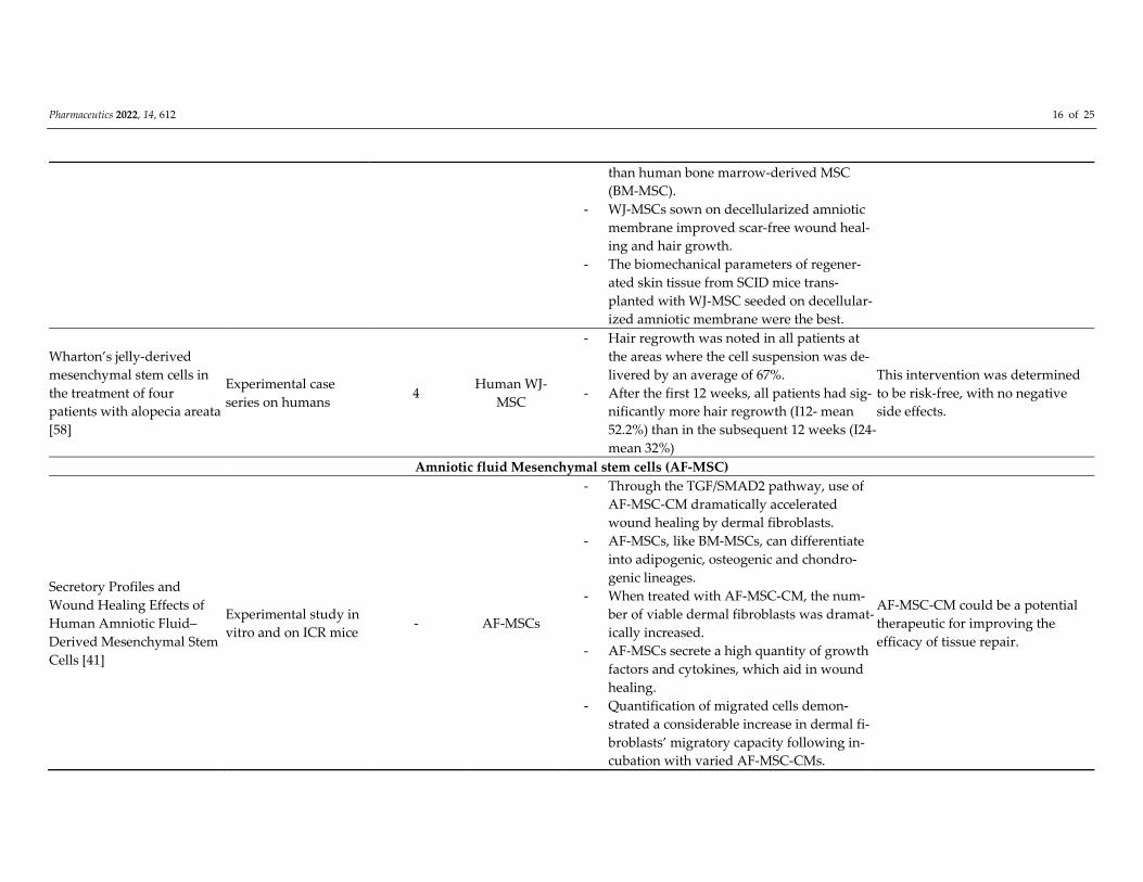
4.2.2. Wharton Jelly Derived Cell
The umbilical cord’s vessels are encased in Wharton jelly, a type of connective tissue.
This connective tissue is a ware house of UC‐MSCs, also known as Wharton’s jelly MSCs
(WJ‐MSCs) and the matrix is rich in growth factors like, IGF, FGF and TGF‐β [122–127].
Wharton jelly can be extracted from three different parts of the umbilical cord: sub‐amni‐
otic, perivascular, and intervascular. Their structure, function, and immunohistochemis‐
try are all different. Their doubling time is less with high proliferation index ex vivo, up
to 300 times yield with six to seven passages. Interestingly with no abnormal karyotypes
[128–133] WJ‐MSCs have tumor suppressor genes in abundance and are not associated
with any teratomas. They are also known to secrete hematopoietic cytokines [134] WJ‐
MSCs can be isolated and expanded using a technique illustrated by Can et al. [135]
Aljitawi et al. [136] and Wand et al. [137].
According to Jadalannagari et al., WJ‐MSCs have the ability to develop into ectodermal
lineage cells [138] When WJ‐MSCs were expanded on decellularized Wharton’s jelly matrix
(DWJM) in the presence of media promoting bone differentiation, they yielded CK19 posi‐
tive cells with structures resembling hair as demonstrated by Aljitawi et al. [136].
A study was conducted on SCID mice to demonstrate the efficacy of WJ‐MSCs on
wound healthy. When amniotic membrane scaffold which was decellularized was im‐
planted with WJ‐MSCs used in skin injury, wound healing improved with decreased scar‐
ring associated with growth of hair. Also, with considerable improvement in biomechan‐
ical properties. They also demonstrated successful expansion of WJ‐MSCs using human

Pharmaceutics 2022, 14, 612 11 of 25
platelet lysate. They conclude that the stemness of fetal MSCs was more than the BM‐
MSCs and the amount of immunomodulatory secretion in proinflammatory state was
more from the fetal MSCs in comparison to the adult MSCs [139]. Similarly, cultured UC‐
MSCs in appropriate medium‐formed structures resembling the normal dermal papilla
along with protein expression pathways similar to DPCs. Intradermal infusion of these
DPLT along with outer root sheath cells to athymic nude mice demonstrated increased
hair growth after 6 weeks [140]. Another study was carried out on athymic mice to eluci‐
date the prospective of MSCs (BM and UC) as an alternative to DPC for treating alopecia.
In vitro culturing of MSCs formed DPLT under special medium. Histologic and immuno‐
histochemical analyses showed that DPLT were similar to native human scalp DPCs. Re‐
constructed DPLTs when transplanted to athymic mice showed the potential to induce
hair follicles [109]. In a study to optimize the reconstruction of DPLTs, it was discovered
that HGF is required for differentiation, although it is costly. This study also demonstrated
that EGF is an inexpensive alternative [141]. 67% growth was observed when intradermal
injection of allogenic WJ‐MSCs was injected for AA. There were more hair growth dy‐
namics during the first 3 months (average 52.2%) when compared to hair regrowth fol‐
lowing 3 months (average 32%). All patients showed improvement without any adverse
effect [58].
4.2.3. Amniotic Fluid‐Derived Cell
Amnion refers to a sac enclosing a developing embryo, the sac is filled with amniotic
fluid (AF). AF composition and amount varies with the gestational age. During early ges‐
tation, osmotic gradient maintains the AF and in the later half it comprises of fetal urine
and secretions from the respiratory tract [142,143] AF has variety of cells from the embryo
(amniotic membrane, genito‐urinary, gastrointestinal and respiratory tracts). Cellular
component of AF gradually increases with the gestational age [59,144].
Cellular component comprises of amniocytes, epitheloid and fibroblastic cells. Re‐
cent studies have isolated amniotic fluid stem cells (AFSC) and amniotic fluid‐MSCs (AF‐
MSCs). They can be cultured from the cellular component [145] AFSCs account for 1% of
cells. They display markers associated with embryonic and adult stem cells, including
CD117, and are capable of differentiating into any of the three germ lines [146,147] AF
aspirated during mid‐trimester of pregnancy is a good source of MSCs. Most of the studies
have utilized AF‐MSCs obtained during the second trimester (16–28 weeks) through am‐
niocentesis [148–152] Very few studies have utilized AF isolated during first trimester and
during parturition [153,154] AF‐MSCs are isolated from AF by centrifuging and then cul‐
turing the cells at specific conditions, then the cells are sorted using CD117. It is difficult
to expand AF‐MSCs on a large scale because of the loss of differentiation potential
[146,155,156].
AF‐MSCs secrete MCP‐1, IL‐8, IL‐6, EGF, SDF‐1 and VEGF into the conditioned me‐
dium to exert its action, which plays a vital role in angiogenesis [157] MCP and IL‐6 se‐
creted by AF‐MSCs help in immune modulation [158] Even though cells derived from AF
are used along with other regenerative molecules in clinical practice for alopecia, not
many randomized blinded clinical studies are available to support the use of AF‐MSCs in
particular.
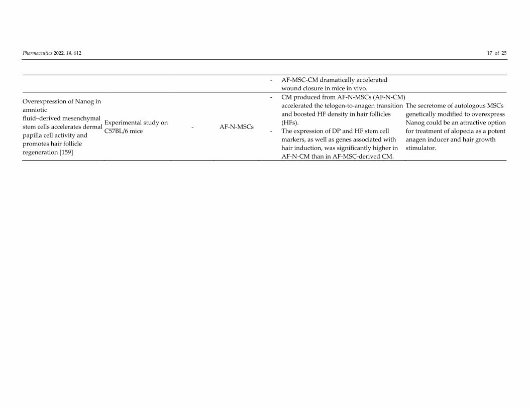
In a study conducted by Park et al., the effects of Nanog overexpression in AF‐MSCs
was assessed for DPCs and regrowth of hair in vivo. They used conditioned medium de‐
rived from Nanog overexpressing AF‐MSCs (AF‐N‐MSCs). There was upregulation BMP,
FGF, IGF, PDGF and WNT families by the presence of AF‐N‐MSCs, among them bFGF,
IGF, Wnt7a, and PDGF‐AA are implicated in hair regeneration by activating DP cells.
They discovered that overexpression of Nanog in MSCs triggered telogen to anagen tran‐
sition and increased hair follicle density by upregulating genes, such as ALP, LEF1, and
versican [159] (Table 2).

Pharmaceutics 2022, 14, 612 12 of 25
Table 2. Summary of studies on cellular therapies in hair regrowth.
Title Study Type Sample Size Active Agent Results Remarks
Adipose tissue derived stem cells (ADSC)
Hair follicle growth by
stromal vascular fraction
enhanced
adipose transplantation in
baldness [82]
Pilot case series in
humans
(MPHL—Grade 2 to 6
and FPHL—Grade 1 to
3)
9
Adipose
tissue
enriched
SVF
when fat + SVF was used, mean increase was 31
hair/cm2. Whereas, for fat alone, it was
14 hairs/cm2.
6 month follow up was available for
only 6 patients
Cellular therapy with human
autologous
adipose‐derived adult cells
of stromal vascular fraction
for alopecia areata [83]
Human clinical study
(Alopecia areata: Grade
1 or 2)
20
Adipose
derived
stromal
vascular cells
It was noted that there was statistically significant
increase in hair density, hair diameter and pull test
value.
‐
Stromal Vascular Fraction
Enhanced
Adipose Transplantation in
Hair Loss: Early Experience
& Active Phase II FDA
Investigation [84]
Prospective, single
blinded human clinical
trial
(AGA; Grade 1 to 4)
9 Adipose tissue
enriched SVF
Compared to baseline, there was a 14 percent
increase in the number of hairs (p = 0.01), with a mean
difference of 28 hairs and a 34 percent increase in the
anagen percentage (p = 0.09).
Only 6 patients were analyzed at 6
months as 3 lost follow up.
Clinical use of conditioned
media of adipose tissue‐
derived
stem cells in female pattern
hair loss: a retrospective case
series study [85]
Retrospective,
observational human
study
(FPHL)
27 ADSC‐CM
(AAPE)
The density of the hair rose from 105.4 to 122.7
hairs/cm2 (p = 0.001). The thickness of the hair
increased from 57.5 μ to 64.0 μ (p = 0.001). Dose: Once per week for 12 weeks
Hair Regeneration Treatment
Using
Adipose‐Derived Stem Cell
Conditioned Medium:
Follow‐up With trichograms
[87]
Prospective human
study on alopecia
22
(Half side
comparison:
10)
ADSC‐CM
(AAPE)
‐ Hair count increased considerably following
therapy in both male (including those who
did not receive finasteride) and female pa‐
tients.
‐ The rise in hair count was substantially
greater on the treatment side than on the
Dose: 6 sessions every 3–5 weeks

Pharmaceutics 2022, 14, 612 13 of 25
placebo side in the half‐side comparative
study.
Innovative method of
alopecia treatment by
autologous adipose‐derived
SVF [88]
Clinical human study
(AGA) 9
Autologous
SVF
‐ Statistically significant hair density at 3 and
6 months.
‐ Improvement in keratin score and hair
thickness was noted.
A single dose of Autologous SVF
was administered
Introducing Platelet‐Rich
Stroma: Platelet Rich Plasma
(PRP) and Stromal Vascular
Fraction (SVF) Combined for
the Treatment of
Androgenetic Alopecia [89]
Clinical human study
(AGA) 10
PRP + SVF
(Platelet rich
stroma)
‐ There was statistically significant increase in
hair density at 6 and 12 weeks.
‐ New hair growth was also observed in hy‐
perkeratotic plugged non‐functioning hair
follicles.
A single dose of Autologous PRP +
SVF was administered
Hair follicular stem cells (HFSC)
Stem cells from human hair
follicles: first mechanical
isolation for immediate
autologous clinical use in
androgenetic alopecia and
hair loss [67]
Prospective human
study
(AGA)
11
HFSC obtained
by mechanical
centrifugation
of punch
biopsy from
hair follicle
‐ After 23 weeks, there was a 29% ± 5% in‐
crease in hair density in the treated area.
‐ Each suspension of scalp tissue contained
approximately 3728.5 ± 664.5 cells. HF‐MSC
(CD44+ from DP): 5% + 0.7% and HF‐ESC
(CD200+) from the bulge was about 2.6% +
0.3%.
Primary outcomes were
microscopic identification and
counting of HFSCs.
Autologous Cellular Method
Using micrografts of Human
Adipose Tissue Derived
Follicle Stem Cells in
Androgenic Alopecia [52]
Retrospective
observational case‐
series, randomized,
evaluator‐blinded,
placebo controlled,
half‐head group study
in human (AGA)
33
Autologous cell
biological
technique (A‐
CBT) based on
micro‐grafts
containing Hair
Follicle
Mesenchymal
Stem Cells (HF‐
MSCs)
‐ Hair density improved, with a mean in‐
crease of 33% ± 75% at week 23 and 27% ±
35% at week 44.
‐ The increase in the number of hair follicles
per mm2 after 11 months was statistically
significant (p = 0.05).
Dose: 3 injections at 45 days interval

Pharmaceutics 2022, 14, 612 14 of 25
Platelet‐Rich Plasma and
Micrografts Enriched with
Autologous Human Follicle
Mesenchymal Stem Cells
Improve Hair Re‐Growth in
Androgenetic Alopecia.
Biomolecular Pathway
Analysis and Clinical
Evaluation [103]
Retrospective
observational case‐
series in humans
(AGA)
21 (HF‐MSC)
57 (A‐PRP)
HF‐MSC
And
A‐PRP
‐ 31% ± 2% increase in hair density in A‐PRP
group.
‐ In HF‐MSC group, 30% ± 5.0% increase in
hair density after 12 weeks, 29% ± 5.0% in 23
weeks.
‐ Dose: 2 sessions 2 months
apart for HF‐MSC, whereas,
3 sessions 30 days apart for
PRP
‐ All cases received low‐level
led treatment (LLLT). 15
days following each treat‐
ment and every three
weeks thereafter until six
months post‐treatment
Autologous Micrografts
from Scalp Tissue:
Trichoscopic and Long‐Term
Clinical Evaluation in Male
and Female Androgenetic
Alopecia [99]
Placebo controlled,
randomized, evaluator‐
blinded, half‐head
group study in humans
(AGA)
27
Micrografts
enriched with
HF‐MSCs
‐ Improvement in the mean hair count was
noted after 58 weeks of 18.0 hair.
‐ The mean increase in total hair density was
23.3 hairs per cm2.
Six patients exhibited dynamic hair
loss after 26 months.
Bone marrow derived stem cells (BMSC)
Stem cell therapy as a novel
therapeutic intervention for
resistant cases of alopecia
areata and androgenetic
alopecia [56]
Double randomized
clinical human study
(AGA and AA)
40 (20 AGA
and 20 AA)
Autologous
bone marrow
derived
mononuclear
cells (BMMCs
or autologous
follicular stem
cells (FSC).
‐ Clinically, there was a considerable im‐
provement six months following stem cell
therapy injection, which was validated by
immunostaining and digital dermoscopy.
‐ The mean improvement was “very good”
across all groups.
‐ In either form of alopecia, there was no sig‐
nificant difference between the two proce‐
dures.
No adverse effects were noted
Application of mesenchymal
stem cells derived from bone
marrow and umbilical cord
in human hair multiplication
[109]
Experimental study in
vitro and on athymic
mice
‐
Culture
expanded
MSCs from
bone marrow
and umbilical
cord of human
beings
The DPLTs created in this technique were identical
in size, shape and expression to actual DP.
MSCs were preconditioned in
dermal papilla formation medium
(DPFM) before being subcultured to
create self‐aggregated DPLTs.

Pharmaceutics 2022, 14, 612 15 of 25
Umbilical cord blood derived cells
Human umbilical cord blood
mesenchymal stem cells
engineered to overexpress
growth factors accelerate
outcomes in hair growth [57]
Experimental study on
C3H/HeJ mice ‐ hUCB‐MSCs
‐ hUCB‐MSCs accelerated anagen initiation
and hair follicle neogenesis.
‐ Co‐culture with hUCB‐MSCs increased the
viability of human dermal papilla cells
(hDPCs) and up‐regulated its hair induc‐
tion‐related proteins.
‐ hUCB‐MSCs stimulate hair growth through
a paracrine mechanism.
IGFBP‐1 had a beneficial influence
on cell survival, VEGF secretion,
alkaline phosphatase (ALP), CD133,
and b‐catenin expression, and the
formation of 3D spheroids of
hDPCs via colocalization of an IGF‐
1 and IGFBP‐1.
Migration Inhibitory Factor
in Conditioned Medium
from Human Umbilical Cord
Blood‐Derived Mesenchymal
Stromal Cells Stimulates
Hair Growth [40]
Double‐blind placebo‐
controlled clinical trial
(AGA)
30 hUCB‐MSC‐
CM
‐ When compared to CM alone, primed MSC‐
derived CM (P‐CM) with combinations of
TGF‐1 and LiCl significantly enhanced the
viability of DPCs.
‐ P‐CM increased hair density by 14.24 per‐
cent (p = 0.001).
‐ At 16 weeks, there was a statistically signifi‐
cant increase in hair thickness and rate of
hair growth.
‐ The macrophage migration
inhibitory factor (MIF) in
the P‐CM released by MSCs
influenced the secretion of
vascular endothelial growth
factor (VEGF) in DPCs,
which acts via VEGF‐re‐
lated β‐catenin and P‐GSK‐
3β [SER9] signaling path‐
way
‐ P‐CM, through a paracrine
mechanism, can boost hair
growth efficacy.
Wharton jelly stem cells
Human Wharton’s Jelly
Mesenchymal Stem Cells
Plasticity Augments Scar‐
Free Skin Wound Healing
with Hair Growth [139]
Experimental study on
black SCID mice ‐
Human WJ‐
MSCs
‐ During long‐term culture, human Whar‐
ton’s Jelly‐derived MSCs (WJ‐MSC) retained
their phenotypic characteristics and in vitro
differentiation plasticity.
‐ During long‐term in vitro cultures, human
WJMSCs retained several inherent MSC
properties.
‐ In the presence of pro‐inflammatory cyto‐
kines, perinatal MSCs exhibited higher
quantities of immunomodulatory molecules
‐ Human platelet lysate was
successfully employed to
grow isolated WJ‐MSCs
from human Wharton jelly
tissue of the umbilical cord.
‐ The fetal MSCs showed
more stemness
compared to gold standard
adult bone marrow derived
MSCs
Pharmaceutics 2022, 14, 612 16 of 25
than human bone marrow‐derived MSC
(BM‐MSC).
‐ WJ‐MSCs sown on decellularized amniotic
membrane improved scar‐free wound heal‐
ing and hair growth.
‐ The biomechanical parameters of regener‐
ated skin tissue from SCID mice trans‐
planted with WJ‐MSC seeded on decellular‐
ized amniotic membrane were the best.
Wharton’s jelly‐derived
mesenchymal stem cells in
the treatment of four
patients with alopecia areata
[58]
Experimental case
series on humans 4
Human WJ‐
MSC
‐ Hair regrowth was noted in all patients at
the areas where the cell suspension was de‐
livered by an average of 67%.
‐ After the first 12 weeks, all patients had sig‐
nificantly more hair regrowth (I12‐ mean
52.2%) than in the subsequent 12 weeks (I24‐
mean 32%)
This intervention was determined
to be risk‐free, with no negative
side effects.
Amniotic fluid Mesenchymal stem cells (AF‐MSC)
Secretory Profiles and
Wound Healing Effects of
Human Amniotic Fluid–
Derived Mesenchymal Stem
Cells [41]
Experimental study in
vitro and on ICR mice ‐ AF‐MSCs
‐ Through the TGF/SMAD2 pathway, use of
AF‐MSC‐CM dramatically accelerated
wound healing by dermal fibroblasts.
‐ AF‐MSCs, like BM‐MSCs, can differentiate
into adipogenic, osteogenic and chondro‐
genic lineages.
‐ When treated with AF‐MSC‐CM, the num‐
ber of viable dermal fibroblasts was dramat‐
ically increased.
‐ AF‐MSCs secrete a high quantity of growth
factors and cytokines, which aid in wound
healing.
‐ Quantification of migrated cells demon‐
strated a considerable increase in dermal fi‐
broblasts’ migratory capacity following in‐
cubation with varied AF‐MSC‐CMs.
AF‐MSC‐CM could be a potential
therapeutic for improving the
efficacy of tissue repair.
Pharmaceutics 2022, 14, 612 17 of 25
‐ AF‐MSC‐CM dramatically accelerated
wound closure in mice in vivo.
Overexpression of Nanog in
amniotic
fluid–derived mesenchymal
stem cells accelerates dermal
papilla cell activity and
promotes hair follicle
regeneration [159]
Experimental study on
C57BL/6 mice ‐ AF‐N‐MSCs
‐ CM produced from AF‐N‐MSCs (AF‐N‐CM)
accelerated the telogen‐to‐anagen transition
and boosted HF density in hair follicles
(HFs).
‐ The expression of DP and HF stem cell
markers, as well as genes associated with
hair induction, was significantly higher in
AF‐N‐CM than in AF‐MSC‐derived CM.
The secretome of autologous MSCs
genetically modified to overexpress
Nanog could be an attractive option
for treatment of alopecia as a potent
anagen inducer and hair growth
stimulator.

Pharmaceutics 2022, 14, 612 18 of 25
5. Conclusions
Alopecia is a difficult condition to treat as hair fall relapses with the discontinuation
of conventionally available FDA approved treatments. We have broadly classified the
available regenerative therapies (“A La Mode Classification”) as growth factor‐rich and
stem cell‐rich for better understanding and clinical utility. We have summarized the avail‐
able regenerative cellular therapies, their mechanism of action and the available clinical
trials in the treatment of non‐scarring alopecia. Almost all the studies utilizing cellular
therapies have shown significant improvement in hair regrowth with no adverse effects.
MSCs are the core of these cellular therapy because of their angiogenic and immunomod‐
ulatory function, which will aid in hair regrowth. These regenerative therapies can be a
boon in the treatment of alopecia if utilized properly.
It is very much clear that there are multiple sources of MSCs available for cellular ther‐
apy. Apart from the sources described in this article to treat alopecia, there are few other
unexplored sources which can be used to isolate MSCs like dental pulp, peripheral blood,
synovium and synovial fluid, endometrium, human foreskin, skin biopsy and muscle [160].
Further research is essential to know the potential of these sources in hair regeneration and
the adverse reactions associated with them. Each of these MSCs have their own pros and
cons with respect to isolation, differentiation capacity, cell count, and the possible adverse
reactions. Further studies are required to compare the efficacy of MSCs derived from vari‐
ous sources. CM has the advantage of low cost, easy storage and transport. Hence, CM is
seeking significant attention from clinical researchers. Standardization of isolation tech‐
nique, culture medium used, dosage, route and depth of injection, clear cut indications and
contraindications along with universally acceptable documentation of results have to be
gracefully addressed with good quality randomized controlled trials.
Author Contributions: Conceptualization: T.C.A.; methodology: T.C.A., S.M., M.J. and S.S.; soft‐
ware and validation: T.C.A., S.M., M.J. and B.‐C.A.; formal analysis and investigation: T.C.A., S.M.,
M.J. and B.‐C.A.; resources: all authors; data curation: S.M., M.J., S.S., R.L.R. and B.‐C.A.; writing—
original draft preparation: T.C.A.; writing—review and editing: all authors; visualization: T.C.A.,
S.M., M.J.; supervision: P.G. and B.‐C.A.; administrative support: S.M., M.J., R.L.R., P.G., P.C.M.,
S.K.J. and B.‐C.A.; funding acquisition: R.L.R. and P.G. All authors have read and agreed to the
published version of the manuscript.
Funding: This research was supported by the Basic Science Research Program through the National
Research Foundation of Korea (NRF) funded by the Ministry of Education (NRF‐
2019R1I1A1A01061296 and NRF‐2021R1I1A1A01040732).
Institutional Review Board Statement: Not applicable.
Informed Consent Statement: Not applicable.
Data Availability Statement: Not applicable.
Acknowledgements: We thank Madhurya Santosh, Junior resident, Department of Dermatology,
Raja Rajeswari Medical College and Hospital, Bengaluru, Karnataka, India; Shirodkar Jaswandi
Dilip, Medical Officer, ESIS hospital (Worli), Mumbai, Maharashtra, India and Ishita Katyal, Senior
resident, Department of plastic surgery, TNMC and BYL Nair charitable hospital, Mumbai, Maha‐
rashtra, India for literature search.
Conflicts of Interest: The authors declare no conflict of interest.
References
1. Chan, L.; Cook, D.K. Female pattern hair loss. Aust. J. Gen. Pract. 2018, 47, 459–464. https://doi.org/10.31128/ajgp‐02‐18‐4498.
2. Tanaka, Y.; Aso, T.; Ono, J.; Hosoi, R.; Kaneko, T. Androgenetic Alopecia Treatment in Asian Men. J. Clin. Aesthetic Dermatol.
2018, 11, 32–35.
3. Jang, W.S.; Son, I.P.; Yeo, I.K.; Park, K.Y.; Li, K.; Kim, B.J.; Seo, S.J.; Kim, M.N.; Hong, C.K. The Annual Changes of Clinical
Manifestation of Androgenetic Alopecia Clinic in Korean Males and Females: A Outpatient‐Based Study. Ann. Dermatol. 2013,
25, 181–188. https://doi.org/10.5021/ad.2013.25.2.181.

Pharmaceutics 2022, 14, 612 19 of 25
4. Yang, C.‐C.; Hsieh, F.‐N.; Lin, L.‐Y.; Hsu, C.‐K.; Sheu, H.‐M.; Chen, W. Higher body mass index is associated with greater
severity of alopecia in men with male‐pattern androgenetic alopecia in Taiwan: A cross‐sectional study. J. Am. Acad. Dermatol.
2014, 70, 297–302.e1. https://doi.org/10.1016/j.jaad.2013.09.036.
5. Sinclair, R.; Torkamani, N.; Jones, L. Androgenetic alopecia: New insights into the pathogenesis and mechanism of hair loss.
F1000Research 2015, 4, 585. https://doi.org/10.12688/f1000research.6401.1.
6. Di Loreto, C.; La Marra, F.; Mazzon, G.; Belgrano, E.; Trombetta, C.; Cauci, S. Immunohistochemical Evaluation of Androgen
Receptor and Nerve Structure Density in Human Prepuce from Patients with Persistent Sexual Side Effects after Finasteride
Use for Androgenetic Alopecia. PLoS ONE 2014, 9, e100237. https://doi.org/10.1371/journal.pone.0100237.
7. Male Androgenetic Alopecia. Available online: https://Pubmed.Ncbi.Nlm.Nih.Gov/25905192/ (accessed on 8 January 2022).
8. Wang, T.; Zhou, C.; Shen, Y.; Wang, X.; Ding, X.; Tian, S.; Liu, Y.; Peng, G.; Xue, S.; Zhou, J.; et al. Prevalence of androgenetic
alopecia in China: A community‐based study in six cities. Br. J. Dermatol. 2010, 162, 843–847. https://doi.org/10.1111/j.1365‐
2133.2010.09640.x.
9. Monselise, A.; Bar‐On, R.; Chan, L.; Leibushor, N.; McElwee, K.; Shapiro, J. Examining the Relationship between Alopecia Ar‐
eata, Androgenetic Alopecia, and Emotional Intelligence. J. Cutan. Med. Surg. 2013, 17, 46–51.
https://doi.org/10.2310/7750.2012.12003.
10. Han, S.‐H.; Byun, J.‐W.; Lee, W.‐S.; Kang, H.; Kye, Y.‐C.; Kim, K.‐H.; Kim, D.‐W.; Kim, M.‐B.; Kim, S.‐J.; Kim, H.‐O.; et al. Quality
of Life Assessment in Male Patients with Androgenetic Alopecia: Result of a Prospective, Multicenter Study. Ann. Dermatol.
2012, 24, 311–318. https://doi.org/10.5021/ad.2012.24.3.311.
11. Schmidt, S.; Fischer, T.; Chren, M.; Strauss, B.; Elsner, P. Strategies of coping and quality of life in women with alopecia. Br. J.
Dermatol. 2001, 144, 1038–1043. https://doi.org/10.1046/j.1365‐2133.2001.04195.x.
12. Sasaki, G.H. Review of Human Hair Follicle Biology: Dynamics of Niches and Stem Cell Regulation for Possible Therapeutic
Hair Stimulation for Plastic Surgeons. Aesthetic Plast. Surg. 2018, 43, 253–266. https://doi.org/10.1007/s00266‐018‐1248‐1.
13. Neuhaus, K.; Schiestl, C.; Adelsberger, R.; Weibel, L.; Meuli, M.; Böttcher‐Haberzeth, S. Bold to do—Bald to be? Outcomes
decades after harvesting the scalp in burned children. Burn. 2018, 45, 543–553. https://doi.org/10.1016/j.burns.2018.09.023.
14. Almohanna, H.M.; Perper, M.; Tosti, A. Safety concerns when using novel medications to treat alopecia. Expert Opin. Drug Saf.
2018, 17, 1115–1128. https://doi.org/10.1080/14740338.2018.1533549.
15. Morris, R.J.; Liu, Y.; Marles, L.; Yang, Z.; Trempus, C.; Li, S.; Lin, J.; Sawicki, J.; Cotsarelis, G. Capturing and profiling adult hair
follicle stem cells. Nat. Biotechnol. 2004, 22, 411–417. https://doi.org/10.1038/nbt950.
16. Tumbar, T.; Guasch, G.; Greco, V.; Blanpain, C.; Lowry, W.E.; Rendl, M.; Fuchs, E. Defining the Epithelial Stem Cell Niche in
Skin. Science 2004, 303, 359–363. https://doi.org/10.1126/science.1092436.
17. Blanpain, C.; Fuchs, E. Epidermal Stem Cells of the Skin. Annu. Rev. Cell Dev. Biol. 2006, 22, 339–373. https://doi.org/10.1146/an‐
nurev.cellbio.22.010305.104357.
18. Blanpain, C.; Lowry, W.E.; Geoghegan, A.; Polak, L.; Fuchs, E. Self‐Renewal, Multipotency, and the Existence of Two Cell Pop‐
ulations within an Epithelial Stem Cell Niche. Cell 2004, 118, 635–648. https://doi.org/10.1016/j.cell.2004.08.012.
19. Blanpain, C.; Fuchs, E. Epidermal homeostasis: A balancing act of stem cells in the skin. Nat. Rev. Mol. Cell Biol. 2009, 10, 207–
217. https://doi.org/10.1038/nrm2636.
20. Lowry, W.E.; Blanpain, C.; Nowak, J.A.; Guasch, G.; Lewis, L.; Fuchs, E. Defining the impact of β‐catenin/Tcf transactivation on
epithelial stem cells. Genes Dev. 2005, 19, 1596–1611. https://doi.org/10.1101/gad.1324905.
21. Dasgupta, R.; Fuchs, E. Multiple roles for activated LEF/TCF transcription complexes during hair follicle development and
differentiation. Development 1999, 126, 4557–4568. https://doi.org/10.1242/dev.126.20.4557.
22. Reddy, S.; Andl, T.; Bagasra, A.; Lu, M.M.; Epstein, D.J.; Morrisey, E.E.; Millar, S.E. Characterization of Wnt gene expression in
developing and postnatal hair follicles and identification of Wnt5a as a target of Sonic hedgehog in hair follicle morphogenesis.
Mech. Dev. 2001, 107, 69–82. https://doi.org/10.1016/s0925‐4773(01)00452‐x.
23. Blessing, M.; Nanney, L.B.; King, L.; Jones, C.M.; Hogan, B.L. Transgenic mice as a model to study the role of TGF‐beta‐related
molecules in hair follicles. Genes Dev. 1993, 7, 204–215. https://doi.org/10.1101/gad.7.2.204.
24. Botchkarev, V.; Botchkareva, N.V.; Roth, W.; Nakamura, M.; Chen, L.‐H.; Herzog, W.; Lindner, G.; McMahon, J.A.; Peters, C.;
Lauster, R.; et al. Noggin is a mesenchymally derived stimulator of hair‐follicle induction. Nat. Cell Biol. 1999, 1, 158–164.
https://doi.org/10.1038/11078.
25. Kobielak, K.; Stokes, N.; de la Cruz, J.; Polak, L.; Fuchs, E. Loss of a quiescent niche but not follicle stem cells in the absence of
bone morphogenetic protein signaling. Proc. Natl. Acad. Sci. USA 2007, 104, 10063–10068.
https://doi.org/10.1073/pnas.0703004104.
26. Kelly, Y.; Blanco, A.; Tosti, A. Androgenetic Alopecia: An Update of Treatment Options. Drugs 2016, 76, 1349–1364.
https://doi.org/10.1007/s40265‐016‐0629‐5.
27. Mella, J.M.; Perret, M.C.; Manzotti, M.; Catalano, H.N.; Guyatt, G. Efficacy and Safety of Finasteride Therapy for Androgenetic
Alopecia. Arch. Dermatol. 2010, 146, 1141–1150. https://doi.org/10.1001/archdermatol.2010.256.
28. Rahimi‐Ardabili, B.; Pourandarjani, R.; Habibollahi, P.; Mualeki, A. Finasteride induced depression: A prospective study. BMC
Clin. Pharmacol. 2006, 6, 7. https://doi.org/10.1186/1472‐6904‐6‐7.
29. D’Amico, A.V.; Roehrborn, C.G. Effect of 1 mg/day finasteride on concentrations of serum prostate‐specific antigen in men with
androgenic alopecia: A randomised controlled trial. Lancet Oncol. 2007, 8, 21–25. https://doi.org/10.1016/s1470‐2045(06)70981‐0.

Pharmaceutics 2022, 14, 612 20 of 25
30. Lachgar, S.; Charveron, M.; Gall, Y.; Bonafe, J.L. Minoxidil upregulates the expression of vascular endothelial growth factor in
human hair dermal papilla cells. Br. J. Dermatol. 1998, 138, 407–411. https://doi.org/10.1046/j.1365‐2133.1998.02115.x.
31. Marubayashi, A.; Nakaya, Y.; Fukui, K.; Li, M.; Arase, S. Minoxidil‐Induced Hair Growth is Mediated by Adenosine in Cultured
Dermal Papilla Cells: Possible Involvement of Sulfonylurea Receptor 2B as a Target of Minoxidil. J. Investig. Dermatol. 2001, 117,
1594–1600. https://doi.org/10.1046/j.0022‐202x.2001.01570.x.
32. FDA. Available online: https://www.accessdata.fda.gov/drugsatfda_docs/nda/97/20834_rogaine%20ex‐
tra%20strength%20for%20men%205%25_medr.pdf (accessed on 8 January 2022).
33. Lucky, A.W.; Piacquadio, D.J.; Ditre, C.M.; Dunlap, F.; Kantor, I.; Pandya, A.G.; Savin, R.C.; Tharp, M.D. A randomized, pla‐
cebo‐controlled trial of 5% and 2% topical minoxidil solutions in the treatment of female pattern hair loss. J. Am. Acad. Dermatol.
2004, 50, 541–553. https://doi.org/10.1016/j.jaad.2003.06.014.
34. Pratt, C.H.; King, L.E.; Messenger, A.G.; Christiano, A.M.; Sundberg, J.P. Alopecia areata. Nat. Rev. Dis. Prim. 2017, 3, 1–17.
https://doi.org/10.1038/nrdp.2017.11.
35. Walker, S.A.; Rothman, S. A Statistical Study and Consideration of Endocrine Influences. J. Investig. Dermatol. 1950, 14, 403–413.
https://doi.org/10.1038/jid.1950.52.
36. Alsantali, A. Alopecia areata: A new treatment plan. Clin. Cosmet. Investig. Dermatol. 2011, 4, 107–115.
https://doi.org/10.2147/ccid.s22767.
37. Gentile, P.; Garcovich, S. Autologous activated platelet‐rich plasma (AA‐PRP) and non‐activated (A‐PRP) in hair growth: A
retrospective, blinded, randomized evaluation in androgenetic alopecia. Expert Opin. Biol. Ther. 2020, 20, 327–337.
https://doi.org/10.1080/14712598.2020.1724951.
38. Totey, S.M.; Dhurat, R.S.; Kadam, P.P.; Sevilla, G.P.; Shetty, G. Safety and efficacy of growth factor concentrate in the treatment
of nasolabial fold correction: Split face pilot study. Indian J. Dermatol. 2015, 60, 520. https://doi.org/10.4103/0019‐5154.159628.
39. Fukuoka, H.; Narita, K.; Suga, H. Hair Regeneration Therapy: Application of Adipose‐Derived Stem Cells. Curr. Stem Cell Res.
Ther. 2017, 12, 531–534. https://doi.org/10.2174/1574888x12666170522114307.
40. Oh, H.A.; Kwak, J.; Kim, B.J.; Jin, H.J.; Park, W.S.; Choi, S.J.; Oh, W.; Um, S. Migration Inhibitory Factor in Conditioned Medium
from Human Umbilical Cord Blood‐Derived Mesenchymal Stromal Cells Stimulates Hair Growth. Cells 2020, 9, 1344.
https://doi.org/10.3390/cells9061344.
41. Yoon, B.S.; Moon, J.‐H.; Jun, E.K.; Kim, J.; Maeng, I.; Kim, J.S.; Lee, J.H.; Baik, C.S.; Kim, A.; Cho, K.S.; et al. Secretory Profiles
and Wound Healing Effects of Human Amniotic Fluid–Derived Mesenchymal Stem Cells. Stem Cells Dev. 2010, 19, 887–902.
https://doi.org/10.1089/scd.2009.0138.
42. Ou, K.‐L.; Kuo, Y.‐W.; Wu, C.‐Y.; Huang, B.‐H.; Pai, F.‐T.; Chou, H.‐H.; Saito, T.; Ueno, T.; Cho, Y.‐C.; Huang, M.‐S. The Potential
of a Hair Follicle Mesenchymal Stem Cell‐Conditioned Medium for Wound Healing and Hair Follicle Regeneration. Appl. Sci.
2020, 10, 2646. https://doi.org/10.3390/app10082646.
43. Payushina, O.V.; Butorina, N.N.; Sheveleva, O.N.; Domaratskaya, E.I. Effect of Mesenchymal Stromal Cells and Conditioned
Media on Healing of Skin Wound. Bull. Exp. Biol. Med. 2018, 165, 572–575. https://doi.org/10.1007/s10517‐018‐4215‐6.
44. Carrasco, E.; Soto‐Heredero, G.; Mittelbrunn, M. The Role of Extracellular Vesicles in Cutaneous Remodeling and Hair Follicle
Dynamics. Int. J. Mol. Sci. 2019, 20, 2758. https://doi.org/10.3390/ijms20112758.
45. Somuncu Özge S.; Taşlı, P.N.; Şişli, H.B.; Somuncu, S.; Şahin, F. Characterization and Differentiation of Stem Cells Isolated from
Human Newborn Foreskin Tissue. Appl. Biochem. Biotechnol. 2015, 177, 1040–1054. https://doi.org/10.1007/s12010‐015‐1795‐8.
46. Yan, H.; Gao, Y.; Ding, Q.; Liu, J.; Li, Y.; Jin, M.; Xu, H.; Ma, S.; Wang, X.; Zeng, W.; et al. Exosomal Micro RNAs Derived from
Dermal Papilla Cells Mediate Hair Follicle Stem Cell Proliferation and Differentiation. Int. J. Biol. Sci. 2019, 15, 1368–1382.
https://doi.org/10.7150/ijbs.33233.
47. Rajendran, R.L.; Gangadaran, P.; Bak, S.S.; Oh, J.M.; Kalimuthu, S.; Lee, H.W.; Baek, S.H.; Zhu, L.; Sung, Y.K.; Jeong, S.Y.; et al.
Extracellular vesicles derived from MSCs activates dermal papilla cell in vitro and promotes hair follicle conversion from
telogen to anagen in mice. Sci. Rep. 2017, 7, 15560. https://doi.org/10.1038/s41598‐017‐15505‐3.
48. Choi, B.Y. Hair‐Growth Potential of Ginseng and Its Major Metabolites: A Review on Its Molecular Mechanisms. Int. J. Mol. Sci.
2018, 19, 2703. https://doi.org/10.3390/ijms19092703.
49. Kwon, T.‐R.; Oh, C.T.; Choi, E.J.; Park, H.M.; Han, H.J.; Ji, H.J.; Kim, B.J. Human placental extract exerts hair growth‐promoting
effects through the GSK‐3β signaling pathway in human dermal papilla cells. Int. J. Mol. Med. 2015, 36, 1088–1096.
https://doi.org/10.3892/ijmm.2015.2316.
50. Vestita, M.; Filoni, A.; Bonamonte, D.; Elia, R.; Giudice, G. Abstract: The Use of Nanofat in Androgenic Alopecia. a Prospective
Blinded Study. Plast. Reconstr. Surg.—Glob. Open 2017, 5, 90. https://doi.org/10.1097/01.gox.0000526293.77976.7f.
51. Epstein, G.K.; Epstein, J.S. Mesenchymal Stem Cells and Stromal Vascular Fraction for Hair Loss: Current Status. Facial Plast.
Surg. Clin. N. Am. 2018, 26, 503–511. https://doi.org/10.1016/j.fsc.2018.06.010.
52. Gentile, P. Autologous Cellular Method Using Micrografts of Human Adipose Tissue Derived Follicle Stem Cells in Androgenic
Alopecia. Int. J. Mol. Sci. 2019, 20, 3446. https://doi.org/10.3390/ijms20143446.
53. Wang, B.; Liu, X.‐M.; Liu, Z.‐N.; Wang, Y.; Han, X.; Lian, A.‐B.; Mu, Y.; Jin, M.‐H.; Liu, J.‐Y. Human hair follicle‐derived mesen‐
chymal stem cells: Isolation, expansion, and differentiation. World J. Stem Cells 2020, 12, 462–470.
https://doi.org/10.4252/wjsc.v12.i6.462.

Pharmaceutics 2022, 14, 612 21 of 25
54. Cheng, C.‐C.; Tsutsui, K.; Taguchi, T.; Sanzen, N.; Nakagawa, A.; Kakiguchi, K.; Yonemura, S.; Tanegashima, C.; Keeley, S.D.;
Kiyonari, H.; et al. Hair follicle epidermal stem cells define a niche for tactile sensation. eLife 2018, 7, e38883.
https://doi.org/10.7554/elife.38883.
55. Chacón‐Martínez, C.A.; Klose, M.; Niemann, C.; Glauche, I.; Wickström, S. Hair follicle stem cell cultures reveal self‐organizing
plasticity of stem cells and their progeny. EMBO J. 2016, 36, 151–164. https://doi.org/10.15252/embj.201694902.
56. Elmaadawi, I.H.; Mohamed, B.M.; Ibrahim, Z.A.S.; Abdou, S.M.; El Attar, Y.A.; Youssef, A.; Shamloula, M.M.; Taha, A.; Met‐
wally, H.G.; El Afandy, M.M.; et al. Stem cell therapy as a novel therapeutic intervention for resistant cases of alopecia areata
and androgenetic alopecia. J. Dermatol. Treat. 2018, 29, 431–440. https://doi.org/10.1080/09546634.2016.1227419.
57. Bak, D.H.; Choi, M.J.; Kim, S.R.; Lee, B.C.; Kim, J.M.; Jeon, E.S.; Oh, W.; Lim, E.S.; Park, B.C.; Kim, M.J.; et al. Human umbilical
cord blood mesenchymal stem cells engineered to overexpress growth factors accelerate outcomes in hair growth. Korean J.
Physiol. Pharmacol. 2018, 22, 555–566. https://doi.org/10.4196/kjpp.2018.22.5.555.
58. Czarnecka, A.; Odziomek, A.; Murzyn, M.; Dubis, J.; Bagłaj‐Oleszczuk, M.; Anita, H.‐G. Wharton’s jelly‐derived mesenchymal
stem cells in the treatment of four patients with alopecia areata. Adv. Clin. Exp. Med. 2021, 30, 211–218.
https://doi.org/10.17219/acem/132069.
59. Cananzi, M.; Atala, A.; De Coppi, P. Stem cells derived from amniotic fluid: New potentials in regenerative medicine. Reprod.
Biomed. Online 2009, 18 (Suppl. 1), 17–27. https://doi.org/10.1016/s1472‐6483(10)60111‐3.
60. Abd‐Allah, S.H.; El‐Shal, A.S.; Shalaby, S.M.; Abd‐Elbary, E.; Mazen, N.F.; Kader, R.R.A. The role of placenta‐derived mesen‐
chymal stem cells in healing of induced full‐thickness skin wound in a mouse model. IUBMB Life 2015, 67, 701–709.
https://doi.org/10.1002/iub.1427.
61. Williams, A.R.; Hare, J.M. Mesenchymal Stem Cells: Biology, Pathophysiology, Translational Findings, and Therapeutic Impli‐
cations for Cardiac Disease. Circ. Res. 2011, 109, 923–940. https://doi.org/10.1161/circresaha.111.243147.
62. Dominici, M.; Le Blanc, K.; Mueller, I.; Slaper‐Cortenbach, I.; Marini, F.C.; Krause, D.S.; Deans, R.J.; Keating, A.; Prockop, D.J.;
Horwitz, E.M. Minimal criteria for defining multipotent mesenchymal stromal cells. The International Society for Cellular Ther‐
apy position statement. Cytotherapy 2006, 8, 315–317. https://doi.org/10.1080/14653240600855905.
63. Qin, X.; Jiang, Q.; Nagano, K.; Moriishi, T.; Miyazaki, T.; Komori, H.; Ito, K.; Von Der Mark, K.; Sakane, C.; Kaneko, H.; et al.
Runx2 is essential for the transdifferentiation of chondrocytes into osteoblasts. PLoS Genet. 2020, 16, e1009169.
https://doi.org/10.1371/journal.pgen.1009169.
64. Caplan, H.; Olson, S.D.; Kumar, A.; George, M.; Prabhakara, K.S.; Wenzel, P.; Bedi, S.; Toledano‐Furman, N.E.; Triolo, F.; Kam‐
hieh‐Milz, J.; et al. Mesenchymal Stromal Cell Therapeutic Delivery: Translational Challenges to Clinical Application. Front.
Immunol. 2019, 10, 1645. https://doi.org/10.3389/fimmu.2019.01645.
65. Asakawa, K.; Toyoshima, K.‐E.; Ishibashi, N.; Tobe, H.; Iwadate, A.; Kanayama, T.; Hasegawa, T.; Nakao, K.; Toki, H.; Noguchi,
S.; et al. Hair organ regeneration via the bioengineered hair follicular unit transplantation. Sci. Rep. 2012, 2, 424.
https://doi.org/10.1038/srep00424.
66. Balañá, M.E. Epidermal stem cells and skin tissue engineering in hair follicle regeneration. World J. Stem Cells 2015, 7, 711–727.
https://doi.org/10.4252/wjsc.v7.i4.711.
67. Gentile, P.; Scioli, M.G.; Bielli, A.; Orlandi, A.; Cervelli, V. Stem cells from human hair follicles: First mechanical isolation for
immediate autologous clinical use in androgenetic alopecia and hair loss. Stem Cell Investig. 2017, 4, 58.
https://doi.org/10.21037/sci.2017.06.04.
68. Marquez‐Curtis, L.A.; Janowska‐Wieczorek, A.; McGann, L.E.; Elliott, J.A. Mesenchymal stromal cells derived from various
tissues: Biological, clinical and cryopreservation aspects. Cryobiology 2015, 71, 181–197. https://doi.org/10.1016/j.cryo‐
biol.2015.07.003.
69. Sheykhhasan, M.; Qomi, R.T.; Ghiasi, M. Fibrin Scaffolds Designing in order to Human Adipose‐derived Mesenchymal Stem
Cells Differentiation to Chondrocytes in the Presence of TGF‐βInt. J. Stem Cells 2015, 8, 219–227.
https://doi.org/10.15283/ijsc.2015.8.2.219.
70. Suh, A.; Pham, A.; Cress, M.J.; Pincelli, T.; TerKonda, S.P.; Bruce, A.J.; Zubair, A.C.; Wolfram, J.; Shapiro, S.A. Adipose‐derived
cellular and cell‐derived regenerative therapies in dermatology and aesthetic rejuvenation. Ageing Res. Rev. 2019, 54, 100933.
https://doi.org/10.1016/j.arr.2019.100933.
71. Bourin, P.; Bunnell, B.A.; Casteilla, L.; Dominici, M.; Katz, A.J.; March, K.L.; Redl, H.; Rubin, J.P.; Yoshimura, K.; Gimble, J.M.
Stromal cells from the adipose tissue‐derived stromal vascular fraction and culture expanded adipose tissue‐derived stro‐
mal/stem cells: A joint statement of the International Federation for Adipose Therapeutics and Science (IFATS) and the Inter‐
national Society for Cellular Therapy (ISCT). Cytotherapy 2013, 15, 641–648. https://doi.org/10.1016/j.jcyt.2013.02.006.
72. Suzuki, E.; Fujita, D.; Takahashi, M.; Oba, S.; Nishimatsu, H. Adipose tissue‐derived stem cells as a therapeutic tool for cardio‐
vascular disease. World J. Cardiol. 2015, 7, 454–465. https://doi.org/10.4330/wjc.v7.i8.454.
73. Traktuev, D.O.; Prater, D.N.; Merfeld‐Clauss, S.; Sanjeevaiah, A.R.; Saadatzadeh, M.R.; Murphy, M.; Johnstone, B.H.; Ingram,
D.A.; March, K.L. Robust functional vascular network formation in vivo by cooperation of adipose progenitor and endothelial
cells. Circ. Res. 2009, 104, 1410–1420. https://doi.org/10.1161/circresaha.108.190926.
74. Traktuev, D.O.; Merfeld‐Clauss, S.; Li, J.; Kolonin, M.; Arap, W.; Pasqualini, R.; Johnstone, B.H.; March, K.L. A Population of
Multipotent CD34‐Positive Adipose Stromal Cells Share Pericyte and Mesenchymal Surface Markers, Reside in a Periendothe‐
lial Location, and Stabilize Endothelial Networks. Circ. Res. 2008, 102, 77–85. https://doi.org/10.1161/circresaha.107.159475.

Pharmaceutics 2022, 14, 612 22 of 25
75. Rehman, J.; Traktuev, D.; Li, J.; Merfeld‐Clauss, S.; Temm‐Grove, C.J.; Bovenkerk, J.E.; Pell, C.L.; Johnstone, B.H.; Considine,
R.V.; March, K.L. Secretion of Angiogenic and Antiapoptotic Factors by Human Adipose Stromal Cells. Circulation 2004, 109,
1292–1298. https://doi.org/10.1161/01.cir.0000121425.42966.f1.
76. Kinnaird, T.; Stabile, E.; Burnett, M.; Lee, C.; Barr, S.; Fuchs, S.; Epstein, S. Marrow‐Derived Stromal Cells Express Genes En‐
coding a Broad Spectrum of Arteriogenic Cytokines and Promote In Vitro and In Vivo Arteriogenesis Through Paracrine Mech‐
anisms. Circ. Res. 2004, 94, 678–685. https://doi.org/10.1161/01.res.0000118601.37875.ac.
77. Whiting, D.A. Scalp Biopsy as a Diagnostic and Prognostic Tool in Androgenetic Alopecia. Dermatol. Ther. 1998, 8, 24–33.
78. Festa, E.; Fretz, J.; Berry, R.; Schmidt, B.; Rodeheffer, M.; Horowitz, M.; Horsley, V. Adipocyte Lineage Cells Contribute to the
Skin Stem Cell Niche to Drive Hair Cycling. Cell 2011, 146, 761–771. https://doi.org/10.1016/j.cell.2011.07.019.
79. Hausman, G.J.; Martin, R.J. The Development of Adipocytes Located around Hair Follicles in the Fetal Pig. J. Anim. Sci. 1982,
54, 1286–1296. https://doi.org/10.2527/jas1982.5461286x.
80. Szöke, K.; Brinchmann, J.E. Concise Review: Therapeutic Potential of Adipose Tissue‐Derived Angiogenic Cells. Stem Cells
Transl. Med. 2012, 1, 658–667. https://doi.org/10.5966/sctm.2012‐0069.
81. Crisan, M.; Yap, S.; Casteilla, L.; Chen, C.‐W.; Corselli, M.; Park, T.S.; Andriolo, G.; Sun, B.; Zheng, B.; Zhang, L.; et al. A Peri‐
vascular Origin for Mesenchymal Stem Cells in Multiple Human Organs. Cell Stem Cell 2008, 3, 301–313.
https://doi.org/10.1016/j.stem.2008.07.003.
82. Perez‐Meza, D.; Ziering, C.; Sforza, M.; Krishnan, G.; Ball, E.; Daniels, E. Hair follicle growth by stromal vascular fraction‐
enhanced adipose transplantation in baldness. Stem Cells Cloning Adv. Appl. 2017, 10, 1–10. https://doi.org/10.2147/sccaa.s131431.
83. Anderi, R.; Makdissy, N.; Azar, A.; Rizk, F.; Hamade, A. Cellular therapy with human autologous adipose‐derived adult cells
of stromal vascular fraction for alopecia areata. Stem Cell Res. Ther. 2018, 9, 1–9. https://doi.org/10.1186/s13287‐018‐0889‐y.
84. Aronowitz, J.A.; Lockhart, R.A.; Birnbaum, Z.E.; Hakakian, C.S.; Daniels, E.; Washenik, K. Abstract: Stromal Vascular Fraction
Enhanced Adipose Transplantation in Hair Loss Early Experience & Active Phase II FDA Investigation. Plast. Reconstr. Surg.—
Glob. Open 2016, 4, 50–55. https://doi.org/10.1097/01.gox.0000502930.76230.6b.
85. Shin, H.; Ryu, H.H.; Kwon, O.; Park, B.‐S.; Jo, S.J. Clinical use of conditioned media of adipose tissue‐derived stem cells in
female pattern hair loss: A retrospective case series study. Int. J. Dermatol. 2015, 54, 730–735. https://doi.org/10.1111/ijd.12650.
86. Shin, H.; Won, C.H.; Chung, W.‐K.; Park, B.‐S. Up‐to‐date Clinical Trials of Hair Regeneration Using Conditioned Media of
Adipose‐Derived Stem Cells in Male and Female Pattern Hair Loss. Curr. Stem Cell Res. Ther. 2017, 12, 524–530.
https://doi.org/10.2174/1574888x12666170504120244.
87. Fukuoka, H.; Suga, H. Hair Regeneration Treatment Using Adipose‐Derived Stem Cell Conditioned Medium: Follow‐up with
Trichograms. Eplasty 2015, 15, e10.
88. Kim, S.J.; Kim, M.J.; Lee, Y.J.; Lee, J.C.; Kim, J.H.; Kim, D.H.; Do, Y.H.; Choi, J.W.; Chung, S.I.; Do, B.‐R. Innovative method of
alopecia treatment by autologous adipose‐derived SVF. Stem Cell Res. Ther. 2021, 12, 486. https://doi.org/10.1186/s13287‐021‐
02557‐6.
89. Stevens, H.P.; Donners, B.S.; De Bruijn, B.J. Introducing Platelet‐Rich Stroma: Platelet‐Rich Plasma (PRP) and Stromal Vascular
Fraction (SVF) Combined for the Treatment of Androgenetic Alopecia. Aesthetic Surg. J. 2018, 38, 811–822.
https://doi.org/10.1093/asj/sjy029.
90. Cho, I.J.; Lui, P.; Obajdin, J.; Riccio, F.; Stroukov, W.; Willis, T.; Spagnoli, F.; Watt, F.M. Mechanisms, Hallmarks, and Implica‐
tions of Stem Cell Quiescence. Stem Cell Rep. 2019, 12, 1190–1200. https://doi.org/10.1016/j.stemcr.2019.05.012.
91. Ji, J.; Qian, G.; Bigliardi, P.L.; Ho, B.S.‐Y.; Xie, X.‐M.; Bigliardi‐Qi, M. Aging in hair follicle stem cells and niche microenviron‐
ment. J. Dermatol. 2017, 44, 1097–1104. https://doi.org/10.1111/1346‐8138.13897.
92. Rompolas, P.; Greco, V. Stem cell dynamics in the hair follicle niche. Semin. Cell Dev. Biol. 2014, 25–26, 34–42.
https://doi.org/10.1016/j.semcdb.2013.12.005.
93. Watt, F.M.; Hogan, B.L.M. Out of Eden: Stem Cells and Their Niches. Science 2000, 287, 1427–1430.
94. Yu, H.; Fang, D.; Kumar, S.M.; Li, L.; Nguyen, T.K.; Acs, G.; Herlyn, M.; Xu, X. Isolation of a Novel Population of Multipotent
Adult Stem Cells from Human Hair Follicles. Am. J. Pathol. 2006, 168, 1879–1888. https://doi.org/10.2353/ajpath.2006.051170.
95. Kang, S.; Chovatiya, G.; Tumbar, T. Epigenetic control in skin development, homeostasis and injury repair. Exp. Dermatol. 2019,
28, 453–463. https://doi.org/10.1111/exd.13872.
96. Waghmare, S.K.; Bansal, R.; Lee, J.; Zhang, Y.V.; McDermitt, D.J.; Tumbar, T. Quantitative proliferation dynamics and random
chromosome segregation of hair follicle stem cells. EMBO J. 2008, 27, 1309–1320. https://doi.org/10.1038/emboj.2008.72.
97. Zhang, Y.V.; Cheong, J.; Ciapurin, N.; McDermitt, D.J.; Tumbar, T. Distinct Self‐Renewal and Differentiation Phases in the Niche
of Infrequently Dividing Hair Follicle Stem Cells. Cell Stem Cell 2009, 5, 267–278. https://doi.org/10.1016/j.stem.2009.06.004.
98. Lee, J.; Tumbar, T. Hairy tale of signaling in hair follicle development and cycling. Semin. Cell Dev. Biol. 2012, 23, 906–916.
https://doi.org/10.1016/j.semcdb.2012.08.003.
99. Gentile, P.; Scioli, M.G.; Cervelli, V.; Orlandi, A.; Garcovich, S. Autologous Micrografts from Scalp Tissue: Trichoscopic and
Long‐Term Clinical Evaluation in Male and Female Androgenetic Alopecia. BioMed Res. Int. 2020, 2020, 7397162.
https://doi.org/10.1155/2020/7397162.
100. Ito, M.; Liu, Y.; Yang, Z.; Nguyen, J.; Liang, F.; Morris, R.J.; Cotsarelis, G. Stem cells in the hair follicle bulge contribute to wound
repair but not to homeostasis of the epidermis. Nat. Med. 2005, 11, 1351–1354. https://doi.org/10.1038/nm1328.
101. Plikus, M.V.; Mayer, J.A.; De La Cruz, D.; Baker, R.E.; Maini, P.K.; Maxson, R.; Chuong, C.‐M. Cyclic dermal BMP signalling
regulates stem cell activation during hair regeneration. Nature 2008, 451, 340–344. https://doi.org/10.1038/nature06457.

Pharmaceutics 2022, 14, 612 23 of 25
102. Garza, L.A.; Yang, C.‐C.; Zhao, T.; Blatt, H.B.; Lee, M.; He, H.; Stanton, D.C.; Carrasco, L.; Spiegel, J.H.; Tobias, J.W.; et al. Bald
scalp in men with androgenetic alopecia retains hair follicle stem cells but lacks CD200‐rich and CD34‐positive hair follicle
progenitor cells. J. Clin. Investig. 2011, 121, 613–622. https://doi.org/10.1172/jci44478.
103. Gentile, P.; Scioli, M.G.; Bielli, A.; De Angelis, B.; De Sio, C.; De Fazio, D.; Ceccarelli, G.; Trivisonno, A.; Orlandi, A.; Cervelli,
V.; et al. Platelet‐Rich Plasma and Micrografts Enriched with Autologous Human Follicle Mesenchymal Stem Cells Improve
Hair Re‐Growth in Androgenetic Alopecia. Biomolecular Pathway Analysis and Clinical Evaluation. Biomedicines 2019, 7, 27.
https://doi.org/10.3390/biomedicines7020027.
104. Dong, L.; Hao, H.; Xia, L.; Liu, J.; Ti, D.; Tong, C.; Hou, Q.; Han, Q.; Zhao, Y.; Liu, H.; et al. Treatment of MSCs with Wnt1a‐
conditioned medium activates DP cells and promotes hair follicle regrowth. Sci. Rep. 2015, 4, 5432.
https://doi.org/10.1038/srep05432.
105. Im, G.‐I. Bone marrow‐derived stem/stromal cells and adipose tissue‐derived stem/stromal cells: Their comparative efficacies
and synergistic effects. J. Biomed. Mater. Res. Part A 2017, 105, 2640–2648. https://doi.org/10.1002/jbm.a.36089.
106. Chahla, J.; Mannava, S.; Cinque, M.E.; Geeslin, A.G.; Codina, D.; LaPrade, R.F. Bone Marrow Aspirate Concentrate Harvesting
and Processing Technique. Arthrosc. Tech. 2017, 6, e441–e445. https://doi.org/10.1016/j.eats.2016.10.024.
107. Imam, M.A.; Mahmoud, S.S.; Holton, J.; Abouelmaati, D.; Elsherbini, Y.; Snow, M. A systematic review of the concept and
clinical applications of Bone Marrow Aspirate Concentrate in Orthopaedics. SICOT J. 2017, 3, 17.
https://doi.org/10.1051/sicotj/2017007.
108. Kim, G.B.; Seo, M.‐S.; Park, W.T.; Lee, G.W. Bone Marrow Aspirate Concentrate: Its Uses in Osteoarthritis. Int. J. Mol. Sci. 2020,
21, 3224. https://doi.org/10.3390/ijms21093224.
109. Yoo, B.‐Y.; Shin, Y.‐H.; Yoon, H.‐H.; Seo, Y.‐K.; Song, K.‐Y.; Park, J.‐K. Application of mesenchymal stem cells derived from
bone marrow and umbilical cord in human hair multiplication. J. Dermatol. Sci. 2010, 60, 74–83. https://doi.org/10.1016/j.jderm‐
sci.2010.08.017.
110. Chung, J.Y.; Song, M.; Ha, C.‐W.; Kim, J.‐A.; Lee, C.‐H.; Park, Y.‐B. Comparison of articular cartilage repair with different hy‐
drogel‐human umbilical cord blood‐derived mesenchymal stem cell composites in a rat model. Stem Cell Res. Ther. 2014, 5, 39.
https://doi.org/10.1186/scrt427.
111. El‐Badri, N.S.; Hakki, A.; Saporta, S.; Liang, X.; Madhusodanan, S.; Willing, A.E.; Sanberg, C.D.; Sanberg, P.R. Cord Blood Mes‐
enchymal Stem Cells: Potential Use in Neurological Disorders. Stem Cells Dev. 2006, 15, 497–506.
https://doi.org/10.1089/scd.2006.15.497.
112. Kim, J.‐Y. Umbilical cord blood mesenchymal stem cells protect amyloid‐β42 neurotoxicityviaparacrine. World J. Stem Cells 2012,
4, 110–116. https://doi.org/10.4252/wjsc.v4.i11.110.
113. Li, X.‐Y.; Zheng, Z.‐H.; Li, X.‐Y.; Guo, J.; Zhang, Y.; Li, H.; Wang, Y.‐W.; Ren, J.; Wu, Z.‐B. Treatment of Foot Disease in Patients
with Type 2 Diabetes Mellitus using Human Umbilical Cord Blood Mesenchymal Stem Cells: Response and Correction of Im‐
munological Anomalies. Curr. Pharm. Des. 2013, 19, 4893–4899. https://doi.org/10.2174/13816128113199990326.
114. Seo, Y.; Yang, S.‐R.; Jee, M.K.; Joo, E.K.; Roh, K.‐H.; Seo, M.‐S.; Han, T.H.; Lee, S.Y.; Ryu, P.D.; Jung, J.‐W.; et al. Human Umbilical
Cord Blood‐Derived Mesenchymal Stem Cells Protect against Neuronal Cell Death and Ameliorate Motor Deficits in Niemann
Pick Type C1 Mice. Cell Transplant. 2011, 20, 1033–1047. https://doi.org/10.3727/096368910x545086.
115. Pawitan, J.A. Prospect of Stem Cell Conditioned Medium in Regenerative Medicine. BioMed Res. Int. 2014, 2014, 965849.
https://doi.org/10.1155/2014/965849.
116. Di Santo, S.; Yang, Z.; Von Ballmoos, M.W.; Voelzmann, J.; Diehm, N.; Baumgartner, I.; Kalka, C. Novel Cell‐Free Strategy for
Therapeutic Angiogenesis: In Vitro Generated Conditioned Medium Can Replace Progenitor Cell Transplantation. PLoS ONE
2009, 4, e5643. https://doi.org/10.1371/journal.pone.0005643.
117. Park, B.‐S.; Kim, W.‐S.; Choi, J.‐S.; Kim, H.‐K.; Won, J.‐H.; Ohkubo, F.; Fukuoka, H. Hair growth stimulated by conditioned
medium of adipose‐derived stem cells is enhanced by hypoxia: Evidence of increased growth factor secretion. Biomed. Res. 2010,
31, 27–34. https://doi.org/10.2220/biomedres.31.27.
118. Arck, P.C.; Handjiski, B.; Hagen, E.; Joachim, R.; Klapp, B.F.; Paus, R. Indications for a brain‐hair follicle axis: Inhibition of
keratinocyte proliferation and up‐regulation of keratinocyte apoptosis in telogen hair follicles by stress and substance P. FASEB
J. 2001, 15, 2536–2538. https://doi.org/10.1096/fj.00‐0699fje.
119. Ito, N.; Ito, T.; Kromminga, A.; Bettermann, A.; Takigawa, M.; Kees, F.; Straub, R.H.; Paus, R. Human hair follicles display a
functional equivalent of the hypothalamic‐pituitary‐adrenal (HPA) axis and synthesize cortisol. FASEB J. 2005, 19, 1332–1334.
https://doi.org/10.1096/fj.04‐1968fje.
120. Slominski, A.; Wortsman, J.; Tuckey, R.; Paus, R. Differential expression of HPA axis homolog in the skin. Mol. Cell. Endocrinol.
2007, 265–266, 143–149. https://doi.org/10.1016/j.mce.2006.12.012.
121. Zhang, X.; Yu, M.; Yu, W.; Weinberg, J.; Shapiro, J.; McElwee, K.J. Development of Alopecia Areata Is Associated with Higher
Central and Peripheral Hypothalamic–Pituitary–Adrenal Tone in the Skin Graft Induced C3H/He, J Mouse Model. J. Investig.
Dermatol. 2009, 129, 1527–1538. https://doi.org/10.1038/jid.2008.371.
122. De Miguel, M.P.; Montiel, F.A.; Iglesias, P.L.; Blázquez‐Martínez, A.; Nistal, M. Epiblast‐derived stem cells in embryonic and
adult tissues. Int. J. Dev. Biol. 2009, 53, 1529–1540. https://doi.org/10.1387/ijdb.072413md.
123. Edmondson, S.R.; Thumiger, S.P.; Werther, G.A.; Wraight, C.J. Epidermal Homeostasis: The Role of the Growth Hormone and
Insulin‐Like Growth Factor Systems. Endocr. Rev. 2003, 24, 737–764. https://doi.org/10.1210/er.2002‐0021.

Pharmaceutics 2022, 14, 612 24 of 25
124. Pappa, K.; Anagnou, N.P. Novel sources of fetal stem cells: Where do they fit on the developmental continuum? Regen. Med.
2009, 4, 423–433. https://doi.org/10.2217/rme.09.12.
125. Shalitin, N.; Schlesinger, H.; Levy, M.J.; Kessler, E.; Kessler‐Icekson, G. Expression of procollagen C‐proteinase enhancer in
cultured rat heart fibroblasts: Evidence for co‐regulation with type I collagen. J. Cell. Biochem. 2003, 90, 397–407.
https://doi.org/10.1002/jcb.10646.
126. Sobolewski, K.; Małkowski, A.; Bańkowski, E.; Jaworski, S. Wharton’s jelly as a reservoir of peptide growth factors. Placenta
2005, 26, 747–752. https://doi.org/10.1016/j.placenta.2004.10.008.
127. Yu, C.; Wang, F.; Jin, C.; Huang, X.; Miller, D.L.; Basilico, C.; McKeehan, W.L. Role of Fibroblast Growth Factor Type 1 and 2 in
Carbon Tetrachloride‐Induced Hepatic Injury and Fibrogenesis. Am. J. Pathol. 2003, 163, 1653–1662.
https://doi.org/10.1016/s0002‐9440(10)63522‐5.
128. Karahuseyinoglu, S.; Cinar, O.; Kilic, E.; Kara, F.; Akay, G.G.; Demiralp, D.Ö.; Tukun, A.; Uckan, D.; Can, A. Biology of Stem
Cells in Human Umbilical Cord Stroma: In Situ and In Vitro Surveys. Stem Cells 2006, 25, 319–331. https://doi.org/10.1634/stem‐
cells.2006‐0286.
129. Lund, R.D.; Wang, S.; Lu, B.; Girman, S.; Holmes, T.; Sauvé, Y.; Messina, D.J.; Harris, I.R.; Kihm, A.J.; Harmon, A.M.; et al. Cells
Isolated from Umbilical Cord Tissue Rescue Photoreceptors and Visual Functions in a Rodent Model of Retinal Disease. Stem
Cells 2009, 25, 602–611. https://doi.org/10.1634/stemcells.2006‐0308.
130. Nanaev, A.; Kohnen, G.; Milovanov, A.; Domogatsky, S.; Kaufmann, P. Stromal differentiation and architecture of the human
umbilical cord. Placenta 1997, 18, 53–64. https://doi.org/10.1016/s0143‐4004(97)90071‐0.
131. Nekanti, U.; Rao, V.B.; Bahirvani, A.G.; Jan, M.; Totey, S.; Ta, M. Long‐Term Expansion and Pluripotent Marker Array Analysis
of Wharton’s Jelly‐Derived Mesenchymal Stem Cells. Stem Cells Dev. 2010, 19, 117–130. https://doi.org/10.1089/scd.2009.0177.
132. Prasanna, S.J.; Gopalakrishnan, D.; Shankar, S.R.; Vasandan, A.B. Pro‐Inflammatory Cytokines, IFNγ and TNFα, Influence Im‐
mune Properties of Human Bone Marrow and Wharton Jelly Mesenchymal Stem Cells Differentially. PLoS ONE 2010, 5, e9016.
https://doi.org/10.1371/journal.pone.0009016.
133. Troyer, D.L.; Weiss, M.L. Concise Review: Wharton’s Jelly‐Derived Cells Are a Primitive Stromal Cell Population. Stem Cells
2008, 26, 591–599. https://doi.org/10.1634/stemcells.2007‐0439.
134. Fong, C.‐Y.; Chak, L.‐L.; Biswas, A.; Tan, J.‐H.; Gauthaman, K.; Chan, W.‐K.; Bongso, A. Human Wharton’s Jelly Stem Cells
Have Unique Transcriptome Profiles Compared to Human Embryonic Stem Cells and Other Mesenchymal Stem Cells. Stem
Cell Rev. Rep. 2011, 7, 1–16. https://doi.org/10.1007/s12015‐010‐9166‐x.
135. Can, A.; Balci, D. Isolation, Culture, and Characterization of Human Umbilical Cord Stroma‐derived Mesenchymal Stem Cells.
Methods Pharmacol. Toxicol. 2011, 698, 51–62. https://doi.org/10.1007/978‐1‐60761‐999‐4_5.
136. Aljitawi, O.S.; Xiao, Y.; Zhang, D.; Stehno‐Bittel, L.; Garimella, R.; Hopkins, R.A.; Detamore, M.S. Generating CK19‐Positive
Cells with Hair‐Like Structures from Wharton’s Jelly Mesenchymal Stromal Cells. Stem Cells Dev. 2013, 22, 18–26.
https://doi.org/10.1089/scd.2012.0184.
137. Wang, L.; Singh, M.; Bonewald, L.F.; Detamore, M.S. Signalling strategies for osteogenic differentiation of human umbilical
cord mesenchymal stromal cells for 3D bone tissue engineering. J. Tissue Eng. Regen. Med. 2009, 3, 398–404.
https://doi.org/10.1002/term.176.
138. Jadalannagari, S.; Aljitawi, O.S. Ectodermal Differentiation of Wharton’s Jelly Mesenchymal Stem Cells for Tissue Engineering
and Regenerative Medicine Applications. Tissue Eng. Part B Rev. 2015, 21, 314–322. https://doi.org/10.1089/ten.teb.2014.0404.
139. Sabapathy, V.; Sundaram, B.; VM, S.; Mankuzhy, P.; Kumar, S. Human Wharton’s Jelly Mesenchymal Stem Cells Plasticity
Augments Scar‐Free Skin Wound Healing with Hair Growth. PLoS ONE 2014, 9, e93726. https://doi.org/10.1371/jour‐
nal.pone.0093726.
140. Yoo, B.Y.; Shin, Y.H.; Yoon, H.H.; Kim, Y.J.; Seo, Y.K.; Song, K.Y.; Park, J.K. Evaluation of Human Umbilical Cord‐Derived
Mesenchymal Stem Cells on in Vivo Hair Inducing Activity. J. Tissue Eng. Regen. Med. 2009, 6, 15–22.
141. Yoo, B.‐Y.; Shin, Y.‐H.; Yoon, H.‐H.; Seo, Y.‐K.; Song, K.‐Y.; Park, J.‐K. Optimization of the reconstruction of dermal papilla like
tissues employing umbilical cord mesenchymal stem cells. Biotechnol. Bioprocess Eng. 2010, 15, 182–190.
https://doi.org/10.1007/s12257‐009‐3050‐z.
142. Calvin, S.E.; Oyen, M.L. Microstructure and Mechanics of the Chorioamnion Membrane with an Emphasis on Fracture Proper‐
ties. Ann. N. Y. Acad. Sci. 2007, 1101, 166–185. https://doi.org/10.1196/annals.1389.009.
143. Lotgering, F.K.; Wallenburg, H.C. Mechanisms of production and clearance of amniotic fluid. Semin. Perinatol. 1986, 10, 10.
144. Gosden, C.; Brock, D.J. Combined use of alphafetoprotein and amniotic fluid cell morphology in early prenatal diagnosis of
fetal abnormalities. J. Med. Genet. 1978, 15, 262–270. https://doi.org/10.1136/jmg.15.4.262.
145. Hoehn, H.; Bryant, E.M.; Karp, L.; Martin, G.M. Cultivated cells from diagnostic amniocentesis in second trimester pregnancies.
II. Cytogenetic parameters as functions of clonal type and preparative technique. Clin. Genet. 1975, 7, 29–36.
146. De Coppi, P.; Bartsch, G.; Siddiqui, M.; Xu, T.; Santos, C.C.; Perin, L.; Mostoslavsky, G.; Serre, A.C.; Snyder, E.Y.; Yoo, J.J.; et al.
Isolation of amniotic stem cell lines with potential for therapy. Nat. Biotechnol. 2007, 25, 100–106. https://doi.org/10.1038/nbt1274.
147. Tsai, M.; Lee, J.; Chang, Y.; Hwang, S. Isolation of human multipotent mesenchymal stem cells from second‐trimester amniotic
fluid using a novel two‐stage culture protocol. Hum. Reprod. 2004, 19, 1450–1456. https://doi.org/10.1093/humrep/deh279.
148. Kaviani, A.; Guleserian, K.; Perry, T.; Jennings, R.W.; Ziegler, M.M.; Fauza, D. Fetal tissue engineering from amniotic fluid. J.
Am. Coll. Surg. 2003, 196, 592–597. https://doi.org/10.1016/s1072‐7515(02)01834‐3.

Pharmaceutics 2022, 14, 612 25 of 25
149. Moschidou, D.; Mukherjee, S.; Blundell, M.P.; Jones, G.N.; Atala, A.J.; Thrasher, A.J.; Fisk, N.M.; De Coppi, P.; Guillot, P.V.
Human Mid‐Trimester Amniotic Fluid Stem Cells Cultured Under Embryonic Stem Cell Conditions with Valproic Acid Acquire
Pluripotent Characteristics. Stem Cells Dev. 2013, 22, 444–458. https://doi.org/10.1089/scd.2012.0267.
150. Prusa, A.; Marton, E.; Rosner, M.; Bernaschek, G.; Hengstschläger, M. Oct‐4‐expressing cells in human amniotic fluid: A new
source for stem cell research? Hum. Reprod. 2003, 18, 1489–1493. https://doi.org/10.1093/humrep/deg279.
151. In’t Anker, P.S.; Scherjon, S.A.; Kleijburg‐van der Keur, C.; Noort, W.A.; Claas, F.H.J.; Willemze, R.; Fibbe, W.E.; Kanhai, H.H.H.
Amniotic fluid as a novel source of mesenchymal stem cells for therapeutic transplantation. Blood 2003, 102, 1548–1549.
https://doi.org/10.1182/blood‐2003‐04‐1291.
152. Zhang, L.; Zhang, X.‐H.; Liang, M.‐Y.; Ren, M.‐H. Prenatal cytogenetic diagnosis study of 2782 cases of high‐risk pregnant
women. Chin. Med. J. 2010, 123, 423–430.
153. Moschidou, D.; Mukherjee, S.; Blundell, M.; Drews, K.; Jones, G.N.; Abdulrazzak, H.; Nowakowska, B.; Phoolchund, A.; Lay,
K.; Ramasamy, T.S.; et al. Valproic Acid Confers Functional Pluripotency to Human Amniotic Fluid Stem Cells in a Transgene‐
free Approach. Mol. Ther. 2012, 20, 1953–1967. https://doi.org/10.1038/mt.2012.117.
154. You, Q.; Cai, L.; Zheng, J.; Tong, X.; Zhang, D.; Zhang, Y. Isolation of human mesenchymal stem cells from third‐trimester
amniotic fluid. Int. J. Gynecol. Obstet. 2008, 103, 149–152. https://doi.org/10.1016/j.ijgo.2008.06.012.
155. Gucciardo, L.; Ochsenbein‐Kölble, N.; Ozog, Y.; Verbist, G.; Van Duppen, V.; Fryns, J.; Lories, R.; Deprest, J. A Comparative
Study on Culture Conditions and Routine Expansion of Amniotic Fluid‐Derived Mesenchymal Progenitor Cells. Fetal Diagn.
Ther. 2013, 34, 225–235. https://doi.org/10.1159/000354895.
156. Roubelakis, M.G.; Bitsika, V.; Zagoura, D.; Trohatou, O.; Pappa, K.I.; Makridakis, M.; Antsaklis, A.; Vlahou, A.; Anagnou, N.P.
In vitro and in vivo properties of distinct populations of amniotic fluid mesenchymal progenitor cells. J. Cell. Mol. Med. 2011,
15, 1896–1913. https://doi.org/10.1111/j.1582‐4934.2010.01180.x.
157. Mirabella, T.; Cilli, M.; Carlone, S.; Cancedda, R.; Gentili, C. Amniotic liquid derived stem cells as reservoir of secreted angio‐
genic factors capable of stimulating neo‐arteriogenesis in an ischemic model. Biomaterials 2011, 32, 3689–3699.
https://doi.org/10.1016/j.biomaterials.2011.01.071.
158. Moorefield, E.C.; McKee, E.E.; Solchaga, L.; Orlando, G.; Yoo, J.J.; Walker, S.; Furth, M.E.; Bishop, C.E. Cloned, CD117 Selected
Human Amniotic Fluid Stem Cells Are Capable of Modulating the Immune Response. PLoS ONE 2011, 6, e26535.
https://doi.org/10.1371/journal.pone.0026535.
159. Park, J.; Jun, E.K.; Son, D.; Hong, W.; Jang, J.; Yun, W.; Yoon, B.S.; Song, G.; Kim, I.Y.; You, S. Overexpression of Nanog in
amniotic fluid–derived mesenchymal stem cells accelerates dermal papilla cell activity and promotes hair follicle regeneration.
Exp. Mol. Med. 2019, 51, 1–15. https://doi.org/10.1038/s12276‐019‐0266‐7.
160. Berebichez‐Fridman, R.; Montero‐Olvera, P.R. Sources and Clinical Applications of Mesenchymal Stem Cells: State‐of‐the‐art
review. Sultan Qaboos Univ. Med. J. 2018, 18, e264–e277. https://doi.org/10.18295/squmj.2018.18.03.002.