Safety Guidelines: Ayurveda: Ayurvedic Therapies - South ...
Upload
khangminh22Category
view
0download
0
�����������������
Citation: Singh, V.; Khurana, A.;
Navik, U.; Allawadhi, P.; Bharani,
K.K.; Weiskirchen, R. Apoptosis and
Pharmacological Therapies for
Targeting Thereof for Cancer
Therapeutics. Sci 2022, 4, 15.
https://doi.org/10.3390/
sci4020015
Academic Editor: Claus Jacob
Received: 12 January 2022
Accepted: 28 March 2022
Published: 6 April 2022
Publisher’s Note: MDPI stays neutral
with regard to jurisdictional claims in
published maps and institutional affil-
iations.
Copyright: © 2022 by the authors.
Licensee MDPI, Basel, Switzerland.
This article is an open access article
distributed under the terms and
conditions of the Creative Commons
Attribution (CC BY) license (https://
creativecommons.org/licenses/by/
4.0/).
Review
Apoptosis and Pharmacological Therapies for Targeting Thereoffor Cancer TherapeuticsVishakha Singh 1, Amit Khurana 2,3,4,*, Umashanker Navik 5 , Prince Allawadhi 6, Kala Kumar Bharani 4,*and Ralf Weiskirchen 2,*
1 Department of Biosciences and Bioengineering, Indian Institute of Technology (IIT), Roorkee 247667, India;[email protected]
2 Institute of Molecular Pathobiochemistry, Experimental Gene Therapy and Clinical Chemistry (IFMPEGKC),RWTH Aachen University Hospital, Pauwelsstr. 30, D-52074 Aachen, Germany
3 Department of Veterinary Pharmacology and Toxicology, College of Veterinary Science (CVSc), Rajendranagar,Hyderabad 500030, India
4 Department of Veterinary Pharmacology and Toxicology, College of Veterinary Science (CVSc), Mamnoor,Warangal 506166, India
5 Department of Pharmacology, Central University of Punjab, Ghudda, Bathinda 151401, India;[email protected]
6 Department of Pharmacy, Vaish Institute of Pharmaceutical Education and Research (VIPER), Pandit BhagwatDayal Sharma University of Health Sciences (Pt. B. D. S. UHS), Rohtak 124001, India;[email protected]
* Correspondence: [email protected] (A.K.); [email protected] (K.K.B.);[email protected] (R.W.)
Abstract: Apoptosis is an evolutionarily conserved sequential process of cell death to maintain ahomeostatic balance between cell formation and cell death. It is a vital process for normal eukaryoticdevelopment as it contributes to the renewal of cells and tissues. Further, it plays a crucial role in theelimination of unnecessary cells through phagocytosis and prevents undesirable immune responses.Apoptosis is regulated by a complex signaling mechanism, which is driven by interactions amongseveral protein families such as caspases, inhibitors of apoptosis proteins, B-cell lymphoma 2 (BCL-2)family proteins, and several other proteases such as perforins and granzyme. The signaling pathwayconsists of both pro-apoptotic and pro-survival members, which stabilize the selection of cellularsurvival or death. However, any aberration in this pathway can lead to abnormal cell proliferation,ultimately leading to the development of cancer, autoimmune disorders, etc. This review aimsto elaborate on apoptotic signaling pathways and mechanisms, interacting members involved insignaling, and how apoptosis is associated with carcinogenesis, along with insights into targetingapoptosis for disease resolution.
Keywords: apoptosis; carcinogenesis; BCL-2; signaling; therapy; caspase; extrinsic pathway; intrinsicpathway; inhibitors; tumor suppressor
1. Introduction
The term “Apoptosis” originates from Greek, which means the shedding of leavesfrom trees in autumn or the falling of petals from flowers. Apoptosis was firstly utilizedby Kerr, Wyllie, and Currie in 1972 for explaining a morphologically discrete way of celldeath. However, multiple concepts of apoptosis were precisely explained several yearsback [1–3]. The main concept of apoptosis emerged from the knowledge of the process ofprogrammed cell death that takes place in the developmental cycle of Caenorhabditis elegans.There are 1090 somatic cells that are produced in the adult worm; out of these, 131 cells gothrough the pathway of programmed cell death or apoptosis during different stages of thedevelopmental cycle [4–6]. It became clear that apoptosis is a recognized and discrete wayof cell death that allows programmed cell death or elimination of genetically determined
Sci 2022, 4, 15. https://doi.org/10.3390/sci4020015 https://www.mdpi.com/journal/sci
Sci 2022, 4, 15 2 of 25
cells. However, some other ways of programmed cell death are also present physiologicallysuch as autophagy and necrosis. In this review, we majorly focus on apoptosis [7,8].
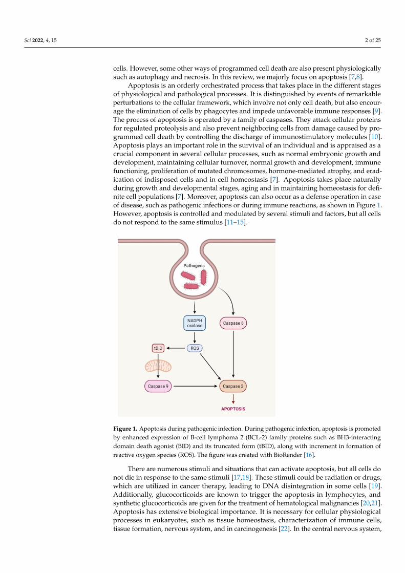
Apoptosis is an orderly orchestrated process that takes place in the different stagesof physiological and pathological processes. It is distinguished by events of remarkableperturbations to the cellular framework, which involve not only cell death, but also encour-age the elimination of cells by phagocytes and impede unfavorable immune responses [9].The process of apoptosis is operated by a family of caspases. They attack cellular proteinsfor regulated proteolysis and also prevent neighboring cells from damage caused by pro-grammed cell death by controlling the discharge of immunostimulatory molecules [10].Apoptosis plays an important role in the survival of an individual and is appraised as acrucial component in several cellular processes, such as normal embryonic growth anddevelopment, maintaining cellular turnover, normal growth and development, immunefunctioning, proliferation of mutated chromosomes, hormone-mediated atrophy, and erad-ication of indisposed cells and in cell homeostasis [7]. Apoptosis takes place naturallyduring growth and developmental stages, aging and in maintaining homeostasis for defi-nite cell populations [7]. Moreover, apoptosis can also occur as a defense operation in caseof disease, such as pathogenic infections or during immune reactions, as shown in Figure 1.However, apoptosis is controlled and modulated by several stimuli and factors, but all cellsdo not respond to the same stimulus [11–15].
Sci 2022, 4, x FOR PEER REVIEW 2 of 25
discrete way of cell death that allows programmed cell death or elimination of genetically determined cells. However, some other ways of programmed cell death are also present physiologically such as autophagy and necrosis. In this review, we majorly focus on apop-tosis [7,8].
Apoptosis is an orderly orchestrated process that takes place in the different stages of physiological and pathological processes. It is distinguished by events of remarkable perturbations to the cellular framework, which involve not only cell death, but also en-courage the elimination of cells by phagocytes and impede unfavorable immune re-sponses [9]. The process of apoptosis is operated by a family of caspases. They attack cel-lular proteins for regulated proteolysis and also prevent neighboring cells from damage caused by programmed cell death by controlling the discharge of immunostimulatory molecules [10]. Apoptosis plays an important role in the survival of an individual and is appraised as a crucial component in several cellular processes, such as normal embryonic growth and development, maintaining cellular turnover, normal growth and develop-ment, immune functioning, proliferation of mutated chromosomes, hormone-mediated atrophy, and eradication of indisposed cells and in cell homeostasis [7]. Apoptosis takes place naturally during growth and developmental stages, aging and in maintaining ho-meostasis for definite cell populations [7]. Moreover, apoptosis can also occur as a defense operation in case of disease, such as pathogenic infections or during immune reactions, as shown in Figure 1. However, apoptosis is controlled and modulated by several stimuli and factors, but all cells do not respond to the same stimulus [11–15].
Figure 1. Apoptosis during pathogenic infection. During pathogenic infection, apoptosis is pro-moted by enhanced expression of B-cell lymphoma 2 (BCL-2) family proteins such as BH3-interact-ing domain death agonist (BID) and its truncated form (tBID), along with increment in formation of reactive oxygen species (ROS). The figure was created with BioRender [16].
There are numerous stimuli and situations that can activate apoptosis, but all cells do not die in response to the same stimuli [17,18]. These stimuli could be radiation or drugs, which are utilized in cancer therapy, leading to DNA disintegration in some cells [19]. Additionally, glucocorticoids are known to trigger the apoptosis in lymphocytes, and syn-thetic glucocorticoids are given for the treatment of hematological malignancies [20,21]. Apoptosis has extensive biological importance. It is necessary for cellular physiological
Figure 1. Apoptosis during pathogenic infection. During pathogenic infection, apoptosis is promotedby enhanced expression of B-cell lymphoma 2 (BCL-2) family proteins such as BH3-interactingdomain death agonist (BID) and its truncated form (tBID), along with increment in formation ofreactive oxygen species (ROS). The figure was created with BioRender [16].
There are numerous stimuli and situations that can activate apoptosis, but all cells donot die in response to the same stimuli [17,18]. These stimuli could be radiation or drugs,which are utilized in cancer therapy, leading to DNA disintegration in some cells [19].Additionally, glucocorticoids are known to trigger the apoptosis in lymphocytes, andsynthetic glucocorticoids are given for the treatment of hematological malignancies [20,21].Apoptosis has extensive biological importance. It is necessary for cellular physiologicalprocesses in eukaryotes, such as tissue homeostasis, characterization of immune cells,tissue formation, nervous system, and in carcinogenesis [22]. In the central nervous system,

Sci 2022, 4, 15 3 of 25
apoptosis is linked with several disorders such as in Alzheimer’s disease, Parkinson’sdisease, lateral sclerosis, Huntington’s disease and other debilitating diseases [23–25].Apoptosis is associated with removal of damaged, unwanted cells, cells with non-repairableDNA and auto-reactive cells in immune system [26]. Hence, it is not surprising thatdysfunction in apoptosis can cause cancer and autoimmune diseases [27,28]. Additionally,some evidence shows that dysregulation of apoptotic pathways leads to the development ofseveral diseases, such as ischemic diseases, acquired immune deficiency syndrome (AIDS),and neurodegenerative disorders [7]. Therefore, the present review aims to highlightthe importance of apoptotic signaling routes and mechanisms, as well as the interactingcomponents involved in signaling, and how apoptosis is linked to carcinogenesis, as wellas insights into using apoptosis to treat disease. Furthermore, we discuss the several targetsand the potential lead compounds under investigation for the treatment of cancer.
2. Process of Apoptosis
The biology of apoptosis is complicated and includes an energy driven process ofmolecular episodes. There are two apoptotic pathways, namely, the extrinsic pathway,which is also named as death receptor pathway, while the other is intrinsic pathway, whichis named as a mitochondrial pathway. These pathways are associated with each other andinfluence the functioning of each other, as shown in Figure 2 [29]. Additionally, there isone more pathway through which apoptosis is induced, which includes T-cell-mediatedcytotoxicity and perforin/granzyme-mediated cell death. All the three pathways combinetogether at the execution pathway [30]. The execution pathway includes the cleavage ofcaspase-3 and finally leads to disintegration of DNA and cytoskeletal as well as nuclearproteins. It also involves the generation of apoptotic bodies, expresses ligands for receptorsof phagocytic cells and is consequently engulfed by phagocytic cells [7].
Sci 2022, 4, x FOR PEER REVIEW 3 of 25
processes in eukaryotes, such as tissue homeostasis, characterization of immune cells, tis-sue formation, nervous system, and in carcinogenesis [22]. In the central nervous system, apoptosis is linked with several disorders such as in Alzheimer’s disease, Parkinson’s dis-ease, lateral sclerosis, Huntington’s disease and other debilitating diseases [23–25]. Apop-tosis is associated with removal of damaged, unwanted cells, cells with non-repairable DNA and auto-reactive cells in immune system [26]. Hence, it is not surprising that dys-function in apoptosis can cause cancer and autoimmune diseases [27,28]. Additionally, some evidence shows that dysregulation of apoptotic pathways leads to the development of several diseases, such as ischemic diseases, acquired immune deficiency syndrome (AIDS), and neurodegenerative disorders [7]. Therefore, the present review aims to high-light the importance of apoptotic signaling routes and mechanisms, as well as the inter-acting components involved in signaling, and how apoptosis is linked to carcinogenesis, as well as insights into using apoptosis to treat disease. Furthermore, we discuss the sev-eral targets and the potential lead compounds under investigation for the treatment of cancer.
2. Process of Apoptosis The biology of apoptosis is complicated and includes an energy driven process of
molecular episodes. There are two apoptotic pathways, namely, the extrinsic pathway, which is also named as death receptor pathway, while the other is intrinsic pathway, which is named as a mitochondrial pathway. These pathways are associated with each other and influence the functioning of each other, as shown in Figure 2 [29]. Additionally, there is one more pathway through which apoptosis is induced, which includes T-cell-mediated cytotoxicity and perforin/granzyme-mediated cell death. All the three pathways combine together at the execution pathway [30]. The execution pathway includes the cleavage of caspase-3 and finally leads to disintegration of DNA and cytoskeletal as well as nuclear proteins. It also involves the generation of apoptotic bodies, expresses ligands for receptors of phagocytic cells and is consequently engulfed by phagocytic cells [7].
Figure 2. Apoptosis is mediated via extrinsic and intrinsic pathways. To begin an energy-driven process of molecular episodes, each pathway requires its unique triggering signal. Both routes acti-vate their own initiator, which is accompanied by caspase-3 activation. Other protein families, such
Figure 2. Apoptosis is mediated via extrinsic and intrinsic pathways. To begin an energy-drivenprocess of molecular episodes, each pathway requires its unique triggering signal. Both routesactivate their own initiator, which is accompanied by caspase-3 activation. Other protein families,such as inhibitors of apoptosis proteins (IAPs) and B-cell lymphoma 2 (BCL-2) family proteins, arealso required for the entire mechanism. The figure was created with BioRender [16].

Sci 2022, 4, 15 4 of 25
2.1. Extrinsic Pathway
The extrinsic signaling is mainly linked with transmembrane receptor-mediated inter-actions, which include death receptors of the tumor necrosis factor (TNF) receptor genesuperfamily. The death domain of death receptors is responsible for transmitting deathsignals from the cell’s surface to the intracellular signaling pathways [31]. There are dif-ferent death receptors, which are well characterized, such as TNF-α/TNFR1, FasL/FasR,apoptosis antigen (APO) 2L/DR4 APO3L/DR3, and APO2L/DR5 [32]. The mechanismof extrinsic pathway is well explained by the FasL/FasR and TNF-α/TNFR1 models [33].The Fas and TNF ligands bind to the Fas and TNF receptors, respectively, which in turnallow the interaction with their respective adapter proteins, namely Fas-associated proteinwith death domain (FADD) and TNFR1-associated death domain protein (TRADD) [34].The binding of the adapter protein is linked with stimulation of procaspase-8 accompaniedby the autocatalytic action of caspase-8 [32]. Once caspase-8 is stimulated, this pathwayconverges with the execution pathway.
Importantly, the apoptosis ligand 2/tumor necrosis factor-related apoptosis-inducingligand (APO2L/TRAIL) pathway plays an important role in the progression and develop-ment of cancer and it acts independently of p53. Further, pro-apoptotic receptor agonists(PARAs) that target DR4 and/or DR5 have potential to give significant clinical benefit bykilling tumor cells that are resistant to traditional chemotherapeutic agents [35]. Hence,targeting extrinsic pathways could be a novel treatment strategy to promote apoptosis inthe cancer cells.
2.2. Perforin/Granzyme Pathway
In this pathway, T-cell cytotoxicity is mediated by cytotoxic T lymphocytes (CTLs),which are enrolled in targeting the cells for killing through the extrinsic pathway, asshown in Figure 3 [36,37]. They show cytotoxic effects by secreting perforin and releasingcytoplasmic granules, which involve serine proteases granzyme A and granzyme B incomplex with serglycin [38]. Granzyme B can activate procaspase-10 and is able to activatecaspase-3 directly in such a way that bypasses the upstream signaling cascade and directlystimulates the execution pathway [39]. Granzyme A is also associated with CTL-mediatedapoptosis in a caspase-independent manner. It stimulates DNA nicking leading to thedegradation of apoptotic DNA [7].
Sci 2022, 4, x FOR PEER REVIEW 4 of 25
as inhibitors of apoptosis proteins (IAPs) and B-cell lymphoma 2 (BCL-2) family proteins, are also required for the entire mechanism. The figure was created with BioRender [16].
2.1. Extrinsic Pathway The extrinsic signaling is mainly linked with transmembrane receptor-mediated in-
teractions, which include death receptors of the tumor necrosis factor (TNF) receptor gene superfamily. The death domain of death receptors is responsible for transmitting death signals from the cell’s surface to the intracellular signaling pathways [31]. There are dif-ferent death receptors, which are well characterized, such as TNF-α/TNFR1, FasL/FasR, apoptosis antigen (APO) 2L/DR4 APO3L/DR3, and APO2L/DR5 [32]. The mechanism of extrinsic pathway is well explained by the FasL/FasR and TNF-α/TNFR1 models [33]. The Fas and TNF ligands bind to the Fas and TNF receptors, respectively, which in turn allow the interaction with their respective adapter proteins, namely Fas-associated protein with death domain (FADD) and TNFR1-associated death domain protein (TRADD) [34]. The binding of the adapter protein is linked with stimulation of procaspase-8 accompanied by the autocatalytic action of caspase-8 [32]. Once caspase-8 is stimulated, this pathway con-verges with the execution pathway.
Importantly, the apoptosis ligand 2/tumor necrosis factor-related apoptosis-inducing ligand (APO2L/TRAIL) pathway plays an important role in the progression and develop-ment of cancer and it acts independently of p53. Further, pro-apoptotic receptor agonists (PARAs) that target DR4 and/or DR5 have potential to give significant clinical benefit by killing tumor cells that are resistant to traditional chemotherapeutic agents [35]. Hence, targeting extrinsic pathways could be a novel treatment strategy to promote apoptosis in the cancer cells.
2.2. Perforin/Granzyme Pathway In this pathway, T-cell cytotoxicity is mediated by cytotoxic T lymphocytes (CTLs),
which are enrolled in targeting the cells for killing through the extrinsic pathway, as shown in Figure 3 [36,37]. They show cytotoxic effects by secreting perforin and releasing cytoplasmic granules, which involve serine proteases granzyme A and granzyme B in complex with serglycin [38]. Granzyme B can activate procaspase-10 and is able to activate caspase-3 directly in such a way that bypasses the upstream signaling cascade and directly stimulates the execution pathway [39]. Granzyme A is also associated with CTL-mediated apoptosis in a caspase-independent manner. It stimulates DNA nicking leading to the degradation of apoptotic DNA [7].
Figure 3. Function of the T cell receptor (TCR) and peptide–MHC complex in apoptosis. The TCR and peptide–MHC complex causes directed release of perforin and granzymes complexed with ser-glycin triggering apoptosis.
Figure 3. Function of the T cell receptor (TCR) and peptide–MHC complex in apoptosis. The TCR andpeptide–MHC complex causes directed release of perforin and granzymes complexed with serglycintriggering apoptosis.

Sci 2022, 4, 15 5 of 25
2.3. Intrinsic Pathway
The intrinsic pathway induces apoptosis via varied types of non-receptor-mediatedstimuli, which are mainly mitochondrial-dependent events [40]. These stimuli generateintracellular signals that target the cell directly either in a positive or negative manner [7].Positive stimuli include viral infections, radiation, hypoxia, toxins, hyperthermia and freeradicals. Negative stimuli involve a lack of some growth factors, cytokines or hormones,which subsequently results in the repression of the death program, in turn, stimulatingapoptosis. These mentioned stimuli bring about modifications in the inner mitochon-drial membrane resulting in the liberation of pro-apoptotic proteins. One group of pro-apoptotic proteins consists of SMAC/direct IAP binding protein with low pI (DIABLO),cytochrome c and HTRA2/OMI, which activate the caspase-associated with the mitochon-drial pathway [41]. Among these proteins, SMAC/DIABLO and HTRA serine peptidase 2(HTRA2)/OMI stimulate apoptosis by preventing the functioning of IAP [41,42]. However,cytochrome c works by stimulating apoptotic protease-activating factor 1 (APAF-1) andprocaspase-9 by binding to them, thereby resulting in the activation of caspase-9 [43,44].Endonuclease G and apoptosis-inducing factor (AIF) are nucleases located in the intermem-brane space of the mitochondria that together with caspase-activated DNase (CAD) lead tochromatin condensation and DNA fragmentation at later stages of apoptosis. Moreover, themembers of the BCL-2 family are crucial in maintaining mitochondrial dependent apoptoticevents as this family of protein regulates the permeability of the mitochondrial membraneand, hence, can act as pro-apoptotic or anti-apoptotic [45]. The pro-apoptotic genes includeBAX, BCL-10, BIK, BAK, BLK, BAD, BIM, BID, PUMA and NOXA, while the anti-apoptoticgenes include BCL-2, BAG, BCL-XS, BCL-XL, BCL-x and BCL-w. All these proteins decidethe fate of cell, i.e., whether the cell undergoes apoptosis or aborts apoptosis [45–47]. Hence,modulating the expression of these proteins can regulate the apoptosis process and, thus,may halt cell proliferation in cancer cells.
2.4. Execution Pathway
The execution pathway connects both the intrinsic and extrinsic pathway and finallyleads to apoptosis. In this cascade, the execution caspases activate and start the final processof apoptosis. Herein, activation of endonucleases and nucleases for the disintegrationof nuclear material and cytoskeletal proteins is evident [7]. The main caspases of theexecution pathway are caspase-3, caspase-6 and caspase-7, which degrade cytokeratins,poly (ADP-ribose) polymerase (PARP), cytoskeletal protein, namely, α-fodrin, the nuclearprotein nuclear-mitotic apparatus protein (NuMA) and others, finally leading to severalbiochemical and morphological alterations in cells undergoing apoptosis [48]. For instance,caspase-3 is significant for cell fate following apoptosis and it activates CAD. In a normalcell, CAD remains in a binding state with its inhibitor, namely, the inhibitor of caspase-activated DNAse (ICAD), while in apoptotic cells, caspase-3 degrades ICAD, therebyreleasing CAD [49]. Finally, CAD performs its function of fragmenting chromosomalDNA, resulting in the condensation of chromatin [49]. It also stimulates the process ofcytoskeleton organization and cell fragmentation into apoptotic bodies.
Overall, this section discussed the involvement of different apoptosis pathways, suchas the extrinsic pathway, the intrinsic pathway, and T-cell mediated and perforin/granzyme-mediated cytotoxicity, which ultimately combine with the execution pathway. Further,aberrant apoptosis is an important reason for resistance to cancer therapeutics. Additionally,cancerous cells can lead to altered expressions of several apoptotic and anti-apoptoticproteins, which ultimately may lead to increased cell proliferation. Hence, targeting theseproteins opens up new possibilities for selectively eradicating cancer cells.
3. Caspases
What causes the biochemical and morphological changes linked with the process werecognize as apoptosis? The answer is caspases. The operation of apoptosis is driven by theactivity of the family of cysteine proteases, which specifically make cuts at their substrate,

Sci 2022, 4, 15 6 of 25
i.e., aspartic acid residues, and are called as caspases [50]. The name caspase is givenfor Cysteine Aspartyl-specific Proteases. In all animal cells, these cysteine proteases areavailable in the form of inactive zymogens, but become stimulated to acquire their activestate and perform proteolytic processing specifically at aspartate residue [51]. Caspasesare crucial in regulating the process of inflammation and cell death. The association ofcaspases in cell death was known long after the discovery of ced-3 in Caenorhabditis elegans.Functionally, the main apoptotic caspases in mammals are caspase-2, -3, -7, -8, -9 and -10,while the inflammatory caspases are -1, -4, -5, -11 and -12. On the basis of a gene sequencearrangement, i.e., the existence or lack of protein interaction domains at the N-terminus,these apoptotic caspases are categorized as initiator caspases and effector caspases [52,53].The initiator caspases consist of either death effector domain (DED domain) or caspase-recruitment domain (CARD domain). The DED domain is included in caspase-8 and -10,while caspase-2, -9, -1 and -11 contain CARD domains. These caspases regulate the processof activation of apoptosis. The functioning of initiator caspases is driven by the mitochon-drial or intrinsic pathway and the extrinsic pathway [54]. As discussed earlier, the intrinsicpathway works in response to stress stimuli and is governed by the BCL-2 family of pro-teins leading to stimulation of BAX and BAK, which allows cytochrome c liberation anddisturbs mitochondrial outer membrane permeability (MOMP) [55]. Cytochrome c bindswith APAF-1 generating apoptosome and finally activates caspase-9. The extrinsic wayof apoptosis is followed by ligand binding and via activating the death domain receptorfamily, such as TNFR, Fas and TNF-related apoptosis-inducing ligand (TRAIL). Theseallow activation of caspase-8 or -10 by generating the death-inducing signaling complex(DISC) [56]. As the activation caspases activate following their respective pathways, theyundergo the activation of execution caspases, namely, caspases-3, -6 and -7. Caspase-2 isalso crucial as it becomes stimulated via stimulus in both upstream and downstream ofMOMP. Caspase-2 is linked to a large multiprotein complex known as PIDDosome, whichconsists of two proteins, namely RIP-associated ICH1/CED3-homologous protein witha death domain (RAIDD) and p53-induced protein with a death domain (PIDD). It caninteract with these two proteins in combination as well as independently [57]. Overall,caspases play a crucial role in the initiation of apoptosis signals and induction of proteolysis,thereby causing cell death. Hence, targeting different caspases could be a crucial target formodulating the apoptotic machinery in cancerous tissue. Table 1 summarizes the role ofcaspases, their origin and their substrates.
Table 1. Caspases with their location, substrate and functions *.
Caspases Presence Substrates Functions
Caspase-1 Spleen, liver, kidney, lung, heart Lamins, InterleukinsInvolved in inflammation,apoptosis induction when
overexpressed [58]
Caspase-2 Liver, CNS, kidney and lungdevelopment in embryo Lamins, Golgin-160 Apoptosis [59]
Caspase-3 Broadly distributed Caspases-6, -7, -9 Apoptosis [60]
Caspase-4 Lung, placenta, ovary, liver Caspase-1 Apoptosis [61]
Caspase-5 Liver, lung Max Apoptosis, inflammation [62]
Caspase-6 Liver, lung, skeletal muscle PARP, caspase-3, NuMA, lamins,FAK, keratin-18 Apoptosis [63]
Caspase-7 Lung, kidney, liver, heart, spleen, testis PARP, GAS2, EMAP II,calpastatin, FAK Apoptosis [64]

Sci 2022, 4, 15 7 of 25
Table 1. Cont.
Caspases Presence Substrates Functions
Caspase-8 Leukocytes, thymus, spleen, liver Caspases-3, -4, -6, -7, -9, -10, -13 Apoptosis [65]
Caspase-9 Heart, liver, skeletal muscle, pancreas Caspase-3, PARP, procaspase-9,caspase-7 Apoptosis [66]
Caspase-10 Tissues Caspases-3, -4, -6, -7, -8, -9 Apoptosis [67]
Caspase-11 Brain microglia Caspases-3, -1 Apoptosis, inflammation [68]
Caspase-12 Endoplasmic reticulum (ER) Caspases-1, -4, -5, -11 Apoptosis-mediated by ERstress [69]
Caspase-13 Lymphocytes, placenta, spleen Caspase-8 Inflammation [69]
Caspase-14 Epidermal cells Caspases-8, -10 Inflammation [70]
* Abbreviations used are: EMAP II, endothelial monocyte-activating polypeptide-II; FAK, focal adhesion kinase;GAS2, growth arrest-specific protein 2; Max, Myc-associated factor X; NuMA, nuclear mitotic apparatus protein;PARP, poly(ADP-ribose) polymerase.
4. Apoptosis-Associated Protein Domains
There are proteins or families of proteins that regulate the caspase activation pathways,namely, the extrinsic or intrinsic pathways, and they are identified depending on theiramino acid sequence or homologue. The interactions facilitated by these protein familiesare driven through protein domains that are linked with the regulation of apoptosis, suchas death domains (DDs), caspase recruitment domains (CARDs), death effector domains(DEDs), BCL-2 family proteins and of IAP-family proteins.
4.1. Death Domain Proteins
DDs consist of a condensed bundle of six alpha helices that interact among themselvesand form an oligomer. The differences in surface residue decide the specificity for partnerselection in the death domain [71]. Several TNF family members of the cytokine receptorhave their death domains in the cytosolic face. The TNF receptor family, including TNFR1,DR3 and DR6, works by binding to adapter protein, namely, TRADD, which contains itshomologous death domain. The DD of TRADD is able to bind with other proteins contain-ing DD, such as Fadd, an adapter protein. The FADD protein has a DD through whichit associates with the TNF family and, hence, links TNF family receptors to caspases [72].Hence, this protein plays a role of mediator. Additionally, a similar example is seen in caseof RAIDD, which is also known as CRADD. This is an adapter protein containing DD alongwith CARDs. It aids in linking the death receptor family with procaspases by binding withthe CARD domain of pro-caspase-2 [73]. Moreover, TRAIL-R1 (DR4) and TRAIL-R2 (DR5)are involved in the induction of apoptosis by binding to the TRAIL ligand. TRAIL ligandcan also bind with DcR1, DcR2 and osteoprotegerin (OPG), which are also TNF familyproteins, but they are considered as decoy receptors as they do not carry a death signal [74].Fas, is also a member of TNF receptor family and is considered a potential stimulator ofapoptosis. It is crucial in the homeostasis of the immune system, removing autoreactivelymphocytes and reducing immune response once foreign antigens are removed [75,76].There are some other proteins that have DD and are involved in apoptosis, such as DAPkinase, but their mechanism is not well understood. However, it has been reported thatthey activate caspase-8, which disturbs the cytoskeleton [54]. Due to the importance ofDD proteins, any imbalance or defect in the regulation and functioning of these proteinsleads to human diseases. For instance, upregulation in the expression of FasR or FasL onlymphocytes has been known to be associated with HIV infection. Moreover, mutation indeath domain of the Fas gene can cause lymphoproliferative syndrome, an autoimmunesyndrome and malignancies [77].
Sci 2022, 4, 15 8 of 25
4.2. Death Effector Domain Proteins
The death effector domain (DED) shares similarity with DD proteins in terms ofstructure. It exists in initiator caspases, namely, caspase-8 and caspase-10. There are twotandem DED motifs present in the prodomain region of initiator caspases. Due to thepresence of DED in caspase-8 and caspase-10, they are able to make an interaction withDED of FADD, and thereby allow their association with the death receptor complexes [78].The DED family proteins regulate apoptosis either by increasing caspase activation orpreventing caspase activation by TNF family death receptors. One of the DED-containingproteins is FLIP, which is also known as FLAME, CLARP, CASH, CASPER, I-FLICE, MRITor Usurpin [79]. It has been documented to be involved in suppressing apoptosis incancer. The amino acid sequence of FLIP shares similarity with pro caspase-8 and -10and, hence, competes with these caspases for binding to FADD, thereby silencing deathreceptor signaling. The increasing level of FLIP in tumor cells results in the development ofresistance against apoptosis induction by Fas-expressing CTLs [80]. This FLIP-associatedFas resistance makes the tumor cell tolerate FasL expression, utilizing this death ligand as aweapon to nearby normal cells, and to stimulate apoptosis of immune cells. However, thereare strategies for down-regulating the expression of FLIP employing antisense technologyand drugs that restore sensitivity of tumor cell lines toward apoptosis towards FasL [81].
4.3. CARD-Family Proteins
There are numerous pro-caspases, namely, caspase-1, -2, -4, -5 and -9, which haveCARDs in their N-terminal prodomains. The CARD has six helices present in DED andDD. CARD-family proteins are crucial in caspase activation via homotypic interactionsthroughout animal evolution. An example of caspase activation can be seen in CED-4 inC. elegans and APAF-1 in humans [82]. Both the proteins carry a CARD domain along witha nucleotide-binding oligomerization domain, called the NB-ARC (Nucleotide-bindingdomain homologous to APAF-1, CED-4 and plant R gene products) [83]. The CARD ofAPAF-1 associates with the CARD of pro-caspase-9, and upon oligomerization, they activatecaspases. APAF-1 contains several WD-40 regulatory domains, which makes it dependenton cytochrome c. It has been seen that the oligomerization of APAF-1 is dependent upondATP and cytochrome c, and once it is oligomerized, it binds and activates caspase-9 [82].Cancer cells have shown innumerable mechanisms for preventing caspase activation viaAPAF-1, such as the inhibition of APAF-1 gene by methylation, increased expression ofheat shock proteins that inhibit their functioning, increased expression of the tumor-up-regulated CARD-containing antagonist of caspase nine (TUCAN), which contains CARDsand is an antagonist of caspase-9, phosphorylating caspase-9 causing inhibition of theiractivity, and the association of CARDs with IAP-family proteins [73].
4.4. Inhibitor of Apoptosis Proteins
The inhibitor of apoptosis proteins or IAPs constitutes a family of suppressors ofapoptosis that are conserved throughout the evolution. The main function of these proteinsis the inhibition of caspases endogenously [84]. All IAPs have a common baculovirusinhibitor of the apoptosis protein repeat (BIR) domain, which is crucial for inhibitingapoptosis. However, only the presence of the BIR domain does not indicate anti-apoptoticactivity as this domain is involved in regulating cell cycle with no impact on cell death [84].Apart from the BIR domain, the IAP family of proteins also consists of other domains,such as RING zinc-fingers, CARDs, Ubiquitin-conjugating enzyme (E2s) domains andputative nucleotide-binding domains [85]. The RINGs and Apollon (BIR domain protein)are associated with ubiquitination machinery. Several IAPs, such as the X-linked inhibitorof the apoptosis protein (XIAP), cIAP1 and cIAP2 bind directly to the initiator caspase-9 andeven to the executioner caspase-3 and caspase-7, thereby inhibiting their function. Differentdomains are required for inhibiting caspases; for instance, in the case of XIAP, a secondBIR domain is required to inhibit caspase-3 and -7, while a third BIR domain is crucial forsuppressing caspase-9 [86]. The IAP family members, namely, Livin and survivin, have

Sci 2022, 4, 15 9 of 25
one BIR domain and suppress caspase-9, but not caspase-3 and -7. The IAPs are highlyselective towards specific caspases; therefore, overexpression of IAPs can inhibit some ofthe apoptotic pathways but not all [87,88]. However, baculovirus p35 protein representsa broad-spectrum activity against most of the caspases, but no cellular homologue ofthis protein has yet been found. Further, IAPs target apoptosis mediated by intrinsic orextrinsic pathways, because their target, i.e., effector caspases are associated with these twopathways. It has been documented that overexpression of IAP family members is associatedwith cancers [89]. For instance, survivin, Livin, XIAP and cIAP1 overexpression are shownin melanomas and tumor cells [87,88]. However, antisense-mediated targeting of XIAP orcIAP1 can stimulate apoptosis in tumor cell lines. There are endogenous antagonists ofIAPs, such as SMAC (DIABLO) and HTRA2 (OMI), which function in promoting apoptosis.These antagonists compete with caspases to bind to IAPs, thereby not allowing caspasesfrom binding to IAP and, hence, favoring apoptosis [90].
4.5. BCL-2 Family Proteins
The BCL-2-family proteins are associated with the mitochondrial dependent pathwayof apoptosis. Some proteins of this family, such as BCL-2, BCL-XL and BAK have a patchof hydrophobic amino acids at the C-terminal end through which these are linked withthe outer mitochondrial membrane [91]. However, this hydrophobic patch is absent inBID, BIM and BAD, but they are associated with mitochondria via specific stimuli [92].The BCL-2-family proteins are found to be conserved in the evolution of metazoan, andtheir homologues are present in vertebrates as well as invertebrates. Innumerable animalvirus genomes, such as herpes simplex virus, Epstein-Barr virus (EBV) and Kaposi sarcomaherpes virus contain BCL-2 homologs [93]. There are 26 members of the BCL-2 familythat are known currently. Their genes code for anti-apoptotic and pro-apoptotic proteinsare, namely, BCL-2, BFL-1 (A1), BCL-XL, MCL-1, BCL-w, BCL-B and BAX, BOK (MTD),BAK, BAD, BIM, BID, BIK, BCL-XS, NIP3, HRK, PUMA, APR (NOXA), BCL-Gs, p193, NIX(BNIP) and BCL-RAMBO (MIL) [91,94–96]. The BCL-2 family members with their locationand functions are summarized in Table 2. Few of the BCL-2 family genes generate morethan two proteins via alternative splicing, which shows contrasting effects on apoptoticregulation, such as BCL-XL versus BCL-XS [97].
Table 2. The BCL-2 family proteins with their location and functions *.
BCL-2 Protein Location Roles Refs
BAX Cytosol Liberation of apoptogenic factors and induction ofcaspases [98]
BAK Integral mitochondrialmembrane protein
Conformational changes in BAK take place to formlarger complexes in apoptosis and create pores in
the mitochondrial membrane to liberateapoptogenic factors to promote apoptosis
[99]
BID Cytosol and membrane Directly activate BAX [100]
BCL-2 Mitochondria, nucleus,endoplasmic reticulum
Prevents apoptosis by maintaining integrity ofmitochondrial membrane integrity [101,102]
BCL-XLMitochondrial
transmembrane
Prevents release of cytochrome c via mitochondrialpore, thereby inhibiting activation of caspases by
cytochrome c[102]
MCL-1 Nucleus, mitochondria Associated with BAK1, BCL-2-associated deathpromoter, NOXA, BCL2L11 and PCNA [103,104]
BCL-w/BCL2L2 Mitochondrion Under cytotoxic conditions downregulateapoptosis [105]

Sci 2022, 4, 15 10 of 25
Table 2. Cont.
BCL-2 Protein Location Roles Refs
A1/BFL-1 Mitochondria, nucleus unknown [106,107]
BIM/BCL2L11 Mitochondria Interacts with BCL-2 or BCL-XL and prevents theiranti-apoptotic actions [108]
PUMA Mitochondria unknown; regulated by p53 transcriptionally [109,110]
BAD MitochondriaGenerate a complex with BCL-2 and BCL-XL,
inhibits them, thereby promotingBAX/BAK-mediated apoptosis
[111]
BIK/BLK Endoplasmic reticulum unknown [112]
NOXA/PMAIP1 Mitochondria unknown [47,113]
BMF Mitochondria unknown [114]
* Abbreviations used are: A1/BFL-1, BCL-2-related protein A1; BAD, BCL-2-associated agonist of cell death;BAK, BCL-2 antagonist killer; BAX, BCL-2-associated protein X; BCL-2, B-cell lymphoma 2; BCL-w/BCL2L2,BCL-2-like 2; BCL-XL, B-cell lymphoma-extra-large; BID, BH3-interacting domain death agonist; BIK/BLK, BCL-2-interacting killer; BIM/BCL2L11, BCL-2-interacting protein BIM; BMF, BCL-2-modifying factor; MCL-1, myeloidcell leukemia 1; NOXA/ PMAIP1, phorbol-12-myristate-13acetate-induced protein 1; PUMA, p53-upregulatedmodulator of apoptosis.
Altogether, this section highlights the importance of DDs, CARDs, DEDs, BCL-2 familyproteins and of IAP-family proteins in the regulation of apoptosis, and their possible linkwith lymphoproliferative syndrome, autoimmune syndrome and other malignancies. Thenext section focuses on the significance of apoptosis in carcinogenesis.
5. Apoptosis and Carcinogenesis
In multicellular organisms, there is a balance between cell formation via mitosis andcell death by apoptosis. Any imbalance in these regulatory processes leads to the devel-opment of cancer. Apoptosis is responsible for cell death, but dysfunction of it can causemalignancies [115]. In the early 1970s, Kerr and colleagues explained the link of apoptosiswith elimination of malignant cells, regress tumor progression and hyperplasia [116]. Itcan be said that decline in apoptosis and resistance towards apoptosis plays a crucial rolein cancer. There are several factors through which malignant cells can escape apoptosis ordevelop resistance against them. Some of the factors include upregulation in expressionof IAPs, decline in expression of caspases, defects or mutations in p53, and misbalancedreceptor signaling pathways and BCL-2 family of proteins, as shown in Figure 4 [117].
Some of the important strategies for targeting apoptosis for the treatment of cancer aresummarized in Table 3. There are several treatment strategies or drugs that can act on apop-totic signaling pathways for the treatment of cancer and are mentioned in Table 3. Over thepast three decades, scientists have been developing therapies using apoptosis for eliminat-ing cancer cells [118,119]. These include the clinical translation of various pro-apoptoticmembers for drug discovery and also for understanding the cancer biology [115,120–122].Collectively, these findings reveal that apoptosis plays an important role in the cytotoxicityin malignant tumors and could lead to the development of novel therapeutic techniquesthat target this process to regulate cancer cell proliferation.

Sci 2022, 4, 15 11 of 25
Sci 2022, 4, x FOR PEER REVIEW 10 of 25
A1/BFL-1 Mitochondria, nucleus unknown [106,107]
BIM/BCL2L11 Mitochondria Interacts with BCL-2 or BCL-XL and prevents their anti-apop-totic actions
[108]
PUMA Mitochondria unknown; regulated by p53 transcriptionally [109,110]
BAD Mitochondria Generate a complex with BCL-2 and BCL-XL, inhibits them,
thereby promoting BAX/BAK-mediated apoptosis [111]
BIK/BLK Endoplasmic reticulum unknown [112] NOXA/PMAIP1 Mitochondria unknown [47,113]
BMF Mitochondria unknown [114] Abbreviations used are: A1/BFL-1, BCL-2-related protein A1; BAD, BCL-2-associated agonist of cell death; BAK, BCL-2 antagonist killer; BAX, BCL-2-associated protein X; BCL-2, B-cell lymphoma 2; BCL-w/BCL2L2, BCL-2-like 2; BCL-XL, B-cell lymphoma-extra-large; BID, BH3-interacting domain death agonist; BIK/BLK, BCL-2-interacting killer; BIM/BCL2L11, BCL-2-interacting protein BIM; BMF, BCL-2-modifying factor; MCL-1, myeloid cell leukemia 1; NOXA/ PMAIP1, phorbol-12-myristate-13acetate-induced protein 1; PUMA, p53-upregulated modulator of apoptosis.
5. Apoptosis and carcinogenesis In multicellular organisms, there is a balance between cell formation via mitosis and
cell death by apoptosis. Any imbalance in these regulatory processes leads to the devel-opment of cancer. Apoptosis is responsible for cell death, but dysfunction of it can cause malignancies [115]. In the early 1970s, Kerr and colleagues explained the link of apoptosis with elimination of malignant cells, regress tumor progression and hyperplasia [116]. It can be said that decline in apoptosis and resistance towards apoptosis plays a crucial role in cancer. There are several factors through which malignant cells can escape apoptosis or develop resistance against them. Some of the factors include upregulation in expression of IAPs, decline in expression of caspases, defects or mutations in p53, and misbalanced receptor signaling pathways and BCL-2 family of proteins, as shown in Figure 4 [117].
Figure 4. Some of the important factors in apoptotic signaling that are dysregulated during cancer. Figure 4. Some of the important factors in apoptotic signaling that are dysregulated during cancer.
Table 3. Targeting apoptosis and associated proteins for the treatment of cancer.
Treatment Remarks Refs
Attacking the BCL-2 family
Oblimersen sodium Showed chemosensitivity along with anticancer drugs withsignificant improvement in myeloid leukemia [123,124]
BCL-2 family inhibitors (Small molecule)
Sodium butyrate, fenretinide, depsipetide and flavipirodoare known to alter gene or protein expression. While
ABT-263, GX15-070, ABT-737, HA14-1 and gossypol, affectthe proteins directly
[125]
BH3 mimetics
ABT-737 inhibit anti-apoptotic proteins namely BCL-2,BCL-XL and BCL-w [126]
ATF4, ATF3 and NOXA prevent MCL-1 functioning [127]
Suppressing the Bcl family anti-apoptoticproteins/genes
BCL-2 specific siRNA prevent target gene expression andpromote anti-proliferation and pro-apoptotic activity in
pancreatic cancer cells[128]
Suppressing BMI-1 is known to decrease the expression ofpAKT and BCL-2, it makes them sensitive to doxorubicin [129]
Targeting p53
p53-based gene therapy
Wild-type p53 genes having retroviral vector introducedinto cancer cells showed significant improvement [130]
Introduction of wild type p53 gene makes head and necktumor cells, and prostate cancers sensitive to radiotherapy [131]
ONYX-015 can disrupt tumor cells deficient in p53 [132]

Sci 2022, 4, 15 12 of 25
Table 3. Cont.
Treatment Remarks Refs
Attacking the BCL-2 family
p53-dependent drug therapy
Small molecules
PhiKan083 (A24275) binds and restores mutant p53 [133]
CP-31398 inserted with DNA and disrupts the DNA-p53complex, leading to restoration of unstable p53 mutants [134]
Other agents
Nutlins disrupt MSM2-p53 interaction, provide stability top53 and promote death in cancer cells [135]
MI-219 breaks MDM2-p53 interaction, leading to inhibitionof cell multiplication and the promotion of apoptosis in
cancer cells[136]
Tenovins reduce tumor growth in vivo [137]
p53-based immunotherapy
Vaccine having recombinant replication-defectiveadenoviral vector in combination with human wild-type
p53 showed improvement[138]
p53-specific T cell responses seen when given p53 peptide [139]
Targeting inhibitors of apoptosis proteins (IAPs)
Targeting XIAP by antisense approachImproved tumor control by radiotherapy [140]
Antisense oligonucleotides increasechemotherapeutic activity [141]
Targeting XIAP by siRNA approach
siRNAs targeting XIAP promote enhanced sensitivitytowards radiotherapy [142]
siRNAs targeting XIAP make hepatoma cells sensitivetowards death receptor and chemotherapy [143]
Targeting survivin by antisense approach
Transfection of anti-sense survivin into melanoma cellspromotes apoptosis [144]
Promote apoptosis and sensitivity of cancer cells towardschemotherapy [145]
Prevent growth of thyroid carcinoma cells [146]
Targeting survivin by siRNA approach
Decrease survivin expression and lower the resistance toradiotherapy in pancreatic cancer cells [147]
Prevent proliferation and promote apoptosis in lungadenocarcinoma cells [148]
Downregulate survivin expression, prevent multiplicationand increase apoptosis in ovarian cancer [149]
Increase radiosensitivity in cancer cells [150]
IAP antagonists(Small molecules antagonists)
Hsp90 inhibitors and Cyclin-dependent kinase inhibitorsare reported to target survivin [151]
Cyclopeptide SMAC mimetics 2 and 3 attaches to XIAP andcIAP-1/2, thereby promoting the induction of
caspases- 9 and -3/-7[152]
SM-164 increases TRAIL functioning [153]
Targeting caspases

Sci 2022, 4, 15 13 of 25
Table 3. Cont.
Treatment Remarks Refs
Caspase-dependentdrug therapy
Apoptin promotes apoptosis in malignant cells [154]
Small molecule caspase activators stimulate caspase,promoting enhanced drug sensitivity in tumor cells [155]
Caspase-dependentgene therapy
Caspase-3 gene therapy is reported to promote induction ofextensive apoptosis [156]
Caspase-3 gene introduction into Huh7 human hepatomacells promotes apoptosis [157]
Immunocaspase-3 in a recombinant adenovirus showedanticancer effect in hepatocellular cancer [158]
6. Targeting Apoptosis6.1. Approaches Targeting Intrinsic Pathway of Apoptosis
The BCL-2 gene family can be targeted to control cancer growth as it has been seenthat this gene is found in follicular lymphoma patients and can be targeted by promotingapoptosis. The approaches include small molecule mimetic of BH3 and small moleculeinhibitors or oligonucleotides targeting BCL-2 expression [121,159]. There are several waysto target intrinsic pathway of apoptosis (Table 4).
Table 4. Strategies to target intrinsic pathway of apoptosis *.
Target Clinical Trial Histology Trial Identity **
Dual BCL-2 andBCL-XL inhibitors
Navitoclax YES (Phase I/II) CLL, melanoma,solid tumors
NCT02079740,NCT02143401,NCT01989585,NCT02520778
APG-1252 YES (Phase I/II) SCLC, solid tumors NCT03387332
AZD4320 No Childhood ALL -
S44563 No Melanoma, SCLC, -
BCL2–32 No NHL -
BM-1197 No Colorectal cancer -
Selective BCL-2inhibitors
Venetoclax Yes (Phase I-III) CLL, AML -
S55746(BCL201) Yes (Phase I) NHL, multiple myeloma NCT02603445,
NCT02920697
APG-2575 Yes (Phase I) NHL, AML NCT03537482,NCT03913949
BCL-XL inhibitors
ABBV-155 * Yes (Phase I) Solid tumors NCT03595059
WEHI-539 No Breast cancer -
A-1155463 No AML -
A-1331852 No Soft-tissue sarcoma -

Sci 2022, 4, 15 14 of 25
Table 4. Cont.
Target Clinical Trial Histology Trial Identity **
MCL-1 inhibitors
AMG 176 Yes (Phase I) NHL, AML NCT02675452,NCT03797261
MIK665 (S64315) Yes (Phase I) NHL, AMLNCT02992483,NCT03672695,NCT02979366
AZD5991 Yes (Phase I) NHL, AML NCT03218683
S63845 No NHL, AML -
UMI-77 No Pancreatic cancer -
A-1210477 No Esophageal carcinoma -
VU661013 No AML -
IAP inhibitors andSMAC mimetic
antagonists
LCL161 Yes (Phase I/II)Colorectal cancer, multiple
myeloma, Polycythemia vera,myelofibrosis
NCT02649673,NCT02098161,NCT03111992
Birinapant (TL32711) Yes (Phase I/II) Advanced solid tumors, NHL NCT03803774,NCT02587962
* Abbreviations used are: ALL, acute lymphoblastic leukemia; AML, acute myelogenous leukemia; BCL-2, B-cell lymphoma 2; BCL-XL, B-cell lymphoma-extra-large; CLL, chronic lymphocytic leukemia; IAP, inhibitor ofapoptosis; MCL-1, myeloid cell leukemia 1; NHL, Non-Hodgkin lymphoma; SCLC, small cell lung cancer; SMAC,small mitochondrial-derived activator of caspase. ** Clinical trial numbers were taken from ClincalTrials.gov [160].
6.1.1. BH3 Mimetics
The first inhibitor molecule designed against BCL-2, BCL-XL and BCL-w was ABT-737.This molecule binds in the hydrophobic pocket of BCL-2 family members, where BH3-onlyprotein binds. It shows good results against lung carcinoma and worked in combinationwith chemotherapy and radiation [161]. This is followed by the discovery of navitoclax(ABT-263), which also showed anti-cancer therapeutic potential, and in synergy with MEKor tyrosine kinase inhibitors it worked against solid tumors [162]. Moreover, venetoclax(ABT-199) is a BCL-2 small molecule inhibitor that exhibits significant results against CLLand non-Hodgkin lymphoma (NHL), which show an increased expression of BCL-2 [163].Additionally, selective BCL-XL inhibitors are also in the development phase, such as aBCL-XL-based vaccine for prostate cancer and the inhibitor of BCL-XL, i.e., an antibody–drug conjugate, namely, ABBV-155, which focuses on solid tumors as a monotherapy oralong with taxanes. Interestingly, BH3 mimetics have been developed successfully dueto the advent in technologies for targeting protein–protein interactions utilizing stapledpeptides, i.e., the synthetic protein becomes trapped within the secondary structure viaa chemical staple. These peptides directly target the protein–protein interaction withenhanced penetration inside the cell [164]. The first example of a staple peptide is SAHBA(stabilized alpha-helix of BCL-2 domains), one which mimics the α-helical BH3 segment ofproapoptotic BID. It easily penetrates inside the leukemia cells and becomes attached toBCL-XL, stimulates apoptosis and shows anti-tumor activity [165].
Additionally, stapled peptides work against the p53-MDM2 interaction for reactivatingp53 and similar compounds as well as targeting anti-apoptotic BFL-1 and MCL-1 [166]. Inlung cancer, apoptosis can be induced by targeting the regulatory site, i.e., S184 of Bax,which controls its localization. This site can be targeted via a small molecule, namely,SMBA1-3, which specifically binds to Bax and prevents the phosphorylation of S184,thereby promoting the liberation of cytochrome c and finally leading to apoptosis [167].Moreover, some other Bax-activating molecules, such as BAM-7 and BTSA1, also showedsignificant results against glioblastoma cell lines, while small molecule inhibitors of Bax,known as Bax activation inhibitors, can also show their allosteric inhibition with significantantitumor potential.
Sci 2022, 4, 15 15 of 25
6.1.2. MCL-1 Inhibitors
MCL-1 is associated with several forms of malignancies and is involved in resistance tochemotherapy and BCL-2 and BCL-XL inhibitors [168]. A small molecule inhibitor, namely,AM-8621, has the ability to bind to the MCL-1 pocket and remove BIM and promoteapoptosis in myeloma cell lines. Additionally, the derivatives of this small molecule,i.e., AMG 176 and AZD5991, have also shown significant results in good synergy withvenetoclax and chemotherapy [169,170]. Moreover, the MCL-1 inhibitors, VU661013 andS63845, showed good results in hematological malignancies and in combination strategiessuch as to combat venetoclax resistance [121].
6.1.3. IAP Inhibitors
IAP inhibitors are considered as a potential tool to promote apoptosis in cancer cells.There are eight known IAPs in humans, and among these, the most significant with anti-apoptotic activity are XIAP, melanoma IAP (ML-IAP), and cellular IAP1 and IAP2 [171].Interestingly, SMAC and OMI/HTRA2 have the ability to specifically and independentlyinhibit XIAP. SMAC itself binds to XIAP, c-IAP1 and c-IAP2, and thereby inhibits XIAPfrom binding caspase-3, -7 and -9. IAP proteins play an important role in cell survivalregulation; hence, inhibitors of IAP and SMAC mimetics could be of great therapeuticapplication especially in cancers [172]. IAP antagonists such as LCL161 showed anti-tumorpotential and in combination with chemotherapy also displayed good results in cases ofmultiple myeloma and head and neck squamous carcinoma models. One more compound,birinapant (TL32711), an IAP antagonist, did not display good activity singly, but whencombined with radiotherapy and anti-PD1 pembrolizumab, it showed significant results inhead and neck cancer [173].
6.2. Approaches Targeting Extrinsic Pathway6.2.1. Death Receptor Agonists
The extrinsic pathway of apoptosis is stimulated by extracellular signals that ac-tivate transmembrane protein members of the TNF receptor superfamily (TNFR) andpro-apoptotic death receptors (DRs). Death receptors, namely, TNFR1, Fas (CD95, APO-1),DR3, DR4 (TRAILR1), DR5 (TRAILR2) and DR6, once bound to their specific ligands, i.e.,Fas, TNFR1, TNFR2, DR4 and DR5, allow binding of an adapter protein such as FADD,leading to the generation of the DISC, which stimulates caspase-8 and -10 and the down-stream signaling of apoptotic pathway [174]. Therapeutic strategies are developed fortargeting DR4 and DR5 by developing recombinant Apo2L/TRAIL (stimulating DR4 andDR5) as well as agonistic monoclonal antibodies targeting DR4 or DR5 [175,176]. Du-lanermin, a recombinant APO2L/TRAIL, was utilized in clinical trials along with otherdrugs and chemotherapy in cases of solid tumors, but these did not show very promisingresults due to its short half-life [177]. Agonist antibodies were also developed againstDR4 and DR5 as they have a good half-life and they showed significant preclinical re-sults. Agonist monoclonal antibodies, namely, mapatumumab and lexatumumab forDR4 and DR5, respectively, were well tolerated in clinical trials and were administeredsingly as well in combination with other drugs and chemotherapy in solid tumors adultsarcomas [178,179]. Several other DR5 agonists were also tried in clinical trials, such asconatumumab (AMG655), tigatuzumab, LBY135 and drozitumab (PRO95780) [180,181].Furthermore, TRAIL-inducing small molecules were also developed as the expression ofTRAIL is associated with anti-tumor activity. One such TRAIL-inducing small moleculeis ONC201 [182]. It binds to dopamine receptors, namely, DRD2 and DRD3, and can alsobind to mitochondrial caseinolytic protease P (CIpP). Once this binding takes place, theintegrated stress response protein ATF4 becomes activated, which leads to the upregulationof DR5 and finally apoptosis [182–184].

Sci 2022, 4, 15 16 of 25
6.2.2. Tumor Suppressor Pathways
There are several therapeutic approaches targeting tumor suppressor pathways thatare being developed as promising anti-tumor agents. Inhibition of unregulated oncogeniceffectors, such as PI3K, AKT, β-catenin, Myc, CDKs, mTOR and VEGF, on mutated tumorsuppressors is under exploration, showing significant clinical outcomes [185]. A largenumber of strategies targeting the p53 pathway are under development as this is oneof the major targets to be inactivated in case of cancers [186]. Moreover, MDM2 andMDMX inhibitors are under investigation as they can block the pathway of wild-typep53 degradation without damaging DNA. MDM2 inhibitors, such as nutlin-3a derivativeRG7388 (idasanutlin), are administered either singly or in combination with venetoclax,atezolizumab or chemotherapy [187]. Some other MDM2 antagonists under investigationinclude AMG-232, HDM201, APG-115, DS-3032b, BI 907828 and ALRN-6924. Trials are alsobeing conducted for combining MDM2 antagonists along with inhibitors of other targetedagents such as MEK 1/2, BCL-2/BCL-XL, PI3K and BRAF inhibitors [121]. Moreover,strategies targeting restoration of the p53 tumor suppressor pathway in tumors havinga p53 mutant can be explained by APR-246. The restoration of the p53 pathway in p53-mutated tumors promotes apoptosis in tumors [188]. Additionally, inhibitors of CDK4/6,such as palbociclib, are approved for breast cancer as they promote cell death by increasingthe cell sensitivity towards TRAIL-mediated apoptosis. They can also promote cell cyclearrest in glioblastoma and myeloma [189].
6.2.3. Epigenetic Approaches Targeting Apoptosis
Histone deacetylases and BET are epigenetic modulators and their inhibitors possess an-ticancer activity via apoptosis and have shown good synergy with BCL-2 inhibitors [190,191].A BET inhibitor, namely, ABBV-075, in combination with venetoclax has shown good resultsin patients having cutaneous T cell lymphoma (CTCL) [191]. Its treatment has shown adecline in the protein expression of MCL-1, BCL-2 and BCL-XL in the acute myelogenousleukemia (AML) cell lines due to BET inhibitor-mediated modulation of gene expressionand chromatin.
Furthermore, the inhibitors of HDAC affect chromatin remodulation and promoteapoptosis by different processes. They increase expression of death receptors, TRAIL, Fasligands, and BH3-only proteins such as BID, BMF, BIM and BAK. In addition, they reduceexpression of BCL-2, BCL-w, BCL-XL and MCL-1, with an increment in the generation ofreactive oxygen species, which in turn stimulates the intrinsic pathway of apoptosis [190].HDAC inhibitors such as panobinostat stimulate NOXA and downregulate MCL-1 in B celllymphoma cell lines and also increase sensitivity towards the BCL-2 inhibitor [192]. HDACinhibitors can also increase the performance of MEK inhibitors and venetoclax in multiplemyeloma. Moreover, a hypomethylating agent, namely, azacytidine, demonstrated goodresults with venetoclax and ABT-737 [193].
6.2.4. Chaperons Targeting Apoptosis
Several strategies that induce stress in cells and finally promote apoptosis are beingexplored to target cancer cells. Chaperones are members of the heat shock protein fam-ily make the proteins stable in cancer cells; hence, inhibitors of the heat shock protein,specifically hsp90, which stabilizes the protein, are being explored [194]. The inhibitors ofhsp90, namely, geldanamycin, ganatespib, onalespib, XL888 and TAS116, are under investi-gation [195]. Moreover, the stress-mediated by the endoplasmic reticulum also promotesapoptosis in association with FADD, protein kinase R-like endoplasmic reticulum kinase(PERK), caspase-8 and enhanced expression of ER chaperone GRP78 [196]. Furthermore,the knowledge of ER-mediated stress, apoptosis and chaperons in cancer pathogenesisshould be explored to a greater extent, and inhibitors targeting chaperons that stabilize theprotein in cancerous cells should be targeted [121]. Table 5 enlists the strategies used fortargeting extrinsic pathways. Finally, the findings from the different trials have reignitedinterest in targeting apoptosis pathways. In particular, interfering with the function of

Sci 2022, 4, 15 17 of 25
specific proteins, modulation of epigenetic modulations or alterations, as well as the ofchaperon activity, are promising strategies for the generation of therapeutic strategiesin oncology.
Table 5. Strategies to target extrinsic pathway and other associated death mechanism of apoptosis.
Target Clinical Trials Cancer Trial Identity *
Death Receptor Agonists (DR4/5)
GEN1029 Yes(Phase I)
Colorectal cancer, renal carcinoma, triplenegative breast cancer, pancreatic cancer,
gastric cancerNCT03576131
ABBV-621 Yes(Phase I) AML, NHL, pancreatic cancer NCT03082209
MM-201 No Sarcoma -
TLY012 No Fibrosis -
Approaches targeting p53
Idasanutlin(RG73882)
Yes(Phase I)
Breast cancer, AML, NHL, multiplemyeloma
NCT03850535, NCT02670044,NCT02545283, NCT02633059,NCT03566485, NCT03135262
AMG-232 Yes(Phase I/II) AML, multiple myeloma, sarcoma NCT03041688, NCT03217266,
NCT03031730
HDM201 Yes(Phase I) AML NCT03940352
APG-115 Yes(Phase I) Advanced solid tumors, AML, melanoma NCT02935907, NCT03611868,
NCT03781986
DS-3032b Yes(Phase I) AML, solid tumors NCT03634228, NCT02319369,
NCT01877382
BI 907828 Yes(Phase I) Solid tumors NCT03449381
ALRN-6924 *** Yes(Phase I) Solid tumors NCT03725436
Restore wild type activity of mutant p53
APR246 Yes(Phase I)
AML, esophageal carcinoma, ovariancancer, melanoma
NCT02999893, NCT02098343,NCT03588078, NCT03391050,
NCT03391050
Other cell death mechanisms associated with apoptosis
ONC201 ** Yes(Phase I/II)
NHL, breast cancer, multiple myeloma,colorectal cancer, endometrial cancer, AML
NCT03099499, NCT02863991,NCT03416530, NCT02420795,NCT03394027, NCT03295396,NCT03791398, NCT02392572
Epigenetic modulators for stimulating intrinsic pathway of apoptosis
Fimepinostat+venetoclax
Yes(Phase I/II) NHL NCT01742988
Azacytidine ordecitabine +venetoclax
Yes(Phase I-III) AML NCT03404193, NCT03941964
Abbreviations used are: AML, acute myelogenous leukemia; NHL, Non-Hodgkin lymphoma. *** ALRN-6924 isthe first-ever clinical stage stapled peptide, ** ONC201 binds to DRD2/DRD3 and CIpP resulting in activation ofstress response protein ATF4 and cell death, * Clinical trial numbers were taken from ClincalTrials.gov [160].
Sci 2022, 4, 15 18 of 25
7. Conclusions
Apoptosis is considered as an energy-dependent process, which is associated withseveral biochemical and morphological characteristics where caspase induction plays acrucial role. However, several other apoptotic proteins, pro-apoptotic and anti-apoptoticproteins, are also involved in the regulation of apoptosis. The understanding of the mecha-nism of apoptosis is crucial because it balances both cell survival and cell death. It has beensuggested that any defects in the apoptotic pathway are involved in cancer and severalapproaches targeting apoptosis for treating cancer are feasible and are under investigation.Therefore, the exploration and knowledge of the mechanism of apoptosis, and associ-ated proteins at the cellular and molecular level, would help in digging deeper insightsinto several disease processes caused by apoptosis and, thereby, aiding in developingtherapeutic strategies.
There is some evidence that suggests that the resistance to apoptosis is one of themajor causes of cancer. So, targeting apoptotic pathways for the treatment of cancer istherapeutically important. Therefore, both targeting of intrinsic and extrinsic pathways ofapoptosis as well as interfering with other associated cell death mechanisms of apoptosisare enticing anti-cancer strategies. Currently, there are several drugs tested in clinical trialsas a monotherapy, as well as in combination with chemotherapy, radiation therapy, orother inhibitors for the management of different cancers. However, it is very challengingto modulate apoptosis pathways using targeted agents as there are several pathwaysinterfering with apoptosis and involved in the formation of cancer resistance. Hence, usingrational combination methods could be the key to their clinical success to overcome theseresistance mechanisms. Overall, we infer that targeting apoptosis mechanisms could be apromising oncology therapy that will continue to evolve in clinical practice in the future.
Author Contributions: Conceptualization, A.K.; resources, K.K.B. and R.W.; writing—original draftpreparation, V.S., A.K., U.N., P.A. and R.W.; writing—review and editing, V.S., A.K., U.N., P.A., K.K.B.and R.W.; visualization, V.S., A.K. and P.A. All authors have read and agreed to the published versionof the manuscript.
Funding: A.K. has received a P.R.I.M.E scholarship from the DAAD. R.W. is sponsored by the GermanResearch Foundation (grants WE 2554/13-1, WE 2554/15-1, and WE 2554/17-1).
Institutional Review Board Statement: Not applicable.
Informed Consent Statement: Not applicable.
Data Availability Statement: Not applicable.
Conflicts of Interest: The authors declare no conflict of interest.
References1. Kerr, J.F.; Wyllie, A.H.; Currie, A.R. Apoptosis: A basic biological phenomenon with wide-ranging implications in tissue kinetics.
Br. J. Cancer 1972, 26, 239–257. [CrossRef]2. Paweletz, N. Walther Flemming: Pioneer of mitosis research. Nat. Rev. Mol. Cell Biol. 2001, 2, 72–75. [CrossRef]3. Kerr, J.F.R. History of the events leading to the formulation of the apoptosis concept. Toxicology 2002, 181, 471–474. [CrossRef]4. Horvitz, H.R. The genetics of programmed cell death in the nematode Caenorhabditis elegans. Cold Spring Harb. Symp. Quant. Biol.
1994, 59, 377–385. [CrossRef]5. Formigli, L.; Papucci, L.; Tani, A.; Schiavone, N.; Tempestini, A.; Orlandini, G.E.; Capaccioli, S.; Zecchi Orlandini, S. Aponecrosis:
Morphological and biochemical exploration of a syncretic process of cell death sharing apoptosis and necrosis. J. Cell. Physiol.2000, 182, 41–49. [CrossRef]
6. Singh, V.; Khurana, A.; Allawadhi, P.; Banothu, A.K.; Bharani, K.K.; Weiskirchen, R. Emerging role of PD-1/PD-L1 inhibitors inchronic liver diseases. Front. Pharmacol. 2021, 12, 790963. [CrossRef]
7. Elmore, S. Apoptosis: A review of programmed cell death. Toxicol. Pathol. 2007, 35, 495–516. [CrossRef]8. Debnath, J.; Baehrecke, E.H.; Kroemer, G. Does autophagy contribute to cell death? Autophagy 2005, 1, 66–74. [CrossRef]9. Norbury, C.J.; Hickson, I.D. Cellular responses to DNA damage. Annu. Rev. Pharmacol. Toxicol. 2001, 41, 367–401. [CrossRef]10. Zhang, A.; Wu, Y.; Lai, H.W.; Yew, D.T. Apoptosis–a brief review. Neuroembryol. Aging 2004, 3, 47–59. [CrossRef]11. Portt, L.; Norman, G.; Clapp, C.; Greenwood, M.; Greenwood, M.T. Anti-apoptosis and cell survival: A review. Biochim. Biophys.
Acta (BBA)-Mol. Cell Res. 2011, 1813, 238–259. [CrossRef]

Sci 2022, 4, 15 19 of 25
12. Allawadhi, P.; Khurana, A.; Allwadhi, S.; Joshi, K.; Packirisamy, G.; Bharani, K.K. Nanoceria as a possible agent for themanagement of COVID-19. Nano Today 2020, 35, 100982. [CrossRef]
13. Allawadhi, P.; Singh, V.; Khurana, I.; Rawat, P.S.; Renushe, A.P.; Khurana, A.; Navik, U.; Allwadhi, S.; Karlapudi, S.K.; Banothu,A.K.; et al. Decorin as a possible strategy for the amelioration of COVID-19. Med. Hypotheses 2021, 152, 110612. [CrossRef]
14. Khurana, I.; Allawadhi, P.; Khurana, A.; Srivastava, A.K.; Navik, U.; Banothu, A.K.; Bharani, K.K. Can bilirubin nanomedicinebecome a hope for the management of COVID-19? Med. Hypotheses 2021, 149, 110534. [CrossRef]
15. Khurana, A.; Sayed, N.; Allawadhi, P.; Weiskirchen, R. It’s all about the spaces between cells: Role of extracellular matrix in liverfibrosis. Ann. Transl. Med. 2021, 9, 728. [CrossRef]
16. BioRender. Create Professional Science Figures in Minutes. Available online: https://biorender.com/ (accessed on 11January 2022).
17. D’Arcy, M.S. Cell death: A review of the major forms of apoptosis, necrosis and autophagy. Cell Biol. Int. 2019, 43, 582–592.[CrossRef]
18. Navik, U.; Sheth, V.G.; Khurana, A.; Jawalekar, S.S.; Allawadhi, P.; Gaddam, R.R.; Bhatti, J.S.; Tikoo, K. Methionine as adouble-edged sword in health and disease: Current perspective and future challenges. Ageing Res. Rev. 2021, 72, 101500.[CrossRef]
19. Allawadhi, P.; Khurana, A.; Allwadhi, S.; Navik, U.S.; Joshi, K.; Banothu, A.K.; Bharani, K.K. Potential of electric stimulation forthe management of COVID-19. Med. Hypotheses 2020, 144, 110259. [CrossRef]
20. Ouyang, L.; Shi, Z.; Zhao, S.; Wang, F.-T.; Zhou, T.-T.; Liu, B.; Bao, J.-K. Programmed cell death pathways in cancer: A review ofapoptosis, autophagy and programmed necrosis. Cell Prolif. 2012, 45, 487–498. [CrossRef]
21. Smith, L.K.; Cidlowski, J.A. Glucocorticoid-induced apoptosis of healthy and malignant lymphocytes. Prog. Brain Res. 2010, 182,1–30. [CrossRef]
22. Häcker, G. The morphology of apoptosis. Cell Tissue Res. 2000, 301, 5–17. [CrossRef]23. Papaliagkas, V.; Anogianaki, A.; Anogianakis, G.; Ilonidis, G. The proteins and the mechanisms of apoptosis: A mini-review of
the fundamentals. Hippokratia 2007, 11, 108–113.24. Hongmei, Z. Extrinsic and Intrinsic Apoptosis Signal Pathway Review. In Apoptosis and Medicine; Ntuli, T., Ed.; IntechOpen:
London, UK, 2012. [CrossRef]25. Singh, V.; Allawadhi, P.; Khurana, A.; Banothu, A.K.; Bharani, K.K. Critical neurological features of COVID-19: Role of imaging
methods and biosensors for effective diagnosis. Sens. Int. 2021, 2, 100098. [CrossRef]26. Wyllie, A.H. “Where, O death, is thy sting?” A brief review of apoptosis biology. Mol. Neurobiol. 2010, 42, 4–9. [CrossRef]27. Allawadhi, P.; Singh, V.; Govindaraj, K.; Khurana, I.; Sarode, L.P.; Navik, U.; Banothu, A.K.; Weiskirchen, R.; Bharani, K.K.;
Khurana, A. Biomedical applications of polysaccharide nanoparticles for chronic inflammatory disorders: Focus on rheumatoidarthritis, diabetes and organ fibrosis. Carbohydr. Polym. 2021, 281, 118923. [CrossRef]
28. Allawadhi, P.; Khurana, A.; Sayed, N.; Godugu, C.; Vohora, D. Ameliorative effect of cerium oxide nanoparticles against Freund’scomplete adjuvant-induced arthritis. Nanomedicine 2022, 17, 383–404. [CrossRef]
29. Igney, F.H.; Krammer, P.H. Death and anti-death: Tumour resistance to apoptosis. Nat. Cancer 2002, 2, 277–288. [CrossRef]30. Martinvalet, D.; Zhu, P.; Lieberman, J. Granzyme A induces caspase-independent mitochondrial damage, a required first step for
apoptosis. Immunity 2005, 22, 355–370. [CrossRef]31. Locksley, R.M.; Killeen, N.; Lenardo, M.J. The TNF and TNF receptor superfamilies: Integrating mammalian biology. Cell 2001,
104, 487–501. [CrossRef]32. Ashkenazi, A.; Dixit, V.M. Death receptors: Signaling and modulation. Science 1998, 281, 1305–1308. [CrossRef]33. Wajant, H. The Fas signaling pathway: More than a paradigm. Science 2002, 296, 1635–1636. [CrossRef]34. Suliman, A.; Lam, A.; Datta, R.; Srivastava, R.K. Intracellular mechanisms of TRAIL: Apoptosis through mitochondrial-dependent
and-independent pathways. Oncogene 2001, 20, 2122–2133. [CrossRef]35. Ashkenazi, A. Targeting the extrinsic apoptosis pathway in cancer. Cytokine Growth Factor Rev. 2008, 19, 325–331. [CrossRef]
[PubMed]36. Trapani, J.A.; Smyth, M. Functional significance of the perforin/granzyme cell death pathway. Nat. Rev. Immunol. 2002, 2, 735–747.
[CrossRef] [PubMed]37. Russell, J.H.; Ley, T.J. Lymphocyte-mediated cytotoxicity. Annu. Rev. Immunol. 2002, 20, 323–370. [CrossRef]38. Brunner, T.; Wasem, C.; Torgler, R.; Cima, I.; Jakob, S.; Corazza, N. Fas (CD95/Apo-1) Ligand regulation in T cell homeostasis,
cell-mediated cytotoxicity and immune pathology. In Seminars in Immunology; Academic Press: Cambridge, MA, USA, 2003;Volume 15, pp. 167–176. [CrossRef]
39. Goping, I.S.; Barry, M.; Liston, P.; Sawchuk, T.; Constantinescu, G.; Michalak, K.M.; Shostak, I.; Roberts, D.; Hunter, A.M.;Korneluk, R.; et al. Granzyme B-induced apoptosis requires both direct caspase activation and relief of caspase inhibition.Immunity 2003, 18, 355–365. [CrossRef]
40. Joza, N.; Susin, S.A.; Daugas, E.; Stanford, W.L.; Cho, S.K.; Li, C.Y.J.; Sasaki, T.; Elia, A.J.; Cheng, H.-Y.M.; Ravagnan, L.; et al.Essential role of the mitochondrial apoptosis-inducing factor in programmed cell death. Nature 2001, 410, 549–554. [CrossRef]
41. Du, C.; Fang, M.; Li, Y.; Li, L.; Wang, X. Smac, a Mitochondrial protein that promotes cytochrome c–dependent caspase activationby eliminating IAP inhibition. Cell 2000, 102, 33–42. [CrossRef]

Sci 2022, 4, 15 20 of 25
42. Van Loo, G.; Van Gurp, M.; Depuydt, B.; Srinivasula, S.M.; Rodriguez, I.; Alnemri, E.S.; Gevaert, K.; Vandekerckhove, J.; Declercq,W.; Vandenabeele, P. The serine protease Omi/HtrA2 is released from mitochondria during apoptosis. Omi interacts withcaspase-inhibitor XIAP and induces enhanced caspase activity. Cell Death Differ. 2002, 9, 20–26. [CrossRef]
43. Chinnaiyan, A.M. The Apoptosome: Heart and soul of the cell death machine. Neoplasia 1999, 1, 5–15. [CrossRef]44. Hill, M.M.; Adrain, C.; Duriez, P.J.; Creagh, E.M.; Martin, S.J. Analysis of the composition, assembly kinetics and activity of native
Apaf-1 apoptosomes. EMBO J. 2004, 23, 2134–2145. [CrossRef] [PubMed]45. Cory, S.; Adams, J.M. The Bcl2 family: Regulators of the cellular life-or-death switch. Nat. Rev. Cancer 2002, 2, 647–656. [CrossRef]
[PubMed]46. Liu, F.-T.; Newland, A.C.; Jia, L. Bax conformational change is a crucial step for PUMA-mediated apoptosis in human leukemia.
Biochem. Biophys. Res. Commun. 2003, 310, 956–962. [CrossRef] [PubMed]47. Oda, E.; Ohki, R.; Murasawa, H.; Nemoto, J.; Shibue, T.; Yamashita, T.; Tokino, T.; Taniguchi, T.; Tanaka, N. Noxa, a BH3-only
member of the Bcl-2 family and candidate mediator of p53-induced apoptosis. Science 2000, 288, 1053–1058. [CrossRef]48. Slee, E.A.; Adrain, C.; Martin, S. Executioner caspase-3, -6, and -7 perform distinct, non-redundant roles during the demolition
phase of apoptosis. J. Biol. Chem. 2001, 276, 7320–7326. [CrossRef]49. Sakahira, H.; Enari, M.; Nagata, S. Cleavage of CAD inhibitor in CAD activation and DNA degradation during apoptosis. Nature
1998, 391, 96–99. [CrossRef]50. Li, J.; Yuan, J. Caspases in apoptosis and beyond. Oncogene 2008, 27, 6194–6206. [CrossRef]51. Salvesen, G.S. Caspases and apoptosis. Essays Biochem. 2002, 38, 9–19. [CrossRef]52. Van Opdenbosch, N.; Lamkanfi, M. Caspases in cell death, inflammation, and disease. Immunity 2019, 50, 1352–1364. [CrossRef]53. Sayed, N.; Allawadhi, P.; Khurana, A.; Singh, V.; Navik, U.; Pasumarthi, S.K.; Khurana, I.; Banothu, A.K.; Weiskirchen, R.; Bharani,
K.K. Gene therapy: Comprehensive overview and therapeutic applications. Life Sci. 2022, 294, 120375. [CrossRef]54. Chen, M.; Wang, J. Initiator caspases in apoptosis signaling pathways. Apoptosis 2002, 7, 313–319. [CrossRef] [PubMed]55. Grütter, M.G. Caspases: Key players in programmed cell death. Curr. Opin. Struct. Biol. 2000, 10, 649–655. [CrossRef]56. Earnshaw, W.C.; Martins, L.M.; Kaufmann, S.H. Mammalian caspases: Structure, activation, substrates, and functions during
apoptosis. Annu. Rev. Biochem. 1999, 68, 383–424. [CrossRef] [PubMed]57. Stennicke, H.R.; Salvesen, G.S. Properties of the caspases. Biochim. Biophys. Acta (BBA)-Protein Struct. Mol. Enzymol. 1998, 1387,
17–31. [CrossRef]58. Molla, M.D.; Akalu, Y.; Geto, Z.; Dagnew, B.; Ayelign, B.; Shibabaw, T. Role of caspase-1 in the pathogenesis of inflammatory-
associated chronic noncommunicable diseases. J. Inflamm. Res. 2020, 13, 749–764. [CrossRef]59. Mancini, M.; Machamer, C.E.; Roy, S.; Nicholson, D.W.; Thornberry, N.A.; Casciola-Rosen, L.A.; Rosen, A. Caspase-2 Is Localized
at the Golgi Complex and Cleaves Golgin-160 during Apoptosis. J. Cell Biol. 2000, 149, 603–612. [CrossRef]60. Asadi, M.; Taghizadeh, S.; Kaviani, E.; Vakili, O.; Taheri-Anganeh, M.; Tahamtan, M.; Savardashtaki, A. Caspase-3: Structure,
function, and biotechnological aspects. Biotechnol. Appl. Biochem. 2021, Online ahead of print. [CrossRef]61. Chen, Q.; Shi, P.; Wang, Y.; Zou, D.; Wu, X.; Wang, D.; Hu, Q.; Zou, Y.; Huang, Z.; Ren, J.; et al. GSDMB promotes non-canonical
pyroptosis by enhancing caspase-4 activity. J. Mol. Cell Biol. 2018, 11, 496–508. [CrossRef]62. Aslan, A.; Gok, O.; Erman, O.; Kuloglu, T. Ellagic acid impedes carbontetrachloride-induced liver damage in rats through
suppression of NF-κB, Bcl-2 and regulating Nrf-2 and caspase pathway. Biomed. Pharmacother. 2018, 105, 662–669. [CrossRef]63. Zhao, P.; Sun, X.; Chaggan, C.; Liao, Z.; Wong, K.I.; He, F.; Singh, S.; Loomba, R.; Karin, M.; Witztum, J.L.; et al. An AMPK–
caspase-6 axis controls liver damage in nonalcoholic steatohepatitis. Science 2020, 367, 652–660. [CrossRef]64. Liao, B.; Sun, Q.; Yuan, Y.; Yin, Y.; Qiao, J.; Jiang, P. Histone deacetylase inhibitor MGCD0103 causes cell cycle arrest, apoptosis,
and autophagy in liver cancer cells. J. Cancer 2020, 11, 1915–1926. [CrossRef]65. Mandal, P.; Feng, Y.; Lyons, J.D.; Berger, S.B.; Otani, S.; DeLaney, A.; Tharp, G.K.; Maner-Smith, K.; Burd, E.M.; Schaeffer, M.; et al.
Caspase-8 collaborates with caspase-11 to drive tissue damage and execution of endotoxic shock. Immunity 2018, 49, 42–55.e6.[CrossRef] [PubMed]
66. Ren, Z.; Gu, X.; Fang, J.; Cai, D.; Zuo, Z.; Liang, S.; Cui, H.; Deng, J.; Ma, X.; Geng, Y.; et al. Effect of intranasal instillation ofEscherichia coli on apoptosis of spleen cells in diet-induced-obese mice. Sci. Rep. 2020, 10, 5109. [CrossRef] [PubMed]
67. Meynier, S.; Rieux-Laucat, F. FAS and RAS related Apoptosis defects: From autoimmunity to leukemia. Immunol. Rev. 2018, 287,50–61. [CrossRef]
68. Yi, Y.-S. Caspase-11 Noncanonical inflammasome: A novel key player in murine models of neuroinflammation and multiplesclerosis. Neuroimmunomodulation 2021, 28, 195–203. [CrossRef]
69. Song, J.; Zhang, Q.; Wang, S.; Yang, F.; Chen, Z.; Dong, Q.; Ji, Q.; Yuan, X.; Ren, D. Cleavage of caspase-12 at Asp94, mediated byendoplasmic reticulum stress (ERS), contributes to stretch-induced apoptosis of myoblasts. J. Cell. Physiol. 2018, 233, 9473–9487.[CrossRef]
70. Markiewicz, A.; Sigorski, D.; Markiewicz, M.; Owczarczyk-Saczonek, A.; Placek, W. Caspase-14—From biomolecular basics toclinical approach. A review of available data. Int. J. Mol. Sci. 2021, 22, 5575. [CrossRef]
71. Kitson, J.; Raven, T.; Jiang, Y.-P.; Goeddel, D.V.; Giles, K.M.; Pun, T.; Grinham, C.J.; Brown, R.; Farrow, S.N. A death-domain-containing receptor that mediates apoptosis. Nature 1996, 384, 372–375. [CrossRef]
72. Chinnaiyan, A.M.; O’Rourke, K.; Tewari, M.; Dixit, V.M. FADD, a novel death domain-containing protein, interacts with the deathdomain of fas and initiates apoptosis. Cell 1995, 81, 505–512. [CrossRef]

Sci 2022, 4, 15 21 of 25
73. Chou, J.J.; Matsuo, H.; Duan, H.; Wagner, G. Solution structure of the RAIDD CARD and model for CARD/CARD interaction incaspase-2 and caspase-9 recruitment. Cell 1998, 94, 171–180. [CrossRef]
74. Gonzalvez, F.; Ashkenazi, A. New insights into apoptosis signaling by Apo2L/TRAIL. Oncogene 2010, 29, 4752–4765. [CrossRef]75. Park, H.H.; Lo, Y.-C.; Lin, S.-C.; Wang, L.; Yang, J.K.; Wu, H. The death domain superfamily in intracellular signaling of apoptosis
and inflammation. Annu. Rev. Immunol. 2007, 25, 561–586. [CrossRef] [PubMed]76. Khurana, A.; Allawadhi, P.; Khurana, I.; Allwadhi, S.; Weiskirchen, R.; Banothu, A.K.; Chhabra, D.; Joshi, K.; Bharani, K.K. Role of
nanotechnology behind the success of mRNA vaccines for COVID-19. Nano Today 2021, 38, 101142. [CrossRef] [PubMed]77. Griffith, T.S.; Ferguson, T.A. The role of FasL-induced apoptosis in immune privilege. Immunol. Today 1997, 18, 240–244. [CrossRef]78. Valmiki, M.G.; Ramos, J.W. Death effector domain-containing proteins. Cell. Mol. Life Sci. 2008, 66, 814–830. [CrossRef] [PubMed]79. Barnhart, B.C.; Lee, J.C.; Alappat, E.C.; Peter, M.E. The death effector domain protein family. Oncogene 2003, 22, 8634–8644.
[CrossRef] [PubMed]80. Tibbetts, M.D.; Zheng, L.; Lenardo, M.J. The death effector domain protein family: Regulators of cellular homeostasis. Nat.
Immunol. 2003, 4, 404–409. [CrossRef]81. Aravind, L.; Dixit, V.M.; Koonin, E.V. The domains of death: Evolution of the apoptosis machinery. Trends Biochem. Sci. 1999, 24,
47–53. [CrossRef]82. Hofmann, K.; Bucher, P.; Tschopp, J. The CARD domain: A new apoptotic signalling motif. Trends Biochem. Sci. 1997, 22, 155–156.
[CrossRef]83. Yang, X.; Chang, H.Y.; Baltimore, D. Essential role of CED-4 oligomerization in CED-3 activation and apoptosis. Science 1998, 281,
1355–1357. [CrossRef]84. Deveraux, Q.L.; Reed, J.C. IAP family proteins—Suppressors of apoptosis. Genes Dev. 1999, 13, 239–252. [CrossRef]85. Yang, Y.; Fang, S.; Jensen, J.P.; Weissman, A.M.; Ashwell, J.D. Ubiquitin protein ligase activity of IAPs and their degradation in
proteasomes in response to apoptotic stimuli. Science 2000, 288, 874–877. [CrossRef] [PubMed]86. Takahashi, R.; Deveraux, Q.; Tamm, I.; Welsh, K.; Assa-Munt, N.; Salvesen, G.S.; Reed, J.C. A single BIR domain of XIAP sufficient
for inhibiting caspases. J. Biol. Chem. 1998, 273, 7787–7790. [CrossRef] [PubMed]87. Reed, J.C. The Survivin saga goes in vivo. J. Clin. Investig. 2001, 108, 965–969. [CrossRef] [PubMed]88. Kasof, G.M.; Gomes, B.C. Livin, a novel inhibitor of apoptosis protein family member. J. Biol. Chem. 2001, 276, 3238–3246.
[CrossRef] [PubMed]89. Vucic, D.; Stennicke, H.R.; Pisabarro, M.T.; Salvesen, G.S.; Dixit, V.M. ML-IAP, a novel inhibitor of apoptosis that is preferentially
expressed in human melanomas. Curr. Biol. 2000, 10, 1359–1366. [CrossRef]90. Deveraux, Q.L.; Takahashi, R.; Salvesen, G.S.; Reed, J.C. X-linked IAP is a direct inhibitor of cell-death proteases. Nature 1997, 388,
300–304. [CrossRef]91. Shamas-Din, A.; Kale, J.; Leber, B.; Andrews, D.W. Mechanisms of action of Bcl-2 family proteins. Cold Spring Harb. Perspect. Biol.
2013, 5, a008714. [CrossRef]92. Green, D.R.; Reed, J.C. Mitochondria and apoptosis. Science 1998, 281, 1309–1312. [CrossRef]93. Reed, J.C. Bcl-2 family proteins. Oncogene 1998, 17, 3225–3236. [CrossRef]94. Hardwick, J.M.; Soane, L. Multiple functions of BCL-2 family proteins. Cold Spring Harb. Perspect. Biol. 2013, 5, a008722. [CrossRef]95. Burlacu, A. Regulation of apoptosis by Bcl-2 family proteins. J. Cell. Mol. Med. 2003, 7, 249–257. [CrossRef] [PubMed]96. Reed, J.C.; Zha, H.; Aime-Sempe, C.; Takayama, S.; Wang, H.G. Structure—function analysis of Bcl-2 family proteins. Mech.
Lymph. Act. Immune Regul. 1996, 6, 99–112. [CrossRef]97. Karkale, S.; Khurana, A.; Saifi, M.A.; Godugu, C.; Talla, V. Oropharyngeal administration of silica in Swiss mice: A robust and
reproducible model of occupational pulmonary fibrosis. Pulm. Pharmacol. Ther. 2018, 51, 32–40. [CrossRef] [PubMed]98. Putcha, G.V.; Le, S.; Frank, S.; Besirli, C.; Clark, K.; Chu, B.; Alix, S.; Youle, R.J.; LaMarche, A.; Maroney, A.C.; et al. JNK-mediated
BIM phosphorylation potentiates BAX-dependent apoptosis. Neuron 2003, 38, 899–914. [CrossRef]99. Moldoveanu, T.; Grace, C.R.; Llambi, F.; Nourse, A.; Fitzgerald, P.; Gehring, K.; Kriwacki, R.W.; Green, D.R. BID-induced
structural changes in BAK promote apoptosis. Nat. Struct. Mol. Biol. 2013, 20, 589–597. [CrossRef]100. Degli Esposti, M. The roles of Bid. Apoptosis 2002, 7, 433–440. [CrossRef]101. Adams, J.M.; Cory, S. Bcl-2-regulated apoptosis: Mechanism and therapeutic potential. Curr. Opin. Immunol. 2007, 19, 488–496.
[CrossRef]102. Zhou, F.; Yang, Y.; Xing, D. Bcl-2 and Bcl-xL play important roles in the crosstalk between autophagy and apoptosis. FEBS J. 2011,
278, 403–413. [CrossRef]103. Stewart, M.L.; Fire, E.; Keating, A.E.; Walensky, L.D. The MCL-1 BH3 helix is an exclusive MCL-1 inhibitor and apoptosis
sensitizer. Nat. Chem. Biol. 2010, 6, 595–601. [CrossRef]104. Akgul, C. Mcl-1 is a potential therapeutic target in multiple types of cancer. Cell. Mol. Life Sci. 2009, 66, 1326–1336. [CrossRef]105. Hartman, M.L.; Czyz, M. BCL-w: Apoptotic and non-apoptotic role in health and disease. Cell Death Dis. 2020, 11, 1–16. [CrossRef]
[PubMed]106. Ottina, E.; Tischner, D.; Herold, M.J.; Villunger, A. A1/Bfl-1 in leukocyte development and cell death. Exp. Cell Res. 2012, 318,
1291–1303. [CrossRef] [PubMed]107. Wang, C.Y.; Guttridge, D.C.; Mayo, M.W.; Baldwin, A.S., Jr. NF-κB induces expression of the Bcl-2 homologue A1/Bfl-1 to
preferentially suppress chemotherapy-induced apoptosis. Mol. Cell. Biol. 1999, 19, 5923–5929. [CrossRef]

Sci 2022, 4, 15 22 of 25
108. O’Connor, L.; Strasser, A.; O’Reilly, L.A.; Hausmann, G.; Adams, J.; Cory, S.; Huang, D. Bim: A novel member of the Bcl-2 familythat promotes apoptosis. EMBO J. 1998, 17, 384–395. [CrossRef]
109. Nakano, K.; Vousden, K.H. PUMA, a novel proapoptotic gene, is induced by p53. Mol. Cell 2001, 7, 683–694. [CrossRef]110. Jeffers, J.R.; Parganas, E.; Lee, Y.; Yang, C.; Wang, J.; Brennan, J.; MacLean, K.H.; Han, J.; Chittenden, T.; Ihle, J.N.; et al. Puma is an
essential mediator of p53-dependent and -independent apoptotic pathways. Cancer Cell 2003, 4, 321–328. [CrossRef]111. Howells, C.C.; Baumann, W.T.; Samuels, D.C.; Finkielstein, C.V. The Bcl-2-associated death promoter (BAD) lowers the threshold
at which the Bcl-2-interacting domain death agonist (BID) triggers mitochondria disintegration. J. Theor. Biol. 2011, 271, 114–123.[CrossRef]
112. Coultas, L.; Bouillet, P.; Stanley, E.G.; Brodnicki, T.C.; Adams, J.; Strasser, A. Proapoptotic BH3-only Bcl-2 family memberBik/Blk/Nbk is expressed in hemopoietic and endothelial cells but is redundant for their programmed death. Mol. Cell. Biol.2004, 24, 1570–1581. [CrossRef]
113. Ploner, C.; Kofler, R.; Villunger, A. Noxa: At the tip of the balance between life and death. Oncogene 2008, 27, S84–S92. [CrossRef]114. Puthalakath, H.; Villunger, A.; O’Reilly, L.A.; Beaumont, J.G.; Coultas, L.; Cheney, R.E.; Huang, D.C.S.; Strasser, A. Bmf: A
proapoptotic BH3-only protein regulated by interaction with the myosin V actin motor complex, activated by Anoikis. Science2001, 293, 1829–1832. [CrossRef]
115. Wong, R.S.Y. Apoptosis in cancer: From pathogenesis to treatment. J. Exp. Clin. Cancer Res. 2011, 30, 87. [CrossRef] [PubMed]116. Hanahan, D.; Weinberg, R.A. The hallmarks of cancer. Cell 2000, 100, 57–70. [CrossRef]117. Wyllie, A.H. Apoptosis and carcinogenesis. Eur. J. Cell Biol. 1997, 73, 189–197. [PubMed]118. Wyllie, A.H.; Bellamy, C.O.; Bubb, V.J.; Clarke, A.R.; Corbet, S.; Curtis, L.; Harrison, D.J.; Hooper, M.L.; Toft, N.; Webb, S.; et al.
Apoptosis and carcinogenesis. Br. J. Cancer 1999, 80, 189–197.119. Rehman, M.U.; Khan, A.; Imtiyaz, Z.; Ali, S.; Makeen, H.A.; Rashid, S.; Arafah, A. Current Nano-therapeutic Approaches
Ameliorating Inflammation in Cancer Progression. In Seminars in Cancer Biology; Academic Press: Cambridge, MA, USA, 2022.[CrossRef]
120. Shoaib, A.; Tabish, M.; Ali, S.; Arafah, A.; Wahab, S.; Almarshad, F.M.; Rashid, S.; Rehman, M.U. Dietary Phytochemicals inCancer Signalling Pathways: Role of miRNA Targeting. Curr. Med. Chem. 2021, 28, 8036–8067. [CrossRef]
121. Carneiro, B.A.; El-Deiry, W.S. Targeting apoptosis in cancer therapy. Nat. Rev. Clin. Oncol. 2020, 17, 395–417. [CrossRef]122. Wani, J.; Majid, S.; Khan, A.; Arafah, A.; Ahmad, A.; Jan, B.; Shah, N.; Kazi, M.; Rehman, M. Clinico-Pathological Importance of
miR-146a in Lung Cancer. Diagnostics 2021, 11, 274. [CrossRef]123. Rai, K.R.; Moore, J.; Wu, J.; Novick, S.C.; O’Brien, S.M. Effect of the addition of oblimersen (Bcl-2 antisense) to fludara-
bine/cyclophosphamide for relapsed/refractory chronic lymphocytic leukemia (CLL) on survival in patients who achieveCR/nPR: Five-year follow-up from a randomized phase III study. J. Clin. Oncol. 2008, 26, 7008. [CrossRef]
124. Abou-Nassar, K.; Brown, J.R. Novel agents for the treatment of chronic lymphocytic leukemia. Clin. Adv. Hematol. Oncol. HO2010, 8, 886–895.
125. Kang, M.H.; Reynolds, C.P. Bcl-2 inhibitors: Targeting mitochondrial apoptotic pathways in cancer therapy. Clin. Cancer Res.2009, 15, 1126–1132. [CrossRef]
126. Oltersdorf, T.; Elmore, S.W.; Shoemaker, A.R.; Armstrong, R.C.; Augeri, D.J.; Belli, B.A.; Bruncko, M.; Deckwerth, T.L.; Dinges, J.;Hajduk, P.J.; et al. An inhibitor of Bcl-2 family proteins induces regression of solid tumours. Nature 2005, 435, 677–681. [CrossRef][PubMed]
127. Albershardt, T.C.; Salerni, B.L.; Soderquist, R.S.; Bates, D.J.; Pletnev, A.A.; Kisselev, A.F.; Eastman, A. Multiple BH3 mimeticsantagonize antiapoptotic MCL1 protein by inducing the endoplasmic reticulum stress response and up-regulating BH3-onlyprotein NOXA. J. Biol. Chem. 2011, 286, 24882–24895. [CrossRef] [PubMed]
128. Ocker, M.; Neureiter, D.; Lueders, M.; Zopf, S.; Ganslmayer, M.; Hahn, E.G.; Herold, C.; Schuppan, D. Variants of bcl-2 specificsiRNA for silencing antiapoptotic bcl-2 in pancreatic cancer. Gut 2005, 54, 1298–1308. [CrossRef] [PubMed]
129. Wu, X.; Liu, X.; Sengupta, J.; Bu, Y.; Yi, F.; Wang, C.; Shi, Y.; Zhu, Y.; Jiao, Q.; Song, F. Silencing of Bmi-1 gene by RNA interferenceenhances sensitivity to doxorubicin in breast cancer cells. Indian J. Exp. Biol. 2011, 49.
130. Roth, J.A.; Nguyen, D.; Lawrence, D.D.; Kemp, B.L.; Carrasco, C.H.; Ferson, D.Z.; Hong, W.K.; Komaki, R.; Lee, J.J.; Nesbitt,J.C.; et al. Retrovirus–mediated wild–type P53 gene transfer to tumors of patients with lung cancer. Nat. Med. 1996, 2, 985–991.[CrossRef]
131. Chène, P. p53 as a drug target in cancer therapy. Expert Opin. Ther. Patents 2001, 11, 923–935. [CrossRef]132. Nemunaitis, J.; Ganly, I.; Khuri, F.; Arseneau, J.; Kuhn, J.; Mccarty, T.; Landers, S.; Maples, P.; Romel, L.; Randlev, B.; et al.
Selective replication and oncolysis in p53 mutant tumors with ONYX-015, an E1B-55kD gene-deleted adenovirus, in patients withadvanced head and neck cancer: A phase II trial. Cancer Res. 2000, 60, 6359–6366.
133. Boeckler, F.; Joerger, A.; Jaggi, G.; Rutherford, T.J.; Veprintsev, D.; Fersht, A.R. Targeted rescue of a destabilized mutant of p53 byan in silico screened drug. Proc. Natl. Acad. Sci. USA 2008, 105, 10360–10365. [CrossRef]
134. Rippin, T.M.; Bykov, V.J.N.; Freund, S.M.V.; Selivanova, G.; Wiman, K.G.; Fersht, A.R. Characterization of the p53-rescue drugCP-31398 in vitro and in living cells. Oncogene 2002, 21, 2119–2129. [CrossRef]
135. Shangary, S.; Wang, S. Small-molecule inhibitors of the MDM2-p53 protein-protein interaction to reactivate p53 function: A novelapproach for cancer therapy. Annu. Rev. Pharmacol. Toxicol. 2009, 49, 223–241. [CrossRef]

Sci 2022, 4, 15 23 of 25
136. Shangary, S.; Qin, D.; McEachern, D.; Liu, M.; Miller, R.S.; Qiu, S.; Nikolovska-Coleska, Z.; Ding, K.; Wang, G.; Chen, J.; et al.Temporal activation of p53 by a specific MDM2 inhibitor is selectively toxic to tumors and leads to complete tumor growthinhibition. Proc. Natl. Acad. Sci. USA 2008, 105, 3933–3938. [CrossRef] [PubMed]
137. Lain, S.; Hollick, J.J.; Campbell, J.; Staples, O.D.; Higgins, M.; Aoubala, M.; McCarthy, A.; Appleyard, V.; Murray, K.E.; Baker,L.; et al. Discovery, in vivo activity, and mechanism of action of a small-molecule p53 activator. Cancer Cell 2008, 13, 454–463.[CrossRef]
138. Kuball, J.; Schuler, M.; Ferreira, E.A.; Herr, W.; Neumann, M.; Obenauer-Kutner, L.; Westreich, L.; Huber, C.; Wölfel, T.; Theobald,M. Generating p53-specific cytotoxic T lymphocytes by recombinant adenoviral vector-based vaccination in mice, but not man.Gene Ther. 2002, 9, 833–843. [CrossRef] [PubMed]
139. Svane, I.M.; Pedersen, A.E.; Johnsen, H.E.; Nielsen, D.; Kamby, C.; Gaarsdal, E.; Nikolajsen, K.; Claesson, M.H. Vaccinationwith p53-peptide? pulsed dendritic cells, of patients with advanced breast cancer: Report from a phase I study. Cancer Immunol.Immunother. 2004, 53, 633–641. [CrossRef] [PubMed]
140. Cao, C.; Mu, Y.; Hallahan, D.E.; Lu, B. XIAP and survivin as therapeutic targets for radiation sensitization in preclinical models oflung cancer. Oncogene 2004, 23, 7047–7052. [CrossRef]
141. Hu, Y.; Cherton-Horvat, G.; Dragowska, V.; Baird, S.; Korneluk, R.G.; Durkin, J.P.; Mayer, L.D.; Lacasse, E.C. Antisenseoligonucleotides targeting XIAP induce apoptosis and enhance chemotherapeutic activity against human lung cancer cells in vitroand in vivo. Clin. Cancer Res. 2003, 9, 2826–2836.
142. Ohnishi, K.; Scuric, Z.; Schiestl, R.H.; Okamoto, N.; Takahashi, A.; Ohnishi, T. siRNA targeting NBS1 or XIAP increases radiationsensitivity of human cancer cells independent of TP53 status. Radiat. Res. 2006, 166, 454–462. [CrossRef]
143. Yamaguchi, Y.; Shiraki, K.; Fuke, H.; Inoue, T.; Miyashita, K.; Yamanaka, Y.; Saitou, Y.; Sugimoto, K.; Nakano, T. Targeting ofX-linked inhibitor of apoptosis protein or survivin by short interfering RNAs sensitize hepatoma cells to TNF-related apoptosis-inducing ligand-and chemotherapeutic agent-induced cell death. Oncol. Rep. 2005, 14, 1311–1316. [CrossRef]
144. Grossman, D.; McNiff, J.M.; Li, F.; Altieri, D.C. Expression and targeting of the apoptosis inhibitor, survivin, in human melanoma.J. Investig. Dermatol. 1999, 113, 1076–1081. [CrossRef]
145. Sharma, H.; Sen, S.; Muzio, L.L.; Mariggiò, M.A.; Singh, N. Antisense-mediated downregulation of anti-apoptotic proteinsinduces apoptosis and sensitizes head and neck squamous cell carcinoma cells to chemotherapy. Cancer Biol. Ther. 2005, 4,720–727. [CrossRef]
146. Du, Z.-X.; Zhang, H.-Y.; Gao, D.-X.; Wang, H.-Q.; Li, Y.-J.; Liu, G.-L. Antisurvivin oligonucleotides inhibit growth and induceapoptosis in human medullary thyroid carcinoma cells. Exp. Mol. Med. 2006, 38, 230–240. [CrossRef] [PubMed]
147. Kami, K.; Doi, R.; Koizumi, M.; Toyoda, E.; Mori, T.; Ito, D.; Kawaguchi, Y.; Fujimoto, K.; Wada, M.; Miyatake, S.-I.; et al.Downregulation of survivin by siRNA diminishes radioresistance of pancreatic cancer cells. Surgery 2005, 138, 299–305. [CrossRef][PubMed]
148. Quanxi, L.I.U.; Chaoying, D.O.N.G.; Lijun, L.I.; Jie, S.U.N.; Chunyun, L.I.; Liang, L.I. Inhibitory effects of the Survivin siRNAtransfection on human lung adenocarcinoma cells SPCA1 and SH77. Chin. J. Lung Cancer 2011, 14, 18–22.
149. Zhang, X.; Li, N.; Wang, Y.H.; Huang, Y.; Xu, N.Z.; Wu, L.Y. Effects of Survivin siRNA on growth, apoptosis and chemosensitivityof ovarian cancer cells SKOV3/DDP. Zhonghua Zhong liu za zhi Chin. J. Oncol. 2009, 31, 174–177.
150. Yang, C.T.; Li, J.M.; Weng, H.H.; Li, Y.C.; Chen, H.C.; Chen, M.F. Adenovirus-mediated transfer of siRNA against survivinenhances the radiosensitivity of human non-small cell lung cancer cells. Cancer Gene Ther. 2010, 17, 120–130. [CrossRef]
151. Pennati, M.; Folini, M.; Zaffaroni, N. Targeting survivin in cancer therapy: Fulfilled promises and open questions. Carcinogenesis2007, 28, 1133–1139. [CrossRef]
152. Sun, H.; Liu, L.; Lu, J.; Qiu, S.; Yang, C.-Y.; Yi, H.; Wang, S. Cyclopeptide Smac mimetics as antagonists of IAP proteins. Bioorg.Med. Chem. Lett. 2010, 20, 3043–3046. [CrossRef]
153. Lu, J.; McEachern, D.; Sun, H.; Bai, L.; Peng, Y.; Qiu, S.; Miller, R.; Liao, J.; Yi, H.; Liu, M.; et al. Therapeutic potential andmolecular mechanism of a novel, potent, nonpeptide, Smac mimetic SM-164 in combination with TRAIL for cancer treatment.Mol. Cancer Ther. 2011, 10, 902–914. [CrossRef]
154. Rohn, J.L.; Noteborn, M.H.M. The viral death effector Apoptin reveals tumor-specific processes. Apoptosis 2004, 9, 315–322.[CrossRef]
155. Philchenkov, A.; Zavelevich, M.; Kroczak, T.J.; Los, M. Caspases and cancer: Mechanisms of inactivation and new treatmentmodalities. Exp. Oncol. 2004, 26, 82–97.
156. Yamabe, K.; Shimizu, S.; Ito, T.; Yoshioka, Y.; Nomura, M.; Narita, M.; Saito, I.; Kanegae, Y.; Matsuda, H. Cancer gene therapyusing a pro-apoptotic gene, caspase-3. Gene Ther. 1999, 6, 1952–1959. [CrossRef] [PubMed]
157. Cam, L.; Boucquey, A.; Coulomb-L’hermine, A.; Weber, A.; Horellou, P. Gene transfer of constitutively active caspase-3 inducesapoptosis in a human hepatoma cell line. J. Gene Med. A Cross-Discip. J. Res. Sci. Gene Transf. Its Clin. Appl. 2005, 7, 30–38.[CrossRef] [PubMed]
158. Li, X.; Fan, R.; Zou, X.; Gao, L.; Jin, H.; Du, R.; Xia, L.; Fan, D. Inhibitory effect of recombinant adenovirus carrying immunocaspase-3 on hepatocellular carcinoma. Biochem. Biophys. Res. Commun. 2007, 358, 489–494. [CrossRef] [PubMed]
159. Neophytou, C.M.; Trougakos, I.P.; Erin, N.; Papageorgis, P. Apoptosis deregulation and the development of cancer multi-drugresistance. Cancers 2021, 13, 4363. [CrossRef]

Sci 2022, 4, 15 24 of 25
160. ClinicalTrials.gov. U.S. National Library of Medicine. Available online: https://www.clinicaltrials.gov/ (accessed on 11January 2022).
161. Del Gaizo Moore, V.; Brown, J.R.; Certo, M.; Love, T.M.; Novina, C.D.; Letai, A. Chronic lymphocytic leukemia requires BCL2 tosequester prodeath BIM, explaining sensitivity to BCL2 antagonist ABT-737. J. Clin. Investig. 2007, 117, 112–121. [CrossRef]
162. Tse, C.; Shoemaker, A.R.; Adickes, J.; Anderson, M.G.; Chen, J.; Jin, S.; Johnson, E.F.; Marsh, K.C.; Mitten, M.J.; Nimmer, P.; et al.ABT-263: A potent and orally bioavailable Bcl-2 family inhibitor. Cancer Res. 2008, 68, 3421–3428. [CrossRef]
163. Souers, A.J.; Leverson, J.D.; Boghaert, E.R.; Ackler, S.L.; Catron, N.D.; Chen, J.; Dayton, B.D.; Ding, H.; Enschede, S.H.; Fairbrother,W.J.; et al. ABT-199, a potent and selective BCL-2 inhibitor, achieves antitumor activity while sparing platelets. Nat. Med. 2013, 19,202–208. [CrossRef]
164. Walensky, L.D.; Kung, A.L.; Escher, I.; Malia, T.J.; Barbuto, S.; Wright, R.D.; Wagner, G.; Verdine, G.L.; Korsmeyer, S.J. Activationof apoptosis in vivo by a hydrocarbon-stapled BH3 helix. Science 2004, 305, 1466–1470. [CrossRef]
165. Ali, A.M.; Atmaj, J.; Van Oosterwijk, N.; Groves, M.R.; Dömling, A. Stapled peptides inhibitors: A new window for target drugdiscovery. Comput. Struct. Biotechnol. J. 2019, 17, 263–281. [CrossRef]
166. Chang, Y.S.; Graves, B.; Guerlavais, V.; Tovar, C.; Packman, K.; To, K.H.; Olson, K.A.; Kesavan, K.; Gangurde, P.; Mukherjee, A.;et al. Stapled α−helical peptide drug development: A potent dual inhibitor of MDM2 and MDMX for p53-dependent cancertherapy. Proc. Natl. Acad. Sci. USA 2013, 110, E3445–E3454. [CrossRef]
167. Li, R.; Ding, C.; Zhang, J.; Xie, M.; Park, D.; Ding, Y.; Chen, G.; Zhang, G.; Gilbert-Ross, M.; Zhou, W.; et al. Modulation of Baxand mTOR for cancer therapeutics. Cancer Res. 2017, 77, 3001–3012. [CrossRef] [PubMed]
168. Wertz, I.E.; Kusam, S.; Lam, C.; Okamoto, T.; Sandoval, W.; Anderson, D.; Helgason, E.; Ernst, J.A.; Eby, M.; Liu, J.; et al. Sensitivityto antitubulin chemotherapeutics is regulated by MCL1 and FBW7. Nature 2011, 471, 110–114. [CrossRef] [PubMed]
169. Caenepeel, S.; Brown, S.P.; Belmontes, B.; Moody, G.; Keegan, K.S.; Chui, D.; Whittington, D.A.; Huang, X.; Poppe, L.; Cheng, A.C.;et al. AMG 176, a selective MCL1 inhibitor, is effective in hematologic cancer models alone and in combination with establishedtherapies. Cancer Discov. 2018, 8, 1582–1597. [PubMed]
170. Tron, A.E.; Belmonte, M.A.; Adam, A.; Aquila, B.M.; Boise, L.; Chiarparin, E.; Cidado, J.; Embrey, K.J.; Gangl, E.; Gibbons, F.D.;et al. Discovery of Mcl-1-specific inhibitor AZD5991 and preclinical activity in multiple myeloma and acute myeloid leukemia.Nat. Commun. 2018, 9, 5341. [CrossRef]
171. Fulda, S.; Vucic, D. Targeting IAP proteins for therapeutic intervention in cancer. Nat. Rev. Drug Discov. 2012, 11, 109–124.[CrossRef]
172. Monian, P.; Jiang, X. Clearing the final hurdles to mitochondrial apoptosis: Regulation post cytochrome C release. Exp. Oncol.2012, 34, 185–191.
173. Amaravadi, R.K.; Schilder, R.J.; Martin, L.P.; Levin, M.; Graham, M.A.; Weng, D.E.; Adjei, A.A. A phase I study of the SMAC-mimetic birinapant in adults with refractory solid tumors or lymphoma. Mol. Cancer Ther. 2015, 14, 2569–2575. [CrossRef]
174. Green, D.R.; Llambi, F. Cell death signaling. Cold Spring Harb. Perspect. Biol. 2015, 7, a006080. [CrossRef]175. Hymowitz, S.G.; Christinger, H.W.; Fuh, G.; Ultsch, M.; O’Connell, M.; Kelley, R.F.; Ashkenazi, A.; de Vos, A.M. Triggering cell
death: The crystal structure of Apo2L/TRAIL in a complex with death receptor 5. Mol. Cell 1999, 4, 563–571. [CrossRef]176. Leblanc, H.N.; Ashkenazi, A. Apo2L/TRAIL and its death and decoy receptors. Cell Death Differ. 2003, 10, 66–75. [CrossRef]177. Soria, J.-C.; Márk, Z.; Zatloukal, P.; Szima, B.; Albert, I.; Juhász, E.; Pujol, J.-L.; Kozielski, J.; Baker, N.; Smethurst, D.; et al.
Randomized phase II study of Dulanermin in combination with Paclitaxel, Carboplatin, and Bevacizumab in advanced non–small-cell lung cancer. J. Clin. Oncol. 2011, 29, 4442–4451. [CrossRef] [PubMed]
178. von Pawel, J.; Harvey, J.H.; Spigel, D.R.; Dediu, M.; Reck, M.; Cebotaru, C.L.; Humphreys, R.C.; Gribbin, M.J.; Fox, N.L.;Camidge, D.R. Phase II Trial of Mapatumumab, a fully human agonist monoclonal antibody to tumor necrosis factor-relatedapoptosis-inducing ligand receptor 1 (TRAIL-R1), in combination with Paclitaxel and Carboplatin in patients with advancednon–small-cell lung cancer. Clin. Lung Cancer 2014, 15, 188–196. [CrossRef] [PubMed]
179. Plummer, R.; Attard, G.; Pacey, S.C.; Li, L.; Razak, A.; Perrett, R.; Barrett, M.; Judson, I.; Kaye, S.; Fox, N.L.; et al. Phase 1 andpharmacokinetic study of Lexatumumab in patients with advanced cancers. Clin. Cancer Res. 2007, 13, 6187–6194. [CrossRef][PubMed]
180. Camidge, D.R.; Herbst, R.S.; Gordon, M.S.; Eckhardt, S.G.; Kurzrock, R.; Durbin, B.; Ing, J.; Tohnya, T.M.; Sager, J.; Ashkenazi, A.; et al.A phase I safety and pharmacokinetic study of the death receptor 5 agonistic antibody PRO95780 in patients with advancedmalignancies. Clin. Cancer Res. 2010, 16, 1256–1263. [CrossRef]
181. Ashkenazi, A. Directing cancer cells to self-destruct with pro-apoptotic receptor agonists. Nat. Rev. Drug Discov. 2008, 7,1001–1012. [CrossRef]
182. Kline, C.L.B.; Ralff, M.D.; Lulla, A.R.; Wagner, J.M.; Abbosh, P.H.; Dicker, D.T.; Allen, J.E.; El-Deiry, W.S. Role of dopaminereceptors in the anticancer activity of ONC201. Neoplasia 2017, 20, 80–91. [CrossRef]
183. Allen, J.E.; Krigsfeld, G.; Mayes, P.A.; Patel, L.; Dicker, D.T.; Patel, A.S.; Dolloff, N.G.; Messaris, E.; Scata, K.A.; Wang, W.; et al.Dual inactivation of Akt and ERK by TIC10 signals Foxo3a nuclear translocation, TRAIL gene induction, and potent antitumoreffects. Sci. Transl. Med. 2013, 5, 71ra17. [CrossRef]
184. Graves, P.R.; Aponte-Collazo, L.J.; Fennell, E.M.J.; Graves, A.C.; Hale, A.E.; Dicheva, N.; Herring, L.E.; Gilbert, T.S.K.; East, M.P.;McDonald, I.M.; et al. Mitochondrial protease ClpP is a target for the anticancer compounds ONC201 and related analogues.ACS Chem. Biol. 2019, 14, 1020–1029. [CrossRef]
Sci 2022, 4, 15 25 of 25
185. Sever, R.; Brugge, J.S. Signal transduction in cancer. Cold Spring Harb. Perspect. Med. 2015, 5, a006098. [CrossRef]186. Levine, A.J. Targeting therapies for the p53 protein in cancer treatments. Annu. Rev. Cancer Biol. 2019, 3, 21–34. [CrossRef]187. Ding, Q.; Zhang, Z.; Liu, J.J.; Jiang, N.; Zhang, J.; Ross, T.M.; Chu, X.J.; Bartkovitz, D.; Podlaski, F.; Janson, C.; et al. Discovery
of RG7388, a potent and selective p53–MDM2 inhibitor in clinical development. J. Med. Chem. 2013, 56, 5979–5983. [CrossRef][PubMed]
188. Zhang, Q.; Bykov, V.J.N.; Wiman, K.G.; Zawacka-Pankau, J. APR-246 reactivates mutant p53 by targeting cysteines 124 and 277.Cell Death Dis. 2018, 9, 439. [CrossRef] [PubMed]
189. Tao, Z.; Le Blanc, J.M.; Wang, C.; Zhan, T.; Zhuang, H.; Wang, P.; Yuan, Z.; Lu, B. Coadministration of Trametinib and Palbociclibradiosensitizes KRAS-mutant non–small cell lung cancers in vitro and in vivo. Clin. Cancer Res. 2016, 22, 122–133. [CrossRef]
190. Bolden, J.E.; Peart, M.J.; Johnstone, R.W. Anticancer activities of histone deacetylase inhibitors. Nat. Rev. Drug Discov. 2006, 5,769–784. [CrossRef]
191. Kim, S.R.; Lewis, J.M.; Cyrenne, B.M.; Monico, P.F.; Mirza, F.N.; Carlson, K.R.; Foss, F.M.; Girardi, M. BET inhibition in advancedcutaneous T cell lymphoma is synergistically potentiated by BCL2 inhibition or HDAC inhibition. Oncotarget 2018, 9, 29193–29207.[CrossRef]
192. Liu, Y.; Mondello, P.; Erazo, T.; Tannan, N.B.; Asgari, Z.; de Stanchina, E.; Nanjangud, G.; Seshan, V.E.; Wang, S.; Wendel, H.-G.; et al.NOXA genetic amplification or pharmacologic induction primes lymphoma cells to BCL2 inhibitor-induced cell death. Proc. Natl.Acad. Sci. USA 2018, 115, 12034–12039. [CrossRef] [PubMed]
193. Dinardo, C.D.; Pratz, K.; Pullarkat, V.; Jonas, B.A.; Arellano, M.; Becker, P.S.; Frankfurt, O.; Konopleva, M.; Wei, A.H.; Kantarjian,H.M.; et al. Venetoclax combined with decitabine or azacitidine in treatment-naive, elderly patients with acute myeloid leukemia.Blood 2018, 133, 7–17. [CrossRef]
194. Neckers, L.; Workman, P. Hsp90 molecular chaperone inhibitors: Are we there yet? Clin. Cancer Res. 2012, 18, 64–76. [CrossRef]195. Bussenius, J.; Blazey, C.M.; Aay, N.; Anand, N.K.; Arcalas, A.; Baik, T.; Bowles, O.J.; Buhr, C.A.; Costanzo, S.; Curtis, J.K.; et al.
Discovery of XL888: A novel tropane-derived small molecule inhibitor of HSP90. Bioorg. Med. Chem. Lett. 2012, 22, 5396–5404.[CrossRef]
196. Park, M.A.; Zhang, G.; Martin, A.P.; Hamed, H.; Mitchell, C.; Hylemon, P.B.; Graf, M.; Rahmani, M.; Ryan, K.; Liu, X.; et al.Vorinostat and sorafenib increase ER stress, autophagy and apoptosis via ceramide-dependent CD95 and PERK activation. CancerBiol. Ther. 2008, 7, 1648–1662. [CrossRef]
Copyright © 2022 FDOKUMEN