Cardiotoxicity of Antineoplastic Therapies and Applications of ...
18
cells Review Cardiotoxicity of Antineoplastic Therapies and Applications of Induced Pluripotent Stem Cell-Derived Cardiomyocytes Mo-Fan Huang 1,2,† , Lon Kai Pang 1,3,† , Yi-Hung Chen 4 , Ruiying Zhao 1, * and Dung-Fang Lee 1,2,5,6, * Citation: Huang, M.-F.; Pang, L.K.; Chen, Y.-H.; Zhao, R.; Lee, D.-F. Cardiotoxicity of Antineoplastic Therapies and Applications of Induced Pluripotent Stem Cell-Derived Cardiomyocytes. Cells 2021, 10, 2823. https://doi.org/ 10.3390/cells10112823 Academic Editors: Miguel Fidalgo, Ana Sevilla and Francesca Aguilo Received: 20 August 2021 Accepted: 15 October 2021 Published: 21 October 2021 Publisher’s Note: MDPI stays neutral with regard to jurisdictional claims in published maps and institutional affil- iations. Copyright: © 2021 by the authors. Licensee MDPI, Basel, Switzerland. This article is an open access article distributed under the terms and conditions of the Creative Commons Attribution (CC BY) license (https:// creativecommons.org/licenses/by/ 4.0/). 1 Department of Integrative Biology and Pharmacology, McGovern Medical School, The University of Texas Health Science Center at Houston, Houston, TX 77030, USA; [email protected] (M.-F.H.); [email protected] (L.K.P.) 2 The University of Texas MD Anderson Cancer Center UTHealth Graduate School of Biomedical Sciences, Houston, TX 77030, USA 3 Department of Neuroscience, Baylor College of Medicine, Houston, TX 77030, USA 4 Department and Institute of Pharmacology, National Yang Ming Chiao Tung University, Taipei 112, Taiwan; [email protected] 5 Center for Stem Cell and Regenerative Medicine, The Brown Foundation Institute of Molecular Medicine for the Prevention of Human Diseases, The University of Texas Health Science Center at Houston, Houston, TX 77030, USA 6 Center for Precision Health, School of Biomedical Informatics, The University of Texas Health Science Center at Houston, Houston, TX 77030, USA * Correspondence: [email protected] (R.Z.); [email protected] (D.-F.L.) † These authors contributed equally to this work. Abstract: The therapeutic landscape for the treatment of cancer has evolved significantly in recent decades, aided by the development of effective oncology drugs. However, many cancer drugs are often poorly tolerated by the body and in particular the cardiovascular system, causing adverse and sometimes fatal side effects that negate the chemotherapeutic benefits. The prevalence and severity of chemotherapy-induced cardiotoxicity warrants a deeper investigation of the mechanisms and implicating factors in this phenomenon, and a consolidation of scientific efforts to develop mitigating strategies. Aiding these efforts is the emergence of induced pluripotent stem cells (iPSCs) in recent years, which has allowed for the generation of iPSC-derived cardiomyocytes (iPSC-CMs): a human-based, patient-derived, and genetically variable platform that can be applied to the study of chemotherapy-induced cardiotoxicity and beyond. After surveying chemotherapy-induced cardiotox- icity and the associated chemotherapeutic agents, we discuss the use of iPSC-CMs in cardiotoxicity modeling, drug screening, and other potential applications. Improvements to the iPSC-CM platform, such as the development of more adult-like cardiomyocytes and ongoing advances in biotechnology, will only enhance the utility of iPSC-CMs in both basic science and clinical applications. Keywords: stem cell; disease model; induced pluripotency; reprogramming; differentiation; chemother- apy; cancer; cardiotoxicity; personalized medicine; pharmacogenomics 1. Induced Pluripotent Stem Cell-Derived Cardiomyocytes Over the last 15 years, induced pluripotent stem cells (iPSCs) have emerged as a valuable tool to the research community. Human iPSCs are available in unlimited supply, on immediate demand, can be maintained for weeks to months in vitro, can be derived directly from patients to capture unique genetic signatures, and are amenable to genome modification and transfection operations. Since their relatively recent development by Kazutoshi Takahashi and Shinya Yamanaka [1], iPSCs continue to find new applications in disease modeling, mechanistic studies, drug development, biobanking, and therapeutic strategies across a diverse range of pathologies [2–5]. Notably, iPSCs have recently emerged as a platform for the study of chemotherapy- induced cardiotoxicity. Cancer is one of the leading causes of death globally; in 2020, there Cells 2021, 10, 2823. https://doi.org/10.3390/cells10112823 https://www.mdpi.com/journal/cells
-
Upload
khangminh22 -
Category
Documents
-
view
3 -
download
0
Transcript of Cardiotoxicity of Antineoplastic Therapies and Applications of ...

cells
Review
Cardiotoxicity of Antineoplastic Therapies and Applications ofInduced Pluripotent Stem Cell-Derived Cardiomyocytes
Mo-Fan Huang 1,2,† , Lon Kai Pang 1,3,† , Yi-Hung Chen 4 , Ruiying Zhao 1,* and Dung-Fang Lee 1,2,5,6,*
�����������������
Citation: Huang, M.-F.; Pang, L.K.;
Chen, Y.-H.; Zhao, R.; Lee, D.-F.
Cardiotoxicity of Antineoplastic
Therapies and Applications of
Induced Pluripotent Stem
Cell-Derived Cardiomyocytes. Cells
2021, 10, 2823. https://doi.org/
10.3390/cells10112823
Academic Editors: Miguel Fidalgo,
Ana Sevilla and Francesca Aguilo
Received: 20 August 2021
Accepted: 15 October 2021
Published: 21 October 2021
Publisher’s Note: MDPI stays neutral
with regard to jurisdictional claims in
published maps and institutional affil-
iations.
Copyright: © 2021 by the authors.
Licensee MDPI, Basel, Switzerland.
This article is an open access article
distributed under the terms and
conditions of the Creative Commons
Attribution (CC BY) license (https://
creativecommons.org/licenses/by/
4.0/).
1 Department of Integrative Biology and Pharmacology, McGovern Medical School, The University of TexasHealth Science Center at Houston, Houston, TX 77030, USA; [email protected] (M.-F.H.);[email protected] (L.K.P.)
2 The University of Texas MD Anderson Cancer Center UTHealth Graduate School of Biomedical Sciences,Houston, TX 77030, USA
3 Department of Neuroscience, Baylor College of Medicine, Houston, TX 77030, USA4 Department and Institute of Pharmacology, National Yang Ming Chiao Tung University, Taipei 112, Taiwan;
[email protected] Center for Stem Cell and Regenerative Medicine, The Brown Foundation Institute of Molecular Medicine for
the Prevention of Human Diseases, The University of Texas Health Science Center at Houston,Houston, TX 77030, USA
6 Center for Precision Health, School of Biomedical Informatics, The University of Texas Health Science Centerat Houston, Houston, TX 77030, USA
* Correspondence: [email protected] (R.Z.); [email protected] (D.-F.L.)† These authors contributed equally to this work.
Abstract: The therapeutic landscape for the treatment of cancer has evolved significantly in recentdecades, aided by the development of effective oncology drugs. However, many cancer drugs areoften poorly tolerated by the body and in particular the cardiovascular system, causing adverseand sometimes fatal side effects that negate the chemotherapeutic benefits. The prevalence andseverity of chemotherapy-induced cardiotoxicity warrants a deeper investigation of the mechanismsand implicating factors in this phenomenon, and a consolidation of scientific efforts to developmitigating strategies. Aiding these efforts is the emergence of induced pluripotent stem cells (iPSCs)in recent years, which has allowed for the generation of iPSC-derived cardiomyocytes (iPSC-CMs): ahuman-based, patient-derived, and genetically variable platform that can be applied to the study ofchemotherapy-induced cardiotoxicity and beyond. After surveying chemotherapy-induced cardiotox-icity and the associated chemotherapeutic agents, we discuss the use of iPSC-CMs in cardiotoxicitymodeling, drug screening, and other potential applications. Improvements to the iPSC-CM platform,such as the development of more adult-like cardiomyocytes and ongoing advances in biotechnology,will only enhance the utility of iPSC-CMs in both basic science and clinical applications.
Keywords: stem cell; disease model; induced pluripotency; reprogramming; differentiation; chemother-apy; cancer; cardiotoxicity; personalized medicine; pharmacogenomics
1. Induced Pluripotent Stem Cell-Derived Cardiomyocytes
Over the last 15 years, induced pluripotent stem cells (iPSCs) have emerged as avaluable tool to the research community. Human iPSCs are available in unlimited supply,on immediate demand, can be maintained for weeks to months in vitro, can be deriveddirectly from patients to capture unique genetic signatures, and are amenable to genomemodification and transfection operations. Since their relatively recent development byKazutoshi Takahashi and Shinya Yamanaka [1], iPSCs continue to find new applications indisease modeling, mechanistic studies, drug development, biobanking, and therapeuticstrategies across a diverse range of pathologies [2–5].
Notably, iPSCs have recently emerged as a platform for the study of chemotherapy-induced cardiotoxicity. Cancer is one of the leading causes of death globally; in 2020, there
Cells 2021, 10, 2823. https://doi.org/10.3390/cells10112823 https://www.mdpi.com/journal/cells

Cells 2021, 10, 2823 2 of 18
were an estimated 19.5 million new cases of cancer, and almost 10.0 million deaths due tocancer [6]. The continuing development of novel therapeutic agents has contributed to animproving trend of survival in cancer patients [7]. However, cardiotoxic side effects fromchemotherapeutic agents remain common with deleterious consequences. Cardiotoxicityhas been observed in multiple classes of chemotherapeutic agents, including anthracyclines,anti-microtubule agents, tyrosine kinase inhibitors (TKIs), and antibody-based drugs, suchas trastuzumab [8]. Yet, the mechanisms of such cardiotoxicity are still poorly understoodand unexpected in occurrence. The available clinical information is typically collectedafter the development of irreversible myocardial injury. For these reasons, chemotherapy-induced cardiotoxicity is still managed supportively following unpredictable onset, ratherthan preventively through mitigative measures [9].
Against this backdrop, iPSC-derived cardiomyocytes (iPSC-CMs) have begun demon-strating their value in the elucidation of chemotherapy-induced cardiotoxicity. iPSCs de-rived from patient cells (patient-derived iPSCs) capture the unique patient-specific genomewhile reprogramming mature cells (including cells exhibiting pathological genotypes) to apluripotent state [10]. Differentiating these iPSCs to CMs through established protocolsresults in a stable cell culture, contrasting with adult CMs that cannot be reliably main-tained in vitro [11,12]. iPSC-CMs are genetically identical to patients and somewhat similarto in vivo CMs. They express most of the cardiac-specific ion channels, boast a versatilecontractile apparatus, possess calcium-handling properties, and beat spontaneously [8,9].An alternative source for CMs is stem cell lines. Additionally, either of these stem cellsources can be genetically modified through gene editing tools such as CRISPR/Cas9 tointroduce genes of interest [13].
These characteristics represent significant improvements to prior modeling platforms.Mouse models have been the mainstay in disease modeling and preclinical toxicity studiesfor decades, due to their high accessibility to entire organ systems and their amenabilityto genetic engineering. However, the substantial biological differences between animalsand humans have led to low transferability of animal model-based findings to humans, asevidenced by the high attrition rate of drugs that make it from preclinical testing to finalapproval by the US Food and Drug Administration. Primary cells extracted directly frompatients better recapitulate human physiology and disease traits, but these are often difficultto maintain in culture (sometimes due to pathogenic variants), and cellular sources arelimited due to small patient sizes and difficulty of extraction (such as with cardiomyocytes).Human embryonic stem cells (hESCs) appeared to address many of these problems, beingeasily differentiable to any cell type and amenable to genetic engineering, but presentedmany ethical challenges due to how they are sourced. With this in mind, iPSCs suchas iPSC-CMs represent the best modeling platform yet due to their human origin, easyaccessibility, amenability to genetic engineering, and avoidance of ethical issues [11,12].
The applications of iPSC-CMs are diverse (Figure 1). Most directly, iPSC-CMs canbe used for the screening of chemotherapeutic agents and the study of their cardiotoxiceffects. Mechanistically, iPSC-CMs enable a closer study of the underlying genetic factorsand biochemical pathways implicated in chemotherapy-induced cardiotoxicity, associatingthe unique characteristics of each drug to downstream implications. Such information canand has already been synthesized into in silico models for the prediction of cardiotoxicity,potentially informing future development of chemotherapeutic agents [14]. Clinically,iPSC-CMs can be used for appropriate dosage determination and the development ofcurative therapies to counteract drug-induced cardiotoxicity. iPSC-CMs are not withoutlimitations, especially their resemblance to immature fetal CMs rather than mature adultCMs [9]. Still, iPSC-CMs have proven their value in the study of many cardiovascular dis-ease, including LEOPARD syndrome [15], long QT syndrome [16], Brugada syndrome [17],LV non-compaction [18], dilated cardiomyopathies [19,20], and hypertrophic cardiomy-opathies [21,22].

Cells 2021, 10, 2823 3 of 18
Cells 2021, 10, x FOR PEER REVIEW 3 of 18
lar disease, including LEOPARD syndrome [15], long QT syndrome [16], Brugada syn-drome [17], LV non-compaction [18], dilated cardiomyopathies [19,20], and hypertrophic cardiomyopathies [21,22].
Figure 1. Features of induced pluripotent stem cell-derived cardiomyocytes (iPSC-CMs) that enhance their suitability for the modeling of anticancer therapy-associated cardiotoxicity. iPSC-CMs can be obtained in unlimited supply from pa-tients, capturing the patient-specific genome while also allowing for genetic modifications. iPSC-CMs can be stable in culture for months, offering extended study times, while possessing unique cardiomyocyte phenotypes.
This review presents an overview of the use of iPSC-CMs in the modeling of chemo-therapy-induced cardiotoxicity, beginning with an analysis of cardiotoxicity and chemo-therapeutic agents implicated in cardiotoxicity, before exploring applications of iPSC-CMs to the field of chemotherapy and cardiotoxicity research.
2. Anticancer Therapy-Induced Cardiotoxicity 2.1. Epidemiology of Cardiotoxicity
The term “cardiotoxicity” as used in chemotherapeutic contexts often refer to myo-cardial dysfunction or heart failure, which are some of the most common and serious car-diotoxic effects [23–25]. Cardiotoxicity can be defined by the following criteria as deter-mined by a heart echocardiograph: (1) an absolute decrease in the ejection fraction (EF) by 10% or more, and (2) an EF of less than 50%. A reduction by more than 15% in the left ventricular global longitudinal strain (GLS) has also been proposed as a predictor of left ventricular dysfunction, and may precede cardiotoxicity [26]. Clinical diagnostic methods include cardiac imaging modalities (such as echocardiographs, nuclear cardiac imaging, and cardiac magnetic resonance imaging) and biomarker screening (such as troponin and natriuretic peptides). Risk factors include current myocardial disease (such as ventricular dysfunction, heart disease, and cardiomyopathies), prior use of cardiotoxicity-associated chemotherapeutics (such as anthracyclines), and demographic and lifestyle factors (family history, tobacco and alcohol use). Primary clinical management strategies focus on reac-tive adjustments to treatments, such as reduction in dosage or substitution of drugs, and continual or preemptive surveillance [26].
Patient risk profiles, and drugs and classes of therapies used, are among many factors contributing to variations in time of onset, permanence, and complexity of cardiotoxic
Figure 1. Features of induced pluripotent stem cell-derived cardiomyocytes (iPSC-CMs) that enhance their suitability forthe modeling of anticancer therapy-associated cardiotoxicity. iPSC-CMs can be obtained in unlimited supply from patients,capturing the patient-specific genome while also allowing for genetic modifications. iPSC-CMs can be stable in culture formonths, offering extended study times, while possessing unique cardiomyocyte phenotypes.
This review presents an overview of the use of iPSC-CMs in the modeling of chemotherapy-induced cardiotoxicity, beginning with an analysis of cardiotoxicity and chemotherapeutic agentsimplicated in cardiotoxicity, before exploring applications of iPSC-CMs to the field of chemother-apy and cardiotoxicity research.
2. Anticancer Therapy-Induced Cardiotoxicity2.1. Epidemiology of Cardiotoxicity
The term “cardiotoxicity” as used in chemotherapeutic contexts often refer to my-ocardial dysfunction or heart failure, which are some of the most common and seriouscardiotoxic effects [23–25]. Cardiotoxicity can be defined by the following criteria as deter-mined by a heart echocardiograph: (1) an absolute decrease in the ejection fraction (EF)by 10% or more, and (2) an EF of less than 50%. A reduction by more than 15% in the leftventricular global longitudinal strain (GLS) has also been proposed as a predictor of leftventricular dysfunction, and may precede cardiotoxicity [26]. Clinical diagnostic methodsinclude cardiac imaging modalities (such as echocardiographs, nuclear cardiac imaging,and cardiac magnetic resonance imaging) and biomarker screening (such as troponin andnatriuretic peptides). Risk factors include current myocardial disease (such as ventriculardysfunction, heart disease, and cardiomyopathies), prior use of cardiotoxicity-associatedchemotherapeutics (such as anthracyclines), and demographic and lifestyle factors (familyhistory, tobacco and alcohol use). Primary clinical management strategies focus on reac-tive adjustments to treatments, such as reduction in dosage or substitution of drugs, andcontinual or preemptive surveillance [26].
Patient risk profiles, and drugs and classes of therapies used, are among many factorscontributing to variations in time of onset, permanence, and complexity of cardiotoxiceffects. For example, anthracycline-induced heart failure occurs at a rate of 0.2 to 8.7 per-cent depending on the cumulative dosage [27], while patients treated with trastuzumabexperience heart failure at a rate of up to 3.8 percent [28].
In addition, anti-cancer therapies have been implicated in other cardiovascular com-plications including coronary artery disease or myocardial ischemia, valvular disease,arrhythmias, such as atrial fibrillation, arterial hypertension, thromboembolic disease,
Cells 2021, 10, 2823 4 of 18
peripheral vascular disease and stroke, pulmonary hypertension, and pericardial com-plications. The risk factors and clinical diagnostic methods are largely similar to that ofmyocardial dysfunction, with echocardiography serving as a key imaging tool. Manage-ment strategies are reactive and symptom-focused, with minimal data supporting moresubstantive preventive or curative measures [26].
2.2. Molecular Mechanism of Anticancer Therapy-Induced Cardiotoxicity
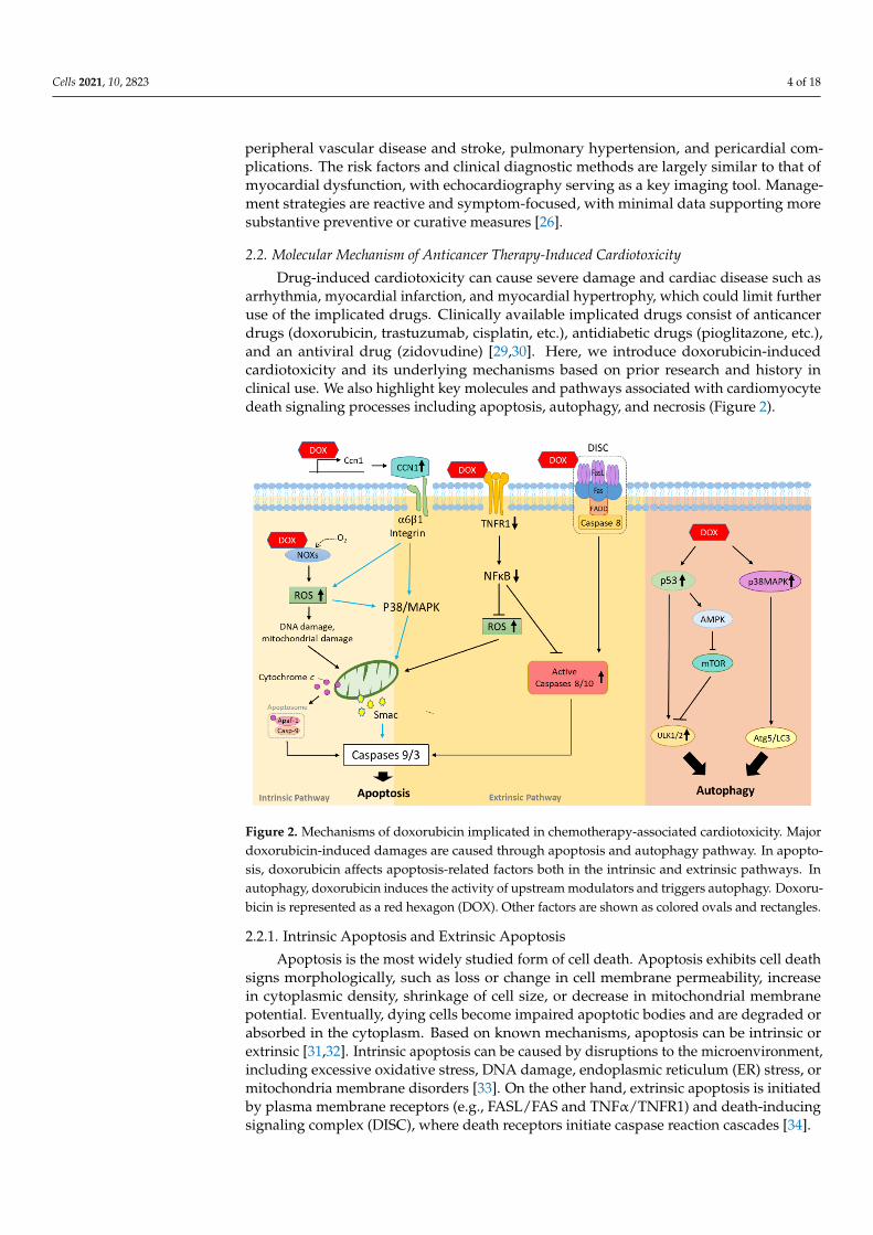
Drug-induced cardiotoxicity can cause severe damage and cardiac disease such asarrhythmia, myocardial infarction, and myocardial hypertrophy, which could limit furtheruse of the implicated drugs. Clinically available implicated drugs consist of anticancerdrugs (doxorubicin, trastuzumab, cisplatin, etc.), antidiabetic drugs (pioglitazone, etc.),and an antiviral drug (zidovudine) [29,30]. Here, we introduce doxorubicin-inducedcardiotoxicity and its underlying mechanisms based on prior research and history inclinical use. We also highlight key molecules and pathways associated with cardiomyocytedeath signaling processes including apoptosis, autophagy, and necrosis (Figure 2).
Cells 2021, 10, x FOR PEER REVIEW 4 of 18
effects. For example, anthracycline-induced heart failure occurs at a rate of 0.2 to 8.7 per-cent depending on the cumulative dosage [27], while patients treated with trastuzumab experience heart failure at a rate of up to 3.8 percent [28].
In addition, anti-cancer therapies have been implicated in other cardiovascular com-plications including coronary artery disease or myocardial ischemia, valvular disease, ar-rhythmias, such as atrial fibrillation, arterial hypertension, thromboembolic disease, pe-ripheral vascular disease and stroke, pulmonary hypertension, and pericardial complica-tions. The risk factors and clinical diagnostic methods are largely similar to that of myo-cardial dysfunction, with echocardiography serving as a key imaging tool. Management strategies are reactive and symptom-focused, with minimal data supporting more sub-stantive preventive or curative measures [26].
2.2. Molecular Mechanism of Anticancer Therapy-Induced Cardiotoxicity Drug-induced cardiotoxicity can cause severe damage and cardiac disease such as
arrhythmia, myocardial infarction, and myocardial hypertrophy, which could limit fur-ther use of the implicated drugs. Clinically available implicated drugs consist of anti-cancer drugs (doxorubicin, trastuzumab, cisplatin, etc.), antidiabetic drugs (pioglitazone, etc.), and an antiviral drug (zidovudine) [29,30]. Here, we introduce doxorubicin-induced cardiotoxicity and its underlying mechanisms based on prior research and history in clin-ical use. We also highlight key molecules and pathways associated with cardiomyocyte death signaling processes including apoptosis, autophagy, and necrosis (Figure 2).
Figure 2. Mechanisms of doxorubicin implicated in chemotherapy-associated cardiotoxicity. Major doxorubicin-induced damages are caused through apoptosis and autophagy pathway. In apoptosis, doxorubicin affects apoptosis-related fac-tors both in the intrinsic and extrinsic pathways. In autophagy, doxorubicin induces the activity of upstream modulators and triggers autophagy. Doxorubicin is represented as a red hexagon (DOX). Other factors are shown as colored ovals and rectangles.
2.2.1. Intrinsic Apoptosis and Extrinsic Apoptosis Apoptosis is the most widely studied form of cell death. Apoptosis exhibits cell death
signs morphologically, such as loss or change in cell membrane permeability, increase in cytoplasmic density, shrinkage of cell size, or decrease in mitochondrial membrane po-tential. Eventually, dying cells become impaired apoptotic bodies and are degraded or
Figure 2. Mechanisms of doxorubicin implicated in chemotherapy-associated cardiotoxicity. Majordoxorubicin-induced damages are caused through apoptosis and autophagy pathway. In apopto-sis, doxorubicin affects apoptosis-related factors both in the intrinsic and extrinsic pathways. Inautophagy, doxorubicin induces the activity of upstream modulators and triggers autophagy. Doxoru-bicin is represented as a red hexagon (DOX). Other factors are shown as colored ovals and rectangles.
2.2.1. Intrinsic Apoptosis and Extrinsic Apoptosis
Apoptosis is the most widely studied form of cell death. Apoptosis exhibits cell deathsigns morphologically, such as loss or change in cell membrane permeability, increasein cytoplasmic density, shrinkage of cell size, or decrease in mitochondrial membranepotential. Eventually, dying cells become impaired apoptotic bodies and are degraded orabsorbed in the cytoplasm. Based on known mechanisms, apoptosis can be intrinsic orextrinsic [31,32]. Intrinsic apoptosis can be caused by disruptions to the microenvironment,including excessive oxidative stress, DNA damage, endoplasmic reticulum (ER) stress, ormitochondria membrane disorders [33]. On the other hand, extrinsic apoptosis is initiatedby plasma membrane receptors (e.g., FASL/FAS and TNFα/TNFR1) and death-inducingsignaling complex (DISC), where death receptors initiate caspase reaction cascades [34].

Cells 2021, 10, 2823 5 of 18
Doxorubicin is known to increase production of reactive oxygen species (ROS) throughmany ways related to oxygen production, for example by affecting triphosphopyridinenucleotide (NADPH) oxidase [35]. Other studies found that chemical interactions be-tween doxorubicin and NADPH increased superoxide formation, eventually causingDNA damage [36]. A previous study demonstrated that doxorubicin treatment inducedcardiomyocyte apoptosis through mitochondrial apoptosis via caspase-3 induction andcytochrome C release pathways [37]. A pathway involving hypoxia-inducible factors is alsoresponsible for the cardioprotective effect of dexrazoxane regulation against doxorubicincardiotoxicity [38].
Doxorubicin is also known to facilitate binding events between death reporters andtheir corresponding ligands to facilitate DISC complex assembly, thereby activating caspasecascades. A matricellular protein CCN1 was found to mediate cardiotoxicity by engagingintegrin a6β1 to promote the activation of mitogen-activated protein kinase (p38-MAPK),culminating in the release of second mitochondrial activator of caspase (SMAC) to inducecardiomyocyte apoptosis in mice [39]. TNFR1 is also involved in doxorubicin-induced car-diomyocyte death whereby changes in TNFα receptor expression modulated doxorubicin-induced H9c2 cardiomyocyte apoptosis via activation of caspase-8 and suppression ofIκBα [40].
2.2.2. Autophagy
Autophagy plays an essential role in maintaining intracellular metabolic homeostasisby degrading or consuming unwanted or damaged cellular components [41]. The mam-malian target of rapamycin (mTOR) is an important signaling protein of autophagy as itcomplexes mammalian target of rapamycin complex 1 (mTORC1) with several other pro-teins stimulated by growth factor and receptor tyrosine kinase, blocking ULK-1-mediatedBeclin 1 phosphorylation and stopping autophagy initiation [42]. Conversely, the adenosine5-monophosphate activated protein kinase (AMPK) promotes autophagy by suppressionof the mTOR-related complex and direct activation of ULK-1 phosphorylation [43].
In the context of doxorubicin, autophagy is mainly triggered by oxidative stress toprotect cells against doxorubicin-induced cardiotoxicity. During doxorubicin treatment,levels of pro-autophagy factors (p53, p38-MAPK, and JNK-MAPK) increase while p85expression decreases to attenuate the phosphoinosmde-3-kinase (PI3K) pathway [44,45].Besides ROS-induced autophagy, doxorubicin also mediates autophagy-related factorsand causes autophagy enhancement [46]. One recent study showed that downregulationof high mobility group box 1 (HMGB1) alleviates doxorubicin-induced cardiomyocytedamage by preventing autophagic cell death [46]. Nutritional deficiency or starvation priorto doxorubicin treatment was also found to decrease cardiotoxicity. For example, caloricrestriction served a protective role by reducing ATP exhaustion and enhancing AMPKactivity, thus attenuating the autophagy caused by doxorubicin [47].
2.2.3. Necrosis
A third form of doxorubicin-induced cardiotoxicity cell death is necrosis, mostly be-lieved to occur under higher doses or longer exposure periods to doxorubicin comparedto apoptosis or autophagy. It has been reported that superoxide and peroxynitrite can in-crease necrosis in doxorubicin-induced cardiomyocyte death [48,49]. The typical dosage ofdoxorubicin is ≤20 mg/kg in vivo and 1 µM in vitro. Evidence of necrosis was observed inmice suffering from cardiac dysfunction following injections of 25 mg/kg doxorubicin [50],and in in vitro cardiac-derived H9c2 myocytes treated with 2 µM doxorubicin [51]. More-over, initial apoptosis facilitated development into necrosis, where cells exhibited earlyDNA impairment and nuclear swelling as the exposure period increases [52]. These studiesconfirm that increased or prolonged doxorubicin dosage encourages necrosis.

Cells 2021, 10, 2823 6 of 18
2.3. Antineoplastic Agents Implicated in Cardiotoxicity
While many approved drugs are used for cancer treatment, some patients experiencedadverse side effects following treatment. One of the most deleterious side effects is car-diovascular toxicity. Traditional and targeted chemotherapeutics are two major sourcesof cardiotoxicity. While the former can cause irreversible destruction to the myocardium,the latter can cause reversible damage to cellular functions and physiology [53–55]. Yet,differences in effects and dosage tolerances to the same drug between patients remainunexplained. In addition, the mechanisms of toxicity of these drugs can be diverse andmultifactorial. Drugs that are known to be involved in toxicity can be classified in multipleways; some of these categories include common chemotherapeutics such as anthracyclines,tyrosine kinase inhibitors (TKI), immune checkpoint inhibitors, and adoptive cellulartherapy (ACT) (Table 1).
Table 1. Overview of chemotherapeutic agents and their effects as empirically documented both physiologically and in vitroin the iPSC-CM platform. The broad specificity of many chemotherapeutics result in unfavorable consequences on thecardiovascular system, ranging from benign asymptomatic structural heart damage, to both chronic presentations (such ashypertension, heart failure, and electrophysiological abnormalities), and acute presentations, such as acute ischemia.
Classes of Antineoplastic Agents Cardiotoxic and Physiologic Effects References
Anthracyclines Long-term arrhythmia, cardiomyocyte dysfunction [56–63](e.g., doxorubicin)
Anti-metabolites Coronary vascular spasms, structural heart damage (symptomaticand asymptomatic), systolic dysfunction, acute ischemia [9,64](e.g., 5-fluorouracil)
Alkylating agents Structural heart damage (symptomatic and asymptomatic),systolic dysfunction, acute ischemia [64,65](e.g., cyclophosphamide)
Anti-microtubule Systolic dysfunction, acute ischemia [64](e.g., paclitaxel)
Monoclonal antibodies Ultrastructural changes, calcium dysregulation, mitochondrialdysfunction [66–68](e.g., trastuzumab)
Tyrosine kinase Hypertension (systemic and pulmonary), myofibril dysfunction,fluid retention, QT prolongation [61,62,69–72]inhibitors
(e.g., nilotinib)
Proteasome inhibitors Impaired left ventricular ejection fraction (LVEF), congestiveheart failure (CHF) [73,74](e.g., bortezomib)
Immunomodulators Sinus bradycardia, thromboembolic events [75](e.g., thalidomide)
Immune checkpoint Myocarditis, cardiogenic shock, atrioventricular (AV) block,ventricular tachycardia [76–78]inhibitors
(e.g., ipilimumab)
Hormonal agents (e.g., letrozole) Cardiac ischemia [79]Antiangiogenic agents (e.g., bevacizumab) Hypertension, CHF, arterial thromboembolic events (ATEs) [80]
2.3.1. Chemotherapy Drug: Anthracycline
Although targeted tyrosine kinase- and monoclonal antibody-based therapies arerecently developed, anthracyclines are still prescribed to 40–50% of breast cancer pa-tients, along with other drugs, such as the alkylating agent cyclophosphamide, or theanti-microtubule agent taxanes, such as Paclitaxel (Taxol). Although doxorubicin hasbeen clinically effective for a wide range of tumor cells, it imposes harsh effects on thehuman body through multiple cardiotoxicity mechanisms with both acute and chronicmanifestations. Acute toxicity often happens immediately after anthracycline absorbanceand manifests in myocardial ischemia, severe hypotension, and cardiac rhythm distur-bance. New dosing guidelines have vastly reduced the incidence of acute cardiotoxicity.Still, chronic and sub-chronic cardiotoxicity remains a significant clinical problem. While

Cells 2021, 10, 2823 7 of 18
sub-chronic toxicity may manifest in weeks or months, chronic toxicity may not surfaceuntil years or decades later. One study reported that children are more susceptible tothe development of cardiomyopathy than adults given equivalent doses of daunomycin,implying the importance of the long-term monitoring of chronic cardiotoxicity [81].
The mechanisms behind anthracycline pharmacodynamics are complex. It has beensuggested that anthracycline facilitates the excessive production of ROS and the activ-ity of many ROS-dependent pathways such as double-stranded DNA damage response,protein synthesis attenuation, and mitochondrial dysfunction [82–84]. Further researchrevealed that deletion of topoisomerase II β (TOP2β) in mouse cardiomyocytes success-fully prevented doxorubicin-induced cardiomyopathy, double-stranded breaks, and theformation of ROS [85]. On top of these, other unexpected cardiotoxic results are associatedwith doxorubicin exposure. Mouse models were used to study the molecular basis ofdoxorubicin-induced cardiotoxicity, revealing that high cumulative doses were associatedwith recalcitrant heart failure. This also resulted in a decline in cardiac systolic function,accompanied by marked atrophy of the heart, low levels of cardiomyocyte apoptosis, anddecreased growth rate [86].
2.3.2. Other Chemotherapy Drugs
Besides anthracycline compounds, other drugs including anti-metabolites and alky-lating agents, platinum agents, and anti-microtubule agents have demonstrated theircardiotoxic potential. For example, 5-fluorouracil (5-FU) induces cardiotoxic effects, suchas disturbances of rhythm and angina. 5-FU toxicity can lead to myocardial damage,vascular toxicity and vasospasm, the accumulation of toxic degradation products, andROS-mediated metabolic damage. Less than 2% of patients treated with 5-FU experiencemore adverse effects such as congestive heart failure (CHF) [87,88]. The remaining classesof traditional chemotherapeutics have not showed cardiotoxic effects due to our limited un-derstanding of their mechanisms of action. One anti-microtubule agent, taxanes, has beenrelated to rhythm disturbances but the evidence for its cardiotoxicity is unsubstantial [8].
2.3.3. Targeted Therapy Agents
The inhibition of growth-related kinases mediating cellular signal transduction, espe-cially tyrosine kinases, is a new therapeutic strategy for diseases such as cancer. Studieshave confirmed that the ATP-binding domain of tyrosine kinases is an attractive targetfor drug design and development. Both the EGFR and the HER2 receptor tyrosine kinase(RTK) inhibitors are effective drugs targeting solid tumors, and demonstrate higher target-ing efficiency and reduced off-target effects compared to older treatments [89]. However,kinase inhibitors still cause adverse effects related to cardiovascular toxicity [90].
Among the HER2-positive metastatic breast cancer patients that reported trastuzumabcardiotoxicity, up to 27% of them experienced cardiac dysfunction and heart failure. Somepresented with an increase in serum cardiac troponin I (cTnI) following mild reduction ofleft ventricular ejection fraction (LVEF) [91]. Combinatorial studies revealed that patientsundergoing treatment with an anthracycline, cyclophosphamide, and trastuzumab concur-rently had significantly increased risk of cardiac dysfunction compared to those only treatedwith an anthracycline and cyclophosphamide [92,93]. Recent investigations confirmed thattrastuzumab disrupts HER2 signaling to mediate autophagy suppression, increases ROSproduction, and activates autophagy-inhibitory Erk/mTOR/Ulk 1 signaling [94]. Despitesignificant efforts to understand the molecular underpinnings of trastuzumab-inducedcardiotoxicity, these mechanisms remain nebulous.
The tyrosine kinase inhibitor, imatinib, raised concerns of cardiotoxicity after 10 pa-tients developed CHF following treatment in 2006 [95]. Human micrographs showedmitochondrial abnormalities and accumulation of membrane whorls in both vacuoles andthe sarco-(endo-)plasmic reticulum after imatinib treatment. Because targeted kinases func-tion through a similar mechanism, they may also induce similar toxicity issues. Currently,the FDA warning list of cardiotoxic drugs include six TKIs such as sunitinib, vemurafenib,

Cells 2021, 10, 2823 8 of 18
and nilotinib. Sunitinib is associated with LVEF declines and QT prolongation [96]. Thesefindings suggest the prevalence of off-target effects as primary causes of cardiotoxicity.
2.3.4. Cancer Immunotherapy: Checkpoint Inhibitors and Adoptive Cellular Therapy
Immune checkpoint inhibitors and adoptive cellular therapies (ACT) are two cate-gories of immunotherapies that play critical roles in the mitigation of cancer and autoim-mune diseases. These cancer therapeutic strategies focus on activating and engaging theimmune systems inherent in patients to destroy cancer cells [97,98]. Unfortunately, theadvantages of immune checkpoint blocking antibodies can be accompanied by adversecardiotoxic effects such as in cases of immune-related cardiotoxicity following treatmentwith ipilimumab, nivolumab, and/or pembrolizumab [99]. Documented cardiac dysfunc-tion includes heart failure, cardiomyopathy, acute myocarditis, myocardial fibrosis, andpericarditis [100].
Furthermore, fatal adverse events, such as cardiac arrest and multiple organ failure,have been documented in patients treated with autologous T-cell receptor (TCR) transducedT cell infusion, an ACT [101]. Following MART-1 TCR transduced T cell treatment, highlevels of NT-proBNP (a marker for heart failure) and IL-6 lead to diminished cardiaccontractile function [102]. While the occurrence of immunotherapy-induced cardiotoxicityremains relatively low, these unfavorable side effects should be taken into consideration intherapeutic development and clinical applications.
3. Modeling Chemotherapy-Induced Cardiotoxicity with iPSC-CMs
iPSC-CMs are an excellent disease model to recapitulate the induction of cardiotoxicityby chemotherapeutic agents in cardiomyocytes. In particular, iPSC-CMs provide an unlim-ited cell source unlike many restrictions of animal models and primary cell lines. iPSC-CMsnot only exhibit many cardiac-specific behaviors, including a cardiac contractile apparatusand calcium-handling features, but also demonstrate physiological and transcriptionalresponses after chemotherapy [103].
Several studies have established distinct technical approaches to model doxorubicin-induced effect on iPSC-CM’s transcriptomes, metabolisms, and functions (Table 2). Tran-scriptome analysis of doxorubicin-treated iPSC-CMs revealed the dynamic changes inglobal gene expression with respect to exposure time and dose [104]. Computationalanalysis of their transcriptomes further highlighted several signaling pathway genes asso-ciated with DNA damage and cell cycle regulation, including BLM, BRCA1, E2F, FANCG,PLK1, PRC1, and RBL1. E2F and RBL1 are known to control the estrogen-mediatedS-phase entry pathway. FOXM1, p53, and E2F have been linked to left ventricular dys-function, cardiomyocyte apoptosis, and heart failure, suggesting that the downregulationof PRC1 by transcription regulator FOXM1 may be a mediator of DNA damage repairresponse pathways induced by doxorubicin [105]. Furthermore, patient iPSC-CMs havebeen proved to recapitulate clinical observations in breast cancer patients and showeddoxorubicin increases cellular ROS production, calcium handling, whole-cell oxidativestress, and eventually double–stranded DNA damage [56]. Transcriptome analyses ofdoxorubicin treatment on patient-specific gene expression reveal the significant down-regulation of several important cardiac development-related transcription factors such asNKX2–5 (homeobox protein Nkx-2.5), MEF2A (myocyte-specific enhancer factor 2), andTBX5 (T-box transcription factor TBX5). Their study also revealed p53, RELA, NFKB1,and p300 play roles in doxorubicin pharmacodynamics and cardiotoxicity [56]. Kitaniet al. discovered that the continued exposure of anti-cancer drugs, such as trastuzumab toiPSC-CMs impairs the contractile and calcium-handling abilities of cardiomyocytes withoutcausing cell death or sarcomeric disorganization [66]. Transcriptome analysis suggestedthat mitochondrial functional defects and alterations to the cardiac energy metabolismpathway are primarily responsible for trastuzumab-induced cardiotoxic manifestations.Patient-derived iPSC-CMs that are more vulnerable to trastuzumab exhibited myocardialcontractile dysfunction following treatment. Notably, activation of AMPK, a regulatory

Cells 2021, 10, 2823 9 of 18
kinase in myocardial energy metabolism, could compensate for mitochondrial dysfunction,contractile dysfunction, and other adverse effects induced by trastuzumab. Maillet et al.engineered CRISPR/Cas9-mediated TOP2β deletions in hESC-CMs to investigate the roleof TOP2β in doxorubicin-induced cardiotoxicity [13]. TOP2β-deleted NKX2-5eGFP/whESC-derived CMs have similar cardiac gene expression with wild-type hESC-derivedCMs but decrease drug susceptibilities in response to doxorubicin, indicating a criticalrole of TOP2β in doxorubicin-induced cardiotoxicity. In addition, Karhu et al. studiedthe chronic cardiotoxicity using iPSC-CMs under long-term low-dose administration ofdoxorubicin [106]. Their in vitro model showed that elevated caspase-3/7 activities areassociated with decreased cell viability and increased apoptosis, and inhibition of GATA4function provides cardioprotective effects in iPSC-CMs by attenuating the doxorubicin-induced elevation of pro-B-type natriuretic peptide expression. These results affirm that theiPSC-CM platform can be productively utilized for the evaluation of strategies to protectagainst or retroactively mitigate cardiomyocyte toxicity. These examples foreshadow themyriad potential applications of iPSC-CMs in cardiotoxicity studies, in vitro compoundscreening, and the wider field of synthetic biology.
Table 2. Use of iPSC-CMs in the study of anthracycline-induced cardiotoxicity. Numerous studies have successfullyemployed the iPSC-CM platform to elucidate the biochemical mechanisms of anthracycline pharmacodynamics, such asthe implication of biomarkers, microRNAs, and genetic factors. The recent emergence of these studies suggest untappedpotential in the field of iPSC-CM modeling. Studies organized chronologically.
Cardiotoxicity-InducedDrug
In-Vitro ObservationParameter: Functional
Change Endpoint
In-Vitro ObservationParameter: Structural
Change EndpointApplication of iPSC-CMs References
Daunorubicin Beating frequency(xCELLigence)
Cell viability, ROS generation,Troponin secretion, lipid
accumulation
Validation of appropriateparameters for testing DIC
in iPSC-CMsDoherty et al. [70]
Doxorubicin,Daunorubicin
Beating frequency(xCELLigence)
Troponin secretion andsarcomere structure.
Identification of biomarkerfrom Doxorubicin-exposed
iPSC-CMs global geneexpression
Chaudhari et al. [57]
Doxorubicin Multielectrode array(Maestro MEA system)
Cell viability, ROS generation,calcium handling,
mitochondrialtransmembrane potential,
Apoptotic feature.
Investigation of themolecular mechanisms ofDIC in a iPSC-CMs model
system
Maillet et al. [13]
Doxorubicin -
DNA damage level (γ-H2AX),calcium handling, ROS
generation, mitochondrialfunction, Sarcomeric protein,
apoptotic feature
Identification of thephenotype of DIC breastcancer patient-derived
iPSC-CMs
Burridge et al. [56]
Doxorubicin - Lactate dehydrogenase (LDH)leakage
Identification microRNAs(miRNAs) expression from
DIC iPSC-CMsChaudhari et al. [57]
Doxorubicin
Beating properties(Relaxation velocity,contraction velocity,
contraction-relaxationduration, and beat rate)
(Video microscopy)
Cardiac troponin, heart fattyacid-binding protein (FABP3),
and N-terminal pro-brainnatriuretic peptide
(NT-proBNP)
Evaluation of the videomicroscopy approach in
predicting chronic DIC iniPSC-CMs
Kopljar et al. [103]
DoxorubicinContractility
(high-throughputcontractility imaging)
Cytotoxicity
High-throughputcontractility and cytotoxicity
assay for cardiotoxicityinduced drugs
Sharma et al. [107]
Doxorubicin - Cardiac troponinTranscriptomic data from
individual-derivediPSC-CMs
Knowles et al. [108]

Cells 2021, 10, 2823 10 of 18
Table 2. Cont.
Cardiotoxicity-InducedDrug
In-Vitro ObservationParameter: Functional
Change Endpoint
In-Vitro ObservationParameter: Structural
Change EndpointApplication of iPSC-CMs References
Doxorubicin - -
Establishment ofmulti-omics data fromDoxorubicin-exposed
iPSC-CMs
Holmgren et al. [109]
DoxorubicinElectrophysiological
feature (cardiac opticalmapping)
Cell viability, DNA damagelevel (γ-H2AX), ROS
generation
In vitro correction of RARGmutation in patient-derivediPSC-CMs by CRISPR-Cas9
Christidi et al. [110]
Doxorubicin -Cell viability, pro-B-type
natriuretic peptide (proBNP),Apoptotic feature
Chronic DIC iPSC-CMsin-vitro model for validating
cardioprotective effectKarhu et al. [106]
Abbreviation: DIC, doxorubicin-induced cardiotoxicity; ROS, reactive oxygen species.
4. Disease Modeling of iPSC-CM in Precision Medicine
The advancement of genetic analysis and modification techniques have paved theway for more precise study of the interactions between genetic variants, responses tochemotherapy, and cardiotoxicity consequences. Patient-derived iPSC-CMs are an excellentsource of unique and personalized genomes that can now be meaningfully used to exploregenetic associations in cardiotoxicity [111] (Figure 3).
Cells 2021, 10, x FOR PEER REVIEW 10 of 18
Doxorubicin
Beating properties (Re-laxation velocity, con-traction velocity, con-traction-relaxation du-ration, and beat rate) (Video microscopy)
Cardiac troponin, heart fatty acid-binding protein (FABP3),
and N-terminal pro-brain natriu-retic peptide (NT-proBNP)
Evaluation of the video microscopy approach in predicting chronic DIC
in iPSC-CMs Kopljar et al. [103]
Doxorubicin Contractility (high-
throughput contractil-ity imaging)
Cytotoxicity High-throughput contractility and cytotoxicity assay for cardiotoxicity
induced drugs Sharma et al. [107]
Doxorubicin - Cardiac troponin Transcriptomic data from individ-
ual-derived iPSC-CMs Knowles et al. [108]
Doxorubicin - - Establishment of multi-omics data from Doxorubicin-exposed iPSC-
CMs
Holmgren et al. [109]
Doxorubicin Electrophysiological
feature (cardiac optical mapping)
Cell viability, DNA damage level (γ-H2AX), ROS generation
In vitro correction of RARG muta-tion in patient-derived iPSC-CMs
by CRISPR-Cas9 Christidi et al. [110]
Doxorubicin - Cell viability, pro-B-type natriu-retic peptide (proBNP), Apop-
totic feature
Chronic DIC iPSC-CMs in-vitro model for validating cardioprotec-
tive effect Karhu et al. [106]
* Abbreviation: DIC, doxorubicin-induced cardiotoxicity; ROS, reactive oxygen species.
4. Disease Modeling of iPSC-CM in Precision Medicine The advancement of genetic analysis and modification techniques have paved the
way for more precise study of the interactions between genetic variants, responses to chemotherapy, and cardiotoxicity consequences. Patient-derived iPSC-CMs are an excel-lent source of unique and personalized genomes that can now be meaningfully used to explore genetic associations in cardiotoxicity [111]. (Figure 3)
Figure 3. Generation and application of patient-derived iPSC-CMs. Somatic cells (fibroblasts) can be easily obtained from patients, reprogrammed into iPSCs, and differentiated into cardiomyocytes. In the individual iPSC-CMs model, iPSC-CMs provide a sophisticated resource to dissect chemo-induced cardiotoxicity as well as a useful platform for drug screening. With sufficient bioinformatics data and physiological records, abundant patient-derived iPSC-CMs models construct a substantial biobank that can be exploited in cardiotoxicity prediction, pharmacogenomics, and precision medicine.
Figure 3. Generation and application of patient-derived iPSC-CMs. Somatic cells (fibroblasts) can be easily obtained frompatients, reprogrammed into iPSCs, and differentiated into cardiomyocytes. In the individual iPSC-CMs model, iPSC-CMsprovide a sophisticated resource to dissect chemo-induced cardiotoxicity as well as a useful platform for drug screening.With sufficient bioinformatics data and physiological records, abundant patient-derived iPSC-CMs models construct asubstantial biobank that can be exploited in cardiotoxicity prediction, pharmacogenomics, and precision medicine.
4.1. iPSC-CM Disease Modeling in Studying Associations between Genetic Variations andSensitivity to Cardiotoxicity
Several heterogenic background studies focusing on anthracycline-induced cardiotox-icity sensitivity and resistance have also been conducted through genome wide associationstudies (GWAS) and single nucleotide polymorphisms (SNPs) arrays in pediatric oncol-ogy cohorts. There were 18 significant genes carrying SNPs, which were known to beinvolved in DNA damage pathways, drug transport, oxidative stress defenses, or ironmetabolism [112]. Missense mutations in the RARG gene were found to be potentiallyimplicated in doxorubicin-induced cardiotoxicity. In one successful study, Christidi et al.

Cells 2021, 10, 2823 11 of 18
employed CRISPR/Cas9 to generate isogenic iPSC-CMs with different RARG mutations,and discovered that doxorubicin-induced cardiotoxicity was reduced in iPSC-CMs with aPARG(S427L) mutation [110]. Another study found that RAC2 (which encodes Rho-GTPaseto regulate the NADPH oxidase) and NADPH oxidase (NOX2) were both correlated withincreased susceptibility to anthracycline-induced cardiotoxicity [113,114]. In the sameway, the iPSC-CM model can be used to explore other SNPs and genetic predispositionsassociated with chemotherapy-induced cardiotoxicity.
In concert, these studies demonstrate that iPSC-CMs are effective not only in reca-pitulating the electrophysiological single-cell phenotype of cardiomyocytes, but also indemonstrating anthracycline-induced cardiotoxicity within a controlled and analyzablemodel system, allowing for the study of mechanisms underlying genetic disorders uniqueto the cardiovasculature.
4.2. Establishment of Personalized Cardiovascular Biobank for Toxicity Pre-Screening, DrugTesting, Therapeutics, and Diagnosis
The application of the iPSC-CM platform to therapeutics is promising. Using patient-derived iPSC-CMs in toxicity screens enable in-depth exploration of multiple candidateparameters associated with anticancer drug cardiotoxicity or drug efficacy. These studiescan contribute meaningfully to the formulation of tailored, effective clinical treatmentsfor each individual patient. The synthesis of data points into a clinical biobank can helpwith prediction and mitigation of harmful side effects and clinical advisory to reducecardiovascular disease risk factors.
4.2.1. Population Biobank for High-Throughput Toxicity Screening
A human cardiotoxicity biobank based on iPSC-CMs provides information on a widerange of clinical cardiotoxic effects, accounting for variations in severity and correla-tions between certain genetic variants and specific classes of targeted chemotherapeuticagents [115,116]. The biobank provides clinicians with a predictive model to make moreinformed decisions tailored to each patient, regarding the choice of chemotherapeutic agent,dosage, and other meaningful interventions. Promising toxicity trials to date have alsoinspired pharmaceutical companies to begin utilizing iPSC-CMs in tests for drug safety,arrhythmogenic potential, and other relevant parameters for preclinical drug testing.
Existing biobanks such as the GWAS Catalog provide accessible information on singlenucleotide polymorphism (SNP)-trait associations, providing a springboard for researchersto investigate the impact of common variants on anthracycline-induced cardiotoxicity. A2008 systematic review and meta-analysis gathered data from Medline, EMBASE, and theCochrane Library on adults and children treated with an anthracycline for breast or ovariancancer, sarcoma, non-Hodgkin’s or Hodgkin’s lymphoma, and myeloma. Their resultsincluded detailed information on defining and measuring cardiotoxicity outcomes, provid-ing valuable perspectives that will inform research on anthracycline-induced cardiotoxicityresearch and the continued development of anthracycline chemotherapeutics [117]. Thisexample foreshadows the insightful role that biobanks can play in toxicity screening forgenetic factors.
4.2.2. Pharmacogenomics
Pharmacogenetics is a field of study focused on the role of genetic variations in physi-ological responses to drugs. Age, environmental factors, and prior cardiovascular incidentscontribute to drug response variability, but each patient’s genetic makeup may also predis-pose them towards certain cardiotoxic responses to chemotherapy [118]. With the growingawareness of precision medicine, pharmacogenetics has expanded to analysis of the entirehuman genome. On top of accounting for gene variants, pharmacogenomics also accountsfor the unique biomarkers and levels present in each individual. For example, polymor-phisms have been reported to have effects on susceptibility to chemotherapy-inducedcardiotoxicity. Individual genetic variations may also affect the determination of treatmentplans that balance drug efficacy and drug safety. Therefore, pharmacogenetic testing in

Cells 2021, 10, 2823 12 of 18
chemotherapy-treated cancer patients is invaluable to understand the relationships be-tween genetic variations and susceptibilities, and ultimately to facilitate the designing ofpersonalized treatment strategies.
Advancements in next-generation sequencing (NGS) have significantly benefited thefield of pharmacogenomics, facilitating an increase in the number of GWAS. GWAS aim toidentify SNPs or the genetic loci associated with common diseases or traits. With the GWASCatalog, oncologists can now better predict the drug response and any potential adversedrug reactions for each patient based on their genome [119]. A number of SNPs associatedwith doxorubicin-induced cardiotoxicity are documented [120]. However, validationstudies for GWAS analysis are challenging due to their reliance on human cardiac biopsies,which are difficult to obtain. Conversely, the ease with which human patient iPSCs anddisease-trait engineered iPSCs/hESCs can be cultured and extended make them the perfectdisease platform to validate GWAS studies in a dish. In addition, iPSC-CMs can be appliedin multi-omic analysis to investigate the links between genotypes and phenotypes inadverse drug reactions. Chaudhari et al. employed microarray statistical data analysisand functional annotation analysis to identify clusters of altered genes that potentiallyconferred doxorubicin sensitivity [57], suggesting the valuable contributions that iPSC-CMmodels can make to the field of pharmacogenomics. In sum, the iPSC-CM platform cansynergize with biotechnological advancements to accelerate the growth of knowledge andconsequently the development of breakthrough therapeutics.
5. Conclusions
The cardiotoxicity of chemotherapeutic compounds has been a major concern in thepharmaceutical and clinical fields. Efforts to minimize the harmful effects of chemotherapyon cancer patients would benefit greatly from a high-fidelity platform for disease modelingand drug screening. This review discussed the utility of iPSC-CMs for the study of drug-induced cardiotoxicity from multiple perspectives and the elucidation of the underlyingmechanisms involved. The similarities between iPSC-CMs and physiological cardiomy-ocytes facilitate the screening of drug-induced alterations in cardiac cellular contractility,electrophysiology, and viability in ways previously inaccessible through animal modelsalone. In fact, high-throughput iPSC-CM models have facilitated the creation of a car-diac safety index (CSI) to grade drugs based on their potential cardiotoxicity and theirquantitative toxicity measurements [107].
Patient-derived iPSC-CMs show great potential in the field of personalized medicine.Patient-specific iPSC-CMs can be used to identify genetic mutations that predispose to-ward or against cardiotoxicity, discover drugs for the treatment of both cancer and othercardiovascular diseases, and accumulate biobanks of data for the development of pre-dictive models of efficacy and toxicity. Yet, challenges exist in the standardization andreproducibility of iPSC-CM generation, and the immature phenotype of iPSC-CMs. Futureefforts should focus on the development of established and reproducible experimentalprotocols, and the improvement of iPSC-CMs to more accurately model mature adultcardiomyocytes. Still, iPSC-CMs will continue to be an excellent platform for the studyof chemotherapy-induced cardiotoxicity and a plethora of other applications in diseasemodeling, toxicity screening, pharmacogenetics, and synthetic biology in general.
Author Contributions: Writing—original draft preparation, M.-F.H., L.K.P.; figure generation,Y.-H.C.; writing—review and editing, M.-F.H., L.K.P., Y.-H.C., R.Z., D.-F.L.; supervision, R.Z., D.-F.L.All authors have read and agreed to the published version of the manuscript.
Funding: This study was supported by grants from the University of Texas Health Science Center atHouston (R.Z. and D.-F.L.). D.-F.L. was supported by CPRIT RR160019, NIH/NCI R01CA246130,Rolanette and Berdon Lawrence bone disease program of Texas, and Pablove Foundation childhoodcancer research grant (690785). D.-F.L. is a CPRIT Scholar in Cancer Research.
Institutional Review Board Statement: Not applicable.
Informed Consent Statement: Not applicable.

Cells 2021, 10, 2823 13 of 18
Data Availability Statement: Not applicable.
Acknowledgments: We sincerely apologize to the authors whose work we could not include owingto space limitations.
Conflicts of Interest: The authors declare no conflict of interest.
References1. Takahashi, K.; Yamanaka, S. Induction of pluripotent stem cells from mouse embryonic and adult fibroblast cultures by defined
factors. Cell 2006, 126, 663–676. [CrossRef]2. Steeg, R.; Neubauer, J.C.; Müller, S.C.; Ebneth, A.; Zimmermann, H. The EBiSC iPSC bank for disease studies. Stem Cell Res. 2020,
49, 102034. [CrossRef]3. Olgasi, C.; Cucci, A.; Follenzi, A. iPSC-Derived liver organoids: A journey from drug screening, to disease modeling, arriving to
regenerative medicine. Int. J. Mol. Sci. 2020, 21, 6215. [CrossRef] [PubMed]4. Chang, C.Y.; Ting, H.-C.; Liu, C.-A.; Su, H.-L.; Chiou, T.-W.; Lin, S.-Z.; Harn, H.-J.; Ho, T.-J. Induced pluripotent stem cell
(iPSC)-based neurodegenerative disease models for phenotype recapitulation and drug screening. Molecules 2020, 25, 2000.[CrossRef] [PubMed]
5. Yoshida, Y.; Yamanaka, S. Induced pluripotent stem cells 10 years later: For cardiac applications. Circ. Res. 2017, 120, 1958–1968.[CrossRef] [PubMed]
6. Ferlay, J.; Colombet, M.; Soerjomataram, I.; Parkin, D.M.; Piñeros, M.; Znaor, A.; Bray, F. Cancer statistics for the year 2020: Anoverview. Int. J. Cancer 2021, 149, 778–789. [CrossRef]
7. Torre, L.A.; Siegel, R.L.; Ward, E.M.; Jemal, A. Global cancer incidence and mortality rates and trends—An update. CancerEpidemiol. Biomark. Prev. 2016, 25, 16–27. [CrossRef]
8. Sayed, N.; Ameen, M.; Wu, J.C. Personalized medicine in cardio-oncology: The role of induced pluripotent stem cell. Cardiovasc.Res. 2019, 115, 949–959. [CrossRef]
9. Gintant, G.; Burrifge, P.; Gepstein, L.; Harding, S.; Herron, T.; Hong, C.; Jalife, J.; Wu, J.C. Use of human induced pluripotentstem cell-derived cardiomyocytes in preclinical cancer drug cardiotoxicity testing: A scientific statement from the american heartassociation. Circ. Res. 2019, 125, e75–e92. [CrossRef]
10. Matsa, E.; Ahrens, J.H.; Wu, J.C. Human induced pluripotent stem cells as a platform for personalized and precision cardiovascularmedicine. Physiol. Rev. 2016, 96, 1093–1126. [CrossRef]
11. Doss, M.X.; Sachinidis, A. Current challenges of iPSC-based disease modeling and therapeutic implications. Cells 2019, 8, 403.[CrossRef] [PubMed]
12. Shi, Y.; Inoue, H.; Wu, J.C.; Yamanaka, S. Induced pluripotent stem cell technology: A decade of progress. Nat. Rev. Drug Discov.2017, 16, 115–130. [CrossRef] [PubMed]
13. Maillet, A.; Tan, K.; Chai, X.; Sadananda, S.N.; Mehta, A.; Ooi, J.; Hayden, M.R.; Pouladi, M.A.; Ghosh, S.; Shim, W.; et al.Modeling doxorubicin-induced cardiotoxicity in human pluripotent stem cell derived-cardiomyocytes. Sci. Rep. 2016, 6, 25333.[CrossRef] [PubMed]
14. Tentner, A.R.; Lee, M.; Ostheimer, G.J.; Samson, L.D.; Lauffenburger, D.A.; Yaffe, M.B. Combined experimental and computationalanalysis of DNA damage signaling reveals context-dependent roles for Erk in apoptosis and G1/S arrest after genotoxic stress.Mol. Syst. Biol. 2012, 8, 568. [CrossRef] [PubMed]
15. Carvajal-Vergara, X.; Sevilla, A.; D’Souza, S.L.; Ang, Y.-S.; Schaniel, C.; Lee, D.-F.; Yang, L.; Kaplan, A.D.; Adler, E.D.; Rozov,R.; et al. Patient-specific induced pluripotent stem-cell-derived models of LEOPARD syndrome. Nature 2010, 465, 808–812.[CrossRef] [PubMed]
16. Wang, Y.; Liang, P.; Lan, F.; Wu, H.; Lisowski, L.; Gu, M.; Hu, S.; Kay, M.A.; Urnov, F.D.; Shinnawi, R.; et al. Genome editing ofisogenic human induced pluripotent stem cells recapitulates long QT phenotype for drug testing. J. Am. Coll. Cardiol. 2014, 64,451–459. [CrossRef] [PubMed]
17. Liang, P.; Sallam, K.; Wu, H.; Li, Y.; Itzhaki, I.; Garg, P.; Zhang, Y.; Vermglinchan, V.; Lan, F.; Gu, M.; et al. Patient-specific andgenome-edited induced pluripotent stem cell-derived cardiomyocytes elucidate single-cell phenotype of brugada syndrome. J.Am. Coll. Cardiol. 2016, 68, 2086–2096. [CrossRef]
18. Kodo, K.; Ong, S.-G.; Jahanbani, F.; Termglinchan, V.; Hirono, K.; Inanloo Rahatloo, K.; Ebert, A.D.; Shukla, P.; Abilez, O.J.; Churko,J.N.; et al. iPSC-derived cardiomyocytes reveal abnormal TGF-beta signalling in left ventricular non-compaction cardiomyopathy.Nat. Cell Biol. 2016, 18, 1031–1042. [CrossRef]
19. Sun, N.; Yazawa, M.; Liu, J.; Han, L.; Sanchez-Freire, V.; Abilez, O.J.; Navarrete, E.G.; Hu, S.; Wang, L.; Lee, A.; et al. Patient-specific induced pluripotent stem cells as a model for familial dilated cardiomyopathy. Sci. Transl. Med. 2012, 4, 130ra47.[CrossRef]
20. Wu, H.; Lee, J.; Vincent, L.G.; Wang, Q.; Gu, W.; Lan, F.; Churko, J.M.; Sallam, K.I.; Matsa, E.; Sharma, A.; et al. Epigeneticregulation of phosphodiesterases 2A and 3A underlies compromised beta-adrenergic signaling in an iPSC model of dilatedcardiomyopathy. Cell Stem Cell. 2015, 17, 89–100. [CrossRef]
Cells 2021, 10, 2823 14 of 18
21. Karakikes, I.; Termglinchan, V.; Cepeda, D.A.; Lee, J.; Diecke, S.; Hendel, A.; Itzhacki, I.; Ameen, M.; Shrestha, R.; Wu, H.;et al. A comprehensive TALEN-based knockout library for generating human-induced pluripotent stem cell-based models forcardiovascular diseases. Circ. Res. 2017, 120, 1561–1571. [CrossRef]
22. Lan, F.; Lee, A.S.; Liang, P.; Sanchez-Freire, V.; Nguyen, P.K.; Wang, L.; Han, L.; Yen, M.; Wang, Y.; Sun, N.; et al. Abnormalcalcium handling properties underlie familial hypertrophic cardiomyopathy pathology in patient-specific induced pluripotentstem cells. Cell Stem Cell 2013, 12, 101–113. [CrossRef]
23. Hall, P.S.; Harshman, L.C.; Srinivas, S.; Witteles, R.M. The frequency and severity of cardiovascular toxicity from targeted therapyin advanced renal cell carcinoma patients. JACC Heart Fail. 2013, 1, 72–78. [CrossRef]
24. Limat, S.; Drouhin, J.; Demesmay, K.; Tissot, E.; Jacquet, M.; Woronofl-Lemsi, M. Incidence and risk factors of preparation errorsin a centralized cytotoxic preparation unit. Pharm. World Sci. 2001, 23, 102–106. [CrossRef]
25. Oeffinger, K.C.; Mertens, A.C.; Sklar, C.A.; Kawashima, T.; Hudson, M.M.; Meadows, A.T.; Friedman, D.L.; Marina, N.; Hobbie,W.; Kadan-Lottick, N.; et al. Chronic health conditions in adult survivors of childhood cancer. N. Engl. J. Med. 2006, 355,1572–1582. [CrossRef] [PubMed]
26. Zamorano, J.L.; Lancellotti, P.; Muñoz Rodriguez, D.; Aboyans, V.; Asteggiano, R.; Galderisi, M.; Habib, G.; Lenihan, D.J.; Lip,G.Y.H.; Lyon, A.R.; et al. 2016 ESC Position Paper on cancer treatments and cardiovascular toxicity developed under the auspicesof the ESC Committee for Practice Guidelines: The Task Force for cancer treatments and cardiovascular toxicity of the EuropeanSociety of Cardiology (ESC). Eur. Heart J. 2016, 37, 2768–2801. [CrossRef] [PubMed]
27. Volkova, M.; Russell, R., 3rd. Anthracycline cardiotoxicity: Prevalence, pathogenesis and treatment. Curr. Cardiol. Rev. 2011, 7,214–220. [CrossRef] [PubMed]
28. Curigliano, G.; Cardinale, D.; Dent, S.; Criscitiello, C.; Aseyev, O.; Lenihan, D.; Cipolla, C.M. Cardiotoxicity of anticancertreatments: Epidemiology, detection, and management. CA Cancer J. Clin. 2016, 66, 309–325. [CrossRef]
29. Ma, W.; Wei, S.; Zhang, B.; Li, W. Molecular mechanisms of cardiomyocyte death in drug-induced cardiotoxicity. Front. Cell Dev.Biol. 2020, 8, 434. [CrossRef]
30. Renu, K.; Abilash, V.G.; Tirupathi Pichiah, P.B.; Arunachalam, S. Molecular mechanism of doxorubicin-induced cardiomyopathy–An update. Eur. J. Pharmacol. 2018, 818, 241–253. [CrossRef] [PubMed]
31. Brumatti, G.; Salmanidis, M.; Ekert, P.G. Crossing paths: Interactions between the cell death machinery and growth factor survivalsignals. Cell. Mol. Life Sci. 2010, 67, 1619–1630. [CrossRef] [PubMed]
32. Roos, W.; Thomas, A.; Kaina, B. DNA damage and the balance between survival and death in cancer biology. Nat. Rev. Cancer2016, 16, 20–33. [CrossRef] [PubMed]
33. Wu, C.-C.; Bratton, S.B. Regulation of the intrinsic apoptosis pathway by reactive oxygen species. Antioxid. Redox Signal. 2013, 19,546–558. [CrossRef] [PubMed]
34. Yang, J.K. Death effecter domain for the assembly of death-inducing signaling complex. Apoptosis 2015, 20, 235–239. [CrossRef][PubMed]
35. Vásquez-Vivar, J.; Martasek, P.; Hogg, N.; Masters, B.S.S.; Pritchard, K.A.; Kalyanaraman, B. Endothelial nitric oxide synthase-dependent superoxide generation from adriamycin. Biochemistry 1997, 36, 11293–11297. [CrossRef] [PubMed]
36. Deng, S.; Kruger, A.; Kleschyov, A.L.; Kalinowski, L.; Daiber, A.; Wojnowski, L. Gp91phox-containing NAD(P)H oxidase increasessuperoxide formation by doxorubicin and NADPH. Free Radic. Biol. Med. 2007, 42, 466–473. [CrossRef]
37. Childs, A.C.; Phaneuf, S.L.; Dirks, A.J.; Phillips, T.; Leeuwenburgh, C. Doxorubicin treatment in vivo causes cytochrome C releaseand cardiomyocyte apoptosis, as well as increased mitochondrial efficiency, superoxide dismutase activity, and Bcl-2:Bax ratio.Cancer Res. 2002, 62, 4592–4598.
38. Spagnuolo, R.D.; Recalcati, S.; Tacchini, L.; Cairo, G. Role of hypoxia-inducible factors in the dexrazoxane-mediated protection ofcardiomyocytes from doxorubicin-induced toxicity. Br. J. Pharmacol. 2011, 163, 299–312. [CrossRef]
39. Hsu, P.L.; Mo, F.E. Matricellular protein CCN1 mediates doxorubicin-induced cardiomyopathy in mice. Oncotarget 2016, 7,36698–36710. [CrossRef]
40. Chiosi, E.; Spina, A.; Sorrentino, A.; Romano, M.; Sorvillo, L.; Senatore, G.; D’Auria, R.; Abbruzzese, A.; Caraglia, M.; Naviglio, S.;et al. Change in TNF-α receptor expression is a relevant event in doxorubicin-induced H9c2 cardiomyocyte cell death. J. Interf.Cytokine Res. 2007, 27, 589–598. [CrossRef] [PubMed]
41. Yang, S.; Liu, J.; Qu, C.; Sun, J.; Zhang, B.-Q.; Sun, Y.-R.; Zou, W. Potassium channels and autophagy. Sheng Li Xue Bao 2017, 69,509–514.
42. Jung, C.H.; Ro, S.-H.; Cao, J.; Otto, N.M.; Kim, D.-H. mTOR regulation of autophagy. FEBS Lett. 2010, 584, 1287–1295. [CrossRef][PubMed]
43. Kim, J.; Kundu, M.; Viollet, B.; Guan, K.-L. AMPK and mTOR regulate autophagy through direct phosphorylation of Ulk1. Nat.Cell Biol. 2011, 13, 132–141. [CrossRef] [PubMed]
44. Ludke, A.; Akolkar, G.; Ayyappan, P.; Sharma, A.K.; Singal, P.K. Time course of changes in oxidative stress and stress-inducedproteins in cardiomyocytes exposed to doxorubicin and prevention by vitamin C. PLoS ONE 2017, 12, e0179452. [CrossRef]
45. Yu, W.; Sun, H.; Zha, W.; Cui, W.; Xu, L.; Min, X.; Wu, J. Apigenin attenuates adriamycin-induced cardiomyocyte apoptosis viathe PI3K/AKT/mTOR pathway. Evid. Based Complement. Alternat. Med. 2017, 2017, 2590676. [CrossRef]
46. Luo, P.; Zhu, Y.; Chen, M.; Yan, H.; Yang, B.; Yang, X.; He, Q. HMGB1 contributes to adriamycin-induced cardiotoxicity viaup-regulating autophagy. Toxicol. Lett. 2018, 292, 115–122. [CrossRef] [PubMed]
Cells 2021, 10, 2823 15 of 18
47. Chen, K.; Xu, X.; Kobayashi, S.; Timm, D.; Jepperson, T.; Liang, Q. Caloric restriction mimetic 2-deoxyglucose antagonizesdoxorubicin-induced cardiomyocyte death by multiple mechanisms. J. Biol. Chem. 2011, 286, 21993–22006. [CrossRef]
48. Mukhopadhyay, P.; Rajesh, M.; Bátkai, S.; Kashiwaya, Y.; Haskó, G.; Liaduet, L.; Szabo, C.; Pacher, P. Role of superoxide, nitricoxide, and peroxynitrite in doxorubicin-induced cell death in vivo and in vitro. Am. J. Physiol. Heart Circ. Physiol. 2009, 296,H1466–H1483. [CrossRef]
49. Fulbright, J.M.; Egas-Bejar, D.E.; Huh, W.W.; Chandra, J. Analysis of redox and apoptotic effects of anthracyclines to delineate acardioprotective strategy. Cancer Chemother. Pharmacol. 2015, 76, 1297–1307. [CrossRef]
50. Li, S.; Wang, W.; Niu, T.; Wang, H.; Li, B.; Shao, L.; Lai, Y.; Li, H.; Janicki, J.S.; Wang, X.; et al. Nrf2 deficiency exaggeratesdoxorubicin-induced cardiotoxicity and cardiac dysfunction. Oxid. Med. Cell Longev. 2014, 2014, 748524. [CrossRef]
51. Bernuzzi, F.; Recalcati, S.; Alberghini, A.; Cairo, G. Reactive oxygen species-independent apoptosis in doxorubicin-treated H9c2cardiomyocytes: Role for heme oxygenase-1 down-modulation. Chem. Biol. Interact. 2009, 177, 12–20. [CrossRef]
52. Rharass, T.; Gbankoto, A.; Canal, C.; Kursunluoglu, G.; Bijoux, A.; Panáková, D.; Ribou, A.-C. Oxidative stress does not play aprimary role in the toxicity induced with clinical doses of doxorubicin in myocardial H9c2 cells. Mol. Cell. Biochem. 2016, 413,199–215. [CrossRef] [PubMed]
53. Rinehart, J.J.; Lewis, R.P.; Balcerzak, S.P. Adriamycin cardiotoxicity in man. Ann. Intern. Med. 1974, 81, 475–478. [CrossRef][PubMed]
54. Florescu, M.; Cinteza, M.; Vinereanu, D. Chemotherapy-induced cardiotoxicity. Maedica 2013, 8, 59–67. [PubMed]55. Ewer, M.S.; Lippman, S.M. Type II chemotherapy-related cardiac dysfunction: Time to recognize a new entity. J. Clin. Oncol. 2005,
23, 2900–2902. [CrossRef] [PubMed]56. Burridge, P.W.; Li, Y.F.; Matsa, E.; Wu, H.; Ong, S.-G.; Sharma, A.; Holmström, A.; Chang, A.C.; Coronado, M.J.; Ebert, A.D.;
et al. Human induced pluripotent stem cell-derived cardiomyocytes recapitulate the predilection of breast cancer patients todoxorubicin-induced cardiotoxicity. Nat. Med. 2016, 22, 547–556. [CrossRef]
57. Chaudhari, U.; Nemade, H.; Wagh, V.; Gaspar, J.A.; Ellis, J.K.; Srinivasan, S.P.; Spitkovski, D.; Nguemo, F.; Louisse, J.; Bremer, S.;et al. Identification of genomic biomarkers for anthracycline-induced cardiotoxicity in human iPSC-derived cardiomyocytes: Anin vitro repeated exposure toxicity approach for safety assessment. Arch. Toxicol. 2016, 90, 2763–2777. [CrossRef]
58. Eldridge, S.; Guo, L.; Mussio, J.; Furniss, M.; Hamre, J.; Davis, M. Examining the protective role of ErbB2 modulation inhuman-induced pluripotent stem cell-derived cardiomyocytes. Toxicol. Sci. 2014, 141, 547–559. [CrossRef] [PubMed]
59. Ewer, M.S.; Ewer, S.M. Cardiotoxicity of anticancer treatments. Nat. Rev. Cardiol. 2015, 12, 620. [CrossRef] [PubMed]60. Hahn, V.S.; Lenihan, D.J.; Ky, B. Cancer therapy-induced cardiotoxicity: Basic mechanisms and potential cardioprotective
therapies. J. Am. Heart Assoc. 2014, 3, e000665. [CrossRef]61. Grimm, F.A.; Iwata, Y.; Sirenko, O.; Bittner, M.; Rusyn, I. High-Content assay multiplexing for toxicity screening in induced
pluripotent stem cell-derived cardiomyocytes and hepatocytes. Assay Drug Dev. Technol. 2015, 13, 529–546. [CrossRef] [PubMed]62. Liang, P.; Lan, F.; Lee, A.S.; Gong, T.; Sanchez-Freire, V.; Wang, Y.; Diecke, S.; Sallam, K.; Knowles, J.W.; Wang, P.J.; et al. Drug
screening using a library of human induced pluripotent stem cell-derived cardiomyocytes reveals disease-specific patterns ofcardiotoxicity. Circulation 2013, 127, 1677–1691. [CrossRef] [PubMed]
63. Navarrete, E.G.; Liang, P.; Lan, F.; Sanchez-Freire, V.; Simmons, C.; Gong, T.; Sharma, A.; Burridge, P.W.; Patlolla, B.; Lee, A.S.; et al.Screening drug-induced arrhythmia using human induced pluripotent stem cell-derived cardiomyocytes and low-impedancemicroelectrode arrays. Circulation 2013, 128, S3–S13. [CrossRef] [PubMed]
64. Madeddu, C.; Deidda, M.; Piras, A.; Cadeddu, C.; Demurtas, L.; Puzzoni, M.; Piscopo, G.; Scartozzi, M.; Mercuro, G. Patho-physiology of cardiotoxicity induced by nonanthracycline chemotherapy. J. Cardiovasc. Med. 2016, 17, S12–S18. [CrossRef][PubMed]
65. Liu, R.; Li, D.; Sun, F.; Rampoldi, A.; Maxwell, J.T.; Wu, R.; Fischbach, P.; Castellino, S.M.; Du, Y.; Fu, H. Melphalan inducescardiotoxicity through oxidative stress in cardiomyocytes derived from human induced pluripotent stem cells. Stem Cell Res. Ther.2020, 11, 470. [CrossRef] [PubMed]
66. Kitani, T.; Ong, S.-G.; Lam, C.K.; Rhee, J.-W.; Zhang, J.Z.; Oikonomopoulos, A.; Ma, N.; Tian, L.; Lee, J.; Telli, M.L. Human-inducedpluripotent stem cell model of trastuzumab-induced cardiac dysfunction in patients with breast cancer. Circulation 2019, 139,2451–2465. [CrossRef]
67. Necela, B.M.; Axenfeld, B.C.; Serie, D.J.; Kachergus, J.M.; Perez, E.A.; Thompson, E.A.; Norton, N. The antineoplastic drug,trastuzumab, dysregulates metabolism in iPSC-derived cardiomyocytes. Clin. Transl. Med. 2017, 6, 5. [CrossRef]
68. Kurokawa, Y.K.; Shang, M.R.; Yin, R.T.; George, S.C. Modeling trastuzumab-related cardiotoxicity in vitro using human stemcell-derived cardiomyocytes. Toxicol. Lett. 2018, 285, 74–80. [CrossRef]
69. Wang, H.; Sheehan, R.P.; Palmer, A.C.; Everley, R.A.; Boswell, S.A.; Ron-Harel, N.; Ringel, A.E.; Holton, K.M.; Jacobson, C.A.;Erickson, A.R.; et al. Adaptation of human iPSC-derived cardiomyocytes to tyrosine kinase inhibitors reduces acute cardiotoxicityvia metabolic reprogramming. Cell Syst. 2019, 8, 412–426.e7. [CrossRef]
70. Doherty, K.R.; Talbert, D.R.; Trusk, P.B.; Moran, D.M.; Shell, S.A.; Bacus, S. Structural and functional screening in humaninduced-pluripotent stem cell-derived cardiomyocytes accurately identifies cardiotoxicity of multiple drug types. Toxicol. Appl.Pharmacol. 2015, 285, 51–60. [CrossRef]

Cells 2021, 10, 2823 16 of 18
71. Talbert, D.R.; Doherty, K.R.; Trusk, P.B.; Moran, D.M.; Shell, S.A.; Bacus, S. A multi-parameter in vitro screen in human stemcell-derived cardiomyocytes identifies ponatinib-induced structural and functional cardiac toxicity. Toxicol. Sci. 2015, 143, 147–155.[CrossRef] [PubMed]
72. Sharma, A.; Burridge, P.W.; McKeithan, W.L.; Serrano, R.; Shukla, P.; Sayed, N.; Churko, J.M.; Kitani, T.; Wu, H.; Holmström,A.; et al. High-throughput screening of tyrosine kinase inhibitor cardiotoxicity with human induced pluripotent stem cells. Sci.Transl. Med. 2017, 9, 2584. [CrossRef] [PubMed]
73. Grafton, F.; Ho, J.; Ranjbarvaziri, S.; Farshidfar, F.; Budan, A.; Steltzer, S.; Maddah, M.; Loewke, K.E.; Green, K.; Patel, S.; et al.Deep learning detects cardiotoxicity in a high-content screen with induced pluripotent stem cell-derived cardiomyocytes. eLife2021, 10, e68714. [CrossRef] [PubMed]
74. Waxman, A.J.; Clasen, S.; Hwang, W.-T.; Garfall, A.; Vogl, D.T.; Carver, J.; O’Quinn, R.; Cohen, A.D.; Stadtmauer, E.A.; Ky, B.; et al.Carfilzomib-associated cardiovascular adverse events: A systematic review and meta-analysis. JAMA Oncol. 2018, 4, e174519.[CrossRef]
75. Griffith, T.M.; Dalal, J.J.; Penny, W.J.; Dart, A.M.; Henderson, A.H. Perverse T waves and chronic beta-blocker treatment. Br. Med.J. 1982, 284, 19–20. [CrossRef]
76. Sharma, A.; Garcia, G.; Wang, Y.; Plummer, J.T.; Morizono, K.; Arumugaswami, V.; Svendsen, C.N. Human iPSC-derivedcardiomyocytes are susceptible to SARS-CoV-2 infection. Cell Rep. Med. 2020, 1, 100052. [CrossRef]
77. Thomas, D.; Shenoy, S.; Sayed, N. Building multi-dimensional induced pluripotent stem cells-based model platforms to assesscardiotoxicity in cancer therapies. Front. Pharmacol. 2021, 12, 607364. [CrossRef]
78. Asnani, A.; Moslehi, J.J.; Adhikari, B.B.; Baik, A.H.; Beyer, A.M.; de Boer, R.A.; Ghigo, A.; Grumbach, I.M.; Jain, S.; Zhu, H.;et al. Preclinical models of cancer therapy-associated cardiovascular toxicity: A scientific statement from the American HeartAssociation. Circ. Res. 2021, 129, e21–e34. [CrossRef]
79. Burstein, H.J.; Lacchetti, C.; Anderson, H.; Buchholz, T.A.; Davidson, N.E.; Gelmon, K.A.; Giordano, S.H.; Hudis, C.A.; Solky, A.J.;Stearns, V.; et al. Adjuvant endocrine therapy for women with hormone receptor-positive breast cancer: ASCO clinical practiceguideline focused update. J. Clin. Oncol. 2019, 37, 423–438. [CrossRef]
80. Economopoulou, P.; Kotsakis, A.; Kapiris, I.; Kentepozidis, N. Cancer therapy and cardiovascular risk: Focus on bevacizumab.Cancer Manag. Res. 2015, 7, 133–143. [CrossRef]
81. Von Hoff, D.D.; Rozencweig, M.; Layard, M.; Slavik, M.; Muggia, F.M. Daunomycin-induced cardiotoxicity in children and adults.A review of 110 cases. Am. J. Med. 1977, 62, 200–208. [CrossRef]
82. Nissanka, N.; Moraes, C.T. Mitochondrial DNA damage and reactive oxygen species in neurodegenerative disease. FEBS Lett.2018, 592, 728–742. [CrossRef]
83. Maynard, S.; Schurman, S.H.; Harboe, C.; de Souza-Pinto, N.C.; Bohr, V.A. Base excision repair of oxidative DNA damage andassociation with cancer and aging. Carcinogenesis 2009, 30, 2–10. [CrossRef] [PubMed]
84. Srinivas, U.S.; Tan, B.W.Q.; Vellayappan, B.A.; Jeyasekharan, A.D. ROS and the DNA damage response in cancer. Redox Biol. 2019,25, 101084. [CrossRef] [PubMed]
85. Lyu, Y.L.; Kerrigan, J.E.; Lin, C.-P.; Azarova, A.M.; Tsai, Y.-C.; Ban, Y.; Liu, L.F. Topoisomerase IIbeta mediated DNA double-strandbreaks: Implications in doxorubicin cardiotoxicity and prevention by dexrazoxane. Cancer Res. 2007, 67, 8839–8846. [CrossRef][PubMed]
86. Zhu, W.; Shou, W.; Payne, R.M.; Caldwell, R.; Field, L.J. A mouse model for juvenile doxorubicin-induced cardiac dysfunction.Pediatr. Res. 2008, 64, 488–494. [CrossRef] [PubMed]
87. Alter, P.; Herzum, M.; Soufi, M.; Schaefer, J.R.; Maisch, B. Cardiotoxicity of 5-fluorouracil. Cardiovasc. Hematol. Agents Med. Chem.2006, 4, 1–5. [CrossRef] [PubMed]
88. Sara, J.D.; Kaur, J.; Khodadadi, R.; Rehman, M.; Lobo, R.; Chakrabarti, S.; Herrmann, J.; Lerman, A.; Grothey, A. 5-fluorouraciland cardiotoxicity: A review. Ther. Adv. Med. Oncol. 2018, 10, 1758835918780140. [CrossRef]
89. Traxler, P.; Bold, G.; Buchdunger, E.; Caravatti, G.; Furet, P.; Manley, P.; O’Reilly, T.; Wood, J.; Zimmermann, J. Tyrosine kinaseinhibitors: From rational design to clinical trials. Med. Res. Rev. 2001, 21, 499–512. [CrossRef]
90. Orphanos, G.S.; Ioannidis, G.N.; Ardavanis, A.G. Cardiotoxicity induced by tyrosine kinase inhibitors. Acta Oncol. 2009, 48,964–970. [CrossRef]
91. Fiszman, G.L.; Jasnis, M.A. Molecular mechanisms of trastuzumab resistance in HER2 overexpressing breast cancer. Int. J. BreastCancer 2011, 2011, 352182. [CrossRef] [PubMed]
92. Mohan, N.; Jiang, J.; Dokmanovic, M.; Wu, W.L. Trastuzumab-mediated cardiotoxicity: Current understanding, challenges, andfrontiers. Antib. Ther. 2018, 1, 13–17. [CrossRef] [PubMed]
93. ElZarrad, M.K.; Mukhopadhyay, P.; Mohan, N.; Hao, E.; Dokmanovic, M.; Hirsch, D.S.; Shen, Y.; Pacher, P.; Wu, W.J. Trastuzumabalters the expression of genes essential for cardiac function and induces ultrastructural changes of cardiomyocytes in mice. PLoSONE 2013, 8, e79543. [CrossRef] [PubMed]
94. Mohan, N.; Shen, Y.; Endo, Y.; El Zarrad, M.K.; Wu, W.J. Trastuzumab, but not pertuzumab, dysregulates HER2 signaling tomediate inhibition of autophagy and increase in reactive oxygen species production in human cardiomyocytes. Mol. Cancer Ther.2016, 15, 1321–1331. [CrossRef]
95. Kerkela, R.; Grazette, L.; Yacobi, R.; Iliescu, C.; Patten, R.; Beahm, C.; Walters, B.; Shevtsov, S.; Pesant, S.; Clubb, F.J.; et al.Cardiotoxicity of the cancer therapeutic agent imatinib mesylate. Nat. Med. 2006, 12, 908–916. [CrossRef]

Cells 2021, 10, 2823 17 of 18
96. Chu, T.F.; Rupnick, M.A.; Kerkela, R.; Dallabrida, S.M.; Zurakowski, D.; Nguyen, L.; Woulfe, K.; Pravda, E.; Cassiola, F.; Desai, J.;et al. Cardiotoxicity associated with tyrosine kinase inhibitor sunitinib. Lancet 2007, 370, 2011–2019. [CrossRef]
97. Rosenberg, S.A.; Restifo, N.P. Adoptive cell transfer as personalized immunotherapy for human cancer. Science 2015, 348, 62–68.[CrossRef]
98. Postow, M.A.; Callahan, M.K.; Wolchok, J.D. Immune checkpoint blockade in cancer therapy. J. Clin. Oncol. 2015, 33, 1974–1982.[CrossRef]
99. Upadhrasta, S.; Elias, H.; Patel, K.; Zheng, L. Managing cardiotoxicity associated with immune checkpoint inhibitors. Chron. Dis.Transl. Med. 2019, 5, 6–14. [CrossRef]
100. Johnson, D.B.; Balko, J.M.; Compton, M.L.; Chalkias, S.; Gorham, J.; Xu, Y.; Hicks, M.; Puzanov, I.; Alexander, M.R.; Bloomer, T.L.;et al. Fulminant myocarditis with combination immune checkpoint blockade. N. Engl. J. Med. 2016, 375, 1749–1755. [CrossRef]
101. Linette, G.P.; Stadtmauer, E.A.; Maus, M.V.; Rapoport, A.P.; Levine, B.L.; Emery, L.; Litzky, L.; Bagg, A.; Carreno, B.M.; Cimino,P.J.; et al. Cardiovascular toxicity and titin cross-reactivity of affinity-enhanced T cells in myeloma and melanoma. Blood 2013,122, 863–871. [CrossRef]
102. Varpula, M.; Pulkki, K.; Karlsson, S.; Ruokonen, E.; Pettilä, V. Predictive value of N-terminal pro-brain natriuretic peptide insevere sepsis and septic shock. Crit. Care Med. 2007, 35, 1277–1283. [CrossRef]
103. Kopljar, I.; De Bondt, A.; Vinken, P.; Teisman, A.; Damiano, B.; Goeminne, N.; Van den Wyngaert, I.; Gallacher, D.J.; Lu, H.R.Chronic drug-induced effects on contractile motion properties and cardiac biomarkers in human induced pluripotent stemcell-derived cardiomyocytes. Br. J. Pharmacol. 2017, 174, 3766–3779. [CrossRef] [PubMed]
104. Blinova, K.; Stohlman, J.; Vicente, J.; Chan, D.; Johannesen, L.; Hortigon-Vinagre, M.P.; Zamora, V.; Smith, G.; Crumb, W.J.; Pang,L.; et al. Comprehensive translational assessment of human-induced pluripotent stem cell derived cardiomyocytes for evaluatingdrug-induced arrhythmias. Toxicol. Sci. 2017, 155, 234–247. [CrossRef] [PubMed]
105. Reyes, M.E.; Ma, J.; Grove, M.L.; Ater, J.L.; Morrison, A.C.; Hildebrandt, M.A.T. RNA sequence analysis of inducible pluripotentstem cell-derived cardiomyocytes reveals altered expression of DNA damage and cell cycle genes in response to doxorubicin.Toxicol. Appl. Pharmacol. 2018, 356, 44–53. [CrossRef] [PubMed]
106. Karhu, S.T.; Kinnunen, S.M.; Tölli, M.; Välimäki, M.J.; Szabó, Z.; Talman, V.; Ruskoaho, H. GATA4-targeted compound exhibitscardioprotective actions against doxorubicin-induced toxicity in vitro and in vivo: Establishment of a chronic cardiotoxicitymodel using human iPSC-derived cardiomyocytes. Arch. Toxicol. 2020, 94, 2113–2130. [CrossRef]
107. Sharma, A.; McKeithan, W.L.; Serrano, R.; Kitani, T.; Burridge, P.W.; del Álamo, J.C.; Mercola, M.; Wu, J.C. Use of human inducedpluripotent stem cell-derived cardiomyocytes to assess drug cardiotoxicity. Nat. Protoc. 2018, 13, 3018–3041. [CrossRef]
108. Knowles, D.A.; Burrows, C.K.; Blischak, J.D.; Patterson, K.M.; Serie, D.J.; Norton, N.; Ober, C.; Pritchard, J.K.; Gilad, Y.Determining the genetic basis of anthracycline-cardiotoxicity by molecular response QTL mapping in induced cardiomyocytes.eLife 2018, 7, e33480. [CrossRef]
109. Holmgren, G.; Sartipy, P.; Andersson, C.X.; Lindahl, A.; Synnergren, J. Expression profiling of human pluripotent stem cell-derived cardiomyocytes exposed to doxorubicin-integration and visualization of multi-omics data. Toxicol. Sci. 2018, 163, 182–195.[CrossRef]
110. Christidi, E.; Huang, H.; Shafaattalab, S.; Maillet, A.; Lin, E.; Huang, K.; Laksman, Z.; Davis, M.K.; Tibbits, G.F.; Brunham, L.R.Variation in RARG increases susceptibility to doxorubicin-induced cardiotoxicity in patient specific induced pluripotent stemcell-derived cardiomyocytes. Sci. Rep. 2020, 10, 10363. [CrossRef]
111. Sayed, N.; Liu, C.; Wu, J.C. Translation of human-induced pluripotent stem cells: From clinical trial in a dish to precision medicine.J. Am. Coll. Cardiol. 2016, 67, 2161–2176. [CrossRef]
112. Tripaydonis, A.; Conyers, R.; Elliott, D.A. Pediatric anthracycline-induced cardiotoxicity: Mechanisms, pharmacogenomics, andpluripotent stem-cell modeling. Clin. Pharmacol. Ther. 2019, 105, 614–624. [CrossRef] [PubMed]
113. Armenian, S.H.; Ding, Y.; Mills, G.; Sun, C.; Venkataraman, K.; Wong, F.L.; Neuhausen, S.L.; Senitzer, D.; Wang, S.; Forman, S.J.;et al. Genetic susceptibility to anthracycline-related congestive heart failure in survivors of haematopoietic cell transplantation.Br. J. Haematol. 2013, 163, 205–213. [CrossRef] [PubMed]
114. Zhao, Y.; McLaughlin, D.; Robinson, E.; Harvey, A.P.; Hookham, M.B.; Shah, A.M.; McDermott, B.J.; Grieve, D.J. Nox2 NADPHoxidase promotes pathologic cardiac remodeling associated with Doxorubicin chemotherapy. Cancer Res. 2010, 70, 9287–9297.[CrossRef]
115. Gintant, G.; Fermini, B.; Stockbridge, N.; Strauss, D. The evolving roles of human iPSC-derived cardiomyocytes in drug safetyand discovery. Cell Stem Cell 2017, 21, 14–17. [CrossRef] [PubMed]
116. Fermini, B.; Hancox, J.C.; Abi-Gerges, N.; Bridgland-Taylor, M.; Chaudhary, K.W.; Colatsky, T.; Correll, K.; Crumb, W.; Damiano,B.; Erdemli, G. A new perspective in the field of cardiac safety testing through the comprehensive in vitro proarrhythmia assayparadigm. J. Biomol. Screen. 2016, 21, 1–11. [CrossRef] [PubMed]
117. Smith, L.A.; Cornelius, V.R.; Plummer, C.J.; Levitt, G.; Verrill, M.; Canney, P.; Jones, A. Cardiotoxicity of anthracycline agentsfor the treatment of cancer: Systematic review and meta-analysis of randomised controlled trials. BMC Cancer 2010, 10, 337.[CrossRef] [PubMed]
118. Pereira, N.L.; Weinshilboum, R.M. Cardiovascular pharmacogenomics and individualized drug therapy. Nat. Rev. Cardiol. 2009,6, 632–638. [CrossRef]

Cells 2021, 10, 2823 18 of 18
119. MacArthur, J.; Bowler, E.; Cerezo, M.; Gil, L.; Hall, P.; Hastings, E.; Junkins, H.; McMahon, A.; Milano, A.; Morales, J.; et al. Thenew NHGRI-EBI Catalog of published genome-wide association studies (GWAS Catalog). Nucl. Acids Res. 2017, 45, D896–D901.[CrossRef]
120. Todorova, V.K.; Makhoul, I.; Dhakal, I.; Wei, J.; Stone, A.; Carter, W.; Owen, A.; Klimberg, V.Z. Polymorphic variations associatedwith doxorubicin-induced cardiotoxicity in breast cancer patients. Oncol. Res. 2017, 25, 1223–1229. [CrossRef]