Micronuclei and chromosome aberrations in subjects occupationally exposed to antineoplastic drugs: a...
13
1 3 Int Arch Occup Environ Health DOI 10.1007/s00420-014-0993-y ORIGINAL ARTICLE Micronuclei and chromosome aberrations in subjects occupationally exposed to antineoplastic drugs: a multicentric approach Massimo Moretti · Maria Giuseppa Grollino · Sofia Pavanello · Roberta Bonfiglioli · Milena Villarini · Massimo Appolloni · Mariella Carrieri · Laura Sabatini · Luca Dominici · Laura Stronati · Giuseppe Mastrangelo · Anna Barbieri · Cristina Fatigoni · Giovanni Battista Bartolucci · Elisabetta Ceretti · Francesca Mussi · Silvano Monarca Received: 23 December 2013 / Accepted: 20 October 2014 © Springer-Verlag Berlin Heidelberg 2014 Central Italy. Evaluation of surface contamination and dermal exposure to ANPD was assessed by determin- ing cyclophosphamide (CP) on selected surfaces (wipes) and on exposed nurses’ clothes (pads). The concentration of unmetabolized CP—as a biomarker of internal dose— was measured in end-shift urine samples. Biomonitoring of genotoxic effects (i.e., biological effect monitoring) was conducted by analyzing micronuclei (MN) and chromo- some aberrations (CA) in peripheral blood lymphocytes. Genetic polymorphisms for enzymes involved in metabolic detoxification (i.e., glutathione S-transferases) were ana- lyzed as well. Results We observed a significant increase in MN frequency (5.30 ± 2.99 and 3.29 ± 1.97; mean val- ues ± standard deviation; p < 0.0001) in exposed nurses versus controls, as well as in CA detection (3.30 ± 2.05 and 1.84 ± 1.67; p < 0.0001), exposed subjects versus con- trols. Our results provide evidence that, despite safety con- trolled conditions, ANPD handling still represents a consid- erable genotoxic risk for occupationally exposed personnel. Conclusions Because both MN and CA have been described as being predictive of group-increased cancer risk, our findings point to a need for improving specific safety procedures in handling and administering ANPD. Keywords Antineoplastic drugs · Occupational exposure · Genotoxic hazard · Micronuclei · Chromosome aberrations · GSTM1 and GSTT1 polymorphisms Introduction Over the past few years, an increasing rate of neoplas- tic diseases has led to a parallel increase in the use of antineoplastic drugs (ANPD), disparate in nature as to Abstract Objectives Recently published works showed that occu- pational exposure to antineoplastic drugs (ANPD) is still frequent in hospital settings, despite significant safety policy improvements. The aim of this study was to assess the current level of occupational exposure to ANPD and any potentially associated cytogenetic damages in hospital nurses routinely handling ANPD. Methods Occupationally ANPD-exposed (n = 71) and ANPD-unexposed (n = 77; control) nurses were recruited on a voluntary basis from five hospitals in Northern and M. Moretti (*) · M. Villarini · L. Dominici · C. Fatigoni · S. Monarca Department of Pharmaceutical Sciences, University of Perugia, Via del Giochetto, 06122 Perugia, Italy e-mail: [email protected] M. G. Grollino · M. Appolloni · L. Stronati Unit of Radiation Biology and Human Health, ENEA CR Casaccia, Via Anguillarese 301, 00123 Rome, Italy S. Pavanello · M. Carrieri · G. Mastrangelo · G. B. Bartolucci Department of Cardiological, Thoracic and Vascular Sciences, University of Padova, Via Giustiniani 2, 35128 Padua, Italy R. Bonfiglioli · L. Sabatini · A. Barbieri Department of Medical and Surgical Sciences, Sant’Orsola-Malpighi Hospital, University of Bologna, Via Palagi 9, 40138 Bologna, Italy E. Ceretti Department of Medical and Surgical Specialties, Radiological Sciences and Public Health, University of Brescia, Viale Europa 11, 25123 Brescia, Italy F. Mussi Department of Genetics, Biology of Microorganisms, Anthropology, Evolution, University of Parma, Parco Area delle Scienze 11A, 43124 Parma, Italy
-
Upload
independent -
Category
Documents
-
view
0 -
download
0
Transcript of Micronuclei and chromosome aberrations in subjects occupationally exposed to antineoplastic drugs: a...

1 3
Int Arch Occup Environ HealthDOI 10.1007/s00420-014-0993-y
ORIGINAL ARTICLE
Micronuclei and chromosome aberrations in subjects occupationally exposed to antineoplastic drugs: a multicentric approach
Massimo Moretti · Maria Giuseppa Grollino · Sofia Pavanello · Roberta Bonfiglioli · Milena Villarini · Massimo Appolloni · Mariella Carrieri · Laura Sabatini · Luca Dominici · Laura Stronati · Giuseppe Mastrangelo · Anna Barbieri · Cristina Fatigoni · Giovanni Battista Bartolucci · Elisabetta Ceretti · Francesca Mussi · Silvano Monarca
Received: 23 December 2013 / Accepted: 20 October 2014 © Springer-Verlag Berlin Heidelberg 2014
Central Italy. Evaluation of surface contamination and dermal exposure to ANPD was assessed by determin-ing cyclophosphamide (CP) on selected surfaces (wipes) and on exposed nurses’ clothes (pads). The concentration of unmetabolized CP—as a biomarker of internal dose—was measured in end-shift urine samples. Biomonitoring of genotoxic effects (i.e., biological effect monitoring) was conducted by analyzing micronuclei (MN) and chromo-some aberrations (CA) in peripheral blood lymphocytes. Genetic polymorphisms for enzymes involved in metabolic detoxification (i.e., glutathione S-transferases) were ana-lyzed as well.Results We observed a significant increase in MN frequency (5.30 ± 2.99 and 3.29 ± 1.97; mean val-ues ± standard deviation; p < 0.0001) in exposed nurses versus controls, as well as in CA detection (3.30 ± 2.05 and 1.84 ± 1.67; p < 0.0001), exposed subjects versus con-trols. Our results provide evidence that, despite safety con-trolled conditions, ANPD handling still represents a consid-erable genotoxic risk for occupationally exposed personnel.Conclusions Because both MN and CA have been described as being predictive of group-increased cancer risk, our findings point to a need for improving specific safety procedures in handling and administering ANPD.
Keywords Antineoplastic drugs · Occupational exposure · Genotoxic hazard · Micronuclei · Chromosome aberrations · GSTM1 and GSTT1 polymorphisms
Introduction
Over the past few years, an increasing rate of neoplas-tic diseases has led to a parallel increase in the use of antineoplastic drugs (ANPD), disparate in nature as to
Abstract Objectives Recently published works showed that occu-pational exposure to antineoplastic drugs (ANPD) is still frequent in hospital settings, despite significant safety policy improvements. The aim of this study was to assess the current level of occupational exposure to ANPD and any potentially associated cytogenetic damages in hospital nurses routinely handling ANPD.Methods Occupationally ANPD-exposed (n = 71) and ANPD-unexposed (n = 77; control) nurses were recruited on a voluntary basis from five hospitals in Northern and
M. Moretti (*) · M. Villarini · L. Dominici · C. Fatigoni · S. Monarca Department of Pharmaceutical Sciences, University of Perugia, Via del Giochetto, 06122 Perugia, Italye-mail: [email protected]
M. G. Grollino · M. Appolloni · L. Stronati Unit of Radiation Biology and Human Health, ENEA CR Casaccia, Via Anguillarese 301, 00123 Rome, Italy
S. Pavanello · M. Carrieri · G. Mastrangelo · G. B. Bartolucci Department of Cardiological, Thoracic and Vascular Sciences, University of Padova, Via Giustiniani 2, 35128 Padua, Italy
R. Bonfiglioli · L. Sabatini · A. Barbieri Department of Medical and Surgical Sciences, Sant’Orsola-Malpighi Hospital, University of Bologna, Via Palagi 9, 40138 Bologna, Italy
E. Ceretti Department of Medical and Surgical Specialties, Radiological Sciences and Public Health, University of Brescia, Viale Europa 11, 25123 Brescia, Italy
F. Mussi Department of Genetics, Biology of Microorganisms, Anthropology, Evolution, University of Parma, Parco Area delle Scienze 11A, 43124 Parma, Italy

Int Arch Occup Environ Health
1 3
origin, chemical structure, and mechanism of cytotoxic-ity. Approximately a dozen ANPD has been classified as Group 1 (human carcinogens) by the International Agency for Research on Cancer, among which are busulfan, chlo-rambucil, cyclophosphamide (CP), etoposide, and tamox-ifen (IARC 2012). Because of the increased use of such potentially mutagenic and carcinogenic ANPD, there is growing concern regarding occupational risks for people involved in handling those drugs, including manufacturers, clinical pharmacists, nurses, and physicians responsible for patients’ care. Although health workers are exposed to much lower doses than cancer patients, low-dose exposure over a long period of time may have long-term effects on the workers’ health. Moreover, such occupational exposure is typically and durably associated with the use of multiple drugs, such that no threshold dose can be clearly identified for their combined genotoxic and carcinogenic effects. This implies that unnecessary exposure to those compounds should be avoided or limited by the use of safety proce-dures (Connor and McDiarmid 2006; Kiffmeyer and Hadt-stein 2007).
Inhalation and skin or mucosa adsorption are consid-ered to be the main potential routes of exposure. Contact with skin or mucosae may occur accidentally at any stage in the handling of these substances (transport, prepara-tion, administration, and disposal). The inhalation of powders and aerosols may occur via aerosolization of the product (e.g., on drawing the needle back through the per-forable cap or opening vials). Other means of penetration into the body can be regarded as incidental events, such as eye contamination by spurts, or hand-to-mouth contact causing absorption of ANPD via the gastrointestinal tract (Turci et al. 2003; Turci and Minoia 2006). The chemical and physical properties of the drug, the quantity adminis-tered, the availability of personal protection devices and/or collective protective equipment, and the worker’s skill all contribute to determining the level of ANPD contamination (Davis et al. 2011).
Environmental monitoring studies of occupational exposures carried out in hospital units have shown detect-able levels of cytotoxic agents in the air (McDevitt et al. 1993; Sessink et al. 1994a), on surfaces (Connor et al. 2005; Larson et al. 2002; Minoia et al. 1998), gloves (Zie-gler et al. 2002), and on different body sites (Fransman et al. 2004). Several biomonitoring methods for the detec-tion of occupational exposure to ANPD have been devel-oped and validated (Sorsa and Anderson 1996). The pres-ence of those drugs in the urine of hospital personnel has been widely studied and documented (DeMeo et al. 1995; Ensslin et al. 1994; Sessink et al. 1994b). Moreover, an association between occupational exposure and increased urine mutagenicity was first reported in 1979 (Falck et al. 1979). Urine mutagenicity was later traced to skin contact
(Labuhn et al. 1998; Sessink et al. 1995). This has led to improved safety instructions for handling of ANPD (ASHP 2006; NIOSH 2010; OSHA 2000) and a decreased risk of occupational contamination in hospitals.
Recently, hygienic guidance values have been proposed for hospital personnel preparing ANPD (Kiffmeyer et al. 2013; Schierl et al. 2009) and health care workers involved in drug administration and nursing (Hedmer and Wohlfart 2012). In Italy, guidelines for the prevention of this occupa-tional exposure were published in 1999 (GURI 1999).
Apart from environmental monitoring and biomonitor-ing of urine samples, it is of particular interest to assess the degree of cytogenetic damage. Many anticancer agents are known to be genotoxic and have a potential for caus-ing genetic alterations in target tissues (Keshava and Ong 1999). Biomonitoring of genotoxic hazards (i.e., biological effect monitoring) has been reported in several studies by the use of cytogenetic assays, such as analysis of chromosome aberrations (CA), micronuclei (MN), and sister chromatid exchanges (SCE) in peripheral blood lymphocytes (PBL) of exposed subjects. Since the early 80s, a lot of studies world-wide have been attempting to identify the possible cytoge-netic consequences of occupational exposure to ANPD. However, those studies generated conflicting data on MN and CA frequencies in PBL from this type of workers (Turci et al. 2003), the discrepancy being probably traceable to the disparate safety procedures adopted in handling cytostatic drugs. In addition, most of the studies reckoned without the possible contribution of factors that may affect the nonspe-cific indicators of genetic damage (mostly, smoking habits).
The aim of the present study was to assess the associa-tion between ANPD exposure and genetic damage (i.e., MN and CA), taking into account the confounding effects of non-occupational exposures and the modulating effects of GSTM1 or GSTT1 gene polymorphisms. The epidemio-logical design of the study was cross-sectional (Moretti et al. 2011).
Materials and methods
Study subjects
For this multicentric approach, health care workers were recruited on a voluntary basis from five hospital depart-ments in Northern and Central Italy (i.e., Bologna, Brescia, Padova, Parma, and Perugia). To reduce the potential impact of confounding factors, the study was carried out on healthy non-smoker female subjects. Exclusion criteria, together with male gender and active smoking, also included radiog-raphy, radiotherapy, or chemotherapy in the past 12 months. A total of 71 exposed subjects handling ANPD and 77 subjects working as nurses in the same hospitals—and

Int Arch Occup Environ Health
1 3
not occupationally exposed to ANPD (controls)—were recruited in the study. Participants were asked to fill in three standardized questionnaires to provide details regard-ing individual and occupational data. Questionnaire A was aimed at obtaining demographic/anthropometric data (age, height, and weight), previous and present diseases, lifestyle habits (diet, passive smoking, alcohol and medicine con-sumption, physical activity, and other leisure activities), non-occupational exposures to mutagenic and carcinogenic agents, and outdoor environment (residence close to intense traffic areas and/or factories). Questionnaires B and C were administered to exposed nurses only, and were aimed at investigating work experience and occupational exposures, years of service, cytostatics most frequently handled in the previous 12 months (Questionnaire B), names and quanti-ties of ANPD handled during the last two work shifts (Ques-tionnaire C) (Moretti et al. 2011). Exposed and non-exposed workers were informed about the aim and the experimen-tal details of the study, and written informed consent was obtained from all participating subjects.
Monitoring of exposure
Because CP was the most widely used ANPD in all of the five considered hospitals, according to Italian Guidelines (GURI 1999), exposure monitoring was carried out by considering the concentration of this drug as a marker for exposure to ANPD. Sampling was carried out only on days when the CP was actually included in the preparation or administration of drugs. Samples were collected as previ-ously described (Moretti et al. 2011). Briefly, wipe and pad samples were used to evaluate contamination of surfaces and clothes of nurses exposed to ANPD, respectively. The stand-ard sampling sites for wipe tests were located on the hood surface (preparation site) or the drip surface (administration site), while the pads were applied to clothes of the left fore-arm (non-dominant arm) of each subject during the working shift. The analysis was carried out by gas chromatography coupled with an ion-trap mass spectrometry (GC–MS/MS).
The concentration of unmetabolized CP as a biomarker of internal dose was also measured in end-shift urine sam-ples from exposed and control subjects. One spot fresh urine sample collected from each subject at the end of the work shift was analyzed by liquid chromatography coupled with a triple-quadrupole mass spectrometer equipped with an elec-trospray source (LC–ESI–MS/MS) after urine sample purifi-cation and concentration using solid-phase extraction (SPE).
Biological effect monitoring
End-shift blood samples were collected by venipuncture in heparinized vacuum tubes (Moretti et al. 2011). Coded tubes were shipped overnight to the laboratories in Perugia and
Rome for subsequent processing. MN and CA analyses were performed at the University of Perugia and at ENEA-Casac-cia, Rome, respectively. Blood sampling of exposed and non-exposed subjects was carried out during the same period.
For the cytokinesis-block micronucleus test, lymphocyte cultures were established by adding 0.3 ml whole blood to 4.7 ml RPMI-1640 medium supplemented with 20 % heat-inactivated fetal calf serum, 2 mM l-glutamine, 2 % phytohemagglutinin (PHA), and penicillin–streptomycin (100 IU/ml and 100 μg/ml, respectively). Whole-blood cultures were incubated for 72 h at 37 °C in a humidified atmosphere with 5 % CO2 (Fenech 2000). To obtain binu-cleated cells, cytochalasin-B (final concentration 3 μg/ml) was added at 44 h of culture (Fenech et al. 1999). Cells were then collected by centrifugation, re-suspended in a pre-warmed hypotonic solution (75 mM KCl) for 15 min at 37 °C and fixed in acetic acid–methanol (1:5 v:v).
For cytogenetic analysis, air-dried preparations were stained with 4 % Giemsa, the slides were coded, and the entire analysis was carried out in a blinded fashion. A total of 2,000 binucleated lymphocytes (1,000 cells/scorer) with preserved cytoplasm were scored for each exposed or con-trol subject. MN evaluation was based on the standard cri-teria (Fenech et al. 2003). For cell cycle analysis, 500 lym-phocytes/subject were scored to evaluate the percentage of binucleated cells, and the nuclear division index (NDI) was then calculated (Eastmond and Tucker 1989).
For CA test, cell cultures were established by adding 0.5 ml whole blood to 4.5 ml RPMI-1640 medium sup-plemented with 10 % heat-inactivated fetal calf serum, 2 mM l-glutamine, 2 % PHA, and penicillin–streptomycin (100 IU/ml and 100 μg/ml, respectively). According to a standard protocol (IAEA 2001), lymphocytes prolifera-tion was induced by PHA exposure for 48 h. Colcemid was added at a final concentration of 0.2 µg ml 90 min before harvesting, to arrest dividing cells at metaphase.
For CA analysis, air-dried metaphase spreads were stained according to the conventional unbanded Giemsa method. Similar to MN evaluation, CA slides were coded and the entire analysis was carried out blindly. An average 100 well-spread metaphases containing 46 (±1) centromeres were analyzed for each subject; CA were classified accord-ing to the International System of Cytogenetic Nomenclature (ISCN) (Savage and Holloway 1988). Total CA included chromosome-type (chromosome-type breaks, ring chro-mosomes, dicentrics) and chromatid-type (chromatid-type breaks and chromatid exchanges) aberrations (CsA and CtA, respectively). Gaps were not scored as aberrations.
Genotype analysis
Blood samples were collected by venipuncture in lithium EDTA vacuum tubes (Moretti et al. 2011). Coded samples

Int Arch Occup Environ Health
1 3
were shipped to the University of Padova for genotyping. DNA from leukocytes was isolated with a Wizard® Genomic DNA purification kit (Promega, Italy). Lysis of red blood cells, fol-lowed by lysis of white blood cells and their nuclei, RNase digestion, salt precipitation of cellular proteins, and final genomic DNA concentration by isopropanol precipitation were performed according to the manufacturer’s specifications.
A multiplex PCR method was used for the simultane-ous detection of the allelic status (i.e., presence or absence) of GSTM1 and GSTT1. In PCR assay, primer pairs for both GSTM1 and GSTT1 were included in the same amplification mixture; a third primer pair for β-globin was also included as an internal positive PCR control (Pavanello et al. 2002). Amplicons were resolved in an ethidium bromide-stained 2 % agarose gel. The presence or the absence of GSTM1 and/or GSTT1 genes was defined by the occurrence of the specific bands (i.e., 215 and 480 bp, respectively); a band at 285 bp (corresponding to β-globin) was used as an internal control to document successful PCR amplification (Hirvonen et al. 1996). The absence of the GSTM1- or GSTT1-specific frag-ment indicates the corresponding null genotype (*0/*0).
Ethical approval
The study protocol was approved by institutional review boards appointed by all universities involved in the research project (Moretti et al. 2011).
Statistical analysis
Statistical analysis was performed to ascertain any associa-tion between occupational exposure to ANPD and biomark-ers of early biological effects (MN and CA) using the SPSS statistical package (SPSS Inc., IL, USA). Pearson’s χ2 test was used to evaluate the differences in distributions for age and genetic polymorphisms between exposed and non-exposed subjects. The distribution of cytogenetic parame-ters was evaluated by the Kolmogorov–Smirnov test, which showed significant departures from the normal distribution; therefore, the presence of possible significant differences between exposed and non-exposed subjects was tested by nonparametric Mann–Whitney U test (significance level set at p < 0.05). Differences between subgroups were investi-gated through Kruskal–Wallis H test. For significant results (p < 0.05), post hoc analysis was performed by running separate Mann–Whitney U tests on the different combina-tions of related groups (multiple pairwise comparisons) with Bonferroni correction of the α in order to maintain the overall probability of a type I error at 0.05.
Significant results in the univariate analyses were included in a multiple linear regression model to examine the influence of exposure status, age, occupational assign-ment, job seniority, and personal protection as independent
variables on the frequency of MN. Moreover, the effect size of exposure, in terms of cytogenetic damage, was meas-ured by calculating the ratio of means (RoM) and the cor-responding conventional confidence intervals at the 95 % (95 % CI) level, RoM being defined as the mean value in the exposed group divided by the mean value in the control group (Friedrich et al. 2011).
Results
Population characteristics
Characteristics of the studied population are summarized in Table 1. The two groups were age-matched, with no signifi-cantly different (Mann–Whitney U test) mean values. Exposed and control subjects were divided according to an age cutoff (i.e., 40 years) defined according to the mean value for age in the whole population (i.e., 39.54 years). Exposed and controls were equally distributed (Pearson’s χ2 test) in the correspond-ing subgroups. The exposed nurses were gathered into two subgroups according to their job seniority: the exposed sub-jects were almost equally distributed in the subgroups, with 39 individuals exposed for less than 10 years and 32 individuals exposed for more than 10 years. Exposed and control subjects were also gathered according to GSTM1 and GSTT1 genotype profiles; similar frequency distributions (Pearson’s χ2 test) were observed in the corresponding subgroups.
Although little is known on the impact of specific diets on chromosomal damage rates, cooking meat at high tem-peratures may result in the formation and ingestion of carcinogenic compounds such as heterocyclic amines and polycyclic aromatic hydrocarbons (PAH). To check for any such confounding factors, we included queries on cooking habits and food consumption (e.g., grilled or barbecued, baked or roasted) in the questionnaire. For each subject, exposure to total PAH was summarized using a cumulative exposure index. Subjects were then classified according to eating habits as having a low or a high PAH score. Moreo-ver, alcohol units drunk per week were also considered.
Surface and dermal contamination (wipes and pads analysis)
On the basis of data gathered from the questionnaires, exposed subjects handled a multiplicity of ANPD, very often in mixtures of two or more drugs. Table 2 summa-rizes the frequencies of ANPD handling reported as the percentage of subjects handling each drug at least on one occasion over a period of 6 months before environmental and biological monitoring.
The results of environmental monitoring are reported in Table 3. All wipe samples were positive, with CP levels

Int Arch Occup Environ Health
1 3
ranging from 0.01 to 1,400 ng/cm2 on working surfaces. All pads positioned on forearms of exposed subjects were also positive, with CP concentrations from 0.03 to 64 ng/cm2. Even though the majority of tested surfaces showed low levels of CP, in eight samples the concentration of CP was between 100 and 500 ng/cm2, in three wipes CP was between 500 and 1,000 ng/cm2, and two wipes had a CP concentration higher than 1,000 ng/cm2. However, although high CP values were mainly found in working places of nurses with an age >40 or a job seniority >10 years, differ-ences did not reach the significance level, possibly owing to a large variability. CP in urine samples was always below the limit of detection (i.e., 0.04 µg/L) in exposed and con-trol subjects, except for samples from two exposed nurses with CP levels of 0.08 and 0.12 µg/L, respectively.
Micronuclei frequencies
MN frequencies in cytokinesis-blocked peripheral blood lymphocytes are reported in Table 4. The response to the mitogenic (PHA) stimulus in lymphocyte cultures was evaluated by determining the NDI. No significant inter-group variations were observed for this parameter (data not shown). In the CBMN test, cytochalasin-B added after 44 h of incubation (before the first mitotic wave for most cells) yielded about 60 % binucleated cells at 72 h of PHA stimu-lation (58.7 and 56.1 % for exposed and control subjects, respectively), in the range of 35–60 % binucleated cells, thus indicating optimal culture conditions (Fenech 2000).
A statistically significant increase in MN frequency was observed in nurses handling ANPD (exposed subjects) as compared to control (unexposed) subjects when the whole population was considered. When exposed and control sub-jects were compared with reference to the city, the level of significance was reached for Bologna, Padova, and Parma. Globally, exposed subjects always showed higher MN fre-quencies, as compared to controls, for both age subgroups (i.e., < or ≥40 years). MN frequencies were not influenced by eating habits, neither in exposed nor in control subjects. Age was not associated with any increase in the frequency of MN in the exposed subjects, whereas a statistically sig-nificant increase in MN with increasing age was observed in the reference group. Among the exposed subjects, no statistically significant differences were observed as to the occurrence of MN in relationship to job seniority.
The results of CBMN test with regard to genetic poly-morphisms are also shown in Table 4. Regarding the GSTM1 and GSTT1 genotypes, no significant increase in MN values was observed among exposed or control indi-viduals for null subjects, with significant results obtained only when exposed subjects were compared with corre-sponding controls.
Chromosome aberrations frequencies
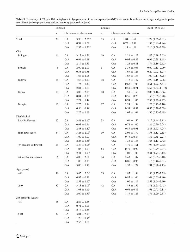
CA group mean values (±SD) for both specific (i.e., CsA and CtA) and total CA found in exposed and control sub-jects are summarized in Table 5. Statistical analysis showed a significantly higher level of total CA and CtA in exposed nurses compared to controls. When exposed and control subjects were compared with reference to the city, the level of significance was reached for Bologna (CtA), Padova (CA, CsA, and CtA), and Parma (Ca and CtA). No influ-ence of age or diet was observed among either exposed or control subjects. Among exposed subjects, a statistically significant increase for CsA was observed in relationship to job seniority. Control subjects with the GSTM1 null geno-type showed a significant increase in total CA and CtA
Table 1 Main characteristics of the study populationa
a According to the inclusion criteria, all subjects were females and non-smokersb Data reported as the number of subjects (% between brackets)c Age and job seniority in the specific task are expressed in years and reported as the group mean ± SDd Cutoff defined according to the mean value (i.e., 39.54 years) of the observed age distribution in the whole populatione Four exposed subjects were not genotyped
Exposed Controls
Subjectsb 71 77
City
Bolognab 17 (23.9) 19 (24.7)
Bresciab 3 (4.2) 3 (3.9)
Padovab 16 (22.5) 20 (26.0)
Parmab 19 (26.8) 18 (23.4)
Perugiab 16 (22.5) 17 (22.1)
Diet/alcohol
Low PAH score 27 (38.0) 38 (49.4)
High PAH score 44 (62.0) 39 (50.6)
≤4 alcohol units/week 65 (91.5) 63 (81.8)
>4 alcohol units/week 6 (8.5) 14 (18.2)
Age (years)c 39.06 ± 7.66 39.99 ± 8.65
<40b,d 38 (53.5) 34 (44.2)
≥40b,d 33 (46.5) 43 (55.8)
Job seniority (years)c 9.20 ± 7.18 –
<10b 39 (54.9) –
≥10b 32 (45.1) –
GSTM1
Positiveb,e 36 (53.7) 36 (46.8)
Nullb,e 31 (46.3) 41 (53.2)
GSTT1
Positiveb,e 59 (88.1) 67 (87.0)
Nullb,e 8 (11.9) 10 (13.0)

Int Arch Occup Environ Health
1 3
when compared to control individuals with a GSTM1-posi-tive genotype.
Regression analysis and effect size
Multiple regression analysis was performed on exposure status, age, job seniority, genetic polymorphism profiles, and environmental CP as independent variables. The analy-sis was first done on the whole population and thereafter on subjects stratified by occupational exposure (Table 6). This statistical approach indicated that variance in MN and CA frequency in the study population was mainly explained by the subjects’ occupation, thus confirming a statistically significant positive association of cytogenetic damage with exposure to ANPD. As shown in Table 6, in the whole population, MN frequency tended to rise in association
with age and a GSTM1 null genotype. In exposed nurses, job seniority was found to be associated with increased frequency of total CA and CsA. In controls, the frequency of MN was influenced by age, whereas GSTM1 polymor-phism influenced CA and CsA counts.
By evaluating the effect size in terms of increased cytogenetic damage (i.e., RoM with 95 % CI) in the exposed subjects considering MN, total CA, and CtA, we observed increases (RoM) of 1.61 (95 % CI 1.34–1.94), 1.79 (95 % CI 1.39–2.31), and 2.10 (95 % CI 1.58–2.79) times over those of unexposed individuals. Among the exposed subjects, job seniority >10 years was associated with a 1.80-fold increase (95 % CI 1.07–3.04) for CsA. In contrast, among controls, age >40 years was associated with a 1.39-fold increase (95 % CI 1.04–1.84) for MN and GSTM1 null genotype to 1.78 (95 % CI 1.13–2.80) times higher total CA. These effects were not observed in the exposed subjects probably because cytogenetic damage is mainly accounted to exposure.
Discussion
In the present study, contamination of work sites by ANPD was assessed by determining CP concentrations in selected surfaces (wipes) and nurses’ clothes (pads). All wipes were positive for the monitored model compound, with CP levels ranging from 0.01 to 1,400 ng/cm2. Dermal exposure also occurred in hospital personnel handling ANPD, with all pads positive with CP concentrations from 0.03 to 64 µg/L. Two subjects had detectable urinary concentration of CP. Over-all, environmental (surface/clothing) contamination levels were found to be similar to those reported for other Italian hospitals (Sottani et al. 2012). We also observed a significant induction of MN (group mean values ± standard deviation: 5.30 ± 2.99 and 3.29 ± 1.97, in exposed nurses and control subjects, respectively) and CA (3.30 ± 2.05 and 1.84 ± 1.67, in exposed and control subjects, respectively) associated with occupationally exposure to ANPD. However, in the exposed subjects, no relationships were observed between MN or CA frequencies and the extent of surface contamination assessed by means of wipes and pads.
These findings demonstrate that, even when the per-sonnel is specifically trained, and drug handling occurred according to the current Italian Guidelines (GURI 1999), the risk of accidental contamination and thus exposure is still present in the administration process. Similar to our findings, positive results for both MN and CA induction were reported in other investigations on workers exposed to ANPD (Bouraoui et al. 2011; El-Ebiary et al. 2013; Kopjar et al. 2009); positive results have been reported also in stud-ies evaluating only MN (Cornetta et al. 2008; Ladeira et al. 2014; Maluf and Erdtmann 2000b; Rekhadevi et al. 2007;
Table 2 Frequencies of ANPD handling
Data reported as the percentage of subject who have handled each drug at least once over a period of 6 months (data obtained from questionnaires)
ANPD classified by the International Agency of Research as 1 car-cinogenic to humans (Group 1), 2A probably carcinogenic to humans (Group 2A), 2B possibly carcinogenic to humans (Group 2B), 3 not classifiable as to its carcinogenicity to humans (Group 3), NL not listed by IARC (refs.: IARC 1976, 1981, 1987, 2000, 2012)
ANPD % ANPD %
Cyclophosphamide1 83.9 GemcitabineNL 56.5
Etoposide1 77.4 PaclitaxelNL 53.2
Methotrexate3 75.8 VinorelbineNL 53.2
Cisplatin2A 71.0 OxaliplatinNL 51.6
CarboplatinNL 64.5 IrinotecanNL 46.8
Doxorubicin2A 64.5 DocetaxelNL 41.9
Ifosfamide3 64.5 Bleomicyn2B 38.7
Vincristine3 61.3 TopotecanNL 38.7
5-Fluorouracil3 59.7 CytarabineNL 33.9
EpirubicinNL 58.1 Daunorubicin2B 33.9
Table 3 Concentrations of CP (ng/cm2) detected on surfaces or clothes over the course of the study
Data are reported as the group mean (±SD)a Each sampled workplace was associated with an exposed subject
n Surfacesa (wipes) Left forearms (pads)
Total 71 112.9 ± 279.7 2.4 ± 8.7
Age (years)
<40 38 100.8 ± 244.8 1.2 ± 1.9
≥40 33 126.7 ± 318.9 3.6 ± 12.4
Job seniority (years)
<10 39 84.81 ± 256.9 1.0 ± 1.9
≥10 32 145.1 ± 305.2 3.9 ± 12.6

Int Arch Occup Environ Health
1 3
Rombaldi et al. 2009), or CA (Burgaz et al. 2002; Jakab et al. 2001; Musak et al. 2009; Testa et al. 2007). However, negative findings have also been reported for MN (Cavallo et al. 2007; Hessel et al. 2001; Laffon et al. 2005; Maluf and Erdtmann 2000a). In a previous study by the group in Perugia (Villarini et al. 2012), no differences in MN fre-quency were observed between nurses with occupational exposure to ANPD and unexposed controls. The Perugia-based subjects in the present and the previous study were not the same; however, regarding the health care workers at the hospital of Perugia, the present results confirm the absence of an exposure-related excess of MN in exposed subjects compared to controls.
It has been reported that the number of MN in PBL increases with age in both males and females (Fenech and Bonassi 2011), whereas the relationship between aging and CA is much less clear (Bolognesi et al. 1997). In our study, MN but not CA frequencies tended to rise with age, although to a significant extent only in the control group, probably because in the exposed subjects the effect of exposure was predominant. No correlation was found between job seniority and the levels of cytogenetic damage;
this finding is in accordance with data retrieved in the lit-erature (Maluf and Erdtmann 2000a; Testa et al. 2007).
In a recently published paper (Buschini et al. 2013), we have reported on the extent of primary, oxidative, and “cryp-tic” DNA damage as evaluated by comet assay in circulat-ing leukocytes from the same nurses (exposed and controls) evaluated herein for cytogenetic damage. In such earlier work, we did not observe any statistically significant dif-ference between exposed nurses and control subjects when primary DNA damage was evaluated in leukocytes (i.e., alkaline comet assay). Similarly, when oxidative DNA dam-age was evaluated (i.e., comet/EndoIII assay) in circulating leukocytes, no statistically significant differences were found when exposed and control nurses were compared. Moreo-ver, to detect low levels of DNA damage, the comet assay protocol was modified by using an inhibitor of DNA repair, such as cytosine arabinoside (i.e., comet/AraC assay). The comet/AraC assay was performed on stimulated lympho-cytes, and unexpectedly, nurses exposed to ANPD showed a lower level of DNA migration than the control subjects.
The discrepancy between our previous and the current results might be related to the use of the total leukocyte
Table 4 Frequency of MN per 1,000 binucleated lymphocytes in nurses exposed to ANPD and non-exposed subjects with respect to age and genetic polymorphisms (whole population), and job seniority (exposed subjects)
Data reported as the group mean (±SD) of individual counts
RoM ratio of means, 95 % CI confidence intervals at the 95 % level
Statistical significance (two-sided p value <0.05, nonparametric Mann–Whitney U test): * whole population, exposed versus controls; # subgroups, exposed versus corresponding non-exposed nurses (post hoc analysis, Mann–Whitney U test multiple pairwise comparisons with Bonferroni correction for positive Kruskal–Wallis H tests); § subgroups, <40 versus ≥40 years (post hoc analysis with Bonferroni correction)
Exposed Controls RoM (95 % CI)
n Micronuclei n Micronuclei
Total 71 5.30 ± 2.99* 77 3.29 ± 1.97 1.61 (1.34–1.94)
City
Bologna 17 4.47 ± 1.64# 19 3.26 ± 2.51 1.37 (0.93–2.02)
Brescia 3 4.17 ± 2.36 3 3.60 ± 3.21 1.16 (0.35–3.83)
Padova 16 4.78 ± 1.88# 20 2.63 ± 1.68 1.82 (1.29–2.55)
Parma 19 7.21 ± 3.88# 18 4.06 ± 2.24 1.78 (1.25–2.52)
Perugia 16 4.66 ± 3.17 17 3.22 ± 1.35 1.45 (0.98–2.13)
Diet/alcohol
Low PAH score 27 5.11 ± 3.33# 38 3.37 ± 1.92 1.52 (1.12–2.06)
High PAH score 44 5.42 ± 2.79# 39 3.21 ± 2.03 1.69 (1.31–2.17)
≤4 alcohol units/week
56 5.55 ± 3.07# 63 3.17 ± 1.87 1.75 (1.43–2.15)
>4 alcohol units/week
6 5.08 ± 2.75# 14 3.82 ± 2.34 1.33 (0.78–2.28)
Age (years)
<40 38 4.78 ± 2.54# 34 2.70 ± 1.95 1.77 (1.32–2.38)
≥40 33 5.91 ± 3.37# 43 3.74 ± 1.87§ 1.58 (1.24–2.02)
Job seniority (years)
<10 39 5.15 ± 2.70 – – –
≥10 32 5.48 ± 3.34 – – –
GSTM1
Positive 36 4.81 ± 2.48# 36 3.05 ± 1.82 1.58 (1.22–2.04)
Null 31 6.19 ± 3.41# 41 3.45 ± 2.09 1.79 (1.37–2.35)
GSTT1
Positive 59 5.62 ± 3.08# 67 3.19 ± 1.98 1.76 (1.44–2.16)
Null 8 4.19 ± 2.07 10 3.90 ± 1.88 1.07 (0.68–1.69)
Int Arch Occup Environ Health
1 3
Table 5 Frequency of CA per 100 metaphases in lymphocytes of nurses exposed to ANPD and controls with respect to age and genetic poly-morphisms (whole population), and job seniority (exposed subjects)
Exposed Controls RoM (95 % CI)
n Chromosome aberrations n Chromosome aberrations
Total 70 CA 3.30 ± 2.05* 75 CA 1.84 ± 1.67 1.79 (1.39–2.31)
CsA 0.97 ± 1.02 CsA 0.73 ± 0.92 1.33 (0.91–1.94)
CtA 2.33 ± 1.50* CtA 1.11 ± 1.18 2.10 (1.58–2.79)
City
Bologna 16 CA 3.13 ± 1.71 19 CA 2.21 ± 1.23 1.42 (0.99–2.03)
CsA 0.94 ± 0.68 CsA 0.95 ± 0.85 0.99 (0.58–1.68)
CtA 2.19 ± 1.33 CtA 1.26 ± 0.81 1.74 (1.16–2.62)
Brescia 3 CA 2.00 ± 2.00 3 CA 3.33 ± 3.06 0.60 (0.13–2.79)
CsA 0.33 ± 0.58 CsA 1.67 ± 1.23 0.20 (0.02–1.71)
CtA 1.67 ± 2.08 CtA 1.67 ± 1.53 1.00 (0.17–5.75)
Padova 16 CA 4.56 ± 2.13 18 CA 1.17 ± 1.47 3.90 (2.15–7.08)
CsA 1.75 ± 1.29 CsA 0.67 ± 1.03 2.61 (1.22–5.61)
CtA 2.81 ± 1.60 CtA 0.50 ± 0.71 5.62 (2.84–11.12)
Parma 19 CA 3.05 ± 2.15 18 CA 1.50 ± 1.50 2.03 (1.16–3.56)
CsA 0.84 ± 0.83 CsA 0.56 ± 0.78 1.50 (0.69–3.28)
CtA 2.21 ± 1.44 CtA 0.94 ± 1.06 2.35 (1.29–4.27)
Perugia 16 CA 2.75 ± 1.84 17 CA 2.24 ± 1.99 1.23 (0.72–2.10)
CsA 0.50 ± 0.89 CsA 0.59 ± 0.87 0.85 (0.28–2.59)
CtA 2.25 ± 1.61 CtA 1.65 ± 1.69 1.36 (0.75–2.48)
Diet/alcohol
Low PAH score 27 CA 3.41 ± 2.12# 38 CA 1.61 ± 1.55 2.12 (1.44–3.11)
CsA 0.93 ± 0.96 CsA 0.74 ± 1.00 1.26 (0.70–2.24)
CtA 2.48 ± 1.42# CtA 0.87 ± 0.91 2.85 (1.92–4.24)
High PAH score 44 CA 3.23 ± 2.03# 39 CA 2.08 ± 1.77 1.55 (1.12–2.15)
CsA 1.00 ± 1.07 CsA 0.73 ± 0.84 1.37 (0.85–2.21)
CtA 2.23 ± 1.56# CtA 1.35 ± 1.38 1.65 (1.13–2.42)
≤4 alcohol units/week 56 CA 3.36 ± 2.06# CA 1.70 ± 1.61 1.98 (1.49–2.62)
CsA 1.05 ± 1.03 63 CsA 0.70 ± 0.92 1.50 (0.99–2.27)
CtA 2.31 ± 1.53# CtA 1.00 ± 1.00 2.31 (1.71–3.12)
>4 alcohol units/week 6 CA 4.00 ± 2.61 14 CA 2.43 ± 1.87 1.65 (0.85–3.18)
CsA 1.00 ± 0.89 CsA 0.86 ± 0.95 1.16 (0.46–2.91)
CtA 3.00 ± 1.90 CtA 1.57 ± 1.74 1.91 (0.88–4.13)
Age (years)
<40 38 CA 3.45 ± 2.04# 33 CA 1.85 ± 1.84 1.86 (1.27–2.75)
CsA 0.92 ± 0.91 CsA 0.85 ± 1.00 1.08 (0.65–1.80)
CtA 2.53 ± 1.62# CtA 1.00 ± 1.19 2.53 (1.64–3.90)
≥40 32 CA 3.13 ± 2.09# 42 CA 1.83 ± 1.55 1.71 (1.21–2.42)
CsA 1.03 ± 1.15 CsA 0.64 ± 0.85 1.61 (0.92–2.81)
CtA 2.09 ± 1.33# CtA 1.19 ± 1.23 1.76 (1.20–2.57)
Job seniority (years)
<10 38 CA 2.87 ± 1.85 – – – –
CsA 0.71 ± 1.01
CtA 2.16 ± 1.35
≥10 32 CA 3.81 ± 2.19 – – – –
CsA 1.28 ± 0.96$
CtA 2.53 ± 1.67

Int Arch Occup Environ Health
1 3
population in our earlier study, as that population is com-posed mainly of cells with short life spans, such as granu-locytes. Such as they are, the results of that previous study indicate the absence of short-term events (i.e., primary
DNA damage) in a population mostly consisting of short-lived cells. In contrast, in the comet/AraC assay in this study, we used a mitogen-stimulated lymphocyte fraction, which represents the longest surviving white cells in the
Data reported as the group mean (±SD) of individual counts
CA total chromosome aberrations, CsA chromosome-type aberrations, CtA chromatid-type aberrations, RoM ratio of means, 95 % CI confidence intervals at the 95 % level
Statistical significance (two-sided p value <0.05, nonparametric Mann–Whitney U test): * whole population, exposed versus controls; # sub-groups, exposed versus corresponding non-exposed nurses (post hoc analysis, Mann–Whitney U test multiple pairwise comparisons with Bon-ferroni correction for positive Kruskal–Wallis H tests); § subgroups, <40 versus ≥40 years (post hoc analysis with Bonferroni correction)
Table 5 continued
Exposed Controls RoM (95 % CI)
n Chromosome aberrations n Chromosome aberrations
GSTM1
Positive 36 CA 3.53 ± 2.06# 36 CA 1.31 ± 1.60 2.69 (1.73–4.19)
CsA 1.11 ± 1.09# CsA 0.47 ± 0.69 2.36 (1.33–4.21)
CtA 2.42 ± 1.48# CtA 0.83 ± 1.36 2.92 (1.65–5.16)
Null 30 CA 3.30 ± 2.04 39 CA 2.33 ± 1.59§ 1.42 (1.04–1.93)
CsA 0.93 ± 0.94 CsA 0.97 ± 1.04 0.96 (0.59–1.57)
CtA 2.37 ± 1.56# CtA 1.36 ± 0.93§ 1.74 (1.27–2.40)
GSTT1
Positive 58 CA 3.52 ± 1.93# 66 CA 1.74 ± 1.61 2.02 (1.55–2.63)
CsA 1.05 ± 1.02 CsA 0.65 ± 0.79 1.62 (1.10–2.37)
CtA 2.47 ± 1.48# CtA 1.09 ± 1.22 2.27 (1.66–3.09)
Null 8 CA 2.75 ± 2.76 9 CA 2.56 ± 2.01 1.07 (0.45–2.55)
CsA 0.88 ± 1.13 CsA 1.33 ± 1.50 0.66 (0.21–2.10)
CtA 1.88 ± 1.73 CtA 1.22 ± 0.83 1.54 (0.71–3.35)
Table 6 Multivariate regression analysisa of confounding factorsb and gene polymorphismsc on MN or CA frequencies in exposed and control nurses
a Backwise procedure: p = 0.05 for entry into the modelb Variables considered: exposure, job seniority, age (years), city, eating habits, alcohol unitsc Genetic polymorphisms: GSTM1 and GSTT1d Unstandardized B (slope of the regression line)e Standardized β
Population Biomarker Independent variableb,c Regression coefficients p
Bd (95 % CI) βe
Whole MN Exposure 2.423 (1.623–3.223) 0.444 <0.0001
Age 0.086 (0.037–0.135) 0.259 0.001
GSTM1 –0.943 (−1.737 to −0.149) −0.174 0.020
CA Exposure 1.586 (0.947–2.226) 0.390 <0.0001
CtA Exposure 1.328 (0.866–1.790) 0.442 <0.0001
Exposed MN Age 0.099 (0.001–0.198) 0.250 0.048
GSTM1 −1.650 (−3.143 to −0.157) −0.274 0.031
CA Job seniority 1.061 (0.007–2.114) 0.254 0.049
Controls MN Age 0.079 (0.030–0.127) 0.346 0.002
CA GSTM1 −1.028 (−1.764 to −0.292) −0.310 0.007
CsA GSTM1 −0.459 (−0.865 to −0.054) −0.251 0.027

Int Arch Occup Environ Health
1 3
blood. Following prolonged exposure to genotoxic xenobi-otics (e.g., causing DNA adducts), lesions do accumulate in circulating lymphocytes and such cryptic lesions may be visualized as intermediates of excision repair activity in cells that are treated with an inhibitor of DNA resynthesis (Collins et al. 1993).
In our opinion, the reduced migration extent observed in the exposed subjects as compared to controls might be related to a potential long-term exposure to cross-linking ANPD. As DNA damage induced in circulating lympho-cytes is likely to persist and accumulate because of the lim-ited excision repair activity in quiescent (G0) cells (Green et al. 1996), the increased frequency of MN observed in the exposed nurses could be associated with the extent of genome damage (including cross-links) that lymphocytes may have accumulated while circulating within the body in the quiescent phase.
Moreover, analysis of chromosome damage was associ-ated with genotype analysis of genes coding for glutathione S-transferases (GST), a family of phase II xenobiotic-metabolizing enzymes involved in catalyzing the conju-gation reactions of reactive electrophilic xenobiotics with cytosolic glutathione. Polymorphisms in these genes, pos-sibly by altering their expression and functional activities, may affect carcinogen activation/detoxification and even-tually DNA damage. Homozygous deletion, or null geno-type, at either the GSTM1 or the GSTT1 locus, resulted in enzyme function loss, and this has been hypothesized to account for a higher susceptibility to cancer (Ginsberg et al. 2009). To assess any influence of genetic background on individual susceptibility, we evaluated the potential effect of polymorphisms in GSTM1 and GSTT1 on risks from occupational exposures to ANPD. The frequencies obtained for GSTM1- and GSTT1 null genotypes in the whole popu-lation (0.50 and 0.13, respectively) were consistent with the data reported in the literature for the European population (Kurose et al. 2012), and the results of this study suggest that GSTM1 and GSTT1 null genotypes do not modify MN frequencies in the presence of exposure to ANPD. We can-not compare our findings to published data in the literature because, to the best of our knowledge, ours is the first study in this occupational setting where gene polymorphisms in GSTM1 and GSTT1 have been associated with MN fre-quency. Similarly, GSTM1 and GSTT1 null genotypes did not modify CA frequencies in the presence of exposure to ANPD. Our findings are in agreement with the results reported in the published papers with a similar approach (Musak et al. 2009; Testa et al. 2007).
Among the above-cited studies in which cytogenetic end points were evaluated, very few have considered an ade-quate exposure evaluation in the study design. Environmen-tal and biological monitoring procedures have been consid-ered by Cavallo et al. (2005), which included measurements
of surface contamination from the most commonly used ANPD (i.e., CP, cytarabine, 5-fluorouracil, gemcitabine, and ifosfamide,), and measurement of the 5-fluorouracil metabolite α-fluoro-β-alanine in urine samples. Biological monitoring of exposure has been considered only in two studies. Ensslin et al. (1997), in hospital personnel regu-larly handling ANPD, quantified the incorporation of CP, ifosfamide, and platinum-containing drugs by the deter-mination of urinary concentrations of these drugs. Burgaz et al. (1999) evaluated exposure to ANPD by measuring the urinary concentrations of CP. Environmental monitoring, by the evaluation of contamination of surfaces by 5-fluo-rouracil (i.e., wipe test), was performed by Ladeira et al. (2014). Overall, the findings in the present and the above-cited studies confirm that ANPD contamination of the work environment in hospital is still possible, and safety meas-ures adopted may not be sufficient to prevent exposure to genotoxic xenobiotics (Hedmer and Wohlfart 2012).
The standard regulation in force regarding this subject in Italy (GURI 1999) incorporates many of the measures included in international warnings and guidelines, as well as rules for safe and appropriate organization of services for ANPD preparations (ASHP 2006; NIOSH 2010; OSHA 2000). On this basis, the nurses exposed to ANPD enrolled in this study had received adequate recommendations for safety handling of ANPD and were recommended to wear appropriate personal protective equipment (i.e., dispos-able single-use gowns, gloves, and masks) whenever han-dling ANPD or contaminated materials. However, despite the adoption of methods for preventing exposure to ANPD (e.g., engineering controls, administrative and work prac-tice controls, training, and personal protective equipment), incorporation of trace amounts of these agents still occurs in hospital personnel with a detectable residual genotoxic risks, as revealed by the increased frequency of MN and CA in the exposed subjects.
Conclusions
In conclusion, our results provide further evidence that handling ANPD, even if under safety controlled conditions, represents a considerable genotoxic risk for healthy sub-jects occupationally exposed to these chemicals. Moreover, CA (Bonassi et al. 2000, 2004, 2008) and MN (Bonassi et al. 2007) are predictive of increased cancer risk, rather than just reflect the exposure level. This finding clearly indicates the necessity to improve some steps in the admin-istration process of ANPD to appropriately cope with genotoxic risk. It is important to modify the guidelines for the evaluation of mutagenic/carcinogenic hazards in occu-pationally exposed subjects by considering an integrated chemical/biotoxicological approach. The use of biomarkers

Int Arch Occup Environ Health
1 3
which measure changes in cellular or molecular endpoints (e.g., DNA damage) will allow us to implement a more complete approach according to not only environmen-tal and biological monitoring but also to biological effect monitoring using genotoxicity biomarkers (Villarini et al. 2012).
CA is certainly the most robust biomarker with predic-tivity related to cancer risk. However, testing of CA has high demands on laboratory training and skills and is very time-consuming. On the other hand, MN is a very prom-ising biomarker with predictivity of cancer and smaller demands on skills (Knudsen and Hansen 2007). The extensive use of CA in future studies is thus limited by the laborious and sensitive procedure of the test. MN test-ing, because of its ability to detect both clastogenic (e.g., chromosome breakage) and aneugenic (e.g., spindle disrup-tion) effects, could have a role in occupational health sur-veillance programs for workers occupationally exposed to ANPD to monitor long-term exposure effects (biomarker of early/preclinical biological effects). Finally, genotyping of exposed subjects for GSTM1 and/or GSTT1 polymorphisms does not seem to have a role in occupational health surveil-lance programs in the studied occupational setting.
Acknowledgments The authors wish to thank the nurses for par-ticipating in the study as exposed subjects or controls. We are grate-ful to Prof. Paolo Puccetti, Department of Experimental Medicine, University of Perugia, for reading the manuscript and offering con-structive comments. The study was funded by the Italian Ministry of Education, University and Scientific Research (MIUR), Research No. 2005-062547.
Conflict of interest The authors declare that they have no conflict of interest.
References
ASHP (2006) ASHP (American Society of Hospital Pharmacists) guidelines on handling hazardous drugs. Am J Hosp Pharm 63:1172–1193
Bolognesi C, Abbondandolo A, Barale R, Casalone R, Dalpra L, De Ferrari M, Degrassi F, Forni A, Lamberti L, Lando C, Migliore L, Padovani P, Pasquini R, Puntoni R, Sbrana I, Stella M, Bonassi S (1997) Age-related increase of baseline frequencies of sister chromatid exchanges, chromosome aberrations, and micronu-clei in human lymphocytes. Cancer Epidemiol Biomarkers Prev 6(4):249–256
Bonassi S, Hagmar L, Stromberg U, Montagud AH, Tinnerberg H, Forni A, Heikkila P, Wanders S, Wilhardt P, Hansteen IL, Knud-sen LE, Norppa H (2000) Chromosomal aberrations in lympho-cytes predict human cancer independently of exposure to car-cinogens. European Study Group on Cytogenetic Biomarkers and Health. Cancer Res 60(6):1619–1625
Bonassi S, Znaor A, Norppa H, Hagmar L (2004) Chromosomal aber-rations and risk of cancer in humans: an epidemiologic perspec-tive. Cytogenet Genome Res 104(1–4):376–382
Bonassi S, Znaor A, Ceppi M, Lando C, Chang WP, Holland N, Kirsch-Volders M, Zeiger E, Ban S, Barale R, Bigatti MP,
Bolognesi C, Cebulska-Wasilewska A, Fabianova E, Fucic A, Hagmar L, Joksic G, Martelli A, Migliore L, Mirkova E, Scarfi MR, Zijno A, Norppa H, Fenech M (2007) An increased micro-nucleus frequency in peripheral blood lymphocytes predicts the risk of cancer in humans. Carcinogenesis 28(3):625–631
Bonassi S, Norppa H, Ceppi M, Stromberg U, Vermeulen R, Znaor A, Cebulska-Wasilewska A, Fabianova E, Fucic A, Gundy S, Han-steen IL, Knudsen LE, Lazutka J, Rossner P, Sram RJ, Boffetta P (2008) Chromosomal aberration frequency in lymphocytes pre-dicts the risk of cancer: results from a pooled cohort study of 22 358 subjects in 11 countries. Carcinogenesis 29(6):1178–1183
Bouraoui S, Brahem A, Tabka F, Mrizek N, Saad A, Elghezal H (2011) Assessment of chromosomal aberrations, micronuclei and proliferation rate index in peripheral lymphocytes from Tunisian nurses handling cytotoxic drugs. Environ Toxicol Pharmacol 31(1):250–257
Burgaz S, Karahalil B, Bayrak P, Taskin L, Yavuzaslan F, Bokesoy I, Anzion RB, Bos RP, Platin N (1999) Urinary cyclophosphamide excretion and micronuclei frequencies in peripheral lymphocytes and in exfoliated buccal epithelial cells of nurses handling anti-neoplastics. Mutat Res 439(1):97–104
Burgaz S, Karahalil B, Canhi Z, Terzioglu F, Ancel G, Anzion RB, Bos RP, Huttner E (2002) Assessment of genotoxic damage in nurses occupationally exposed to antineoplastics by the analysis of chromosomal aberrations. Hum Exp Toxicol 21(3):129–135
Buschini A, Villarini M, Feretti D, Mussi F, Dominici L, Zerbini I, Moretti M, Ceretti E, Bonfiglioli R, Carrieri M, Gelatti U, Rossi C, Monarca S, Poli P (2013) Multicentre study for the evaluation of mutagenic/carcinogenic risk in nurses exposed to antineo-plastic drugs: assessment of DNA damage. Occup Environ Med 70(11):789–794
Cavallo D, Ursini CL, Perniconi B, Francesco AD, Giglio M, Rubino FM, Marinaccio A, Iavicoli S (2005) Evaluation of genotoxic effects induced by exposure to antineoplastic drugs in lympho-cytes and exfoliated buccal cells of oncology nurses and phar-macy employees. Mutat Res 587(1–2):45–51
Cavallo D, Ursini CL, Omodeo-Sale E, Iavicoli S (2007) Micronu-cleus induction and FISH analysis in buccal cells and lympho-cytes of nurses administering antineoplastic drugs. Mutat Res 628(1):11–18
Collins AR, Duthie SJ, Dobson VL (1993) Direct enzymic detection of endogenous oxidative base damage in human lymphocyte DNA. Carcinogenesis 14(9):1733–1735
Connor TH, McDiarmid MA (2006) Preventing occupational expo-sures to antineoplastic drugs in health care settings. CA Cancer J Clin 56(6):354–365
Connor TH, Sessink PJ, Harrison BR, Pretty JR, Peters BG, Alfaro RM, Bilos A, Beckmann G, Bing MR, Anderson LM, Dechristo-foro R (2005) Surface contamination of chemotherapy drug vials and evaluation of new vial-cleaning techniques: results of three studies. Am J Health Syst Pharm 62(5):475–484
Cornetta T, Padua L, Testa A, Ievoli E, Festa F, Tranfo G, Baccelliere L, Cozzi R (2008) Molecular biomonitoring of a population of nurses handling antineoplastic drugs. Mutat Res 638(1–2):75–82
Davis J, McLauchlan R, Connor TH (2011) Exposure to hazardous drugs in healthcare: an issue that will not go away. J Oncol Pharm Pract 17(1):9–13
DeMeo MP, Merono S, DeBaille AD, Botta A, Laget M, Guiraud H, Dumenil G (1995) Monitoring exposure of hospital personnel handling cytostatic drugs and contaminated materials. Int Arch Occup Environ Health 66(6):363–368
Eastmond DA, Tucker JD (1989) Identification of aneuploidy-induc-ing agents using cytokinesis-blocked human lymphocytes and an antikinetochore antibody. Environ Mol Mutagen 13(1):34–43
El-Ebiary AA, Abuelfadl AA, Sarhan NI (2013) Evaluation of genotoxicity induced by exposure to antineoplastic drugs in

Int Arch Occup Environ Health
1 3
lymphocytes of oncology nurses and pharmacists. J Appl Toxicol 33(3):196–201
Ensslin AS, Stoll Y, Pethran A, Pfaller A, Rommelt H, Fruhmann G (1994) Biological monitoring of cyclophosphamide and ifosfa-mide in urine of hospital personnel occupationally exposed to cytostatic drugs. Occup Environ Med 51(4):229–233
Ensslin AS, Huber R, Pethran A, Rommelt H, Schierl R, Kulka U, Fruhmann G (1997) Biological monitoring of hospital phar-macy personnel occupationally exposed to cytostatic drugs: uri-nary excretion and cytogenetics studies. Int Arch Occup Environ Health 70(3):205–208
Falck K, Grohn P, Sorsa M, Vainio H, Heinonen E, Holsti LR (1979) Mutagenicity in urine of nurses handling cytostatic drugs. Lancet 1(8128):1250–1251
Fenech M (2000) The in vitro micronucleus technique. Mutat Res 455(1–2):81–95
Fenech M, Bonassi S (2011) The effect of age, gender, diet and life-style on DNA damage measured using micronucleus frequency in human peripheral blood lymphocytes. Mutagenesis 26(1):43–49
Fenech M, Holland N, Chang WP, Zeiger E, Bonassi S (1999) The HUman MicroNucleus Project—an international collaborative study on the use of the micronucleus technique for measuring DNA damage in humans. Mutat Res 428(1–2):271–283
Fenech M, Chang WP, Kirsch-Volders M, Holland N, Bonassi S, Zeiger E (2003) HUMN project: detailed description of the scor-ing criteria for the cytokinesis-block micronucleus assay using isolated human lymphocyte cultures. Mutat Res 534(1–2):65–75
Fransman W, Vermeulen R, Kromhout H (2004) Occupational dermal exposure to cyclophosphamide in Dutch hospitals: a pilot study. Ann Occup Hyg 48(3):237–244
Friedrich JO, Adhikari NK, Beyene J (2011) Ratio of means for ana-lyzing continuous outcomes in meta-analysis performed as well as mean difference methods. J Clin Epidemiol 64(5):556–564
Ginsberg G, Smolenski S, Hattis D, Guyton KZ, Johns DO, Son-awane B (2009) Genetic polymorphism in glutathione trans-ferases (GST): population distribution of GSTM1, T1, and P1 conjugating activity. J Toxicol Environ Health B Crit Rev 12(5–6):389–439
Green MH, Waugh AP, Lowe JE, Harcourt SA, Clingen PH, Cole J, Arlett CF (1996) Protective effect of deoxyribonucleosides on UV-irradiated human peripheral blood T-lymphocytes: possibili-ties for the selective killing of either cycling or non-cycling cells. Mutat Res 350(1):239–246
GURI (1999) Provvedimento di Linee-Guida per la sicurezza e la salute dei lavoratori esposti a chemioterapici antiblastici in ambi-ente sanitario (Repertorio Atti No. 736) Gazzetta Ufficiale della Repubblica Italiana (Official Gazette of the Italian Republic), 07 October 1999, No 236. Rome
Hedmer M, Wohlfart G (2012) Hygienic guidance values for wipe sampling of antineoplastic drugs in Swedish hospitals. J Environ Monit 14(7):1968–1975
Hessel H, Radon K, Pethran A, Maisch B, Grobmair S, Sautter I, Fruhmann G (2001) The genotoxic risk of hospital, phar-macy and medical personnel occupationally exposed to cyto-static drugs—evaluation by the micronucleus assay. Mutat Res 497(1–2):101–109
Hirvonen A, Saarikoski ST, Linnainmaa K, Koskinen K, Husgafvel-Pursiainen K, Mattson K, Vainio H (1996) Glutathione S-trans-ferase and N-acetyltransferase genotypes and asbestos-associated pulmonary disorders. J Natl Cancer Inst 88(24):1853–1856
IAEA (2001) Cytogenetic analysis for radiation dose assessment (Technical Reports Series No. 405). International Atomic Energy Agency, Vienna
IARC (1976) Some naturally occurring substances, IARC mono-graphs on the evaluation of carcinogenic risks to humans, vol 10. International Agency for Research on Cancer, Lyon
IARC (1981) Some antineoplastic and immunosuppressive agents, IARC monographs on the evaluation of carcinogenic risks to humans, vol 26. International Agency for Research on Cancer, Lyon
IARC (1987) Overall evaluations of carcinogenicity: an updating of IARC monographs volumes 1 to 42, IARC monographs on the evaluation of carcinogenic risks to humans, supplement 7. Inter-national Agency for Research on Cancer, Lyon
IARC (2000) Some antiviral and antineoplastic drugs, and other pharmaceutical agents, IARC monographs on the evaluation of carcinogenic risks to humans, vol 76. International Agency for Research on Cancer, Lyon
IARC (2012) A review of human carcinogens. Part A: pharmaceuti-cals, IARC monographs on the evaluation of carcinogenic risks to humans, vol 100. International Agency for Research on Cancer, Lyon
Jakab MG, Major J, Tompa A (2001) Follow-up genotoxicological monitoring of nurses handling antineoplastic drugs. J Toxicol Environ Health A 62(5):307–318
Keshava N, Ong TM (1999) Occupational exposure to genotoxic agents. Mutat Res 437(2):175–194
Kiffmeyer T, Hadtstein C (2007) Handling of chemotherapeutic drugs in the OR: hazards and safety considerations. Cancer Treat Res 134:275–290
Kiffmeyer TK, Tuerk J, Hahn M, Stuetzer H, Hadtstein C, Heinemann A, Eickmann U (2013) Application and assessment of a regular environmental monitoring of the antineoplastic drug contamina-tion level in pharmacies—the MEWIP project. Ann Occup Hyg 57(4):444–455
Knudsen LE, Hansen AM (2007) Biomarkers of intermediate end-points in environmental and occupational health. Int J Hyg Envi-ron Health 210(3–4):461–470
Kopjar N, Garaj-Vrhovac V, Kasuba V, Rozgaj R, Ramic S, Pavlica V, Zeljezic D (2009) Assessment of genotoxic risks in Croa-tian health care workers occupationally exposed to cytotoxic drugs: a multi-biomarker approach. Int J Hyg Environ Health 212(4):414–431
Kurose K, Sugiyama E, Saito Y (2012) Population differences in major functional polymorphisms of pharmacokinetics/pharmaco-dynamics-related genes in Eastern Asians and Europeans: impli-cations in the clinical trials for novel drug development. Drug Metab Pharmacokinet 27(1):9–54
Labuhn K, Valanis B, Schoeny R, Loveday K, Vollmer WM (1998) Nurses’ and pharmacists’ exposure to antineoplastic drugs: find-ings from industrial hygiene scans and urine mutagenicity tests. Cancer Nurs 21(2):79–89
Ladeira C, Viegas S, Padua M, Gomes M, Carolino E, Gomes MC, Brito M (2014) Assessment of genotoxic effects in nurses handling cytostatic drugs. J Toxicol Environ Health A 77(14–16):879–887
Laffon B, Teixeira JP, Silva S, Loureiro J, Torres J, Pasaro E, Men-dez J, Mayan O (2005) Genotoxic effects in a population of nurses handling antineoplastic drugs, and relationship with genetic polymorphisms in DNA repair enzymes. Am J Ind Med 48(2):128–136
Larson RR, Khazaeli MB, Dillon HK (2002) Monitoring method for surface contamination caused by selected antineoplastic agents. Am J Health Syst Pharm 59(3):270–277
Maluf SW, Erdtmann B (2000a) Follow-up study of the genetic damage in lymphocytes of pharmacists and nurses handling antineoplastic drugs evaluated by cytokinesis-block micronuclei analysis and sin-gle cell gel electrophoresis assay. Mutat Res 471(1–2):21–27
Maluf SW, Erdtmann B (2000b) Evaluation of occupational genotoxic risk in a Brazilian hospital. Genet Mol Biol 23(2):485–488
McDevitt JJ, Lees PS, McDiarmid MA (1993) Exposure of hospital pharmacists and nurses to antineoplastic agents. J Occup Med 35(1):57–60
Int Arch Occup Environ Health
1 3
Minoia C, Turci R, Sottani C, Schiavi A, Perbellini L, Angeleri S, Draicchio F, Apostoli P (1998) Application of high performance liquid chromatography/tandem mass spectrometry in the environ-mental and biological monitoring of health care personnel occu-pationally exposed to cyclophosphamide and ifosfamide. Rapid Commun Mass Spectrom 12(20):1485–1493
Moretti M, Bonfiglioli R, Feretti D, Pavanello S, Mussi F, Grollino MG, Villarini M, Barbieri A, Ceretti E, Carrieri M, Buschini A, Appolloni M, Dominici L, Sabatini L, Gelatti U, Bartolucci GB, Poli P, Stronati L, Mastrangelo G, Monarca S (2011) A study pro-tocol for the evaluation of occupational mutagenic/carcinogenic risks in subjects exposed to antineoplastic drugs: a multicentric project. BMC Public Health 11:195
Musak L, Halasova E, Matakova T, Letkova L, Vodickova L, Buchan-cova J, Hudeckova H, Osina O, Soucek P, Vodicka P (2009) Comparison of chromosomal aberrations frequency and polymor-phism of GSTs genes in workers occupationally exposed to cyto-statics or anaesthetics. Interdiscip Toxicol 2(3):190–194
NIOSH (2010) NIOSH list of antineoplastic and other hazardous drugs in healthcare settings. In: U.S. Department of Health and Human Services, Centers for Disease Control and Prevention (ed) National Institute for Occupational Safety and Health, Cincinnati
OSHA (2000) OSHA technical manual. Hospital investigations: health hazard—section VI. Occupational Safety and Health Administration, US Department of Labor, Washington
Pavanello S, Simioli P, Lupi S, Gregorio P, Clonfero E (2002) Expo-sure levels and cytochrome P450 1A2 activity, but not N-acetyl-transferase, glutathione S-transferase (GST) M1 and T1, influ-ence urinary mutagen excretion in smokers. Cancer Epidemiol Biomarkers Prev 11(10 Pt 1):998–1003
Rekhadevi PV, Sailaja N, Chandrasekhar M, Mahboob M, Rahman MF, Grover P (2007) Genotoxicity assessment in oncology nurses handling anti-neoplastic drugs. Mutagenesis 22(6):395–401
Rombaldi F, Cassini C, Salvador M, Saffi J, Erdtmann B (2009) Occupational risk assessment of genotoxicity and oxidative stress in workers handling anti-neoplastic drugs during a working week. Mutagenesis 24(2):143–148
Savage JR, Holloway M (1988) Induction of sister-chromatid exchanges by d(42 MeV)-Be neutrons in unstimulated human-blood lymphocytes. Br J Radiol 61(723):231–234
Schierl R, Bohlandt A, Nowak D (2009) Guidance values for surface monitoring of antineoplastic drugs in German pharmacies. Ann Occup Hyg 53(7):703–711
Sessink PJ, Cerna M, Rossner P, Pastorkova A, Bavarova H, Frank-ova K, Anzion RB, Bos RP (1994a) Urinary cyclophosphamide excretion and chromosomal aberrations in peripheral blood lym-phocytes after occupational exposure to antineoplastic agents. Mutat Res 309(2):193–199
Sessink PJ, Van de Kerkhof MC, Anzion RB, Noordhoek J, Bos RP (1994b) Environmental contamination and assessment of exposure to antineoplastic agents by determination of cyclo-phosphamide in urine of exposed pharmacy technicians: is skin absorption an important exposure route? Arch Environ Health 49(3):165–169
Sessink PJ, Kroese ED, van Kranen HJ, Bos RP (1995) Cancer risk assessment for health care workers occupationally exposed to cyclophosphamide. Int Arch Occup Environ Health 67(5):317–323
Sorsa M, Anderson D (1996) Monitoring of occupational exposure to cytostatic anticancer agents. Mutat Res 355(1–2):253–261
Sottani C, Porro B, Imbriani M, Minoia C (2012) Occupational expo-sure to antineoplastic drugs in four Italian health care settings. Toxicol Lett 213(1):107–115
Testa A, Giachelia M, Palma S, Appolloni M, Padua L, Tranfo G, Spagnoli M, Tirindelli D, Cozzi R (2007) Occupational expo-sure to antineoplastic agents induces a high level of chromosome damage. Lack of an effect of GST polymorphisms. Toxicol Appl Pharmacol 223(1):46–55
Turci R, Minoia C (2006) Residual hazard assessment related to handling of antineoplastic drugs: safety system evolution and quality assurance of analytical measurement. Ann N Y Acad Sci 1076:649–656
Turci R, Sottani C, Spagnoli G, Minoia C (2003) Biological and envi-ronmental monitoring of hospital personnel exposed to antineo-plastic agents: a review of analytical methods. J Chromatogr B Analyt Technol Biomed Life Sci 789(2):169–209
Villarini M, Dominici L, Fatigoni C, Muzi G, Monarca S, Moretti M (2012) Biological effect monitoring in peripheral blood lym-phocytes from subjects occupationally exposed to antineoplas-tic drugs: assessment of micronuclei frequency. J Occup Health 54(6):405–415
Ziegler E, Mason HJ, Baxter PJ (2002) Occupational exposure to cytotoxic drugs in two UK oncology wards. Occup Environ Med 59(9):608–612