Keywords: Micronuclei Mobile phone Plutonium Chromosome aberrations
8
This article appeared in a journal published by Elsevier. The attached copy is furnished to the author for internal non-commercial research and education use, including for instruction at the authors institution and sharing with colleagues. Other uses, including reproduction and distribution, or selling or licensing copies, or posting to personal, institutional or third party websites are prohibited. In most cases authors are permitted to post their version of the article (e.g. in Word or Tex form) to their personal website or institutional repository. Authors requiring further information regarding Elsevier’s archiving and manuscript policies are encouraged to visit: http://www.elsevier.com/copyright
Transcript of Keywords: Micronuclei Mobile phone Plutonium Chromosome aberrations
This article appeared in a journal published by Elsevier. The attachedcopy is furnished to the author for internal non-commercial researchand education use, including for instruction at the authors institution
and sharing with colleagues.
Other uses, including reproduction and distribution, or selling orlicensing copies, or posting to personal, institutional or third party
websites are prohibited.
In most cases authors are permitted to post their version of thearticle (e.g. in Word or Tex form) to their personal website orinstitutional repository. Authors requiring further information
regarding Elsevier’s archiving and manuscript policies areencouraged to visit:
http://www.elsevier.com/copyright
Author's personal copy
Mutation Research 750 (2013) 27– 33
Contents lists available at SciVerse ScienceDirect
Mutation Research/Genetic Toxicology andEnvironmental Mutagenesis
journa l h omepage: www.elsev ier .com/ locate /gentoxC om mun i ty a ddress : www.elsev ier .com/ locate /mutres
Comparison of cytotoxic and genotoxic effects of plutonium-239 alpha particlesand mobile phone GSM 900 radiation in the Allium cepa test
Dmitry S. Pesnya ∗, Anton V. RomanovskyI.D. Papanin Institute for Biology of Inland Waters, Russian Academy of Sciences, 152742 Borok, Nekouz, Yaroslavl region, Russia
a r t i c l e i n f o
Article history:Received 6 March 2012Received in revised form 5 August 2012Accepted 30 August 2012Available online 8 October 2012
Keywords:MicronucleiMobile phonePlutoniumChromosome aberrationsMitotic indexAllium cepa test
a b s t r a c t
The goal of this study was to compare the cytotoxic and genotoxic effects of plutonium-239 alpha par-ticles and GSM 900 modulated mobile phone (model Sony Ericsson K550i) radiation in the Allium cepatest. Three groups of bulbs were exposed to mobile phone radiation during 0 (sham), 3 and 9 h. A positivecontrol group was treated during 20 min with plutonium-239 alpha-radiation. Mitotic abnormalities,chromosome aberrations, micronuclei and mitotic index were analyzed. Exposure to alpha-radiationfrom plutonium-239 and exposure to modulated radiation from mobile phone during 3 and 9 h signif-icantly increased the mitotic index. GSM 900 mobile phone radiation as well as alpha-radiation fromplutonium-239 induced both clastogenic and aneugenic effects. However, the aneugenic activity ofmobile phone radiation was more pronounced. After 9 h of exposure to mobile phone radiation, polyploidcells, three-groups metaphases, amitoses and some unspecified abnormalities were detected, which werenot registered in the other experimental groups. Importantly, GSM 900 mobile phone radiation increasedthe mitotic index, the frequency of mitotic and chromosome abnormalities, and the micronucleus fre-quency in a time-dependent manner. Due to its sensitivity, the A. cepa test can be recommended asa useful cytogenetic assay to assess cytotoxic and genotoxic effects of radiofrequency electromagneticfields.
© 2012 Elsevier B.V. All rights reserved.
1. Introduction
In the past few years the use of cell phones has becomewidespread. Many studies have been carried out to assess potentialhazards to human health and the environment due to the increasedexposure to radiofrequency electromagnetic fields (RF-EMF) emit-ted by mobile phones and other wireless devices.
Most of the attention on possible adverse effects of electromag-netic radiation has been focused on human health [1–5]. Moreover,some studies suggested higher risk for malignant brain tumorsin people with ipsilateral mobile phone use [6–8]. Recently, theIARC classified radiofrequency electromagnetic fields as possiblycarcinogenic to humans (Group 2B) [9].
Additionally, the mobile phone radiation seems to have effectsupon different biological species: bacteria [10], protozoa [11],plants [12,13], insects [14,15], amphibians [16], birds [17] andmammals [18–21]. These reports have contributed to a betterknowledge of the interactions between EMF and living organisms.
Amongst many biological targets, the DNA molecule hasreceived the greatest attention with respect to potential RF-EMF
∗ Corresponding author at: Dmitry Sergeevich Pesnya, Avenue Frunze, h.2, ap.77,150030 Yaroslavl, Yaroslavl region, Russia. Tel.: +7 4852 524196.
E-mail addresses: [email protected] (D.S. Pesnya), [email protected](A.V. Romanovsky).
damage [22]. Alterations of DNA in somatic cells are one of the keyevents in the process of carcinogenesis and any agent with geno-toxic activity may also be suspected to be carcinogenic [23–25].However, genotoxic data from different non-mammalian speciesare very limited. Information on invertebrates and plants is partic-ularly lacking.
Along with other plant species, Allium cepa L. has been used toevaluate DNA damages, such as chromosome aberrations, micronu-clei and disturbances in the mitotic cycle. The A. cepa test is nowfrequently used for environmental monitoring [26] and also forassessing effects of ionizing and non-ionizing radiation [13,27].
In the present study the effects of RF-EMF on the mitoticindex, and on the frequencies of chromosomal abnormalities andmicronuclei were investigated in root meristematic cells of A. cepaL., in comparison with the effects of alpha particles.
2. Materials and methods
2.1. The A. cepa test
The A. cepa test was used to analyze genotoxic effects [28,29].We used onion bulbs (A. cepa L., 2n = 16) of the Stuttgarten-Risen variety, average
weight 25 g. The bulbs were placed in glass jars with their basal ends dipping indistilled water, and germinated at room temperature (24 ± 3 ◦C). When the newlyemerged roots were 0.50–1 cm in length, they were used in the experiments.
After the treatment, root-tips were fixed in a solution of ethanol (96%) andglacial acetic acid (3:1, v/v) for 48 h, washed with distilled water, and then stainedwith aceto-orcein for 1 h. The squash technique was applied to prepare samples for
1383-5718/$ – see front matter © 2012 Elsevier B.V. All rights reserved.http://dx.doi.org/10.1016/j.mrgentox.2012.08.010
Author's personal copy
28 D.S. Pesnya, A.V. Romanovsky / Mutation Research 750 (2013) 27– 33
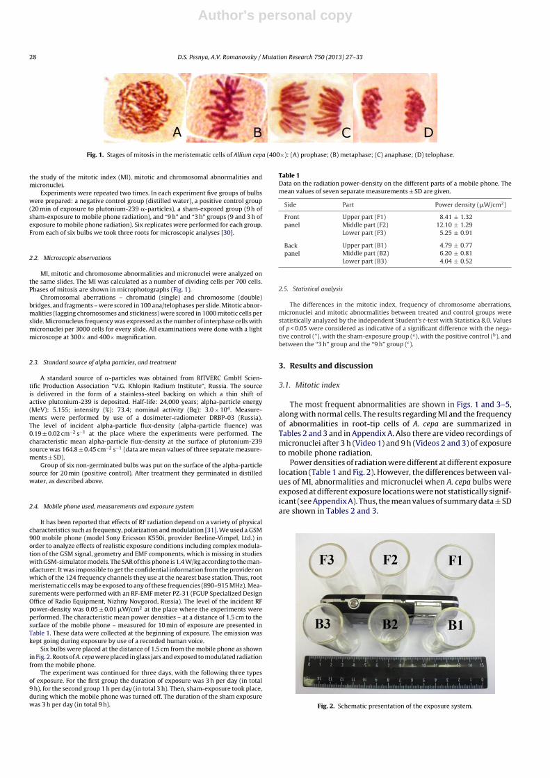
Fig. 1. Stages of mitosis in the meristematic cells of Allium cepa (400×): (A) prophase; (B) metaphase; (C) anaphase; (D) telophase.
the study of the mitotic index (MI), mitotic and chromosomal abnormalities andmicronuclei.
Experiments were repeated two times. In each experiment five groups of bulbswere prepared: a negative control group (distilled water), a positive control group(20 min of exposure to plutonium-239 �-particles), a sham-exposed group (9 h ofsham-exposure to mobile phone radiation), and “9 h” and “3 h” groups (9 and 3 h ofexposure to mobile phone radiation). Six replicates were performed for each group.From each of six bulbs we took three roots for microscopic analyses [30].
2.2. Microscopic observations
MI, mitotic and chromosome abnormalities and micronuclei were analyzed onthe same slides. The MI was calculated as a number of dividing cells per 700 cells.Phases of mitosis are shown in microphotographs (Fig. 1).
Chromosomal aberrations – chromatid (single) and chromosome (double)bridges, and fragments – were scored in 100 ana/telophases per slide. Mitotic abnor-malities (lagging chromosomes and stickiness) were scored in 1000 mitotic cells perslide. Micronucleus frequency was expressed as the number of interphase cells withmicronuclei per 3000 cells for every slide. All examinations were done with a lightmicroscope at 300× and 400× magnification.
2.3. Standard source of alpha particles, and treatment
A standard source of �-particles was obtained from RITVERC GmbH Scien-tific Production Association “V.G. Khlopin Radium Institute”, Russia. The sourceis delivered in the form of a stainless-steel backing on which a thin shift ofactive plutonium-239 is deposited. Half-life: 24,000 years; alpha-particle energy(MeV): 5.155; intensity (%): 73.4; nominal activity (Bq): 3.0 × 10⁴. Measure-ments were performed by use of a dosimeter-radiometer DRBP-03 (Russia).The level of incident alpha-particle flux-density (alpha-particle fluence) was0.19 ± 0.02 cm−2 s−1 at the place where the experiments were performed. Thecharacteristic mean alpha-particle flux-density at the surface of plutonium-239source was 164.8 ± 0.45 cm−2 s−1 (data are mean values of three separate measure-ments ± SD).
Group of six non-germinated bulbs was put on the surface of the alpha-particlesource for 20 min (positive control). After treatment they germinated in distilledwater, as described above.
2.4. Mobile phone used, measurements and exposure system
It has been reported that effects of RF radiation depend on a variety of physicalcharacteristics such as frequency, polarization and modulation [31]. We used a GSM900 mobile phone (model Sony Ericsson K550i, provider Beeline-Vimpel, Ltd.) inorder to analyze effects of realistic exposure conditions including complex modula-tion of the GSM signal, geometry and EMF components, which is missing in studieswith GSM-simulator models. The SAR of this phone is 1.4 W/kg according to the man-ufacturer. It was impossible to get the confidential information from the provider onwhich of the 124 frequency channels they use at the nearest base station. Thus, rootmeristematic cells may be exposed to any of these frequencies (890–915 MHz). Mea-surements were performed with an RF-EMF meter PZ-31 (FGUP Specialized DesignOffice of Radio Equipment, Nizhny Novgorod, Russia). The level of the incident RFpower-density was 0.05 ± 0.01 �W/cm2 at the place where the experiments wereperformed. The characteristic mean power densities – at a distance of 1.5 cm to thesurface of the mobile phone – measured for 10 min of exposure are presented inTable 1. These data were collected at the beginning of exposure. The emission waskept going during exposure by use of a recorded human voice.
Six bulbs were placed at the distance of 1.5 cm from the mobile phone as shownin Fig. 2. Roots of A. cepa were placed in glass jars and exposed to modulated radiationfrom the mobile phone.
The experiment was continued for three days, with the following three typesof exposure. For the first group the duration of exposure was 3 h per day (in total9 h), for the second group 1 h per day (in total 3 h). Then, sham-exposure took place,during which the mobile phone was turned off. The duration of the sham exposurewas 3 h per day (in total 9 h).
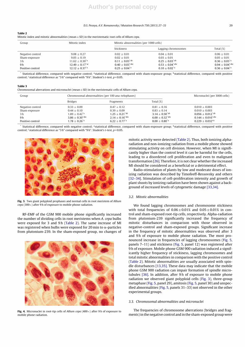
Table 1Data on the radiation power-density on the different parts of a mobile phone. Themean values of seven separate measurements ± SD are given.
Side Part Power density (�W/cm2)
Frontpanel
Upper part (F1) 8.41 ± 1.32Middle part (F2) 12.10 ± 1.29Lower part (F3) 5.25 ± 0.91
Backpanel
Upper part (B1) 4.79 ± 0.77Middle part (B2) 6.20 ± 0.81Lower part (B3) 4.04 ± 0.52
2.5. Statistical analysis
The differences in the mitotic index, frequency of chromosome aberrations,micronuclei and mitotic abnormalities between treated and control groups werestatistically analyzed by the independent Student’s t-test with Statistica 8.0. Valuesof p < 0.05 were considered as indicative of a significant difference with the nega-tive control (*), with the sham-exposure group (a), with the positive control (b), andbetween the “3 h” group and the “9 h” group (c).
3. Results and discussion
3.1. Mitotic index
The most frequent abnormalities are shown in Figs. 1 and 3–5,along with normal cells. The results regarding MI and the frequencyof abnormalities in root-tip cells of A. cepa are summarized inTables 2 and 3 and in Appendix A. Also there are video recordings ofmicronuclei after 3 h (Video 1) and 9 h (Videos 2 and 3) of exposureto mobile phone radiation.
Power densities of radiation were different at different exposurelocation (Table 1 and Fig. 2). However, the differences between val-ues of MI, abnormalities and micronuclei when A. cepa bulbs wereexposed at different exposure locations were not statistically signif-icant (see Appendix A). Thus, the mean values of summary data ± SDare shown in Tables 2 and 3.
Fig. 2. Schematic presentation of the exposure system.
Author's personal copy
D.S. Pesnya, A.V. Romanovsky / Mutation Research 750 (2013) 27– 33 29
Table 2Mitotic index and mitotic abnormalities (mean ± SD) in the meristematic root cells of Allium cepa.
Group Mitotic index Mitotic abnormalities (per 1000 cells)
Stickiness Lagging chromosomes Total (%)
Negative control 9.08 ± 0.27 0.02 ± 0.01 0.04 ± 0.01 0.06 ± 0.01Sham-exposure 9.05 ± 0.19 0.02 ± 0.01 0.03 ± 0.01 0.05 ± 0.013 h 11.62 ± 0.30*a 0.11 ± 0.03*ab 0.25 ± 0.03*ab 0.36 ± 0.03*a
9 h 12.49 ± 0.17*ac 0.40 ± 0.03*abc 0.53 ± 0.04*abc 0.94 ± 0.04*abc
Positive control 12.12 ± 0.37*a 0.25 ± 0.04*a 0.10 ± 0.02*a 0.36 ± 0.04*a
* Statistical difference, compared with negative control; astatistical difference, compared with sham-exposure group; bstatistical difference, compared with positivecontrol; cstatistical difference as “3 h” compared with “9 h”. Student’s t-test, p < 0.05.
Table 3Chromosomal aberrations and micronuclei (mean ± SD) in the meristematic cells of Allium cepa.
Group Chromosomal abnormalities (per 100 ana–telophases) Micronuclei (per 3000 cells)
Bridges Fragments Total (%)
Negative control 0.33 ± 0.09 0.47 ± 0.12 0.81 ± 0.16 0.010 ± 0.003Sham-exposure 0.44 ± 0.10 0.39 ± 0.09 0.83 ± 0.14 0.010 ± 0.0033 h 1.89 ± 0.42*a 1.25 ± 0.21*ab 3.14 ± 0.50*ab 0.056 ± 0.012*ab
9 h 3.86 ± 0.30*abc 2.14 ± 0.16*abc 6.00 ± 0.32*abc 0.144 ± 0.010*abc
Positive control 1.78 ± 0.26*a 6.22 ± 0.77*a 8.00 ± 0.80*a 0.229 ± 0.032*a
* Statistical difference, compared with negative control; astatistical difference, compared with sham-exposure group; bstatistical difference, compared with positivecontrol; cstatistical difference as “3 h” compared with “9 h”. Student’s t-test, p < 0.05.
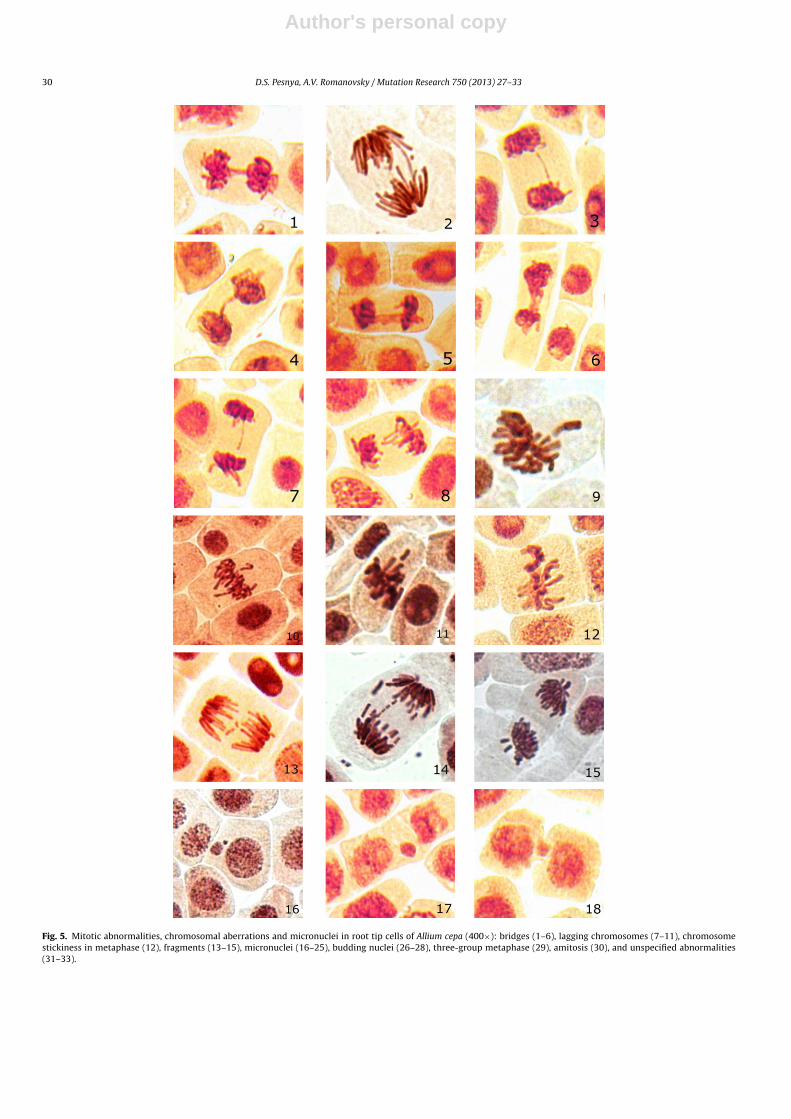
Fig. 3. Two giant polyploid prophases and normal cells in root meristem of Alliumcepa (300×) after 9 h of exposure to mobile phone radiation.
RF-EMF of the GSM 900 mobile phone significantly increasedthe number of dividing cells in root meristems when A. cepa bulbswere exposed for 3 and 9 h (Table 2). The same increase of MIwas registered when bulbs were exposed for 20 min to �-particlesfrom plutonium-239. In the sham-exposed group, no changes of
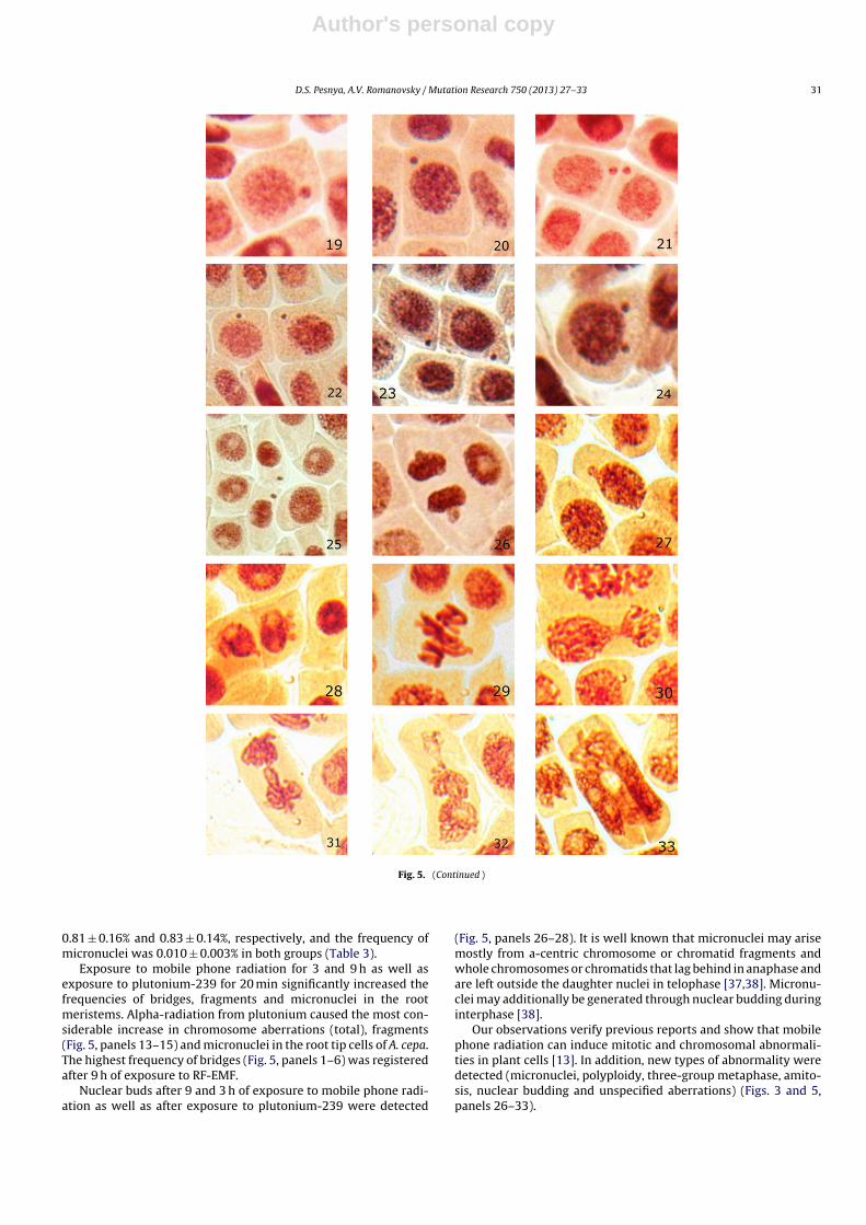
Fig. 4. Micronuclei in root-tip cells of Allium cepa (400×) after 9 h of exposure tomobile phone radiation.
mitotic activity were detected (Table 2). Thus, both ionizing alpha-radiation and non-ionizing radiation from a mobile phone showedstimulating activity on cell division. However, when MI is signifi-cantly higher than the control level it can be harmful for the cells,leading to a disordered cell proliferation and even to malignanttransformation [26]. Therefore, it is not clear whether the increasedMI should be considered as a beneficial or a detrimental effect.
Radio-stimulation of plants by low and moderate doses of ion-izing radiation was described by Timofeeff-Ressovsky and others[32–34]. Stimulation of cell-proliferation intensity and growth ofplant shoots by ionizing radiation have been shown against a back-ground of increased levels of cytogenetic damage [33,34].
3.2. Mitotic abnormalities
We found lagging chromosomes and chromosome stickinesswith total frequencies of 0.06 ± 0.01% and 0.05 ± 0.01% in con-trol and sham-exposed root-tip cells, respectively. Alpha-radiationfrom plutonium-239 significantly increased the frequency ofmitotic disturbances in comparison with those observed innegative-control and sham-exposed groups. Significant increasein the frequency of mitotic abnormalities was observed after 3and 9 h of exposure to mobile phone radiation. The most pro-nounced increase in frequencies of lagging chromosomes (Fig. 5,panels 7–11) and stickiness (Fig. 5, panel 12) was registered after9 h of exposure. Mobile phone GSM 900 radiation induced a signif-icantly higher frequency of stickiness, lagging chromosomes andtotal mitotic abnormalities in comparison with the positive control(Table 2). Mitotic abnormalities are usually associated with spin-dle disturbances [13,35]. These data may indicate that the mobilephone GSM 900 radiation can impair formation of spindle micro-tubules [36]. In addition, after 9 h of exposure to mobile phoneradiation we observed giant polyploid cells (Fig. 3), three-groupmetaphase (Fig. 5, panel 29), amitosis (Fig. 5, panel 30) and unspec-ified abnormalities (Fig. 5, panels 31–33) not observed in the otherexperimental groups.
3.3. Chromosomal abnormalities and micronuclei
The frequencies of chromosome aberrations (bridges and frag-ments) in the negative control and in the sham-exposed group were
Author's personal copy
30 D.S. Pesnya, A.V. Romanovsky / Mutation Research 750 (2013) 27– 33
Fig. 5. Mitotic abnormalities, chromosomal aberrations and micronuclei in root tip cells of Allium cepa (400×): bridges (1–6), lagging chromosomes (7–11), chromosomestickiness in metaphase (12), fragments (13–15), micronuclei (16–25), budding nuclei (26–28), three-group metaphase (29), amitosis (30), and unspecified abnormalities(31–33).
Author's personal copy
D.S. Pesnya, A.V. Romanovsky / Mutation Research 750 (2013) 27– 33 31
Fig. 5. (Continued )
0.81 ± 0.16% and 0.83 ± 0.14%, respectively, and the frequency ofmicronuclei was 0.010 ± 0.003% in both groups (Table 3).
Exposure to mobile phone radiation for 3 and 9 h as well asexposure to plutonium-239 for 20 min significantly increased thefrequencies of bridges, fragments and micronuclei in the rootmeristems. Alpha-radiation from plutonium caused the most con-siderable increase in chromosome aberrations (total), fragments(Fig. 5, panels 13–15) and micronuclei in the root tip cells of A. cepa.The highest frequency of bridges (Fig. 5, panels 1–6) was registeredafter 9 h of exposure to RF-EMF.
Nuclear buds after 9 and 3 h of exposure to mobile phone radi-ation as well as after exposure to plutonium-239 were detected
(Fig. 5, panels 26–28). It is well known that micronuclei may arisemostly from a-centric chromosome or chromatid fragments andwhole chromosomes or chromatids that lag behind in anaphase andare left outside the daughter nuclei in telophase [37,38]. Micronu-clei may additionally be generated through nuclear budding duringinterphase [38].
Our observations verify previous reports and show that mobilephone radiation can induce mitotic and chromosomal abnormali-ties in plant cells [13]. In addition, new types of abnormality weredetected (micronuclei, polyploidy, three-group metaphase, amito-sis, nuclear budding and unspecified aberrations) (Figs. 3 and 5,panels 26–33).
Author's personal copy
32 D.S. Pesnya, A.V. Romanovsky / Mutation Research 750 (2013) 27– 33
4. Conclusions
The mitotic index was significantly increased after 3 and 9 h ofexposure to modulated radiation from a mobile phone, as well asafter exposure to alpha-radiation from plutonium-239.
Similar to alpha-radiation from plutonium-239, mobile phoneGSM 900 radiation showed both clastogenic and aneugenic activity.However, the aneugenic activity of mobile phone radiation is morepronounced, as is evident from the higher frequency of mitoticabnormalities. On the other hand, alpha-radiation from plutonium-239 represents more pronounced clastogenic activity, due to thehigher frequency of chromosomal aberrations.
All investigated effects after 9 h of exposure to the mobilephone GSM 900 radiation were significantly higher than those after3 h of exposure. Consequently, mobile phone GSM 900 radiationincreased MI, the frequency of mitotic and chromosome abnormal-ities, and micronucleus formation in a time-dependent manner.
Polyploid cells, three-group metaphases, amitosis and someunspecified abnormalities not registered in the other experimen-tal groups were found after 9 h of exposure to mobile phoneradiation.
In view of its overall sensitivity, the A. cepa test can be recom-mended as a useful cytogenetic assay to study the genotoxic effectsof RF-EMF.
Conflicts of interests
None.
Acknowledgements
The authors would like to express their gratitude to Dr.Igor Belyaev who kindly corrected the manuscript, Dr. Inna M.Prokhorova, Vladimir E. Serednyakov, Dr. Grigory M. Chuiko,Tatiana S. Pesnya, Dr. Sergey A. Pesnya, Dr. Yaroslav V. Stroynov,and Dr. Roman Fedorov for their helpful advice and two anonymousreviewers, whose comments improved the manuscript.
Appendix B. Supplementary data
Supplementary data associated with this article can befound, in the online version, at http://dx.doi.org/10.1016/j.mrgentox.2012.08.010.
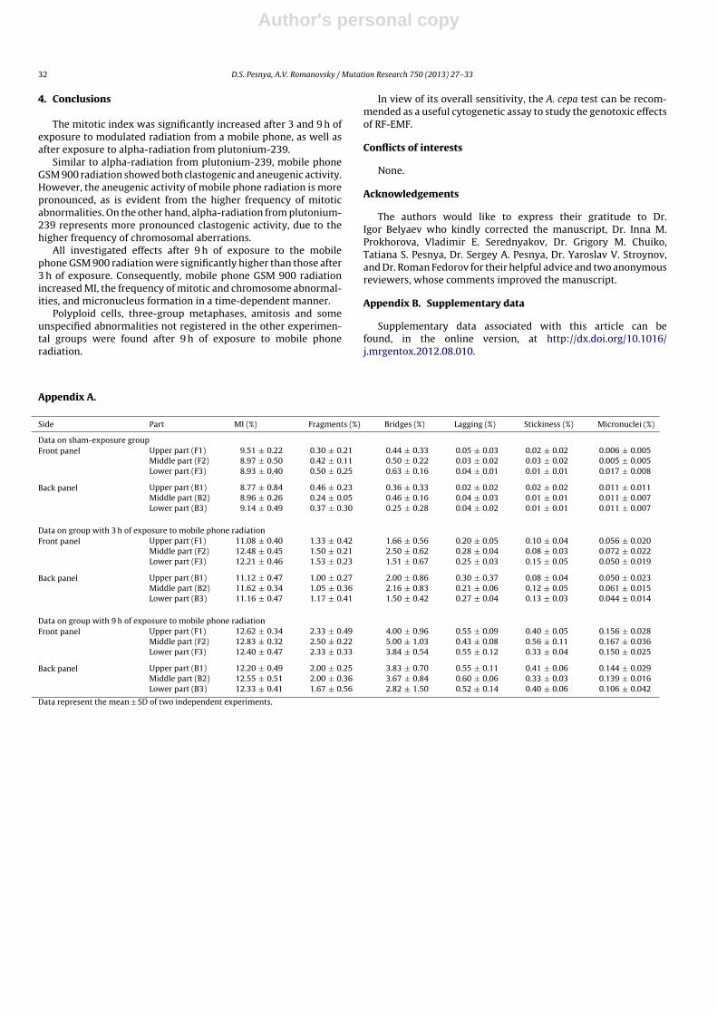
Appendix A.
Side Part MI (%) Fragments (%) Bridges (%) Lagging (%) Stickiness (%) Micronuclei (%)
Data on sham-exposure groupFront panel Upper part (F1) 9.51 ± 0.22 0.30 ± 0.21 0.44 ± 0.33 0.05 ± 0.03 0.02 ± 0.02 0.006 ± 0.005
Middle part (F2) 8.97 ± 0.50 0.42 ± 0.11 0.50 ± 0.22 0.03 ± 0.02 0.03 ± 0.02 0.005 ± 0.005Lower part (F3) 8.93 ± 0.40 0.50 ± 0.25 0.63 ± 0.16 0.04 ± 0.01 0.01 ± 0.01 0.017 ± 0.008
Back panel Upper part (B1) 8.77 ± 0.84 0.46 ± 0.23 0.36 ± 0.33 0.02 ± 0.02 0.02 ± 0.02 0.011 ± 0.011Middle part (B2) 8.96 ± 0.26 0.24 ± 0.05 0.46 ± 0.16 0.04 ± 0.03 0.01 ± 0.01 0.011 ± 0.007Lower part (B3) 9.14 ± 0.49 0.37 ± 0.30 0.25 ± 0.28 0.04 ± 0.02 0.01 ± 0.01 0.011 ± 0.007
Data on group with 3 h of exposure to mobile phone radiationFront panel Upper part (F1) 11.08 ± 0.40 1.33 ± 0.42 1.66 ± 0.56 0.20 ± 0.05 0.10 ± 0.04 0.056 ± 0.020
Middle part (F2) 12.48 ± 0.45 1.50 ± 0.21 2.50 ± 0.62 0.28 ± 0.04 0.08 ± 0.03 0.072 ± 0.022Lower part (F3) 12.21 ± 0.46 1.53 ± 0.23 1.51 ± 0.67 0.25 ± 0.03 0.15 ± 0.05 0.050 ± 0.019
Back panel Upper part (B1) 11.12 ± 0.47 1.00 ± 0.27 2.00 ± 0.86 0.30 ± 0.37 0.08 ± 0.04 0.050 ± 0.023Middle part (B2) 11.62 ± 0.34 1.05 ± 0.36 2.16 ± 0.83 0.21 ± 0.06 0.12 ± 0.05 0.061 ± 0.015Lower part (B3) 11.16 ± 0.47 1.17 ± 0.41 1.50 ± 0.42 0.27 ± 0.04 0.13 ± 0.03 0.044 ± 0.014
Data on group with 9 h of exposure to mobile phone radiationFront panel Upper part (F1) 12.62 ± 0.34 2.33 ± 0.49 4.00 ± 0.96 0.55 ± 0.09 0.40 ± 0.05 0.156 ± 0.028
Middle part (F2) 12.83 ± 0.32 2.50 ± 0.22 5.00 ± 1.03 0.43 ± 0.08 0.56 ± 0.11 0.167 ± 0.036Lower part (F3) 12.40 ± 0.47 2.33 ± 0.33 3.84 ± 0.54 0.55 ± 0.12 0.33 ± 0.04 0.150 ± 0.025
Back panel Upper part (B1) 12.20 ± 0.49 2.00 ± 0.25 3.83 ± 0.70 0.55 ± 0.11 0.41 ± 0.06 0.144 ± 0.029Middle part (B2) 12.55 ± 0.51 2.00 ± 0.36 3.67 ± 0.84 0.60 ± 0.06 0.33 ± 0.03 0.139 ± 0.016Lower part (B3) 12.33 ± 0.41 1.67 ± 0.56 2.82 ± 1.50 0.52 ± 0.14 0.40 ± 0.06 0.106 ± 0.042
Data represent the mean ± SD of two independent experiments.
Author's personal copy
D.S. Pesnya, A.V. Romanovsky / Mutation Research 750 (2013) 27– 33 33
References
[1] E. Diem, C. Schwarz, F. Adlkofer, O. Jahn, H. Rüdiger, Non-thermal DNA breakageby mobile-phone radiation in human fibroblasts and in GFSH-R17 rat granulosacells in vitro, Mutat. Res. 2 (2005) 178–183 (see also: Letter of Concern, Mutat.Res. 695 (2010) 1).
[2] A.S. Yadav, M.K. Sharma, Increased frequency of micronucleated exfoliated cellsamong humans exposed in vivo to mobile telephone radiations, Mutat. Res. 650(2) (2008) 175–180.
[3] A. Agarwal, N.R. Desai, K. Makker, A. Varghese, R. Mouradi, E. Sabanegh, R.Sharma, Effects of radiofrequency electromagnetic waves (RF-EMW) from cel-lular phones on human ejaculated semen: an in vitro pilot study, Fertil. Steril.92 (4) (2009) 1318–1325.
[4] I. Belyaev, E. Markova, L. Malmgren, Microwaves from mobile phones inhibit53BP1 focus formation in human stem cells stronger than in differentiatedcells: possible mechanistic link to cancer risk, Environ. Health Perspect. 118(3) (2010) 394–399.
[5] C. Avendano, A. Mata, C.A. Sanchez Sarmiento, G.F. Doncel, Use of laptop com-puters connected to internet through Wi-Fi decreases human sperm motilityand increases sperm DNA fragmentation, Fertil. Steril. 97 (1) (2012) 39–45.
[6] V.G. Khurana, C. Teo, M. Kundi, L. Hardell, M. Carlberg, Cell phones and braintumors: a review including the long-term epidemiologic data, Surg. Neurol. 72(2009) 205–215.
[7] L. Hardell, M. Carlberg, K. Hansson Mild, Pooled analysis of case–controlstudies on malignant brain tumours and the use of mobile and cordlessphones including living and deceased subjects, Int. J. Oncol. 38 (5) (2011)1465–1474.
[8] E. Cardis, B.K. Armstrong, J.D. Bowman, G.G. Giles, M. Hours, D. Krewski, M.McBride, M.E. Parent, S. Sadetzki, A. Woodward, J. Brown, A. Chetrit, J. Figuerola,C. Hoffmann, A. Jarus-Hakak, L. Montestruq, L. Nadon, L. Richardson, R. Villegas,M. Vrijheid, Risk of brain tumours in relation to estimated RF dose from mobilephones: results from five Interphone countries, Occup. Environ. Med. 68 (9)(2011) 631–640.
[9] R. Baan, Y. Grosse, B. Lauby-Secretan, F. El Ghissassi, V. Bouvard, L. Benbrahim-Tallaa, N. Guha, F. Islami, L. Galichet, K. Straif, Carcinogenicity of radiofrequencyelectromagnetic fields, Lancet Oncol. 12 (7) (2011) 624–626.
[10] I. Belyaev, Toxicity and SOS response to ELF magnetic field and nalidixic acidin E. coli cells, Mutat. Res. 722 (1) (2011) 56–61.
[11] E.I. Sarapultseva, J.V. Igolkina, A.V. Litovchenko, Evaluation of the maximumpermissible level of low-intensity electromagnetic radiation at mobile connec-tion frequency (1 GHz) by changes in motor activity of Spirostomum ambiguum,Bull. Exp. Biol. Med. 147 (4) (2009) 431–433.
[12] V.P. Sharma, H.P. Singh, R.K. Kohli, D.R. Batish, Mobile phone radiation inhibitsVigna radiata root growth by inducing oxidative stress, Sci. Total Environ. 21(2009) 5543–5547.
[13] M. Tkalec, K. Malaric, M. Pavlica, B. Pevalek-Kozlina, Z. Vidakovic-Cifrek, Effectsof radiofrequency electromagnetic fields on germination and root meristem ofAllium cepa L., Mutat. Res. 672 (2) (2009) 76–81.
[14] D.J. Panagopoulos, L.H. Margaritis, The effect of exposure duration on the bio-logical activity of mobile telephony radiation, Mutat. Res. 699 (1–2) (2010)17–22.
[15] E.D. Chavdoula, D.J. Panagopoulos, L.H. Margaritis, Comparison of biologi-cal effects between continuous and intermittent exposure to GSM-900-MHzmobile phone radiation: detection of apoptotic cell-death features, Mutat. Res.700 (1–2) (2010) 51–61.
[16] A. Balmori, Mobile phone mast effects on common frog (Rana temporaria) tad-poles: the city turned into a laboratory, Electromagn. Biol. Med. 29 (1–2) (2010)31–35.
[17] A. Balmori, Possible effects of electromagnetic fields from phone masts on apopulation of white stork (Ciconia ciconia), Electromagn. Biol. Med. 24 (2005)109–119.
[18] A.R. Ferreira, T. Knakievicz, M.A. Pasquali, D.P. Gelain, F. Dal-Pizzol,C.E. Fernandez, A.A. de Salles, H.B. Ferreira, J.C. Moreira, Ultra high
frequency-electromagnetic field irradiation during pregnancy leads to anincrease in erythrocytes micronuclei incidence in rat offspring, Life Sci. 80 (1)(2006) 43–50.
[19] S. Dasdag, M.Z. Akdag, E. Ulukaya, A.K. Uzunlar, A.R. Ocak, Effect of mobile phoneexposure on apoptotic glial cells and status of oxidative stress in rat brain,Electromagn. Biol. Med. 28 (4) (2009) 342–354.
[20] M. Ballardin, I. Tusa, N. Fontana, A. Monorchio, C. Pelletti, A. Rogovich, R. Barale,R. Scarpato, Non-thermal effects of 2.45 GHz microwaves on spindle assembly,mitotic cells and viability of Chinese hamster V-79 cells, Mutat. Res. 716 (2011)1–9.
[21] E. Karaca, B. Durmaz, H. Altug, T. Yildiz, C. Guducu, M. Irgi, M.G. Koksal, F. Ozk-inay, C. Gunduz, O. Cogulu, The genotoxic effect of radiofrequency waves onmouse brain, J. Neurooncol. 106 (1) (2012) 53–58.
[22] S. Franzellitti, P. Valbonesi, N. Ciancaglini, C. Biondi, A. Contin, F. Bersani, E. Fab-bri, Transient DNA damage induced by high-frequency electromagnetic fields(GSM 1.8 GHz) in the human trophoblast HTR-8/SVneo cell line evaluated withthe alkaline comet assay, Mutat. Res. 683 (2010) 35–42.
[23] H.W. Ruediger, Genotoxic effects of RF electromagnetic fields, Pathophys 16(2–3) (2009) 89–102.
[24] J.L. Phillips, N.P. Singh, H. Lai, Electromagnetic fields and DNA damage, Patho-physiology 16 (2009) 79–88.
[25] C. Zhijian, L. Xiaoxue, L. Yezhen, C. Shijie, J. Lifen, L. Jianlin, L. Deqiang, H. Jiliang,Impact of 1.8-GHz radiofrequency radiation (RFR) on DNA damage and repairinduced by doxorubicin in human B-cell lymphoblastoid cells, Mutat. Res. 695(1–2) (2010) 16–21.
[26] D.M. Leme, M.A. Marine-Morels, Allium cepa test in environmental monitoring:a review on its applications, Mutat. Res. 682 (2009) 71–81.
[27] M. Saghirzadeh, M.R. Gharaati, Sh. Mohammadi, M. Ghiassi-Nejad, Evaluationof DNA damage in the root cells of Allium cepa seeds growing in soil of highbackground radiation areas of Ramsar – Iran, J. Environ. Radioact. 99 (2008)1698–1702.
[28] M.J. Constantin, E.T. Owens, Introduction and perspectives of plant genetic andcytogenetic assays a report of the U.S. environmental protection agency Gene-Tox program, Mutat. Res. 99 (1982) 1–12.
[29] G. Fiskesjo, The Allium test as a standard in environmental monitoring, Hered-itas 102 (1985) 99–112.
[30] A. Barbério, J.C. Voltolini, M.L.S. Mello, Standardization of bulb and root samplesizes for the Allium cepa test, Ecotoxicology 2 (2011) 927–935.
[31] Belyaev I., Dependence of non-thermal biological effects of microwaves onphysical and biological variables: implications for reproducibility and safetystandards. An ICEMS Monograph. L. Giuliani and M. Soffritti, Non-thermaleffects and mechanisms of interaction between electromagnetic fields and liv-ing matter, Mattioli 1885 (ISBN 978-88-6261-166-4, 403 pp.): 2010, vol. 5, pp.187-217.
[32] N.W. Timofeeff-Ressovsky, N.V. Luchnik, Radiation stimulation of plantsand its possible theoretical interpretation, Radiobiologiya (1958) 258–266(in Russian).
[33] D. Marciulioniene, B. Luksiene, D. Kiponas, G. Maksimov, J. Darginaviciene, V.Gaveliene, Effects of 137Cs and 90Sr on the plant Lepidium sativum L. growthpeculiarities, Ecologija 53 (1) (2007) 65–70.
[34] N.G. Shestopalova, E.Yu. Baeva, Radioadaptive response of leaf meristem cellsof inbred and heterosis Helianthus annuus L. plants during ontogenesis, Tsitol.Genet. 41 (6) (2007) 44–49.
[35] I.M. Prokhorova, M.I. Kovaleva, A.N. Fomicheva, O.V. Babanazarova, Spatial andtemporal dynamics of mutagenic activity of water in lake Nero, Inl. Wa. Bio. 2(2008) 17–25 (in Russian).
[36] M. Cifra, J.Z. Fields, A. Farhadi, Electromagnetic cellular interactions, Prog. Bio-phys. Mol. Biol. 105 (3) (2011) 223–246.
[37] M. Fenech, The in vitro micronucleus technique, Mutat. Res. 455 (2000)81–95.
[38] H.K. Lindberg, X. Wang, H. Jarventaus, G.C.-M. Falcka, Ha. Norppa, M. Fenech,Origin of nuclear buds and micronuclei in normal and folate-deprived humanlymphocytes, Mutat. Res. 617 (2007) 33–45.