Melatonin ameliorates ionizing radiation-induced oxidative organ damage in rats
Upload
khangminh22Category
view
3download
0
HAL Id: hal-00615361https://hal.archives-ouvertes.fr/hal-00615361
Submitted on 19 Aug 2011
HAL is a multi-disciplinary open accessarchive for the deposit and dissemination of sci-entific research documents, whether they are pub-lished or not. The documents may come fromteaching and research institutions in France orabroad, or from public or private research centers.
L’archive ouverte pluridisciplinaire HAL, estdestinée au dépôt et à la diffusion de documentsscientifiques de niveau recherche, publiés ou non,émanant des établissements d’enseignement et derecherche français ou étrangers, des laboratoirespublics ou privés.
Ionizing radiation or mitomycin-induced micronuclei inlymphocytes of or mutation carriers
Sara Gutiérrez-Enríquez, Teresa Ramón y Cajal, Carmen Alonso, AnnaCorral, Pablo Carrasco, Mónica Cornet, Judith Sanz, Montserrat Ribas,
Montserrat Baiget, Orland Diez
To cite this version:Sara Gutiérrez-Enríquez, Teresa Ramón y Cajal, Carmen Alonso, Anna Corral, Pablo Carrasco, et al..Ionizing radiation or mitomycin-induced micronuclei in lymphocytes of or mutation carriers. BreastCancer Research and Treatment, Springer Verlag, 2010, 127 (3), pp.611-622. �10.1007/s10549-010-1017-6�. �hal-00615361�
1
25 / June / 2010
IONIZING RADIATION OR MITOMYCIN INDUCED-MICRONUCLEI IN
LYMPHOCYTES OF BRCA1 OR BRCA2 MUTATION CARRIERS
Sara Gutiérrez-Enríquez1,2
, Teresa Ramón y Cajal3, Carmen Alonso
3, Anna
Corral4, Pablo Carrasco
5, Mónica Cornet
6, Judith Sanz
3, Montserrat Ribas
5,
Montserrat Baiget6, Orland Diez
1,7
1Vall d’Hebron Institute of Oncology (VHIO), Barcelona, Spain
2Medical Oncology Program, Vall d'Hebron University Hospital Research Institute,
Barcelona, Spain
3Medical Oncology Department, Santa Creu and Sant Pau Hospital, Barcelona, Spain
4Group of Mutagenesis, Genetic and Microbiology Department, Autonomous
University of Barcelona, Cerdanyola del Vallès, Spain
5Radiophysics and Radioprotection Department, Santa Creu and Sant Pau Hospital,
Barcelona, Spain
6Genetics Department, Santa Creu and Sant Pau Hospital, Barcelona, Spain
7Oncogenetics Laboratory, University Hospital Vall d’Hebron, Barcelona, Spain
Correspondence author: Sara Gutiérrez-Enríquez, PhD Oncogenetics Laboratory, Clinical Laboratories Vall d'Hebron University Hospital
Passeig Vall d'Hebron 119-129, Barcelona 08035 Telf: + 34 93 489 30 00 / 93 274 60 00 ext. 4826, [email protected]
2
ABSTRACT
BRCA1 and BRCA2 genes are essential in preserving the integrity of genome,
and it is not unambiguously clear whether the heterozygosity status may affect BRCA1
or BRCA2 functions. This may have implications for the clinical management of
BRCA1 and BRCA2 mutation carriers both in breast cancer (BC) screening modality and
in cancer treatment based on DNA-damaging or DNA-repair-inhibiting drugs. We
investigated whether lymphocytes carrying BRCA1 or BRCA2 mutations displayed an
increased sensitivity to radiation or mitomycin C in vitro treatments.
Peripheral blood from 21 BRCA1 mutation carriers (12 with BC and 9 healthy),
24 BRCA2 carriers (13 with BC and 11 healthy), 15 familial BC patients without
detected mutation in BRCA1 or BRCA2, and 16 controls without familial history of
cancer (5 with BC and 11 healthy) were irradiated or treated with mitomycin C (MMC).
Chromosomal damage was measured using the cytokinesis-block micronucleus assay.
We evaluated micronuclei (MN) and nucleoplasmic bridges (NPB).
The BRCA2 mutation carriers and familial BC patients without detected
mutation in BRCA1 or BRCA2 showed less basal NPB than BRCA1 carriers and
controls. The BRCA1+/- or BRCA2+/-
lymphocytes did not have increased frequencies of
MN or NPB after irradiation. In contrast, BRCA2+/- lymphocytes presented higher levels
of MN after MMC exposure than BRCA1 carriers and controls.
The monoallelic BRCA1 or BRCA2 pathogenic mutations seem not to be
associated with an enhanced radiosensitivity. The mutation of one BRCA2 allele
conferred an increased sensitivity to MMC, presumably because of the role of this gene
in the repair of MMC-induced DNA damage. This finding indicates that the MMC-
induced MN analysis could be useful in identifying functional deficiencies of BRCA2 or
genes related to BRCA2. Since MMC can be used as an anti-cancer drug, these data may
be relevant for the management and follow-up of BRCA2 mutation carriers.
Keywords: in vitro radiation, mitomycin c, micronuclei, BRCA1 and BRCA2.
3
INTRODUCTION
Heterozygous germline mutations in BRCA1 or BRCA2 tumour suppressor genes
(BRCA1/2) confer susceptibility to breast (BC) and ovarian cancers (OC) with high
penetrance, and account for approximately 20% of BC cases associated with family
history [reviewed in ref. 1].
BRCA1/2 genes are important in the maintenance of genome stability. The main
role of BRCA2 appears to involve regulation of the RAD51 function in DNA repair by
homologous recombination. BRCA1 is an E3 ubiquitin ligase that has a broader role
upstream of BRCA2, participating in DNA repair, transcriptional regulation, cell cycle
progression and meiotic sex chromosome inactivation [reviewed in refs. 2,3]. BRCA1-/-
or BRCA2 -/-
cells from chicken DT40 knockout cell lines, knockout mice and tumour-
derived human cell lines exhibit spontaneous chromosomal instability and are
hypersensitive to DNA-damaging agents such as ionising radiation (IR) and DNA cross-
linking agents [2,3].
In contrast, whether some functions of the BRCA1/2 proteins are diminished or
deregulated while still heterozygous or only after the loss of both functional alleles is
not yet well established [4,5]. The heterozygous status (BRCA1+/- or BRCA2+/-
) might
contribute to the impairment of genomic stability and increase the risk of cancer
promoting mutations, contributing to the loss of the remaining wild-type BRCA1/2
allele [4-6]. Since BRCA1 and BRCA2 proteins are involved in DNA repair, BRCA1+/-
or BRCA2+/- cells of mutation carriers may also have enhanced sensitivity to DNA-
damaging agents, such as ionising radiation (IR) or mitomycin C (MMC); this
phenotype may increase toxicity and the carcinogenic risk of chemotherapy,
radiotherapy and mammography screening in BRCA1 or BRCA2 mutation carriers [4,5].
If the functions of the BRCA1/2 proteins are affected by heterozygosity status, two
mechanisms may explain this phenotype: haploinsufficiency or reduction in gene
dosage, and/or dominant negative mutations. Several studies have claimed the existence
of phenotypes associated with BRCA1/2 haploinsufficiency, whereas others have raised
the possibility that BRCA1 or BRCA2 mutated proteins may block the function of the
remaining wild-type BRCA1/2 allele [reviewed in ref. 4].
Several studies in blood lymphocytes to know whether the heterozygosity for
BRCA1/2 mutations confers detectable sensitivity to genotoxic agents have been
performed with contradictory results [reviewed in ref. 7]. Here, we have studied
chromosomal sensitivity to ionising radiation or MMC treatments of peripheral blood
lymphocytes from BRCA1 or BRCA2 germ-line mutation carriers with or without BC,
familial BC patients without detected mutation in BRCA1/2, and controls with or
without BC and no familial history of cancer. The chromosomal damage was measured
by quantification of micronuclei (MN) and nucleoplasmic bridges (NPBs) using the
cytokinesis-block micronucleus assay (CBMN). The in vitro proliferation capacity of
the treated lymphocytes was also evaluated by the nuclear division index (NDI).
The MN in dividing cells are the result of fragments or whole acentric
chromosomes/chromatids that lag behind in anaphase and are not included in the
daughter nuclei in telophase [8]. These fragments can be originated from non-repaired
or misrepaired DNA double-strand breaks (DSBs) [8]. Misrepair of DSBs could also
lead to the formation of asymmetrical chromosome rearrangements producing dicentrics
or ring chromosomes and acentric fragments. The centromeres of the dicentric
chromosomes, dicentric ring chromosomes or concatenated double rings can be pulled
to opposite poles of the cells at anaphase resulting in the formation of a nucleoplasmic
bridges (NPB) between the daughter nuclei [9]. Therefore, the presence of MN or NPB
could indirectly reflect the cell DNA repair capacity [8]. In fact, increased frequencies
of spontaneous and mutagen induced MN or NPBs have been observed in different cells
4
deficient in DNA repair mechanisms such as non-homologous DNA end-joining [10] or
homologous recombination [11,12].
The cytokinesis-block micronucleus (CBMN) assay is the preferred method for
measuring MN in cultured human cells because scoring is specifically restricted to
once-divided binucleated (BN) cells, which are the cells that can show MN [8]. The
inhibition of cytokinesis by cytochalasin B allows to discriminate between cells that did
not divide after treatment and cells that did divide, thus preventing the confounding
effects caused by differences in cell division kinetics [8]. Because cells are blocked in
the BN stage, it is also possible to measure nucleoplasmic bridges [9]. In addition to
micronuclei in binucleated cells and NPB, the CBMN assay also allows for scoring of
other critical events, such as cell division and cell cytotoxicity by scoring of proportion
of mono-, bi- and multicucleated cells to calculate the nuclear division index (NDI) [8].
To date the CBMN assay is one of the best-validated methods for measuring
chromosome damage in human lymphocytes [13].
5
MATERIALS AND METHODS
Subjects and study design
Heparinized blood samples were collected from 76 women between 2004 to
2007: 60 with a family history of BC and OC and 16 without a family history of cancer.
The ethical committee of the hospital approved the study and all women participating
signed an informed consent.
Women with a family history of cancer were recruited from those attending the
Cancer Genetics Clinic of Hospital de la Santa Creu i Sant Pau, Barcelona, Spain.
Subjects were referred for genetic assessment because of familial BC/OC, early onset
BC or previous identified BRCA1/2 mutation in the family. The mutation analysis of the
BRCA1 and BRCA2 genes was performed in women fulfilling one of the following
criteria: families with one or more cases of BC (at least 1 case diagnosed before age 50)
and 1 or more cases of OC; site-specific female BC families with 2 or more cases (at
least 1 diagnosed at 50 years of age); families with at least 1 case of BC or OC in
addition to at least 1 male case of BC.
The entire coding sequence of BRCA1 and BRCA2 was analyzed for sequence
variants using denaturing high-performance liquid chromatography (DHPLC) and for
large deletions and duplications using Multiplex Ligation-dependent Probe
Amplification (MLPA) (MRC Holland). In those women referred due to a BRCA1/2
mutation previously identified in the family, the analysis was performed by direct
sequencing or MLPA.
The mutational status of the 60 individuals with family history was: 21 BRCA1
mutation carriers (12 with BC and 9 healthy), 24 BRCA2 mutation carriers (13 with BC
and 11 healthy) and 15 females with familial BC/OC without detected mutation in
BRCA1/2 (14 with BC and 1 with OC). The BRCA1/2 mutations are shown in Table 1.
As controls, blood samples were also collected from 16 women without family
history of BC/OC: five with sporadic BC and 11 healthy women. The sporadic BC
patients came from those attending the Medical Oncology Department and the healthy
subjects were recruited from the laboratory staff of the Genetics Department of the
Hospital.
Considering the disease status of the analysed subjects, 31 were healthy and 45
had BC/OC. Most of the BC/OC patients (n=31) had received different combinations of
adjuvant chemotherapy: cyclophosphamide, methotrexate and 5-fluorouracil (CMF)
(n=7); 5-fluorouracil, epirubicin and cyclophosphamide (FEC) (n=6); adriamycin and
cyclophosphamide (n=3); FEC in a high-dose chemotherapy regimen (with autologous
transplantation) (n=2); FEC, trastuzumab and taxanes (n=2); CMF, adriamycin, taxol
and capecitabine (n=2). The rest of patients (n=9) received individually different
regimens each: FEC, taxanes and carboplatin; cyclophosphamide, adriamycin and taxol;
FAC (5-fluorouracil, adriamycin and cyclophosphamide); FEC and taxanes; FAC and
taxanes; FEC and CMF; CMF, MMC and vinblastine; anthracyclines;
cyclophosphamide. Four patients required neo-adjuvant chemotherapy (2 patients with
CMF, 1 with FEC and 1 with epirubicin and doxorubicin). Three patients required
chemotherapy because of BC recurrence (one patient was treated with CMF, one with
CMF, MMC and vinblastine, and one with MMC and vinblastine).
Culture and treatment with mutagenic agents
Peripheral blood cultures were set up by adding 0.5 mL of whole blood to 4.5
mL of RPMI 1640 medium (PAA Laboratories GmbH, Pasching, Austria)
supplemented with 15% heat-inactivated foetal calf serum (PAA Laboratories GmbH),
1% antibiotics (penicillin and streptomycin), 1% L-glutamine (provided by Gibco,
Invitrogen Corporation, Paisley, United Kingdom) and 1% phytohaemagglutinin (PHA)

6
to stimulate the division of T-lymphocytes (Gibco). Six cultures were set up for each
subject immediately after drawing blood: 2 that will be treated in the G0 phase with 2
Gy of gamma irradiation (before incubation at 37ºC with PHA), 2 that will be treated in
the G1/S phase with 0.05 g/ml of MMC (24h after the beginning of the culture with
PHA) and 2 replicates without any treatment.
Irradiation of the two respective cultures was done just after re-suspending the
blood with the supplemented RPMI 1640 medium. All irradiations were performed at
room temperature with 2 Gy of gamma irradiation from a source of Co-60 (Theratron
780 radiotherapy unit, Theratronics Ltd., Canada) at a high dose rate (HDR) of 0.6 -0.8
Gy/min. To minimize the dose gradient within the tubes, the cultures were irradiated at
a large distance (80 cm from the cobalt source). A polymethyl methacrylate (PMMA)
scattering slab was placed on the irradiated tubes to minimize the build-up effect and to
guarantee uniform irradiation.
Immediately after irradiation the 6 cultures per individual were incubated at
37ºC. After 24h of incubation and stimulation with PHA, mitomycin-C (Sigma, St.
Louis, Missouri, USA) was added to the 2 corresponding cultures to obtain a final
concentration of 0.05 µg/mL and cultures were returned to the incubator.
After 44 h of incubation with PHA at 37ºC, cytochalasin-B (Cyt-B, Sigma, St.
Louis, Missouri, USA) was added to the 6 cultures to a final concentration of 6 µg/mL
to arrest cytokinesis. Seventy-two hours after incubation with PHA at 37ºC the cells
were centrifuged (800 rpm for 8 min at room temperature), the supernatant was
aspirated, and the cells were re-suspended in a hypotonic solution (0.075 M KCl) for 3
min at 4°C. The cells were re-centrifuged, and a 3:1 (v/v) methanol:acetic acid solution
gently added. This fixation step was repeated twice and the cells were then re-suspended
in a small volume of fixative solution and dropped onto clean slides. The air-dried slides
were subsequently stained with 1 g/mL of 4’,6-diamidino-2-phenylindole (DAPI;
Sigma, St. Louis, Missouri, USA) and 2 g/mL of propidium iodide (PI; Sigma) in an
antifade solution (Vectashield; Vector Laboratories, Burlingame, CA, USA). The slides
were stored at 4°C until assessed by microscopy. All slides were randomized and coded
before being scored by the same observer to minimize variability. The slides were
scored under the 100x objective of a Nikon Eclipse E400 fluorescence microscope
(Nikon, Tokyo, Japan) equipped with a triple-band-pass filter to visualize the nuclei and
MN, in bright blue (DAPI), and the cytoplasm, in red (PI). The presence of MN was
confirmed using the filter to view only the DAPI stain.
To determine the presence of both micronuclei (MN) and nucleoplasmic bridges
(NPB), a total of 1000 binucleated cells with well-preserved cytoplasm for each subject
(500 per replicate) were blind scored on coded slides according to previously described
criteria [8]. In addition, a total of 500 lymphocytes (250 cells per replicate) were
examined to determine the proportion of cells with different numbers of nuclei (1-4
nuclei) in order to calculate the nuclear division index (NDI) [8].
Statistical methods
In the untreated cultures, the outcome variables analyzed were the basal values
of MN, NPB and NDI. In the treated cultures, the cytogenetic endpoints evaluated were
the number of radiation or mitomycin-C-induced MN (calculated by subtracting the
basal yields from the yields obtained in the treated samples), the absolute values of NPB
after the mutagenic treatments and, the NDI index values in untreated cultures less NDI
values in treated cultures.
The analysis included 2 factors: carrier/non-carrier, with 4 levels (BRCA1
carrier, BRCA2 carrier, familial BC without detected mutations in BRCA1/2, and
controls without familial history of BC/OC) and, disease with 2 levels (healthy women
or cancer patients groups). Potential confounding factors of MN were also considered.
7
Age at time of experiments, smoking habit, and number of cigarettes were recorded for
all subjects. Given that the blood samples were not obtained concurrently and that
experimental conditions may have varied between groups, the days elapsed since the
first blood culture were calculated (as a centered variable) for each analyzed subject.
For cancer patients the age at cancer diagnosis and the years elapsed between the end of
radiotherapy and chemotherapy and time of MN cultures were also recorded.
Basic descriptive statistics of outcome and continuous variables were computed
for all subgroups. The means for between-group demographic and clinical variables
were compared using the t-test. To quantify the potential association among continuous
and outcome variables, correlation matrices analyses were performed using the Pearson
correlation coefficient.
The between-group differences in basal and treatment-induced MN, NPB and
NDI were analyzed using generalized linear models (GLZ). A logarithmic link function
was applied to the basal level of MN and NPB to achieve a best fit. The continuous
variables that were shown by the correlation analysis to have a possible effect were
included in the analyses. In the GLZ analysis of NPB after mutagens treatment, the
basal NPB values were considered as a covariable since the treatment-induced NPB
values, obtained by subtracting the basal yields from yields obtained after treatment,
encompassed negative values which would be difficult to adjust in the GLZ analyses. A
backward selection method was used and potential confounding factors that did not
reach statistical significance were not considered in the final model. Post hoc
comparisons using the sequential Bonferroni correction for multiple comparisons were
also carried out.
The figures illustrating the between-group differences in this study show the
estimated marginal means, which correspond to the mean adjusted for the other terms in
the model.
For all analyses, differences were regarded as statistically significant at P < 0.05.
Statistical computations were performed using the SPSS v.15.0 software (SPSS,
Chicago, IL, USA) and the STATISTICA v.8.0 statistical software package (StatSoft,
Tulsa, OK, USA).
8
RESULTS
Demographic, clinical and experimental factors
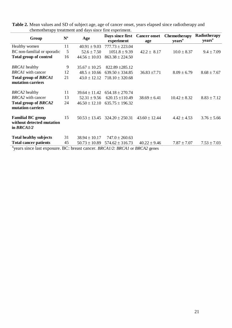
The mean value of age, days elapsed since first experiment as well as the clinical
characteristics of the cancer patient groups are presented in Table 2. The group of all
healthy women was significantly younger than the overall group of cancer patients
(P<0.001). The peripheral blood cultures for cancer patients were established earlier
than the cultures for healthy women and this difference in time, measured as the days
elapsed since the first blood culture, was statistically significant (P<0.05). The blood
cultures of BRCA2 mutation carriers (with and without BC) were set up significantly
earlier than the cultures of control group (BC patients and healthy women without
familial history of BC) (P<0.01). The cultures of familial BC patients negative for
BRCA1/2 mutations were established earlier than the cultures of the all other groups and
this difference in time is statistically significant (P<0.001). The average of the years
elapsed since last chemotherapy and radiotherapy sessions in the group of familial BC
patients that had no detected mutation in BRCA1/2 is lower, although not statistically
significant, than the average of the other BC patient groups.
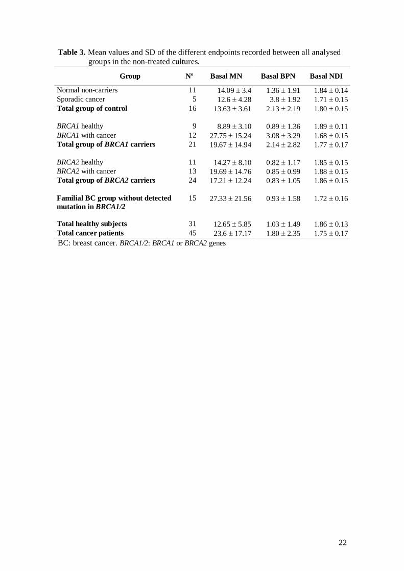
Basal levels of MN, NPB and NDI
Table 3 shows the data (Mean ± SD) for the 3 endpoints evaluated in the
untreated lymphocytes from all groups. The analysis of correlation matrices revealed
higher basal MN levels when less time had elapsed since the last session of either
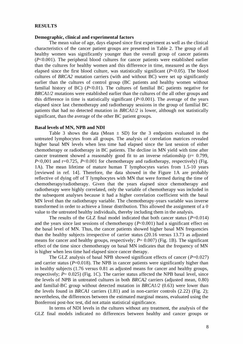
chemotherapy or radiotherapy in BC patients. The decline in MN yield with time after
cancer treatment showed a reasonably good fit to an inverse relationship (r= 0.799,
P<0,001 and r=0.725, P<0.001 for chemotherapy and radiotherapy, respectively) (Fig.
1A). The mean lifetime of mature human T lymphocytes varies from 1.5-10 years
[reviewed in ref. 14]. Therefore, the data showed in the Figure 1A are probably
reflective of dying off of T lymphocytes with MN that were formed during the time of
chemotherapy/radiotherapy. Given that the years elapsed since chemotherapy and
radiotherapy were highly correlated, only the variable of chemotherapy was included in
the subsequent analyses because it had a higher correlation coefficient with the basal
MN level than the radiotherapy variable. The chemotherapy-years variable was inverse
transformed in order to achieve a linear distribution. This allowed the assignment of a 0
value to the untreated healthy individuals, thereby including them in the analysis.
The results of the GLZ final model indicated that both cancer status (P=0.014)
and the years since last sessions of chemotherapy (P<0.001) had a significant effect on
the basal level of MN. Thus, the cancer patients showed higher basal MN frequencies
than the healthy subjects irrespective of carrier status (20.16 versus 13.73 as adjusted
means for cancer and healthy groups, respectively; P= 0.007) (Fig. 1B). The significant
effect of the time since chemotherapy on basal MN indicates that the frequency of MN
is higher when less time had elapsed since cancer therapy.
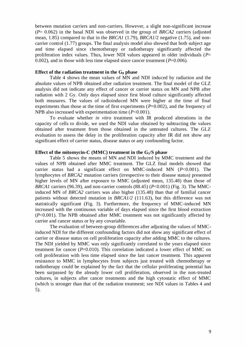
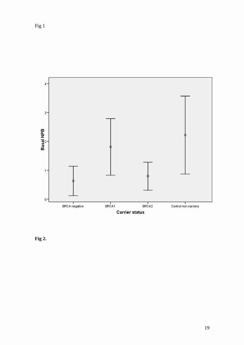
The GLZ analysis of basal NPB showed significant effects of cancer (P=0.027)
and carrier status (P=0.018). The NPB in cancer patients were significantly higher than
in healthy subjects (1.76 versus 0.81 as adjusted means for cancer and healthy groups,
respectively; P= 0.025) (Fig. 1C). The carrier status affected the NPB basal level, since
the levels of NPB in untreated cultures in both BRCA2 carriers (adjusted mean, 0.80)
and familial-BC group without detected mutation in BRCA1/2 (0.63) were lower than
the levels found in BRCA1 carriers (1.81) and in non-carrier controls (2.22) (Fig. 2);
nevertheless, the differences between the estimated marginal means, evaluated using the
Bonferroni post-hoc test, did not attain statistical significance.
In terms of NDI levels in the cultures without any treatment, the analysis of the
GLZ final models indicated no differences between healthy and cancer groups or
9
between mutation carriers and non-carriers. However, a slight non-significant increase
(P= 0.062) in the basal NDI was observed in the group of BRCA2 carriers (adjusted
mean, 1.85) compared to that in the BRCA1 (1.79), BRCA1/2 negative (1.75), and non-
carrier control (1.77) groups. The final analysis model also showed that both subject age
and time elapsed since chemotherapy or radiotherapy significantly affected the
proliferation index values. Thus, lower NDI values appeared in older individuals (P=
0.002), and in those with less time elapsed since cancer treatment (P=0.006).
Effect of the radiation treatment in the G0 phase
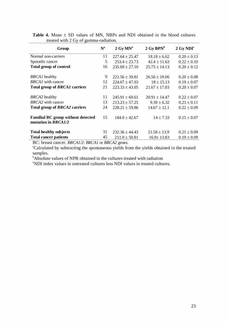
Table 4 shows the mean values of MN and NDI induced by radiation and the
absolute values of NPB obtained after radiation treatment. The final model of the GLZ
analysis did not indicate any effect of cancer or carrier status on MN and NPB after
radiation with 2 Gy. Only days elapsed since first blood culture significantly affected
both measures. The values of radioinduced MN were higher at the time of final
experiments than those at the time of first experiments (P=0.002), and the frequency of
NPB also increased with experimentation time (P<0.001).
To evaluate whether in vitro treatment with IR produced alterations in the
capacity of cells to divide, we used the NDI value obtained by subtracting the values
obtained after treatment from those obtained in the untreated cultures. The GLZ
evaluation to assess the delay in the proliferation capacity after IR did not show any
significant effect of carrier status, disease status or any confounding factor.
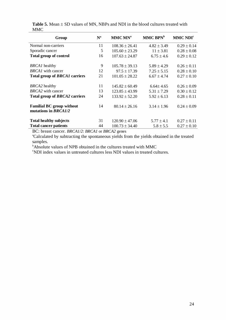
Effect of the mitomycin-C (MMC) treatment in the G1/S phase
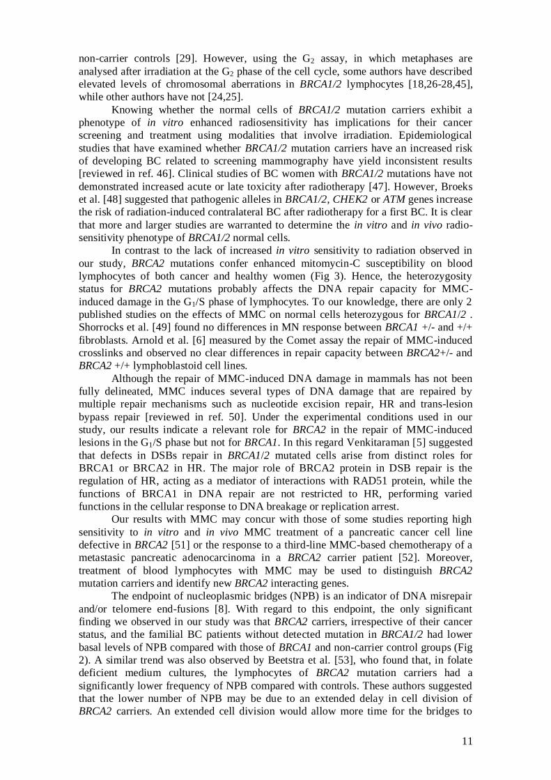
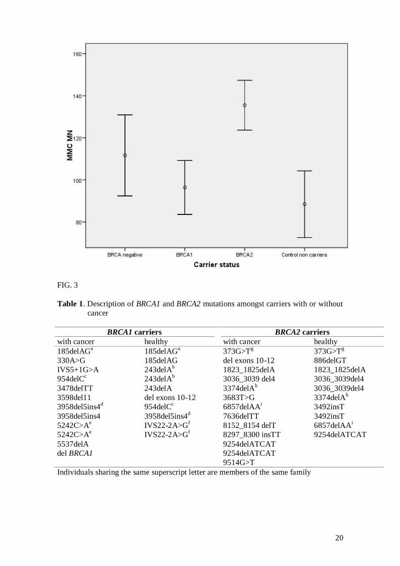
Table 5 shows the means of MN and NDI induced by MMC treatment and the
values of NPB obtained after MMC treatment. The GLZ final models showed that
carrier status had a significant effect on MMC-induced MN (P<0.001). The
lymphocytes of BRCA2 mutation carriers (irrespective to their disease status) presented
higher levels of MN after exposure to MMC (adjusted mean, 135.48) than those of
BRCA1 carriers (96.39), and non-carrier controls (88.45) (P<0.001) (Fig. 3). The MMC-
induced MN of BRCA2 carriers was also higher (135.48) than that of familial cancer
patients without detected mutation in BRCA1/2 (111.63), but this difference was not
statistically significant (Fig. 3). Furthermore, the frequency of MMC-induced MN
increased with the continuous variable of days elapsed since the first blood extraction
(P<0.001). The NPB obtained after MMC treatment was not significantly affected by
carrier and cancer status or by any covariable.
The evaluation of between-group differences after adjusting the values of MMC-
induced NDI for the different confounding factors did not show any significant effect of
carrier or disease status on cell proliferation capacity after adding MMC to the cultures.
The NDI yielded by MMC was only significantly correlated to the years elapsed since
treatment for cancer (P=0.010). This correlation indicated a lower effect of MMC on
cell proliferation with less time elapsed since the last cancer treatment. This apparent
resistance to MMC in lymphocytes from subjects just treated with chemotherapy or
radiotherapy could be explained by the fact that the cellular proliferating potential has
been surpassed by the already lower cell proliferation, observed in the non-treated
cultures, in subjects after cancer treatments and the high cytostatic effect of MMC
(which is stronger than that of the radiation treatment; see NDI values in Tables 4 and
5).

10
DISCUSSION
Chromosomal sensitivity (CRS) to radiation and chemical mutagens, quantified
by G0 MN or G2 chromosome aberrations assays, has been proposed as a marker for
low-penetrance predisposition to several common cancers including breast cancer [7,15-
17]. Several independent studies have shown enhanced in vitro sensitivity to the
chromosome-damaging effects of ionizing radiation and other mutagens in a significant
number of BC patients compared to normal healthy controls [18-22]. It has been also
reported that the enhanced CRS to different mutagens is a heritable trait [19,21,23].
The studies that have investigated CRS to mutagens in blood lymphocytes from
patients with germline mutations in BRCA1 or BRCA2 have yielded inconsistent results
[18,24-35]. While it has been well documented that biallelic inactivation of BRCA1 o
BRCA2 (BRCA1-/- or BRCA2-/-
) genes leads to repair deficiency, and hence to
significantly enhanced CRS after treatment with different mutagens, the situation in
heterozygous cells (BRCA1+/- or BRCA2+/-
) remains elusive.
In our study we assessed the chromosome sensitivity of BRCA1+/- or BRCA2+/-
lymphocytes to ionising radiation or mitomycin-C treatment measuring MN and NPB
with the MN G0 phase assay. Our results showed that, in both cancer and healthy
women, heterozygosity for BRCA1/2 mutations did not associated with an increased
sensitivity to ionising radiation exposure in the G0 phase. This result contrasts with the
findings in lymphocytes heterozygote for BRCA1/2 mutations showing evidence of an
enhanced MN radiation induction [7,18,32-35]. However, other studies have not
reported a significant increase of MN after radiation in BRCA1/2 mutated lymphocytes
[18,24,30]. These inconsistent results may be due to small population sizes,
heterogeneous reference groups and different techniques used for both irradiation and
evaluation of MN [7,36].
We found that the frequency of radiation-induced MN as well as the MN
induced by MMC increase with the days elapsed since first blood culture. This could
not be related to a change in experimental conditions or to reported sources of MN
variability such as storage time of the blood samples [37,38] and differences between
scorers [39]. To reduce the inter-test variability, a direct comparison of cases with
concurrent controls and/or automated counting by image analysis has been suggested
[7,32].
The lack of increased radiosensitivity of BRCA1+/- or BRCA2+/-
lymphocytes
shown in our study may also suggest that the BRCA1/2 proteins do not play a role in the
repair mechanisms acting on the radiation-induced DNA damage in the G0/G1 phase
such as non-homologous DNA end-joining (NHEJ). NHEJ reseals DNA double-strand
breaks (DSBs) efficiently throughout all cell cycle phases but especially in the G1 phase
[40]. Homologous recombination (HR), the other mechanism that the cell employs to
repair radiation induced DSBs, acts in the S and G2 phase when a duplicated copy of
DNA is available [41,42]. Despite some studies have described that BRCA1-deficient
cells had significantly reduced NHEJ activity, it is not definitively clear whether BRCA1
or BRCA2 are required for efficient NHEJ of radiation-induced DNA breaks [43,44]. In
contrast, it is well known the role of BRCA1/2 in the functioning of HR pathway [2,3].
Given that HR is the most important mechanism for the repair of DSBs induced
in the S and G2 phases [41,42], impaired repair of radiation-induced DSBs may be
expected at these phases in lymphocytes of BRCA1/2 mutation carriers. When analysing
the metaphases after irradiation at S phase Barwell et al. [26] found that the
lymphocytes of BRCA1/2 mutation carriers without cancer had increased chromosome
breaks compared to age-matched unaffected controls. Blood cells of BRCA1 mutation
carriers irradiated with 8 Gy 24 hours after stimulation and cultured 6 days following
irradiation exhibited a significantly higher level of chromosomal damage than those of
11
non-carrier controls [29]. However, using the G2 assay, in which metaphases are
analysed after irradiation at the G2 phase of the cell cycle, some authors have described
elevated levels of chromosomal aberrations in BRCA1/2 lymphocytes [18,26-28,45],
while other authors have not [24,25].
Knowing whether the normal cells of BRCA1/2 mutation carriers exhibit a
phenotype of in vitro enhanced radiosensitivity has implications for their cancer
screening and treatment using modalities that involve irradiation. Epidemiological
studies that have examined whether BRCA1/2 mutation carriers have an increased risk
of developing BC related to screening mammography have yield inconsistent results
[reviewed in ref. 46]. Clinical studies of BC women with BRCA1/2 mutations have not
demonstrated increased acute or late toxicity after radiotherapy [47]. However, Broeks
et al. [48] suggested that pathogenic alleles in BRCA1/2, CHEK2 or ATM genes increase
the risk of radiation-induced contralateral BC after radiotherapy for a first BC. It is clear
that more and larger studies are warranted to determine the in vitro and in vivo radio-
sensitivity phenotype of BRCA1/2 normal cells.
In contrast to the lack of increased in vitro sensitivity to radiation observed in
our study, BRCA2 mutations confer enhanced mitomycin-C susceptibility on blood
lymphocytes of both cancer and healthy women (Fig 3). Hence, the heterozygosity
status for BRCA2 mutations probably affects the DNA repair capacity for MMC-
induced damage in the G1/S phase of lymphocytes. To our knowledge, there are only 2
published studies on the effects of MMC on normal cells heterozygous for BRCA1/2 .
Shorrocks et al. [49] found no differences in MN response between BRCA1 +/- and +/+
fibroblasts. Arnold et al. [6] measured by the Comet assay the repair of MMC-induced
crosslinks and observed no clear differences in repair capacity between BRCA2+/- and
BRCA2 +/+ lymphoblastoid cell lines.
Although the repair of MMC-induced DNA damage in mammals has not been
fully delineated, MMC induces several types of DNA damage that are repaired by
multiple repair mechanisms such as nucleotide excision repair, HR and trans-lesion
bypass repair [reviewed in ref. 50]. Under the experimental conditions used in our
study, our results indicate a relevant role for BRCA2 in the repair of MMC-induced
lesions in the G1/S phase but not for BRCA1. In this regard Venkitaraman [5] suggested
that defects in DSBs repair in BRCA1/2 mutated cells arise from distinct roles for
BRCA1 or BRCA2 in HR. The major role of BRCA2 protein in DSB repair is the
regulation of HR, acting as a mediator of interactions with RAD51 protein, while the
functions of BRCA1 in DNA repair are not restricted to HR, performing varied
functions in the cellular response to DNA breakage or replication arrest.
Our results with MMC may concur with those of some studies reporting high
sensitivity to in vitro and in vivo MMC treatment of a pancreatic cancer cell line
defective in BRCA2 [51] or the response to a third-line MMC-based chemotherapy of a
metastasic pancreatic adenocarcinoma in a BRCA2 carrier patient [52]. Moreover,
treatment of blood lymphocytes with MMC may be used to distinguish BRCA2
mutation carriers and identify new BRCA2 interacting genes.
The endpoint of nucleoplasmic bridges (NPB) is an indicator of DNA misrepair
and/or telomere end-fusions [8]. With regard to this endpoint, the only significant
finding we observed in our study was that BRCA2 carriers, irrespective of their cancer
status, and the familial BC patients without detected mutation in BRCA1/2 had lower
basal levels of NPB compared with those of BRCA1 and non-carrier control groups (Fig
2). A similar trend was also observed by Beetstra et al. [53], who found that, in folate
deficient medium cultures, the lymphocytes of BRCA2 mutation carriers had a
significantly lower frequency of NPB compared with controls. These authors suggested
that the lower number of NPB may be due to an extended delay in cell division of
BRCA2 carriers. An extended cell division would allow more time for the bridges to
12
break during anaphase prior to cell harvesting. Beetstra et al. [53] based their hypothesis
on data obtained by Daniels et al. [54], who reported that the period taken for cells to
progress from anaphase onset to completion of cell division is significantly extended in
cells with targeted gene disruption of BRCA2 or reduced transcription of BRCA2 by
RNA interference when compared with controls. Likewise, Jonsdottir et al. [55] have
demonstrated that BRCA2 mutation carrier fibroblasts had delayed cytokinesis, being
the mean cell division time 6 min longer compared with BRCA2 wild type cells.
However, in contrast to results from these two studies, Lekomtsev et al. [56] have
recently indicated that BRCA2 does not regulate cytokinesis in human cells. Thus,
further research is required to verify a role of BRCA2 in the regulation of cell division.
Interestingly, in the familial BC patients without detected mutation in BRCA1/2 a
decrease in the basal NPB formation was also detected. It is tempting to speculate that
BRCA2 and other genes related to cell division might be associated with cancer
susceptibility in familial BC patients negative for BRCA1/2 mutations.
It should be mentioned that the group of familial BC patients who tested
negative for BRCA1/2 mutations did not present a different chromosomal sensitivity
(CRS) to either radiation or MMC treatment compared with control non-carriers. These
data do not support the findings of the studies carried out by Baeyens et al. [18,24].
They showed that the lymphocytes of non-BRCA1/2 familial BC patients had a higher
CRS to radiation, measured by G0 MN and G2 assays, than the control group. Further
research will be needed to more precisely determine the CRS in the group of familial
BC with no BRCA1/2 mutations.
The nuclear division index (NDI), provides a measure of the proliferative status
of the viable lymphocytes, indicating both the lymphocyte mitogenic response and the
cytostatic effects of agents examined in the assay [8]. We did not find any significant
difference in either basal or mutagen-induced levels of this proliferation index between
BRCA1/2 carriers and non carrier groups; this would indicate that the lymphocytes from
heterozygous BRCA1/2 mutation carriers have no important defects in the mitogenic
response or cellular growth in culture after mutagenic treatments. However, it is
interesting to note that, although differences were not statistically significant, the
BRCA2 carriers showed higher basal levels of NDI values than the other groups
analysed. A similar finding has been reported by Beetstra et al. [53], who detected that
the NDI of BRCA2 mutation carriers in lymphocytes cultured with folate deficiency
tended to be higher than in controls. Thus the BRCA2 carriers may have a defect in
cytokinesis that entails an increased sensitivity to the cytokinesis-blocking action of
cytochalasin-B [53]. This may be attributed to an extended time for these cells to
progress from anaphase onset to completed cell division, that allows the accumulation
of binucleated cells in culture [54,55].
In conclusion, the BRCA1 or BRCA2 heterozygote status in peripheral blood
lymphocytes is not related to an increased radiosensitivity. But the monoallelic BRCA2
mutations are associated with a higher level of chromosomal damage induced by MMC,
probably due to an impaired DNA repair capacity. This finding suggests that the MMC-
induced MN analysis may be used in the identification of individuals with a deficiency
in BRCA2 or genes related to BRCA2. Given that MMC can be used as an anti-cancer
drug, these data may be relevant for the management and follow-up of BRCA2 mutation
carriers.
13
ACKNOWLEDGMENTS
This study was supported by two grants of the “Fondo de Investigación
Sanitaria” from the Spanish Health Ministry: FIS 04/1832 and FIS 05/2181 and by a
grant of the “Fundación Mutua Madrileña” (2008-2011).
CONFLICT OF INTEREST STATEMENT
The authors declare that they have no competing interests.
FIGURE CAPTIONS
FIG. 1 Effect of previous chemotherapy and radiotherapy of BC/OC patients on
MN and NPB endpoints (A) Relationship between the basal values of MN of the
cancer patients and the time passed since last session of chemotherapy. (B) Means
adjusted for the other terms in the model and 95% confidence limits showing the
differences of levels of MN in the non-treated cultures between cancer and healthy
groups. (C) Means adjusted for the other terms in the model and 95% confidence limits
illustrating the differences of basal levels of NPB in the non-treated cultures between
cancer and healthy groups.
FIG. 2 Adjusted means and 95% confidence limits showing the differences of basal
levels of NPB in the non-treated cultures between carrier and non-carrier groups.
BRCA negative: familial breast cancer patients without detected mutation in BRCA1 or
BRCA2. Control non carriers: healthy women and sporadic BC patients without history
of familial cancer.
FIG. 3 Adjusted means and 95% confidence limits illustrating the differences of MN
levels after MMC treatment between carrier and non-carrier groups.
BRCA negative: familial breast cancer patients without detected mutation in BRCA1 or
BRCA2. Control non carriers: healthy women and sporadic BC patients without history
of familial cancer.
14
References
1. Fackenthal JD, Olopade OI (2007) Breast cancer risk associated with BRCA1
and BRCA2 in diverse populations. Nat Rev Cancer 7:937-948
2. Boulton SJ (2006) Cellular functions of the BRCA tumour-suppressor proteins.
Biochem Soc Trans 34:633-645
3. Gudmundsdottir K, Ashworth A (2006) The roles of BRCA1 and BRCA2 and
associated proteins in the maintenance of genomic stability. Oncogene 25:5864-
5874
4. Cousineau I, Belmaaza A (2007) BRCA1 haploinsufficiency, but not
heterozygosity for a BRCA1-truncating mutation, deregulates homologous
recombination. Cell Cycle 6:962-971
5. Venkitaraman AR (2009) Linking the cellular functions of BRCA genes to
cancer pathogenesis and treatment. Annu Rev Pathol 4:461-487
6. Arnold K, Kim MK, Frerk K, Edler L, Savelyeva L, Schmezer P, Wiedemeyer R
(2006) Lower level of BRCA2 protein in heterozygous mutation carriers is
correlated with an increase in DNA double strand breaks and an impaired DSB
repair. Cancer Lett 243:90-100
7. Speit G, Trenz K (2004) Chromosomal mutagen sensitivity associated with
mutations in BRCA genes. Cytogenet Genome Res 104:325-332
8. Fenech M (2007) Cytokinesis-block micronucleus cytome assay. Nat Protoc
2:1084-1104
9. Thomas P, Umegaki K, Fenech M (2003) Nucleoplasmic bridges are a sensitive
measure of chromosome rearrangement in the cytokinesis-block micronucleus
assay. Mutagenesis 18:187-194
10. Fell LJ, Paul ND, McMillan TJ (2002) Role for non-homologous end-joining in
the repair of UVA-induced DNA damage. Int J Radiat Biol 78:1023-1027
11. Ouyang Y, Kwon YT, An JY, Eller D, Tsai SC, Diaz-Perez S, Troke JJ, Teitell
MA, Marahrens Y (2006) Loss of Ubr2, an E3 ubiquitin ligase, leads to
chromosome fragility and impaired homologous recombinational repair. Mutat
Res 596:64-75
12. Yue J, Wang Q, Lu H, Brenneman M, Fan F, Shen Z (2009) The cytoskeleton
protein filamin-A is required for an efficient recombinational DNA double
strand break repair. Cancer Res 69:7978-7985
13. Fenech M (2010) The lymphocyte cytokinesis-block micronucleus cytome assay
and its application in radiation biodosimetry. Health Phys 98:234-243
14. Lee TK, Allison RR, O'Brien KF, Naves JL, Karlsson UL, Wiley AL (2002)
Persistence of micronuclei in lymphocytes of cancer patients after radiotherapy.
Radiat Res 157:678-684
15
15. Baria K, Warren C, Roberts SA, West CM, Scott D (2001) Chromosomal
radiosensitivity as a marker of predisposition to common cancers? Br J Cancer
84:892-896
16. El-Zein RA, Schabath MB, Etzel CJ, Lopez MS, Franklin JD, Spitz MR (2006)
Cytokinesis-blocked micronucleus assay as a novel biomarker for lung cancer
risk. Cancer Res 66:6449-6456
17. El-Zein RA, Fenech M, Lopez MS, Spitz MR, Etzel CJ (2008) Cytokinesis-
blocked micronucleus cytome assay biomarkers identify lung cancer cases
amongst smokers. Cancer Epidemiol Biomarkers Prev 17:1111-1119
18. Baeyens A, Thierens H, Claes K, Poppe B, Messiaen L, de RL, Vral A (2002)
Chromosomal radiosensitivity in breast cancer patients with a known or putative
genetic predisposition. Br J Cancer 87:1379-1385
19. Burrill W, Barber JB, Roberts SA, Bulman B, Scott D (2000) Heritability of
chromosomal radiosensitivity in breast cancer patients: a pilot study with the
lymphocyte micronucleus assay. Int J Radiat Biol 76:1617-1619
20. Parshad R, Price FM, Bohr VA, Cowans KH, Zujewski JA, Sanford KK (1996)
Deficient DNA repair capacity, a predisposing factor in breast cancer. Br J
Cancer 74:1-5
21. Roberts SA, Spreadborough AR, Bulman B, Barber JB, Evans DG, Scott D
(1999) Heritability of cellular radiosensitivity: a marker of low-penetrance
predisposition genes in breast cancer? Am J Hum Genet 65:784-794
22. Scott D, Barber JB, Spreadborough AR, Burrill W, Roberts SA (1999) Increased
chromosomal radiosensitivity in breast cancer patients: a comparison of two
assays. Int J Radiat Biol 75:1-10
23. Wu X, Spitz MR, Amos CI et al (2006) Mutagen sensitivity has high
heritability: evidence from a twin study. Cancer Res 66:5993-5996
24. Baeyens A, Thierens H, Claes K, Poppe B, de RL, Vral A (2004) Chromosomal
radiosensitivity in BRCA1 and BRCA2 mutation carriers. Int J Radiat Biol
80:745-756
25. Baria K, Warren C, Roberts SA, West CM, Evans DG, Varley JM, Scott D
(2001) Correspondence re: A. Rothfuss et al., Induced micronucleus frequencies
in peripheral blood lymphocytes as a screening test for carriers of a BRCA1
mutation in breast cancer families. Cancer Res., 60: 390-394, 2000. Cancer Res
61:5948-5949
26. Barwell J, Pangon L, Georgiou A et al (2007) Lymphocyte radiosensitivity in
BRCA1 and BRCA2 mutation carriers and implications for breast cancer
susceptibility. Int J Cancer 121:1631-1636
27. Buchholz TA, Wu X, Hussain A et al (2002) Evidence of haplotype
insufficiency in human cells containing a germline mutation in BRCA1 or
BRCA2. Int J Cancer 97:557-561

16
28. Febrer E, Mestres M, Caballin MR, Barrios L, Ribas M, Gutierrez-Enriquez S,
Alonso C, Cajal T, Francesc BJ (2008) Mitotic delay in lymphocytes from
BRCA1 heterozygotes unable to reduce the radiation-induced chromosomal
damage. DNA Repair (Amst) 7:1907-1911
29. Kote-Jarai Z, Salmon A, Mengitsu T et al (2006) Increased level of
chromosomal damage after irradiation of lymphocytes from BRCA1 mutation
carriers. Br J Cancer 94:308-310
30. Kotsopoulos J, Chen Z, Vallis KA, Poll A, Ainsworth P, Narod SA (2007) DNA
repair capacity as a possible biomarker of breast cancer risk in female BRCA1
mutation carriers. Br J Cancer 96:118-125
31. Kowalska E, Narod SA, Huzarski T, Zajaczek S, Huzarska J, Gorski B, Lubinski
J (2005) Increased rates of chromosome breakage in BRCA1 carriers are
normalized by oral selenium supplementation. Cancer Epidemiol Biomarkers
Prev 14:1302-1306
32. Rothfuss A, Schutz P, Bochum S, Volm T, Eberhardt E, Kreienberg R, Vogel
W, Speit G (2000) Induced micronucleus frequencies in peripheral lymphocytes
as a screening test for carriers of a BRCA1 mutation in breast cancer families.
Cancer Res 60:390-394
33. Trenz K, Rothfuss A, Schutz P, Speit G (2002) Mutagen sensitivity of peripheral
blood from women carrying a BRCA1 or BRCA2 mutation. Mutat Res 500:89-
96
34. Trenz K, Lugowski S, Jahrsdorfer U, Jainta S, Vogel W, Speit G (2003)
Enhanced sensitivity of peripheral blood lymphocytes from women carrying a
BRCA1 mutation towards the mutagenic effects of various cytostatics. Mutat
Res 544:279-288
35. Varga D, Hoegel J, Maier C et al (2006) On the difference of micronucleus
frequencies in peripheral blood lymphocytes between breast cancer patients and
controls. Mutagenesis 21:313-320
36. Vogel W, Surowy H (2007) Reduced DNA repair in BRCA1 mutation carriers
undetectable before onset of breast cancer? Br J Cancer 97:1184-1186
37. Bonassi S, Fenech M, Lando C et al (2001) HUman MicroNucleus project:
international database comparison for results with the cytokinesis-block
micronucleus assay in human lymphocytes: I. Effect of laboratory protocol,
scoring criteria, and host factors on the frequency of micronuclei. Environ Mol
Mutagen 37:31-45
38. Patino-Garcia B, Hoegel J, Varga D, Hoehne M, Michel I, Jainta S, Kreienberg
R, Maier C, Vogel W (2006) Scoring variability of micronuclei in binucleated
human lymphocytes in a case-control study. Mutagenesis 21:191-197
39. Fenech M, Bonassi S, Turner J et al (2003) Intra- and inter-laboratory variation
in the scoring of micronuclei and nucleoplasmic bridges in binucleated human
lymphocytes. Results of an international slide-scoring exercise by the HUMN
project. Mutat Res 534:45-64

17
40. Burma S, Chen BP, Chen DJ (2006) Role of non-homologous end joining
(NHEJ) in maintaining genomic integrity. DNA Repair (Amst) 5:1042-1048
41. Frankenberg-Schwager M, Gebauer A, Koppe C, Wolf H, Pralle E, Frankenberg
D (2009) Single-strand annealing, conservative homologous recombination,
nonhomologous DNA end joining, and the cell cycle-dependent repair of DNA
double-strand breaks induced by sparsely or densely ionizing radiation. Radiat
Res 171:265-273
42. Koch K, Wrona A, Dikomey E, Borgmann K (2009) Impact of homologous
recombination on individual cellular radiosensitivity. Radiother Oncol 90:265-
272
43. Baldeyron C, Jacquemin E, Smith J, Jacquemont C, De O, I, Gad S, Feunteun J,
Stoppa-Lyonnet D, Papadopoulo D (2002) A single mutated BRCA1 allele leads
to impaired fidelity of double strand break end-joining. Oncogene 21:1401-1410
44. Bau DT, Mau YC, Shen CY (2006) The role of BRCA1 in non-homologous
end-joining. Cancer Lett 240:1-8
45. Ernestos B, Nikolaos P, Koulis G, Eleni R, Konstantinos B, Alexandra G,
Michael K (2010) Increased chromosomal radiosensitivity in women carrying
BRCA1/BRCA2 mutations assessed with the G2 assay. Int J Radiat Oncol Biol
Phys 76:1199-1205
46. Berrington de GA, Berg CD, Visvanathan K, Robson M (2009) Estimated risk
of radiation-induced breast cancer from mammographic screening for young
BRCA mutation carriers. J Natl Cancer Inst 101:205-209
47. Shanley S, McReynolds K, Ardern-Jones A et al (2006) Late toxicity is not
increased in BRCA1/BRCA2 mutation carriers undergoing breast radiotherapy
in the United Kingdom. Clin Cancer Res 12:7025-7032
48. Broeks A, Braaf LM, Huseinovic A et al (2007) Identification of women with an
increased risk of developing radiation-induced breast cancer: a case only study.
Breast Cancer Res 9:R26
49. Shorrocks J, Tobi SE, Latham H, Peacock JH, Eeles R, Eccles D, McMillan TJ
(2004) Primary fibroblasts from BRCA1 heterozygotes display an abnormal
G1/S cell cycle checkpoint following UVA irradiation but show normal levels of
micronuclei following oxidative stress or mitomycin C treatment. Int J Radiat
Oncol Biol Phys 58:470-478
50. Lee YJ, Park SJ, Ciccone SL, Kim CR, Lee SH (2006) An in vivo analysis of
MMC-induced DNA damage and its repair. Carcinogenesis 27:446-453
51. van der Heijden MS, Brody JR, Dezentje DA et al (2005) In vivo therapeutic
responses contingent on Fanconi anemia/BRCA2 status of the tumor. Clin
Cancer Res 11:7508-7515
52. Chalasani P, Kurtin S, Dragovich T (2008) Response to a third-line mitomycin
C (MMC)-based chemotherapy in a patient with metastatic pancreatic
adenocarcinoma carrying germline BRCA2 mutation. JOP 9:305-308

18
53. Beetstra S, Salisbury C, Turner J, Altree M, McKinnon R, Suthers G, Fenech M
(2006) Lymphocytes of BRCA1 and BRCA2 germ-line mutation carriers, with
or without breast cancer, are not abnormally sensitive to the chromosome
damaging effect of moderate folate deficiency. Carcinogenesis 27:517-524
54. Daniels MJ, Wang Y, Lee M, Venkitaraman AR (2004) Abnormal cytokinesis in
cells deficient in the breast cancer susceptibility protein BRCA2. Science
306:876-879
55. Jonsdottir AB, Vreeswijk MP, Wolterbeek R, Devilee P, Tanke HJ, Eyfjord JE,
Szuhai K (2009) BRCA2 heterozygosity delays cytokinesis in primary human
fibroblasts. Cell Oncol 31:191-201
56. Lekomtsev S, Guizetti J, Pozniakovsky A, Gerlich DW, Petronczki M (2010)
Evidence that the tumor-suppressor protein BRCA2 does not regulate
cytokinesis in human cells. J Cell Sci 123:1395-1400
(A)
(B) (C)
19
Fig 1
Fig 2.
20
FIG. 3
Table 1. Description of BRCA1 and BRCA2 mutations amongst carriers with or without
cancer
BRCA1 carriers BRCA2 carriers
with cancer healthy with cancer healthy
185delAGa 185delAG
a 373G>T
g 373G>T
g
330A>G 185delAG del exons 10-12 886delGT
IVS5+1G>A 243delAb 1823_1825delA 1823_1825delA
954delCc 243delA
b 3036_3039 del4 3036_3039del4
3478delTT 243delA 3374delAh 3036_3039del4
3598del11 del exons 10-12 3683T>G 3374delAh
3958del5ins4d 954delC
c 6857delAA
i 3492insT
3958del5ins4 3958del5ins4d 7636delTT 3492insT
5242C>Ae IVS22-2A>G
f 8152_8154 delT 6857delAA
i
5242C>Ae IVS22-2A>G
f 8297_8300 insTT 9254delATCAT
5537delA 9254delATCAT
del BRCA1 9254delATCAT
9514G>T
Individuals sharing the same superscript letter are members of the same family
21
Table 2. Mean values and SD of subject age, age of cancer onset, years elapsed since radiotherapy and
chemotherapy treatment and days since first experiment.
Group Nº Age Days since first
experiment
Cancer onset
age
Chemotherapy
yearsa
Radiotherapy
yearsa
Healthy women 11 40.91 9.03 777.73 223.04
BC non-familial or sporadic 5 52.6 7.50 1051.8 9.39 42.2 8.17 10.0 8.37 9.4 7.09 Total group of control 16 44.56 10.03 863.38 224.50
BRCA1 healthy 9 35.67 10.25 822.89 285.12
BRCA1 with cancer 12 48.5 10.66 639.50 334.85 36.83 7.71 8.09 6.79 8.68 7.67
Total group of BRCA1
mutation carriers
21 43.0 12.12 718.10 320.68
BRCA2 healthy 11 39.64 11.42 654.18 270.74
BRCA2 with cancer 13 52.31 9.56 620.15 110.49 38.69 6.41 10.42 8.32 8.83 7.12
Total group of BRCA2
mutation carriers
24 46.50 12.10 635.75 196.32
Familial BC group
without detected mutation
in BRCA1/2
15 50.53 13.45 324.20 250.31 43.60 12.44 4.42 4.53 3.76 5.66
Total healthy subjects 31 38.94 10.17 747.0 260.63
Total cancer patients 45 50.73 10.89 574.62 316.73 40.22 9.46 7.87 7.07 7.53 7.03 ayears since last exposure. BC: breast cancer. BRCA1/2: BRCA1 or BRCA2 genes
22
Table 3. Mean values and SD of the different endpoints recorded between all analysed
groups in the non-treated cultures.
Group Nº Basal MN Basal BPN Basal NDI
Normal non-carriers 11 14.09 3.4 1.36 1.91 1.84 0.14 Sporadic cancer 5 12.6 4.28 3.8 1.92 1.71 0.15
Total group of control 16 13.63 3.61 2.13 2.19 1.80 0.15 BRCA1 healthy 9 8.89 3.10 0.89 1.36 1.89 0.11
BRCA1 with cancer 12 27.75 15.24 3.08 3.29 1.68 0.15 Total group of BRCA1 carriers 21 19.67 14.94 2.14 2.82 1.77 0.17
BRCA2 healthy 11 14.27 8.10 0.82 1.17 1.85 0.15 BRCA2 with cancer 13 19.69 14.76 0.85 0.99 1.88 0.15 Total group of BRCA2 carriers 24 17.21 12.24 0.83 1.05 1.86 0.15
Familial BC group without detected
mutation in BRCA1/2
15 27.33 21.56 0.93 1.58 1.72 0.16
Total healthy subjects 31 12.65 5.85 1.03 1.49 1.86 0.13
Total cancer patients 45 23.6 17.17 1.80 2.35 1.75 0.17
BC: breast cancer. BRCA1/2: BRCA1 or BRCA2 genes
23
Table 4. Mean SD values of MN, NBPs and NDI obtained in the blood cultures
treated with 2 Gy of gamma-radiation.
Group Nº 2 Gy MNa 2 Gy BPN
b 2 Gy NDI
c
Normal non-carriers 11 227.64 25.47 18.18 6.62 0.20 0.13
Sporadic cancer 5 253.4 23.73 42.4 11.63 0.22 0.10 Total group of control 16 235.69 27.10 25.75 14.13 0.20 0.12
BRCA1 healthy 9 221.56 39.81 26.56 19.06 0.20 0.08 BRCA1 with cancer 12 224.67 47.03 18 15.13 0.19 0.07
Total group of BRCA1 carriers 21 223.33 43.05 21.67 17.03 0.20 0.07 BRCA2 healthy 11 245.91 60.61 20.91 14.47 0.22 0.07
BRCA2 with cancer 13 213.23 57.25 9.39 6.32 0.23 0.11 Total group of BRCA2 carriers 24 228.21 59.86 14.67 12.1 0.22 0.09
Familial BC group without detected
mutation in BRCA1/2
15 184.0 42.67 14 7.33 0.15 0.07
Total healthy subjects 31 232.36 44.43 21.58 13.9 0.21 0.09
Total cancer patients 45 211.0 50.81 16.9 13.83 0.19 0.09
BC: breast cancer. BRCA1/2: BRCA1 or BRCA2 genes. aCalculated by subtracting the spontaneous yields from the yields obtained in the treated
samples. bAbsolute values of NPB obtained in the cultures treated with radiation cNDI index values in untreated cultures less NDI values in treated cultures.
24
Table 5. Mean SD values of MN, NBPs and NDI in the blood cultures treated with
MMC
Group Nº MMC MNa MMC BPN
b MMC NDI
c
Normal non-carriers 11 108.36 26.41 4.82 3.49 0.29 0.14 Sporadic cancer 5 105.60 23.29 11 3.81 0.28 0.08
Total group of control 16 107.63 24.87 6.75 4.6 0.29 0.12 BRCA1 healthy 9 105.78 39.13 5.89 4.29 0.26 0.11
BRCA1 with cancer 12 97.5 17.39 7.25 5.15 0.28 0.10 Total group of BRCA1 carriers 21 101.05 28.22 6.67 4.74 0.27 0.10
BRCA2 healthy 11 145.82 60.49 6.64 4.65 0.26 0.09 BRCA2 with cancer 13 123.85 43.99 5.31 7.29 0.30 0.12
Total group of BRCA2 carriers 24 133.92 52.20 5.92 6.13 0.28 0.11 Familial BC group without
mutations in BRCA1/2
14 80.14 26.16 3.14 1.96 0.24 0.09
Total healthy subjects 31 120.90 47.06 5.77 4.1 0.27 0.11 Total cancer patients 44 100.73 34.40 5.8 5.5 0.27 0.10
BC: breast cancer. BRCA1/2: BRCA1 or BRCA2 genes aCalculated by subtracting the spontaneous yields from the yields obtained in the treated
samples. bAbsolute values of NPB obtained in the cultures treated with MMC cNDI index values in untreated cultures less NDI values in treated cultures.
Copyright © 2022 FDOKUMEN