Role of Doa1 in the Saccharomyces cerevisiae DNA Damage Response
Upload
independentCategory
view
0download
0
BioMed CentralBMC Genomics
ss
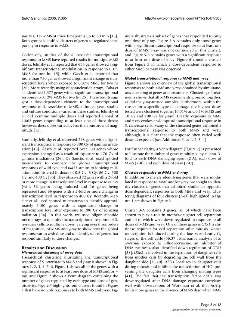
Open AcceResearch articleAnalyzing the dose-dependence of the Saccharomyces cerevisiae global transcriptional response to methyl methanesulfonate and ionizing radiationMichael G Benton1, Swetha Somasundaram1, Jeremy D Glasner2 and Sean P Palecek*1Address: 1Department of Chemical & Biological Engineering, 1415 Engineering Drive, University of Wisconsin, Madison, WI 53706, USA and 2Genome Center of Wisconsin, 425 Henry Mall, University of Wisconsin, Madison, WI 53706, USA
Email: Michael G Benton - [email protected]; Swetha Somasundaram - [email protected]; Jeremy D Glasner - [email protected]; Sean P Palecek* - [email protected]
* Corresponding author
AbstractBackground: One of the most crucial tasks for a cell to ensure its long term survival is preservingthe integrity of its genetic heritage via maintenance of DNA structure and sequence. While theDNA damage response in the yeast Saccharomyces cerevisiae, a model eukaryotic organism, has beenextensively studied, much remains to be elucidated about how the organism senses and respondsto different types and doses of DNA damage. We have measured the global transcriptionalresponse of S. cerevisiae to multiple doses of two representative DNA damaging agents, methylmethanesulfonate (MMS) and gamma radiation.
Results: Hierarchical clustering of genes with a statistically significant change in transcriptionillustrated the differences in the cellular responses to MMS and gamma radiation. Overall, MMSproduced a larger transcriptional response than gamma radiation, and many of the genes modulatedin response to MMS are involved in protein and translational regulation. Several clusters ofcoregulated genes whose responses varied with DNA damaging agent dose were identified.Perhaps the most interesting cluster contained four genes exhibiting biphasic induction in responseto MMS dose. All of the genes (DUN1, RNR2, RNR4, and HUG1) are involved in the Mec1p kinasepathway known to respond to MMS, presumably due to stalled DNA replication forks. The biphasicresponses of these genes suggest that the pathway is induced at lower levels as MMS doseincreases. The genes in this cluster with a threefold or greater transcriptional response to gammaradiation all showed an increased induction with increasing gamma radiation dosage.
Conclusion: Analyzing genome-wide transcriptional changes to multiple doses of externalstresses enabled the identification of cellular responses that are modulated by magnitude of thestress, providing insights into how a cell deals with genotoxicity.
Published: 01 December 2006
BMC Genomics 2006, 7:305 doi:10.1186/1471-2164-7-305
Received: 14 April 2006Accepted: 01 December 2006
This article is available from: http://www.biomedcentral.com/1471-2164/7/305
© 2006 Benton et al; licensee BioMed Central Ltd. This is an Open Access article distributed under the terms of the Creative Commons Attribution License (http://creativecommons.org/licenses/by/2.0), which permits unrestricted use, distribution, and reproduction in any medium, provided the original work is properly cited.
Page 1 of 18(page number not for citation purposes)
BMC Genomics 2006, 7:305 http://www.biomedcentral.com/1471-2164/7/305
BackgroundDNA is the conduit for the transmission of genetic infor-mation between generations of a species [1]. The linearsequence of DNA is labile and can be altered by bothintracellular and extracellular sources in a variety of ways.For example, within a cell errors by replication machinerycan lead to point mutations while the physical stresses ofreplication can fracture strands of DNA. External sourcescan mutate DNA by alkylating single strands, causinginterstrand crosslinks, or even breaking strands apart,resulting in the loss of some bases upon ligation [2-4].
One of a cell's most crucial tasks is maintaining the integ-rity of genetic information from one generation to thenext [5]. Although some mutations are beneficial in thelong term by allowing for adaptation to changing envi-ronments via natural selection, most mutations are detri-mental and can lead to consequences as serious as celldeath or transformation; the correlation between muta-genic activity and carcinogenic activity is approximately90% [6]. This high correlation has increased interest inidentifying causes of DNA damage and the effects of thisdamage on the cellular environment with the hopes ofboth preventing cancer and developing better chemother-apeutic agents for the treatment of existing cancerous cells[7-10].
Saccharomyces cerevisiae is commonly used as a modelorganism for studying the DNA damage response becauseits genome is well-annotated and the cells are easily cul-tured and genetically manipulated. Furthermore, becauseyeasts are eukaryotic, the gene regulation and biochemicalpathways involved in responding to DNA damage areexpected to be similar to, although perhaps simpler than,those of higher organisms. Indeed, the basic cellularresponses to DNA damage have been shown to be similarin yeast and humans [11,12]. Many DNA damage sensoryand repair mechanisms in humans have been identifiedfrom their homology to similar mechanisms in yeast [13].
The study of the DNA damage response in yeast hasuncovered a broad variety of mechanisms used by the cellsto sense and repair the damage. When cells sense a defectin their DNA, several signal transduction pathways areinduced which in turn activate various checkpoints thatcause cell-cycle arrest at the G1, S, or G2 phases until thedefect can be repaired [14]. These signal transductionpathways regulate genome-wide expression patterns,causing hundreds of genes to be induced or repressed indirect response to the perceived damage [15]. Many of theinduced genes are also upregulated in response to cellcycle arrest in stationary phase or in response to generalcell stresses, indicating that DNA damage response path-ways in yeast overlap with other cell cycle pathways[16,17]. Once cell division is arrested, repairs can be made
in a variety of ways depending on the type of damagewhich has occurred. Single-strand lesions can be repairedby base excision repair, nucleotide excision repair, or mis-match repair using the non-altered strand as a template,while double-strand breaks are repaired by homologousrecombination or non-homologous end joining [18-22].
To better define the cellular pathways involved in the rec-ognition and repair of DNA damage, it is important toidentify the global set of genes induced or repressed inresponse to the damage. These genes have been identifiedin a number of ways. Traditionally, pools of mutants werescreened for sensitivity to various DNA damaging agents,enabling the proposal of epistasis groups based on com-mon responses [23,24]. In the last 10 years, the prolifera-tion of spotted and oligonucleotide microarrays haveenabled genome-wide profiling in response to multipleDNA damaging agents [15,25-27]. These types of studiesidentified hundreds of genes not previously known to bemodulated in response to DNA damage. Recently the twoapproaches have been combined by culture of gene dele-tion mutant strains tagged with unique identificationsequences in the presence of a variety of DNA damagingagents, followed by PCR amplification and microarrayhybridization of the identification sequences to assess therelative fitness level of each strain as a result of the expo-sure [28-31]. With each new study, genes not previouslyknown to be regulated by DNA damage are identified,suggesting that there are additional genes and pathwayswhose control is crucial in DNA damage response yet tobe distinguished.
In this study, we identified genes involved in the transcrip-tional response to DNA damage based on variation inexpression upon exposure to two representative DNAdamaging agents, methylmethane sulfonate (MMS) andionizing gamma radiation (γ-ray), over three orders ofmagnitude of agent dose. MMS is a DNA alkylating agentknown to methylate guanine residues at the N-7 position[30,32]. While this methylation is often tolerable to thecells, MMS can also methylate guanine at the O-6 positionor adenine at the N-3 site [30,32]. These latter lesionsinhibit DNA synthesis and must be repaired for replica-tion to properly occur [30,32]. Ionizing radiation inducesmultiple types of DNA damage, but double strand breaksare considered to be the most lethal [33]. MMS generallyproduces single-strand lesions which inhibit DNA replica-tion at the S phase checkpoint, while double-strandbreaks caused by γ-ray inhibit the G2/M checkpoint [34].Using spotted microarrays, Gasch et al. performed studieswhere S. cerevisiae cells were cultured in the presence of0.02% MMS and the transcriptional response wasassessed at multiple timepoints up to 120 min [26]. Jelin-sky et al. performed a similar work using oligonucleotidearrays to monitor the transcriptional response of S. cerevi-
Page 2 of 18(page number not for citation purposes)
BMC Genomics 2006, 7:305 http://www.biomedcentral.com/1471-2164/7/305
siae to 0.1% MMS at three timepoints up to 60 min [15].Both groups identified clusters of genes co-regulated tem-porally in response to MMS.
Collectively, studies of the S. cerevisiae transcriptionalresponse to MMS have reported results for multiple MMSdoses. Jelinsky et al. reported that 693 genes showed a sig-nificant transcriptional modulation in response to 0.1%MMS for one hr [15], while Gasch et al. reported thatmore than 750 genes showed a significant change in tran-scription levels when exposed to 0.02% MMS for two hr[26]. More recently, using oligonucleotide arrays, Caba etal. identified 1,377 genes with a significant transcriptionalresponse to 0.12% MMS for two hr [25]. These results sug-gest a dose-dependent element to the transcriptionalresponse of S. cerevisiae to MMS, although yeast strainsand culture conditions varied in these studies. Jelinsky etal. did examine multiple doses and reported a total of1,863 genes responding to at least one of three doses;however, these doses varied by less than one order of mag-nitude [15].
Similarly, Jelinsky et al. observed 248 genes with a signif-icant transcriptional response to 300 Gy of gamma irradi-ation [15]. Gasch et al. reported over 500 genes whoseexpression changed as a result of exposure to 170 Gy ofgamma irradiation [26]. De Sanctis et al. used spottedmicroarrays to compare the global transcriptionalresponses of wild-type and rad53 strains to ionizing radi-ation administered in doses of 0.8 Gy, 8 Gy, 80 Gy, 300Gy, and 400 Gy [35]. They observed 72 genes with a 2-foldor more change in transcription level in response to 80 Gy(with 56 genes being induced and 16 genes beingrepressed) and 86 genes with a 2-fold or more change intranscription level in response to 400 Gy. Recently, Mer-cier et al. used spotted microarrays to identify approxi-mately 1400 genes with a significant change intranscription level after exposure to 200 Gy of ionizingradiation [34]. In this work, we used oligonucleotidemicroarrays to quantify the transcriptional response of S.cerevisiae cells to multiple doses, varying over three ordersof magnitude, of MMS and γ-ray to show how the globalresponse varies with dose and to identify sets of genes thatrespond similarly to dose changes.
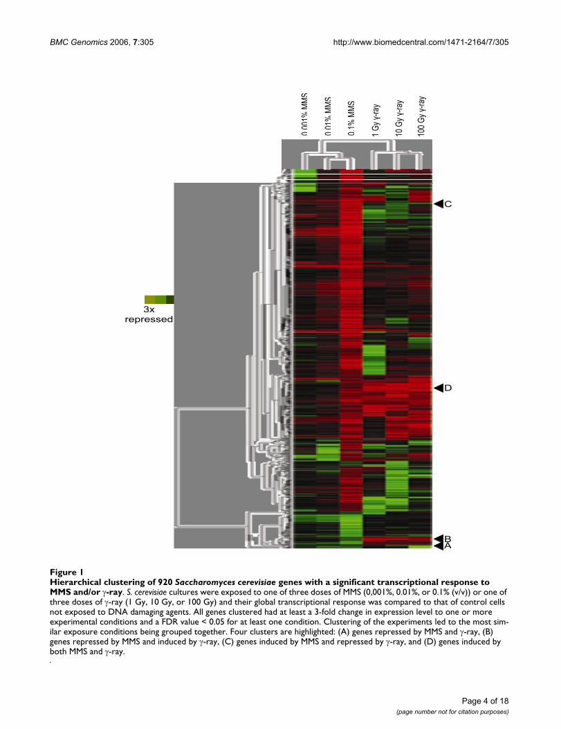
Results and DiscussionHierarchical clustering of array dataHierarchical clustering illustrating the transcriptionalresponse of S. cerevisiae to MMS and γ-ray is shown in Fig-ures 1, 2, 3, 4, 5, 6. Figure 1 shows all of the genes with asignificant response to at least one dose of MMS and/or γ-ray, and Figure 2 shows a Venn diagram containing thenumber of genes regulated by each type and dose of gen-otoxicity. Figure 3 highlights four clusters found in Figure1 that have notable responses to both MMS and γ-ray. Fig-
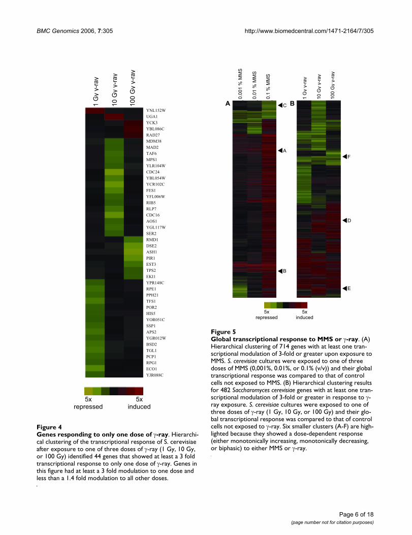
ure 4 illustrates a subset of genes that responded to onlyone dose of γ-ray. Figure 5-A contains only those geneswith a significant transcriptional response to at least onedose of MMS (γ-ray was not considered in this cluster),and Figure 5-B contains genes with a significant responseto at least one dose of γ-ray. Figure 6 contains clustersfrom Figure 5 in which a dose-dependent response toeither MMS or γ-ray was observed.
Global transcriptional response to MMS and γ-rayFigure 1 shows an overview of the global transcriptionalresponses to both MMS and γ-ray, obtained by simultane-ous clustering of genes and treatments. Clustering of treat-ments shows that all MMS experiments grouped together,as did the γ-ray-treated samples. Furthermore, within thecluster for a specific type of damage, the highest dosestested were clustered together (0.01% and 0.1% MMS and10 Gy and 100 Gy for γ-ray). Clearly, exposure to MMSand γ-ray evokes a widespread transcriptional response inS. cerevisiae cells. Many of the clustered genes exhibited atranscriptional response to both MMS and γ-ray,although, it is clear that the response often varied withdose, as expected (see Additional Files 1, 2, 3, 4).
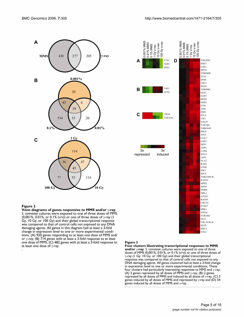
For further clarity, a Venn diagram (Figure 2) is presentedto illustrate the number of genes modulated by at least 3-fold to each DNA damaging agent (2-A), each dose ofMMS (2-B), and each dose of γ-ray (2-C).
Clusters responsive to MMS and γ-rayIn addition to merely identifying genes that were modu-lated in response to MMS and/or γ-ray, we sought to iden-tify clusters of genes that exhibited similar or oppositedose dependent responses to both MMS and γ-ray. Clus-tering diagrams of four clusters (A-D) highlighted in Fig-ure 1 are shown in Figure 3.
Cluster 3-A contains 3 genes, all of which have beenshown to play a role in mother-daughter cell separationand all of which were down-regulated in response to alldoses of MMS and γ-ray. One of these genes is CTS1, a chi-tinase required for cell separation after mitosis, whosetranscription is induced during the late M and early G1stages of the cell cycle [36,37]. Microarray analysis of S.cerevisiae exposed to 5-fluorocytosine, an inhibitor ofDNA synthesis, also identified down-regulation of CTS1[38]. DSE2 is involved in the separation of daughter cellsfrom mother cells by degrading the cell wall from thedaughter side [39,40]. ASH1 localizes in daughter cellsduring mitosis and inhibits the transcription of HO, pre-venting the daughter cells from changing mating types[41]. The fact that the transcription factor ASH1 wasdownregulated after DNA damage exposure correlateswell with observations of Workman et al. that Ash1pbinds more genes in the absence of MMS than when MMS
Page 3 of 18(page number not for citation purposes)
BMC Genomics 2006, 7:305 http://www.biomedcentral.com/1471-2164/7/305
Page 4 of 18(page number not for citation purposes)
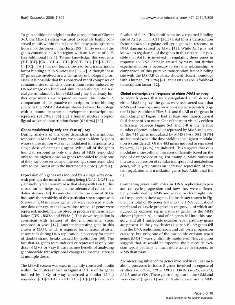
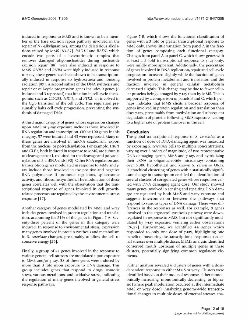
Hierarchical clustering of 920 Saccharomyces cerevisiae genes with a significant transcriptional response to MMS and/or γ-rayFigure 1Hierarchical clustering of 920 Saccharomyces cerevisiae genes with a significant transcriptional response to MMS and/or γ-ray. S. cerevisiae cultures were exposed to one of three doses of MMS (0,001%, 0.01%, or 0.1% (v/v)) or one of three doses of γ-ray (1 Gy, 10 Gy, or 100 Gy) and their global transcriptional response was compared to that of control cells not exposed to DNA damaging agents. All genes clustered had at least a 3-fold change in expression level to one or more experimental conditions and a FDR value < 0.05 for at least one condition. Clustering of the experiments led to the most sim-ilar exposure conditions being grouped together. Four clusters are highlighted: (A) genes repressed by MMS and γ-ray, (B) genes repressed by MMS and induced by γ-ray, (C) genes induced by MMS and repressed by γ-ray, and (D) genes induced by both MMS and γ-ray.
0.00
1%M
MS
0.1%
MM
S
0.01
%M
MS
1 G
y -r
ay
100
Gy
-ray
10 G
y -r
ay
3xrepressed
3xinduced
AB
C
D
BMC Genomics 2006, 7:305 http://www.biomedcentral.com/1471-2164/7/305
Page 5 of 18(page number not for citation purposes)
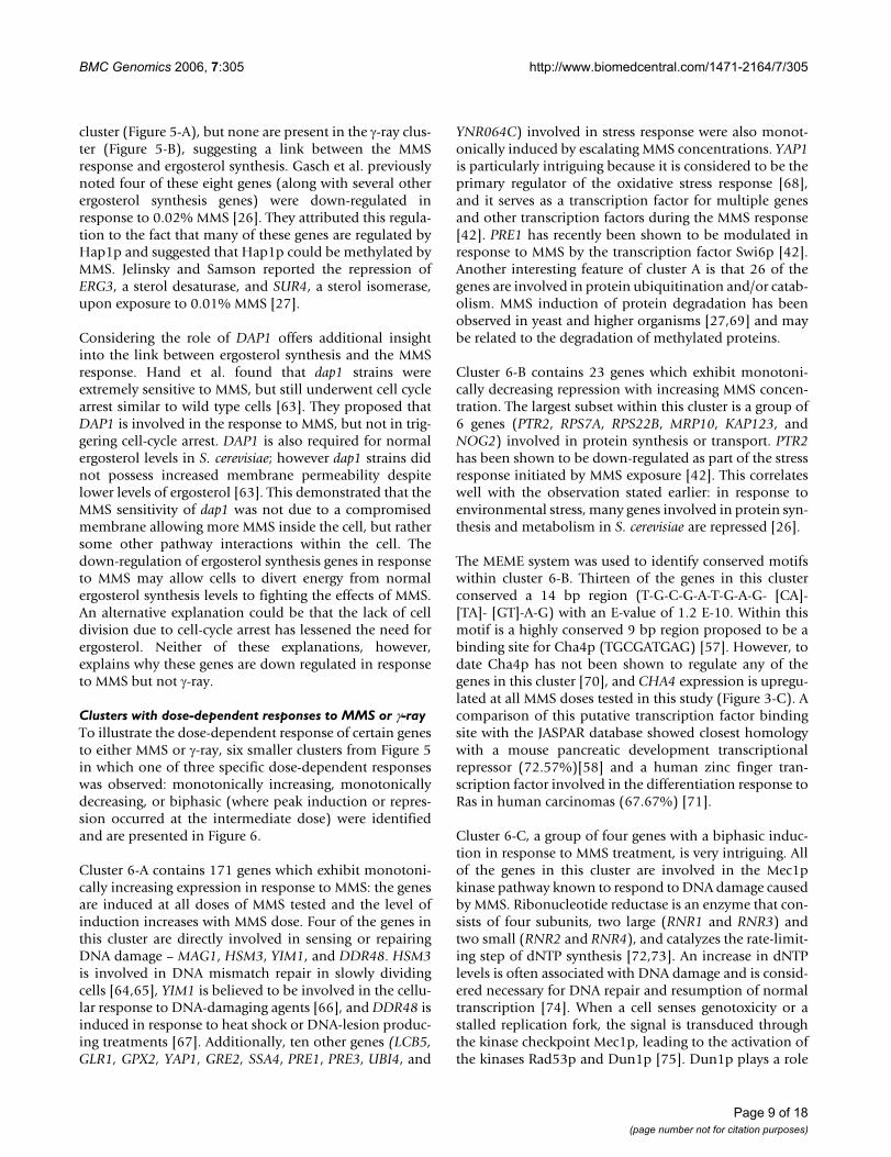
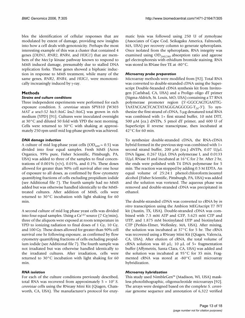
Four clusters illustrating transcriptional responses to MMS and/or γ-rayFigure 3Four clusters illustrating transcriptional responses to MMS and/or γ-ray. S. cerevisiae cultures were exposed to one of three doses of MMS (0,001%, 0.01%, or 0.1% (v/v)) or one of three doses of γ-ray (1 Gy, 10 Gy, or 100 Gy) and their global transcriptional response was compared to that of control cells not exposed to any DNA damaging agents. All genes clustered had at least a 3-fold change in expression level to one or more experimental conditions. These four clusters had particularly interesting responses to MMS and γ-ray: (A) 3 genes repressed by all doses of MMS and γ-ray, (B) 2 genes repressed by all doses of MMS and induced by all doses of γ-ray, (C) 2 genes induced by all doses of MMS and repressed by γ-ray and (D) 54 genes induced by all doses of MMS and γ-ray.
0.0
01%
MM
S
YHL029C
KRE5
LGE1
MSN1
YPR090W
FUS3
DSS4
CDC1
MAS1
YDR090C
GLE1
HAP2
BEM3
SMD1
GTS1
APP1
PIM1
PTC4
VIP1
GAL83
YAP1801
YBR056W
PHO2
STE5
CUE3
GAD1
DEP1
ADP1
CAX4
STE13
ASF2
NCA2
RAD1
COG6
PSF2
SEC6
YDR210W-B
RAD16
MPD2
ARG4
NMD4
MRL1
ATG26
RAD54
CWC23
ECM37
HRK1
VPS34
YGR146C
NEJ1
YBL101W-B
SED5
SWC5
NTG1
D
0.1
% M
MS
0.0
1%
MM
S
1 G
y-r
ay
10
0 G
y-r
ay
CTS1
ASH1
DSE2
FAS1
ACS2
CHA4
YDL016C
A
B
C
0.0
01%
MM
S
0.1
% M
MS
0.0
1%
MM
S
1 G
y-r
ay
10
0 G
y-r
ay
10
Gy
-ray
3xrepressed
3xinduced
10
Gy
-ray
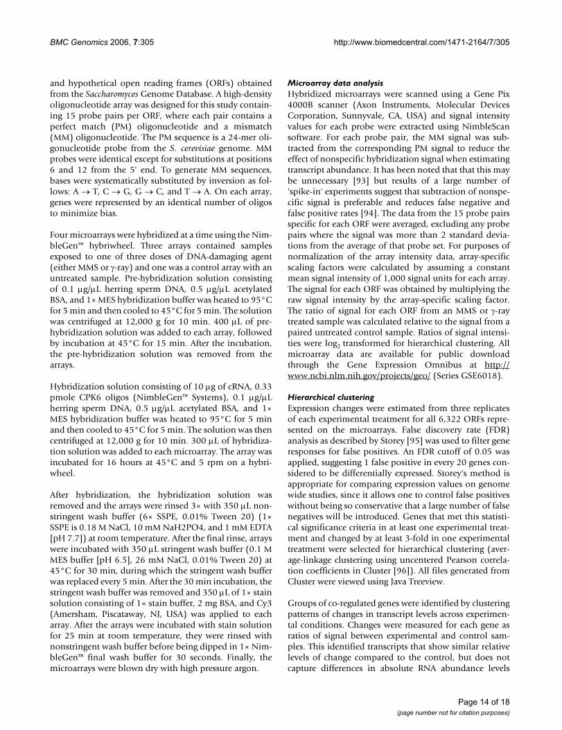
Venn diagrams of genes responsive to MMS and/or γ-rayFigure 2Venn diagrams of genes responsive to MMS and/or γ-ray. S. cerevisiae cultures were exposed to one of three doses of MMS (0,001%, 0.01%, or 0.1% (v/v)) or one of three doses of γ-ray (1 Gy, 10 Gy, or 100 Gy) and their global transcriptional response was compared to that of control cells not exposed to any DNA damaging agents. All genes in this diagram had at least a 3-fold change in expression level to one or more experimental condi-tions. (A) 920 genes responding to at least one dose of MMS and/or γ-ray, (B) 714 genes with at least a 3-fold response to at least one dose of MMS, (C) 482 genes with at least a 3-fold response to at least one dose of γ-ray.
MMS 438 -ray205277
A
20
0.001%
534 20
6
53
43
39
0.1% 0.01%
B
114
1 Gy
100 Gy
73
10 Gy
114
47
53
36
45
C
BMC Genomics 2006, 7:305 http://www.biomedcentral.com/1471-2164/7/305
Page 6 of 18(page number not for citation purposes)
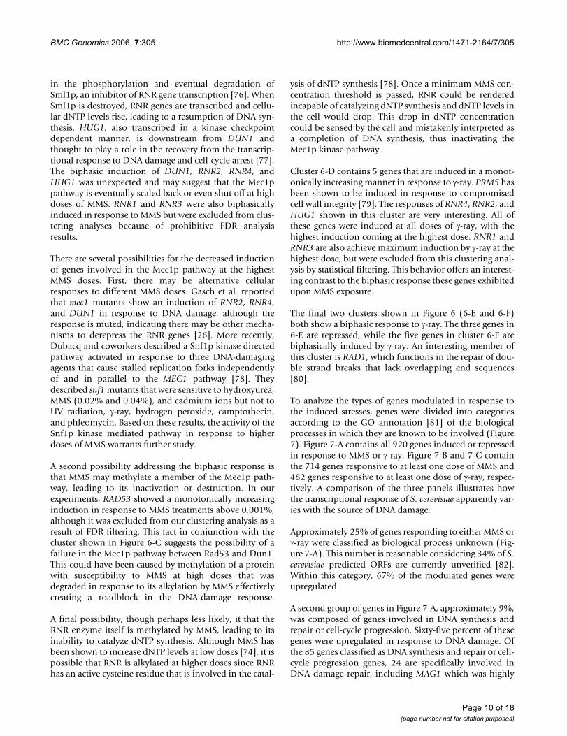
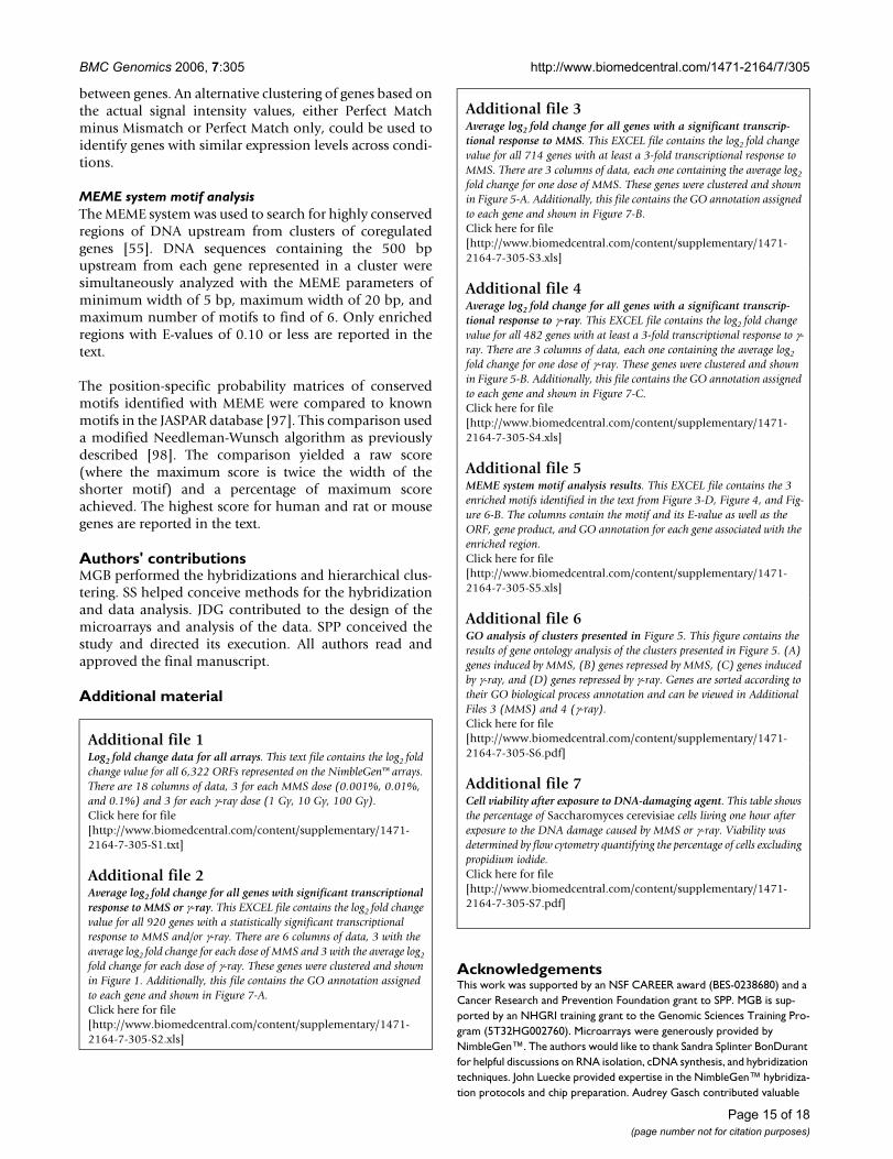
Global transcriptional response to MMS or γ-rayFigure 5Global transcriptional response to MMS or γ-ray. (A) Hierarchical clustering of 714 genes with at least one tran-scriptional modulation of 3-fold or greater upon exposure to MMS. S. cerevisiae cultures were exposed to one of three doses of MMS (0,001%, 0.01%, or 0.1% (v/v)) and their global transcriptional response was compared to that of control cells not exposed to MMS. (B) Hierarchical clustering results for 482 Saccharomyces cerevisiae genes with at least one tran-scriptional modulation of 3-fold or greater in response to γ-ray exposure. S. cerevisiae cultures were exposed to one of three doses of γ-ray (1 Gy, 10 Gy, or 100 Gy) and their glo-bal transcriptional response was compared to that of control cells not exposed to γ-ray. Six smaller clusters (A-F) are high-lighted because they showed a dose-dependent response (either monotonically increasing, monotonically decreasing, or biphasic) to either MMS or γ-ray.
0.0
01 %
MM
S
0.0
1 %
MM
S
0.1
% M
MS
A
B
CA
1 G
y-r
ay
10
Gy
-ray
100 G
y-r
ay
D
E
F
B
5xrepressed
5xinduced
Genes responding to only one dose of γ-rayFigure 4Genes responding to only one dose of γ-ray. Hierarchi-cal clustering of the transcriptional response of S. cerevisiae after exposure to one of three doses of γ-ray (1 Gy, 10 Gy, or 100 Gy) identified 44 genes that showed at least a 3 fold transcriptional response to only one dose of γ-ray. Genes in this figure had at least a 3 fold modulation to one dose and less than a 1.4 fold modulation to all other doses.
y
yYNL152W
UGA1
YCK3
YBL086C
RAD27
MDM38
MAD2
TAF6
MPS1
YLR104W
CDC24
YBL054W
YCR102C
FES1
YFL006W
RIB5
RLP7
CDC16
AOS1
YGL117W
SER2
RMD1
DSE2
ASH1
PIR1
EST3
TPS2
EKI1
YPR148C
RPE1
PPH21
TFS1
POR2
HIS5
YOR051C
SSP1
APS2
YGR012W
BSD2
TGL1
PCP1
RPG1
ECO1
YJR088C
5xrepressed
5xinduced
1 G
y-r
ay
10 G
y-r
a
100 G
y-r
a
BMC Genomics 2006, 7:305 http://www.biomedcentral.com/1471-2164/7/305
is present [42]. The co-regulation of these 3 genes impliesthat the cell may be actively trying to stop transmission ofdamaged DNA to daughter cells by inhibiting cell divi-sion.
Cluster 3-B contains 2 genes repressed in response toMMS and induced by γ-ray exposure. FAS1 is involved inthe synthesis of fatty acids [43,44]. A third gene with sim-ilar behavior but excluded from this cluster due to higher
than acceptable FDR values, HMG1, catalyzes the rate-lim-iting step in sterol biosynthesis [45]. Gasch et al. observedthe repression of genes responsible for ergosterol synthe-sis in response to MMS [26], while De Sanctis et al. notedmany of these same genes were induced in response tolow doses of ionizing radiation, approximately 80 Gy orless [35].
Cluster 3-C contains 2 genes induced in response to MMSand repressed in response to γ-ray. One of the genes in thiscluster, YDL016C, is a hypothetical ORF. The other gene,CHA4, is a transcription factor for CHA1. In the presenceof serine or threonine as the sole nitrogen source, CHA1 isactively transcribed, but its expression is not detectablewhen cells are grown in the presence of other nitrogensources such as ammonium [46]. A recent study by Yanget al. suggests CHA4 may play a role in cell-cycle regula-tion [47] as one of four putative interaction partners of thetwo forkhead transcription factors, FKH1 and FKH2,which are involved in cell-cycle regulation and mitoticexit and seem to play a role in yeast dimorphism [48]. Thisis particularly interesting since 5 of the genes clustered inFigure 1 (ASH1, BEM3, CDC24, TEC1, and UBA4) play arole in regulating pseudohyphal growth in S. cerevisiae.
The cluster highlighted in Figure 3-D is composed of 54genes whose transcription is induced upon exposure to alldoses of MMS and γ-ray used in this study. Several of thegenes in this cluster are known to be involved in cellularresponses to stress or DNA damage. GAD1 has beenshown to mediate resistance to oxidative stress induced byhydrogen peroxide and diamide [49], and NTG1 has alsobeen identified as crucial in repairing oxidative stress inDNA [50]. RAD16 is part of the nuclear excision repairpathway and has been shown to be involved in double-strand break repair in the fission yeast, Schizosaccharomycespombe, suggesting an explanation for its induction inresponse to γ-ray [51]. NEJ1 is involved in the regulationof non-homologous end joining repair in response todouble-strand breaks [52]. A potential reason for NEJ1induction by MMS may involve creation of single-strandbreaks following excision repair of methylated bases.When the replication fork confronts these breaks, double-strand breaks can be caused by the forces of replication[53], which could elicit a partial transcriptional responsein genes responsible for repairing double-strand breaks.This could also address induction of RAD54, known to becrucial in homologous recombination for the repair ofdouble-strand breaks [54], in response to MMS. The genesshown in this cluster suggest there is some overlap ofDNA-damage repair pathways in sensing and respondingto damage induced by MMS and γ-ray, as members of thebase excision repair pathway, nucleotide excision repairpathway, non-homologous end joining pathway, andhomologous recombination pathway were all induced.
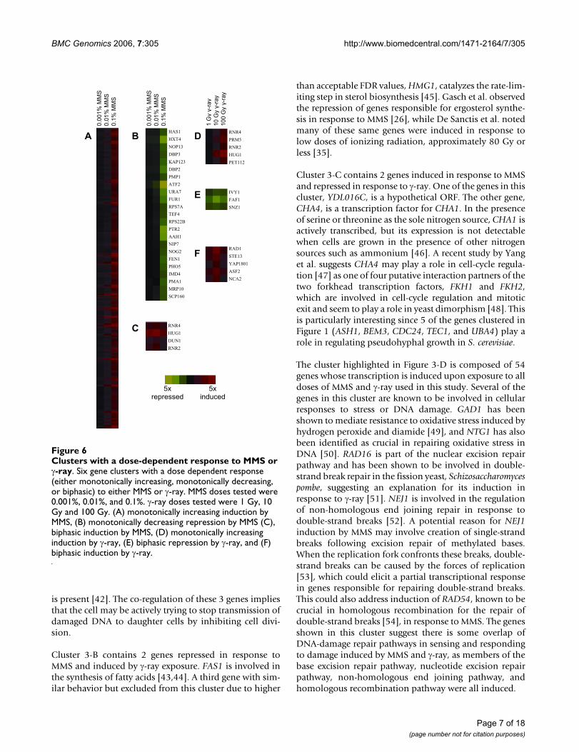
Clusters with a dose-dependent response to MMS or γ-rayFigure 6Clusters with a dose-dependent response to MMS or γ-ray. Six gene clusters with a dose dependent response (either monotonically increasing, monotonically decreasing, or biphasic) to either MMS or γ-ray. MMS doses tested were 0.001%, 0.01%, and 0.1%. γ-ray doses tested were 1 Gy, 10 Gy and 100 Gy. (A) monotonically increasing induction by MMS, (B) monotonically decreasing repression by MMS (C), biphasic induction by MMS, (D) monotonically increasing induction by γ-ray, (E) biphasic repression by γ-ray, and (F) biphasic induction by γ-ray.
0.0
01%
MM
S
0.1
% M
MS
0.0
1%
MM
S
1 G
y-r
ay
10
0 G
y-r
ay
10
Gy
-ray
0.0
01%
MM
S
0.1
% M
MS
0.0
1%
MM
S
HAS1
HXT4
NOP13
DBP3
KAP123
DBP2
PMP1
ATF2
URA7
FUR1
RPS7A
TEF4
RPS22B
PTR2
AAH1
NIP7
NOG2
FEN1
PHO5
IMD4
PMA1
MRP10
SCP160
RNR4
PRM5
RNR2
HUG1
PET112
A B D
E IVY1
FAF1
SNZ1
RAD1
STE13
YAP1801
ASF2
NCA2
F
C RNR4
HUG1
DUN1
RNR2
5xrepressed
5xinduced
Page 7 of 18(page number not for citation purposes)
BMC Genomics 2006, 7:305 http://www.biomedcentral.com/1471-2164/7/305
To gain additional insight into the coregulation of Cluster3-D, the MEME system was used to identify highly con-served motifs within the regions 500 base pairs upstreamfrom all of the genes in the cluster [55]. Thirty seven of thegenes contained a 16 bp region with an E-value of 0.05(see Additional file 5). To our knowledge, this sequence(T-T- [CT]- [CA]- [CT]-C- [CT]- [CA]-T- [TC]- [TC]-T- [TC]-C- [TC]- [TA]) has not been shown to be a transcriptionfactor binding site in S. cerevisiae [56,57]. Although these37 genes are involved in a wide variety of biological proc-esses, it is possible that this conserved motif comprises orcontains a site to which a transcription factor induced byDNA-damage can bind and simultaneously regulate sev-eral genes induced by both MMS and γ-ray, but clearly fur-ther experiments are required to prove this notion. Acomparison of this putative transcription factor bindingsite with the JASPAR database showed closest homologywith a mouse pancreatic development transcriptionalrepressor (81.78%) [58] and a human nuclear receptorligand activated transcription factor (67.67%) [59].
Genes modulated by only one dose of γ-rayDuring analysis of the dose dependent transcriptionalresponse to MMS and γ-ray, we sought to identify geneswhose transcription was only modulated in response to asingle dose of damaging agent. While all of the genesfound to respond to only one dose of MMS respondedonly to the highest dose, 44 genes responded to only oneof the γ-ray doses tested and interestingly some respondedonly to the lowest or to the intermediate dose (Figure 4).
Expression of 5 genes was induced by a single γ-ray dose,with perhaps the most interesting being UGA1. UGA1 is aγ-aminobutyrate transaminase that along with GAD1, dis-cussed earlier, helps regulate the tolerance of cells to oxi-dative stresses [49]. Its induction at the low dose of 10 Gyindicates the sensitivity of this particular stress response inS. cerevisiae. Many more genes, 39, were repressed at onlyone dose of γ-ray. At the lowest dose tested, 16 genes wererepressed, including 3 involved in protein synthesis regu-lation (TFS1, BSD2, and PPH21). This down-regulation isconsistent with features of the environmental stressresponse in yeast [17]. Another interesting gene in thiscluster is ECO1, which is required for cohesion of sisterchromatids during DNA replication, a necessity for repairof double-strand breaks caused by replication [60]. Thefact that 44 genes were induced or repressed at only onedose of MMS or γ-ray illustrates one benefit of analyzinggenome-wide transcriptional changes to external stressesat multiple doses.
The MEME system was used to identify conserved motifswithin the clusters shown in Figure 4. All 16 of the genesinduced by 1 Gy of γ-ray conserved a similar 12 bpsequence ([CG]-T-T-T-T-T-T-T- [TC]- [TC]- [TA]-T) with an
E-value of 0.08. This motif contains a reported bindingsite of Azf1p, TTTTTCTT [56,57]. Azf1p is a transcriptionfactor shown to regulate cell cycle genes in response toDNA damage caused by MMS [42]. While Azf1p is notknown to regulate all of the genes in this cluster, it is pos-sible that Azf1p is involved in regulating these genes inresponse to DNA damage caused by γ-ray, but furtherexperimentation is necessary to test this relationship. Acomparison of this putative transcription factor bindingsite with the JASPAR database showed closest homologywith a human (79.17%) [61] and a rat (80.45%) forkheadtranscription factor [62].
Global transcriptional response to either MMS or γ-rayTo identify genes that were coregulated at all doses ofeither MMS or γ-ray, the genes were reclustered such thatMMS and γ-ray exposure were considered separately (Fig-ure 5) (see Additional Files 3, 4, and 6). All of the genes ineach cluster in Figure 5 had at least one transcriptionalfold change of 3 or more. One of the most visually evidentdifferences between Figure 5-A and 5-B is the relativenumber of genes induced or repressed by MMS and γ-ray.Of the 714 genes modulated by MMS (5-A), 581 (81%)are induced (when the dose yielding the highest modula-tion is considered). Of the 482 genes induced or repressedby γ-ray, 228 (47%) are induced. This suggests that cellsmodulate entire cellular processes differently based on thetype of damage occurring. For example, MMS causes anincreased repression of cellular transport and metabolismgenes, while γ-ray causes an increased repression of pro-tein regulation and translation genes (see Additional file6).
Comparing genes with roles in DNA replication/repairand cell-cycle progression and how they were differen-tially modulated by MMS and γ-ray provides insight intocell responses to these agents. In the cluster shown in Fig-ure 1, a total of 85 genes fell into the DNA replication/repair and cell-cycle progression category, 4 of which arenucleotide excision repair pathway genes. In the MMScluster (Figure 5-A), a total of 63 genes fell into this cate-gory, and all 4 nucleotide excision repair pathway genesare present. In the γ-ray cluster (Figure 5-B), 59 genes fellinto the DNA replication/repair and cell-cycle progressioncategory, but only one of the nucleotide excision repairgenes, RAD16, was significantly modulated. This variationsuggests that, as would be expected, the nucleotide exci-sion repair pathway is much more active in response toMMS than γ-ray.
An interesting subset of the genes involved in cellular met-abolic processes includes 8 genes involved in ergosterolsynthesis – ERG28, ERG2, ERG11, ERG6, ERG25, ERG13,ERG1, and MVD1. These genes all appear in the MMS andγ-ray cluster (Figure 1) and all 8 also appear in the MMS
Page 8 of 18(page number not for citation purposes)
BMC Genomics 2006, 7:305 http://www.biomedcentral.com/1471-2164/7/305
cluster (Figure 5-A), but none are present in the γ-ray clus-ter (Figure 5-B), suggesting a link between the MMSresponse and ergosterol synthesis. Gasch et al. previouslynoted four of these eight genes (along with several otherergosterol synthesis genes) were down-regulated inresponse to 0.02% MMS [26]. They attributed this regula-tion to the fact that many of these genes are regulated byHap1p and suggested that Hap1p could be methylated byMMS. Jelinsky and Samson reported the repression ofERG3, a sterol desaturase, and SUR4, a sterol isomerase,upon exposure to 0.01% MMS [27].
Considering the role of DAP1 offers additional insightinto the link between ergosterol synthesis and the MMSresponse. Hand et al. found that dap1 strains wereextremely sensitive to MMS, but still underwent cell cyclearrest similar to wild type cells [63]. They proposed thatDAP1 is involved in the response to MMS, but not in trig-gering cell-cycle arrest. DAP1 is also required for normalergosterol levels in S. cerevisiae; however dap1 strains didnot possess increased membrane permeability despitelower levels of ergosterol [63]. This demonstrated that theMMS sensitivity of dap1 was not due to a compromisedmembrane allowing more MMS inside the cell, but rathersome other pathway interactions within the cell. Thedown-regulation of ergosterol synthesis genes in responseto MMS may allow cells to divert energy from normalergosterol synthesis levels to fighting the effects of MMS.An alternative explanation could be that the lack of celldivision due to cell-cycle arrest has lessened the need forergosterol. Neither of these explanations, however,explains why these genes are down regulated in responseto MMS but not γ-ray.
Clusters with dose-dependent responses to MMS or γ-rayTo illustrate the dose-dependent response of certain genesto either MMS or γ-ray, six smaller clusters from Figure 5in which one of three specific dose-dependent responseswas observed: monotonically increasing, monotonicallydecreasing, or biphasic (where peak induction or repres-sion occurred at the intermediate dose) were identifiedand are presented in Figure 6.
Cluster 6-A contains 171 genes which exhibit monotoni-cally increasing expression in response to MMS: the genesare induced at all doses of MMS tested and the level ofinduction increases with MMS dose. Four of the genes inthis cluster are directly involved in sensing or repairingDNA damage – MAG1, HSM3, YIM1, and DDR48. HSM3is involved in DNA mismatch repair in slowly dividingcells [64,65], YIM1 is believed to be involved in the cellu-lar response to DNA-damaging agents [66], and DDR48 isinduced in response to heat shock or DNA-lesion produc-ing treatments [67]. Additionally, ten other genes (LCB5,GLR1, GPX2, YAP1, GRE2, SSA4, PRE1, PRE3, UBI4, and
YNR064C) involved in stress response were also monot-onically induced by escalating MMS concentrations. YAP1is particularly intriguing because it is considered to be theprimary regulator of the oxidative stress response [68],and it serves as a transcription factor for multiple genesand other transcription factors during the MMS response[42]. PRE1 has recently been shown to be modulated inresponse to MMS by the transcription factor Swi6p [42].Another interesting feature of cluster A is that 26 of thegenes are involved in protein ubiquitination and/or catab-olism. MMS induction of protein degradation has beenobserved in yeast and higher organisms [27,69] and maybe related to the degradation of methylated proteins.
Cluster 6-B contains 23 genes which exhibit monotoni-cally decreasing repression with increasing MMS concen-tration. The largest subset within this cluster is a group of6 genes (PTR2, RPS7A, RPS22B, MRP10, KAP123, andNOG2) involved in protein synthesis or transport. PTR2has been shown to be down-regulated as part of the stressresponse initiated by MMS exposure [42]. This correlateswell with the observation stated earlier: in response toenvironmental stress, many genes involved in protein syn-thesis and metabolism in S. cerevisiae are repressed [26].
The MEME system was used to identify conserved motifswithin cluster 6-B. Thirteen of the genes in this clusterconserved a 14 bp region (T-G-C-G-A-T-G-A-G- [CA]-[TA]- [GT]-A-G) with an E-value of 1.2 E-10. Within thismotif is a highly conserved 9 bp region proposed to be abinding site for Cha4p (TGCGATGAG) [57]. However, todate Cha4p has not been shown to regulate any of thegenes in this cluster [70], and CHA4 expression is upregu-lated at all MMS doses tested in this study (Figure 3-C). Acomparison of this putative transcription factor bindingsite with the JASPAR database showed closest homologywith a mouse pancreatic development transcriptionalrepressor (72.57%)[58] and a human zinc finger tran-scription factor involved in the differentiation response toRas in human carcinomas (67.67%) [71].
Cluster 6-C, a group of four genes with a biphasic induc-tion in response to MMS treatment, is very intriguing. Allof the genes in this cluster are involved in the Mec1pkinase pathway known to respond to DNA damage causedby MMS. Ribonucleotide reductase is an enzyme that con-sists of four subunits, two large (RNR1 and RNR3) andtwo small (RNR2 and RNR4), and catalyzes the rate-limit-ing step of dNTP synthesis [72,73]. An increase in dNTPlevels is often associated with DNA damage and is consid-ered necessary for DNA repair and resumption of normaltranscription [74]. When a cell senses genotoxicity or astalled replication fork, the signal is transduced throughthe kinase checkpoint Mec1p, leading to the activation ofthe kinases Rad53p and Dun1p [75]. Dun1p plays a role
Page 9 of 18(page number not for citation purposes)
BMC Genomics 2006, 7:305 http://www.biomedcentral.com/1471-2164/7/305
in the phosphorylation and eventual degradation ofSml1p, an inhibitor of RNR gene transcription [76]. WhenSml1p is destroyed, RNR genes are transcribed and cellu-lar dNTP levels rise, leading to a resumption of DNA syn-thesis. HUG1, also transcribed in a kinase checkpointdependent manner, is downstream from DUN1 andthought to play a role in the recovery from the transcrip-tional response to DNA damage and cell-cycle arrest [77].The biphasic induction of DUN1, RNR2, RNR4, andHUG1 was unexpected and may suggest that the Mec1ppathway is eventually scaled back or even shut off at highdoses of MMS. RNR1 and RNR3 were also biphasicallyinduced in response to MMS but were excluded from clus-tering analyses because of prohibitive FDR analysisresults.
There are several possibilities for the decreased inductionof genes involved in the Mec1p pathway at the highestMMS doses. First, there may be alternative cellularresponses to different MMS doses. Gasch et al. reportedthat mec1 mutants show an induction of RNR2, RNR4,and DUN1 in response to DNA damage, although theresponse is muted, indicating there may be other mecha-nisms to derepress the RNR genes [26]. More recently,Dubacq and coworkers described a Snf1p kinase directedpathway activated in response to three DNA-damagingagents that cause stalled replication forks independentlyof and in parallel to the MEC1 pathway [78]. Theydescribed snf1 mutants that were sensitive to hydroxyurea,MMS (0.02% and 0.04%), and cadmium ions but not toUV radiation, γ-ray, hydrogen peroxide, camptothecin,and phleomycin. Based on these results, the activity of theSnf1p kinase mediated pathway in response to higherdoses of MMS warrants further study.
A second possibility addressing the biphasic response isthat MMS may methylate a member of the Mec1p path-way, leading to its inactivation or destruction. In ourexperiments, RAD53 showed a monotonically increasinginduction in response to MMS treatments above 0.001%,although it was excluded from our clustering analysis as aresult of FDR filtering. This fact in conjunction with thecluster shown in Figure 6-C suggests the possibility of afailure in the Mec1p pathway between Rad53 and Dun1.This could have been caused by methylation of a proteinwith susceptibility to MMS at high doses that wasdegraded in response to its alkylation by MMS effectivelycreating a roadblock in the DNA-damage response.
A final possibility, though perhaps less likely, it that theRNR enzyme itself is methylated by MMS, leading to itsinability to catalyze dNTP synthesis. Although MMS hasbeen shown to increase dNTP levels at low doses [74], it ispossible that RNR is alkylated at higher doses since RNRhas an active cysteine residue that is involved in the catal-
ysis of dNTP synthesis [78]. Once a minimum MMS con-centration threshold is passed, RNR could be renderedincapable of catalyzing dNTP synthesis and dNTP levels inthe cell would drop. This drop in dNTP concentrationcould be sensed by the cell and mistakenly interpreted asa completion of DNA synthesis, thus inactivating theMec1p kinase pathway.
Cluster 6-D contains 5 genes that are induced in a monot-onically increasing manner in response to γ-ray. PRM5 hasbeen shown to be induced in response to compromisedcell wall integrity [79]. The responses of RNR4, RNR2, andHUG1 shown in this cluster are very interesting. All ofthese genes were induced at all doses of γ-ray, with thehighest induction coming at the highest dose. RNR1 andRNR3 are also achieve maximum induction by γ-ray at thehighest dose, but were excluded from this clustering anal-ysis by statistical filtering. This behavior offers an interest-ing contrast to the biphasic response these genes exhibitedupon MMS exposure.
The final two clusters shown in Figure 6 (6-E and 6-F)both show a biphasic response to γ-ray. The three genes in6-E are repressed, while the five genes in cluster 6-F arebiphasically induced by γ-ray. An interesting member ofthis cluster is RAD1, which functions in the repair of dou-ble strand breaks that lack overlapping end sequences[80].
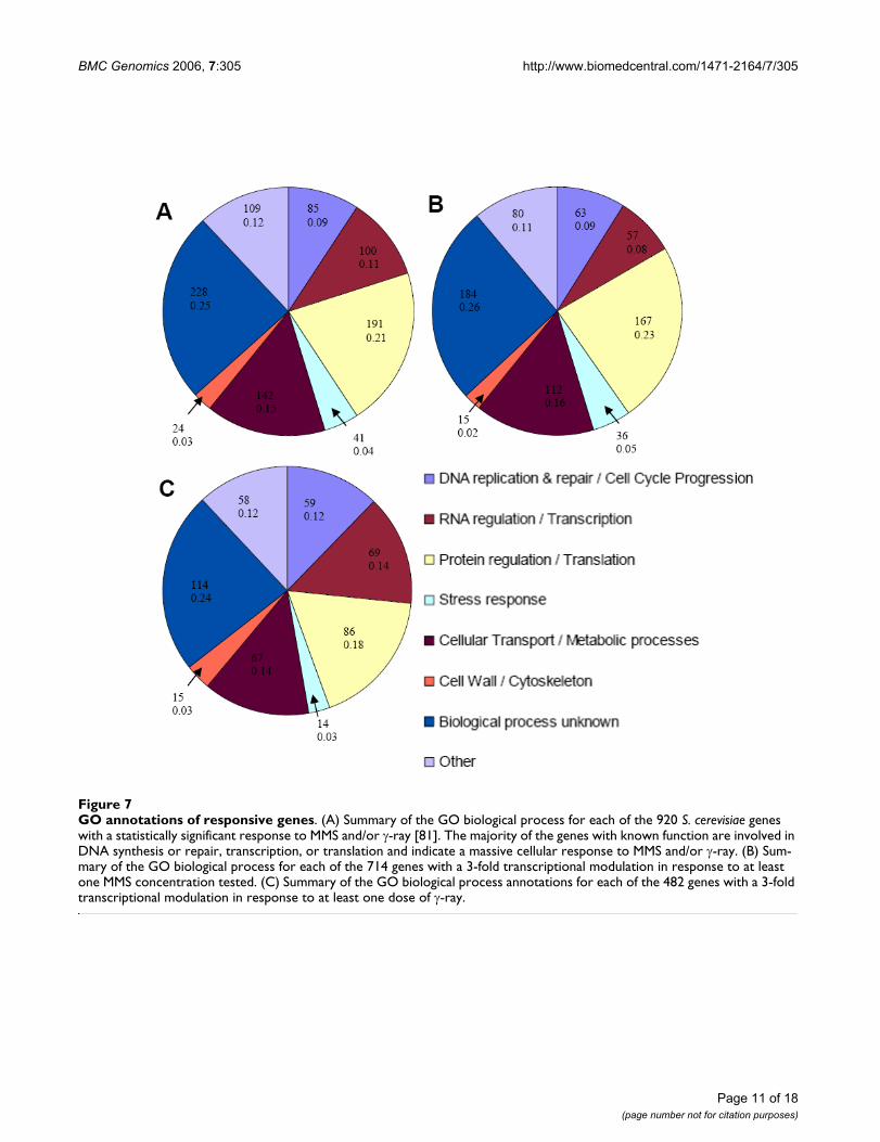
To analyze the types of genes modulated in response tothe induced stresses, genes were divided into categoriesaccording to the GO annotation [81] of the biologicalprocesses in which they are known to be involved (Figure7). Figure 7-A contains all 920 genes induced or repressedin response to MMS or γ-ray. Figure 7-B and 7-C containthe 714 genes responsive to at least one dose of MMS and482 genes responsive to at least one dose of γ-ray, respec-tively. A comparison of the three panels illustrates howthe transcriptional response of S. cerevisiae apparently var-ies with the source of DNA damage.
Approximately 25% of genes responding to either MMS orγ-ray were classified as biological process unknown (Fig-ure 7-A). This number is reasonable considering 34% of S.cerevisiae predicted ORFs are currently unverified [82].Within this category, 67% of the modulated genes wereupregulated.
A second group of genes in Figure 7-A, approximately 9%,was composed of genes involved in DNA synthesis andrepair or cell-cycle progression. Sixty-five percent of thesegenes were upregulated in response to DNA damage. Ofthe 85 genes classified as DNA synthesis and repair or cell-cycle progression genes, 24 are specifically involved inDNA damage repair, including MAG1 which was highly
Page 10 of 18(page number not for citation purposes)
BMC Genomics 2006, 7:305 http://www.biomedcentral.com/1471-2164/7/305
Page 11 of 18(page number not for citation purposes)
GO annotations of responsive genesFigure 7GO annotations of responsive genes. (A) Summary of the GO biological process for each of the 920 S. cerevisiae genes with a statistically significant response to MMS and/or γ-ray [81]. The majority of the genes with known function are involved in DNA synthesis or repair, transcription, or translation and indicate a massive cellular response to MMS and/or γ-ray. (B) Sum-mary of the GO biological process for each of the 714 genes with a 3-fold transcriptional modulation in response to at least one MMS concentration tested. (C) Summary of the GO biological process annotations for each of the 482 genes with a 3-fold transcriptional modulation in response to at least one dose of γ-ray.
BMC Genomics 2006, 7:305 http://www.biomedcentral.com/1471-2164/7/305
induced in response to MMS and is known to be a mem-ber of the base excision repair pathway involved in therepair of N7-alkylguanines, among the deleterious alkyla-tions caused by MMS [83-87]. RAD16 and RAD7, whichencode two parts of a heterotrimeric complex thatremoves damaged oligonucleotides during nucleotideexcision repair [88], were also induced in response toMMS. RNR2 and RNR4 were highly induced in responseto γ-ray; these genes have been shown to be transcription-ally induced in response to hydroxyurea and ionizingradiation [89]. A second subset of the DNA synthesis andrepair or cell-cycle progression genes includes 9 genes (6induced and 3 repressed) that function in cell-cycle check-points, such as CDC53, HRT1, and PTK2, all involved inthe G1/S transition of the cell cycle. This regulation pre-sumably halts cell cycle progression, preventing the syn-thesis of damaged DNA.
A third major category of genes whose expression changesupon MMS or γ-ray exposure includes those involved inRNA regulation and transcription. Of the 100 genes in thiscategory, 57 were induced and 43 were repressed. Many ofthese genes are involved in mRNA catabolism, exportfrom the nucleus, or polyadenylation. For example, HRP1and CLP1, both induced in response to MMS, are subunitsof cleavage factor I, required for the cleavage and polyade-nylation of 3' mRNA ends [90]. Other RNA regulation andtranscription genes modulated in response to MMS and γ-ray include those involved in the positive and negativeRNA polymerase II promoter regulators, spliceosomeactivity, and ribosome synthesis. The modulation of thesegenes correlates well with the observation that the tran-scriptional response of genes involved in cell growth-related processes are regulated by the environmental stressresponse [17].
Another category of genes modulated by MMS and γ-rayincludes genes involved in protein regulation and transla-tion, accounting for 21% of the genes in Figure 7-A. Sev-enty-three percent of the genes in this category wereinduced. In response to environmental stress, expressionmany genes involved in protein synthesis and metabolismin S. cerevisiae changes, presumably to allow the cell toconserve energy [26].
Finally, a group of 41 genes involved in the response tovarious general cell stresses are modulated upon exposureto MMS and/or γ-ray. 38 of these genes were induced bymore than 3 fold upon exposure to DNA damage. Thisgroup includes genes that respond to drugs, osmoticstress, various metal ions, and oxidative stress, indicatingthe regulation of many genes involved in general stressresponse pathways.
Figure 7-B, which shows the functional classification ofgenes with a 3 fold or greater transcriptional response toMMS only, shows little variation from panel A in the frac-tion of genes composing each functional category.Changes from panel A to panel C, which shows genes withat least a 3 fold transcriptional response to γ-ray only,were mildly more apparent. Additionally, the percentageof genes involved in DNA replication/repair and cell-cycleprogression increased slightly while the fraction of genesinvolved in protein metabolism and translation and thefraction involved in general cellular metabolismdecreased slightly. This change may be due to fewer cellu-lar proteins being damaged by γ-ray than by MMS. This issupported by a comparison of panels B and C, which per-haps indicates that MMS elicits a broader response ofgenes involved in protein regulation and translation thandoes γ-ray, presumably from methylation and subsequentdegradation of proteins following MMS exposure, leadingto a higher rate of protein turnover in the cell.
ConclusionThe global transcriptional response of S. cerevisiae as afunction of dose of DNA-damaging agent was measuredby exposing S. cerevisiae cells to multiple concentrations,varying over 3 orders of magnitude, of two representativeDNA-damaging agents, MMS and γ-ray, and hybridizingtheir cRNA to oligonucleotide microarrays containingover 6,300 hypothetical and known S. cerevisiae ORFs.Hierarchical clustering of genes with a statistically signifi-cant change in transcription enabled the identification ofseveral clusters of coregulated genes whose responses var-ied with DNA damaging agent dose. Our study showedmany genes involved in sensing and repairing DNA dam-age are regulated by both MMS and γ-ray exposure andsuggests interconnection between the pathways thatrespond to various types of DNA damage. There were dif-ferences in the responses as well. For example, 8 genesinvolved in the ergosterol synthesis pathway were down-regulated in response to MMS, but not significantly mod-ulated by γ-ray exposure, verifying earlier observations[26,27]. Furthermore, we identified 44 genes whichresponded to only one dose of γ-ray, highlighting onebenefit of measuring the transcriptional response to exter-nal stresses over multiple doses. MEME analysis identifiedconserved motifs upstream of multiple genes in theseclusters, potentially signifying common regulatory ele-ments.
Further analysis revealed 6 clusters of genes with a dose-dependent response to either MMS or γ-ray. Clusters wereidentified based on their mode of response, either monot-onically increasing, monotonically decreasing, or bipha-sic (where peak modulation occurred at the intermediateMMS or γ-ray dose). Analyzing genome-wide transcrip-tional changes to multiple doses of external stresses ena-
Page 12 of 18(page number not for citation purposes)
BMC Genomics 2006, 7:305 http://www.biomedcentral.com/1471-2164/7/305
bles the identification of cellular responses that aremodulated by extent of damage, providing new insightsinto how a cell deals with genotoxicity. Perhaps the mostinteresting example of this was a cluster that contained 4genes (DUN1, RNR2, RNR4, and HUG1) that are mem-bers of the Mec1p kinase pathway known to respond toMMS induced damage, presumably due to stalled DNAreplication forks. These genes showed a biphasic induc-tion in response to MMS treatment, while many of thesame genes, RNR2, RNR4, and HUG1, were monotoni-cally increasingly induced by γ-ray.
MethodsStrains and culture conditionsThree independent experiments were performed for eachexposure condition. S. cerevisiae strain SPY810 (W303MAT a ura3-52 his3::hisG leu2::hisG) was grown in richmedium (YPD) [91]. Cultures were inoculated overnightat 30°C and diluted 50 fold with YPD the next morning.Cells were returned to 30°C with shaking at approxi-mately 250 rpm until mid log-phase growth was achieved.
DNA damage inductionA culture of mid log-phase yeast cells (OD600 ~ 0.5) wasdivided into four equal samples. Fresh MMS (AcrosOrganics, 99% pure, Fisher Scientific, Pittsburgh, PA,USA) was added to three of the samples to final concen-trations of 0.001% (v/v), 0.01%, and 0.1%. These dosesallowed for greater than 90% cell survival after one hourof exposure to all doses, as confirmed by flow cytometryquantifying fractions of cells excluding propidium iodide(see Additional file 7). The fourth sample had no MMSadded but was otherwise handled identically to the MMS-treated cultures. After addition of MMS, cells werereturned to 30°C incubation with light shaking for 60min.
A second culture of mid log phase yeast cells was dividedinto four equal samples. Using a Cs137 source (7 Gy/min),three of the aliquots were exposed at room temperature inYPD to ionizing radiation to final doses of 1 Gy, 10 Gy,and 100 Gy. These doses allowed for greater than 90% cellsurvival one hr following exposure, as confirmed by flowcytometry quantifying fractions of cells excluding propid-ium iodide (see Additional file 7). The fourth sample wasnot irradiated but was otherwise handled identically tothe irradiated cultures. After irradiation, cells werereturned to 30°C incubation with light shaking for 60min.
RNA isolationFor each of the culture conditions previously described,total RNA was recovered from approximately 5 × 10⁷ S.cerevisiae cells using the RNeasy Mini Kit (Qiagen, Chats-worth, CA, USA). The manufacturer's protocol for enzy-
matic lysis was followed using 250 U of zymolyase(Associates of Cape Cod, Seikagaku America, Falmouth,MA, USA) per recovery column to generate spheroplasts.Once isolated from the spheroplasts, RNA integrity wasexamined using OD260/280 absorption ratio and agarosegel electrophoresis with ethidium bromide staining. RNAwas stored in RNase free TE at -80°C.
Microarray probe preparationMicroarray methods were modified from [92]. Total RNAwas converted to double-stranded cDNA using the Super-script Double-Stranded cDNA synthesis kit from Invitro-gen (Carlsbad, CA, USA) and a Proligo oligo dT primer(Sigma-Aldrich, St. Louis, MO, USA) containing a T7 RNApolymerase promoter region (5'-GGCCAGTGAATTG-TAATACGACTCACTATAGGGAGGCGG-T24-3'). To syn-thesize the first strand of cDNA, 5 µg denatured total RNAwas combined with 1× first strand buffer, 10 mM DTT,500 µM (ea.) dNTPs, 5 pmol dT primer, and 600 U ofSuperScript II reverse transcriptase, then incubated at42°C for 60 min.
To synthesize double-stranded cDNA, the RNA-cDNAhybrid formed in the previous step was combined with 1×second strand buffer, 200 µM (ea.) dNTPs, 0.07 U/µLDNA ligase, 0.267 U/µL DNA polymerase I, and 0.0133U/µL RNase H and incubated at 16°C for 2 hr. After 2 hr,the ends were polished with T4 DNA polymerase for 5min. The reaction was stopped by adding 0.5 M EDTA. Anequal volume of 25:24:1 phenol:chloroform:isoamylalcohol (Fisher Scientific, Pittsburgh, PA, USA) was addedand the solution was vortexed. The aqueous phase wasremoved and double-stranded cDNA was precipitated inethanol.
The double-stranded cDNA was converted to cRNA by invitro transcription using the Ambion MEGAscript T7 IVTkit (Austin, TX, USA). Double-stranded cDNA was com-bined with 7.5 mM ATP and GTP, 5.625 mM CTP andUTP, and 1.875 mM biotinylated UTP and biotinylatedCTP (Perkin-Elmer, Wellesley, MA, USA). After mixing,the solution was incubated at 37°C for 5 hr. The cRNAwas recovered using a RNeasy Mini Kit (Qiagen, Valencia,CA, USA). After elution of cRNA, the total volume ofcRNA solution was 40 µL; 10 µL of 5× fragmentationbuffer (Affymetrix, Santa Clara, CA, USA) was added andthe solution was incubated at 95°C for 35 min. Frag-mented cRNA was stored at -80°C until microarrayhybridization.
Microarray hybridizationThis study used NimbleGen™ (Madison, WI, USA) mask-less photolithographic, oligonucleotide microarrays [92].The arrays were designed based on the complete S. cerevi-siae genome sequence and annotation of 6,322 verified
Page 13 of 18(page number not for citation purposes)
BMC Genomics 2006, 7:305 http://www.biomedcentral.com/1471-2164/7/305
and hypothetical open reading frames (ORFs) obtainedfrom the Saccharomyces Genome Database. A high-densityoligonucleotide array was designed for this study contain-ing 15 probe pairs per ORF, where each pair contains aperfect match (PM) oligonucleotide and a mismatch(MM) oligonucleotide. The PM sequence is a 24-mer oli-gonucleotide probe from the S. cerevisiae genome. MMprobes were identical except for substitutions at positions6 and 12 from the 5' end. To generate MM sequences,bases were systematically substituted by inversion as fol-lows: A → T, C → G, G → C, and T → A. On each array,genes were represented by an identical number of oligosto minimize bias.
Four microarrays were hybridized at a time using the Nim-bleGen™ hybriwheel. Three arrays contained samplesexposed to one of three doses of DNA-damaging agent(either MMS or γ-ray) and one was a control array with anuntreated sample. Pre-hybridization solution consistingof 0.1 µg/µL herring sperm DNA, 0.5 µg/µL acetylatedBSA, and 1× MES hybridization buffer was heated to 95°Cfor 5 min and then cooled to 45°C for 5 min. The solutionwas centrifuged at 12,000 g for 10 min. 400 µL of pre-hybridization solution was added to each array, followedby incubation at 45°C for 15 min. After the incubation,the pre-hybridization solution was removed from thearrays.
Hybridization solution consisting of 10 µg of cRNA, 0.33pmole CPK6 oligos (NimbleGen™ Systems), 0.1 µg/µLherring sperm DNA, 0.5 µg/µL acetylated BSA, and 1×MES hybridization buffer was heated to 95°C for 5 minand then cooled to 45°C for 5 min. The solution was thencentrifuged at 12,000 g for 10 min. 300 µL of hybridiza-tion solution was added to each microarray. The array wasincubated for 16 hours at 45°C and 5 rpm on a hybri-wheel.
After hybridization, the hybridization solution wasremoved and the arrays were rinsed 3× with 350 µL non-stringent wash buffer (6× SSPE, 0.01% Tween 20) (1×SSPE is 0.18 M NaCl, 10 mM NaH2PO4, and 1 mM EDTA[pH 7.7]) at room temperature. After the final rinse, arrayswere incubated with 350 µL stringent wash buffer (0.1 MMES buffer [pH 6.5], 26 mM NaCl, 0.01% Tween 20) at45°C for 30 min, during which the stringent wash bufferwas replaced every 5 min. After the 30 min incubation, thestringent wash buffer was removed and 350 µL of 1× stainsolution consisting of 1× stain buffer, 2 mg BSA, and Cy3(Amersham, Piscataway, NJ, USA) was applied to eacharray. After the arrays were incubated with stain solutionfor 25 min at room temperature, they were rinsed withnonstringent wash buffer before being dipped in 1× Nim-bleGen™ final wash buffer for 30 seconds. Finally, themicroarrays were blown dry with high pressure argon.
Microarray data analysisHybridized microarrays were scanned using a Gene Pix4000B scanner (Axon Instruments, Molecular DevicesCorporation, Sunnyvale, CA, USA) and signal intensityvalues for each probe were extracted using NimbleScansoftware. For each probe pair, the MM signal was sub-tracted from the corresponding PM signal to reduce theeffect of nonspecific hybridization signal when estimatingtranscript abundance. It has been noted that that this maybe unnecessary [93] but results of a large number of'spike-in' experiments suggest that subtraction of nonspe-cific signal is preferable and reduces false negative andfalse positive rates [94]. The data from the 15 probe pairsspecific for each ORF were averaged, excluding any probepairs where the signal was more than 2 standard devia-tions from the average of that probe set. For purposes ofnormalization of the array intensity data, array-specificscaling factors were calculated by assuming a constantmean signal intensity of 1,000 signal units for each array.The signal for each ORF was obtained by multiplying theraw signal intensity by the array-specific scaling factor.The ratio of signal for each ORF from an MMS or γ-raytreated sample was calculated relative to the signal from apaired untreated control sample. Ratios of signal intensi-ties were log2 transformed for hierarchical clustering. Allmicroarray data are available for public downloadthrough the Gene Expression Omnibus at http://www.ncbi.nlm.nih.gov/projects/geo/ (Series GSE6018).
Hierarchical clusteringExpression changes were estimated from three replicatesof each experimental treatment for all 6,322 ORFs repre-sented on the microarrays. False discovery rate (FDR)analysis as described by Storey [95] was used to filter generesponses for false positives. An FDR cutoff of 0.05 wasapplied, suggesting 1 false positive in every 20 genes con-sidered to be differentially expressed. Storey's method isappropriate for comparing expression values on genomewide studies, since it allows one to control false positiveswithout being so conservative that a large number of falsenegatives will be introduced. Genes that met this statisti-cal significance criteria in at least one experimental treat-ment and changed by at least 3-fold in one experimentaltreatment were selected for hierarchical clustering (aver-age-linkage clustering using uncentered Pearson correla-tion coefficients in Cluster [96]). All files generated fromCluster were viewed using Java Treeview.
Groups of co-regulated genes were identified by clusteringpatterns of changes in transcript levels across experimen-tal conditions. Changes were measured for each gene asratios of signal between experimental and control sam-ples. This identified transcripts that show similar relativelevels of change compared to the control, but does notcapture differences in absolute RNA abundance levels
Page 14 of 18(page number not for citation purposes)
BMC Genomics 2006, 7:305 http://www.biomedcentral.com/1471-2164/7/305
between genes. An alternative clustering of genes based onthe actual signal intensity values, either Perfect Matchminus Mismatch or Perfect Match only, could be used toidentify genes with similar expression levels across condi-tions.
MEME system motif analysisThe MEME system was used to search for highly conservedregions of DNA upstream from clusters of coregulatedgenes [55]. DNA sequences containing the 500 bpupstream from each gene represented in a cluster weresimultaneously analyzed with the MEME parameters ofminimum width of 5 bp, maximum width of 20 bp, andmaximum number of motifs to find of 6. Only enrichedregions with E-values of 0.10 or less are reported in thetext.
The position-specific probability matrices of conservedmotifs identified with MEME were compared to knownmotifs in the JASPAR database [97]. This comparison useda modified Needleman-Wunsch algorithm as previouslydescribed [98]. The comparison yielded a raw score(where the maximum score is twice the width of theshorter motif) and a percentage of maximum scoreachieved. The highest score for human and rat or mousegenes are reported in the text.
Authors' contributionsMGB performed the hybridizations and hierarchical clus-tering. SS helped conceive methods for the hybridizationand data analysis. JDG contributed to the design of themicroarrays and analysis of the data. SPP conceived thestudy and directed its execution. All authors read andapproved the final manuscript.
Additional material
AcknowledgementsThis work was supported by an NSF CAREER award (BES-0238680) and a Cancer Research and Prevention Foundation grant to SPP. MGB is sup-ported by an NHGRI training grant to the Genomic Sciences Training Pro-gram (5T32HG002760). Microarrays were generously provided by NimbleGen™. The authors would like to thank Sandra Splinter BonDurant for helpful discussions on RNA isolation, cDNA synthesis, and hybridization techniques. John Luecke provided expertise in the NimbleGen™ hybridiza-tion protocols and chip preparation. Audrey Gasch contributed valuable
Additional file 1Log2 fold change data for all arrays. This text file contains the log2 fold change value for all 6,322 ORFs represented on the NimbleGen™ arrays. There are 18 columns of data, 3 for each MMS dose (0.001%, 0.01%, and 0.1%) and 3 for each γ-ray dose (1 Gy, 10 Gy, 100 Gy).Click here for file[http://www.biomedcentral.com/content/supplementary/1471-2164-7-305-S1.txt]
Additional file 2Average log2 fold change for all genes with significant transcriptional response to MMS or γ-ray. This EXCEL file contains the log2 fold change value for all 920 genes with a statistically significant transcriptional response to MMS and/or γ-ray. There are 6 columns of data, 3 with the average log2 fold change for each dose of MMS and 3 with the average log2
fold change for each dose of γ-ray. These genes were clustered and shown in Figure 1. Additionally, this file contains the GO annotation assigned to each gene and shown in Figure 7-A.Click here for file[http://www.biomedcentral.com/content/supplementary/1471-2164-7-305-S2.xls]
Additional file 3Average log2 fold change for all genes with a significant transcrip-tional response to MMS. This EXCEL file contains the log2 fold change value for all 714 genes with at least a 3-fold transcriptional response to MMS. There are 3 columns of data, each one containing the average log2
fold change for one dose of MMS. These genes were clustered and shown in Figure 5-A. Additionally, this file contains the GO annotation assigned to each gene and shown in Figure 7-B.Click here for file[http://www.biomedcentral.com/content/supplementary/1471-2164-7-305-S3.xls]
Additional file 4Average log2 fold change for all genes with a significant transcrip-tional response to γ-ray. This EXCEL file contains the log2 fold change value for all 482 genes with at least a 3-fold transcriptional response to γ-ray. There are 3 columns of data, each one containing the average log2
fold change for one dose of γ-ray. These genes were clustered and shown in Figure 5-B. Additionally, this file contains the GO annotation assigned to each gene and shown in Figure 7-C.Click here for file[http://www.biomedcentral.com/content/supplementary/1471-2164-7-305-S4.xls]
Additional file 5MEME system motif analysis results. This EXCEL file contains the 3 enriched motifs identified in the text from Figure 3-D, Figure 4, and Fig-ure 6-B. The columns contain the motif and its E-value as well as the ORF, gene product, and GO annotation for each gene associated with the enriched region.Click here for file[http://www.biomedcentral.com/content/supplementary/1471-2164-7-305-S5.xls]
Additional file 6GO analysis of clusters presented in Figure 5. This figure contains the results of gene ontology analysis of the clusters presented in Figure 5. (A) genes induced by MMS, (B) genes repressed by MMS, (C) genes induced by γ-ray, and (D) genes repressed by γ-ray. Genes are sorted according to their GO biological process annotation and can be viewed in Additional Files 3 (MMS) and 4 (γ-ray).Click here for file[http://www.biomedcentral.com/content/supplementary/1471-2164-7-305-S6.pdf]
Additional file 7Cell viability after exposure to DNA-damaging agent. This table shows the percentage of Saccharomyces cerevisiae cells living one hour after exposure to the DNA damage caused by MMS or γ-ray. Viability was determined by flow cytometry quantifying the percentage of cells excluding propidium iodide.Click here for file[http://www.biomedcentral.com/content/supplementary/1471-2164-7-305-S7.pdf]
Page 15 of 18(page number not for citation purposes)
BMC Genomics 2006, 7:305 http://www.biomedcentral.com/1471-2164/7/305
guidance on microarray data analysis. George Bell provided valuable guid-ance on FDR analysis.
References1. Hershey AD, Chase M: Independent functions of viral protein
and nucleic acid in growth of bacteriophage. The Journal of gen-eral physiology 1952, 36(1):39-56.
2. Grzesiuk E: The role of mutation frequency decline and SOSrepair systems in methyl methanesulfonate mutagenesis. InActa Biochimica Polonica Volume 45. Issue 2 POLAND ; 1998:523-533.
3. Nohmi T, Masumura K: Molecular nature of intrachromosomaldeletions and base substitutions induced by environmentalmutagens. In Environmental and molecular mutagenesis Volume 45.Issue 2-3 United States ; 2005:150-161.
4. Sankaranarayanan K, Wassom JS: Ionizing radiation and geneticrisks XIV. Potential research directions in the post-genomeera based on knowledge of repair of radiation-induced DNAdouble-strand breaks in mammalian somatic cells and theorigin of deletions associated with human genomic disor-ders. In Mutation research Volume 578. Issue 1-2 Netherlands ;2005:333-370.
5. Friedberg EC, Walker GC, Siede W: DNA Repair and Mutagene-sis. Washington, DC , ASM Press; 1995.
6. Brusick DJ: In vitro mutagenesis assays as predictors of chem-ical carcinogenesis in mammals. In Clinical toxicology Volume 10.Issue 1 UNITED STATES ; 1977:79-109.
7. Madhusudan S, Middleton MR: The emerging role of DNA repairproteins as predictive, prognostic and therapeutic targets incancer. In Cancer treatment reviews Volume 31. Issue 8 England ;2005:603-617.
8. Marriott SJ, Semmes OJ: Impact of HTLV-I Tax on cell cycleprogression and the cellular DNA damage repair response.In Oncogene Volume 24. Issue 39 England ; 2005:5986-5995.
9. Mullighan CG, Flotho C, Downing JR: Genomic assessment ofpediatric acute leukemia. In Cancer journal (Sudbury, Mass) Volume11. Issue 4 United States ; 2005:268-282.
10. Seve P, Dumontet C: Chemoresistance in non-small cell lungcancer. In CurrMedChemAnticancer Agents Volume 5. Issue 1 Nether-lands ; 2005:73-88.
11. Critchlow SE, Jackson SP: DNA end-joining: from yeast to man.In Trends in biochemical sciences Volume 23. Issue 10 ENGLAND ;1998:394-398.
12. Weinert T: Yeast checkpoint controls and relevance to can-cer. In Cancer surveys Volume 29. UNITED STATES ; 1997:109-132.
13. Nickoloff JA, Hoekstra MF: DNA Damage and Repair: Contem-porary Cancer Research. Totowa, NJ , Humana Press; 1998.
14. Elledge SJ: Cell cycle checkpoints: preventing an identity crisis.In Science Volume 274. Issue 5293 UNITED STATES ; 1996:1664-1672.
15. Jelinsky SA, Estep P, Church GM, Samson LD: Regulatory net-works revealed by transcriptional profiling of damaged Sac-charomyces cerevisiae cells: Rpn4 links base excision repairwith proteasomes. In Molecular and cellular biology Volume 20. Issue21 UNITED STATES ; 2000:8157-8167.
16. Fry RC, Begley TJ, Samson LD: Genome-wide responses to DNA-damaging agents. In Annual Review of Microbiology Volume 59.United States ; 2005:357-377.
17. Gasch AP, Spellman PT, Kao CM, Carmel-Harel O, Eisen MB, StorzG, Botstein D, Brown PO: Genomic expression programs in theresponse of yeast cells to environmental changes. In Molecularbiology of the cell Volume 11. Issue 12 UNITED STATES ;2000:4241-4257.
18. Aylon Y, Kupiec M: DSB repair: the yeast paradigm. In DNARepair (Amst) Volume 3. Issue 8-9 Netherlands ; 2004:797-815.
19. Doetsch PW, Morey NJ, Swanson RL, Jinks-Robertson S: Yeast baseexcision repair: interconnections and networks. In Progress innucleic acid research and molecular biology Volume 68. United States ;2001:29-39.
20. Hoeijmakers JH: Nucleotide excision repair. II: From yeast tomammals. In Trends in genetics : TIG Volume 9. Issue 6 ENGLAND ;1993:211-217.
21. Marti TM, Kunz C, Fleck O: DNA mismatch repair and muta-tion avoidance pathways. In Journal of cellular physiology Volume191. Issue 1 United States , Wiley-Liss, Inc; 2002:28-41.
22. Pastwa E, Blasiak J: Non-homologous DNA end joining. In ActaBiochimica Polonica Volume 50. Issue 4 Poland ; 2003:891-908.
23. Prakash L, Prakash S: Isolation and characterization of MMS-sensitive mutants of Saccharomyces cerevisiae. In Genetics Vol-ume 86. Issue 1 UNITED STATES ; 1977:33-55.
24. Snow R: Mutants of yeast sensitive to ultraviolet light. In Jour-nal of Bacteriology Volume 94. Issue 3 UNITED STATES ; 1967:571-575.
25. Caba E, Dickinson DA, Warnes GR, Aubrecht J: Differentiatingmechanisms of toxicity using global gene expression analysisin Saccharomyces cerevisiae. In Mutation research Volume 575.Issue 1-2 Netherlands ; 2005:34-46.
26. Gasch AP, Huang M, Metzner S, Botstein D, Elledge SJ, Brown PO:Genomic expression responses to DNA-damaging agentsand the regulatory role of the yeast ATR homolog Mec1p. InMolecular biology of the cell Volume 12. Issue 10 United States ;2001:2987-3003.
27. Jelinsky SA, Samson LD: Global response of Saccharomyces cer-evisiae to an alkylating agent. In Proceedings of the National Acad-emy of Sciences of the United States of America Volume 96. Issue 4UNITED STATES ; 1999:1486-1491.
28. Giaever G, Chu AM, Ni L, Connelly C, Riles L, Veronneau S, Dow S,Lucau-Danila A, Anderson K, Andre B, Arkin AP, Astromoff A, El-Bakkoury M, Bangham R, Benito R, Brachat S, Campanaro S, CurtissM, Davis K, Deutschbauer A, Entian KD, Flaherty P, Foury F, GarfinkelDJ, Gerstein M, Gotte D, Guldener U, Hegemann JH, Hempel S, Her-man Z, Jaramillo DF, Kelly DE, Kelly SL, Kotter P, LaBonte D, LambDC, Lan N, Liang H, Liao H, Liu L, Luo C, Lussier M, Mao R, MenardP, Ooi SL, Revuelta JL, Roberts CJ, Rose M, Ross-Macdonald P, Sche-rens B, Schimmack G, Shafer B, Shoemaker DD, Sookhai-Mahadeo S,Storms RK, Strathern JN, Valle G, Voet M, Volckaert G, Wang CY,Ward TR, Wilhelmy J, Winzeler EA, Yang Y, Yen G, Youngman E, YuK, Bussey H, Boeke JD, Snyder M, Philippsen P, Davis RW, JohnstonM: Functional profiling of the Saccharomyces cerevisiaegenome. In Nature Volume 418. Issue 6896 England ; 2002:387-391.
29. Hanway D, Chin JK, Xia G, Oshiro G, Winzeler EA, Romesberg FE:Previously uncharacterized genes in the UV- and MMS-induced DNA damage response in yeast. In Proceedings of theNational Academy of Sciences of the United States of America Volume 99.Issue 16 United States ; 2002:10605-10610.
30. Lee W, St Onge RP, Proctor M, Flaherty P, Jordan MI, Arkin AP, DavisRW, Nislow C, Giaever G: Genome-Wide Requirements forResistance to Functionally Distinct DNA-Damaging Agents.PLoS Genet 2005, 1(2):e24.
31. Winzeler EA, Shoemaker DD, Astromoff A, Liang H, Anderson K,Andre B, Bangham R, Benito R, Boeke JD, Bussey H, Chu AM, Con-nelly C, Davis K, Dietrich F, Dow SW, El Bakkoury M, Foury F, FriendSH, Gentalen E, Giaever G, Hegemann JH, Jones T, Laub M, Liao H,Liebundguth N, Lockhart DJ, Lucau-Danila A, Lussier M, M'Rabet N,Menard P, Mittmann M, Pai C, Rebischung C, Revuelta JL, Riles L, Rob-erts CJ, Ross-MacDonald P, Scherens B, Snyder M, Sookhai-MahadeoS, Storms RK, Veronneau S, Voet M, Volckaert G, Ward TR, WysockiR, Yen GS, Yu K, Zimmermann K, Philippsen P, Johnston M, DavisRW: Functional characterization of the S. cerevisiae genomeby gene deletion and parallel analysis. In Science Volume 285.Issue 5429 UNITED STATES ; 1999:901-906.
32. Chang M, Bellaoui M, Boone C, Brown GW: A genome-widescreen for methyl methanesulfonate-sensitive mutantsreveals genes required for S phase progression in the pres-ence of DNA damage. In Proceedings of the National Academy of Sci-ences of the United States of America Volume 99. Issue 26 United States; 2002:16934-16939.
33. Friedl AA, Beisker W, Hahn K, Eckardt-Schupp F, Kellerer AM:Application of pulsed field gel electrophoresis to determinegamma-ray-induced double-strand breaks in yeast chromo-somal molecules. In International journal of radiation biology Volume63. Issue 2 ENGLAND ; 1993:173-181.
34. Mercier G, Berthault N, Touleimat N, Kepes F, Fourel G, Gilson E,Dutreix M: A haploid-specific transcriptional response to irra-diation in Saccharomyces cerevisiae. In Nucleic acids researchVolume 33. Issue 20 England ; 2005:6635-6643.
35. De Sanctis V, Bertozzi C, Costanzo G, Di Mauro E, Negri R: Cellcycle arrest determines the intensity of the global transcrip-tional response of Saccharomyces cerevisiae to ionizing radi-ation. In Radiation research Volume 156. Issue 4 United States ;2001:379-387.
36. Kuranda MJ, Robbins PW: Chitinase is required for cell separa-tion during growth of Saccharomyces cerevisiae. In Journal of
Page 16 of 18(page number not for citation purposes)
BMC Genomics 2006, 7:305 http://www.biomedcentral.com/1471-2164/7/305
Biological Chemistry Volume 266. Issue 29 UNITED STATES ;1991:19758-19767.
37. O'Conallain C, Doolin MT, Taggart C, Thornton F, Butler G: Regu-lated nuclear localisation of the yeast transcription factorAce2p controls expression of chitinase (CTS1) in Saccharo-myces cerevisiae. In Molecular & general genetics : MGG Volume 262.Issue 2 GERMANY ; 1999:275-282.
38. Zhang L, Zhang Y, Zhou Y, Zhao Y, Zhou Y, Cheng J: Expressionprofiling of the response of Saccharomyces cerevisiae to 5-fluorocytosine using a DNA microarray. In International journalof antimicrobial agents Volume 20. Issue 6 Netherlands , Elsevier Sci-ence B.V. and International Society of Chemotherapy; 2002:444-450.
39. Tadi D, Hasan RN, Bussereau F, Boy-Marcotte E, Jacquet M: Selec-tion of genes repressed by cAMP that are induced by nutri-tional limitation in Saccharomyces cerevisiae. In Yeast(Chichester, West Sussex) Volume 15. Issue 16 ENGLAND , John Wiley& Sons, Ltd; 1999:1733-1745.
40. Ufano S, Pablo ME, Calzada A, del Rey F, Vazquez de Aldana CR:Swm1p subunit of the APC/cyclosome is required for activa-tion of the daughter-specific gene expression program medi-ated by Ace2p during growth at high temperature inSaccharomyces cerevisiae. In Journal of cell science Volume 117.Issue Pt 4 England ; 2004:545-557.
41. Bobola N, Jansen RP, Shin TH, Nasmyth K: Asymmetric accumu-lation of Ash1p in postanaphase nuclei depends on a myosinand restricts yeast mating-type switching to mother cells. InCell Volume 84. Issue 5 UNITED STATES ; 1996:699-709.
42. Workman CT, Mak HC, McCuine S, Tagne JB, Agarwal M, Ozier O,Begley TJ, Samson LD, Ideker T: A systems approach to mappingDNA damage response pathways. Science 2006,312(5776):1054-1059.
43. Schweizer M, Roberts LM, Holtke HJ, Takabayashi K, Hollerer E,Hoffmann B, Muller G, Kottig H, Schweizer E: The pentafunctionalFAS1 gene of yeast: its nucleotide sequence and order of thecatalytic domains. In Molecular & general genetics : MGG Volume203. Issue 3 GERMANY, WEST ; 1986:479-486.
44. Stukey JE, McDonough VM, Martin CE: The OLE1 gene of Saccha-romyces cerevisiae encodes the delta 9 fatty acid desaturaseand can be functionally replaced by the rat stearoyl-CoAdesaturase gene. In Journal of Biological Chemistry Volume 265. Issue33 UNITED STATES ; 1990:20144-20149.
45. Parks LW, Smith SJ, Crowley JH: Biochemical and physiologicaleffects of sterol alterations in yeast--a review. Lipids 1995,30(3):227-230.
46. Petersen JG, Kielland-Brandt MC, Nilsson-Tillgren T, Bornaes C, Hol-mberg S: Molecular genetics of serine and threonine catabo-lism in Saccharomyces cerevisiae. In Genetics Volume 119. Issue3 UNITED STATES ; 1988:527-534.
47. Yang YL, Suen J, Brynildsen MP, Galbraith SJ, Liao JC: Inferring yeastcell cycle regulators and interactions using transcription fac-tor activities. In BMC genomics [computer file] Volume 6. Issue 1 Eng-land ; 2005:90.
48. Zhu G, Spellman PT, Volpe T, Brown PO, Botstein D, Davis TN,Futcher B: Two yeast forkhead genes regulate the cell cycleand pseudohyphal growth. In Nature Volume 406. Issue 6791ENGLAND ; 2000:90-94.
49. Coleman ST, Fang TK, Rovinsky SA, Turano FJ, Moye-Rowley WS:Expression of a glutamate decarboxylase homologue isrequired for normal oxidative stress tolerance in Saccharo-myces cerevisiae. In Journal of Biological Chemistry Volume 276. Issue1 UNITED STATES ; 2001:244-250.
50. Alseth I, Eide L, Pirovano M, Rognes T, Seeberg E, Bjoras M: The Sac-charomyces cerevisiae homologues of endonuclease III fromEscherichia coli, Ntg1 and Ntg2, are both required for effi-cient repair of spontaneous and induced oxidative DNAdamage in yeast. In Molecular and cellular biology Volume 19. Issue 5UNITED STATES ; 1999:3779-3787.
51. Prudden J, Evans JS, Hussey SP, Deans B, O'Neill P, Thacker J, Hum-phrey T: Pathway utilization in response to a site-specificDNA double-strand break in fission yeast. In The EMBO journalVolume 22. Issue 6 England ; 2003:1419-1430.
52. Ooi SL, Shoemaker DD, Boeke JD: A DNA microarray-basedgenetic screen for nonhomologous end-joining mutants inSaccharomyces cerevisiae. In Science Volume 294. Issue 5551United States ; 2001:2552-2556.
53. Lundin C, North M, Erixon K, Walters K, Jenssen D, Goldman AS,Helleday T: Methyl methanesulfonate (MMS) produces heat-labile DNA damage but no detectable in vivo DNA double-strand breaks. In Nucleic acids research Volume 33. Issue 12 England; 2005:3799-3811.
54. Symington LS: Role of RAD52 epistasis group genes in homol-ogous recombination and double-strand break repair. InMicrobiology and molecular biology reviews : MMBR Volume 66. Issue 4United States ; 2002:630-70, table of contents.
55. Bailey TL, Elkan C: Fitting a mixture model by expectationmaximization to discover motifs in biopolymers. Proc Int ConfIntell Syst Mol Biol 1994, 2:28-36.
56. Harbison CT, Gordon DB, Lee TI, Rinaldi NJ, Macisaac KD, DanfordTW, Hannett NM, Tagne JB, Reynolds DB, Yoo J, Jennings EG, Zei-tlinger J, Pokholok DK, Kellis M, Rolfe PA, Takusagawa KT, Lander ES,Gifford DK, Fraenkel E, Young RA: Transcriptional regulatorycode of a eukaryotic genome. Nature 2004, 431(7004):99-104.
57. MacIsaac KD, Wang T, Gordon DB, Gifford DK, Stormo GD, Fraen-kel E: An improved map of conserved regulatory sites for Sac-charomyces cerevisiae. BMC Bioinformatics 2006, 7:113.
58. Smith SB, Ee HC, Conners JR, German MS: Paired-homeodomaintranscription factor PAX4 acts as a transcriptional repressorin early pancreatic development. Mol Cell Biol 1999,19(12):8272-8280.
59. Okuno M, Arimoto E, Ikenobu Y, Nishihara T, Imagawa M: DualDNA-binding specificity of peroxisome-proliferator-acti-vated receptor gamma controlled by heterodimer forma-tion with retinoid X receptor alpha. Biochem J 2001, 353(Pt2):193-198.
60. Sjogren C, Nasmyth K: Sister chromatid cohesion is requiredfor postreplicative double-strand break repair in Saccharo-myces cerevisiae. In Current biology : CB Volume 11. Issue 12 England; 2001:991-995.
61. Overdier DG, Ye H, Peterson RS, Clevidence DE, Costa RH: Thewinged helix transcriptional activator HFH-3 is expressed inthe distal tubules of embryonic and adult mouse kidney. J BiolChem 1997, 272(21):13725-13730.
62. Overdier DG, Porcella A, Costa RH: The DNA-binding specificityof the hepatocyte nuclear factor 3/forkhead domain is influ-enced by amino-acid residues adjacent to the recognitionhelix. Mol Cell Biol 1994, 14(4):2755-2766.
63. Hand RA, Jia N, Bard M, Craven RJ: Saccharomyces cerevisiaeDap1p, a novel DNA damage response protein related to themammalian membrane-associated progesterone receptor.In EukaryotCell Volume 2. Issue 2 United States ; 2003:306-317.
64. Fedorova IV, Gracheva LM, Kovaltzova SV, Evstuhina TA, AlekseevSY, Korolev VG: The yeast HSM3 gene acts in one of the mis-match repair pathways. Genetics 1998, 148(3):963-973.
65. Fedorova IV, Kovaltzova SV, Korolev VG: The yeast HSM3 gene isinvolved in DNA mismatch repair in slowly dividing cells.Genetics 2000, 154(1):495-496.
66. Birrell GW, Brown JA, Wu HI, Giaever G, Chu AM, Davis RW,Brown JM: Transcriptional response of Saccharomyces cere-visiae to DNA-damaging agents does not identify the genesthat protect against these agents. Proc Natl Acad Sci U S A 2002,99(13):8778-8783.
67. Treger JM, McEntee K: Structure of the DNA damage-induciblegene DDR48 and evidence for its role in mutagenesis in Sac-charomyces cerevisiae. In Molecular and cellular biology Volume 10.Issue 6 UNITED STATES ; 1990:3174-3184.
68. Rodrigues-Pousada CA, Nevitt T, Menezes R, Azevedo D, Pereira J,Amaral C: Yeast activator proteins and stress response: anoverview. In FEBS letters Volume 567. Issue 1 Netherlands ;2004:80-85.
69. van Laar T, van der Eb AJ, Terleth C: A role for Rad23 proteins in26S proteasome-dependent protein degradation? In Mutationresearch Volume 499. Issue 1 Netherlands ; 2002:53-61.
70. Lee TI, Rinaldi NJ, Robert F, Odom DT, Bar-Joseph Z, Gerber GK,Hannett NM, Harbison CT, Thompson CM, Simon I, Zeitlinger J, Jen-nings EG, Murray HL, Gordon DB, Ren B, Wyrick JJ, Tagne JB, VolkertTL, Fraenkel E, Gifford DK, Young RA: Transcriptional regulatorynetworks in Saccharomyces cerevisiae. Science 2002,298(5594):799-804.
71. Thiagalingam A, De Bustros A, Borges M, Jasti R, Compton D, Dia-mond L, Mabry M, Ball DW, Baylin SB, Nelkin BD: RREB-1, a novelzinc finger protein, is involved in the differentiation response
Page 17 of 18(page number not for citation purposes)
BMC Genomics 2006, 7:305 http://www.biomedcentral.com/1471-2164/7/305
Publish with BioMed Central and every scientist can read your work free of charge
"BioMed Central will be the most significant development for disseminating the results of biomedical research in our lifetime."
Sir Paul Nurse, Cancer Research UK
Your research papers will be:
available free of charge to the entire biomedical community
peer reviewed and published immediately upon acceptance
cited in PubMed and archived on PubMed Central
yours — you keep the copyright
Submit your manuscript here:http://www.biomedcentral.com/info/publishing_adv.asp
BioMedcentral
to Ras in human medullary thyroid carcinomas. Mol Cell Biol1996, 16(10):5335-5345.
72. Elledge SJ, Davis RW: Two genes differentially regulated in thecell cycle and by DNA-damaging agents encode alternativeregulatory subunits of ribonucleotide reductase. In Genes &development Volume 4. Issue 5 UNITED STATES ; 1990:740-751.
73. Huang M, Elledge SJ: Identification of RNR4, encoding a secondessential small subunit of ribonucleotide reductase in Sac-charomyces cerevisiae. In Molecular and cellular biology Volume 17.Issue 10 UNITED STATES ; 1997:6105-6113.
74. Chabes A, Georgieva B, Domkin V, Zhao X, Rothstein R, ThelanderL: Survival of DNA damage in yeast directly depends onincreased dNTP levels allowed by relaxed feedback inhibi-tion of ribonucleotide reductase. In Cell Volume 112. Issue 3United States ; 2003:391-401.
75. Zhou Z, Elledge SJ: DUN1 encodes a protein kinase that con-trols the DNA damage response in yeast. In Cell Volume 75.Issue 6 UNITED STATES ; 1993:1119-1127.
76. Zhao X, Rothstein R: The Dun1 checkpoint kinase phosphor-ylates and regulates the ribonucleotide reductase inhibitorSml1. In Proceedings of the National Academy of Sciences of the UnitedStates of America Volume 99. Issue 6 United States ; 2002:3746-3751.
77. Basrai MA, Velculescu VE, Kinzler KW, Hieter P: NORF5/HUG1 isa component of the MEC1-mediated checkpoint response toDNA damage and replication arrest in Saccharomyces cere-visiae. In Molecular and cellular biology Volume 19. Issue 10 UNITEDSTATES ; 1999:7041-7049.
78. Dubacq C, Chevalier A, Mann C: The protein kinase Snf1 isrequired for tolerance to the ribonucleotide reductase inhib-itor hydroxyurea. In Molecular and cellular biology Volume 24. Issue6 United States ; 2004:2560-2572.
79. Jung US, Levin DE: Genome-wide analysis of gene expressionregulated by the yeast cell wall integrity signalling pathway.Mol Microbiol 1999, 34(5):1049-1057.
80. Ma JL, Kim EM, Haber JE, Lee SE: Yeast Mre11 and Rad1 proteinsdefine a Ku-independent mechanism to repair double-strandbreaks lacking overlapping end sequences. Mol Cell Biol 2003,23(23):8820-8828.
81. Gene Ontology C: Creating the gene ontology resource:design and implementation. In Genome research Volume 11. Issue8 United States ; 2001:1425-1433.
82. Hong EL, Balakrishnan R, Christie KR, Costanzo MC, Dwight SS, EngelSR, Fisk DG, Hirschman JE, Livestone MS, Nash R, Park J, OughtredR, Skrzypek M, Starr B, Theesfeld CL, Andrada R, Binkley G, Dong Q,Lane C, Hitz B, Miyasato S, Schroeder M, Sethuraman A, Weng S,Dolinski K, Botstein D, Cherry JM: Saccharomyces GenomeDatabase. 2006, 2006:.
83. Chen J, Derfler B, Maskati A, Samson L: Cloning a eukaryoticDNA glycosylase repair gene by the suppression of a DNArepair defect in Escherichia coli. Proc Natl Acad Sci U S A 1989,86(20):7961-7965.
84. Chen J, Derfler B, Samson L: Saccharomyces cerevisiae 3-meth-yladenine DNA glycosylase has homology to the AlkA glyco-sylase of E. coli and is induced in response to DNA alkylationdamage. Embo J 1990, 9(13):4569-4575.
85. Chen J, Samson L: Induction of S.cerevisiae MAG 3-methylade-nine DNA glycosylase transcript levels in response to DNAdamage. Nucleic Acids Res 1991, 19(23):6427-6432.
86. Kunz BA, Henson ES, Karthikeyan R, Kuschak T, McQueen SA, ScottCA, Xiao W: Defects in base excision repair combined withelevated intracellular dCTP levels dramatically reducemutation induction in yeast by ethyl methanesulfonate andN-methyl-N'-nitro-N-nitrosoguanidine. In Environmental andmolecular mutagenesis Volume 32. Issue 2 UNITED STATES ;1998:173-178.
87. McHugh PJ, Gill RD, Waters R, Hartley JA: Excision repair of nitro-gen mustard-DNA adducts in Saccharomyces cerevisiae. InNucleic acids research Volume 27. Issue 16 ENGLAND ;1999:3259-3266.
88. Yu S, Owen-Hughes T, Friedberg EC, Waters R, Reed SH: The yeastRad7/Rad16/Abf1 complex generates superhelical torsion inDNA that is required for nucleotide excision repair. In DNARepair (Amst) Volume 3. Issue 3 Netherlands , Elsevier B.V;2004:277-287.
89. Mulder KW, Winkler GS, Timmers HT: DNA damage and repli-cation stress induced transcription of RNR genes is depend-
ent on the Ccr4-Not complex. In Nucleic acids research Volume 33.Issue 19 England ; 2005:6384-6392.
90. Kessler MM, Henry MF, Shen E, Zhao J, Gross S, Silver PA, Moore CL:Hrp1, a sequence-specific RNA-binding protein that shuttlesbetween the nucleus and the cytoplasm, is required formRNA 3'-end formation in yeast. In Genes & development Volume11. Issue 19 UNITED STATES ; 1997:2545-2556.
91. Sherman F: Getting started with yeast. Methods Enzymol 2002,350:3-41.
92. Nuwaysir EF, Huang W, Albert TJ, Singh J, Nuwaysir K, Pitas A, Rich-mond T, Gorski T, Berg JP, Ballin J, McCormick M, Norton J, PollockT, Sumwalt T, Butcher L, Porter D, Molla M, Hall C, Blattner F, Suss-man MR, Wallace RL, Cerrina F, Green RD: Gene expression anal-ysis using oligonucleotide arrays produced by masklessphotolithography. In Genome research Volume 12. Issue 11 UnitedStates ; 2002:1749-1755.
93. Zhou Y, Abagyan R: Match-only integral distribution (MOID)algorithm for high-density oligonucleotide array analysis.BMC Bioinformatics 2002, 3:3.
94. Choe SE, Boutros M, Michelson AM, Church GM, Halfon MS: Pre-ferred analysis methods for Affymetrix GeneChips revealedby a wholly defined control dataset. Genome Biol 2005,6(2):R16.
95. Storey JD, Tibshirani R: Statistical significance for genomewidestudies. Proc Natl Acad Sci U S A 2003, 100(16):9440-9445.
96. Eisen MB, Spellman PT, Brown PO, Botstein D: Cluster analysisand display of genome-wide expression patterns. In Proceed-ings of the National Academy of Sciences of the United States of AmericaVolume 95. Issue 25 UNITED STATES ; 1998:14863-14868.
97. Sandelin A, Alkema W, Engstrom P, Wasserman WW, Lenhard B:JASPAR: an open-access database for eukaryotic transcrip-tion factor binding profiles. Nucleic Acids Res 2004, 32(Databaseissue):D91-4.
98. Sandelin A, Hoglund A, Lenhard B, Wasserman WW: Integratedanalysis of yeast regulatory sequences for biologically linkedclusters of genes. Funct Integr Genomics 2003, 3(3):125-134.
Page 18 of 18(page number not for citation purposes)
Copyright © 2022 FDOKUMEN