ADP Regulates SNF1, the Saccharomyces cerevisiae Homolog of AMP-Activated Protein Kinase
8
Cell Metabolism Short Article ADP Regulates SNF1, the Saccharomyces cerevisiae Homolog of AMP-Activated Protein Kinase Faith V. Mayer, 1,4 Richard Heath, 2,4 Elizabeth Underwood, 2,4 Matthew J. Sanders, 1,4,5 David Carmena, 1 Rhonda R. McCartney, 3 Fiona C. Leiper, 1 Bing Xiao, 2 Chun Jing, 2 Philip A. Walker, 2 Lesley F. Haire, 2 Roksana Ogrodowicz, 2 Stephen R. Martin, 2 Martin C. Schmidt, 3 Steven J. Gamblin, 2, * and David Carling 1, * 1 MRC Clinical Sciences Centre, Cellular Stress Group, Hammersmith Hospital Campus, Imperial College, DuCane Road, London W12 0NN, UK 2 MRC National Institute for Medical Research, The Ridgeway, Mill Hill, London NW7 1AA, UK 3 Department of Microbiology and Molecular Genetics, University of Pittsburgh School of Medicine, Pittsburgh, PA 15261, USA 4 These authors contributed equally to this work 5 Present address: MRC National Institute for Medical Research, The Ridgeway, Mill Hill, London NW7 1AA, UK *Correspondence: [email protected] (S.J.G.), [email protected] (D.C.) DOI 10.1016/j.cmet.2011.09.009 SUMMARY The SNF1 protein kinase complex plays an essential role in regulating gene expression in response to the level of extracellular glucose in budding yeast. SNF1 shares structural and functional similarities with mammalian AMP-activated protein kinase. Both kinases are activated by phosphorylation on a threo- nine residue within the activation loop segment of the catalytic subunit. Here we show that ADP is the long- sought metabolite that activates SNF1 in response to glucose limitation by protecting the enzyme against dephosphorylation by Glc7, its physiologically rele- vant protein phosphatase. We also show that the re- gulatory subunit of SNF1 has two ADP binding sites. The tighter site binds AMP, ADP, and ATP com- petitively with NADH, whereas the weaker site does not bind NADH, but is responsible for mediating the protective effect of ADP on dephosphorylation. Mutagenesis experiments suggest that the general mechanism by which ADP protects against dephos- phorylation is strongly conserved between SNF1 and AMPK. INTRODUCTION Glucose repression is the process whereby high levels of this sugar repress the expression of specific genes (Gancedo, 1992). In the budding yeast Saccharomyces cerevisiae, glucose depletion leads to activation of the SNF1 complex, which plays an essential role in derepression of glucose-repressed genes (Celenza and Carlson, 1986). SNF1 is the yeast homolog of mammalian AMP-activated protein kinase (AMPK). In mammals, activation of AMPK during conditions of energy stress, when intracellular ATP levels fall, leads to increased ATP generation and decreased ATP consumption (Carling, 2004; Kahn et al., 2005), and recent work has implicated it as a potential target for therapeutic intervention in metabolic diseases (Lage et al., 2008; Zhang et al., 2009). In budding yeast, SNF1 is essential for the adaptation of yeast for growth on low-glucose or on non- glucose carbon sources, such as sucrose (Celenza and Carlson, 1986). SNF1 is required for increased transcription of genes involved in metabolism of nonglucose carbon sources (Celenza and Carlson, 1986) and for a number of other processes, in- cluding sporulation, glycogen storage, and peroxisome biogen- esis (Hardie et al., 1998). SNF1, like AMPK, is a heterotrimeric complex, consisting of a catalytic subunit (Snf1 in yeast; a in mammals) and two regulatory subunits (Sip1, Sip2, or Gal83 in yeast; b in mammals and Snf4 in yeast; g in mammals) (Hardie, 2007; Hardie et al., 1998). SNF1 activity requires phosphoryla- tion of a threonine residue in the activation loop (T-loop) of the kinase subunit (Thr210) (McCartney and Schmidt, 2001), and three protein kinases have been identified that phosphorylate and activate SNF1: Sak1, Elm1, and Tos3 (Hong et al., 2003; Sutherland et al., 2003). Lowering the concentration of extracel- lular glucose leads to the rapid activation of SNF1 (Wilson et al., 1996; Woods et al., 1994), correlating with increased phosphor- ylation levels of Thr210 resulting from a greatly reduced rate of Thr210 dephosphorylation (Rubenstein et al., 2008). In contrast, the activities of the upstream kinases in the cascade remain unaffected by a fall in glucose levels (Rubenstein et al., 2008). These findings suggest that activation of SNF1 is controlled at the level of the dephosphorylation step. This is similar to a mechanism previously described for the regulation of mamma- lian AMPK, where AMP protects against dephosphorylation of Thr172 by protein phosphatases (Davies et al., 1995; Sanders et al., 2007; Suter et al., 2006). Despite numerous studies, the identity of the metabolic signal that regulates SNF1 activity in response to glucose availability has remained elusive. Previous studies have shown that SNF1 is not regulated by AMP, either via an allosteric effect (Mitchelhill et al., 1994; Wilson et al., 1996; Woods et al., 1994) or by protecting it from dephosphory- lation (Sanders et al., 2007). RESULTS ADP Protects against Dephosphorylation of Thr210 in SNF1 We reported recently that ADP protects AMPK from dephos- phorylation (Xiao et al., 2011). In order to investigate the effect Cell Metabolism 14, 1–8, November 2, 2011 ª2011 Elsevier Inc. 1 Please cite this article in press as: Mayer et al., ADP Regulates SNF1, the Saccharomyces cerevisiae Homolog of AMP-Activated Protein Kinase, Cell Metabolism (2011), doi:10.1016/j.cmet.2011.09.009
-
Upload
independent -
Category
Documents
-
view
0 -
download
0
Transcript of ADP Regulates SNF1, the Saccharomyces cerevisiae Homolog of AMP-Activated Protein Kinase

Please cite this article in press as: Mayer et al., ADP Regulates SNF1, the Saccharomyces cerevisiae Homolog of AMP-Activated Protein Kinase, CellMetabolism (2011), doi:10.1016/j.cmet.2011.09.009
Cell Metabolism
Short Article
ADP Regulates SNF1, the Saccharomyces cerevisiaeHomolog of AMP-Activated Protein KinaseFaith V. Mayer,1,4 Richard Heath,2,4 Elizabeth Underwood,2,4 Matthew J. Sanders,1,4,5 David Carmena,1
Rhonda R. McCartney,3 Fiona C. Leiper,1 Bing Xiao,2 Chun Jing,2 Philip A. Walker,2 Lesley F. Haire,2
Roksana Ogrodowicz,2 Stephen R. Martin,2 Martin C. Schmidt,3 Steven J. Gamblin,2,* and David Carling1,*1MRC Clinical Sciences Centre, Cellular Stress Group, Hammersmith Hospital Campus, Imperial College, DuCane Road,London W12 0NN, UK2MRC National Institute for Medical Research, The Ridgeway, Mill Hill, London NW7 1AA, UK3Department of Microbiology and Molecular Genetics, University of Pittsburgh School of Medicine, Pittsburgh, PA 15261, USA4These authors contributed equally to this work5Present address: MRC National Institute for Medical Research, The Ridgeway, Mill Hill, London NW7 1AA, UK
*Correspondence: [email protected] (S.J.G.), [email protected] (D.C.)
DOI 10.1016/j.cmet.2011.09.009
SUMMARY
The SNF1 protein kinase complex plays an essentialrole in regulating gene expression in response to thelevel of extracellular glucose in budding yeast. SNF1shares structural and functional similarities withmammalian AMP-activated protein kinase. Bothkinases are activated by phosphorylation on a threo-nine residuewithin the activation loop segment of thecatalytic subunit. Here we show that ADP is the long-sought metabolite that activates SNF1 in response toglucose limitation by protecting the enzyme againstdephosphorylation by Glc7, its physiologically rele-vant protein phosphatase. We also show that the re-gulatory subunit of SNF1 has two ADP binding sites.The tighter site binds AMP, ADP, and ATP com-petitively with NADH, whereas the weaker site doesnot bind NADH, but is responsible for mediatingthe protective effect of ADP on dephosphorylation.Mutagenesis experiments suggest that the generalmechanism by which ADP protects against dephos-phorylation is strongly conserved between SNF1 andAMPK.
INTRODUCTION
Glucose repression is the process whereby high levels of
this sugar repress the expression of specific genes (Gancedo,
1992). In the budding yeast Saccharomyces cerevisiae, glucose
depletion leads to activation of the SNF1 complex, which plays
an essential role in derepression of glucose-repressed genes
(Celenza and Carlson, 1986). SNF1 is the yeast homolog of
mammalian AMP-activated protein kinase (AMPK). In mammals,
activation of AMPK during conditions of energy stress, when
intracellular ATP levels fall, leads to increased ATP generation
and decreased ATP consumption (Carling, 2004; Kahn et al.,
2005), and recent work has implicated it as a potential target
for therapeutic intervention in metabolic diseases (Lage et al.,
2008; Zhang et al., 2009). In budding yeast, SNF1 is essential
for the adaptation of yeast for growth on low-glucose or on non-
glucose carbon sources, such as sucrose (Celenza and Carlson,
1986). SNF1 is required for increased transcription of genes
involved in metabolism of nonglucose carbon sources (Celenza
and Carlson, 1986) and for a number of other processes, in-
cluding sporulation, glycogen storage, and peroxisome biogen-
esis (Hardie et al., 1998). SNF1, like AMPK, is a heterotrimeric
complex, consisting of a catalytic subunit (Snf1 in yeast; a in
mammals) and two regulatory subunits (Sip1, Sip2, or Gal83 in
yeast; b in mammals and Snf4 in yeast; g in mammals) (Hardie,
2007; Hardie et al., 1998). SNF1 activity requires phosphoryla-
tion of a threonine residue in the activation loop (T-loop) of the
kinase subunit (Thr210) (McCartney and Schmidt, 2001), and
three protein kinases have been identified that phosphorylate
and activate SNF1: Sak1, Elm1, and Tos3 (Hong et al., 2003;
Sutherland et al., 2003). Lowering the concentration of extracel-
lular glucose leads to the rapid activation of SNF1 (Wilson et al.,
1996; Woods et al., 1994), correlating with increased phosphor-
ylation levels of Thr210 resulting from a greatly reduced rate of
Thr210 dephosphorylation (Rubenstein et al., 2008). In contrast,
the activities of the upstream kinases in the cascade remain
unaffected by a fall in glucose levels (Rubenstein et al., 2008).
These findings suggest that activation of SNF1 is controlled
at the level of the dephosphorylation step. This is similar to
amechanism previously described for the regulation of mamma-
lian AMPK, where AMP protects against dephosphorylation of
Thr172 by protein phosphatases (Davies et al., 1995; Sanders
et al., 2007; Suter et al., 2006). Despite numerous studies, the
identity of the metabolic signal that regulates SNF1 activity in
response to glucose availability has remained elusive. Previous
studies have shown that SNF1 is not regulated by AMP, either
via an allosteric effect (Mitchelhill et al., 1994; Wilson et al.,
1996; Woods et al., 1994) or by protecting it from dephosphory-
lation (Sanders et al., 2007).
RESULTS
ADP Protects against Dephosphorylation of Thr210in SNF1We reported recently that ADP protects AMPK from dephos-
phorylation (Xiao et al., 2011). In order to investigate the effect
Cell Metabolism 14, 1–8, November 2, 2011 ª2011 Elsevier Inc. 1

Figure 1. ADP Protects against Dephos-
phorylation of Thr210 in SNF1
(A) Recombinant SNF1 (Snf1(1-633)Gal83(1-417)Snf4(1-322)) was phosphorylated with CaMKKb and
incubated in the presence or absence of recom-
binant PP2Ca (26 ng) with increasing concentra-
tions (0–1500 mM) of either AMP, ADP, or ATP.
After incubation for 10 min at 37�C, the reactions
were stopped by the addition of SDS sample
buffer. Thr210 phosphorylation and total Snf1
levels were determined by western blot analysis
and quantified using the LI-COR Odyssey infrared
imaging system. Results shown are the mean ±
SEM (n = 4 independent experiments) and are
plotted as the ratio of P-Thr210:total Snf1 as a
percentage of the control (in the absence of
PP2C). Representative blots from an experiment
using ADP are shown.
(B) Active SNF1 was incubated at 37�C with PP2C
in the presence or absence of ADP (800 mM).
Aliquots were removed at the indicated times, and
the reaction was stopped by addition of SDS
sample buffer. Thr210 phosphorylation was deter-
mined as above, and the results shown are the
mean ± SEM (n = 5 independent experiments).
Representative blots from a single experiment are
shown.
(C) Recombinant SNF1 complexes comprised
of either Snf1Gal83Snf4 or Snf1Sip2(154-415)Snf4
were phosphorylated with CaMKKb and incu-
bated with PP2C in the presence or absence of
ADP (800 mM) for 10 min at 37�C. Thr210 phos-
phorylation was determined for each complex and
plotted as the ratio of P-Thr210:total Snf1 as
a percentage of the control value (measured in the
absence of PP2C). Results shown are the mean ±
SEM (n = 4 independent experiments), and a
representative blot is shown for each complex.
(D) SNF1 complexes purified from yeast grown in
the presence of sucrose as the carbon source
were incubated with PP2C in the presence of
increasing concentrations (0–800 mM) of either
AMP or ADP. After incubation for 10 min at 37�C,the reactions were stopped by the addition of SDS
sample buffer, and the level of Thr210 phosphor-
ylation and total Snf1 were determined by western
blotting. Data from a single experiment together
with the corresponding blots are shown. The level
of Thr210 phosphorylation in a control incubation
lacking PP2C is shown in each case, and the
dashed line indicates the removal of intervening
lanes from the samples run on the same gel.
(E) TAP-tagged Glc7 was purified from yeast cells and used to dephosphorylate native SNF1 complexes in the presence or absence of varying concentrations of
ADP for 10min at 37�C. Thr210 phosphorylation was determined and plotted as the ratio of P-Thr210:total Snf1 as a percentage of the control value (measured in
the absence of Glc7). Representative blots showing Thr210 phosphorylation and total Snf1 are shown below the graph.
Cell Metabolism
ADP Regulates SNF1
Please cite this article in press as: Mayer et al., ADP Regulates SNF1, the Saccharomyces cerevisiae Homolog of AMP-Activated Protein Kinase, CellMetabolism (2011), doi:10.1016/j.cmet.2011.09.009
of ADP on SNF1, we purified the heterotrimeric SNF1 complex
(Snf1, Gal83, Snf4, which represents the predominant form of
SNF1 in vivo) following expression in E. coli and activated it
in vitro by phosphorylation with calcium/calmodulin-dependent
protein kinase kinase b (CaMKKb). Neither AMP nor ADP alloste-
rically regulated SNF1 activity (data not shown), but ADP, and
not AMP or ATP, protected SNF1 from dephosphorylation on
Thr210 by recombinant protein phosphatase 2C (Figures 1A
and 1B). The half-maximal effect of ADP in this assay was
�500 mM (Figure 1A), which fits well with previous studies
2 Cell Metabolism 14, 1–8, November 2, 2011 ª2011 Elsevier Inc.
showing that total (extractable) ADP concentrations in yeast
range from 0.4 to 1.5 mM (Canelas et al., 2008; Ditzelmuller
et al., 1983; Larsson et al., 1997). ADP also produced a similar
degree of protection when a heterotrimeric SNF1 complex con-
taining the Sip2 b subunit isoform, rather thanGal83, was used in
the dephosphorylation assay (Figure 1C). We have also shown
the same effect of ADP using SNF1 complexes purified from
yeast. We expressed a TAP-tagged form of Snf1 to purify the
complex from cells grown on sucrose medium, a growth condi-
tion in which Snf1 is phosphorylated on Thr210. The purified
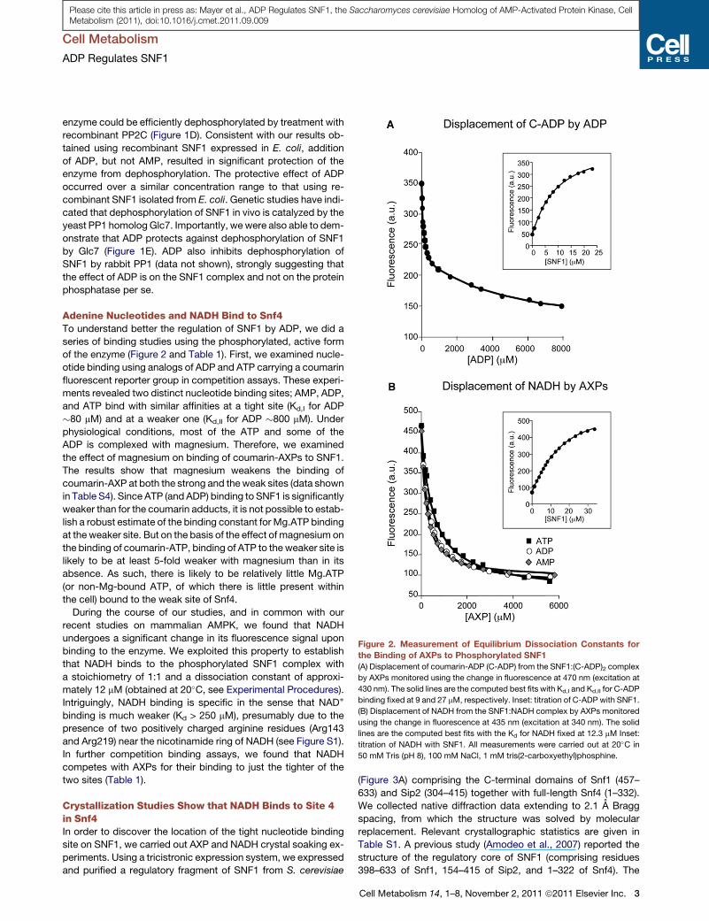
Figure 2. Measurement of Equilibrium Dissociation Constants for
the Binding of AXPs to Phosphorylated SNF1(A) Displacement of coumarin-ADP (C-ADP) from the SNF1:(C-ADP)2 complex
by AXPs monitored using the change in fluorescence at 470 nm (excitation at
430 nm). The solid lines are the computed best fits with Kd,I and Kd,II for C-ADP
binding fixed at 9 and 27 mM, respectively. Inset: titration of C-ADP with SNF1.
(B) Displacement of NADH from the SNF1:NADH complex by AXPs monitored
using the change in fluorescence at 435 nm (excitation at 340 nm). The solid
lines are the computed best fits with the Kd for NADH fixed at 12.3 mM Inset:
titration of NADH with SNF1. All measurements were carried out at 20�C in
50 mM Tris (pH 8), 100 mM NaCl, 1 mM tris(2-carboxyethyl)phosphine.
Cell Metabolism
ADP Regulates SNF1
Please cite this article in press as: Mayer et al., ADP Regulates SNF1, the Saccharomyces cerevisiae Homolog of AMP-Activated Protein Kinase, CellMetabolism (2011), doi:10.1016/j.cmet.2011.09.009
enzyme could be efficiently dephosphorylated by treatment with
recombinant PP2C (Figure 1D). Consistent with our results ob-
tained using recombinant SNF1 expressed in E. coli, addition
of ADP, but not AMP, resulted in significant protection of the
enzyme from dephosphorylation. The protective effect of ADP
occurred over a similar concentration range to that using re-
combinant SNF1 isolated from E. coli. Genetic studies have indi-
cated that dephosphorylation of SNF1 in vivo is catalyzed by the
yeast PP1 homolog Glc7. Importantly, wewere also able to dem-
onstrate that ADP protects against dephosphorylation of SNF1
by Glc7 (Figure 1E). ADP also inhibits dephosphorylation of
SNF1 by rabbit PP1 (data not shown), strongly suggesting that
the effect of ADP is on the SNF1 complex and not on the protein
phosphatase per se.
Adenine Nucleotides and NADH Bind to Snf4To understand better the regulation of SNF1 by ADP, we did a
series of binding studies using the phosphorylated, active form
of the enzyme (Figure 2 and Table 1). First, we examined nucle-
otide binding using analogs of ADP and ATP carrying a coumarin
fluorescent reporter group in competition assays. These experi-
ments revealed two distinct nucleotide binding sites; AMP, ADP,
and ATP bind with similar affinities at a tight site (Kd,I for ADP
�80 mM) and at a weaker one (Kd,II for ADP �800 mM). Under
physiological conditions, most of the ATP and some of the
ADP is complexed with magnesium. Therefore, we examined
the effect of magnesium on binding of coumarin-AXPs to SNF1.
The results show that magnesium weakens the binding of
coumarin-AXP at both the strong and theweak sites (data shown
in Table S4). Since ATP (and ADP) binding to SNF1 is significantly
weaker than for the coumarin adducts, it is not possible to estab-
lish a robust estimate of the binding constant for Mg.ATP binding
at theweaker site. But on the basis of the effect of magnesium on
the binding of coumarin-ATP, binding of ATP to the weaker site is
likely to be at least 5-fold weaker with magnesium than in its
absence. As such, there is likely to be relatively little Mg.ATP
(or non-Mg-bound ATP, of which there is little present within
the cell) bound to the weak site of Snf4.
During the course of our studies, and in common with our
recent studies on mammalian AMPK, we found that NADH
undergoes a significant change in its fluorescence signal upon
binding to the enzyme. We exploited this property to establish
that NADH binds to the phosphorylated SNF1 complex with
a stoichiometry of 1:1 and a dissociation constant of approxi-
mately 12 mM (obtained at 20�C, see Experimental Procedures).
Intriguingly, NADH binding is specific in the sense that NAD+
binding is much weaker (Kd > 250 mM), presumably due to the
presence of two positively charged arginine residues (Arg143
and Arg219) near the nicotinamide ring of NADH (see Figure S1).
In further competition binding assays, we found that NADH
competes with AXPs for their binding to just the tighter of the
two sites (Table 1).
Crystallization Studies Show that NADH Binds to Site 4in Snf4In order to discover the location of the tight nucleotide binding
site on SNF1, we carried out AXP and NADH crystal soaking ex-
periments. Using a tricistronic expression system, we expressed
and purified a regulatory fragment of SNF1 from S. cerevisiae
(Figure 3A) comprising the C-terminal domains of Snf1 (457–
633) and Sip2 (304–415) together with full-length Snf4 (1–332).
We collected native diffraction data extending to 2.1 A Bragg
spacing, from which the structure was solved by molecular
replacement. Relevant crystallographic statistics are given in
Table S1. A previous study (Amodeo et al., 2007) reported the
structure of the regulatory core of SNF1 (comprising residues
398–633 of Snf1, 154–415 of Sip2, and 1–322 of Snf4). The
Cell Metabolism 14, 1–8, November 2, 2011 ª2011 Elsevier Inc. 3

Table 1. Equilibrium Dissociation Constants for the Binding of
AXPs to Phosphorylated SNF1
Ligand
Kd (mM) Kd,I (mM) Kd,II (mM)
Versus NADH Versus C-AXPs
AMP 55 (12) 62 (15) 1450 (350)
ADP 72 (14) 91 (25) 760 (200)
ATP 120 (25) 85 (22) 1200 (300)
Dissociation constants were determined by competition against NADH
or 30-(7-diethylaminocoumarin-3-carbonylamino)-30-deoxy-AXP (C-AXP).
The Kd values shown are the mean (±SD) determined from at least three
independent measurements. All measurements were carried out at 20�Cin 50 mM Tris (pH 8), 100 mM NaCl, 1 mM tris(2-carboxyethyl)phosphine.
Themethods employed in analyzing thefluorescencebindingdata are fully
described elsewhere (Xiao et al., 2011).
Cell Metabolism
ADP Regulates SNF1
Please cite this article in press as: Mayer et al., ADP Regulates SNF1, the Saccharomyces cerevisiae Homolog of AMP-Activated Protein Kinase, CellMetabolism (2011), doi:10.1016/j.cmet.2011.09.009
overall architecture of the structure reported here corresponds
closely with the previously reported structure (Figure S1),
although the structure reported in our current study lacks the
glycogen binding domain of Sip2. Data sets from soaked crystals
show strong, well-resolved electron density for AMP, ADP, and
NADH (Figures 3B and S1) at one of the four canonical nucleotide
binding sites on Snf4 (labeled site 4 according to the convention
suggested by Kemp [Kemp et al., 2007]). Given that NADH binds
substantially more tightly to SNF1 than AXPs, and given the rela-
tive concentrations of AXPs and NADH in yeast (Canelas et al.,
2008), we conclude that under most conditions this site will
be occupied by NADH. The situation is therefore analogous to
mammalian AMPK, where the equivalent site contains a nonex-
changeable AMP (Xiao et al., 2007). We think it likely that nucle-
otide occupancy at this site, in both SNF1 and AMPK, is im-
portant for the stability of the enzyme, since protein stability
experiments (Figure S2A) indicate that SNF1 is substantially
more stable in the presence of NADH.
ADP Binding at the Weaker Site Protects againstDephosphorylation of SNF1Two lines of evidence lead us to conclude that binding of ADP at
the weaker of the two binding sites is responsible for protection
of the enzyme against dephosphorylation. First, as described
above, NADH competes for the tighter nucleotide binding site,
but has no effect on SNF1 activity or on the ability of ADP to
protect against dephosphorylation (Figure S2B). In contrast,
and as would be predicted, AMP, which binds at both nucleotide
binding sites, competes with ADP in the dephosphorylation
protection assay (Figure S2C). Second, the half-maximal effect
of ADP on dephosphorylation of SNF1 occurred at a much higher
concentration (>500mMfor recombinant SNF1) than theKd for the
tighterbindingsite (�80mM),but close to theKdof theweaker site.
Presumably reflecting the weak binding constant and the
lattice packing, we have been unable to obtain crystals of
SNF1 with nucleotide present in the weaker site. As an alterna-
tive approach, we carried out extensive mutagenesis studies
aimed at disrupting the individual nucleotide binding sites within
Snf4, but, in common with our experience with mammalian
AMPK, we have not been able to identify mutations that specif-
ically ablate AXP or NADH binding (see Tables S2 and S3 and
Figure S3). These data clearly show that mutation of the aspartic
acid residues that are seen to interact with the ribose moiety of
4 Cell Metabolism 14, 1–8, November 2, 2011 ª2011 Elsevier Inc.
AXPs and NADH do not lead to the ablation of ligand binding.
The notion that these aspartic acid residues were required for
binding was first suggested by us to explain why one of the
four ‘‘canonical’’ binding sites in AMPK did not bind AXPs
(Xiao et al., 2007). However, our suggestion was untested at
that time, and presumably the lack of effect of these mutations
reflects the fact that the energetic costs of desolvating the
carboxyl of the aspartic acid and the hydroxyls of the ribose
offsets the contribution to binding of the hydrogen bonds that
the aspartic acids make with AXPs. Importantly, however, in
the absence of mutants that ablate AXP binding, our structural
studies provide valuable insights into the likely mechanism of
ADP-mediated protection against dephosphorylation.
ADP Promotes Interaction of the T-Loopwith the Regulatory Core of SNF1We recently determined the structure of an active form of
mammalian AMPK in which the T-loop, containing phosphory-
lated Thr172, is well ordered and interacts with the C-terminal
domains of the a and b subunits (Xiao et al., 2011). In this confor-
mation, Thr172 is protected from dephosphorylation because
access to protein phosphatases is restricted. We showed that
part of the linker between the N-terminal kinase and the
C-terminal domain of the a subunit formed an a-hook structure
that enhanced the association of the kinase domain with the
regulatory fragment when ADP, but not ATP, is bound (Xiao
et al., 2011). The sequences of the T-loop and the residues
with which it interacts are highly conserved between the yeast
and mammalian enzymes, but the sequences of the linker region
are not (Xiao et al., 2007, 2011) (Figure 3A). His233 in AMPKb1
(His235 in b2) lies at the interface of the regulatory domain with
the kinase domain, and mutation of this residue in AMPK signif-
icantly increases the rate of dephosphorylation (Xiao et al.,
2011). Mutation of His379 in Gal83 (corresponding to His233 in
AMPKb1 and His375 in Sip2; see Figure 3A) also significantly
increased the rate of dephosphorylation of SNF1, while leaving
protection by ADP intact (Figures 3C and 3D), as was also found
for the equivalent mutation in AMPK (Xiao et al., 2011).
DISCUSSION
An essential role for SNF1 in glucose repression has been known
for nearly 30 years (Carlson et al., 1981, 1984), and activation of
SNF1 during glucose limitation has been appreciated for nearly
20 years (Woods et al., 1994), but the metabolic signal that trig-
gers it has remained elusive. Here we show that ADP binds to
Snf4 and protects SNF1 against dephosphorylation of Thr210,
suggesting that ADP is the metabolic signal that leads to activa-
tion of SNF1 in response to glucose starvation in budding yeast.
We recently showed that ADP protects mammalian AMPK
against dephosphorylation (Xiao et al., 2011), and while our
manuscript was under review, Oakhill et al. reported that ADP,
as well as AMP, stimulates phosphorylation of AMPK on Thr172
(Oakhill et al., 2011). AMP and ADP stimulation of phosphoryla-
tion requires myristoylation of the b subunit of AMPK (Oakhill
et al., 2010). However, Gal83, the predominant b isoform of
S. cerevisiae SNF1, does not contain a consensus sequence for
N-terminal myristoylation, suggesting that Gal83-containing
SNF1 complexes are not subject to this form of regulation.
Cell Metabolism
ADP Regulates SNF1
Please cite this article in press as: Mayer et al., ADP Regulates SNF1, the Saccharomyces cerevisiae Homolog of AMP-Activated Protein Kinase, CellMetabolism (2011), doi:10.1016/j.cmet.2011.09.009
In mammalian AMPK, we showed that ADP protects against
dephosphorylation by promoting the interaction of the T-loop
region with the regulatory regions of the a and b subunits (Xiao
et al., 2011). Our current study suggests that a similar mecha-
nism operates for ADP-mediated protection against dephos-
phorylation of SNF1. However, the lack of sequence conserva-
tion between SNF1 and AMPK in the a-hook region (Xiao et al.,
2011) means that we are less clear about the molecular mecha-
nism for coupling nucleotide binding to the recruitment, and
protection against dephosphorylation, of the kinase domain.
Nonetheless, the fact that the interaction of the kinase domain
with the regulatory fragment is highly asymmetric means that it
must be one of the ADP binding sites on the surface of Snf4
that faces the kinase domain (labeled site 2 and site 3 in Fig-
ure S3) that mediates protection against dephosphorylation.
Given that the linker region is not conserved between mammals
and yeast, and that ADP has been shown to bind to site 2 in the
S. pombe homolog of AMPK (Jin et al., 2007), we think it is likely
that ADP binding at site 2 in Snf4 mediates the protective effect
against dephosphorylation of SNF1. In this model we envisage
that a region of Snf1 between the kinase domain and the ordered
C-terminal domain, which is not conserved in AMPKa, plays
a role analogous to the a-hook in AMPK. How this mechanism
discriminates for ADP over AMP and ATP for protection against
dephosphorylation is not clear. In order to address this, addi-
tional structures of full-length SNF1 containing the phosphory-
lated kinase domain, together with the Snf4 subunit in complex
with AMP, ADP, and ATP, will be required. Importantly, the con-
centration of ADP required for protection of SNF1 against de-
phosphorylation in vitro is much higher than that required for
mammalian AMPK, in keeping with the higher concentrations
of ADP present in yeast cells relative to mammalian cells. Our
results show that magnesium weakens the binding of ATP to
Snf4, and therefore we would expect very little ATP to be bound
at the regulatory site. Thus, in contrast to mammalian AMPK,
where ADP competes for binding with Mg.ATP, SNF1 may only
bind significant amounts of ADP. These data lead us to a conclu-
sion similar to that reached by Shapiro and colleagues in their
study of the AMPK homolog fromSchizosaccharomyces pombe,
where they could not identify magnesium in their ATP-bound
crystal structure (Townley and Shapiro, 2007), leading them to
suggest that the discrimination against magnesium-bound ATP
would enable that enzyme to respond to the much lower levels
of AMP in the presence of high competitive levels of total ATP.
Given our demonstration that for S. cerevisiae the relevant ligand
is ADP, and given its binding constant relative to Mg.ATP, we
suggest that in S. cerevisiae SNF1 may respond to absolute
changes in ADP concentration. Finally, the finding that ADP pro-
tects both yeast SNF1 and mammalian AMPK against dephos-
phorylation raises the exciting possibility that ADP might repre-
sent a ‘‘unifying trigger’’ for activation of AMPK orthologs in all
species.
EXPERIMENTAL PROCEDURES
Production of Recombinant SNF1
DNAencodingSnf1 andSnf4were cloned intopRSFDuet (Novagen), andcDNA
encoding Gal83 was cloned into pETDuet1; both were generous gifts from
Marian Carlson (Columbia University). For expression of SNF1, E. coli BL21
(DE3) cells were cotransformed with these vectors, and the SNF1 com-
plex was purified as described below. Tricistronic vectors expressing His-
Snf1Sip2(154-415)Snf4 or His-Snf1(457-633)Sip2(304-415)Snf4 were constructed
and used to transform E. coli BL21 (DE3) cells for expression of SNF1
complexes. Initially, SNF1 complexes were purified by affinity chromatography
using nickel-Sepharose (Sanders et al., 2007) and used for further studies.
Tandem Affinity Purification of Glc7 and Snf1
The Snf1 andGlc7 proteins were purified from yeast cell extracts using tandem
affinity purification (TAP) method (Rigaut et al., 1999). For both genes, the TAP
tag was added at the C terminus of the open reading frame. Low-copy number
plasmids expressing the TAP constructs were introduced to cells lacking the
endogenous chromosomal SNF1 or GLC7 gene. Cells were grown on media
with sucrose or glucose as the carbon source, respectively. TAP-tagged
proteins were purified as previously described for the Snf1-TAP protein (Elbing
et al., 2006). Dephosphorylation reactions using Glc7 were performed immedi-
ately following purification, as the enzyme rapidly loses activity over time.
SNF1 Functional Assays
Recombinant SNF1 complexes expressed in E. coli were phosphorylated by
incubation with CaMKKb as described previously (Sanders et al., 2007). An
aliquot of the phosphorylated protein (�1 mg) was incubated in 50 mM HEPES
(pH 7.4), 2.5mMMgCl2, and in the presence or absence of PP2Ca (26 ng) in the
presence or absence of nucleotides (as stated in the figure legends) for 10 min
at 37�C. The level of Thr210 phosphorylation was determined by western blot
analysis using an anti-AMPK Thr172 phosphospecific antibody (Cell Sig-
naling). Total Snf1 was detected using an anti-6xHis antibody (Abcam). In
some cases, SNF1 activity was measured directly using the SAMS peptide
assay (Woods et al., 1994). Dephosphorylation of TAP-SNF1 by recombinant
PP2Ca was monitored using phosphopeptide-specific antiserum (McCartney
and Schmidt, 2001), while the level of total Snf1 protein was determined with
His-probe antibodies (Santa Cruz Biotechnology). Primary antibodies were
detected using LI-COR IRDye Infrared Dye secondary antibodies and visual-
ized using an Odyssey Infrared Imager (LI-COR Biotechnology). Quantification
of results was performed using Odyssey software and expressed as a ratio of
the signal obtained with the phosphospecific antibody relative to the appro-
priate total antibody.
Binding Studies
Unless stated otherwise, all binding measurements were performed at 20�C in
50 mM Tris (pH 8), 100 mM NaCl, 1 mM tris(2-carboxyethyl)phosphine. Free
NADH has an emission maximum at 465 nm (excitation at 340 nm). In the pres-
ence of saturating SNF1, the emission maximum is blue shifted to 435 nm, and
the fluorescence emission intensity is increased. These fluorescence changes
were used to determine equilibrium dissociation constants by titrating 4–10 mM
NADH with SNF1. Experiments performed at higher NADH concentration and
low temperature confirmed that NADH binds to SNF1 with 1:1 stoichiometry.
The coumarin analogs of ADP and ATP (C-ADP, C-ATP: 30-(7-diethylamino-
coumarin-3-carbonylamino)-30-deoxyadenosine 50 di- and triphosphate) have
emission maxima at 479 nm (excitation at 430 nm). In the presence of satu-
rating SNF1 the emission maxima are blue shifted to�470 nm and the fluores-
cence intensity is increased. These fluorescence changes were used to deter-
mine equilibrium dissociation constants by titrating 4–6 mMC-AXP with SNF1.
Experiments performed at higher C-AXP concentration and low temperature
confirmed that these analogs bind to SNF1 with 2:1 stoichiometry. Dissocia-
tion constants for NADH and C-AXPs to phosphorylated and nonphosphory-
lated SNF1 are shown in Table S4. Dissociation constants for AMP, ADP,
and ATPwere determined using NADH and the coumarin analogs as reporters.
Competition assays were performed in which a mixture of SNF1 and the fluo-
rescent nucleotide was titrated with the nonfluorescent nucleotide. These data
were then analyzed with the previously determined Kd values for the fluores-
cent nucleotides held constant in the analysis. The dissociation constant for
NAD+ was estimated using NADH as the reporter.
Crystallographic Studies
Truncated SNF1 (Snf4(1-322)Sip2(304-415)His-Snf1(457-633)) was cloned into a tri-
cistronic vector and subsequently expressed in E. coli BL21 (OneShot BL21
Star [DE3] cells, Invitrogen). Proteins were purified by nickel affinity
Cell Metabolism 14, 1–8, November 2, 2011 ª2011 Elsevier Inc. 5
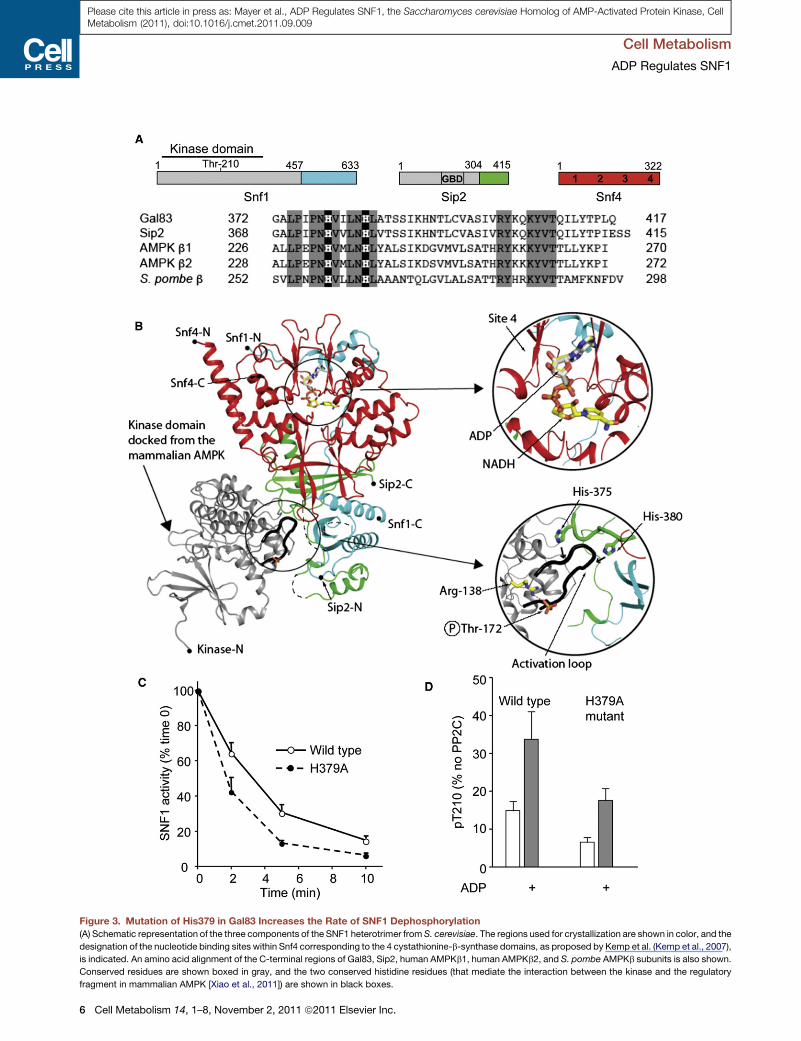
Figure 3. Mutation of His379 in Gal83 Increases the Rate of SNF1 Dephosphorylation
(A) Schematic representation of the three components of the SNF1 heterotrimer from S. cerevisiae. The regions used for crystallization are shown in color, and the
designation of the nucleotide binding sites within Snf4 corresponding to the 4 cystathionine-b-synthase domains, as proposed by Kemp et al. (Kemp et al., 2007),
is indicated. An amino acid alignment of the C-terminal regions of Gal83, Sip2, human AMPKb1, human AMPKb2, and S. pombe AMPKb subunits is also shown.
Conserved residues are shown boxed in gray, and the two conserved histidine residues (that mediate the interaction between the kinase and the regulatory
fragment in mammalian AMPK [Xiao et al., 2011]) are shown in black boxes.
Cell Metabolism
ADP Regulates SNF1
6 Cell Metabolism 14, 1–8, November 2, 2011 ª2011 Elsevier Inc.
Please cite this article in press as: Mayer et al., ADP Regulates SNF1, the Saccharomyces cerevisiae Homolog of AMP-Activated Protein Kinase, CellMetabolism (2011), doi:10.1016/j.cmet.2011.09.009

Cell Metabolism
ADP Regulates SNF1
Please cite this article in press as: Mayer et al., ADP Regulates SNF1, the Saccharomyces cerevisiae Homolog of AMP-Activated Protein Kinase, CellMetabolism (2011), doi:10.1016/j.cmet.2011.09.009
chromatography (His-Trap, GE Healthcare), anion exchange (Mono Q, GE
Healthcare), and gel filtration (Superdex 200, GE Healthcare). The His-tag
was removed by digestion with PreScission protease, and the SNF1 complex
protein was purified by gel filtration (Superdex 200, GE Healthcare). Protein
complex stock solution was prepared at 15 mg/ml in 50 mM Tris (pH 7.0),
300 mM NaCl, and 1 mM TCEP. Crystals were grown by vapor diffusion tech-
nique at 18�C in hanging drops. Drops were prepared bymixing equal volumes
of protein complex with reservoir solution containing 1.0M succinic acid, 0.1M
HEPES (pH 7.0), 1% (w/v) polyethylene glycol monomethyl ether 2000 (Index
Reagent 34, Hampton Research). Crystals were first transferred into mother
liquor with an additional 12.5% ethylene glycol, then 25%ethylene glycol, prior
to plunging into liquid nitrogen. For crystal soaking experiments, ADP, AMP,
and NADH were made up in reservoir buffer, and crystals were soaked over-
night prior to flash cooling. Diffraction data were collected on an in-house
MicroMax-007 HF rotating anode coupled to a RaxisIV2+ detector. Data
were integrated using Denzo and scaled with Scalepack (Otwinowski and
Minor, 1993). The structure was solved bymolecular replacement using Amore
(Navaza, 1994) using 2QLV.pdb as the search model. Standard refinement
was carried out with Refmac5 (CCP4, 1994) with manual model building with
Coot. Figures were created with PyMOL.
ACCESSION NUMBERS
Coordinates have been deposited in the Protein Data Bank with accession
codes 3TDH, 3T4N, and 3TE5.
SUPPLEMENTAL INFORMATION
Supplemental Information includes three figures, four tables, and Supple-
mental References and can be found with this article online at doi:10.1016/
j.cmet.2011.09.009.
ACKNOWLEDGMENTS
This study was supported by theMedical Research Council UK (D. Carling and
S.J.G.) and grant GM46443 from the National Institutes of Health (M.C.S.). We
thank Martin Webb for the coumarin-labeled nucleotides. We are grateful to
Stephen Smerdon for helpful discussion of the manuscript. We gratefully
acknowledge Diamond Light Source for synchrotron access.
Received: June 7, 2011
Revised: August 24, 2011
Accepted: September 12, 2011
Published online: October 20, 2011
REFERENCES
Amodeo, G.A., Rudolph, M.J., and Tong, L. (2007). Crystal structure of the
heterotrimer core of Saccharomyces cerevisiae AMPK homologue SNF1.
Nature 449, 492–495.
Canelas, A.B., van Gulik, W.M., and Heijnen, J.J. (2008). Determination of the
cytosolic free NAD/NADH ratio in Saccharomyces cerevisiae under steady-
state and highly dynamic conditions. Biotechnol. Bioeng. 100, 734–743.
Carling, D. (2004). The AMP-activated protein kinase cascade—a unifying
system for energy control. Trends Biochem. Sci. 29, 18–24.
Carlson, M., Osmond, B.C., and Botstein, D. (1981). Mutants of yeast defective
in sucrose utilization. Genetics 98, 25–40.
(B) Ribbon representation of the structure of the regulatory fragment determine
domain of Sip2 is colored green, and the Snf4 subunit is colored red, with the a
complex. The positions of both ADP and NADH, determined in this study by cryst
inset panel on the right shows a closer view of ADP and NADH binding to site 4, w
and two histidine residues from Sip2.
(C) Dephosphorylation rate of wild-type or Gal83 His379 to alanine (H379A) SNF
(D) Protection of dephosphorylation of wild-type and H379A mutant by ADP (800
independent experiments.
Carlson, M., Osmond, B.C., Neigeborn, L., and Botstein, D. (1984). A
suppressor of SNF1 mutations causes constitutive high-level invertase
synthesis in yeast. Genetics 107, 19–32.
Celenza, J.L., and Carlson, M. (1986). A yeast gene that is essential for release
from glucose repression encodes a protein kinase. Science 233, 1175–1180.
CCP4 (Collaborative Computational Project, Number 4). (1994). The CCP4
suite: programs for protein crystallography. Acta Crystallogr. D Biol.
Crystallogr. 50, 760–763.
Davies, S.P., Helps, N.R., Cohen, P.T., and Hardie, D.G. (1995). 50-AMP
inhibits dephosphorylation, as well as promoting phosphorylation, of the
AMP-activated protein kinase. Studies using bacterially expressed human
protein phosphatase-2C alpha and native bovine protein phosphatase-2AC.
FEBS Lett. 377, 421–425.
Ditzelmuller, G., Wohrer, W., Kubicek, C.P., and Rohr, M. (1983). Nucleotide
pools of growing, synchronized and stressed cultures of Saccharomyces
cerevisiae. Arch. Microbiol. 135, 63–67.
Elbing, K., Rubenstein, E.M., McCartney, R.R., and Schmidt, M.C. (2006).
Subunits of the Snf1 kinase heterotrimer show interdependence for associa-
tion and activity. J. Biol. Chem. 281, 26170–26180.
Gancedo, J.M. (1992). Carbon catabolite repression in yeast. Eur. J. Biochem.
206, 297–313.
Hardie, D.G. (2007). AMP-activated/SNF1 protein kinases: conserved guard-
ians of cellular energy. Nat. Rev. Mol. Cell Biol. 8, 774–785.
Hardie, D.G., Carling, D., and Carlson, M. (1998). The AMP-activated/SNF1
protein kinase subfamily: metabolic sensors of the eukaryotic cell? Annu.
Rev. Biochem. 67, 821–855.
Hong, S.P., Leiper, F.C., Woods, A., Carling, D., and Carlson, M. (2003).
Activation of yeast Snf1 and mammalian AMP-activated protein kinase by
upstream kinases. Proc. Natl. Acad. Sci. USA 100, 8839–8843.
Jin, X., Townley, R., and Shapiro, L. (2007). Structural insight into AMPK regu-
lation: ADP comes into play. Structure 15, 1285–1295.
Kahn, B.B., Alquier, T., Carling, D., and Hardie, D.G. (2005). AMP-activated
protein kinase: ancient energy gauge provides clues to modern understanding
of metabolism. Cell Metab. 1, 15–25.
Kemp, B.E., Oakhill, J.S., and Scott, J.W. (2007). AMPK structure and regula-
tion from three angles. Structure 15, 1161–1163.
Lage, R., Dieguez, C., Vidal-Puig, A., and Lopez, M. (2008). AMPK: ametabolic
gauge regulating whole-body energy homeostasis. Trends Mol. Med. 14,
539–549.
Larsson, C., Nilsson, A., Blomberg, A., and Gustafsson, L. (1997). Glycolytic
flux is conditionally correlated with ATP concentration in Saccharomyces
cerevisiae: a chemostat study under carbon- or nitrogen-limiting conditions.
J. Bacteriol. 179, 7243–7250.
McCartney, R.R., and Schmidt, M.C. (2001). Regulation of Snf1 kinase.
Activation requires phosphorylation of threonine 210 by an upstream kinase
as well as a distinct step mediated by the Snf4 subunit. J. Biol. Chem. 276,
36460–36466.
Mitchelhill, K.I., Stapleton, D., Gao, G., House, C., Michell, B., Katsis, F.,
Witters, L.A., and Kemp, B.E. (1994). Mammalian AMP-activated protein
kinase shares structural and functional homology with the catalytic domain
of yeast Snf1 protein kinase. J. Biol. Chem. 269, 2361–2364.
Navaza, J. (1994). AMoRe: an automated package for molecular replacement.
Acta. Crystallogr. 50, 157–163.
d in this study; the C-terminal domain of Snf1 is colored blue, the C-terminal
ctive kinase domain of mammalian AMPK (colored in gray) docked onto the
al soaking experiments, are shown in ball-and-stick representation. The upper
hile the lower inset panel shows the activation loop region of the kinase domain
1 complex.
mM) after incubation with PP2C for 10 min. Results are the mean ± SEM for 3–4
Cell Metabolism 14, 1–8, November 2, 2011 ª2011 Elsevier Inc. 7

Cell Metabolism
ADP Regulates SNF1
Please cite this article in press as: Mayer et al., ADP Regulates SNF1, the Saccharomyces cerevisiae Homolog of AMP-Activated Protein Kinase, CellMetabolism (2011), doi:10.1016/j.cmet.2011.09.009
Oakhill, J.S., Chen, Z.P., Scott, J.W., Steel, R., Castelli, L.A., Ling, N.,
Macaulay, S.L., and Kemp, B.E. (2010). b-Subunit myristoylation is the gate-
keeper for initiating metabolic stress sensing by AMP-activated protein kinase
(AMPK). Proc. Natl. Sci. Acad. USA 107, 19237–19241.
Oakhill, J.S., Steel, R., Chen, Z.P., Scott, J.W., Ling, N., Tam, S., and Kemp,
B.E. (2011). AMPK is a direct adenylate charge-regulated protein kinase.
Science 332, 1433–1435.
Otwinowski, Z., and Minor, W. (1993). In Data Collection and Processing, L.
Sawyer, N. Isaacs, and S. Bailey, eds. (Warrington, UK: SERC Daresbury
Laboratory), pp. 556–562.
Rigaut, G., Shevchenko, A., Rutz, B., Wilm, M., Mann, M., and Seraphin, B.
(1999). A generic protein purificationmethod for protein complex characteriza-
tion and proteome exploration. Nat. Biotechnol. 17, 1030–1032.
Rubenstein, E.M., McCartney, R.R., Zhang, C., Shokat, K.M., Shirra, M.K.,
Arndt, K.M., and Schmidt, M.C. (2008). Access denied: Snf1 activation loop
phosphorylation is controlled by availability of the phosphorylated threonine
210 to the PP1 phosphatase. J. Biol. Chem. 283, 222–230.
Sanders, M.J., Grondin, P.O., Hegarty, B.D., Snowden, M.A., and Carling, D.
(2007). Investigating the mechanism for AMP activation of the AMP-activated
protein kinase cascade. Biochem. J. 403, 139–148.
Suter, M., Riek, U., Tuerk, R., Schlattner, U., Wallimann, T., and Neumann, D.
(2006). Dissecting the role of 50-AMP for allosteric stimulation, activation, and
deactivation of AMP-activated protein kinase. J. Biol. Chem. 281, 32207–
32216.
8 Cell Metabolism 14, 1–8, November 2, 2011 ª2011 Elsevier Inc.
Sutherland, C.M., Hawley, S.A., McCartney, R.R., Leech, A., Stark, M.J.R.,
Schmidt, M.C., and Hardie, D.G. (2003). Elm1p is one of three upstream
kinases for the Saccharomyces cerevisiae SNF1 complex. Curr. Biol. 13,
1299–1305.
Townley, R., and Shapiro, L. (2007). Crystal structures of the adenylate sensor
from fission yeast AMP-activated protein kinase. Science 315, 1726–1729.
Wilson, W.A., Hawley, S.A., and Hardie, D.G. (1996). Glucose repression/
derepression in budding yeast: SNF1 protein kinase is activated by phosphor-
ylation under derepressing conditions, and this correlates with a high
AMP:ATP ratio. Curr. Biol. 6, 1426–1434.
Woods, A., Munday, M.R., Scott, J., Yang, X., Carlson, M., and Carling, D.
(1994). Yeast SNF1 is functionally related to mammalian AMP-activated
protein kinase and regulates acetyl-CoA carboxylase in vivo. J. Biol. Chem.
269, 19509–19515.
Xiao, B., Heath, R., Saiu, P., Leiper, F.C., Leone, P., Jing, C., Walker, P.A.,
Haire, L., Eccleston, J.F., Davis, C.T., et al. (2007). Structural basis for AMP
binding to mammalian AMP-activated protein kinase. Nature 449, 496–500.
Xiao, B., Sanders, M.J., Underwood, E., Heath, R., Mayer, F.V., Carmena, D.J.,
Jing, C., Walker, P.A., Eccleston, J.F., Haire, L.F., et al. (2011). Structure of
mammalian AMPK and its regulation by ADP. Nature 472, 230–233.
Zhang, B.B., Zhou, G., and Li, C. (2009). AMPK: an emerging drug target for
diabetes and the metabolic syndrome. Cell Metab. 9, 407–416.