MYOC Rule Specifications for the ACMG/AMP Variant ...
27
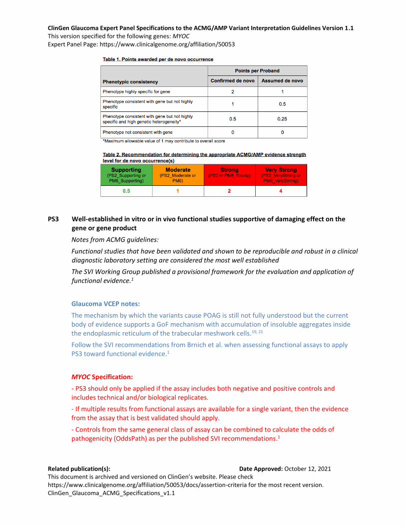
ClinGen Glaucoma Expert Panel Specifications to the ACMG/AMP Variant Interpretation Guidelines Version 1.1 This version specified for the following genes: MYOC Expert Panel Page: https://www.clinicalgenome.org/affiliation/50053 Related publication(s): Date Approved: October 12, 2021 This document is archived and versioned on ClinGen’s website. Please check https://www.clinicalgenome.org/affiliation/50053/docs/assertion-criteria for the most recent version. ClinGen_Glaucoma_ACMG_Specifications_v1.1 MYOC Rule Specifications for the ACMG/AMP Variant Curation Guidelines Release notes from v1: Added MYOC HGNC ID, Replaced disease identifiers Primary open-angle glaucoma (POAG) and juvenile onset open-angle glaucoma (JOAG) (MIM: 137750) with primary open angle glaucoma (MONDO:0007665) and juvenile open angle glaucoma (MONDO:0020367) (Approved November 2021) Gene Disease (MONDO ID) Clinically significant transcript MYOC (HGNC: 7610) primary open angle glaucoma (MONDO:0007665) juvenile open angle glaucoma (MONDO:0020367) NM_000261.1 Pathogenic Criteria Criteria Original Criteria Description Specification(s) Very Strong Criteria PVS1 Null variant in a gene where LOF is a known mechanism of disease Not applicable See PM4 Strong Criteria PS1 Same amino acid change as a previously established pathogenic variant regardless of nucleotide change The novel change must not affect splicing (SpliceAI ≤ 0.2) PS2/PM6_Strong De novo in a patient with disease and no family history ≥2 confirmed de novo in JOAG (Use the proposed SVI point recommendations for “phenotype consistent with gene but not highly specific” for JOAG) - Both maternity and paternity need to be proven for confirmed de novo variants. - Parents need to be clinically assessed and not have a diagnosis of glaucoma. PS3 Well-established in vitro or in vivo, functional studies supportive of a Assays with OddsPath >18.7 as per the SVI recommendations. 1
-
Upload
khangminh22 -
Category
Documents
-
view
1 -
download
0
Transcript of MYOC Rule Specifications for the ACMG/AMP Variant ...

ClinGen Glaucoma Expert Panel Specifications to the ACMG/AMP Variant Interpretation Guidelines Version 1.1 This version specified for the following genes: MYOC Expert Panel Page: https://www.clinicalgenome.org/affiliation/50053
Related publication(s): Date Approved: October 12, 2021 This document is archived and versioned on ClinGen’s website. Please check https://www.clinicalgenome.org/affiliation/50053/docs/assertion-criteria for the most recent version. ClinGen_Glaucoma_ACMG_Specifications_v1.1
MYOC Rule Specifications for the ACMG/AMP Variant Curation Guidelines
Release notes from v1: Added MYOC HGNC ID, Replaced disease identifiers Primary open-angle glaucoma (POAG)
and juvenile onset open-angle glaucoma (JOAG) (MIM: 137750) with primary open angle glaucoma
(MONDO:0007665) and juvenile open angle glaucoma (MONDO:0020367) (Approved November 2021)
Gene Disease (MONDO ID) Clinically significant transcript
MYOC (HGNC: 7610) primary open angle glaucoma (MONDO:0007665) juvenile open angle glaucoma (MONDO:0020367)
NM_000261.1
Pathogenic Criteria
Criteria Original Criteria Description Specification(s)
Very Strong Criteria
PVS1 Null variant in a gene where LOF is a
known mechanism of disease
Not applicable
See PM4
Strong Criteria
PS1 Same amino acid change as a
previously established pathogenic
variant regardless of nucleotide
change
The novel change must not affect splicing
(SpliceAI ≤ 0.2)
PS2/PM6_Strong De novo in a patient with disease and
no family history
≥2 confirmed de novo in JOAG
(Use the proposed SVI point recommendations
for “phenotype consistent with gene but not
highly specific” for JOAG)
- Both maternity and paternity need to be
proven for confirmed de novo variants.
- Parents need to be clinically assessed and not
have a diagnosis of glaucoma.
PS3
Well-established in vitro or in vivo,
functional studies supportive of a
Assays with OddsPath >18.7 as per the SVI
recommendations.1

ClinGen Glaucoma Expert Panel Specifications to the ACMG/AMP Variant Interpretation Guidelines Version 1.1 This version specified for the following genes: MYOC Expert Panel Page: https://www.clinicalgenome.org/affiliation/50053
Related publication(s): Date Approved: October 12, 2021 This document is archived and versioned on ClinGen’s website. Please check https://www.clinicalgenome.org/affiliation/50053/docs/assertion-criteria for the most recent version. ClinGen_Glaucoma_ACMG_Specifications_v1.1
damaging effect on the gene or gene
product
Applies to the following functional studies:
- Solubility or secretion assays.
-OR-
- Animal models that replicate the glaucoma
phenotype.
PS4 The prevalence of the variant in
affected individuals is significantly
increased compared with the
prevalence in controls
≥ 15 probands from multiple independent
studies
- Probands counted should be clinically assessed
and have a diagnosis of JOAG or POAG
- PM2_Supporting must be met
PP1_Strong Cosegregation with disease in
multiple affected family members in
a gene definitively known to cause
the disease
≥7 meioses in >1 family
- BA1 and BS1 must not be met
- Only genotype positive/phenotype positive
and obligate carriers/phenotype positive
individuals should be counted as segregations.
- Phenotype positive need to be clinically
assessed and either have a diagnosis of
glaucoma or suspicious signs of glaucoma.
Moderate Criteria
PM1 Located in a mutational hot spot
and/or critical and well-established
functional domain without benign
variation
Not applicable
PM2 Absent in population databases (or at
extremely low frequency if recessive)
Not applicable
See PM2_Supporting
PM3 For recessive disorders, detected in
trans with a pathogenic variant
Not applicable
PM4 Protein length changes as a result of
in-frame deletions/insertions in a
nonrepeat region or stop-loss
variants
In-frame del/ins and truncating variants
involving >10% of the protein and located
within the conserved olfactomedin domain (AA
246-502).
PM5 Novel missense change at an amino
acid residue where a different
missense change determined to be
pathogenic has been seen before
Same residue as a previously established
pathogenic variant (assessed independently of
PM5) or 2 previously established likely
pathogenic variants (both assessed
independently of PM5).

ClinGen Glaucoma Expert Panel Specifications to the ACMG/AMP Variant Interpretation Guidelines Version 1.1 This version specified for the following genes: MYOC Expert Panel Page: https://www.clinicalgenome.org/affiliation/50053
Related publication(s): Date Approved: October 12, 2021 This document is archived and versioned on ClinGen’s website. Please check https://www.clinicalgenome.org/affiliation/50053/docs/assertion-criteria for the most recent version. ClinGen_Glaucoma_ACMG_Specifications_v1.1
- The novel change must not affect splicing
(SpliceAI ≤ 0.2), must meet PP3 and have a
Grantham score equal or greater than the
previously established P/LP variants.
PS1_Moderate Same amino acid change as a
previously established pathogenic
variant regardless of nucleotide
change
Same amino acid change as a previously
established likely pathogenic variant
- The novel change must not affect splicing
(SpliceAI ≤ 0.2)
PS2_Moderate/PM6 De novo in a patient with disease and
no family history
≥2 confirmed de novo in POAG or 1 confirmed de novo in JOAG or ≥2 assumed de novo in JOAG
Use proposed SVI point recommendations for
“phenotype consistent with the gene but not
highly specific and with high genetic
heterogeneity” for POAG and “phenotype
consistent with gene but not highly specific” for
JOAG
- Both maternity and paternity need to be
proven for confirmed de novo variants.
- Parents need to be clinically assessed and not
have a diagnosis of glaucoma.
PS3_Moderate
Well-established in vitro or in vivo,
functional studies supportive of a
damaging effect on the gene or gene
product
Assays with OddsPath >4.3 as per the SVI
recommendations.1
Applies to the following functional studies:
- Solubility or secretion assays.
-OR-
- Animal models that replicate the glaucoma
phenotype.
PS4_Moderate The prevalence of the variant in
affected individuals is significantly
increased compared with the
prevalence in controls
≥ 6 probands from multiple independent
studies
- Probands counted should be clinically assessed
and have a diagnosis of JOAG or POAG
- PM2_Supporting must be met
PP1_Moderate Cosegregation with disease in
multiple affected family members in
a gene definitively known to cause
the disease
≥ 5 meioses
- BA1 and BS1 must not be met
- Only genotype positive/phenotype positive
and obligate carriers/phenotype positive
individuals should be counted as segregations.
ClinGen Glaucoma Expert Panel Specifications to the ACMG/AMP Variant Interpretation Guidelines Version 1.1 This version specified for the following genes: MYOC Expert Panel Page: https://www.clinicalgenome.org/affiliation/50053
Related publication(s): Date Approved: October 12, 2021 This document is archived and versioned on ClinGen’s website. Please check https://www.clinicalgenome.org/affiliation/50053/docs/assertion-criteria for the most recent version. ClinGen_Glaucoma_ACMG_Specifications_v1.1
- Phenotype positive need to be clinically
assessed and either have a diagnosis of
glaucoma or suspicious signs of glaucoma.
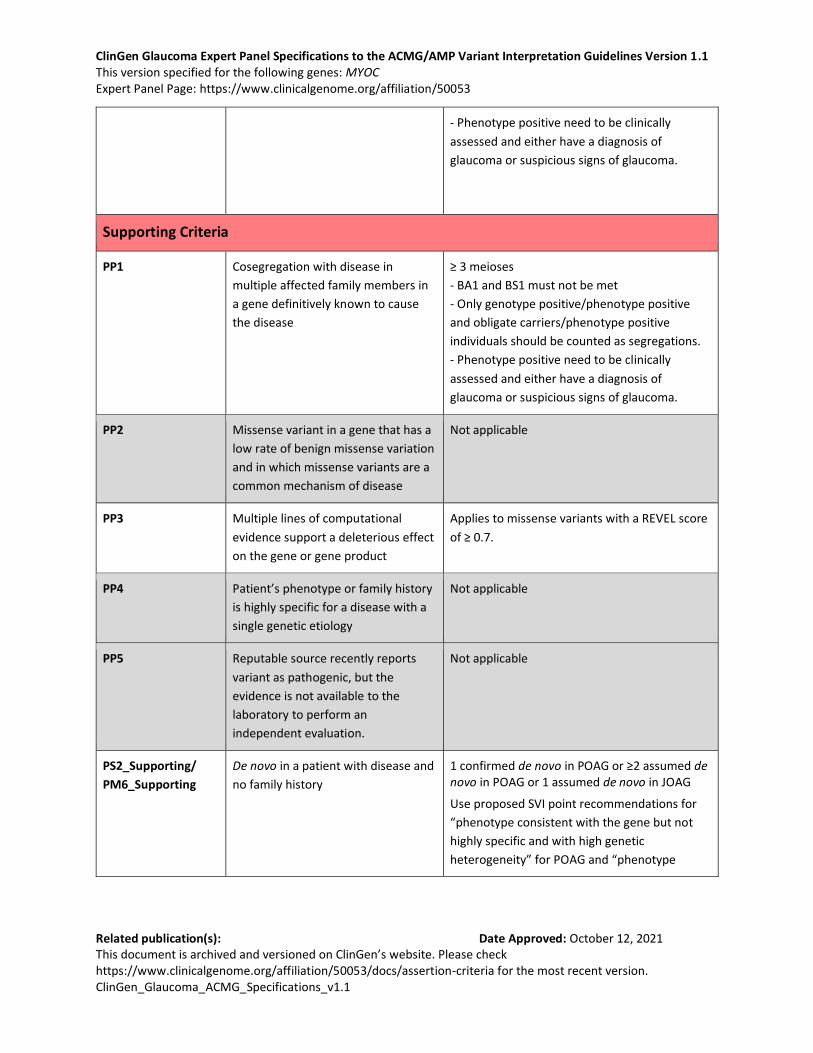
Supporting Criteria
PP1 Cosegregation with disease in
multiple affected family members in
a gene definitively known to cause
the disease
≥ 3 meioses
- BA1 and BS1 must not be met
- Only genotype positive/phenotype positive
and obligate carriers/phenotype positive
individuals should be counted as segregations.
- Phenotype positive need to be clinically
assessed and either have a diagnosis of
glaucoma or suspicious signs of glaucoma.
PP2 Missense variant in a gene that has a
low rate of benign missense variation
and in which missense variants are a
common mechanism of disease
Not applicable
PP3 Multiple lines of computational
evidence support a deleterious effect
on the gene or gene product
Applies to missense variants with a REVEL score
of ≥ 0.7.
PP4
Patient’s phenotype or family history
is highly specific for a disease with a
single genetic etiology
Not applicable
PP5 Reputable source recently reports
variant as pathogenic, but the
evidence is not available to the
laboratory to perform an
independent evaluation.
Not applicable
PS2_Supporting/
PM6_Supporting
De novo in a patient with disease and
no family history
1 confirmed de novo in POAG or ≥2 assumed de novo in POAG or 1 assumed de novo in JOAG
Use proposed SVI point recommendations for
“phenotype consistent with the gene but not
highly specific and with high genetic
heterogeneity” for POAG and “phenotype

ClinGen Glaucoma Expert Panel Specifications to the ACMG/AMP Variant Interpretation Guidelines Version 1.1 This version specified for the following genes: MYOC Expert Panel Page: https://www.clinicalgenome.org/affiliation/50053
Related publication(s): Date Approved: October 12, 2021 This document is archived and versioned on ClinGen’s website. Please check https://www.clinicalgenome.org/affiliation/50053/docs/assertion-criteria for the most recent version. ClinGen_Glaucoma_ACMG_Specifications_v1.1
consistent with gene but not highly specific” for
JOAG
- Both maternity and paternity need to be
proven for confirmed de novo variants.
- Parents need to be clinically assessed and not
have a diagnosis of glaucoma.
PS3_Supporting
Well-established in vitro or in vivo,
functional studies supportive of a
damaging effect on the gene or gene
product
Assays with OddsPath >2.1 as per the SVI
recommendations.1
Applies to the following functional studies:
- Solubility or secretion assays.
-OR-
- Animal models that replicate the glaucoma
phenotype.
PS4_Supporting The prevalence of the variant in
affected individuals is significantly
increased compared with the
prevalence in controls
≥ 2 probands from multiple independent
studies
- Probands counted should be clinically assessed
and have a diagnosis of JOAG or POAG
- PM2_Supporting must be met
PM2_Supporting Absent in population databases (or at
extremely low frequency if recessive)
Allele frequency ≤ 0.0001 in population
databases.
- Highest allele frequency in population
databases should be used
- Only applies to populations of ≥ 10,000 alleles
PM4_Supporting Protein length changes as a result of
in-frame deletions/insertions in a
nonrepeat region or stop-loss
variants
In-frame del/ins and truncating variants
involving ≤10% of the protein and located
within the conserved olfactomedin domain (AA
246-502).
PM5_Supporting Novel missense change at an amino
acid residue where a different
missense change determined to be
pathogenic has been seen before
Same residue as a previously established likely
pathogenic variant (assessed independently of
PM5).
- The novel change must not affect splicing
(SpliceAI ≤ 0.2), must meet PP3, and have a
Grantham score equal or greater than the
previously established LP variants.
Benign Criteria

ClinGen Glaucoma Expert Panel Specifications to the ACMG/AMP Variant Interpretation Guidelines Version 1.1 This version specified for the following genes: MYOC Expert Panel Page: https://www.clinicalgenome.org/affiliation/50053
Related publication(s): Date Approved: October 12, 2021 This document is archived and versioned on ClinGen’s website. Please check https://www.clinicalgenome.org/affiliation/50053/docs/assertion-criteria for the most recent version. ClinGen_Glaucoma_ACMG_Specifications_v1.1
Criteria Original Criteria Description Specification(s)
Stand Alone Criteria
BA1 Allele frequency is greater than
expected for disorder
Allele frequency ≥ 0.01 in population databases.
- The highest allele frequency in population
databases should be used
- Variant must be present in ≥ 5 alleles in any
validated general continental population
dataset of at least 2,000 observed alleles.
Strong Criteria
BS1 Allele frequency is greater than
expected for the disorder
Allele frequency ≥ 0.001 in population
databases.
- The highest allele frequency in population
databases should be used
- Variant must be present in ≥ 5 alleles in any
validated general continental population
dataset of at least 2,000 observed alleles.
- Does not apply to p.Gln368Ter
BS2 Observed in a healthy adult
individual for a recessive
(homozygous), dominant
(heterozygous), or X-linked
(hemizygous) disorder, with full
penetrance expected at an early age
Not applicable
BS3
Well-established in vitro or in vivo
functional studies show no damaging
effect on protein function or splicing
Not applicable
See BS3_Supporting
BS4 Lack of segregation in affected
members of a family
Not applicable
Moderate Criteria
BS3_Moderate Well-established in vitro or in vivo
functional studies show no damaging
effect on protein function or splicing
Applies to variants showing solubility or
secretion in functional assays for studies with
OddsPath <0.23 as per the SVI
recommendations.1

ClinGen Glaucoma Expert Panel Specifications to the ACMG/AMP Variant Interpretation Guidelines Version 1.1 This version specified for the following genes: MYOC Expert Panel Page: https://www.clinicalgenome.org/affiliation/50053
Related publication(s): Date Approved: October 12, 2021 This document is archived and versioned on ClinGen’s website. Please check https://www.clinicalgenome.org/affiliation/50053/docs/assertion-criteria for the most recent version. ClinGen_Glaucoma_ACMG_Specifications_v1.1
Supporting Criteria
BP1 Missense variant in a gene for which
primarily truncating variants are
known to cause disease
Not applicable
BP2 Observed in trans with a pathogenic
variant for a fully penetrant
dominant gene/disorder or observed
in cis with a pathogenic variant in any
inheritance pattern.
Not applicable
BP3 In-frame deletions/insertions in a
repetitive region without a known
function
Not applicable
BP4 Multiple lines of computational
evidence suggest no impact on
gene/gene product
Applies to:
- Missense variants: REVEL ≤ 0.15
- Synonymous variants or noncoding variants:
● CADD ≤ 10 AND
● SpliceAI ≤ 0.2
BP5 Variant found in a case with an
alternate molecular basis for disease
Not applicable
BP6 Reputable source recently reports
variant as benign, but the evidence is
not available to the laboratory to
perform an independent evaluation
Not applicable
BP7 A synonymous (silent) variant for
which splicing prediction algorithms
predict no impact to the splice
consensus sequence nor the creation
of a new splice site AND the
nucleotide is not highly conserved.
Applies to:
- Synonymous variants or noncoding variants
with no impact on splicing (SpliceAI ≤ 0.2)
AND
- GERP <0 for conservation
BS3_Supporting Well-established in vitro or in vivo
functional studies show no damaging
effect on protein function or splicing
Applies to variants showing solubility or
secretion in functional assays for studies with
OddsPath <0.48 as per the SVI
recommendations.1
Rules in grey are not applicable

ClinGen Glaucoma Expert Panel Specifications to the ACMG/AMP Variant Interpretation Guidelines Version 1.1 This version specified for the following genes: MYOC Expert Panel Page: https://www.clinicalgenome.org/affiliation/50053
Related publication(s): Date Approved: October 12, 2021 This document is archived and versioned on ClinGen’s website. Please check https://www.clinicalgenome.org/affiliation/50053/docs/assertion-criteria for the most recent version. ClinGen_Glaucoma_ACMG_Specifications_v1.1
RULES FOR COMBINING PATHOGENIC & BENIGN CRITERIA
There are no Very Strong criteria for MYOC: PVS1 does not apply and no other criterion has been upgraded to Very Strong Variants meeting BA1 are automatically classified as benign without assessing other evidence for or against pathogenicity and are therefore not part of the point system.2, 4 The Glaucoma VCEP agreed to use the point system developed by Tavtigian et al.2, 3 with the following modification: Likely Benign is defined as -2 to -6 point ranges instead of -1 to -6
Table 2 from Tavtigian et al 2020: Point Values for ACMG/AMP strength of evidence categories
Evidence Strength Point scale
Pathogenic Benign
Indeterminate 0 0
Supporting 1 -1
Moderate 2 -2
Strong 4 -4
Very Strong 8 -8
Table 3 from Tavtigian et al 2020 modified: Point-based variant classification categories
Category Point ranges
Pathogenic ≥10
Likely Pathogenic 6 to 9
Uncertain -1 to 5
Likely Benign -2 to -6
Benign ≤-7

ClinGen Glaucoma Expert Panel Specifications to the ACMG/AMP Variant Interpretation Guidelines Version 1.1 This version specified for the following genes: MYOC Expert Panel Page: https://www.clinicalgenome.org/affiliation/50053
Related publication(s): Date Approved: October 12, 2021 This document is archived and versioned on ClinGen’s website. Please check https://www.clinicalgenome.org/affiliation/50053/docs/assertion-criteria for the most recent version. ClinGen_Glaucoma_ACMG_Specifications_v1.1
Introduction to Myocilin (MYOC)
Glaucoma refers to a heterogeneous group of disorders characterised by progressive optic neuropathy
with associated visual field defects.5 Primary open-angle glaucoma (POAG) is the most common type of
glaucoma and is characterised by an open angle and no developmental defects or other underlying
disease. Early age of onset POAG is called juvenile open-angle glaucoma (JOAG) and is defined as an age
at diagnosis under 30 to 40 years old. Individuals who have suspicious optic discs, suspicious visual fields
and/or increased intraocular pressure (the main risk factor for glaucoma) but do not have glaucoma are
called glaucoma suspects.
Prevalence: POAG affects 50 to 60 million individuals worldwide, including 3.5% of individuals over the
age of 40 years.6, 7 The prevalence of POAG is 1/40 in Europeans, with the highest prevalence in Africans
(1/24).6, 7
Genetics: MYOC variants are the most common cause of adult-onset glaucoma and are found across
several populations. They account for 2-4% of POAG8, 9 and 8-36% of JOAG.10-12 The MYOC database that
lists all published variants currently comprises 336 variants (www.myocilin.com).13 MYOC variants are
transmitted in an autosomal dominant manner with an age-related penetrance. The most common
pathogenic variant is p.Gln368Ter, accounting for 1.6-2.6% of individuals with POAG.8, 9 This variant is
present in 1/450 individuals from gnomAD (allele frequency 0.001110) and previous studies support a
common European founder effect14 with incomplete penetrance for p.Gln368Ter.15 Recent data on
p.Gln368Ter suggest that the penetrance is much lower in population-based studies (7.6%) than in family-
based studies (56%).15
Mechanism of disease: MYOC comprises of 3 exons and most reported disease-causing variants are
located in the olfactomedin domain in exon 3 (AA 246-502 inclusive according to NCBI).13 Both
truncating and missense variants are associated with the disease. MYOC deletions have been shown not
to cause disease, both in humans16 and in mice models,17 ruling out haploinsufficiency as a disease
mechanism. The mechanism by which the variants cause POAG is still not fully understood but the
current body of evidence supports a gain of function mechanism. MYOC is usually glycosylated via the
endoplasmic reticulum (ER) and secreted extracellularly.18, 19 Previous in-vitro studies have shown that
mutant and wild-type proteins interact together to form hetero-oligomers.20 Evidence suggests that
MYOC variants lead to misfolded protein21-23 and that the mutant protein is not secreted
extracellularly.18, 20, 22, 24, 25 Moreover, mutant MYOC protein is not processed correctly in the ER and
leads to accumulation of insoluble aggregates inside the trabecular meshwork cells19, 21 that induce the
unfolded protein response, leading to ER stress-induced apoptosis.26

ClinGen Glaucoma Expert Panel Specifications to the ACMG/AMP Variant Interpretation Guidelines Version 1.1 This version specified for the following genes: MYOC Expert Panel Page: https://www.clinicalgenome.org/affiliation/50053
Related publication(s): Date Approved: October 12, 2021 This document is archived and versioned on ClinGen’s website. Please check https://www.clinicalgenome.org/affiliation/50053/docs/assertion-criteria for the most recent version. ClinGen_Glaucoma_ACMG_Specifications_v1.1
ACMG Classification Rule Specifications for Myocilin
SUMMARY OF CLASSIFICATION CRITERIA Rules in grey are not applicable, specified rules are in red
PATHOGENIC Very Strong
PVS1 – Null variant in a gene where LOF is a known mechanism of disease
Strong PS1 – Same amino acid change from different DNA change PS2 – Confirmed de novo in a patient with the disease and no family history PS3 – Well-established in vitro or in vivo functional studies supportive of a damaging effect PS4 – Prevalence of variant in affected individuals is significantly increased over controls
Moderate PM1 – Located in functional region without benign variation PM2_Supporting – Rarity/Absence in population databases PM3 – For recessive disorders, detected in trans with a pathogenic variant PM4 – Protein length changes PM5 – Missense change at same codon as another pathogenic missense variant PM6 – Assumed de novo in a patient with the disease and no family history
Supporting PP1 – Cosegregation with disease in multiple affected family members PP2 – Missense variant in a gene with low rate of benign missense variation PP3 – Multiple lines of computational evidence supporting a deleterious effect PP4 – Phenotype and family history specific for disease with single genetic etiology PP5 – Reputable source reports as pathogenic
BENIGN Stand-Alone
BA1 – Allele frequency is >5% in population databases Strong
BS1 – Allele frequency is greater than expected for disorder BS2 – Observed in a healthy individual BS3_Supporting – Well-established in vitro or in vivo functional studies show no effect BS4 – Lack of segregation in affected relatives
Supporting BP1 – Missense variant in gene where LOF causes disease BP2 – Biallelic variants for a fully penetrant disorder BP3 – In-frame deletions/insertions in a repetitive region without a known function BP4 – Multiple lines of computational evidence support no impact BP5 – Variant found in a case with an alternate molecular basis for disease BP6 – Reputable source reports as benign BP7 – Synonymous variant for which splicing prediction algorithms predict no impact

ClinGen Glaucoma Expert Panel Specifications to the ACMG/AMP Variant Interpretation Guidelines Version 1.1 This version specified for the following genes: MYOC Expert Panel Page: https://www.clinicalgenome.org/affiliation/50053
Related publication(s): Date Approved: October 12, 2021 This document is archived and versioned on ClinGen’s website. Please check https://www.clinicalgenome.org/affiliation/50053/docs/assertion-criteria for the most recent version. ClinGen_Glaucoma_ACMG_Specifications_v1.1
VERY STRONG EVIDENCE OF PATHOGENICITY
PVS1 Null variant (nonsense, frameshift, canonical ±1 or 2 splice sites, initiation codon, single or multiexon deletion) in a gene where LOF is a known mechanism of disease
Caveats:
- Beware of genes where LoF is not a known disease mechanism (e.g., GFAP, MYH7)
- Use caution interpreting LoF variants at the extreme 3′ end of a gene
- Use caution with splice variants that are predicted to lead to exon skipping but leave the remainder of the protein intact
- Use caution in the presence of multiple transcripts
Glaucoma VCEP notes:
MYOC variants cause JOAG/POAG through a gain of function (GoF) disease mechanism and not loss of function (LoF). Truncating variants in exon 3 are expected to be pathogenic because they escape nonsense-mediated decay. The Glaucoma VCEP decided to not use PVS1 because GoF is the disease mechanism for MYOC variants, not LoF. The group decided to use PM4 for truncating variants in exon 3 that escape NMD instead.
STRONG EVIDENCE OF PATHOGENICITY
PS1 Same amino acid change as a previously established pathogenic variant regardless of nucleotide change
Example: Val→Leu caused by either G>C or G>T in the same codon
Caveat: Beware of changes that impact splicing rather than at the amino acid/protein level
Glaucoma VCEP notes:
The MYOC database currently lists one variant with a different nucleotide change but the same amino acid as a previously reported variant (c.1440C>A and c.1440C>G both leading to p.Asn480Lys).
MYOC Specification:
- The novel change must not affect splicing (SpliceAI ≤ 0.2)
- The following levels of evidence apply:
PS1: Same amino acid change as previously established pathogenic variant
PS1_Moderate: Same amino acid change as previously established likely pathogenic variant

ClinGen Glaucoma Expert Panel Specifications to the ACMG/AMP Variant Interpretation Guidelines Version 1.1 This version specified for the following genes: MYOC Expert Panel Page: https://www.clinicalgenome.org/affiliation/50053
Related publication(s): Date Approved: October 12, 2021 This document is archived and versioned on ClinGen’s website. Please check https://www.clinicalgenome.org/affiliation/50053/docs/assertion-criteria for the most recent version. ClinGen_Glaucoma_ACMG_Specifications_v1.1
PS2 De novo (both maternity and paternity confirmed) in a patient with the disease and no family history
Notes from the ACMG guidelines:
- Confirmation of paternity only is insufficient. Egg donation, surrogate motherhood, errors in embryo transfer, and so on, can contribute to non-maternity
- Parents need to be unaffected for a dominant disorder
- More than one sibling may be affected because of germline mosaicism
- The phenotype of the patient needs to match the gene’s disease association with reasonable specificity
Glaucoma VCEP notes:
MYOC de novo variants are rare with only 2 reports in the literature:
- p.Val251Ala confirmed de novo27
- p.Pro254Leu confirmed de novo28
MYOC Specification:
- Both paternity and maternity need to be proven for confirmed de novo variants
- Both parents need to be clinically assessed and should not have a diagnosis of glaucoma
If a parent has suspicious signs of glaucoma, the age and the severity of the symptoms should be taken into account before applying criteria
- Use the proposed SVI point recommendation table to determine the appropriate strength of evidence (see table below).
- Phenotype:
The phenotype of POAG is not highly specific and has substantial genetic heterogeneity. Apply the point-based system for a “phenotype consistent with the gene but not highly specific and with high genetic heterogeneity” for POAG.
The phenotype of JOAG is more specific. Apply a higher point-based system for JOAG, using a “phenotype consistent with gene but not highly specific”.
Examples:
PS2: ≥2 confirmed de novo in JOAG
PS2_Moderate: ≥2 confirmed de novo in POAG or 1 confirmed de novo in JOAG or ≥2 assumed de novo in JOAG
PS2_Supporting: 1 confirmed de novo in POAG or ≥2 assumed de novo in POAG or 1 assumed de novo in JOAG
ClinGen Glaucoma Expert Panel Specifications to the ACMG/AMP Variant Interpretation Guidelines Version 1.1 This version specified for the following genes: MYOC Expert Panel Page: https://www.clinicalgenome.org/affiliation/50053
Related publication(s): Date Approved: October 12, 2021 This document is archived and versioned on ClinGen’s website. Please check https://www.clinicalgenome.org/affiliation/50053/docs/assertion-criteria for the most recent version. ClinGen_Glaucoma_ACMG_Specifications_v1.1
PS3 Well-established in vitro or in vivo functional studies supportive of damaging effect on the gene or gene product
Notes from ACMG guidelines:
Functional studies that have been validated and shown to be reproducible and robust in a clinical diagnostic laboratory setting are considered the most well established
The SVI Working Group published a provisional framework for the evaluation and application of functional evidence.1
Glaucoma VCEP notes:
The mechanism by which the variants cause POAG is still not fully understood but the current body of evidence supports a GoF mechanism with accumulation of insoluble aggregates inside the endoplasmic reticulum of the trabecular meshwork cells.19, 21
Follow the SVI recommendations from Brnich et al. when assessing functional assays to apply PS3 toward functional evidence.1
MYOC Specification:
- PS3 should only be applied if the assay includes both negative and positive controls and includes technical and/or biological replicates.
- If multiple results from functional assays are available for a single variant, then the evidence from the assay that is best validated should apply.
- Controls from the same general class of assay can be combined to calculate the odds of pathogenicity (OddsPath) as per the published SVI recommendations.1

ClinGen Glaucoma Expert Panel Specifications to the ACMG/AMP Variant Interpretation Guidelines Version 1.1 This version specified for the following genes: MYOC Expert Panel Page: https://www.clinicalgenome.org/affiliation/50053
Related publication(s): Date Approved: October 12, 2021 This document is archived and versioned on ClinGen’s website. Please check https://www.clinicalgenome.org/affiliation/50053/docs/assertion-criteria for the most recent version. ClinGen_Glaucoma_ACMG_Specifications_v1.1
- If results from different assays are conflicting for a single variant, then the level of validation of each assay should be considered to decide whether the results from one assay can override the results from another.
- Assays considered suitable include:
Assays that report on the solubility and secretion of MYOC
OR
Animal models that replicate the glaucoma phenotype
PS3 at different levels of evidence can be applied according to the OddsPath from the published SVI recommendations.1
PS3: Assays with OddsPath >18.7
PS3_Moderate: Assays with OddsPath >4.3
PS3_Supporting: Assays with OddsPath >2.1
PS4 The prevalence of the variant in affected individuals is significantly increased compared with the prevalence in controls
Notes from the ACMG guidelines:
1) Relative risk or OR, as obtained from case-control studies, is >5.0, and the confidence interval around the estimate of relative risk or OR does not include 1.0.
2) In instances of very rare variants where case-control studies may not reach statistical significance, the prior observation of the variant in multiple unrelated patients with the same phenotype, and its absence in controls, may be used as moderate level of evidence.
Caution should be exercised to not duplicate counting of one proband reported multiple times in the literature or within both the literature and a clinical testing cohort.
Glaucoma VCEP notes:
Case-control data for MYOC variants is limited due to control cohorts often being too small to reflect the true prevalence of variants in a true control population. Instead, the Glaucoma VCEP recommends using PS4 for counting probands from multiple independent studies using a “quasi case-control study” approach.
The Glaucoma VCEP calculated ORs similar to Kelly et al.29 using an internal cohort of 3,522 probands that had been sequenced for MYOC and the gnomAD database as a proxy for controls. This is a conservative approach considering gnomAD will include individuals who may have glaucoma and have not been diagnosed or excluded and individuals who may not have glaucoma yet due to age-related penetrance.
Due to incomplete penetrance, late age of onset of glaucoma and the rate of undiagnosed glaucoma in the general population, pathogenic variants may be present in population databases. Therefore, rule PM2_supporting has to be met.

ClinGen Glaucoma Expert Panel Specifications to the ACMG/AMP Variant Interpretation Guidelines Version 1.1 This version specified for the following genes: MYOC Expert Panel Page: https://www.clinicalgenome.org/affiliation/50053
Related publication(s): Date Approved: October 12, 2021 This document is archived and versioned on ClinGen’s website. Please check https://www.clinicalgenome.org/affiliation/50053/docs/assertion-criteria for the most recent version. ClinGen_Glaucoma_ACMG_Specifications_v1.1
ORs of 10, 30 and 100 were reached at a similar number of probands to Kelly et al., both in the whole cohort and in Europeans only (See Supplementary Tables 1 & 2). Therefore, the Glaucoma VCEP decided to use the same thresholds as Kelly et al. for counting probands of 2, 6 and 15 for Supporting, Moderate and Strong levels respectively.
MYOC Specification:
- Use PS4 for counting probands from multiple independent studies
- Probands need to be clinically assessed and have JOAG or POAG
- PM2 needs to be met
PS4: ≥ 15 probands
PS4_Moderate: ≥ 6 probands
PS4_Supporting: ≥ 2 probands
MODERATE EVIDENCE OF PATHOGENICITY
PM1 Located in a mutational hot spot and/or critical and well-established functional domain (e.g., active site of an enzyme) without benign variation
This rule does not apply. MYOC has no mutational hot spot and benign variants are present though the well-characterised olfactomedin domain in exon 3.13
PM2_Supporting Absent from controls (or at extremely low frequency if recessive) in Exome Sequencing Project, 1000 Genomes Project, or Exome Aggregation Consortium
Glaucoma VCEP notes:
The filtering allele frequency for PM2 was set one order of magnitude lower than BS1. The Glaucoma VCEP agreed that this is a conservative approach: some pathogenic variants are expected to be present in population databases due to POAG being a complex disease with late onset and age-related penetrance. Moreover, most MYOC pathogenic variants are absent from large population databases. The Glaucoma VCEP followed the SVI recommendations to apply PM2 at a Supporting level of evidence.
MYOC Specification:
Allele frequency is ≤ 0.0001 (0.01%) in population databases
- The highest allele frequency in population databases should be used
- PM2 only applies to populations of ≥ 10,000 alleles
- PM2 should be used at a Supporting level as per the SVI recommendations

ClinGen Glaucoma Expert Panel Specifications to the ACMG/AMP Variant Interpretation Guidelines Version 1.1 This version specified for the following genes: MYOC Expert Panel Page: https://www.clinicalgenome.org/affiliation/50053
Related publication(s): Date Approved: October 12, 2021 This document is archived and versioned on ClinGen’s website. Please check https://www.clinicalgenome.org/affiliation/50053/docs/assertion-criteria for the most recent version. ClinGen_Glaucoma_ACMG_Specifications_v1.1
PM3 For recessive disorders, detected in trans with a pathogenic variant
This rule does not apply. MYOC variants have an autosomal dominant mode of inheritance.
PM4 Protein length changes as a result of in-frame deletions/insertions in a nonrepeat region or stop-loss variants
Glaucoma VCEP notes:
There are only four in-frame variants reported in the MYOC database. There is a lack of current data to support a benign/pathogenic classification of in-frame del/ins:
- p.Glu162dup (exon 1)30
- p.Gly367_Gln368delinsVal (exon 3)31
- p.Asp395_Glu396insAspPro (exon 3)32
- p.Glu396dup (exon 3)33
The ACGS 2020 guidelines recommend caution for single amino acid in-frame deletions or insertions where PM4 may be used at a supporting level unless there is gene-specific evidence to warrant use at a moderate level.34
The disease mechanism for MYOC variants is GoF, not LoF. The Glaucoma VCEP decided to use PM4 instead of PVS1 for truncating variants in the olfactomedin domain.
MYOC Specification:
- Only applies to in-frame deletions/insertions AND truncating variants located within the conserved olfactomedin domain (AA 246-502)
PM4: in-frame del/ins and truncating variants involving ≥10% of the protein AND located within the conserved olfactomedin domain (AA 246-502)
PM4_Supporting: in-frame del/ins and truncating variants involving <10% of the protein AND located within the conserved olfactomedin domain (AA 246-502)
PM5 Novel missense change at an amino acid residue where a different missense change determined to be pathogenic has been seen before
Example: Arg156His is pathogenic; now you observe Arg156Cys
Caveat: Beware of changes that impact splicing rather than at the amino acid/protein level
Glaucoma VCEP notes:
The MYOC database currently lists 30 variants with different amino acid changes at the same residue.

ClinGen Glaucoma Expert Panel Specifications to the ACMG/AMP Variant Interpretation Guidelines Version 1.1 This version specified for the following genes: MYOC Expert Panel Page: https://www.clinicalgenome.org/affiliation/50053
Related publication(s): Date Approved: October 12, 2021 This document is archived and versioned on ClinGen’s website. Please check https://www.clinicalgenome.org/affiliation/50053/docs/assertion-criteria for the most recent version. ClinGen_Glaucoma_ACMG_Specifications_v1.1
MYOC Specification:
- The novel change must not affect splicing (SpliceAI ≤ 0.2) and must meet PP3
- The novel change must have a Grantham score equal or greater than the previously established pathogenic or likely pathogenic variant
PM5: same residue as previously established pathogenic variant (assessed independently of PM5) or 2 previously established likely pathogenic variants (in which case both variants must be assessed independently of PM5)
PM5_Supporting: same residue as previously established likely pathogenic variant (assessed independently of PM5)
PM6 Assumed de novo, but without confirmation of paternity and maternity
MYOC Specification:
- Use the proposed SVI point recommendation table to determine the appropriate strength of evidence (see PS2).
SUPPORTING EVIDENCE OF PATHOGENICITY
PP1 Cosegregation with disease in multiple affected family members in a gene definitely known to cause the disease
Issue with the ACMG definition: segregation is not quantitatively defined
A higher level of evidence can be applied depending on the level of segregation
Glaucoma VCEP notes:
Two studies have provided quantitative guidelines for assessing segregation of a variant.
Jarvik et al. calculated a probability that the observed variant and affection status occurs by chance rather than due to cosegregation.35 Under a dominant model, this probability is N = (1/2)m, where m is the number of meioses of the variant of interest. Their proposed model is:
● PP1: ≤1/8 in a single family (≥3 meioses) or ≤1/4 in >1 family (≥2 meioses)
● PP1_Moderate: ≤1/16 in a single family (≥4 meioses) or ≤1/8 in >1 family (≥3 meioses)
● PP1_Strong: ≤1/32 (≥5 meioses) in a single family or ≤1/16 in >1 family (≥4 meioses)
Kelly et al. adopted three levels of evidence based on the number of meioses for an autosomal dominant model:29
● PP1: ≥3 meioses in ≥1 family (LOD 0.9, likelihood ratio of 10)
● PP1_Moderate: ≥5 meioses in ≥1 family (LOD 1.5, likelihood ratio of 30)
● PP1_Strong: ≥7 meioses in ≥1 family (LOD 2.1, likelihood ratio of 100)

ClinGen Glaucoma Expert Panel Specifications to the ACMG/AMP Variant Interpretation Guidelines Version 1.1 This version specified for the following genes: MYOC Expert Panel Page: https://www.clinicalgenome.org/affiliation/50053
Related publication(s): Date Approved: October 12, 2021 This document is archived and versioned on ClinGen’s website. Please check https://www.clinicalgenome.org/affiliation/50053/docs/assertion-criteria for the most recent version. ClinGen_Glaucoma_ACMG_Specifications_v1.1
The Glaucoma VCEP piloted both approaches on 81 MYOC variants. Two variants had a different classification using the two approaches: p.Ala363Thr had 5 meioses in 3 families, meeting PP1_Moderate according to Kelly et al. but PP1_Strong according to Jarvik et al. The classification remained LP with both approaches. p.Asn480Lys (c.1440C>G) had 4 meioses in 1 family, meeting PP1 according to Kelly et al. but PP1_Moderate according to Jarvik et al. This variant was classified as LP when using Kelly et al. but as P when using Jarvik et al. The Glaucoma VCEP agreed to apply a conservative approach and use the recommendations from Kelly et al.
MYOC Specification:
- BA1 and BS1 must not be met
- Multiple families are jointly considered by adding the informative meioses across families according to Kelly et al.29
- Only genotype positive/phenotype positive and obligate carriers/phenotype positive individuals should be counted as segregations
- Individuals who are genotype positive/phenotype negative and individuals who are genotype negative/phenotype negative should not be counted
- Phenotype positive need to be clinically assessed and either have a diagnosis of glaucoma or suspicious signs of glaucoma (e.g. ocular hypertension, suspicious discs)
- Do not apply Strong level of evidence if the variant is present in a single family due to risk of other variant in linkage disequilibrium
PP1_Strong: ≥7 meioses in >1 family
PP1_Moderate: ≥5 meioses regardless of the number of families
PP1: ≥3 meioses regardless of the number of families
PP2 Missense variant in a gene that has a low rate of benign missense variation and in which missense variants are a common mechanism of disease
This rule does not apply. Although pathogenic missense variants are common in MYOC, the gene also has a significant amount of benign missense variants as shown by the missense constraint z score in gnomAD (z = 0.52) supporting tolerance to variation.
PP3 Multiple lines of computational evidence support a deleterious effect on the gene or gene product (conservation, evolutionary, splicing impact, etc.)
Glaucoma VCEP notes:
CADD and REVEL scores were reviewed for MYOC variants listed in ClinVar. Overall, predicted pathogenic variants in exon 3 seem to score high with both predictors, but variants in exon 1 expected to be benign had low REVEL scores while having mixed CADD scores. REVEL does not score truncating variants but CADD does. Truncating variants across all 3 exons had CADD scores >20, suggesting that CADD cannot predict potential for nonsense-mediated decay.

ClinGen Glaucoma Expert Panel Specifications to the ACMG/AMP Variant Interpretation Guidelines Version 1.1 This version specified for the following genes: MYOC Expert Panel Page: https://www.clinicalgenome.org/affiliation/50053
Related publication(s): Date Approved: October 12, 2021 This document is archived and versioned on ClinGen’s website. Please check https://www.clinicalgenome.org/affiliation/50053/docs/assertion-criteria for the most recent version. ClinGen_Glaucoma_ACMG_Specifications_v1.1
The Glaucoma VCEP recommended using only one in silico predictor, in line with a recent study showing a lower rate of concordance when multiple software are used.36 REVEL scores are recommended based on their high level of accuracy toward variant prediction.37
MYOC Specification:
PP3 applies to missense variants only with a REVEL score of ≥ 0.7
PP4 Patient’s phenotype or family history is highly specific for a disease with a single genetic etiology
This rule does not apply. The phenotype associated with MYOC variants is not highly specific and there is genetic heterogeneity.
PP5 Reputable source recently reports variant as pathogenic, but the evidence is not available to the laboratory to perform an independent evaluation
This rule does not apply. The Glaucoma VCEP follows the recommendation of the SVI Working Group to use primary data instead of relying on assertions and therefore to not use PP5.
STAND-ALONE EVIDENCE OF BENIGN IMPACT
BA1 Allele frequency is >5% in Exome Sequencing Project, 1000 Genomes Project, or Exome Aggregation Consortium
Notes from Whiffin et al. (2017):38
“For a penetrant dominant Mendelian allele to be disease-causing, it cannot be present in the general population more frequently than the disease it causes. Furthermore, if the disease is genetically heterogeneous, it must not be more frequent than the proportion of cases attributable to that gene, or indeed to any single variant.”
Glaucoma VCEP notes:
The Whiffin/Ware calculator was used to obtain a population allele frequency threshold for BA1 using conservative figures.38
Maximum credible population AF = prevalence x max allelic contribution x 1/penetrance
- The prevalence of POAG in the African population, which is the highest among all populations, was used (1/24)6, 7
- The maximum allelic contribution for the most common MYOC variant (p.Gln368Ter) was set at 2.6% using data from large disease registries (the Australian and New Zealand of Advanced Glaucoma and the Glaucoma Inheritance Study in Tasmania with data on over 3,236 individuals)9
- A conservative estimate for the penetrance at 7.6% was used based on the penetrance of p.Gln368Ter in a population-based study using data from the UK biobank.15

ClinGen Glaucoma Expert Panel Specifications to the ACMG/AMP Variant Interpretation Guidelines Version 1.1 This version specified for the following genes: MYOC Expert Panel Page: https://www.clinicalgenome.org/affiliation/50053
Related publication(s): Date Approved: October 12, 2021 This document is archived and versioned on ClinGen’s website. Please check https://www.clinicalgenome.org/affiliation/50053/docs/assertion-criteria for the most recent version. ClinGen_Glaucoma_ACMG_Specifications_v1.1
The maximum credible allele frequency calculated was 0.007, which was rounded up to 0.01
MYOC Specification:
Allele frequency is ≥ 0.01 (1%) in population databases
- The highest allele frequency in population databases should be used
- The variant must be present in ≥ 5 alleles in any validated general continental population dataset of at least 2,000 observed alleles
STRONG EVIDENCE OF BENIGN IMPACT
BS1 Allele frequency is greater than expected for disorder
Glaucoma VCEP notes:
The Whiffin/Ware calculator was used to obtain a population allele frequency threshold for BS1 using a more realistic estimate of the penetrance.38
- The prevalence of POAG and the maximum allelic contribution used were the same as for BA1. A penetrance at 56% was used based on the penetrance of p.Gln368Ter in family-based studies using data from the Australian and New Zealand of Advanced Glaucoma and the Glaucoma Inheritance Study in Tasmania.15 The maximum credible allele frequency calculated was 0.001
MYOC p.Gln368Ter is a well-established pathogenic variant but displays incomplete penetrance. Its allele frequency in gnomAD is 0.001588 in European Non-Finnish, 0.003344 in European Finnish, and is 0.0025 in the UKBB. Evidence supports a European founder effect.14
MYOC Specification:
Allele frequency is ≥ 0.001 (0.1%) in population databases
- The highest allele frequency in population databases should be used
- The variant must be present in ≥ 5 alleles in any validated general continental population dataset of at least 2,000 observed alleles
- Exception rule for the p.Gln368Ter variant based on its incomplete penetrance and the presence of a common disease haplotype in all carriers
BS2 Observed in a healthy adult individual for a recessive (homozygous), dominant (heterozygous), or X-linked (hemizygous) disorder, with full penetrance expected at an early age
This rule does not apply. MYOC variants have an incomplete penetrance and late age of onset.39

ClinGen Glaucoma Expert Panel Specifications to the ACMG/AMP Variant Interpretation Guidelines Version 1.1 This version specified for the following genes: MYOC Expert Panel Page: https://www.clinicalgenome.org/affiliation/50053
Related publication(s): Date Approved: October 12, 2021 This document is archived and versioned on ClinGen’s website. Please check https://www.clinicalgenome.org/affiliation/50053/docs/assertion-criteria for the most recent version. ClinGen_Glaucoma_ACMG_Specifications_v1.1
BS3_Moderate Well-established in vitro or in vivo functional studies show no damaging effect on protein function or splicing
Glaucoma VCEP notes:
The Glaucoma VCEP reviewed the evidence on the solubility and secretion assays for benign variants. Among the 27 coding variants curated in the pilot phase as B/LB, functional evidence was available for 16 of them and all were secreted and/or soluble. Similarly, among the 21 coding variants showing evidence for solubility and/or secretion, none were classified as P/LP, 10 were classified as B/LB and 11 were classified as VUS in the pilot phase. Based on this, the group agreed to apply BS3 to variants showing solubility or secretion in functional assays that meet the odds of pathogenicity for a moderate or a supporting level according to Brnich et al.1 Although there is no evidence currently supporting other protein functions leading to the condition, the Glaucoma VCEP decided to not apply BS3 at a strong level, as we cannot completely rule out other mechanisms for pathogenicity.
MYOC Specification:
- BS3 should only be applied if the assay includes both negative and positive controls and includes technical and/or biological replicates.
- If multiple results from functional assays are available for a single variant, then the evidence from the assay that is best validated should apply.
- Controls from the same general class of assay can be combined to calculate the odds of pathogenicity (OddsPath) as per the published SVI recommendations.1
- If results from different assays are conflicting for a single variant, then the level of validation of each assay should be considered to decide whether the results from one assay can override the results from another.
- BS3 at a Moderate or Supporting level applies to variants showing solubility or secretion in functional assays for studies with OddsPath as per the published SVI recommendations.1
BS3_Moderate: Assays with OddsPath <0.23
BS3_Supporting: Assays with OddsPath <0.48
BS4 Lack of segregation in affected members of a family
This rule does not apply. The presence of phenocopies, the reduced age-related penetrance and the possibility that more than one pathogenic variant can contribute to the phenotype observed in families make non-segregation difficult to assess in the context of MYOC and POAG.
SUPPORTING EVIDENCE OF BENIGN IMPACT
BP1 Missense variant in a gene for which primarily truncating variants are known to cause disease
This rule does not apply. Both truncating and missense MYOC variants are causative.

ClinGen Glaucoma Expert Panel Specifications to the ACMG/AMP Variant Interpretation Guidelines Version 1.1 This version specified for the following genes: MYOC Expert Panel Page: https://www.clinicalgenome.org/affiliation/50053
Related publication(s): Date Approved: October 12, 2021 This document is archived and versioned on ClinGen’s website. Please check https://www.clinicalgenome.org/affiliation/50053/docs/assertion-criteria for the most recent version. ClinGen_Glaucoma_ACMG_Specifications_v1.1
BP2 Observed in trans with a pathogenic variant for a fully penetrant dominant gene/disorder or observed in cis with a pathogenic variant in any inheritance pattern
This rule does not apply. Biallelic variants (either compound heterozygotes or homozygotes) have been reported (with variable phenotype)40, 41 and are not incompatible with life. Two missense variants in cis could act synergistically or the effect of a variant occurring after a truncating variant may not be predicted.
BP3 In-frame deletions/insertions in a repetitive region without a known function
This rule does not apply. MYOC does not have a repetitive region without a known function.
BP4 Multiple lines of computational evidence suggest no impact on gene or gene product (conservation, evolutionary, splicing impact, etc.)
Glaucoma VCEP notes:
Similar to PP3, CADD and REVEL scores were reviewed for MYOC variants listed in ClinVar. REVEL is more conservative and better than CADD at accurately scoring MYOC missense variants classified as benign in ClinVar. Therefore, the Glaucoma VCEP recommended using REVEL for missense variants. However, REVEL does not score synonymous variants but CADD does. Predicted benign synonymous variants score low on CADD and the Glaucoma VCEP agreed to use CADD for synonymous variants.
The Glaucoma VCEP assessed different splicing and conservation predictors. One study reported best predictive value for combination of HSF, SFF-like and MES but slightly different for 5’ and 3’ splice sites.42 However HSF is no longer publicly available, and SSF is not available online. The Neural Network (BDGP - http://www.fruitfly.org/seq_tools/splice.html) and SpliceAI (https://spliceailookup.broadinstitute.org/) only look for donor and acceptor sites, not for enhancers or repressors compared to HSF. Using SpliceAI, none of the 19 synonymous variants in ClinVar were predicted to have an effect on splicing. The Glaucoma VCEP agreed that SpliceAI would be appropriate for MYOC which only has 3 exons, one transcript and for which splicing is not known to vary.
Based on the disease mechanism and the absence of current evidence supporting pathogenicity of intronic/noncoding variants, the Glaucoma VCEP agreed to apply BP4 to noncoding variants.
MYOC Specification:
- BP4 applies to missense variants with a REVEL score of ≤ 0.15
- BP4 applies to synonymous variants or noncoding variants with:
• CADD ≤ 10 AND

ClinGen Glaucoma Expert Panel Specifications to the ACMG/AMP Variant Interpretation Guidelines Version 1.1 This version specified for the following genes: MYOC Expert Panel Page: https://www.clinicalgenome.org/affiliation/50053
Related publication(s): Date Approved: October 12, 2021 This document is archived and versioned on ClinGen’s website. Please check https://www.clinicalgenome.org/affiliation/50053/docs/assertion-criteria for the most recent version. ClinGen_Glaucoma_ACMG_Specifications_v1.1
• SpliceAI ≤ 0.2
BP5 Variant found in a case with an alternate molecular basis for disease
This rule does not apply. Multiple molecular diagnoses are possible and variants in different genes could have an additive effect.
BP6 Reputable source recently reports variant as benign, but the evidence is not available to the laboratory to perform an independent evaluation
This rule does not apply. The Glaucoma VCEP follows the recommendation of the SVI Working Group to use primary data instead of relying on assertions and therefore to not use BP6.
BP7 A synonymous (silent) variant for which splicing prediction algorithms predict no impact to the splice consensus sequence nor the creation of a new splice site AND the nucleotide is not highly conserved.
Glaucoma VCEP notes:
For conservation, GERP and PhyloP scores broadly agree on conservation for all the synonymous variants in ClinVar (both negative or both positive). For both, a negative score indicates lack of conservation. The Glaucoma VCEP agreed to extend BP7 to noncoding variants.
MYOC Specification:
- Synonymous variant or noncoding variants with no impact on splicing (SpliceAI ≤ <0.2)
AND
- GERP <0 for conservation

ClinGen Glaucoma Expert Panel Specifications to the ACMG/AMP Variant Interpretation Guidelines Version 1.1 This version specified for the following genes: MYOC Expert Panel Page: https://www.clinicalgenome.org/affiliation/50053
Related publication(s): Date Approved: October 12, 2021 This document is archived and versioned on ClinGen’s website. Please check https://www.clinicalgenome.org/affiliation/50053/docs/assertion-criteria for the most recent version. ClinGen_Glaucoma_ACMG_Specifications_v1.1
Supplementary Table 1: OR (95%CI) and P-value (Fishers Exact Test) for various allele counts in gnomAD V2.1.1 full population (n=125,748) versus various proband counts
1 allele in gnomAD 3 alleles in gnomAD 6 alleles in gnomAD 9 alleles in gnomAD 12 alleles in gnomAD
Prob OR (95%CI) P-value OR (95%CI) P-value OR (95%CI) P-value OR (95%CI) P-value OR (95%CI) P-value
2 71.40 (3.7-4072) 0.002186 23.81 (2.0-207.9) 0.00703 11.91 (1.2-66.6) 0.01860 7.94 (0.8-38.4) 0.034660 5.95 (0.6-26.7) 0.054360
3 107.40 (8.6-5387) 7.92E-05 35.73 (4.8-266.3) 0.00038 17.87 (2.9-83.9) 0.00150 11.91 (2.1-47.8) 0.003696 8.93 (1.6-33.1) 0.007192
4 142.88 (14.1-6675) 2.69E-06 47.63 (8.1-323.6) 1.80E-05 23.83 (4.9-100.8) 0.00010 15.88 (3.6-56.9) 0.000323 11.91 (2.8-39.3) 0.000770
5 179.29 (20.0-7935) 8.78E-08 59.56 (11.6-380.5) 7.83E-07 29.79 (7.2-117.3) 6.03E-06 19.86 (5.2-66.0) 2.44E-05 14.89 (4.1-45.4) 7.04E-05
6 214.78 (26.0-9166) 2.79E-09 71.49 (15.3-439.5) 3.19E-08 35.76 (9.6-133.8) 3.27E-07 23.84 (7.0-75.1) 1.65E-06 17.88 (5.5-51.4) 5.70E-06
7 250.48 (32.2-10371) 8.65E-11 83.32 (19.0-498.6) 1.24E-09 41.71 (12.0-150.0) 1.65E-08 27.82 (8.8-84.0) 1.02E-07 20.87 (7.0-57.5) 4.18E-07
8 286.27 (38.3-11548) 2.65E-12 95.38 (22.9-557.2) 4.62E-11 47.69 (14.5-167.5) 7.81E-10 31.83 (10.7-93.0) 5.88E-09 23.85 (8.5-63.5) 2.83E-08
9 322.10 (44.6-12700) 7.99E-14 107.58 (26.8-614.8) 1.67E-12 53.71 (17.1-184.1) 3.53E-11 35.78 (12.6-101.8) 3.19E-10 26.84 (10.0-69.4) 1.79E-09
10 357.98 (50.9-13827) 2.39E-15 119.08 (30.7-671.0) 5.91E-14 59.64 (19.6-200.0) 1.53E-12 39.77 (14.5-110.6) 1.64E-11 29.84 (11.5-75.6) 1.06E-10
11 395.74 (57.1-14930) <2.2E-16 131.16 (34.7-725.7) 2.04E-15 65.75 (22.2-216.9) 6.43E-14 43.76 (16.5-119.5) 8.10E-13 32.83 (13.1-81.3) 6.04E-12
12 434.63 (63.5-16008) <2.2E-16 143.20 (38.6-779.0) <2.2E-16 71.61 (24.9-231.3) 2.62E-15 47.74 (18.5-128.5) 3.84E-14 35.81 (14.7-87.1) 3.28E-13
13 460.13 (69.9-16384) <2.2E-16 154.85 (42.6-867.5) <2.2E-16 77.54 (27.5-249.0) <2.2E-16 51.75 (20.4-137.7) 1.76E-15 38.80 (16.3-93.2) 1.71E-14
14 496.76 (76.2-16384) <2.2E-16 168.02 (46.6-910.3) <2.2E-16 83.48 (30.2-265.1) <2.2E-16 55.66 (22.5-146.5) <2.2E-16 41.80 (17.9-99.3) 8.60E-16
15 533.29 (82.6-16384) <2.2E-16 179.81 (50.7-941.2) <2.2E-16 89.57 (32.8-283.8) <2.2E-16 59.72 (24.5-154.9) <2.2E-16 44.80 (19.6-105.3) <2.2E-16
16 569.76 (89.3-16384) <2.2E-16 191.67 (54.7-1034) <2.2E-16 95.60 (35.5-296.3) <2.2E-16 63.74 (26.5-164.4) <2.2E-16 47.79 (21.2-111.1) <2.2E-16
17 606.19 (95.5-16384) <2.2E-16 203.57 (58.8-1055) <2.2E-16 101.61 (38.2-314.3) <2.2E-16 67.75 (28.5-171.9) <2.2E-16 50.81 (22.8-116.5) <2.2E-16
18 642.50 (101.9-16384) <2.2E-16 215.51 (62.7-1120) <2.2E-16 107.85 (40.9-332.1) <2.2E-16 71.73 (30.6-181.0) <2.2E-16 53.85 (24.5-122.4) <2.2E-16
19 678.93 (108.2-16384) <2.2E-16 227.48 (66.9-1177) <2.2E-16 113.30 (43.6-349.1) <2.2E-16 75.70 (32.6-189.8) <2.2E-16 56.75 (26.2-128.4) <2.2E-16
20 715.28 (114.5-16384) <2.2E-16 239.47 (70.9-1301) <2.2E-16 119.41 (46.4-366.5) <2.2E-16 79.67 (34.7-198.1) <2.2E-16 59.80 (27.8-134.7) <2.2E-16

ClinGen Glaucoma Expert Panel Specifications to the ACMG/AMP Variant Interpretation Guidelines Version 1.1 This version specified for the following genes: MYOC Expert Panel Page: https://www.clinicalgenome.org/affiliation/50053
Related publication(s): Date Approved: October 12, 2021 This document is archived and versioned on ClinGen’s website. Please check https://www.clinicalgenome.org/affiliation/50053/docs/assertion-criteria for the most recent version. ClinGen_Glaucoma_ACMG_Specifications_v1.1
Supplementary Table 2: OR (95%CI) and P-value (Fishers Exact Test) for various allele counts in gnomAD V2.1.1 Non-Finnish European population (n=56,885) versus various proband
1 allele in gnomAD 3 alleles in gnomAD 6 alleles in gnomAD
Prob OR (95%CI) P-value OR (95%CI) P-value OR (95%CI) P-value
2 34.73 (1.8-2018) 0.00858 11.58 (1.0-101.2) 0.02656 6.95 (0.7-42.5) 0.05187
3 52.09 (4.2-2684) 0.00062 17.37 (2.3-129.4) 0.00285 10.42 (1.6-53.6) 0.00735
4 69.38 (6.9-3344) 4.20E-05 23.18 (3.9-158.3) 0.00027 13.90 (2.8-64.6) 0.00089
5 86.97 (9.7-3999) 2.74E-06 28.97 (5.6-186.6) 2.33E-05 17.38 (4.0-75.5) 9.56E-05
6 104.34 (12.6-4647) 1.74E-07 34.77 (7.4-214.8) 1.89E-06 20.85 (5.3-86.5) 9.47E-06
7 121.75 (15.6-5289) 1.08E-08 40.58 (9.3-242.8) 1.47E-07 24.33 (6.6-97.2) 8.78E-07
8 139.18 (18.6-5924) 6.58E-10 46.34 (11.1-270.6) 1.09E-08 27.83 (8.0-108.2) 7.73E-08
9 156.62 (21.7-6553) 3.97E-11 52.18 (13.0-298.0) 7.91E-10 31.32 (9.4-119.2) 6.51E-09
10 174.07 (24.8-7175) 2.37E-12 58.01 (14.9-325.1) 5.58E-11 34.81 (10.8-129.7) 5.29E-10
11 192.00 (27.8-7790) 1.41E-13 63.84 (16.9-353.9) 3.85E-12 38.32 (12.3-140.7) 4.17E-11
12 210.15 (30.9-8399) 8.26E-15 69.54 (18.8-382.8) 2.61E-13 41.80 (13.7-151.2) 3.20E-12
13 226.61 (34.0-9002) 4.83E-16 75.62 (20.7-411.9) 1.74E-14 45.36 (15.1-162.9) 2.40E-13
14 244.08 (37.1-9598) <2.2E-16 81.42 (22.7-441.0) 1.15E-15 48.76 (16.6-172.5) 1.77E-14
15 261.57 (40.2-10188) <2.2E-16 87.23 (24.6-469.4) <2.2E-16 52.27 (18.1-184.1) 1.28E-15
16 279.07 (43.3-10772) <2.2E-16 93.06 (26.6-497.7) <2.2E-16 55.79 (19.5-195.1) <2.2E-16
17 296.59 (46.4-11349) <2.2E-16 98.89 (28.6-525.9) <2.2E-16 59.30 (21.0-206.1) <2.2E-16
18 314.12 (49.5-11921) <2.2E-16 104.72 (30.5-554.1) <2.2E-16 62.81 (22.5-217.4) <2.2E-16
19 331.67 (52.6-12487) <2.2E-16 110.57 (32.5-582.4) <2.2E-16 66.27 (23.9-228.9) <2.2E-16
20 349.23 (55.8-13047) <2.2E-16 116.42 (34.5-610.7) <2.2E-16 69.71 (25.4-237.3) <2.2E-16

ClinGen Glaucoma Expert Panel Specifications to the ACMG/AMP Variant Interpretation Guidelines Version 1.1 This version specified for the following genes: MYOC Expert Panel Page: https://www.clinicalgenome.org/affiliation/50053
Related publication(s): Date Approved: October 12, 2021 This document is archived and versioned on ClinGen’s website. Please check https://www.clinicalgenome.org/affiliation/50053/docs/assertion-criteria for the most recent version. ClinGen_Glaucoma_ACMG_Specifications_v1.1
REFERENCES
1. Brnich SE, Abou Tayoun AN, Couch FJ, et al. Recommendations for application of the functional evidence PS3/BS3 criterion using the ACMG/AMP sequence variant interpretation framework. Genome Med 2019;12:3. 2. Tavtigian SV, Greenblatt MS, Harrison SM, et al. Modeling the ACMG/AMP variant classification guidelines as a Bayesian classification framework. Genet Med 2018;20:1054-60. 3. Tavtigian SV, Harrison SM, Boucher KM, et al. Fitting a naturally scaled point system to the ACMG/AMP variant classification guidelines. Hum Mutat 2020;41:1734-7. 4. Ghosh R, Harrison SM, Rehm HL, et al. Updated recommendation for the benign stand-alone ACMG/AMP criterion. Hum Mutat 2018;39:1525-30. 5. Casson RJ, Chidlow G, Wood JP, et al. Definition of glaucoma: clinical and experimental concepts. Clin Experiment Ophthalmol 2012;40:341-9. 6. Tham YC, Li X, Wong TY, et al. Global prevalence of glaucoma and projections of glaucoma burden through 2040: a systematic review and meta-analysis. Ophthalmology 2014;121:2081-90. 7. Quigley HA, Broman AT. The number of people with glaucoma worldwide in 2010 and 2020. Br J Ophthalmol 2006;90:262-7. 8. Fingert JH, Heon E, Liebmann JM, et al. Analysis of myocilin mutations in 1703 glaucoma patients from five different populations. Hum Mol Genet 1999;8:899-905. 9. Souzeau E, Burdon KP, Dubowsky A, et al. Higher prevalence of myocilin mutations in advanced glaucoma in comparison with less advanced disease in an Australasian disease registry. Ophthalmology 2013;120:1135-43. 10. Shimizu S, Lichter PR, Johnson AT, et al. Age-dependent prevalence of mutations at the GLC1A locus in primary open-angle glaucoma. Am J Ophthalmol 2000;130:165-77. 11. Wiggs JL, Allingham RR, Vollrath D, et al. Prevalence of mutations in TIGR/Myocilin in patients with adult and juvenile primary open-angle glaucoma. Am J Hum Genet 1998;63:1549-52. 12. Vincent AL, Billingsley G, Buys Y, et al. Digenic inheritance of early-onset glaucoma: CYP1B1, a potential modifier gene. Am J Hum Genet 2002;70:448-60. 13. Hewitt AW, Mackey DA, Craig JE. Myocilin allele-specific glaucoma phenotype database. Hum Mutat 2008;29:207-11. 14. Baird PN, Craig JE, Richardson AJ, et al. Analysis of 15 primary open-angle glaucoma families from Australia identifies a founder effect for the Q368STOP mutation of myocilin. Hum Genet 2003;112:110-6. 15. Han X, Souzeau E, Ong JS, et al. Myocilin gene Gln368Ter variant penetrance and association with glaucoma in population-based and registry-based studies. JAMA Ophthalmol 2019;137:28-35. 16. Wiggs JL, Vollrath D. Molecular and clinical evaluation of a patient hemizygous for TIGR/MYOC. Arch Ophthalmol 2001;119:1674-8. 17. Kim BS, Savinova OV, Reedy MV, et al. Targeted disruption of the myocilin gene (Myoc) suggests that human glaucoma-causing mutations are gain of function. Mol Cell Biol 2001;21:7707-13. 18. Caballero M, Rowlette LL, Borrás T. Altered secretion of a TIGR/MYOC mutant lacking the olfactomedin domain. Biochim Biophys Acta 2000;1502:447-60. 19. Caballero M, Borrás T. Inefficient processing of an olfactomedin-deficient myocilin mutant: potential physiological relevance to glaucoma. Biochem Biophys Res Commun 2001;282:662-70. 20. Gobeil S, Rodrigue MA, Moisan S, et al. Intracellular sequestration of hetero-oligomers formed by wild-type and glaucoma-causing myocilin mutants. Invest Ophthalmol Vis Sci 2004;45:3560-7. 21. Liu Y, Vollrath D. Reversal of mutant myocilin non-secretion and cell killing: implications for glaucoma. Hum Mol Genet 2004;13:1193-204.

ClinGen Glaucoma Expert Panel Specifications to the ACMG/AMP Variant Interpretation Guidelines Version 1.1 This version specified for the following genes: MYOC Expert Panel Page: https://www.clinicalgenome.org/affiliation/50053
Related publication(s): Date Approved: October 12, 2021 This document is archived and versioned on ClinGen’s website. Please check https://www.clinicalgenome.org/affiliation/50053/docs/assertion-criteria for the most recent version. ClinGen_Glaucoma_ACMG_Specifications_v1.1
22. Vollrath D, Liu Y. Temperature sensitive secretion of mutant myocilins. Exp Eye Res 2006;82:1030-6. 23. Burns JN, Turnage KC, Walker CA, et al. The stability of myocilin olfactomedin domain variants provides new insight into glaucoma as a protein misfolding disorder. Biochemistry 2011;50:5824-33. 24. Jacobson N, Andrews M, Shepard AR, et al. Non-secretion of mutant proteins of the glaucoma gene myocilin in cultured trabecular meshwork cells and in aqueous humor. Hum Mol Genet 2001;10:117-25. 25. Izumi K, Mashima Y, Obazawa M, et al. Variants of the myocilin gene in Japanese patients with normal-tension glaucoma. Ophthalmic Res 2003;35:345-50. 26. Yam GH, Gaplovska-Kysela K, Zuber C, et al. Aggregated myocilin induces russell bodies and causes apoptosis: implications for the pathogenesis of myocilin-caused primary open-angle glaucoma. Am J Pathol 2007;170:100-9. 27. Kuchtey J, Chowdhury UR, Uptegraft CC, et al. A de novo MYOC mutation detected in juvenile open angle glaucoma associated with reduced myocilin protein in aqueous humor. Eur J Med Genet 2013;56:292-6. 28. Souzeau E, Burdon KP, Ridge B, et al. A novel de novo Myocilin variant in a patient with sporadic juvenile open angle glaucoma. BMC Med Genet 2016;17:30. 29. Kelly MA, Caleshu C, Morales A, et al. Adaptation and validation of the ACMG/AMP variant classification framework for MYH7-associated inherited cardiomyopathies: recommendations by ClinGen's Inherited Cardiomyopathy Expert Panel. Genet Med 2018;20:351-9. 30. Alward WL, Kwon YH, Khanna CL, et al. Variations in the myocilin gene in patients with open-angle glaucoma. Arch Ophthalmol 2002;120:1189-97. 31. Angius A, Pisano M, Sanca A, et al. Molecular basis of open-angle glaucoma in Italy. Acta Ophthalmol Scand Suppl 1998;16-7. 32. Braghini CA, Neshich IA, Neshich G, et al. New mutation in the myocilin gene segregates with juvenile-onset open-angle glaucoma in a Brazilian family. Gene 2013;523:50-7. 33. Alward WL, Fingert JH, Coote MA, et al. Clinical features associated with mutations in the chromosome 1 open-angle glaucoma gene (GLC1A). N Engl J Med 1998;338:1022-7. 34. Ellard S, Baple EL, Callaway A, et al. ACGS Best Practice Guidelines for Variant Classification in Rare Disease. 2020. 35. Jarvik GP, Browning BL. Consideration of Cosegregation in the Pathogenicity Classification of Genomic Variants. Am J Hum Genet 2016;98:1077-81. 36. Ghosh R, Oak N, Plon SE. Evaluation of in silico algorithms for use with ACMG/AMP clinical variant interpretation guidelines. Genome Biol 2017;18:225. 37. Tian Y, Pesaran T, Chamberlin A, et al. REVEL and BayesDel outperform other in silico meta-predictors for clinical variant classification. Sci Rep 2019;9:12752. 38. Whiffin N, Minikel E, Walsh R, et al. Using high-resolution variant frequencies to empower clinical genome interpretation. Genet Med 2017;19:1151-8. 39. Craig JE, Baird PN, Healey DL, et al. Evidence for genetic heterogeneity within eight glaucoma families, with the GLC1A Gln368STOP mutation being an important phenotypic modifier. Ophthalmology 2001;108:1607-20. 40. Young TK, Souzeau E, Liu L, et al. Compound heterozygote myocilin mutations in a pedigree with high prevalence of primary open-angle glaucoma. Mol Vis 2012;18:3064-9. 41. Wirtz MK, Konstas AG, Samples JR, et al. Myocilin variations and familial glaucoma in Taxiarchis, a small Greek village. Mol Vis 2008;14:774-81. 42. Moles-Fernández A, Duran-Lozano L, Montalban G, et al. Computational Tools for Splicing Defect Prediction in Breast/Ovarian Cancer Genes: How Efficient Are They at Predicting RNA Alterations? Front Genet 2018;9:366.