Characterization of a canine homolog of hepatitis C virus
6
Characterization of a canine homolog of hepatitis C virus Amit Kapoor a,1 , Peter Simmonds b , Gisa Gerold c , Natasha Qaisar a , Komal Jain a , Jose A. Henriquez a , Cadhla Firth a , David L. Hirschberg a , Charles M. Rice c , Shelly Shields d , and W. Ian Lipkin a a Center for Infection and Immunity, Columbia University, New York, NY 10032; b Centre for Immunology, Infection and Evolution, Ashworth Laboratories, University of Edinburgh, Edinburgh EH9 3JT, United Kingdom; c Center for the Study of Hepatitis C, Laboratory of Virology and Infectious Disease, The Rockefeller University, New York, NY 10065; and d Pfizer Veterinary Medicine Research and Development, New York, NY 10017 Edited by Robert H. Purcell, National Institutes of Health, Bethesda, MD, and approved April 28, 2011 (received for review February 1, 2011) An estimated 3% of the world’s population is chronically infected with hepatitis C virus (HCV). Although HCV was discovered more than 20 y ago, its origin remains obscure largely because no closely related animal virus homolog has been identified; furthermore, efforts to understand HCV pathogenesis have been hampered by the absence of animal models other than chimpanzees for human disease. Here we report the identification in domestic dogs of a nonprimate hepacivirus. Comparative phylogenetic analysis of the canine hepacivirus (CHV) confirmed it to be the most geneti- cally similar animal virus homolog of HCV. Bayesian Markov chains Monte Carlo and associated time to most recent common ancestor analyses suggest a mean recent divergence time of CHV and HCV clades within the past 500–1,000 y, well after the domestication of canines. The discovery of CHV may provide new insights into the origin and evolution of HCV and a tractable model system with which to probe the pathogenesis, prevention, and treatment of diseases caused by hepacivirus infection. flavivirus | zoonosis V iral zoonoses account for up to 70% of human emerging infectious diseases; nonetheless, biological and epidemio- logical barriers to interspecies transmission are high (1, 2), and the majority of viruses that infect wildlife and domestic animals do not infect humans. Sustained contact between humans and other species increases the likelihood of the emergence of a virus adapted to infect and replicate in humans either directly or through intermediate hosts (2). Identification and characteriza- tion of animal virus homologs provide new insights into patho- genesis of human viruses, and, in some instances, models for investigating prevention and treatment of human disease must be pursued with related animal viruses (3–5). Comparative genetic analysis of closely related viruses can also identify genomic regions (RNA structures, amino acid motifs, and residues) im- portant for virus receptor binding, entry, replication, immunity, and other biological functions (6–10). Since its discovery about 20 y ago (11, 12), the origin of hep- atitis C virus (HCV) remains obscure largely because a closely related animal virus homolog has not been identified (13, 14). Worldwide, 200 million people are chronically infected with HCV (13, 15) and are at risk for developing liver fibrosis, cir- rhosis, and hepatocellular carcinoma. Given precedent with other blood-borne pathogens like HIV and hepatitis B virus, efforts to find homologs of HCV have focused on nonhuman primates. To date, these efforts have been unsuccessful (14). Dogs were domesticated as early as 8,000 BCE (16) and, as companion and working animals, occupy a unique niche at the human–animal interface. During an investigation to characterize respiratory viruses infecting domestic dogs, we identified several flavivirus-like sequences. Phylogenetic analysis of ∼6,500 nt of continuous genomic sequence revealed the presence of a virus genetically most similar to HCV, tentatively named canine hepacivirus (CHV). HCV belongs to the genus Hepacivirus, one of the four genera in the family Flaviviridae (14). These viruses are classified in three established genera (Flavivirus, Pestivirus, and Hepacivirus) and one proposed genus, Pegivirus (14). Studies to understand the pathogenesis of HCV have been hampered by its restricted replication in humans or chimpanzees and, until recently, its inability to replicate in cell culture (17, 18). One alternative model is the distantly related GB virus B (GBV-B) that infects tamarins (Saguinus sp.) (19–21). However, GBV-B is highly di- vergent from HCV. Moreover, its elusive origins and ongoing uncertainty over whether tamarins are the natural host for GBV-B further restrict its value as a model system to study HCV path- ogenesis (14). Here we report the discovery and unique genomic features of a hepacivirus that infects domestic dogs and is ge- netically most related to HCV. Interestingly, CHV was found in respiratory samples as well as in liver; because titers in liver specimens were low, it remains to be confirmed whether CHV is hepatotropic. Our results indicate that hepaciviruses are not restricted to primates and suggest the possibility that HCV may have been introduced in the human population through contact with canines or other nonprimate species. Results Discovery of CHV. This study was undertaken to characterize the viral flora of companion animals. Respiratory samples of dogs associated with respiratory illness outbreaks were enriched for viral nucleic acids (22), randomly amplified, and subjected to unbiased high-throughput sequencing (23). Bioinformatic anal- ysis of sequences at the predicted amino acid level revealed the presence of several sequences substantially similar to flaviviruses. Sequence fragments were mapped to prototypic flavivirus ge- nomes, and gaps in genomic sequences were filled by PCR using specific and degenerate primers. Preliminary phylogenetic anal- ysis of ∼6,500 nt of continuous genomic sequence revealed the presence of a unique virus most closely related to HCV, tenta- tively named CHV. Thereafter, specific primers targeting highly conserved helicase gene motifs in CHV were used in RT-PCR to screen samples from 33 dogs representing five different out- breaks of respiratory disease. Six of 9 animals in one outbreak and 3 of 5 in another were positive for CHV. Quantitative PCR assay yielded >10 7 copies of CHV RNA per nasal swab of most infected animals. Partial CHV sequences from the NS3 gene Author contributions: A.K., S.S., and W.I.L. designed research; A.K., N.Q., K.J., J.A.H., and D.L.H. performed research; A.K., P.S., G.G., K.J., C.F., D.L.H., and S.S. contributed new reagents/analytic tools; A.K., P.S., G.G., N.Q., J.A.H., C.F., C.M.R., and W.I.L. analyzed data; and A.K., P.S., C.F., C.M.R., and W.I.L. wrote the paper. The authors declare no conflict of interest. This article is a PNAS Direct Submission. Data deposition: The sequences reported in this paper have been deposited in the Gen- Bank database (accession nos. JF744991 and JF744997). 1 To whom correspondence should be addressed. E-mail: [email protected]. This article contains supporting information online at www.pnas.org/lookup/suppl/doi:10. 1073/pnas.1101794108/-/DCSupplemental. 11608–11613 | PNAS | July 12, 2011 | vol. 108 | no. 28 www.pnas.org/cgi/doi/10.1073/pnas.1101794108
-
Upload
independent -
Category
Documents
-
view
0 -
download
0
Transcript of Characterization of a canine homolog of hepatitis C virus
Characterization of a canine homolog ofhepatitis C virusAmit Kapoora,1, Peter Simmondsb, Gisa Geroldc, Natasha Qaisara, Komal Jaina, Jose A. Henriqueza, Cadhla Firtha,David L. Hirschberga, Charles M. Ricec, Shelly Shieldsd, and W. Ian Lipkina
aCenter for Infection and Immunity, Columbia University, New York, NY 10032; bCentre for Immunology, Infection and Evolution, Ashworth Laboratories,University of Edinburgh, Edinburgh EH9 3JT, United Kingdom; cCenter for the Study of Hepatitis C, Laboratory of Virology and Infectious Disease, TheRockefeller University, New York, NY 10065; and dPfizer Veterinary Medicine Research and Development, New York, NY 10017
Edited by Robert H. Purcell, National Institutes of Health, Bethesda, MD, and approved April 28, 2011 (received for review February 1, 2011)
An estimated 3% of the world’s population is chronically infectedwith hepatitis C virus (HCV). Although HCV was discovered morethan 20 y ago, its origin remains obscure largely because no closelyrelated animal virus homolog has been identified; furthermore,efforts to understand HCV pathogenesis have been hampered bythe absence of animal models other than chimpanzees for humandisease. Here we report the identification in domestic dogs ofa nonprimate hepacivirus. Comparative phylogenetic analysis ofthe canine hepacivirus (CHV) confirmed it to be the most geneti-cally similar animal virus homolog of HCV. Bayesian Markov chainsMonte Carlo and associated time to most recent common ancestoranalyses suggest a mean recent divergence time of CHV and HCVclades within the past 500–1,000 y, well after the domestication ofcanines. The discovery of CHV may provide new insights into theorigin and evolution of HCV and a tractable model system withwhich to probe the pathogenesis, prevention, and treatment ofdiseases caused by hepacivirus infection.
flavivirus | zoonosis
Viral zoonoses account for up to 70% of human emerginginfectious diseases; nonetheless, biological and epidemio-
logical barriers to interspecies transmission are high (1, 2), andthe majority of viruses that infect wildlife and domestic animalsdo not infect humans. Sustained contact between humans andother species increases the likelihood of the emergence of a virusadapted to infect and replicate in humans either directly orthrough intermediate hosts (2). Identification and characteriza-tion of animal virus homologs provide new insights into patho-genesis of human viruses, and, in some instances, models forinvestigating prevention and treatment of human disease must bepursued with related animal viruses (3–5). Comparative geneticanalysis of closely related viruses can also identify genomicregions (RNA structures, amino acid motifs, and residues) im-portant for virus receptor binding, entry, replication, immunity,and other biological functions (6–10).Since its discovery about 20 y ago (11, 12), the origin of hep-
atitis C virus (HCV) remains obscure largely because a closelyrelated animal virus homolog has not been identified (13, 14).Worldwide, 200 million people are chronically infected withHCV (13, 15) and are at risk for developing liver fibrosis, cir-rhosis, and hepatocellular carcinoma. Given precedent withother blood-borne pathogens like HIV and hepatitis B virus,efforts to find homologs of HCV have focused on nonhumanprimates. To date, these efforts have been unsuccessful (14).Dogs were domesticated as early as 8,000 BCE (16) and, ascompanion and working animals, occupy a unique niche at thehuman–animal interface. During an investigation to characterizerespiratory viruses infecting domestic dogs, we identified severalflavivirus-like sequences. Phylogenetic analysis of ∼6,500 nt ofcontinuous genomic sequence revealed the presence of a virusgenetically most similar to HCV, tentatively named caninehepacivirus (CHV).
HCV belongs to the genus Hepacivirus, one of the four generain the family Flaviviridae (14). These viruses are classified inthree established genera (Flavivirus, Pestivirus, and Hepacivirus)and one proposed genus, Pegivirus (14). Studies to understandthe pathogenesis of HCV have been hampered by its restrictedreplication in humans or chimpanzees and, until recently, itsinability to replicate in cell culture (17, 18). One alternativemodel is the distantly related GB virus B (GBV-B) that infectstamarins (Saguinus sp.) (19–21). However, GBV-B is highly di-vergent from HCV. Moreover, its elusive origins and ongoinguncertainty over whether tamarins are the natural host for GBV-Bfurther restrict its value as a model system to study HCV path-ogenesis (14). Here we report the discovery and unique genomicfeatures of a hepacivirus that infects domestic dogs and is ge-netically most related to HCV. Interestingly, CHV was found inrespiratory samples as well as in liver; because titers in liverspecimens were low, it remains to be confirmed whether CHV ishepatotropic. Our results indicate that hepaciviruses are notrestricted to primates and suggest the possibility that HCV mayhave been introduced in the human population through contactwith canines or other nonprimate species.
ResultsDiscovery of CHV. This study was undertaken to characterize theviral flora of companion animals. Respiratory samples of dogsassociated with respiratory illness outbreaks were enriched forviral nucleic acids (22), randomly amplified, and subjected tounbiased high-throughput sequencing (23). Bioinformatic anal-ysis of sequences at the predicted amino acid level revealed thepresence of several sequences substantially similar to flaviviruses.Sequence fragments were mapped to prototypic flavivirus ge-nomes, and gaps in genomic sequences were filled by PCR usingspecific and degenerate primers. Preliminary phylogenetic anal-ysis of ∼6,500 nt of continuous genomic sequence revealed thepresence of a unique virus most closely related to HCV, tenta-tively named CHV. Thereafter, specific primers targeting highlyconserved helicase gene motifs in CHV were used in RT-PCR toscreen samples from 33 dogs representing five different out-breaks of respiratory disease. Six of 9 animals in one outbreakand 3 of 5 in another were positive for CHV. Quantitative PCRassay yielded >107 copies of CHV RNA per nasal swab of mostinfected animals. Partial CHV sequences from the NS3 gene
Author contributions: A.K., S.S., and W.I.L. designed research; A.K., N.Q., K.J., J.A.H., andD.L.H. performed research; A.K., P.S., G.G., K.J., C.F., D.L.H., and S.S. contributed newreagents/analytic tools; A.K., P.S., G.G., N.Q., J.A.H., C.F., C.M.R., and W.I.L. analyzed data;and A.K., P.S., C.F., C.M.R., and W.I.L. wrote the paper.
The authors declare no conflict of interest.
This article is a PNAS Direct Submission.
Data deposition: The sequences reported in this paper have been deposited in the Gen-Bank database (accession nos. JF744991 and JF744997).1To whom correspondence should be addressed. E-mail: [email protected].
This article contains supporting information online at www.pnas.org/lookup/suppl/doi:10.1073/pnas.1101794108/-/DCSupplemental.
11608–11613 | PNAS | July 12, 2011 | vol. 108 | no. 28 www.pnas.org/cgi/doi/10.1073/pnas.1101794108

(399 nt) of viruses from different animals of the two outbreaksshowed 99.2% sequence convergence (all substitutions occurredat synonymous sites). This high degree of genetic relatednesslikely arose because both outbreaks occurred in same animalshelter facility within a period of 2 wk. To explore the epide-miology of CHV infection in dogs, we screened nasal swabscollected from 60 healthy pets. No healthy animals were foundinfected with CHV or its related variants. We also tested 19 liverand five lung samples from 19 unrelated dogs that had died ofunexplained gastrointestinal illness. In five dogs (three fromMichigan and two from Montana), we found CHV RNA in liverbut not in lung tissue. Whereas respiratory samples contained>107 copies of CHV genomic RNA per 2 ng of total RNA, levelsin liver samples were <103 copies of CHV genomic RNA inequivalent input. Blood, peripheral blood mononuclear cell, orother samples from these animals were not available for thestudy. The viral variants found in liver samples showed foursynonymous and one nonsynonymous mutation compared withCHV (GenBank accession nos. JF744992 to JF744996). To testfor the presence of CHV in liver by using an independentmethod, we used an in situ hybridization assay that revealed focalas well as dispersed infection of canine liver and presence of viralRNA predominantly in the cytoplasm of hepatocytes (Fig. 1).We have been unable to culture CHV in vitro using two con-tinuous (Madin–Darby canine kidney and D17) and one primary(dog kidney primary) canine cell line. Although our findings areintriguing, further studies are necessary to determine the tissuetropism, pathogenic potential, and disease association of CHV.
Genomic Characterization of CHV. The genome sequence of CHVwas determined directly from a respiratory sample of one of thenine dogs with acute respiratory illness. The CHV genomecomprises at least 9,195 nt (GenBank accession no. JF744991)and encodes a 2,942-aa polyprotein and a short 5′ UTR (Fig.2A). We were unable to clone and sequence the complete 3′UTR of CHV but note that the 3′ UTR of HCV was not clonedfor several years after the initial identification of the virus (24,25). Based on analogy to HCV and GBV-B viruses, which con-tain a poly-U tract upstream of 3′-terminal RNA structures (3′UTR), we used a poly-A primer to initiate reverse transcription
in efforts to clone and characterize the 3′ UTR of CHV. Theseefforts were unsuccessful after several attempts. However, cDNAconstruction using a poly-T primer resulted in early terminationof CHV genome, indicating the presence of a poly-A stretch nearthe 3′ terminus. This finding was confirmed by using an adapterligation approach similar to the one described for cloning the 3′terminus of HCV (25). The sequence is submitted in GenBankunder accession number JF744997. These results are unusualand will require further study using high-titer CHV samples.The G + C content of CHV (50.7%) is similar to GBV-B
(50.6%), lower than HCV (55.9–58.4%) or pegiviruses (55.9–60.6%), and higher than pestiviruses (45.5–46.7%) and the ma-jority of classical flaviviruses (38.4–54.9%). Similar to HCV andGBV-B, the CpG (and UpA) dinucleotides were underrepresented(72% of expected value based on G + C content) compared withflaviviruses and pestiviruses. Using a sliding window analysis, thedegree of amino acid sequence divergence of CHV from otherhepaciviruses was determined for the complete predicted poly-protein-coding region (Fig. 2). CHV is more similar to HCVthan to GBV-B throughout the genome (including the 5′ UTRs)(Fig. 3). Furthermore, regions of greater diversity between HCVgenotypes are also more divergent between HCV and CHV (E2and 5′ end of nonstructural protein NS5A). Nonstructural pro-teins NS3 and NS5B of CHV have maximum amino acid identityto HCV (>55–65%), whereas E1 (the N-terminal half of E2),NS2, and the C terminus of NS5A have the least amino acididentity (<35–45%) (Fig. 2).In hepaciviruses, the structural and nonstructural proteins are
typically generated by proteolytic cleavage by virus- and host-encoded proteinases. The hypothetical cleavage map of the CHVpolyprotein was derived by alignment with representativesequences from the seven HCV genotypes (13). Similar to HCV,cleavage of the CHV polyprotein is predicted to create 10 viralproteins in a typical hepacivirus genomic organization (Fig. 2C).Signalase motifs (typically Ala-X-Ala preceding the cleavagesite) are moderately conserved, as are the NS2–NS3 Zn-dependent cysteine protease and NS3–NS4A serine proteasecleavage sites in the nonstructural polyprotein. Similar to HCVand GBV-B, the predicted CHV core protein is highly basic(pI = 11.4), consistent with an RNA binding/packaging function.Like HCV, the genome of CHV contains a ribosomal slipperysequence (A5nnA5) in the core protein-coding region, possiblyfor generation of a frame-shift [F protein or alternative readingframe protein (ARFP)] product (26, 27); however, the other twoassociated alternative reading frames in CHV are interrupted bystop codons and have no significant homology to HCV F pro-teins. GBV-B also lacks an F protein ortholog, a finding thatsuggests that the F protein has a unique role in the life cycle ofHCV. Between HCV and GBV-B, we observed several longamino acid stretches in the E1E2 region with virtually no iden-tifiable homology (>90% amino acid divergence). However, theE1E2 regions of CHV were readily aligned with HCV, withmarked similarity in the C-terminal half of E2 (Fig. 2B). Thetranslated E1 and E2 sequences contained 4 and 10 N-linkedglycosylation sites similar to sites predicted for HCV (5 and 11,respectively) (ref. 28; Fig. S1) and greater than the number ofsites in GBV-B (3 and 6, respectively) and members of thePegivirus genus (e.g., 1 and 3 in GBV-A) (14). The ectodomain ofE2 envelope protein of all HCV genotypes contain 18 highlyconserved cysteine residues that form nine disulphide bondscrucial for proper 3D folding of the HCV structural protein (29).Interestingly, CHV E2 contained 14 of the 18 highly conservedcysteine residues known to form disulfide bridges in HCV E2,whereas GBV-B lacked these cysteines (Fig. S1). The similargenome organization, presence of conserved protein motifs,processing sites, and the overall high sequence homologybetween CHV and the other hepaciviruses (as determined withthe National Center for Biotechnology Information Conserved
Fig. 1. In situ hybridization of CHV RNA in canine liver. (A) Uninfected liver.(B and C) Infected liver. Top, Middle, and Bottom represent fluorescent,bright-field, and superimposed images, respectively (bright red dots indicateprobe bound to CHV genomic RNA; blue is hematoxylin counterstain).
Kapoor et al. PNAS | July 12, 2011 | vol. 108 | no. 28 | 11609
MICRO
BIOLO
GY

Domains Database) enables a robust prediction of the locationand function of all CHV-encoded proteins (Fig. 2D) (18, 30).
Secondary RNA Structures in the CHV Genome. The 5′ UTR of CHVhas 366 nt, which is more similar in length to HCV (341 nt) thanto GBV-B (445 nt). Standard ClustalW-based sequence align-ment demonstrated 66% and 57% nucleotide identity with HCVand GBV-B, respectively. The secondary structure of the CHV 5′UTR RNA was predicted by using a simple thermodynamicfolding energy minimization algorithm (MFOLD) (Fig. 3) toreveal four stem-loops with moderate/high Pnum values (mea-sure of the robustness of the predicted paired/unpaired state ateach base position), two of which corresponded in position andshape to previously designated stem-loops II and III in the typeIV internal ribosome entry sites of HCV, GBV-B, pestiviruses,and a few genera within the picornavirus family (31). Sequenceconservation among CHV, HCV, and GBV-B is most apparentin the pseudoknot region (the IIIf, IIIe, and IIIc stem-loops),whereas other regions (large parts of stem-loop II, the IIIb ter-minal loop, IIIa, and IIId) show conservation only in the pre-dicted RNA secondary structure (multiple covariant sites ornonhomologous base pairings). Substantial structural differencesand lack of sequence homology are most evident at the 5′ end ofthe 5′ UTR between CHV (and HCV) and the much longerequivalent region in GBV-B (10). Furthermore, the first stem-loop predicted for CHV is distinct from stem-loop I of HCV;indeed, based on the alignment, the 5′ base of the HCV genomefalls within the 3′ half of the CHV structure (Fig. 3). Intriguingly,a second stem-loop of CHV is formed by a stretch of 30 nt(positions 55–84) with evident homology to HCV (and, toa lesser extent, to GBV-B) (Fig. 3). However, in HCV, this re-gion is predicted to be unpaired and contains binding sites forthe human microRNA, miR-122 (32, 33). Whether the stem-loop
evident in CHV precludes equivalent binding of the canine ho-molog of miR-122 (and indeed whether sequence variability inthe first seed match is compatible with binding) remains to bedetermined (Fig. 3). A scan of the dog microRNA registry failedto identify any high-probability alternative seed matches in theCHV 5′ UTR. Finally, the predicted CHV 5′ UTR structurelacks the stem-loop around the polyprotein initiation codonfound in HCV and GBV-B (but not in pestiviruses). The trans-lational control mechanisms proposed for HCV mediated by thispairing (34) are thus unlikely to apply in CHV.RNA secondary structure within coding regions of the genome
was characterized by extensive internal base pairing. For example,MFOLD analysis of the last 540 nt of the coding region of theCHV genome (corresponding to the region containing a cis-rep-licating element in HCV) predicts three stable stem-loop struc-tures (each spanning 120–145 nt) with long duplex regions (Fig.S2). RNA viruses containing genome-scale ordered RNA structure(GORS) with high mean folding energies (MFEs) are more likelyto cause persistent infection (35). The CHV genome sequence wasanalyzed for evidence of GORS by comparing folding energies ofconsecutive fragments of nucleotide sequence with random se-quence-order controls (35). The MFE difference value of theCHV genome at 13.8% is higher than that of HCV (7.8–9.5%),GBV-B (9.5%), or pegiviruses (10.3–14.4%). Based on the char-acteristics of other RNA viruses with GORS, these observationspredict that CHV infections may be persistent in its natural hosts.
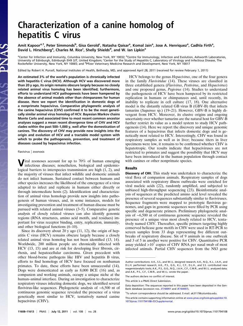
Phylogenetic and Evolutionary Analysis of CHV. CHV was phyloge-netically classified by determining its genetic relatedness torepresentative viruses of different genera of Flaviviridae. Theseviruses are classified in three established genera (Flavivirus,Pestivirus, and Hepacivirus) and one proposed genus, Pegivirus(14). Comparative phylogenetic analysis of conserved regions in
Fig. 2. Structural and functional map of the CHV genome. (A) Structural protein cleavage is mediated by cellular signal peptidase (black triangle); NS2–NS3cleavage is mediated by the NS2–NS3 autoprotease (white triangle); and cleavage of other nonstructural proteins is mediated by NS3–NS4A protease complex(gray triangles). (B) Amino acid sequence divergence scan of CHV polyprotein, HCV genotypes, and GBV-B. (C) Amino acid sequence of different virusesadjacent to predicted protease cleavage sites (10 aa on each side are shown). (D) Reported functional role of different proteins in the virus life cycle.
11610 | www.pnas.org/cgi/doi/10.1073/pnas.1101794108 Kapoor et al.

the predicted helicase (NS3) and RNA-dependent RNA poly-merase (RdRp or NS5B) regions were congruent with CHVconsistently closest and equidistant from the seven HCV geno-types (Fig. 4). These clusters were consistent with the establishedtaxonomic groups and supported by bootstrap values of above90% (of 1,000 replicates) (36). The phylogenetic position ofCHV relative to HCV and GBV-B is consistent with theirpairwise distances (Table S1). The dissimilarity in S regionsequences between GBV-B and other hepaciviruses is reflectedin the virtual absence of sequence similarity of amino acidsequences after alignment (11–12%) compared with 36% aminoacid similarity between CHV and HCV. The degree of sequencedivergence between HCV and CHV is almost the same as thatobserved between GBV-C (a human virus) and GBV-A (foundin chimpanzees and New World primates) (Fig. 4). Consideringthe results of phylogenetic analyses, pairwise protein distances,and identification of CHV in a different natural host, we suggestclassification of CHV as a prototype new virus species in thegenus Hepacivirus.To determine the evolutionary relation between CHV and
other hepaciviruses, a Bayesian Markov chains Monte Carlo(MCMC) estimation of the time to most recent common an-cestor (TMRCA) for the HCV genotypes, GBV-B, and CHVwas performed by using an external rate calibration based on theevolutionary rates estimated for (i) HCV subtypes 1a and 1b (37)and (ii) HCV subtype 6 (38). The mean estimated TMRCA forthe Hepacivirus clade and CHV is 341 y before present (ybp)
[95% highest posterior density (HPD) = 69–705 ybp] based onthe HCV subtype 1 calibration and 1,680 ybp (521–3,291 ybp)based on the HCV subtype 6 calibration (Fig. S4). Thus, weestimate that the shared common ancestor between CHV andthe HCV genotypes probably existed between 500 and 1,000 ybp.However, this should only be regarded as a minimum estimategiven the difficulties associated with extrapolating short-termsubstitution rates to longer evolutionary periods (39, 40). Weestimated the TMRCA between CHV and HCV using a sub-stitution rate previously used to infer the divergence times withinHCV genotypes/subtypes (37, 38). However, whether these ratescan be reliably applied to much more divergent CHV and HCVsequences is unclear. Observation of time dependency in sub-stitution rates in host genes (41) and recent evidence from en-dogenous viral elements for substantially slower long-termsubstitution rates in a variety of animal viruses argue againstsimple extrapolation of substitution rates measured over shortobservation periods (42–45).
DiscussionMolecular characterization of CHV indicates that it is the mostgenetically related homolog of HCV. Viral structural proteinstypically contain major determinants of viral immunogenicityand host/cell tropism. The envelope protein E2 of HCV is amongthe most variable portions of its genome, yet it has remarkablesequence similarity with CHV. Moreover, the number and po-sition of cysteine residues in E2 protein of CHV indicate that
Fig. 3. Sequence and secondary structure of CHV 5′ UTR. Bases conserved among different hepaciviruses are shown with different colored circles. The miR-122 binding sites and different internal ribosome entry site stems are labeled according to previously reported hepacivirus 5′ UTR structures (34).
Kapoor et al. PNAS | July 12, 2011 | vol. 108 | no. 28 | 11611
MICRO
BIOLO
GY
even the tertiary structure of CHV is likely to be more similar toHCV than to other genetically related viruses (29). However,there are notable differences between CHV and HCV that mayhave biological significance. Most strikingly, the potential oc-clusion of the binding site of miR-122 in the CHV 5′ UTR andthe absence of microRNA sequences in the dog genome capableof binding to the equivalent site in CHV suggest that the in-teraction, which enhances the replication of HCV in human liver(32, 33), may not be needed in CHV infections. It remains to bedetermined whether the unique stem-loop I of CHV allows it toreplicate in a manner independent of miR-122 or influencestissue tropism. Although reverse-genetic experiments whereingenomic regions are swapped between HCV and CHV will berequired to address these questions, we note that, although CHVis found at high levels in respiratory samples of infected animals,only low or undetectable levels of HCV RNA are typicallydetected in respiratory samples from HCV-infected humans(46). A significant difference in life span of humans and caninescan also affect the disease pattern caused by genetically relatedviruses. The availability of CHV genome and its comparativegenetic analysis with HCV genotypes is therefore likely to ad-vance our understanding of the role genetic elements and pro-teins play in the viral life cycle. Moreover, the sequence datapresented here will help in designing reagents necessary to fur-ther explore the biology, pathogenesis, and tissue tropism ofCHV and its variants.The limited genetic diversity observed among CHV variants is
atypical for RNA viruses including HCV and is likely attributableto the study animals being in close contact (same disease out-break) or the highly specific PCR assay used in this study. Weexpect that the use of broadly reactive reagents (e.g., degenerateprimers) and samples from unrelated dogs from different geog-raphies will result in identification of many diverse CHV-relatedviruses. Without further information on the distribution of HCV-related viruses in other mammalian species, it is too early to drawconclusions on the evolutionary events underlying their distri-bution in humans and dogs and their apparent absence in non-
human primates. Indeed, there is virtually no information on theexistence of HCV-like viruses in other mammalian orders, whichhave probably remained untested because of a primate focus inscreening paradigms. Hepaciviruses may be widely distributedamong different mammalian species, perhaps highly host-specific,effectively transmitted by nonparenteral routes and largely non-pathogenic (as appears to be the case for pegiviruses in primates)(35). An alternative scenario is one where hepaciviruses areprimarily canine viruses and HCV in humans arose zoonoticallyfrom contact from dogs or other related members of carnivoremammalian order that harbor these types of viruses. A zoonoticorigin for HCV and lack of host adaptation would explain its highdegree of pathogenicity in humans, inefficient transmission bynonparenteral routes, and apparent absence of HCV homolog innonhuman primates. However, the miR-122 interaction appearsto be human-specific and likely represents a virus/host co-adaptation, unlikely responsible for a recent zoonosis.Although its hepatotropism and ability to establish persistence
remain to be determined, the presence of CHV in hepatocytes isreminiscent of HCV infections in humans. Furthermore, thepresence of GORS in CHV is consistent with persistence inother viral systems. Irrespective of its evolutionary origins, thediscovery of CHV provides the exciting prospect of a uniqueexperimental model for HCV infections in humans and opensfuture avenues for research into the pathogenesis, prevention,and treatment of hepacivirus infections.
Materials and MethodsSamples and High-Throughput Sequencing. Respiratory samples (nasal swabs)were collected in 3 mL of MEM from affected dogs in five respiratory diseaseoutbreaks in four shelters (one each in Texas and Utah and three in Penn-sylvania). Postmortem lung and liver samples were from euthanized dogsin clinics in Montana and Missouri. Centrifuged respiratory sample (140 μL)was filtered through a 0.45-μm filter to remove eukaryotic and bacterial-sized particles. The filtrate was then treated with nucleases to digest non-particle-protected nucleic acid. RNA from filtered (0.45-μm) respiratorysamples or tissue homogenates was treated with DNase before randomamplification and pyrosequencing (22, 47). After assembly (Newbler v2.3;
Fig. 4. Phylogenetic analysis of conserved regions in the helicase (motifs I–VI) (A) and RdRp (B) genes of CHV aligned with representative members of theHepacivirus, Pegivirus (GBV viruses A, C, and D), Pestivirus, and Flavivirus genera. Translated amino acid sequences were aligned with the program ClustalW.Trees were constructed by neighbor joining of pairwise amino acid distances with the program MEGA5 (according to the distance scale provided). Bootstrapresampling was used to determine robustness of branches; values of ≥70% (from 1,000 replicates) are shown. Regions compared corresponded to positions3697–4477 (helicase domain of NS3) and 7705–8550 (RdRp in NS5B; numbered according to the AF011751 HCV genotype 1a reference sequence). A listing ofvirus abbreviations and original accession numbers for each sequence are provided in Table S2.
11612 | www.pnas.org/cgi/doi/10.1073/pnas.1101794108 Kapoor et al.

454 Life Sciences), sequence contigs and singletons were analyzed by usingBlastX against a National Center for Biotechnology Information nonredun-dant protein database (47).
Genome Sequencing and Phylogenetic and Evolutionary Analyses. Details areprovided in SI Materials and Methods.
RNA Structure and GORS Predictions. Independent of phylogenetic in-formation, the secondary structure of the CHCV 5′ UTR RNA was modeledwith MFOLD. Labeling of the predicted structures in the 5′ UTR followednumbering used for reported homologous structures in HCV and GBV-B (10,34). The CHV genome sequence was analyzed for evidence of GORS bycomparing folding energies of consecutive fragments of nucleotide se-quence with random sequence-order controls (35). MFEs of CHCV werecalculated by using default setting in the program ZIPFOLD. MFE resultswere expressed as MFE differences, i.e., the percentage difference betweenthe MFE of the native sequence from that of the mean value of the 50 se-quence order–randomized controls.
In Situ Hybridization. For in situ hybridization assay, multiple branched-chainprobe amplifiers labeled with alkaline phosphatase (Panomics/Affymetrix)were used against CHV genomic RNA (nucleotides 840–2040 of CHV genomecorresponding to the coding region for partial core, envelope glycoproteinE1/E2, and partial NS1 protein). Liver sections (10 μm) were fixed with 4%formaldehyde at 4 °C overnight, dehydrated, permeabilized, and stainedwith Fast Red substrate (QG ViewRNA Chromogenic Signal Amplification Kit;Panomics/Affymetrix) for light and florescent microscopy.
ACKNOWLEDGMENTS. We thank Natasha Mehta, Jan Medina, and AhmedSandhu for excellent technical assistance. We are grateful to Roche 454 LifeSciences for high-throughput sequencing support. This work was supportedby National Institutes of Health Grants AI090196, AI079231, AI57158,AI070411, AI090055, AI072613, and EY017404; the Department of Defense;US Department of Agriculture Grant 58-1275-7-370; the Greenberg MedicalResearch Institute; the Starr Foundation; German Academy of SciencesLeopoldina Grant LPDS-2009-9; and International Human Frontier ScienceProgram Organization Grant LT000048/2009-L.
1. Parrish CR, Kawaoka Y (2005) The origins of new pandemic viruses: The acquisition ofnew host ranges by canine parvovirus and influenza A viruses. Annu Rev Microbiol 59:553–586.
2. Parrish CR, et al. (2008) Cross-species virus transmission and the emergence of newepidemic diseases. Microbiol Mol Biol Rev 72:457–470.
3. Hatziioannou T, et al. (2009) A macaque model of HIV-1 infection. Proc Natl Acad SciUSA 106:4425–4429.
4. Tsai CC, et al. (1995) Prevention of SIV infection in macaques by (R)-9-(2-phosphonylmethoxypropyl)adenine. Science 270:1197–1199.
5. Van Rompay KK (2010) Evaluation of antiretrovirals in animal models of HIV infection.Antiviral Res 85:159–175.
6. Kapoor A, et al. (2009) Multiple novel astrovirus species in human stool. J Gen Virol90:2965–2972.
7. Kapoor A, et al. (2010) Identification and characterization of a new bocavirus speciesin gorillas. PLoS ONE 5:e11948.
8. Kapoor A, et al. (2008) A highly divergent picornavirus in a marine mammal. J Virol82:311–320.
9. Klatt NR, et al. (2010) CD8+ lymphocytes control viral replication in SIVmac239-infected rhesus macaques without decreasing the lifespan of productively infectedcells. PLoS Pathog 6:e1000747.
10. Rijnbrand R, Abell G, Lemon SM (2000) Mutational analysis of the GB virus B internalribosome entry site. J Virol 74:773–783.
11. Alter HJ (1989) Discovery of the non-A, non-B hepatitis virus: The end of thebeginning or the beginning of the end. Transfus Med Rev 3:77–81.
12. Choo QL, et al. (1989) Isolation of a cDNA clone derived from a blood-borne non-A,non-B viral hepatitis genome. Science 244:359–362.
13. Simmonds P (2004) Genetic diversity and evolution of hepatitis C virus—15 years on.J Gen Virol 85:3173–3188.
14. Stapleton JT, Foung S, Muerhoff AS, Bukh J, Simmonds P (2011) The GB viruses:A review and proposed classification of GBV-A, GBV-C (HGV), and GBV-D in genusPegivirus within the family Flaviviridae. J Gen Virol 92:233–246.
15. Ray Kim W (2002) Global epidemiology and burden of hepatitis C. Microbes Infect 4:1219–1225.
16. Protsch R, Berger R (1973) Earliest radiocarbon dates for domesticated animals:Europe is added to the Near East as another early center of domestication. Science179:235–239.
17. Lohmann V, et al. (1999) Replication of subgenomic hepatitis C virus RNAs ina hepatoma cell line. Science 285:110–113.
18. Lindenbach BD, et al. (2005) Complete replication of hepatitis C virus in cell culture.Science 309:623–626.
19. Nam JH, et al. (2004) In vivo analysis of the 3′ untranslated region of GB virus B afterin vitro mutagenesis of an infectious cDNA clone: Persistent infection in a transfectedtamarin. J Virol 78:9389–9399.
20. Bukh J, Apgar CL, Govindarajan S, Purcell RH (2001) Host range studies of GB virus-Bhepatitis agent, the closest relative of hepatitis C virus, in New World monkeys andchimpanzees. J Med Virol 65:694–697.
21. Bukh J, Apgar CL, Yanagi M (1999) Toward a surrogate model for hepatitis C virus: Aninfectious molecular clone of the GB virus-B hepatitis agent. Virology 262:470–478.
22. Kapoor A, et al. (2008) A highly prevalent and genetically diversified Picornaviridaegenus in South Asian children. Proc Natl Acad Sci USA 105:20482–20487.
23. Epstein JH, et al. (2010) Identification of GBV-D, a novel GB-like flavivirus from oldworld frugivorous bats (Pteropus giganteus) in Bangladesh. PLoS Pathog 6:e1000972.
24. Tanaka T, Kato N, Cho MJ, Shimotohno K (1995) A novel sequence found at the 3′terminus of hepatitis C virus genome. Biochem Biophys Res Commun 215:744–749.
25. Kolykhalov AA, Feinstone SM, Rice CM (1996) Identification of a highly conservedsequence element at the 3′ terminus of hepatitis C virus genome RNA. J Virol 70:3363–3371.
26. Branch AD, Stump DD, Gutierrez JA, Eng F, Walewski JL (2005) The hepatitis C virusalternate reading frame (ARF) and its family of novel products: The alternate readingframe protein/F-protein, the double-frameshift protein, and others. Semin Liver Dis25:105–117.
27. Xu Z, et al. (2001) Synthesis of a novel hepatitis C virus protein by ribosomalframeshift. EMBO J 20:3840–3848.
28. Mohr EL, Stapleton JT (2009) GB virus type C interactions with HIV: The role ofenvelope glycoproteins. J Viral Hepat 16:757–768.
29. Krey T, et al. (2010) The disulfide bonds in glycoprotein E2 of hepatitis C virus revealthe tertiary organization of the molecule. PLoS Pathog 6:e1000762.
30. Lindenbach BD, Rice CM (2005) Unravelling hepatitis C virus replication from genometo function. Nature 436:933–938.
31. Kieft JS (2008) Viral IRES RNA structures and ribosome interactions. Trends BiochemSci 33:274–283.
32. Jangra RK, Yi M, Lemon SM (2010) Regulation of hepatitis C virus translation andinfectious virus production by the microRNA miR-122. J Virol 84:6615–6625.
33. Jopling CL, Yi M, Lancaster AM, Lemon SM, Sarnow P (2005) Modulation of hepatitis Cvirus RNA abundance by a liver-specific MicroRNA. Science 309:1577–1581.
34. Honda M, Brown EA, Lemon SM (1996) Stability of a stem-loop involving the initiatorAUG controls the efficiency of internal initiation of translation on hepatitis C virusRNA. RNA 2:955–968.
35. Simmonds P, Tuplin A, Evans DJ (2004) Detection of genome-scale ordered RNAstructure (GORS) in genomes of positive-stranded RNA viruses: Implications for virusevolution and host persistence. RNA 10:1337–1351.
36. Tamura K, Dudley J, Nei M, Kumar S (2007) MEGA4: Molecular Evolutionary GeneticsAnalysis (MEGA) software version 4.0. Mol Biol Evol 24:1596–1599.
37. Magiorkinis G, et al. (2009) The global spread of hepatitis C virus 1a and 1b: Aphylodynamic and phylogeographic analysis. PLoS Med 6:e1000198.
38. Pybus OG, et al. (2009) Genetic history of hepatitis C virus in East Asia. J Virol 83:1071–1082.
39. Keckesova Z, Ylinen LM, Towers GJ, Gifford RJ, Katzourakis A (2009) Identification ofa RELIK orthologue in the European hare (Lepus europaeus) reveals a minimum ageof 12 million years for the lagomorph lentiviruses. Virology 384:7–11.
40. Worobey M, et al. (2010) Island biogeography reveals the deep history of SIV. Science329:1487.
41. Ho SY, Phillips MJ, Cooper A, Drummond AJ (2005) Time dependency of molecularrate estimates and systematic overestimation of recent divergence times. Mol BiolEvol 22:1561–1568.
42. Kapoor A, Simmonds P, Lipkin WI (2010) Discovery and characterization ofmammalian endogenous parvoviruses. J Virol 84:12628–12635.
43. Belyi VA, Levine AJ, Skalka AM (2010) Unexpected inheritance: Multiple integrationsof ancient bornavirus and ebolavirus/marburgvirus sequences in vertebrate genomes.PLoS Pathog 6:e1001030.
44. Horie M, et al. (2010) Endogenous non-retroviral RNA virus elements in mammaliangenomes. Nature 463:84–87.
45. Katzourakis A, Gifford RJ (2010) Endogenous viral elements in animal genomes. PLoSGenet 6:e1001191.
46. Ferreiro MC, Dios PD, Scully C (2005) Transmission of hepatitis C virus by saliva? OralDis 11:230–235.
47. Victoria JG, et al. (2009) Metagenomic analyses of viruses in stool samples fromchildren with acute flaccid paralysis. J Virol 83:4642–4651.
Kapoor et al. PNAS | July 12, 2011 | vol. 108 | no. 28 | 11613
MICRO
BIOLO
GY