Chronic viral hepatitis induced by hepatitis C but not hepatitis B virus infection correlates with...
6
Chronic Viral Hepatitis Induced by Hepatitis C but Not Hepatitis B Virus Infection Correlates With Increased Liver Angiogenesis ROBERTO MAZZANTI, 1 LUCA MESSERINI, 2 LAURA MONSACCHI, 1 GIAMPIERO BUZZELLI, 1 ANNA LINDA ZIGNEGO, 1 MARCO FOSCHI, 1 MONICA MONTI, 1 GIACOMO LAFFI, 1 LUCIA MORBIDELLI, 3 ORNELLA FANTAPPIE ´ , 1 FRANCESCO BARTOLONI SAINT OMER, 2 AND MARINA ZICHE 3 hepatitis C virus (HCV) chronic infections increase the risk Chronic hepatitis B virus (HBV) and hepatitis C virus of HCC, although the mechanisms involved are not clear. 4-6 (HCV) infections lead to cirrhosis and increase the risk For HBV, several mechanisms have been proposed which for the development of hepatocellular carcinoma (HCC). suggest that the virus could cause liver cancer. 7-10 However, Angiogenesis is an essential step in oncogenesis and con- for HCV, apart from the fact that it causes chronic liver in- tributes to tumor progression in adult organs; however, flammation and cirrhosis, mechanisms are far from being to what extent angiogenesis occurs in the liver during clarified. 11 Nevertheless, HCV infection is a major factor in chronic viral hepatitis has not been studied. Ninety-nine the increasing risk of HCC, even in the absence of liver cirrho- matched patients affected by chronic hepatitis due to sis. 12 Thus, in addition to virus-related cirrhosis or to chronic either HBV or HCV were studied together with 13 con- stimulation of hepatocellular regeneration, other mecha- trols (5 patients were affected by familial hyperbiliru- nisms probably contribute to the HCV-related liver oncogene- binemia with normal liver histology; 6 patients with sis. stage II primary biliary cirrhosis; and 2 patients with Angiogenesis is implicated in the cancer development of pseudo inflammatory tumor). Microvessel density was adult organs, favoring the progression, growth, and metasta- assessed in liver biopsies by immunostaining using two sis of cancer. 13,14 Angiogenic factors, produced by inflamma- different antibodies against endothelial cell antigens, tory cells, are involved in the earlier phases of oncogenesis. 15-17 QB-END/10 and Factor VIII. In addition, the liver homog- The formation of new vessels has been suggested during enates and sera of HCV- or HBV-positive patients and chronic viral hepatitis; 18 thus, it is possible to hypothesize controls were tested for their capacity to stimulate the that the occurrence of angiogenesis could contribute to the migration and proliferation of freshly isolated human increased risk of HCC in patients with HBV or HCV chronic endothelial cells in vitro. Evidence of angiogenesis was infection. This study was performed to investigate this possi- significantly more frequent in HCV-positive patients bility. compared with HBV-infected subjects or controls (74% Angiogenesis was quantified in the liver biopsies of pa- vs. 39% vs. 8%) (x 2 Å 20.78; P õ .0001) (HCV / vs. HBV / vs. tients with chronic HBV or HCV hepatitis by counting the controls). The degree of microvessel density was also microvessel density, evidenced by immunostaining with anti- higher in HCV– than in HBV-positive patients or con- bodies against two different endothelial antigens, Factor VIII trols (x 2 Å 12.28; P õ .005). In addition, HCV-positive sera and QB-END/10. Moreover, the presence of angiogenic activ- and liver homogenates stimulated a higher migration ity in sera and liver homogenates was evaluated by experi- and proliferation of human endothelial cells in vitro ments on the growth and migration of cultured human endo- compared with HBV-positive or control sera and liver thelial cells. homogenates. These observations indicate that angio- genesis is particularly linked to HCV infection, sug- PATIENTS AND METHODS gesting a possible contribution to HCV-related liver on- cogenesis. (HEPATOLOGY 1997;25:229-234.) Methods. Ninety-nine patients who were affected by chronic hepatitis were studied. Forty-nine patients (36 male and 13 fe- male; age range, 15-69 years; mean, 40 years) had chronic HBV Liver oncogenesis is a multistep process that proceeds from hepatitis and 50 (31 male and 19 female; age range, 17-68 years; the initiation of one or more hepatocytes to a diffuse disease. mean, 50 years) had HCV-related hepatitis. All were affected by Cirrhosis is a risk factor for hepatocellular carcinoma chronic, moderately active hepatitis (histological activity index (HCC). 1-3 There is evidence that hepatitis B virus (HBV) and range, 9-12) with or without cirrhosis, according to international criteria 19,20 with matched liver function tests. 21 In addition, 13 controls (4 men and 9 women; mean age, 47 years; age range, 34- 65 years) were also enrolled. Five had familial hyperbilirubinemia Abbreviations: HBV, hepatitis B virus; HCV, hepatitis C virus; HCC, hepatocellular with normal liver histology, 6 (all women) had stage II primary carcinoma; HUVEC, human umbilical cord vein endothelial cells. biliary cirrhosis, and 2 had the so-called inflammatory pseudo tu- From the 1 Institute of Internal Medicine, the 2 Institute of Anatomic Pathology, and the mor. All of the controls had negative serological markers for HBV 3 Department of Pharmacology, University of Florence, Florence, Italy. and HCV infections. Patients with a positive history for drug and/ Received June 20, 1996; accepted September 13, 1996. Supported by grants from Ministero dell’Universita ` e della Ricerca Scientifica (Progetto or alcohol abuse, human immunodeficiency virus, Epstein-Barr Nazionale Patologia da Radicali Liberi) to R. Mazzanti and F. Bartoloni St. Omer (Progetto virus, cytomegalovirus infections were excluded. All patients and Nazionale Cirrosi Epatica), and from AIRC, Milano, Italy and CNR, Rome, Italy, to M. controls were previously investigated by routine liver function test Ziche. Financial support also came from The Italian Liver Foundation, Florence, Italy. assessment, liver ultrasound scan, percutaneous needle liver bi- Presented in part at the 42nd Meeting of the American Association for the Study of opsy, and determination of serological markers of viral infections Liver Diseases, Chicago, IL, November 1991. and autoimmune diseases. This study was approved by the local Address reprint requests to: Roberto Mazzanti, M.D., Institute of Internal Medicine, Ethical Committee, Firenze, Italy. All subjects enrolled gave their University of Florence, viale Morgagni, 85, Florence, Italy. informed consent to participate in the study and to undergo liver Copyright q 1997 by the American Association for the Study of Liver Diseases. 0270-9139/97/2501-0042$3.00/0 biopsy for diagnostic purposes. 229 AID Hepa 0032 / 5p1b$$$621 12-12-96 09:03:21 hpta WBS: Hepatology
-
Upload
independent -
Category
Documents
-
view
4 -
download
0
Transcript of Chronic viral hepatitis induced by hepatitis C but not hepatitis B virus infection correlates with...
Chronic Viral Hepatitis Induced by Hepatitis C but NotHepatitis B Virus Infection Correlates With Increased
Liver Angiogenesis
ROBERTO MAZZANTI,1 LUCA MESSERINI,2 LAURA MONSACCHI,1 GIAMPIERO BUZZELLI,1 ANNA LINDA ZIGNEGO,1
MARCO FOSCHI,1 MONICA MONTI,1 GIACOMO LAFFI,1 LUCIA MORBIDELLI,3 ORNELLA FANTAPPIE,1
FRANCESCO BARTOLONI SAINT OMER,2 AND MARINA ZICHE3
hepatitis C virus (HCV) chronic infections increase the riskChronic hepatitis B virus (HBV) and hepatitis C virusof HCC, although the mechanisms involved are not clear.4-6(HCV) infections lead to cirrhosis and increase the riskFor HBV, several mechanisms have been proposed whichfor the development of hepatocellular carcinoma (HCC).suggest that the virus could cause liver cancer.7-10 However,Angiogenesis is an essential step in oncogenesis and con-for HCV, apart from the fact that it causes chronic liver in-tributes to tumor progression in adult organs; however,flammation and cirrhosis, mechanisms are far from beingto what extent angiogenesis occurs in the liver duringclarified.11 Nevertheless, HCV infection is a major factor inchronic viral hepatitis has not been studied. Ninety-ninethe increasing risk of HCC, even in the absence of liver cirrho-matched patients affected by chronic hepatitis due tosis.12 Thus, in addition to virus-related cirrhosis or to chroniceither HBV or HCV were studied together with 13 con-stimulation of hepatocellular regeneration, other mecha-trols (5 patients were affected by familial hyperbiliru-nisms probably contribute to the HCV-related liver oncogene-binemia with normal liver histology; 6 patients withsis.stage II primary biliary cirrhosis; and 2 patients with
Angiogenesis is implicated in the cancer development ofpseudo inflammatory tumor). Microvessel density wasadult organs, favoring the progression, growth, and metasta-assessed in liver biopsies by immunostaining using twosis of cancer.13,14 Angiogenic factors, produced by inflamma-different antibodies against endothelial cell antigens,tory cells, are involved in the earlier phases of oncogenesis.15-17QB-END/10 and Factor VIII. In addition, the liver homog-The formation of new vessels has been suggested duringenates and sera of HCV- or HBV-positive patients andchronic viral hepatitis;18 thus, it is possible to hypothesizecontrols were tested for their capacity to stimulate thethat the occurrence of angiogenesis could contribute to themigration and proliferation of freshly isolated humanincreased risk of HCC in patients with HBV or HCV chronicendothelial cells in vitro. Evidence of angiogenesis wasinfection. This study was performed to investigate this possi-significantly more frequent in HCV-positive patientsbility.compared with HBV-infected subjects or controls (74%
Angiogenesis was quantified in the liver biopsies of pa-vs. 39% vs. 8%) (x2 Å 20.78; P õ .0001) (HCV/ vs. HBV/ vs.tients with chronic HBV or HCV hepatitis by counting thecontrols). The degree of microvessel density was alsomicrovessel density, evidenced by immunostaining with anti-higher in HCV– than in HBV-positive patients or con-bodies against two different endothelial antigens, Factor VIIItrols (x2Å 12.28; Põ .005). In addition, HCV-positive seraand QB-END/10. Moreover, the presence of angiogenic activ-and liver homogenates stimulated a higher migrationity in sera and liver homogenates was evaluated by experi-and proliferation of human endothelial cells in vitroments on the growth and migration of cultured human endo-compared with HBV-positive or control sera and liverthelial cells.homogenates. These observations indicate that angio-
genesis is particularly linked to HCV infection, sug-PATIENTS AND METHODSgesting a possible contribution to HCV-related liver on-
cogenesis. (HEPATOLOGY 1997;25:229-234.) Methods. Ninety-nine patients who were affected by chronichepatitis were studied. Forty-nine patients (36 male and 13 fe-male; age range, 15-69 years; mean, 40 years) had chronic HBVLiver oncogenesis is a multistep process that proceeds fromhepatitis and 50 (31 male and 19 female; age range, 17-68 years;the initiation of one or more hepatocytes to a diffuse disease.mean, 50 years) had HCV-related hepatitis. All were affected byCirrhosis is a risk factor for hepatocellular carcinomachronic, moderately active hepatitis (histological activity index(HCC).1-3 There is evidence that hepatitis B virus (HBV) andrange, 9-12) with or without cirrhosis, according to internationalcriteria19,20 with matched liver function tests.21 In addition, 13controls (4 men and 9 women; mean age, 47 years; age range, 34-65 years) were also enrolled. Five had familial hyperbilirubinemia
Abbreviations: HBV, hepatitis B virus; HCV, hepatitis C virus; HCC, hepatocellular with normal liver histology, 6 (all women) had stage II primarycarcinoma; HUVEC, human umbilical cord vein endothelial cells.
biliary cirrhosis, and 2 had the so-called inflammatory pseudo tu-From the 1Institute of Internal Medicine, the 2Institute of Anatomic Pathology, and themor. All of the controls had negative serological markers for HBV3Department of Pharmacology, University of Florence, Florence, Italy.and HCV infections. Patients with a positive history for drug and/Received June 20, 1996; accepted September 13, 1996.
Supported by grants from Ministero dell’Universita e della Ricerca Scientifica (Progetto or alcohol abuse, human immunodeficiency virus, Epstein-BarrNazionale Patologia da Radicali Liberi) to R. Mazzanti and F. Bartoloni St. Omer (Progetto virus, cytomegalovirus infections were excluded. All patients andNazionale Cirrosi Epatica), and from AIRC, Milano, Italy and CNR, Rome, Italy, to M. controls were previously investigated by routine liver function testZiche. Financial support also came from The Italian Liver Foundation, Florence, Italy. assessment, liver ultrasound scan, percutaneous needle liver bi-
Presented in part at the 42nd Meeting of the American Association for the Study of opsy, and determination of serological markers of viral infectionsLiver Diseases, Chicago, IL, November 1991.
and autoimmune diseases. This study was approved by the localAddress reprint requests to: Roberto Mazzanti, M.D., Institute of Internal Medicine,Ethical Committee, Firenze, Italy. All subjects enrolled gave theirUniversity of Florence, viale Morgagni, 85, Florence, Italy.informed consent to participate in the study and to undergo liverCopyright q 1997 by the American Association for the Study of Liver Diseases.
0270-9139/97/2501-0042$3.00/0 biopsy for diagnostic purposes.
229
AID Hepa 0032 / 5p1b$$$621 12-12-96 09:03:21 hpta WBS: Hepatology

230 MAZZANTI ET AL. HEPATOLOGY January 1997
Virological Assessment. All patients were tested for serum hepati- atmosphere containing 5% CO2. At the end of the incubation period,cells were pulsed for 1 hour with 0.5 mCi [3H] thymidine (Amersham,tis B surface antigen, antibody to hepatitis B surface antigen, hepati-
tis B e antigen, antibody to hepatitis B e antigen, antibody to hepati- Buckinghamshire, UK) per well.27
Cell Migration Experiments. These experiments were performedtis B core antigen, HBV DNA (Abbott Laboratories, North Chicago,IL), and for anti-HCV antibodies by second generation enzyme-linked using the Boyden Chamber as described in other reports.27 That
method is based on the passage of endothelial cells across porousimmunosorbant assay (Ortho Diagnostic Systems, Inc., Raritan, NJ)and radio immunoblotting assay (Chiron Corporation, Emeryville, filters against a concentration gradient of the migration effector.27
The Neuro Probe 48-well chamber containing microfilters of policar-CA). Confirmation for the presence of serum viral HCV RNA in anti-bonate (Nuclepore Corp., Pleasanton, CA) and pores of 5 mm wasHCV–positive patients was also performed using the polymeraseused. Filters were previously coated with type 1 collagen and fibro-chain reaction technique.22 Patients with evidence of coinfectionnectin to favor cell migration.27 Liver homogenate (0.1% or 1% in the(HBV and HCV) were excluded from the study.culture medium) was added into the lower part of the well whileHepatic Specimens. Liver biopsies were coded, formalin-fixed, par-endothelial cells (2.51 104) were plated in the upper part of the well.affin-embedded, cut into 4-micrometer-thick sections, and stainedIncubation at 377C was performed in a humidified atmosphere withwith standard techniques.5% CO2 for 4 hours. At the end of the incubation, the filters wereEvaluation of Angiogenesis. Each liver biopsy was examined forremoved and cells were fixed and stained with Diff-Quick (Mertz-the presence of angiogenesis by light microscopy and immunostain-Dade AG, Dudungen, Switzerland). The endothelial cells, havinging on paraffin-embedded sections. The endothelial cells were visual-moved to the inferior part of the filter, were counted using an opticalized using two different antibodies against specific antigens: 1) mono-microscope equipped with a graticule at 2001 magnification. Tenclonal antibody QB-END/10 against CD34 antigen (Unipath, Milan,random fields per each well were counted, and cell migration wasItaly) (dilution 1:50); and 2) polyclonal antibody F-VIII against factorexpressed as the number of cells migrated per well. Results are ex-VIII related antigen (DAKO Corporation, Carpinteria, CA) (dilutionpressed as means { SEM, compared with M199 and 0.1% bovine1:100).23 The specimens were incubated with the primary antibodyserum albumin alone.and processed using the streptavidin-biotin complex method ac-
Statistical Analysis. Statistical analysis was performed usingcording to Stemberger’s amplificated method (immunoperoxidase).23
Yates’ corrected x2 analysis and by the Fisher Exact test. Student’sNegative controls were obtained by omitting the presence of the pri-t test was also used where indicated.mary antibody. Only small vessels and clusters of endothelial cells
Materials. All of the reagents for the cell culture were obtainedwithout lumen, that were positive for QB-END/10 and/or anti Factorfrom GIBCO, UK. Fetal calf serum was obtained from Hyclone Labo-VIII antibodies, were counted. Large vessels and vessels with thickratories, Logan, UT. All other reagents were obtained from Sigmamuscular walls and channels, which were negative by immunostain-Chemical Co., Milano, Italy.ing, were not considered. Angiogenesis was assessed by counting the
microvessel density, as described by study of Weidner et al. andRESULTSmodified by the study of Fox et al.24,25 In particular, the microvessel
density was calculated in the three different portal spaces where the Angiogenesis in Liver Biopsies. The staining of endothelialstaining was more intense at light microscopy on a 2501 field. An cells by QB-END/10 and anti Factor VIII was seen alongaverage value of the microvessel density degree for each patient was vascular channels, in groups of cells that apparently did notthen calculated. Portal spaces of more intense vascularization were border vascular lumina and in groups of cells located in portalidentified by scanning each biopsy at a low magnification, this was tracts and septa (Fig. 1).done by two observers simultaneously using a double-headed light Thirty-seven of 50 (74%) HCV-positive patients were posi-microscope. Both investigators had to agree on what constituted a
tive to immunostaining with the anti QB-END/10 antigen insingle microvessel before any vessel was included in the count. Incomparison with 19 of 49 (39%) HBV-positive patients and 1the case of a discordant evaluation, the preparation was reviewedof 13 (7.7%) controls (x2 Å 20.78; P õ .0001) (Table 1). Theby a third experienced hepatologist.25 The highest number of mi-microvessel density degree was higher in patients with HCV-crovessels observed in one single microscopic field (2501) was 30.
Angiogenesis was graded on a scale of 0 to 3: 0, 1 to 2 microvessels positive chronic hepatitis than in HBV-positive patients oror clusters of endothelial cells; 1, 3 to 10 microvessels or clusters of controls (1.120 { .133 vs. .408 { .105 vs. .076 { .146) (meansendothelial cells, 2, 11 to 20 microvessels or clusters of endothelial { SEM) (x2 Å 12.28; Põ .005). In using the antibody againstcells; and 3, 21 to 30 microvessels structures or clusters of endothelial Factor VIII, a significant difference was also observed in thecells. frequence and in the severity of angiogenesis among the three
Liver Homogenate Preparation. A part of liver samples were imme- groups (54% vs. 18% vs. 8%) (Table 1) (P õ .0001).diately frozen when possible in liquid nitrogen and stored at 0707C.The presence of cirrhosis did not significantly affect theWhen thawed, samples were weighed and ground in the M199 me-
positive effect to immunostaining with either QB-END/10 ordium, containing 0.1% bovine serum albumin using a glass/glassanti Factor VIII antibody. After excluding all patients withhomogenizer.histological evidence of cirrhosis, angiogenesis was observedThe volume of homogenization was calculated on the basis of sam-
ple weight to have 50 mg of tissue per milliliter. This concentration in 21 of the 29 (72%) patients with chronic HCV hepatitiswas considered to be 100%. Further dilutions were made in M199 and in 12 of the 40 (30%) in HBV-positive patients usingand 0.1% bovine serum albumin. the QB-END/10 antibody. The difference between HCV- and
Endothelial Cell Culture. Human umbilical cord vein endothelial HBV-positive patients was highly significant (Yates’ cor-cells (HUVEC) were isolated by a collagenase perfusion of term um- rected x2 Å 9.93; P õ .005). Similar results were observedbilical cords26; the primary cultures were then used in the experi- using the polyclonal antibody against Factor VIII (data notments. HUVEC were grown in M199 supplemented with 20% fetal
shown).calf serum (Hyclone Laboratories, Logan, UT), antibiotics (100 U/mLAngiogenesis In Vitro. The occurrence of angiogenesis isof penicillin and 100 mg/mL of streptomycin), 10 ng/mL recombinant-
linked to the presence of soluble factors.15-17 Endothelial cellbasic fibroblast growth factor, and 5 U/mL heparin to confluence.mobilization and growth are early events in angiogenesis thatCells between passages 2 and 5 were used for the studies. HUVEC
were identified by immunofluorescence determination of factor VIII can be studied in vitro on cultured cells.27 Because patientsantigen (more than 90%-95% of the cells in random culture were with chronic HCV infection exhibited an increased angiogen-positive), and by uniform cobblestone morphology at confluence by esis in the liver on histological observation, we testedphase-contrast microscopy.26
whether angiogenic activity could be detected in the serum[3H] Thymidine Incorporation. DNA synthesis was quantified by and liver biopsies of patients with chronic HCV hepatitis
[3H] thymidine incorporation of subconfluent cell monolayers.27
compared with HBV-positive patients and controls.HUVEC were seeded onto 24 multiwell plates overnight (1 1 104
Cell Migration Studies. HUVEC mobilization toward a gra-cells per well) in the M199 medium supplemented with 5% FCS.dient concentration of normal (8 cases), HBV- (6 cases), andAfter the synchronization of the cell cycle for 48 hours in sera-freeHCV- (5 cases) positive sera was studied using the Boydenmedia (0.5% FCS), the media were removed and the cells were incu-Chamber procedure. Results are expressed as recovered cpmbated with 0.1%, 5%, and 10% of normal, HBV- or HCV-positive sera
for 24 hours. The cells were maintained at 377C in a humidified per well. These studies showed that the migration of HUVEC
AID Hepa 0032 / 5p1b$$$622 12-12-96 09:03:21 hpta WBS: Hepatology

FIG. 1. (A) Marked portal inflammation in chronic HBV-positive hepatitis shows no evidence of angiogenesis (immunoperoxidase stain for QB-END/10,2001). (B) Evidence of angiogenesis in portal tract in HCV-positive hepatitis (immunoperoxidase stain for QB-END/10, 3501). (C and D) High density ofimmunostained microvessels demonstrating active angiogenesis in chronic HCV-related hepatitis (immunoperoxidase stain for QB-END/10, 3501).
12-12-96 09:03:21 hpta WBS: Hepatology

232 MAZZANTI ET AL. HEPATOLOGY January 1997
TABLE 1. Angiogenesis in Liver Biopsies Evidenced by Using Immunostaining of QB-END/10and Factor VIII Antigens With Specific Antibodies
Microvessel Density Degree*
QB-END/10 Factor VIII
Viral Status (n) 0 1 2 3 0 1 2 3
Control 13 (100%) 12 (92%) 1 (8%) — — 12 (92%) 1 (8%) — —HBV/ 49 (100%) 30 (61%) 18 (37%) 1 (2%) — 40 (82%) 9 (18%) — —HCV/** 50 (100%) 13 (26%) 22 (44%) 11 (22%) 4 (8%) 23 (46%) 15 (30%) 9 (18%) 3 (6%)
* On a scale of 0 to 3; see Patients and Methods.** Yates’ corrected x2 test; P õ .0005 HCV vs. HBV and controls.
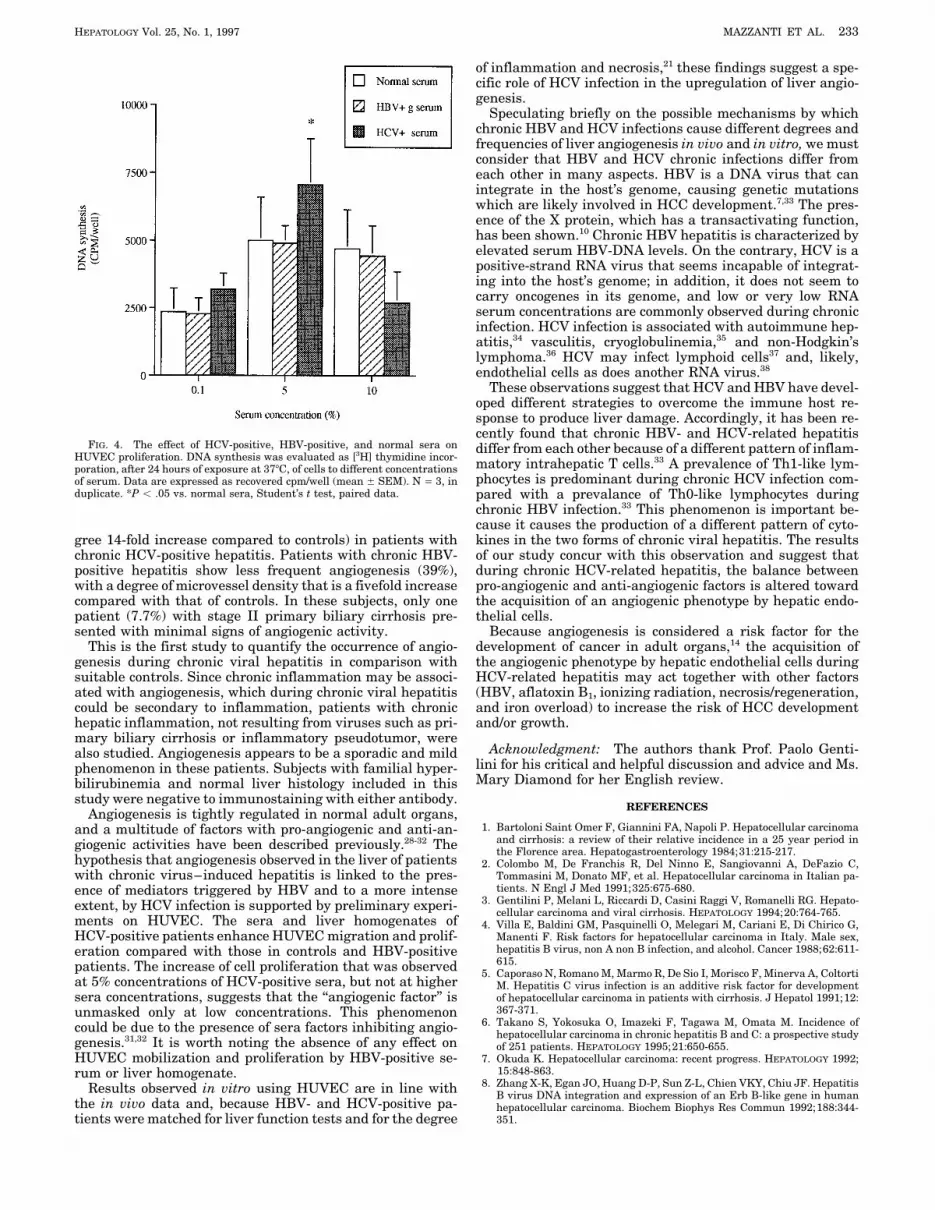
was significantly enhanced by all the sera tested in a concen- enates did not induce HUVEC mobilization (Fig. 3) and, ata 0.01% concentration, HUVEC mobilization was actuallytration-dependent manner (Fig. 2). However, an increased
pattern of mobilization was apparent for HCV-positive versus reduced (data not shown).Cell Proliferation Studies. Based on the results of migra-normal serum at all of the concentrations tested (P õ .05;
HCV-positive vs. normal; Student’s t test). The mobilization tion, HCV-positive and HBV-positive sera from patientswhose cells showed the greatest effect on cell migration wereinduced by HBV-positive sera was lower than that elicited
by HCV-positive sera and was not statistically different from again tested to measure their effect on cell proliferation; thateffect was then compared with normal serum. The exposurenormal controls.
To test whether the effects observed with sera were re- for 24 hours to normal sera induced cell proliferation as mea-sured by DNA synthesis (Fig. 4). The effect was evident attained by liver tissue, fragments of liver specimens from 5
HCV-positive and 5 HBV-positive patients were homogenized 5% and 10% serum concentrations. The proliferative effect ofHBV-positive sera was comparable with that elicited by nor-and tested for HUVEC migration. The control liver tissue
was selected from three biopsies of normal histological ap- mal sera. When cells were exposed to HCV-positive sera, astatistically significant increase in DNA synthesis was ob-pearence (those with familial hyperbilirubinemia). The expo-
sure of HUVEC to normal, HBV-, and HCV-positive liver tained at a 5% concentration (P õ .05; Student’s t test). Ata 10% concentration, HCV-positive sera did not present anyhomogenates at 10%, 50%, and 100% were cytotoxic as mea-
sured by Trypan blue uptake by HUVEC (data not shown). significant effect on the incorporation of [3H] thymidine com-pared with control and HBV-positive sera (Fig. 4).On the contrary, lower concentrations (0.01%, 0.1%, and 1%)
of liver homogenates were nontoxic. Thus, the experimentsDISCUSSIONwere conducted on the basis of cell migration toward these
homogenate dilutions. Low concentrations of liver homoge- This study shows that angiogenesis in the liver is frequentnates from HCV-positive patients promoted a significant in- (74% of patients) and more intense (microvessel density de-crement of HUVEC mobilization in comparison with normalliver homogenates (P õ .05; Student’s t test) (Fig. 3). At a1% concentration, no difference was observed between HCV-positive and normal homogenates. HBV-positive liver homog-
FIG. 2. Effect of HCV-positive, HBV-positive, and normal sera on HUVECmigration. Cellular migration was evaluated using the Boyden chamber proce-
FIG. 3. Effect of HCV-positive, HBV-positive, and normal liver homoge-dure. HUVEC were exposed to increasing concentrations of sera for 4 hours.Data are expressed as the increase in cell number per well over basal control nates on HUVEC migration. The cellular migration was evaluated as described
in Fig. 2. N Å 5 for HCV-positive, n Å 5 for HBV-positive, and n Å 3 for normal(M199 / 0.1% bovine serum albumin alone). Numbers represent mean { SEMof n Å 5 for HCV-positive, n Å 6 for HBV-positive, and n Å 8 experiments for liver homogenates. *P õ .05 vs. normal liver homogenates, Student’s t test,
paired data.normal sera. *P õ .05 vs. normal sera, Student’s t test, paired data.
AID Hepa 0032 / 5p1b$$$622 12-12-96 09:03:21 hpta WBS: Hepatology
HEPATOLOGY Vol. 25, No. 1, 1997 MAZZANTI ET AL. 233
of inflammation and necrosis,21 these findings suggest a spe-cific role of HCV infection in the upregulation of liver angio-genesis.
Speculating briefly on the possible mechanisms by whichchronic HBV and HCV infections cause different degrees andfrequencies of liver angiogenesis in vivo and in vitro, we mustconsider that HBV and HCV chronic infections differ fromeach other in many aspects. HBV is a DNA virus that canintegrate in the host’s genome, causing genetic mutationswhich are likely involved in HCC development.7,33 The pres-ence of the X protein, which has a transactivating function,has been shown.10 Chronic HBV hepatitis is characterized byelevated serum HBV-DNA levels. On the contrary, HCV is apositive-strand RNA virus that seems incapable of integrat-ing into the host’s genome; in addition, it does not seem tocarry oncogenes in its genome, and low or very low RNAserum concentrations are commonly observed during chronicinfection. HCV infection is associated with autoimmune hep-atitis,34 vasculitis, cryoglobulinemia,35 and non-Hodgkin’slymphoma.36 HCV may infect lymphoid cells37 and, likely,endothelial cells as does another RNA virus.38
These observations suggest that HCV and HBV have devel-oped different strategies to overcome the immune host re-sponse to produce liver damage. Accordingly, it has been re-cently found that chronic HBV- and HCV-related hepatitis
FIG. 4. The effect of HCV-positive, HBV-positive, and normal sera on differ from each other because of a different pattern of inflam-HUVEC proliferation. DNA synthesis was evaluated as [3H] thymidine incor- matory intrahepatic T cells.33 A prevalence of Th1-like lym-poration, after 24 hours of exposure at 377C, of cells to different concentrations
phocytes is predominant during chronic HCV infection com-of serum. Data are expressed as recovered cpm/well (mean { SEM). N Å 3, induplicate. *P õ .05 vs. normal sera, Student’s t test, paired data. pared with a prevalance of Th0-like lymphocytes during
chronic HBV infection.33 This phenomenon is important be-cause it causes the production of a different pattern of cyto-kines in the two forms of chronic viral hepatitis. The resultsgree 14-fold increase compared to controls) in patients with
chronic HCV-positive hepatitis. Patients with chronic HBV- of our study concur with this observation and suggest thatduring chronic HCV-related hepatitis, the balance betweenpositive hepatitis show less frequent angiogenesis (39%),
with a degree of microvessel density that is a fivefold increase pro-angiogenic and anti-angiogenic factors is altered towardthe acquisition of an angiogenic phenotype by hepatic endo-compared with that of controls. In these subjects, only one
patient (7.7%) with stage II primary biliary cirrhosis pre- thelial cells.Because angiogenesis is considered a risk factor for thesented with minimal signs of angiogenic activity.
This is the first study to quantify the occurrence of angio- development of cancer in adult organs,14 the acquisition ofthe angiogenic phenotype by hepatic endothelial cells duringgenesis during chronic viral hepatitis in comparison with
suitable controls. Since chronic inflammation may be associ- HCV-related hepatitis may act together with other factors(HBV, aflatoxin B1, ionizing radiation, necrosis/regeneration,ated with angiogenesis, which during chronic viral hepatitis
could be secondary to inflammation, patients with chronic and iron overload) to increase the risk of HCC developmentand/or growth.hepatic inflammation, not resulting from viruses such as pri-
mary biliary cirrhosis or inflammatory pseudotumor, wereAcknowledgment: The authors thank Prof. Paolo Genti-also studied. Angiogenesis appears to be a sporadic and mild
lini for his critical and helpful discussion and advice and Ms.phenomenon in these patients. Subjects with familial hyper-Mary Diamond for her English review.bilirubinemia and normal liver histology included in this
study were negative to immunostaining with either antibody.REFERENCESAngiogenesis is tightly regulated in normal adult organs,
1. Bartoloni Saint Omer F, Giannini FA, Napoli P. Hepatocellular carcinomaand a multitude of factors with pro-angiogenic and anti-an-and cirrhosis: a review of their relative incidence in a 25 year period ingiogenic activities have been described previously.28-32 The the Florence area. Hepatogastroenterology 1984;31:215-217.
hypothesis that angiogenesis observed in the liver of patients 2. Colombo M, De Franchis R, Del Ninno E, Sangiovanni A, DeFazio C,Tommasini M, Donato MF, et al. Hepatocellular carcinoma in Italian pa-with chronic virus–induced hepatitis is linked to the pres-tients. N Engl J Med 1991;325:675-680.ence of mediators triggered by HBV and to a more intense
3. Gentilini P, Melani L, Riccardi D, Casini Raggi V, Romanelli RG. Hepato-extent, by HCV infection is supported by preliminary experi- cellular carcinoma and viral cirrhosis. HEPATOLOGY 1994;20:764-765.ments on HUVEC. The sera and liver homogenates of 4. Villa E, Baldini GM, Pasquinelli O, Melegari M, Cariani E, Di Chirico G,
Manenti F. Risk factors for hepatocellular carcinoma in Italy. Male sex,HCV-positive patients enhance HUVEC migration and prolif-hepatitis B virus, non A non B infection, and alcohol. Cancer 1988;62:611-eration compared with those in controls and HBV-positive615.patients. The increase of cell proliferation that was observed 5. Caporaso N, Romano M, Marmo R, De Sio I, Morisco F, Minerva A, Coltorti
at 5% concentrations of HCV-positive sera, but not at higher M. Hepatitis C virus infection is an additive risk factor for developmentof hepatocellular carcinoma in patients with cirrhosis. J Hepatol 1991;12:sera concentrations, suggests that the ‘‘angiogenic factor’’ is367-371.unmasked only at low concentrations. This phenomenon
6. Takano S, Yokosuka O, Imazeki F, Tagawa M, Omata M. Incidence ofcould be due to the presence of sera factors inhibiting angio- hepatocellular carcinoma in chronic hepatitis B and C: a prospective studygenesis.31,32 It is worth noting the absence of any effect on of 251 patients. HEPATOLOGY 1995;21:650-655.
7. Okuda K. Hepatocellular carcinoma: recent progress. HEPATOLOGY 1992;HUVEC mobilization and proliferation by HBV-positive se-15:848-863.rum or liver homogenate.
8. Zhang X-K, Egan JO, Huang D-P, Sun Z-L, Chien VKY, Chiu JF. HepatitisResults observed in vitro using HUVEC are in line with B virus DNA integration and expression of an Erb B-like gene in humanthe in vivo data and, because HBV- and HCV-positive pa- hepatocellular carcinoma. Biochem Biophys Res Commun 1992;188:344-
351.tients were matched for liver function tests and for the degree
AID Hepa 0032 / 5p1b$$$622 12-12-96 09:03:21 hpta WBS: Hepatology
234 MAZZANTI ET AL. HEPATOLOGY January 1997
9. Paterlini P, Driss F, Nalpas B, Pisi E, Franco D, Berthelot P, Brechot C. 24. Weidner N, Semple JP, Welch WH, Folkman J. Tumor angiogenesis andmetastasis. Correlation in invasive breast carcinoma. N Engl J Med 1991;Persistence of hepatitis B and hepatitis C viral genomes in primary liver
cancers from HBsAg-negative patients: a study of a low-endemic area. 324:1-8.25. Fox SB, Leek RD, Weeks MP, Whitehouse RM, Gatter KC, Harris AL.HEPATOLOGY 1993;17:20-29.
10. Paterlini P, Poussin K, Kew M, Franco D, Brechot C. Selective accumula- Quantitation and prognostic value of breast cancer angiogenesis: compari-tion of the X transcript of hepatitis B virus in patients negative for hepati- son of microvessel density, chalkley count, and computer image analysis.tis B surface antigen with hepatocellular carcinoma. HEPATOLOGY 1995; J Pathol 1995;177:275-283.21:313-321. 26. Gimbrone MA, Cotran RS, Folkman J. Human vascular endothelial cells
11. Simonetti RG, Camma C, Fiorello F, Cottone M, Rapicetta M, Marino in culture. J Cell Biol 1974;60:673-684.L, Fiorentino G, et al. Hepatitis C virus infection as a risk factor for 27. Ziche M, Morbidelli L, Masini E, Amerini S, Granger HJ, Maggi CA, Gep-hepatocellular carcinoma in patients with cirrhosis. Ann Intern Med 1992; petti P, et al. Nitric oxide mediates angiogenesis in vivo and endothelial116:97-102. cell growth and migration in vitro promoted by substance P. J Clin Invest
12. De Mitri MS, Poussin K, Baccarini P, Pontisso P, D’Errico A, Simon N, 1994;94:2036-2044.Grigioni W, et al. HCV-associated liver cancer without cirrhosis. Lancet 28. Folkman J. Clinical applications of research on angiogenesis. N Engl J1995;345:413-415. Med 1995;333:1757-1763.
13. Ziche M, Gullino PM. Angiogenesis and neoplastic progression in vitro. 29. Ferrara N. Missing link in angiogenesis. Nature 1995;376:467.Monogr Natl Cancer Inst 1982;69:483-487. 30. Koch AE, Halloran MM, Haskell CJ, Shah MR, Polverini PJ. Angiogenesis
14. Gullino PM, Ziche M, Alessandri G. Gangliosides, copper ions and angio- mediated by soluble forms of E-selectin and vascular adhesion molecule-genic capacity of adult tissues. Cancer Metastasis Rev 1990;9:239-251. 1. Nature 1995;376:517-519.
15. Folkman J, Shing Y. Angiogenesis. J Biol Chem 1992;267:10931-10934. 31. O’Reilly MS, Holmgren L, Shing Y, Chen C, Rosenthal RA, Moses M, Lane16. Kock AE, Polverini PJ, Kunkel SL, Harlow SA, Di Pietro LA, Elner VM, WS, et al. Angiostatin: a novel angiogenesis inhibitor that mediates the
Elner SG, Strieter RM. Interleukin-8:as a macrophage-derived mediator suppression of metastases by a Lewis lung carcinoma. Cell 1994;79:315-of angiogenesis. Science 1992;258:1798-1801. 328.
17. Folkman J. Angiogenesis in cancer, vascular, rheumatoid and other dis- 32. Shanahan F, O’Sullivan GC. Angiostatin: candidate for molecule of theease. Nat Med 1995;1:27-31. year! Gastroenterology 1995;108:1946-1948.18. Garcıa-Monzon C, Sanchez-Madrid F, Garcıa-Buey L, Garcıa-Arroyo A, 33. Bertoletti A, D’Elios MM, De Carli M, Zignego AL, Durazzo M, Boni C,Garcıa-Sanchez A, Moreno-Otero R. Vascular adhesion molecule expres-Missale G, et al. Different cytokine profiles of intrahepatic T cells insion in viral chronic hepatitis: evidence of neoangiogenesis in portal tracts.chronic hepatitis B and hepatitis C virus infections. Gastroenterology, inGastroenterology 1995;108:231-241.press.19. Knodell RG, Ishac KG, Black WC, Chen TS, Craig R, Kaplowitz N, Kier-
34. Cassani F, Muratori L, Manotti P, Lenzi M, Fusconi M, Ballardini G,man TW, et al. Formulation and application of a numerical scoring systemSelleri L, et al. Serum autoantibodies and the diagnosis of type-1 autoim-for assessing histological activity in asymptomatic chronic active hepatitis.mune hepatitis in Italy: a reappraisal at the light of hepatitis C virusHEPATOLOGY 1981;5:431-435.infection. Gut 1993;33:1260-1263.20. Desmet VJ, Gerber MA, Hoofnagle JH, Manns M, Scheuer PJ. Classifica-
35. Agnello V, Chung RT, Kaplan LM. A role for hepatitis C virus infectiontion of chronic hepatitis: diagnosis, grading and staging. HEPATOLOGYin type II cryoglobulinemia. N Engl J Med 1992;327:1490-1495.1994;19:1513-1520.
36. Ferri C, La Civita L, Monti M, Longobardo G, Pasero G, Zignego AL. Can21. Gentilini P, Mazzanti R, Foschi M, Bartoloni Saint Omer F, Messerini L.type C hepatitis be complicated by B-cell malignant lymphoma? LancetComparative analysis of histologic features of HBV and HCV related1995;346:1426-1427.chronic active hepatitis [Abstract]. HEPATOLOGY 1991;14:69A.
37. Zignego AL, De Carli M, Monti M, Careccia G, La Villa G, Giannini C,22. Zignego AL, Foschi M, Laffi G, Monti M, Careccia G, Romanelli RG, DeDel Prete G, et al. Hepatitis C virus infection of mononuclear cells fromMajo E, et al. ‘‘Inapparent’’ hepatitis B virus infection and hepatitis Cperipheral blood and liver infiltrate in chronically infected subjects. J Medvirus replication in alcoholic subjects with and without liver disease. HEPA-Virol 1995;47:58-64.TOLOGY 1994;19:577-582.
38. Schnittler HJ, Mahner F, Drenckhahn D, Klenk HD, Feldmann H. Replica-23. Ramani P, Bradley NJ, Fletcher CDM. QB-END/10, a new monoclonaltion of Marbourg virus in human endothelial cells. J Clin Invest 1993;91:antibody to endothelium: assessment of its diagnostic utility in paraffin
sections. Histopathology 1990;17:237-242. 1301-1309.
AID Hepa 0032 / 5p1b$$$623 12-12-96 09:03:21 hpta WBS: Hepatology