ADP ribose is an endogenous ligand for the purinergic P2Y1 receptor
Upload
khangminh22Category
view
1download
0
�����������������
Citation: Manco, G.; Lacerra, G.;
Porzio, E.; Catara, G.
ADP-Ribosylation Post-Translational
Modification: An Overview with a
Focus on RNA Biology and New
Pharmacological Perspectives.
Biomolecules 2022, 12, 443. https://
doi.org/10.3390/biom12030443
Academic Editor: Donald Cameron
Received: 31 January 2022
Accepted: 10 March 2022
Published: 13 March 2022
Publisher’s Note: MDPI stays neutral
with regard to jurisdictional claims in
published maps and institutional affil-
iations.
Copyright: © 2022 by the authors.
Licensee MDPI, Basel, Switzerland.
This article is an open access article
distributed under the terms and
conditions of the Creative Commons
Attribution (CC BY) license (https://
creativecommons.org/licenses/by/
4.0/).
biomolecules
Review
ADP-Ribosylation Post-Translational Modification:An Overview with a Focus on RNA Biology and NewPharmacological PerspectivesGiuseppe Manco 1,* , Giuseppina Lacerra 2 , Elena Porzio 1 and Giuliana Catara 1,*
1 Institute of Biochemistry and Cell Biology, National Research Council of Italy, Via P. Castellino 111,80131 Naples, Italy; [email protected]
2 Institute of Genetics and Biophysics “Adriano Buzzati-Traverso”, National Research Council of Italy,Via P. Castellino 111, 80131 Naples, Italy; [email protected]
* Correspondence: [email protected] (G.M.); [email protected] (G.C.)
Abstract: Cellular functions are regulated through the gene expression program by the transcriptionof new messenger RNAs (mRNAs), alternative RNA splicing, and protein synthesis. To this end,the post-translational modifications (PTMs) of proteins add another layer of complexity, creatinga continuously fine-tuned regulatory network. ADP-ribosylation (ADPr) is an ancient reversiblemodification of cellular macromolecules, regulating a multitude of key functional processes asdiverse as DNA damage repair (DDR), transcriptional regulation, intracellular transport, immuneand stress responses, and cell survival. Additionally, due to the emerging role of ADP-ribosylationin pathological processes, ADP-ribosyltransferases (ARTs), the enzymes involved in ADPr, areattracting growing interest as new drug targets. In this review, an overview of human ARTs and theirrelated biological functions is provided, mainly focusing on the regulation of ADP-ribosyltransferaseDiphtheria toxin-like enzymes (ARTD)-dependent RNA functions. Finally, in order to unravel novelgene functional relationships, we propose the analysis of an inventory of human gene clusters,including ARTDs, which share conserved sequences at 3′ untranslated regions (UTRs).
Keywords: ADP-ribosylation; ARTs; mRNA regulation; MARylation; PARylation; poly-ADP-ribosylpolymerases (PARPs)
1. Introduction
The activation of gene expression programs serves the cell to ensure coordinatedcellular processes and fate resolutions [1]. This coordination can be achieved both at thetranscriptional and post-transcriptional levels. Indeed, the transcription of new mRNAmolecules, alternative mRNA splicing processes, mRNA stabilization/destabilization, aswell as de novo protein synthesis are regularly switched on/off by the cell in response tointracellular/extracellular stimuli. In addition to this, the PTMs of proteins have beenestablished to play a pivotal role in mediating cell response upon specific inputs, thuscontributing to the arrangement of a continuously fine-tuned regulatory network [2]. As aresult, post-translationally modified proteins can turn on/off their activity state, changecellular localization, turnover, and establish new protein–protein interactions [2]. Theimprovement of mass spectrometry (MS) technologies has largely increased the numberof identified PTMs, which now accounts for around 400 [3], including acetylation, propi-onylation, methylation, phosphorylation, sumoylation, and ubiquitination, which havebeen proven to be involved in the regulation of cellular processes at diverse subcellularcompartments. Of note, a growing body of literature has also outlined the interplay be-tween the different PTMs [4–6], suggesting that the crosstalk is compelling for the rightfulfillment of such diverse cell functions as, for example, gene expression, genome orga-nization, cell division and DNA damage response. Given the fundamental roles exerted
Biomolecules 2022, 12, 443. https://doi.org/10.3390/biom12030443 https://www.mdpi.com/journal/biomolecules
Biomolecules 2022, 12, 443 2 of 23
by PTMs, the discovery of specific inhibitors to address therapeutic treatments of diseasesincluding cancer has greatly expanded in the last decade [7]. Likewise, the implementationof bioinformatic predictive tools has paved the way to the discovery of alternative drugtargets for therapeutic intervention [8].
ADP-ribosylation is a reversible phylogenetically ancient mechanism of protein modifi-cation where the ADP-ribose moiety is covalently transferred from β-nicotinamide adeninedinucleotide (NAD+) onto target proteins with the release of nicotinamide [9,10].
In addition to proteins, ADPr reaction is responsible for the chemical modification ofdifferent targets as small molecules and nucleic acids, with a broad spectrum of effects inliving organisms [11–15].
ADP-ribose transfer reactions are catalyzed by at least three evolutionarily unre-lated superfamilies of enzymes, namely the ADP-ribosyltransferases, the Sirtuins, andthe less studied TM1506 superfamily [10,16–18]. The TM1506 superfamily contains ADP-ribosylating enzymes only found in bacteria, such as the prototype protein from Thermotogamaritima, showing a deaminase-like fold that displays auto-ADP-ribosylation onto aspartateresidue [18].
ADPr systems have been reported in all domains of life [11,19,20] including Ar-chaea [21], Bacteria [22–24], viruses ([23] and the references therein), and the large part ofEukarya [9,10,25,26].
In this paper, after a brief review on ARTs distribution in the three domains of life,we refer to ADPr reactions in humans with a focus on the most recent findings in RNAbiology, suggestive of new biological functions in physiological processes. Likewise, thedysregulation of some of the ADPr enzymes is also causative of pathological mechanisms,thus providing evidence of the need of an efficient gene expression regulatory network. Tothis end, we also consider the post-translational gene expression mechanism, known as“RNA operon”, as a putative regulatory system affecting mRNA transcripts that enclosesthose coding for ART enzymes as well. Therefore, the evaluation of novel therapeuticpharmacological opportunities targeting the ADPr reaction is also suggested.
2. ADP-Ribosyltransferase Activity during Evolution in the Three Domains of Life
ARTs represent one of the superfamilies of enzymes catalyzing ADP-ribosylation.They are ubiquitous, conserved throughout evolution and common to all three domains oflife: Archaea, Bacteria and Eukarya. In Bacteria, ADP-ribosylation modification regulates ahigh number of cellular processes, including cell homeostasis as well as bacterial and viralpathogenic mechanisms, to cite the most known [27–33].
ADPr was originally discovered as a post-translational protein modification catalyzedby Diphtheria Toxin (DT) and Cholera Toxin (CT). Both of them harbor an ART catalyticdomain showing very limited sequence similarity [34,35], despite a conserved structuralorganization of the core fold, as found in all members of the ART superfamily [12–15,34–36].
ARTs fall into two major subclasses, distinguished based on conserved structuralmotifs in the catalytic triad: H-Y-E-containing enzymes in the ADP-ribosyltransferaseDiphtheria toxin-like (ARTD) subfamily, formerly known as poly-ADP-ribosylpolymerases(PARPs); and R-S-E-containing enzymes in the ADP-ribosyltransferase Clostridium toxin-like (ARTC) subfamily (related to C2 and C3 Clostridial Toxins).
Recently, by reconstructing the evolutionary history for the different members of ARTsuperfamily, ARTs have been further divided in three major higher order clades where themost primordial branching clade results to be the H-H-h clade followed by a crown groupcomprised of the H-Y-[EDQ] and the R-S-E clades [11–13]. The H-H-h and H-Y-[EDQ]include the ARTD proteins, while the R-S-E clade includes the ARTC proteins, in thepreviously presented nomenclature for ART domains [12–15,34–36]. Thus, the presenceof an active H residue in the first branch appears to be the ancestral feature of the ARTsuperfamily, with the R in the R-S-E clade being a derived feature [11].
In Prokaryotes, according to Perina and colleagues (2014) [19], 28 PARP homologuescan be found in 27 bacterial species, belonging to 6 different phyla of the Bacteria domain;
Biomolecules 2022, 12, 443 3 of 23
therefore, this suggests a horizontal PARP gene transfer in Bacteria. Only one bacterium,Microscilla marina, possesses two PARP genes in its genome. Most of bacterial PARPscontain only two domains (WGR and PARP domains) and are most similar to the DNArepair-specific Clade 1 PARP homologues. The catalytic triad H-Y-E essential for thepoly-ADP-ribosylation activity of ARTDs is conserved in the largest part (27 out of 28)of analyzed bacterial PARPs. In the remaining PARPs, only the third amino acid residueof the catalytic triad is conserved. This protein is most similar to PARP1 homologues,indicating that the mutations, which may have changed its ADPr function (poly- vs mono-ADP-ribosylation), occurred after horizontal gene transfer [19].
Despite the fact that ARTD genes have not been found in archaeal genomes, en-dogenous ADPr activity has been detected in the archaeon Saccharolobus solfataricus. ThePARP-like enzyme from S. solfataricus (PARPSso) has been partially purified, possessingan oligo-ADP-ribosyltransferase activity with non-specific DNA binding activity [21]. Thethermozyme undergoes auto-modification and modifies as target protein the 7-kDa protein(Sso7), an histone-like protein in sulphur-dependent extremophiles [37]. PARPSso is notstructurally related to the known ARTDs [38] and localizes at the edge of the sulfolobalcell membrane [39]. Structurally, PARPSso belongs to the family of DING proteins, andsimilarly to other members of the DING family, it is a monomer of 40 kDa and has a singlehighly conserved phosphate-binding site that is involved in its phosphatase activity asother DING proteins from different organisms [40].
Recently, PARPSso has been reported as a multifunctional enzyme also displayingATPase and low phosphatase activities in addition to the ability to ADP-ribosylate sub-strates [41]. The presence of multiple activities suggests that ADPr activity has evolvedthroughout the evolution up to humans, from promiscuous enzymes, as described forPARPSso [42]. However, even though genes encoding poly-ADP-ribosylglycohydrolases(PARG) enzymes have not been found, many archaeal genomes have been shown to en-code other macrodomain-containing proteins. To date, Af1521 from Archaeoglobus fulgidusremains the best characterized archaeal macrodomain protein [43] capable of bindingboth ADP-ribose and PAR, displaying hydrolyzing activity onto mono-ADP-ribosylatedsubstrates [44,45].
PARP genes have also been identified in four genomes of dsDNA viruses [19]. Mostlikely, the genes have been gained from their hosts. The catalytic triad H-Y-E is fullyconserved in three of these viral PARPs, whereas only one has replaced the E residuewith D, which may suggest that these enzymes are active ARTs. Some viruses use PARmetabolism for their replication. For example, the Herpes Simplex Virus and Epstein–BarrVirus require PARP activity for efficient replication [46,47].
Other examples of novel ADPr players are provided by the viral macro domains,present in many dangerous viruses, including the SARS and SARS-CoV-2 coronaviruses [48],and by the recently characterized novel ART-like domain (DUF3715) in the human trans-gene activation suppressor (TASOR) protein involved in gene silencing [49–51]. However,it is not clear whether members of the ART superfamily were present in the Last UniversalCommon Ancestor (LUCA) or whether they firstly arose in Bacteria [11]. It is possiblethat starting from an ancestral version the ART superfamily diverged rapidly in termsof sequence, structure and active site residues, recognizing a wide range of substrates inthe bacterial super-kingdom [11]. Probably, ARTs evolving in these systems have beenrepeatedly acquired by lateral transfer throughout eukaryotic evolution.
3. Human Proteins in ADPr Biology3.1. ADP-Ribosyltransferases
ADPr reaction in humans is mainly catalyzed by members of the ART superfamily,including both ARTD and ARTC enzymes [12,36]. Although in this paper we will reportonly about ARTs, a few members of the Sirtuins (SIRTs), a structurally distinct superfamilyof NAD+-dependent deacylases, target substrates through ADPr [52,53].
Biomolecules 2022, 12, 443 4 of 23
Based on the number of ADP-ribose units, single or multiple, transferred onto tar-get substrates, ARTs can be further referred to as MARylating or PARylating enzymes,respectively [54].
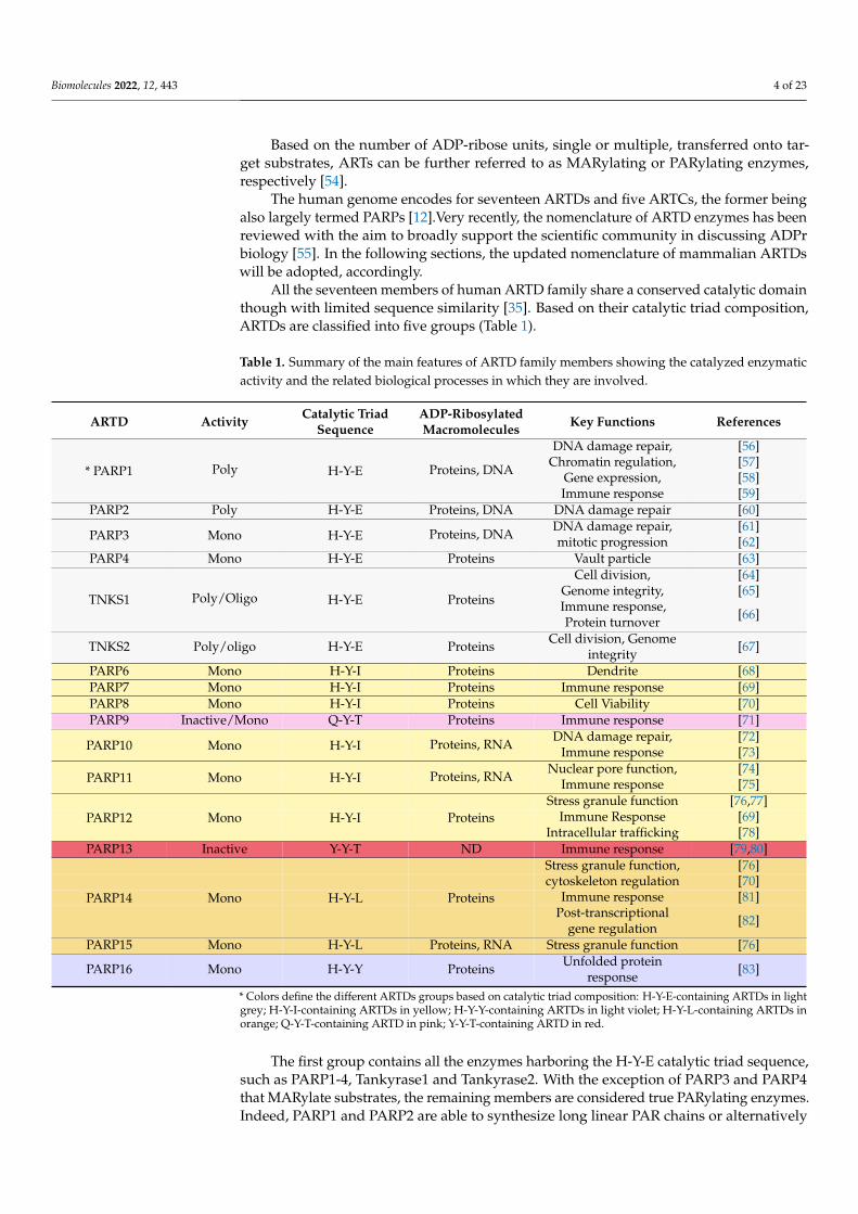
The human genome encodes for seventeen ARTDs and five ARTCs, the former beingalso largely termed PARPs [12].Very recently, the nomenclature of ARTD enzymes has beenreviewed with the aim to broadly support the scientific community in discussing ADPrbiology [55]. In the following sections, the updated nomenclature of mammalian ARTDswill be adopted, accordingly.
All the seventeen members of human ARTD family share a conserved catalytic domainthough with limited sequence similarity [35]. Based on their catalytic triad composition,ARTDs are classified into five groups (Table 1).
Table 1. Summary of the main features of ARTD family members showing the catalyzed enzymaticactivity and the related biological processes in which they are involved.
ARTD Activity Catalytic TriadSequence
ADP-RibosylatedMacromolecules Key Functions References
DNA damage repair, [56]Chromatin regulation, [57]
Gene expression, [58]* PARP1 Poly H-Y-E Proteins, DNA
Immune response [59]PARP2 Poly H-Y-E Proteins, DNA DNA damage repair [60]
DNA damage repair, [61]PARP3 Mono H-Y-E Proteins, DNA mitotic progression [62]PARP4 Mono H-Y-E Proteins Vault particle [63]
Cell division, [64]Genome integrity, [65]
TNKS1 Poly/Oligo H-Y-E Proteins Immune response,Protein turnover [66]
TNKS2 Poly/oligo H-Y-E Proteins Cell division, Genomeintegrity [67]
PARP6 Mono H-Y-I Proteins Dendrite [68]PARP7 Mono H-Y-I Proteins Immune response [69]PARP8 Mono H-Y-I Proteins Cell Viability [70]PARP9 Inactive/Mono Q-Y-T Proteins Immune response [71]
DNA damage repair, [72]PARP10 Mono H-Y-I Proteins, RNA Immune response [73]
Nuclear pore function, [74]PARP11 Mono H-Y-I Proteins, RNA Immune response [75]
Stress granule function [76,77]Immune Response [69]PARP12 Mono H-Y-I Proteins
Intracellular trafficking [78]PARP13 Inactive Y-Y-T ND Immune response [79,80]
Stress granule function, [76]cytoskeleton regulation [70]
Immune response [81]PARP14 Mono H-Y-L ProteinsPost-transcriptional
gene regulation [82]
PARP15 Mono H-Y-L Proteins, RNA Stress granule function [76]
PARP16 Mono H-Y-Y Proteins Unfolded proteinresponse [83]
* Colors define the different ARTDs groups based on catalytic triad composition: H-Y-E-containing ARTDs in lightgrey; H-Y-I-containing ARTDs in yellow; H-Y-Y-containing ARTDs in light violet; H-Y-L-containing ARTDs inorange; Q-Y-T-containing ARTD in pink; Y-Y-T-containing ARTD in red.
The first group contains all the enzymes harboring the H-Y-E catalytic triad sequence,such as PARP1-4, Tankyrase1 and Tankyrase2. With the exception of PARP3 and PARP4that MARylate substrates, the remaining members are considered true PARylating enzymes.Indeed, PARP1 and PARP2 are able to synthesize long linear PAR chains or alternatively
Biomolecules 2022, 12, 443 5 of 23
branched segments of PAR [70]. Tankyrase1 and Tankyrase2, instead, modify their sub-strates by the addition of a few units of ADP-ribose to produce short PAR chains of about20 units in length [70].
The second group harboring the H-Y-I catalytic triad sequence encloses PARP6-8and PARP10-12; the H-Y-Y catalytic triad sequence is found in PARP16; the H-Y-L isfound in PARP14 and PARP15; the Q-Y-T/Y-Y-T are, respectively, found in PARP9 andPARP13. Furthermore, a divergent PARP-like enzyme referred to as TpT1 or KptA, namelyARTD18 [84], contains the triad H-H-V.
Due to technology advancements in the last decade, it has become clear that themajority of ARTDs, i.e., PARP3, PARP4, and PARP6–PARP16, catalyze MARylation reactionwith the exception of PARP13, which has been reported to be inactive in agreement to thelack of a conserved catalytic triad composition [12,85]. Recent structural studies on PARP13show that, in addition to the zinc-finger domains, the WWE-domains are necessary topotentiate PARP13 antiviral activity, supporting the interaction with PAR [86]. The mainfeatures of human ARTDs are reported in Table 1.
ARTCs mostly comprise extracellular membrane-bound enzymes, with the exceptionof ARTC5 that is a secreted enzyme [87]. They have been shown to MARylate substratesboth extracellularly and intracellularly, thus regulating cellular functions, including stressresponse [87,88].
3.2. ADP-Ribosylhydrolases
As with other PTMs, ADPr signaling is tightly regulated in the cell by the activity ofspecialized ADP-ribose processing enzymes known as ADP-ribosylhydrolases. DraG-likeADP-ribosyl-acceptor hydrolases (ARHs) and macrodomain-containing enzymes representthe ADP-ribosylhydrolases known to support this function in humans. Indeed, the humangenome encodes three DraG-related ARHs (ARH1, ARH2, and ARH3) involved in theregulation of stress response [89–91]. Notably, ARH1 reverses the arginine-ADP-ribosylatedsubstrates catalyzed by mammalian ARTCs and ARH3 shows activity toward Ser-ADPrsubstrates [91], whilst ARH2 appears to be inactive.
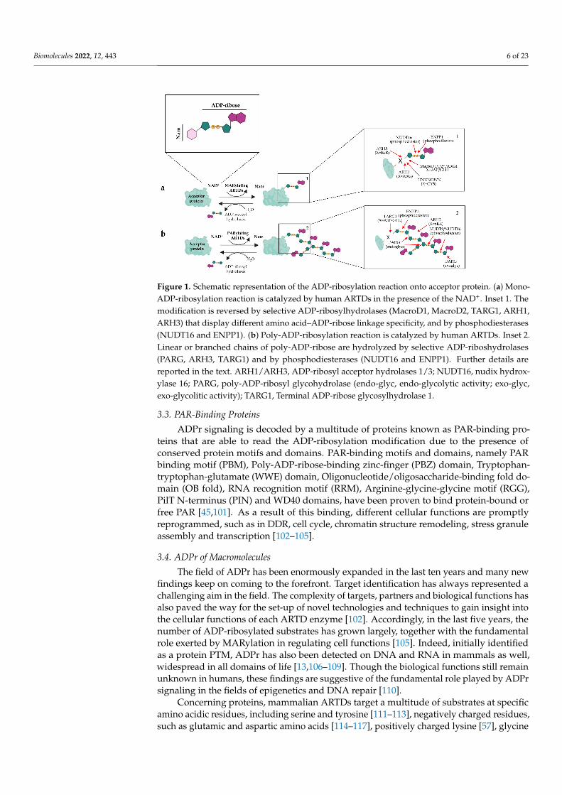
Macrodomain-containing enzymes harbor a common ADP-ribose binding domain, knownas macrodomain, and as said above are widely distributed in all domains of life [45,92,93]. Theseprotein modules are able to recognize ADP-ribose, PAR or O-acetyl-ADP-ribose and have beenfound in the poly-ADP-ribosyl glycohydrolase (PARG), MacroD1, MacroD2 and Terminal ADP-ribose glycosylhydrolase 1 (TARG1) macrodomain-containing enzymes [45,92,94,95]. PARGdisplays an exo- or endo-glycosyl hydrolytic activity that results in an efficient cleavage of PARchains, though it is unable to remove the terminal ADP-ribose attached to protein substrates [94].Conversely, the remaining macrodomain-containing hydrolase, namely TARG1, MacroD1, andMacroD2, are able to remove the ADP-ribose from MARylated substrates [44,96–98]. In addition,the enzymes NUDT16 and ENPP1, respectively belonging to the nucleoside diphosphate-linkedmoiety X (Nudix) and nucleotide pyrophosphatase/phosphodiesterases (NPP) families, areable to hydrolyse the phosphodiester bond within ADP-ribosylated substrates [99,100]. Figure 1shows the counterplay of ADPr removal.
Biomolecules 2022, 12, 443 6 of 23
Biomolecules 2022, 12, x FOR PEER REVIEW 6 of 28
Figure 1. Schematic representation of the ADP-ribosylation reaction onto acceptor protein. (a) Mono-ADP-ribosylation reaction is catalyzed by human ARTDs in the presence of the NAD+. Inset 1. The modification is reversed by selective ADP-ribosylhydrolases (MacroD1, MacroD2, TARG1, ARH1, ARH3) that display different amino acid–ADP-ribose linkage specificity, and by phos-phodiesterases (NUDT16 and ENPP1). (b) Poly-ADP-ribosylation reaction is catalyzed by human ARTDs. Inset 2. Linear or branched chains of poly-ADP-ribose are hydrolyzed by selective ADP-riboshydrolases (PARG, ARH3, TARG1) and by phosphodiesterases (NUDT16 and ENPP1). Further details are reported in the text. ARH1/ARH3, ADP-ribosyl acceptor hydrolases 1/3; NUDT16, nudix hydroxylase 16; PARG, poly-ADP-ribosyl glycohydrolase (endo-glyc, endo-glycolytic activity; exo-glyc, exo-glycolitic activity); TARG1, Terminal ADP-ribose glycosylhydrolase 1.
3.3. PAR-Binding Proteins ADPr signaling is decoded by a multitude of proteins known as PAR-binding pro-
teins that are able to read the ADP-ribosylation modification due to the presence of con-served protein motifs and domains. PAR-binding motifs and domains, namely PAR bind-ing motif (PBM), Poly-ADP-ribose-binding zinc-finger (PBZ) domain, Tryptophan-tryp-tophan-glutamate (WWE) domain, Oligonucleotide/oligosaccharide-binding fold domain (OB fold), RNA recognition motif (RRM), Arginine-glycine-glycine motif (RGG), PilT N-terminus (PIN) and WD40 domains, have been proven to bind protein-bound or free PAR [45,101]. As a result of this binding, different cellular functions are promptly repro-grammed, such as in DDR, cell cycle, chromatin structure remodeling, stress granule as-sembly and transcription [102-105].
3.4. ADPr of Macromolecules The field of ADPr has been enormously expanded in the last ten years and many new
findings keep on coming to the forefront. Target identification has always represented a challenging aim in the field. The complexity of targets, partners and biological functions has also paved the way for the set-up of novel technologies and techniques to gain insight into the cellular functions of each ARTD enzyme [102]. Accordingly, in the last five years, the number of ADP-ribosylated substrates has grown largely, together with the funda-mental role exerted by MARylation in regulating cell functions [105]. Indeed, initially identified as a protein PTM, ADPr has also been detected on DNA and RNA in mammals as well, widespread in all domains of life [13,106-109]. Though the biological functions
Figure 1. Schematic representation of the ADP-ribosylation reaction onto acceptor protein. (a) Mono-ADP-ribosylation reaction is catalyzed by human ARTDs in the presence of the NAD+. Inset 1. Themodification is reversed by selective ADP-ribosylhydrolases (MacroD1, MacroD2, TARG1, ARH1,ARH3) that display different amino acid–ADP-ribose linkage specificity, and by phosphodiesterases(NUDT16 and ENPP1). (b) Poly-ADP-ribosylation reaction is catalyzed by human ARTDs. Inset 2.Linear or branched chains of poly-ADP-ribose are hydrolyzed by selective ADP-riboshydrolases(PARG, ARH3, TARG1) and by phosphodiesterases (NUDT16 and ENPP1). Further details arereported in the text. ARH1/ARH3, ADP-ribosyl acceptor hydrolases 1/3; NUDT16, nudix hydrox-ylase 16; PARG, poly-ADP-ribosyl glycohydrolase (endo-glyc, endo-glycolytic activity; exo-glyc,exo-glycolitic activity); TARG1, Terminal ADP-ribose glycosylhydrolase 1.
3.3. PAR-Binding Proteins
ADPr signaling is decoded by a multitude of proteins known as PAR-binding pro-teins that are able to read the ADP-ribosylation modification due to the presence ofconserved protein motifs and domains. PAR-binding motifs and domains, namely PARbinding motif (PBM), Poly-ADP-ribose-binding zinc-finger (PBZ) domain, Tryptophan-tryptophan-glutamate (WWE) domain, Oligonucleotide/oligosaccharide-binding fold do-main (OB fold), RNA recognition motif (RRM), Arginine-glycine-glycine motif (RGG),PilT N-terminus (PIN) and WD40 domains, have been proven to bind protein-bound orfree PAR [45,101]. As a result of this binding, different cellular functions are promptlyreprogrammed, such as in DDR, cell cycle, chromatin structure remodeling, stress granuleassembly and transcription [102–105].
3.4. ADPr of Macromolecules
The field of ADPr has been enormously expanded in the last ten years and many newfindings keep on coming to the forefront. Target identification has always represented achallenging aim in the field. The complexity of targets, partners and biological functions hasalso paved the way for the set-up of novel technologies and techniques to gain insight intothe cellular functions of each ARTD enzyme [102]. Accordingly, in the last five years, thenumber of ADP-ribosylated substrates has grown largely, together with the fundamentalrole exerted by MARylation in regulating cell functions [105]. Indeed, initially identifiedas a protein PTM, ADPr has also been detected on DNA and RNA in mammals as well,widespread in all domains of life [13,106–109]. Though the biological functions still remainunknown in humans, these findings are suggestive of the fundamental role played by ADPrsignaling in the fields of epigenetics and DNA repair [110].
Concerning proteins, mammalian ARTDs target a multitude of substrates at specificamino acidic residues, including serine and tyrosine [111–113], negatively charged residues,such as glutamic and aspartic amino acids [114–117], positively charged lysine [57], glycine
Biomolecules 2022, 12, 443 7 of 23
as in the case of PARP9 of ubiquitin [118], and cysteine as reported for PARP7 [119,120]and PARP8 [70,121]. However, the biological significance of these modified residues is juststarting to emerge, since recent proteomic analyses show that several residues, as serine forinstance, are mainly modified in response to genotoxic stimuli but not under unperturbedconditions [122]. By contrast, arginine has been identified as the main aminoacidic acceptor,suggesting an evident contribution of ARTC1 regulating functions related to Endoplasmicreticulum and the Golgi apparatus at the basal level. Cysteine residues, instead, have beenidentified as ADPr acceptor sites in auto-modified PARP8 under physiological conditions,though the role still remains unclear [122].
4. The Role of ADPr in RNA Biology
Reversible ADPr reaction regulates a plethora of fundamental biological processesin mammals, as diverse as DDR [26,122–126], transcription, cell division [62,64,127], cellproliferation [128–130], cell death [131–134] immune and stress responses [76,77], as wellas other emerging pathways [78,135,136]. For further details on the functional outcomesmodulated by ADPr, we refer the reader to the valuable and specialized reviews in thefield [24,26,137–142]. Furthermore, the imbalance between the addition and the removalof ADP-ribose from substrates plays also a role in different pathological processes, asdiverse as neurological disorders [96], tumorigenesis and tumor progression [83], as wellas bacterial- and viral-mediated infections [30,69,137,143–148].
In the section below, we focus on the role that ADPr signaling exerts in post-transcriptionalregulation, modulating different steps of RNA biology at molecular level. Indeed, RNA biol-ogy represents an expanding topic of research in the field over the last ten years, where themodification acts as key player in controlling important steps in RNA maturation.
4.1. ADPr-Mediated Regulation of Nuclear RNA Processing
Gene expression is a highly regulated biological process that leads to the synthesis ofnew RNA molecules for the modulation of different functional outcomes. It can be drivenin response to intrinsic and extrinsic stimuli with the aim to support cell proliferation,survival, or death. Gene expression occurs through several steps that foresee: (i) chromatinmodulation; (ii) transcription; (iii) co-regulation; (iv) DNA methylation; and (v) RNAregulation. Early studies in the field were centered on the role of PARP1 and PAR asregulators of the first steps of the process that is tackled by valuable reviews elsewhere [58].
Concerning RNA regulation processes, they enclose: (i) mRNA maturation in thenucleus; (ii) shuttling of mature mRNA to the cytoplasm; (iii) translation into protein;(iv) degradation (mRNA decay); (v) silencing by microRNA (miRNA). All these stepsare modulated by a cluster of proteins, namely the RNA-binding proteins (RBPs), whoseactivity can be affected by PTMs that modulate their interaction with specific RNAs andand/or proteins with effects on processing, localization, translation or stability [149].
Several ARTDs are known to regulate this process, either through direct modificationof substrates or by a non-covalent interaction with PAR, thereby leading in both cases to again or a loss of function as discussed in detail below.
4.1.1. RNA Regulation at the Nucleus
Nuclear mRNA regulation relies on three steps namely capping, splicing and poly-Adenylation of primary RNA transcripts [150]. Early studies in the field demonstratedthe involvement of PARP1 in RNA maturation with implications on the functional roleof a sub-group of RBPs, namely heterogeneous nuclear-ribonucleoproteins (hn-RNPs),which are key players in multiple steps of RNA maturation [151]. Indeed, proteomicapproaches [152] have demonstrated that many hn-RNPs are PAR-binding proteins, as inthe case of hn-RNP A1 that binds poly-ADP-ribose deriving from PARP1 auto-modification.This finding strongly suggested a link between ADPr activity and altered mRNA trafficking.Similarly, Ji and Tulin (2013) [153] have shown that hn-RNPs in Drosophila melanogasterare also modulated by ADP-ribosylation, and that PAR-binding causes the dissociation of
Biomolecules 2022, 12, 443 8 of 23
hnRNPs from pre-mRNA, thus impairing the intron splicing step [153]. Additionally, theregulatory role of ADPr signaling in alternative splicing is also provided by the alternativesplicing factor/splicing factor 2 (ASF/FS2) factor, whose binding to PAR prevents itsphosphorylation from DNA Topoisomerase. As ASF/FS2 phosphorylation triggers itssplicing activity, it is likely that PAR-binding modulates alternative splicing either inhibitingphosphorylation or by hampering the ASF/FS2 interaction with mRNAs [154]. Likewise,other splicing factors, such as SF3B1, SF3A1 and SF3B2, have been found to be linked withPAR [155].
Additionally, upon heat shock stress, PARP1 has been found to modulate poly-Adenylation, the final step before mature mRNA leaves the nucleus to reach the cyto-plasm [156]. Under this condition, Poly(A) polymerase (PAP), the enzyme responsible forpoly-Adenylation, becomes PARylated losing its polymerase activity, with the resultingblockade of mRNA maturation synthesis.
PARP1-dependent ADPr also affects a group of RNA-binding proteins (RBPs) involvedin the nuclear/cytoplasm export of mRNA molecules. Within this group; embryonic lethalabnormal vision-like 1/Hu antigen R (ELAVL1/HuR), a stabilizing mRNA protein able torecognize the AU-rich elements (ARES) at 3′UTR of mRNA, has been shown to interactwith PARP1 after lipopolysaccharide (LPS) treatment. Following LPS administration tocells, ELAVL1/HuR results to be poly-ADP-ribosylated and the pharmacological inhibitionof PARP1 with PJ34 strongly decreases ELAVL1/HuR shuttling to the cytoplasm [157].As a result, an increase in ELAVL1/HuR cytosolic localization is observed, together withELAVL1/HuR mRNA target stability [157]. Collectively, these data highlight the posi-tively regulatory function of PARylation in post-transcriptional regulation in response toinflammatory stimuli.
A recent study has further gained insight into this finding underlining the molecularmechanism promoting ELAVL1/HuR translocation [158]. Indeed, PARylation promotesmultimeric organization of ELAVL1/HuR that stably interacts with mRNA targets. Atthe same time, the oligomerization allows the dissociation of the miRNA-induced si-lencing complex (miRISC) from the target RNA, thus resulting in the stabilization ofARE-containing mRNAs.
Recently, the finding of a novel and selective PARP11 inhibitor, 8-methyl-7-(prop-1-yn-1-yl)-2-((pyrimidin-2-ylthio)methyl)quinazolin-4(3H)-one (ITK7), has also suggested a rolefor PARP11 at the nuclear envelope [159]. Indeed, HeLa cells overexpressing GFP-PARP11show a strong reduction in the signal corresponding to mono-ADP-ribosylation around thenuclear envelope after treatment with ITK7, with respect to control cells. This finding issuggestive of a role at nuclear compartment for PARP11 that results to be strictly dependenton catalytic activity.
4.1.2. rRNA at the Nucleolus
Under normal cellular homeostasis, the majority of PARP1 and nuclear PAR are local-ized in the nucleolus, where they are supposed to be involved in the regulation of rRNAprocessing and ribosome biogenesis. In D. melanogaster, the PARP1 depletion or chemi-cal inhibition of PARP1 has been found to correlate with nucleolus disassembly, storageof rRNA intermediates and decreased polysome formation in the cytoplasm [160], thusstrongly suggesting the regulatory role of PARP1 in nucleolar structure maintenance andrRNA processing. Furthermore, following PARP1 inhibition, several nucleolar resident pro-teins, including coilin and fibrillarin, have been shown to be mislocalized in the cytoplasm,suggesting a role for PARP1 in the regulation of rRNA splicing processes [161].
PARP1 also regulates rRNA transcription through binding to TTF-1-interacting protein5 (TIP5), a component of the nucleolar remodeling complex (NoRC) [161]. PARP1 is acti-vated by the interaction with the non-coding RNA (pRNA), which promotes the formationof PARP1-pRNA-TIP5 complex. As result, active PARP1 ADP-ribosylates itself, TIP5 andhistones, hence leading to transcriptional repression.
Biomolecules 2022, 12, 443 9 of 23
4.2. ADPr-Mediated Regulation of RNA Processing at the Cytoplasm
Several steps of the mRNA maturation process are affected by ADPr modification inthe cytoplasm, similarly to what occurs in the nucleus. Of note, a group of ARTDs arealso identified as RNA-binding proteins due to the presence either of conserved CCCHRNA-binding domains (PARP7, PARP10, PARP11, PARP12 and PARP13) or of RRM motifs(PARP10 and PARP14) [162]. Their role in RNA biology is discussed in the following section.
Once mRNA transcripts reach the cytoplasm, they can undergo protein translation,can be silenced by micro-RNA (miRNA)-mediated post-transcriptional repression, or canbe degraded by mRNA decay.
Referring to translation, the primary step, namely initiation, generally occurs by therecognition of 5′CAP modification at the mRNA molecule, even if it can also be achievedthrough the binding of RBPs to Internal Ribosome Entry Sites (IRES) present on targetmRNAs. Inhibition of the interaction between RBPs and IRES elements on target mRNAshalters the translation of the transcript. An example is provided by hnRNA binding proteinHRB98DE/HRP38 (HRP38), an RBP in D. melanogaster, ortholog of human hnRNPA1,which is described to recruit ribosomes to IRES along the DE-Cadherin mRNA to promoteits translation. HRP38 non-covalent binding to PAR hampers binding of ribosomes toIRES region on DE-cadherin mRNA resulting into a block of DE-Cadherin mRNA transla-tion [163]. More recent studies have demonstrated that HRP38 regulates the translation ofNanos mRNA through the binding of its 3′UTR. HRP38 binding to PAR causes a decreasein HRP38 interaction to Nanos 3′UTR, thus leading to an increase in the translation in vivoand in vitro, hence concluding that HRP38 blocks Nanos translation, whereas ADPr relievesthe repression effect [163].
A further example of the ADPr regulatory role in translation is provided by Challaand colleagues (2021), who have shown the important role played by PARP16-mediatedMARylation of ribosomal proteins in ovarian cancer cells [164]. With the aim to analyzethe contribution of NAD+ signaling in cancer cells, this study has revealed that PARP16supports the ADPr of ribosomal protein utilizing NAD+ synthesized in the cytoplasm byNAMNT-2, whose protein level is upregulated here. As a result, cytoplasmic increase inmono-ADP-ribose has been demonstrated to correlate with an impairment of polysomesassembly and mRNA loading, thus inhibiting protein synthesis and maintaining proteinhomeostasis. It followed that under these conditions the ovarian cancer cells were ableto grow. Notably, this study proposes a novel therapeutic treatment based on NAMNT-2depletion or PARP16 inhibition relying on the strategical approach to target the NAD+
availability in cancer cells, thereby affecting the downstream NAD+-consuming enzy-matic pathways.
The impairment of translation in an ADPr-dependent manner is also described asanti-viral mechanism against alphaviruses [68]. The RNA-binding ARTDs PARP7, PARP10and PARP12 have been shown to strongly induce the clearance of Venezuelan equineencephalitis virus mutants and other alphavirus from infected cells through the regulationof cellular translation and virus replication [69]. In particular, the three enzymes, whosegene expression is upregulated in response to interferon, mediate the antiviral response.The PARP12 long isoform (PARP12L) antiviral activity relies on its own ADPr activityand also on the ability to bind to different interactors in the cytoplasm [69]. The presenceof a fully active catalytic domain is required for the efficacious anti-viral response thatrelies upon the host translational repression, though the underlining molecular mechanismstill remains unknown. For more details, we refer the reader to valuable reviews in theADPr-mediated host–virus interaction field [165,166].
Furthermore, under stressful conditions, several ARTD family members have beenreported to regulate gene expression at a post-transcriptional level. Indeed, under oxidativestress, the enzymes PARP12, PARP13.1, PARP13.2, PARP14, PARP15, Tankyrase1 and twoisoforms of PARGs assemble in the presence of PAR polymer in the cytoplasm, formingparticular structures known as stress granules [24,76,167]. These transient structures alsocontain the four miRNA-binding Argonaute proteins that result to be poly-ADP-ribosylated
Biomolecules 2022, 12, 443 10 of 23
upon stress. Consequently, a relief of miRNA-mediated translational repression occurs dueto the ADPr-mediated function.
mRNA decay is another ADPr-regulated functional process. PARP13, which isknown as inactive ARTD family member, has been described to target cellular mRNAsfollowing the interaction with mRNA transcripts due to the presence of its zinc-fingerdomains [79,168,169]. The knocking down of PARP13 determines a mis-regulation of thetranscriptome with evident effects on mRNAs containing signal sequences for sorting toendoplasmic reticulum, the subcellular compartment where PARP13 is located. Amongthese transcripts, tumor necrosis factor (TNF)-related apoptosis-inducing ligand receptor 4(TRAILR4) mRNA is mainly affected, since the PARP13 binding to 3′UTR allows TRAILR4mRNA degradation through the exosomal pathway [80]. Thus, through the modulationof PARP13, cells can escape the cell death avoiding the accumulation of elevated levels ofTRAIL4 mRNA.
A further example is provided by PARP14, a RNA-binding ARTD that is also includedin the subclass of the macrodomain-containing-PARPs (MACRO-PARPs) [162]. PARP14is selectively involved in the post-transcriptional regulation of Tissue factor (TF) (CD142),a key mediator of thrombosis and inflammation, in macrophages [82]. The mechanismrelies on the control of TF mRNA stability after the formation of a complex where PARP14and the mRNA-destabilizing protein tristetraprolin (TTP) interact through the adenylate-uridylate-rich element in the TF mRNA 3′UTR, thus providing a novel insight for thepost-transcriptional regulation of TF expression. However, the molecular mechanismunderlining this finding remains to be further characterized.
The regulatory role of PARP14 in RNA biology has been further corroborated by thefollowing studies aimed at identifying protein substrates by combining chemical geneticswith proximity-dependent labelling. This approach led Carter-O’Connell and colleagues(2018) to identify 114 substrates selectively modified by PARP14, several of which wereRNA regulatory proteins [170]. Notably, PARP14 was shown to interact with DEAD-BoxHelicase 6 (DDX6) at processing-bodies (P-bodies), membrane-less ribonucleoprotein (RNP)granules, composed of translationally repressed mRNAs and proteins related to mRNAdecay, thereby suggesting a role in post-transcriptional regulation. Intriguingly, PARP13,known to play a role in regulating mRNA stability (see above), has been found ADP-ribosylated by PARP14 at different fourteen amino acidic sites, leading to speculate onthe possible crosstalk between the different members of ARTD subfamily within the RNAbiology process.
Collectively, these data add new layers of complexity to ADPr signaling.
5. Targeting ADPr Signaling for Therapeutic Treatment5.1. Chemical Inhibition of ARTDs
Small-compound inhibitors have been fundamental tools to unveil ARTDs’ biologicalfunctions. In addition, the pivotal role played by ADPr in regulating pathophysiologi-cal processes has fostered the search for new molecules to be exploited as inhibitors fortherapeutic treatment development.
Pioneering studies focused on the ARTD enzymes that carry out PARylation withimplications in genome integrity and signal transduction. Thus, it followed that targetingDDR could represent a promising strategy to counteract cancer cells. PARP1 small-moleculeinhibitors, designed as NAD+ analogs, induce conformational changes in PARP1 andstabilize the complex between PARP1 and DNA, thereby “trapping” the complexes atDNA lesions. Therefore, PARP1 inhibitors have been applied as anticancer agents relyingon the strategical induction of “synthetic lethality” in cancer cells carrying Breast-Cancer1/2 (BRCA1/2) mutations [171,172]. PARP-inhibitors approved by the U.S. Food and DrugAdministration (FDA), namely niraparib, olaparib, talazoparib, and rucaparib, are currentlyused in the therapeutic treatment of BRCA-mutated breast and ovarian cancers, as well asin tumors with dysfunctional BRCA genes [173]. The range of treated cancers and the usesof PARP inhibitors may increase as more research is completed.

Biomolecules 2022, 12, 443 11 of 23
Intriguingly, a more recent approach aims at harnessing these inhibitors in the treat-ment of non-oncological diseases, including oxidative stress, inflammatory responses, aswell as neurodegenerative and cardiovascular diseases [137,174,175].
There is a marked over-activation of PARP1 in the reperfused myocardium, whichparallels with the decline of the contractile function and myocardial NAD+ and ATPcontents in preclinical models of myocardial infarction and cardiopulmonary bypass. Thepharmacological inhibition of PARP with various inhibitors or its genetic deletion, markedlyimproves the outcome of myocardial ischemia-reperfusion damage (in all in vitro or ex vivomodels), which is also a subject of numerous overviews [176]. Aside from the DNA damagerepair, other ADPr-dependent molecular pathways have been evaluated as therapeutictargets, including those in which hydrolases revert the reaction. We direct the reader torecent valuable reviews providing a comprehensive state of the art on this topic [177,178].
Since one concern is always the specificity, it is worth noting that for niraparib theresults from the last phase III trial “support the hypothesis that niraparib has mechanismsof action other than those involved in the repair of DNA damage”, as the Authors note.These could include PARP-regulated gene transcription, ribosome biogenesis and immuneactivation, they speculate [179].
The mitotic checkpoint protein CHFR (Checkpoint With Forkhead And Ring FingerDomains) has emerged as a major mediator of taxane resistance in cancer. It has beenshown that CHFR’s PAR-binding zinc finger domain (PBZ) mediates a protein interactionwith poly-ADP-ribosylated PARP1 leading to the stabilization of CHFR. The disruptionof the CHFR-PARP1 interaction through a PBZ domain peptide induces the loss of CHFRprotein expression. These findings provide a proof-of-principle that the small moleculeinhibition of the CHFR-PBZ domain interaction can be exploited to increase the efficacy oftaxane-based chemotherapy in cancer [180,181].
Therefore, compounds that are capable of disrupting the protein–protein interactions ofPARP1 provide an alternative by inhibiting its activities with improved selectivity profiles.
Another major limitation depends on the shortage of novel compounds to inhibit selec-tively all the enzymes of the ARTD family and the research in this field is still ongoing [173].The latest findings on the role of MARylating enzymes highlight the need of new inhibitorsto unravel and dissect the emerging signaling pathways in which mono-ADP-ribosylationplays a role.
As such, the finding of several ARTDs, other than PARP1, as regulators of RNAbiology, may provide alternative strategies to exploit as potential therapeutic targets. Ofnote, the observation that NAMNT2-dependent NAD+ synthesis parallels PARP16 activityin ovarian cancer cells, suggests novel approaches in cancer therapies, which include theenzymes responsible for NAD+ biosynthetic pathways as well [169]. The impairment ofthe crosstalk between signaling pathways and RNA processing is frequently linked topathologies, such as neurodegenerative diseases and cancer. The current knowledge aboutthe modulation of RNA biology is still at the beginning. The number of RBPs involved iscontinuously expanding, likewise the role played by different PTMs in modulating theirfunctions, localizations or degradation. As the dysregulation of PTMs in RBPs has beenassociated with the pathophysiological mechanism of diverse diseases, it is reasonable toconsider the ADP-ribosylating enzymes involved in the modulation of RNA processing asgood candidates for the assessment of novel therapeutic options.
Novel strategies have to be addressed for the development of new classes of ARTDinhibitors evaluating the potential of binding sites and motifs other than catalytic sites [173,177].
Last but not least, the post-transcriptional regulation of ARTD-encoding mRNAsmay represent a further strategy to take into consideration. Indeed, PARP1 expression isregulated by the nuclear factor 90 (NF90), a RNA binding protein able to bind and stabilizePARP1 mRNA [182]. Targeting this interaction and therefore mRNA stability might beassessed as alternative therapeutic intervention as well.
Biomolecules 2022, 12, 443 12 of 23
5.2. New Avenues: The Post-Transcriptional Regulation of ARTDs mRNA
RBPs have been shown to control the expression of many genes by binding to the re-spective mRNA species, encoding proto-oncogenes, growth factors, cytokines, transcriptionfactors, and other proteins in various cell types [183].
The 3′UTRs and the 5′UTRs are the transcript target sequences involved in the RBPsbinding and mediating the formation of the so-called “RNA operon”. This is a functionalunit in which multiple physiologically related transcripts are coordinately regulated duringsplicing, export, stability, localization, and translation. These subpopulations of mRNAsbind the same RBPs in a dynamic manner because each mRNA can join different “RNAoperons” [184–186].
As a proof of concept, we have recently reported about the “RNA operon” regulatorymechanism for the human Major Histocompatibility Complex II gene [187] and for thehuman Paraoxonase 2 (PON2) [117].
The modulation of mRNA stability is now considered as a novel therapeutic approachtogether with other functions related to the RNA biology [188–190].
The competition of miRNAs and antisense oligonucleotides (ASO) with RBPs forbinding to the same RNA binding site or the modification of RBPs affinity via PTMs,proteins or small molecules interactions are now commonly used [149,191].
Post-transcriptional gene regulation via RBPs is an adaptable reprograming mecha-nism that may drive PARP inhibitor resistance. Chand and colleagues (2017) have previ-ously shown that the RBP ELAVL1/HuR promotes a drug-resistant phenotype, through itsstress-induced cytoplasmic translocation and stabilization of pro-survival mRNA targets,among which PARG [192]. The role of ELAVL1/HuR is further discussed in Section 4.1.1.
In the vein of this research, we recently analyzed a database of human genes catego-rized on the basis of conserved short sequences (n-mers) at the 3′UTR [183]. The Authorsdemonstrated how the database provides evidence that these n-mers are potentially in-volved in regulatory functions. The identified n-mers in fact overlap with previouslyrecognised binding sites for ELAVL1/HuR and T-Cell-Restricted Intracellular Antigen-1(TIA-1) and with ARE and GU-rich element (GRE) sequences. It was established also that n-mers overlap with predicted miRNA target sites. Finally, a method to cluster n-mer groupsallowed the identification of putative gene networks. This analysis indicated that severalsets of gene clusters share commonalities in terms of pathway and function. Moreover,interacting proteins turned out to be present in numerous clusters [183].
Based on this research, we decided to explore the clusters containing the genes forARTDs proteins.
As reported in Table 2, among the seventeen ARTDs no clusters were found for PARPs1–4, 5b, 6, 7, 10, and 13.
Table 2. Gene cluster database analysis for genes including PARPs.
ARTDGene
Total Numberof Clusters
Containing theARTD Gene
Number of Genes in Each Cluster
PARP5a 10 34 32 30 28 23 22 22 21 21 21PARP8 1 23PARP9 3 48 21 20
PARP11 20 125 93 41 40 40 37 32 30 26 26 25 24 24 23 23 22 22 21 21 21PARP12 2 37 21PARP14 18 53 50 48 47 47 44 41 41 40 39 35 34 32 30 26 24 24 23PARP15 1 24PARP16 2 33 22
Cluster numbering 1 2 3 4 5 6 7 8 9 10 11 12 13 14 15 16 17 18 19 20
For the ARTDs involved in the clusters, their number was quite different, ranging from1 (PARP8 and 15) to 20 for PARP11. In addition, the number of genes in each cluster was

Biomolecules 2022, 12, 443 13 of 23
quite various, ranging from a minimum of 20 to a maximum of 125. Given that all the genesbelonging to a single cluster share a conserved sequence at the 3′UTR, this leads to assessthe possibility that their transcripts can be co-regulated at a post-transcriptional level.
As an example, the regions conserved at the 3′UTR of the two PARP12 clusters are thefollowing: AGUUUUAGUUUU (cluster 1) and UAAUGAUUUUU (cluster 2).
Regarding the cluster 1 signature, this is closely related to the hsa-mir-3925 precursormiRNA found using the mirBase program (https://www.mirbase.org/search-rnacentral.shtml, accessed on 24 February 2022).
This sequence is part of the miRNA stem and by the binding of a hypothetical RBPthere might be an interference in the Dicer processing. The gene DICER1, detected in cluster2, encodes a ribonuclease that trims double stranded RNA or pre-miRNA to form smallinterfering RNA or microRNA, respectively. These processed RNAs are incorporated intothe RNA-induced silencing complex (RISC), which targets messenger RNA to preventtranslation. This specific miRNA has been related to cancer in females [131,193].
Supplementary Table S1 provides the list of the 21 genes related to cluster 2 with theindicated function as reported in the Genecard database (https://www.genecards.org/,accessed on 28 January 2022). The genes have been further categorized by the GeneOntology analysis using Panther Classification system (http://pantherdb.org/, accessedon 28 January 2022) and reported in Figure 2.
Out of the 21 genes of cluster 2, 13 were grouped according to 4 molecular functions(Figure 2b), whereas 15 according to 10 specific protein classes (Figure 2a). The remaining6 genes are RBPs and DNA binding proteins that are not classified, likely, because of beingpart of many pathways.
Intriguingly, this cluster contains, among other genes, DICER1, which acts as a strongantiviral agent with activity against RNA viruses, including the Zika and SARS-CoV-2viruses. This activity resembles the above-described antiviral role of PARP12 and PARP13.The protein encoded by ELAVL1/HuR gene as said above is a member of the ELAVLfamily of RBPs that binds AU-rich elements (AREs) found at the 3′UTR of many mRNAs.AREs by binding to RBPs or miRNA signal the degradation/stabilization of mRNAs as ameans to regulate gene expression. The ELAVL family of proteins in general plays a role instabilizing ARE-containing mRNAs. This gene family has been implicated in a variety ofbiological processes and has been linked to a number of diseases, including cancer [194].ELAVLl binds to the AU-rich element in FOS and nterleukin-3 (IL3) mRNAs. In the caseof the FOS AU-rich element, it binds to a core element of 27 nucleotides that containAUUUA, AUUUUA, and AUUUUUA motifs. These motifs are reminiscent of the twoconserved sequences reported above. ELAVL1 is also known to be PARylated by PARP1 inresponse to LPS exposure. That increases ELAVL1/HuR nucleo-plasmatic translocation andmRNA stabilization, affecting downstream regulation [157]. We are tempted to speculatethat PARP12 is able to PARylate ELAVL1/HuR as well, and that both are co-regulated.In agreement with this idea, it has been reported: (i) the elevated expression in HuR-double-transgenic Traffic/HuR Pdx1/Cre mice (TC mice) of two ARTDs, PARP12 andPARP14 [195]; (ii) the physical interaction between ELAVL1/HuR and PARP12 [196].
Additional RBPs contained in the cluster are LUZP4 and UPF3B. LUPZP4 is involved inmRNA export from the nucleus. UPF3B is involved in the regulation of nonsense mediatedmRNA Decay (Table S1 [194,197–218]). Experimental studies based on RBP depletion byRNA interfering, measuring of gene expression by real-time PCR, immunoprecipitationof RBP(s) bound mRNA and sequencing after PCR amplification, in situ RNA-labelledprobing of SDS-PAGE separated proteins followed by identification by MS, analysis of generelationships by many different algorithms are needed to prove the interactions amongmembers of the cluster and with the relevant PARP.
Biomolecules 2022, 12, 443 14 of 23
Biomolecules 2022, 12, x FOR PEER REVIEW 14 of 28
PARP1 in response to LPS exposure. That increases ELAVL1/HuR nucleo-plasmatic trans-location and mRNA stabilization, affecting downstream regulation [157]. We are tempted to speculate that PARP12 is able to PARylate ELAVL1/HuR as well, and that both are co-regulated. In agreement with this idea, it has been reported: (i) the elevated expression in HuR-double-transgenic Traffic/HuR Pdx1/Cre mice (TC mice) of two ARTDs, PARP12 and PARP14 [195]; (ii) the physical interaction between ELAVL1/HuR and PARP12 [196].
Figure 2. Representation of the GO analysis using the 21 genes list of Cluster 2 as input (Table S1 [194,197-218]): (a) categorization by protein class; (b) grouping by molecular functions. Six genes, namely PARP12, DICER1, LUZP4, UPF3B, ZFAND3, and OSGIN2, are not included.
Additional RBPs contained in the cluster are LUZP4 and UPF3B. LUPZP4 is involved in mRNA export from the nucleus. UPF3B is involved in the regulation of nonsense me-diated mRNA Decay (Table S1 [194,197-218]). Experimental studies based on RBP deple-tion by RNA interfering, measuring of gene expression by real-time PCR, immunoprecip-itation of RBP(s) bound mRNA and sequencing after PCR amplification, in situ RNA-la-belled probing of SDS-PAGE separated proteins followed by identification by MS, analy-sis of gene relationships by many different algorithms are needed to prove the interactions among members of the cluster and with the relevant PARP.
6. Conclusions Gene expression programs support the cell in division, differentiation, as well as in
adaptation to intrinsic and extrinsic stimuli or fate decision. The maintenance of cell ho-meostasis is gained through well inter-connected regulatory processes that are finely tuned by PTMs. In this paper, the regulatory role of ADP-ribosylation in RNA biology has been reviewed, highlighting the RNA processes known to date that are under its control. Indeed, protein substrate modification or non-covalent interaction with PAR induce a range of effects that lead to enzyme inactivation, change in subcellular localization, or stall in the assembly of intracellular organelles with the consequent alteration, loss or gain, of functions as discussed above. However, novel functional processes related to RNA pro-cessing regulation are expected to emerge in the near future as an improvement of alter-native approaches, as we propose by dealing with the “RNA operon” approach. In addi-tion, the discovery that few of the ARTD family members (PARP10, PARP11 and PARP15) are capable of modifying RNA in vitro adds another layer of complexity in the modulation of RNA biology. Further efforts have to be directed at elucidating the functional role of RNA ADP-ribosylation in cells.
Commented [GL3]: These are the right references related to the Supplementary Table 1.
Commented [GL4]: These are the right references related to the Supplementary Table 1.
Figure 2. Representation of the GO analysis using the 21 genes list of Cluster 2 as input(Table S1 [194,197–218]): (a) categorization by protein class; (b) grouping by molecular functions.Six genes, namely PARP12, DICER1, LUZP4, UPF3B, ZFAND3, and OSGIN2, are not included.
6. Conclusions
Gene expression programs support the cell in division, differentiation, as well asin adaptation to intrinsic and extrinsic stimuli or fate decision. The maintenance of cellhomeostasis is gained through well inter-connected regulatory processes that are finelytuned by PTMs. In this paper, the regulatory role of ADP-ribosylation in RNA biology hasbeen reviewed, highlighting the RNA processes known to date that are under its control.Indeed, protein substrate modification or non-covalent interaction with PAR induce arange of effects that lead to enzyme inactivation, change in subcellular localization, orstall in the assembly of intracellular organelles with the consequent alteration, loss orgain, of functions as discussed above. However, novel functional processes related toRNA processing regulation are expected to emerge in the near future as an improvementof alternative approaches, as we propose by dealing with the “RNA operon” approach.In addition, the discovery that few of the ARTD family members (PARP10, PARP11 andPARP15) are capable of modifying RNA in vitro adds another layer of complexity in themodulation of RNA biology. Further efforts have to be directed at elucidating the functionalrole of RNA ADP-ribosylation in cells.
In the last decade, the understanding of ADPr biology has increased substantially. Thedevelopment of new and more specific technologies has pushed the knowledge in the fieldenormously, with particular emphasis on MARylation reaction, leading to the identificationof novel targets and the discovery of unknown biological functions. In addition, severalARTs have been shown to be involved in the pathogenesis of human diseases, thus evidenc-ing the need for novel specific inhibitors to address ARTs pharmacological targeting. In thisreview, we propose an alternative approach to analyze the post-transcriptional regulationof ARTs in order to provide new insights into the finding of novel drug targets.
ADPr is important for many fundamental biological processes. However, a com-plete inventory of modification sites, also depending on changing of specificity, has notbeen established.
Furthermore, although PTM coverage by MS-based methods is impressive, it stillneeds to be improved, especially in tissues and in clinically relevant systems.
Regulatory sequences, either placed at promoter regions or on UTRs, function astargets recognized by regulators that can then activate or repress different groups ofgenes according to the necessity. While regulatory sequences involved in transcriptionare quite well documented, there is a lack of information on sequence elements involvedin post-transcriptional regulation. Here, we reconsidered an already published study,by highlighting conserved sequences in clusters of genes containing those coding forADP-ribosyltransferase enzymes.

Biomolecules 2022, 12, 443 15 of 23
As reported for PARP12 as an example, this analysis can reveal some new relationshipsamong genes that can be useful to understand the functions and propose new opportunitiesfor therapeutic interventions.
By looking at the data of Table 2, there are a lot of clusters that need to be experimen-tally analyzed, containing around 1900 genes in total. On the other hand, much informationis accumulating on the identification of the pathways in which ARTs are involved. Thecombination of these two areas of research is expected to foster the knowledge on themechanisms of action of these enzymes and the possibility to use new therapeutic tools.
Supplementary Materials: The following supporting information can be downloaded at: https://www.mdpi.com/article/10.3390/biom12030443/s1, Table S1: PARP12 cluster of genes with theirfunction (GeneCard).
Author Contributions: Conceptualization, G.M. and G.C.; methodology, G.M.; software, G.L.;writing—original draft preparation, G.C.; writing—review and editing, G.C., G.M., E.P. and G.L.;visualization, G.C., G.M., E.P. and G.L; supervision, G.M.; funding acquisition, G.M. All authors haveread and agreed to the published version of the manuscript.
Funding: This research was funded by CNR grant Interomics Proteoma and Proteoma2 and MURSIABIO POC01_00042.
Institutional Review Board Statement: Not applicable.
Informed Consent Statement: Not applicable.
Data Availability Statement: Not applicable.
Acknowledgments: We would like to thank Romeo Prezioso, technician at IGB-CNR, for the infor-matics support.
Conflicts of Interest: The authors declare no conflict of interest.
References1. Pope, S.D.; Medzhitov, R. Emerging Principles of Gene Expression Programs and Their Regulation. Mol. Cell 2018, 71, 389–397.
[CrossRef]2. Walsh, G.; Jefferis, R. Post-translational modifications in the context of therapeutic proteins. Nat. Biotechnol. 2006, 24, 1241–1252.
[CrossRef]3. Ramazi, S.; Zahiri, J. Posttranslational modifications in proteins: Resources, tools and prediction methods. Database 2021, 2021,
baab012. [CrossRef]4. Vu, L.D.; Gevaert, K.; De Smet, I. Protein Language: Post-Translational Modifications Talking to Each Other. Trends Plant Sci. 2018,
23, 1068–1080. [CrossRef]5. van der Laarse, S.A.M.; Leney, A.C.; Heck, A.J.R. Crosstalk between phosphorylation and O-GlcNAcylation: Friend or foe. FEBS
J. 2018, 285, 3152–3167. [CrossRef]6. Zhao, Y.; Brickner, J.R.; Majid, M.C.; Mosammaparast, N. Crosstalk between ubiquitin and other post-translational modifications
on chromatin during double-strand break repair. Trends Cell Biol. 2014, 24, 426–434. [CrossRef]7. Kishore, C. Epigenetic Regulation and Promising Therapies in Colorectal Cancer. Curr. Mol. Pharmacol. 2021, 14, 838–852.
[CrossRef]8. He, W.; Wei, L.; Zou, Q. Research progress in protein posttranslational modification site prediction. Brief. Funct. Genom. 2018, 18,
220–229. [CrossRef]9. Ueda, K.; Hayaishi, O. ADP-ribosylation. Annu. Rev. Biochem. 1985, 54, 73–100. [CrossRef]10. Corda, D.; Di Girolamo, M. Functional aspects of protein mono-ADP-ribosylation. EMBO J. 2003, 22, 1953–1958. [CrossRef]11. Aravind, L.; Zhang, D.; de Souza, R.F.; Anand, S.; Iyer, L.M. The natural history of ADP-ribosyltransferases and the ADP-
ribosylation system. Curr. Top. Microbiol. Immunol. 2015, 384, 3–32. [CrossRef] [PubMed]12. Cohen, M.S.; Chang, P. Insights into the biogenesis, function, and regulation of ADP-ribosylation. Nat. Chem. Biol. 2018, 14,
236–243. [CrossRef] [PubMed]13. Munnur, D.; Ahel, I. Reversible mono-ADP-ribosylation of DNA breaks. FEBS J. 2017, 284, 4002–4016. [CrossRef]14. Otto, H.; Reche, P.A.; Bazan, F.; Dittmar, K.; Haag, F.; Koch-Nolte, F. In silico characterization of the family of PARP-like
poly(ADP-ribosyl)transferases (pARTs). BMC Genom. 2005, 6, 139. [CrossRef]15. Teloni, F.; Altmeyer, M. Readers of poly(ADP-ribose): Designed to be fit for purpose. Nucleic Acids Res. 2016, 44, 993–1006.
[CrossRef] [PubMed]
Biomolecules 2022, 12, 443 16 of 23
16. Hassa, P.O.; Haenni, S.S.; Elser, M.; Hottiger, M.O. Nuclear ADP-ribosylation reactions in mammalian cells: Where are we todayand where are we going? Microbiol. Mol. Biol. Rev. 2006, 70, 789–829. [CrossRef]
17. Hawse, W.F.; Wolberger, C. Structure-based mechanism of ADP-ribosylation by sirtuins. J. Biol. Chem. 2009, 284, 33654–33661.[CrossRef]
18. Iyer, L.M.; Zhang, D.; Rogozin, I.B.; Aravind, L. Evolution of the deaminase fold and multiple origins of eukaryotic editing andmutagenic nucleic acid deaminases from bacterial toxin systems. Nucleic Acids Res. 2011, 39, 9473–9497. [CrossRef]
19. Perina, D.; Mikoc, A.; Ahel, J.; Cetkovic, H.; Žaja, R.; Ahel, I. Distribution of protein poly(ADP-ribosyl)ation systems across alldomains of life. DNA Repair 2014, 23, 4–16. [CrossRef]
20. Palazzo, L.; Mikoc, A.; Ahel, I. ADP-ribosylation: New facets of an ancient modification. FEBS J. 2017, 284, 2932–2946. [CrossRef]21. Faraone-Mennella, M.R.; Gambacorta, A.; Nicolaus, B.; Farina, B. Purification and biochemical characterization of a poly(ADP-
ribose) polymerase-like enzyme from the thermophilic archaeon Sulfolobus solfataricus. Biochem. J. 1998, 335 Pt 2, 441–447.[CrossRef] [PubMed]
22. Grimaldi, G.; Corda, D.; Catara, G. From toxins to mammalian enzymes: The diversity of mono-ADP-ribosylation. Front. Biosci.2015, 20, 389–404. [CrossRef]
23. Mikolcevic, P.; Hloušek-Kasun, A.; Ahel, I.; Mikoc, A. ADP-ribosylation systems in bacteria and viruses. Comput. Struct. Biotechnol.J. 2021, 19, 2366–2383. [CrossRef] [PubMed]
24. Catara, G.; Corteggio, A.; Valente, C.; Grimaldi, G.; Palazzo, L. Targeting ADP-ribosylation as an antimicrobial strategy. Biochem.Pharmacol. 2019, 167, 13–26. [CrossRef]
25. Koch-Nolte, F. Endogenous ADP-Ribosylation. Endog. Adp-Ribosylation 2015, 384, 1–213. [CrossRef]26. Gupte, R.; Liu, Z.; Kraus, W.L. PARPs and ADP-ribosylation: Recent advances linking molecular functions to biological outcomes.
Genes Dev. 2017, 31, 101–126. [CrossRef]27. Ludden, P.W. Reversible ADP-ribosylation as a mechanism of enzyme regulation in procaryotes. Mol. Cell. Biochem. 1994, 138,
123–129. [CrossRef]28. Ma, Y.; Ludden, P.W. Role of the dinitrogenase reductase arginine 101 residue in dinitrogenase reductase ADP-ribosyltransferase
binding, NAD binding, and cleavage. J. Bacteriol. 2001, 183, 250–256. [CrossRef]29. Simon, N.C.; Aktories, K.; Barbieri, J.T. Novel bacterial ADP-ribosylating toxins: Structure and function. Nat. Rev. Microbiol. 2014,
12, 599–611. [CrossRef]30. Leung, A.K.L.; McPherson, R.L.; Griffin, D.E. Macrodomain ADP-ribosylhydrolase and the pathogenesis of infectious diseases.
PLoS Pathog. 2018, 14, e1006864. [CrossRef]31. Krueger, K.M.; Barbieri, J.T. The family of bacterial ADP-ribosylating exotoxins. Clin. Microbiol. Rev. 1995, 8, 34–47. [CrossRef]
[PubMed]32. Li, C.; Debing, Y.; Jankevicius, G.; Neyts, J.; Ahel, I.; Coutard, B.; Canard, B. Viral Macro Domains Reverse Protein ADP-
Ribosylation. J. Virol. 2016, 90, 8478–8486. [CrossRef] [PubMed]33. Katada, T.; Ui, M. Direct modification of the membrane adenylate cyclase system by islet-activating protein due to ADP-
ribosylation of a membrane protein. Proc. Natl. Acad. Sci. USA 1982, 79, 3129–3133. [CrossRef] [PubMed]34. de Souza, R.F.; Aravind, L. Identification of novel components of NAD-utilizing metabolic pathways and prediction of their
biochemical functions. Mol. Biosyst. 2012, 8, 1661–1677. [CrossRef] [PubMed]35. Fieldhouse, R.J.; Turgeon, Z.; White, D.; Merrill, A.R. Cholera- and anthrax-like toxins are among several new ADP-
ribosyltransferases. PLoS Comput. Biol. 2010, 6, e1001029. [CrossRef]36. Hottiger, M.O.; Hassa, P.O.; Lüscher, B.; Schüler, H.; Koch-Nolte, F. Toward a unified nomenclature for mammalian ADP-
ribosyltransferases. Trends Biochem. Sci. 2010, 35, 208–219. [CrossRef]37. Castellano, S.; Farina, B.; Faraone-Mennella, M.R. The ADP-ribosylation of Sulfolobus solfataricus Sso7 modulates protein/DNA
interactions in vitro. FEBS Lett. 2009, 583, 1154–1158. [CrossRef] [PubMed]38. Di Maro, A.; De Maio, A.; Castellano, S.; Parente, A.; Farina, B.; Faraone-Mennella, M.R. The ADP-ribosylating thermozyme from
Sulfolobus solfataricus is a DING protein. Biol. Chem. 2009, 390, 27–30. [CrossRef] [PubMed]39. Porzio, E.; Bianchi, A.R.; Baccigalupi, L.; Isticato, R.; Mennella, M.R.F. The DINGGG thermoprotein is membrane bound in the
Crenarchaeon Sulfolobus solfataricus. Chem. Biol. Technol. Agric. 2016, 3, 8. [CrossRef]40. Porzio, E.; De Maio, A.; Ricciardi, T.; Mistretta, C.; Manco, G.; Faraone-Mennella, M.R. Comparison of the DING protein from the
archaeon Sulfolobus solfataricus with human phosphate-binding protein and Pseudomonas fluorescence DING counterparts.Extremophiles 2018, 22, 177–188. [CrossRef] [PubMed]
41. De Maio, A.; Porzio, E.; Rotondo, S.; Bianchi, A.R.; Faraone-Mennella, M.R. In Sulfolobus solfataricus, the Poly(ADP-Ribose)Polymerase-Like Thermoprotein Is a Multifunctional Enzyme. Microorganisms 2020, 8, 1523. [CrossRef] [PubMed]
42. Porzio, E.; Faraone Mennella, M.R.; Manco, G. DING Proteins Extend to the Extremophilic World. Int. J. Mol. Sci. 2021, 22, 2035.[CrossRef] [PubMed]
43. Allen, M.D.; Buckle, A.M.; Cordell, S.C.; Löwe, J.; Bycroft, M. The crystal structure of AF1521 a protein from Archaeoglobusfulgidus with homology to the non-histone domain of macroH2A. J. Mol. Biol. 2003, 330, 503–511. [CrossRef]
44. Karras, G.I.; Kustatscher, G.; Buhecha, H.R.; Allen, M.D.; Pugieux, C.; Sait, F.; Bycroft, M.; Ladurner, A.G. The macro domain is anADP-ribose binding module. EMBO J. 2005, 24, 1911–1920. [CrossRef] [PubMed]
Biomolecules 2022, 12, 443 17 of 23
45. Rosenthal, F.; Feijs, K.L.; Frugier, E.; Bonalli, M.; Forst, A.H.; Imhof, R.; Winkler, H.C.; Fischer, D.; Caflisch, A.; Hassa, P.O.; et al.Macrodomain-containing proteins are new mono-ADP-ribosylhydrolases. Nat. Struct. Mol. Biol. 2013, 20, 502–507. [CrossRef][PubMed]
46. Li, Z.; Yamauchi, Y.; Kamakura, M.; Murayama, T.; Goshima, F.; Kimura, H.; Nishiyama, Y. Herpes simplex virus requirespoly(ADP-ribose) polymerase activity for efficient replication and induces extracellular signal-related kinase-dependent phospho-rylation and ICP0-dependent nuclear localization of tankyrase 1. J. Virol. 2012, 86, 492–503. [CrossRef] [PubMed]
47. Mattiussi, S.; Tempera, I.; Matusali, G.; Mearini, G.; Lenti, L.; Fratarcangeli, S.; Mosca, L.; D’Erme, M.; Mattia, E. Inhibition ofPoly(ADP-ribose)polymerase impairs Epstein Barr Virus lytic cycle progression. Infect. Agent Cancer 2007, 2, 18. [CrossRef]
48. Saikatendu, K.S.; Joseph, J.S.; Subramanian, V.; Clayton, T.; Griffith, M.; Moy, K.; Velasquez, J.; Neuman, B.W.; Buchmeier, M.J.;Stevens, R.C.; et al. Structural basis of severe acute respiratory syndrome coronavirus ADP-ribose-1”-phosphate dephosphoryla-tion by a conserved domain of nsP3. Structure 2005, 13, 1665–1675. [CrossRef]
49. Douse, C.H.; Tchasovnikarova, I.A.; Timms, R.T.; Protasio, A.V.; Seczynska, M.; Prigozhin, D.M.; Albecka, A.; Wagstaff, J.;Williamson, J.C.; Freund, S.M.V.; et al. TASOR is a pseudo-PARP that directs HUSH complex assembly and epigenetic transposoncontrol. Nat. Commun. 2020, 11, 4940. [CrossRef]
50. Tchasovnikarova, I.A.; Timms, R.T.; Matheson, N.J.; Wals, K.; Antrobus, R.; Göttgens, B.; Dougan, G.; Dawson, M.A.; Lehner, P.J.GENE SILENCING. Epigenetic silencing by the HUSH complex mediates position-effect variegation in human cells. Science 2015,348, 1481–1485. [CrossRef]
51. Wyzewski, Z.; Gradowski, M.; Krysinska, M.; Dudkiewicz, M.; Pawłowski, K. A novel predicted ADP-ribosyltransferase-likefamily conserved in eukaryotic evolution. PeerJ 2021, 9, e11051. [CrossRef]
52. Haigis, M.C.; Sinclair, D.A. Mammalian sirtuins: Biological insights and disease relevance. Annu. Rev. Pathol. 2010, 5, 253–295.[CrossRef] [PubMed]
53. Chang, H.C.; Guarente, L. SIRT1 and other sirtuins in metabolism. Trends Endocrinol. Metab. 2014, 25, 138–145. [CrossRef][PubMed]
54. Challa, S.; Stokes, M.S.; Kraus, W.L. MARTs and MARylation in the Cytosol: Biological Functions, Mechanisms of Action, andTherapeutic Potential. Cells 2021, 10, 313. [CrossRef] [PubMed]
55. Lüscher, B.; Ahel, I.; Altmeyer, M.; Ashworth, A.; Bai, P.; Chang, P.; Cohen, M.; Corda, D.; Dantzer, F.; Daugherty, M.D.; et al.ADP-ribosyltransferases, an update on function and nomenclature. FEBS J. 2021. [CrossRef] [PubMed]
56. Langelier, M.F.; Planck, J.L.; Roy, S.; Pascal, J.M. Structural basis for DNA damage-dependent poly(ADP-ribosyl)ation by humanPARP-1. Science 2012, 336, 728–732. [CrossRef] [PubMed]
57. Messner, S.; Altmeyer, M.; Zhao, H.; Pozivil, A.; Roschitzki, B.; Gehrig, P.; Rutishauser, D.; Huang, D.; Caflisch, A.; Hottiger, M.O.PARP1 ADP-ribosylates lysine residues of the core histone tails. Nucleic Acids Res. 2010, 38, 6350–6362. [CrossRef] [PubMed]
58. Kim, D.S.; Challa, S.; Jones, A.; Kraus, W.L. PARPs and ADP-ribosylation in RNA biology: From RNA expression and processingto protein translation and proteostasis. Genes Dev. 2020, 34, 302–320. [CrossRef]
59. Kim, C.; Wang, X.D.; Yu, Y. PARP1 inhibitors trigger innate immunity via PARP1 trapping-induced DNA damage response. Elife2020, 9, e01969. [CrossRef] [PubMed]
60. Chen, Q.; Kassab, M.A.; Dantzer, F.; Yu, X. PARP2 mediates branched poly ADP-ribosylation in response to DNA damage. Nat.Commun. 2018, 9, 3233. [CrossRef]
61. Rouleau, M.; McDonald, D.; Gagné, P.; Ouellet, M.E.; Droit, A.; Hunter, J.M.; Dutertre, S.; Prigent, C.; Hendzel, M.J.; Poirier, G.G.PARP-3 associates with polycomb group bodies and with components of the DNA damage repair machinery. J. Cell. Biochem.2007, 100, 385–401. [CrossRef] [PubMed]
62. Boehler, C.; Gauthier, L.R.; Mortusewicz, O.; Biard, D.S.; Saliou, J.M.; Bresson, A.; Sanglier-Cianferani, S.; Smith, S.; Schreiber,V.; Boussin, F.; et al. Poly(ADP-ribose) polymerase 3 (PARP3), a newcomer in cellular response to DNA damage and mitoticprogression. Proc. Natl. Acad. Sci. USA 2011, 108, 2783–2788. [CrossRef]
63. Kickhoefer, V.A.; Siva, A.C.; Kedersha, N.L.; Inman, E.M.; Ruland, C.; Streuli, M.; Rome, L.H. The 193-kD vault protein, VPARP, isa novel poly(ADP-ribose) polymerase. J. Cell Biol. 1999, 146, 917–928. [CrossRef]
64. Chang, P.; Coughlin, M.; Mitchison, T.J. Tankyrase-1 polymerization of poly(ADP-ribose) is required for spindle structure andfunction. Nat. Cell Biol. 2005, 7, 1133–1139. [CrossRef] [PubMed]
65. Azarm, K.; Smith, S. Nuclear PARPs and genome integrity. Genes Dev. 2020, 34, 285–301. [CrossRef] [PubMed]66. Huang, S.M.; Mishina, Y.M.; Liu, S.; Cheung, A.; Stegmeier, F.; Michaud, G.A.; Charlat, O.; Wiellette, E.; Zhang, Y.; Wiessner, S.;
et al. Tankyrase inhibition stabilizes axin and antagonizes Wnt signalling. Nature 2009, 461, 614–620. [CrossRef] [PubMed]67. Damale, M.G.; Pathan, S.K.; Shinde, D.B.; Patil, R.H.; Arote, R.B.; Sangshetti, J.N. Insights of tankyrases: A novel target for drug
discovery. Eur. J. Med. Chem. 2020, 207, 112712. [CrossRef]68. Huang, J.Y.; Wang, K.; Vermehren-Schmaedick, A.; Adelman, J.P.; Cohen, M.S. PARP6 is a Regulator of Hippocampal Dendritic
Morphogenesis. Sci. Rep. 2016, 6, 18512. [CrossRef]69. Atasheva, S.; Frolova, E.I.; Frolov, I. Interferon-stimulated poly(ADP-Ribose) polymerases are potent inhibitors of cellular
translation and virus replication. J. Virol. 2014, 88, 2116–2130. [CrossRef]70. Vyas, S.; Matic, I.; Uchima, L.; Rood, J.; Zaja, R.; Hay, R.T.; Ahel, I.; Chang, P. Family-wide analysis of poly(ADP-ribose) polymerase
activity. Nat. Commun. 2014, 5, 4426. [CrossRef] [PubMed]
Biomolecules 2022, 12, 443 18 of 23
71. Zhang, Y.; Mao, D.; Roswit, W.T.; Jin, X.; Patel, A.C.; Patel, D.A.; Agapov, E.; Wang, Z.; Tidwell, R.M.; Atkinson, J.J.; et al.PARP9-DTX3L ubiquitin ligase targets host histone H2BJ and viral 3C protease to enhance interferon signaling and control viralinfection. Nat. Immunol. 2015, 16, 1215–1227. [CrossRef]
72. Chou, H.Y.; Chou, H.T.; Lee, S.C. CDK-dependent activation of poly(ADP-ribose) polymerase member 10 (PARP10). J. Biol. Chem.2006, 281, 15201–15207. [CrossRef]
73. Venkannagari, H.; Verheugd, P.; Koivunen, J.; Haikarainen, T.; Obaji, E.; Ashok, Y.; Narwal, M.; Pihlajaniemi, T.; Lüscher, B.; Lehtiö,L. Small-Molecule Chemical Probe Rescues Cells from Mono-ADP-Ribosyltransferase ARTD10/PARP10-Induced Apoptosis andSensitizes Cancer Cells to DNA Damage. Cell Chem. Biol. 2016, 23, 1251–1260. [CrossRef] [PubMed]
74. Meyer-Ficca, M.L.; Ihara, M.; Bader, J.J.; Leu, N.A.; Beneke, S.; Meyer, R.G. Spermatid head elongation with normal nuclearshaping requires ADP-ribosyltransferase PARP11 (ARTD11) in mice. Biol. Reprod. 2015, 92, 80. [CrossRef]
75. Li, L.; Shi, Y.; Li, S.; Liu, J.; Zu, S.; Xu, X.; Gao, M.; Sun, N.; Pan, C.; Peng, L.; et al. ADP-ribosyltransferase PARP11 suppressesZika virus in synergy with PARP12. Cell Biosci. 2021, 11, 116. [CrossRef] [PubMed]
76. Leung, A.K.; Vyas, S.; Rood, J.E.; Bhutkar, A.; Sharp, P.A.; Chang, P. Poly(ADP-ribose) regulates stress responses and microRNAactivity in the cytoplasm. Mol. Cell 2011, 42, 489–499. [CrossRef] [PubMed]
77. Catara, G.; Grimaldi, G.; Schembri, L.; Spano, D.; Turacchio, G.; Lo Monte, M.; Beccari, A.R.; Valente, C.; Corda, D. PARP1-produced poly-ADP-ribose causes the PARP12 translocation to stress granules and impairment of Golgi complex functions. Sci.Rep. 2017, 7, 14035. [CrossRef] [PubMed]
78. Grimaldi, G.; Filograna, A.; Schembri, L.; Lo Monte, M.; Di Martino, R.; Pirozzi, M.; Spano, D.; Beccari, A.R.; Parashuraman, S.;Luini, A.; et al. PKD-dependent PARP12-catalyzed mono-ADP-ribosylation of Golgin-97 is required for E-cadherin transportfrom Golgi to plasma membrane. Proc. Natl. Acad. Sci. USA 2022, 119, e2026494119. [CrossRef] [PubMed]
79. Guo, X.; Ma, J.; Sun, J.; Gao, G. The zinc-finger antiviral protein recruits the RNA processing exosome to degrade the targetmRNA. Proc. Natl. Acad. Sci. USA 2007, 104, 151–156. [CrossRef]
80. Todorova, T.; Bock, F.J.; Chang, P. PARP13 regulates cellular mRNA post-transcriptionally and functions as a pro-apoptotic factorby destabilizing TRAILR4 transcript. Nat. Commun. 2014, 5, 5362. [CrossRef] [PubMed]
81. Grunewald, M.E.; Chen, Y.; Kuny, C.; Maejima, T.; Lease, R.; Ferraris, D.; Aikawa, M.; Sullivan, C.S.; Perlman, S.; Fehr, A.R.The coronavirus macrodomain is required to prevent PARP-mediated inhibition of virus replication and enhancement of IFNexpression. PLoS Pathog. 2019, 15, e1007756. [CrossRef]
82. Iqbal, M.B.; Johns, M.; Cao, J.; Liu, Y.; Yu, S.C.; Hyde, G.D.; Laffan, M.A.; Marchese, F.P.; Cho, S.H.; Clark, A.R.; et al. PARP-14combines with tristetraprolin in the selective posttranscriptional control of macrophage tissue factor expression. Blood 2014, 124,3646–3655. [CrossRef]
83. Jwa, M.; Chang, P. PARP16 is a tail-anchored endoplasmic reticulum protein required for the PERK- and IRE1α-mediatedunfolded protein response. Nat. Cell Biol. 2012, 14, 1223–1230. [CrossRef]
84. Demény, M.A.; Virág, L. The PARP Enzyme Family and the Hallmarks of Cancer Part 1. Cell Intrinsic Hallmarks. Cancers 2021,13, 2042. [CrossRef]
85. Karlberg, T.; Klepsch, M.; Thorsell, A.G.; Andersson, C.D.; Linusson, A.; Schüler, H. Structural basis for lack of ADP-ribosyltransferase activity in poly(ADP-ribose) polymerase-13/zinc finger antiviral protein. J. Biol. Chem. 2015, 290, 7336–7344.[CrossRef]
86. Xue, G.; Braczyk, K.; Gonçalves-Carneiro, D.; Dawidziak, D.M.; Sanchez, K.; Ong, H.; Wan, Y.; Zadrozny, K.K.; Ganser-Pornillos,B.K.; Bieniasz, P.D.; et al. Poly(ADP-ribose) potentiates ZAP antiviral activity. PLoS Pathog. 2022, 18, e1009202. [CrossRef]
87. Menzel, S.; Adriouch, S.; Bannas, P.; Haag, F.; Koch-Nolte, F. Monitoring Expression and Enzyme Activity of Ecto-ARTCs. MethodsMol. Biol. 2018, 1813, 167–186. [CrossRef]
88. Di Girolamo, M.; Fabrizio, G. Overview of the mammalian ADP-ribosyl-transferases clostridia toxin-like (ARTCs) family. Biochem.Pharmacol. 2019, 167, 86–96. [CrossRef]
89. O’Sullivan, J.; Tedim Ferreira, M.; Gagné, J.P.; Sharma, A.K.; Hendzel, M.J.; Masson, J.Y.; Poirier, G.G. Emerging roles of eraserenzymes in the dynamic control of protein ADP-ribosylation. Nat. Commun. 2019, 10, 1182. [CrossRef] [PubMed]
90. Mashimo, M.; Kato, J.; Moss, J. ADP-ribosyl-acceptor hydrolase 3 regulates poly (ADP-ribose) degradation and cell death duringoxidative stress. Proc. Natl. Acad. Sci. USA 2013, 110, 18964–18969. [CrossRef]
91. Fontana, P.; Bonfiglio, J.J.; Palazzo, L.; Bartlett, E.; Matic, I.; Ahel, I. Serine ADP-ribosylation reversal by the hydrolase ARH3. Elife2017, 6, e28533. [CrossRef] [PubMed]
92. Barkauskaite, E.; Jankevicius, G.; Ahel, I. Structures and Mechanisms of Enzymes Employed in the Synthesis and Degradation ofPARP-Dependent Protein ADP-Ribosylation. Mol. Cell 2015, 58, 935–946. [CrossRef] [PubMed]
93. Alhammad, Y.M.O.; Kashipathy, M.M.; Roy, A.; Gagné, J.P.; McDonald, P.; Gao, P.; Nonfoux, L.; Battaile, K.P.; Johnson, D.K.;Holmstrom, E.D.; et al. The SARS-CoV-2 Conserved Macrodomain Is a Mono-ADP-Ribosylhydrolase. J. Virol. 2021, 95, e01969-20.[CrossRef] [PubMed]
94. Slade, D.; Dunstan, M.S.; Barkauskaite, E.; Weston, R.; Lafite, P.; Dixon, N.; Ahel, M.; Leys, D.; Ahel, I. The structure and catalyticmechanism of a poly(ADP-ribose) glycohydrolase. Nature 2011, 477, 616–620. [CrossRef] [PubMed]
95. Dunstan, M.S.; Barkauskaite, E.; Lafite, P.; Knezevic, C.E.; Brassington, A.; Ahel, M.; Hergenrother, P.J.; Leys, D.; Ahel, I. Structureand mechanism of a canonical poly(ADP-ribose) glycohydrolase. Nat. Commun. 2012, 3, 878. [CrossRef]
Biomolecules 2022, 12, 443 19 of 23
96. Sharifi, R.; Morra, R.; Appel, C.D.; Tallis, M.; Chioza, B.; Jankevicius, G.; Simpson, M.A.; Matic, I.; Ozkan, E.; Golia, B.; et al.Deficiency of terminal ADP-ribose protein glycohydrolase TARG1/C6orf130 in neurodegenerative disease. EMBO J. 2013, 32,1225–1237. [CrossRef]
97. Jankevicius, G.; Hassler, M.; Golia, B.; Rybin, V.; Zacharias, M.; Timinszky, G.; Ladurner, A.G. A family of macrodomain proteinsreverses cellular mono-ADP-ribosylation. Nat. Struct. Mol. Biol. 2013, 20, 508–514. [CrossRef]
98. Rack, J.G.M.; Zorzini, V.; Zhu, Z.; Schuller, M.; Ahel, D.; Ahel, I. Viral macrodomains: A structural and evolutionary assessmentof the pharmacological potential. Open Biol. 2020, 10, 200237. [CrossRef]
99. Palazzo, L.; Thomas, B.; Jemth, A.S.; Colby, T.; Leidecker, O.; Feijs, K.L.; Zaja, R.; Loseva, O.; Puigvert, J.C.; Matic, I.; et al.Processing of protein ADP-ribosylation by Nudix hydrolases. Biochem. J. 2015, 468, 293–301. [CrossRef]
100. Palazzo, L.; Daniels, C.M.; Nettleship, J.E.; Rahman, N.; McPherson, R.L.; Ong, S.E.; Kato, K.; Nureki, O.; Leung, A.K.; Ahel, I.ENPP1 processes protein ADP-ribosylation in vitro. FEBS J. 2016, 283, 3371–3388. [CrossRef]
101. Krietsch, J.; Rouleau, M.; Pic, É.; Ethier, C.; Dawson, T.M.; Dawson, V.L.; Masson, J.Y.; Poirier, G.G.; Gagné, J.P. Reprogrammingcellular events by poly(ADP-ribose)-binding proteins. Mol. Asp. Med. 2013, 34, 1066–1087. [CrossRef]
102. Sowa, S.T.; Galera-Prat, A.; Wazir, S.; Alanen, H.I.; Maksimainen, M.M.; Lehtiö, L. A molecular toolbox for ADP-ribosyl bindingproteins. Cell Rep. Methods 2021, 1, 100121. [CrossRef] [PubMed]
103. Althaus, F.R.; Kleczkowska, H.E.; Malanga, M.; Müntener, C.R.; Pleschke, J.M.; Ebner, M.; Auer, B. Poly ADP-ribosylation: ADNA break signal mechanism. Mol. Cell Biochem. 1999, 193, 5–11. [CrossRef] [PubMed]
104. Wei, H.; Yu, X. Functions of PARylation in DNA Damage Repair Pathways. Genom. Proteom. Bioinform. 2016, 14, 131–139.[CrossRef] [PubMed]
105. Popp, O.; Veith, S.; Fahrer, J.; Bohr, V.A.; Bürkle, A.; Mangerich, A. Site-specific noncovalent interaction of the biopolymerpoly(ADP-ribose) with the Werner syndrome protein regulates protein functions. ACS Chem. Biol. 2013, 8, 179–188. [CrossRef]
106. Jankevicius, G.; Ariza, A.; Ahel, M.; Ahel, I. The Toxin-Antitoxin System DarTG Catalyzes Reversible ADP-Ribosylation of DNA.Mol. Cell 2016, 64, 1109–1116. [CrossRef] [PubMed]
107. Watanabe, M.; Takamura-Enya, T.; Kanazawa, T.; Totsuka, Y.; Matsushima-Hibiya, Y.; Koyama, K.; Sugimura, T.; Wakabayashi, K.Mono(ADP-ribosyl)ation of DNA by apoptosis-inducing protein, pierisin. Nucleic Acids Res. Suppl. 2002, 2, 243–244. [CrossRef]
108. Carpusca, I.; Jank, T.; Aktories, K. Bacillus sphaericus mosquitocidal toxin (MTX) and pierisin: The enigmatic offspring from thefamily of ADP-ribosyltransferases. Mol. Microbiol. 2006, 62, 621–630. [CrossRef]
109. Munnur, D.; Bartlett, E.; Mikolcevic, P.; Kirby, I.T.; Rack, J.G.M.; Mikoc, A.; Cohen, M.S.; Ahel, I. Reversible ADP-ribosylation ofRNA. Nucleic Acids Res. 2019, 47, 5658–5669. [CrossRef]
110. Weixler, L.; Schäringer, K.; Momoh, J.; Lüscher, B.; Feijs, K.L.H.; Žaja, R. ADP-ribosylation of RNA and DNA: From in vitrocharacterization to in vivo function. Nucleic Acids Res. 2021, 49, 3634–3650. [CrossRef]
111. Palazzo, L.; Leidecker, O.; Prokhorova, E.; Dauben, H.; Matic, I.; Ahel, I. Serine is the major residue for ADP-ribosylation uponDNA damage. Elife 2018, 7, e34334. [CrossRef] [PubMed]
112. Larsen, S.C.; Hendriks, I.A.; Lyon, D.; Jensen, L.J.; Nielsen, M.L. Systems-wide Analysis of Serine ADP-Ribosylation RevealsWidespread Occurrence and Site-Specific Overlap with Phosphorylation. Cell Rep. 2018, 24, 2493–2505. [CrossRef]
113. Leslie Pedrioli, D.M.; Leutert, M.; Bilan, V.; Nowak, K.; Gunasekera, K.; Ferrari, E.; Imhof, R.; Malmström, L.; Hottiger, M.O.Comprehensive ADP-ribosylome analysis identifies tyrosine as an ADP-ribose acceptor site. EMBO Rep. 2018, 19, e45310.[CrossRef] [PubMed]
114. Zarkovic, G.; Belousova, E.A.; Talhaoui, I.; Saint-Pierre, C.; Kutuzov, M.M.; Matkarimov, B.T.; Biard, D.; Gasparutto, D.; Lavrik,O.I.; Ishchenko, A.A. Characterization of DNA ADP-ribosyltransferase activities of PARP2 and PARP3: New insights into DNAADP-ribosylation. Nucleic Acids Res. 2018, 46, 2417–2431. [CrossRef]
115. Tao, Z.; Gao, P.; Liu, H.W. Identification of the ADP-ribosylation sites in the PARP-1 automodification domain: Analysis andimplications. J. Am. Chem. Soc. 2009, 131, 14258–14260. [CrossRef] [PubMed]
116. Zhang, Y.; Wang, J.; Ding, M.; Yu, Y. Site-specific characterization of the Asp- and Glu-ADP-ribosylated proteome. Nat. Methods2013, 10, 981–984. [CrossRef]
117. Carusone, T.M.; Cardiero, G.; Cerreta, M.; Mandrich, L.; Moran, O.; Porzio, E.; Catara, G.; Lacerra, G.; Manco, G. WTAP andBIRC3 are involved in the posttranscriptional mechanisms that impact on the expression and activity of the human lactonasePON2. Cell Death Dis. 2020, 11, 324. [CrossRef]
118. Yang, C.S.; Jividen, K.; Spencer, A.; Dworak, N.; Ni, L.; Oostdyk, L.T.; Chatterjee, M.; Kusmider, B.; Reon, B.; Parlak, M.; et al.Ubiquitin Modification by the E3 Ligase/ADP-Ribosyltransferase Dtx3L/Parp9. Mol. Cell 2017, 66, 503–516. [CrossRef]
119. Gomez, A.; Bindesbøll, C.; Satheesh, S.V.; Grimaldi, G.; Hutin, D.; MacPherson, L.; Ahmed, S.; Tamblyn, L.; Cho, T.; Nebb, H.I.;et al. Characterization of TCDD-inducible poly-ADP-ribose polymerase (TIPARP/ARTD14) catalytic activity. Biochem. J. 2018,475, 3827–3846. [CrossRef]
120. Kamata, T.; Yang, C.S.; Paschal, B.M. PARP7 mono-ADP-ribosylates the agonist conformation of the androgen receptor in thenucleus. Biochem. J. 2021, 478, 2999–3014. [CrossRef]
121. Buch-Larsen, S.C.; Hendriks, I.A.; Lodge, J.M.; Rykær, M.; Furtwängler, B.; Shishkova, E.; Westphall, M.S.; Coon, J.J.; Nielsen,M.L. Mapping Physiological ADP-Ribosylation Using Activated Ion Electron Transfer Dissociation. Cell Rep. 2020, 32, 108176.[CrossRef] [PubMed]
Biomolecules 2022, 12, 443 20 of 23
122. De Vos, M.; Schreiber, V.; Dantzer, F. The diverse roles and clinical relevance of PARPs in DNA damage repair: Current state ofthe art. Biochem. Pharmacol. 2012, 84, 137–146. [CrossRef] [PubMed]
123. D’Amours, D.; Desnoyers, S.; D’Silva, I.; Poirier, G.G. Poly(ADP-ribosyl)ation reactions in the regulation of nuclear functions.Biochem. J. 1999, 342 Pt 2, 249–268. [CrossRef] [PubMed]
124. Langelier, M.F.; Eisemann, T.; Riccio, A.A.; Pascal, J.M. PARP family enzymes: Regulation and catalysis of the poly(ADP-ribose)posttranslational modification. Curr. Opin. Struct. Biol. 2018, 53, 187–198. [CrossRef] [PubMed]
125. Krishnakumar, R.; Kraus, W.L. PARP-1 regulates chromatin structure and transcription through a KDM5B-dependent pathway.Mol. Cell 2010, 39, 736–749. [CrossRef]
126. Gibson, B.A.; Zhang, Y.; Jiang, H.; Hussey, K.M.; Shrimp, J.H.; Lin, H.; Schwede, F.; Yu, Y.; Kraus, W.L. Chemical genetic discoveryof PARP targets reveals a role for PARP-1 in transcription elongation. Science 2016, 353, 45–50. [CrossRef]
127. Hsiao, S.J.; Smith, S. Tankyrase function at telomeres, spindle poles, and beyond. Biochimie 2008, 90, 83–92. [CrossRef]128. Yu, M.; Schreek, S.; Cerni, C.; Schamberger, C.; Lesniewicz, K.; Poreba, E.; Vervoorts, J.; Walsemann, G.; Grötzinger, J.; Kremmer,
E.; et al. PARP-10, a novel Myc-interacting protein with poly(ADP-ribose) polymerase activity, inhibits transformation. Oncogene2005, 24, 1982–1993. [CrossRef]
129. Bachmann, S.B.; Frommel, S.C.; Camicia, R.; Winkler, H.C.; Santoro, R.; Hassa, P.O. DTX3L and ARTD9 inhibit IRF1 expressionand mediate in cooperation with ARTD8 survival and proliferation of metastatic prostate cancer cells. Mol. Cancer 2014, 13, 125.[CrossRef]
130. Schleicher, E.M.; Galvan, A.M.; Imamura-Kawasawa, Y.; Moldovan, G.L.; Nicolae, C.M. PARP10 promotes cellular proliferationand tumorigenesis by alleviating replication stress. Nucleic Acids Res. 2018, 46, 8908–8916. [CrossRef]
131. Andrabi, S.A.; Kim, N.S.; Yu, S.W.; Wang, H.; Koh, D.W.; Sasaki, M.; Klaus, J.A.; Otsuka, T.; Zhang, Z.; Koehler, R.C.; et al.Poly(ADP-ribose) (PAR) polymer is a death signal. Proc. Natl. Acad. Sci. USA 2006, 103, 18308–18313. [CrossRef]
132. Andrabi, S.A.; Kang, H.C.; Haince, J.F.; Lee, Y.I.; Zhang, J.; Chi, Z.; West, A.B.; Koehler, R.C.; Poirier, G.G.; Dawson, T.M.; et al.Iduna protects the brain from glutamate excitotoxicity and stroke by interfering with poly(ADP-ribose) polymer-induced celldeath. Nat. Med. 2011, 17, 692–699. [CrossRef] [PubMed]
133. Luo, X.; Kraus, W.L. On PAR with PARP: Cellular stress signaling through poly(ADP-ribose) and PARP-1. Genes Dev. 2012, 26,417–432. [CrossRef] [PubMed]
134. Virág, L.; Robaszkiewicz, A.; Rodriguez-Vargas, J.M.; Oliver, F.J. Poly(ADP-ribose) signaling in cell death. Mol. Asp. Med. 2013,34, 1153–1167. [CrossRef] [PubMed]
135. Abd Elmageed, Z.Y.; Naura, A.S.; Errami, Y.; Zerfaoui, M. The poly(ADP-ribose) polymerases (PARPs): New roles in intracellulartransport. Cell. Signal. 2012, 24, 1–8. [CrossRef]
136. Cardamone, M.D.; Gao, Y.; Kwan, J.; Hayashi, V.; Sheeran, M.; Xu, J.; English, J.; Orofino, J.; Emili, A.; Perissi, V. Neuralized-likeprotein 4 (NEURL4) mediates ADP-ribosylation of mitochondrial proteins. J. Cell Biol. 2022, 221, e202101021. [CrossRef]
137. Palazzo, L.; Mikolcevic, P.; Mikoc, A.; Ahel, I. ADP-ribosylation signalling and human disease. Open Biol. 2019, 9, 190041.[CrossRef]
138. Schützenhofer, K.; Rack, J.G.M.; Ahel, I. The Making and Breaking of Serine-ADP-Ribosylation in the DNA Damage Response.Front. Cell Dev. Biol. 2021, 9, 745922. [CrossRef]
139. Chung, W.C.; Song, M.J. Virus-Host Interplay Between Poly (ADP-Ribose) Polymerase 1 and Oncogenic Gammaherpesviruses.Front. Microbiol. 2021, 12, 811671. [CrossRef]
140. Alhammad, Y.M.O.; Fehr, A.R. The Viral Macrodomain Counters Host Antiviral ADP-Ribosylation. Viruses 2020, 12, 384.[CrossRef]
141. Fehr, A.R.; Singh, S.A.; Kerr, C.M.; Mukai, S.; Higashi, H.; Aikawa, M. The impact of PARPs and ADP-ribosylation on inflammationand host-pathogen interactions. Genes Dev. 2020, 34, 341–359. [CrossRef]
142. Richard, I.A.; Burgess, J.T.; O’Byrne, K.J.; Bolderson, E. Beyond PARP1: The Potential of Other Members of the Poly (ADP-Ribose)Polymerase Family in DNA Repair and Cancer Therapeutics. Front. Cell Dev. Biol. 2021, 9, 801200. [CrossRef]
143. Brady, P.N.; Goel, A.; Johnson, M.A. Poly(ADP-Ribose) Polymerases in Host-Pathogen Interactions, Inflammation, and Immunity.Microbiol. Mol. Biol. Rev. 2019, 83, e00038-18. [CrossRef] [PubMed]
144. Fehr, A.R.; Jankevicius, G.; Ahel, I.; Perlman, S. Viral Macrodomains: Unique Mediators of Viral Replication and Pathogenesis.Trends Microbiol. 2018, 26, 598–610. [CrossRef] [PubMed]
145. Lüscher, B.; Bütepage, M.; Eckei, L.; Krieg, S.; Verheugd, P.; Shilton, B.H. ADP-Ribosylation, a Multifaceted PosttranslationalModification Involved in the Control of Cell Physiology in Health and Disease. Chem. Rev. 2018, 118, 1092–1136. [CrossRef]
146. Kunze, F.A.; Hottiger, M.O. Regulating Immunity via ADP-Ribosylation: Therapeutic Implications and Beyond. Trends Immunol.2019, 40, 159–173. [CrossRef] [PubMed]
147. Atasheva, S.; Akhrymuk, M.; Frolova, E.I.; Frolov, I. New PARP gene with an anti-alphavirus function. J. Virol. 2012, 86, 8147–8160.[CrossRef]
148. Li, L.; Zhao, H.; Liu, P.; Li, C.; Quanquin, N.; Ji, X.; Sun, N.; Du, P.; Qin, C.F.; Lu, N.; et al. suppresses Zika virus infection throughPARP-dependent degradation of NS1 and NS3 viral proteins. Sci. Signal. 2018, 11, eaas9332. [CrossRef]
149. Velázquez-Cruz, A.; Baños-Jaime, B.; Díaz-Quintana, A.; De la Rosa, M.A.; Díaz-Moreno, I. Post-translational Control ofRNA-Binding Proteins and Disease-Related Dysregulation. Front. Mol. Biosci. 2021, 8, 658852. [CrossRef]
Biomolecules 2022, 12, 443 21 of 23
150. Moore, M.J.; Proudfoot, N.J. Pre-mRNA processing reaches back to transcription and ahead to translation. Cell 2009, 136, 688–700.[CrossRef]
151. Chaudhury, A.; Chander, P.; Howe, P.H. Heterogeneous nuclear ribonucleoproteins (hnRNPs) in cellular processes: Focus onhnRNP E1’s multifunctional regulatory roles. RNA 2010, 16, 1449–1462. [CrossRef] [PubMed]
152. Gagné, J.P.; Hunter, J.M.; Labrecque, B.; Chabot, B.; Poirier, G.G. A proteomic approach to the identification of heterogeneousnuclear ribonucleoproteins as a new family of poly(ADP-ribose)-binding proteins. Biochem. J. 2003, 371, 331–340. [CrossRef][PubMed]
153. Ji, Y.; Tulin, A.V. Post-transcriptional regulation by poly(ADP-ribosyl)ation of the RNA-binding proteins. Int. J. Mol. Sci. 2013, 14,16168–16183. [CrossRef] [PubMed]
154. Malanga, M.; Czubaty, A.; Girstun, A.; Staron, K.; Althaus, F.R. Poly(ADP-ribose) binds to the splicing factor ASF/SF2 andregulates its phosphorylation by DNA topoisomerase I. J. Biol. Chem. 2008, 283, 19991–19998. [CrossRef]
155. Isabelle, M.; Gagné, J.P.; Gallouzi, I.E.; Poirier, G.G. Quantitative proteomics and dynamic imaging reveal that G3BP-mediatedstress granule assembly is poly(ADP-ribose)-dependent following exposure to MNNG-induced DNA alkylation. J. Cell Sci. 2012,125, 4555–4566. [CrossRef]
156. Di Giammartino, D.C.; Shi, Y.; Manley, J.L. PARP1 represses PAP and inhibits polyadenylation during heat shock. Mol. Cell 2013,49, 7–17. [CrossRef]
157. Ke, Y.; Han, Y.; Guo, X.; Wen, J.; Wang, K.; Jiang, X.; Tian, X.; Ba, X.; Boldogh, I.; Zeng, X. PARP1 promotes gene expression at thepost-transcriptiona level by modulating the RNA-binding protein HuR. Nat. Commun. 2017, 8, 14632. [CrossRef]
158. Ke, Y.; Lv, X.; Fu, X.; Zhang, J.; Bohio, A.A.; Zeng, X.; Hao, W.; Wang, R.; Boldogh, I.; Ba, X. Poly(ADP-ribosyl)ation enhances HuRoligomerization and contributes to pro-inflammatory gene mRNA stabilization. Cell Mol. Life Sci. 2021, 78, 1817–1835. [CrossRef]
159. Kirby, I.T.; Kojic, A.; Arnold, M.R.; Thorsell, A.G.; Karlberg, T.; Vermehren-Schmaedick, A.; Sreenivasan, R.; Schultz, C.; Schüler,H.; Cohen, M.S. A Potent and Selective PARP11 Inhibitor Suggests Coupling between Cellular Localization and Catalytic Activity.Cell Chem. Biol. 2018, 25, 1547–1553. [CrossRef]
160. Boamah, E.K.; Kotova, E.; Garabedian, M.; Jarnik, M.; Tulin, A.V. Poly(ADP-Ribose) polymerase 1 (PARP-1) regulates ribosomalbiogenesis in Drosophila nucleoli. PLoS Genet. 2012, 8, e1002442. [CrossRef]
161. Guetg, C.; Scheifele, F.; Rosenthal, F.; Hottiger, M.O.; Santoro, R. Inheritance of silent rDNA chromatin is mediated by PARP1 vianoncoding RNA. Mol. Cell 2012, 45, 790–800. [CrossRef] [PubMed]
162. Vyas, S.; Chesarone-Cataldo, M.; Todorova, T.; Huang, Y.H.; Chang, P. A systematic analysis of the PARP protein family identifiesnew functions critical for cell physiology. Nat. Commun. 2013, 4, 2240. [CrossRef] [PubMed]
163. Ji, Y.; Tulin, A.V. Poly(ADP-ribose) controls DE-cadherin-dependent stem cell maintenance and oocyte localization. Nat. Commun.2012, 3, 760. [CrossRef] [PubMed]
164. Challa, S.; Khulpateea, B.R.; Nandu, T.; Camacho, C.V.; Ryu, K.W.; Chen, H.; Peng, Y.; Lea, J.S.; Kraus, W.L. Ribosome ADP-ribosylation inhibits translation and maintains proteostasis in cancers. Cell 2021, 184, 4531–4546. [CrossRef] [PubMed]
165. Leung, A.K.L.; Griffin, D.E.; Bosch, J.; Fehr, A.R. The Conserved Macrodomain Is a Potential Therapeutic Target for Coronavirusesand Alphaviruses. Pathogens 2022, 11, 94. [CrossRef] [PubMed]
166. Malgras, M.; Garcia, M.; Jousselin, C.; Bodet, C.; Lévêque, N. The Antiviral Activities of Poly-ADP-Ribose Polymerases. Viruses2021, 13, 582. [CrossRef] [PubMed]
167. Leung, A.; Todorova, T.; Ando, Y.; Chang, P. Poly(ADP-ribose) regulates post-transcriptional gene regulation in the cytoplasm.RNA Biol. 2012, 9, 542–548. [CrossRef] [PubMed]
168. Gao, G.; Guo, X.; Goff, S.P. Inhibition of retroviral RNA production by ZAP, a CCCH-type zinc finger protein. Science 2002, 297,1703–1706. [CrossRef]
169. Zhu, Y.; Chen, G.; Lv, F.; Wang, X.; Ji, X.; Xu, Y.; Sun, J.; Wu, L.; Zheng, Y.T.; Gao, G. Zinc-finger antiviral protein inhibits HIV-1infection by selectively targeting multiply spliced viral mRNAs for degradation. Proc. Natl. Acad. Sci. USA 2011, 108, 15834–15839.[CrossRef]
170. Carter-O’Connell, I.; Vermehren-Schmaedick, A.; Jin, H.; Morgan, R.K.; David, L.L.; Cohen, M.S. Combining Chemical Geneticswith Proximity-Dependent Labeling Reveals Cellular Targets of Poly(ADP-ribose) Polymerase 14 (PARP14). ACS Chem. Biol.2018, 13, 2841–2848. [CrossRef]
171. Patel, M.; Nowsheen, S.; Maraboyina, S.; Xia, F. The role of poly(ADP-ribose) polymerase inhibitors in the treatment of cancer andmethods to overcome resistance: A review. Cell Biosci. 2020, 10, 35. [CrossRef]
172. Poltronieri, P.; Miwa, M.; Masutani, M. ADP-Ribosylation as Post-Translational Modification of Proteins: Use of Inhibitors inCancer Control. Int. J. Mol. Sci. 2021, 22, 10829. [CrossRef] [PubMed]
173. Kirby, I.T.; Cohen, M.S. Small-Molecule Inhibitors of PARPs: From Tools for Investigating ADP-Ribosylation to Therapeutics.Curr. Top. Microbiol. Immunol. 2019, 420, 211–231. [CrossRef]
174. Wasyluk, W.; Zwolak, A. PARP Inhibitors: An Innovative Approach to the Treatment of Inflammation and Metabolic Disorders inSepsis. J. Inflamm. Res. 2021, 14, 1827–1844. [CrossRef] [PubMed]
175. Henning, R.J.; Bourgeois, M.; Harbison, R.D. Poly(ADP-ribose) Polymerase (PARP) and PARP Inhibitors: Mechanisms of Actionand Role in Cardiovascular Disorders. Cardiovasc. Toxicol. 2018, 18, 493–506. [CrossRef]
176. Pacher, P.; Szabó, C. Role of poly(ADP-ribose) polymerase 1 (PARP-1) in cardiovascular diseases: The therapeutic potential ofPARP inhibitors. Cardiovasc. Drug Rev. 2007, 25, 235–260. [CrossRef]
Biomolecules 2022, 12, 443 22 of 23
177. Fu, W.; Yao, H.; Bütepage, M.; Zhao, Q.; Lüscher, B.; Li, J. The search for inhibitors of macrodomains for targeting the readers anderasers of mono-ADP-ribosylation. Drug Discov. Today 2021, 26, 2547–2558. [CrossRef] [PubMed]
178. Pollock, K.; Liu, M.; Zaleska, M.; Meniconi, M.; Pfuhl, M.; Collins, I.; Guettler, S. Fragment-based screening identifies moleculestargeting the substrate-binding ankyrin repeat domains of tankyrase. Sci. Rep. 2019, 9, 19130. [CrossRef]
179. Mullard, A. FDA approves oral version of diabetes biologic. Nat. Rev. Drug Discov. 2019, 18, 814. [CrossRef]180. Na, Z.; Peng, B.; Ng, S.; Pan, S.; Lee, J.S.; Shen, H.M.; Yao, S.Q. A small-molecule protein-protein interaction inhibitor of PARP1
that targets its BRCT domain. Angew Chem. Int. Ed. Engl. 2015, 54, 2515–2519. [CrossRef] [PubMed]181. Brodie, S.A.; Li, G.; Harvey, D.; Khuri, F.R.; Vertino, P.M.; Brandes, J.C. Small molecule inhibition of the CHFR-PARP1 interaction
as novel approach to overcome intrinsic taxane resistance in cancer. Oncotarget 2015, 6, 30773–30786. [CrossRef] [PubMed]182. Song, D.; Huang, H.; Wang, J.; Zhao, Y.; Hu, X.; He, F.; Yu, L.; Wu, J. NF90 regulates PARP1 mRNA stability in hepatocellular
carcinoma. Biochem. Biophys. Res. Commun. 2017, 488, 211–217. [CrossRef] [PubMed]183. Yoon, K.; Ko, D.; Doderer, M.; Livi, C.B.; Penalva, L.O. Over-represented sequences located on 3’ UTRs are potentially involved in
regulatory functions. RNA Biol. 2008, 5, 255–262. [CrossRef] [PubMed]184. Hao, S.; Baltimore, D. The stability of mRNA influences the temporal order of the induction of genes encoding inflammatory
molecules. Nat. Immunol. 2009, 10, 281–288. [CrossRef] [PubMed]185. Salmena, L.; Poliseno, L.; Tay, Y.; Kats, L.; Pandolfi, P.P. A ceRNA hypothesis: The Rosetta Stone of a hidden RNA language? Cell
2011, 146, 353–358. [CrossRef] [PubMed]186. Keene, J.D. RNA regulons: Coordination of post-transcriptional events. Nat. Rev. Genet. 2007, 8, 533–543. [CrossRef] [PubMed]187. Corso, C.; Pisapia, L.; Citro, A.; Cicatiello, V.; Barba, P.; Cigliano, L.; Abrescia, P.; Maffei, A.; Manco, G.; Del Pozzo, G. EBP1 and
DRBP76/NF90 binding proteins are included in the major histocompatibility complex class II RNA operon. Nucleic Acids Res.2011, 39, 7263–7275. [CrossRef] [PubMed]
188. Eberhardt, W.; Doller, A.; Akool, E.-S.; Pfeilschifter, J. Modulation of mRNA stability as a novel therapeutic approach. Pharmacol.Ther. 2007, 114, 56–73. [CrossRef]
189. Harries, L.W. RNA Biology Provides New Therapeutic Targets for Human Disease. Front. Genet. 2019, 10, 205. [CrossRef]190. Zhang, Y.; Wei, Z.; Dong, H.; Zhou, J.; Yuan, J.; Ni, B.; Wu, Y.; Han, C.; Tian, Y. Regulation of mRNA stability by RBPs and
noncoding RNAs contributing to the pathogenicity of Th17 cells. RNA Biol. 2021, 18, 647–656. [CrossRef]191. Wang, P.; Zhou, Y.; Richards, A.M. Effective tools for RNA-derived therapeutics: siRNA interference or miRNA mimicry.
Theranostics 2021, 11, 8771–8796. [CrossRef] [PubMed]192. Chand, S.N.; Zarei, M.; Schiewer, M.J.; Kamath, A.R.; Romeo, C.; Lal, S.; Cozzitorto, J.A.; Nevler, A.; Scolaro, L.; Londin, E.; et al.
Posttranscriptional Regulation of PARG mRNA by HuR Facilitates DNA Repair and Resistance to PARP Inhibitors. Cancer Res.2017, 77, 5011–5025. [CrossRef] [PubMed]
193. Persson, H.; Kvist, A.; Rego, N.; Staaf, J.; Vallon-Christersson, J.; Luts, L.; Loman, N.; Jonsson, G.; Naya, H.; Hoglund, M.; et al.Identification of new microRNAs in paired normal and tumor breast tissue suggests a dual role for the ERBB2/Her2 gene. CancerRes. 2011, 71, 78–86. [CrossRef] [PubMed]
194. Ma, W.J.; Cheng, S.; Campbell, C.; Wright, A.; Furneaux, H. Cloning and characterization of HuR, a ubiquitously expressedElav-like protein. J. Biol. Chem. 1996, 271, 8144–8151. [CrossRef] [PubMed]
195. Peng, W.; Furuuchi, N.; Aslanukova, L.; Huang, Y.H.; Brown, S.Z.; Jiang, W.; Addya, S.; Vishwakarma, V.; Peters, E.; Brody,J.R.; et al. Elevated HuR in Pancreas Promotes a Pancreatitis-Like Inflammatory Microenvironment That Facilitates TumorDevelopment. Mol. Cell Biol. 2018, 38, e00427-17. [CrossRef] [PubMed]
196. Abdelmohsen, K.; Srikantan, S.; Yang, X.; Lal, A.; Kim, H.H.; Kuwano, Y.; Galban, S.; Becker, K.G.; Kamara, D.; de Cabo, R.; et al.Ubiquitin-mediated proteolysis of HuR by heat shock. EMBO J. 2009, 28, 1271–1282. [CrossRef] [PubMed]
197. Tassabehji, M.; Fang, Z.M.; Hilton, E.N.; McGaughran, J.; Zhao, Z.; de Bock, C.E.; Howard, E.; Malass, M.; Donnai, D.; Diwan, A.;et al. Mutations in GDF6 are associated with vertebral segmentation defects in Klippel-Feil syndrome. Hum. Mutat. 2008, 29,1017–1027. [CrossRef]
198. Lykke-Andersen, J.; Shu, M.D.; Steitz, J.A. Human Upf proteins target an mRNA for nonsense-mediated decay when bounddownstream of a termination codon. Cell 2000, 103, 1121–1131. [CrossRef]
199. Sowa, M.E.; Bennett, E.J.; Gygi, S.P.; Harper, J.W. Defining the human deubiquitinating enzyme interaction landscape. Cell 2009,138, 389–403. [CrossRef] [PubMed]
200. Jirawatnotai, S.; Hu, Y.; Michowski, W.; Elias, J.E.; Becks, L.; Bienvenu, F.; Zagozdzon, A.; Goswami, T.; Wang, Y.E.; Clark, A.B.;et al. A function for cyclin D1 in DNA repair uncovered by protein interactome analyses in human cancers. Nature 2011, 474,230–234. [CrossRef]
201. Kumar, P.; Chimenti, M.S.; Pemble, H.; Schönichen, A.; Thompson, O.; Jacobson, M.P.; Wittmann, T. Multisite phosphorylationdisrupts arginine-glutamate salt bridge networks required for binding of cytoplasmic linker-associated protein 2 (CLASP2) toend-binding protein 1 (EB1). J. Biol. Chem. 2012, 287, 17050–17064. [CrossRef]
202. Maki, T.; Grimaldi, A.D.; Fuchigami, S.; Kaverina, I.; Hayashi, I. CLASP2 Has Two Distinct TOG Domains That ContributeDifferently to Microtubule Dynamics. J. Mol. Biol. 2015, 427, 2379–2395. [CrossRef] [PubMed]
203. Xu, Z.P.; Dutra, A.; Stellrecht, C.M.; Wu, C.; Piatigorsky, J.; Saunders, G.F. Functional and structural characterization of the humangene BHLHB5, encoding a basic helix-loop-helix transcription factor. Genomics 2002, 80, 311–318. [CrossRef] [PubMed]
Biomolecules 2022, 12, 443 23 of 23
204. Yang, X.; Yu, K.; Hao, Y.; Li, D.M.; Stewart, R.; Insogna, K.L.; Xu, T. LATS1 tumour suppressor affects cytokinesis by inhibitingLIMK1. Nat. Cell Biol. 2004, 6, 609–617. [CrossRef]
205. Partanen, J.; Armstrong, E.; Mäkelä, T.P.; Korhonen, J.; Sandberg, M.; Renkonen, R.; Knuutila, S.; Huebner, K.; Alitalo, K. A novelendothelial cell surface receptor tyrosine kinase with extracellular epidermal growth factor homology domains. Mol. Cell Biol.1992, 12, 1698–1707. [CrossRef] [PubMed]
206. Li, P.; Hu, M.; Wang, C.; Feng, X.; Zhao, Z.; Yang, Y.; Sahoo, N.; Gu, M.; Xiao, S.; Sah, R.; et al. LRRC8 family proteins withinlysosomes regulate cellular osmoregulation and enhance cell survival to multiple physiological stresses. Proc. Natl. Acad. Sci.USA 2020, 117, 29155–29165. [CrossRef] [PubMed]
207. Kumagai, H.; Sato, N.; Yamada, M.; Mahony, D.; Seghezzi, W.; Lees, E.; Arai, K.; Masai, H. A novel growth- and cell cycle-regulated protein, ASK, activates human Cdc7-related kinase and is essential for G1/S transition in mammalian cells. Mol. CellBiol. 1999, 19, 5083–5095. [CrossRef]
208. Tenca, P.; Brotherton, D.; Montagnoli, A.; Rainoldi, S.; Albanese, C.; Santocanale, C. Cdc7 is an active kinase in human cancer cellsundergoing replication stress. J. Biol. Chem. 2007, 282, 208–215. [CrossRef]
209. Otterlei, M.; Haug, T.; Nagelhus, T.A.; Slupphaug, G.; Lindmo, T.; Krokan, H.E. Nuclear and mitochondrial splice forms of humanuracil-DNA glycosylase contain a complex nuclear localisation signal and a strong classical mitochondrial localisation signal,respectively. Nucleic Acids Res. 1998, 26, 4611–4617. [CrossRef]
210. Conaway, J.W.; Florens, L.; Sato, S.; Tomomori-Sato, C.; Parmely, T.J.; Yao, T.; Swanson, S.K.; Banks, C.A.; Washburn, M.P.;Conaway, R.C. The mammalian Mediator complex. FEBS Lett. 2005, 579, 904–908. [CrossRef]
211. Devroe, E.; Erdjument-Bromage, H.; Tempst, P.; Silver, P.A. Human Mob proteins regulate the NDR1 and NDR2 serine-threoninekinases. J. Biol. Chem. 2004, 279, 24444–24451. [CrossRef] [PubMed]
212. Katoh, M. Identification and characterization of human TIPARP gene within the CCNL amplicon at human chromosome 3q25.31.Int. J. Oncol. 2003, 23, 541–547. [CrossRef] [PubMed]
213. Hill, D.A.; Ivanovich, J.; Priest, J.R.; Gurnett, C.A.; Dehner, L.P.; Desruisseau, D.; Jarzembowski, J.A.; Wikenheiser-Brokamp, K.A.;Suarez, B.K.; Whelan, A.J.; et al. DICER1 mutations in familial pleuropulmonary blastoma. Science 2009, 325, 965. [CrossRef]
214. Türeci, O.; Sahin, U.; Koslowski, M.; Buss, B.; Bell, C.; Ballweber, P.; Zwick, C.; Eberle, T.; Zuber, M.; Villena-Heinsen, C.; et al. Anovel tumour associated leucine zipper protein targeting to sites of gene transcription and splicing. Oncogene 2002, 21, 3879–3888.[CrossRef] [PubMed]
215. Velayos-Baeza, A.; Vettori, A.; Copley, R.R.; Dobson-Stone, C.; Monaco, A.P. Analysis of the human VPS13 gene family. Genomics2004, 84, 536–549. [CrossRef] [PubMed]
216. Usener, D.; Schadendorf, D.; Koch, J.; Dübel, S.; Eichmüller, S. cTAGE: A cutaneous T cell lymphoma associated antigen familywith tumor-specific splicing. J. Investig. Dermatol. 2003, 121, 198–206. [CrossRef] [PubMed]
217. Schuster, A.; Klein, E.; Neirinckx, V.; Knudsen, A.M.; Fabian, C.; Hau, A.C.; Dieterle, M.; Oudin, A.; Nazarov, P.V.; Golebiewska,A.; et al. AN1-type zinc finger protein 3 (ZFAND3) is a transcriptional regulator that drives Glioblastoma invasion. Nat. Commun.2020, 11, 6366. [CrossRef]
218. Tauchi, H.; Matsuura, S.; Isomura, M.; Kinjo, T.; Nakamura, A.; Sakamoto, S.; Kondo, N.; Endo, S.; Komatsu, K.; Nakamura, Y.Sequence analysis of an 800-kb genomic DNA region on chromosome 8q21 that contains the Nijmegen breakage syndrome gene,NBS1. Genomics 1999, 55, 242–247. [CrossRef] [PubMed]
Copyright © 2022 FDOKUMEN