Micronuclei from misaligned chromosomes that satisfy the ...
67
Article Micronuclei from misaligned chromosomes that satisfy the spindle assembly checkpoint in cancer cells Graphical abstract Highlights d Chromosome alignment defects of distinct molecular nature often satisfy the SAC d Missegregation of misaligned chromosomes is a strong predictor of micronucleation d Micronuclei formation from misaligned chromosomes is frequent in cancer cells d Misaligned chromosomes that satisfy the SAC may drive genomic instability in cancer Authors Ana Margarida Gomes, Bernardo Orr, Marco Novais-Cruz, ..., Carolina Lemos, Cristina Ferra ´ s, Helder Maiato Correspondence [email protected] In brief Gomes et al. use high-content live-cell imaging combined with RNAi to systematically investigate how human cells respond to chromosome alignment defects of distinct molecular nature. They find that misaligned chromosomes that satisfy the SAC are a strong predictor of micronuclei formation and may account for genomic instability in cancer. Gomes et al., 2022, Current Biology 32, 4240–4254 October 10, 2022 ª 2022 The Author(s). Published by Elsevier Inc. https://doi.org/10.1016/j.cub.2022.08.026 ll
-
Upload
khangminh22 -
Category
Documents
-
view
1 -
download
0
Transcript of Micronuclei from misaligned chromosomes that satisfy the ...

Article
Micronuclei from misalign
ed chromosomes thatsatisfy the spindle assembly checkpoint in cancercellsGraphical abstract
Highlights
d Chromosome alignment defects of distinct molecular nature
often satisfy the SAC
d Missegregation of misaligned chromosomes is a strong
predictor of micronucleation
d Micronuclei formation from misaligned chromosomes is
frequent in cancer cells
d Misaligned chromosomes that satisfy the SAC may drive
genomic instability in cancer
Gomes et al., 2022, Current Biology 32, 4240–4254October 10, 2022 ª 2022 The Author(s). Published by Elsevier Inchttps://doi.org/10.1016/j.cub.2022.08.026
Authors
Ana Margarida Gomes, Bernardo Orr,
Marco Novais-Cruz, ...,
Carolina Lemos, Cristina Ferras,
Helder Maiato
In brief
Gomes et al. use high-content live-cell
imaging combined with RNAi to
systematically investigate how human
cells respond to chromosome alignment
defects of distinct molecular nature. They
find that misaligned chromosomes that
satisfy the SAC are a strong predictor of
micronuclei formation and may account
for genomic instability in cancer.
.ll

OPEN ACCESS
llArticle
Micronuclei from misaligned chromosomesthat satisfy the spindle assemblycheckpoint in cancer cellsAna Margarida Gomes,1,2,3 Bernardo Orr,1,2,6 Marco Novais-Cruz,1,2,3,6 Filipe De Sousa,1,2,7 Joana Macario-Monteiro,1,2,3
Carolina Lemos,2,3,4 Cristina Ferras,1,2 and Helder Maiato1,2,5,8,9,*1Chromosome Instability & Dynamics Group, i3S - Instituto de Investigacao e Inovacao em Saude, Universidade do Porto, Rua Alfredo Allen
208, 4200-135 Porto, Portugal2Instituto de Biologia Molecular e Celular, Universidade do Porto, Rua Alfredo Allen 208, 4200-135 Porto, Portugal3Instituto de Ciencias Biom�edicas Abel Salazar, Universidade do Porto, Rua Jorge de Viterbo Ferreira 228, 4050-313 Porto, Portugal4UnIGENe, i3S - Instituto de Investigacao e Inovacao em Saude, Universidade do Porto, Rua Alfredo Allen 208, 4200-135 Porto, Portugal5Cell DivisionGroup, Department of Biomedicine, Faculdade deMedicina, Universidade do Porto, Alameda Prof. Hernani Monteiro, 4200-319
Porto, Portugal6These authors contributed equally7Present address: Radiation Oncology Division, Geneva University Hospitals (HUG), Avenue de la Roseraie 53, 1205 Geneva, Switzerland8Twitter: @mitosisrocks9Lead contact*Correspondence: [email protected]
https://doi.org/10.1016/j.cub.2022.08.026
SUMMARY
Chromosome alignment to the spindle equator is a hallmark of mitosis thought to promote chromosomesegregation fidelity in metazoans. Yet chromosome alignment is only indirectly supervised by the spindle as-sembly checkpoint (SAC) as a byproduct of chromosome bi-orientation, and the consequences of defectivechromosome alignment remain unclear. Here, we investigated how human cells respond to chromosomealignment defects of distinct molecular nature by following the fate of live HeLa cells after RNAi-mediateddepletion of 125 proteins previously implicated in chromosome alignment. We confirmed chromosome align-ment defects upon depletion of 108/125 proteins. Surprisingly, in all confirmed cases, depleted cellsfrequently entered anaphase after a delay with misaligned chromosomes. Using depletion of prototype pro-teins resulting in defective chromosome alignment, we show that misaligned chromosomes often satisfy theSAC and directly missegregate without lagging behind in anaphase. In-depth analysis of specific molecularperturbations that prevent proper kinetochore-microtubule attachments revealed that misaligned chromo-somes that missegregate frequently result in micronuclei. Higher-resolution live-cell imaging indicatedthat, contrary to most anaphase lagging chromosomes that correct and reintegrate the main nuclei, mis-aligned chromosomes are a strong predictor of micronuclei formation in a cancer cell model of chromosomalinstability, but not in non-transformed near-diploid cells. We provide evidence supporting that intrinsic dif-ferences in kinetochore-microtubule attachment stability on misaligned chromosomes account for thisdistinct outcome. Thus, misaligned chromosomes that satisfy the SAC may represent a previously over-looked mechanism driving chromosomal/genomic instability during cancer cell division, and we unveil ge-netic conditions predisposing for these events.
INTRODUCTION
Chromosome alignment in human cells relies on the concerted
action of motor-dependent and -independent mechanisms,
which are determined by chromosome positioning at nuclear en-
velope breakdown (NEB), the establishment of end-on or lateral
kinetochore-microtubule interactions and specific tubulin post-
translational modifications.1–8 Despite its key role in promoting
mitotic fidelity,9–11 chromosome alignment is only indirectly
supervised by the spindle assembly checkpoint (SAC), which
monitors the establishment of end-on kinetochore-microtubule
4240 Current Biology 32, 4240–4254, October 10, 2022 ª 2022 The AThis is an open access article under the CC BY-NC-ND license (http://
attachments required for chromosome bi-orientation and regu-
lates the metaphase-anaphase transition.12,13 It is therefore
widely assumed that, under physiological conditions, cells only
enter anaphase once all chromosomes align and bi-orient.14–22
However, chromosome alignment may occur independently of
end-on kinetochore-microtubule attachments and chromosome
bi-orientation,5,23,24 and conditions exist in which vertebrate cells
may enter anaphase in the presence of misaligned chromo-
somes.25 Additionally, misaligned chromosomes generated after
functional perturbation of the kinetochore-associated CENP-E/
Kinesin-7 in primary mouse fibroblasts and human HeLa cells in
uthor(s). Published by Elsevier Inc.creativecommons.org/licenses/by-nc-nd/4.0/).

Figure 1. Schematic illustration of the high-
content analysis of chromosome alignment
defects
Different steps between protocol optimization and
automated live-cell imaging of 125 different RNAi
conditions against genes previously implicated in
chromosome congression.
llOPEN ACCESSArticle
culture, aswell as in regenerating hepatocytes in vivo, did not pre-
vent anaphase onset in approximately 25%, 40%, and 95% of
cell divisions, respectively, resulting in missegregation and aneu-
ploidy.26–28 Importantly, as opposed to massive aneuploidy that
renders cells unviable and has a tumor suppressing effect,29,30
gain/loss of just one or few chromosomes that are unable to com-
plete alignment represents a real threat to chromosomal stability
and has been shown to contribute to tumorigenesis in vivo.31
Thus, understanding how human cells respond to chromosome
alignment defects and determining what happens to an enduring
misaligned chromosome remain fundamental unanswered ques-
tions with strong clinical implications.
RESULTS
A broad range of chromosome alignment defectsdirectly lead to missegregationTo systematically inquire how human cells respond to chromo-
some alignment defects of distinct molecular nature, we used
siRNAs to knockdown 125 different proteins previously impli-
cated in this process (Data S1, S2, and S3), combined with
high-content live-cell microscopy in human HeLa cells stably ex-
pressing histone H2B-GFP (to visualize chromosomes) and
a-tubulin-mRFP (to visualize mitotic spindles) (Figure 1) (see
also http://chromosomecongression.i3s.up.pt). Control cells un-
derwent consecutive rounds of mitosis and completed chromo-
some alignment in 23 ± 8 min (mean ± SD, n = 7,229 cells), indi-
cating no relevant phototoxicity. In contrast, experimental
Current Biolo
perturbation of chromosome alignment
led to three main mitotic phenotypes: (1)
cells that entered anaphase after a delay
in completing chromosome alignment
(R2 SD in control-depleted cells), (2)
cells that entered anaphase without
completing chromosome alignment, and
(3) cells that died without completing
chromosome alignment (Figures 2A and
2B). In some cases (ILK, septin-7, Aki,
HIP1r, ANKRD53, ASB7, NuMA,
CENP-U, CEP164, CDCA4, and MCAK),
we were unable to detect any significant
defect in chromosome alignment under
our experimental conditions (Data S1),
while others (Shp2, GAK, CEP72,
CEP90, CENP-H, and Mis12) turned out
to be off-targets (Figure S1) and were
not pursued further. Interestingly, upon
depletion of several Augmin complex
subunits,32 CLASPs33 or the Ska com-
plex,34 among others, a fraction of cells
was also unable to maintain chromosome alignment after
completing congression to the spindle equator and showed
signs that resembled cohesion fatigue and/or loss of spindle
pole integrity (Figures S2A and S2B). Not surprisingly, defective
chromosome alignment was often associated with a significant
mitotic delay, indicating a functional SAC whose timely satisfac-
tion was nevertheless compromised (Figures 2B and S3). More-
over, the severity of the observed chromosome alignment de-
fects varied extensively, suggesting that certain proteins, such
as CENP-E, several cytoplasmic Dynein subunits, members of
the KNL1, Mis12, and Ndc80 (KMN) network,35 the Ska com-
plex,34 and the Augmin complex,32 are more crucial for this pro-
cess than others (Figure 2B). However, less penetrant pheno-
types due to sub-optimal protein depletion cannot be
excluded. Most relevant, and regardless of the underlying mo-
lecular nature, cells frequently entered anaphasewithmisaligned
chromosomes that often missegregated.
Mild, yet penetrant, chromosome alignment defects arecompatible with mitotic progression and cell viabilityNext, we investigated how the extent of chromosome alignment
defects impacts cell viability during and after mitosis (Figures 3A
and 3B). We found a strong positive correlation between the pro-
pensity of cells to die inmitosis and the time they spent in mitosis
due to chromosome alignment defects (Figures 3B–3D). A posi-
tive, yet weaker correlationwas also observed between the likeli-
hood of cells to die in the subsequent interphase and the time
they spent in mitosis due to chromosome alignment defects
gy 32, 4240–4254, October 10, 2022 4241

Figure 2. A broad range of chromosome align-
ment defects directly lead to missegregation
(A) Examples of time-lapse sequences illustrating the
three main mitotic phenotypes observed. Arrows indicate
chromosomes at the poles in cells exhibiting chromo-
some alignment defects. Pixels were saturated for
optimal visualization of misaligned chromosomes. Scale
bars, 5 mm. Time, h:min.
(B) Quantification of congression phenotypes in control
(siScramble) and siRNA-depleted cells. At least 2 inde-
pendent experiments per condition were performed.
The total number of cells analyzed for each condition is
indicated in Data S1. *p% 0.05, **p% 0.01, ***p% 0.001,
****p% 0.0001; ns, not significantly different from control;
Fisher’s exact two-tailed test; # highlights a possible off-
target associated with siRNA oligo 1 against HURP.
llOPEN ACCESS
4242 Current Biology 32, 4240–4254, October 10, 2022
Article

(legend on next page)
llOPEN ACCESS
Current Biology 32, 4240–4254, October 10, 2022 4243
Article

llOPEN ACCESS Article
(Figures 3B, 3E, and 3F). In one particular case (NUP107 RNAi),
most cells died in the subsequent interphase likely due to a well-
established role in nuclear pore complex assembly and func-
tion.36 Interestingly, a direct comparison between CENP-E-
depleted (with mild, yet highly penetrant chromosome alignment
problems) and Ndc80-depleted cells (with severe, but less pene-
trant chromosome alignment problems), revealed a clear link be-
tween the extent of chromosome alignment defects and cell
death, either in mitosis or in the subsequent interphase
(Figures S4A–S4B0). Importantly, conditions such as CENP-E
or Kif18a depletion, in which cells entered anaphase with only
one or few misaligned chromosomes, and/or a less compact
metaphase plate,10 were compatible with mitotic progression
and cell viability (Figure 3B), thereby representing a threat to
chromosomal stability.
Cells with misaligned chromosomes enter anaphaseafter satisfying the spindle assembly checkpointIn contrast to cells that satisfy the SAC, human cells undergoing
mitotic slippage37 upon complete microtubule depolymerization
with nocodazole retain the SAC proteins, Mad1, Mad2, and
BubR1 at kinetochores and very slowly degrade cyclin B1 due
to residual APC/C activity.38–40 To distinguish between these
possibilities, we used live imaging in HeLa cells stably express-
ing Mad2-GFP to monitor the status of the SAC in control- or
CENP-E-depleted cells that entered anaphase with one or few
misaligned chromosomes at very high frequency after a mitotic
delay (Figure 2B; see also Maia et al.27 and Tanudji et al.28). As
expected, in cells treated with a control siRNA Mad2-GFP accu-
mulated at kinetochores during prometaphase and gradually
disappeared as chromosomes bi-oriented and aligned at the
metaphase plate, being undetectable at kinetochores when cells
entered anaphase (Figure 4A; Video S1). Likewise, Mad2-GFP
accumulated exclusively at the kinetochores from those few
chromosomes that never completed alignment after CENP-E
depletion, becoming undetectable before anaphase onset and
throughout anaphase (Figure 4A; Video S1). To obtain a more
quantitative picture, we used immunofluorescence in fixed
HeLa cells to measure the fluorescence of the SAC protein
Mad1 relative to CENP-C (a constitutive kinetochore compo-
nent) on misaligned chromosomes after CENP-E depletion in
early anaphase (Figure 4B). We found that, in striking contrast
to misaligned chromosomes during prometaphase where
Mad1 signal was clearly detected at kinetochores in both con-
trol- and CENP-E-depleted cells (Figure 4C), virtually no Mad1
signal was detected at both kinetochores from misaligned chro-
mosomes (an indication of syntelic attachments in which both ki-
netochores of amisaligned chromosome are oriented toward the
Figure 3. Mild, yet penetrant, chromosome alignment defects are com
(A) Examples of time-lapse sequences illustrating the fates exhibited by HeLa ce
from nuclear envelope breakdown (NEB) to each cellular outcome. Scale bars, 5
(B) Frequency of cells that either died in mitosis (magenta) or died in interphase (gr
each condition is indicated in Data S1. *p % 0.05, **p % 0.01, ***p % 0.001, ****p
test; # highlights a possible off-target associated with siRNA oligo 1 against HUR
(C) Correlation between mitotic duration and cell death in mitosis for each condi
(D) Correlation between the severity of the congression phenotypes and the freq
(E) Correlation between the mitotic duration after siRNA treatment and cell death
(F) Correlation between congression severity and the frequency of cell death in in
plots (two-tailed test).
4244 Current Biology 32, 4240–4254, October 10, 2022
same spindle pole) that persisted in early anaphase after
CENP-E depletion (Figure 4C). Together, these data suggest
that cells with misaligned chromosomes enter anaphase after
a delay by satisfying the SAC.
To further validate this conclusion, we used time-lapse fluores-
cence microscopy in HeLa and non-transformed near-diploid
RPE-1 cells to quantify the levels and monitor the respective
degradation kinetics of endogenously tagged cyclin B1 with the
fluorescent protein Venus40 after depletion of CENP-E, or a sec-
ond unrelated protein (TACC3) whose depletion also resulted in
misaligned chromosomes41 (Figures 2B and S5A–S5C). Consis-
tent with previous reports15,42 and in stark contrast with cyclin
B1 degradation kinetics over more than 12 h during mitotic slip-
page/death upon complete microtubule depolymerization with
nocodazole (Figures S5D and S5E; see also Brito and Rieder,38
Gascoigne and Taylor,39 and Novais-Cruz et al.40), cyclin B1
starts to be steadily degraded a few minutes before the onset
of anaphase and continues to decline throughout anaphase in
control HeLa or RPE-1 cells, becoming undetectable as chromo-
somes decondense in telophase (Figures 5A and 5B; Videos S2
and S3). Similar degradation kinetics were observed in CENP-
E-depleted or TACC3-depleted cells that entered anaphase,
with or without completion of chromosome alignment
(Figures 5A, 5B, S5A, and S5B; Videos S2 and S3). In this partic-
ular set of experiments �40% of the CENP-E-depleted and
�20% of the TACC3-depleted anaphase HeLa cells formed mi-
cronuclei directly from chromosomes that never aligned at the
spindle equator and missegregated (Figure 5C). The frequency
of these events was significantly lower in RPE-1 cells, likely due
to higher efficiency in chromosome alignment after CENP-E
depletion (Figure S5C). Taken together, these data indicate that
cells with misaligned chromosomes may enter anaphase after
satisfying the SAC and undergoing normal cyclin B1 degradation.
Although most micronuclei originate from anaphaselagging chromosomes, misaligned chromosomes are astronger predictor of micronuclei formationThe origin of micronuclei has been linked to the presence of lag-
ging chromosomes during anaphase that form due to incorrect
merotelic kinetochore-microtubule attachments (when individ-
ual kinetochores bind to microtubules oriented to both spindle
poles).43,44 More recently, DNA bridges that persist during
anaphase were also implicated in micronuclei formation.45
Here, we sought to compare the relative contributions of lagging
andmisaligned chromosomes, as well as DNA bridges, tomicro-
nuclei formation during HeLa cell division (Figure 6A). To do so,
we focused our analysis on a subset of experimental conditions
that are recognized to prevent proper kinetochore-microtubule
patible with mitotic progression and cell viability
lls undergoing congression defects following siRNA knockdown. Time, h:min,
mm.
een) in control and siRNA-depleted cells. The total number of cells analyzed for
% 0.0001; ns, not significantly different from control; Fisher’s exact two-tailed
P.
tion.
uency of cell death in mitosis.
in interphase.
terphase. Pearson’s correlation (r) and respective p values are indicated in the

Figure 4. Cells with misaligned chromosomes enter anaphase after satisfying the spindle assembly checkpoint
(A) Selected time frames of representative HeLa cells stably expressing Mad2-GFP (green) and chromosomes labeled with SiR-DNA (magenta) in control and
after CENP-E depletion. White arrowheads point to a misaligned chromosome during anaphase. Time, min:s. Time 00:00, anaphase onset.
(B) Immunofluorescence of HeLa cells stained for DNA (blue), Mad1 (green), CENP-C (white), and b-tubulin (magenta). Insets show higher magnification of
selected regions with misaligned chromosomes (grayscale for single channels of Mad1 and CENP-C). Images are maximum-intensity projections of deconvolved
z stacks. Scale bars, 5 mm.
(C) Quantification of the fluorescence intensity of Mad1 relative to CENP-C on misaligned chromosomes. Each dot represents an individual kinetochore. The
horizontal line indicates the mean of all quantified kinetochores, and the error bars represent the standard deviation from a pool of two independent experiments
(mock/prometaphase, n = 90 kinetochores, 9 cells; siCENP-E/prometaphase, n = 72 kinetochores, 17 cells; siCENP-E/anaphase, n = 19 kinetochores, 14 cells;
****p % 0.0001 relative to control, Mann-Whitney test).
llOPEN ACCESSArticle
attachments (Figure 6B). As a rule, and in line with our previous
findings,46 these conditions led to a substantial increase in the
frequency of daughter cells with micronuclei (9.0% ± 7.3%,
mean ± SD of all conditions, and up to 40% on specific condi-
tions such as KNL1 depletion) when compared with daughter
cells treated with a control siRNA (1.4%) (Figure 6B). As ex-
pected, most of the resulting micronuclei derived from anaphase
lagging chromosomes (62% ± 19%,mean ± SD of all conditions)
and only few (8.5% ± 6.2%, mean ± SD of all conditions) origi-
nated from DNA bridges (Figure 6B). However, we also found
Current Biology 32, 4240–4254, October 10, 2022 4245
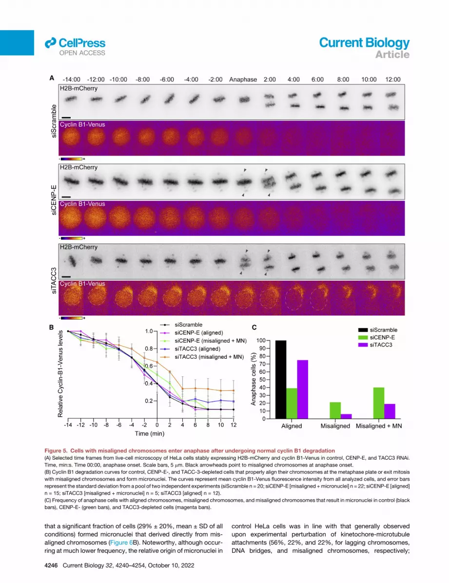
Figure 5. Cells with misaligned chromosomes enter anaphase after undergoing normal cyclin B1 degradation
(A) Selected time frames from live-cell microscopy of HeLa cells stably expressing H2B-mCherry and cyclin B1-Venus in control, CENP-E, and TACC3 RNAi.
Time, min:s. Time 00:00, anaphase onset. Scale bars, 5 mm. Black arrowheads point to misaligned chromosomes at anaphase onset.
(B) Cyclin B1 degradation curves for control, CENP-E-, and TACC-3-depleted cells that properly align their chromosomes at the metaphase plate or exit mitosis
with misaligned chromosomes and form micronuclei. The curves represent mean cyclin B1-Venus fluorescence intensity from all analyzed cells, and error bars
represent the standard deviation from a pool of two independent experiments (siScramble n = 20; siCENP-E [misaligned +micronuclei] n = 22; siCENP-E [aligned]
n = 15; siTACC3 [misaligned + micronuclei] n = 5; siTACC3 [aligned] n = 12).
(C) Frequency of anaphase cells with aligned chromosomes, misaligned chromosomes, and misaligned chromosomes that result in micronuclei in control (black
bars), CENP-E- (green bars), and TACC3-depleted cells (magenta bars).
llOPEN ACCESS Article
that a significant fraction of cells (29% ± 20%, mean ± SD of all
conditions) formed micronuclei that derived directly from mis-
aligned chromosomes (Figure 6B). Noteworthy, although occur-
ring at much lower frequency, the relative origin of micronuclei in
4246 Current Biology 32, 4240–4254, October 10, 2022
control HeLa cells was in line with that generally observed
upon experimental perturbation of kinetochore-microtubule
attachments (56%, 22%, and 22%, for lagging chromosomes,
DNA bridges, and misaligned chromosomes, respectively;

Figure 6. Although most micronuclei originate from
anaphase lagging chromosomes, misaligned chromo-
somes are a stronger predictor of micronuclei formation
(A) Examples of time-lapse sequences illustrating the different ori-
gins of micronuclei. Time, min:s. Time 00:00, anaphase onset. White
arrowheads track misaligned chromosomes, DNA bridges, or lag-
ging chromosomes until they eventually form micronuclei. Pixels
were saturated for optimal visualization of misaligned chromosomes,
DNA bridges, and lagging chromosomes. Scale bars, 5 mm.
(B) Frequency of daughter cells with micronuclei that derived either
from lagging chromosomes (black bars), DNA bridges (green bars),
or misaligned chromosomes (magenta bars) under the specified
conditions (siScramble, n = 1,700;MonWO, n = 327; siAstrin, n = 423;
siBub1, n = 457; siKif18a, n = 540; siCENP-N, n = 422; siSka1, n =
395; siTACC3, n = 485; siNsl1, n = 400; siSka3, n = 383; siZw10, n =
404; siNdc80, n = 440; siAurora A, n = 388; siCLERC, n = 263; siNuf2,
n = 428; siCENP-I, n = 389; siAurora B, n = 499; siDsn1, n = 688;
siCENP-E, n = 346; siSpc24, n = 418; siBubR1, n = 387; siSpc25, n =
425; siHURP_oligo1, n = 296; siHURP_oligo2, n = 200; siKNL1, n =
413; pool of 2 independent experiments for each siRNAi per condi-
tion, with the exception of Aurora A and CLERC in which only 1
experiment for the second siRNAi was performed. All independent
experiments were pooled). *p% 0.05, **p% 0.01, ***p% 0.001, ****p
% 0.0001; ns, not significantly different from control; Fisher’s exact
two-tailed test; # highlight a possible off-target associated with
siRNA oligo 1 against HURP.
(C) Relative probability (sum of the 3 independent absolute proba-
bilities normalized to 1) of micronuclei formation from a lagging
chromosome (black bars), a DNA bridge (green bars), or amisaligned
chromosome (magenta bars) under the specified conditions (*p %
0.05, **p % 0.01, ***p % 0.001, ****p % 0.0001; ns, no significant
difference from what would be expected if all missegregation events
were equally likely to cause micronuclei in each experimental con-
dition; chi-square test).
llOPEN ACCESS
Current Biology 32, 4240–4254, October 10, 2022 4247
Article

(legend on next page)
llOPEN ACCESS
4248 Current Biology 32, 4240–4254, October 10, 2022
Article

llOPEN ACCESSArticle
n = 1,700 cells) (Figure 6B). This scenario changed significantly
both regarding frequency and origin of micronuclei uponmonas-
trol treatment and washout, which induces the formation of erro-
neous kinetochore-microtubule attachments leading to a high
frequency of anaphase lagging chromosomes47,48 (Figure 6B).
Next, we determined the respective probabilities of micronu-
clei formation given a specific condition, which can either be a
lagging chromosome, a DNA bridge, or a misaligned chromo-
some. Surprisingly, and despite the fact that most micronuclei
derived from anaphase lagging chromosomes, we found that
in unperturbed HeLa cells treated with a control siRNA the abso-
lute and relative probability of micronuclei formation from a mis-
aligned chromosome (0.92 and 0.70, respectively) clearly out-
competed the other two classes, including anaphase lagging
chromosomes (0.28 and 0.21, for absolute and relative probabil-
ities, respectively) (Figures 6C and S6A). Those probabilities
were significantly higher than what would be expected if all mis-
segregation events were equally likely to cause micronuclei
(p < 0.0001; chi-square test). Interestingly, although the experi-
mental perturbation of kinetochore-microtubule attachment sta-
bility did not result in gross alterations of the relative origins ofmi-
cronuclei, in most cases, it reverted or attenuated the much
higher probability of micronuclei formation from misaligned
chromosomes observed in unperturbed cells (Figures 6C and
S6A). This result is consistent with a role of stable kinetochore-
microtubule attachments in anaphase error correction and
micronuclei prevention from lagging chromosomes.46 One
noticeable exception was HURP, which gave rise to muchmilder
congression problems with no obvious bias for micronuclei for-
mation from misaligned chromosomes with a second siRNA, in
contrast with the original siRNA, despite equivalent depletion ef-
ficiency (Data S3). We suspect that the first siRNA against HURP
might by hitting the SAC component MAD2, which is highly
prone to off-targeting49 and would force HURP-depleted cells
with incomplete chromosome congression to enter anaphase
prematurely, directly leading to micronuclei formation due to
incomplete chromosome alignment. Overall, we conclude that,
although the majority of micronuclei originate from anaphase
lagging chromosomes, misaligned chromosomes are a stronger
predictor of micronuclei formation during HeLa cell division.
Micronuclei formation frommisaligned chromosomes isa frequent outcome in a cancer cell model ofchromosomal instability, but not in non-transformednear-diploid cellsNext, we set out to investigate the origin of micronuclei that
form spontaneously during cell division in RPE-1 and
Figure 7. Micronuclei formation from misaligned chromosomes is a fre
not in non-transformed cells
(A and B) Examples of time-lapse sequences illustrating possible origins of micron
White arrowheads track misaligned chromosomes, DNA bridges, or lagging ch
optimal visualization of misaligned chromosomes, DNA bridges, and lagging chr
(C) Frequency of RPE-1 and U2OS daughter cells with micronuclei that derived
misaligned chromosomes (magenta bars) in control, siCENP-E, and after monas
95; MonWO, n = 105. U2OS cells: control, n = 250; siCENP-E, n = 81; MonWO,
(D) Relative probability (sum of the 3 independent absolute probabilities normalize
bridge (green bars), or a misaligned chromosome (magenta bars) in RPE-1 and
washout. *p% 0.05, **p% 0.01, ***p% 0.001, ****p% 0.0001; ns, no significant di
likely to cause micronuclei in each experimental condition; chi-square test.
chromosomally unstable U2OS cells.50 To visualize the entire
chromosome set and spindle microtubules, these cell lines
were engineered to stably express histone H2B-GFP and
mRFP-a-tubulin and were inspected by 4D live-cell spin-
ning-disk confocal microscopy, with a temporal resolution be-
tween 30 s and 2 min (Figures 7A and 7B). In parallel, we pro-
moted chromosome missegregation by performing either
CENP-E depletion or a monastrol treatment and washout. Un-
perturbed RPE-1 cells showed only a residual (1.2%) forma-
tion of micronuclei after cell division and none derived from
a misaligned chromosome (Figure 7C). CENP-E depletion or
monastrol treatment/washout in RPE-1 cells significantly
increased the frequency of micronuclei formation (3.2% and
4.2%, respectively), most of which (71% and 89%, respec-
tively) derived from anaphase lagging chromosomes, and
only very few derived from a misaligned chromosome (2.1%
and 0.95% of the cells, respectively) (Figure 7C), further
demonstrating a robust chromosome alignment capacity in
normal cells. This scenario was strikingly different even in un-
perturbed U2OS cells, which formed micronuclei in 5.8% of
the cases, of which 53% derived from anaphase lagging chro-
mosomes, 14% from DNA bridges and 33% from misaligned
chromosomes (Figure 7C). Monastrol treatment/washout
only slightly increased (without statistical significance) the
percentage of dividing U2OS cells that formed micronuclei,
which in this case derived mostly from anaphase lagging
chromosomes (80%), likely due to an increase in merotelic at-
tachments.47 In contrast, CENP-E depletion in U2OS cells
significantly increased the percentage of dividing U2OS cells
that formed micronuclei (17.2%), of which 62% derived from
anaphase lagging chromosomes, 21% from DNA bridges
and 17% from misaligned chromosomes (Figure 7C).
We next determined the relative probabilities of micronuclei
formation from lagging chromosomes, DNA bridges, and mis-
aligned chromosomes scored in both RPE-1 and U2OS cells,
with and without CENP-E, as well as with and without monastrol
treatment/washout (Figure 7D) (for absolute probabilities, see
Figure S6B). In line with our previous observations in HeLa cells
(Figures 6C and S6A), this analysis revealed that misaligned
chromosomes have the highest absolute and relative probability
of resulting in micronuclei in unperturbed chromosomally unsta-
ble U2OS cells (0.63 and 0.80, respectively) (Figures 7D and
S6B). These probabilities were significantly higher than what
would be expected if all missegregation events were equally
likely to causemicronuclei (p < 0.0001; chi-square test). In agree-
ment with our findings in HeLa cells, both CENP-E depletion and
monastrol treatment/washout reverted this tendency in U2OS
quent outcome in a chromosomally unstable cancer cell model, but
uclei in RPE-1 (A) and U2OS (B) cells. Time, min:s. Time 00:00, anaphase onset.
romosomes until they eventually form micronuclei. Pixels were saturated for
omosomes. Scale bars, 5 mm.
either from lagging chromosomes (black bars), DNA bridges (green bars), or
trol treatment/washout (MonWO). RPE-1 cells: control, n = 163; siCENP-E, n =
n = 49 (Fisher’s exact two-tailed test).
d to 1) of micronuclei formation from a lagging chromosome (black bars), a DNA
U2OS cells in control and after CENP-E depletion or monastrol treatment/
fference fromwhat would be expected if all missegregation events were equally
Current Biology 32, 4240–4254, October 10, 2022 4249

llOPEN ACCESS Article
cells, likely due to a significant increase in the frequency of
anaphase lagging chromosomes (Figures 7C, 7D, and S6B).47
Most striking, and in sharp contrast to unperturbed HeLa and
U2OS cells, unperturbed RPE-1 cells always entered anaphase
after completing chromosome alignment and, consequently,
no micronuclei from misaligned chromosomes were ever
detected in our recordings (Figures 7D and S6B). Likewise,
human primary fibroblasts were previously shown to never enter
anaphase with misaligned chromosomes even after nocodazole
treatment and washout, and the resulting lagging chromosomes
appeared during anaphase after completing chromosome align-
ment during metaphase.44 We concluded that spontaneousmis-
aligned chromosomes in unperturbed chromosomally unstable
cancer cell models, but not in non-transformed near-diploid
cells, have a strong probability to missegregate and result in
micronuclei.
Misaligned chromosomes in chromosomally unstablecancer cells have hyper-stabilized kinetochore-microtubule attachmentsChromosomally unstable cancer cells have hyper-stabilized
kinetochore-microtubule attachments and a poor error correc-
tion capacity.50,51 To investigate whether increased kineto-
chore-microtubule attachment stability in chromosomally unsta-
ble cancer cells allows misaligned chromosomes to satisfy the
SAC, we implemented a protocol that promotes the formation
of few misaligned chromosomes after nocodazole treatment
and washout (Figure S7A) (STAR Methods), followed by quanti-
fication of fluorescence intensity after a nocodazole shock to
completely depolymerizemicrotubules in fixed cells (Figure S7A).
Both qualitative and quantitative analyses revealed that, under
these experimental conditions, kinetochore microtubules in
chromosomally unstable U2OS cells are more resistant to depo-
lymerization when compared with non-transformed near-diploid
RPE-1 cells (Figures S7A and S7B). Measurement of the respec-
tive half-life of polymerized tubulin, confirmed �2-fold increase
in U2OS cells relative to RPE1 cells (Figures S7A and S7B).
These results provide an explanation for the inefficient correction
of few misaligned chromosomes that eventually satisfy the SAC
in a chromosomally unstable cancer cell model and thus may
represent important drivers of chromosomal instability and
micronuclei formation in human cancers.
DISCUSSION
It is currently thought that anaphase lagging chromosomes re-
sulting from erroneous merotelic attachments that satisfy the
SAC are major drivers of genomic instability in human can-
cers.52,53 Although anaphase lagging chromosomes resulting
from merotelic attachments rarely missegregate,54,55 they may
fail to incorporate into the respective daughter nuclei during
cell division and result in the formation of micronuclei. Micronu-
clei were recently implicated as key intermediates of chromo-
thripsis, a series of massive genomic rearrangements that may
drive rapid tumor evolution and account for acquired drug resis-
tance and oncogene activation.43,56–59 We now show that
although most micronuclei derive from anaphase lagging chro-
mosomes, simply because these events occur at a very high fre-
quency in chromosomally unstable cancer cells,54 misaligned
4250 Current Biology 32, 4240–4254, October 10, 2022
chromosomes that satisfy the SAC often directly missegregate
(without lagging behind in anaphase) and have the highest prob-
ability to form micronuclei, specifically in human cancer cell
models (see graphical abstract). This is consistent with recent
high-resolution live-cell studies in both cancer and non-cancer
human cells that showed that the vast majority of lagging chro-
mosomes have a transient nature and are corrected during
anaphase by an Aurora-B-dependent mechanism that prevents
micronuclei formation,46,60 and the relatively low frequency of
micronuclei formation even after induction of massive chromo-
some segregation errors by experimental abrogation of the
SAC.61,62
Defects in chromosome alignment are normally avoided by
increased Aurora B activity at centromeres of misaligned chro-
mosomes.27 However, the correction of erroneous attachments
underlying some chromosome alignment defects (e.g., syntelic
attachments) appears to be less robust in cancer cells that
also show overly stabilized kinetochore-microtubule attach-
ments.50,51 Indeed, RPE-1 cells treated with microtubule-target-
ing drugs at concentrations that stabilize microtubules satisfy
the SAC in the presence of misaligned chromosomes and do
so faster under conditions that promote the formation of syntelic
attachments.62–64 In addition to direct missegregation from
misaligned chromosomes, late-aligning chromosomes are also
more prone to lag behind in anaphase and missegregate at
higher frequencies in human cancer cells, or upon SAC inactiva-
tion or stabilization of incorrect kinetochore-microtubule attach-
ments in normal cells.62,65 Together with the fact that non-trans-
formed human cells rely on a robust p53-dependent mechanism
that limits the proliferation of aneuploid cells,66 the present work
helps to explain how spontaneous misaligned chromosomes in
cancer cells eventually satisfy the SAC and may constitute a
direct route to chromosomal instability.
This work also unveils a wide range of genetic perturbations
that predispose for these events and might account for the un-
derlying chromosomal and genomic instability commonly
observed in human cancers. A paramount case is the perturba-
tion of CENP-E function that has been linked to tumorigenesis
in vivo.31 Previous studies have shown that �40% of CENP-E-
depleted HeLa cells enter anaphase with misaligned chromo-
somes.27,28 Fixed-cell analysis revealed that these misaligned
chromosomes accumulate Mad2, but micronuclei generated
from CENP-E-depleted cells did not, suggesting that mis-
aligned chromosomes satisfy the SAC.27 Although suggestive,
the origin of the scored micronuclei was not determined in
these fixed-cell experiments, and so it remains possible that
the scored micronuclei did not derive directly from misaligned
chromosomes (they may alternatively derive from anaphase
lagging chromosomes; Figures 6B and 6C), and cells with mis-
aligned chromosomes entered anaphase without satisfying the
SAC. Indeed, previous experiments in fixed CENP-E KO MEFs
revealed continued localization of SAC proteins at misaligned
chromosomes seen in anaphase cells, suggesting ongoing
SAC signaling.26 Our live-cell imaging of Mad2-GFP upon
CENP-E depletion in HeLa cells, supported by quantitative an-
alyses in fixed cells soon after anaphase onset, show that
Mad1/Mad2 dissociate from kinetochores of misaligned chro-
mosomes in cells that entered anaphase, suggesting SAC
satisfaction. Moreover, live-cell imaging revealed a normal

llOPEN ACCESSArticle
degradation kinetics of cyclin B1 in CENP-E-depleted or unre-
lated TACC3-depleted cells that entered anaphase with
misaligned chromosomes. This contrasts with the pattern
observed upon mitotic slippage, in which mitotic cells that
cannot satisfy the SAC exit mitosis with high Mad1/Mad2 levels
at kinetochores and after very slow and prolonged degradation
of cyclin B1.39,64,67 Combined, these data provide direct evi-
dence that, at least under certain conditions, cancer cells
with misaligned chromosomes may enter anaphase after SAC
satisfaction and have a high risk of forming micronuclei (see
graphical abstract). In line with these findings, recent experi-
ments in which CENP-E activity was inhibited in human
RPE-1 cells suggest that endomembrane ‘‘ensheathing’’ of
misaligned chromosomes may facilitate micronuclei formation
and delay SAC satisfaction.68
Our systematic analysis of more than 100 different molecular
perturbations further indicates that entering anaphase with mis-
aligned chromosomes might be a frequent outcome in cancer
cells. In particular, perturbations such as CENP-E or Kif18a
depletion were largely compatible with cell viability, despite
the high incidence of cells that entered anaphase in the pres-
ence of misaligned chromosomes. This contrasts with more
drastic scenarios that result from perturbation of end-on kinet-
ochore-microtubule attachments (e.g., depletion of KMN com-
ponents) that often result in massive chromosome missegrega-
tion and cell death. Noteworthy, while the loss of Kif18a, which
causes asynchronous segregation of misaligned chromosomes
due to loss of interchromosome compaction during anaphase,
does not promote chromosomal instability and tumorigen-
esis,10,69 the loss of CENP-E that typically originates one or
few pole-proximal chromosomes directly leads to aneuploidy
and the spontaneous formation of lymphomas and lung tumors
in aged animals.26,31 These data suggest that the origin and
properties of the resulting micronuclei is genetically determined
and might have implications for the propensity to undergo
massive chromosome rearrangements, such as those
commonly observed in chromothripsis. Interestingly, micronu-
clei derived from segregation errors associated with Kif18a
loss of function appear to form stable nuclear envelopes.69
However, because misaligned chromosomes that form after
perturbation of CENP-E function are brought very close to
Aurora A activity at the spindle poles,2 this might compromise
proper nuclear envelope formation.70,71 In agreement, micronu-
clei derived from misaligned chromosomes after CENP-E
perturbation were recently suggested to activate the cGAS-
STING pathway in cancer cells.72 Thus, cellular response to mi-
cronuclei might depend on their relative origin. Overall, our find-
ings incite for an in-depth characterization of the properties and
fate of micronuclei of different origins, while evaluating their
respective potential to drive and/or sustain cell transformation.
In this regard, our study indicates that micronuclei formation
from misaligned chromosomes appears to be a specific
outcome of cancer cells and may represent a possible thera-
peutic opportunity in human cancers.
STAR+METHODS
Detailed methods are provided in the online version of this paper
and include the following:
d KEY RESOURCES TABLE
d RESOURCE AVAILABILITY
B Lead contact
B Materials availability
B Data and code availability
d EXPERIMENTAL MODEL AND SUBJECT DETAILS
B Cell lines
d METHOD DETAILS
B High-content live-cell imaging RNAi screen
B Other RNAi experiments
B Drug treatments
B High-resolution time-lapse microscopy
B Immunofluorescence microscopy
B Western Blotting
d QUANTIFICATION AND STATISTICAL ANALYSIS
B Quantification of mitotic errors
B Quantitative image analysis
B Statistical analysis
SUPPLEMENTAL INFORMATION
Supplemental information can be found online at https://doi.org/10.1016/j.
cub.2022.08.026.
ACKNOWLEDGMENTS
We thank Andr�e Maia for technical assistance. A.M.G., M.N.-C., and J.M.-M.
are recipients of PhD studentships from Fundacao para a Ciencia e a
Tecnologia (FCT) (SFRH/BD/130938/2017, SFRH/BD/117063/2016, and
2021.07945.BD). This work was funded by the European Research Council
consolidator grant CODECHECK, under the European Union’s Horizon 2020
research and innovation program (681443), FCT (PTDC/MED-ONC/3479/
2020), and a La Caixa Health Research Grant (LCF/PR/HR21/52410025).
AUTHOR CONTRIBUTIONS
Conceptualization, supervision, project administration, and funding acquisi-
tion, H.M.; methodology, A.M.G., B.O., M.N.-C., F.D.S., and C.F.; investiga-
tion, formal analysis, and validation, A.M.G., B.O., M.N.-C., F.D.S., C.F.,
J.M.-M., C.L., and H.M.; visualization, A.M.G., B.O., M.N.-C., F.D.S., C.F.,
J.M.-M., and H.M.; writing – original draft, A.M.G. and H.M.; writing – review
& editing, A.M.G., B.O., M.N.-C., C.F., and H.M.
DECLARATION OF INTERESTS
B.O. declares that he is a consultant specialist at Volastra Therapeutics.
Received: February 14, 2022
Revised: July 22, 2022
Accepted: August 11, 2022
Published: September 2, 2022
REFERENCES
1. Maiato, H., Gomes, A.M., Sousa, F., and Barisic, M. (2017). Mechanisms
of chromosome congression during mitosis. Biology 6, 13. https://doi.
org/10.3390/biology6010013.
2. Barisic, M., Aguiar, P., Geley, S., and Maiato, H. (2014). Kinetochore mo-
tors drive congression of peripheral polar chromosomes by overcoming
random arm-ejection forces. Nat. Cell Biol. 16, 1249–1256. https://doi.
org/10.1038/ncb3060.
3. Yang, Z., Tulu, U.S., Wadsworth, P., and Rieder, C.L. (2007). Kinetochore
dynein is required for chromosome motion and congression independent
of the spindle checkpoint. Curr. Biol. 17, 973–980.
Current Biology 32, 4240–4254, October 10, 2022 4251

llOPEN ACCESS Article
4. Vorozhko, V.V., Emanuele, M.J., Kallio, M.J., Stukenberg, P.T., and
Gorbsky, G.J. (2008). Multiple mechanisms of chromosome movement
in vertebrate cells mediated through the Ndc80 complex and dynein/dy-
nactin. Chromosoma 117, 169–179.
5. Kapoor, T.M., Lampson, M.A., Hergert, P., Cameron, L., Cimini, D.,
Salmon, E.D., McEwen, B.F., and Khodjakov, A. (2006). Chromosomes
can congress to the metaphase plate before biorientation. Science 311,
388–391. https://doi.org/10.1126/science.1122142.
6. Wood, K.W., Sakowicz, R., Goldstein, L.S., and Cleveland, D.W. (1997).
CENP-E is a plus end-directed kinetochore motor required for metaphase
chromosome alignment. Cell 91, 357–366.
7. Barisic, M., Silva e Sousa, R., Tripathy, S.K., Magiera, M.M., Zaytsev, A.V.,
Pereira, A.L., Janke, C., Grishchuk, E.L., and Maiato, H. (2015). Mitosis.
Microtubule detyrosination guides chromosomes during mitosis.
Science 348, 799–803. https://doi.org/10.1126/science.aaa5175.
8. Barisic, M., and Maiato, H. (2016). The tubulin code: a navigation system
for chromosomes during mitosis. Trends Cell Biol. 26, 766–775. https://
doi.org/10.1016/j.tcb.2016.06.001.
9. Orr, B., andMaiato, H. (2019). No chromosome left behind: the importance
of metaphase alignment for mitotic fidelity. J. Cell Biol. 218, 1086–1088.
https://doi.org/10.1083/jcb.201902041.
10. Fonseca, C.L., Malaby, H.L.H., Sepaniac, L.A., Martin, W., Byers, C.,
Czechanski, A., Messinger, D., Tang, M., Ohi, R., Reinholdt, L.G., et al.
(2019). Mitotic chromosome alignment ensures mitotic fidelity by promot-
ing interchromosomal compaction during anaphase. J. Cell Biol. 218,
1148–1163. https://doi.org/10.1083/jcb.201807228.
11. Matos, I., Pereira, A.J., Lince-Faria, M., Cameron, L.A., Salmon, E.D., and
Maiato, H. (2009). Synchronizing chromosome segregation by flux-depen-
dent force equalization at kinetochores. J. Cell Biol. 186, 11–26. https://
doi.org/10.1083/jcb.200904153.
12. Lara-Gonzalez, P., Pines, J., and Desai, A. (2021). Spindle assembly
checkpoint activation and silencing at kinetochores. Semin. Cell Dev.
Biol. 117, 86–98. https://doi.org/10.1016/j.semcdb.2021.06.009.
13. Rieder, C.L., Schultz, A., Cole, R., and Sluder, G. (1994). Anaphase onset
in vertebrate somatic cells is controlled by a checkpoint that monitors sis-
ter kinetochore attachment to the spindle. J. Cell Biol. 127, 1301–1310.
14. Taylor, S.S., and McKeon, F. (1997). Kinetochore localization of murine
Bub1 is required for normal mitotic timing and checkpoint response to
spindle damage. Cell 89, 727–735. https://doi.org/10.1016/s0092-
8674(00)80255-x.
15. Clute, P., and Pines, J. (1999). Temporal and spatial control of cyclin B1
destruction in metaphase. Nat. Cell Biol. 1, 82–87. https://doi.org/10.
1038/10049.
16. Howell, B.J., Hoffman, D.B., Fang, G., Murray, A.W., and Salmon, E.D.
(2000). Visualization of Mad2 dynamics at kinetochores, along spindle fi-
bers, and at spindle poles in living cells. J. Cell Biol. 150, 1233–1250.
https://doi.org/10.1083/jcb.150.6.1233.
17. Maresca, T.J., and Salmon, E.D. (2009). Intrakinetochore stretch is asso-
ciated with changes in kinetochore phosphorylation and spindle assembly
checkpoint activity. J. Cell Biol. 184, 373–381. https://doi.org/10.1083/jcb.
200808130.
18. Murray, A.W. (2011). A brief history of error. Nat. Cell Biol. 13, 1178–1182.
https://doi.org/10.1038/ncb2348.
19. Pesenti, M.E., Weir, J.R., and Musacchio, A. (2016). Progress in the struc-
tural and functional characterization of kinetochores. Curr. Opin. Struct.
Biol. 37, 152–163. https://doi.org/10.1016/j.sbi.2016.03.003.
20. Acquaviva, C., Herzog, F., Kraft, C., and Pines, J. (2004). The anaphase
promoting complex/cyclosome is recruited to centromeres by the spindle
assembly checkpoint. Nat. Cell Biol. 6, 892–898. https://doi.org/10.1038/
ncb1167.
21. Pereira, A.J., and Maiato, H. (2012). Maturation of the kinetochore-micro-
tubule interface and the meaning of metaphase. Chromosome Res. 20,
563–577. https://doi.org/10.1007/s10577-012-9298-8.
4252 Current Biology 32, 4240–4254, October 10, 2022
22. Hein, J.B., and Nilsson, J. (2014). Stable MCC binding to the APC/C is
required for a functional spindle assembly checkpoint. EMBO Rep. 15,
264–272. https://doi.org/10.1002/embr.201337496.
23. Cai, S., O’Connell, C.B., Khodjakov, A., and Walczak, C.E. (2009).
Chromosome congression in the absence of kinetochore fibres. Nat.
Cell Biol. 11, 832–838. https://doi.org/10.1038/ncb1890.
24. Khodjakov, A., Cole, R.W., McEwen, B.F., Buttle, K.F., and Rieder, C.L.
(1997). Chromosome fragments possessing only one kinetochore can
congress to the spindle equator. J. Cell Biol. 136, 229–240.
25. Rieder, C.L., Davison, E.A., Jensen, L.C., Cassimeris, L., and Salmon, E.D.
(1986). Oscillatory movements of monooriented chromosomes and their
position relative to the spindle pole result from the ejection properties of
the aster and half-spindle. J. Cell Biol. 103, 581–591.
26. Weaver, B.A., Bonday, Z.Q., Putkey, F.R., Kops, G.J., Silk, A.D., and
Cleveland, D.W. (2003). Centromere-associated protein-E is essential for
the mammalian mitotic checkpoint to prevent aneuploidy due to single
chromosome loss. J. Cell Biol. 162, 551–563. https://doi.org/10.1083/
jcb.200303167.
27. Maia, A.F., Feijao, T., Vromans, M.J., Sunkel, C.E., and Lens, S.M. (2010).
Aurora B kinase cooperates with CENP-E to promote timely anaphase
onset. Chromosoma 119, 405–413. https://doi.org/10.1007/s00412-010-
0265-x.
28. Tanudji, M., Shoemaker, J., L’Italien, L., Russell, L., Chin, G., and
Schebye, X.M. (2004). Gene silencing of CENP-E by small interfering
RNA in HeLa cells leads to missegregation of chromosomes after a mitotic
delay. Mol. Biol. Cell 15, 3771–3781. https://doi.org/10.1091/mbc.e03-07-
0482.
29. Kops, G.J., Foltz, D.R., and Cleveland, D.W. (2004). Lethality to human
cancer cells through massive chromosome loss by inhibition of the mitotic
checkpoint. Proc. Natl. Acad. Sci. USA 101, 8699–8704. https://doi.org/
10.1073/pnas.0401142101.
30. Silk, A.D., Zasadil, L.M., Holland, A.J., Vitre, B., Cleveland, D.W., and
Weaver, B.A. (2013). Chromosome missegregation rate predicts whether
aneuploidy will promote or suppress tumors. Proc. Natl. Acad. Sci. USA
110, E4134–E4141. https://doi.org/10.1073/pnas.1317042110.
31. Weaver, B.A., Silk, A.D., Montagna, C., Verdier-Pinard, P., and Cleveland,
D.W. (2007). Aneuploidy acts both oncogenically and as a tumor suppres-
sor. Cancer Cell 11, 25–36. https://doi.org/10.1016/j.ccr.2006.12.003.
32. Uehara, R., Nozawa, R.-S., Tomioka, A., Petry, S., Vale, R.D., Obuse, C.,
and Goshima, G. (2009). The augmin complex plays a critical role in spin-
dle microtubule generation for mitotic progression and cytokinesis in hu-
man cells. Proc. Natl. Acad. Sci. USA 106, 6998–7003. https://doi.org/
10.1073/pnas.0901587106.
33. Logarinho, E., Maffini, S., Barisic, M., Marques, A., Toso, A., Meraldi, P.,
and Maiato, H. (2012). CLASPs prevent irreversible multipolarity by
ensuring spindle-pole resistance to traction forces during chromosome
alignment. Nat. Cell Biol. 14, 295–303. https://doi.org/10.1038/ncb2423.
34. Gaitanos, T.N., Santamaria, A., Jeyaprakash, A.A., Wang, B., Conti, E.,
and Nigg, E.A. (2009). Stable kinetochore-microtubule interactions
depend on the Ska complex and its new component Ska3/C13Orf3.
EMBO J. 28, 1442–1452. https://doi.org/10.1038/emboj.2009.96.
35. Cheeseman, I.M., Chappie, J.S., Wilson-Kubalek, E.M., and Desai, A.
(2006). The conserved KMN network constitutes the core microtubule-
binding site of the kinetochore. Cell 127, 983–997. https://doi.org/10.
1016/j.cell.2006.09.039.
36. Beck,M., and Hurt, E. (2017). The nuclear pore complex: understanding its
function through structural insight. Nat. Rev. Mol. Cell Biol. 18, 73–89.
https://doi.org/10.1038/nrm.2016.147.
37. Rieder, C.L., and Maiato, H. (2004). Stuck in division or passing through:
what happens when cells cannot satisfy the spindle assembly checkpoint.
Dev. Cell 7, 637–651. https://doi.org/10.1016/j.devcel.2004.09.002.
llOPEN ACCESSArticle
38. Brito, D.A., and Rieder, C.L. (2006). Mitotic checkpoint slippage in humans
occurs via cyclin B destruction in the presence of an active checkpoint.
Curr. Biol. 16, 1194–1200. https://doi.org/10.1016/j.cub.2006.04.043.
39. Gascoigne, K.E., and Taylor, S.S. (2008). Cancer cells display profound
intra- and interline variation following prolonged exposure to antimitotic
drugs. Cancer Cell 14, 111–122. https://doi.org/10.1016/j.ccr.2008.07.
002.
40. Novais-Cruz, M., Alba Abad, M., van IJcken, W.F., Galjart, N.,
Jeyaprakash, A.A., Maiato, H., and Ferras, C. (2018). Mitotic progression,
arrest, exit or death relies on centromere structural integrity, rather than de
novo transcription. eLife 7, e36898. https://doi.org/10.7554/eLife.36898.
41. Gergely, F., Draviam, V.M., and Raff, J.W. (2003). The ch-TOG/XMAP215
protein is essential for spindle pole organization in human somatic cells.
Genes Dev. 17, 336–341. https://doi.org/10.1101/gad.245603.
42. Afonso, O., Castellani, C.M., Cheeseman, L.P., Ferreira, J.G., Orr, B.,
Ferreira, L.T., Chambers, J.J., Morais-de-Sa, E., Maresca, T.J., and
Maiato, H. (2019). Spatiotemporal control of mitotic exit during anaphase
by an aurora B-Cdk1 crosstalk. eLife 8, e47646. https://doi.org/10.7554/
eLife.47646.
43. Crasta, K., Ganem, N.J., Dagher, R., Lantermann, A.B., Ivanova, E.V., Pan,
Y., Nezi, L., Protopopov, A., Chowdhury, D., and Pellman, D. (2012). DNA
breaks and chromosome pulverization from errors in mitosis. Nature 482,
53–58. https://doi.org/10.1038/nature10802.
44. Cimini, D., Fioravanti, D., Salmon, E.D., and Degrassi, F. (2002). Merotelic
kinetochore orientation versus chromosomemono-orientation in the origin
of lagging chromosomes in human primary cells. J. Cell Sci. 115, 507–515.
45. Umbreit, N.T., Zhang, C.Z., Lynch, L.D., Blaine, L.J., Cheng, A.M.,
Tourdot, R., Sun, L., Almubarak, H.F., Judge, K., Mitchell, T.J., et al.
(2020). Mechanisms generating cancer genome complexity from a single
cell division error. Science 368, eaba0712. https://doi.org/10.1126/sci-
ence.aba0712.
46. Orr, B., De Sousa, F., Gomes, A.M., Afonso, O., Ferreira, L.T., Figueiredo,
A.C., and Maiato, H. (2021). An anaphase surveillance mechanism pre-
vents micronuclei formation from frequent chromosome segregation er-
rors. Cell Rep. 37, 109783. https://doi.org/10.1016/j.celrep.2021.109783.
47. Cimini, D., Moree, B., Canman, J.C., and Salmon, E.D. (2003). Merotelic
kinetochore orientation occurs frequently during early mitosis in mamma-
lian tissue cells and error correction is achieved by two different mecha-
nisms. J. Cell Sci. 116, 4213–4225. https://doi.org/10.1242/jcs.00716.
48. Lampson, M.A., Renduchitala, K., Khodjakov, A., and Kapoor, T.M. (2004).
Correcting improper chromosome-spindle attachments during cell divi-
sion. Nat. Cell Biol. 6, 232–237. https://doi.org/10.1038/ncb1102.
49. Sigoillot, F.D., Lyman, S., Huckins, J.F., Adamson, B., Chung, E.,
Quattrochi, B., and King, R.W. (2012). A bioinformatics method identifies
prominent off-targeted transcripts in RNAi screens. Nat. Methods 9,
363–366. https://doi.org/10.1038/nmeth.1898.
50. Bakhoum, S.F., Genovese, G., and Compton, D.A. (2009). Deviant kineto-
chore microtubule dynamics underlie chromosomal instability. Curr. Biol.
19, 1937–1942. https://doi.org/10.1016/j.cub.2009.09.055.
51. Salimian, K.J., Ballister, E.R., Smoak, E.M., Wood, S., Panchenko, T.,
Lampson, M.A., and Black, B.E. (2011). Feedback control in sensing chro-
mosome biorientation by the aurora B kinase. Curr. Biol. 21, 1158–1165.
https://doi.org/10.1016/j.cub.2011.06.015.
52. Bakhoum, S.F., and Cantley, L.C. (2018). The multifaceted role of chromo-
somal instability in cancer and its microenvironment. Cell 174, 1347–1360.
https://doi.org/10.1016/j.cell.2018.08.027.
53. Soto, M., Raaijmakers, J.A., and Medema, R.H. (2019). Consequences of
genomic diversification induced by segregation errors. Trends Genet. 35,
279–291. https://doi.org/10.1016/j.tig.2019.01.003.
54. Thompson, S.L., and Compton, D.A. (2011). Chromosomemissegregation
in human cells arises through specific types of kinetochore-microtubule
attachment errors. Proc. Natl. Acad. Sci. USA 108, 17974–17978.
https://doi.org/10.1073/pnas.1109720108.
55. Cimini, D., Cameron, L.A., and Salmon, E.D. (2004). Anaphase spindle me-
chanics prevent mis-segregation of merotelically oriented chromosomes.
Curr. Biol. 14, 2149–2155. https://doi.org/10.1016/j.cub.2004.11.029.
56. Zhang, C.Z., Spektor, A., Cornils, H., Francis, J.M., Jackson, E.K., Liu, S.,
Meyerson, M., and Pellman, D. (2015). Chromothripsis from DNA damage
in micronuclei. Nature 522, 179–184. https://doi.org/10.1038/na-
ture14493.
57. Shoshani, O., Brunner, S.F., Yaeger, R., Ly, P., Nechemia-Arbely, Y., Kim,
D.H., Fang, R., Castillon, G.A., Yu, M., Li, J.S.Z., et al. (2021).
Chromothripsis drives the evolution of gene amplification in cancer.
Nature 591, 137–141. https://doi.org/10.1038/s41586-020-03064-z.
58. Stephens, P.J., Greenman, C.D., Fu, B., Yang, F., Bignell, G.R., Mudie,
L.J., Pleasance, E.D., Lau, K.W., Beare, D., Stebbings, L.A., et al. (2011).
Massive genomic rearrangement acquired in a single catastrophic event
during cancer development. Cell 144, 27–40. https://doi.org/10.1016/j.
cell.2010.11.055.
59. Janssen, A., van der Burg, M., Szuhai, K., Kops, G.J., and Medema, R.H.
(2011). Chromosome segregation errors as a cause of DNA damage and
structural chromosome aberrations. Science 333, 1895–1898. https://
doi.org/10.1126/science.1210214.
60. Sen, O., Harrison, J.U., Burroughs, N.J., and McAinsh, A.D. (2021).
Kinetochore life histories reveal the origins of chromosome mis-segrega-
tion and correction mechanisms. Preprint at bioRxiv. https://doi.org/10.
1101/2021.03.30.436326.
61. Cohen-Sharir, Y., McFarland, J.M., Abdusamad, M., Marquis, C.,
Bernhard, S.V., Kazachkova, M., Tang, H., Ippolito, M.R., Laue, K.,
Zerbib, J., et al. (2021). Aneuploidy renders cancer cells vulnerable to
mitotic checkpoint inhibition. Nature 590, 486–491. https://doi.org/10.
1038/s41586-020-03114-6.
62. Klaasen, S.J., Truong, M.A., van Jaarsveld, R.H., Koprivec, I., �Stimac, V.,
de Vries, S.G., Risteski, P., Kodba, S., Vuku�si�c, K., de Luca, K.L., et al.
(2022). Nuclear chromosome locations dictate segregation error fre-
quencies. Nature 607, 604–609. https://doi.org/10.1038/s41586-022-
04938-0.
63. Yang, Z., Kenny, A.E., Brito, D.A., and Rieder, C.L. (2009). Cells satisfy the
mitotic checkpoint in Taxol, and do so faster in concentrations that stabi-
lize syntelic attachments. J. Cell Biol. 186, 675–684. https://doi.org/10.
1083/jcb.200906150.
64. Brito, D.A., Yang, Z., and Rieder, C.L. (2008). Microtubules do not promote
mitotic slippage when the spindle assembly checkpoint cannot be satis-
fied. J. Cell Biol. 182, 623–629. https://doi.org/10.1083/jcb.200805072.
65. Kuniyasu, K., Iemura, K., and Tanaka, K. (2018). Delayed chromosome
alignment to the spindle equator increases the rate of chromosomemisse-
gregation in cancer cell lines. Biomolecules 9, 10. https://doi.org/10.3390/
biom9010010.
66. Thompson, S.L., and Compton, D.A. (2010). Proliferation of aneuploid hu-
man cells is limited by a p53-dependent mechanism. J. Cell Biol. 188,
369–381. https://doi.org/10.1083/jcb.200905057.
67. Canman, J.C., Sharma, N., Straight, A., Shannon, K.B., Fang, G., and
Salmon, E.D. (2002). Anaphase onset does not require the microtubule-
dependent depletion of kinetochore and centromere-binding proteins.
J. Cell Sci. 115, 3787–3795. https://doi.org/10.1242/jcs.00057.
68. Ferrandiz, N., Downie, L., Starling, G.P., and Royle, S.J. (2021).
Endomembranes promote chromosome missegregation by ensheathing
misaligned chromosomes. Preprint at bioRxiv. https://doi.org/10.1101/
2021.04.23.441091.
69. Sepaniac, L.A., Martin, W., Dionne, L.A., Stearns, T.M., Reinholdt, L.G.,
and Stumpff, J. (2021). Micronuclei in Kif18a mutant mice form stable mi-
cronuclear envelopes and do not promote tumorigenesis. J. Cell Biol. 220,
e202101165. https://doi.org/10.1083/jcb.202101165.
70. Portier, N., Audhya, A., Maddox, P.S., Green, R.A., Dammermann, A.,
Desai, A., and Oegema, K. (2007). A microtubule-independent role for
Current Biology 32, 4240–4254, October 10, 2022 4253

llOPEN ACCESS Article
centrosomes and aurora a in nuclear envelope breakdown. Dev. Cell 12,
515–529. https://doi.org/10.1016/j.devcel.2007.01.019.
71. Hachet, V., Canard, C., and Gonczy, P. (2007). Centrosomes promote
timely mitotic entry in C. elegans embryos. Dev. Cell 12, 531–541.
https://doi.org/10.1016/j.devcel.2007.02.015.
72. Hakozaki, Y., Kashima, Y., Morita, T.Y., Tanaka, K., Kobayashi, S.S., and
Ohashi, A. (2021). Abstract 1030: CENP-E inhibition generates micronu-
cleus formation activating the cGAS-STING pathway in cancer cells.
Cancer Res. 81, 1030. https://doi.org/10.1158/1538-7445.AM2021-1030.
4254 Current Biology 32, 4240–4254, October 10, 2022
73. Maffini, S., Maia, A.R.R., Manning, A.L., Maliga, Z., Pereira, A.L.,
Junqueira, M., et al. (2009). Motor-independent targeting of CLASPs to ki-
netochores by CENP-E promotes microtubule turnover and poleward flux.
Curr. Biol. 19, 1566–1572.
74. Schweizer, N., Ferras, C., Kern, D.M., Logarinho, E., Cheeseman, I.M., and
Maiato, H. (2013). Spindle assembly checkpoint robustness requires Tpr-
mediated regulation of Mad1/Mad2 proteostasis. J. Cell Biol. 203,
883–893. https://doi.org/10.1083/jcb.201309076.

llOPEN ACCESSArticle
STAR+METHODS
KEY RESOURCES TABLE
REAGENT or RESOURCE SOURCE IDENTIFIER
Antibodies
mouse anti-Mad1 Merck Millipore Cat#MABE867; RRID:AB_2910099
mouse anti-a-tubulin Sigma T5168; RRID:AB_477579
rabbit anti-b-tubulin Abcam Ab6046; RRID:AB_2210370
guinea pig anti-CENP-C MBL International PD030; RRID:AB_10693556
mouse anti-Aim1 BD Biosciences Cat#611083; RRID:AB_398396
mouse anti-Hec1 (9GA) Abcam Ab3613; RRID:AB_303949
mouse anti-Dsn1 Gift from A. Musacchio N/A
mouse anti-ATRX Santa Cruz Biotechnology sc-55584; RRID:AB_831012
rabbit anti-CEP72 Novus Biologicals NB100-60661; RRID:AB_920952
mouse anti-GAK R&D Systems MAB6918; RRID:AB_10972463
rabbit anti-WDHD1/And-1 Novus Biologicals NBP1-89091; RRID:AB_11041095
rabbit anti-Aurora A Novus Biologicals NB100-267; RRID:AB_10002481
rabbit anti-HURP Gift from P. Meraldi N/A
mouse anti-INCENP Santa Cruz Biotechnology sc-376514; RRID:AB_11149761
mouse anti-Sgo1 (F-8) Santa Cruz Biotechnology sc-393993; RRID:AB_2910101
rabbit anti-DHC ThermoFisher Scientific PA5-49373; RRID:AB_2634827
sheep anti-Bub1 Gift from S. Taylor N/A
rabbit anti-Septin-2 Novus Biologicals NBP1-85212; RRID:AB_11002313
mouse anti-Ska3 Santa Cruz Biotechnology sc-390326; RRID:AB_2923182
rabbit anti-CEP90 Novus Biologicals NBP2-56805; RRID:AB_2923183
mouse anti-Ska2 Santa Cruz Biotechnology sc-514495; RRID:AB_2923184
mouse anti-4.1r (B-11) Santa Cruz Biotechnology sc-166759; RRID:AB_2098363
rabbit anti-Astrin (N-terminal) Gift from D. Compton N/A
rabbit anti-Kif4a ThermoFisher Scientific pa5-30492; RRID:AB_2547966
rat anti-CLASP1 Maffini et al.73 N/A
rat anti-CLASP2 Maffini et al.73 N/A
rabbit anti-BubR1 Abcam Ab4637; RRID:AB_2066074
mouse anti-Nde1 Abnova H00054820-M01; RRID:AB_425994
rabbit anti-SHP2 Abcam Ab10555; RRID:AB_297290
rabbit anti-survivin Novus Biologicals NB500-201; RRID:AB_10001517
mouse anti-GAPDH Proteintech 60004-1-Ig; RRID:AB_2107436
rabbit anti-vinculin ThermoFisher Scientific 700062; RRID:AB_2532280
rabbit anti-Mis12 Gift from C. Sunkel N/A
rabbit anti-Kif18a Bethyl Laboratories A301-079A; RRID:AB_873056
rabbit anti-KNL1 Novus Biologicals NBP2-92855; RRID:AB_2923185
rabbit anti-Nsl1 Novus Biologicals NBP2-58614; RRID:AB_2923186
rabbit anti-Ska1 Gift from P. Meraldi N/A
goat anti-TACC3 Novus Biologicals AF5720-SP; RRID:AB_2923187
rabbit anti-Zw10 Novus Biologicals NBP2-38644; RRID:AB_2923188
rabbit anti-CENP-E Abcam Ab133583; RRID:AB_2910100
rabbit anti-CENP-I Gift from P. Meraldi N/A
rabbit anti-CENP-H Novus Biologicals NBP1-82546; RRID:AB_11032306
rabbit anti-CENP-N Novus Biologicals NBP1-79664; RRID:AB_11004955
(Continued on next page)
Current Biology 32, 4240–4254.e1–e5, October 10, 2022 e1

Continued
REAGENT or RESOURCE SOURCE IDENTIFIER
rabbit anti-LRRCC1/CLERC Abcam Ab95450; RRID:AB_10680341
Goat anti-mouse Alexa Fluor 488 Thermo Fisher Scientific Cat# A-11029; RRID:AB_2534088
Goat anti-mouse Alexa Fluor 568 Thermo Fisher Scientific Cat# A-11031; RRID:AB_144696
Goat anti-rabbit Alexa Fluor 568 Thermo Fisher Scientific Cat# A-11011; RRID:AB_143157
Goat anti- guinea pig Alexa Fluor 568 Thermo Fisher Scientific Cat# A-21450; RRID:AB_2735091
anti-mouse-HRP Jackson ImmunoResearch Laboratories Cat#115-035-003; RRID:AB_10015289
anti-rabbit-HRP Jackson ImmunoResearch Laboratories Cat#111-035-003; RRID:AB_2313567
anti-sheep-HRP Jackson ImmunoResearch Laboratories Cat#713-035-003; RRID:AB_2340709
anti-rat-HRP Jackson ImmunoResearch Laboratories Code: 112-035-143; RRID:AB_2338138
anti-goat-HRP Jackson ImmunoResearch Laboratories Cat#305-035-003; RRID:AB_2339400
Chemicals, peptides, and recombinant proteins
Nocodazole Sigma-Aldrich Cat#M1401
MG132 EMD Millipore Cat#133407-82-6
Monastrol Tocris Cat#1305
Deposited data
Additional videos and quantifications for all
analyzed siRNAs
This paper http://chromosomecongression.i3s.up.pt
Experimental models: Cell lines
Human HeLa parental Gift from Y.Mimori-Kiyosue N/A
Human HeLa H2B-GFP, a-tubulin-mRFP Generated by lentiviral transduction N/A
Human HeLa Mad2-GFP Schweizer et al.74 N/A
Human HeLa Cyclin B1-Venus Gift from J. Pines N/A
Human HeLa Cyclin B1-Venus, H2B-mRFP Generated by lentiviral transduction N/A
Human U2OS parental Gift from S. Geley N/A
Human U2OS H2B-GFP, mCherry-
a-tubulin
Gift from S. Geley N/A
Human hTERT-RPE-1 (RPE-1) parental Gift from Ben Black N/A
Human RPE-1 H2B-GFP, mCherry-
a-tubulin
Generated by lentiviral transduction N/A
Human RPE-1 Cyclin B1-Venus Gift from J. Pines N/A
Human RPE-1 Cyclin B1-Venus, H2B-
mRFP
Generated by lentiviral transduction N/A
Oligonucleotides
Data S2 N/A N/A
Software and algorithms
Fiji/ImageJ ImageJ N/A
Nikon Elements Nikon Instruments https://www.microscope.healthcare.nikon.
com/products/software/nis-elements
llOPEN ACCESS Article
RESOURCE AVAILABILITY
Lead contactFurther information and requests for resources and reagents should be directed to and will be fulfilled by the lead contact, Helder
Maiato ([email protected]).
Materials availabilityAll reagents generated in this study are available from the lead contact without restriction.
e2 Current Biology 32, 4240–4254.e1–e5, October 10, 2022

llOPEN ACCESSArticle
Data and code availability
d A public repository where time-lapse videos, phenotypical fingerprints, siRNA sequences and available western blotting anal-
ysis for each condition can be conveniently browsed and is freely available as a community resource at http://
chromosomecongression.i3s.up.pt.
d This paper does not report original code.
d Any additional information required to reanalyze the data reported in this paper is available from the lead contact upon request.
EXPERIMENTAL MODEL AND SUBJECT DETAILS
Cell linesAll cell lines were cultured at 37�C in 5% CO2 atmosphere in Dulbecco’s modified medium (DMEM, Gibco, Thermofisher) containing
10% fetal bovine serum (FBS, Gibco, Thermofisher). HeLa H2B-GFP/a-tubulin-mRFP, HeLa Cyclin B1-Venus/H2B-mRFP, RPE-1
H2B-GFP/mCherry-a-tubulin and RPE-1 Cyclin B1-Venus/H2B-mRFP cells were generated by lentiviral transduction. HeLa parental
was kindly provided by Y. Mimori-Kiyosue (RIKEN, Japan). U2OS parental and H2B-GFP/mCherry-a-tubulin were kindly provided by
S. Geley (Innsbruck Medical University, Innsbruck, Austria). hTERT-RPE-1 (RPE-1) parental (ATCC CRL-400) was kindly provided by
Ben Black (U. Pennsylvania, PA, USA). HeLa Mad2-GFP cells were previously described.74 HeLa and RPE-1 cells expressing Cyclin
B1-Venus were kindly provided by J. Pines (Cancer Research Institute, London, UK).
METHOD DETAILS
High-content live-cell imaging RNAi screenAll siRNA sequences used were either a commercial predesigned siRNA from Sigma-Aldrich (MISSION siRNA) or Dharmacon,
many of which were previously validated by other published studies (see Data S2). For each protein, depletion efficiency was
first optimized after preliminary phenotypic analysis between 24-96 h upon siRNA transfection (for specific conditions see
Data S1) and confirmed by western blotting whenever antibodies against specific proteins were available (Data S3). For few
proteins whose role in chromosome congression remained unclear at the mechanistic level or were followed-up in subsequent
experiments, a second siRNA was used to rule-out possible off-targeting effects. This led to the identification of six proteins
(Shp2, GAK, CEP72, CEP90, CENP-H and Mis12), where no discernable congression phenotype was observed with the second
siRNA, despite a clear reduction in protein levels with both siRNA sequences (Figure S1), or a clear congression phenotype was
observed despite no evident reduction in protein levels with two siRNA sequences, suggesting that they are off-targets. A sec-
ond siRNA was also used to validate all selected conditions that were followed-up to determine the origin of micronuclei
(Data S2). Whenever the results obtained with the second siRNA oligonucleotide were consistent with those obtained with
the original siRNA oligonucleotide, the data from both experiments was pooled for statistical analysis. All exceptions (Arp1,
Haspin, CENP-F, HAUS4 and CENP-T) that could not be validated by western blotting due to the poor quality of the antibodies
we had access to are clearly marked in the respective figures and main text, and were not followed-up in subsequent exper-
iments. Treatment with scramble siRNA was undistinguishable from mock transfection (Lipofectamine only) and was therefore
used as a negative control throughout the manuscript. A total of 125 proteins were analyzed in this study (Data S1). For high-
content live-cell imaging, HeLa cells stably expressing H2B-GFP/a-tubulin-mRFP were plated onto 96-well plate in DMEM sup-
plemented with 5% FBS and after 1 h transfected with siRNA oligonucleotides (Data S2) at a final concentration of 50 nM.
Transfections were performed using Lipofectamine RNAiMAX in Opti-MEM medium (both from Thermo Fisher Scientific) ac-
cording to the manufacturer’s instructions. Transfection medium was replaced with complete medium after 6 h. For time-lapse
microscopy acquisition, cell culture medium was changed to DMEM without phenol red supplemented with 10% FBS 6-12 h
before acquisition. Cells were imaged for 72 h in an IN CELL Analyzer 2000 microscope (GE Healthcare, Chicago, IL, USA)
equipped with temperature and CO2 controller, using a Nikon 20x/0.45 NA Plan Fluor objective according to manufacturer in-
structions. For some validation experiments with a second siRNA oligonucleotide a Nikon ECLIPSE TI microscope (Nikon,
Japan) equipped with temperature and CO2 controller, using a Nikon 20x/0.45 NA Plan Fluor objective according to the man-
ufacturer’s instructions, using 24-well plates. Single planes were acquired every 10 min for approximately 72 h. Images were
processed using ImageJ software. Long-term recordings of HeLa Cyclin B1-venus treated with nocodazole and MG132
were also performed under similar conditions using the same IN CELL Analyzer 2000 microscope system, imaged every
15 min for 13 h.
Other RNAi experimentsFor high-resolution live cell imaging and immunofluorescence analysis of CENP-E depletion (siCENP-E), cells were plated at 50-60%
confluence onto 22 x 22 mm No. 1.5 glass coverslips in DMEM supplemented with 5% of FBS. RNAi transfection was performed
using Lipofectamine RNAiMAX reagent (Thermofisher) with 20 nM of siRNA against human CENP-E (see siRNA sequence in Data
S2), diluted in serum-free media (Opti-MEM, Thermofisher). Depletion of CENP-E was maximal at 24 h after siRNA transfection
and all of the analysis was performed at 24 h.
Current Biology 32, 4240–4254.e1–e5, October 10, 2022 e3

llOPEN ACCESS Article
Drug treatmentsMicrotubule depolymerization was induced by nocodazole (Sigma-Aldrich) at 1 mM. To inhibit the proteasome, induce a metaphase
arrest, and prevent exit due to a compromised SAC, cells were treated with 5 mMMG132 (EMDMillipore). To promote chromosome
missegregation, a monastrol washout assay was performed. Briefly, cells were incubated during 8-10 h with 100 mMmonastrol. After
this period, monastrol was washed twice with warm PBS followed by washing with warm fresh medium and entry in anaphase was
monitored under the microscope.
High-resolution time-lapse microscopyFor high-resolution time-lapse microscopy, cells were plated onto 22 x 22mmNo. 1.5 glass coverslips (Corning) and cell culture me-
dium was changed to phenol-red-free DMEM CO2-independent medium (Invitrogen) supplemented with 10% FBS 6-12 h before
mounting. Coverslips were mounted onto 35-mm magnetic chambers (14 mm, no. 1.5, MaTek corporation) immediately before im-
aging. Time-lapse imagingwas performed in a heated chamber (37�C) using a 100x oil-immersion 1.40 NAPlan-Apochromatic objec-
tive mounted on an invertedmicroscope (Eclipse TE2000U; Nikon) equipped with a CSU-X1 spinning-disk confocal head (Yokogawa
Corporation of America) controlled by NIS-Elements software and with three laser lines (488nm, 561nm, and 647 nm). Images were
detected with a iXonEM+ EM-CCD camera (Andor Technology). Images of U2OS and RPE-1 expressing H2B-GFP, mCherry-
a-tubulin were collected every 2 minutes or 30 seconds: 9 x 2 mm z-stacks spanning a total volume of 16 mm. For imaging of
HeLaMad2-GFP andHeLa andRPE-1 expressing Cyclin-B1-Venus/H2B-mRFP eleven 1-mm-separated z-planes covering the entire
volume of the mitotic spindle were collected every 2 min. All displayed images represent maximum-intensity projections of Z-stacks,
analysed with the open source image analysis software ImageJ.
Immunofluorescence microscopyFor immunofluorescence processing, cells were fixed with 4% Paraformaldehyde (Electron Microscopy Sciences) for 10 min fol-
lowed by extraction with 0.3% Triton X-100 in PBS (Sigma-Aldrich) for 10 min. After blocking with 10% FBS in PBS with 0.1% Triton
X-100, all primary antibodies were incubated at 4�C overnight. Then, the cells were washed with PBS containing 0.1% Triton X-100
and incubated with the respective secondary antibodies for 1 h at room temperature. Primary antibodies used were: mouse anti-
Mad1 (1:500; Merck Millipore); mouse anti a-tubulin (1:2000; Sigma); rabbit anti-b-tubulin (1:2000; Abcam); anti-guinea pig
CENP-C (1:1000; MBL International). Secondary antibodies used were Alexa Fluor 488, Alexa Fluor 568 and Alexa Fluor 647
(1:1000; Themofisher). DNA was counterstained with 1 mg/mL DAPI (4’,6’-diamino-2-fenil-indol; Sigma-Aldrich) and mounted onto
glass slides with 20 mM Tris pH8, 0.5 N-propyl gallate and 90% glycerol. Images were acquired using an AxioImager Z1 (63x,
Plan oil differential interference contract objective lens, 1.46 NA; from Carl Zeiss), coupled with a CCD camera (ORCA-R2; Hama-
matsu Photonics) and the Zen software (Carl Zeiss). Blind deconvolution of 3D image datasets was performed using Autoquant X
software (Media Cybernetics).
Western BlottingCell extracts were collected after trypsinization and centrifuged at 1200 rpm for 5min, washed and re-suspended in Lysis Buffer (NP-
40, 20 nM HEPES/KOH pH 7.9; 1 mM EDTA pH 8; 1 mM EGTA; 150 mM NaCl; 0.5% NP40; 10% glycerol, 1:50 protease inhibitor;
1:100 Phenylmethylsulfonyl fluoride). The samples were then flash frozen in liquid nitrogen and kept on ice for 30 min. After centri-
fugation at 14000 rpm for 20 min at 4�C the supernatant was collected and protein concentration determined by the Bradford protein
assay (Bio-Rad). Fifty micrograms of total extract were then loaded in SDS-polyacrylamide gels and transferred onto nitrocellulose
membranes for western blot analysis. The membranes were blocked with 5%milk in TBS with 0.1% Tween-20 (TBS-T) at room tem-
perature during 1 h, and all primary antibodies were incubated at 4�C overnight. After three washes in TBS-T the membranes were
incubated with the secondary antibody for 1 h at room temperature. The membranes were washed in the same conditions than pre-
viously and the detection was performed with Clarity Western ECL Substrate (Bio-Rad). The following antibodies were used for west-
ern blot: mouse anti-Hec1 (9GA) (1:500; Abcam), mouse anti-Dsn1 (1:1000; a gift fromAndreaMusacchio, MPI, Dortmund, Germany),
rabbit anti-CENP-E (1:250; Abcam), mouse anti-Aim1 (1:1000; BD Bioscience), mouse anti-ATRX (1:1000; Santa Cruz Biotech-
nology), rabbit anti-CEP72 (1:1000; Novus Biologicals), mouse anti-GAK (1:500; R&D Systems), rabbit anti- WDHD1/And-1
(1:1000; Novus Biologicals), rabbit anti-Aurora-A (1:1000; Novus Biologicals), rabbit anti-HURP (1:500, a gift from Patrick Meraldi),
mouse anti-INCENP (1:500; Santa Cruz Biotechnology), rabbit anti-LRRCC1/CLERC (1:1000; Abcam), mouse anti-Sgo-1 (F-8)
(1:1000; Santa Cruz Biotechnology), rabbit anti-DHC (1:500; ThermoFisher Scientific), mouse anti-Nde1 (1:1000; Abnova), sheep
anti-Bub1 (1:1000; a gift from Stephen Taylor); rabbit anti-Septin-2 (1:500; Novus Biologicals), rabbit anti-CEP90/PIBF1 (1:1000; No-
vus Biologicals), mouse anti-Ska2 (1:1000; Santa Cruz Biotechnology), mouse anti-4.1r (B-11) (1:1000; Santa Cruz Biotechnology),
rabbit anti-Astrin (N-terminal) (1:500; a gift from Duane Compton), rabbit anti-Kif4a (1:1000; ThermoFisher Scientific), rat anti-
CLASP1 (1:50; Maffini et al., 2009), rat anti-CLASP2 (1:50; Maffini et al., 2009), rabbit anti-BubR1 (1:1000; Abcam), rabbit anti-
SHP2 (1:1000; Abcam), rabbit anti-survivin (1:1000; Novus Biologicals), mouse anti-Ska3 (1:500; Santa Cruz Biotechnology), rabbit
anti-Mis12 (1:1000, a gift from Claudio Sunkel), rabbit anti-Kif18a (1:1000; Bethyl Laboratories), rabbit anti-KNL1 (1:1000; Novus Bi-
ologicals), rabbit anti-Nsl1 (1:500, Novus Biologicals), rabbit anti-Ska1 (1:500; a gift from Patrick Meraldi), goat anti-TACC3 (1:1000;
Novus Biologicals), rabbit anti-Zw10 (1:1500; Novus Biologicals), rabbit anti-CENP-I (1:250; a gift from Patrick Meraldi), rabbit anti-
CENP-H (1:500; Novus Biologicals), rabbit anti-CENP-N (1:500; Novus Biologicals), rabbit anti-CLERC (1:500, Abcam), mouse anti-
GAPDH (1:40000; Proteintech), rabbit anti-vinculin (1:1000;ThermoFisher Sientific), mouse anti-a-tubulin (clone B-512; 1:5000;
e4 Current Biology 32, 4240–4254.e1–e5, October 10, 2022

llOPEN ACCESSArticle
Sigma-Aldrich) were used as primary antibodies, and anti-mouse-HRP, anti-rabbit-HRP, anti-sheep-HRP, anti-rat-HRP and anti-
goat-HRP were used as secondary antibodies (1:5000; Jackson ImmunoResearch Laboratories,).
QUANTIFICATION AND STATISTICAL ANALYSIS
Quantification of mitotic errorsMitotic errors were tracked and quantifiedmanually through the assessment of H2B localization in single plane images. Mitotic errors
were divided into 3 main classes: lagging chromosomes, DNA bridges or misaligned chromosomes and these were discriminated
according to location and morphology associated with H2B localization. Lagging chromosomes retained normal DNA condensation
and emerged at different stages during anaphase. Any H2B-positive material between the two chromosomes masses, but distin-
guishably separated from them, was counted as lagging chromosomes. DNA bridges were characterized by stretches of DNA
that connected both daughter nuclei and often displayed aberrant DNA condensation as judged by H2B localization. Misaligned
chromosomeswere characterized by any H2B-positive material that remained near the spindle pole or clearly outside themetaphase
plate. To determine micronuclei origin, fully formed micronuclei were backtracked to reveal whether these originated from lagging
chromosomes, DNA bridges or misaligned chromosomes. The absolute probability of micronucleus formation from a lagging chro-
mosomewas determined by the ratio between the number of daughter cells withmicronuclei derived from lagging chromosomes and
the total number of cells with lagging chromosomes. The absolute probability of micronucleus formation from a DNA bridge was
determined by the ratio between the number of daughter cells with micronuclei derived from DNA bridges and the total number of
cells with DNA bridges. The absolute probability of micronucleus formation from a misaligned chromosome was determined by
the ratio between the number of daughter cells with micronuclei derived frommisaligned chromosomes and the total number of cells
that exit mitosis with a misaligned chromosome. For the relative probabilities, the sum of the 3 independent absolute probability
values was normalized to 1.
Quantitative image analysisFor quantification of Mad1 fluorescence intensity, images were analysed using ImageJ. Briefly, individual kinetochores were identi-
fied by CENP-C staining and marked by a region of interest (ROI). The average fluorescence intensity of signals of Mad1 at kineto-
chores wasmeasured on the focused z plan. The background signal wasmeasured within a neighbouring region and was subtracted
from the measured fluorescence intensity the region of interest. Fluorescence intensity measurements were normalized to the
CENP-C signals. Mad1 negative values were considered zero, since resulted from the high background fluorescence observed in
early anaphase cells. Approximately 90 kinetochore pairs from 9 cells were analysed for control prometaphase cells, 72 kinetochore
pairs from 14 cells for prometaphase in CENP-E depleted cells and 19 kinetochore pairs from 14 cells for early anaphase in CENP-E
depleted cells. The fluorescence levels of Cyclin B1 in HeLa cells treated with nocodazole weremeasured using the IN Cell Developer
Toolbox software (GEHealthcare). After background subtraction, fluorescence intensities were normalized to the level at time = 0 and
represented as a function of time. The levels of Cyclin B1 in siScramble, siCENP-E, siTACC3 HeLa cells and siScramble, siCENP-E
RPE-1 were measured using ImageJ. A small square region of interest (ROI) was defined, and Cyclin B1 fluorescence intensity
measured, throughout time in the cell. The same ROI was used to measure the background outside the region of interest. All fluo-
rescence intensity values were then background corrected and the values were normalized at 14 or 8minutes before anaphase onset
in HeLa and RPE-1 cells, respectively. The microtubule depolymerization rate after nocodazole treatment in U2OS and RPE-1 cells
was determined by the proportion of total and soluble a-tubulin levels. The total a-tubulin intensity wasmeasured by drawing a larger
oval shaped region of interest (ROI) contained the entire cell in sum-projected images (ImageJ). The soluble a-tubulin levels were
determined by drawing five smaller oval shapedROI outside the chromosome region and the average of these valueswere calculated
in sum-projected images. The fluorescence intensities were normalized to the level at time = 0 and represented as a function of time.
Statistical analysisAll results presented in this manuscript were obtained from pooling data from at least 2 independent experiments unless otherwise
stated. Sample sizes and statistical tests used for each experiment are indicated in the respective figure legends. Quantifications of
mitotic errors (i.e. cell death and micronuclei) were analyzed using the Fisher’s exact two-tailed test. Correlations were calculated
using two-tailed Pearson0s correlation coefficients. When only two experimental groups were compared, we used either a parametric
t test or a nonparametric Mann-Whitney test. Distribution normalities were assessed using the D0Agostino-Pearson omnibus test. For
the comparison of the single exponential fitting curve extra sum-of square F test was used. Probabilities were calculated using Chi-
squared test. For each graph, where applicable, ns= non-significant, *p% 0.05, **p% 0.01 ***p% 0.001 and ****p% 0.0001, unless
stated otherwise. In all plots error bars represent standard deviation. All statistical analysis was performed using GraphPad Prism V7
(GraphPad Software).
Current Biology 32, 4240–4254.e1–e5, October 10, 2022 e5

Current Biology, Volume 32
Supplemental Information
Micronuclei from misaligned chromosomes
that satisfy the spindle assembly
checkpoint in cancer cells
Ana Margarida Gomes, Bernardo Orr, Marco Novais-Cruz, Filipe De Sousa, JoanaMacário-Monteiro, Carolina Lemos, Cristina Ferrás, and Helder Maiato

Figure S1. Identification of off-targets, related to Figure 1. Protein lysates obtained
after RNAi treatment were immunoblotted with an antibody specific for each protein of
interest (Mis12, CEP90, CENP-H, SHP2, CEP72 and GAK, upper bands). The bottom
band corresponds to antibody detection of GAPDH or Vinculin, which were used as
loading controls.


Figure S2. In addition to chromosome alignment defects, some genetic
conditions also compromise the maintenance of chromosome alignment at the
metaphase plate, related to Figure 2. a) Examples of time-lapse sequences
illustrating the three main mitotic phenotypes of chromosome alignment maintenance
defects observed: 1) cells showed a prolonged delay in chromosome alignment but
eventually completed congression, after which chromosomes/chromatids underwent
gradual scattering from the metaphase plate; 2) chromosomes aligned normally at the
metaphase plate, but then underwent gradual scattering; 3) chromosomes aligned
normally at the metaphase plate, followed by spindle pole fragmentation and
chromosome scattering. Arrows indicate scattered chromosomes/chromatids in cells
that were unable to maintain chromosome alignment at the metaphase plate. Scale
bar = 5 µm. Time: h:min, from nuclear envelope breakdown (NEB) to cell death or
mitotic exit. b) Frequency of cells exhibiting problems in the maintenance of
chromosome alignment at the metaphase plate. Only the conditions exhibiting
problems in the maintenance of chromosome alignment were included. At least 2
independent experiments were analyzed. The total number of cells analyzed for each
condition is indicated in Table S1. (*p≤0.05, **p≤0.01, ***p≤0.001, ****p≤0.0001, ns
corresponds to not significantly different from control, Fisher´s exact two-tailed test).
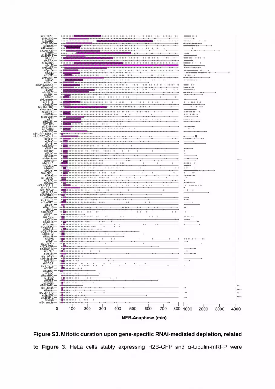
Figure S3. Mitotic duration upon gene-specific RNAi-mediated depletion, related
to Figure 3. HeLa cells stably expressing H2B-GFP and α-tubulin-mRFP were

acquired every 10 minutes. Mitotic duration was determined by measuring the time
between nuclear envelope breakdown (NEB) and anaphase onset, shown in minutes.
Data was presented as box-and-whiskers and each point corresponds to one cell. The
difference between mean values of each RNAi condition was statistically significant
from the control mean values. At least 2 independent experiments per condition were
performed. (*p≤0.05, **p≤0.01, ***p≤0.001, ****p≤0.0001, ns corresponds to not
significantly different from control, Mann-Whitney test).

Figure S4. Cell fate upon induction of chromosome alignment defects of distinct
molecular nature, related to Figure 3. a) Cell fate profiles of HeLa cells with delayed
or failed chromosome alignment after Ndc80 depletion. Each line indicates a single
cell and respective outcome. a´) Time that cells spent until mitotic exit (blue), death in
mitosis (purple) or in subsequent interphase (green) after Ndc80 depletion. Each dot
represents a single cell. The horizontal line indicates the mean of all quantified cells
and the error bars represent the standard deviation from a pool of four independent
experiments (Mitotic exit, 289±377 min, n=83; Death in mitosis, 894±292 min, n=45;
Post-mitotic death 1162±359 min, n=42; ****p≤0.0001 relative to control, analyzed
using a Mann-Whitney Test). b) Cell fate profiles of HeLa cells with delayed or failed
chromosome alignment after CENP-E depletion. b´) Time that cells spent until mitotic
exit (blue), death in mitosis (purple) or in the subsequent interphase (green) after

CENP-E depletion. Each dot represents a single cell. The horizontal line indicates the
mean of all quantified cells and the error bars represent the standard deviation from a
pool of five independent experiments (Mitotic exit, 243±193 min, n=358; Death in
mitosis 996±322 min, n=15; Post-mitotic death 814±408 min, n=32; ****p≤0.0001
relative to control, analyzed using a Mann-Whitney Test).

Figure S5. Further characterization of Cyclin B1 degradation profiles in RPE-1
and HeLa cells, related to Figure 5. a) Selective time frames from live-cell

microscopy of RPE-1 cells stably expressing H2B-mRFP and Cyclin B1-Venus in
control and siCENP-E. Images were acquired every 2 min. Time = min:sec. Time 00:00
= anaphase onset. Black arrowheads point to misaligned chromosomes that remain
upon anaphase onset. b) Cyclin B1 degradation profile for control, CENP-E- depleted
cells that properly align their chromosomes at the metaphase plate and CENP-E-
depleted cells that exit mitosis with misaligned chromosomes give rise to micronuclei.
Fluorescence intensities were normalized to the levels at time = -8. The curves
represent mean Cyclin B1-Venus fluorescence intensity from all analyzed cells and
errors bars represent the standard derivation from a pool of three independent
experiments (siScramble n=30; siCENP-E (aligned) n=30; siCENP-E
(misaligned+micronuclei) n=5). c) Frequency of anaphase cells with aligned
chromosomes, misaligned chromosomes and misaligned chromosomes that result in
micronuclei in control (black bars) and CENP-E-depleted cells (green bars). d)
Selected time frames from phase-contrast and fluorescence microscopy of Cyclin B1-
Venus HeLa cells treated with nocodazole with or without MG132. Images were
acquired every 15 min. Scale bar = 5 µm. Time = h:min. e) Cyclin B1 degradation
profiles of nocodazole-treated Cyclin B1-Venus HeLa cells in the presence or absence
of MG132. Fluorescence intensities were normalized to the levels at time = 0. The
curves depict mean Cyclin B1-Venus fluorescence intensity from all analyzed cells per
condition (nocodazole n=12; nocodazole + MG132 n=10), and error bars represent
the standard deviation. Note that acquisition in the presence of MG132 was terminated
earlier relative to acquisition without MG132 due to cell death.
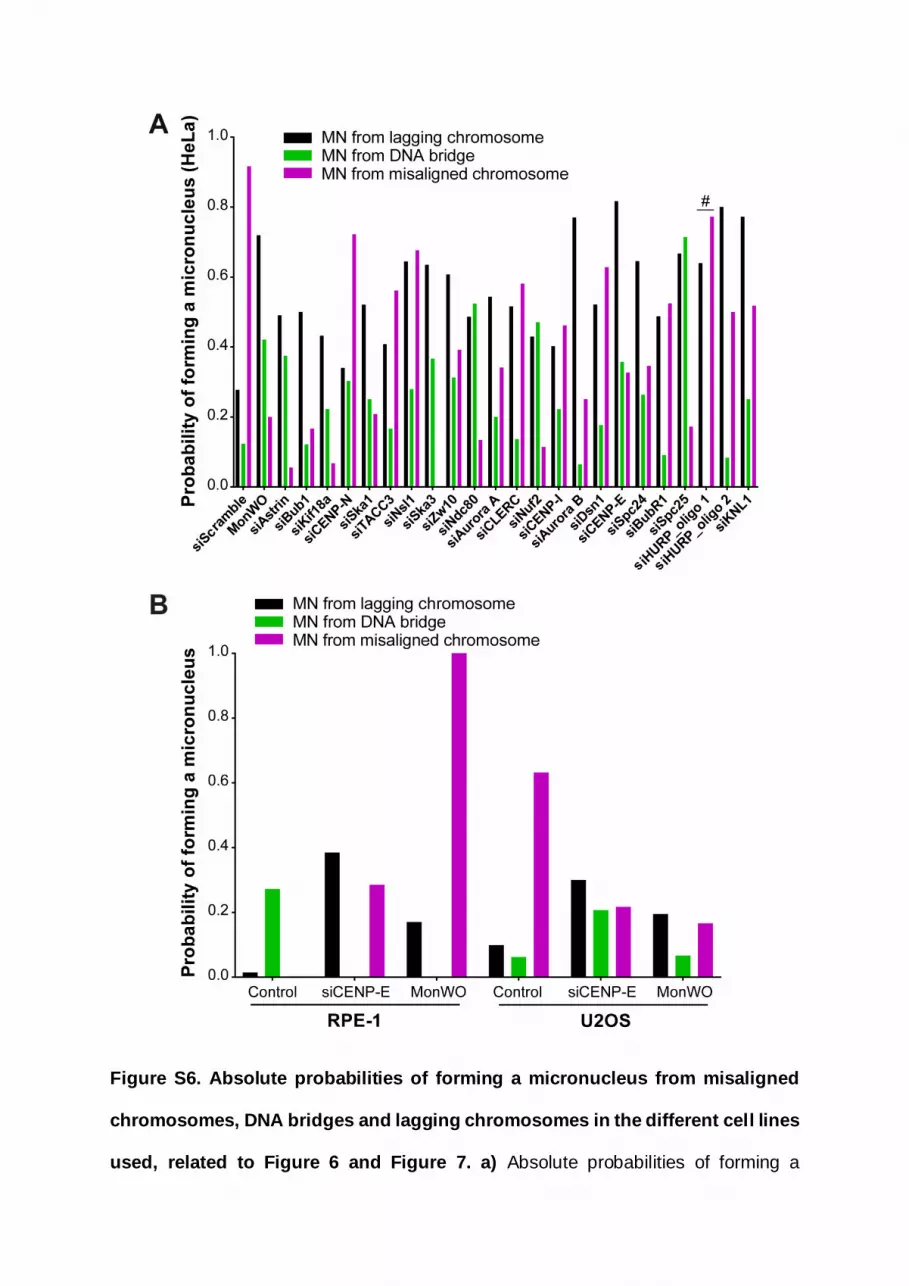
Figure S6. Absolute probabilities of forming a micronucleus from misaligned
chromosomes, DNA bridges and lagging chromosomes in the different cell lines
used, related to Figure 6 and Figure 7. a) Absolute probabilities of forming a

micronucleus of different origins in unperturbed HeLa cells and after molecular
perturbations that weaken kinetochore-microtubule attachments or promote the
formation of anaphase lagging chromosomes after monastrol treatment and washout
(MonWO). [siScramble n=1700, MonWO n=327, siAstrin n=423, siBub1 n=457,
siKif18a n= 540, siCENP-N n= 422, siSka1 n=395, siTACC3 n=485, siNsl1 n=400,
siSka3 n= 383, siZw10 n= 404, siNdc80 n=440, siAurora A n= 388, siCLERC n=263,
siNuf2 n=428, siCENP-I n= 389, siAurora B n=499, siDsn1 n=688, siCENP-E n= 346,
siSpc24 n=418, siBubR1 n=387, siSpc25 n= 425, siHURP_oligo1 n=296,
siHURP_oligo2 n=200, siKNL1 n=413; pool of 2 independent experiments for each
siRNAi oligonucleotide per condition, with the exception of Aurora A and CLERC in
which only 1 experiment for the second siRNAi oligonucleotide was performed. All
independent experiments were pooled]. b) Absolute probabilities of forming a
micronucleus of different origins in unperturbed RPE-1 and U2OS cells and after
CENP-E depletion (siCENP-E) or monastrol treatment and washout. [RPE-1 cells:
control, n=163; siCENP-E, n=95; MonWO, n=105]. [U2OS cells: control, n=250;
siCENP-E, n=81; MonWO, n=49].

Figure S7. Misaligned chromosomes in chromosomally unstable cancer cells
have hyper-stabilized kinetochore-microtubule attachments, related to Figure 7.
a) Representative immunofluorescence images of RPE-1 and U2OS cells stained for

DNA (green) and α-tubulin (magenta). RPE-1 and U2OS cells upon nocodazole
treatment and washout to generate misaligned chromosomes were processed for
immunofluorescence microscopy after a subsequent nocodazole shock 5, 15 and 30
min after drug addition. Representative immunofluorescence images of the mitotic
spindle at each stage are shown. Images are maximum intensity projections of
deconvolved z-stacks. Scale bar = 5 µm. b) Normalized α-tubulin fluorescence
intensity at indicated time points in RPE-1 and U2OS cells after nocodazole shock.
Fluorescence intensities were normalized to the levels at time = 0. Data represent
mean ± s.d., U2OS n=22 cells, RPE-1 n=22 cells, from 2 independent experiments.
Whole lines show single exponential fitting curve (**p≤0.01, extra sum-of-squares F
test).

Supplemental References
S1. DeLuca, J.G., Moree, B., Hickey, J.M., Kilmartin, J.V., and Salmon, E.D. (2002).
hNuf2 inhibition blocks stable kinetochore-microtubule attachment and induces
mitotic cell death in HeLa cells. J Cell Biol 159, 549-555.
10.1083/jcb.200208159.
S2. DeLuca, J.G., Dong, Y., Hergert, P., Strauss, J., Hickey, J.M., Salmon, E.D.,
and McEwen, B.F. (2005). Hec1 and nuf2 are core components of the
kinetochore outer plate essential for organizing microtubule attachment sites.
Mol Biol Cell 16, 519-531. 10.1091/mbc.e04-09-0852.
S3. Sundin, L.J., Guimaraes, G.J., and Deluca, J.G. (2011). The NDC80 complex
proteins Nuf2 and Hec1 make distinct contributions to kinetochore-microtubule
attachment in mitosis. Molecular biology of the cell 22, 759-768.
10.1091/mbc.E10-08-0671
mbc.E10-08-0671 [pii].
S4. Fremont, S., Gerard, A., Galloux, M., Janvier, K., Karess, R.E., and Berlioz-
Torrent, C. (2013). Beclin-1 is required for chromosome congression and
proper outer kinetochore assembly. EMBO Rep 14, 364-372.
10.1038/embor.2013.23.
S5. Tanenbaum, M.E., Galjart, N., van Vugt, M.A., and Medema, R.H. (2006). CLIP-
170 facilitates the formation of kinetochore-microtubule attachments. EMBO J
25, 45-57. 10.1038/sj.emboj.7600916.
S6. Amin, M.A., Kobayashi, K., and Tanaka, K. (2015). CLIP-170 tethers
kinetochores to microtubule plus ends against poleward force by dynein for

stable kinetochore-microtubule attachment. FEBS Lett 589, 2739-2746.
10.1016/j.febslet.2015.07.036.
S7. Ritchie, K., Seah, C., Moulin, J., Isaac, C., Dick, F., and Berube, N.G. (2008).
Loss of ATRX leads to chromosome cohesion and congression defects. J Cell
Biol 180, 315-324. 10.1083/jcb.200706083.
S8. Archinti, M., Lacasa, C., Teixido-Travesa, N., and Luders, J. (2010). SPICE--a
previously uncharacterized protein required for centriole duplication and mitotic
chromosome congression. J Cell Sci 123, 3039-3046. 10.1242/jcs.069963.
S9. Deretic, J., Kerr, A., and Welburn, J.P.I. (2019). A rapid computational approach
identifies SPICE1 as an Aurora kinase substrate. Mol Biol Cell 30, 312-323.
10.1091/mbc.E18-08-0495.
S10. Santamaria, A., Nagel, S., Sillje, H.H.W., and Nigg, E.A. (2008). The spindle
protein CHICA mediates localization of the chromokinesin Kid to the mitotic
spindle. Curr Biol 18, 723-729. 10.1016/j.cub.2008.04.041.
S11. Dunsch, A.K., Hammond, D., Lloyd, J., Schermelleh, L., Gruneberg, U., and
Barr, F.A. (2012). Dynein light chain 1 and a spindle-associated adaptor
promote dynein asymmetry and spindle orientation. The Journal of cell biology
198, 1039-1054. 10.1083/jcb.201202112.
S12. Wang, L., Zhu, G., Yang, D., Li, Q., Li, Y., Xu, X., He, D., and Zeng, C. (2008).
The spindle function of CDCA4. Cell motility and the cytoskeleton 65, 581-593.
10.1002/cm.20286.
S13. Jang, C.Y., Wong, J., Coppinger, J.A., Seki, A., Yates, J.R., 3rd, and Fang, G.
(2008). DDA3 recruits microtubule depolymerase Kif2a to spindle poles and
controls spindle dynamics and mitotic chromosome movement. J Cell Biol 181,
255-267. 10.1083/jcb.200711032.

S14. Ganem, N.J., and Compton, D.A. (2004). The KinI kinesin Kif2a is required for
bipolar spindle assembly through a functional relationship with MCAK. J Cell
Biol 166, 473-478. 10.1083/jcb.200404012.
S15. Park, S.J. (2010). Huntingtin-interacting protein 1-related is required for
accurate congression and segregation of chromosomes. BMB Rep 43, 795-
800. 10.5483/BMBRep.2010.43.12.795.
S16. Amin, M.A., Matsunaga, S., Uchiyama, S., and Fukui, K. (2008).
Nucleophosmin is required for chromosome congression, proper mitotic spindle
formation, and kinetochore-microtubule attachment in HeLa cells. FEBS Lett
582, 3839-3844. 10.1016/j.febslet.2008.10.023.
S17. Holt, S.V., Vergnolle, M.A., Hussein, D., Wozniak, M.J., Allan, V.J., and Taylor,
S.S. (2005). Silencing Cenp-F weakens centromeric cohesion, prevents
chromosome alignment and activates the spindle checkpoint. Journal of cell
science 118, 4889-4900. 118/20/4889 [pii]
10.1242/jcs.02614.
S18. Yang, Z., Guo, J., Chen, Q., Ding, C., Du, J., and Zhu, X. (2005). Silencing
mitosin induces misaligned chromosomes, premature chromosome
decondensation before anaphase onset, and mitotic cell death. Mol Cell Biol
25, 4062-4074. 10.1128/MCB.25.10.4062-4074.2005.
S19. Nishino, M., Kurasawa, Y., Evans, R., Lin, S.H., Brinkley, B.R., and Yu-Lee,
L.Y. (2006). NudC is required for Plk1 targeting to the kinetochore and
chromosome congression. Curr Biol 16, 1414-1421.
10.1016/j.cub.2006.05.052.
S20. Chuang, C., Pan, J., Hawke, D.H., Lin, S.H., and Yu-Lee, L.Y. (2013). NudC
deacetylation regulates mitotic progression. PLoS One 8, e73841.
10.1371/journal.pone.0073841.
S21. Gambe, A.E., Matsunaga, S., Takata, H., Ono-Maniwa, R., Baba, A.,
Uchiyama, S., and Fukui, K. (2009). A nucleolar protein RRS1 contributes to
chromosome congression. FEBS Lett 583, 1951-1956.
10.1016/j.febslet.2009.05.033.
S22. Zhang, L., Iyer, J., Chowdhury, A., Ji, M., Xiao, L., Yang, S., Chen, Y., Tsai,
M.Y., and Dong, J. (2012). KIBRA regulates aurora kinase activity and is
required for precise chromosome alignment during mitosis. J Biol Chem 287,
34069-34077. 10.1074/jbc.M112.385518.
S23. Ma, N., Matsunaga, S., Takata, H., Ono-Maniwa, R., Uchiyama, S., and Fukui,
K. (2007). Nucleolin functions in nucleolus formation and chromosome
congression. Journal of cell science 120, 2091-2105. 10.1242/jcs.008771.
S24. Li, N., Yuan, K., Yan, F., Huo, Y., Zhu, T., Liu, X., Guo, Z., and Yao, X. (2009).
PinX1 is recruited to the mitotic chromosome periphery by Nucleolin and
facilitates chromosome congression. Biochem Biophys Res Commun 384, 76-
81. 10.1016/j.bbrc.2009.04.077.
S25. Jang, C.Y., Coppinger, J.A., Yates, J.R., 3rd, and Fang, G. (2010). Phospho-
regulation of DDA3 function in mitosis. Biochem Biophys Res Commun 393,
259-263. 10.1016/j.bbrc.2010.01.115.
S26. Jang, C.Y., Coppinger, J.A., Yates, J.R., 3rd, and Fang, G. (2011). Mitotic
kinases regulate MT-polymerizing/MT-bundling activity of DDA3. Biochem
Biophys Res Commun 408, 174-179. 10.1016/j.bbrc.2011.04.004.

S27. Jang, C.Y., and Fang, G. (2011). DDA3 associates with MCAK and controls
chromosome congression. Biochem Biophys Res Commun 407, 610-614.
10.1016/j.bbrc.2011.03.081.
S28. Park, J.E., Song, H., Kwon, H.J., and Jang, C.Y. (2016). Ska1 cooperates with
DDA3 for spindle dynamics and spindle attachment to kinetochore. Biochem
Biophys Res Commun 470, 586-592. 10.1016/j.bbrc.2016.01.101.
S29. Johnson, V.L., Scott, M.I., Holt, S.V., Hussein, D., and Taylor, S.S. (2004).
Bub1 is required for kinetochore localization of BubR1, Cenp-E, Cenp-F and
Mad2, and chromosome congression. Journal of cell science 117, 1577-1589.
10.1242/jcs.01006
117/8/1577 [pii].
S30. Morrow, C.J., Tighe, A., Johnson, V.L., Scott, M.I., Ditchfield, C., and Taylor,
S.S. (2005). Bub1 and aurora B cooperate to maintain BubR1-mediated
inhibition of APC/CCdc20. J Cell Sci 118, 3639-3652. 10.1242/jcs.02487.
S31. Meraldi, P., and Sorger, P.K. (2005). A dual role for Bub1 in the spindle
checkpoint and chromosome congression. The EMBO journal 24, 1621-1633.
7600641 [pii]
10.1038/sj.emboj.7600641.
S32. Ditchfield, C., Johnson, V.L., Tighe, A., Ellston, R., Haworth, C., Johnson, T.,
Mortlock, A., Keen, N., and Taylor, S.S. (2003). Aurora B couples chromosome
alignment with anaphase by targeting BubR1, Mad2, and Cenp-E to
kinetochores. J Cell Biol 161, 267-280. 10.1083/jcb.200208091.
S33. Lampson, M.A., and Kapoor, T.M. (2005). The human mitotic checkpoint
protein BubR1 regulates chromosome-spindle attachments. Nat Cell Biol 7, 93-
98. 10.1038/ncb1208.
S34. Raaijmakers, J.A., Tanenbaum, M.E., Maia, A.F., and Medema, R.H. (2009).
RAMA1 is a novel kinetochore protein involved in kinetochore-microtubule
attachment. J Cell Sci 122, 2436-2445. 10.1242/jcs.051912.
S35. Gaitanos, T.N., Santamaria, A., Jeyaprakash, A.A., Wang, B., Conti, E., and
Nigg, E.A. (2009). Stable kinetochore-microtubule interactions depend on the
Ska complex and its new component Ska3/C13Orf3. EMBO J 28, 1442-1452.
10.1038/emboj.2009.96.
S36. Daum, J.R., Wren, J.D., Daniel, J.J., Sivakumar, S., McAvoy, J.N., Potapova,
T.A., and Gorbsky, G.J. (2009). Ska3 is required for spindle checkpoint
silencing and the maintenance of chromosome cohesion in mitosis. Curr Biol
19, 1467-1472. 10.1016/j.cub.2009.07.017.
S37. Sivakumar, S., Daum, J.R., Tipton, A.R., Rankin, S., and Gorbsky, G.J. (2014).
The spindle and kinetochore-associated (Ska) complex enhances binding of
the anaphase-promoting complex/cyclosome (APC/C) to chromosomes and
promotes mitotic exit. Molecular biology of the cell 25, 594-605.
10.1091/mbc.E13-07-0421.
S38. Welburn, J.P., Grishchuk, E.L., Backer, C.B., Wilson-Kubalek, E.M., Yates,
J.R., 3rd, and Cheeseman, I.M. (2009). The human kinetochore Ska1 complex
facilitates microtubule depolymerization-coupled motility. Dev Cell 16, 374-385.
10.1016/j.devcel.2009.01.011.
S39. Garrett, S., Auer, K., Compton, D.A., and Kapoor, T.M. (2002). hTPX2 is
required for normal spindle morphology and centrosome integrity during
vertebrate cell division. Curr Biol 12, 2055-2059. 10.1016/s0960-
9822(02)01277-0.

S40. Goshima, G. (2011). Identification of a TPX2-like microtubule-associated
protein in Drosophila. PLoS One 6, e28120. 10.1371/journal.pone.0028120.
S41. Itoh, G., Sugino, S., Ikeda, M., Mizuguchi, M., Kanno, S., Amin, M.A., Iemura,
K., Yasui, A., Hirota, T., and Tanaka, K. (2013). Nucleoporin Nup188 is required
for chromosome alignment in mitosis. Cancer Sci 104, 871-879.
10.1111/cas.12159.
S42. Mazumdar, M., Sundareshan, S., and Misteli, T. (2004). Human chromokinesin
KIF4A functions in chromosome condensation and segregation. J Cell Biol 166,
613-620. 10.1083/jcb.200401142.
S43. Yang, Z., Tulu, U.S., Wadsworth, P., and Rieder, C.L. (2007). Kinetochore
dynein is required for chromosome motion and congression independent of the
spindle checkpoint. Curr Biol 17, 973-980. 10.1016/j.cub.2007.04.056.
S44. Li, Y., Yu, W., Liang, Y., and Zhu, X. (2007). Kinetochore dynein generates a
poleward pulling force to facilitate congression and full chromosome alignment.
Cell Res 17, 701-712. 10.1038/cr.2007.65.
S45. McHedlishvili, N., Wieser, S., Holtackers, R., Mouysset, J., Belwal, M., Amaro,
A.C., and Meraldi, P. (2012). Kinetochores accelerate centrosome separation
to ensure faithful chromosome segregation. J Cell Sci 125, 906-918.
10.1242/jcs.091967.
S46. Raemaekers, T., Ribbeck, K., Beaudouin, J., Annaert, W., Van Camp, M.,
Stockmans, I., Smets, N., Bouillon, R., Ellenberg, J., and Carmeliet, G. (2003).
NuSAP, a novel microtubule-associated protein involved in mitotic spindle
organization. J Cell Biol 162, 1017-1029. 10.1083/jcb.200302129.

S47. Li, C., Xue, C., Yang, Q., Low, B.C., and Liou, Y.C. (2016). NuSAP governs
chromosome oscillation by facilitating the Kid-generated polar ejection force.
Nat Commun 7, 10597. 10.1038/ncomms10597.
S48. Ma, N., Matsunaga, S., Morimoto, A., Sakashita, G., Urano, T., Uchiyama, S.,
and Fukui, K. (2011). The nuclear scaffold protein SAF-A is required for
kinetochore-microtubule attachment and contributes to the targeting of Aurora-
A to mitotic spindles. J Cell Sci 124, 394-404. 10.1242/jcs.063347.
S49. Yang, S., Liu, X., Yin, Y., Fukuda, M.N., and Zhou, J. (2008). Tastin is required
for bipolar spindle assembly and centrosome integrity during mitosis. FASEB J
22, 1960-1972. 10.1096/fj.07-081463.
S50. Dynek, J.N., and Smith, S. (2004). Resolution of sister telomere association is
required for progression through mitosis. Science 304, 97-100.
10.1126/science.1094754.
S51. Chang, P., Coughlin, M., and Mitchison, T.J. (2005). Tankyrase-1
polymerization of poly(ADP-ribose) is required for spindle structure and
function. Nat Cell Biol 7, 1133-1139. 10.1038/ncb1322.
S52. Hanisch, A., Sillje, H.H., and Nigg, E.A. (2006). Timely anaphase onset requires
a novel spindle and kinetochore complex comprising Ska1 and Ska2. EMBO J
25, 5504-5515. 10.1038/sj.emboj.7601426.
S53. Auckland, P., Clarke, N.I., Royle, S.J., and McAinsh, A.D. (2017). Congressing
kinetochores progressively load Ska complexes to prevent force-dependent
detachment. J Cell Biol 216, 1623-1639. 10.1083/jcb.201607096.
S54. Sillje, H.H., Nagel, S., Korner, R., and Nigg, E.A. (2006). HURP is a Ran-
importin beta-regulated protein that stabilizes kinetochore microtubules in the
vicinity of chromosomes. Curr Biol 16, 731-742. 10.1016/j.cub.2006.02.070.

S55. Wong, J., and Fang, G. (2006). HURP controls spindle dynamics to promote
proper interkinetochore tension and efficient kinetochore capture. The Journal
of cell biology 173, 879-891. 10.1083/jcb.200511132.
S56. Ye, F., Tan, L., Yang, Q., Xia, Y., Deng, L.W., Murata-Hori, M., and Liou, Y.C.
(2011). HURP regulates chromosome congression by modulating kinesin
Kif18A function. Curr Biol 21, 1584-1591. 10.1016/j.cub.2011.08.024.
S57. Zhang, Y., Tan, L., Yang, Q., Li, C., and Liou, Y.C. (2018). The microtubule-
associated protein HURP recruits the centrosomal protein TACC3 to regulate
K-fiber formation and support chromosome congression. J Biol Chem 293,
15733-15747. 10.1074/jbc.RA118.003676.
S58. Nakamura, A., Arai, H., and Fujita, N. (2009). Centrosomal Aki1 and cohesin
function in separase-regulated centriole disengagement. J Cell Biol 187, 607-
614. 10.1083/jcb.200906019.
S59. Krauss, S.W., Spence, J.R., Bahmanyar, S., Barth, A.I., Go, M.M., Czerwinski,
D., and Meyer, A.J. (2008). Downregulation of protein 4.1R, a mature centriole
protein, disrupts centrosomes, alters cell cycle progression, and perturbs
mitotic spindles and anaphase. Mol Cell Biol 28, 2283-2294.
10.1128/MCB.02021-07.
S60. Wu, G., Lin, Y.T., Wei, R., Chen, Y., Shan, Z., and Lee, W.H. (2008). Hice1, a
novel microtubule-associated protein required for maintenance of spindle
integrity and chromosomal stability in human cells. Mol Cell Biol 28, 3652-3662.
10.1128/MCB.01923-07.
S61. Lawo, S., Bashkurov, M., Mullin, M., Ferreria, M.G., Kittler, R., Habermann, B.,
Tagliaferro, A., Poser, I., Hutchins, J.R., Hegemann, B., et al. (2009). HAUS,

the 8-subunit human Augmin complex, regulates centrosome and spindle
integrity. Curr Biol 19, 816-826. 10.1016/j.cub.2009.04.033.
S62. Huang, Y., Yao, Y., Xu, H.Z., Wang, Z.G., Lu, L., and Dai, W. (2009). Defects
in chromosome congression and mitotic progression in KIF18A-deficient cells
are partly mediated through impaired functions of CENP-E. Cell Cycle 8, 2643-
2649. 10.4161/cc.8.16.9366.
S63. Stumpff, J., von Dassow, G., Wagenbach, M., Asbury, C., and Wordeman, L.
(2008). The kinesin-8 motor Kif18A suppresses kinetochore movements to
control mitotic chromosome alignment. Dev Cell 14, 252-262.
10.1016/j.devcel.2007.11.014.
S64. Liu, X.S., Zhao, X.D., Wang, X., Yao, Y.X., Zhang, L.L., Shu, R.Z., Ren, W.H.,
Huang, Y., Huang, L., Gu, M.M., et al. (2010). Germinal Cell Aplasia in Kif18a
Mutant Male Mice Due to Impaired Chromosome Congression and
Dysregulated BubR1 and CENP-E. Genes & cancer 1, 26-39.
10.1177/1947601909358184.
S65. Stumpff, J., Wagenbach, M., Franck, A., Asbury, C.L., and Wordeman, L.
(2012). Kif18A and chromokinesins confine centromere movements via
microtubule growth suppression and spatial control of kinetochore tension. Dev
Cell 22, 1017-1029. 10.1016/j.devcel.2012.02.013.
S66. Mayr, M.I., Hummer, S., Bormann, J., Gruner, T., Adio, S., Woehlke, G., and
Mayer, T.U. (2007). The human kinesin Kif18A is a motile microtubule
depolymerase essential for chromosome congression. Curr Biol 17, 488-498.
10.1016/j.cub.2007.02.036.
S67. Fonseca, C.L., Malaby, H.L.H., Sepaniac, L.A., Martin, W., Byers, C.,
Czechanski, A., Messinger, D., Tang, M., Ohi, R., Reinholdt, L.G., and Stumpff,

J. (2019). Mitotic chromosome alignment ensures mitotic fidelity by promoting
interchromosomal compaction during anaphase. The Journal of cell biology
218, 1148-1163. 10.1083/jcb.201807228.
S68. Kimura, M., Yoshioka, T., Saio, M., Banno, Y., Nagaoka, H., and Okano, Y.
(2013). Mitotic catastrophe and cell death induced by depletion of centrosomal
proteins. Cell Death Dis 4, e603. 10.1038/cddis.2013.108.
S69. Schneider, L., Essmann, F., Kletke, A., Rio, P., Hanenberg, H., Wetzel, W.,
Schulze-Osthoff, K., Nurnberg, B., and Piekorz, R.P. (2007). The transforming
acidic coiled coil 3 protein is essential for spindle-dependent chromosome
alignment and mitotic survival. J Biol Chem 282, 29273-29283.
10.1074/jbc.M704151200.
S70. Lin, C.H., Hu, C.K., and Shih, H.M. (2010). Clathrin heavy chain mediates
TACC3 targeting to mitotic spindles to ensure spindle stability. The Journal of
cell biology 189, 1097-1105. 10.1083/jcb.200911120.
S71. Cheeseman, L.P., Harry, E.F., McAinsh, A.D., Prior, I.A., and Royle, S.J.
(2013). Specific removal of TACC3-ch-TOG-clathrin at metaphase deregulates
kinetochore fiber tension. J Cell Sci 126, 2102-2113. 10.1242/jcs.124834.
S72. Gergely, F., Draviam, V.M., and Raff, J.W. (2003). The ch-TOG/XMAP215
protein is essential for spindle pole organization in human somatic cells. Genes
Dev 17, 336-341. 10.1101/gad.245603.
S73. Muto, Y., Yoshioka, T., Kimura, M., Matsunami, M., Saya, H., and Okano, Y.
(2008). An evolutionarily conserved leucine-rich repeat protein CLERC is a
centrosomal protein required for spindle pole integrity. Cell Cycle 7, 2738-2748.
10.4161/cc.7.17.6591.

S74. Oshimori, N., Ohsugi, M., and Yamamoto, T. (2006). The Plk1 target Kizuna
stabilizes mitotic centrosomes to ensure spindle bipolarity. Nat Cell Biol 8,
1095-1101. 10.1038/ncb1474.
S75. Fielding, A.B., Dobreva, I., McDonald, P.C., Foster, L.J., and Dedhar, S. (2008).
Integrin-linked kinase localizes to the centrosome and regulates mitotic spindle
organization. J Cell Biol 180, 681-689. 10.1083/jcb.200710074.
S76. Dunsch, A.K., Linnane, E., Barr, F.A., and Gruneberg, U. (2011). The astrin-
kinastrin/SKAP complex localizes to microtubule plus ends and facilitates
chromosome alignment. The Journal of cell biology 192, 959-968.
10.1083/jcb.201008023.
S77. Schmidt, J.C., Kiyomitsu, T., Hori, T., Backer, C.B., Fukagawa, T., and
Cheeseman, I.M. (2010). Aurora B kinase controls the targeting of the Astrin-
SKAP complex to bioriented kinetochores. J Cell Biol 191, 269-280.
10.1083/jcb.201006129.
S78. Fang, L., Seki, A., and Fang, G. (2009). SKAP associates with kinetochores
and promotes the metaphase-to-anaphase transition. Cell Cycle 8, 2819-2827.
10.4161/cc.8.17.9514.
S79. Huang, Y., Wang, W., Yao, P., Wang, X., Liu, X., Zhuang, X., Yan, F., Zhou, J.,
Du, J., Ward, T., et al. (2012). CENP-E kinesin interacts with SKAP protein to
orchestrate accurate chromosome segregation in mitosis. J Biol Chem 287,
1500-1509. 10.1074/jbc.M111.277194.
S80. Logarinho, E., Maffini, S., Barisic, M., Marques, A., Toso, A., Meraldi, P., and
Maiato, H. (2012). CLASPs prevent irreversible multipolarity by ensuring
spindle-pole resistance to traction forces during chromosome alignment. Nat
Cell Biol 14, 295-303. 10.1038/ncb2423.

S81. Haren, L., Gnadt, N., Wright, M., and Merdes, A. (2009). NuMA is required for
proper spindle assembly and chromosome alignment in prometaphase. BMC
Res Notes 2, 64. 10.1186/1756-0500-2-64.
S82. Iwakiri, Y., Kamakura, S., Hayase, J., and Sumimoto, H. (2013). Interaction of
NuMA protein with the kinesin Eg5: its possible role in bipolar spindle assembly
and chromosome alignment. Biochem J 451, 195-204. 10.1042/BJ20121447.
S83. Wong, R.W., Blobel, G., and Coutavas, E. (2006). Rae1 interaction with NuMA
is required for bipolar spindle formation. Proc Natl Acad Sci U S A 103, 19783-
19787. 10.1073/pnas.0609582104.
S84. Blower, M.D., Nachury, M., Heald, R., and Weis, K. (2005). A Rae1-containing
ribonucleoprotein complex is required for mitotic spindle assembly. Cell 121,
223-234. 10.1016/j.cell.2005.02.016.
S85. Joseph, J., Liu, S.T., Jablonski, S.A., Yen, T.J., and Dasso, M. (2004). The
RanGAP1-RanBP2 complex is essential for microtubule-kinetochore
interactions in vivo. Curr Biol 14, 611-617. 10.1016/j.cub.2004.03.031.
S86. Barisic, M., Sohm, B., Mikolcevic, P., Wandke, C., Rauch, V., Ringer, T., Hess,
M., Bonn, G., and Geley, S. (2010). Spindly/CCDC99 is required for efficient
chromosome congression and mitotic checkpoint regulation. Molecular biology
of the cell 21, 1968-1981. 10.1091/mbc.E09-04-0356
E09-04-0356 [pii].
S87. Raaijmakers, J.A., Tanenbaum, M.E., and Medema, R.H. (2013). Systematic
dissection of dynein regulators in mitosis. The Journal of cell biology 201, 201-
215. 10.1083/jcb.201208098.
S88. Torres, J.Z., Summers, M.K., Peterson, D., Brauer, M.J., Lee, J., Senese, S.,
Gholkar, A.A., Lo, Y.C., Lei, X., Jung, K., et al. (2011). The STARD9/Kif16a

kinesin associates with mitotic microtubules and regulates spindle pole
assembly. Cell 147, 1309-1323. 10.1016/j.cell.2011.11.020.
S89. Holmfeldt, P., Zhang, X., Stenmark, S., Walczak, C.E., and Gullberg, M. (2005).
CaMKIIgamma-mediated inactivation of the Kin I kinesin MCAK is essential for
bipolar spindle formation. EMBO J 24, 1256-1266. 10.1038/sj.emboj.7600601.
S90. Maia, A.F., Feijao, T., Vromans, M.J., Sunkel, C.E., and Lens, S.M. (2010).
Aurora B kinase cooperates with CENP-E to promote timely anaphase onset.
Chromosoma 119, 405-413. 10.1007/s00412-010-0265-x.
S91. Stevens, D., Gassmann, R., Oegema, K., and Desai, A. (2011). Uncoordinated
loss of chromatid cohesion is a common outcome of extended metaphase
arrest. PLoS One 6, e22969. 10.1371/journal.pone.0022969.
S92. Tanudji, M., Shoemaker, J., L'Italien, L., Russell, L., Chin, G., and Schebye,
X.M. (2004). Gene silencing of CENP-E by small interfering RNA in HeLa cells
leads to missegregation of chromosomes after a mitotic delay. Molecular
biology of the cell 15, 3771-3781. 10.1091/mbc.e03-07-0482.
S93. Barisic, M., Aguiar, P., Geley, S., and Maiato, H. (2014). Kinetochore motors
drive congression of peripheral polar chromosomes by overcoming random
arm-ejection forces. Nat Cell Biol 16, 1249-1256. 10.1038/ncb3060.
S94. Royle, S.J., Bright, N.A., and Lagnado, L. (2005). Clathrin is required for the
function of the mitotic spindle. Nature 434, 1152-1157. 10.1038/nature03502.
S95. Foraker, A.B., Camus, S.M., Evans, T.M., Majeed, S.R., Chen, C.Y., Taner,
S.B., Correa, I.R., Jr., Doxsey, S.J., and Brodsky, F.M. (2012). Clathrin
promotes centrosome integrity in early mitosis through stabilization of
centrosomal ch-TOG. The Journal of cell biology 198, 591-605.
10.1083/jcb.201205116.

S96. Oshimori, N., Li, X., Ohsugi, M., and Yamamoto, T. (2009). Cep72 regulates
the localization of key centrosomal proteins and proper bipolar spindle
formation. The EMBO journal 28, 2066-2076. 10.1038/emboj.2009.161.
S97. Kim, K., and Rhee, K. (2011). The pericentriolar satellite protein CEP90 is
crucial for integrity of the mitotic spindle pole. Journal of cell science 124, 338-
347. 10.1242/jcs.078329.
S98. Mimori-Kiyosue, Y., Grigoriev, I., Lansbergen, G., Sasaki, H., Matsui, C.,
Severin, F., Galjart, N., Grosveld, F., Vorobjev, I., Tsukita, S., and Akhmanova,
A. (2005). CLASP1 and CLASP2 bind to EB1 and regulate microtubule plus-
end dynamics at the cell cortex. J Cell Biol 168, 141-153.
10.1083/jcb.200405094.
S99. Girao, H., Okada, N., Rodrigues, T.A., Silva, A.O., Figueiredo, A.C., Garcia, Z.,
Moutinho-Santos, T., Hayashi, I., Azevedo, J.E., Macedo-Ribeiro, S., and
Maiato, H. (2020). CLASP2 binding to curved microtubule tips promotes flux
and stabilizes kinetochore attachments. J Cell Biol 219.
10.1083/jcb.201905080.
S100. Maiato, H., Rieder, C.L., Earnshaw, W.C., and Sunkel, C.E. (2003). How do
kinetochores CLASP dynamic microtubules? Cell Cycle 2, 511-514.
10.4161/cc.2.6.576.
S101. Fuller, B.G., Lampson, M.A., Foley, E.A., Rosasco-Nitcher, S., Le, K.V.,
Tobelmann, P., Brautigan, D.L., Stukenberg, P.T., and Kapoor, T.M. (2008).
Midzone activation of aurora B in anaphase produces an intracellular
phosphorylation gradient. Nature 453, 1132-1136. 10.1038/nature06923.
S102. Hauf, S., Cole, R.W., LaTerra, S., Zimmer, C., Schnapp, G., Walter, R., Heckel,
A., van Meel, J., Rieder, C.L., and Peters, J.M. (2003). The small molecule

Hesperadin reveals a role for Aurora B in correcting kinetochore-microtubule
attachment and in maintaining the spindle assembly checkpoint. J Cell Biol 161,
281-294. 10.1083/jcb.200208092.
S103. Hegarat, N., Smith, E., Nayak, G., Takeda, S., Eyers, P.A., and Hochegger, H.
(2011). Aurora A and Aurora B jointly coordinate chromosome segregation and
anaphase microtubule dynamics. J Cell Biol 195, 1103-1113.
10.1083/jcb.201105058.
S104. Thein, K.H., Kleylein-Sohn, J., Nigg, E.A., and Gruneberg, U. (2007). Astrin is
required for the maintenance of sister chromatid cohesion and centrosome
integrity. J Cell Biol 178, 345-354. 10.1083/jcb.200701163.
S105. Hoar, K., Chakravarty, A., Rabino, C., Wysong, D., Bowman, D., Roy, N., and
Ecsedy, J.A. (2007). MLN8054, a small-molecule inhibitor of Aurora A, causes
spindle pole and chromosome congression defects leading to aneuploidy. Mol
Cell Biol 27, 4513-4525. 10.1128/MCB.02364-06.
S106. Sasai, K., Parant, J.M., Brandt, M.E., Carter, J., Adams, H.P., Stass, S.A.,
Killary, A.M., Katayama, H., and Sen, S. (2008). Targeted disruption of Aurora
A causes abnormal mitotic spindle assembly, chromosome misalignment and
embryonic lethality. Oncogene 27, 4122-4127. 10.1038/onc.2008.47.
S107. De Luca, M., Lavia, P., and Guarguaglini, G. (2006). A functional interplay
between Aurora-A, Plk1 and TPX2 at spindle poles: Plk1 controls centrosomal
localization of Aurora-A and TPX2 spindle association. Cell Cycle 5, 296-303.
10.4161/cc.5.3.2392.
S108. De Luca, M., Brunetto, L., Asteriti, I.A., Giubettini, M., Lavia, P., and
Guarguaglini, G. (2008). Aurora-A and ch-TOG act in a common pathway in

control of spindle pole integrity. Oncogene 27, 6539-6549.
10.1038/onc.2008.252.
S109. Mackay, A.M., Ainsztein, A.M., Eckley, D.M., and Earnshaw, W.C. (1998). A
dominant mutant of inner centromere protein (INCENP), a chromosomal
protein, disrupts prometaphase congression and cytokinesis. J Cell Biol 140,
991-1002. 10.1083/jcb.140.5.991.
S110. Xu, Z., Ogawa, H., Vagnarelli, P., Bergmann, J.H., Hudson, D.F., Ruchaud, S.,
Fukagawa, T., Earnshaw, W.C., and Samejima, K. (2009). INCENP-aurora B
interactions modulate kinase activity and chromosome passenger complex
localization. The Journal of cell biology 187, 637-653. 10.1083/jcb.200906053.
S111. Dai, J., Sultan, S., Taylor, S.S., and Higgins, J.M. (2005). The kinase haspin is
required for mitotic histone H3 Thr 3 phosphorylation and normal metaphase
chromosome alignment. Genes & development 19, 472-488.
10.1101/gad.1267105.
S112. Dai, J., Kateneva, A.V., and Higgins, J.M. (2009). Studies of haspin-depleted
cells reveal that spindle-pole integrity in mitosis requires chromosome
cohesion. J Cell Sci 122, 4168-4176. 10.1242/jcs.054122.
S113. Xu, P., Virshup, D.M., and Lee, S.H. (2014). B56-PP2A regulates motor
dynamics for mitotic chromosome alignment. J Cell Sci 127, 4567-4573.
10.1242/jcs.154609.
S114. Bharadwaj, R., Qi, W., and Yu, H. (2004). Identification of two novel
components of the human NDC80 kinetochore complex. J Biol Chem 279,
13076-13085. 10.1074/jbc.M310224200.
S115. McCleland, M.L., Kallio, M.J., Barrett-Wilt, G.A., Kestner, C.A., Shabanowitz,
J., Hunt, D.F., Gorbsky, G.J., and Stukenberg, P.T. (2004). The vertebrate

Ndc80 complex contains Spc24 and Spc25 homologs, which are required to
establish and maintain kinetochore-microtubule attachment. Curr Biol 14, 131-
137. 10.1016/j.cub.2003.12.058.
S116. Lo, K.W., Kogoy, J.M., and Pfister, K.K. (2007). The DYNLT3 light chain directly
links cytoplasmic dynein to a spindle checkpoint protein, Bub3. J Biol Chem
282, 11205-11212. 10.1074/jbc.M611279200.
S117. Vergnolle, M.A., and Taylor, S.S. (2007). Cenp-F links kinetochores to
Ndel1/Nde1/Lis1/dynein microtubule motor complexes. Curr Biol 17, 1173-
1179. 10.1016/j.cub.2007.05.077.
S118. Moon, H.M., Youn, Y.H., Pemble, H., Yingling, J., Wittmann, T., and Wynshaw-
Boris, A. (2014). LIS1 controls mitosis and mitotic spindle organization via the
LIS1-NDEL1-dynein complex. Hum Mol Genet 23, 449-466.
10.1093/hmg/ddt436.
S119. Auckland, P., and McAinsh, A.D. (2015). Building an integrated model of
chromosome congression. J Cell Sci 128, 3363-3374. 10.1242/jcs.169367.
S120. Horgan, C.P., Hanscom, S.R., and McCaffrey, M.W. (2011). Dynein LIC1
localizes to the mitotic spindle and midbody and LIC2 localizes to spindle poles
during cell division. Cell Biol Int 35, 171-178. 10.1042/CBI20100284.
S121. Einarson, M.B., Cukierman, E., Compton, D.A., and Golemis, E.A. (2004).
Human enhancer of invasion-cluster, a coiled-coil protein required for passage
through mitosis. Mol Cell Biol 24, 3957-3971. 10.1128/MCB.24.9.3957-
3971.2004.
S122. Lens, S.M., Wolthuis, R.M., Klompmaker, R., Kauw, J., Agami, R.,
Brummelkamp, T., Kops, G., and Medema, R.H. (2003). Survivin is required for

a sustained spindle checkpoint arrest in response to lack of tension. EMBO J
22, 2934-2947. 10.1093/emboj/cdg307.
S123. Carvalho, A., Carmena, M., Sambade, C., Earnshaw, W.C., and Wheatley, S.P.
(2003). Survivin is required for stable checkpoint activation in taxol-treated
HeLa cells. J Cell Sci 116, 2987-2998. 10.1242/jcs.00612.
S124. Uren, A.G., Wong, L., Pakusch, M., Fowler, K.J., Burrows, F.J., Vaux, D.L., and
Choo, K.H. (2000). Survivin and the inner centromere protein INCENP show
similar cell-cycle localization and gene knockout phenotype. Curr Biol 10, 1319-
1328. 10.1016/s0960-9822(00)00769-7.
S125. Cho, J.H., Chang, C.J., Chen, C.Y., and Tang, T.K. (2006). Depletion of CPAP
by RNAi disrupts centrosome integrity and induces multipolar spindles.
Biochem Biophys Res Commun 339, 742-747. 10.1016/j.bbrc.2005.11.074.
S126. Martin-Lluesma, S., Stucke, V.M., and Nigg, E.A. (2002). Role of Hec1 in
spindle checkpoint signaling and kinetochore recruitment of Mad1/Mad2.
Science 297, 2267-2270. 10.1126/science.1075596.
S127. Li, L., Yang, L., Scudiero, D.A., Miller, S.A., Yu, Z.X., Stukenberg, P.T.,
Shoemaker, R.H., and Kotin, R.M. (2007). Development of recombinant adeno-
associated virus vectors carrying small interfering RNA (shHec1)-mediated
depletion of kinetochore Hec1 protein in tumor cells. Gene Ther 14, 814-827.
10.1038/sj.gt.3302933.
S128. Lee, M.H., Lin, L., Equilibrina, I., Uchiyama, S., Matsunaga, S., and Fukui, K.
(2011). ASURA (PHB2) Is Required for Kinetochore Assembly and Subsequent
Chromosome Congression. Acta Histochem Cytochem 44, 247-258.
10.1267/ahc.11033.

S129. Gassmann, R., Carvalho, A., Henzing, A.J., Ruchaud, S., Hudson, D.F., Honda,
R., Nigg, E.A., Gerloff, D.L., and Earnshaw, W.C. (2004). Borealin: a novel
chromosomal passenger required for stability of the bipolar mitotic spindle. J
Cell Biol 166, 179-191. 10.1083/jcb.200404001.
S130. Beauchene, N.A., Diaz-Martinez, L.A., Furniss, K., Hsu, W.S., Tsai, H.J.,
Chamberlain, C., Esponda, P., Gimenez-Abian, J.F., and Clarke, D.J. (2010).
Rad21 is required for centrosome integrity in human cells independently of its
role in chromosome cohesion. Cell Cycle 9, 1774-1780. 10.4161/cc.9.9.11524.
S131. Diaz-Martinez, L.A., Beauchene, N.A., Furniss, K., Esponda, P., Gimenez-
Abian, J.F., and Clarke, D.J. (2010). Cohesin is needed for bipolar mitosis in
human cells. Cell Cycle 9, 1764-1773. 10.4161/cc.9.9.11525.
S132. Woolner, S., O'Brien, L.L., Wiese, C., and Bement, W.M. (2008). Myosin-10
and actin filaments are essential for mitotic spindle function. J Cell Biol 182, 77-
88. 10.1083/jcb.200804062.
S133. Wang, X., Yang, Y., Duan, Q., Jiang, N., Huang, Y., Darzynkiewicz, Z., and Dai,
W. (2008). sSgo1, a major splice variant of Sgo1, functions in centriole
cohesion where it is regulated by Plk1. Dev Cell 14, 331-341.
10.1016/j.devcel.2007.12.007.
S134. McGuinness, B.E., Hirota, T., Kudo, N.R., Peters, J.M., and Nasmyth, K.
(2005). Shugoshin prevents dissociation of cohesin from centromeres during
mitosis in vertebrate cells. PLoS Biol 3, e86. 10.1371/journal.pbio.0030086.
S135. McKinley, K.L., Sekulic, N., Guo, L.Y., Tsinman, T., Black, B.E., and
Cheeseman, I.M. (2015). The CENP-L-N Complex Forms a Critical Node in an
Integrated Meshwork of Interactions at the Centromere-Kinetochore Interface.
Mol Cell 60, 886-898. 10.1016/j.molcel.2015.10.027.

S136. Prendergast, L., van Vuuren, C., Kaczmarczyk, A., Doering, V., Hellwig, D.,
Quinn, N., Hoischen, C., Diekmann, S., and Sullivan, K.F. (2011). Premitotic
assembly of human CENPs -T and -W switches centromeric chromatin to a
mitotic state. PLoS Biol 9, e1001082. 10.1371/journal.pbio.1001082.
S137. Wood, L., Booth, D.G., Vargiu, G., Ohta, S., deLima Alves, F., Samejima, K.,
Fukagawa, T., Rappsilber, J., and Earnshaw, W.C. (2016). Auxin/AID versus
conventional knockouts: distinguishing the roles of CENP-T/W in mitotic
kinetochore assembly and stability. Open Biol 6, 150230.
10.1098/rsob.150230.
S138. Ali, A., Veeranki, S.N., Chinchole, A., and Tyagi, S. (2017). MLL/WDR5
Complex Regulates Kif2A Localization to Ensure Chromosome Congression
and Proper Spindle Assembly during Mitosis. Dev Cell 41, 605-622 e607.
10.1016/j.devcel.2017.05.023.
S139. Chun, Y., Kim, R., and Lee, S. (2016). Centromere Protein (CENP)-W Interacts
with Heterogeneous Nuclear Ribonucleoprotein (hnRNP) U and May Contribute
to Kinetochore-Microtubule Attachment in Mitotic Cells. PLoS One 11,
e0149127. 10.1371/journal.pone.0149127.
S140. Liu, X., Zheng, H., and Qu, C.K. (2012). Protein tyrosine phosphatase Shp2
(Ptpn11) plays an important role in maintenance of chromosome stability.
Cancer Res 72, 5296-5306. 10.1158/0008-5472.CAN-12-1495.
S141. Takata, H., Matsunaga, S., Morimoto, A., Ma, N., Kurihara, D., Ono-Maniwa,
R., Nakagawa, M., Azuma, T., Uchiyama, S., and Fukui, K. (2007). PHB2
protects sister-chromatid cohesion in mitosis. Curr Biol 17, 1356-1361.
10.1016/j.cub.2007.07.009.

S142. Equilibrina, I., Matsunaga, S., Morimoto, A., Hashimoto, T., Uchiyama, S., and
Fukui, K. (2013). ASURA (PHB2) interacts with Scc1 through chromatin.
Cytogenet Genome Res 139, 225-233. 10.1159/000350004.
S143. Orthaus, S., Ohndorf, S., and Diekmann, S. (2006). RNAi knockdown of human
kinetochore protein CENP-H. Biochem Biophys Res Commun 348, 36-46.
10.1016/j.bbrc.2006.06.187.
S144. Amaro, A.C., Samora, C.P., Holtackers, R., Wang, E., Kingston, I.J., Alonso,
M., Lampson, M., McAinsh, A.D., and Meraldi, P. (2010). Molecular control of
kinetochore-microtubule dynamics and chromosome oscillations. Nat Cell Biol
12, 319-329. 10.1038/ncb2033.
S145. Carleton, M., Mao, M., Biery, M., Warrener, P., Kim, S., Buser, C., Marshall,
C.G., Fernandes, C., Annis, J., and Linsley, P.S. (2006). RNA interference-
mediated silencing of mitotic kinesin KIF14 disrupts cell cycle progression and
induces cytokinesis failure. Mol Cell Biol 26, 3853-3863.
10.1128/MCB.26.10.3853-3863.2006.
S146. Zhu, C., Zhao, J., Bibikova, M., Leverson, J.D., Bossy-Wetzel, E., Fan, J.B.,
Abraham, R.T., and Jiang, W. (2005). Functional analysis of human
microtubule-based motor proteins, the kinesins and dyneins, in
mitosis/cytokinesis using RNA interference. Mol Biol Cell 16, 3187-3199.
10.1091/mbc.e05-02-0167.
S147. Draviam, V.M., Stegmeier, F., Nalepa, G., Sowa, M.E., Chen, J., Liang, A.,
Hannon, G.J., Sorger, P.K., Harper, J.W., and Elledge, S.J. (2007). A functional
genomic screen identifies a role for TAO1 kinase in spindle-checkpoint
signalling. Nat Cell Biol 9, 556-564. 10.1038/ncb1569.

S148. Shrestha, R.L., Tamura, N., Fries, A., Levin, N., Clark, J., and Draviam, V.M.
(2014). TAO1 kinase maintains chromosomal stability by facilitating proper
congression of chromosomes. Open Biol 4, 130108. 10.1098/rsob.130108.
S149. Hashizume, C., Nakano, H., Yoshida, K., and Wong, R.W. (2010).
Characterization of the role of the tumor marker Nup88 in mitosis. Mol Cancer
9, 119. 1476-4598-9-119 [pii]
10.1186/1476-4598-9-119.
S150. Uematsu, K., Okumura, F., Tonogai, S., Joo-Okumura, A., Alemayehu, D.H.,
Nishikimi, A., Fukui, Y., Nakatsukasa, K., and Kamura, T. (2016). ASB7
regulates spindle dynamics and genome integrity by targeting DDA3 for
proteasomal degradation. J Cell Biol 215, 95-106. 10.1083/jcb.201603062.
S151. Jaramillo-Lambert, A., Hao, J., Xiao, H., Li, Y., Han, Z., and Zhu, W. (2013).
Acidic nucleoplasmic DNA-binding protein (And-1) controls chromosome
congression by regulating the assembly of centromere protein A (CENP-A) at
centromeres. J Biol Chem 288, 1480-1488. 10.1074/jbc.M112.429266.
S152. Zhu, M., Wang, F., Yan, F., Yao, P.Y., Du, J., Gao, X., Wang, X., Wu, Q., Ward,
T., Li, J., et al. (2008). Septin 7 interacts with centromere-associated protein E
and is required for its kinetochore localization. J Biol Chem 283, 18916-18925.
10.1074/jbc.M710591200.
S153. Kim, S., and Jang, C.Y. (2016). ANKRD53 interacts with DDA3 and regulates
chromosome integrity during mitosis. Biochem Biophys Res Commun 470, 484-
491. 10.1016/j.bbrc.2016.01.144.
S154. Milev, M.P., Hasaj, B., Saint-Dic, D., Snounou, S., Zhao, Q., and Sacher, M.
(2015). TRAMM/TrappC12 plays a role in chromosome congression,

kinetochore stability, and CENP-E recruitment. The Journal of cell biology 209,
221-234. 10.1083/jcb.201501090.
S155. Spiliotis, E.T., Kinoshita, M., and Nelson, W.J. (2005). A mitotic septin scaffold
required for Mammalian chromosome congression and segregation. Science
307, 1781-1785. 10.1126/science.1106823.
S156. Zuccolo, M., Alves, A., Galy, V., Bolhy, S., Formstecher, E., Racine, V., Sibarita,
J.B., Fukagawa, T., Shiekhattar, R., Yen, T., and Doye, V. (2007). The human
Nup107-160 nuclear pore subcomplex contributes to proper kinetochore
functions. EMBO J 26, 1853-1864. 10.1038/sj.emboj.7601642.
S157. Platani, M., Santarella-Mellwig, R., Posch, M., Walczak, R., Swedlow, J.R., and
Mattaj, I.W. (2009). The Nup107-160 nucleoporin complex promotes mitotic
events via control of the localization state of the chromosome passenger
complex. Mol Biol Cell 20, 5260-5275. 10.1091/mbc.E09-05-0377.
S158. Bancroft, J., Auckland, P., Samora, C.P., and McAinsh, A.D. (2015).
Chromosome congression is promoted by CENP-Q- and CENP-E-dependent
pathways. Journal of cell science 128, 171-184. 10.1242/jcs.163659.
S159. Koliou, X., Fedonidis, C., Kalpachidou, T., and Mangoura, D. (2016). Nuclear
import mechanism of neurofibromin for localization on the spindle and function
in chromosome congression. J Neurochem 136, 78-91. 10.1111/jnc.13401.
S160. Zhuo, X., Guo, X., Zhang, X., Jing, G., Wang, Y., Chen, Q., Jiang, Q., Liu, J.,
and Zhang, C. (2015). Usp16 regulates kinetochore localization of Plk1 to
promote proper chromosome alignment in mitosis. J Cell Biol 210, 727-735.
10.1083/jcb.201502044.
S161. Oh, H.J., Kim, M.J., Song, S.J., Kim, T., Lee, D., Kwon, S.H., Choi, E.J., and
Lim, D.S. (2010). MST1 limits the kinase activity of aurora B to promote stable

kinetochore-microtubule attachment. Curr Biol 20, 416-422.
10.1016/j.cub.2009.12.054.
S162. Shimizu, H., Nagamori, I., Yabuta, N., and Nojima, H. (2009). GAK, a regulator
of clathrin-mediated membrane traffic, also controls centrosome integrity and
chromosome congression. J Cell Sci 122, 3145-3152. 10.1242/jcs.052795.
S163. Basilico, F., Maffini, S., Weir, J.R., Prumbaum, D., Rojas, A.M., Zimniak, T., De
Antoni, A., Jeganathan, S., Voss, B., van Gerwen, S., et al. (2014). The pseudo
GTPase CENP-M drives human kinetochore assembly. Elife 3, e02978.
10.7554/eLife.02978.
S164. Foltz, D.R., Jansen, L.E., Black, B.E., Bailey, A.O., Yates, J.R., 3rd, and
Cleveland, D.W. (2006). The human CENP-A centromeric nucleosome-
associated complex. Nat Cell Biol 8, 458-469. 10.1038/ncb1397.
S165. Hua, S., Wang, Z., Jiang, K., Huang, Y., Ward, T., Zhao, L., Dou, Z., and Yao,
X. (2011). CENP-U cooperates with Hec1 to orchestrate kinetochore-
microtubule attachment. J Biol Chem 286, 1627-1638.
10.1074/jbc.M110.174946.
S166. He, J., Zhang, Z., Ouyang, M., Yang, F., Hao, H., Lamb, K.L., Yang, J., Yin, Y.,
and Shen, W.H. (2016). PTEN regulates EG5 to control spindle architecture
and chromosome congression during mitosis. Nat Commun 7, 12355.
10.1038/ncomms12355.
S167. O'Regan, L., Sampson, J., Richards, M.W., Knebel, A., Roth, D., Hood, F.E.,
Straube, A., Royle, S.J., Bayliss, R., and Fry, A.M. (2015). Hsp72 is targeted to
the mitotic spindle by Nek6 to promote K-fiber assembly and mitotic
progression. J Cell Biol 209, 349-358. 10.1083/jcb.201409151.

S168. Serio, G., Margaria, V., Jensen, S., Oldani, A., Bartek, J., Bussolino, F., and
Lanzetti, L. (2011). Small GTPase Rab5 participates in chromosome
congression and regulates localization of the centromere-associated protein
CENP-F to kinetochores. Proc Natl Acad Sci U S A 108, 17337-17342.
10.1073/pnas.1103516108.
S169. Liu, S.T., Hittle, J.C., Jablonski, S.A., Campbell, M.S., Yoda, K., and Yen, T.J.
(2003). Human CENP-I specifies localization of CENP-F, MAD1 and MAD2 to
kinetochores and is essential for mitosis. Nat Cell Biol 5, 341-345.
10.1038/ncb953.
S170. Nishihashi, A., Haraguchi, T., Hiraoka, Y., Ikemura, T., Regnier, V., Dodson,
H., Earnshaw, W.C., and Fukagawa, T. (2002). CENP-I is essential for
centromere function in vertebrate cells. Dev Cell 2, 463-476. 10.1016/s1534-
5807(02)00144-2.
S171. Kline, S.L., Cheeseman, I.M., Hori, T., Fukagawa, T., and Desai, A. (2006). The
human Mis12 complex is required for kinetochore assembly and proper
chromosome segregation. J Cell Biol 173, 9-17. 10.1083/jcb.200509158.
S172. Lin, Y.T., Chen, Y., Wu, G., and Lee, W.H. (2006). Hec1 sequentially recruits
Zwint-1 and ZW10 to kinetochores for faithful chromosome segregation and
spindle checkpoint control. Oncogene 25, 6901-6914. 10.1038/sj.onc.1209687.
S173. Wang, H., Hu, X., Ding, X., Dou, Z., Yang, Z., Shaw, A.W., Teng, M., Cleveland,
D.W., Goldberg, M.L., Niu, L., and Yao, X. (2004). Human Zwint-1 specifies
localization of Zeste White 10 to kinetochores and is essential for mitotic
checkpoint signaling. J Biol Chem 279, 54590-54598.
10.1074/jbc.M407588200.

S174. Ghongane, P., Kapanidou, M., Asghar, A., Elowe, S., and Bolanos-Garcia, V.M.
(2014). The dynamic protein Knl1 - a kinetochore rendezvous. J Cell Sci 127,
3415-3423. 10.1242/jcs.149922.
S175. Caldas, G.V., and DeLuca, J.G. (2014). KNL1: bringing order to the
kinetochore. Chromosoma 123, 169-181. 10.1007/s00412-013-0446-5.
S176. Mackay, D.R., Elgort, S.W., and Ullman, K.S. (2009). The nucleoporin Nup153
has separable roles in both early mitotic progression and the resolution of
mitosis. Mol Biol Cell 20, 1652-1660. 10.1091/mbc.E08-08-0883.
S177. Lussi, Y.C., Shumaker, D.K., Shimi, T., and Fahrenkrog, B. (2010). The
nucleoporin Nup153 affects spindle checkpoint activity due to an association
with Mad1. Nucleus 1, 71-84. 10.4161/nucl.1.1.10244.
S178. Chatel, G., and Fahrenkrog, B. (2011). Nucleoporins: leaving the nuclear pore
complex for a successful mitosis. Cell Signal 23, 1555-1562.
10.1016/j.cellsig.2011.05.023.
S179. Honda, S., Marumoto, T., Hirota, T., Nitta, M., Arima, Y., Ogawa, M., and Saya,
H. (2004). Activation of m-calpain is required for chromosome alignment on the
metaphase plate during mitosis. J Biol Chem 279, 10615-10623.
10.1074/jbc.M308841200.
S180. Asteriti, I.A., Giubettini, M., Lavia, P., and Guarguaglini, G. (2011). Aurora-A
inactivation causes mitotic spindle pole fragmentation by unbalancing
microtubule-generated forces. Mol Cancer 10, 131. 10.1186/1476-4598-10-
131.
S181. Karamysheva, Z., Diaz-Martinez, L.A., Crow, S.E., Li, B., and Yu, H. (2009).
Multiple anaphase-promoting complex/cyclosome degrons mediate the

degradation of human Sgo1. J Biol Chem 284, 1772-1780.
10.1074/jbc.M807083200.
S182. Labade, A.S., Karmodiya, K., and Sengupta, K. (2016). HOXA repression is
mediated by nucleoporin Nup93 assisted by its interactors Nup188 and
Nup205. Epigenetics Chromatin 9, 54. 10.1186/s13072-016-0106-0.
S183. Kops, G.J., Kim, Y., Weaver, B.A., Mao, Y., McLeod, I., Yates, J.R., 3rd,
Tagaya, M., and Cleveland, D.W. (2005). ZW10 links mitotic checkpoint
signaling to the structural kinetochore. J Cell Biol 169, 49-60.
10.1083/jcb.200411118.
S184. Chan, Y.W., Fava, L.L., Uldschmid, A., Schmitz, M.H., Gerlich, D.W., Nigg,
E.A., and Santamaria, A. (2009). Mitotic control of kinetochore-associated
dynein and spindle orientation by human Spindly. J Cell Biol 185, 859-874.
10.1083/jcb.200812167.
S185. Thomas, G.E., Bandopadhyay, K., Sutradhar, S., Renjith, M.R., Singh, P.,
Gireesh, K.K., Simon, S., Badarudeen, B., Gupta, H., Banerjee, M., et al.
(2016). EB1 regulates attachment of Ska1 with microtubules by forming
extended structures on the microtubule lattice. Nat Commun 7, 11665.
10.1038/ncomms11665.
S186. Fielding, A.B., Lim, S., Montgomery, K., Dobreva, I., and Dedhar, S. (2011). A
critical role of integrin-linked kinase, ch-TOG and TACC3 in centrosome
clustering in cancer cells. Oncogene 30, 521-534. 10.1038/onc.2010.431.
S187. Srivastava, S., and Panda, D. (2018). A centrosomal protein STARD9 promotes
microtubule stability and regulates spindle microtubule dynamics. Cell Cycle
17, 2052-2068. 10.1080/15384101.2018.1513764.

S188. Vassilopoulos, S., Esk, C., Hoshino, S., Funke, B.H., Chen, C.Y., Plocik, A.M.,
Wright, W.E., Kucherlapati, R., and Brodsky, F.M. (2009). A role for the CHC22
clathrin heavy-chain isoform in human glucose metabolism. Science 324, 1192-
1196. 10.1126/science.1171529.
S189. Cheng, T.S., Hsiao, Y.L., Lin, C.C., Hsu, C.M., Chang, M.S., Lee, C.I., Yu, R.C.,
Huang, C.Y., Howng, S.L., and Hong, Y.R. (2007). hNinein is required for
targeting spindle-associated protein Astrin to the centrosome during the S and
G2 phases. Exp Cell Res 313, 1710-1721. 10.1016/j.yexcr.2007.02.023.
S190. Kuang, P., Chen, Z., Wang, J., Liu, Z., Wang, J., Gao, J., and Shen, L. (2017).
Characterization of Aurora A and Its Impact on the Effect of Cisplatin-Based
Chemotherapy in Patients with Non-Small Cell Lung Cancer. Transl Oncol 10,
367-377. 10.1016/j.tranon.2017.02.010.
S191. Kesisova, I.A., Nakos, K.C., Tsolou, A., Angelis, D., Lewis, J., Chatzaki, A.,
Agianian, B., Giannis, A., and Koffa, M.D. (2013). Tripolin A, a novel small-
molecule inhibitor of aurora A kinase, reveals new regulation of HURP's
distribution on microtubules. PLoS One 8, e58485.
10.1371/journal.pone.0058485.
S192. Liu, D., Ding, X., Du, J., Cai, X., Huang, Y., Ward, T., Shaw, A., Yang, Y., Hu,
R., Jin, C., and Yao, X. (2007). Human NUF2 interacts with centromere-
associated protein E and is essential for a stable spindle microtubule-
kinetochore attachment. J Biol Chem 282, 21415-21424.
10.1074/jbc.M609026200.
S193. Chun, Y., Lee, M., Park, B., and Lee, S. (2013). CSN5/JAB1 interacts with the
centromeric components CENP-T and CENP-W and regulates their

proteasome-mediated degradation. J Biol Chem 288, 27208-27219.
10.1074/jbc.M113.469221.
S194. Kim, S., and Yu, H. (2015). Multiple assembly mechanisms anchor the KMN
spindle checkpoint platform at human mitotic kinetochores. J Cell Biol 208, 181-
196. 10.1083/jcb.201407074.
S195. Tsang, Y.H., Han, X., Man, W.Y., Lee, N., and Poon, R.Y. (2012). Novel
functions of the phosphatase SHP2 in the DNA replication and damage
checkpoints. PLoS One 7, e49943. 10.1371/journal.pone.0049943.
S196. Xu, H., Choe, C., Shin, S.H., Park, S.W., Kim, H.S., Jung, S.H., Yim, S.H., Kim,
T.M., and Chung, Y.J. (2014). Silencing of KIF14 interferes with cell cycle
progression and cytokinesis by blocking the p27(Kip1) ubiquitination pathway
in hepatocellular carcinoma. Exp Mol Med 46, e97. 10.1038/emm.2014.23.
S197. Bernad, R., van der Velde, H., Fornerod, M., and Pickersgill, H. (2004).
Nup358/RanBP2 attaches to the nuclear pore complex via association with
Nup88 and Nup214/CAN and plays a supporting role in CRM1-mediated
nuclear protein export. Mol Cell Biol 24, 2373-2384. 10.1128/MCB.24.6.2373-
2384.2004.
S198. Zhu, W., Ukomadu, C., Jha, S., Senga, T., Dhar, S.K., Wohlschlegel, J.A., Nutt,
L.K., Kornbluth, S., and Dutta, A. (2007). Mcm10 and And-1/CTF4 recruit DNA
polymerase alpha to chromatin for initiation of DNA replication. Genes Dev 21,
2288-2299. 10.1101/gad.1585607.
S199. Yoshizawa-Sugata, N., and Masai, H. (2009). Roles of human AND-1 in
chromosome transactions in S phase. J Biol Chem 284, 20718-20728.
10.1074/jbc.M806711200.

S200. Kremer, B.E., Haystead, T., and Macara, I.G. (2005). Mammalian septins
regulate microtubule stability through interaction with the microtubule-binding
protein MAP4. Mol Biol Cell 16, 4648-4659. 10.1091/mbc.e05-03-0267.
S201. Park, H.J., Lee, S.J., Sohn, Y.B., Jin, H.S., Han, J.H., Kim, Y.B., Yim, H., and
Jeong, S.Y. (2013). NF1 deficiency causes Bcl-xL upregulation in Schwann
cells derived from neurofibromatosis type 1-associated malignant peripheral
nerve sheath tumors. Int J Oncol 42, 657-666. 10.3892/ijo.2012.1751.
S202. Qian, Y., Wang, B., Ma, A., Zhang, L., Xu, G., Ding, Q., Jing, T., Wu, L., Liu, Y.,
Yang, Z., and Liu, Y. (2016). USP16 Downregulation by Carboxyl-terminal
Truncated HBx Promotes the Growth of Hepatocellular Carcinoma Cells. Sci
Rep 6, 33039. 10.1038/srep33039.
S203. Lee, D.W., Zhao, X., Zhang, F., Eisenberg, E., and Greene, L.E. (2005).
Depletion of GAK/auxilin 2 inhibits receptor-mediated endocytosis and
recruitment of both clathrin and clathrin adaptors. J Cell Sci 118, 4311-4321.
10.1242/jcs.02548.
S204. Wu, H., Zhou, Y., Wu, H., Xu, L., Yan, Y., Tong, X., and Yan, H. (2021). CENPN
Acts as a Novel Biomarker that Correlates With the Malignant Phenotypes of
Glioma Cells. Front Genet 12, 732376. 10.3389/fgene.2021.732376.
S205. Chan, Y.W., Jeyaprakash, A.A., Nigg, E.A., and Santamaria, A. (2012). Aurora
B controls kinetochore-microtubule attachments by inhibiting Ska complex-
KMN network interaction. J Cell Biol 196, 563-571. 10.1083/jcb.201109001.
S206. Zhang, G., Lischetti, T., Hayward, D.G., and Nilsson, J. (2015). Distinct
domains in Bub1 localize RZZ and BubR1 to kinetochores to regulate the
checkpoint. Nat Commun 6, 7162. 10.1038/ncomms8162.

S207. Goshima, G., Kiyomitsu, T., Yoda, K., and Yanagida, M. (2003). Human
centromere chromatin protein hMis12, essential for equal segregation, is
independent of CENP-A loading pathway. The Journal of cell biology 160, 25-
39. 10.1083/jcb.200210005.
S208. Schleicher, K., Porter, M., Ten Have, S., Sundaramoorthy, R., Porter, I.M., and
Swedlow, J.R. (2017). The Ndc80 complex targets Bod1 to human mitotic
kinetochores. Open Biol 7. 10.1098/rsob.170099.
S209. Hahn, P., Schmidt, C., Weber, M., Kang, J., and Bielke, W. (2004). RNA
interference: PCR strategies for the quantification of stable degradation-
fragments derived from siRNA-targeted mRNAs. Biomol Eng 21, 113-117.
10.1016/j.bioeng.2004.09.001.