Open versus laparoscopic liver resection for colorectal liver ...
Upload
khangminh22Category
view
1download
0
Edited by
New Therapies of Liver Diseases
Pierluigi Toniutto
Printed Edition of the Special Issue Published in Journal of Clinical Medicine
www.mdpi.com/journal/jcm

New Therapies of Liver Diseases


New Therapies of Liver Diseases
Editor
Pierluigi Toniutto
MDPI • Basel • Beijing • Wuhan • Barcelona • Belgrade • Manchester • Tokyo • Cluj • Tianjin

Editor
Pierluigi Toniutto
Hepatology and Liver
Transplantation Unit,
University of Udine
Italy
Editorial Office
MDPI
St. Alban-Anlage 66
4052 Basel, Switzerland
This is a reprint of articles from the Special Issue published online in the open access journal
Journal of Clinical Medicine (ISSN 2077-0383) (available at: https://www.mdpi.com/journal/jcm/
special issues/new therapies liver diseases).
For citation purposes, cite each article independently as indicated on the article page online and as
indicated below:
LastName, A.A.; LastName, B.B.; LastName, C.C. Article Title. Journal Name Year, Volume Number,
Page Range.
ISBN 978-3-0365-3859-4 (Hbk)
ISBN 978-3-0365-3860-0 (PDF)
© 2022 by the authors. Articles in this book are Open Access and distributed under the Creative
Commons Attribution (CC BY) license, which allows users to download, copy and build upon
published articles, as long as the author and publisher are properly credited, which ensures maximum
dissemination and a wider impact of our publications.
The book as a whole is distributed by MDPI under the terms and conditions of the Creative Commons
license CC BY-NC-ND.

Contents
Pierluigi Toniutto
Special Issue “New Therapies of Liver Diseases”Reprinted from: J. Clin. Med. 2022, 11, 1798, doi:10.3390/jcm11071798 . . . . . . . . . . . . . . . . 1
Marta Mazzetti, Giulia Marconi, Martina Mancinelli, Antonio Benedetti, Marco Marzioni
and Luca Maroni
The Management of Cholestatic Liver Diseases: Current Therapies and EmergingNew PossibilitiesReprinted from: J. Clin. Med. 2021, 10, 1763, doi:10.3390/jcm10081763 . . . . . . . . . . . . . . . . 9
Anne-Christin Beatrice Wilde, Charlotte Lieb, Elise Leicht, Lena Maria Greverath,
Lara Marleen Steinhagen, Nina Wald de Chamorro, Jorg Petersen, Wolf Peter Hofmann,
Holger Hinrichsen, Renate Heyne, Thomas Berg, Uwe Naumann, Jeannette Schwenzer,
Johannes Vermehren, Andreas Geier, Frank Tacke and Tobias Muller
Real-World Clinical Management of Patients with Primary BiliaryCholangitis—A Retrospective Multicenter Study from GermanyReprinted from: J. Clin. Med. 2021, 10, 1061, doi:10.3390/jcm10051061 . . . . . . . . . . . . . . . . 29
Giacomo Zaccherini, Manuel Tufoni, Mauro Bernardi and Paolo Caraceni
Prevention of Cirrhosis Complications: Looking for Potential Disease Modifying AgentsReprinted from: J. Clin. Med. 2021, 10, 4590, doi:10.3390/jcm10194590 . . . . . . . . . . . . . . . . 41
Giacomo Zaccherini, Manuel Tufoni, Giulia Iannone and Paolo Caraceni
Management of Ascites in Patients with Cirrhosis: An UpdateReprinted from: J. Clin. Med. 2021, 10, 5226, doi:10.3390/jcm10225226 . . . . . . . . . . . . . . . . 61
Atsushi Hosui, Takafumi Tanimoto, Toru Okahara, Munehiro Ashida, Kohsaku Ohnishi,
Yuhhei Wakahara, Yukihiro Kusumoto, Toshio Yamaguchi, Yuka Sueyoshi, Motohiro Hirao,
Takuya Yamada and Naoki Hiramatsu
Early Administration of Tolvaptan Can Improve Survival in Patients with Cirrhotic AscitesReprinted from: J. Clin. Med. 2021, 10, 294, doi:10.3390/jcm10020294 . . . . . . . . . . . . . . . . 75
Alberto Zanetto, Sarah Shalaby, Paolo Feltracco, Martina Gambato, Giacomo Germani,
Francesco Paolo Russo, Patrizia Burra and Marco Senzolo
Recent Advances in the Management of Acute Variceal HemorrhageReprinted from: J. Clin. Med. 2021, 10, 3818, doi:10.3390/jcm10173818 . . . . . . . . . . . . . . . . 85
Chiara Mangini and Sara Montagnese
New Therapies of Liver Diseases: Hepatic EncephalopathyReprinted from: J. Clin. Med. 2021, 10, 4050, doi:10.3390/jcm10184050 . . . . . . . . . . . . . . . . 99
Carmine Gambino, Salvatore Piano and Paolo Angeli
Acute-on-Chronic Liver Failure in CirrhosisReprinted from: J. Clin. Med. 2021, 10, 4406, doi:10.3390/jcm10194406 . . . . . . . . . . . . . . . . 111
Valentina Zuccaro, Erika Asperges, Marta Colaneri, Lea Nadia Marvulli and Raffaele Bruno
HBV and HDV: New Treatments on the HorizonReprinted from: J. Clin. Med. 2021, 10, 4054, doi:10.3390/jcm10184054 . . . . . . . . . . . . . . . . 123
Anthony Vignone, Francesca Biancaniello, Marco Casadio, Ludovica Pesci,
Vincenzo Cardinale, Lorenzo Ridola and Domenico Alvaro
Emerging Therapies for Advanced Cholangiocarcinoma: An Updated Literature ReviewReprinted from: J. Clin. Med. 2021, 10, 4901, doi:10.3390/jcm10214901 . . . . . . . . . . . . . . . . 137
vi
Maria Corina Plaz Torres, Quirino Lai, Fabio Piscaglia, Eugenio Caturelli,
Giuseppe Cabibbo, Elisabetta Biasini, Filippo Pelizzaro, Fabio Marra, Franco Trevisani
and Edoardo G. Giannini
Treatment of Hepatocellular Carcinoma with Immune Checkpoint Inhibitors and Applicabilityof First-Line Atezolizumab/Bevacizumab in a Real-Life SettingReprinted from: J. Clin. Med. 2021, 10, 3201, doi:10.3390/jcm10153201 . . . . . . . . . . . . . . . . 149
Pierluigi Toniutto, Elisa Fumolo, Ezio Fornasiere and Davide Bitetto
Liver Transplantation in Patients with Hepatocellular Carcinoma beyond the Milan Criteria:A Comprehensive ReviewReprinted from: J. Clin. Med. 2021, 10, 3932, doi:10.3390/jcm10173932 . . . . . . . . . . . . . . . . 171
Chiara Becchetti, Sarah Gabriela Gschwend, Jean-Francois Dufour and Vanessa Banz
COVID-19 in Liver Transplant Recipients: A Systematic ReviewReprinted from: J. Clin. Med. 2021, 10, 4015, doi:10.3390/jcm10174015 . . . . . . . . . . . . . . . . 187
Alberto Zanetto, Sarah Shalaby, Martina Gambato, Giacomo Germani, Marco Senzolo,
Debora Bizzaro, Francesco Paolo Russo and Patrizia Burra
New Indications for Liver TransplantationReprinted from: J. Clin. Med. 2021, 10, 3867, doi:10.3390/jcm10173867 . . . . . . . . . . . . . . . . 209
vii


Citation: Toniutto, P. Special Issue
“New Therapies of Liver Diseases”. J.
Clin. Med. 2022, 11, 1798. https://
doi.org/10.3390/jcm11071798
Received: 17 March 2022
Accepted: 19 March 2022
Published: 24 March 2022
Publisher’s Note: MDPI stays neutral
with regard to jurisdictional claims in
published maps and institutional affil-
iations.
Copyright: © 2022 by the author.
Licensee MDPI, Basel, Switzerland.
This article is an open access article
distributed under the terms and
conditions of the Creative Commons
Attribution (CC BY) license (https://
creativecommons.org/licenses/by/
4.0/).
Journal of
Clinical Medicine
Editorial
Special Issue “New Therapies of Liver Diseases”
Pierluigi Toniutto
Hepatology and Liver Transplantation Unit, Academic Hospital, University of Udine, 33100 Udine, Italy;[email protected]
Medical and surgical treatments aimed at curing severe liver diseases and prolongingthe survival of patients have improved dramatically in recent years. These advances havemainly been achieved by obtaining a better understanding of the pathophysiology ofliver diseases [1]. New and old pharmacological therapies have been applied in a betterway based on the new insights obtained in the pathophysiological studies. Moreover, theincreased application of technology innovations, both in diagnostic imaging [2] and insurgery [3], enable the cure rate to be increased in patients with advanced liver diseases.
A Special Issue in the Journal of Clinical Medicine (JCM) has been dedicated to collectinghigh-quality scientific contributions from leading experts by focusing on updating thehorizon of new pharmacological therapies and new surgical approaches that can be appliedto cure several types of liver diseases as a method to address this challenging topic ingreater detail.
Two studies investigated the current and future management of cholestatic liver dis-eases [4,5]. The first study retrospectively evaluated the real-world clinical management ofpatients with primary biliary cholangitis (PBC), according to the indications of the recentupdated clinical guidelines. A study conducted in a large cohort of European patientsrevealed that biochemical response rates adopting the standard first-line treatment withursodeoxycholic acid (UDCA) were achieved in a large proportion of patients, dependingon the response criteria adopted. In UDCA nonresponders, second-line treatment regimensin which obeticholic acid or bezafibrate were added were promptly applied, leading to sig-nificantly increased response rates. These results confirm in real clinical practice that UDCAfirst-line standard treatment is largely effective in patients with PBC, but highlight the needto detect high-risk patients with an insufficient response to UDCA early in life, since earlytreatment modification significantly increases subsequent response rates. In addition toobeticholic acid and fibrates, several other molecules are currently under evaluation aspotential new therapies both for patients with PBC and with primary sclerosing cholangitis(PSC). This issue has been extensively discussed in a subsequent study [5]. Given the com-plex nature of PBC and PSC, future treatments for these diseases will probably be based ona combination of drugs, aimed at influencing specific pathophysiological mechanisms indifferent stages of disease severity.
The current therapeutic strategies for the management of patients with cirrhosis arefocused on the prevention or treatment of specific clinical complications such as ascites,gastrointestinal bleeding and hepatic encephalopathy [6]. “Etiologic therapy”, which isdesigned to remove the causative agents of the disease (i.e., viruses or alcohol), preventsclinical decompensation in most patients with cirrhosis. In contrast, a significant proportionof patients with decompensated cirrhosis remain at risk of further disease progressiondespite the application of etiologic treatments. Thus, the identification of new therapiestargeting specific key points in the complex pathophysiological cascade of decompensatedcirrhosis is urgently needed. These therapies are presented in updated detail in thisSpecial Issue of JCM [7]. Poorly absorbable oral antibiotics, statins, and albumin have beenproposed as potential disease-modifying agents for cirrhosis (DMAC), since clinical studieshave shown their capacity to prolong survival. The ideal DMACs candidate should be
J. Clin. Med. 2022, 11, 1798. https://doi.org/10.3390/jcm11071798 https://www.mdpi.com/journal/jcm
1

J. Clin. Med. 2022, 11, 1798
directed to modify the key mechanisms in the pathogenetic network of the gut-liver axis,such systemic inflammation, and immune dysfunction.
The development of ascites is one of the typical complications of advanced cirrhosis.Treatment of non-tense ascites comprises the assumption of a combination of furosemideand anti-aldosterone diuretics, accompanied by a restrictive sodium and water diet [6].Tolvaptan, a selective vasopressin type 2 receptor antagonist, was approved in somecountries for treating ascites in patients who responded insufficiently to conventionaldiuretics. Several still unresolved questions persist regarding both the long-term efficacy oftolvaptan and its effect on the survival of patients with cirrhotic ascites. A recent Japaneseretrospective study presented in this issue of JCM seems to show that the addition oftolvaptan prolongs the survival of patients with cirrhotic ascites compared to standarddiuretic drug combination alone, especially when tolvaptan is started before high-dosefurosemide administration [8]. Although these results are encouraging, further prospectivestudies in different countries must be performed to standardize the use of aquaretic drugsin treating cirrhotic ascites.
More challenging is the treatment of ascites when it reaches the stage of refractoriness.An updated and exhaustive analysis of this topic has been reported in this issue of JCM,including the more recent data regarding the placement of a transjugular intrahepaticportosystemic shunt (TIPS) and chronic albumin administration [9]. TIPS reduces portalhypertension, allows greater control of ascites, and in some cases improves the clinicalcourse of the disease. Some concerns persist regarding both the correct selection of patientswith ascites who may truly benefit from TIPS and the prevention of cardiac and neurologiccomplications after TIPS placement in the long term. The effect of long-term humanalbumin administration in treating grade 2–3 ascites has been studied in the ANSWER [10]and MATCH [11] randomized clinical trials, producing contradictory results. The differentresults might be at least partially explained by differences in disease severity of the patientsenrolled (slightly less severe in the ANSWER trial) and dosage and duration of albumintreatment (higher and longer, respectively, in the ANSWER trial). Regardless, the long-term albumin administration in patients with persistent ascites remains not systematicallyused in various parts of the world, despite proved effectiveness both in ascites controland long-term survival. The placement of implantable ascites drainage devices has beenexperimented with contradictory results. To date, there is no clear indication to use thesedevices except as part of controlled clinical trials.
In addition to ascites, gastrointestinal bleeding and the development of hepatic en-cephalopathy (HE) represent the other key determinants of the transition from clinicallycompensated to decompensated liver cirrhosis. Two contributions to the present issue ofJCM are devoted to exploring these clinical complications of cirrhosis. The most recentadvances in the management of esophageal variceal hemorrhage in cirrhotic patients havebeen updated [12]. These guidelines were derived from the applications of specific treat-ment algorithms involving the use of indirect measurement of portal pressure (HVPG) andthe rescue placement of TIPS, in addition to vasoconstrictors, endoscopic band ligationof esophageal varices and antibiotic prophylaxis. Patients who may benefit more fromearly rescue TIPS placement are active bleeders with poor predictors of the response tostandard medical treatment (Child C class, portal vein thrombosis, HVPG > 20 mmHg,and systolic blood pressure < 100 mmHg at admission). Although the use of preemptiveTIPS in these patients has been recommended since the Baveno V consensus [13], only aminority of potential candidates finally undergo a preemptive TIPS. This finding indicatesthat preemptive TIPS is largely underutilized in real-life practice. This is a topic where it isprobably necessary to implement scientific information in the community of hepatologists,in order not to lose the clinical and survival advantages that the correct indication of thepositioning of the preemptive TIPS can bring to patients.
HE in cirrhosis has profound implications in terms of the patients’ ability to fulfiltheir family and social roles, to drive and to provide for themselves. The past few yearshave been characterized by significantly more attention to HE and its implications. Its
2

J. Clin. Med. 2022, 11, 1798
definition has been refined, and a small number of new drugs or alternative managementstrategies have become available, while others are underway [14]. Currently, overt HE isgenerally managed by the correction of any identified precipitating factors and institutionof ammonia-lowering treatment with nonabsorbable disaccharides and nonabsorbableantibiotics [15]. Many therapies other than nonabsorbable disaccharides and nonabsorbableantibiotics have been studied. Among them, L-Ornithine L-Aspartate (LOLA), which is asubstrate for the urea cycle and increases urea production in peri-portal hepatocytes, hasbeen extensively studied. Despite promising results, a recent review and meta-analysissuggests that the effect of LOLA is comparable to other ammonia-lowering agents intreating HE regardless of clinical severity. The use of nonurea nitrogen scavengers (sodiumbenzoate, sodium, glycerol phenylbutyrate, and ornithine phenylacetate) has not beenshown to be superior to placebo or to standard treatment in clinically improving HE.Muscle loss impacts nitrogen and ammonia metabolism and is associated with an increasedrisk of HE. Thus, the maintenance of adequate daily energy (35–40 kcal/kg ideal bodyweight) and protein intake (1.2–1.5 g/kg ideal body weight) has been associated with theimprovement of psychometric performance and quality of life and with the reduction in therisk of overt HE development. In addition to small meals consumed throughout the day, alate evening snack comprising complex carbohydrates should be strongly recommended,as they reduce protein catabolism and interrupt the long fast between dinner and breakfast.The postulated efficacy of branched chain amino acids (BCAAs) administration in treatingHE is probably a surrogate for an increase in the intake of proteins containing BCAAs,particularly in patients consuming vegetable diets.
If the development of ascites, gastrointestinal bleeding, HE or any combination ofthese conditions are the distinct features of acute decompensation of liver cirrhosis, acute-on-chronic liver failure (ACLF) is a distinct syndrome that develops in patients with acutelydecompensated chronic liver disease and is characterized by a high 28-day mortality rate.Thus, a special article dedicated to ACLF has been presented in this issue of JCM [16].The key elements identifying the appearance of ACLF are the strong association withprecipitating factor(s), the development of single- or multiple organ failures (OFs) and anintense systemic inflammation. Excessive inflammation is responsible for tissue damageand for necrotic cell death, leading to the release of damage-associated molecular patterns(DAMPs) that maintain inflammation by binding to pattern-recognition receptors (PRRs).Although many of the pathophysiological mechanisms responsible for the developmentof ACLF have been elucidated, additional knowledge is needed to develop treatmentsbesides supportive measures for OFs. To date, early liver transplantation (LT) producesgood outcomes in a subset of patients presenting grade 3 ACLF, thus these patients mustbe early referred to a liver transplant center to verify the feasibility of liver transplant.
Due to the availability of direct antiviral agents (DAAs) for curing hepatitis C virusinfection [17], the only two major hepatitis viruses are still awaiting a definitive cure arehepatitis B (HBV) and hepatitis D (HDV). More recent advances in treating these viruseshave been highlighted in another article presented in this issue of JCM [18]. The mainendpoint of all current treatment strategies for these chronic infections is the suppressionof HBV DNA and HDV RNA for those patients coinfected with HDV. Unfortunately, theprofound suppression of viral replication does not translate to an effective and completecure of HBV or HBV/HDV coinfection. Among the known barriers to achieve a “functionalcure”, the most worrisome is HBV covalently closed circular DNA (cccDNA), whichallows the virus to permanently persist in hepatocytes and against which nucleot(s)ideanalogs have little effect. Regarding HDV infection, the ideal goal of treatment is to obtainsimultaneously the clearance of HBsAg and a sustained HDV virological suppression, atleast 6 months after stopping the treatment. Unfortunately, both aims are still not reachable.Improving knowledge of the structure and replication cycle of both HBV and HDV hasfacilitated the development of novel antivirals directly targeting multiple steps in virusreplication and preventing the synthesis of new cccDNA. Furthermore, immunomodulatorsmay also be needed to reverse the state of tolerance typical of the chronic phase of viral
3
J. Clin. Med. 2022, 11, 1798
infection and subsequently promote the immune-induced death of infected hepatocytes,which is crucial for the neutralization of circulating virions. New nucleotide analogs inadvanced phase of development are besifovir, metacavir and two prodrugs of tenofovir(tenofovir exalidex and tenofovir disoproxil orotate). Other drugs in development are theattachment/entry inhibitors, such as bulevirtide, which acts upon the sodium taurocholateco-transporting polypeptide (NTCP), a receptor of both HBV and HDV. Therefore, this newdrug blocks both HBV and HDV entry in the hepatocytes. Bulevirtide was approved in theEuropean Union in July 2020 as the first effective drug for the treatment of chronic HDV inpatients with compensated liver disease. In addition to bulevirtide, a further new drug islonafarnib (LNF), a farnesyl transferase inhibitor that blocks the assembly and secretionof virions in the cell through HDV antigen prenylation. Preliminary data seem to supportthe combined use of LNF with ritonavir (RTV). Nucleic acid polymers (NAPs), such REP2139, are under clinical evaluation and produced promising results, as among 12 enrolledpatients 7 have become HDV RNA- and 5 hepatitis B surface antigen (HBsAg)-negative,respectively, after a follow-up of 1 year.
Three contributions presented in this issue of JCM have been devoted to providingthe main updated knowledge regarding the approach to treat patients with hepatocellularcarcinoma (HCC) and cholangiocarcinoma (CCA). One article focused on CCA [19], and twofocused on HCC [20,21]. CCA is anatomically classified in intrahepatic (iCCA), perihilar(pCCA) and distal (dCCA) CCA. Surgical resection, obtaining negative margins, representsthe best curative therapy for CCA. Systemic treatment with cisplatin plus gemcitabine(GEMCIS) is the first-line approach for patients with advanced-stage CCA, but the resultsare unsatisfactory, with a 5-year survival rate of approximately 5–15%. Targeted therapies,specific molecular profiling and biomarkers are needed to select new effective therapies foreach patient with CCA. For example, approximately 15–20% of iCCAs have been observedto contain FGRF2 translocations, which are implicated in promoting cell proliferationand angiogenesis. Thus, several FGFR 1–3 inhibitors (i.e., pemigatinib and infigratinib)are being evaluated in phase III trials involving patients with advanced CCA, and thepreliminary results seem to be encouraging. Mutations in epidermal growth factor receptor(EGFR) have a great importance in guiding treatments in different cancers, nevertheless,no evidence of their efficacy against CCA has been demonstrated. Immune checkpointinhibitors (ICIs), peptide- and dendritic cell-based vaccines, and adoptive cell therapy,are under investigation to treat patients with CCA. Although the use of immunotherapyin patients with CCA is still limited, several clinical trials are currently evaluating thetherapeutic properties of anti-CTLA-4 monoclonal antibodies, the targeting of PD-L1 orits receptor, PD-1, as well as chimeric antigen receptor T (CAR-T) cell immunotherapy.Unfortunately, ICI monotherapy has shown insufficient efficacy in patients with CCA.However, a better understanding of immunologically based therapeutic strategies shouldbe reached, before to design a real precision medicine strategy allowing to reduce clinicalaggressiveness of the tumor and to improve the prognosis of patients with CCA.
Compared to patients with CCA, very different treatment scenarios are on the horizonfor patients with HCC. In addition to the well-known therapeutic options referring tosurgical resection or to locoregional treatments of the tumor, a very large quantity of data isexpected from new systemic treatments based on the use of ICIs in combination with otheragents, among which vascular endothelial growth factor (VEGFR)-targeted therapies gen-erated very encouraging results. Therefore, atezolizumab (a monoclonal antibody againstPD-L1) plus bevacizumab (a monoclonal antibody against VEGF) has been approved as thefirst-line treatment option for advanced HCC, becoming the standard of care for these pa-tients. Immunotherapy-based treatments will increase the landscape of HCC therapy soon.A very attractive first-line treatment modality in patients with intermediate-stage HCC isto combine locoregional treatments with ICIs, since ablative and intraarterial techniquesindirectly induce a peripheral immune response that may enhance the effect of ICIs. Bothradiofrequency ablation and transarterial chemoembolization induce necrosis of tumorcells, promoting the release of tumor antigens and the activation of immune-mediated
4
J. Clin. Med. 2022, 11, 1798
death of tumor cells, which in turn stimulates a peripheral systemic immune responsethat is potentially amplified by the administration of ICIs. In contrast, the survival benefitfor patients’ candidates for second-line treatment options (regorafenib/cabozantinib orramucirumab), although significant, is still modest. Thus, nivolumab with or withoutipilimumab and pembrolizumab received FDA approval as second-line treatments.
In addition to locoregional and systemic treatments, liver transplantation (LT) remainsthe better treatment option for a subset of patients with HCC, since the surgical procedureremoves both the tumor and the liver at the same time, which remains the potential sourceof new neoplastic clones. The Milan criteria (MC) were developed more than 25 yearsago and are still considered the benchmark for LT in patients with HCC. However, thestrict application of MC might exclude some patients who may receive a clinical benefitfrom LT. Several expanded criteria have been proposed. Some consider pretransplantmorphological and biological variables of the tumor, others consider post-LT variablessuch as the histology of the tumor, and others combine pre- and post-LT variables. Morerecently, the HCC response to locoregional treatments before transplantation emerged asa surrogate marker of the biological aggressiveness of the tumor to be used as a betterselection criterion for LT in patients beyond the MC at presentation. These issues have beencomprehensively updated in this JCM Special Issue [21] to present new policies that may beapplied to better select patients with HCC for LT. The main innovative approach to selectpatients for LT presenting at baseline beyond the MC is to evaluate the characteristics andthe duration of tumor response after locoregional (or systemic) therapies (downstagingtreatment) and consider it a surrogate marker of biological HCC aggressiveness and ofthe risk of recurrence. It is mandatory to assess the success of downstaging treatments, toconfirm the absence of tumor progression during an observation period of at least 3 monthsafter treatment. Patients experiencing a successful downstaging are those eligible for LTas they present a less aggressive tumor biology and a better post-LT survival. Thus, theAmerican and European associations for the study of the liver guidelines are concordant inrecommending the adoption of locoregional (systemic) treatment procedures in patientswith HCC beyond MC at baseline and the consideration of those who achieved successfuldownstaging for at least 3–6 months as suitable candidates for LT [22,23].
HCC represents approximately 50% of the indications for LT in Europe and the US.Constant indications for LT are decompensated liver cirrhosis due to cholestatic and au-toimmune liver diseases, as well as chronic HBV infection. Decompensated cirrhosis due tochronic HCV infection is declining as an indication for LT, while alcohol- and non-alcoholicsteato-hepatitis (NASH)-related liver diseases have increased progressively as indicationsfor LT in recent years. In addition to the established indications for LT, clinical conditionshistorically considered exclusion criteria for LT, such as severe alcoholic hepatitis (AH),acute-on-chronic liver failure (ACLF), colorectal cancer metastases and cholangiocarcinoma,have been considered new indications for LT in recent years, producing promising survivaladvantages for patients. This topic has been highlighted in a very updated review [24]presented in this issue of JCM, where pros and cons for every new potential indication forLT have been critically discussed. Importantly, all newer indications for LT increase thepressure in an already difficult context of organ shortage. Strategies are therefore needed toincrease the pool of transplantable organs, aiming to ensure a better balance between newcandidate patients and available resources (organs). Moreover, a very challenging issuewill be to optimize the patient selection criteria to ensure a clear gain in life expectancy forthose who undergo LT, avoiding the increase in waiting list mortality for those patientswho continue to await LT. A multidisciplinary transplant team is needed soon to face andsolve this very delicate problem. Furthermore, the new scenario of transplants makes itessential to review and standardize ethical considerations across countries to ensure thesame treatment options for all patients.
The COVID-19 pandemic has completely disrupted the global landscape of healthsystems. The repercussions have been highlighted in all sectors and in that of liver dis-eases. Data regarding the effect of COVID-19 on LT recipients are still scarce and often
5

J. Clin. Med. 2022, 11, 1798
contradictory. A recent systematic review [25] presented in this issue of JCM showed thatthe COVID-19 clinical outcome of the LT population was not per se worse than that of thegeneral population, although careful management of immunosuppressive therapy may beneeded. In this regard, complete therapy discontinuation is not encouraged, but cautionis needed in the use of mycophenolate mofetil (MMF), favoring tacrolimus (TAC) use.Anti-SARS-CoV-2 mRNA vaccine immunogenicity appeared to be low in LT patients, despitea booster dose being strongly recommended by the main scientific societies. The newestSARS-CoV-2 variants, such as Omicron, may further reduce vaccine-induced immunogenicity,suggesting that the level of surveillance should remain very high in this population.
A large body of new insights are derived from the collective work presented in thisSpecial Issue of JCM entitled “New therapies for liver diseases”. All of them should beconsidered the beginning of a new era in exploring the pathophysiology of liver diseasesand the mechanisms inducing cancer transformation of the liver with the help of tech-nology, artificial intelligence and human perspectives. These new insights will promotethe development of new and more effective treatments for several liver diseases that willimprove quality of life and patient survival. As the Guest Editor of this Special Issue of JCM,I would like to express special thanks to the authors for their remarkable contributions andthe reviewers for their professional comments. Furthermore, I would like to thank the JCMteam for their professional and exceptional support that enabled the project to be achieved.
Funding: This research received no external funding.
Conflicts of Interest: The author declares no conflict of interest.
References
1. Arroyo, V.; Angeli, P.; Moreau, R.; Jalan, R.; Claria, J.; Trebicka, J.; Fernandez, J.; Gustot, T.; Caraceni, P.; Bernardi, M.; et al. Thesystemic inflammation hypothesis: Towards a new paradigm of acute decompensation and multiorgan failure in cirrhosis. J.Hepatol. 2021, 74, 670–685. [PubMed]
2. Renzulli, M.; Tovoli, F.; Clemente, A.; Ierardi, A.M.; Pettinari, I.; Peta, G.; Marasco, G.; Festi, D.; Piscaglia, F.; Cappabianca, S.; et al.Ablation for hepatocellular carcinoma: Beyond the standard indications. Med. Oncol. 2020, 37, 23. [PubMed]
3. Cozzi, E.; Schneeberger, S.; Bellini, M.I.; Berglund, E.; Bohmig, G.; Fowler, K.; Hoogduijn, M.; Jochmans, I.; Marckmann,G.; Marson, L.; et al. Organ transplants of the future: Planning for innovations including xenotransplantation. Transpl. Int.2021, 34, 2006–2018.
4. Wilde, A.B.; Lieb, C.; Leicht, E.; Greverath, L.M.; Steinhagen, L.M.; Chamorro, N.W.D.; Petersen, J.; Hofmann, W.P.; Hinrichsen,H.; Heyne, R.; et al. Real-World Clinical Management of Patients with Primary Biliary Cholangitis-A Retrospective MulticenterStudy from Germany. J. Clin. Med. 2021, 10, 1061.
5. Mazzetti, M.; Marconi, G.; Mancinelli, M.; Benedetti, A.; Marzioni, M.; Maroni, L. The Management of Cholestatic Liver Diseases:Current Therapies and Emerging New Possibilities. J. Clin. Med. 2021, 10, 1763.
6. European Association for the Study of the Liver. EASL Clinical Practice Guidelines: The diagnosis and management of patientswith primary biliary cholangitis. J. Hepatol. 2018, 69, 406–460.
7. Zaccherini, G.; Tufoni, M.; Bernardi, M.; Caraceni, P. Prevention of Cirrhosis Complications: Looking for Potential DiseaseModifying Agents. J. Clin. Med. 2021, 10, 4590.
8. Hosui, A.; Tanimoto, T.; Okahara, T.; Ashida, M.; Ohnishi, K.; Wakahara, Y.; Kusumoto, Y.; Yamaguchi, T.; Sueyoshi, Y.; Hirao, M.;et al. Early Administration of Tolvaptan Can Improve Survival in Patients with Cirrhotic Ascites. J. Clin. Med. 2021, 10, 294.
9. Zaccherini, G.; Tufoni, M.; Iannone, G.; Caraceni, P. Management of Ascites in Patients with Cirrhosis: An Update. J. Clin. Med.2021, 10, 5226.
10. Caraceni, P.; Riggio, O.; Angeli, P.; Alessandria, C.; Neri, S.; Foschi, F.G.; Levantesi, F.; Airoldi, A.; Boccia, S.; Svegliati-Baroni, G.;et al. Long-term albumin administration in decompensated cirrhosis (ANSWER): An open-label randomised trial. Lancet. 2018,391, 2417–2429.
11. Sola, E.; Sole, C.; Simon-Talero, M.; Martin-Llahi, M.; Castellote, J.; Garcia-Martinez, R.; Moreira, R.; Torrens, M.; Marquez, F.;Fabrellas, N.; et al. Midodrine and albumin for prevention of complications in patients with cirrhosis awaiting liver transplantation.A randomized placebo-controlled trial. J. Hepatol. 2018, 69, 1250–1259. [PubMed]
12. Zanetto, A.; Shalaby, S.; Feltracco, P.; Gambato, M.; Germani, G.; Russo, F.P.; Burra, P.; Senzolo, M. Recent Advances in theManagement of Acute Variceal Hemorrhage. J. Clin. Med. 2021, 10, 3818. [PubMed]
13. de Franchis, R.; Baveno, V.F. Revising consensus in portal hypertension: Report of the Baveno V consensus workshop onmethodology of diagnosis and therapy in portal hypertension. J. Hepatol. 2010, 53, 762–768. [PubMed]
14. Mangini, C.; Montagnese, S. New Therapies of Liver Diseases: Hepatic Encephalopathy. J. Clin. Med. 2021, 10, 4050. [PubMed]
6

J. Clin. Med. 2022, 11, 1798
15. Vilstrup, H.; Amodio, P.; Bajaj, J.; Cordoba, J.; Ferenci, P.; Mullen, K.D.; Weissenborn, K.; Wong, P. Hepatic encephalopathy inchronic liver disease: 2014 Practice Guideline by the American Association for the Study of Liver Diseases and the EuropeanAssociation for the Study of the Liver. Hepatology 2014, 60, 715–735. [PubMed]
16. Gambino, C.; Piano, S.; Angeli, P. Acute-on-Chronic Liver Failure in Cirrhosis. J. Clin. Med. 2021, 10, 4406.17. Houghton, M. Hepatitis C Virus: 30 Years after Its Discovery. Cold. Spring. Harb. Perspect. Med. 2019, 9, a037096.18. Zuccaro, V.; Asperges, E.; Colaneri, M.; Marvulli, L.N.; Bruno, R. HBV and HDV: New Treatments on the Horizon. J. Clin. Med.
2021, 10, 4054.19. Vignone, A.; Biancaniello, F.; Casadio, M.; Pesci, L.; Cardinale, V.; Ridola, L.; Alvaro, D. Emerging Therapies for Advanced
Cholangiocarcinoma: An Updated Literature Review. J. Clin. Med. 2021, 10, 4901.20. Plaz Torres, M.C.; Lai, Q.; Piscaglia, F.; Caturelli, E.; Cabibbo, G.; Biasini, E.; Pelizzaro, F.; Marra, F.; Trevisani, F.; Gian-
nini, E.G. Treatment of Hepatocellular Carcinoma with Immune Checkpoint Inhibitors and Applicability of First-Line Ate-zolizumab/Bevacizumab in a Real-Life Setting. J. Clin. Med. 2021, 10, 3021.
21. Toniutto, P.; Fumolo, E.; Fornasiere, E.; Bitetto, D. Liver Transplantation in Patients with Hepatocellular Carcinoma beyond theMilan Criteria: A Comprehensive Review. J. Clin. Med. 2021, 10, 3932. [PubMed]
22. European Association for the Study of the Liver. EASL Clinical Practice Guidelines: Management of hepatocellular carcinoma. J.Hepatol. 2018, 69, 182–236.
23. Heimbach, J.K.; Kulik, L.M.; Finn, R.S.; Sirlin, C.B.; Abecassis, M.M.; Roberts, L.R.; Zhu, A.X.; Murad, M.H.; Marrero, J.A. AASLDguidelines for the treatment of hepatocellular carcinoma. Hepatology 2018, 67, 358–380.
24. Zanetto, A.; Shalaby, S.; Gambato, M.; Germani, G.; Senzolo, M.; Bizzaro, D.; Russo, F.P.; Burra, P. New Indications for LiverTransplantation. J. Clin. Med. 2021, 10, 3867. [PubMed]
25. Becchetti, C.; Gschwend, S.G.; Dufour, J.F.; Banz, V. COVID-19 in Liver Transplant Recipients: A Systematic Review. J. Clin. Med.2021, 10, 4015.
7


Journal of
Clinical Medicine
Review
The Management of Cholestatic Liver Diseases: CurrentTherapies and Emerging New Possibilities
Marta Mazzetti 1,2,*, Giulia Marconi 1, Martina Mancinelli 1, Antonio Benedetti 1, Marco Marzioni 1 and
Luca Maroni 1
Citation: Mazzetti, M.; Marconi, G.;
Mancinelli, M.; Benedetti, A.;
Marzioni, M.; Maroni, L. The
Management of Cholestatic Liver
Diseases: Current Therapies and
Emerging New Possibilities. J. Clin.
Med. 2021, 10, 1763. https://doi.org/
10.3390/jcm10081763
Academic Editor: Pierluigi Toniutto
Received: 21 March 2021
Accepted: 15 April 2021
Published: 18 April 2021
Publisher’s Note: MDPI stays neutral
with regard to jurisdictional claims in
published maps and institutional affil-
iations.
Copyright: © 2021 by the authors.
Licensee MDPI, Basel, Switzerland.
This article is an open access article
distributed under the terms and
conditions of the Creative Commons
Attribution (CC BY) license (https://
creativecommons.org/licenses/by/
4.0/).
1 Clinic of Gastroenterology and Hepatology, Università Politecnica delle Marche, 60126 Ancona, Italy;[email protected] (G.M.); [email protected] (M.M.);[email protected] (A.B.); [email protected] (M.M.);[email protected] (L.M.)
2 Department of Gastroenterology, Azienda Sanitaria Unica Regionale Marche Area Vasta 3,62100 Macerata, Italy
* Correspondence: [email protected]
Abstract: Primary biliary cholangitis (PBC) and primary sclerosing cholangitis (PSC) are two chroniccholestatic liver diseases affecting bile ducts that may progress to biliary cirrhosis. In the past fewyears, the increasing knowledge in the pathogenesis of both diseases led to a growing number ofclinical trials and possible new targets for therapy. In this review, we provide an update on thetreatments in clinical use and summarize the new drugs in trials for PBC and PSC patients. FarnesoidX Receptor (FXR) agonists and Pan-Peroxisome Proliferator-Activated Receptor (PPAR) agonistsare the most promising agents and have shown promising results in both PBC and PSC. FibroblastGrowth Factor 19 (FGF19) analogues also showed good results, especially in PBC, while, althoughPBC and PSC are autoimmune diseases, immunosuppressive drugs had disappointing effects. Sincethe gut microbiome could have a potential role in the pathogenesis of PSC, recent research focusedon molecules that could change the microbiome, with good results. The near future of the medicalmanagement of these diseases may include new treatments or a combination of multiple drugstargeting different signaling pathways at different stages of the diseases.
Keywords: primary biliary cholangitis (PBC); primary sclerosing cholangitis (PSC); clinical trials;ursodeoxycholic acid (UDCA); Farnesoid X Receptor (FXR) agonist; Pan-Peroxisome Proliferator-Activated Receptor (PPAR) agonists
1. Introduction
Primary biliary cholangitis (PBC) and primary sclerosing cholangitis (PSC) are twochronic inflammatory autoimmune diseases of the bile ducts, which could culminate inbiliary cirrhosis. Very few treatment options were available for decades, but in the pastyears many new targets and therapies were investigated, and clinical trials were performed.
The aim of this review is to provide an update on new targets and novel therapies thatmay change the management of these diseases in the near future.
2. Primary Biliary Cholangitis
PBC is a chronic autoimmune cholestatic liver disease that predominantly affectswomen. It is characterized by cholestasis, serologic reactivity to antimitochondrial anti-bodies (AMA) or to specific antinuclear antibodies (ANA) such as Sp100 and Gp210, andhistologic evidence of chronic non-suppurative, granulomatous, lymphocytic small bileduct cholangitis. Many aspects of the aetiology and the pathogenesis of the disease are stilluncertain, and the disease is often progressive, resulting in chronic cholestasis and possiblycirrhosis [1,2]. The main treatment goals include the prevention of the progression of thedisease and the management of the symptoms, which may have a strong negative impact
J. Clin. Med. 2021, 10, 1763. https://doi.org/10.3390/jcm10081763 https://www.mdpi.com/journal/jcm
9

J. Clin. Med. 2021, 10, 1763
on the quality of life of patients. The only two medications approved by the Food andDrug Administration (FDA) are ursodeoxycholic acid (UDCA) and obeticholic acid (OCA).However, over the past years, given the strong support of randomized clinical studies,new therapies entered into the clinical practice of many experts in the field. Moreover,others molecules are actively being investigated in different clinical trials with promisingresults [3]. In this section, we are going to review the principal drugs in clinical use, inclinical trial, an in a preclinical phase for PBC.
2.1. Therapies in Clinical Use2.1.1. UDCA
UDCA, at a dosage of 13–15 mg/kg/day, is the first-line treatment for PBC [1]. Itis the 7-β epimer of the chenodeoxycholic acid, a human bile acid. The complex mecha-nisms of action of UDCA and the evidence for its clinical use are extensively reviewedelsewhere [2,4]. Several molecular mechanisms contribute to the beneficial effect of UDCAin PBC patients. Indeed, many studies have shown that UDCA has anti-cholestatic effectsdue to complex post-transcriptional molecular mechanisms, a cytoprotective property,thanks to its action on endoplasmic reticulum stress, and an anti-inflammatory activity,inhibiting prostaglandin E2 [5]. UDCA administration also makes the endogenous bileacid pool more hydrophilic, and it improves therefore the biliary bicarbonate (HCO3−) um-brella, which is thought to create a protective layer on the apical surface of cholangiocytesagainst the permeation of protonated bile acids [6]. Moreover, UDCA interferes with thepathogenesis of autoimmune diseases by decreasing the expression of Major Histocompati-bility Complex (MHC) class I and class II, the eosinophil levels in blood, and the immunereaction against PAMPs [7]. The administration of UDCA in PBC patients induces a reduc-tion in markers of cholestasis, IgM, and AMA level [8]; improves liver histology [9]; anddecreases mortality, especially when started at early stage [10]. Unfortunately, one-third ofthe patients have an inadequate response to UDCA treatment, defined according to severalscoring systems, including the Barcelona, Paris I, Paris II, Rotterdam, Toronto, Ehime,GLOBE, and UK-PBC scoring systems [1]. Recently, the UDCA Response Score (URS),calculated with pre-treatment parameters, was used to predict the UDCA response [11].A lower probability of UDCA response was significantly associated with a higher levelof ALP (p < 0.0001), higher levels of total bilirubin (p = 0.0003), lower aminotransferaseconcentration (p = 0.0012), younger age (p < 0.0001), longer gap from diagnosis to UDCAtreatment (p < 0.0001), and worsening of ALP from diagnosis (p < 0.0001). Based on thesevariables, the score reached an area under the receiver operating characteristic curve of 0.83in predicting UDCA response. Other factors that contribute to the response to treatmentare male sex [12], PBC-specific ANA positivity [1], and histology [11].
2.1.2. Steroidal FXR Agonist: Obethicolic Acid (OCA)
OCA is an analogue of chenodeoxycholic acid (CDCA), with the addition of an ethylgroup which gives a strong affinity for the nuclear farnesoid X receptor (FXR). FXR is theprimary regulator of bile acid homeostasis, thanks to its effect on reducing productionand reabsorption and increasing excretion [13]. After the good results of two phaseII studies and one phase III clinical trial (POISE), in October 2016, OCA reached theEMA authorization for PBC treatment. The POISE study was a 12-month, double-blind,randomized, placebo-controlled phase III trial, evaluating 216 patients. The study includedthree treatment arms: OCA 10 mg ± UDCA, titration arm (OCA 5 mg ± UDCA for sixmonths and then OCA 10 mg for the following six months), and placebo ± UDCA. Theprimary endpoint (i.e., ALP < 1.67 together with ALP reduction of at least 15% frombaseline and normalization in total bilirubin) was reached by 46% and 47% of patientsin the 5–10 mg and 10 mg OCA arms, respectively, and by 10% in the placebo group.Treatment arms also had a reduction in ALP, AST, and GGT that reached their lowest levelsafter three months of treatment and were maintained up to 48 months. The main adverseevent was pruritus, which caused the study interruption for 7 out of 73 patients in the
10

J. Clin. Med. 2021, 10, 1763
OCA 10 mg group, and in 1 out of 70 in the titration arm. Concerning the lipid profile, atransient increase in LDL and a decrease in HDL, VLDL, total cholesterol, and triglycerideswere detected [14,15]. The long-term efficacy and safety of OCA for PBC patients whoare intolerant to UDCA or have an inadequate response to UDCA were confirmed in thethree-year interim analysis of the five-year open-label extension of the pivotal phase 3POISE trial [16]. Moreover, a sub-analysis of data from the POISE study showed thatOCA treatment was associated with improvement or stabilization of histological featuresof the disease (ductular injury, fibrosis, and collagen deposition), but final analyses offibrosis-related endpoints are ongoing [17]. OCA monotherapy (10 mg and 50 mg) was alsostudied in a double-blind, placebo-controlled phase 2 study in patients with PBC. Afterthree months, a significant decrease in ALP was observed in both of the groups, and asimilar effect was detected through six years of open-label extension treatment [18]. Thus,OCA is recommended by international guidelines as a first-line therapy in patients whoare intolerant to UDCA, and as a second-line therapy in addition to UDCA in patients withan incomplete response to UDCA. Of note, special attention should be paid in cirrhoticpatients. In fact, severe liver injury or death was reported in patients treated with incorrectlyhigh doses, and the FDA has issued a Black Box Warning for OCA. Guidelines recommendstarting OCA at a dose of 5 mg weekly (with a maximum dose of 10 mg twice weekly) inChild Pugh B or C cirrhotic patients, and to use caution in Child Pugh A patients [1,19,20].
2.1.3. PPARs Agonist: Bezafibrate
Bezafibrate is a pan-peroxisome proliferator-activated receptor (PPAR) agonist and, incombination with UDCA, was demonstrated to have a potent activity in PBC due to itsspecific anticholestatic properties. PPARs are nuclear receptors regulating the transcriptionof genes involved in metabolic pathways and inflammation. They exist in three isotypes(PPAR-α, PPAR-γ, and PPAR-β/δ), with different tissue distributions and actions. PPARαare mainly expressed in hepatocytes, where they stimulate multidrug resistance protein3 (MDR3) expression, which protects cholangiocytes against bile salt due to its effect onphosphatidylcholine secretion [21]. Moreover, PPARα has an anti-inflammatory action thatis based on trans-repression of AP1 and NF-kB signaling, transcription factors responsiblefor the expression of many genes involved in inflammation, oncogenesis, and apoptosis [2].PPARβ/δ, specifically expressed in hepatocytes, cholangiocytes, Kupffer cells, and hepaticstellate cells, plays a role in the progression of PBC due to its anti-inflammatory effects.PPARδ is also involved in the transport and the absorption of bile components [22]. PPAR-γ, expressed in Kupffer cells, has anti-inflammatory activity, and its agonist is proved toreduce portal inflammation in murine models of PBC [23]. Bezafibrate was evaluated in theBEZURSO trial, a two-month, double-blind, randomized, placebo-controlled phase 3 trial,in which the combination of UDCA and bezafibrate 400 mg was compared with UDCA andplacebo in 100 patients who had an inadequate response to UDCA according to the Paris2 criteria. The primary endpoint of the study was a complete biochemical normalizationat 24 months. Interesting, the primary endpoint was achieved by 37% of patients treatedwith bezafibrate and 0% of patients in the control group. Moreover, 67% of the patientstreated with bezafibrate reported a normalization of ALP, compared to 2% in the placebogroup. Itch improved in almost one-third of patients. Histologic data were too limitedto determine whether bezafibrate had a role in the reduction of liver fibrosis and hepaticinflammation; however, a significant decrease in liver stiffness and Enhanced Liver Fibrosisscore was observed. With the exception of the well-known side effects of fibrates (myalgiasand increases in creatinine and transaminases), no statistical differences regarding adverseevents between the two groups were observed. As a precaution, bezafibrate should beadministered with caution in patients at risk for chronic kidney disease (e.g., diabetes,hypertension, or established renal disease) [24]. Moreover, another study on PBC patientswith a suboptimal response to UDCA proved that a long-term treatment with UDCAand bezafibrate has an excellent effect on pruritus. As a matter of fact, after a median of
11

J. Clin. Med. 2021, 10, 1763
38 months, all but one patient reported a partial or complete itching relief, and a recurrenceor worsening of pruritus was observed after bezafibrate discontinuation [25].
Fenofibrate is another PPARα-agonist, and it was also studied in PBC patients. Aretrospective study on patients treated with UDCA and fenofibrate, compared with patientstreated only with UDCA, proved that the fenofibrate-treated group had a significantimprovement in the biochemical parameters, in particular ALP and ALT [26]. The sameeffect on ALP was demonstrated in another retrospective study on PBC patients with asuboptimal response to UDCA treated with fenofibrate and UDCA [27], but more studiesand randomized controlled trials are needed to understand its role in PBC.
2.1.4. Corticosteroid: Budesonide
Budesonide is a potent synthetic corticosteroid with a high first-pass metabolismwithin the liver, resulting in few systemic side effects compared to other systemic steroids.It is an agonist of the nuclear glucocorticoid receptor (GR) and pregnane X receptor (PXR).Budesonide and UDCA have a synergic activity in increasing the expression of the biliarychloride/bicarbonate anion exchanger 2 (AE2) with the result of an increase in biliarysecretion of bicarbonate and stabilization of the biliary bicarbonate umbrella [3]. Previousstudies showed that budesonide improves liver histology and biochemistry in PBC patientswith interface hepatitis on biopsy [28,29]. In contrast, in a recent three-year phase-III,double-blind, randomized trial comparing budesonide vs. placebo, patients treated withUDCA showed that budesonide combined with UDCA was not associated with an im-provement in liver histology in patients with PBC and an inadequate response to UDCA. Itis important to mention that the study was underpowered for the evaluation of the liverhistology due to challenges in patient recruitment. Improvements in biochemical markersof disease activity were demonstrated in secondary analyses [30]. Budesonide should beavoided in cirrhotic patients because of the increased risk of portal vein thrombosis anduncontrolled systemic shunting of the drug [31].
2.2. Therapies Evaluated in Clinical Trials
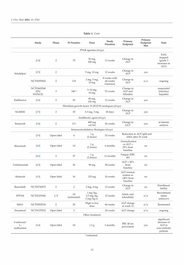
The main aspects of the clinical trials are described in Table 1.
Table 1. Principal characteristics of the study of the drugs in clinical trials.
Study Phase Pt Number DoseStudy
DurationPrimary
Endpoint
PrimaryEndpoint
MetNote
Non-Bile Acids FXR agonists (drugs)
Cilofexor [32] 2 71 30 mg,100 mg 12 weeks
Safety andtolerabilityof Cilofexor
yes
Tropifexor [33] 2 61 30 μg, 60 μg,90 μg 12 weeks
Change inGGT in4 weeks
yes at interimanalysis
EDP-305 NCT03394924 2 68 Dose 1 dose2 12 weeks
20%reduction inALP or nor-
malization ofALP in
12 weeks
n/a ongoing
12
J. Clin. Med. 2021, 10, 1763
Table 1. Cont.
Study Phase Pt Number DoseStudy
DurationPrimary
Endpoint
PrimaryEndpoint
MetNote
PPAR agonists (drugs)
Seladelpar
[34] 2 70 50 mg,200 mg 12 weeks Change in
ALP
Earlystopped(grade 3
increases inALT)
[35] 2 5 mg, 10 mg 12 weeks Change inALP yes
NCT02955602 2 119 2 mg, 5 mg,10 mg
8 weeks with44 weeksextension
Change inALP n/a ongoing
NCT03602560(EN-
HANCE)3 240 * 5–10 mg,
10 mg 52 weeksChange inALP andbilirubin
suspended(interfacehepatits)
Elafibranor [36] 2 45 80 mg,120 mg 12 weeks Change in
ALP yes
Fibroblast growth factor 19 (FGF19) analogues (drugs)
NGM282 [37] 2 45 0.3 mg, 3 mg 28 days Change inALP yes
Antifibrotic agent (drugs)
Setanaxib [38] 2 111 400 mgod/bd 24 weeks Change in
GGT yes at interimanalysis
Immunomodulatory Strategies (drugs)
Rituximab
[39] Open label 6 1 g(2 doses) 52 weeks Reduction in ALP, IgM and
AMA after 36 week
[40] Open label 14 1 g(2 doses) 6 months
Normalizationor ALP <25% frombaseline
no
[41] 2 57 1 g(2 doses) 12 months Fatigue (PBC
40) no
Ustekimumab [42] Open label 20 90 mg 28 weeksALP < 40%
frombaseline
no
Abataceb [43] Open label 16 125 mg 24 weeks
ALP normal-ization or
<40% frombaseline
no
Baricitinib NCT03742973 2 2 2 mg, 4 mg 12 weeks Change inALP no Enrollment
futility
FFP104 NCT02193360 1/2 24(estimated)
1 mg/kg,2.5 mg/kg,2 mg/kg ev
12 weeks Safety andtolerability n/a
Recruitmentstatus
unknown
E6011 NCT03092765 2 29 High or lowdose 64 weeks ALP change
at week 12 n/a Terminated
Etrasimod NCT03155932 Open label 2 24 weeks ALP change n/a ongoing
Other treatment
S-adenosyl-L-
methionine[44] Open label 24 1.2 g 6 months PBC 40 im-
provement yes
significantdecrease of
ALP innon-cirrhotic
patients
* estimated.
13
J. Clin. Med. 2021, 10, 1763
2.2.1. Non-Bile Acids FXR Agonists
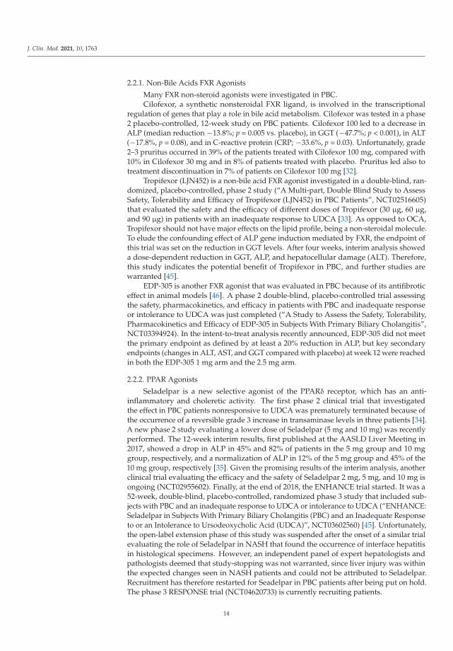
Many FXR non-steroid agonists were investigated in PBC.Cilofexor, a synthetic nonsteroidal FXR ligand, is involved in the transcriptional
regulation of genes that play a role in bile acid metabolism. Cilofexor was tested in a phase2 placebo-controlled, 12-week study on PBC patients. Cilofexor 100 led to a decrease inALP (median reduction −13.8%; p = 0.005 vs. placebo), in GGT (−47.7%; p < 0.001), in ALT(−17.8%, p = 0.08), and in C-reactive protein (CRP; −33.6%, p = 0.03). Unfortunately, grade2–3 pruritus occurred in 39% of the patients treated with Cilofexor 100 mg, compared with10% in Cilofexor 30 mg and in 8% of patients treated with placebo. Pruritus led also totreatment discontinuation in 7% of patients on Cilofexor 100 mg [32].
Tropifexor (LJN452) is a non-bile acid FXR agonist investigated in a double-blind, ran-domized, placebo-controlled, phase 2 study (“A Multi-part, Double Blind Study to AssessSafety, Tolerability and Efficacy of Tropifexor (LJN452) in PBC Patients”, NCT02516605)that evaluated the safety and the efficacy of different doses of Tropifexor (30 μg, 60 μg,and 90 μg) in patients with an inadequate response to UDCA [33]. As opposed to OCA,Tropifexor should not have major effects on the lipid profile, being a non-steroidal molecule.To elude the confounding effect of ALP gene induction mediated by FXR, the endpoint ofthis trial was set on the reduction in GGT levels. After four weeks, interim analysis showeda dose-dependent reduction in GGT, ALP, and hepatocellular damage (ALT). Therefore,this study indicates the potential benefit of Tropifexor in PBC, and further studies arewarranted [45].
EDP-305 is another FXR agonist that was evaluated in PBC because of its antifibroticeffect in animal models [46]. A phase 2 double-blind, placebo-controlled trial assessingthe safety, pharmacokinetics, and efficacy in patients with PBC and inadequate responseor intolerance to UDCA was just completed (“A Study to Assess the Safety, Tolerability,Pharmacokinetics and Efficacy of EDP-305 in Subjects With Primary Biliary Cholangitis”,NCT03394924). In the intent-to-treat analysis recently announced, EDP-305 did not meetthe primary endpoint as defined by at least a 20% reduction in ALP, but key secondaryendpoints (changes in ALT, AST, and GGT compared with placebo) at week 12 were reachedin both the EDP-305 1 mg arm and the 2.5 mg arm.
2.2.2. PPAR Agonists
Seladelpar is a new selective agonist of the PPARδ receptor, which has an anti-inflammatory and choleretic activity. The first phase 2 clinical trial that investigatedthe effect in PBC patients nonresponsive to UDCA was prematurely terminated because ofthe occurrence of a reversible grade 3 increase in transaminase levels in three patients [34].A new phase 2 study evaluating a lower dose of Seladelpar (5 mg and 10 mg) was recentlyperformed. The 12-week interim results, first published at the AASLD Liver Meeting in2017, showed a drop in ALP in 45% and 82% of patients in the 5 mg group and 10 mggroup, respectively, and a normalization of ALP in 12% of the 5 mg group and 45% of the10 mg group, respectively [35]. Given the promising results of the interim analysis, anotherclinical trial evaluating the efficacy and the safety of Seladelpar 2 mg, 5 mg, and 10 mg isongoing (NCT02955602). Finally, at the end of 2018, the ENHANCE trial started. It was a52-week, double-blind, placebo-controlled, randomized phase 3 study that included sub-jects with PBC and an inadequate response to UDCA or intolerance to UDCA (“ENHANCE:Seladelpar in Subjects With Primary Biliary Cholangitis (PBC) and an Inadequate Responseto or an Intolerance to Ursodeoxycholic Acid (UDCA)”, NCT03602560) [45]. Unfortunately,the open-label extension phase of this study was suspended after the onset of a similar trialevaluating the role of Seladelpar in NASH that found the occurrence of interface hepatitisin histological specimens. However, an independent panel of expert hepatologists andpathologists deemed that study-stopping was not warranted, since liver injury was withinthe expected changes seen in NASH patients and could not be attributed to Seladelpar.Recruitment has therefore restarted for Seadelpar in PBC patients after being put on hold.The phase 3 RESPONSE trial (NCT04620733) is currently recruiting patients.
14

J. Clin. Med. 2021, 10, 1763
Elafibranor, a dual PPAR-α/δ agonist, also studied in non-alcoholic steatohepatitis(NASH) [47], was recently tested in a multicenter, randomized, double-blind, placebo-controlled phase 2 study clinical trial recruiting patients with PBC non-responders to UDCA.Data were discussed at the International Liver Congress in Vienna in April 2019 [36]. Forty-five patients were randomized into three arms: Elafibranor 80 mg, Elafibranor 120 mg, andplacebo. After 12 weeks of treatment, a reduction in ALP from baseline was observed in48% patients in the 80 mg group and in 41% in the 120 mg arm; an increase of 3% wasdetected with placebo. Moreover, 67% patients in the 80 mg group (p = 0.001) and 79% ofpatients in the 120 mg group (p < 0.001) reached the secondary endpoint (serum ALP < 1.67ULN, ALP decrease > 15%, total bilirubin < ULN) (NCT03124108). Thus, in July 2019, theUSA FDA and the European Medicines Agency approved Orphan Drug Designation toElafibranor for the treatment of PBC [48].
2.2.3. Fibroblast Growth Factor 19 (FGF19) Analogues
FGF19 acts as a hormone on a cell surface receptor complex in hepatocytes, decreas-ing bile acid synthesis, gluconeogenesis, and lipogenesis. FGF19 expression is inducedby bile-acid-mediated activation of FXR in the gut [49], and it reaches the liver throughportal circulation. In the liver, FGF19 suppresses bile acid synthesis due to the inhibitionof cholesterol 7-α-hydroxylase (CYP7A1) and sterol 12-α-hydroxylase (CYP8B1). More-over, FXR decreases hepatic fibrogenesis by reducing collagen and by increasing matrixmetalloprotease activity in hepatic stellate cells [50].
NGM282 (Aldafermin), an engineered analogue of FGF19, was tested in a 28-day,double-blind, placebo-controlled phase 2 trial. Forty-five PBC patients with an inadequateresponse to UDCA were treated with subcutaneous daily doses of NGM282 at 0.3 mg(n = 14), 3 mg (n = 16), or placebo (n = 15). ALP level had a significant drop in the treatmentgroup, as well as transaminase levels and markers of cholestasis, hepatocellular injury, andinflammation (IgM levels). The reduction in complement component 4 (C4) levels suggeststhat NGM282 acts with a direct inhibition in the de-novo bile acid synthesis through theclassical pathway. The main adverse effect was diarrhea. No effect on itch was detected [37].In contrast to FGF19, no increase in liver cancer risk was observed in animal models treatedwith NGM282 [51]. Longer studies are needed to evaluate the long-term efficacy and safetyof this molecule.
2.2.4. Antifibrotic Agent
Setanaxib (GKT137831) is an inhibitor of Nicotinamide Adenine Dinucleotide Phos-phate (NADPH) oxidases isoforms 1 and 4. NADPH oxidase enzymes, generating reactivespecies of oxygen, play a central role in inflammation and stellate cell-mediated fibrogene-sis [52]. It was demonstrated in animal models of acute biliary injury and steatohepatitisthat GKT137831 reduces hepatocyte apoptosis and liver fibrosis [53]. Thus, a multicenter,randomized, double-blind, placebo-controlled phase 2 study evaluating the safety andthe efficacy of GKT137831 OD or BID in 111 patients with PBC and incomplete responseto UDCA was performed (NCT03226067). Interim analysis showed a reduction in GGTand ALP level in six weeks, without a significant concomitant adverse event. A decreasein GGT of 7%, 12%, and 23% were observed in the placebo, 400 mg OD, and 400 mg BIDgroups, respectively (p < 0.01 for 400 mg BID vs. placebo). A greater GGT reduction wasreached in patients with more advanced disease (GGT ≥ 2.5 X ULN at baseline). Changesin ALP were statistically significant in the 400 mg BID versus placebo [38].
2.2.5. Immunomodulatory Strategies
Since PBC is an autoimmune condition characterized by anti-mitochondrial autoan-tibodies (AMA) and high levels of immunoglobulin M (IgM), many immunosuppres-sive drugs were studied in PBC, including corticosteroid [54], azathioprine [55], cy-closporine [56], methotrexate [57], and mycophenolate mofetil [58]. However, resultswere consistently unsatisfactory. Recently, other molecules were studied in PBC.
15

J. Clin. Med. 2021, 10, 1763
Rituximab, an anti-CD20 antibody currently used in lymphomas and autoimmunesyndromes, was evaluated in PBC due to its promising results in murine models of autoim-mune cholangitis [58]. Three clinical trials in PBC patients with an incomplete response toUDCA were reported. In an open label study, Rituximab (two doses of 1000 mg) induced adecrease in AMA and IgM levels, with only a marginal reduction of ALP after 36 weeks [39].Unfortunately, a similar study including 14 PBC patients showed a significant but onlytransitory reduction in ALP [40]. Finally, Rituximab was demonstrated not to have animpact on fatigue, assessed by PBC-40 [41].
Ustekinumab is an anti-interleukin (IL)-12/23 monoclonal antibody commonly usedin several autoimmune syndromes and inflammatory bowel diseases (IBD). IL-12 and IL-23-mediated Th1/Th17 signaling pathways play a role in the etiopathogenesis of PBC [59].Unfortunately, a multicenter open label trial did not reach the primary endpoint of re-duction in ALP of 40% from the baseline. However, at week 24, a statistically significantdecrease of 12.1% in ALP from baseline was observed [42].
Abatacept is a Cytotoxic T-Lymphocyte Antigen 4 IgG antibody used in rheumatoidand psoriatic arthritis. An open-label, 24-week trial was performed in PBC patients, but nosignificant changes in biochemical enzymes were observed [43].
The efficacy of Baricitinib (LY3009104), a reversible inhibitor of Janus kinase 1 (JAK1)and JAK2 currently used in rheumatoid arthritis, is currently being evaluated in an ongoing,placebo controlled phase 2 trial (NCT03742973) [45].
Other types of molecules are undergoing clinical evaluation in phase 1 and phase 2trials: FFP104 blocks the CD40/CD40L interaction between CD4+ T helper lymphocytesand B cells that are involved in the pathogenesis of PBC (NCT02193360) [60]; E6011 isan anti-chemokine-adhesion molecule CX3CL1 (fractalkine) antibody, which is elevatedin the serum of PBC patients (NCT03092765); Etrasimod is a selective sphingosine-1-phosphate (S1P) receptor (S1PR) modulator targeting S1P receptor subtypes 1, 4, and 5,leading to an inhibition of activated lymphocytes from migrating to sites of inflammation(NCT03155932) [3].
2.2.6. Other Treatment
S-adenosyl-L-methionine, added to UDCA in non-cirrhotic PBC patients, was demon-strated to have a positive effect on markers of cholestasis and quality of life, probablydue to its hepatoprotective effects [44]. In this open label on 24 PBC patients, there wasa significant decrease of ALP, GGT, and total cholesterol over a period of six months. Asignificant improvement of fatigue and pruritus on the PBC-40 questionnaire was alsoobserved.
2.3. Therapies Evaluated in Pre-Clinical Studies
24-norursodeoxycholic acid (norUDCA) differs from UDCA due to the resistancein conjugation with taurine or glycine. NorUDCA increases the cholehepatic shunt ofbile salts, leading to a supra-physiological secretion of bicarbonate. NorUDCA showedpromising results in the treatment of PSC [61], but its efficacy in PBC has yet to be clarified.Up to now, improvements in fibrosis and inflammation were demonstrated in preclinicalstudies on animal model with cholestatic liver diseases [2].
Na+ -Taurocholate Cotransporting Polypeptide (NTCP) is a hepatocellular uptaketransporter of bile salts, and its inhibition by myrcludex B results in hepatoprotectiveeffects, increasing the biliary phospholipid/bile salt ratio. In 3.5-diethoxycarbonyl-1.4-dihydrocollidine-fed mice, a murine model of cholestasis, and in Atp8b1-G308V mice,used for chronic cholestasis, bile salt levels increased in treated animals from 604 ± 277to 1746 ± 719 μm and from 432 ± 280 to 762 ± 288 μm, respectively, while phospholipidoutput was maintained, resulting in a higher phospholipid/bile salt ratio. Thus, it may bebeneficial in some forms of cholestasis, but further studies need to be performed [62].
16

J. Clin. Med. 2021, 10, 1763
3. Primary Sclerosing Cholangitis
Primary sclerosing cholangitis (PSC) is a chronic bile duct disease with a prevalenceof 1–16 per 100,000. PSC is more common in men (comprising 60–70% of patients) and isreported more frequently in Northern European countries and in North America. Moreover,70% of the patients have ulcerative colitis [63]. The diagnosis is based on a combinationof clinical, laboratory, imaging, and histological factors. Endoscopic retrograde cholan-giopancreatography (ERCP) plays a very limited role in the diagnosis of PSC, while itmay be used for the treatment of dominant stenosis [64]. It is well-known that patientsaffected by PSC have a higher risk of cholangiocarcinoma and gallbladder cancer. Up tonow, no pharmacological treatment is universally approved for PSC. The lack of a clearpathogenesis and the absence of consistent endpoints have contributed to the difficultiesin unravelling novel molecular targets and in designing effective clinical trials for PSCtreatment [45]. The principal promising treatments and ongoing trials will be summarizedin this section.
3.1. Therapies in Clinical UseUDCA
The use of UDCA in PSC patients remains controversial to date. Previous smalland uncontrolled studies of short duration consistently reported an improvement in livertests in PSC treated with UDCA [65,66]. The first randomized controlled trial of UDCA(13 to 15 mg/kg) in PSC patients appeared in 1992. Beuers et al. showed a significantimprovement of biochemical parameters, such as bilirubin, ALP, GGT, and transaminases,in six PSC patients treated for one year as compared to placebo [67]. A number of sub-sequent studies evaluated the effect of UDCA at different dosages in PSC. Despite theamelioration of biochemical parameters that appears to be relatively constant in all studies,definite proof for an improvement in “hard endpoints” such as survival, liver transplanta-tion, or progression to CCA is still lacking. In a small cohort of 26 PSC patients, Mitchellet al. reported beneficial effects of UDCA (20 mg/kg) not only on liver tests but alsoon the cholagiographic appearance of the biliary tree evaluated by ERCP and liver fibro-sis [68]. A subsequent randomized controlled trial in 219 PSC patients treated with UDCA(17 to 23 mg/kg) or placebo failed to show a significant improvement in the combinedendpoint “death or liver transplantation”, despite a trend to a reduction in both (31%and 34% reduction, respectively) [69]. Moreover, high doses of UDCA in the range of28–30 mg/kg were shown to be associated with an increased risk of disease progression tocirrhosis, development of varices, CCA, liver transplantation, or death [70]. Unfortunately,three meta-analyses also failed to show an effect of UDCA on mortality or liver transplanta-tion [71–73]. To date, the most recent guidelines by the British Society of Gastroenterologyrecommend not to treat newly diagnosed PSC patients with UDCA routinely [74].
3.2. Therapies Evaluated in Clinical Trials
The principal characteristics of the clinical trials are described in Table 2.
Table 2. Principal characteristics of the study of the drugs in clinical trials.
Study Phase Pt Number DoseStudy
DurationPrimary
Endpoint
PrimaryEndpoint
MetNote
24-norursodeoxycholic
acid (norUDCA)
[61] 2 161 500 mg, 1 g,1.5 gr 16 weeks Change in
ALP yes
NCT03872921 3 300 * 250 mg 6cps/d 2 years
Change inALP andhistology
n/a ongoing
FXR agonist (drugs)
OCA [75] 2 77 1.5–3 mg5–10 mg 24 weeks Change in
ALP yes 5–10 mg
17

J. Clin. Med. 2021, 10, 1763
Table 2. Cont.
Study Phase Pt Number DoseStudy
DurationPrimary
Endpoint
PrimaryEndpoint
MetNote
Cilofexor [76] 2 52 100 mg30 mg 12 weeks
Safety andliver enzyme
improve-ment
yes
NGM282 [77] 2 62 1 mg3 mg 12 weeks Change in
ALP no
ATRA [78] Pilotstudy 15 45 mg/m/d 12 weeks
ALP < 30%from
baselineno
Decreasein ALTand C4
NCT03359174 2 2 10 mg bd 24 weeks Change inALP n/a ongoing
PPAR agonists
Bezafibrates [79] 2 11 200 mg BID 12 weeksimprovements
in liverfunction test
yes
Bezafibrates [79] 2 11 200 mg BID 12 weeksimprovements
in liverfunction test
yes
Antifibrotic therapy (drugs)
Simtuzumab [80] 2 234 75 mg,125 mg 96 weeks Hepaticcollagencontentno
Immunomodulator (drugs)
Timolumab NCT02239211 2 23 8 mg/kg 11 weeksALP < 25%
frombaseline
n/a Awaitingresults
Cenicriviroc NCT02653625 Openlabel 24 150 mg 24 weeks Change in
ALP yes
Vedolizumab [81] Retrospective 102 412 days(median) no
ALP <20%from
baseline
Vidofludimus NCT03722576 2 14 30 mg 6 months Change inALP n/a Awaiting
results
Modulation of gut microbioma (drugs)
Vancomycin NCT03710122 2/3 102 * 24 months Change inALP n/a ongoing
Rifaximin [82] Openlabel 16 550 mg bd 12 weeks Change in
ALP no
Minocycline [83] Pilotstudy 16 100 mg bd 1 year Change in
biochemistry yes
FMT [84] Openlabel 10 24 weeks safety yes
Other treatments (drugs)
Sulfasalazine NCT03561584 2 42 500 mg bd 14 weeks Change inALP n/a ongoing
Curcumin [85] Openlabel 258 750 mg bd 12 weeks
ALP < 1.5ULN or
<40% frombaseline
no
HTD1801 NCT03333928 2 59 500 mg1 gr 18 weeks Change in
ALP n/a Awaitingresults
18
J. Clin. Med. 2021, 10, 1763
Table 2. Cont.
Study Phase Pt Number DoseStudy
DurationPrimary
Endpoint
PrimaryEndpoint
MetNote
DUR-928 NCT03394781 2 5 10 mg50 mg 28 days Change in
ALP n/a ongoing
Docosahexaenoicacid [86] Open
label 23 800 mg bd 12 monthsChange inALP and
safetyyes
Hymecromone NCT02780752 1 18 *1.2 gr2.4 gr3.6 gr
4 days Change inspu n/a ongoing
Orbcel-C NCT02997878 2 56 *0.5, 1.0, 2.5
millioncells/kg
56 daysSafety,
change inALP e ALT
n/a ongoing
* estimated.
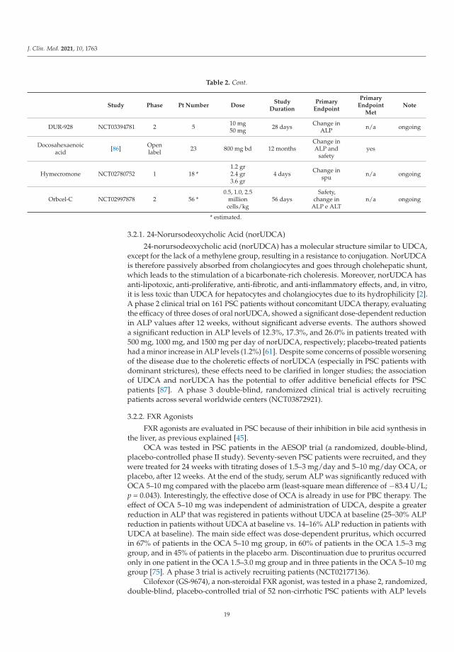
3.2.1. 24-Norursodeoxycholic Acid (norUDCA)
24-norursodeoxycholic acid (norUDCA) has a molecular structure similar to UDCA,except for the lack of a methylene group, resulting in a resistance to conjugation. NorUDCAis therefore passively absorbed from cholangiocytes and goes through cholehepatic shunt,which leads to the stimulation of a bicarbonate-rich choleresis. Moreover, norUDCA hasanti-lipotoxic, anti-proliferative, anti-fibrotic, and anti-inflammatory effects, and, in vitro,it is less toxic than UDCA for hepatocytes and cholangiocytes due to its hydrophilicity [2].A phase 2 clinical trial on 161 PSC patients without concomitant UDCA therapy, evaluatingthe efficacy of three doses of oral norUDCA, showed a significant dose-dependent reductionin ALP values after 12 weeks, without significant adverse events. The authors showeda significant reduction in ALP levels of 12.3%, 17.3%, and 26.0% in patients treated with500 mg, 1000 mg, and 1500 mg per day of norUDCA, respectively; placebo-treated patientshad a minor increase in ALP levels (1.2%) [61]. Despite some concerns of possible worseningof the disease due to the choleretic effects of norUDCA (especially in PSC patients withdominant strictures), these effects need to be clarified in longer studies; the associationof UDCA and norUDCA has the potential to offer additive beneficial effects for PSCpatients [87]. A phase 3 double-blind, randomized clinical trial is actively recruitingpatients across several worldwide centers (NCT03872921).
3.2.2. FXR Agonists
FXR agonists are evaluated in PSC because of their inhibition in bile acid synthesis inthe liver, as previous explained [45].
OCA was tested in PSC patients in the AESOP trial (a randomized, double-blind,placebo-controlled phase II study). Seventy-seven PSC patients were recruited, and theywere treated for 24 weeks with titrating doses of 1.5–3 mg/day and 5–10 mg/day OCA, orplacebo, after 12 weeks. At the end of the study, serum ALP was significantly reduced withOCA 5–10 mg compared with the placebo arm (least-square mean difference of −83.4 U/L;p = 0.043). Interestingly, the effective dose of OCA is already in use for PBC therapy. Theeffect of OCA 5–10 mg was independent of administration of UDCA, despite a greaterreduction in ALP that was registered in patients without UDCA at baseline (25–30% ALPreduction in patients without UDCA at baseline vs. 14–16% ALP reduction in patients withUDCA at baseline). The main side effect was dose-dependent pruritus, which occurredin 67% of patients in the OCA 5–10 mg group, in 60% of patients in the OCA 1.5–3 mggroup, and in 45% of patients in the placebo arm. Discontinuation due to pruritus occurredonly in one patient in the OCA 1.5–3.0 mg group and in three patients in the OCA 5–10 mggroup [75]. A phase 3 trial is actively recruiting patients (NCT02177136).
Cilofexor (GS-9674), a non-steroidal FXR agonist, was tested in a phase 2, randomized,double-blind, placebo-controlled trial of 52 non-cirrhotic PSC patients with ALP levels
19

J. Clin. Med. 2021, 10, 1763
greater than 1.67 ULN. Patients treated with Cilofexor 100 mg had a significant drop inALP, gamma-GT, ALT, and primary bile acids (ALP mean reduction of −13.8%, p = 0.005;gamma-GT mean reduction of 47.7%, p < 0.001; ALT mean reduction of −17.8%, p = 0.08;primary bile acids reduction of −30.5%, p = 0.0008). The main limitations of this trial werethe inclusion of only large-duct PSC cases without cirrhosis and the low prevalence ofIBD [76].
NGM282, a FGF19 analogue, was recently studied in a phase 2, randomized, double-blind, placebo-controlled trial in PSC patients with ALP levels greater than 1.5 × ULN.Despite that no significant changes in ALP from baseline were observed, fibrosis biomark-ers (Enhanced Liver Fibrosis test score and Pro-C3) were significantly improved in thetreatment group [77]. This trial has stimulated discussion about the most appropriatedtarget in PSC [88]. There are no established endpoints in PSC. A recent consensus ofthe International Primary Sclerosing Cholangitis Study Group, reviewing available litera-ture, concluded that the only few candidates as surrogate endpoints in PSC may be ALP,transient elastography, histology, or the combination of ALP and histology and bilirubin;however, no one exceeds level 3 validation [89].
All-trans retinoic acid (ATRA), currently used in acne and in acute promyelocyticleukemia, represses bile acid synthesis through the FXR/RXR nuclear receptor complexpathway [90]. The efficacy of the combination of UDCA (15–23 mg/kg/day) and ATRA(45 mg/m2/day) was tested in 15 PSC patients. Despite ATRA, admiration did not reachthe primary endpoint of the study (30% reduction in serum ALP), and a decrease in ALTand C4 levels were observed [78]. An open-label phase 2 trial evaluating efficacy and thesafety of a lower dose of ATRA is currently ongoing (NCT03359174).
3.2.3. PPAR Agonists
There is a rising number of studies on the efficacy of fibrates in PSC. However, themajority of available data comes from observational or retrospective analyses [3]. A recentretrospective French-Spanish study reported a 40% reduction in ALP levels, together withamelioration of pruritus, after fenofibrate 200 mg/day or bezafibrate 400 mg/day treatment(median duration of therapy of about 1.5 years) in 20 PSC patients [91]. Interestingly,the authors reported a rebound in ALP levels after discontinuation of the PPAR agonistbased on occurrence of biliary stones, tolerability, or worsening of liver tests. It hasto be mentioned, however, that the liver stiffness evaluated by transient elastographysignificantly increased during the study. A small prospective study evaluated the efficacyof bezafibrate (200 mg bid) in 11 PSC patients. After 12 weeks of treatment, ALP and ALTlevels significantly improved in 7 out of 11 (64%) patients and subsequently increased aftertreatment discontinuation [79]. Further studies on fibrates for PSC are warranted.
3.2.4. Antifibrotic Therapy
Despite fibrosis being central in the pathogenesis of the disease, very few antifibroticdrugs have been studied. Lysyl oxidase like-2 (LOXL2) is an enzyme that catalyzes thecrosslinking of collagen and elastin fibers, thereby strengthening the extracellular matrixstructure. Previous studies showed that LOXL2 levels in the serum and liver of PSC patientsare correlated with disease severity [92]. Moreover, the administration of a LOXL2 inhibitorin rodents was shown to reduce the accumulation of hepatic and biliary fibrosis and alsoaccelerate its reversal [93,94]. Unfortunately, no improvement in liver fibrosis was observedin a placebo-controlled, phase 2b trial testing Simtuzumab, a LOXL2 inhibitor. In the trial,a total of 234 patients with compensated PSC were randomized on a 1:1:1 basis to receiveplacebo, weekly subcutaneous injections of Simtuzumab 75 mg, or weekly subcutaneousinjections of Simtuzumab 125 mg for 96 weeks. The study failed to demonstrate any effectof Simtuzumab on hepatic collagen content (measured by morphometry on liver biopsy)and fibrosis stage (measured by the Ishak fibrosis stage) [80].
20

J. Clin. Med. 2021, 10, 1763
3.2.5. Immunomodulators
Although PSC is an immune-mediated disease, traditional immunosuppressive ap-proaches so far failed to demonstrate a clinical benefit in PSC [95]. Timolumab (BTT1023), ahuman monoclonal anti-VAP-1 antibody, was shown to prevent fibrosis in murine modelsof liver injury [96]. A phase 2 clinical trial (BUTEO trial) evaluating the effect of Timolumabin PSC over a 78-day treatment (primary endpoint: reduction of ALP levels by >25% frombaseline) is still ongoing (NCT02239211) [97]. Cenicriviroc, a CCR2/CCR5 antagonist, wastested in a phase 2 trial (PERSEUS trial), and it was proven to cause a modest reductionin ALP (median 18%) after 24 weeks among 24 patients [98]. Moreover, it was shown tohave anti-inflammatory and antifibrotic effects in NASH animal models and in Abcb4(Mdr2−/−) mice [99].
Vedolizumab is a monoclonal antibody directed against the α4β7 integrin, whichis used in the treatment of inflammatory bowel disease. MADCAM-1 is the ligand forα4β7 integrin and is normally expressed in the gut. Since MADCAM-1 is also foundin the liver, administration of vedolizumab is thought to reduce MADCAM-1-inducedleucocyte migration between the gut and the liver [100]. Despite these promising premises,in a retrospective analysis, Vedolizumab treatment did not show any improvement inliver biochemistry in patients affected by IBD and PSC who received at least three dosesof vedolizumab. About 20% of patients experienced a reduction of at least 20% in ALPlevels; however, this outcome was independently associated only with the presence ofcirrhosis [81]. Similar results were reported in a previous retrospective study in 34 patientswith PSC and IBD (16 patients affected by Crohn’s disease and 18 patients by ulcerativecolitis) treated with vedolizumab [101].
Vidofludimus is an inhibitor of the dihydroorotate dehydrogenase that blocks thereplication of activated T- and B-cells and interferes with the JAK/signal transducer [45].A phase 2, open-label clinical trial evaluating the safety and the efficacy of vidofludimus inpatients with PSC will start in 2020 (NCT03722576).
3.2.6. Modulation of the Gut Microbiome
Recent research focused on the gut microbiome as a potential element in the pathogen-esis of PSC. One of the hypothesis is that gut microbiome activates innate immunity withinthe liver, resulting in inflammation and fibrosis in the bile duct [102]. Moreover, studies onthe microbiome and PSC demonstrated that the microbiome of PSC patients is differentfrom healthy controls and IBD-patients [103]. Thus, changing the composition of the gutmicrobiome might reduce inflammation and fibrosis in the bile ducts.
Vancomycin is a glycopeptide antibiotic that also has an immunomodulatory effectdue to the decrease in T cell cytokine production [104]. Vancomycin was compared tometronidazole [105] and to placebo [106] in two randomized trials in PSC patients with orwithout IBD, and a significant reduction in ALP levels and the Mayo score was reported.A phase 2, multicenter clinical trial aiming to recruit 102 adult participants with PSC andevaluating ALP levels at 6, 12, and 18 months is still ongoing (NCT03710122).
Other interesting antibiotics are rifaximin and minocycline. Rifaximin had no effectin decreasing cholestatic markers and the Mayo score in 16 PSC patients [82]. In contrast,minocycline was shown to cause an improvement in ALP levels and the Mayo score in 16patients [83].
Fecal Microbiome Transplantation (FMT) is a promising treatment for PSC patients.In one small pilot study, patients with PSC underwent FMT, and three of them experienceda ≥50% decrease in ALP levels. Its effect may be correlated with the bacterial diversity anddonor engraftment [84].
3.2.7. Other Treatments
Anti-inflammatory drugs such as sulfasalazine and curcumin were tested in PSCpatients. A multicenter, randomized, double-blinded, placebo-controlled trial to assess thebenefit and the safety of sulfasalazine in the treatment of PSC just ended, and results are
21
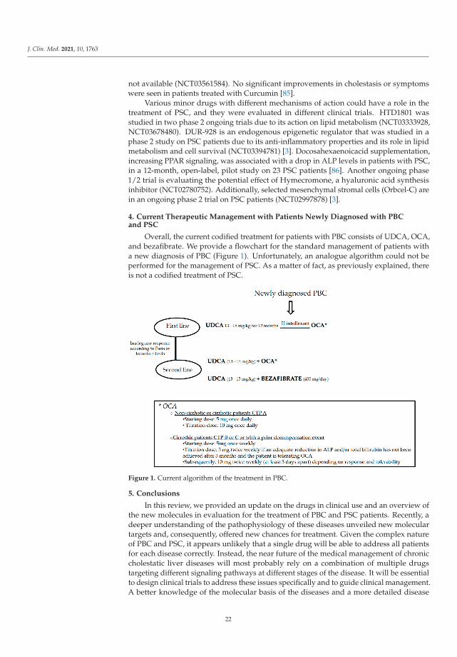
J. Clin. Med. 2021, 10, 1763
not available (NCT03561584). No significant improvements in cholestasis or symptomswere seen in patients treated with Curcumin [85].
Various minor drugs with different mechanisms of action could have a role in thetreatment of PSC, and they were evaluated in different clinical trials. HTD1801 wasstudied in two phase 2 ongoing trials due to its action on lipid metabolism (NCT03333928,NCT03678480). DUR-928 is an endogenous epigenetic regulator that was studied in aphase 2 study on PSC patients due to its anti-inflammatory properties and its role in lipidmetabolism and cell survival (NCT03394781) [3]. Docosahexaenoicacid supplementation,increasing PPAR signaling, was associated with a drop in ALP levels in patients with PSC,in a 12-month, open-label, pilot study on 23 PSC patients [86]. Another ongoing phase1/2 trial is evaluating the potential effect of Hymecromone, a hyaluronic acid synthesisinhibitor (NCT02780752). Additionally, selected mesenchymal stromal cells (Orbcel-C) arein an ongoing phase 2 trial on PSC patients (NCT02997878) [3].
4. Current Therapeutic Management with Patients Newly Diagnosed with PBCand PSC
Overall, the current codified treatment for patients with PBC consists of UDCA, OCA,and bezafibrate. We provide a flowchart for the standard management of patients witha new diagnosis of PBC (Figure 1). Unfortunately, an analogue algorithm could not beperformed for the management of PSC. As a matter of fact, as previously explained, thereis not a codified treatment of PSC.
Figure 1. Current algorithm of the treatment in PBC.
5. Conclusions
In this review, we provided an update on the drugs in clinical use and an overview ofthe new molecules in evaluation for the treatment of PBC and PSC patients. Recently, adeeper understanding of the pathophysiology of these diseases unveiled new moleculartargets and, consequently, offered new chances for treatment. Given the complex natureof PBC and PSC, it appears unlikely that a single drug will be able to address all patientsfor each disease correctly. Instead, the near future of the medical management of chroniccholestatic liver diseases will most probably rely on a combination of multiple drugstargeting different signaling pathways at different stages of the disease. It will be essentialto design clinical trials to address these issues specifically and to guide clinical management.A better knowledge of the molecular basis of the diseases and a more detailed disease
22
J. Clin. Med. 2021, 10, 1763
stratification based on patient characteristics and disease behavior remain therefore thecornerstones to devise new effective treatments for PBC and PSC patients.
Author Contributions: M.M. (Marta Mazzetti) writing—original draft preparation, conceptualisa-tion, methodology; G.M. data curation, writing—review and editing; M.M. (Martina Mancinelli)data curation, writing—review and editing; A.B. supervision, validation; M.M. (Marco Marzioni)supervision, validation; L.M. conceptualisation, validation, writing—review and editing. All authorshave read and agreed to the published version of the manuscript.
Funding: This research received no external funding.
Institutional Review Board Statement: Not applicable.
Informed Consent Statement: Not applicable.
Data Availability Statement: No new data were created or analyzed in this study. Data sharing isnot applicable to this article.
Conflicts of Interest: The authors declare no conflict of interest.
References
1. European Association for the Study of the Liver. Electronic address eee, European Association for the Study of the L: EASLClinical Practice Guidelines: The diagnosis and management of patients with primary biliary cholangitis. J. Hepatol. 2017, 67,145–172. [CrossRef]
2. Beuers, U.; Trauner, M.; Jansen, P.; Poupon, R. New paradigms in the treatment of hepatic cholestasis: From UDCA to FXR, PXRand beyond. J. Hepatol. 2015, 62 (Suppl. 1), S25–S37. [CrossRef]
3. Wagner, M.; Fickert, P. Drug Therapies for Chronic Cholestatic Liver Diseases. Annu. Rev. Pharmacol. Toxicol. 2020, 60, 503–527.[CrossRef] [PubMed]
4. Beuers, U. Drug insight: Mechanisms and sites of action of ursodeoxycholic acid in cholestasis. Nat. Clin. Pract. Gastroenterol.Hepatol. 2006, 3, 318–328. [CrossRef] [PubMed]
5. Paumgartner, G.; Beuers, U. Ursodeoxycholic acid in cholestatic liver disease: Mechanisms of action and therapeutic use revisited.Hepatology 2002, 36, 525–531. [CrossRef] [PubMed]
6. Beuers, U.; Maroni, L.; Elferink, R.O. The biliary HCO(3)(-) umbrella: Experimental evidence revisited. Curr. Opin. Gastroenterol.2012, 28, 253–257. [CrossRef] [PubMed]
7. Floreani, A.; Mangini, C. Primary biliary cholangitis: Old and novel therapy. Eur. J. Intern. Med. 2018, 47, 1–5. [CrossRef][PubMed]
8. Ter Borg, P.C.; Schalm, S.W.; Hansen, B.E.; van Buuren, H.R.; Dutch, P.B.C.S.G. Prognosis of ursodeoxycholic Acid-treated patientswith primary biliary cirrhosis. Results of a 10-yr cohort study involving 297 patients. Am. J. Gastroenterol. 2006, 101, 2044–2050.[CrossRef]
9. Corpechot, C.; Carrat, F.; Bonnand, A.M.; Poupon, R.E.; Poupon, R. The effect of ursodeoxycholic acid therapy on liver fibrosisprogression in primary biliary cirrhosis. Hepatology 2000, 32, 1196–1199. [CrossRef]
10. Harms, M.H.; van Buuren, H.R.; Corpechot, C.; Thorburn, D.; Janssen, H.L.A.; Lindor, K.D.; Hirschfield, G.M.; Pares, A.; Floreani,A.; Mayo, M.J.; et al. Ursodeoxycholic acid therapy and liver transplant-free survival in patients with primary biliary cholangitis.J. Hepatol. 2019, 71, 357–365. [CrossRef]
11. Carbone, M.; Nardi, A.; Flack, S.; Carpino, G.; Varvaropoulou, N.; Gavrila, C.; Spicer, A.; Badrock, J.; Bernuzzi, F.; Cardinale, V.;et al. Pretreatment prediction of response to ursodeoxycholic acid in primary biliary cholangitis: Development and validation ofthe UDCA Response Score. Lancet Gastroenterol. Hepatol. 2018, 3, 626–634. [CrossRef]
12. Carbone, M.; Mells, G.F.; Pells, G.; Dawwas, M.F.; Newton, J.L.; Heneghan, M.A.; Neuberger, J.M.; Day, D.B.; Ducker, S.J.;Consortium, U.P.; et al. Sex and age are determinants of the clinical phenotype of primary biliary cirrhosis and response toursodeoxycholic acid. Gastroenterology 2013, 144, 560–569. [CrossRef] [PubMed]
13. Invernizzi, P.; Floreani, A.; Carbone, M.; Marzioni, M.; Craxi, A.; Muratori, L.; Vespasiani Gentilucci, U.; Gardini, I.; Gasbarrini,A.; Kruger, P.; et al. Primary Biliary Cholangitis: Advances in management and treatment of the disease. Dig. Liver Dis. 2017, 49,841–846. [CrossRef] [PubMed]
14. Hirschfield, G.M.; Mason, A.; Luketic, V.; Lindor, K.; Gordon, S.C.; Mayo, M.; Kowdley, K.V.; Vincent, C.; Bodhenheimer, H.C., Jr.;Pares, A.; et al. Efficacy of obeticholic acid in patients with primary biliary cirrhosis and inadequate response to ursodeoxycholicacid. Gastroenterology 2015, 148, 751–761.e758. [CrossRef]
15. Nevens, F.; Andreone, P.; Mazzella, G.; Strasser, S.I.; Bowlus, C.; Invernizzi, P.; Drenth, J.P.; Pockros, P.J.; Regula, J.; Beuers, U.;et al. A Placebo-Controlled Trial of Obeticholic Acid in Primary Biliary Cholangitis. N. Engl. J. Med. 2016, 375, 631–643. [CrossRef]
16. Trauner, M.; Nevens, F.; Shiffman, M.L.; Drenth, J.P.H.; Bowlus, C.L.; Vargas, V.; Andreone, P.; Hirschfield, G.M.; Pencek, R.;Malecha, E.S.; et al. Long-term efficacy and safety of obeticholic acid for patients with primary biliary cholangitis: 3-year resultsof an international open-label extension study. Lancet Gastroenterol. Hepatol. 2019, 4, 445–453. [CrossRef]
23
J. Clin. Med. 2021, 10, 1763
17. Bowlus, C.L.; Pockros, P.J.; Kremer, A.E.; Pares, A.; Forman, L.M.; Drenth, J.P.H.; Ryder, S.D.; Terracciano, L.; Jin, Y.; Liberman, A.;et al. Long-Term Obeticholic Acid Therapy Improves Histological Endpoints in Patients with Primary Biliary Cholangitis. Clin.Gastroenterol. Hepatol. 2020, 18, 1170–1178.e1176. [CrossRef]
18. Kowdley, K.V.; Luketic, V.; Chapman, R.; Hirschfield, G.M.; Poupon, R.; Schramm, C.; Vincent, C.; Rust, C.; Parés, A.; Mason,A.; et al. A randomized trial of obeticholic acid monotherapy in patients with primary biliary cholangitis. Hepatology 2018, 67,1890–1902. [CrossRef]
19. Hirschfield, G.M.; Dyson, J.K.; Alexander, G.J.M.; Chapman, M.H.; Collier, J.; Hübscher, S.; Patanwala, I.; Pereira, S.P.; Thain, C.;Thorburn, D.; et al. The British Society of Gastroenterology/UK-PBC primary biliary cholangitis treatment and managementguidelines. Gut 2018, 67, 1568–1594. [CrossRef]
20. Lindor, K.D.; Bowlus, C.L.; Boyer, J.; Levy, C.; Mayo, M. Primary Biliary Cholangitis: 2018 Practice Guidance from the AmericanAssociation for the Study of Liver Diseases. Hepatology 2019, 69, 394–419.
21. Kok, T.; Bloks, V.W.; Wolters, H.; Havinga, R.; Jansen, P.L.; Staels, B.; Kuipers, F. Peroxisome proliferator-activated receptor alpha(PPARalpha)-mediated regulation of multidrug resistance 2 (Mdr2) expression and function in mice. Biochem. J. 2003, 369 Pt 3,539–547. [CrossRef]
22. Mukundan, L.; Odegaard, J.I.; Morel, C.R.; Heredia, J.E.; Mwangi, J.W.; Ricardo-Gonzalez, R.R.; Goh, Y.P.; Eagle, A.R.; Dunn, S.E.;Awakuni, J.U.; et al. PPAR-delta senses and orchestrates clearance of apoptotic cells to promote tolerance. Nat. Med. 2009, 15,1266–1272. [CrossRef]
23. Nozaki, Y.; Harada, K.; Sanzen, T.; Nakanuma, Y. PPARgamma ligand attenuates portal inflammation in the MRL-lpr mouse: Anew strategy to restrain cholangiopathy in primary biliary cirrhosis. Med. Mol. Morphol. 2013, 46, 153–159. [CrossRef] [PubMed]
24. Corpechot, C.; Chazouillères, O.; Rousseau, A.; Le Gruyer, A.; Habersetzer, F.; Mathurin, P.; Goria, O.; Potier, P.; Minello, A.;Silvain, C.; et al. A Placebo-Controlled Trial of Bezafibrate in Primary Biliary Cholangitis. N. Engl. J. Med. 2018, 378, 2171–2181.[CrossRef] [PubMed]
25. Reig, A.; Sese, P.; Pares, A. Effects of Bezafibrate on Outcome and Pruritus in Primary Biliary Cholangitis with SuboptimalUrsodeoxycholic Acid Response. Am. J. Gastroenterol. 2018, 113, 49–55. [CrossRef]
26. Cheung, A.C.; Lapointe-Shaw, L.; Kowgier, M.; Meza-Cardona, J.; Hirschfield, G.M.; Janssen, H.L.; Feld, J.J. Combined ursodeoxy-cholic acid (UDCA) and fenofibrate in primary biliary cholangitis patients with incomplete UDCA response may improveoutcomes. Aliment. Pharm. Ther. 2016, 43, 283–293. [CrossRef]
27. Duan, W.; Ou, X.; Wang, X.; Wang, Y.; Zhao, X.; Wang, Q.; Wu, X.; Zhang, W.; Ma, H.; You, H.; et al. Efficacy and safety offenofibrate add-on therapy for patients with primary biliary cholangitis and a suboptimal response to UDCA. Rev. Esp. Enferm.Dig. 2018, 110, 557–563. [CrossRef] [PubMed]
28. Rautiainen, H.; Karkkainen, P.; Karvonen, A.L.; Nurmi, H.; Pikkarainen, P.; Nuutinen, H.; Farkkila, M. Budesonide combinedwith UDCA to improve liver histology in primary biliary cirrhosis: A three-year randomized trial. Hepatology 2005, 41, 747–752.[CrossRef]
29. Leuschner, M.; Maier, K.P.; Schlichting, J.; Strahl, S.; Herrmann, G.; Dahm, H.H.; Ackermann, H.; Happ, J.; Leuschner, U. Oralbudesonide and ursodeoxycholic acid for treatment of primary biliary cirrhosis: Results of a prospective double-blind trial.Gastroenterology 1999, 117, 918–925. [CrossRef]
30. Hirschfield, G.M.; Beuers, U.; Kupcinskas, L.; Ott, P.; Bergquist, A.; Farkkila, M.; Manns, M.P.; Pares, A.; Spengler, U.; Stiess, M.;et al. A placebo-controlled randomised trial of budesonide for PBC following an insufficient response to UDCA. J. Hepatol. 2021,74, 321–329. [CrossRef]
31. Hempfling, W.; Grunhage, F.; Dilger, K.; Reichel, C.; Beuers, U.; Sauerbruch, T. Pharmacokinetics and pharmacodynamic action ofbudesonide in early- and late-stage primary biliary cirrhosis. Hepatology 2003, 38, 196–202. [CrossRef]
32. Kowdley, K.V.; Minuk, G.Y.; Pagadala, M.R.; Gulamhusein, A.; Swain, M.G.; Neff, G.W.; Zogg, D.; Bowlus, C.L.; Agarwal,K.; Yoshida, E.M. The nonsteroidal farnesoid x receptor (FXR) agonist cilofexor improves liver biochemistry in patients withprimary biliary cholangitis (PBC): A phase 2, randomized, placebo-controlled trial. In Hepatology; Wiley: Hoboken, NJ, USA, 2019;pp. 31A–32A.
33. Schramm, C.; Hirschfield, G.; Mason, A.; Wedemeyer, H.; Klickstein, L.; Neelakantham, S.; Koo, P.; Sanni, J.; Badman, M.; Jones,D. Early assessment of safety and efficacy of tropifexor, a potent non bile-acid FXR agonist, in patients with primary biliarycholangitis: An interim analysis of an ongoing phase 2 study. J. Hepatol. 2018, 68, S103. [CrossRef]
34. Jones, D.; Boudes, P.F.; Swain, M.G.; Bowlus, C.L.; Galambos, M.R.; Bacon, B.R.; Doerffel, Y.; Gitlin, N.; Gordon, S.C.; Odin, J.A.;et al. Seladelpar (MBX-8025), a selective PPAR-delta agonist, in patients with primary biliary cholangitis with an inadequateresponse to ursodeoxycholic acid: A double-blind, randomised, placebo-controlled, phase 2, proof-of-concept study. LancetGastroenterol. Hepatol. 2017, 2, 716–726. [CrossRef]
35. Treatment Efficacy and Safety of Low Dose Seladelpar, a Selective PPAR-δ Agonist, in Patients with Primary Biliary Cholan-Gitis: Twelve-Week Interim Analysis of an International, Randomized, Dose Ranging, Phase 2 Study. Available online: https://aasldpubs.onlinelibrary.wiley.com/doi/abs/10.1002/hep.29634 (accessed on 21 January 2021).
36. Jörn, P.D.S.; Pares, A.; Kowdley, K.V.; Heneghan, M.; Caldwell, S.; Pratt, D.; Bonder, A.; Hirschfield, G.M.; Bchir, M.; Cynthia, L.LBO-02-Elafibranor, a peroxisome proliferator-activted receptor alpha and delta agonist demonstrates favourable efficacy andsafety in patients with primary biliary cholangitis and inadequate response to ursodeoxycholic acid treatment. J. Hepatol. 2019,70, e128. [CrossRef]
24

J. Clin. Med. 2021, 10, 1763
37. Mayo, M.J.; Wigg, A.J.; Leggett, B.A.; Arnold, H.; Thompson, A.J.; Weltman, M.; Carey, E.J.; Muir, A.J.; Ling, L.; Rossi, S.J.;et al. NGM282 for Treatment of Patients with Primary Biliary Cholangitis: A Multicenter, Randomized, Double-Blind, Placebo-Controlled Trial. Hepatol. Commun. 2018, 2, 1037–1050. [CrossRef] [PubMed]
38. Dalekos, G.; Invernizzi, P.; Nevens, F.; Hans, V.V.; Zigmond, E.; Andrade, R.J.; Ben Ari, Z.; Heneghan, M.; Huang, J.; Harrison, S.Efficacy of GKT831 in patients with primary biliary cholangitis and inadequate response to ursodeoxycholic acid: Interim efficacyresults of a phase 2 clinical trial. J. Hepatol. 2019, 70, E1–E2. [CrossRef]
39. Tsuda, M.; Moritoki, Y.; Lian, Z.X.; Zhang, W.; Yoshida, K.; Wakabayashi, K.; Yang, G.X.; Nakatani, T.; Vierling, J.; Lindor, K.;et al. Biochemical and immunologic effects of rituximab in patients with primary biliary cirrhosis and an incomplete response toursodeoxycholic acid. Hepatology 2012, 55, 512–521. [CrossRef]
40. Myers, R.P.; Swain, M.G.; Lee, S.S.; Shaheen, A.A.; Burak, K.W. B-cell depletion with rituximab in patients with primary biliarycirrhosis refractory to ursodeoxycholic acid. Am. J. Gastroenterol. 2013, 108, 933–941. [CrossRef] [PubMed]
41. Khanna, A.; Jopson, L.; Howel, D.; Bryant, A.; Blamire, A.; Newton, J.L.; Jones, D.E. Rituximab Is Ineffective for Treatment ofFatigue in Primary Biliary Cholangitis: A Phase 2 Randomized Controlled Trial. Hepatology 2019, 70, 1646–1657. [CrossRef][PubMed]
42. Hirschfield, G.M.; Gershwin, M.E.; Strauss, R.; Mayo, M.J.; Levy, C.; Zou, B.; Johanns, J.; Nnane, I.P.; Dasgupta, B.; Li, K.;et al. Ustekinumab for patients with primary biliary cholangitis who have an inadequate response to ursodeoxycholic acid: Aproof-of-concept study. Hepatology 2016, 64, 189–199. [CrossRef] [PubMed]
43. Bowlus, C.L.; Yang, G.X.; Liu, C.H.; Johnson, C.R.; Dhaliwal, S.S.; Frank, D.; Levy, C.; Peters, M.G.; Vierling, J.M.; Gershwin, M.E.Therapeutic trials of biologics in primary biliary cholangitis: An open label study of abatacept and review of the literature. J.Autoimmun. 2019, 101, 26–34. [CrossRef] [PubMed]
44. Wunsch, E.; Raszeja-Wyszomirska, J.; Barbier, O.; Milkiewicz, M.; Krawczyk, M.; Milkiewicz, P. Effect of S-adenosyl-L-methionineon liver biochemistry and quality of life in patients with primary biliary cholangitis treated with ursodeoxycholic acid. Aprospective, open label pilot study. J. Gastrointestin. Liver Dis. 2018, 27, 273–279. [CrossRef] [PubMed]
45. Gerussi, A.; D’Amato, D.; Cristoferi, L.; O’Donnell, S.E.; Carbone, M.; Invernizzi, P. Multiple therapeutic targets in rare cholestaticliver diseases: Time to redefine treatment strategies. Ann. Hepatol. 2020, 19, 5–16. [CrossRef]
46. An, P.; Wei, G.; Huang, P.; Li, W.; Qi, X.; Lin, Y.; Vaid, K.A.; Wang, J.; Zhang, S.; Li, Y.; et al. A novel non-bile acid FXR agonistEDP-305 potently suppresses liver injury and fibrosis without worsening of ductular reaction. Liver Int. 2020, 40, 1655–1669.[CrossRef]
47. Ratziu, V.; Harrison, S.A.; Francque, S.; Bedossa, P.; Lehert, P.; Serfaty, L.; Romero-Gomez, M.; Boursier, J.; Abdelmalek, M.;Caldwell, S.; et al. Elafibranor, an Agonist of the Peroxisome Proliferator-Activated Receptor-alpha and -delta, Induces Resolutionof Nonalcoholic Steatohepatitis Without Fibrosis Worsening. Gastroenterology 2016, 150, 1147–1159.e1145. [CrossRef]
48. Galoosian, A.; Hanlon, C.; Zhang, J.; Holt, E.W.; Yimam, K.K. Clinical Updates in Primary Biliary Cholangitis: Trends, Epidemiol-ogy, Diagnostics, and New Therapeutic Approaches. J. Clin. Transl. Hepatol. 2020, 8, 49–60. [CrossRef]
49. Kliewer, S.A.; Mangelsdorf, D.J. Bile Acids as Hormones: The FXR-FGF15/19 Pathway. Dig. Dis. 2015, 33, 327–331. [CrossRef]50. Schumacher, J.D.; Kong, B.; Wu, J.; Rizzolo, D.; Armstrong, L.E.; Chow, M.D.; Goedken, M.; Lee, Y.H.; Guo, G.L. Direct and
Indirect Effects of Fibroblast Growth Factor (FGF) 15 and FGF19 on Liver Fibrosis Development. Hepatology 2020, 71, 670–685.[CrossRef]
51. Zhou, M.; Learned, R.M.; Rossi, S.J.; DePaoli, A.M.; Tian, H.; Ling, L. Engineered fibroblast growth factor 19 reduces liver injuryand resolves sclerosing cholangitis in Mdr2-deficient mice. Hepatology 2016, 63, 914–929. [CrossRef]
52. Lambeth, J.D. NOX enzymes and the biology of reactive oxygen. Nat. Rev. Immunol. 2004, 4, 181–189. [CrossRef] [PubMed]53. Aoyama, T.; Paik, Y.H.; Watanabe, S.; Laleu, B.; Gaggini, F.; Fioraso-Cartier, L.; Molango, S.; Heitz, F.; Merlot, C.; Szyndralewiez,
C.; et al. Nicotinamide adenine dinucleotide phosphate oxidase in experimental liver fibrosis: GKT137831 as a novel potentialtherapeutic agent. Hepatology 2012, 56, 2316–2327. [CrossRef]
54. Wolfhagen, F.H.; van Buuren, H.R.; Schalm, S.W. Combined treatment with ursodeoxycholic acid and prednisone in primarybiliary cirrhosis. Neth. J. Med. 1994, 44, 84–90.
55. Wolfhagen, F.H.; van Hoogstraten, H.J.; van Buuren, H.R.; van Berge-Henegouwen, G.P.; ten Kate, F.J.; Hop, W.C.; van der Hoek,E.W.; Kerbert, M.J.; van Lijf, H.H.; den Ouden, J.W.; et al. Triple therapy with ursodeoxycholic acid, prednisone and azathioprinein primary biliary cirrhosis: A 1-year randomized, placebo-controlled study. J. Hepatol. 1998, 29, 736–742. [CrossRef]
56. Wiesner, R.H.; Ludwig, J.; Lindor, K.D.; Jorgensen, R.A.; Baldus, W.P.; Homburger, H.A.; Dickson, E.R. A controlled trial ofcyclosporine in the treatment of primary biliary cirrhosis. N. Engl. J. Med. 1990, 322, 1419–1424. [CrossRef] [PubMed]
57. Combes, B.; Emerson, S.S.; Flye, N.L.; Munoz, S.J.; Luketic, V.A.; Mayo, M.J.; McCashland, T.M.; Zetterman, R.K.; Peters, M.G.;Di Bisceglie, A.M.; et al. Methotrexate (MTX) plus ursodeoxycholic acid (UDCA) in the treatment of primary biliary cirrhosis.Hepatology 2005, 42, 1184–1193. [CrossRef] [PubMed]
58. Treiber, G.; Malfertheiner, P. Mycophenolate mofetil for the treatment of primary biliary cirrhosis in patients with an incompleteresponse to ursodeoxycholic acid. J. Clin. Gastroenterol. 2005, 39, 837–838; author reply 838. [CrossRef]
59. Yang, C.Y.; Ma, X.; Tsuneyama, K.; Huang, S.; Takahashi, T.; Chalasani, N.P.; Bowlus, C.L.; Yang, G.X.; Leung, P.S.; Ansari, A.A.;et al. IL-12/Th1 and IL-23/Th17 biliary microenvironment in primary biliary cirrhosis: Implications for therapy. Hepatology 2014,59, 1944–1953. [CrossRef] [PubMed]
25

J. Clin. Med. 2021, 10, 1763
60. Tanaka, H.; Yang, G.X.; Iwakoshi, N.; Knechtle, S.J.; Kawata, K.; Tsuneyama, K.; Leung, P.; Coppel, R.L.; Ansari, A.A.; Joh, T.; et al.Anti-CD40 ligand monoclonal antibody delays the progression of murine autoimmune cholangitis. Clin. Exp. Immunol. 2013, 174,364–371. [CrossRef]
61. Fickert, P.; Hirschfield, G.M.; Denk, G.; Marschall, H.U.; Altorjay, I.; Farkkila, M.; Schramm, C.; Spengler, U.; Chapman, R.;Bergquist, A.; et al. norUrsodeoxycholic acid improves cholestasis in primary sclerosing cholangitis. J. Hepatol. 2017, 67, 549–558.[CrossRef] [PubMed]
62. Slijepcevic, D.; Roscam Abbing, R.L.P.; Fuchs, C.D.; Haazen, L.C.M.; Beuers, U.; Trauner, M.; Oude Elferink, R.P.J.; van de Graaf,S.F.J. Na(+) -taurocholate cotransporting polypeptide inhibition has hepatoprotective effects in cholestasis in mice. Hepatology2018, 68, 1057–1069. [CrossRef]
63. Tabibian, J.H.; Ali, A.H.; Lindor, K.D. Primary Sclerosing Cholangitis, Part 1: Epidemiology, Etiopathogenesis, Clinical Features,and Treatment. Gastroenterol. Hepatol. 2018, 14, 293–304.
64. European Society of Gastrointestinal Endoscopy; European Association for the Study of the Liver. Role of endoscopy in primarysclerosing cholangitis: European Society of Gastrointestinal Endoscopy (ESGE) and European Association for the Study of theLiver (EASL) Clinical Guideline. J. Hepatol. 2017, 66, 1265–1281. [CrossRef] [PubMed]
65. Chazouilleres, O.; Poupon, R.; Capron, J.P.; Metman, E.H.; Dhumeaux, D.; Amouretti, M.; Couzigou, P.; Labayle, D.; Trinchet, J.C.Ursodeoxycholic acid for primary sclerosing cholangitis. J. Hepatol. 1990, 11, 120–123. [CrossRef]
66. O’Brien, C.B.; Senior, J.R.; Arora-Mirchandani, R.; Batta, A.K.; Salen, G. Ursodeoxycholic acid for the treatment of primarysclerosing cholangitis: A 30-month pilot study. Hepatology 1991, 14, 838–847. [CrossRef] [PubMed]
67. Beuers, U.; Spengler, U.; Kruis, W.; Aydemir, U.; Wiebecke, B.; Heldwein, W.; Weinzierl, M.; Pape, G.R.; Sauerbruch, T.;Paumgartner, G. Ursodeoxycholic acid for treatment of primary sclerosing cholangitis: A placebo-controlled trial. Hepatology1992, 16, 707–714. [CrossRef] [PubMed]
68. Mitchell, S.A.; Bansi, D.S.; Hunt, N.; Von Bergmann, K.; Fleming, K.A.; Chapman, R.W. A preliminary trial of high-doseursodeoxycholic acid in primary sclerosing cholangitis. Gastroenterology 2001, 121, 900–907. [CrossRef]
69. Olsson, R.; Boberg, K.M.; de Muckadell, O.S.; Lindgren, S.; Hultcrantz, R.; Folvik, G.; Bell, H.; Gangsoy-Kristiansen, M.; Matre,J.; Rydning, A.; et al. High-dose ursodeoxycholic acid in primary sclerosing cholangitis: A 5-year multicenter, randomized,controlled study. Gastroenterology 2005, 129, 1464–1472. [CrossRef]
70. Lindor, K.D.; Kowdley, K.V.; Luketic, V.A.; Harrison, M.E.; McCashland, T.; Befeler, A.S.; Harnois, D.; Jorgensen, R.; Petz, J.;Keach, J.; et al. High-dose ursodeoxycholic acid for the treatment of primary sclerosing cholangitis. Hepatology 2009, 50, 808–814.[CrossRef]
71. Shi, J.; Li, Z.; Zeng, X.; Lin, Y.; Xie, W.F. Ursodeoxycholic acid in primary sclerosing cholangitis: Meta-analysis of randomizedcontrolled trials. Hepatol. Res. 2009, 39, 865–873. [CrossRef]
72. Poropat, G.; Giljaca, V.; Stimac, D.; Gluud, C. Bile acids for primary sclerosing cholangitis. Cochrane Database Syst. Rev. 2011,CD003626. [CrossRef]
73. Triantos, C.K.; Koukias, N.M.; Nikolopoulou, V.N.; Burroughs, A.K. Meta-analysis: Ursodeoxycholic acid for primary sclerosingcholangitis. Aliment. Pharm. Ther. 2011, 34, 901–910. [CrossRef]
74. Chapman, M.H.; Thorburn, D.; Hirschfield, G.M.; Webster, G.G.J.; Rushbrook, S.M.; Alexander, G.; Collier, J.; Dyson, J.K.; Jones,D.E.; Patanwala, I.; et al. British Society of Gastroenterology and UK-PSC guidelines for the diagnosis and management ofprimary sclerosing cholangitis. Gut 2019, 68, 1356–1378. [CrossRef] [PubMed]
75. Kowdley, K.V.; Vuppalanchi, R.; Levy, C.; Floreani, A.; Andreone, P.; LaRusso, N.F.; Shrestha, R.; Trotter, J.; Goldberg, D.;Rushbrook, S.; et al. A randomized, placebo-controlled, phase II study of obeticholic acid for primary sclerosing cholangitis. J.Hepatol. 2020, 73, 94–101. [CrossRef] [PubMed]
76. Trauner, M.; Gulamhusein, A.; Hameed, B.; Caldwell, S.; Shiffman, M.L.; Landis, C.; Eksteen, B.; Agarwal, K.; Muir, A.; Rushbrook,S.; et al. The Nonsteroidal Farnesoid X Receptor Agonist Cilofexor (GS-9674) Improves Markers of Cholestasis and Liver Injuryin Patients with Primary Sclerosing Cholangitis. Hepatology 2019, 70, 788–801. [CrossRef] [PubMed]
77. Hirschfield, G.M.; Chazouilleres, O.; Drenth, J.P.; Thorburn, D.; Harrison, S.A.; Landis, C.S.; Mayo, M.J.; Muir, A.J.; Trotter,J.F.; Leeming, D.J.; et al. Effect of NGM282, an FGF19 analogue, in primary sclerosing cholangitis: A multicenter, randomized,double-blind, placebo-controlled phase II trial. J. Hepatol. 2019, 70, 483–493. [CrossRef] [PubMed]
78. Assis, D.N.; Abdelghany, O.; Cai, S.Y.; Gossard, A.A.; Eaton, J.E.; Keach, J.C.; Deng, Y.; Setchell, K.D.; Ciarleglio, M.; Lindor,K.D.; et al. Combination Therapy of All-Trans Retinoic Acid with Ursodeoxycholic Acid in Patients with Primary SclerosingCholangitis: A Human Pilot Study. J. Clin. Gastroenterol. 2017, 51, e11–e16. [CrossRef]
79. Mizuno, S.; Hirano, K.; Isayama, H.; Watanabe, T.; Yamamoto, N.; Nakai, Y.; Sasahira, N.; Tada, M.; Omata, M.; Koike, K.Prospective study of bezafibrate for the treatment of primary sclerosing cholangitis. J. Hepatobiliary Pancreat. Sci. 2015, 22, 766–770.[CrossRef]
80. Muir, A.J.; Levy, C.; Janssen, H.L.A.; Montano-Loza, A.J.; Shiffman, M.L.; Caldwell, S.; Luketic, V.; Ding, D.; Jia, C.; McColgan,B.J.; et al. Simtuzumab for Primary Sclerosing Cholangitis: Phase 2 Study Results with Insights on the Natural History of theDisease. Hepatology 2019, 69, 684–698. [CrossRef]
81. Lynch, K.D.; Chapman, R.W.; Keshav, S.; Montano-Loza, A.J.; Mason, A.L.; Kremer, A.E.; Vetter, M.; de Krijger, M.; Ponsioen, C.Y.;Trivedi, P.; et al. Effects of Vedolizumab in Patients with Primary Sclerosing Cholangitis and Inflammatory Bowel Diseases. Clin.Gastroenterol. Hepatol. 2020, 18, 179–187. [CrossRef]
26

J. Clin. Med. 2021, 10, 1763
82. Tabibian, J.H.; Gossard, A.; El-Youssef, M.; Eaton, J.E.; Petz, J.; Jorgensen, R.; Enders, F.B.; Tabibian, A.; Lindor, K.D. ProspectiveClinical Trial of Rifaximin Therapy for Patients with Primary Sclerosing Cholangitis. Am. J. Ther. 2017, 24, e56–e63. [CrossRef]
83. Silveira, M.G.; Torok, N.J.; Gossard, A.A.; Keach, J.C.; Jorgensen, R.A.; Petz, J.L.; Lindor, K.D. Minocycline in the treatment ofpatients with primary sclerosing cholangitis: Results of a pilot study. Am. J. Gastroenterol. 2009, 104, 83–88. [CrossRef]
84. Allegretti, J.R.; Kassam, Z.; Carrellas, M.; Mullish, B.H.; Marchesi, J.R.; Pechlivanis, A.; Smith, M.; Gerardin, Y.; Timberlake, S.;Pratt, D.S.; et al. Fecal Microbiota Transplantation in Patients with Primary Sclerosing Cholangitis: A Pilot Clinical Trial. Am. J.Gastroenterol. 2019, 114, 1071–1079. [CrossRef] [PubMed]
85. Eaton, J.E.; Nelson, K.M.; Gossard, A.A.; Carey, E.J.; Tabibian, J.H.; Lindor, K.D.; LaRusso, N.F. Efficacy and safety of curcumin inprimary sclerosing cholangitis: An open label pilot study. Scand. J. Gastroenterol. 2019, 54, 633–639. [CrossRef]
86. Martin, C.R.; Blanco, P.G.; Keach, J.C.; Petz, J.L.; Zaman, M.M.; Bhaskar, K.R.; Cluette-Brown, J.E.; Gautam, S.; Sheth, S.; Afdhal,N.H.; et al. The safety and efficacy of oral docosahexaenoic acid supplementation for the treatment of primary sclerosingcholangitis—A pilot study. Aliment. Pharm. Ther. 2012, 35, 255–265. [CrossRef] [PubMed]
87. Chazouilleres, O. 24-Norursodeoxycholic acid in patients with primary sclerosing cholangitis: A new “urso saga” on the horizon?J. Hepatol. 2017, 67, 446–447. [CrossRef] [PubMed]
88. Tabibian, J.H.; Lindor, K.D. NGM282, an FGF19 analogue, in primary sclerosing cholangitis: A nebulous matter. J. Hepatol. 2019,70, 348–350. [CrossRef] [PubMed]
89. Ponsioen, C.Y.; Chapman, R.W.; Chazouilleres, O.; Hirschfield, G.M.; Karlsen, T.H.; Lohse, A.W.; Pinzani, M.; Schrumpf, E.;Trauner, M.; Gores, G.J. Surrogate endpoints for clinical trials in primary sclerosing cholangitis: Review and results from anInternational PSC Study Group consensus process. Hepatology 2016, 63, 1357–1367. [CrossRef]
90. Cai, S.Y.; He, H.; Nguyen, T.; Mennone, A.; Boyer, J.L. Retinoic acid represses CYP7A1 expression in human hepatocytes andHepG2 cells by FXR/RXR-dependent and independent mechanisms. J. Lipid Res. 2010, 51, 2265–2274. [CrossRef]
91. Lemoinne, S.; Pares, A.; Reig, A.; Ben Belkacem, K.; Kemgang Fankem, A.D.; Gaouar, F.; Poupon, R.; Housset, C.; Corpechot,C.; Chazouilleres, O. Primary sclerosing cholangitis response to the combination of fibrates with ursodeoxycholic acid: French-Spanish experience. Clin. Res. Hepatol. Gastroenterol. 2018, 42, 521–528. [CrossRef]
92. Muir, A.; Goodman, Z.; Bowlus, C.; Caldwell, S.; Invernizzi, P.; Luketic, V.; Minuk, G.; Hirschfield, G.; Myers, R.; Ding, D. Serumlysyl oxidase-like-2 (SLOXL2) levels correlate with disease severity in patients with primary sclerosing cholangitis. J. Hepatol.2016, 64, S428. [CrossRef]
93. Ikenaga, N.; Peng, Z.W.; Vaid, K.A.; Liu, S.B.; Yoshida, S.; Sverdlov, D.Y.; Mikels-Vigdal, A.; Smith, V.; Schuppan, D.; Popov, Y.V.Selective targeting of lysyl oxidase-like 2 (LOXL2) suppresses hepatic fibrosis progression and accelerates its reversal. Gut 2017,66, 1697–1708. [CrossRef] [PubMed]
94. Barry-Hamilton, V.; Spangler, R.; Marshall, D.; McCauley, S.; Rodriguez, H.M.; Oyasu, M.; Mikels, A.; Vaysberg, M.; Ghermazien,H.; Wai, C.; et al. Allosteric inhibition of lysyl oxidase-like-2 impedes the development of a pathologic microenvironment. Nat.Med. 2010, 16, 1009–1017. [CrossRef] [PubMed]
95. Karlsen, T.H.; Folseraas, T.; Thorburn, D.; Vesterhus, M. Primary sclerosing cholangitis—A comprehensive review. J. Hepatol.2017, 67, 1298–1323. [CrossRef]
96. Weston, C.J.; Shepherd, E.L.; Claridge, L.C.; Rantakari, P.; Curbishley, S.M.; Tomlinson, J.W.; Hubscher, S.G.; Reynolds, G.M.;Aalto, K.; Anstee, Q.M.; et al. Vascular adhesion protein-1 promotes liver inflammation and drives hepatic fibrosis. J. Clin.Investig. 2015, 125, 501–520. [CrossRef] [PubMed]
97. Arndtz, K.; Corrigan, M.; Rowe, A.; Kirkham, A.; Barton, D.; Fox, R.P.; Llewellyn, L.; Athwal, A.; Wilkhu, M.; Chen, Y.Y.; et al.Investigating the safety and activity of the use of BTT1023 (Timolumab), in the treatment of patients with primary sclerosingcholangitis (BUTEO): A single-arm, two-stage, open-label, multi-centre, phase II clinical trial protocol. BMJ Open 2017, 7, e015081.[CrossRef] [PubMed]
98. Eksteen, B.; Bowlus, C.L.; Montano-Loza, A.J.; Lefebvre, E.; Fischer, L.; Vig, P.; Martins, E.B.; Ahmad, J.; Yimam, K.K.; Pockros,P.J.; et al. Efficacy and Safety of Cenicriviroc in Patients with Primary Sclerosing Cholangitis: PERSEUS Study. Hepatol. Commun.2021, 5, 478–490. [CrossRef] [PubMed]
99. Guicciardi, M.E.; Trussoni, C.E.; Krishnan, A.; Bronk, S.F.; Lorenzo Pisarello, M.J.; O’Hara, S.P.; Splinter, P.L.; Gao, Y.; Vig, P.;Revzin, A.; et al. Macrophages contribute to the pathogenesis of sclerosing cholangitis in mice. J. Hepatol. 2018, 69, 676–686.[CrossRef] [PubMed]
100. Wiest, R.; Albillos, A.; Trauner, M.; Bajaj, J.S.; Jalan, R. Targeting the gut-liver axis in liver disease. J. Hepatol. 2017, 67, 1084–1103.[CrossRef]
101. Christensen, B.; Micic, D.; Gibson, P.R.; Yarur, A.; Bellaguarda, E.; Corsello, P.; Gaetano, J.N.; Kinnucan, J.; Rao, V.L.; Reddy, S.;et al. Vedolizumab in patients with concurrent primary sclerosing cholangitis and inflammatory bowel disease does not improveliver biochemistry but is safe and effective for the bowel disease. Aliment. Pharm. Ther. 2018, 47, 753–762. [CrossRef]
102. Dupont, H.L.; Jiang, Z.D.; Dupont, A.W.; Utay, N.S. The Intestinal Microbiome in Human Health and Disease. Trans. Am. Clin.Climatol. Assoc. 2020, 131, 178–197.
103. Kummen, M.; Holm, K.; Anmarkrud, J.A.; Nygard, S.; Vesterhus, M.; Hoivik, M.L.; Troseid, M.; Marschall, H.U.; Schrumpf, E.;Moum, B.; et al. The gut microbial profile in patients with primary sclerosing cholangitis is distinct from patients with ulcerativecolitis without biliary disease and healthy controls. Gut 2017, 66, 611–619. [CrossRef] [PubMed]
27
J. Clin. Med. 2021, 10, 1763
104. Maurice, J.B.; Thorburn, D. Precision medicine in primary sclerosing cholangitis. J. Dig. Dis. 2019, 20, 346–356. [CrossRef][PubMed]
105. Tabibian, J.H.; Weeding, E.; Jorgensen, R.A.; Petz, J.L.; Keach, J.C.; Talwalkar, J.A.; Lindor, K.D. Randomised clinical trial:Vancomycin or metronidazole in patients with primary sclerosing cholangitis—A pilot study. Aliment. Pharm. Ther. 2013, 37,604–612. [CrossRef] [PubMed]
106. Rahimpour, S.; Nasiri-Toosi, M.; Khalili, H.; Ebrahimi-Daryani, N.; Nouri-Taromlou, M.K.; Azizi, Z. A Triple Blinded, Randomized,Placebo-Controlled Clinical Trial to Evaluate the Efficacy and Safety of Oral Vancomycin in Primary Sclerosing Cholangitis: APilot Study. J. Gastrointestin. Liver Dis. 2016, 25, 457–464. [CrossRef]
28
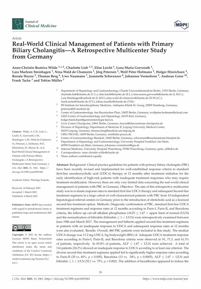
Journal of
Clinical Medicine
Article
Real-World Clinical Management of Patients with PrimaryBiliary Cholangitis—A Retrospective Multicenter Studyfrom Germany
Anne-Christin Beatrice Wilde 1,*,†, Charlotte Lieb 1,†, Elise Leicht 1, Lena Maria Greverath 1,
Lara Marleen Steinhagen 1, Nina Wald de Chamorro 1, Jörg Petersen 2, Wolf Peter Hofmann 3, Holger Hinrichsen 4,
Renate Heyne 5, Thomas Berg 6, Uwe Naumann 7, Jeannette Schwenzer 8, Johannes Vermehren 9, Andreas Geier 10,
Frank Tacke 1 and Tobias Müller 1
Citation: Wilde, A.-C.B.; Lieb, C.;
Leicht, E.; Greverath, L.M.;
Steinhagen, L.M.; Wald de Chamorro,
N.; Petersen, J.; Hofmann, W.P.;
Hinrichsen, H.; Heyne, R.; et al.
Real-World Clinical Management of
Patients with Primary Biliary
Cholangitis—A Retrospective
Multicenter Study from Germany. J.
Clin. Med. 2021, 10, 1061. https://
doi.org/10.3390/jcm10051061
Academic Editor: Pierluigi Toniutto
Received: 14 February 2021
Accepted: 1 March 2021
Published: 4 March 2021
Publisher’s Note: MDPI stays neutral
with regard to jurisdictional claims in
published maps and institutional affil-
iations.
Copyright: © 2021 by the authors.
Licensee MDPI, Basel, Switzerland.
This article is an open access article
distributed under the terms and
conditions of the Creative Commons
Attribution (CC BY) license (https://
creativecommons.org/licenses/by/
4.0/).
1 Department of Hepatology and Gastroenterology, Charité Universitätsmedizin Berlin, 13353 Berlin, Germany;[email protected] (C.L.); [email protected] (E.L.); [email protected] (L.M.G.);[email protected] (L.M.S.); [email protected] (N.W.d.C.);[email protected] (F.T.); [email protected] (T.M.)
2 IFI Institute for Interdisciplinary Medicine, Asklepios Klinik St. Georg, 20099 Hamburg, Germany;[email protected]
3 Center of Gastroenterology Am Bayerischen Platz, 10825 Berlin, Germany; [email protected] GHZ-Center of Gastroenterology and Hepatology, 24105 Kiel, Germany;
[email protected] Liver Center Checkpoint, 10961 Berlin, Germany; [email protected] Division of Hepatology, Department of Medicine II, Leipzig University, Medical Center,
04103 Leipzig, Germany; [email protected] UBN/PRAXIS, 14059 Berlin, Germany; [email protected] Center of Gastroenterology Biesdorf, 12683 Berlin, Germany; [email protected] Department of Hepatology and Gastroenterology, University Hospital Frankfurt Am Main,
60590 Frankfurt am Main, Germany; [email protected] Internal Medicine, University Hospital Wuerzburg, 97080 Wuerzburg, Germany; [email protected]* Correspondence: [email protected]† These authors contributed equally.
Abstract: Background: Clinical practice guidelines for patients with primary biliary cholangitis (PBC)have been recently revised and implemented for well-established response criteria to standardfirst-line ursodeoxycholic acid (UDCA) therapy at 12 months after treatment initiation for theearly identification of high-risk patients with inadequate treatment responses who may requiretreatment modification. However, there are only very limited data concerning the real-world clinicalmanagement of patients with PBC in Germany. Objective: The aim of this retrospective multicenterstudy was to evaluate response rates to standard first-line UDCA therapy and subsequent Second-linetreatment regimens in a large cohort of well-characterized patients with PBC from 10 independenthepatological referral centers in Germany prior to the introduction of obeticholic acid as a licensedsecond-line treatment option. Methods: Diagnostic confirmation of PBC, standard first-line UDCAtreatment regimens and response rates at 12 months according to Paris-I, Paris-II, and Barcelonacriteria, the follow-up cut-off alkaline phosphatase (ALP) ≤ 1.67 × upper limit of normal (ULN)and the normalization of bilirubin (bilirubin ≤ 1 × ULN) were retrospectively examined betweenJune 1986 and March 2017. The management and hitherto applied second-line treatment regimensin patients with an inadequate response to UDCA and subsequent response rates at 12 monthswere also evaluated. Results: Overall, 480 PBC patients were included in this study. The medianUDCA dosage was 13.2 mg UDCA/kg bodyweight (BW)/d. Adequate UDCA treatment responserates according to Paris-I, Paris-II, and Barcelona criteria were observed in 91, 71.3, and 61.3%of patients, respectively. In 83.8% of patients, ALP ≤ 1.67 × ULN were achieved. A total of116 patients (24.2%) showed an inadequate response to UDCA according to at least one criterion. Thediverse second-line treatment regimens applied led to significantly higher response rates accordingto Paris-II (35 vs. 60%, p = 0.005), Barcelona (13 vs. 34%, p = 0.0005), ALP ≤ 1.67 × ULN andbilirubin ≤ 1 × ULN (52.1 vs. 75%, p = 0.002). The addition of bezafibrates appeared to induce the
J. Clin. Med. 2021, 10, 1061. https://doi.org/10.3390/jcm10051061 https://www.mdpi.com/journal/jcm
29

J. Clin. Med. 2021, 10, 1061
strongest beneficial effect in this cohort (Paris II: 24 vs. 74%, p = 0.004; Barcelona: 50 vs. 84%,p = 0.046; ALP < 1.67 × ULN and bilirubin ≤ 1 × ULN: 33 vs. 86%, p = 0.001). Conclusion: Our largeretrospective multicenter study confirms high response rates following UDCA first-line standardtreatment in patients with PBC and highlights the need for close monitoring and early treatmentmodification in high-risk patients with an insufficient response to UDCA since early treatmentmodification significantly increases subsequent response rates of these patients.
Keywords: primary biliary cholangitis; autoantibodies; ursodeoxycholic acid; treatment response;second line therapy
1. Introduction
Primary biliary cholangitis (PBC) is a rare chronic inflammatory biliary disease charac-terized by the progressive destruction of intrahepatic bile ducts, leading to cholestasis andsubsequent liver damage. Although immune-mediated processes are widely consideredas a major underlying cause, the exact etiology of biliary inflammation still remains to befully elucidated but likely comprises additional factors such as environmental stimuli andepigenetic factors [1]. If untreated, PBC may lead to end-stage liver cirrhosis and eventuallyliver failure. Orthotopic liver transplantation is the only definitive therapy for PBC patientswith end-stage liver disease. Early treatment with weight-based ursodeoxycholic acid(UDCA) has been proven to extend transplant-free survival and is therefore recommendedat a dosage of 13 to 15 mg UDCA/kg bodyweight (BW)/d as a standard first-line therapyby national and international PBC clinical practice guidelines [2–4]. These guidelines alsoimplemented the evaluation of specific response criteria at 12 months after the initiation ofUDCA treatment for the early detection of high-risk patients with inadequate treatmentresponses who need treatment modification. The most commonly applied Paris-I, Paris-II,and Barcelona criteria have been widely accepted for treatment monitoring includinglaboratory parameters such as bilirubin and alkaline phosphatase (ALP). Recently, ALPlevels ≤ 1.67 × upper limit of normal (ULN)or ALP normalization and the normalization ofbilirubin values (bilirubin ≤ 1 × ULN) have been proven to yield prognostic relevance [5,6].The risk of PBC disease progression to severe liver disease in patients with PBC may alsobe estimated using the UK-PBC risk score, which is based on continuous variables thathave been specifically developed to identify patients at an increased risk for progression todeath or liver transplantation after 5, 10, and 15 years [7,8].
In December 2016, obeticholic acid was licensed as a second-line treatment option incombination with UDCA for patients with an inadequate UDCA response or monotherapyfor patients with an intolerance to UDCA and is therefore recommended by current PBCclinical practice guidelines. Bezafibrates have also been shown to increase response rates inpatients with an inadequate response to first-line UDCA therapy [9] and have thereforebeen depicted as a potential off-label second-line treatment option in combination withUDCA in the current guidelines.
There are only very limited data concerning the real-world clinical management ofpatients with PBC in Germany [10]. Moreover, a recent German population-based studyreported the rising prevalence of PBC and deficits in their subsequent clinical management [11].We therefore aimed to evaluate real-world diagnostic approaches, standard first-line UDCAtreatment regimens and respective response rates at 12 months applying Paris-I, Paris-II, andBarcelona criteria, ALP levels ≤ 1.67 × ULN, and bilirubin ≤ 1 × ULN in a large cohort ofwell-characterized patients with PBC in a retrospective multicenter study from 10 independenthepatological referral centers in Germany. The real-world management of patients with aninadequate response to standard first-line UDCA treatment including the hitherto appliedindividual second-line treatment regimens prior to the introduction of obeticholic acid andthe subsequent response rates at 12 months were also evaluated.
30
J. Clin. Med. 2021, 10, 1061
2. Methods
2.1. Diagnostic Criteria
The records of patients suspected to have PBC between June 1986 and March 2017were evaluated in a large retrospective multicenter study from 10 independent Germanhepatological referral centers, comprising four tertiary-care university hospitals in Berlin,Frankfurt am Main, Würzburg, and Leipzig and six hepatological centers in Berlin, Ham-burg, and Kiel. The study was approved by the local Ethics Committees of the Universitiesof Berlin, Frankfurt, Leipzig, and Würzburg and written informed consent was obtainedfrom all participants. The diagnosis of PBC was accepted if the patients fulfilled at least twoof the following criteria (1) chronic cholestasis for more than six months; (2) the presence ofanti-mitochondrial antibody (AMA) titer > 1:40 or other specific autoantibodies, includingsp100 or gp210, if AMA-negative (3) the histological conformation of lymphocytic de-structive cholangitis and destruction of interlobular bile ducts. Patients with concomitantfeatures of autoimmune hepatitis (AIH) as defined by the current PBC treatment guidelines(according to liver biopsy or high simplified score according to Hennes et al. or patientswho fulfilled the Paris criteria for AIH-PBC-Overlap) [3,4,12] showed lower response ratesto UDCA in a previous study [13]; therefore, these patients were excluded to eliminate thisbias. Furthermore, patients with concomitant features of primary sclerosing cholangitis,biliary obstruction, drug-induced cholestatic liver disease, severe non-alcoholic fatty liverdisease, hemochromatosis, Wilson’s disease, alpha1-antitrypsin deficiency, alcohol abuse,chronic hepatitis B, or hepatitis C were excluded from the study by extended laboratorytesting and imaging including abdominal ultrasound and magnetic resonance cholan-giopancreatography. All patients underwent thorough clinical exams supplemented bylaboratory tests at baseline and follow-up visits every three to four months in an outpa-tient setting. The clinical course of disease including the development of liver cirrhosiswas evaluated.
2.2. Baseline Characteristics
Baseline characteristics comprised sex, age at onset of therapy, weight, serum levelsof alaninaminotransferase (ALT), aspartataminotransferase (AST), alkaline phosphatase(ALP), γ-glutamyltransferase (γGT), bilirubin, albumin, prothrombin time, platelet count,immunoglobulin A (IgA), G (IgG), M (IgM), anti-mitochondrial antibodies (AMA), anti-smooth muscle antibodies (SMA), anti-nuclear antibodies (ANA), anti-sp100, anti-gp210,and, if available, a histological evaluation of PBC.
2.3. Evaluation of Standard First-Line UDCA Treatment Regimens and Response Rates at 12 Months
Standard first-line UDCA treatment regimens in mg UDCA/kg bodyweight per day(mg UDCA/kg BW/d) were evaluated. At 12 months, the ALP, AST, and bilirubin levelswere assessed to evaluate Paris-I, Paris-II, and Barcelona criteria, ALP levels ≤ 1.67 × ULN,and bilirubin ≤ 1 × ULN to define an adequate respectively inadequate UDCA response.
2.4. Management of Patients with an Inadequate Response to First-Line UDCA Treatment
The management of patients with an inadequate response to first-line UDCA treatmentincluding the evaluation of hitherto applied second-line treatment regimens was evaluated.Subsequent response rates at 12 months after treatment modification were assessed byapplying Paris-I, Paris-II, and Barcelona criteria, ALP cut-off levels ≤ 1.67 × ULN, andbilirubin ≤ 1 × ULN.
2.5. Statistical Analysis
Analyses were performed by IBM SPSS Statistic Version 24 for Windows (IBM, Ar-monk, NY, USA) and Prism 6.0 (GraphPad Software, La Jolla, CA, USA). Comparisonbetween groups were made by using the Kruskal–Wallis test, Mann–Whitney test, andFischer’s exact test. Data are presented as the median and interquartile range (IQR). TheKaplan–Meier survival curve with the Mantel–Cox test was examined to assess the rela-
31
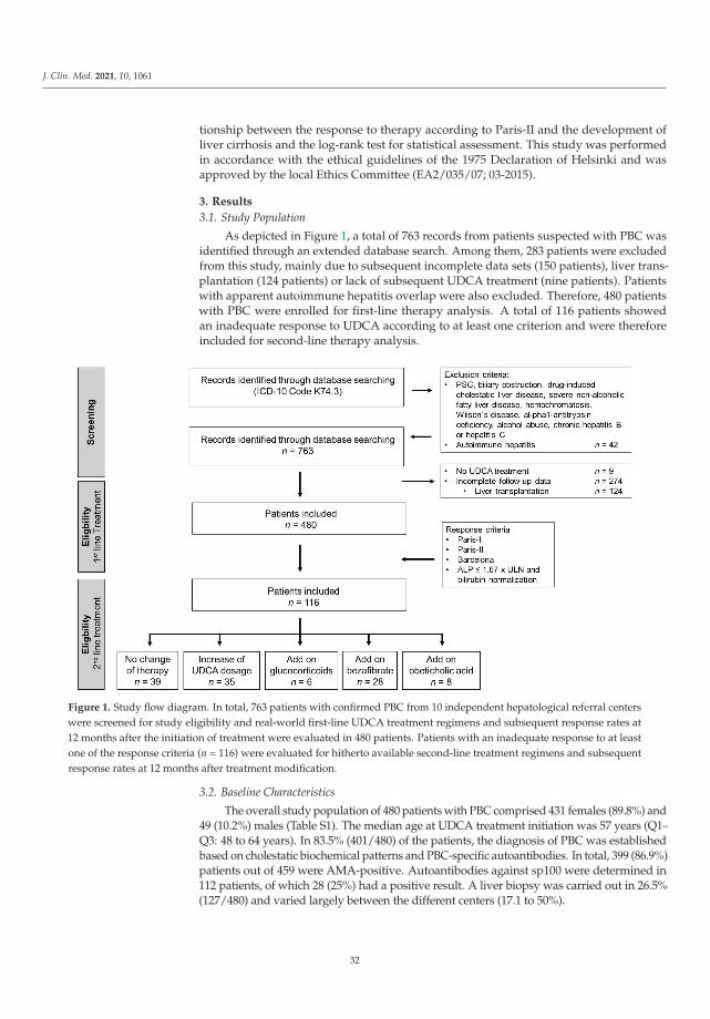
J. Clin. Med. 2021, 10, 1061
tionship between the response to therapy according to Paris-II and the development ofliver cirrhosis and the log-rank test for statistical assessment. This study was performedin accordance with the ethical guidelines of the 1975 Declaration of Helsinki and wasapproved by the local Ethics Committee (EA2/035/07; 03-2015).
3. Results
3.1. Study Population
As depicted in Figure 1, a total of 763 records from patients suspected with PBC wasidentified through an extended database search. Among them, 283 patients were excludedfrom this study, mainly due to subsequent incomplete data sets (150 patients), liver trans-plantation (124 patients) or lack of subsequent UDCA treatment (nine patients). Patientswith apparent autoimmune hepatitis overlap were also excluded. Therefore, 480 patientswith PBC were enrolled for first-line therapy analysis. A total of 116 patients showedan inadequate response to UDCA according to at least one criterion and were thereforeincluded for second-line therapy analysis.
Figure 1. Study flow diagram. In total, 763 patients with confirmed PBC from 10 independent hepatological referral centerswere screened for study eligibility and real-world first-line UDCA treatment regimens and subsequent response rates at12 months after the initiation of treatment were evaluated in 480 patients. Patients with an inadequate response to at leastone of the response criteria (n = 116) were evaluated for hitherto available second-line treatment regimens and subsequentresponse rates at 12 months after treatment modification.
3.2. Baseline Characteristics
The overall study population of 480 patients with PBC comprised 431 females (89.8%) and49 (10.2%) males (Table S1). The median age at UDCA treatment initiation was 57 years (Q1–Q3: 48 to 64 years). In 83.5% (401/480) of the patients, the diagnosis of PBC was establishedbased on cholestatic biochemical patterns and PBC-specific autoantibodies. In total, 399 (86.9%)patients out of 459 were AMA-positive. Autoantibodies against sp100 were determined in112 patients, of which 28 (25%) had a positive result. A liver biopsy was carried out in 26.5%(127/480) and varied largely between the different centers (17.1 to 50%).
32
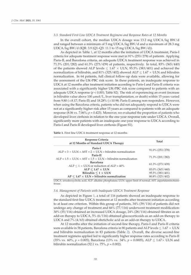
J. Clin. Med. 2021, 10, 1061
3.3. Standard First-Line UDCA Treatment Regimens and Response Rates at 12 Months
In the overall cohort, the median UDCA dosage was 13.2 mg UDCA/kg BW/dand ranged between a minimum of 5 mg UDCA/kg BW/d and a maximum of 28.3 mgUDCA/kg BW/d (IQR: 3.9 (Q1–Q3: 11.1 to 15 mg UDCA/kg BW/d)).
As depicted in Table 1, at 12 months after the initiation of UDCA treatment, Paris-Icriteria for adequate treatment response were met in 91% (253/278) of patients. ApplyingParis-II, and Barcelona criteria, an adequate UDCA treatment response was achieved in71.3% (201/282) and 61.3% (273/439) of patients, respectively. In total, 83% (365/440)of the patients showed ALP levels ≤ 1.67 × ULN, 95.5% (383/401) and achieved thenormalization of bilirubin, and 81% (325/402) showed ALP ≤ 1.67 × ULN and bilirubinnormalization. In 64 patients, full clinical follow-up data were available, allowing forthe assessment of the UK-PBC risk score. In these patients, an inadequate response toUDCA at 12 months after treatment initiation according to Paris-I and Paris-II criteria wasassociated with a significantly higher UK-PBC risk score compared to patients with anadequate UDCA response (p < 0.001; Table S2). The risk of experiencing an event (increasein bilirubin value above 100 μmol/L, liver transplantation, or death) within 15 years variedfrom 9.80 (±8.17; Paris-II) and 18.28% (±10.98; Paris-I) among non-responders. However,when using the Barcelona criteria, patients who did not adequately respond to UDCA werenot at a significantly higher risk after 15 years as compared to patients with an adequateresponse (8.06 vs. 7.06%; p = 0.423). Moreover, we evaluated the proportion of patients whodeveloped liver cirrhosis in relation to the one-year response rate under UDCA. Overall,significantly more patients with an inadequate one-year response to UDCA according toParis-I and Paris-II developed liver cirrhosis (Figure S1).
Table 1. First line UDCA treatment response at 12 months.
Response Criteriaat 12 Months of Standard UDCA Therapy
Total
Paris-IALP < 3 × ULN + AST < 2 × ULN + bilirubin normalization 91.0% (253/278)
Paris-IIALP < 1.5 × ULN + AST < 1.5 × ULN + bilirubin normalization 71.3% (201/282)
BarcelonaALP ≤ 1 × ULN or reduction of ALP > 40% 61.3% (273/439)
ALP ≤ 1.67 × ULN 83.0% (365/440)Bilirubin ≤ 1 × ULN 95.5% (383/401)
AP ≤ 1.67 × ULN + bilirubin normalization 80.8% (325/402)UDCA: ursodeoxycholic acid; ALP: alkaline phosphatase; ULN: upper limit of normal; AST: aspartataminotrans-ferase.
3.4. Management of Patients with Inadequate UDCA Treatment Response
As depicted in Figure 1, a total of 116 patients showed an inadequate response tothe standard first-line UDCA treatment at 12 months after treatment initiation accordingto at least one criterion. Within this group of patients, 34% (39/116) of patients did notundergo any change of treatment and 66% (77/116) underwent treatment modification:30% (35/116) obtained an increased UDCA dosage, 24% (28/116) obtained fibrates as anadd-on therapy to UDCA, 5% (6/116) obtained glucocorticoids as an add-on therapy toUDCA and 7% (4/63) obtained obeticholic acid as an add-on therapy to UDCA.
At 12 months after the initiation of second-line therapy, Paris-I and Paris-II criteriawere available in 58 patients, Barcelona criteria in 90 patients and ALP levels ≤ 1.67 × ULNand bilirubin normalization in 83 patients (Table 2). Overall, the diverse second-linetreatment regimens applied led to significantly higher response rates according to Paris-II(35% vs. 60%, p = 0.005), Barcelona (13% vs. 34%, p = 0.0005), ALP ≤ 1.67× ULN andbilirubin normalization (52.1 vs. 75%, p = 0.002).
33

J. Clin. Med. 2021, 10, 1061
Table 2. Second line treatment response in patients with inadequate UDCA response.
Response Criteria at 12 Months of UDCATherapy
Before Initiation of 2nd LineTherapy and After at Least 12 of
Therapy with UDCA
12 Months After Initiation of2nd Line Therapy
p
Paris-IALP < 3 × ULN + AST < 2 × ULN +
Bilirubin ≤ 1mg/dL76.8% (53/69) 84.5% (49/58) 0.371
Paris-IIALP < 1.5 × ULN + AST < 1.5 × ULN +
Bilirubin ≤ 1mg/dL34.9% (24/69) 60.3% (35/58) 0.005
BarcelonaALP ≤ 1 × ULN or reduction of ALP > 40% 12.9% (13/101) 34.4% (31/90) 0.0005
ALP ≤ 1.67 × ULN + Bilirubin ≤ 1 × ULN 52.1% (50/96) 74.7% (62/83) 0.002
Bold values indicates a statistical significance (p < 0.05).
As depicted in Figure 2 (Table S3), the UDCA dosage intensification and the additionof glucocorticoids did not increase response rates in patients with an inadequate UDCAresponse, whereas the addition of fibrates significantly enhanced response rates accordingto Paris-II (p = 0.004) and Barcelona criteria (p = 0.046) and ALP levels ≤ 1.67 × ULNand bilirubin normalization (p = 0.001) at 12 months after the initiation of treatmentmodification. In the small group of patients obtaining additional obeticholic acid sinceapproval in December 2016, there was a trend towards higher response rates with respectto ALP levels ≤ 1.67 × ULN and bilirubin normalization.
Figure 2. Management of patients with inadequate response to standard UDCA first-line treatment. The real-worldmanagement of 116 patients with inadequate response to standard first-line UDCA treatment according to at least onecriterion is depicted, including patients without treatment modification (n = 39), increase of UDCA dosage (n = 35), addition ofglucocorticoids (n = 6), bezafibrates (n = 28) or obeticholic acid (n = 4). Treatment response rates according to Paris-I, Paris-II,and Barcelona criteria and ALP levels ≤ 1.67 × ULN and bilirubin ≤ 1 × ULN at 12 months after treatment modification,respectively; continuation of UDCA-monotherapy was analyzed. * p < 0.05, ** p < 0.01, not significant (ns) = p ≥ 0.05.
3.5. Evaluation of Liver Biochemistry
As depicted in Figure 3, intensified UDCA treatment and/or the addition of glucocor-ticoids did not improve liver biochemistry, whereas the addition of fibrates significantlyreduced ALP (p < 0.001) and gamma-glutamyl transferase (gGT) levels (p = 0.023). Addi-tional treatment with obeticholic acid also showed a reduction of gGT levels (p = 0.043).Bilirubin levels, which normalized in the vast majority of patients during first-line therapy,showed no further decrease irrespective of the second-line therapy applied.
34
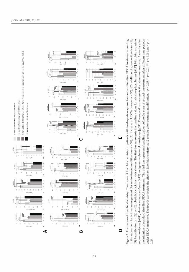
J. Clin. Med. 2021, 10, 1061
Fig
ure
3.
Eva
luat
ion
ofliv
erbi
oche
mis
try.
The
cour
seof
liver
bioc
hem
istr
yin
pati
ents
wit
hin
adeq
uate
resp
onse
tost
and
ard
first
-lin
eU
DC
Atr
eatm
enta
ccor
din
gto
the
subs
equ
enti
ndiv
idu
alm
anag
emen
t(A
):no
trea
tmen
tmod
ifica
tion
(n=
39),
(B):
incr
ease
ofU
DC
Ad
osag
e(n
=35
),(C
):ad
dit
ion
ofgl
uco
cort
icoi
ds
(n=
6),
(D):
beza
fibr
ates
(n=
28)o
r(E
):ob
etic
holic
acid
(n=
4)is
show
n.T
hefi
rstb
arre
pre
sent
sth
eba
selin
eva
lues
for
alka
line
pho
spha
tase
[AL
P],
bilir
ubi
n,as
par
tate
amin
otra
nsfe
rase
[AST
],al
anin
eam
inot
rans
fera
se[A
LT]a
ndga
mm
a-gl
utam
yltr
ansf
eras
e[g
GT]
.The
seco
ndba
rre
pres
ents
the
labo
rato
ryre
sults
at12
mon
ths
afte
rth
ein
itia
tion
ofst
and
ard
firs
t-lin
eU
DC
Atr
eatm
ent.
The
thir
dba
rre
pres
ents
base
line
valu
esbe
fore
the
star
tofs
econ
d-l
ine
trea
tmen
taft
erd
iffe
rent
tim
epe
riod
sun
der
UD
CA
trea
tmen
t.Th
efo
urth
bar
depi
cts
the
effe
cton
liver
bioc
hem
istr
yat
12m
onth
saf
ter
trea
tmen
tmod
ifica
tion.
*p
<0.
05,*
*p
<0.
01,*
**p
<0.
001,
ns=
p≥
0.05
.
35
J. Clin. Med. 2021, 10, 1061
4. Discussion
Primary biliary cholangitis may be associated with considerable morbidity despiterecent advances in the management of this disease [2]. Thus, there is an unmet clinical needto improve the management of PBC, in particular in patients with an inadequate responseto standard first-line treatment with weight-based UDCA. In this retrospective multicenterstudy, so-far the largest, we provide insight into the real-world clinical management ofpatients with PBC with respect to the recently revised national and international clinicalpractice guidelines. In this study from 10 independent hepatological centers in Germany,the diagnosis of PBC was established by serological parameters in 83.5% of all patients,i.e., chronic cholestasis and specific autoantibodies, whereas in 26.5% of all patients, anadditional histological examination were carried out. Detailed analysis of the treatmentregimens applied by the different centers revealed a median UDCA dosage of 13.2 mg/kgBW/d as the standard first-line therapy in the study cohort, achieving adequate treatmentresponse rates in 60 to 90% of patients at 12 months after the initiation of therapy dependingon the response criteria applied. Therefore, up to 40% of patients showed an inadequateUDCA response at 12 months after the standard UDCA first-line therapy. Of note, in almostone third of these patients in this high-risk group, there was no obvious treatment modifi-cation, whereas in the majority of cases the UDCA dosage was increased. Other treatmentmodifications included the off-label addition of fibrates or, less frequently, the addition ofbudesonide. Since 2017, the patients were also treated with obeticholic acid as the onlylicensed second-line therapy. Therefore, the number of patients receiving obeticholic acidwas very limited. Taken together, the diverse second-line treatment regimens applied ledto significantly higher response rates according to Paris-II and Barcelona criteria, and ALP≤ 1.67 × ULN and bilirubin normalization. The addition of fibrates appeared to inducethe strongest beneficial effect in this cohort.
Several limitations of the present study need to be acknowledged. Due to the ret-rospective character of this study, a systematic evaluation of all biochemical parametersin every study participant was not available, leading to inconsistent patient numbers de-pending on the different response criteria in the overall cohort. Additionally, patients whounderwent liver transplantation were excluded due to incomplete data. Another limitationof the study is that the data available on further follow-up in patients without treatmentmodification are limited and therefore do not allow to draw a solid conclusion. Moreover,a certain selection bias cannot be excluded since all study participants were recruitedfrom four tertiary care centers and six hepatological referral centers, leading to a potentialselection of difficult-to-diagnose cases, which likely explains the rather low proportionof patients with PBC-specific autoantibodies in the present cohort. However, a recentpopulation-based study with non-selected PBC patients revealed no obvious difference toearlier hospital-based studies [14].
However, we would like to strengthen the fact that this is, so far, the largest retro-spective multicenter study addressing the real-world management of patients with PBCin Germany, comprising patients from 10 different hepatological referral centers. Thebaseline characteristics of the present PBC cohort were consistent with previous PBC studypopulations, showing a clear predominance of female patients with a female-to-male ratioof 9:1. An increasing proportion of male patients, as postulated in a recent epidemio-logical study [15] and a recent German retrospective study [11], was not observed in thepresent cohort. The median age at treatment initiation was 57 years, which was in linewith the age peak described in the literature between the fifth and seventh decade oflife. Of note, depending on the individual participating center, between 5 and 16% ofall PBC patients were AMA-negative, whereas previous studies described only 5 to 10%AMA-negative patients [16–18]. However, in a recent Polish study [19], the proportionof AMA-negative patients was 18.9%, suggesting that the proportion of AMA-negativepatients in special populations might be higher than previously reported. In our first-linestudy, 399 of 459 patients were AMA-positive whereas only 112 cases were tested for thepresence of specific autoantibodies against protein sp100 with a positive result in 25% of
36

J. Clin. Med. 2021, 10, 1061
patients, which might be due to the fact that these antibodies are not readily availablein daily routine use. However, our observation was in line with the very heterogeneousresults of previous meta-analyses from 2014, describing antibodies against sp100 in 7 to60% of all patients [20].
With respect to the applied first-line UDCA therapy, the median dosage of 13.2 mgUDCA/kg BW/d was close to the lower limit of the recommended dosage of 13 to 15 mgUDCA/kg BW/d. Adequate UDCA response rates in 91% of patients at 12 months aftertreatment initiation according to Paris-I criteria was higher compared to previous studiesof other countries. In a Dutch study from 2009, the overall response rate according toParis-I criteria was 66% [21]; in a French study from 2011, it was 76% [22]; and in a largemulticenter British study from 2013, it was 79% [23]. This discrepancy might be explainedby the large proportion of patients with early PBC stages in the present cohort defined bynormal bilirubin levels prior to the start of UDCA treatment. Interestingly, a recent Germanprospective study from 2019 showed a similarly high response rate according to Paris-Icriteria [10]. This study also described a high number of patients in an early stage of PBC.Therefore, Paris-II criteria might be more adequate to examine the response rate in thepresent cohort. In a recently published study, Murillo-Perez et al. highlighted that not onlya reduction of ALP level, but both bilirubin normalization and an ALP level below 1 × ULNsignificantly contributes to a more accurate risk stratification and improved survival [6].Therefore, not only one criterion but several criteria (the Barcelona criterion together withParis-II criterion and ALP level ≤ 1.67 × ULN with bilirubin normalization) should be usedfor each patient; furthermore, a complete normalization of ALP and bilirubin level shouldpossibly be targeted in the future to determine the lowest risk for a disease progressionand, if needed, to intensify the therapy.
In line with this hypothesis, applying Paris-II and Barcelona criteria, adequate re-sponse rates of 71 (Paris-II) and 61% (Barcelona) at 12 months after treatment initiationwere consistent with previous observations [22,24].
Our findings demonstrate higher treatment response rates at 12 months after treatmentmodification under real-world conditions. Of note, comparison of different treatmentregimens in patients with an inadequate therapy response showed particularly higherresponse rates among patients receiving additional fibrates. These results are in linewith previous studies showing higher response rates for fibrates as an add-on therapy toUDCA [9,25,26]. In the other large cohort of the study—patients without any change oftreatment regime—there was no significant improvement in treatment response accordingto Paris-I, Paris-II, and Barcelona criteria. However, there was a significant increase inpatients achieving ALP levels ≤ 1.67 × ULN and bilirubin normalization. These resultsare in line with a recent study from Germany by Sebode et al., who also showed that theguideline recommended treatment regime of patients with PBC was partly not applied [11].Follow-up data for treatment modification with obeticholic acid were missing due to theshort period of this recently licensed therapy. However, we already observed a clear trendtowards a better therapy response rate regarding the ALP-level ≤ 1.67 × ULN and bilirubinnormalization and a significant reduction of gGT after the addition of obeticholic acid toUDCA. However, our data are in line with the findings by Nevens et al. who showedan improved response rate according to ALP levels below 1.67 × ULN and bilirubinnormalization under obeticholic acid [27].
In conclusion, this large real-world multicenter study confirms high response ratesfollowing UDCA first-line standard treatment in patients with PBC and highlights theneed for close monitoring and early treatment modification in high-risk patients with aninsufficient response to UDCA since early treatment modification significantly increasesthe subsequent response rates of these patients. A large prospective observation study hasalready been initiated and will provide further long-term outcomes.
37

J. Clin. Med. 2021, 10, 1061
Supplementary Materials: The following are available online at https://www.mdpi.com/2077-0383/10/5/1061/s1. Figure S1: Development of liver cirrhosis in relation to 12 months UDCA treatmentresponse, Table S1: Baseline characteristics, Table S2: UK-PBC-Risk-Score in relation to 12 monthsUDCA treatment response, Table S3: Treatment response before and after 12 months of second linetherapy in detail.
Author Contributions: Conceptualization, T.M. and A.-C.B.W.; methodology, T.M. and A.-C.B.W.;formal analysis, A.-C.B.W., C.L. and E.L.; investigation, A.-C.B.W., C.L., E.L., L.M.G., L.M.S., N.W.d.C.,J.P., W.P.H., H.H., R.H., T.B., U.N., J.S., J.V. and A.G.; writing-original draft preparation, A.-C.B.W.;writing-review and editing, T.M., F.T.; visualization, A.-C.B.W.; supervision, T.M.; project administra-tion, T.M.; funding acquisition, T.M. All authors have read and agreed to the published version of themanuscript.
Funding: This study was supported by the German Research Foundation Grants MU 2864/1-3 andMU 2864/3-1 and by unrestricted research grants from Intercept Pharmaceuticals and Falk PharmaGmbH. The supporting parties had no influence on the study design, data collection and analyses,writing of the manuscript or on the decision to submit the manuscript for publication.
Institutional Review Board Statement: The study was approved by the local Ethics Committeesof the Universities of Berlin, Frankfurt, Leipzig and Würzburg and written informed consent wasobtained from all participants. This study was performed in accordance with the ethical guidelinesof the 1975 Declaration of Helsinki and was approved by the local Ethics Committee (EA2/035/07;03-2015).
Informed Consent Statement: Informed consent was obtained from all subjects involved in the study.
Data Availability Statement: The data presented in this study are available on request.
Conflicts of Interest: The authors declare no conflict of interest. The funders had no role in the designof the study; in the collection, analyses, or interpretation of data; in the writing of the manuscript, orin the decision to publish the results.
References
1. Carey, E.J.; Ali, A.H.; Lindor, K.D. Primary biliary cirrhosis. Lancet 2015, 386, 1565–1575. [CrossRef]2. Lindor, K.D.; Bowlus, C.L.; Boyer, J.; Levy, C.; Mayo, M. Primary Biliary Cholangitis: 2018 Practice Guidance from the American
Association for the Study of Liver Diseases. J. Hepatol. 2018, 69, 394–419. [CrossRef]3. Hirschfield, G.M.; Beuers, U.; Corpechot, C.; Invernizzi, P.; Jones, D.; Marzioni, M. EASL Clinical Practice Guidelines: The
diagnosis and management of patients with primary biliary cholangitis. J. Hepatol. 2017, 67, 145–172. [CrossRef]4. Strassburg, C.P.; Beckebaum, S.; Geier, A.; Gotthardt, D.; Klein, R.; Melter, M.; Schott, E.; Spengler, U.; Tacke, F.; Trauner, M.; et al.
S2k Leitlinie Autoimmune Lebererkrankungen. Zeitschrift für Gastroenterologie 2017, 55, 1135–1226. [CrossRef]5. Lammert, C.; Juran, B.D.; Schlicht, E.; Chan, L.L.; Atkinson, E.J.; de Andrade, M.; Lazaridis, K.N. Biochemical response to
ursodeoxycholic acid predicts survival in a North American cohort of primary biliary cirrhosis patients. J. Gastroenterol. 2014, 49,1414–1420. [CrossRef]
6. Perez, C.F.; Harms, M.H.; Lindor, K.D.; Van Buuren, H.R.; Hirschfield, G.M.; Corpechot, C.; Van Der Meer, A.J.; Feld, J.J.;Gulamhusein, A.; Lammers, W.J.; et al. Goals of treatment for improved survival in primary biliary cholangitis: Treatment targetshould be bilirubin within the normal range and normalization of alkaline phosphatase. Am. J. Gastroenterol. 2020, 115, 1066–1074.[CrossRef] [PubMed]
7. Carbone, M.; Sharp, S.J.; Flack, S.; Paximadas, D.; Spiess, K.; Adgey, C.; Griffiths, L.; Lim, R.; Trembling, P.; Williamson, K.; et al.The UK-PBC risk scores: Derivation and validation of a scoring system for long-term prediction of end-stage liver disease inprimary biliary cholangitis. Hepatology 2016, 63, 930–950. [CrossRef] [PubMed]
8. Efe, C.; Tas, K. Validation of Risk Scoring Systems in Ursodeoxycholic Acid-Treated Patients with Primary Biliary Cholangitis. J.Gastroenterol. 2019, 114, 1101–1108. [CrossRef] [PubMed]
9. Corpechot, C.; Chazouillères, O.; Rousseau, A.; Le Gruyer, A.; Habersetzer, F.; Mathurin, P.; Goria, O.; Potier, P.; Minello, A.;Silvain, C.; et al. A Placebo-Controlled Trial of Bezafibrate in Primary Biliary Cholangitis. N. Engl. J. Med. 2018, 378, 2171–2181.[CrossRef]
10. Kaps, L.; Grambihler, A.; Yemane, B.; Nagel, M.; Labenz, C.; Ploch, P.; Michel, M.; Galle, P.R.; Wörns, M.A.; Schattenberg, J.M.Symptom Burden and Treatment Response in Patients with Primary Biliary Cholangitis (PBC). Dig. Dis. Sci. 2019, 1–8. [CrossRef]
11. Sebode, M.; Kloppenburg, A.; Aigner, A.; Lohse, A.W.; Schramm, C.; Linder, R. Population-based study of autoimmune hepatitisand primary biliary cholangitis in Germany: Rising prevalences based on ICD codes, yet defits in medical treatment. Zeitschriftfür Gastroenterologie 2020, 58, 431–438.
38

J. Clin. Med. 2021, 10, 1061
12. Hennes, E.M.; Zeniya, M.; Czaja, A.J.; Parés, A.; Dalekos, G.N.; Krawitt, E.L.; Bittencourt, P.L.; Porta, G.; Boberg, K.M.; Hofer, H.;et al. Simplified criteria for the diagnosis of autoimmune hepatitis. Hepatology 2008, 48, 169–176. [CrossRef]
13. Wen, M.; Men, R.; Fan, X.; Shen, Y.; Ni, P.; Hu, Z.; Yang, L. Worse Response to Ursodeoxycholic Acid in Primary Biliary CholangitisPatients with Autoimmune Hepatitis Features. Dig. Dis. 2020. [CrossRef] [PubMed]
14. Örnolfsson, K.T.; Lund, S.H.; Olafsson, S.; Bergmann, O.M.; Björnsson, E.S. Biochemical response to ursodeoxycholic acid amongPBC patients: A nationwide population-based study. Scand. J. Gastroenterol. 2019, 54, 609–616. [CrossRef]
15. Lleo, A.; Jepsen, P.; Morenghi, E.; Carbone, M.; Moroni, L.; Battezzati, P.M.; Podda, M.; Mackay, I.R.; Gershwin, M.E.; Invernizzi,P. Evolving Trends in Female to Male Incidence and Male Mortality of Primary Biliary Cholangitis. Sci. Rep. 2016, 6, 25906.[CrossRef]
16. Lindor, K.D.; Gershwin, M.E.; Poupon, R.; Kaplan, M.; Bergasa, N.V.; Heathcote, E.J. Primary biliary cirrhosis. Hepatology 2009, 50,291–308. [CrossRef] [PubMed]
17. Liu, B.; Shi, X.H.; Zhang, F.C.; Zhang, W.; Gao, L.X. Antimitochondrial antibody-negative primary biliary cirrhosis: A subset ofprimary biliary cirrhosis. Liver Int. 2008, 28, 233–239. [CrossRef] [PubMed]
18. Hirschfield, G.M.; Heathcote, E.J. Antimitochondrial antibody-negative primary biliary cirrhosis. Clin. Liver Dis. 2008, 12, 323–331.[CrossRef]
19. Raszeja-Wyszomirska, J.; Wunsch, E.; Krawczyk, M.; Rigopoulou, E.I.; Kostrzewa, K.; Norman, G.L.; Bogdanos, D.P.; Milkiewicz,P. Assessment of health related quality of life in polish patients with primary biliary cirrhosis. Clin. Res. Hepatol. Gastroenterol.2016, 40, 471–479. [CrossRef] [PubMed]
20. Hu, S.-L.; Zhao, F.-R.; Hu, Q.; Chen, W.-X. Meta-analysis assessment of GP210 and SP100 for the diagnosis of primary biliarycirrhosis. PLoS ONE 2014, 9, e101916. [CrossRef] [PubMed]
21. Kuiper, E.M.; Hansen, B.E.; de Vries, R.A.; den Ouden–Muller, J.W.; Van Ditzhuijsen, T.J.; Haagsma, E.B.; Houben, M.H.; Witteman,B.J.; van Erpecum, K.J.; van Buuren, H.R.; et al. Improved Prognosis of Patients With Primary Biliary Cirrhosis That Have aBiochemical Response to Ursodeoxycholic Acid. Gastroenterology 2009, 136, 1281–1287. [CrossRef] [PubMed]
22. Corpechot, C.; Chazouillères, O.; Poupon, R. Early primary biliary cirrhosis: Biochemical response to treatment and prediction oflong-term outcome. J. Hepatol. 2011, 55, 1361–1367. [CrossRef]
23. Carbone, M.; Mells, G.F.; Pells, G.; Dawwas, M.F.; Newton, J.L.; Heneghan, M.A.; Neuberger, J.M.; Day, D.B.; Ducker, S.J.;Sandford, R.N.; et al. Sex and age are determinants of the clinical phenotype of primary biliary cirrhosis and response toursodeoxycholic acid. Gastroenterology 2013, 144, 560–569.e7. [CrossRef] [PubMed]
24. Parés, A.; Caballería, L.; Rodés, J. Excellent Long-Term Survival in Patients With Primary Biliary Cirrhosis and BiochemicalResponse to Ursodeoxycholic Acid. Gastroenterology 2006, 130, 715–720. [CrossRef]
25. Itakura, J.; Izumi, N.; Nishimura, Y.; Inoue, K.; Ueda, K.; Nakanishi, H.; Tsuchiya, K.; Hamano, K.; Asahina, Y.; Kurosaki, M.; et al.Prospective randomized crossover trial of combination therapy with bezafibrate and UDCA for primary biliary cirrhosis. Hepatol.Res. 2008, 38, 557–564. [CrossRef] [PubMed]
26. Iwasaki, S.; Ohira, H.; Nishiguchi, S.; Zeniya, M.; Kaneko, S.; Onji, M.; Ishibashi, H.; Sakaida, I.; Kuriyama, S.; Ichida, T.; et al. Theeficacy of ursodeoxycholic acid and bezafibrate combination therapy for primary biliary cirrhosis: Aprospectiv, multicenter study.Hepatol. Res. 2008, 38, 557–564. [CrossRef] [PubMed]
27. Nevens, F.; Andreone, P.; Mazzella, G.; Strasser, S.I.; Bowlus, C.; Invernizzi, P.; Drenth, J.P.; Pockros, P.J.; Regula, J.; Beuers, U.;et al. A placebo-controlled trial of obeticholic acid in primary biliary cholangitis. N. Engl. J. Med. 2016, 375, 631–643. [CrossRef][PubMed]
39

Journal of
Clinical Medicine
Review
Prevention of Cirrhosis Complications: Looking for PotentialDisease Modifying Agents
Giacomo Zaccherini 1, Manuel Tufoni 2, Mauro Bernardi 1 and Paolo Caraceni 1,2,3,*
Citation: Zaccherini, G.; Tufoni, M.;
Bernardi, M.; Caraceni, P. Prevention
of Cirrhosis Complications: Looking
for Potential Disease Modifying
Agents. J. Clin. Med. 2021, 10, 4590.
https://doi.org/10.3390/jcm10194590
Academic Editors: Pierluigi Toniutto
and Hiroki Nishikawa
Received: 25 July 2021
Accepted: 4 October 2021
Published: 5 October 2021
Publisher’s Note: MDPI stays neutral
with regard to jurisdictional claims in
published maps and institutional affil-
iations.
Copyright: © 2021 by the authors.
Licensee MDPI, Basel, Switzerland.
This article is an open access article
distributed under the terms and
conditions of the Creative Commons
Attribution (CC BY) license (https://
creativecommons.org/licenses/by/
4.0/).
1 Department of Medical and Surgical Sciences, University of Bologna, 40138 Bologna, Italy;[email protected] (G.Z.); [email protected] (M.B.)
2 IRCCS AOU di Bologna—Policlinico di S. Orsola, 40138 Bologna, Italy; [email protected] Center for Biomedical Applied Research, University of Bologna, 40138 Bologna, Italy* Correspondence: [email protected]
Abstract: The current therapeutic strategies for the management of patients with cirrhosis rely on theprevention or treatment of specific complications. The removal of the causative agents (i.e., virusesor alcohol) prevents decompensation in the vast majority of patients with compensated cirrhosis. Incontrast, even when etiological treatment has been effective, a significant proportion of patients withdecompensated cirrhosis remains at risk of further disease progression. Therefore, therapies targetingspecific key points in the complex pathophysiological cascade of decompensated cirrhosis couldrepresent a new approach for the management of these severely ill patients. Some of the interventionscurrently employed for treating or preventing specific complications of cirrhosis or used in otherdiseases (i.e., poorly absorbable oral antibiotics, statins, albumin) have been proposed as potentialdisease-modifying agents in cirrhosis (DMAC) since clinical studies have shown their capacity ofimproving survival. Additional multicenter, large randomized clinical trials are awaited to confirmthese promising results. Finally, new drugs able to antagonize key pathophysiological mechanismsare under pre-clinical development or at the initial stages of clinical assessment.
Keywords: decompensated cirrhosis; portal hypertension; ascites; non-selective beta-blockers; TIPS;rifaximin; human albumin; statins
1. Introduction
In the natural history of liver cirrhosis, the onset of a major complication of the diseaserepresents a crucial clinical event. Indeed, the occurrence of ascites, portal hypertensive gas-trointestinal bleeding, hepatic encephalopathy, and deep jaundice, alone or in combination,hallmarks the transition from the compensated to the decompensated phase of cirrhosis.About 5–7% of patients with compensated cirrhosis cross this border every year, thusentering a clinical history punctuated by further complications, frequent hospitalizations,worsening of quality of life and shortening of life expectancy [1].
In this context, the current therapeutic strategies aim to treat or control each complica-tion individually [2]. However, even though their efficacy has been proven in randomizedclinical trials (RCTs), such approaches do not significantly affect the course of the disease,thus making it difficult to bring out a clear survival benefit for patients [3,4]. Treatmentsproven to prevent the progression of the disease are not currently available. However,some potential candidates are under investigation and subject to debate in the scientificcommunity [5,6].
The only interventions showing a clear influence on the natural history of cirrhosisso far are etiological treatments targeted to the elimination of the cause of the disease (i.e.,antivirals for hepatitis B and C or prolonged abstinence for alcohol use disorders). Inthe decompensated stage, even though a slowdown of the disease progression or even areversion to the compensated state can occur, progression can be seen in up to one-third ofcases [4,7,8]. Thus, alternative approaches are warranted.
J. Clin. Med. 2021, 10, 4590. https://doi.org/10.3390/jcm10194590 https://www.mdpi.com/journal/jcm
41
J. Clin. Med. 2021, 10, 4590
Recent investigations contributed to uncover hitherto unknown pathophysiologicalmechanisms of decompensated cirrhosis [9]. It is now clear that the systemic spread ofbacterial products from the gut due to abnormal translocation and substances from the liverwhere inflammation and cell death occur activates immune cells. The consequent release ofpro-inflammatory cytokines and chemokines gives rise to sustained systemic inflammationand oxidative stress. These events lead to cardiocirculatory dysfunction responsible forreducing effective volemia and diffuse microvascular damage. These mechanisms providethe pathophysiological substrate for multi-organ dysfunction and, ultimately failure [10].
From this knowledge, novel therapeutic perspectives emerged relying on mechanisticapproaches. Their goal is to counteract key pathophysiological events: portal hypertension,bacterial translocation, circulatory dysfunction, systemic inflammation, oxidative stress,and immunological dysfunction. This kind of mechanism is exploited by disease-modifyingagents (DMA) who have proven to exert beneficial effects on the course of a disease. Theseinterventions are well established in several fields of medicine, especially in rheumatologicand neurologic disorders. Whether this approach is transferable to cirrhosis (DMAC)has recently emerged as the subject of debate [6]. In any case, some basic principles areunavoidable. Indeed, once a strong rationale for the use of a potential DMAC is establishedand its efficacy at the pre-clinical level achieved, solid evidence of clinical benefit has to bepursued, preferably through multicenter phase III randomized clinical trials (RCTs) havingpatient survival as the primary endpoint [11,12]. Given the high clinical heterogeneityof decompensated cirrhosis and its rapidly evolving clinical course [1,13,14], two othercharacteristics should be clearly identified: the target population receiving the highestbenefit from the treatment, and temporary or permanent “stopping rules” in case of loss ofefficacy or potential harm.
This review will critically analyze the available evidence supporting that interven-tions able to counteract key pathophysiological mechanisms can play a role as DMAC indecompensated cirrhosis.
2. Etiological Treatments
Whenever possible, removing or neutralizing the cause of the persisting liver insult isthe ideal goal to be attempted in all patients with chronic liver disease. Indeed, successfuletiological treatments can halt or at least slow down the progression of cirrhosis [5,15].
This is particularly true in the compensated stage of the disease, when the removal ofthe causative agent generally prevents decompensation, reduces the risk of hepatocellularcarcinoma (HCC) and prolongs survival. Thus, etiological treatments undoubtedly repre-sent the first choice in this setting [15]. Etiological treatments remain of primary importancein decompensated cirrhosis, but the extent of their effects is variable. Indeed, the successfulelimination of the causative agent can temper the turbulent course of decompensatedcirrhosis, or even induce the regression of the disease to a significant extent. However,a portion of patients do not obtain a relevant clinical improvement, remaining at risk offurther decompensation, poor quality of life, and reduced life expectancy [5,7,16].
Patients with HBV-related cirrhosis should be treated with specific anti-viral drugsas evidence shows that clinical improvement occurs in most cases [16–18]. Once thedecompensated stage has been reached, improvement in the severity of cirrhosis andsurvival rate occurs in about half to two-thirds of cases [19]. There is solid evidence thatclinical improvement generally occurs once viral clearance is obtained in patients with HCV-related cirrhosis [20,21]. However, long-term effects on the occurrence of complicationsand survival remain less defined. First, not all patients benefit to the same extent. Patientswith severe portal hypertension and advanced disease, as defined by a model for end-stageliver disease (MELD) score exceeding a certain threshold (between 15 and 20), seem to beless susceptible to sustained clinical improvement [7,21,22]. Second, most patients withdecompensated cirrhosis followed for a median period of 4 years after viral clearance didnot significantly improve their liver function and MELD score remaining at risk for severecomplications, death and need of liver transplantation [8]. Another important issue is
42

J. Clin. Med. 2021, 10, 4590
related to patients waitlisted for liver transplantation that achieve a modest reduction inthe MELD score after HCV viral clearance without improving their clinical conditions to asubstantial extent. As a result, they remain at risk of complications and death, yet theirpriority on the waiting list is negatively affected. Thus, an appropriate evaluation of thetiming of antiviral therapy is needed [23,24].
Alcohol withdrawal and abstinence from alcohol are crucial to improving the clinicalcourse and outcomes in patients with chronic alcoholic liver disease, regardless of theirdisease state [2]. In patients with compensated cirrhosis, such an approach can preventdisease progression and decompensation [15], while in decompensated patients it can atleast prevent further deterioration and stabilize the clinical course in most cases [25,26].
No clearly effective and approved drugs are available for patients with non-alcoholicfatty liver disease (NAFLD), so the treatment cornerstone relies on correcting or attenuatingthe cofactors of the metabolic syndrome, such as optimizing glycemic control in diabeticpatients and or lowering body weight in case of obesity. However, great caution is neededin recommending unsupervised weight loss in patients with cirrhosis, since sarcopenia is ahighly prevalent and impactful comorbidity [27,28] that can be worsened by incautiousweight loss. However, a monitored weight loss associated with lifestyle interventions(like tailored dietary counseling and adapted physical activity) can obtain improvement inportal hypertension and general conditions [29], thus rendering patients more suitable forliver transplantation.
Finally, sporadic data are only available on the effects of etiological treatments inpatients with cirrhosis of less frequent etiologies. Indeed, the beneficial effects of removingcausative factors in these settings have yet to be demonstrated. The sole exception isautoimmune cirrhosis, whose response to immunosuppressive therapy is followed by animprovement in long-term outcomes [30]. However, an appropriate balance between effec-tive immune suppression and risk for infection can be difficult to achieve in this context.
Summarizing the available evidence, etiological treatments are often effective inslowing down the course of cirrhosis and are of primary importance in the management ofpatients. However, they present limitations in the decompensated stage of the disease [2,5].Indeed, once cirrhosis has reached a self-perpetuating “point of no return”, even theremoval of the causative agent cannot arrest the disease progression so that the patientlong-term prognosis is not influenced. Therefore, the effective management of patientswith advanced cirrhosis also requires the adoption of strategies able to counteract the keypathophysiological mechanisms.
3. Pathophysiological Treatments
Besides new approaches still under development, interventions directed at counteract-ing pathophysiological mechanisms include treatments currently used to manage specificcomplications of cirrhosis. However, to employ them as DMAC, they should be handledin the context of novel strategies. This section will review the available treatments, bothalready tested and under investigation, able to antagonize one or more key events in thecomplex pathophysiological network of decompensated cirrhosis (Figure 1).
43

J. Clin. Med. 2021, 10, 4590
Figure 1. Targets of action of available treatments antagonizing key pathophysiological events indecompensated cirrhosis. See text for details. TIPS: trans-jugular intra-hepatic porto-systemic shunt;STAT: statins; NSBB: non-selective beta-blockers; G-CSF: granulocyte colony stimulating factors;PAA: poorly absorbable antibiotics.
3.1. Non-Selective β-Blockers
Non-selective β-blockers (NSBBs) were introduced in the management of patientswith cirrhosis about 40 years ago when their lowering effect on portal pressure was demon-strated. At present, NSBBs still are the only drugs recommended for long-term treatmentof portal hypertension [2,31,32].
Propranolol was the first NSBB employed. Its effect on portal pressure is mainly relatedto the reduction in cardiac output and the unopposed α-adrenergic tone in the splanchnicarterial bed. As a result, portal pressure declines because of a reduced splanchnic bloodinflow [33]. About 20 years ago, carvedilol was also introduced in the management ofportal hypertension [34]. This NSBB is provided with intrinsic anti-α1-adrenergic activity,contributing to lower portal pressure by decreasing intrahepatic vascular resistance. Thischaracteristic, however, may induce a greater reduction in systemic blood pressure [35].NSBBs also exert non-hemodynamic effects, as they decrease intestinal permeability, bacte-rial translocation, and systemic inflammation [36]. Whether these effects are relevant inmediating their effect in humans is still unclear.
The RCTs that evaluated the effect of NSBBs in cirrhosis had the prevention of thefirst or recurrent variceal bleeding as primary endpoints. Even though most of these trialsexcluded patients in the advanced stages of cirrhosis, several also included patients withdecompensated disease. Interestingly, metanalyses showed that bleeding prevention wasmore pronounced in decompensated than in compensated patients [37,38].
Unfortunately, data to ascertain if NSBBs administration to patients with decompen-sated cirrhosis prevents other complications beyond variceal bleeding are insufficient. Afirst systematic search did not find sufficient evidence supporting a NSBBs effect on othercomplications of cirrhosis. This negative result was possibly due to the underreporting of
44
J. Clin. Med. 2021, 10, 4590
non-bleeding complications [39]. Indeed, a more recent meta-analysis pooling data from15 studies showed that patients lowering portal pressure gradient below 12 mmHg or morethan 20% from baseline exhibited lower bleeding rates, lower incidence of complications,and a greater improvement in survival than non-responders [40]. This suggests that NSBBsmay substantially modify the course of the disease.
Two recent clinical trials reinforced this concept. The PREDESCI trial showed that NS-BBs administration to patients with compensated cirrhosis delays decompensation, mainlypreventing ascites formation [41]. Another study [42] assessed the effects of carvedilol,carefully titrated based on blood pressure and heart rate, in patients with acute-on-chronicliver failure diagnosed according to the definition of the Asian-Pacific Association for theStudy of the Liver [43]. In this placebo-controlled trial, carvedilol reduced the incidence ofbacterial infections and acute kidney injury and lowered mortality.
Soon after the first reports on the use of propranolol in patients with cirrhosis, aword of caution rose on its use in patients with ascites as they may risk for developinghepatorenal syndrome (HRS) [44]. Safety signals on either survival or kidney functiondid not emerge from the numerous RCTs comparing NSBBs with placebo in patients withcirrhosis. However, most studies did not enroll patients with refractory ascites. Furtheremphasis on this matter derived from observational studies reporting worse outcomes inpatients with refractory ascites [45] or spontaneous bacterial peritonitis (SBP) [46]. Theconcept of a “therapeutic window” beyond which NSBBs may become detrimental wasproposed [47], and several subsequent studies were dedicated to this matter. Unfortunately,conflicting results were provided [48].
Different effects of NSBBs on intrarenal hemodynamics of patients with responsiveor refractory ascites may explain their potential adverse effects in the latter. Indeed, asopposed to patients with diuretic responsive ascites, NSBBs lower renal perfusion pressurebelow the critical threshold for autoregulation (65 mmHg) in 55% of the patients withrefractory ascites. This phenomenon is likely due to an excessive reduction of cardiac outputdue to systolic dysfunction [49]. Thus, the question arises as to how identifying patientswho may receive harm from NSBBs treatment. Possibly, arterial pressure is the simplestbiomarker to establish when the therapeutic window for NSBBs closes. A prospectiveobservational study reported that mean arterial pressure levels <65 mmHg defines thethreshold below which NSBBs do not improve transplant-free survival of patients withascites, particularly in those with SBP and ACLF [50]. Similar results were observed usingthe threshold of 90 mmHg of systolic blood pressure, which is recommended in currentguidelines as a threshold for dose reduction or discontinuation of NSBBs [2,31,32].
In summary, the current indication for NSBBs administration to patients with cirrhosisremains the prevention of variceal bleeding or rebleeding. Nonetheless, when an effectivelowering in portal pressure is achieved, more general effects can be observed on theincidence of complications and patients’ survival, suggesting the potential role of NSBBsas DMAC. However, besides the prophylactic action on portal hypertensive bleeding or re-bleeding, NSBBs effects vary in different stages of the disease. On the one hand, preventionof decompensation (mainly ascites) can be seen in compensated cirrhosis; on the other,renal dysfunction and HRS can occur in patients with very advanced cirrhosis. Therefore,further studies are eagerly needed to identify target subgroups of patients who may benefitmost from long-term NSBBs administration.
3.2. Transjugular Intrahepatic Porto-Systemic Shunt
As already reported, portal hypertension plays a crucial causative role in most compli-cations of cirrhosis. Therefore, interventions targeted to decrease portal pressure, such astransjugular intrahepatic porto-systemic shunt (TIPS), can potentially modify the long-termclinical course of patients with cirrhosis [51]. To date, the main clinical settings for theuse of TIPS are the management of variceal bleeding and the control of refractory ascites.However, the main contentious points remain the identification of target patients andappropriate timing for TIPS placement.
45

J. Clin. Med. 2021, 10, 4590
The role of “pre-emptive” TIPS as a potential DMAC emerged from 3 RCTs in patientswith cirrhosis and variceal hemorrhage [52–54] (Table 1). They showed that TIPS placementwithin 24 or 72 h is effective in controlling bleeding, preventing rebleeding, and improvingsurvival in patients at high risk of uncontrolled bleeding and bleeding-related mortality(HVPG > 20 mmHg, Child C [10–13 points] or B with active bleeding). Moreover, 2 recentlarge multicenter observational studies highlighted that “pre-emptive” TIPS also improvessurvival in patients with acute variceal bleeding and ACLF [55,56]. In patients withpersistent bleeding or severe rebleeding within five days, “rescue/salvage” TIPS is usedwhen other therapeutic alternatives are not available, but, despite a high rate of bleedingcontrol, a clear benefit on survival has not been established [31,32]. Similarly, althoughthere is consensus for TIPS insertion as secondary prophylaxis of bleeding in patients whofailed to respond to endoscopic banding plus non-selective beta-blockers (NSBBs), a benefiton survival has not been consistently observed likely because of the high heterogeneity ofthe patients included in the studies [31,32].
Table 1. RCTs assessing pre-emptive TIPS vs. endoscopic treatment in acute variceal bleeding.
ReferenceStudy Population
(Randomized Patients)ExclusionCriteria
Survival-Related Endpointsof the Study
Effect onSurvival
Monescillo A., et al.(Hepatology, 2004)
52 patients with cirrhosisadmitted for AVB and
HVPG ≥20 mmHg
Age <18/>75 yearsHCCPVT
Previous TIPSHIV infection
Chronic heart or renal failure
Primary endpoint:Prospective assessment oftreatment failure as well as
short- and long-term survival
Mortality reduced by TIPS:In hospital: 11% vs. 31%, p = 0.02;
ARR 20%1-year: 38% vs. 65%, p = 0.01;
ARR 27%Bleeding-related: 19% vs. 38%,
p < 0.05
Garcia-Pagan JC, et al.(N. Engl. J. Med., 2010)
63 patients with cirrhosisadmitted for AVB
(CTP B with active bleedingat endoscopyor CTP C ≤13
points)
Age <18/>75 yearsHCC outside Milan criteria
Occlusive PVTPrevious TIPS
Failure of NSBB plus EVL,Bleeding from GV/ectopic
varicesCreatinine >3 mg/dLChronic heart failure
Secondary endpoint:6-weeks and 1-year mortality
Mortality reduced by TIPS:6-week: 3% vs. 33%; ARR 30%1-year: 14% vs. 39%, ARR 25%
(p = 0.001)
Lv Y., et al.(Lancet Gastroenterol.
Hepatol., 2019)
132 patients with cirrhosisadmitted for AVB
(CTP B patients with andwithout active bleeding at
endoscopyor CTP C ≤13 points)
As above+
Recurrent HE (withoutprecipitating factors)
Primary endpoint:Transplant-free survival
Survival improved by TIPS:6-weeks: 99 vs. 84%; p = 0.02
1-year: 86% vs. 73%; p = 0.0462-years: 79% vs. 64%; p = 0.04
ARR: absolute risk reduction; AVB: acute variceal bleeding; CTP: Child–Turcotte–Pugh; EVL: endoscopic variceal ligation; HCC: hepatocel-lular carcinoma; HE: hepatic encephalopathy; HVPG: hepatic vein pressure gradient; IGV: isolated gastric varices; NSBB: non-selectivebeta-blockers; PVT: portal vein thrombosis; RCT: randomized clinical trial.
When TIPS insertion is related to the treatment of ascites, the available evidence for itscapacity of modifying the course of the disease is less clearly defined. So far, seven RCTscompared the effect of TIPS versus large-volume paracentesis plus albumin, the standardof care for patients with refractory ascites [57–63] (Table 2). TIPS showed a superior efficacyin controlling ascites in all trials, although only shunts performed with covered stentspositively affected survival [63]. A similar effect on survival occurred in patients with lessadvanced disease [62] or “recurrent/recidivant” ascites not fulfilling criteria for refractoryascites [58,61,63]. Furthermore, the high incidence of adverse events, such as hepaticencephalopathy, liver failure, and cardiac dysfunction, requires great caution in TIPS use inthese patients [51], thus strengthening the need for a thoughtful selection of target patients.Promising results for expanding the application of TIPS could come from the use of smallerdiameter stents (6–8 mm instead of 10 mm), which showed similar efficacy, but a lowerincidence of adverse events, likely by preventing excessive shunting [64,65].
46
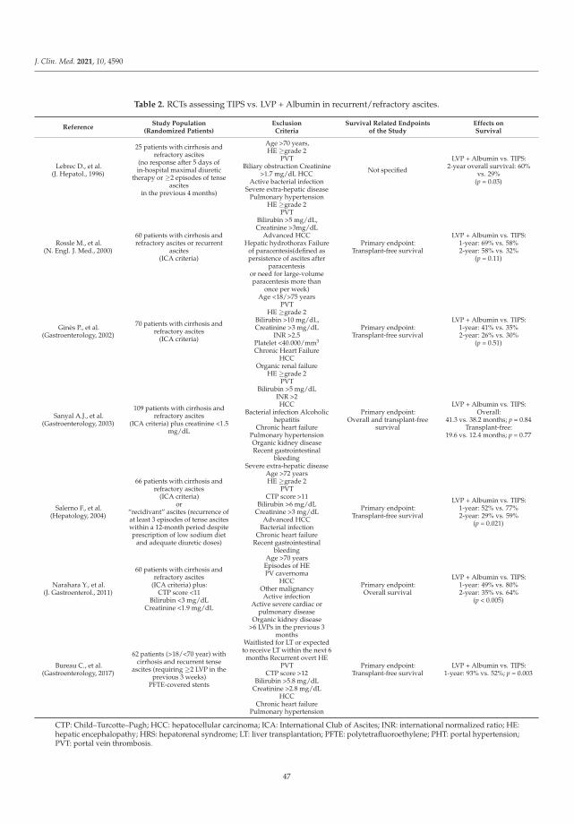
J. Clin. Med. 2021, 10, 4590
Table 2. RCTs assessing TIPS vs. LVP + Albumin in recurrent/refractory ascites.
ReferenceStudy Population
(Randomized Patients)ExclusionCriteria
Survival Related Endpointsof the Study
Effects onSurvival
Lebrec D., et al.(J. Hepatol., 1996)
25 patients with cirrhosis andrefractory ascites
(no response after 5 days ofin-hospital maximal diuretic
therapy or ≥2 episodes of tenseascites
in the previous 4 months)
Age >70 years,HE ≥grade 2
PVTBiliary obstruction Creatinine
>1.7 mg/dL HCCActive bacterial infection
Severe extra-hepatic diseasePulmonary hypertension
Not specified
LVP + Albumin vs. TIPS:2-year overall survival: 60%
vs. 29%(p = 0.03)
Rossle M., et al.(N. Engl. J. Med., 2000)
60 patients with cirrhosis andrefractory ascites or recurrent
ascites(ICA criteria)
HE ≥grade 2PVT
Bilirubin >5 mg/dL,Creatinine >3mg/dL
Advanced HCCHepatic hydrothorax Failure
of paracentesis(defined aspersistence of ascites after
paracentesisor need for large-volumeparacentesis more than
once per week)
Primary endpoint:Transplant-free survival
LVP + Albumin vs. TIPS:1-year: 69% vs. 58%2-year: 58% vs. 32%
(p = 0.11)
Ginès P., et al.(Gastroenterology, 2002)
70 patients with cirrhosis andrefractory ascites
(ICA criteria)
Age <18/>75 yearsPVT
HE ≥grade 2Bilirubin >10 mg/dL,Creatinine >3 mg/dL
INR >2.5Platelet <40.000/mm3
Chronic Heart FailureHCC
Organic renal failure
Primary endpoint:Transplant-free survival
LVP + Albumin vs. TIPS:1-year: 41% vs. 35%2-year: 26% vs. 30%
(p = 0.51)
Sanyal A.J., et al.(Gastroenterology, 2003)
109 patients with cirrhosis andrefractory ascites
(ICA criteria) plus creatinine <1.5mg/dL
HE ≥grade 2PVT
Bilirubin >5 mg/dLINR >2HCC
Bacterial infection Alcoholichepatitis
Chronic heart failurePulmonary hypertensionOrganic kidney diseaseRecent gastrointestinal
bleedingSevere extra-hepatic disease
Primary endpoint:Overall and transplant-free
survival
LVP + Albumin vs. TIPS:Overall:
41.3 vs. 38.2 months; p = 0.84Transplant-free:
19.6 vs. 12.4 months; p = 0.77
Salerno F., et al.(Hepatology, 2004)
66 patients with cirrhosis andrefractory ascites
(ICA criteria)or
“recidivant” ascites (recurrence ofat least 3 episodes of tense asciteswithin a 12-month period despiteprescription of low sodium diet
and adequate diuretic doses)
Age >72 yearsHE ≥grade 2
PVTCTP score >11
Bilirubin >6 mg/dLCreatinine >3 mg/dL
Advanced HCCBacterial infection
Chronic heart failureRecent gastrointestinal
bleeding
Primary endpoint:Transplant-free survival
LVP + Albumin vs. TIPS:1-year: 52% vs. 77%2-year: 29% vs. 59%
(p = 0.021)
Narahara Y., et al.(J. Gastroenterol., 2011)
60 patients with cirrhosis andrefractory ascites
(ICA criteria) plus:CTP score <11
Bilirubin <3 mg/dLCreatinine <1.9 mg/dL
Age >70 yearsEpisodes of HEPV cavernoma
HCCOther malignancyActive infection
Active severe cardiac orpulmonary disease
Organic kidney disease
Primary endpoint:Overall survival
LVP + Albumin vs. TIPS:1-year: 49% vs. 80%2-year: 35% vs. 64%
(p < 0.005)
Bureau C., et al.(Gastroenterology, 2017)
62 patients (>18/<70 year) withcirrhosis and recurrent tense
ascites (requiring ≥2 LVP in theprevious 3 weeks)
PFTE-covered stents
>6 LVPs in the previous 3months
Waitlisted for LT or expectedto receive LT within the next 6
months Recurrent overt HEPVT
CTP score >12Bilirubin >5.8 mg/dL
Creatinine >2.8 mg/dLHCC
Chronic heart failurePulmonary hypertension
Primary endpoint:Transplant-free survival
LVP + Albumin vs. TIPS:1-year: 93% vs. 52%; p = 0.003
CTP: Child–Turcotte–Pugh; HCC: hepatocellular carcinoma; ICA: International Club of Ascites; INR: international normalized ratio; HE:hepatic encephalopathy; HRS: hepatorenal syndrome; LT: liver transplantation; PFTE: polytetrafluoroethylene; PHT: portal hypertension;PVT: portal vein thrombosis.
47

J. Clin. Med. 2021, 10, 4590
The reduction in portal pressure induced by TIPS insertion is associated with beneficialeffects on renal function in patients with cirrhosis and ascites, including an increase insodium and water excretion and reduced activity of vasoconstrictor and antinatriureticsystems [2,51]. This could represent a valid rationale for the use of TIPS also in patientswith HRS. The results of the available trials [66–68], although limited by a non-controlleddesign and small sample sizes, support this therapeutical approach, especially for non-transplantable patients. Furthermore, the practical applicability is usually limited by theconcomitant severe degree of liver failure [2].
In summary, by acting against a key pathophysiological mechanism as portal hy-pertension, TIPS has a great potential to serve as DMAC. However, two factors limit itswidespread use to this end. On the one hand, available studies evaluated the effects of TIPSin patients who develop either portal hypertensive bleeding or difficult-to-treat ascites.Its impact in patients with clinically significant portal hypertension but without thesecomplications has never been assessed. On the other, the occurrence of severe TIPS-relatedcomplications often prevents its insertion in patients with refractory ascites. The use of asmall-diameter covered stent may help in overcoming these limitations.
3.3. Poorly Absorbable Antibiotics
Acting on the intestinal microbiota to limit the abnormal translocation of bacteria andbacterial products is another treatment strategy of relevant importance in patients withcirrhosis. The use of antibiotics would appear an obvious choice, whose main limitation de-rives from the induction of bacterial resistance. Most available studies assessed the effects ofnon- or poorly-adsorbable antibiotics to limit their activities in the intestinal environment.
3.3.1. Quinolones
The enhanced susceptibility to bacterial infections by patients with cirrhosis has longbeen known. The high prevalence of episodes sustained by Gram-negative microorganismsof intestinal origin led to recognize the relevance of the abnormal bacterial translocationfrom the gut [69–71].
The proof-of-concept of the efficacy of antibiotic prophylaxis derives from an RCTcomparing 12 months of treatment with norfloxacin versus placebo for 12 months toprevent SBP recurrence [72]. Norfloxacin remarkably reduced the recurrences due to Gram-negative bacteria, without side effects. Subsequent studies showed that either norfloxacinor ciprofloxacin significantly prevented SBP occurrence in high-risk patients with lowprotein concentration in ascites, liver failure, and/or gastrointestinal bleeding. The benefitderiving from either norfloxacin or ciprofloxacin in either primary or secondary prophylaxisof SBP was confirmed by most meta-analyses [73,74]. Interestingly, norfloxacin reducedcirculating bacterial products associated with improved systemic inflammatory markersand circulatory dysfunction [75].
These results potentially demonstrate a pathophysiological effect beyond SBP preven-tion. However, a benefit on survival and the occurrence of complications of cirrhosis stillneeds to be convincingly established. A pivotal study in decompensated patients with alow protein content in ascitic fluid (<1.5 g/dL) and severe cirrhosis (Child–Pugh ≥9 withserum bilirubin ≥3 mg/dL) or impaired renal function (serum creatinine ≥1.2 mg/dL,blood urea nitrogen ≥25 mg/dL or serum sodium ≤130 mEq/L), showed that norfloxacinprophylaxis not only reduced the incidence of SBP but also lessened one-year risk for HRSand improved one-year survival compared with placebo [76]. However, both survival andincidence of HRS were secondary endpoints, and the study sample size was inadequate.More recently, a large multicenter RCT did not demonstrate a survival advantage in ChildC patients receiving norfloxacin prophylaxis, although a post-hoc analysis unveiled areduction in six-month mortality confined to the subgroup with low ascites protein con-centration [77]. Finally, concerns about the safety of quinolones being a risk factor forthe development of infections due to drug-resistant bacteria have been raised over theyears [71], although more recent reports did not confirm these findings [77,78].
48

J. Clin. Med. 2021, 10, 4590
In conclusion, several important questions regarding the use of quinolones in decom-pensated cirrhosis remain unanswered, so that their administration as a DMAC cannot becurrently proposed.
3.3.2. Rifaximin
There has been growing interest in the possible efficacy of rifaximin in preventinginfections and other complications of portal hypertension in cirrhosis. Rifaximin is aminimally absorbed antibiotic with activity against Gram-negative and Gram-positivebacteria [79]. Moreover, it can improve the gut epithelial layer homeostasis, decreaseinflammatory pathways, impair bacterial adhesion to enterocytes, and modulate the gutmicrobiome, thus positively affecting the entire gut liver-axis [80–82].
Currently, the only recognized indication for rifaximin use in patients with cirrhosis isthe prevention of recurrent hepatic encephalopathy (secondary prophylaxis) [2], mainlybased on the pivotal RCT by Bass et al. in 2010 [83]. However, both observational studiesand a few small-scale RCTs showed an association between rifaximin treatment and im-portant clinical outcomes, including better control of ascites [84,85], a reduced incidenceof decompensation, hospitalizations, variceal bleeding, SBP, and HRS with a decreasedrisk of renal replacement therapy [86–93]. Some studies even suggested an improvementin mortality [85,87,89]. However, several metanalyses [94–96] highlighted the overall lowquality of these studies precluding any generalization on the beneficial impact of rifaximinon the clinical course of decompensated cirrhosis. Therefore, before using rifaximin as aDMAC can be advocated in clinical practice, well-designed RCTs with hard endpoints andadequate sample size are needed.
3.4. Statins
The first proposal of statins for treating portal hypertension in cirrhosis dates backto the early 2000s [97] since they reduce intrahepatic vascular resistance by enhancingnitric oxide (NO) production in liver sinusoids. Subsequent investigations in experimentalcirrhosis also unveiled anti-inflammatory and hepatoprotective properties [98–102]. Statinsexert their effects on liver inflammation and fibrogenesis by upregulating the endothelialKruppel-like factor 2 (KLF2) [101]. This transcription factor regulates the expression of sev-eral vasoprotective genes controlling apoptosis, inflammation, oxidative stress, thrombosis,and vasodilation. Furthermore, it inhibits RhoA/Rho-kinase signaling, which is partlyresponsible for the contractility of hepatic stellate cells [102].
Several observational studies reported the beneficial effects of statins on variousclinical aspects of advanced liver disease. They include lower rates of decompensation,liver cancer, and bacterial infections along with increased survival [103,104]. However,these results require caution due to potential flaws in observational studies [105]. DifferentRCTs convincingly showed that simvastatin decreases portal pressure [106,107]. Furtherstudies compared the lowering effect on portal pressure induced by combining statins withbeta-blockers vs. beta-blockers alone, providing discordant results [108,109].
Only one randomized trial evaluated the effects of statins on the complications ofcirrhosis [110]. This trial included patients with recent variceal bleeding and assessed theeffects of adding simvastatin 40 mg daily or placebo to the standard therapy to preventrebleeding (NSBBs and endoscopic variceal ligation). Simvastatin did not significantlyinfluence the primary endpoint, which was a composite of rebleeding and death. Never-theless, it was associated with a significant survival benefit, mainly related to decreasedmortality from bleeding and bacterial infections. Patient subgroup analysis showed thatthis result was limited to patients belonging to Child-Pugh classes A and B. This ap-parent contradiction (no prevention of complications but improved survival) suggeststhat the effect on portal pressure might be less relevant than the non-hemodynamic ef-fects of statins, attenuating the intense inflammatory response triggered by infections orbleeding events [99,100,111,112] that plays a major role in the development of ACLF andmortality [6,113].
49
J. Clin. Med. 2021, 10, 4590
In conclusion, although there is a robust rationale suggesting that statins might bebeneficial for patients with cirrhosis, the evidence from randomized trials is scarce, limitedto a single study with a positive result on a secondary endpoint. Non-hemodynamic effectsof statins might be more important than the effects of portal pressure.
The currently ongoing LiverHope Efficacy study, a multicenter double-blind trial com-paring the effects of simvastatin plus rifaximin vs. placebo in the prevention of ACLF andmortality (www.liverhope-h2020.eu, accessed on 4 October 2021), should clarify whetherstatins are effective and safe in advanced cirrhosis. Growing evidence support the roleof bacterial translocation and systemic inflammation as key drivers of cirrhosis progres-sion and ACLF development. On this pathophysiological background, the associationof an agent effective in preventing bacterial translocation (rifaximin) and a drug withanti-inflammatory properties (simvastatin) could provide a dual effect able to counteractdisease progression. It is important to emphasize that although there have been no safetyissues in patients with compensated cirrhosis, statin pharmacokinetic is markedly alteredin patients with decompensated liver disease [110], with a very high risk of muscle toxicitycompared to the general population [110]. Very recent data from the LiverHope double-blind dose-finding safety trial showed that a dose of 20 mg/day was associated with nomuscle toxicity [114].
3.5. Human Albumin
The well-established recommendations for human albumin (HA) use in patientswith decompensated cirrhosis pertain to conditions characterized by an acute worseningof effective volemia. Indeed, one-shot or short-term HA administration is employed toprevent paracentesis-induced circulatory dysfunction (PICD), prevent renal dysfunctioninduced by SBP, and diagnose and treat HRS in association with vasoconstrictors [2,115].
The oncotic properties of HA make it an optimal candidate to correct or attenuateeffective hypovolemia. However, HA exerts several functions not related to its oncoticproperties. These pleiotropic functions include binding of damaging molecules, modulatinginflammation and immune responses, exerting antioxidant activity, improving cardiacfunction, and restoring endothelial integrity [116]. Therefore, from a pathophysiologicalperspective, HA could act as a multitarget agent, potentially modifying the clinical courseof decompensated cirrhosis. Such an approach would imply long-term HA administration.
In 2018, two RCTs and one prospective observational study evaluated the efficacyof long-term HA in patients with ascites, opening new perspectives for the treatment ofdecompensated cirrhosis [117–119]. The ANSWER study [117], a multicenter open-labelRCT, enrolled patients with uncomplicated grade 2 or 3 ascites. Those patients included inthe active arm of the study received 40 g of HA per week for the initial two weeks, then40 g weekly for a maximal duration of 18 months. The primary endpoint of the study wasreached, as patients receiving HA had a significantly better 18-month overall survival, witha 38% reduction in mortality hazard ratio. Moreover, the management of ascites becameeasier, as the need for paracentesis and the incidence of refractory ascites were reduced byabout 50%. HA administration also lowered the incidence rate of the major complicationsof cirrhosis. As a result, patients receiving albumin needed fewer and shorter liver-relatedhospitalizations and preserved their quality of life. Similar results were also reported by asingle-center, prospective, non-randomized study in patients with refractory ascites [118].In contrast, the MACHT study [119], a multicenter placebo-controlled RCT performed inpatients with ascites waitlisted for liver transplantation, showed no differences in either theprobability of developing complications or death between patients treated or not with HA.
These divergent results should not advise against long-term HA use in decompensatedpatients. Contrariwise, their comparison provides essential information about patients tobe treated and, mainly, the dose and schedule of HA administration. The ANSWER andthe MACHT studies differed in terms of design, baseline patient characteristics, length offollow-up, and dosage and timing of albumin administration. A consequence of the loweramount of HA given to patients enrolled in the MACHT trial was that serum albumin
50
J. Clin. Med. 2021, 10, 4590
concentration remained steady throughout the follow-up [119]. In contrast, serum albuminconcentration rose by 0.7–0.8 g/L to almost 4 g/dL in the ANSWER study [117]. A post-hocanalysis of the latter trial showed that on-treatment serum albumin concentration at onemonth predicts the probability of 18-month overall survival, which was greater than 90%in patients whose serum albumin concentration reached levels 4 g/dL [120]. Baselineserum albumin and MELD score value independently predicted the achievement of thisthreshold. This would imply that patients with severe hypoalbuminema and very highMELD score should receive greater amounts of HA to achieve the best results [120]. Twoother pieces of evidence support the importance of steadily increasing serum albuminconcentration beyond a certain level. First, in the pilot-PRECIOSA study, only a “highdose” of HA (1.5 g/kg b.w. every week) made serum albumin rise to a concentrationclose to 4 g/dl, while a lower dose (1 g/kg b.w. every 10 days) failed to normalize serumalbumin in most cases. Notably, only patients who received the high HA dose improvedtheir cardiocirculatory function [121]. Second, a serum albumin concentration greater than4 g/dL is physiologically present in more than 90% of healthy adult individuals [122].
The recently published ATTIRE Trial [123], an open-label, multicenter RCT whichincluded hospitalized patients with acute decompensation of cirrhosis (with or withoutACLF) deserves some comments. In this study, albumin was administered for up to 14 dayswith the goal to maintain a serum albumin level >3.0 g/dL. No differences were observedin the incidence of infections, renal dysfunction, and death between the two study arms.Moreover, some safety concerns have been raised, mainly due to a higher incidence of fluidoverload and pulmonary edema in treated patients. Therefore, albumin administrationdoes not appear to modify the short-term course of cirrhosis in severely ill patients admittedto hospital for an acute complication, at least with the dose and schedule of administrationchosen in the ATTIRE study. So far, indeed, the efficacy of short-term albumin use has beendemonstrated only in patients with SBP or HRS. Acutely ill hospitalized patients, however,represent a completely different clinical setting compared to chronic administration tostable decompensated patients.
In conclusion, there is growing evidence that long-term HA administration can modifythe natural history of decompensated cirrhosis, thus acting as a DMAC rather than a specifictreatment for ascites and their acute complications. Future studies are warranted to bettercharacterize patient subgroups who could benefit the most from this novel approach.There is also a need to go beyond a fixed dosage and schedule of HA administration,ideally tending to an individualized and patient-tailored approach. Future investigationsshould prove or challenge this hypothesis and other open issues, including the definition ofstopping rules and the cost-effectiveness (in settings other than Italy [117]) of this relativelydemanding treatment in terms of logistics and patient adherence.
3.6. Granulocyte Colony-Stimulating Factor
Several RCTs assessed the effects of granulocyte colony-stimulating factor (G-CSF),either alone or in combination with bone marrow stem cell transplantation, in patients withacute alcoholic hepatitis (AH) and/or ACLF and in patients with more stable decompen-sated cirrhosis. Among the pleiotropic effects of GCS-F, the stimulation of liver regenerationand the improvement in immune dysfunction have been proposed as the mechanismsof action [124]. A recent meta-analysis including 7 RCTs [125–131] in patients with AHand/or ACLF showed a significant benefit of 90-day survival in favor of G-CSF [132].However, these results were not confirmed by another analysis [133]. Conclusions are alsochallenging due to the high heterogeneity of the studies and by the different outcomesfound in Europe and Asia [132].
Similar results have been reported in RCTs assessing G-CSF in patients with more sta-ble decompensated cirrhosis. Studies from India provided positive results [134–137]. In con-trast, a trial performed in the United Kingdom showed no improvement in MELD score atthree months with G-CSF, with or without hematopoietic stem-cell infusion (CD133+) [138].Moreover, there was an increase in adverse events and sepsis.
51

J. Clin. Med. 2021, 10, 4590
RCTs demonstrating the efficacy of G-CSF in European and US cohorts are neededif G-CSF is to be considered a therapeutic option in the future. Aspects to be evaluatedinclude the ideal dose and schedule of administration, the duration of therapy, the useof combinatorial therapies, the identification of the most appropriate target population,a better understanding of the mechanisms of action, including those potentially adverse,and, finally, understanding whether geographical differences affect the response.
4. Controversial Areas and Future Perspectives
Based on the data reported above, no treatments are so far available to effectivelymanage patients with cirrhosis, especially in the decompensated stage of the disease.Besides various etiological treatments, that in patients with compensated cirrhosis canoften prevent decompensations and complications, no solid data support the efficacy asa DMAC of any currently available treatment for decompensated patients. Indeed, someinterventions given to treat/prevent a specific complication or comorbidities appear toact as a DMAC (i.e., albumin or TIPS) or have the potential to act (i.e., rifaximin, NSBBs,statins, G-CSF) in certain subgroups of patients.
Since the goal of a DMAC is to halt or at least slow down the progression of thedisease, or even partially revert decompensation, its efficacy should be tested by RCTs withpatients’ survival as a primary endpoint. However, the very large sample size required toreveal statistically significant differences forces researchers to use alternative “surrogateprimary endpoints”, such as the incidence of complications and/or ACLF. These latterevents, however, would work better as secondary endpoints, which should also includehospitalizations, quality of life, and cost-effectiveness of treatment [11,12]. Unfortunately,up to now, very few published RCTs have addressed the above endpoints. Examples ofsuch trials are the ANSWER and MACTH trials on albumin [117,119] or the NORFLOCIRtrial on norfloxacin [77]. In contrast, the low-quality data generated by a long seriesof observational studies and small-scale RCTs, which even fairly consistently show animprovement in survival and incidence of complications [84–93], have to date precluded theuse of rifaximin beyond the evidence-based indication of the prevention of HE recurrence.As a result, in the future reliable and conclusive data on DMACs can only derive frommulticenter, possibly international, well-designed and adequately powered RCTs, thushighlighting the importance of scientific consortia for promoting and coordinating theseresearch projects.
Moreover, due to the complexity and heterogeneity of patients included in the defi-nition of decompensated cirrhosis, some interventions could act as DMAC only in well-defined subgroups of patients. An example is given by albumin, which has been foundeffective in the ANSWER trial, if administered long-term in patients with stable decom-pensated cirrhosis and persistent grade 2 or 3 uncomplicated ascites [117], but not in theATTIRE study, which enrolled severe and acutely decompensated patients admitted to hos-pital, with or without ACLF [123]. In addition to the target population, other open issuesfor any intervention include transferability to daily clinical practice, the definition of dosageand schedule of administration, factors guiding treatment, temporary or permanent stop-ping rules, the use of combinatorial approaches, cost-effectiveness for healthcare systems,and access to treatment worldwide. Hopefully, some ongoing RCTs will clarify whetherthe course of decompensated cirrhosis could be positively impacted in well-defined groupsof patients by drugs or interventions that are currently used for more limited indications(NCT04072601, NCT03780673, NCT02401490, NCT03451292).
The increasing understanding of the pathophysiological mechanisms underlyingdecompensated cirrhosis and leading to hepatic and extra-hepatic organ failure couldpotentially provide new interventions, drugs, and biological substances. Novel candi-date DMACs should be able to target gut microbiota or key mechanisms in the patho-genetic network of gut-liver axis, systemic inflammation, and immune dysfunction. Onesuch approach, which is already under development, aims to antagonize a crucial up-stream step of the inflammatory cascade by TAK-242, an inhibitor of toll-like receptor 4
52
J. Clin. Med. 2021, 10, 4590
(TLR4) [139], which binds the circulating pathogen-associated molecular patterns (PAMPs),such as lipopolysaccharides (LPS) and gram-negative endotoxins [140], as well as damage-associated molecular patterns (DAMPs), such as cleaved nucleosomes, histones, andhigh-mobility group box 1 proteins (HMGB1) [141].
In the context of acutely decompensated disease with an abnormal burst of systemicinflammation, interesting perspectives could derive from studies assessing albumin-basedextracorporeal liver assist devices. The molecular adsorbent recirculating system (MARS)and Prometheus provided proof of concept that such a strategy could be successful butdid not show improvement in survival [142–144]. A novel device, DIALIVE, has beendeveloped to remove and replace the damaged albumin while also removing DAMPsand PAMPs [145]. A multicenter RCT (NCT03065699) aiming to assess the safety andperformance of DIALIVE in patients with ACLF has been recently completed and positiveresults have been publicly announced. Moreover, as an extension of this concept, anotherphase III, multicenter, RCT on plasma exchange in patients with ACLF (NCT03702920) iscurrently ongoing. Their results are eagerly needed to improve the management of thesesevere acute patients.
The identification of new DMACs will be also aided by innovative research technolo-gies and approaches, such as high throughput -omics techniques and systems medicineanalysis. When applied to large cohorts of patients with detailed clinical data, treatmenthistory and outcome as well as biological samples, these approaches will support the iden-tification or development of new DMACs and biomarkers to predict patient prognosis andresponse to therapies, such as the DECISION project (www.decision-for-liver.eu, accessedon 4 October 2021) is currently pursuing in the perspective of personalized medicine.
The next decade will reveal whether patients with cirrhosis, especially in its decompen-sated stage, could benefit from treatments that globally manage their disease by reducingthe occurrence of complications and ACLF, preventing hospitalization, and ultimatelyimproving survival and quality of life, as already occurs for other impactful diseases.
Author Contributions: Writing—Original draft preparation, G.Z.; writing—review and editing, M.T.;writing—review and editing, M.B.; writing—review and editing, P.C. All authors have read andagreed to the published version of the manuscript.
Funding: This project has received funding from the European Union’s Horizon 2020 Research andInnovation Programme under Grant Agreement No 731875.
Institutional Review Board Statement: Not applicable.
Informed Consent Statement: Not applicable.
Data Availability Statement: No new data were created or analyzed in this study. Data sharing isnot applicable to this article.
Conflicts of Interest: Zaccherini is part of the speakers’ bureau for Grifols SA and Octapharma SA,outside the submitted work. Tufoni is part of the speakers’ bureau for Grifols SA and OctapharmaSA, outside the submitted work. Bernardi is part of the speakers’ bureau for Grifols SA, OctapharmaAG, Takeda, CSL Behring GmbH, and PPTA, and is a consultant for CLS Behring GmbH, Grifols SAand Takeda, outside the submitted work. Caraceni is part of the speakers’ bureau for Grifols SA,Octapharma SA, Kedrion Biopharma SpA, Mallinkrodt SA, Gilead SA and Takeda SA, outside thesubmitted work.
References
1. D’Amico, G.; Morabito, A.; D’Amico, M.; Pasta, L.; Malizia, G.; Rebora, P.; Valsecchi, M.G. Clinical states of cirrhosis andcompeting risks. J. Hepatol. 2018, 68, 563–576. [CrossRef]
2. Angeli, P.; Bernardi, M.; Villanueva, C.; Francoz, C.; Mookerjee, R.; Trebicka, J.; Krag, A.; Laleman, W.; Gines, P. EASL Clinicalpractice guidelines for the management of patients with decompensated cirrhosis. J. Hepatol. 2018, 69, 406–460. [CrossRef]
3. Bernardi, M.; Caraceni, P.; Navickis, R.J.; Wilkes, M.M. Albumin infusion in patients undergoing large-volume paracentesis: Ameta-analysis of randomized trials. Hepatology 2012, 55, 1172–1181. [CrossRef]
4. Bai, M.; Qi, X.S.; Yang, Z.P.; Yang, M.; Fan, D.M.; Han, G.H. TIPS improves liver transplantation-free survival in cirrhotic patientswith refractory ascites: An updated meta-analysis. World J. Gastroenterol. 2014, 20, 2704–2714. [CrossRef]
53
J. Clin. Med. 2021, 10, 4590
5. Bernardi, M.; Caraceni, P. Novel perspectives in the management of decompensated cirrhosis. Nat. Rev. Gastroenterol. Hepatol.2018, 15, 753–764. [CrossRef]
6. Caraceni, P.; Abraldes, J.G.; Ginès, P.; Newsome, P.N.; Sarin, S.K. The search for disease-modifying agents in decompensatedcirrhosis: From drug repurposing to drug discovery. J. Hepatol. 2021, 75, S118–S134. [CrossRef]
7. Lens, S.; Baiges, A.; Alvarado-Tapias, E.; Llop, E.; Martinez, J.; Fortea, J.I.; Ibáñez-Samaniego, L.; Mariño, Z.; Rodríguez-Tajes, S.;Gallego, A.; et al. Clinical outcome and hemodynamic changes following HCV eradication with oral antiviral therapy in patientswith clinically significant portal hypertension. J. Hepatol. 2020, 73, 1415–1424. [CrossRef] [PubMed]
8. Verna, E.C.; Morelli, G.; Terrault, N.A.; Lok, A.S.; Lim, J.K.; Di Bisceglie, A.M.; Zeuzem, S.; Landis, C.S.; Kwo, P.; Hassan, M.; et al.DAA therapy and long-term hepatic function in advanced/decompensated cirrhosis: Real-world experience from HCV-TARGETcohort. J. Hepatol. 2020, 73, 540–548. [CrossRef] [PubMed]
9. Bernardi, M.; Moreau, R.; Angeli, P.; Schnabl, B.; Arroyo, V. Mechanisms of decompensation and organ failure in cirrhosis: Fromperipheral arterial vasodilation to systemic inflammation hypothesis. J. Hepatol. 2015, 63, 1272–1284. [CrossRef] [PubMed]
10. Engelmann, C.; Clària, J.; Szabo, G.; Bosch, J.; Bernardi, M. Pathophysiology of decompensated cirrhosis: Portal hypertension,circulatory dysfunction, inflammation, metabolism and mitochondrial dysfunction. J. Hepatol. 2021, 75, S49–S66. [CrossRef]
11. Solà, E.; Pose, E.; Campion, D.; Piano, S.; Roux, O.; Simon-Talero, M.; Uschner, F.; de Wit, K.; Zaccherini, G.; Alessandria, C.; et al.Endpoints and design of clinical trials in patients with decompensated cirrhosis: Position paper of the LiverHope Consortium. J.Hepatol. 2020, 74, 200–219. [CrossRef]
12. Abraldes, J.G.; Trebicka, J.; Chalasani, N.; D’Amico, G.; Rockey, D.C.; Shah, V.H.; Bosch, J.; Garcia-Tsao, G. Prioritization oftherapeutic targets and trial design in cirrhotic portal hypertension. Hepatology 2018, 69, 1287–1299. [CrossRef] [PubMed]
13. Trebicka, J.; Fernández, J.; Papp, M.; Caraceni, P.; Laleman, W.; Gambino, C.; Giovo, I.; Uschner, F.E.; Jimenez, C.; Mookerjee, R.;et al. The PREDICT study uncovers three clinical courses of acutely decompensated cirrhosis that have distinct pathophysiology.J. Hepatol. 2020, 73, 842–854. [CrossRef]
14. Moreau, R.; Jalan, R.; Gines, P.; Pavesi, M.; Angeli, P.; Cordoba, J.; Durand, F.; Gustot, T.; Saliba, F.; Domenicali, M.; et al. Acute-on-chronic liver failure is a distinct syndrome that develops in patients with acute decompensation of cirrhosis. Gastroenterology2013, 144, 1426–1437.e9. [CrossRef]
15. Mandorfer, M.; Simbrunner, B. Prevention of first decompensation in advanced chronic liver disease. Clin. Liver Dis. 2021, 25,291–310. [CrossRef] [PubMed]
16. Lok, A.S.; McMahon, B.J.; Brown, R.S., Jr.; Wong, J.B.; Ahmed, A.T.; Farah, W.; Almasri, J.; Alahdab, F.; Benkhadra, K.; Mouchli,M.A.; et al. Antiviral therapy for chronic hepatitis B viral infection in adults: A systematic review and meta-analysis. Hepatology2015, 63, 284–306. [CrossRef] [PubMed]
17. Peng, C.-Y.; Chien, R.-N.; Liaw, Y.-F. Hepatitis B virus-related decompensated liver cirrhosis: Benefits of antiviral therapy. J.Hepatol. 2012, 57, 442–450. [CrossRef]
18. Jang, J.W.; Choi, J.Y.; Kim, Y.S.; Yoo, J.-J.; Woo, H.Y.; Choi, S.K.; Jun, C.H.; Lee, C.H.; Sohn, J.H.; Tak, W.Y.; et al. Effects of virologicresponse to treatment on short- and long-term outcomes of patients with chronic hepatitis b virus infection and decompensatedcirrhosis. Clin. Gastroenterol. Hepatol. 2018, 16, 1954–1963.e3. [CrossRef]
19. Ju, Y.C.; Jun, D.W.; Choi, J.; Saeed, W.K.; Lee, H.Y.; Oh, H.W. Long term outcome of antiviral therapy in patients with hepatitis Bassociated decompensated cirrhosis. World J. Gastroenterol. 2018, 24, 4606–4614. [CrossRef]
20. Mendizabal, M.; Piñero, F.; Ridruejo, E.; Herz Wolff, F.; Anders, M.; Reggiardo, V.; Ameigeiras, B.; Palazzo, A.; Alonso, C.;Schinoni, M.I.; et al. Disease progression in patients with hepatitis C virus infection treated with Direct-Acting Antiviral agents.Clin. Gastroenterol. Hepatol. 2020, 28, 30263–30269. [CrossRef]
21. Belli, L.S.; Berenguer, M.; Cortesi, P.A.; Strazzabosco, M.; Rockenschaub, S.-R.; Martini, S.; Morelli, C.; Donato, F.; Volpes, R.;Pageaux, G.-P.; et al. Delisting of liver transplant candidates with chronic hepatitis C after viral eradication: A European study. J.Hepatol. 2016, 65, 524–531. [CrossRef]
22. El-Sherif, O.; Jiang, Z.G.; Tapper, E.B.; Huang, K.; Zhong, A.; Osinusi, A.; Charlton, M.; Manns, M.; Afdhal, N.; Mukamal, K.; et al.Baseline factors associated with improvements in decompensated cirrhosis after direct-acting antiviral therapy for hepatitis Cvirus infection. Gastroenterology 2018, 154, 2111–2121.e8. [CrossRef]
23. European Association for the Study of the Liver. EASL Clinical Practice Guidelines: Liver transplantation. J. Hepatol. 2016, 64,433–485. [CrossRef]
24. Terrault, N.A.; Mccaughan, G.; Curry, M.P.; Gane, E.; Fagiuoli, S.; Fung, J.; Agarwal, K.; Lilly, L.; Strasser, S.I.; Brown, K.A.;et al. International liver transplantation society consensus statement on Hepatitis C management in liver transplant candidates.Transplantation 2017, 101, 945–955. [CrossRef] [PubMed]
25. Altamirano, J.; López-Pelayo, H.; Michelena, J.; Jones, P.D.; Ortega, L.; Ginès, P.; Caballería, J.; Gual, A.; Bataller, R.; Lligoña,A. Alcohol abstinence in patients surviving an episode of alcoholic hepatitis: Prediction and impact on long-term survival.Hepatology 2017, 66, 1842–1853. [CrossRef] [PubMed]
26. Degré, D.; Stauber, R.E.; Englebert, G.; Sarocchi, F.; Verset, L.; Rainer, F.; Spindelboeck, W.; Njimi, H.; Trépo, E.; Gustot, T.; et al.Long-term outcomes in patients with decompensated alcohol-related liver disease, steatohepatitis and Maddrey’s discriminantfunction <32. J. Hepatol. 2020, 72, 636–642. [CrossRef] [PubMed]
27. El Sherif, O.; Dhaliwal, A.; Newsome, P.N.; Armstrong, M.J. Sarcopenia in nonalcoholic fatty liver disease: New challenges forclinical practice. Expert Rev. Gastroenterol. Hepatol. 2020, 14, 197–205. [CrossRef]
54
J. Clin. Med. 2021, 10, 4590
28. Tandon, P.; Ney, M.; Irwin, I.; Ma, M.M.; Gramlich, L.; Bain, V.G.; Esfandiari, N.; Baracos, V.; Montano-Loza, A.J.; Myers, R.P.Severe muscle depletion in patients on the liver transplant wait list: Its prevalence and independent prognostic value. LiverTransplant. 2012, 18, 1209–1216. [CrossRef]
29. Berzigotti, A.; Albillos, A.; Villanueva, C.; Genescá, J.; Ardevol, A.; Augustín, S.; Calleja, J.L.; Bañares, R.; García-Pagán, J.C.;Mesonero, F.; et al. Effects of an intensive lifestyle intervention program on portal hypertension in patients with cirrhosis andobesity: The SportDiet study. Hepatology 2017, 65, 1293–1305. [CrossRef]
30. Hoeroldt, B.; McFarlane, E.; Dube, A.; Basumani, P.; Karajeh, M.; Campbell, M.J.; Gleeson, D. Long-term outcomes of patientswith auto-immune hepatitis managed at a nontransplant center. Gastroenterology 2011, 140, 1980–1989. [CrossRef]
31. De Franchis, R.; Baveno, V.I.F. Expanding consensus in portal hypertension: Report of the Baveno VI Consensus Workshop:Stratifying risk and individualizing care for portal hypertension. J. Hepatol. 2015, 63, 743–752. [CrossRef]
32. Garcia-Tsao, G.; Abraldes, J.G.; Berzigotti, A.; Bosch, J. Portal hypertensive bleeding in cirrhosis: Risk stratification, diagnosis, andmanagement: 2016 practice guidance by the American Association for the study of liver diseases. Hepatology 2016, 65, 310–335.[CrossRef] [PubMed]
33. Bendtsen, F.; Henriksen, J.H.; Sørensen, T.I. Propranolol and haemodynamic response in cirrhosis. J. Hepatol. 1991, 13, 144–148.[CrossRef]
34. Rodrigues, S.G.; Mendoza, Y.P.; Bosch, J. Beta-blockers in cirrhosis: Evidence-based indications and limitations. JHEP Rep. 2019, 2,100063. [CrossRef] [PubMed]
35. Bañares, R.; Moitinho, E.; Matilla, A.; García-Pagán, J.C.; Lampreave, J.L.; Piera, C.; Abraldes, J.G.; De Diego, A.; Albillos, A.;Bosch, J. Randomized comparison of long-term carvedilol and propranolol administration in the treatment of portal hypertensionin cirrhosis. Hepatology 2002, 36, 1367–1373. [CrossRef]
36. Reiberger, T.; Ferlitsch, A.; Payer, B.A.; Mandorfer, M.; Heinisch, B.B.; Hayden, H.; Lammert, F.; Trauner, M.; Peck-Radosavljevic,M.; Vogelsang, H. Non-selective betablocker therapy decreases intestinal permeability and serum levels of LBP and IL-6 inpatients with cirrhosis. J. Hepatol. 2012, 58, 911–921. [CrossRef]
37. Poynard, T.; Calès, P.; Pasta, L.; Ideo, G.; Pascal, J.P.; Pagliaro, L.; Lebrec, D.; Franco–Italian Multicenter Study Group. Beta-adrenergic-antagonist drugs in the prevention of gas-trointestinal bleeding in patients with cirrhosis and esophageal varices. Ananalysis of data and prognostic factors in 589 patients from four randomized clinical trials. Franco-Italian Multicenter StudyGroup. N. Engl. J. Med. 1991, 324, 1532–1538. [PubMed]
38. Albillos, A.; Zamora, J.; Martínez, J.; Arroyo, D.; Ahmad, I.; De-La-Peña, J.; Garcia-Pagán, J.-C.; Lo, G.-H.; Sarin, S.; Sharma,B.; et al. Stratifying risk in the prevention of recurrent variceal hemorrhage: Results of an individual patient meta-analysis.Hepatology 2017, 66, 1219–1231. [CrossRef]
39. Ripoll, C.; Augustin, S.; Reiberger, T.; Moreau, R.; Salerno, F.; Albillos, A.; Abraldes, J.G.; García-Tsao, G. Effect of Current TherapiesAimed at Preventing Variceal Rebleeding on Other Complications of Cirrhosis; Springer: Berlin/Heidelberg, Germany, 2016; pp. 333–339.[CrossRef]
40. Turco, L.; Villanueva, C.; La Mura, V.; García-Pagán, J.C.; Reiberger, T.; Genescà, J.; Groszmann, R.J.; Sharma, B.C.; Merkel, C.;Bureau, C.; et al. Lowering portal pressure improves out-comes of patients with cirrhosis, with or without ascites: A meta-analysis.Clin. Gastroenterol. Hepatol. 2020, 18, 313–327. [CrossRef]
41. Villanueva, C.; Albillos, A.; Genescà, J.; Garcia-Pagan, J.C.; Calleja, J.L.; Aracil, C.; Bañares, R.; Morillas, R.M.; Poca, M.;Peñas, B.; et al. beta blockers to prevent decompensation of cirrhosis in patients with clinically significant portal hypertension(PREDESCI): A randomised, double-blind, placebo-controlled, multicentre trial. Lancet 2019, 393, 1597–1608. [CrossRef]
42. Kumar, M.; Kainth, S.; Choudhury, A.; Maiwall, R.; Mitra, L.G.; Saluja, V.; Agarwal, P.M.; Shasthry, S.M.; Jindal, A.; Bhardwaj, A.;et al. Treatment with carvedilol improves survival of patients with acute-on-chronic liver failure: A randomized controlled trial.Hepatol. Int. 2019, 13, 800–813. [CrossRef] [PubMed]
43. Sarin, S.K.; APASL ACLF Research Consortium (AARC) for APASL ACLF Working Party; Choudhury, A.; Sharma, M.K.; Maiwall,R.; Al Mahtab, M.; Rahman, S.; Saigal, S.; Saraf, N.; Soin, A.S.; et al. Acute-on-chronic liver failure: Consensus recommendationsof the Asian Pacific association for the study of the liver (APASL): An update. Hepatol. Int. 2019, 13, 353–390. [CrossRef]
44. Conn, H.O.; Pope, A. Propranolol in the treatment of portal hypertension: A caution. Hepatology 2007, 2, 641S–644S. [CrossRef]45. Sersté, T.; Melot, C.; Francoz, C.; Durand, F.; Rautou, P.-E.; Valla, D.; Moreau, R.; Lebrec, D. Deleterious effects of beta-blockers on
survival in patients with cirrhosis and refractory ascites. Hepatology 2010, 52, 1017–1022. [CrossRef]46. Mandorfer, M.; Bota, S.; Schwabl, P.; Bucsics, T.; Pfisterer, N.; Kruzik, M.; Hagmann, M.; Blacky, A.; Ferlitsch, A.; Sieghart, W.;
et al. Nonselective beta blockers increase risk for hepato-renal syndrome and death in patients with cirrhosis and spontaneousbacterial peritonitis. Gastroenterology 2014, 146, 1680–1690. [CrossRef] [PubMed]
47. Krag, A.; Wiest, R.; Albillos, A.; Gluud, L.L. The window hypothesis: Haemodynamic and non-haemodynamic effects ofbe-ta-blockers improve survival of patients with cirrhosis during a window in the disease. Gut 2012, 61, 967–969. [CrossRef]
48. Chirapongsathorn, S.; Valentin, N.; Alahdab, F.; Krittanawong, C.; Erwin, P.J.; Murad, M.H.; Kamath, P.S. Nonselective beta-blockers and survival in patients with cirrhosis and ascites: A systematic review and meta-analysis. Clin. Gastroenterol. Hepatol.2016, 14, 1096–1104. [CrossRef]
49. Téllez, L.; Ibáñez-Samaniego, L.; del Villar, C.P.; Yotti, R.; Martínez, J.; Carrión, L.; de Santiago, E.R.; Rivera, M.; González-Mansilla,A.; Pastor, O.; et al. Non-selective beta-blockers impair global circulatory homeostasis and renal function in cirrhotic patientswith refractory ascites. J. Hepatol. 2020, 73, 1404–1414. [CrossRef]
55
J. Clin. Med. 2021, 10, 4590
50. Tergast, T.L.; Kimmann, M.; Laser, H.; Gerbel, S.; Manns, M.P.; Cornberg, M.; Maasoumy, B. Systemic arterial blood pressuredetermines the therapeutic window of non-selective beta blockers in decompensated cirrhosis. Aliment. Pharmacol. Ther. 2019, 50,696–706. [CrossRef]
51. Garcia-Pagan, J.C.; Saffo, S.; Mandorfer, M.; Garcia-Tsao, G. Where does TIPS fit in the management of patients with cirrhosis? J.Hep. Rep. 2020, 2, 100122.
52. Monescillo, A.; Martínez-Lagares, F.; Ruiz-Del-Arbol, L.; Sierra, A.; Guevara, C.; Jiménez, E.; Marrero, J.M.; Buceta, E.; Sánchen, J.;Castellot, A.; et al. Influence of portal hypertension and its early decompression by TIPS placement on the outcome of varicealbleeding. Hepatology 2004, 40, 793–801. [CrossRef] [PubMed]
53. Garcia-Pagan, J.C.; Caca, K.; Bureau, C.; Laleman, W.; Appenrodt, B.; Luca, A.; Gonzalez-Abraldes, J.; Nevens, F.; Vinel, J.P.;Mössner, J.; et al. Early use of TIPS in patients with cirrhosis and variceal bleeding. N. Engl. J. Med. 2010, 362, 2370–2379.[CrossRef] [PubMed]
54. Lv, Y.; Yang, Z.; Liu, L.; Li, K.; He, C.; Wang, Z.; Bai, W.; Guo, W.; Yu, T.; Yuan, X.; et al. Early TIPS with covered stentsversus standard treatment for acute variceal bleeding in patients with advanced cirrhosis: A randomised controlled trial. LancetGastroenterol. Hepatol. 2019, 4, 587–598. [CrossRef]
55. Trebicka, J.; Gu, W.; Ibáñez-Samaniego, L.; Hernández-Gea, V.; Pitarch, C.; Garcia, E.; Procopet, B.; Giráldez, A.; Amitrano, L.;Villanueva, C.; et al. Rebleeding and mortality risk are increased by ACLF but reduced by pre-emptive TIPS. J. Hepatol. 2020, 73,1082–1091. [CrossRef]
56. Kumar, R.; Kerbert, A.J.; Sheikh, M.F.; Roth, N.; Calvao, J.A.; Mesquita, M.D.; Barreira, A.I.; Gurm, H.S.; Ramsahye, K.; Mookerjee,R.P.; et al. Determinants of mortality in patients with cirrhosis and uncontrolled variceal bleeding. J. Hepatol. 2020, 74, 66–79.[CrossRef] [PubMed]
57. Lebrec, D.; Giuily, N.; Hadengue, A.; Vilgrain, V.; Moreau, R.; Poynard, T.; Gadano, A.; Lassen, C.; Benhamou, J.-P.; Erlinger, S.Transjugular intrahepatic portosystemic shunts: Comparison with paracentesis in patients with cirrhosis and refractory ascites: Arandomized trial. J. Hepatol. 1996, 25, 135–144. [CrossRef]
58. Rössle, M.; Ochs, A.; Gülberg, V.; Siegerstetter, V.; Holl, J.; Deibert, P.; Olschewski, M.; Reiser, M.; Gerbes, A.L. A comparison ofparacentesis and transjugular intrahepatic portosystemic shunting in patients with ascites. New Engl. J. Med. 2000, 342, 1701–1707.[CrossRef]
59. Ginès, P.; Uriz, J.; Calahorra, B.; Garcia–Tsao, G.; Kamath, P.S.; Del Arbol, L.R.; Planas, R.; Bosch, J.; Arroyo, V.; Rodés, J. Tran-sjugular intrahepatic portosystemic shunting versus paracentesis plus albumin for refractory ascites in cirrhosis. Gastroenterology2002, 123, 1839–1847. [CrossRef]
60. Sanyal, A.J.; Genning, C.; Reddy, K.; Wong, F.; Kowdley, K.V.; Benner, K.; McCashland, T. The North American study for thetreatment of refractory ascites. Gastroenterology 2003, 124, 634–641. [CrossRef]
61. Salerno, F.; Merli, M.; Riggio, O.; Cazzaniga, M.; Valeriano, V.; Pozzi, M.; Nicolini, A.; Salvatori, F.; GIST. Randomized controlledstudy of TIPS versus para-centesis plus albumin in cirrhosis with severe ascites. Hepatology 2004, 40, 629–635. [CrossRef][PubMed]
62. Narahara, Y.; Kanazawa, H.; Fukuda, T.; Matsushita, Y.; Harimoto, H.; Kidokoro, H.; Katakura, T.; Atsukawa, M.; Taki, Y.; Kimura,Y.; et al. Transjugular intrahepatic portosystemic shunt versus paracentesis plus albumin in patients with refractory ascites whohave good hepatic and renal function: A prospective randomized trial. J. Gastroenterol. 2011, 46, 78–85. [CrossRef]
63. Bureau, C.; Thabut, D.; Oberti, F.; Dharancy, S.; Carbonell, N.; Bouvier, A.; Mathurin, P.; Otal, P.; Cabarrou, P.; Péron, J.M.; et al.Transjugular intrahepatic portosystemic shunts with covered stents increase transplant-free survival of patients with cirrhosisand recurrent ascites. Gastroenterology 2017, 152, 157–163. [CrossRef] [PubMed]
64. Miraglia, R.; Maruzzelli, L.; Tuzzolino, F.; Petridis, I.; D’Amico, M.; Luca, A. Transjugular intrahepatic portosystemic shunts inpatients with cirrhosis with refractory ascites: Comparison of clinical outcomes by using 8- and 10-mm PTFE-covered stents.Radiology 2017, 284, 281–288. [CrossRef] [PubMed]
65. Trebicka, J.; Bastgen, D.; Byrtus, J.; Praktiknjo, M.; Terstiegen, S.; Meyer, C.; Thomas, D.; Fimmers, R.; Treitl, M.; Euringer, W.;et al. Smaller-diameter covered transjugular intrahepatic portosystemic shunt stents are associated with increased survival. Clin.Gastroenterol. Hepatol. 2019, 17, 2793–2799.e1. [CrossRef] [PubMed]
66. Brensing, K.A.; Textor, J.; Perz, J.; Schiedermaier, P.; Raab, P.; Strunk, H.; Klehr, H.U.; Kramer, H.J.; Spengler, U.; Schild, H.;et al. Long term outcome after transjugular intrahepatic portosystemic stentshunt in non-transplant cirrhotics with hepatorenalsyndrome: A phase II study. Gut 2000, 47, 288–295. [CrossRef] [PubMed]
67. Guevara, M.; Ginès, P.; Bandi, J.C.; Gilabert, R.; Sort, P.; Jiménez, W.; Garcia-Pagán, J.C.; Bosch, J.; Arroyo, V.; Rodés, J. Transjugularintrahepatic portosystemic shunt in hepato-renal syndrome: Effects on renal function and vasoactive systems. Hepatology 1998,28, 416–422. [CrossRef]
68. Testino, G.; Ferro, C.; Sumberaz, A.; Messa, P.; Morelli, N.; Guadagni, B.; Ardizzone, G.; Valente, U. Type-2 hepatorenal syndromeand refractory ascites: Role of transjugular intrahepatic portosystemic stent-shunt in eighteen patients with advanced cirrhosisawaiting orthotopic liver transplantation. Hepatogastroenterology 2003, 50, 1753–1755.
69. Conn, H.O.; Fessel, J.M. Spontaneous bacterial peritonitis: Variations on a theme. Medicine 1971, 50, 161–197. [CrossRef]70. Rimola, A.; Soto, R.; Bory, F.; Arroyo, V.; Piera, C.; Rodes, J. Reticuloendothelial system phagocytic activity in cirrhosis and its
relation to bacterial infections and prognosis. Hepatology 1984, 4, 53–58. [CrossRef]
56

J. Clin. Med. 2021, 10, 4590
71. Jalan, R.; Fernandez, J.; Wiest, R.; Schnabl, B.; Moreau, R.; Angeli, P.; Stadlbauer, V.; Gustot, T.; Bernardi, M.; Canton, R.; et al.Bacterial infections in cirrhosis: A position statement based on the EASL Special Conference. J. Hepatol. 2014, 60, 1310–1324.[CrossRef]
72. Ginés, P.; Rimola, A.; Planas, R.; Vargas, V.; Marco, F.; Almela, M.; Forne, M.; Miranda, M.L.; Llach, J.; Salmerón, J.M.; et al.Norfloxacin prevents spontaneous bacterial peritonitis re-currence in cirrhosis: Results of a double-blind, placebo-controlled trial.Hepatology 1990, 12, 716–724. [CrossRef]
73. Facciorusso, A.; Papagiouvanni, I.; Cela, M.; Buccino, V.R.; Sacco, R. Comparative efficacy of long-term antibiotic treatments inthe primary prophylaxis of spontaneous bacterial peritonitis. Liver Int. 2019, 39, 1448–1458. [CrossRef] [PubMed]
74. Mücke, M.M.; Mücke, V.T.; Graf, C.; Schwarzkopf, K.M.; Ferstl, P.G.; Fernandez, J.; Zeuzem, S.; Trebicka, J.; Lange, C.M.;Herrmann, E. Efficacy of norfloxacin prophylaxis to prevent spontaneous bacterial peritonitis: A systematic review and meta-analysis. Clin. Transl. Gastroenterol. 2020, 11, e00223. [CrossRef] [PubMed]
75. Albillos, A.; de la Hera, A.; González, M.; Moya, J.; Calleja, J.; Monserrat, J.; Ruiz-Del-Arbol, L.; Alvarez-Mon, M. Increasedlipopolysaccharide binding protein in cirrhotic patients with marked immune and hemodynamic derangement. Hepatology 2003,37, 208–217. [CrossRef]
76. Fernández, J.; Navasa, M.; Planas, R.; Montoliu, S.; Monfort, D.; Soriano, G.; Vila, C.; Pardo, A.; Quintero, E.; Vargas, V.; et al.Primary prophylaxis of spontaneous bacterial peritonitis delays hepatorenal syndrome and improves survival in cirrhosis.Gastroenterology 2007, 133, 818–824. [CrossRef] [PubMed]
77. Moreau, R.; Elkrief, L.; Bureau, C.; Perarnau, J.-M.; Thévenot, T.; Saliba, F.; Louvet, A.; Nahon, P.; Lannes, A.; Anty, R.; et al.Effects of long-term norfloxacin therapy in patients with advanced cirrhosis. Gastroenterology 2018, 155, 1816–1827.e9. [CrossRef][PubMed]
78. Piano, S.; Singh, V.; Caraceni, P.; Maiwall, R.; Alessandria, C.; Fernandez, J.; Soares, E.C.; Kim, D.J.; Kim, S.E.; Marino, M.; et al.Epidemiology and effects of bacterial infections in patients with cirrhosis worldwide. Gastroenterology 2019, 156, 1368–1380.[CrossRef]
79. Scarpignato, C.; Pelosini, I. Rifaximin, a poorly absorbed antibiotic: Pharmacology and clinical potential. Chemotherapy 2005, 51,36–66. [CrossRef]
80. Bajaj, J.S. Review article: Potential mechanisms of action of rifaximin in the management of hepatic encephalopathy and othercomplications of cirrhosis. Aliment. Pharmacol. Ther. 2015, 43, 11–26. [CrossRef] [PubMed]
81. Vlachogiannakos, J.; Saveriadis, A.S.; Viazis, N.; Theodoropoulos, I.; Foudoulis, K.; Manolakopoulos, S.; Raptis, S.; Karamanolis,D.G. Intestinal decontami-nation improves liver haemodynamics in patients with alcohol-related decompensated cirrhosis.Aliment. Pharmacol. Ther. 2009, 29, 992–999. [CrossRef]
82. Esposito, G.; Nobile, N.; Gigli, S.; Seguella, L.; Pesce, M.; d’Alessandro, A.; Bruzzese, E.; Capoccia, E.; Steardo, L.; Cuomo, R.; et al.Rifaximin improves Clostridium difficile toxin A-induced toxicity in Caco-2 cells by the PXR-dependent TLR4/MyD88/NF-κBpathway. Front. Pharmacol. 2016, 7, 1–8. [CrossRef] [PubMed]
83. Bass, N.M.; Mullen, K.D.; Sanyal, A.; Poordad, F.; Neff, G.; Leevy, C.B.; Sigal, S.; Sheikh, M.Y.; Beavers, K.; Frederick, T.; et al.Rifaximin treatment in hepatic encephalopathy. N. Engl. J. Med. 2010, 362, 1071–1081. [CrossRef] [PubMed]
84. Hanafy, A.; Hassaneen, A.M. Rifaximin and midodrine improve clinical outcome in refractory ascites including renal function,weight loss, and short-term survival. Eur. J. Gastroenterol. Hepatol. 2016, 28, 1455–1461. [CrossRef]
85. Lv, X.-Y.; Ding, H.-G.; Zheng, J.-F.; Fan, C.-L.; Li, L. Rifaximin improves survival in cirrhotic patients with refractory ascites: Areal-world study. World J. Gastroenterol. 2020, 26, 199–218. [CrossRef] [PubMed]
86. Kang, S.H.; Lee, Y.B.; Lee, J.-H.; Nam, J.Y.; Chang, Y.; Cho, H.; Yoo, J.-J.; Cho, Y.Y.; Cho, E.J.; Yu, S.J.; et al. Rifaximin treatmentis associated with reduced risk of cirrhotic complications and prolonged overall survival in patients experiencing hepaticencephalopathy. Aliment. Pharmacol. Ther. 2017, 46, 845–855. [CrossRef] [PubMed]
87. Flamm, S.L.; Mullen, K.D.; Heimanson, Z.; Sanyal, A.J. Rifaximin has the potential to prevent complications of cirrhosis. Ther.Adv. Gastroenterol. 2018, 11. [CrossRef]
88. Salehi, S.; Tranah, T.H.; Lim, S.; Heaton, N.; Heneghan, M.; Aluvihare, V.; Patel, V.; Shawcross, D.L. Rifaximin reduces theincidence of spontaneous bacterial peritonitis, variceal bleeding and all-cause admissions in patients on the liver transplantwaiting list. Aliment. Pharmacol. Ther. 2019, 50, 435–441. [CrossRef]
89. Vlachogiannakos, J.; Viazis, N.; Vasianopoulou, P.; Vafiadis, I.; Karamanolis, D.G.; Ladas, S.D. Long-term administration ofrifaximin improves the prognosis of patients with decompensated alcoholic cirrhosis. J. Gastroenterol. Hepatol. 2013, 28, 450–455.[CrossRef]
90. Ibrahim, E.-S.; Alsebaey, A.; Zaghla, H.; Abdelmageed, S.M.; Gameel, K.; Abdelsameea, E. Long-term rifaximin therapy as aprimary prevention of hepatorenal syndrome. Eur. J. Gastroenterol. Hepatol. 2017, 29, 1247–1250. [CrossRef]
91. Dong, T.; Aronsohn, A.; Reddy, K.G.; Te, H.S. Rifaximin decreases the incidence and severity of acute kidney injury andhepatorenal syndrome in cirrhosis. Dig. Dis. Sci. 2016, 61, 3621–3626. [CrossRef]
92. Assem, M.; Elsabaawy, M.; Abdelrashed, M.; Elemam, S.; Khodeer, S.; Hamed, W.; Abdelaziz, A.; El-Azab, G. Efficacy andsafety of alternating norfloxacin and rifaximin as primary prophylaxis for spontaneous bacterial peritonitis in cirrhotic ascites: Aprospective randomized open-label comparative multicenter study. Hepatol. Int. 2015, 10, 377–385. [CrossRef]
93. Elfert, A.; Ali, L.A.; Soliman, S.; Ibrahim, S.; Abd-Elsalam, S. Randomized-controlled trial of rifaximin versus norfloxacin forsecondary prophylaxis of spontaneous bacterial peritonitis. Eur. J. Gastroenterol. Hepatol. 2016, 28, 1450–1454. [CrossRef]
57
J. Clin. Med. 2021, 10, 4590
94. Kamal, F.; Khan, M.A.; Cholankeril, G.; Khan, Z.; Lee, W.M.; Gadiparthi, C.; Ahmed, A.; Howden, C.W.; Nair, S.; Satapathy, S.K.Rifaximin for prevention of spontaneous bacterial peritonitis and hepatorenal syndrome in cirrhosis: A systematic review andmeta-analysis. Gastroenterology 2017, 152, S1151–S1152. [CrossRef]
95. Goel, A.; Rahim, U.; Nguyen, L.; Stave, C. Systematic review with meta-analysis: Rifaximin for the prophylaxis of spontaneousbacterial peritonitis. Aliment. Pharmacol. Ther. 2017, 46, 1029–1036. [CrossRef] [PubMed]
96. Komolafe, O.; Roberts, D.; Freeman, S.C.; Wilson, P.; Sutton, A.J.; Cooper, N.J.; Pavlov, C.S.; Milne, E.J.; Hawkins, N.; Cowlin, M.;et al. Antibiotic prophylaxis to prevent spontaneous bacterial peritonitis in people with liver cirrhosis: A network meta-analysis.Cochrane Database Syst. Rev. 2020, 1, CD013125. [CrossRef]
97. Zafra, C.; Abraldes, J.G.; Turnes, J.; Berzigotti, A.; Fernández, M.; Garcia-Pagán, J.C.; Rodés, J.; Bosch, J. Simvastatin enhanceshepatic nitric oxide production and decreases the hepatic vascular tone in patients with cirrhosis. Gastroenterology 2004, 126,749–755. [CrossRef] [PubMed]
98. Bosch, J.; Gracia-Sancho, J.; Abraldes, J.G. Cirrhosis as new indication for statins. Gut 2020, 69, 953–962. [CrossRef] [PubMed]99. Meireles, C.Z.; Pasarin, M.; Lozano, J.J.; García-Calderó, H.; Gracia-Sancho, J.; Garcia-Pagan, J.C.; Bosch, J.; Abraldes, J.G.
Simvastatin attenuates liver injury in rodents with biliary cirrhosis submitted to hemorrhage/resuscitation. Shock 2017, 47,370–377. [CrossRef] [PubMed]
100. Tripathi, D.M.; Vilaseca, M.; Lafoz, E.; García-Calderó, H.; Haute, G.V.; Fernández-Iglesias, A.; de Oliveira, J.R.; Garcia-Pagan,J.C.; Bosch, J.; Gracia-Sancho, J. Simvastatin prevents progression of acute on chronic liver failure in rats with cirrhosis and portalhypertension. Gastroenterology 2018, 155, 1564–1577. [CrossRef]
101. Marrone, G.; Maeso-Díaz, R.; García-Cardena, G.; Abraldes, J.G.; Garcia-Pagan, J.C.; Bosch, J.; Gracia-Sancho, J. KLF2 exertsantifibrotic and vasoprotective effects in cirrhotic rat livers: Behind the molecular mechanisms of statins. Gut 2014, 64, 1434–1443.[CrossRef]
102. Trebicka, J.; Hennenberg, M.; Laleman, W.; Shelest, N.; Biecker, E.; Schepke, M.; Nevens, F.; Sauerbruch, T.; Heller, J. Atorvastatinlowers portal pressure in cirrhotic rats by inhibition of RhoA/Rho-kinase and activation of endothelial nitric oxide synthase.Hepatology 2007, 46, 242–253. [CrossRef] [PubMed]
103. Kim, R.G.; Loomba, R.; Prokop, L.J.; Singh, S. Statin use and risk of cirrhosis and related complications in patients with chronicliver diseases: A systematic review and meta-analysis. Clin. Gastroenterol. Hepatol. 2017, 15, 1521–1530.e8. [CrossRef] [PubMed]
104. Pose, E.; Trebicka, J.; Mookerjee, R.P.; Angeli, P.; Gines, P. Statins: Old drugs as new therapy for liver diseases? J. Hepatol. 2019, 70,194–202. [CrossRef] [PubMed]
105. Dickerman, B.A.; Garcia-Albeniz, X.; Logan, R.W.; Denaxas, S.; Hernan, M.A. Avoidable flaws in observational analyses: Anapplication to statins and cancer. Nat. Med. 2019, 25, 1601–1606. [CrossRef]
106. Abraldes, J.G.; Albillos, A.; Bañares, R.; Turnes, J.; González, R.; Garcia-Pagan, J.C.; Bosch, J. Simvastatin lowers portal pressure inpatients with cirrhosis and portal hypertension: A randomized controlled trial. Gastroenterology 2009, 136, 1651–1658. [CrossRef]
107. Pollo-Flores, P.; Soldan, M.; Santos, U.C.; Kunz, D.G.; Mattos, D.E.; da Silva, A.C.; Marchiori, R.C.; Rezende, G.F.D.M. Threemonths of simvastatin therapy vs. placebo for severe portal hypertension in cirrhosis: A randomized controlled trial. Dig. LiverDis. 2015, 47, 957–963. [CrossRef] [PubMed]
108. Bishnu, S.; Ahammed, S.K.; Sarkar, A.; Hembram, J.; Chatterjee, S.; Das, K.; Dhali, G.K.; Chowdhury, A.; Das, K. Effects ofatorvastatin on portal hemodynamics and clinical outcomes in patients with cirrhosis with portal hypertension: A proof-of-concept study. Eur. J. Gastroenterol. Hepatol. 2018, 30, 54–59. [CrossRef]
109. Vijayaraghavan, R.; Jindal, A.; Arora, V.; Choudhary, A.; Kumar, G.; Sarin, S.K. Hemodynamic effects of adding simvastatin tocarvedilol for primary prophylaxis of variceal bleeding: A randomized controlled trial. Am. J. Gastroenterol. 2020, 115, 729–737.[CrossRef]
110. Abraldes, J.G.; Villanueva, C.; Aracil, C.; Turnes, J.; Hernandez-Guerra, M.; Genesca, J.; Rodriguez, M.; Castellote, J.; Garcia-Pagan,J.C.; Torres, F.; et al. Addition of simvastatin to standard therapy for the prevention of variceal rebleeding does not reducerebleeding but increases survival in patients with cirrhosis. Gastroenterology 2016, 150, 1160–1170.e3. [CrossRef]
111. La Mura, V.; Pasarín, M.; Meireles, C.Z.; Miquel, R.; Rodríguez-Vilarrupla, A.; Hide, D.; Gracia-Sancho, J.; García-Pagán, J.C.;Bosch, J. Effects of simvastatin administration on rodents with lipopolysaccharide-induced liver microvascular dysfunction.Hepatology 2013, 57, 1172–1181. [CrossRef]
112. Hide, D.; Ribera, M.O.; Garcia-Pagan, J.C.; Peralta, C.; Bosch, J.; Gracia-Sancho, J. Effects of warm ischemia and reperfusion onthe liver microcirculatory phenotype of rats: Underlying mechanisms and pharmacological therapy. Sci. Rep. 2016, 6, 22107.[CrossRef] [PubMed]
113. Arroyo, V.; Moreau, R.; Jalan, R. Acute-on-chronic liver failure. N. Engl. J. Med. 2020, 382, 2137–2145. [CrossRef] [PubMed]114. Pose, E.; Napoleone, L.; Amin, A.; Campion, D.; Jimenez, C.; Piano, S.; Roux, O.; Uschner, F.E.; de Wit, K.; Zaccherini, G.; et al.
Safety of two different doses of simvastatin plus rifaximin in decompensated cirrhosis (LIVERHOPE-SAFETY): A randomised,double-blind, placebo-controlled, phase 2 trial. Lancet Gastroenterol. Hepatol. 2019, 5, 31–41. [CrossRef]
115. Biggins, S.W.; Angeli, P.; Garcia-Tsao, G.; Ginès, P.; Ling, S.C.; Nadim, M.K.; Wong, F.; Kim, W.R. Diagnosis, Evaluation, andManagement of Ascites, Spontaneous Bacterial Peritonitis and Hepatorenal Syndrome: 2021 Practice Guidance by the AmericanAssociation for the Study of Liver Diseases. Hepatology 2021, 74, 1014–1048. [CrossRef] [PubMed]
116. Bernardi, M.; Angeli, P.; Claria, J.; Moreau, R.; Gines, P.; Jalan, R.; Caraceni, P.; Fernandez, J.; Gerbes, A.L.; O’Brien, A.J.; et al.Albumin in decompensated cirrhosis: New concepts and perspectives. Gut 2020, 69, 1127–1138. [CrossRef]
58

J. Clin. Med. 2021, 10, 4590
117. Caraceni, P.; Riggio, O.; Angeli, P.; Alessandria, C.; Neri, S.; Foschi, F.G.; Levantesi, F.; Airoldi, A.; Boccia, S.; Svegliati-Baroni, G.; et al.Long-term albumin administration in decompensated cirrhosis (ANSWER): An open-label randomised trial. Lancet 2018, 391,2417–2429. [CrossRef]
118. Di Pascoli, M.; Fasolato, S.; Piano, S.; Bolognesi, M.; Angeli, P. Faculty opinions recommendation of long-term administration ofhuman albumin improves survival in patients with cirrhosis and refractory ascites. Liver Int. 2021, 39, 98–105. [CrossRef]
119. Solà, E.; Solé, C.; Simón-Talero, M.; Martín-Llahí, M.; Castellote, J.; Martinez, R.G.; Moreira, R.; Torrens, M.; Márquez, F.;Fabrellas, N.; et al. Midodrine and albumin for prevention of complications in patients with cirrhosis awaiting liver transplanta-tion. A randomized placebo-controlled trial. J. Hepatol. 2018, 69, 1250–1259. [CrossRef]
120. Caraceni, P.; Tufoni, M.; Zaccherini, G.; Riggio, O.; Angeli, P.; Alessandria, C.; Neri, S.; Foschi, F.G.; Levantesi, F.; Airoldi, A.; et al.On-treatment serum albumin level can guide long-term treatment in patients with cirrhosis and uncomplicated ascites. J. Hepatol.2020, 74, 340–349. [CrossRef]
121. Fernández, J.; Clària, J.; Amorós, A.; Aguilar, F.; Castro, M.; Casulleras, M.; Acevedo, J.; Duran-Güell, M.; Nuñez, L.;Costa, M.; et al. Effects of albumin treatment on systemic and portal hemodynamics and systemic inflammation in patients withdecompensated cirrhosis. Gastroenterology 2019, 157, 149–162. [CrossRef]
122. Campion, E.W.; Delabry, L.O.; Glynn, R.J. The effect of age on serum albumin in healthy males: Report from the normative agingstudy. J. Gerontol. 1988, 43, M18–M20. [CrossRef] [PubMed]
123. China, L.; Freemantle, N.; Forrest, E.; Kallis, Y.; Ryder, S.D.; Wright, G.; Portal, A.J.; Becares Salles, N.; Gilroy, D.W.; O’Brien, A. Arandomized trial of albumin infusions in hospitalized patients with cirrhosis. N. Engl. J. Med. 2021, 384, 808–817. [CrossRef][PubMed]
124. Gustot, T. Beneficial role of G-CSF in acute-on-chronic liver failure: Effects on liver regeneration, inflammation/immunoparalysisor both? Liver Int. 2014, 34, 484–486. [CrossRef]
125. Spahr, L.; Lambert, J.-F.; Rubbia-Brandt, L.; Chalandon, Y.; Frossard, J.-L.; Giostra, E.; Hadengue, A. Granulocyte-colonystimulating factor induces proliferation of hepatic progenitors in alcoholic steatohepatitis: A randomized trial. Hepatology 2008,48, 221–229. [CrossRef]
126. Garg, V.; Garg, H.; Khan, A.; Trehanpati, N.; Kumar, A.; Sharma, B.C.; Sakhuja, P.; Sarin, S.K. granulocyte colony–stimulatingfactor mobilizes CD34+ cells and improves survival of patients with acute-on-chronic liver failure. Gastroenterology 2012, 142,505–512. [CrossRef]
127. Singh, V.; Sharma, A.K.; Narasimhan, L.R.; Bhalla, A.; Sharma, N.; Sharma, R. Granulocyte colony-stimulating factor in severealcoholic hepatitis: A randomized pilot study. Am. J. Gastroenterol. 2014, 109, 1417–1423. [CrossRef]
128. Sharma, A.; Setia, A.; Rai, R.R. Effect of granulocyte colony-stimulating factor (G-CSF) on mortality and complications viz. sepsis,encephalopathy, hepatorenal syndrome, and gastrointestinal bleed in severe alcoholic hepatitis: A randomized con-trolled study.United Eur. Gastroenterol. J. 2017, 5, A17.
129. Singh, V.; Keisham, A.; Bhalla, A.; Sharma, N.; Agarwal, R.; Sharma, R.; Singh, A. Efficacy of granulocyte colony-stimulatingfactor and N-acetylcysteine therapies in patients with severe alcoholic hepatitis. Clin. Gastroenterol. Hepatol. 2018, 16, 1650–1656.[CrossRef]
130. Shasthry, S.M.; Sharma, M.K.; Shasthry, V.; Pande, A.; Sarin, S.K. Efficacy of granulocyte colony-stimulating factor in the man-agement of steroid- nonresponsive severe alcoholic hepatitis: A double-blind randomized controlled trial. Hepatology 2019, 70,802–811. [CrossRef]
131. Engelmann, C.H.A.; Bruns, T.; Schiefke, I.; Zipprich, A.; Schiedeknecht, A.; Zeuzem, S.; Goeser, T.; Canbay, A.E.; Trebicka, C.;Berg, J. Granulocyte-Colony Stimulating Factor (G-CSF) to Treat Acute-on-Chronic Liver Failure (Graft Trial): Interim Analysis ofthe First Randomised European Multicentre Trial. In Proceedings of the American Association for the Study of Liver Diseases:The Liver Meeting 2019, Boston, MA, USA, 8–12 November 2019.
132. Marot, A.; Singal, A.K.; Moreno, C.; Deltenre, P. Granulocyte colony-stimulating factor for alcoholic hepatitis: A systematicreview and meta-analysis of randomised controlled trials. JHEP Rep. 2020, 2, 100139. [CrossRef] [PubMed]
133. Philips, C.A.; Augustine, P.; Rajesh, S.K.; Ahamed, R.; George, T.; Padsalgi, G.; Paramaguru, R.; Valiathan, G.; John, S.K.Granulocyte colony-stimulating factor use in decompensated cirrhosis: Lack of survival benefit. J. Clin. Exp. Hepatol. 2019, 10,124–134. [CrossRef] [PubMed]
134. Prajapati, R.; Arora, A.; Sharma, P.; Bansal, N.; Singla, V.; Kumar, A. Granulocyte colony-stimulating factor improves survival ofpatients with decompensated cirrhosis: A randomized-controlled trial. Eur. J. Gastroenterol. Hepatol. 2017, 29, 448–455. [CrossRef]
135. Kedarisetty, C.K.; Anand, L.; Bhardwaj, A.; Bhadoria, A.S.; Kumar, G.; Vyas, A.K.; David, P.; Trehanpati, N.; Rastogi, A.;Bihari, C.; et al. Combination of granulocyte colo-ny-stimulating factor and erythropoietin improves outcomes of patients withdecompensated cirrhosis. Gastroenterology 2015, 148, 1362–1370. [CrossRef]
136. Verma, N.; Kaur, A.; Sharma, R.; Bhalla, A.; Sharma, N.; De, A.; Singh, V. Outcomes after multiple courses of granulocytecolo-ny-stimulating factor and growth hormone in decompensated cirrhosis: A randomized trial. Hepatology 2018, 68, 1559–1573.[CrossRef]
137. De, A.; Kumari, S.; Singh, A.; Kaur, A.; Sharma, R.; Bhalla, A.; Sharma, N.; Kalra, N.; Singh, V. Multiple cycles of granulo-cyte colony-stimulating factor increase survival times of patients with decompensated cirrhosis in a randomized trial. Clin.Gastroenterol. Hepatol. 2020, 19, 375–383.e5. [CrossRef] [PubMed]
59

J. Clin. Med. 2021, 10, 4590
138. Newsome, P.N.; Fox, R.; King, A.L.; Barton, D.; Than, N.-N.; Moore, J.; Corbett, C.; Townsend, S.; Thomas, J.; Guo, K.; et al.Granulocyte colony-stimulating factor and autologous CD133-positive stem-cell therapy in liver cirrhosis (REALISTIC): Anopen-label, randomised, controlled phase 2 trial. Lancet Gastroenterol. Hepatol. 2017, 3, 25–36. [CrossRef]
139. Engelmann, C.; Sheikh, M.; Sharma, S.; Kondo, T.; Loeffler-Wirth, H.; Zheng, Y.B.; Novelli, S.; Hall, A.; Kerbert, A.J.;Macnaughtan, J.; et al. Toll-like receptor 4 is a therapeutic target for prevention and treatment of liver failure. J. Hepatol. 2020, 73,102–112. [CrossRef] [PubMed]
140. Poltorak, A.; He, X.; Smirnova, I.; Liu, M.-Y.; Van Huffel, C.; Du, X.; Birdwell, D.; Alejos, E.; Silva, M.; Galanos, C.; et al. DefectiveLPS signaling in C3H/HeJ and C57BL/10ScCr mice: Mutations in Tlr4 gene. Science 1998, 282, 2085–2088. [CrossRef] [PubMed]
141. Wang, J.; He, G.Z.; Wang, Y.K.; Zhu, Q.K.; Chen, W.; Guo, T. TLR4-HMGB1-, MyD88- and TRIF-dependent signaling in mousein-testinal ischemia/reperfusion injury. World J. Gastroenterol. 2015, 21, 8314–8325. [CrossRef]
142. Kribben, A.; Gerken, G.; Haag, S.; Herget–Rosenthal, S.; Treichel, U.; Betz, C.; Sarrazin, C.; Hoste, E.; Van Vlierberghe, H.;Escorsell, A.; et al. Effects of fractionated plasma separation and adsorption on survival in patients with acute-on-chronic liverfailure. Gastroenterology 2012, 142, 782–789.e3. [CrossRef]
143. Bañares, R.; Nevens, F.; Larsen, F.S.; Jalan, R.; Albillos, A.; Dollinger, M.; Saliba, F.; Sauerbruch, T.; Klammt, S.; Ockenga, J.; et al.Extracorporeal albumin dialysis with the molecular adsorbent recirculating system in acute-on-chronic liver failure: The RELIEFtrial. Hepatology 2012, 57, 1153–1162. [CrossRef] [PubMed]
144. Hassanein, T.I.; Tofteng, F.; Brown, R.S.; McGuire, B.; Lynch, P.; Mehta, R.; Larsen, F.S.; Gornbein, J.; Stange, J.; Blei, A.T.Randomized controlled study of extracorporeal albumin dialysis for hepatic encephalopathy in advanced cirrhosis. Hepatology2007, 46, 1853–1862. [CrossRef] [PubMed]
145. Lee, K.C.; Baker, L.A.; Stanzani, G.; Alibhai, H.; Chang, Y.M.; Palacios, C.J.; Leckie, P.J.; Giordano, P.; Priestnall, S.;Antoine, D.J.; et al. Extracorporeal liver assist device to exchange albumin and remove endotoxin in acute liver failure: Results ofa pivotal pre-clinical study. J. Hepatol. 2015, 63, 634–642. [CrossRef] [PubMed]
60

Journal of
Clinical Medicine
Review
Management of Ascites in Patients with Cirrhosis: An Update
Giacomo Zaccherini 1, Manuel Tufoni 2, Giulia Iannone 1 and Paolo Caraceni 1,2,3,*
Citation: Zaccherini, G.; Tufoni, M.;
Iannone, G.; Caraceni, P.
Management of Ascites in Patients
with Cirrhosis: An Update. J. Clin.
Med. 2021, 10, 5226. https://doi.org/
10.3390/jcm10225226
Academic Editor: Pierluigi Toniutto
Received: 30 August 2021
Accepted: 6 November 2021
Published: 10 November 2021
Publisher’s Note: MDPI stays neutral
with regard to jurisdictional claims in
published maps and institutional affil-
iations.
Copyright: © 2021 by the authors.
Licensee MDPI, Basel, Switzerland.
This article is an open access article
distributed under the terms and
conditions of the Creative Commons
Attribution (CC BY) license (https://
creativecommons.org/licenses/by/
4.0/).
1 Department of Medical and Surgical Sciences, University of Bologna, 40138 Bologna, Italy;[email protected] (G.Z.); [email protected] (G.I.)
2 IRCCS AOU di Bologna—Policlinico di S. Orsola, 40138 Bologna, Italy; [email protected] Center for Biomedical Applied Research, University of Bologna, 40126 Bologna, Italy* Correspondence: [email protected]
Abstract: Ascites represents a critical event in the natural history of liver cirrhosis. From a prognosticperspective, its occurrence marks the transition from the compensated to the decompensated stage ofthe disease, leading to an abrupt worsening of patients’ life expectancy. Moreover, ascites heralds aturbulent clinical course, characterized by acute events and further complications, frequent hospital-izations, and eventually death. The pathophysiology of ascites classically relies on hemodynamicmechanisms, with effective hypovolemia as the pivotal event. Recent discoveries, however, inte-grated this hypothesis, proposing systemic inflammation and immune system dysregulation as keymechanisms. The mainstays of ascites treatment are represented by anti-mineralocorticoids and loopdiuretics, and large volume paracentesis. When ascites reaches the stage of refractoriness, however,diuretics administration should be cautious due to the high risk of adverse events, and patientsshould be treated with periodic execution of paracentesis or with the placement of a trans-jugularintra-hepatic portosystemic shunt (TIPS). TIPS reduces portal hypertension, eases ascites control, andpotentially modify the clinical course of the disease. Further studies are required to expand its indica-tions and improve the management of complications. Long-term human albumin administrationhas been studied in two RCTs, with contradictory results, and remains a debated issue worldwide,despite a potential effectiveness both in ascites control and long-term survival. Other treatments(vaptans, vasoconstrictors, or implantable drainage systems) present some promising aspects butcannot be currently recommended outside clinical protocols or a case-by-case evaluation.
Keywords: decompensated cirrhosis; portal hypertension; effective hypovolemia; anti-mineralocorticoids;loop diuretics; vaptans; TIPS; human albumin
1. Introduction
The development of ascites is the most frequent decompensation event in patients withliver cirrhosis. Five to ten percent of patients with compensated cirrhosis per year developascites, an event that represents a cornerstone in the natural history of the disease, so thatit has become accustomed considering it the hallmark of the transition to decompensatedcirrhosis [1]. Indeed, it often marks the border between a stable and a turbulent clinicalcourse, burdened with acute events of decompensation, including acute-on-chronic liverfailure (ACLF), bacterial infections, and frequent hospitalizations, thus determining adramatic worsening in quality of life and prognosis [2]. Consequently, 5-year survivaldrops from ~80% in compensated patients to ~30% after ascites onset, and the overallmedian survival is around two years [1].
Such unfavorable prognosis can be explained—at least partially—by considering thatascites development results from the concurrence of multiple and interrelated pathogeneticmechanisms, involving splanchnic and systemic hemodynamics, along with liver andextrahepatic organs dysfunction (mainly kidney and heart) [3]. Therefore, the onset ofascites presupposes that such abnormalities have reached a critical threshold.
J. Clin. Med. 2021, 10, 5226. https://doi.org/10.3390/jcm10225226 https://www.mdpi.com/journal/jcm
61

J. Clin. Med. 2021, 10, 5226
In this context, an appropriate treatment of ascites is a crucial goal in managingpatients with cirrhosis [4]. Preventing ascites onset and controlling its evolution meansoffering the patients a better quality of life, reducing the incidence of acute decompensationsand emergency hospitalizations, and improving survival, thus also leading to a moreappropriate long-term allocation of healthcare resources [4].
The present review, after a brief recall of the main pathogenetic mechanisms un-derlying decompensation and ascites formation in patients with cirrhosis, will discussthe currently available approaches for ascites management, along with some emergingperspectives and areas for future research.
2. Pathophysiology of Ascites and Decompensation
The classical pathophysiological paradigm of ascites formation in patients with livercirrhosis relies on the so-called peripheral arterial vasodilatation hypothesis [5]. The pri-mary event is the progressive disruption of the normal structure of the liver that leadsto portal hypertension as a result of the increased intrahepatic vascular resistance andsinusoidal pressure. In turn, portal hypertension favors the production of endogenousvasodilating substances, such as nitric oxide (NO), endocannabinoids, and carbon monox-ide (CO), that exert their action on systemic vascular resistances, mainly affecting thesplanchnic arteriolar bed which becomes abnormally dilated. Because of this dysregulatedsplanchnic vasodilation, effective hypovolemia develops. Effective hypovolemia is thecrucial event in the pathogenetic cascade of decompensation in patients with liver cirrhosis,as it causes the activation of neuro-humoral systems able to promote vasoconstriction andrenal retention of sodium and water, such as the renin–angiotensin–aldosterone (RAA) axis,the sympathetic nervous system (SNS), arginine–vasopressin (ADH), thus producing acompensatory increase in cardiac output. As the disease progresses and these mechanismsare sustained over time, the exhaustion of left ventricular function and the developmentof cirrhotic cardiomyopathy could lead to an impairment of cardiac output and a furtherdecrease of effective volemia, thus leading to peripheral hypoperfusion and contributingto multi-organ failure [5].
This pathophysiological interpretation, however, does not fully explain all the clinicalmanifestations of decompensated cirrhosis, with the subsequent need to integrate it withnew fundamental discoveries. In recent years, indeed, newly available evidence led toconsider systemic inflammation and immune system activation as major drivers of organimpairment and failure in decompensated cirrhosis [3]. The key event is a dysregulated ac-tivation of the immune system from two major drivers: first, portal hypertension increasesintestinal mucosal permeability and favors the translocation from gut lumen of pathogen-associated molecular patterns (PAMPs), such as bacterial products, lipopolysaccharide(LPS), and bacterial DNA [6], and, second, the chronic liver damage with hepatocytenecrosis releases circulating damage-associated molecular patterns (DAMPs), intracellularcomponents released by dying or damaged host cells [7]. The resulting increased produc-tion of cytokines and other proinflammatory molecules, reactive oxygen species (ROS)and vasodilating substances can cause peripheral organ damage and failure via tissuehypoperfusion, immune-mediated tissue damage, and mitochondrial dysfunction [3]. Thedetailed discussion of these complex mechanisms, however, falls beyond the scope of thisreview.
3. Diagnosis of Ascites
Cirrhosis and portal hypertension are the main causes of ascites, accounting forabout 80% of cases in Western countries, but many other etiologies, such as malignancies,congestive heart failure, nephrotic syndrome, or tuberculosis, may be responsible of ascitesformation [8]. Previous patient’s history, physical examination, laboratory tests, abdominalultrasound and diagnostic paracentesis are therefore recommended in all patients withnew onset ascites [4].
62

J. Clin. Med. 2021, 10, 5226
A serum–ascites albumin gradient (SAAG) of 1.1 mg/dL or above is suggestive ofthe presence of portal hypertension and helps to discriminate the underlying conditionwhen the causative disease is unclear [9]. Therefore, in case of paracentesis, ascitic totalprotein and albumin concentration should be measured. Moreover, neutrophil countand ascitic fluid culture should be routinely performed to exclude spontaneous bacterialperitonitis (SBP), a severe and potentially life-threatening complication in patients withdecompensated cirrhosis [4,10]. To notice, an ascitic total protein concentration below1.5 mg/dL may identify patients at high risk for SBP development, so that a long-termantibiotic prophylaxis could be considered [11,12]. Other ascitic fluid analysis (e.g., cytologyor culture for mycobacteria) could be performed, depending on clinical suspicion [4].
As regards a quantitative classification, ascites could be graded as mild, moderate, orsevere (grade 1 to 3) according to the total amount of fluid in the abdomen [8].
4. Management of Uncomplicated Ascites
With the term “uncomplicated ascites” it is generally defined any ascites that is notrefractory, not infected nor associated with renal failure (i.e., hepatorenal syndrome) [8].Grade 1 (or mild) ascites does not generally require a specific treatment since only few dataon its long-term evolution and prognosis are available, nor clear evidence on the effects oftherapies on its natural history [4,13]. In patients developing grade 2 (moderate) ascites, aspecific treatment should be initiated [4].
4.1. Dietary Salt Restriction
As a positive sodium balance is a major determinant of ascites accumulation, thereduction of dietary salt intake and the increase of renal sodium excretion are the twocornerstones of moderate ascites management. Dietary salt restriction should be suggestedwith caution and carefully supervised: although low-sodium diets can induce or supportascites resolution in a portion of patients [14], an extreme or exaggerated salt restrictioncould even favor hyponatremia and renal failure [15], along with a worsening in nutritionalstatus, due to a reduced calories intake and impaired food palatability [16]. Therefore,international guidelines currently recommend a moderate salt restriction (medium intakeof 80–120 mmol/day), mainly to avoid excessive intake [4].
4.2. Diuretic Therapy
The initiation of a diuretic therapy has the goal to induce natriuresis and consequentlya negative sodium balance. From a mechanistic point of view, diuretics can be consideredsymptomatic treatments, not clearly affecting the general course of the disease, since theyact downstream in the pathophysiological cascade. As already reported, the main patho-genetic mechanism of renal sodium retention in patients with decompensated cirrhosis andpreserved renal perfusion is secondary hyperaldosteronism [17]. Therefore, the mainstaysof ascites treatment so far are the anti-mineralocorticoid drugs, such as spironolactone,canrenone or potassium-canrenoate, at the initial dose of 100 mg/day, up to a maximum of400 mg/day [4]. These drugs block the aldosterone pathway in the distal convoluted tubulethrough a slow action (involving cytosolic and nuclear receptors), so that the natriureticeffect begins after 72 h from the first dose and dose changes should be managed accordingly.If patients could not be treated with anti-mineralocorticoids due to intolerance or severeadverse effects, amiloride could be used. Amiloride acts on aldosterone pathway in therenal collecting duct, but it is less effective than spironolactone [18].
As a second step, in non-responder patients (defined as subjects presenting a weightloss of less than 2 kg/week or side effects such as hyperkalemia) or in patients withlong lasting ascites, a combination therapy should be considered, adding loop diuret-ics (furosemide at a starting dose of 25–40 mg, and up to 160 mg in 25–40 mg steps) toanti-mineralocorticoids [4]. Indeed, as portal hypertension progresses, proximal tubulesodium reabsorption becomes relatively prevalent due to the activation of RAAS andSNS and reduced renal perfusion [19]. Therefore, combining loop diuretics to anti-
63

J. Clin. Med. 2021, 10, 5226
mineralocorticoids has been demonstrated to be more effective in controlling ascites thananti-mineralocorticoids alone, also preventing serum potassium alterations [20]. In patientsshowing an inadequate response to furosemide, torasemide can be considered, as it showeda more effective natriuresis in one randomized trial [21].
The target of therapy in patients with moderate to severe ascites is a weight loss ofmaximum 0.5 kg/day in patients without peripheral edema, and of maximum 1 kg/day inpatient with peripheral edema, to avoid the development of renal impairment and adverseeffects, such as hyponatremia [22]. In parallel with the effective mobilization of ascites(i.e., its consistent reduction until resolution), diuretic therapy dosage should be graduallyreduced to the minimal effective dose [4].
In case of high dose diuretic therapy, especially at the beginning, patients should befrequently monitored to notice adverse effect (Table 1). The most common side effects offurosemide are hyponatremia and hypokalemia, while anti-mineralocorticoids can leadto hyperkalemia and painful gynecomastia. Moreover, diuretic-induced rapid reductionof extracellular volume or electrolyte imbalance can favor the occurrence of other severecomplications such as overt HE, acute kidney injury (AKI) until renal failure, and musclecramps. According to the severity of side effects, dose reduction or even temporaryinterruption of diuretic therapy could be necessary [4] (Table 1).
Table 1. Complications and adverse events related to diuretic therapy and recommendations ondiuretics management according to major international guidelines [4].
Adverse Event/Complication Recommendations
Renal failure or acute kidney injuryDiscontinuation (or at least reduction)of diuretic therapy
Overt hepatic encephalopathySevere hyponatremia (<125 mmol/L)Incapacitating muscle cramps
Severe hyperkalemia (>6 mmol/L) Anti-mineralocorticoids withdrawal
Severe hypokalemia (<3 mmol/L) Loop diuretics withdrawal
4.3. Therapeutic Paracentesis
In patients developing grade 3 (severe/tense) ascites, large volume paracentesis (LVP)represents the treatment of choice due to its efficacy and low rate of complications [4]. Theprocedure is associated with a very low risk of bleeding, even in patients with alteredinternational normalized ratio (INR > 1.5) and platelet count < 50,000/microl [23]. LVPshould be avoided in patients with disseminated intravascular coagulation; moreover,no evidence supports the routine use of fresh frozen plasma or pooled platelets beforeLVP execution in case of mild or moderate coagulopathy [4]. Although not routinelyrecommended by international guidelines, the use of bedside ultrasound guidance canreduce the incidence of adverse events, particularly in settings where LVPs are performedby non-physician healthcare providers [24].
After drainage of large volumes of ascitic fluid (especially > 5 L), plasma volumeexpansion is recommended to avoid paracentesis-induced circulatory dysfunction (PICD),a severe syndrome due to the acute worsening of effective hypovolemia and the consequentincrease in plasma renin activity, leading to renal failure, severe hyponatremia, hepaticencephalopathy (HE), and eventually death [25]. Human albumin, at the recommendeddose of 8 g per liter of tapped ascites, has been demonstrated to be the plasma expander ofchoice and should be administered to all patients undergoing LVP [4,25]. Other plasmaexpanders (such as dextran-70, polygeline, or saline solution) show similar efficacy in pre-venting PICD compared to albumin only in case of small volume paracentesis (<5 L) [26,27].Drainage of less than 5 L could require human albumin administration in case of concomi-tant of acute-on-chronic liver failure (ACLF) or in patients at high risk of renal failuredevelopment [24,28].
64
J. Clin. Med. 2021, 10, 5226
4.4. Referral for Liver Transplantation
All patients with cirrhosis and grade 2 or 3 ascites should be considered for liver trans-plantation (LT) [4,24]. The presence of hyponatremia, a reduced renal sodium excretionand glomerular filtration rate, and hypotension are all predictors of mortality in thesepatients [29]. Therefore, a major issue in this setting is the appropriate prioritization fororgan allocation, since nor Child–Pugh score, nor MELD/MELD-Na score fully reflects thepotentially poor prognosis of patients with ascites [30,31]. Indeed, in case of an excessivetime on waitlist for LT, decompensated patients could develop further complications andsevere acute events (mainly infections) leading to a significant clinical deterioration. Thedevelopment of innovative and specific prognostic tools for patients with ascites is a majorobjective for future research.
5. Management of Refractory Ascites
Refractory ascites (RA) is generally defined as “ascites that cannot be mobilized orthe early recurrence of which (i.e., after LVP) cannot be satisfactorily prevented by medicaltreatment” [8], both for a progressive lack of response to diuretic therapy (diuretic-resistantRA) and for the development of diuretics-related complications (diuretic-intractable RA).To notice, the diagnostic criteria for RA should be evaluated in clinically stable patients,without recent acute complications. Recurrent ascites, which is defined as ascites thatrecurs at least three times within 1 year despite dietary sodium restriction and adequatediuretic dosage, could be a forerunner of RA, although its natural history and prognosticsignificance are only partially known [8,13].
Besides challenges in the therapeutic management of patients, RA also dramaticallyworsen patients’ prognosis, reducing the median survival to about six months. Therefore,a prompt evaluation for LT or the immediate referral to a transplant center are stronglyrecommended for any patients with RA [4,32]. In this regard, as already reported, a majorproblem is the appropriate patient prioritization on the waitlist. Indeed, the main liverfunction parameters could often be only moderately altered, so that the main prognosticscores (Child-Pugh and MELD/MELD-Na) do not fully reflect patient urgency. Someproposals have been made to refine patients’ priority and improve organ allocation. Thisis the case, for example, of the Italian Score for Organ allocation (ISO), that introducesome “MELD exceptions” and provide additional points to patients, based on specificcomplications that heavily affect prognosis and ease further complications [33].
Periodic execution of LVPs is generally agreed to be the treatment of choice—botheffective and safe—for patients with RA [4,32]. Plasma volume expansion with albumin(8 g/L of tapped ascites) should always follow any LVP to prevent PICD [34]. A lower doseof albumin (4 g/L of tapped ascites) has been proposed in patients undergoing paracentesisof less than 5 L of ascites drained [35]. According to the major guidelines, in these cases theuse of albumin could be administered on a case-by-case basis (e.g., patients at risk of renalimpairment or failure) [4,32].
As regards diuretic therapy, it should be modulated or withdrawn due the highrisk of diuretic-related adverse effects, such as worsening in glomerular filtration rateand electrolytes disturbances [8]. Instead, non-selective beta-blockers (NSBBs) couldbe administered unless severe hypotension, hyponatremia or renal failure develop, asclarified in the BAVENO VI consensus [36]; to note, carvedilol is not recommended at thisstage [4,37].
6. Trans-Jugular Intra-Hepatic Portosystemic Shunt
Currently recommended therapeutic strategies for patients with decompensated cir-rhosis and ascites act downstream on the complex pathophysiological cascade leadingfrom portal hypertension to ascites development. Indeed, the mainstays of ascites treat-ment, anti-mineralocorticoids, loop diuretics and LVPs, can be considered symptomatictreatments from a mechanistic point of view. The trans-jugular intra-hepatic portosys-
65

J. Clin. Med. 2021, 10, 5226
temic shunt (TIPS), conversely, addresses portal hypertension, a key upstream event in thepathophysiology of decompensated cirrhosis.
TIPS consists of creating an artificial shunt between portal and hepatic vein, thusdecreasing portal hypertension. Currently, the main clinical settings for the use of TIPSare the management of variceal bleeding and the control of refractory ascites [38]. TIPSleads to an increase in cardiac output and a decrease in systemic vascular resistance.Therefore, it causes an improvement in effective hypovolemia and renal perfusion, thusinducing natriuresis [39]. In clinical practice, however, the main contentious points on TIPSplacement remain the identification of target patients and appropriate timing for its use.The main side effect of TIPS is the occurrence or worsening of HE [40]; moreover, majorpossible complications are related to dysfunction due to stent stenosis or thrombosis [41].The introduction of polytetrafluoroethylene (PTFE)-covered stent, currently the standard ofcare, instead of bare stent grafts has been shown to significantly reduce these risks [42,43].Absolute and relative contraindications to TIPS placement are reported in Table 2.
Table 2. Contraindications to TIPS placement.
Absolute Contraindications Relative Contraindications
Very advanced disease (Child-Pugh > 13) Hepatic tumors(especially if centrally located)
Overt or recurrent hepatic encephalopathy Obstruction of all hepatic veins
Congestive heart failure History of episodic hepatic encephalopathy
Severe tricuspid regurgitation Portal vein thrombosis
Severe pulmonary hypertension(mean pulmonary pressure > 45 mmHg) Severe thrombocytopenia (<20,000/microL)
Polycystic liver disease Mild/moderate pulmonary hypertension
Active systemic infection or sepsis
Unrelieved biliary obstruction
In patients with decompensated cirrhosis and ascites, seven RCTs [44–50] comparedTIPS to LVPs plus albumin, the standard of care for patients with RA, showing a superiorefficacy in controlling ascites in all of them. However, beyond a beneficial effect on ascites,the trials showed slightly different results. Indeed, TIPS positively affected survival whenperformed using PTFE-covered stents [50] and in patients with a less advanced disease [49]or recurrent’/recidivant’ ascites not fulfilling criteria for refractory ascites [45,48,50]. At thesame time, TIPS placement requires great caution and a careful selection of target patients,because of the high risk of adverse events, such as hepatic encephalopathy, liver failure, andcardiac dysfunction [38]. The use of smaller diameter stents (6–8 mm of diameter insteadof standard 10 mm) seems promising for the expansion of TIPS indications, since theyshowed a similar efficacy with a lower incidence of adverse events, likely by preventingexcessive shunting [42,43].
In summary, addressing a key pathophysiological mechanism as portal hypertension,TIPS eases ascites control and shows a great potential in increasing patients’ survival.However, two factors currently limit its widespread use in clinical practice: First, availablestudies evaluated TIPS efficacy in patients with portal hypertensive bleeding or difficult-to-treat/refractory ascites; its impact in patients with clinically significant portal hypertensionbut without these complications has never been assessed. Second, the occurrence of severeTIPS-related complications often prevents its placement in patients with RA refractoryascites. The use of small diameter covered stent may help in overcoming these limitations.
7. Long-Term Human Albumin Administration
Albumin use in patients with cirrhosis is currently recommended for the treatment orprevention of conditions characterized by an acute worsening of effective volemia: its well-
66

J. Clin. Med. 2021, 10, 5226
established indications are the prevention of paracentesis-induced circulatory dysfunction(PICD), of renal dysfunction induced by SBP, and the diagnosis and treatments of HRSin association with vasoconstrictors [4,32]. Indeed, with its oncotic properties, albumincan counteract effective hypovolemia, a central event in cirrhosis pathophysiology. Atthe same time, albumin molecule exerts several functions not related to its oncotic power(the so called non-oncotic properties), including antioxidant activities, binding with manyendogenous and exogenous substances, modulation of immune response and inflammation,restoration of endothelial integrity, and cardiac function [51]. These pleiotropic effectsmake it a multitarget agent in a mechanistic perspective, thus supporting a potential rolein modifying the long-term clinical course of decompensated cirrhosis.
Recently, the ANSWER trial [52] showed for the first time that long-term albuminadministration, on top of a standard diuretic therapy, could be a novel therapeutic ap-proach for patients with cirrhosis and grade 2–3 uncomplicated ascites. Indeed, albuminadministration obtained a 38% reduction in 18-month mortality hazard ratio, eased themanagement of ascites (with a 50% reduction in the need for LVPs and RA diagnosis)and reduced the incidence of major complications of cirrhosis [52]. Following the positiveresults of the ANSWER trial, a single-center non-randomized trial showed that long-termhuman albumin administration could improve 24-month survival in patients with RA [53].The results of the ANSWER trial have been challenged by the MACHT trial, that didnot obtain differences between the two arms, both in survival and in the incidence ofcomplications of cirrhosis [54]. However, instead of simply advise against long-term al-bumin use, the careful comparison of the two studies can provide essential informationfor its appropriate use [55]. The different results could be at least partially explained bydifferences in disease severity of patients enrolled (slightly less severe in the ANSWERtrial), and dosage and duration of albumin treatment (higher and longer, respectively, inthe ANSWER trial) (Table 3).
Table 3. Comparison of the two available RCTs on long-term albumin use for decompensated cirrhosis and ascites [52,54].
Feature of the Study Answer Trial [51] Macht Trial [53]
Study design RandomizedOpen label
RandomizedPlacebo-controlled
Number of patients 431 (218 HA/213 SMT) 173 (87 HA/86 SMT)
Baseline MELD score 12/13 17/18
Albumin dose 40 g weekly (with a loading dose of 40 gtwice a week for the first 2 weeks)
40 g every 2 weeks(+midodrine)
Duration of treatment 17.6 (8.0–18.0) months § 63 days ‡
Effects on albumin concentration Increase in SA level (0.6–0.8 g/dL)in about 4 weeks No changes in SA levels
Outcomes of the interventional arm Reduction of mortalityand complications of cirrhosis
No effect on mortalityor complications
§ duration of follow-up in the treated group according to reverse Kaplan–Meier method; ‡ median duration of follow up in the treatedgroup. HA, human albumin; SMT, standard medical treatment; MELD, model for end-stage liver disease; SA, serum albumin.
One of the most important findings of the ANSWER trial was the increase of serumalbumin concentration (by approximately 0.6–0.8 g/dL), leading to the normalization ofalbuminemia (up to close 4 g/dL) in treated patients [52]. This result was further exploredin a post hoc analysis [56] that showed two interesting findings: first, the best 18-monthsurvival probability (greater than 90%) was obtained by patients reaching an on-treatmentserum albumin concentration of at least 4 g/dL (not only a normalization above 3.5 g/dL);second, baseline MELD score and serum albumin value independently predicted theachievement of this threshold. Consequently, it could be assumed that patients with severehypoalbuminemia and high MELD score could require greater amounts of albumin toobtain long-term beneficial effects. Last, the serum albumin threshold of 4 g/dL was not
67
J. Clin. Med. 2021, 10, 5226
arbitrarily assumed, as such a concentration represents the normal serum albumin level inhealthy individuals in their eighth or even ninth decade [57].
In summary, growing evidence support long-term albumin use in patients with decom-pensated cirrhosis and ascites, showing a potential role in modifying the natural historyof the disease, beyond the treatment of ascites or other specific complications. Furtherstudies are needed to better characterize target subgroups, who could benefit the mostfrom this innovative approach, and to establish different dosages and schedules of albu-min administration, ideally tending to an individualization of treatment. However, theeffectiveness of chronic albumin administration is still a debated issue worldwide, and themajor international guidelines do not recommend long-term albumin as an establishedtreatments [4,32]. The only exception, so far, is represented by the recently released Italianclinical practice guidelines [58], that include albumin among the medical treatment optionsfor decompensated patients with ascites.
8. Other Proposed Treatments for Ascites
8.1. Vaptans
Vaptans antagonize vasopressin by blocking V2 receptors in the renal collectingducts, thus inducing diuresis without excretion of electrolytes [59]. Their use in managingascites with hyponatremia is controversial. Indeed, these drugs improved serum sodiumconcentration in patients with hyponatremia and eased ascites control, according to twometanalyses [60,61], although no benefits were demonstrated on cirrhosis complications ormortality. However, a small single-center real-life study did not show the effectiveness ofvaptans in patients with severe hyponatremia [62], perhaps due to reduced response to thetreatment related to renal impairment, common in advanced cirrhosis. So far, the availablestudies did not demonstrate a clear survival benefit of vaptans in patients with ascites.Interestingly, a non-randomized single-center clinical trial showed an improvement insurvival in a subgroup of patients, treated with Tolvaptan and a low-dose of furosemide [63].Although interesting, these results need further confirmations to be generalized.
The European Medicines Agency (EMA) approved the administration of vaptans onlyfor the syndrome of inappropriate antidiuretic hormone secretion (SIADH). On the otherhand, the Food and Drug Administration (FDA) also included heart failure and cirrhosis,until 2013. In 2013, indeed, cases of severe hepatic damage occurred in patients withautosomal dominant polycystic renal disease treated with vaptans in a trial [64], so thatFDA currently do not recommend the use of vaptans in patients with liver disease.
In conclusion, no evidence currently supports the routine use of vaptans in patientswith cirrhosis and ascites. Moreover, according to some clinical practice guidelines, vaptansshould only be administered for a short period of time in hospital setting with strictelectrolytes monitoring, due to the risks of a rapid sodium correction [4,65].
8.2. Midodrine and Clonidine
Splanchnic vasodilation plays a major role in the development and maintenance ofeffective hypovolemia, a key step in the pathophysiology of ascites in cirrhosis. Therefore,the use of vasopressor such as midodrine, an alfa-adrenergic agonist, could theoreticallyhelp in the management of ascites. Indeed, in non-azotemic patients with ascites, mido-drine showed an increase in mean arterial pressure and in renal sodium excretion, with adecrease in plasma renin activity and aldosterone [66]. Two small RCTs showed benefits ofmidodrine in the control of ascites [67,68], although larger studies are needed to confirmthese findings. A possible alternative is clonidine, an alfa-2-adrenergic agonist whichblocks RAA and SNS activity and, in association with diuretics, may enhance diureticresponse [69,70]. Based on the currently available evidence, however, the use of midodrineor clonidine in patients with cirrhosis and ascites could not be recommended and shouldbe considered only on a case-by-case basis.
68

J. Clin. Med. 2021, 10, 5226
8.3. Automated Low-Flow Ascites Pump (Alfapump)
The Alfapump is a subcutaneously implanted battery-powered programmable pumpconnected to catheters that move ascites from the peritoneal cavity to the bladder, fromwhich it is eliminated with urine. It could be considered, in experienced centers, forpatients with RA and contraindications to TIPS placement. The available evidence showsthat Alfapump can reduce the need for LVPs and improve patients’ quality of life andnutritional status [71,72]. However, important side effects have been reported and deserveconsideration. First, device-related infective complications are relatively frequent (mainlySBPs and urinary tract infections) [73]; the routine use of antibiotic prophylaxis reducedtheir occurrence, but long-term antibiotic administration remains a debated issue in patientswith decompensated cirrhosis. Second, renal impairment or failure develop in a proportionof patients [73,74], probably as a form of PICD due to the continuous ascites tapping withoutalbumin use. Intermittent albumin administration has been proposed but its efficacy (aswell as its dose and timing) needs to be demonstrated in clinical trials. Moreover, nosurvival benefit of Alfapump has been showed so far [73]. In conclusion the routine use ofAlfapump is currently not an established option in patients with cirrhosis and refractoryascites.
9. Conclusions
Currently recommended treatments for ascites are based on symptomatic measures,aiming to excrete the excess of water and sodium by the kidney (with diuretics) or directlydrain ascites from the abdomen (with LVPs). Alternative approaches, like vasoconstrictors(i.e., midodrine) or automated drainage systems (i.e., alfapump) present some promisingaspects but did not show clear and undoubted beneficial effects, so far.
Future research should focus on pathophysiological treatments, able to treat or preventascites in a wider context, ideally modifying the long-term clinical course of the disease,thus improving survival and quality of life for patients [75]. Such measures could derivefrom a better knowledge—and extended use—of currently available treatment (e.g., TIPSand albumin administration), from the repurposing or repositioning of existing drugs [76],or even from the development of innovative approaches or molecules.
Author Contributions: Writing—original draft preparation, G.Z.; writing—review and editing, M.T.;writing—review and editing, G.I.; writing—review and editing, P.C. All authors have read andagreed to the published version of the manuscript.
Funding: This research has received funding from the European Union’s Horizon 2020 Researchand Innovation Programme under Grant Agreement No 731875 “Simvastatin and Rifaximin as newtherapy for patients with decompensated cirrhosis—LIVERHOPE Project”.
Institutional Review Board Statement: Not applicable.
Informed Consent Statement: Not applicable.
Data Availability Statement: No new data were created or analyzed in this study. Data sharing isnot applicable to this article.
Conflicts of Interest: Zaccherini is part of the speakers’ bureau for Grifols SA and Octapharma SA,outside the submitted work. Tufoni is part of the speakers’ bureau for Grifols SA and OctapharmaSA, outside the submitted work. Iannone has nothing to disclose. Caraceni is part of the speakers’bureau for Grifols SA, Octapharma SA, Kedrion Biopharma SpA, Mallinkrodt SA, Gilead SA andTakeda SA, outside the submitted work.
References
1. D’Amico, G.; Garcia-Tsao, G.; Pagliaro, L. Natural history and prognostic indicators of survival in cirrhosis: A systematic reviewof 118 studies. J. Hepatol. 2006, 44, 217–231. [CrossRef]
2. D’Amico, G.; Morabito, A.; D’Amico, M.; Pasta, L.; Malizia, G.; Rebora, P.; Valsecchi, M.G. Clinical states of cirrhosis andcompeting risks. J. Hepatol. 2018, 68, 563–576. [CrossRef]
69
J. Clin. Med. 2021, 10, 5226
3. Bernardi, M.; Moreau, R.; Angeli, P.; Schnabl, B.; Arroyo, V. Mechanisms of decompensation and organ failure in cirrhosis: Fromperipheral arterial vasodilation to systemic inflammation hypothesis. J. Hepatol. 2015, 63, 1272–1284. [CrossRef] [PubMed]
4. Angeli, P.; Bernardi, M.; Villanueva, C.; Francoz, C.; Mookerjee, R.; Trebicka, J.; Krag, A.; Laleman, W.; Gines, P. EASL ClinicalPractice Guidelines for the management of patients with decompensated cirrhosis. J. Hepatol. 2018, 69, 406–460. [CrossRef][PubMed]
5. Schrier, R.W.; Arroyo, V.; Bernardi, M.; Epstein, M.; Henriksen, J.H.; Rodes, J. Peripheral arterial vasodilation hypothesis: Aproposal for the initiation of renal sodium and water retention in cirrhosis. Hepatology 1988, 8, 1151–1157. [CrossRef] [PubMed]
6. Wiest, R.; Lawson, M.; Geuking, M. Pathological bacterial translocation in liver cirrhosis. J. Hepatol. 2014, 60, 197–209. [CrossRef][PubMed]
7. Kubes, P.; Mehal, W.Z. Sterile Inflammation in the Liver. Gastroenterology 2012, 143, 1158–1172. [CrossRef]8. Moore, K.P.; Wong, F.; Ginès, P.; Bernardi, M.; Ochs, A.; Salerno, F.; Angeli, P.; Porayko, M.; Moreau, R.; Garcia-Tsao, G.; et al. The
management of ascites in cirrhosis: Report on the consensus conference of the International Ascites Club. Hepatology 2003, 38,258–266. [CrossRef]
9. Runyon, B.A.; Montano, A.A.; Akriviadis, E.A.; Antillon, M.R.; Irving, M.A.; McHutchison, J.G. The serum-ascites albumingradient is superior to the exudate-transudate concept in the differential diagnosis of ascites. Ann. Intern. Med. 1992, 117, 215–220.[CrossRef]
10. Wiest, R.; Krag, A.; Gerbes, A. Spontaneous bacterial peritonitis: Recent guidelines and beyond. Gut 2012, 61, 297–310. [CrossRef]11. Rimola, A.; García-Tsao, G.; Navasa, M.; Piddock, L.J.; Planas, R.; Bernard, B.; Inadomi, J. Diagnosis, treatment and prophylaxis of
spontaneous bacterial peritonitis: A consensus document. International Ascites Club. J. Hepatol. 2000, 32, 142–153. [CrossRef]12. Bruns, T.; Lutz, P.; Stallmach, A.; Nischalke, H.D. Low ascitic fluid protein does not indicate an increased risk for spontaneous
bacterial peritonitis in current cohorts. J. Hepatol. 2015, 63, 527–528. [CrossRef] [PubMed]13. Tonon, M.; Piano, S.; Gambino, C.G.; Romano, A.; Pilutti, C.; Incicco, S.; Brocca, A.; Sticca, A.; Bolognesi, M.; Angeli, P. Outcomes
and mortality of grade 1 ascites and recurrent ascites in patients with cirrhosis. Clin. Gastroenterol. Hepatol. 2021, 19, 358–366.e8.[CrossRef] [PubMed]
14. Bernardi, M.; Laffi, G.; Salvagnini, M.; Azzena, G.; Bonato, S.; Marra, F.; Trevlsani, F.; Gasbarrini, G.; Naccarato, R.; Gentillni,P. Efficacy and safety of the stepped care medical treatment of ascites in liver cirrhosis: A randomized controlled clinical trialcomparing two diets with different sodium content. Liver 1993, 13, 156–162. [CrossRef]
15. Reynolds, T.B.; Lieberman, F.L.; Goodman, A.R. Advantages of treatment of ascites without sodium restriction and withoutcomplete removal of excess fluid. Gut 1978, 19, 549–553. [CrossRef] [PubMed]
16. Morando, F.; Rosi, S.; Gola, E.; Nardi, M.; Piano, S.; Fasolato, S.; Stanco, M.; Cavallin, M.; Romano, A.; Sticca, A.; et al. Adherenceto a moderate sodium restriction diet in outpatients with cirrhosis and ascites: A real-life cross-sectional study. Liver Int. 2015, 35,1508–1515. [CrossRef]
17. Bernardi, M.; Trevisani, F.; Gasbarrini, A.; Gasbarrini, G. Hepatorenal disorders: Role of the renin-angiotensin-aldosterone system.Semin. Liver Dis. 1994, 14, 23–34. [CrossRef]
18. Angeli, P.; Dalla Pria, M.; De Bei, E.; Albino, G.; Caregaro, L.; Merkel, C.; Ceolotto, G.; Gatta, A. Randomized clinical study ofthe efficacy of amiloride and potassium canrenoate in nonazotemic cirrhotic patients with ascites. Hepatology 1994, 19, 72–79.[CrossRef]
19. Angeli, P.; Gatta, A.; Caregaro, L.; Menon, F.; Sacerdoti, D.; Merkel, C.; Rondana, M.; DE Toni, R.; Ruol, A. Tubular site of renalsodium retention in ascitic liver cirrhosis evaluated by lithium clearance. Eur. J. Clin. Investig. 1990, 20, 111–117. [CrossRef]
20. Angeli, P.; Fasolato, S.; Mazza, E.; Okolicsanyi, L.; Maresio, G.; Velo, E.; Galioto, A.; Salinas, F.; D’Aquino, M.; Sticca, A.; et al.Combined vs. sequential diuretic treatment of ascites in non-azotaemic patients with cirrhosis: Results of an open randomisedclinical trial. Gut 2010, 59, 98–104. [CrossRef]
21. Gerbes, A.L.; Bertheau-Reitha, U.; Falkner, C.; Jüngst, D.; Paumgartner, G. Advantages of the new loop diuretic torasemideover furosemide in patients with cirrhosis and ascites. A randomized, double blind crossover trial. J. Hepatol. 1993, 17, 353–358.[CrossRef]
22. Pockros, P.J.; Reynolds, T.B. Rapid diuresis in patients with ascites from chronic liver disease: The importance of peripheraledema. Gastroenterology 1986, 90, 1827–1833. [CrossRef]
23. Lin, C.H.; Shih, F.Y.; Ma, M.H.; Chiang, W.C.; Yang, C.W.; Ko, P.C. Should bleeding tendency deter abdominal paracentesis? Dig.Liver Dis. 2005, 37, 946–951. [CrossRef] [PubMed]
24. Aithal, G.P.; Palaniyappan, N.; China, L.; Härmälä, S.; Macken, L.; Ryan, J.M.; Wilkes, E.A.; Moore, K.; Leithead, J.A.; Hayes, P.C.;et al. Guidelines on the management of ascites in cirrhosis. Gut 2021, 70, 9–29. [CrossRef]
25. Ginès, P.; Titó, L.; Arroyo, V.; Planas, R.; Panes, J.; Viver, J.; Torres, M.; Humbert, P.; Rimola, A.; Llach, J.; et al. Randomizedcomparative study of therapeutic paracentesis with and without intravenous albumin in cirrhosis. Gastroenterology 1988, 94,1493–1502. [CrossRef]
26. Gines, A.; Fernandez-Esparrach, G.; Monescillo, A.; Vila, C.; Domenech, E.; Abecasis, R.; Angeli, P.; Ruiz-Del-Arbol, L.; Planas,R.; Sola, R.; et al. Randomized trial comparing albumin, dextran 70, and polygeline in cirrhotic patients with ascites treated byparacentesis. Gastroenterology 1996, 111, 1002–1010. [CrossRef]
70
J. Clin. Med. 2021, 10, 5226
27. Sola-Vera, J.; Minana, J.S.; Miñana, J.; Ricart, E.; Planella, M.; González, B.; Torras, X.; Rodríguez, J.; Such, J.; Pascual, S.; et al.Randomized trial comparing albumin and saline in the prevention of paracentesis-induced circulatory dysfunction in cirrhoticpatients with ascites. Hepatology 2003, 37, 1147–1153. [CrossRef] [PubMed]
28. Arora, V.; Vijayaraghavan, R.; Maiwall, R.; Sahney, A.; Thomas, S.S.; Ali, R.; Jain, P.; Kumar, G.; Sarin, S.K. Paracentesis-InducedCirculatory Dysfunction with Modest-Volume Paracentesis Is Partly Ameliorated by Albumin Infusion in Acute-on-ChronicLiver Failure. Hepatology 2020, 72, 1043–1055. [CrossRef]
29. Llach, J.; Ginès, P.; Arroyo, V.; Rimola, A.; Titó, L.; Badalamenti, S.; Jiménez, W.; Gaya, J.; Rivera, F.; Rodés, J. Prognostic valueof arterial pressure, endogenous vasoactive systems, and renal function in cirrhotic patients admitted to the hospital for thetreatment of ascites. Gastroenterology 1988, 94, 482–487. [CrossRef]
30. Caregaro, L.; Menon, F.; Angeli, P.; Amodio, P.; Merkel, C.; Bortoluzzi, A.; Alberino, F.; Gatta, A. Limitations of serum creatininelevel and creatinine clearance as filtration markers in cirrhosis. Arch. Intern. Med. 1994, 154, 201–205. [CrossRef]
31. Bernardi, M.; Gitto, S.; Biselli, M. The MELD score in patients awaiting liver transplant: Strengths and weaknesses. J. Hepatol.2011, 54, 1297–1306. [CrossRef] [PubMed]
32. Biggins, S.W.; Angeli, P.; Garcia-Tsao, G.; Ginès, P.; Ling, S.; Nadim, M.K.; Wong, F.; Kim, W.R. Diagnosis, evaluation, andmanagement of ascites and hepatorenal syndrome. Hepatology 2021. [CrossRef]
33. Cillo, U.; Burra, P.; Mazzaferro, V.; Belli, L.; Pinna, A.D.; Spada, M.; Costa, A.N.; Toniutto, P.; On Behalf of the I-BELT (ItalianBoard of Experts in the Field of Liver Transplantation). A Multistep, Consensus-Based Approach to Organ Allocation in LiverTransplantation: Toward a “Blended Principle Model”. Am. J. Transplant. 2015, 15, 2552–2561. [CrossRef] [PubMed]
34. Bernardi, M.; Caraceni, P.; Navickis, R.J.; Wilkes, M.M. Albumin infusion in patients undergoing large-volume paracentesis: Ameta-analysis of randomized trials. Hepatology 2012, 55, 1172–1181. [CrossRef]
35. Alessandria, C.; Elia, C.; Mezzabotta, L.; Risso, A.; Andrealli, A.; Spandre, M.; Morgando, A.; Marzano, A.; Rizzetto, M. Preventionof paracentesis-induced circulatory dysfunction in cirrhosis: Standard vs. half albumin doses. A prospective, randomized,unblinded pilot study. Dig. Liver Dis. 2011, 43, 881–886. [CrossRef] [PubMed]
36. De Franchis, R.; Baveno VI Faculty. Expanding consensus in portal hypertension: Report of the Baveno VI Consensus Workshop:Stratifying risk and individualizing care for portal hypertension. J. Hepatol. 2015, 63, 743–752. [CrossRef] [PubMed]
37. Reiberger, T.; Mandorfer, M. Beta adrenergic blockade and decompensated cirrhosis. J. Hepatol. 2017, 66, 849–859. [CrossRef]38. Garcia-Pagan, J.C.; Saffo, S.; Mandorfer, M.; Garcia-Tsao, G. Where does TIPS fit in the management of patients with cirrhosis?
JHEP Rep. 2020, 2, 100122. [CrossRef]39. Wong, F.; Sniderman, K.; Liu, P.; Allidina, Y.; Sherman, M.; Blendis, L. Transjugular intrahepatic portosystemic stent shunt: Effects
on hemodynamics and sodium homeostasis in cirrhosis and refractory ascites. Ann. Intern. Med. 1995, 122, 816–822. [CrossRef]40. Riggio, O.; Angeloni, S.; Salvatori, F.M.; De Santis, A.; Cerini, F.; Farcomeni, A.; Attili, A.F.; Merli, M. Incidence, natural history, and
risk factors of hepatic encephalopathy after transjugular intrahepatic portosystemic shunt with polytetrafluoroethylene-coveredstent grafts. Am. J. Gastroenterol. 2008, 103, 2738–2746. [CrossRef]
41. Casado, M.; Bosch, J.; Garcia-Pagan, J.C.; Bru, C.; Bañares, R.; Bandi, J.C.; Escorsell, A.; Rodríguez-Láiz, J.M.; Gilabert, R.;Feu, F.; et al. Clinical events after transjugular intrahepatic portosystemic shunt: Correlation with hemodynamic findings.Gastroenterology 1998, 114, 1296–1303. [CrossRef]
42. Miraglia, R.; Maruzzelli, L.; Tuzzolino, F.; Petridis, I.; D’Amico, M.; Luca, A. Transjugular intrahepatic portosystemic shunts inpatients with cirrhosis with refractory ascites: Comparison of clinical outcomes by using 8- and 10-mm PTFE-covered stents.Radiology 2017, 284, 281–288. [CrossRef] [PubMed]
43. Trebicka, J.; Bastgen, D.; Byrtus, J.; Praktiknjo, M.; Terstiegen, S.; Meyer, C.; Thomas, D.; Fimmers, R.; Treitl, M.; Euringer, W.;et al. Smaller-diameter covered transjugular intrahepatic portosystemic shunt stents are associated with increased survival. Clin.Gastroenterol. Hepatol. 2019, 17, 2793–2799. [CrossRef] [PubMed]
44. Lebrec, D.; Giuily, N.; Hadengue, A.; Vilgrain, V.; Moreau, R.; Poynard, T.; Gadano, A.; Lassen, C.; Benhamou, J.-P.; Erlinger, S.Transjugular intrahepatic portosystemic shunts: Comparison with paracentesis in patients with cirrhosis and refractory ascites: Arandomized trial. French Group of Clinicians and a Group of Biologists. J. Hepatol. 1996, 25, 135–144. [CrossRef]
45. Rössle, M.; Ochs, A.; Gülberg, V.; Siegerstetter, V.; Holl, J.; Deibert, P.; Olschewski, M.; Reiser, M.; Gerbes, A.L. A comparison ofparacentesis and transjugular intrahepatic portosystemic shunting in patients with ascites. N. Engl. J. Med. 2000, 342, 1701–1707.[CrossRef]
46. Ginès, P.; Uriz, J.; Calahorra, B.; Garcia-Tsao, G.; Kamath, P.S.; Del Arbol, L.R.; Planas, R.; Bosch, J.; Arroyo, V.; Rodés, J. Transjugu-lar intrahepatic portosystemic shunting versus paracentesis plus albumin for refractory ascites in cirrhosis. Gastroenterology 2002,123, 1839–1847. [CrossRef]
47. Sanyal, A.J.; Genning, C.; Reddy, K.; Wong, F.; Kowdley, K.V.; Benner, K.; McCashland, T. The North American study for thetreatment of refractory ascites. Gastroenterology 2003, 124, 634–641. [CrossRef]
48. Salerno, F.; Merli, M.; Riggio, O.; Cazzaniga, M.; Valeriano, V.; Pozzi, M.; Nicolini, A.; Salvatori, F. Randomized controlled studyof TIPS versus paracentesis plus albumin in cirrhosis with severe ascites. Hepatology 2004, 40, 629–635. [CrossRef]
49. Narahara, Y.; Kanazawa, H.; Fukuda, T.; Matsushita, Y.; Harimoto, H.; Kidokoro, H.; Katakura, T.; Atsukawa, M.; Taki, Y.; Kimura,Y.; et al. Transjugular intrahepatic portosystemic shunt versus paracentesis plus albumin in patients with refractory ascites whohave good hepatic and renal function: A prospective randomized trial. J. Gastroenterol. 2011, 46, 78–85. [CrossRef]
71
J. Clin. Med. 2021, 10, 5226
50. Bureau, C.; Thabut, D.; Oberti, F.; Dharancy, S.; Carbonell, N.; Bouvier, A.; Mathurin, P.; Otal, P.; Cabarrou, P.; Péron, J.M.; et al.Transjugular intrahepatic portosystemic shunts with covered stents increase transplant-free survival of patients with cirrhosisand recurrent ascites. Gastroenterology 2017, 152, 157–163. [CrossRef]
51. Bernardi, M.; Angeli, P.; Claria, J.; Moreau, R.; Gines, P.; Jalan, R.; Caraceni, P.; Fernandez, J.; Gerbes, A.L.; O’Brien, A.J.; et al.Albumin in decompensated cirrhosis: New concepts and perspectives. Gut 2020, 69, 1127–1138. [CrossRef]
52. Caraceni, P.; Riggio, O.; Angeli, P.; Alessandria, C.; Neri, S.; Foschi, F.G.; Levantesi, F.; Airoldi, A.; Boccia, S.; Svegliati-Baroni, G.;et al. Long-term albumin administration in decompensated cirrhosis (ANSWER): An open-label randomised trial. Lancet 2018,391, 2417–2429. [CrossRef]
53. Di Pascoli, M.; Fasolato, S.; Piano, S.; Bolognesi, M.; Angeli, P. Long-term administration of human albumin improves survival inpatients with cirrhosis and refractory ascites. Liver Int. 2019, 39, 98–105. [CrossRef] [PubMed]
54. Solà, E.; Solé, C.; Simón-Talero, M.; Martín-Llahí, M.; Castellote, J.; Martinez, R.G.; Moreira, R.; Torrens, M.; Márquez, F.; Fabrellas,N.; et al. Midodrine and albumin for prevention of complications in patients with cirrhosis awaiting liver transplantation. Arandomized placebo-controlled trial. J. Hepatol. 2018, 69, 1250–1259. [CrossRef] [PubMed]
55. Tufoni, M.; Zaccherini, G.; Caraceni, P. Prolonged albumin administration in patients with decompensated cirrhosis: The amountmakes the difference. Ann. Transl. Med. 2019, 7 (Suppl. S6), S201. [CrossRef] [PubMed]
56. Caraceni, P.; Tufoni, M.; Zaccherini, G.; Riggio, O.; Angeli, P.; Alessandria, C.; Neri, S.; Foschi, F.G.; Levantesi, F.; Airoldi, A.; et al.On-treatment serum albumin level as guide for long-term albumin treatment of patients with cirrhosis and uncomplicated ascites.J. Hepatol. 2021, 74, 340–349. [CrossRef] [PubMed]
57. Campion, E.W.; deLabry, L.O.; Glynn, R.J. The effect of age on serum albumin in healthy males: Report from the NormativeAging Study. J. Gerontol. 1988, 43, M18–M20. [CrossRef]
58. Italian Association for the Study of the Liver (AISF). Portal Hypertension and Ascites: Patient- and Population-centered ClinicalPractice Guidelines by the Italian Association for the Study of the Liver (AISF). Dig. Liver Dis. 2021, 53, 1089–1104. [CrossRef][PubMed]
59. Decaux, G.; Soupart, A.; Vassart, G. Non-peptide arginine-vasopressin antagonists: The vaptans. Lancet 2008, 371, 1624–1632.[CrossRef]
60. Dahl, E.; Gluud, L.L.; Kimer, N.; Krag, A. Meta-analysis: The safety and efficacy of vaptans (tolvaptan, satavaptan and lixivaptan)in cirrhosis with ascites or hyponatraemia. Aliment. Pharmacol. Ther. 2012, 36, 619–626. [CrossRef] [PubMed]
61. Yan, L.; Xie, F.; Lu, J.; Ni, Q.; Shi, C.; Tang, C.-X.; Yang, J. The treatment of vasopressin V2-receptor antagonists in cirrhosis patientswith ascites: A meta-analysis of randomized controlled trials. BMC Gastroenterol. 2015, 15, 65. [CrossRef]
62. Pose, E.; Solà, E.; Piano, S.; Gola, E.; Graupera, I.; Guevara, M.; Cárdenas, A.; Angeli, P.; Ginès, P. Limited Efficacy of Tolvaptan inPatients with Cirrhosis and Severe Hyponatremia: Real-Life Experience. Am. J. Med. 2017, 130, 372–375. [CrossRef] [PubMed]
63. Hosui, A.; Tanimoto, T.; Okahara, T.; Ashida, M.; Ohnishi, K.; Wakahara, Y.; Kusumoto, Y.; Yamaguchi, T.; Sueyoshi, Y.; Hirao,M.; et al. Early Administration of Tolvaptan Can Improve Survival in Patients with Cirrhotic Ascites. J. Clin. Med. 2021, 10, 294.[CrossRef]
64. Torres, V.E.; Chapman, A.B.; Devuyst, O.; Gansevoort, R.T.; Grantham, J.J.; Higashihara, E.; Perrone, R.D.; Krasa, H.B.; Ouyang, J.;Czerwiec, F.S. Tolvaptan in patients with autosomal dominant polycystic kidney disease. N. Engl. J. Med. 2012, 367, 2407–2418.[CrossRef] [PubMed]
65. Spasovski, G.; Vanholder, R.; Allolio, B.; Annane, D.; Ball, S.; Bichet, D.-G.; Decaux, G.; Fenske, W.; Hoorn, E.J.; Ichai, C.; et al.Clinical practice guideline on diagnosis and treatment of hyponatraemia. Intensive Care Med. 2014, 40, 320–331. [CrossRef]
66. Kalambokis, G.; Fotopoulos, A.; Economou, M.; Pappas, K.; Tsianos, E.V. Effects of a 7-day treatment with midodrine innon-azotemic cirrhotic patients with and without ascites. J. Hepatol. 2007, 46, 213–221. [CrossRef]
67. Singh, V.; Dhungana, S.P.; Singh, B.; Vijayvergiya, R.; Nain, C.K.; Sharma, N.; Bhalla, A.; Gupta, P.K. Midodrine in patients withcirrhosis and refractory or recurrent ascites: A randomized pilot study. J. Hepatol. 2012, 56, 348–354. [CrossRef]
68. Ali, A.; Farid, S.; Amin, M.; Kassem, M.; Al-Garem, N. Clinical study on the therapeutic role of midodrine in non azotemiccirrhotic patients with tense ascites: A double-blind, placebo-controlled, randomized trial. Hepatogastroenterology 2014, 61,1915–1924.
69. Lenaerts, A.; Codden, T.; Meunier, J.C.; Henry, J.P.; Ligny, G. Effects of clonidine on diuretic response in ascitic patients withcirrhosis and activation of sympathetic nervous system. Hepatology 2006, 44, 844–849. [CrossRef]
70. Singh, V.; Singh, A.; Singh, B.; Vijayvergiya, R.; Sharma, N.; Ghai, A.; Bhalla, A. Midodrine and clonidine in patients with cirrhosisand refractory or recurrent ascites: A randomized pilot study. Am. J. Gastroenterol. 2013, 108, 560–567. [CrossRef] [PubMed]
71. Bellot, P.; Welker, M.-W.; Soriano, G.; von Schaewen, M.; Appenrodt, B.; Wiest, R.; Whittaker, S.; Tzonev, R.; Handshiev, S.;Verslype, C.; et al. Automated low flow pump system for the treatment of refractory ascites: A multi-center safety and efficacystudy. J. Hepatol. 2013, 58, 922–927. [CrossRef] [PubMed]
72. Bureau, C.; Adebayo, D.; de Rieu, M.C.; Elkrief, L.; Valla, D.; Peck-Radosavljevic, M.; McCune, A.; Vargas, V.; Simon-Talero, M.;Cordoba, J.; et al. Alfapump® system vs. large volume paracentesis for refractory ascites: A multicenter randomized controlledstudy. J. Hepatol. 2017, 67, 940–949. [CrossRef] [PubMed]
73. Lepida, A.; Marot, A.; Trépo, E.; Degré, D.; Moreno, C.; Deltenre, P. Systematic review with meta-analysis: Automated low-flowascites pump therapy for refractory ascites. Aliment. Pharmacol. Ther. 2019, 50, 978–987. [CrossRef]
72

J. Clin. Med. 2021, 10, 5226
74. Solà, E.; Sánchez-Cabús, S.; Rodriguez, E.; Elia, C.; Cela, R.; Moreira, R.; Pose, E.; Sánchez-Delgado, J.; Cañete, N.; Morales-Ruiz,M.; et al. Effects of alfapump system on kidney and circulatory function in patients with cirrhosis and refractory ascites. LiverTranspl. 2017, 23, 583–593. [CrossRef]
75. Bernardi, M.; Caraceni, P. Novel perspectives in the management of decompensated cirrhosis. Nat. Rev. Gastroenterol. Hepatol.2018, 15, 753–764. [CrossRef] [PubMed]
76. Caraceni, P.; Abraldes, J.G.; Ginès, P.; Newsome, P.N.; Sarin, S.K. The search for disease-modifying agents in decompensatedcirrhosis: From drug repurposing to drug discovery. J. Hepatol. 2021, 75 (Suppl. S1), S118–S134. [CrossRef] [PubMed]
73


Journal of
Clinical Medicine
Article
Early Administration of Tolvaptan Can Improve Survival inPatients with Cirrhotic Ascites
Atsushi Hosui *, Takafumi Tanimoto, Toru Okahara, Munehiro Ashida, Kohsaku Ohnishi,
Yuhhei Wakahara, Yukihiro Kusumoto, Toshio Yamaguchi, Yuka Sueyoshi, Motohiro Hirao ,
Takuya Yamada and Naoki Hiramatsu
Citation: Hosui, A.; Tanimoto, T.;
Okahara, T.; Ashida, M.; Ohnishi, K.;
Wakahara, Y.; Kusumoto, Y.;
Yamaguchi, T.; Sueyoshi, Y.; Hirao,
M.; et al. Early Administration of
Tolvaptan Can Improve Survival in
Patients with Cirrhotic Ascites. J. Clin.
Med. 2021, 10, 294. https://doi.org/
10.3390/jcm10020294
Received: 14 December 2020
Accepted: 11 January 2021
Published: 14 January 2021
Publisher’s Note: MDPI stays neutral
with regard to jurisdictional clai-ms
in published maps and institutio-nal
affiliations.
Copyright: © 2021 by the authors.
Licensee MDPI, Basel, Switzerland.
This article is an open access article
distributed under the terms and
conditions of the Creative Commons
Attribution (CC BY) license (https://
creativecommons.org/licenses/by/
4.0/).
Department of Gastroenterology and Hepatology, Osaka-Rosai Hospital, 1179-3 Nagasone, Kitaku, Sakai,Osaka 591-8025, Japan; [email protected] (T.T.); [email protected] (T.O.);[email protected] (M.A.); [email protected] (K.O.); [email protected] (Y.W.);[email protected] (Y.K.); [email protected] (T.Y.);[email protected] (Y.S.); [email protected] (M.H.); [email protected] (T.Y.);[email protected] (N.H.)* Correspondence: [email protected]; Tel.: +81-72-252-3561; Fax: +81-72-255-3349
Abstract: (1) Backgrounds and aim: Tolvaptan, a selective vasopressin type 2 receptor antagonist,was approved for ascites, and its short-term efficacy and safety have been confirmed. However, it isstill unclear whether this novel drug may improve long-term survival rates in cirrhotic patients withascites. (2) Patients and methods: A total of 206 patients who responded insufficiently to conventionaldiuretics and were hospitalized for refractory ascites for the first time were retrospectively enrolledin this study. Among them, the first 57 consecutive patients were treated with conventional diuretics(the conventional therapy group); the latter 149 consecutive patients were treated with tolvaptan inaddition to the conventional therapy (the tolvaptan group). (3) Results: The exacerbation of renalfunction was significantly milder in the tolvaptan group than in the conventional therapy group. Theprognostic factors for survival in the tolvaptan group were being male, having hyperbilirubinemia,having a high blood urea nitrogen (BUN), and receiving high-dose furosemide at the start of tolvaptantreatment. The one-year and three-year cumulative survival rates were 67.8 and 45.3%, respectively,in patients with low-dose furosemide (<40 mg/day) at the start of tolvaptan treatment. The prognosiswas significantly better in the tolvaptan group with low-dose furosemide than in the conventionaltherapy group (p < 0.001). (4) Conclusion: Tolvaptan can improve survival in patients with cirrhoticascites, especially when tolvaptan is started before high-dose furosemide administration.
Keywords: tolvaptan; cirrhotic ascites; survival rate; furosemide
1. Introduction
Progression of liver diseases is characterized by a large decrease in the excretion of uri-nary sodium and accumulation of retained fluid within the abdominal cavity. For patientswith liver cirrhosis who have ascites, current guidelines recommend the administration ofa diuretic drug if the efficacy of sodium intake restriction is inadequate [1,2]. Conventionaldiuretics are natriuretic drugs that block sodium reabsorption in the nephrons, increasingrenal sodium excretion to achieve a negative sodium balance [3,4]. Although ascites canbe controlled through the restriction of sodium intake and administration of a natriureticmedication, some patients with ascites develop resistance to conventional therapy, whichis referred to as refractory ascites (RA). For the treatment of diuretic-intractable ascites,an effective diuretic dosage has not yet been determined because of the development ofsevere diuretic-related side effects [5]. The strategy for treating ascites refractory to diuretictherapy has still not been established.
Recently, several studies have evaluated the effects of aquaretic drugs, such as tolvap-tan, for treating ascites resistant to conventional diuretics [6,7]. Tolvaptan, which blocks
J. Clin. Med. 2021, 10, 294. https://doi.org/10.3390/jcm10020294 https://www.mdpi.com/journal/jcm
75

J. Clin. Med. 2021, 10, 294
arginine vasopressin (AVP) from binding to V2 receptors in the distal nephrons and thusrestricts water reabsorption, is an ideal aquaretic drug for the treatment of hyponatremiain conditions associated with increased circulating levels of antidiuretic hormones, such asdecompensated liver cirrhosis [8,9]. Tolvaptan was approved on September 2013 in Japan,and many investigators have reported that tolvaptan is effective against RA for a shorttime and have shown many parameters predictive of its effectiveness [10–12]. However, itremains unclear whether a combination therapy with natriuretic and aquaretic medicationsis more effective than the conventional therapy with a natriuretic medication for patientswith liver cirrhosis who have ascites for an extended time. It is also unknown whentolvaptan should be used in combination with conventional diuretics. Administrationof conventional diuretics for an extended time causes activation of the renin-aldosteronesystem and, finally, worsens renal function. The one-year probability of survival afterdeveloping RA was reported to be approximately 30%, and the mean survival was onlyseven ± two days in patients developing hepatorenal syndrome [13]. It is important thatpatients with RA do not develop renal dysfunction. To clarify these issues, we comparedthe effects of combination diuretic therapy with conventional diuretic therapy in cirrhoticascites patients.
2. Materials and Methods
The primary outcome was the overall survival, and the secondary outcomes were theprognostic factors for survival and contributing factors to a good response to tolvaptan.A total of 206 patients who responded insufficiently to conventional diuretics and who werehospitalized for RA for the first time were retrospectively enrolled in this study. The first57 consecutive patients were treated with conventional diuretics and intravenous albuminadministration between January 2010 and November 2013 (for approximately four years)in the conventional therapy group; a historical control was used for the group withoutadministration of tolvaptan, which we treated only with conventional diuretics. Thelatter 149 consecutive patients were treated with tolvaptan in addition to the conventionaltherapy between December 2013 and December 2018 (for five years) in the tolvaptan group.In the conventional therapy group, the dose of furosemide or spironolactone was basicallyincreased; sometimes, ascitic fluid was removed and a human serum albumin preparationwas dripped intravenously. In the tolvaptan group, tolvaptan was administered withoutincreasing conventional diuretics, and all other treatments were identical to those in theconventional therapy group. The initial administration dose of tolvaptan was 3.75 mg, andthe dose was increased to 7.5 mg if ascites was not improved. We usually continue to treattolvaptan, thus, the duration of treatment is the same as the observation period. The initialtherapeutic effect of tolvaptan is defined as the “body weight [decreasing] by 1.5 kg ormore within a week from the start of tolvaptan administration” [14]. The tolvaptan groupwas next divided into two groups according to the administration dose of furosemide onadmission (<40 mg/day, low-dose furosemide group; ≥40 mg/day, high-dose furosemidegroup) (Figure 1).
The study observation period was from 1 January 2010 to 31 December 2019. Thestarting point was the hospitalization day. The conventional therapy group was treatedwith increasing conventional diuretics and/or administration of intravenous albuminand/or removing ascites (n = 57). The tolvaptan group was started on tolvaptan withoutincreasing conventional therapy (n = 149).
Liver function was examined at least every two months, and imaging (computedtomography, ultrasound, or magnetic resonance imaging) results were evaluated everythree months. This study was approved by the ethics committee of Osaka-Rosai Hospital.
Data were analyzed using the statistical software JMP 11.0.1 (SAS Institute, Tokyo,Japan), and the data are presented as means ± SEs. Data from the two groups werecompared using unpaired t-tests. Multiple comparisons were performed by the Coxproportional hazards regression test; p < 0.05 was considered statistically significant. Thelog-rank test was used to assess the cumulative incidence rates for survival.
76
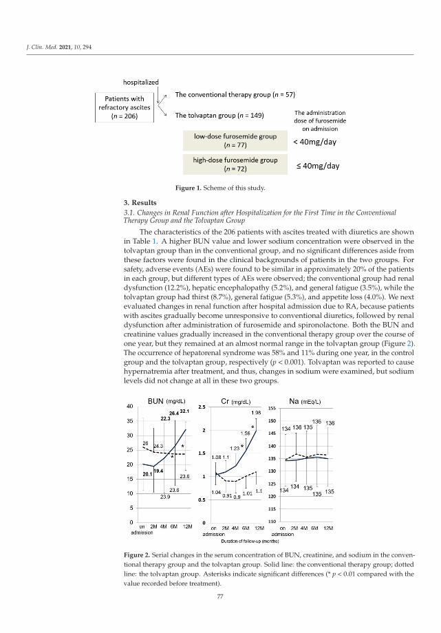
J. Clin. Med. 2021, 10, 294
Figure 1. Scheme of this study.
3. Results
3.1. Changes in Renal Function after Hospitalization for the First Time in the ConventionalTherapy Group and the Tolvaptan Group
The characteristics of the 206 patients with ascites treated with diuretics are shownin Table 1. A higher BUN value and lower sodium concentration were observed in thetolvaptan group than in the conventional group, and no significant differences aside fromthese factors were found in the clinical backgrounds of patients in the two groups. Forsafety, adverse events (AEs) were found to be similar in approximately 20% of the patientsin each group, but different types of AEs were observed; the conventional group had renaldysfunction (12.2%), hepatic encephalopathy (5.2%), and general fatigue (3.5%), while thetolvaptan group had thirst (8.7%), general fatigue (5.3%), and appetite loss (4.0%). We nextevaluated changes in renal function after hospital admission due to RA, because patientswith ascites gradually become unresponsive to conventional diuretics, followed by renaldysfunction after administration of furosemide and spironolactone. Both the BUN andcreatinine values gradually increased in the conventional therapy group over the course ofone year, but they remained at an almost normal range in the tolvaptan group (Figure 2).The occurrence of hepatorenal syndrome was 58% and 11% during one year, in the controlgroup and the tolvaptan group, respectively (p < 0.001). Tolvaptan was reported to causehypernatremia after treatment, and thus, changes in sodium were examined, but sodiumlevels did not change at all in these two groups.
Figure 2. Serial changes in the serum concentration of BUN, creatinine, and sodium in the conven-tional therapy group and the tolvaptan group. Solid line: the conventional therapy group; dottedline: the tolvaptan group. Asterisks indicate significant differences (* p < 0.01 compared with thevalue recorded before treatment).
77
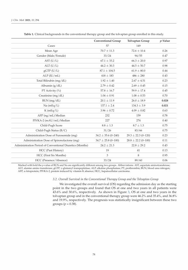
J. Clin. Med. 2021, 10, 294
Table 1. Clinical backgrounds in the conventional therapy group and the tolvaptan group enrolled in this study.
Conventional Group Tolvaptan Group p-Value
Cases 57 149
Mean Age 70.7 ± 11.3 72.6 ± 10.4 0.26
Gender (Male/Female) 33/24 94/55 0.47
AST (U/L) 67.1 ± 33.2 66.3 ± 20.0 0.97
ALT (U/L) 44.2 ± 30.3 44.5 ± 50.7 0.98
gGTP (U/L) 87.1 ± 104.5 61.9 ± 80.0 0.44
ALP (IU/mL) 418 ± 183 486 ± 280 0.43
Total Bilirubin (mg/dL) 1.92 ± 1.40 2.67 ± 4.51 0.23
Albumin (g/dL) 2.79 ± 0.42 2.69 ± 0.45 0.15
PT Activity (%) 57.8 ± 16.7 59.9 ± 17.4 0.45
Creatinine (mg/dL) 1.04 ± 0.91 1.08 ± 0.53 0.70
BUN (mg/dL) 20.1 ± 11.9 26.0 ± 18.9 0.028
Na (mEq/L) 137.1 ± 2.4 134.3 ± 3.9 0.021
K (mEq/L) 3.96 ± 0.72 4.09 ± 0.82 0.63
AFP (ng/mL) Median 232 159 0.78
PIVKA-2 (mAU/mL) Median 227 274 0.40
Child-Pugh Score 8.8 ± 1.3 8.7 ± 1.3 0.75
Child-Pugh Status (B/C) 31/26 83/66 0.75
Administration Dose of Furosemide (mg) 34.2 ± 35.6 (0–240) 29.3 ± 22.3 (0–120) 0.23
Administration Dose of Spironolactone (mg) 34.7 ± 25.8 (0–100) 28.8 ± 22.2 (0–100) 0.11
Administration Period of Conventional Diuretics (Months) 24.2 ± 21.3 22.8 ± 29.2 0.43
HCC (Past History) 19 41 0.13
HCC (First Six Months) 3 8 0.95
HCC (Presence/Absence) 33/24 89/60 0.06
Marked with bold the p-value of BUN and Na are significantly different among two groups. Abbreviations: AST, aspartate aminotransferase;ALT, alanine amino transferase; gGTP, γ-glutamyl transpeptidase; ALP, alkaline phosphatase; PT, prothrombin; BUN, blood urea nitrogen;AFP, α-fetoprotein; PIVKA-2, protein induced by vitamin K absence; HCC, hepatocellular carcinoma.
3.2. Overall Survival in the Conventional Therapy Group and the Tolvaptan Group
We investigated the overall survival (OS) regarding the admission day as the startingpoint in the two groups and found that OS at one and two years in all patients were43.6% and 30.6%, respectively. As shown in Figure 3, OS at one and two years in thetolvaptan group and in the conventional therapy group were 46.2% and 35.4%, and 36.8%and 19.9%, respectively. The prognosis was statistically insignificant between these twogroups (p = 0.38).
78
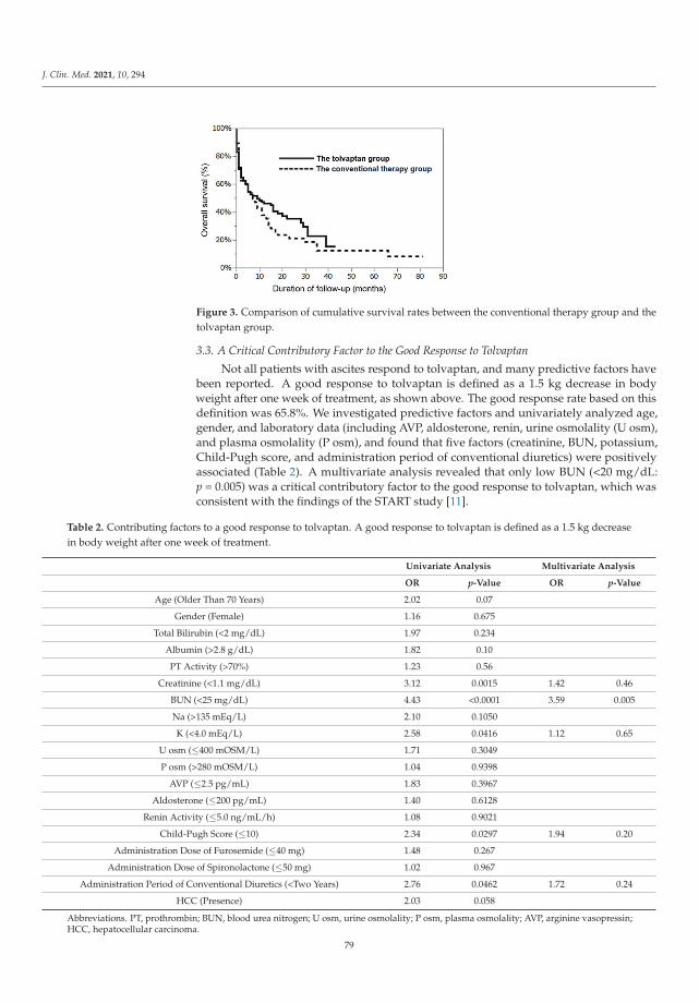
J. Clin. Med. 2021, 10, 294
Figure 3. Comparison of cumulative survival rates between the conventional therapy group and thetolvaptan group.
3.3. A Critical Contributory Factor to the Good Response to Tolvaptan
Not all patients with ascites respond to tolvaptan, and many predictive factors havebeen reported. A good response to tolvaptan is defined as a 1.5 kg decrease in bodyweight after one week of treatment, as shown above. The good response rate based on thisdefinition was 65.8%. We investigated predictive factors and univariately analyzed age,gender, and laboratory data (including AVP, aldosterone, renin, urine osmolality (U osm),and plasma osmolality (P osm), and found that five factors (creatinine, BUN, potassium,Child-Pugh score, and administration period of conventional diuretics) were positivelyassociated (Table 2). A multivariate analysis revealed that only low BUN (<20 mg/dL:p = 0.005) was a critical contributory factor to the good response to tolvaptan, which wasconsistent with the findings of the START study [11].
Table 2. Contributing factors to a good response to tolvaptan. A good response to tolvaptan is defined as a 1.5 kg decreasein body weight after one week of treatment.
Univariate Analysis Multivariate Analysis
OR p-Value OR p-Value
Age (Older Than 70 Years) 2.02 0.07
Gender (Female) 1.16 0.675
Total Bilirubin (<2 mg/dL) 1.97 0.234
Albumin (>2.8 g/dL) 1.82 0.10
PT Activity (>70%) 1.23 0.56
Creatinine (<1.1 mg/dL) 3.12 0.0015 1.42 0.46
BUN (<25 mg/dL) 4.43 <0.0001 3.59 0.005
Na (>135 mEq/L) 2.10 0.1050
K (<4.0 mEq/L) 2.58 0.0416 1.12 0.65
U osm (≤400 mOSM/L) 1.71 0.3049
P osm (>280 mOSM/L) 1.04 0.9398
AVP (≤2.5 pg/mL) 1.83 0.3967
Aldosterone (≤200 pg/mL) 1.40 0.6128
Renin Activity (≤5.0 ng/mL/h) 1.08 0.9021
Child-Pugh Score (≤10) 2.34 0.0297 1.94 0.20
Administration Dose of Furosemide (≤40 mg) 1.48 0.267
Administration Dose of Spironolactone (≤50 mg) 1.02 0.967
Administration Period of Conventional Diuretics (<Two Years) 2.76 0.0462 1.72 0.24
HCC (Presence) 2.03 0.058
Abbreviations. PT, prothrombin; BUN, blood urea nitrogen; U osm, urine osmolality; P osm, plasma osmolality; AVP, arginine vasopressin;HCC, hepatocellular carcinoma.
79

J. Clin. Med. 2021, 10, 294
3.4. The Prognostic Factors for Survival in the Tolvaptan Group
We next examined the prognostic factors among age, gender, renal and liver functions,administration dose of furosemide or spironolactone, and the presence/absence of HCCin the tolvaptan group. As shown in Table 3, a univariate analysis revealed that the prog-nostic factors were being male, having hyperbilirubinemia, having a high BUN, receivinghigh-dose furosemide at the start of tolvaptan treatment, and the presence of hepatocel-lular carcinoma (HCC). Finally, a multivariate analysis of these five factors clarified thatthe prognostic factors were being male (OR 1.59, p = 0.049), having hyperbilirubinemia(>2 mg/dL, OR 1.89, p = 0.009), having a high BUN (>25 mg/dL, OR 1.67, p = 0.031), andreceiving high-dose furosemide (≥40 mg/day, OR 2.63, p < 0.001).
Table 3. The prognostic factors for survival in the tolvaptan group.
UnivariateAnalysis
MultivariateAnalysis
OR p-Value OR p-Value
Age (Older Than 70 Years) 1.40 0.16
Gender (Male) 1.42 0.013 1.59 0.049
Total Bilirubin (>2 mg/dL) 1.73 0.0158 1.89 0.009
Albumin (<2.8 g/dL) 1.39 0.15
PT Activity (<70%) 1.56 0.076
Creatinine (>1.1 mg/dL) 1.43 0.12
BUN (>25 mg/dL) 1.67 0.024 1.67 0.031
Administration Dose of Furosemide (≥40 mg) 3.20 < 0.001 2.63 < 0.001
Administration Dose of Spironolactone (≥50 mg) 1.32 0.22
HCC (Presence) 1.73 0.0164 1.47 0.11
Abbreviations: PT, prothrombin; BUN, blood urea nitrogen; HCC, hepatocellular carcinoma.
3.5. The Prognosis in the Tolvaptan Group with Low- or High-Dose Furosemide and theConventional Therapy Group
The dose of furosemide administered was one of the most important prognosticfactors, thus, the tolvaptan group was divided into two groups according to the dose offurosemide at the start of tolvaptan treatment (<40 mg/day, low-dose furosemide group;≥40 mg/day, high-dose furosemide group). The clinical backgrounds of these two groupsand the conventional therapy group are shown in Table 4. Renal function had alreadyworsened in the high-dose furosemide group compared to that in the other groups, andthere were no differences except renal function between the low-dose furosemide groupand the conventional therapy group.
As shown in Figure 4, OS at one and two years in the tolvaptan group with low-dosefurosemide and in the conventional therapy group were 67.8% and 52.8% and 36.8% and19.9%, respectively. The prognosis was significantly better in the low-dose furosemidegroup than in the conventional therapy group (log-rank test p < 0.0001).
This result shows the possibility that tolvaptan can improve survival in patients withcirrhotic ascites, especially in those whose tolvaptan treatment was started before high-dosefurosemide administration.
The overall survival rate was better in the tolvaptan group with low-dose furosemidethan in the conventional therapy group or in the tolvaptan group with high-dose furosemide(log-rank test p < 0.001).
80

J. Clin. Med. 2021, 10, 294
Table 4. Clinical backgrounds in the two tolvaptan groups. The tolvaptan group was divided intotwo groups according to the dose of furosemide administered on admission (<40 mg/day, low-dosefurosemide group; ≥40 mg/day, high-dose furosemide group).
Tolvaptan Group
p-ValueHigh-DoseFurosemide
Low-DoseFurosemide
Cases 72 77
Mean Age 72.8 ± 10.3 72.3 ± 10.5 ns
Gender (Male/Female) 44/28 50/27 ns
Total Bilirubin (mg/dL) 2.69 ± 3.43 2.66 ± 5.34 ns
Albumin (g/dL) 2.64 ± 0.49 2.73 ± 0.39 ns
PT Activity (%) 59.3 ± 17.1 60.4 ± 17.7 ns
Creatinine (mg/dL) 1.14 ± 0.54 1.02 ± 0.52 ns
BUN (mg/dL) 29.9 ± 21.1 22.4 ± 15.8 0.014
Administration Dose of Furosemide (mg) 45.8 ± 15.2 16.5 ± 7.4 <0.001
Administration Dose of Spironolactone (mg) 33.3 ± 26.2 24.5 ± 16.5 0.0015
HCC (Presence/Absence) 49/23 40/37 ns
Abbreviations: PT, prothrombin; BUN, blood urea nitrogen; HCC, hepatocellular carcinoma.
Figure 4. Comparison of cumulative survival rates among the conventional therapy group and thetolvaptan groups with low- or high-dose furosemide.
4. Discussion
We have experienced difficulties with and inadequacies in conventional diuretictreatments in patients with cirrhotic ascites. Although the deterioration of renal function isfrequently observed with conventional diuretic therapy, renal dysfunction has been shownto be related to poor prognosis. The therapeutic strategy for cirrhotic ascites has changeddrastically since tolvaptan was approved in 2013 [15]. Tolvaptan, an oral AVP V2 receptorantagonist, has been used as a new diuretic for ascites in combination with conventionaldiuretics. It is unknown whether this novel drug may help with the maintenance of renalfunction and improve long-term survival rates in cirrhotic patients with ascites. Tolvaptanhas been reported to be quite effective against RA for a short time [7,15]. Thus, it wasdifficult to do a randomized controlled trial for a longer time.
81

J. Clin. Med. 2021, 10, 294
The prognosis was statistically insignificant between the conventional therapy groupand the tolvaptan group in this study. There were two reasons for the negative result. First,the tolvaptan group had a higher BUN value and lower serum sodium than those in theconventional therapy group. A higher BUN was associated with renal dysfunction. Lowserum sodium was also related to an increased risk of death in patients with cirrhosis [16].These clinical backgrounds might adversely affect the result. Second, a higher BUN showednot only renal dysfunction, but also unresponsiveness to tolvaptan. Tolvaptan is quiteeffective, but not all patients with cirrhotic ascites respond to this new drug. As BUNvalues worsened, patients were less likely to respond to tolvaptan [11]. The patients withhigher BUN levels are presumed not to have enough water in their blood vessels to beexcreted by tolvaptan, or to have worse renal perfusion, thus, tolvaptan is not effective inpatients with higher BUN levels [17].
OS among patients with HCC and without HCC in this cohort was investigated, andwe found that it is significantly better in the HCC (−) group than in the HCC (+) group(Supplementary Figure S1). However, the prognostic factor among patients with refractoryascites was gender (male), liver function (high value of total bilirubin), renal function (highvalue of BUN), and administration dose of furosemide (high dose of furosemide), but notthe presence of HCC in the multivariate analysis. Patients with refractory ascites havepoor liver and renal function, and, in that case, the prognosis might not depend on thepresence/absence of HCC.
Tolvaptan can control RA, and, as a result, it leads to a good prognosis when it isstarted before high-dose furosemide administration. There are some reasons why tolvaptanimproves OS in patients with ascites. First, it can decrease hepatic ascites and body fluidretention without worsening renal function, as shown in this manuscript. Before tolvaptancan be used, we had to increase the dose of conventional diuretics, which was finallyfollowed by renal dysfunction. Second, tolvaptan can control RA more quickly thanthe conventional therapy, and, as a result, the nutritional status of patients may rapidlyimprove. Skeletal muscle mass decreases with age, especially in patients with liver cirrhosis(LC). Sarcopenia is characterized by the loss of muscle mass and is significantly associatedwith mortality in patients with LC [18]. Improvement in nutritional status can suppressthe reduction of muscle mass, and it might be related to a good prognosis. On the otherhand, loop diuretics directly suppress the differentiation of myofibroblasts, and sarcopeniain patients with LC may be attributable to treatment with loop diuretics [19]. Third, astolvaptan can control ascites, the number of complications of decompensated cirrhosis(such as spontaneous bacterial peritonitis) decreases, and patients do not need to be inthe hospital.
Not all patients with ascites respond to tolvaptan. The effectiveness rate has beenreported to be approximately 50–60%, and non-responders have a poor prognosis, whichis similar to those who are treated with conventional diuretics [11]. The present studyshowed that tolvaptan should be started before the BUN value becomes too high in orderto reduce the number of non-responders. This high BUN value implies not only renaldysfunction, but also the deterioration of osmotic pressure in the renal interstitium, whichis mainly caused by the administration of high-dose loop diuretics. Thus, we should becareful not to give high-dose loop diuretics in order to obtain a good response to tolvaptan.
Some investigators have already reported the efficacy and safety of tolvaptan. Uchidahas shown that the ascites-related events-free duration was prolonged following tolvaptantreatment compared with that before treatment [20]. Kogiso also explained the impactof continued administration of tolvaptan and proved that long-term tolvaptan treatmentincreased serum levels of albumin, decreased ammonia levels, and preserved renal func-tion [21]. We have never found a study in which the OS was compared between thetolvaptan group and the conventional therapy group in more than 200 patients withrefractory ascites for more than four years.
82
J. Clin. Med. 2021, 10, 294
5. Conclusions
This is the first report to reveal that tolvaptan can improve survival, especially whentolvaptan treatment is started before high-dose furosemide administration. Therapy for RAhas drastically changed, and clinicians give tolvaptan early for RA rather than increasingthe doses of conventional diuretics. The limitations of this study are the small number ofpatients and the retrospective nature of this study. Prospective and double-blind studiesshould be better, but tolvaptan has been reported to be quite effective in short-term andlong-term administration. It might be difficult to prove it by prospective study.
Supplementary Materials: The following are available online at https://www.mdpi.com/2077-0383/10/2/294/s1, Figure S1: Comparison of cumulative survival rates between the tolvaptan groupwith HCC and without HCC.
Author Contributions: A.H.: study concept and design, acquisition of data; T.T., T.O., M.A., K.O.,Y.W., Y.K., T.Y. (Toshio Yamaguchi), Y.S., M.H., and T.Y. (Takuya Yamada), acquisition of data; N.H.,analysis and interpretation of data, funding procurement. All authors have read and agreed to thepublished version of the manuscript.
Funding: This research received no external funding.
Institutional Review Board Statement: The study was conducted according to the guidelines of theDeclaration of Helsinki and approved by the ethics committee of Osaka-Rosai Hospital (protocol20150024, the date of approval; 5 September 2015).
Informed Consent Statement: Informed consent was obtained from all subjects involved in the study.
Data Availability Statement: Data is contained within the article or Supplementary Materials.
Acknowledgments: We are grateful to Shokura H, Izutani Y and Kuyama Y (our medical assistants)for collecting the large amounts of data analyzed in this study. This study was supported by aGrant-in-Aid for Research on Hepatitis from the Ministry of Health, Labor, and Welfare of Japan andthe Japan Agency for Medical Research and Development (17fk0210106h0901).
Conflicts of Interest: The authors declare no conflict of interest.
References
1. Runyon, B.A. Introduction to the revised American Association for the Study of Liver Diseases Practice Guideline managementof adult patients with ascites due to cirrhosis 2012. Hepatology 2013, 57, 1651–1653. [CrossRef]
2. Fukui, H. Gut-liver axis in liver cirrhosis: How to manage leaky gut and endotoxemia. World J. Hepatol. 2015, 7, 425–442.[CrossRef]
3. Schuppan, D.; Afdhal, N.H. Liver cirrhosis. Lancet 2008, 371, 838–851. [CrossRef]4. Gines, P.; Cardenas, A.; Arroyo, V.; Rodes, J. Management of cirrhosis and ascites. N. Engl. J. Med. 2004, 350, 1646–1654. [CrossRef]5. Salerno, F.; Guevara, M.; Bernardi, M.; Moreau, R.; Wong, F.; Angeli, P.; Garcia-Tsao, G.; Lee, S.S. Refractory ascites: Pathogenesis,
definition and therapy of a severe complication in patients with cirrhosis. Liver Int. 2010, 30, 937–947. [CrossRef]6. Okita, K.; Kawazoe, S.; Hasebe, C.; Kajimura, K.; Kaneko, A.; Okada, M.; Sakaida, I. Dose-finding trial of tolvaptan in liver
cirrhosis patients with hepatic edema: A randomized, double-blind, placebo-controlled trial. Hepatol. Res. 2014, 44, 83–91.[CrossRef]
7. Sakaida, I.; Kawazoe, S.; Kajimura, K.; Saito, T.; Okuse, C.; Takaguchi, K.; Okada, M.; Okita, K. Tolvaptan for improvementof hepatic edema: A phase 3, multicenter, randomized, double-blind, placebo-controlled trial. Hepatol. Res. 2014, 44, 73–82.[CrossRef] [PubMed]
8. Jaber, B.L.; Almarzouqi, L.; Borgi, L.; Seabra, V.F.; Balk, E.M.; Madias, N.E. Short-term efficacy and safety of vasopressin receptorantagonists for treatment of hyponatremia. Am. J. Med. 2011, 124, 977.e1–977.e9. [CrossRef]
9. Schrier, R.W.; Gross, P.; Gheorghiade, M.; Berl, T.; Verbalis, J.G.; Czerwiec, F.S.; Orlandi, C.; Investigators, S. Tolvaptan, a selectiveoral vasopressin V2-receptor antagonist, for hyponatremia. N. Engl. J. Med. 2006, 355, 2099–2112. [CrossRef] [PubMed]
10. Kawaratani, H.; Fukui, H.; Moriya, K.; Noguchi, R.; Namisaki, T.; Uejima, M.; Kitade, M.; Takeda, K.; Okura, Y.; Kaji, K.; et al.Predictive parameter of tolvaptan effectiveness in cirrhotic ascites. Hepatol. Res. 2017, 47, 854–861. [CrossRef] [PubMed]
11. Sakaida, I.; Terai, S.; Kurosaki, M.; Yasuda, M.; Okada, M.; Bando, K.; Fukuta, Y. Effectiveness and safety of tolvaptan in livercirrhosis patients with edema: Interim results of post-marketing surveillance of tolvaptan in liver cirrhosis (START study).Hepatol. Res. 2017, 47, 1137–1146. [CrossRef] [PubMed]
83
J. Clin. Med. 2021, 10, 294
12. Atsukawa, M.; Tsubota, A.; Kato, K.; Abe, H.; Shimada, N.; Asano, T.; Ikegami, T.; Koeda, M.; Okubo, T.; Arai, T.; et al. Analysisof factors predicting the response to tolvaptan in patients with liver cirrhosis and hepatic edema. J. Gastroenterol. Hepatol. 2018,33, 1256–1263. [CrossRef] [PubMed]
13. Planas, R.; Montoliu, S.; Balleste, B.; Rivera, M.; Miquel, M.; Masnou, H.; Galeras, J.A.; Gimenez, M.D.; Santos, J.; Cirera, I.; et al.Natural history of patients hospitalized for management of cirrhotic ascites. Clin. Gastroenterol. Hepatol. 2006, 4, 1385–1394.[CrossRef] [PubMed]
14. Hiramine, Y.; Uojima, H.; Nakanishi, H.; Hiramatsu, A.; Iwamoto, T.; Kimura, M.; Kawaratani, H.; Terai, S.; Yoshiji, H.; Uto, H.;et al. Response criteria of tolvaptan for the treatment of hepatic edema. J. Gastroenterol. 2018, 53, 258–268. [CrossRef]
15. Sakaida, I.; Yamashita, S.; Kobayashi, T.; Komatsu, M.; Sakai, T.; Komorizono, Y.; Okada, M.; Okita, K. Efficacy and safety of a14-day administration of tolvaptan in the treatment of patients with ascites in hepatic oedema. J. Int. Med. Res. 2013, 41, 835–847.[CrossRef]
16. Londono, M.C.; Cardenas, A.; Guevara, M.; Quinto, L.; de Las Heras, D.; Navasa, M.; Rimola, A.; Garcia-Valdecasas, J.C.;Arroyo, V.; Gines, P. MELD score and serum sodium in the prediction of survival of patients with cirrhosis awaiting livertransplantation. Gut 2007, 56, 1283–1290. [CrossRef]
17. Sakaida, I.; Terai, S.; Kurosaki, M.; Okada, M.; Hirano, T.; Fukuta, Y. Real-world effectiveness and safety of tolvaptan inliver cirrhosis patients with hepatic edema: Results from a post-marketing surveillance study (START study). J. Gastroenterol.2020, 55, 800–810. [CrossRef]
18. Hanai, T.; Shiraki, M.; Nishimura, K.; Ohnishi, S.; Imai, K.; Suetsugu, A.; Takai, K.; Shimizu, M.; Moriwaki, H. Sarcopenia impairsprognosis of patients with liver cirrhosis. Nutrition 2015, 31, 193–199. [CrossRef]
19. Mandai, S.; Furukawa, S.; Kodaka, M.; Hata, Y.; Mori, T.; Nomura, N.; Ando, F.; Mori, Y.; Takahashi, D.; Yoshizaki, Y.; et al. Loopdiuretics affect skeletal myoblast differentiation and exercise-induced muscle hypertrophy. Sci. Rep. 2017, 7, 46369. [CrossRef]
20. Uchida, Y.; Tsuji, S.; Uemura, H.; Kouyama, J.; Naiki, K.; Sugawara, K.; Nakao, M.; Inao, M.; Nakayama, N.; Imai, Y.; et al.Furosemide as a factor to deteriorate therapeutic efficacy of tolvaptan in patients with decompensated cirrhosis. Hepatol. Res2020, 50, 1355–1364. [CrossRef]
21. Kogiso, T.; Sagawa, T.; Kodama, K.; Taniai, M.; Tokushige, K. Impact of continued administration of tolvaptan on cirrhoticpatients with ascites. BMC Pharmacol. Toxicol. 2018, 19, 87. [CrossRef] [PubMed]
84

Journal of
Clinical Medicine
Review
Recent Advances in the Management of AcuteVariceal Hemorrhage
Alberto Zanetto 1, Sarah Shalaby 1, Paolo Feltracco 2, Martina Gambato 1, Giacomo Germani 1,
Francesco Paolo Russo 1, Patrizia Burra 1 and Marco Senzolo 1,*
Citation: Zanetto, A.; Shalaby, S.;
Feltracco, P.; Gambato, M.; Germani,
G.; Russo, F.P.; Burra, P.; Senzolo, M.
Recent Advances in the Management
of Acute Variceal Hemorrhage. J. Clin.
Med. 2021, 10, 3818. https://doi.org/
10.3390/jcm10173818
Academic Editors: Pierluigi Toniutto
and Hidekazu Suzuki
Received: 30 July 2021
Accepted: 24 August 2021
Published: 25 August 2021
Publisher’s Note: MDPI stays neutral
with regard to jurisdictional claims in
published maps and institutional affil-
iations.
Copyright: © 2021 by the authors.
Licensee MDPI, Basel, Switzerland.
This article is an open access article
distributed under the terms and
conditions of the Creative Commons
Attribution (CC BY) license (https://
creativecommons.org/licenses/by/
4.0/).
1 Gastroenterology and Multivisceral Transplant Unit, Department of Surgery, Oncology, and Gastroenterology,Padova University Hospital, Via Giustiniani 2, 35128 Padova, Italy; [email protected] (A.Z.);[email protected] (S.S.); [email protected] (M.G.); [email protected] (G.G.);[email protected] (F.P.R.); [email protected] (P.B.)
2 Anesthesiology and Intensive Care Unit, Department of Medicine, Padova University Hospital,35128 Padova, Italy; [email protected]
* Correspondence: [email protected]
Abstract: Gastrointestinal bleeding is one of the most relevant causes of death in patients withcirrhosis and clinically significant portal hypertension, with gastroesophageal varices being the mostfrequent source of hemorrhage. Despite survival has improved thanks to the standardization onmedical treatment aiming to decrease portal hypertension and prevent infections, mortality remainssignificant. In this review, our goal is to discuss the most recent advances in the managementof esophageal variceal hemorrhage in cirrhosis with specific attention to the treatment algorithmsinvolving the use of indirect measurement of portal pressure (HVPG) and transjugular intrahepaticportosystemic shunt (TIPS), which aim to further reduce mortality in high-risk patients after acutevariceal hemorrhage and in the setting of secondary prophylaxis.
Keywords: cirrhosis; decompensation; bleeding; varices; survival; infection
1. Introduction
Gastrointestinal (GI) bleeding is the second most frequent decompensating event incirrhosis [1], with gastroesophageal varices representing the most frequent source of bleed-ing. Despite significant advances in the management of this complication, developmentof acute variceal hemorrhage (VH) is still associated with a six-week mortality risk of~15–20% [2]. In patients who recover from VH, the risk of rebleeding is influenced by thetreatment of underlying portal hypertension, with ~60% of untreated patients that willexperience recurrent bleeding within on to two years, in contrast with only ~30% of thosereceiving therapies that lower portal pressure [3].
In this review, we discuss the management of patients with cirrhosis presenting withesophageal VH, including both treatment of the acute event (first section) and strategiesto prevent recurrent hemorrhage (second section). The management of gastric varicealhemorrhage requires specific consideration and has been recently reviewed in depthelsewhere [4], therefore, it will not be included in the present review.
2. Control of Hemorrhage
The main goals of therapy in hospitalized patients with cirrhosis presenting with acuteupper GI bleeding are (a) to control bleeding and (b) to prevent early rebleeding and death.Management can be divided into general measures, before the source of bleeding has beenidentified, and specific measures, once upper endoscopy has determined that hemorrhageis from esophageal varices.
J. Clin. Med. 2021, 10, 3818. https://doi.org/10.3390/jcm10173818 https://www.mdpi.com/journal/jcm
85
J. Clin. Med. 2021, 10, 3818
2.1. General Measures
In combination with initial systemic stabilization (i.e., protection of circulatory andrespiratory status) and start of intravenous proton pump inhibitors (PPIs), as in anypatient hospitalized with upper GI bleeding, specific nuances in the management ofpatients with cirrhosis include a restrictive transfusion strategy and the use of prophylacticantibiotic therapy [5–8]. Additional measures include management of both coagulopathyand therapy with PPIs. For patients with alcohol-related liver disease, immediate andsustained cessation of alcohol consumption is particularly important to improve liverfunction and reduce risks of further bleeding, decompensation and mortality by reducingliver damage and portal pressure [9].
2.1.1. Blood Transfusion Strategy
The main driver for development of esophageal VH is clinically significant portalhypertension [10]. In a way, the acute loss of intravascular volume due to bleeding re-duces splanchnic pressure and may lead to self-limitation or self-interruption of activehemorrhage. By contrast, a sudden restitution of intravascular volume is associated with arebound increase in portal pressure, which in turn may lead to failure to control bleedingand/or early rebleeding [11]. In a seminal randomized control trial (RCT), a “restrictive”transfusion strategy (hemoglobin threshold for transfusion of 7 g/dL with target rangeof 7–9 g/dL) was associated with a significantly higher probability of survival comparedwith a “liberal” strategy (hemoglobin threshold for transfusion of 9 g/dL with target rangeof 9–11 g/dL) [12]. Therefore, current guidelines recommend initiating transfusions incirrhosis when hemoglobin levels decrease to <7 g/dL, with a target level of 7–9 g/dL [5–8].
Restitution of intravascular volume should be managed with large peripheral lines(16–18 gauges), and blood loss has to be replaced by red blood packed cells [8]. Replace-ment of fluids and electrolytes is important to prevent pre-renal acute kidney injury, whichis common in cirrhosis with GI bleeding and is associated with increased mortality [13].Nephrotoxic drugs such as nonsteroidal anti-inflammatory drugs, non-selective beta-blockers (NSBBs), and other hypotensive drugs may be suspended during the acute courseof VH [6]. As occurrence of acute decompensation may be associated with instability in thefeeble hemostatic balance of decompensated cirrhosis, the need for invasive proceduresshould be evaluated carefully on an individual basis. As discussed below, clotting factorsmay be replaced only to correct an eventual dilutional coagulopathy, whereas there isno indication to prophylactically correct a prolonged prothrombin time or a low plateletcount [5–8].
2.1.2. Antibiotic Prophylaxis
Bacterial infections are observed in up to 50% patients with cirrhosis hospitalizedfor GI bleeding, and are associated with strong risks of failure to control bleeding, earlyre-bleeding and mortality [14–16]. A recent meta-analysis including 12 studies compar-ing antibiotic prophylaxis vs. either placebo or no intervention demonstrated that theadministration of prophylactic antibiotics was associated with reduced all-cause mortality(relative risk (RR): 0.79, 95% CI: 0.63–0.98), infection-driven mortality (RR: 0.43, 95% CI:0.19–0.97), risk of bacterial infection (RR: 0.35, 95% CI: 0.26–0.47), rebleeding (RR: 0.53,95% CI: 0.38–0.74), and length of stay (mean difference −1.9 days, 95% CI: −3.8–0.02) [17].Therefore, a timely, short-term course of prophylactic antibiotics is an important step in themanagement of patients with cirrhosis and VH, and shall be instituted as early as possibleupon admission, before upper endoscopy [5–8].
Whether severity of cirrhosis affects the importance of prophylaxis is unclear. Infact, while the role of prophylaxis is incontrovertible in patients with most advanced liverdysfunction (Child B and C), in those with less advanced liver disease conflicting datahave been reported. In one retrospective analysis, Child A patients had lower risks ofinfection in the absence of prophylaxis (2%), and no difference in mortality was observedin treated vs. non treated patients [18]. The same study showed that the use of antibiotics
86

J. Clin. Med. 2021, 10, 3818
was associated with a substantial reduction in mortality in Child C class [18]. However,prospective data are required to evaluate whether antibiotic prophylaxis can be avoidedin Child A and current recommendation is to administer prophylaxis in all patients withcirrhosis presenting with VH, independent of child [5–8].
Intravenous ceftriaxone (1 g/24 h) for 7 days is the first choice in patients belongingto Child B and C classes, in those who were on quinolone prophylaxis, and in hospitalsin which there is a high frequency of quinolone-resistant bacteria. Norfloxacin 400 mgtwice daily may be used in the other patients. However, due to widespread quinoloneresistance, ceftriaxone (a third-generation cephalosporin) has become the antibiotic ofchoice [5–7]. As approximately 30% of infection are from multidrug resistance antibioticsbacteria [19], evaluation of local resistance, if doable, may further improve definitionof antibiotic regimen and should be considered [8]. Prophylactic antibiotics should beadministered for a maximum of seven days, and their use should not be extended afterdischarge from the hospital [5–7]. In patients discharged before Day 7, transition to an oralantibiotic with the goal of completing seven days of treatment may be considered [8].
In a recent, nationwide study from Spain including 1656 patients with cirrhosis hospi-talized for VH between 2013 and 2015, Martinez et al. investigated current epidemiologyand trends of bacterial infections in these patients [20]. Interestingly, despite prophylaxisas currently recommended by international guidelines [5–7], 20% of patients developedbacterial infections, particularly respiratory tract infection. Development of infection wasobserved early (median time from admission 3 days) and was independently associatedwith Child C class (odds ratio (OR): 3.1; 95% CI: 1.4–6.7), Grade III–IV encephalopathyat admission (OR: 2.8; 95% CI: 1.8–4.4), orotracheal intubation for endoscopy (OR: 2.6;95% CI: 1.8–3.8), and placement of nasogastric tube/balloon tamponade (OR: 1.7; 95%CI: 1.2–2.4 and 2.4; 95% CI: 1.2–4.9, respectively) [20]. Such procedures should, therefore,be minimized whenever possible, particularly in patients with additional risk factors,and active screening for respiratory infections shall be performed in case of early clinicaldeterioration. Whether patients at risk for respiratory infection would benefit from tailoredregiments of antibiotic prophylaxis, particularly in settings with high risk of resistantstrains bacteria, it remains to be evaluated in further studies.
2.1.3. Additional MeasuresNo Need for Correction of Coagulopathy
Hospitalized patients with decompensated cirrhosis have severe coagulopathy [21–23].However, a prolonged prothrombin time does not reflect an increased bleeding tendencyin these patients [21,23], and correction of INR by fresh frozen plasma should not be per-formed [5–8]. Not surprisingly, administration of recombinant FVII, which can correctprolongation of INR, was not associated with additional benefit compared with standard ofcare in an individual patient data meta-analysis of two RCTs [24]. Administration of plasmato correct coagulopathy in cirrhosis with bleeding is a very common practice [25]; however,this practice not only is ineffective, but is also likely harmful [26]. In a recent, multicentercohort study administration of fresh frozen plasma in cirrhosis with VH was independentlyassociated with increased risks of 42-day mortality (primary outcome, OR: 9.41, 95% CI:3.71–23.90), failure to control bleeding at five days (OR: 3.87, 95% CI: 1.28–11.70) and lengthof stay (adjusted OR: 1.88, 95% CI: 1.03–3.42) (secondary outcomes) [27]. No specific dataexist regarding the management of severe thrombocytopenia in the setting of VH, andtherefore, no recommendation can be made. In patients without chronic liver disease,desmopressin increases levels of plasmatic Von Willebrand factor/procoagulant FactorVIII, and its use was associated with reduced bleeding time in an old study including com-pensated patients [28]. However, in a subsequent RCT no difference in control of VH wasobserved between patients randomized to terlipressin alone vs. patients treated with terli-pressin plus desmopressin [29]. Therefore, desmopressin is not currently recommended.
87
J. Clin. Med. 2021, 10, 3818
Limited Usefulness of PPIs
As peptic ulcers are the source of bleeding in ~30% of patients with cirrhosis pre-senting with GI bleeding [30], intravenous PPIs should be initiated as soon as possible.However, when portal hypertensive bleeding is confirmed at endoscopy, discontinuationof PPIs may be considered as they have shown no efficacy in this clinical setting. Limitedevidence suggested that a short-term use (10 days) of PPIs might reduce banding ulcersize [31], however, this was not associated with a significant reduction of bleeding risk.PPIs in decompensated cirrhosis are associated with significantly increased risks of hepaticencephalopathy, bacterial infection, and readmission at 30-days [32–34]. In a landmarkanalysis including 1198 patients from three RCTs evaluating the use of satavaptan inpatients with cirrhosis and ascites, Dam et al. demonstrated that the use of PPIs was asso-ciated with a significantly increased risk of encephalopathy (OR: 1.88, 95% CI: 1.21–1.91)and spontaneous bacterial peritonitis (OR: 1.72, 95% CI: 1.10–2.69) during follow-up [33].Recent data with extended period of follow-up confirmed that regular use of PPIs notonly is associated with increased risk of spontaneous bacterial peritonitis, but also predictsliver-related mortality independent of MELD and stage of cirrhosis (OR: 2.01, 95% CI:1.38–2.93) [34]. Therefore, their use should not be extended past discharge.
2.2. Specific Management of Acute Esophageal VH
Standard therapy for acute VH includes intravenous splanchnic vasoconstrictors andplacement of rubber bands around esophageal varices, especially the one that is expected tobe the source of bleeding [5–8]. Endotracheal intubation to protect the airway system maybe considered in patients with massive bleeding prior to endoscopy. However, whetherintubation is really protective or increases the risk of respiratory infections is unclear [20],therefore, it cannot be recommended for every patient.
2.2.1. Intravenous Splanchnic Vasoconstrictors
Three intravenous splanchnic vasoconstrictors are available: terlipressin, somatostatinor octreotide. These drugs exert their action by reducing splanchnic blood flow, thereforelowering portal pressure [35]. They are very effective and a recent meta-analysis clearlydemonstrated that the use of vasoconstrictors is associated with a significantly higherprobability of bleeding control and a lower seven-day mortality [36]. As a proof of concept,treatment with vasoconstrictors alone was previously found to control bleeding in >80%of patients [37]. It is most likely the widespread adoption of these drugs, together withthe optimization of general medical care, that has significantly lowered the VH-relatedshort-term mortality in the recent years [38].
A vasoconstrictor shall be initiated as soon as possible and early administration isassociated with improved survival [5–8]. A placebo-controlled trial in which terlipressinwas administered during the ambulance transfer showed that such early timing of admin-istration was associated with increased probability to control of bleeding and survival inthe treatment arm [39].
In clinical practice, the choice among these three intravenous vasoconstrictors isdictated by local availability and cost [40]. Recommended dose for terlipressin of 2 mg/4 hduring the first 48 h, followed by 1 mg/4 h. If terlipressin is contraindicated, somatostatinis an alternative and should be administered as a continuous infusion of 250 mg/h (thatcan be increased up to 500 mg/h), with an initial bolus of 250 mg. The recommended doseof octreotide is a continuous infusion of 50 mg/h with an initial bolus of 50 mg [5–8].
Vasoconstrictors should be continued up to five days after the confirmation of VHbecause the risk of rebleeding during this time is particularly high [5–8]. However, asvasoconstrictors may be associated with potentially serious adverse events, the feasibilityof a shorter administration (i.e., 24–48 h vs. 3–5 days) has been considered. In a recentmeta-analysis, although the risk of 42-day mortality was not significantly different betweenone to three and five days, risk stratification was missing [41]. It may be that Child A
88
J. Clin. Med. 2021, 10, 3818
patients could receive a shorter duration of therapy, whereas all others would require fivedays, but further studies are required to answer this question.
In summary, guidelines recommend that an intravenous splanchnic vasoconstrictorshall be initiated as soon as possible, prior to diagnostic endoscopy, and be administeredfor three to five days [5–8].
2.2.2. Endoscopic Therapy
Once hemodynamic stability has been reached, an upper GI endoscopy shall beperformed to determine the cause of bleeding and to provide specific treatment [42]. Earlydata and one relatively recent retrospective study suggested that endoscopy within 12 hfrom the index event might be associated with reduced rates of recurrent bleeding andmortality [43]. On the other hand, in a larger multicenter study including 1373 patientswith cirrhosis and VH, endoscopy within 24 h from admission was associated with lowermortality in patients with Child A or B cirrhosis (OR: 0.38, 95% CI: 0.16–0.86; p = 0.020)and in those with systolic blood pressure <90 mmHg (OR: 0.053, 95% CI: 0.006–0.51;p = 0.011) [44]. In contrast, performance of endoscopy within either 6 or 12 h was notassociated with a further reduction in mortality compared with endoscopy within 24 h.Interestingly, the association between endoscopy within 24 h and reduced mortality wasseen in Child A and B patients, but not in the overall group including also Child C [44].
This notwithstanding, current guidelines recommend that once hemodynamic stabilityhas been achieved, endoscopy should be performed as early as possible, and within 12 hsince presentation [5–8]. When VH is confirmed, either by the presence of a bleedingvarix, a clot, or a “white nipple” over the varix, or when varices are the only abnormalityobserved that would explain the hemorrhage, all esophageal varices should be ligated,particularly the one that is considered the source of hemorrhage. Endoscopic varicealligation (EVL) should be performed within the same endoscopy session. EVL is moreeffective than sclerotherapy, is associated with fewer adverse effects, and does not lead tofurther increase in portal hypertension [45]. Therefore, sclerotherapy should be restrictedto the rare cases in whom ligation is not technically feasible. Hemostatic powder appliedendoscopically may be considered as a rescue therapy, however, few data exist and itsapplicability remains to be determined [46,47].
In patients with uncontrolled bleeding, guidelines recommend placement of balloontamponade (Sengastaken-Blackemore or Minnesota tubes) [5–8]. However, tamponadecarries a high risk of complications, particularly respiratory infection [20], and shall beconsidered only as a temporary (maximum 24 h) bridge to TIPS [5]. Recent data suggestthat placement of a self-expandable esophageal metal stent (placed orally or endoscopically)may be associated with greater bleeding control and lower adverse events compared toballoon [48]. As these stents may remain in place for up to 7 days, this would allowmore time to plan for a definitive treatment. Per the last Baveno consensus, if available,the application of stents may be considered as a preferred alternative compared withballoons [5].
2.2.3. Transjugular Intrahepatic Portosystemic Shunt (TIPS)Rescue TIPS (in Patients Who Fail Standard Therapy)
Despite combination therapy with prophylactic antibiotics, intravenous splanchnicvasoconstrictors, and EVL, 10–15% of patients will have either persistent bleeding or earlyrebleeding, which are associated with high risk of death [49]. Negative predictors forfailure to control bleeding or early rebleeding include Child C class, portal vein thrombosis,severity of portal hypertension as defined by a hepatic venous pressure gradient (HVPG)>20 mmHg, and systolic blood pressure < 100 mmHg at admission [50,51].
In patients with mild-moderate rebleeding, a second session of endoscopy with liga-tion may be attempted. In patients with persistent or severe rebleeding (i.e., those withfailure of endoscopic therapy), rescue TIPS is the therapy of choice [5–8]. In fact, by con-
89
J. Clin. Med. 2021, 10, 3818
necting the hypertensive portal venous system to the normotensive system of inferior venacava, TIPS will quickly reduce portal pressure and resolve bleeding.
Given the lack of therapeutic alternatives, the only factor that would limit the useof rescue TIPS is futility. One issue to consider is patient’s eligibility for transplantation.Additional factors include number and severity of organ failures. However, developmentof acute on-chronic liver failure (ACLF) per se is not an absolute contraindication forplacement of rescue TIPS. In fact, in a recent multicenter study including 174 patients witheither acute decompensation or ACLF and uncontrolled variceal bleeding, the insertion ofrescue TIPS was an independent predictor of survival at 42 days [52]. There are multiplestudies that looked for predictors of futility in patients undergoing rescue TIPS. In a recent,large multicenter cohort including 164 patients who received rescue TIPS, those witharterial lactate ≤ 2.5 mmol/L and MELD score ≤ 15 had 6-week survival > 85%, whereasthose with baseline lactate level ≥ 12 mmol/L and/or MELD score ≥ 30 had >90% risk ofdeath [53]. A recent, large observational study of rescue TIPS showed that stay in intensivecare unit prior to TIPS, MELD, and Child-Pugh score were independently associated withmortality at six weeks, and the authors commented on the futility of rescue TIPS in patientswith Pugh score > 13 [54].
Preemptive TIPS (In Patients at High-Risk of Failing Standard Therapy)
Patients with acute VH who are more likely to fail, despite initial control ofhemorrhage by standard therapy, are those belonging to Child C class or those withHVPG > 20 mmHg [50,55]. It was, therefore, postulated that the placement of a TIPS be-fore failure of standard therapy (“pre-emptive TIPS”) could improve survival [56]. A firstRCT in which 52 patients with HVPG > 20 mmHg was randomized to standard treat-ment vs. pre-emptive TIPS (uncovered) demonstrated significantly lower failure rates andall cause short-term mortality in the TIPS arm (12% vs. 50% and 17% vs. 38%, respec-tively) [57]. When covered TIPS became the standard-of-care, a second RCT confirmedan improved survival in patients randomized to TIPS vs. standard of care [58]. Both theone-year rate of failure to control bleeding/rebleeding and mortality were decreased byTIPS, the absolute reduction being 47% and 25%, respectively [58]. In this RCT, high-riskpatients were defined as those with Child C cirrhosis and a Pugh score of 10–13, or thosebelonging to a Child B class with active bleeding at time of endoscopy.
Later on, however, large international observational cohorts confirmed the beneficialeffect on survival of pre-emptive TIPS only in Child C patients (score 10–13), but not inthose with Child B and active bleeding [59,60]. Therefore, the inclusion of Child B withactive bleeding at endoscopy was questioned for potentially overrating the risk of mortality.
A recent RCT from China including 132 patients in Child B and C class and random-ized 2:1 to pre-emptive TIPS vs. standard of care reported better transplant-free survivalat six weeks and one year (OR: 0.50, 95% CI: 0·25–0·98; p = 0.04) and improved controlof bleeding or rebleeding with early TIPS (OR: 0.26, 95% CI: 0.12–0.55; p < 0.0001) [61].Importantly, the survival benefit was found in all subgroups, independent of either ac-tive bleeding or stage of cirrhosis, and no difference was found in the rate of hepaticencephalopathy. However, the reduction in mortality risk was relatively small (one-yearsurvival 86% vs. 73% in pre-emptive TIPS vs. standard of care, respectively), which likelyreflects the inclusion of patients at relatively lower risk of failure (57% of patients wereChild B with no active bleeding and proportion of Child C was only 22%) [61]. It is alsoimportant to note that 75% of patients had HBV-related cirrhosis, which could have in-fluenced the outcomes and may limit applicability of these results to Eastern countries.Furthermore, sclerotherapy was used in more than 5% of patients in standard of care group,which is not in line with current guidelines [5–7].
Another RCT from England in 58 patients with Child–Pugh score ≥ 8 showed nodifference in survival (OR: 1.154, 95% CI: 0.3289–3.422; p = 0.79) and risk of rebleedingbetween pre-emptive TIPS and standard treatment, independent of severity of cirrhosisor active bleeding. However, the study was underpowered and only 23/29 patients (79%)
90

J. Clin. Med. 2021, 10, 3818
underwent preemptive TIPS, with only 13/23 within 72 h (therefore, not deemed earlyby definition) [62]. Remarkably, the one-year transplant-free survival in the control armwas higher than that in the 2010 RCT by Garcia-Pagan (76% vs. 61%) [58]. This may berelated to the significant improvements in overall management of VH in cirrhosis, whichwould question the extrapolation of results from the 2010 study by Garcia-Pagan to presenttimes [58]. On the other hand, an alternative explanation could be that patients includedby Dunne et al. were not at high-risk of failure to control bleeding, which would not haveallowed to assess the true benefits of pre-emptive TIPS [62].
Opposite to Dunne’s findings, a large meta-analysis with individual data from 1327 pa-tients included in seven studies, of whom 602 were Child B with active bleeding, found thatplacement of pre-emptive TIPS was associated with improved survival not only in the over-all group (OR = 0.443, CI 95%: 0.323–0.607, p < 0.001), but also when Child B (OR = 0.524,CI 95%: 0.307–0.896, p = 0.018) and Child C (OR = 0.374, CI 95%: 0.253–0.553, p < 0.001)patients were analyzed separately [63]. This would support the use of pre-emptive TIPSin both Child C and Child B patients with active bleeding. However, results in ChildB are less convincing/consistent compared with those obtained in Child C. Additionallimitations are the inclusion or more observational studies than RCTs (four versus three),definition of high risk patients by only one specific criterion (therefore, not being able toassess if additional criteria might have better classified patients with VH at high risk), andthe heterogeneity in both TIPS expertise and treatments in standard of care arms acrossdifferent centers [63].
Therefore, a multicenter trial collecting large numbers of patients undergoing pre-emptive TIPS remains a research priority in this field. Importantly, such trial should assessnot only which group(s) of patients are most likely to benefit from pre-emptive TIPS, butalso whether there is a maximum threshold of severity of liver disease above which thereis no improvement of survival.
While awaiting such trial, guidelines recommend to consider pre-emptive TIPS inpatients with Child C cirrhosis (score 10–13) [5–8]. Patients with Child A cirrhosis andthose with Child B cirrhosis without active bleeding should not be considered for pTIPS.Further data are required before to make a strong recommendation in patients with ChildB and active bleeding at time of endoscopy.
As one major goal of pre-emptive TIPS is to prevent development of ACLF, onewould think that pre-emptive TIPS should not be considered in patients who have al-ready developed ACLF at time of admission/decision making. However, a recent studyfrom a European collaborative group found that ACLF is an independent predictor ofbleeding-related mortality, and that pre-emptive TIPS may improve outcomes in selectedpatients with ACLF [64]. Although prospective data are needed, these preliminary findingsindicate that ACLF per se is not an absolute contraindication for pre-emptive TIPS, andinstead eligibility should be a case-by-case decision according to number and severity oforgan failures.
Despite RCTs and observational cohorts have demonstrated that pre-emptive TIPS isassociated with a survival benefit, and the use of pre-emptive TIPS in these patients hasbeen recommended since the Baveno V consensus (published 11 years ago) [65], a recentFrench survey revealed that only 7% of eligible patients finally received a pre-emptiveTIPS [60]. Similarly, in another large observational cohort, a pre-emptive TIPS was placedin only 13% of high-risk patients [59]. This indicates a significant underutilization of pre-emptive TIPS in real-life practice, which is somewhat concerning considering its substantialeffect on patient survival. Further efforts are required to lower the bar for a widespreadadoption of pre-emptive TIPS in daily practice. These efforts include creation of dedicatednetworks through which selected patients may be referred early to tertiary care centers withspecific expertise in invasive management of portal hypertension and its complications.
91
J. Clin. Med. 2021, 10, 3818
3. Prevention of Recurrent Hemorrhage
Per current guidelines, patients who had a TIPS placed after VH do not require furthermedical or endoscopic therapy for secondary prophylaxis, and should instead be referredfor liver transplant evaluation in case they have additional complications of cirrhosis [5–8].Patency of TIPS should be assessed at regular intervals by doppler ultrasound togetherwith screening for hepatocellular carcinoma.
3.1. First Line Therapy
Combined therapy with NSBBs (propranolol or nadolol) plus EVL is the first linetherapy in prevention of rebleeding [5–8]. This recommendation is based on multiplemeta-analyses of RCTs performed to prevent rebleeding. One of these meta-analysesdemonstrated that the added effect of NSBBs to EVL improved the efficacy of EVL aloneand reduced mortality, whereas the added effect of EVL to NSBB was only associatedwith a non-significant decrease of rebleeding with no effect on survival [66]. A recentindividual patient data meta-analysis evaluated data from three trials comparing NSBBsvs. combination therapy and from four trials analyzing EVL vs. combination therapy [67].As these were individual data, the authors were able to perform risk stratification in ChildA vs. Child B/C patients. Interestingly, in Child A (mostly compensated), combinationtherapy was associated with lower all-source rebleeding, without an effect on mortality. InChild B/C patients (mostly decompensated), combination therapy was associated withlower all-source rebleeding rates only in trials in which it was compared to EVL alone,indicating that NSBBs alone could be enough to prevent all-source rebleeding in thesepatients. Importantly, mortality was also lower in trials in which combination therapywas compared to EVL alone, suggesting that NSBBs not only are essential in preventingrebleeding, but also death [67].
These data, obtained from RCTs, are in contrast with a previous cohort study includingpatients with refractory ascites in which mortality was significantly higher in those receiv-ing NSBBs [68]. However, study groups were different at baseline and patients were sickerin the NSBB group. Additionally, the determination of NSBBs was evaluated at diagnosisof refractory ascites with no information on their use thorough follow-up. Multiple trials indifferent groups of patients with decompensated cirrhosis have been conducted to confirmor refute these findings, and two meta-analyses have summarized these data both showingthat the use of NSBBs is not associated with a higher mortality [69,70].
In studies that showed a detrimental effect of NSBBs [68,71], the arterial pressure inNSBBs users was lower than that in non-users, and a higher dose of propranolol was used,or a higher percentage of patients were treated with carvedilol. This indicates that patientsin whom a negative inotropic effect or a vasodilatory effect from NSBBs/carvedilol werethe ones that were negatively affected by beta-blockers [72]. In a way, this can be expectedas this clinically-evident, likely dose-related deleterious hemodynamic effect of NSBBswould worsen the already vasodilated state of decompensated patients, leading to renalhypoperfusion, renal failure and death [73]. Indeed, in a propensity-matched analysisincluding only patients with refractory ascites, the use of propranolol was associated withan increased survival, except for the subgroup on a high dose (160 mg/day or more) [74].
Propranolol and nadolol should be used cautiously in patients with ascites and shouldbe started at a lower dose than in patients without ascites, and the maximum dose shouldbe capped also at a lower dose: propranolol should be capped to 160 mg/day (320 mg/dayin patients without ascites) and nadolol to 80 mg/day (160 mg/day in patients withoutascites) [7]. Importantly, the dose of NSBB should be reduced or drug should be discon-tinued in patients with refractory ascites who developed circulatory dysfunction definedby systolic blood pressure < 90 mm Hg, serum sodium < 130 meq/L, or acute kidneyinjury [7].
In summary, current guidelines recommend that first line therapy to prevent recurrentVH is the combination of NSBBs (propranolol or nadolol) plus EVL, independent of thepresence or absence of ascites/refractory ascites or other complications of cirrhosis [5–8].
92

J. Clin. Med. 2021, 10, 3818
However, it is possible that refinements in risk stratification could lead to identificationof “higher” risk patients in whom an aggressive approach, such as placement of TIPS,may be beneficial as first line therapy (i.e., before development of recurrent VH). This wasrecently evaluated by La Mura and Bosch in a retrospective study including 424 patientswith cirrhosis candidates to secondary prophylaxis [75]. Inclusion criteria were diagnosisof cirrhosis, admission for VH within the previous seven days, baseline HVPG ≥ 12 mmHg,subsequent long-term treatment with propranolol or nadolol plus EVL, and a secondHVPG assessment after one to three months of continued NSBBs. By combining clinicaldata (i.e., presence of ascites or encephalopathy) plus severity of portal hypertension(HVPG ≥ 16 mmHg), they identified two groups of patients at significantly different risksof rebleeding and mortality during follow-up. “Low” risk group included patients withoutascites or encephalopathy and patients with VH plus ascites or encephalopathy but HVPG< 16 mmHg. “High” risk group included patients with VH plus one among the follows:ascites or HE, HVPG ≥ 16 mmHg, and lack of response to NSBBs as defined by an HVPGdecrease by at least 20% of <12 mmHg. If confirmed by prospective series, this algorithmmay improve risk stratification and lead to a more tailored management of patients withcirrhosis and history of VH. In fact, as shown in previous studies for acute VH [58] and“difficult ascites” [76], anticipating a decision for TIPS may be better compared with usingit as rescue therapy.
In patients receiving secondary prophylaxis for VH, assessment of baseline HVPGand its response to NSBBs may provide useful information and guide therapy [77–79].In this setting, the “goal-standard” is to measure HVPG at baseline and then re-assessHVPG after chronic administration of NSBBs (i.e., after four to six weeks) [80]. However,the measurement of “acute” HVPG response to intravenous propranolol may be a pre-ferred alternative as it would be quicker and has an acceptable correlation with chronicresponse [77]. In one seminal RCT by Villanueva et al., an HVPG-guided therapy based onacute response to intravenous NSBBs significantly lowered the risk of portal hypertensionrelated complications and mortality compared with standard of care [78]. In another retro-spective study including both candidates for primary and secondary prophylaxis, acuteresponse to intravenous propranolol was independently correlated with a 50% decreasein the probability of re-bleeding (23% at 2 years vs. 46% in non-responders; p = 0.032)and a better survival (95% vs. 65%; p = 0.003) [79]. Although further evidence is requiredto evaluate benefits and cost of such approach, current data suggest that HVPG-guidedtherapy in patients who are not deemed candidates for pre-emptive TIPS, could improvethe management of secondary prophylaxis by reducing costs and adverse events due toineffective therapy [77].
3.2. Second Line Therapy
Current guidelines recommend covered TIPS as second line therapy of choice in pa-tients who experience rebleeding despite combination therapy with NSBB plus EVL [5–8].
Regarding prevention of recurrent VH by TIPS, RCTs comparing uncovered TIPSvs. NSBBs plus EVL (standard of care) agreed that TIPS is very effective in preventingrebleeding, but it is associated with higher risk of over encephalopathy and does notimprove survival [81]. Comparable findings were confirmed by two RCTs in which coveredTIPS was used [82,83]. Therefore, TIPS is considered the treatment of choice only in patientswho fail first-line therapy (NSBBs plus EVL), in whom the risk of bleeding-related mortalityis very high and exceeds those associated with TIPS [5–8].
Patients who experience the first episode of VH while on primary prophylaxis withNSBBs have a higher risk of rebleeding and mortality compared to those who experienceVH not being on NSBBs, despite being treated with recommended combination therapy [84].Although the best treatment strategy in these patients is unknown, they may benefit froma more aggressive strategy, and TIPS may be considered earlier rather than later in thesepatients. A second group of patients in whom to consider TIPS before failure of standardtherapy are those who are or who become intolerant to NSBBs. In fact, as mentioned before,
93

J. Clin. Med. 2021, 10, 3818
NSBBs are the cornerstone of combined therapy, particularly in decompensated patients(Child B and C) [67].
In patients with cirrhosis and portal vein thrombosis who have recently bled, varicealobliteration with EVL takes longer and varices recur at a higher rate compared to patientswithout thrombosis [85]. Additionally, a small RCT showed that TIPS is more effective thanEVL and NSBBs in preventing rebleeding in patients with cirrhosis and portal vein throm-bosis, with a higher rate of thrombus resolution but without differences in mortality [61].Patients with cirrhosis and portal vein thrombosis, which is the most common thromboticcomplications in cirrhosis [86–88], may be a third group in which to consider TIPS earlierrather than later in the setting of secondary prophylaxis. This would be particularly impor-tant if the patient is awaiting liver transplantation, as the presence of thrombosis at time oftransplantation is associated with a higher risk of post-transplant mortality [89].
A major clinical challenge in patients who receive TIPS for second-line prophylaxis ofVH remains prediction of survival and prognosis (i.e., identification of patients with pooroutcomes after TIPS in whom early evaluation for transplantation should be indicated).Recently, Bettinger et al. proposed to combine four simple variables (age, bilirubin, albuminand creatinine) in a new score named the “Freiburg index of post-TIPS survival” (FIPSscore) [90]. In a very large cohort of patients who received TIPS for various indication,including second line prophylaxis of VH, the FIPS score was able to identify those athigher-risk for progression and death, and its prognostic discrimination was superior toother currently used score such as MELD, MELD-Na, and Child-Pugh [90].
4. Conclusions
Development of variceal hemorrhage in patients with cirrhosis poses a complexchallenge requiring a multidisciplinary approach that is important to prevent rebleedingand improve survival. The management of variceal hemorrhage in these patients shouldtake into consideration the severity of underlying portal hypertension and the presence (orabsence) of other complications of cirrhosis, especially ascites. In patients presenting withvariceal hemorrhage, the advances in the therapy of portal hypertension have resulted inlower rates of rebleeding and death, particularly for therapies associated with a decreaseof portal pressure. Further improvement in risk stratification and in therapies of patientswith cirrhosis and variceal hemorrhage are eagerly awaited.
Author Contributions: Conceptualization, A.Z. and M.S.; methodology, A.Z., S.S., P.F., M.G.,G.G., F.P.R., P.B., M.S.; writing—original draft preparation, A.Z., S.S., P.F., M.G., G.G., F.P.R., P.B.;writing—review and editing, A.Z., M.S.; supervision, M.S. All authors have read and agreed to thepublished version of the manuscript.
Funding: This research received no external funding.
Institutional Review Board Statement: Not applicable.
Informed Consent Statement: Not applicable.
Data Availability Statement: Not applicable.
Conflicts of Interest: The authors declare no conflict of interest.
References
1. D’Amico, G.; Garcia-Tsao, G.; Pagliaro, L. Natural history and prognostic indicators of survival in cirrhosis: A systematic reviewof 118 studies. J. Hepatol. 2006, 44, 217–231. [CrossRef]
2. Bosch, J.; Garcia-Pagan, J.C. Prevention of variceal rebleeding. Lancet 2003, 361, 952–954. [CrossRef]3. D’Amico, G.; Pagliaro, L.; Bosch, J. Pharmacological treatment of portal hypertension: An evidence-based approach. Semin. Liver
Dis. 1999, 19, 475–505. [CrossRef]4. Henry, Z.; Patel, K.; Patton, H.; Saad, W. AGA Clinical Practice Update on Management of Bleeding Gastric Varices: Expert
Review. Clin. Gastroenterol. Hepatol. 2021, 19, 1098–1107.e1091. [CrossRef]5. de Franchis, R.; Baveno, V.I.F. Expanding consensus in portal hypertension: Report of the Baveno VI Consensus Workshop:
Stratifying risk and individualizing care for portal hypertension. J. Hepatol. 2015, 63, 743–752. [CrossRef] [PubMed]
94
J. Clin. Med. 2021, 10, 3818
6. European Association for the Study of the Liver. Electronic address eee, European Association for the Study of the L. EASL ClinicalPractice Guidelines for the management of patients with decompensated cirrhosis. J. Hepatol. 2018, 69, 406–460. [CrossRef][PubMed]
7. Garcia-Tsao, G.; Abraldes, J.G.; Berzigotti, A.; Bosch, J. Portal hypertensive bleeding in cirrhosis: Risk stratification, diagnosis, andmanagement: 2016 practice guidance by the American Association for the study of liver diseases. Hepatology 2017, 65, 310–335.[CrossRef]
8. Bruno, R.; Cammà, C.; Caraceni, P.; D’Amico, G.; Grattagliano, I.; La Mura, V.; Riggio, O.; Schepis, F.; Senzolo, M.; Angeli, P.; et al.Portal Hypertension and Ascites: Patient- and Population-centered Clinical Practice Guidelines by the Italian Association for theStudy of the Liver (AISF). Dig. Liver Dis. 2021, 21. [CrossRef]
9. Burra, P.; Zanetto, A.; Germani, G. Liver Transplantation for Alcoholic Liver Disease and Hepatocellular Carcinoma. Cancers2018, 10, 46. [CrossRef] [PubMed]
10. Zanetto, A.; Barbiero, G.; Battistel, M.; Sciarrone, S.S.; Shalaby, S.; Pellone, M.; Battistella, S.; Gambato, M.; Germani, G.;Russo, F.P.; et al. Management of portal hypertension severe complications. Minerva Gastroenterol. (Torino) 2021, 67, 26–37.[CrossRef]
11. Kravetz, D.; Bosch, J.; Arderiu, M.; Pilar Pizcueta, M.; Rodes, J. Hemodynamic effects of blood volume restitution following ahemorrhage in rats with portal hypertension due to cirrhosis of the liver: Influence of the extent of portal-systemic shunting.Hepatology 1989, 9, 808–814. [CrossRef]
12. Villanueva, C.; Colomo, A.; Bosch, A.; Concepcion, M.; Hernandez-Gea, V.; Aracil, C.; Graupera, I.; Poca, M.; Alvarez-Urturi, C.;Gordillo, J.; et al. Transfusion strategies for acute upper gastrointestinal bleeding. N. Engl. J. Med. 2013, 368, 11–21. [CrossRef]
13. Cardenas, A.; Gines, P.; Uriz, J.; Bessa, X.; Salmeron, J.M.; Mas, A.; Ortega, R.; Calahorra, B.; De Las Heras, D.; Bosch, J.; et al.Renal failure after upper gastrointestinal bleeding in cirrhosis: Incidence, clinical course, predictive factors, and short-termprognosis. Hepatology 2001, 34, 671–676. [CrossRef]
14. Ferrarese, A.; Zanetto, A.; Becchetti, C.; Sciarrone, S.S.; Shalaby, S.; Germani, G.; Gambato, M.; Russo, F.P.; Burra, P.; Senzolo, M.Management of bacterial infection in the liver transplant candidate. World J. Hepatol. 2018, 10, 222–230. [CrossRef]
15. Goulis, J.; Armonis, A.; Patch, D.; Sabin, C.; Greenslade, L.; Burroughs, A.K. Bacterial infection is independently associated withfailure to control bleeding in cirrhotic patients with gastrointestinal hemorrhage. Hepatology 1998, 27, 1207–1212. [CrossRef]
16. Hou, M.C.; Lin, H.C.; Liu, T.T.; Kuo, B.I.; Lee, F.Y.; Chang, F.Y.; Lee, S.D. Antibiotic prophylaxis after endoscopic therapy preventsrebleeding in acute variceal hemorrhage: A randomized trial. Hepatology 2004, 39, 746–753. [CrossRef]
17. Chavez-Tapia, N.C.; Barrientos-Gutierrez, T.; Tellez-Avila, F.; Soares-Weiser, K.; Mendez-Sanchez, N.; Gluud, C.; Uribe, M.Meta-analysis: Antibiotic prophylaxis for cirrhotic patients with upper gastrointestinal bleeding—An updated Cochrane review.Aliment. Pharmacol. Ther. 2011, 34, 509–518. [CrossRef] [PubMed]
18. Tandon, P.; Abraldes, J.G.; Keough, A.; Bastiampillai, R.; Jayakumar, S.; Carbonneau, M.; Wong, E.; Kao, D.; Bain, V.G.; Ma, M.Risk of Bacterial Infection in Patients With Cirrhosis and Acute Variceal Hemorrhage, Based on Child-Pugh Class, and Effects ofAntibiotics. Clin. Gastroenterol. Hepatol. 2015, 13, 1189–1196.e1182. [CrossRef]
19. Ferrarese, A.; Zanetto, A.; Germani, G.; Burra, P.; Senzolo, M. Rethinking the role of non-selective beta blockers in patients withcirrhosis and portal hypertension. World J. Hepatol. 2016, 8, 1012–1018. [CrossRef] [PubMed]
20. Martinez, J.; Hernandez-Gea, V.; Rodriguez-de-Santiago, E.; Tellez, L.; Procopet, B.; Giraldez, A.; Amitrano, L.; Villanueva,C.; Thabut, D.; Ibanez-Samaniego, L.; et al. Bacterial infections in patients with acute variceal bleeding in the era of antibioticprophylaxis. J. Hepatol. 2021, 75, 342–350. [CrossRef] [PubMed]
21. Campello, E.; Zanetto, A.; Bulato, C.; Maggiolo, S.; Spiezia, L.; Russo, F.P.; Gavasso, S.; Mazzeo, P.; Tormene, D.; Burra, P.; et al.Coagulopathy is not predictive of bleeding in patients with acute decompensation of cirrhosis and acute-on-chronic liver failure.Liver Int. 2021, 15001. [CrossRef]
22. Zanetto, A.; Rinder, H.M.; Campello, E.; Saggiorato, G.; Deng, Y.; Ciarleglio, M.; Wilson, F.P.; Senzolo, M.; Gavasso, S.;Bulato, C.; et al. Acute Kidney Injury in Decompensated Cirrhosis Is Associated With Both Hypo-coagulable and Hyper-coagulable Features. Hepatology 2020, 72, 1327–1340. [CrossRef]
23. Zanetto, A.; Rinder, H.M.; Senzolo, M.; Simioni, P.; Garcia-Tsao, G. Reduced Clot Stability by Thromboelastography as a PotentialIndicator of Procedure-Related Bleeding in Decompensated Cirrhosis. Hepatol. Commun. 2021, 5, 272–282. [CrossRef]
24. Bendtsen, F.; D’Amico, G.; Rusch, E.; de Franchis, R.; Andersen, P.K.; Lebrec, D.; Thabut, D.; Bosch, J. Effect of recombinantFactor VIIa on outcome of acute variceal bleeding: An individual patient based meta-analysis of two controlled trials. J. Hepatol.2014, 61, 252–259. [CrossRef] [PubMed]
25. Zanetto, A.; Senzolo, M.; Blasi, A. Perioperative management of antithrombotic treatment. Best Pract. Res. Clin. Anaesthesiol.2020, 34, 35–50. [CrossRef] [PubMed]
26. Lisman, T.; Procopet, B. Fresh frozen plasma in treating acute variceal bleeding: Not effective and likely harmful. Liver Int.2021, 41, 1710–1712. [CrossRef] [PubMed]
27. Mohanty, A.; Kapuria, D.; Canakis, A.; Lin, H.; Amat, M.J.; Rangel Paniz, G.; Placone, N.T.; Thomasson, R.; Roy, H.;Chak, E.; et al. Fresh frozen plasma transfusion in acute variceal haemorrhage: Results from a multicentre cohort study. Liver Int.2021, 41, 1901–1908. [CrossRef]
28. Burroughs, A.K.; Matthews, K.; Qadiri, M.; Thomas, N.; Kernoff, P.; Tuddenham, E.; McIntyre, N. Desmopressin and bleedingtime in patients with cirrhosis. Br. Med. J. (Clin. Res. Ed.) 1985, 291, 1377–1381. [CrossRef]
95
J. Clin. Med. 2021, 10, 3818
29. de Franchis, R.; Arcidiacono, P.G.; Carpinelli, L.; Andreoni, B.; Cestari, L.; Brunati, S.; Zambelli, A.; Battaglia, G.; Mannucci, P.M.Randomized controlled trial of desmopressin plus terlipressin vs. terlipressin alone for the treatment of acute variceal hemorrhagein cirrhotic patients: A multicenter, double-blind study. New Italian Endoscopic Club. Hepatology 1993, 18, 1102–1107. [CrossRef]
30. Hsu, Y.C.; Lin, J.T.; Chen, T.T.; Wu, M.S.; Wu, C.Y. Long-term risk of recurrent peptic ulcer bleeding in patients with liver cirrhosis:A 10-year nationwide cohort study. Hepatology 2012, 56, 698–705. [CrossRef]
31. Shaheen, N.J.; Stuart, E.; Schmitz, S.M.; Mitchell, K.L.; Fried, M.W.; Zacks, S.; Russo, M.W.; Galanko, J.; Shrestha, R. Pantoprazolereduces the size of postbanding ulcers after variceal band ligation: A randomized, controlled trial. Hepatology 2005, 41, 588–594.[CrossRef]
32. Bajaj, J.S.; Acharya, C.; Fagan, A.; White, M.B.; Gavis, E.; Heuman, D.M.; Hylemon, P.B.; Fuchs, M.; Puri, P.; Schubert, M.L.; et al.Proton Pump Inhibitor Initiation and Withdrawal affects Gut Microbiota and Readmission Risk in Cirrhosis. Am. J. Gastroenterol.2018, 113, 1177–1186. [CrossRef]
33. Dam, G.; Vilstrup, H.; Watson, H.; Jepsen, P. Proton pump inhibitors as a risk factor for hepatic encephalopathy and spontaneousbacterial peritonitis in patients with cirrhosis with ascites. Hepatology 2016, 64, 1265–1272. [CrossRef]
34. Janka, T.; Tornai, T.; Borbely, B.; Tornai, D.; Altorjay, I.; Papp, M.; Vitalis, Z. Deleterious effect of proton pump inhibitors on thedisease course of cirrhosis. Eur. J. Gastroenterol. Hepatol. 2020, 32, 257–264. [CrossRef] [PubMed]
35. Kulkarni, A.V.; Arab, J.P.; Premkumar, M.; Benitez, C.; Tirumalige Ravikumar, S.; Kumar, P.; Sharma, M.; Reddy, D.N.; Si-monetto, D.A.; Rao, P.N. Terlipressin has stood the test of time: Clinical overview in 2020 and future perspectives. Liver Int.2020, 40, 2888–2905. [CrossRef] [PubMed]
36. Wells, M.; Chande, N.; Adams, P.; Beaton, M.; Levstik, M.; Boyce, E.; Mrkobrada, M. Meta-analysis: Vasoactive medications forthe management of acute variceal bleeds. Aliment. Pharmacol. Ther. 2012, 35, 1267–1278. [CrossRef] [PubMed]
37. D’Amico, G.; Pietrosi, G.; Tarantino, I.; Pagliaro, L. Emergency sclerotherapy versus vasoactive drugs for variceal bleeding incirrhosis: A Cochrane meta-analysis. Gastroenterology 2003, 124, 1277–1291. [CrossRef]
38. Carbonell, N.; Pauwels, A.; Serfaty, L.; Fourdan, O.; Levy, V.G.; Poupon, R. Improved survival after variceal bleeding in patientswith cirrhosis over the past two decades. Hepatology 2004, 40, 652–659. [CrossRef]
39. Levacher, S.; Letoumelin, P.; Pateron, D.; Blaise, M.; Lapandry, C.; Pourriat, J.L. Early administration of terlipressin plus glyceryltrinitrate to control active upper gastrointestinal bleeding in cirrhotic patients. Lancet 1995, 346, 865–868. [CrossRef]
40. Zanetto, A.; Garcia-Tsao, G. Gastroesophageal Variceal Bleeding Management. In The Critically Ill Cirrhotic Patient; Rahimi, R.S.,Ed.; Springer: Cham, Switzerland, 2020. [CrossRef]
41. Yan, P.; Tian, X.; Li, J. Is additional 5-day vasoactive drug therapy necessary for acute variceal bleeding after successful endoscopichemostasis? A systematic review and meta-analysis. Medicine (Baltimore) 2018, 97, e12826. [CrossRef] [PubMed]
42. Zanetto, A.; Garcia-Tsao, G. Management of acute variceal hemorrhage. F1000Research 2019, 8, 966. [CrossRef]43. Chen, P.H.; Chen, W.C.; Hou, M.C.; Liu, T.T.; Chang, C.J.; Liao, W.C.; Su, C.W.; Wang, H.M.; Lin, H.C.; Lee, F.Y.; et al. Delayed
endoscopy increases re-bleeding and mortality in patients with hematemesis and active esophageal variceal bleeding: A cohortstudy. J. Hepatol. 2012, 57, 1207–1213. [CrossRef]
44. Laursen, S.B.; Stanley, A.; Hernandez-Gea, V.; Procopet, B.; Giraldez, A.; Amitrano, L.; Villanueva, C.; Thabut, D.; Ibanez-Samaniego, L.; Silva-Junior, G.; et al. Optimal timing of endoscopy is associated with lower 42-day mortality in variceal bleeding.J. Hepatol. 2019, 70, e952. [CrossRef]
45. Laine, L.; el-Newihi, H.M.; Migikovsky, B.; Sloane, R.; Garcia, F. Endoscopic ligation compared with sclerotherapy for thetreatment of bleeding esophageal varices. Ann. Intern. Med. 1993, 119, 1–7. [CrossRef]
46. Ibrahim, M.; El-Mikkawy, A.; Abdalla, H.; Mostafa, I.; Deviere, J. Management of acute variceal bleeding using hemostaticpowder. United Eur. Gastroenterol. J. 2015, 3, 277–283. [CrossRef]
47. Ibrahim, M.; El-Mikkawy, A.; Abdel Hamid, M.; Abdalla, H.; Lemmers, A.; Mostafa, I.; Deviere, J. Early application of haemostaticpowder added to standard management for oesophagogastric variceal bleeding: A randomised trial. Gut 2019, 68, 844–853.[CrossRef] [PubMed]
48. Escorsell, A.; Pavel, O.; Cardenas, A.; Morillas, R.; Llop, E.; Villanueva, C.; Garcia-Pagan, J.C.; Bosch, J.; Variceal BleedingStudy, G. Esophageal balloon tamponade versus esophageal stent in controlling acute refractory variceal bleeding: A multicenterrandomized, controlled trial. Hepatology 2016, 63, 1957–1967. [CrossRef]
49. Garcia-Tsao, G.; Bosch, J. Management of varices and variceal hemorrhage in cirrhosis. N. Engl. J. Med. 2010, 362, 823–832.[CrossRef] [PubMed]
50. Abraldes, J.G.; Villanueva, C.; Banares, R.; Aracil, C.; Catalina, M.V.; Garci, A.P.J.C.; Bosch, J.; Spanish Cooperative Group forPortal, H.; Variceal, B. Hepatic venous pressure gradient and prognosis in patients with acute variceal bleeding treated withpharmacologic and endoscopic therapy. J. Hepatol. 2008, 48, 229–236. [CrossRef]
51. Amitrano, L.; Guardascione, M.A.; Manguso, F.; Bennato, R.; Bove, A.; DeNucci, C.; Lombardi, G.; Martino, R.; Menchise, A.;Orsini, L.; et al. The effectiveness of current acute variceal bleed treatments in unselected cirrhotic patients: Refining short-termprognosis and risk factors. Am. J. Gastroenterol. 2012, 107, 1872–1878. [CrossRef] [PubMed]
52. Kumar, R.; Kerbert, A.J.C.; Sheikh, M.F.; Roth, N.; Calvao, J.A.F.; Mesquita, M.D.; Barreira, A.I.; Gurm, H.S.; Ramsahye, K.;Mookerjee, R.P.; et al. Determinants of mortality in patients with cirrhosis and uncontrolled variceal bleeding. J. Hepatol.2021, 74, 66–79. [CrossRef]
96
J. Clin. Med. 2021, 10, 3818
53. Walter, A.; Rudler, M.; Olivas, P.; Moga, L.; Trepo, E.; Robic, M.A.; Ollivier-Hourmand, I.; Baiges, A.; Sutter, O.; Bouzbib, C.; et al.Combination of Model for End-Stage Liver Disease and Lactate Predicts Death in Patients Treated With Salvage TransjugularIntrahepatic Portosystemic Shunt for Refractory Variceal Bleeding. Hepatology 2021, 31913. [CrossRef]
54. Maimone, S.; Saffioti, F.; Filomia, R.; Alibrandi, A.; Isgro, G.; Calvaruso, V.; Xirouchakis, E.; Guerrini, G.P.; Burroughs, A.K.;Tsochatzis, E.; et al. Predictors of Re-bleeding and Mortality Among Patients with Refractory Variceal Bleeding UndergoingSalvage Transjugular Intrahepatic Portosystemic Shunt (TIPS). Dig. Dis. Sci 2019, 64, 1335–1345. [CrossRef] [PubMed]
55. Moitinho, E.; Escorsell, A.; Bandi, J.C.; Salmeron, J.M.; Garcia-Pagan, J.C.; Rodes, J.; Bosch, J. Prognostic value of early measure-ments of portal pressure in acute variceal bleeding. Gastroenterology 1999, 117, 626–631. [CrossRef]
56. Magaz, M.; Baiges, A.; Hernandez-Gea, V. Precision medicine in variceal bleeding: Are we there yet? J. Hepatol. 2020, 72, 774–784.[CrossRef] [PubMed]
57. Monescillo, A.; Martinez-Lagares, F.; Ruiz-del-Arbol, L.; Sierra, A.; Guevara, C.; Jimenez, E.; Marrero, J.M.; Buceta, E.; Sanchez, J.;Castellot, A.; et al. Influence of portal hypertension and its early decompression by TIPS placement on the outcome of varicealbleeding. Hepatology 2004, 40, 793–801. [CrossRef]
58. Garcia-Pagan, J.C.; Caca, K.; Bureau, C.; Laleman, W.; Appenrodt, B.; Luca, A.; Abraldes, J.G.; Nevens, F.; Vinel, J.P.;Mossner, J.; et al. Early use of TIPS in patients with cirrhosis and variceal bleeding. N. Engl. J. Med. 2010, 362, 2370–2379.[CrossRef]
59. Hernandez-Gea, V.; Procopet, B.; Giraldez, A.; Amitrano, L.; Villanueva, C.; Thabut, D.; Ibanez-Samaniego, L.; Silva-Junior, G.;Martinez, J.; Genesca, J.; et al. Preemptive-TIPS Improves Outcome in High-Risk Variceal Bleeding: An Observational Study.Hepatology 2019, 69, 282–293. [CrossRef] [PubMed]
60. Thabut, D.; Pauwels, A.; Carbonell, N.; Remy, A.J.; Nahon, P.; Causse, X.; Cervoni, J.P.; Cadranel, J.F.; Archambeaud, I.;Bramli, S.; et al. Cirrhotic patients with portal hypertension-related bleeding and an indication for early-TIPS: A large multicentreaudit with real-life results. J. Hepatol. 2017, 68, 73–81. [CrossRef] [PubMed]
61. Lv, Y.; Qi, X.; He, C.; Wang, Z.; Yin, Z.; Niu, J.; Guo, W.; Bai, W.; Zhang, H.; Xie, H.; et al. Covered TIPS versus endoscopicband ligation plus propranolol for the prevention of variceal rebleeding in cirrhotic patients with portal vein thrombosis: Arandomised controlled trial. Gut 2018, 67, 2156–2168. [CrossRef] [PubMed]
62. Dunne, P.D.J.; Sinha, R.; Stanley, A.J.; Lachlan, N.; Ireland, H.; Shams, A.; Kasthuri, R.; Forrest, E.H.; Hayes, P.C. Randomisedclinical trial: Standard of care versus early-transjugular intrahepatic porto-systemic shunt (TIPSS) in patients with cirrhosis andoesophageal variceal bleeding. Aliment. Pharmacol. Ther. 2020, 52, 98–106. [CrossRef]
63. Nicoara-Farcau, O.; Han, G.; Rudler, M.; Angrisani, D.; Monescillo, A.; Torres, F.; Casanovas, G.; Bosch, J.; Lv, Y.; Thabut, D.; et al.Effects of Early Placement of Transjugular Portosystemic Shunts in Patients With High-Risk Acute Variceal Bleeding: A Meta-analysis of Individual Patient Data. Gastroenterology 2021, 160, 193–205.e110. [CrossRef]
64. Trebicka, J.; Gu, W.; Ibanez-Samaniego, L.; Hernandez-Gea, V.; Pitarch, C.; Garcia, E.; Procopet, B.; Giraldez, A.; Amitrano, L.;Villanueva, C.; et al. Rebleeding and mortality risk are increased by ACLF but reduced by pre-emptive TIPS. J. Hepatol.2020, 73, 1082–1091. [CrossRef]
65. de Franchis, R.; Baveno, V.F. Revising consensus in portal hypertension: Report of the Baveno V consensus workshop onmethodology of diagnosis and therapy in portal hypertension. J. Hepatol. 2010, 53, 762–768. [CrossRef] [PubMed]
66. Puente, A.; Hernandez-Gea, V.; Graupera, I.; Roque, M.; Colomo, A.; Poca, M.; Aracil, C.; Gich, I.; Guarner, C.; Villanueva, C.Drugs plus ligation to prevent rebleeding in cirrhosis: An updated systematic review. Liver Int. 2014, 34, 823–833. [CrossRef]
67. Albillos, A.; Zamora, J.; Martinez, J.; Arroyo, D.; Ahmad, I.; De-la-Pena, J.; Garcia-Pagan, J.C.; Lo, G.H.; Sarin, S.; Sharma, B.; et al.Stratifying risk in the prevention of recurrent variceal hemorrhage: Results of an individual patient meta-analysis. Hepatology2017, 66, 1219–1231. [CrossRef] [PubMed]
68. Serste, T.; Melot, C.; Francoz, C.; Durand, F.; Rautou, P.E.; Valla, D.; Moreau, R.; Lebrec, D. Deleterious effects of beta-blockers onsurvival in patients with cirrhosis and refractory ascites. Hepatology 2010, 52, 1017–1022. [CrossRef]
69. Chirapongsathorn, S.; Valentin, N.; Alahdab, F.; Krittanawong, C.; Erwin, P.J.; Murad, M.H.; Kamath, P.S. Nonselective beta-Blockers and Survival in Patients With Cirrhosis and Ascites: A Systematic Review and Meta-analysis. Clin. Gastroenterol. Hepatol.2016, 14, 1096–1104.e1099. [CrossRef]
70. Facciorusso, A.; Roy, S.; Livadas, S.; Fevrier-Paul, A.; Wekesa, C.; Kilic, I.D.; Chaurasia, A.K.; Sadeq, M.; Muscatiello, N.Nonselective Beta-Blockers Do Not Affect Survival in Cirrhotic Patients with Ascites. Dig. Dis. Sci. 2018, 63, 1737–1746.[CrossRef] [PubMed]
71. Mandorfer, M.; Bota, S.; Schwabl, P.; Bucsics, T.; Pfisterer, N.; Kruzik, M.; Hagmann, M.; Blacky, A.; Ferlitsch, A.; Sieghart, W.; et al.Nonselective beta blockers increase risk for hepatorenal syndrome and death in patients with cirrhosis and spontaneous bacterialperitonitis. Gastroenterology 2014, 146, 1680–1690.e1681. [CrossRef] [PubMed]
72. Garcia-Tsao, G. Beta blockers in cirrhosis: The window re-opens. J. Hepatol. 2016, 64, 532–534. [CrossRef] [PubMed]73. Turco, L.; Garcia-Tsao, G.; Magnani, I.; Bianchini, M.; Costetti, M.; Caporali, C.; Colopi, S.; Simonini, E.; De Maria, N.;
Banchelli, F.; et al. Cardiopulmonary hemodynamics and C-reactive protein as prognostic indicators in compensated and decom-pensated cirrhosis. J. Hepatol. 2018, 68, 949–958. [CrossRef] [PubMed]
74. Bang, U.C.; Benfield, T.; Hyldstrup, L.; Jensen, J.E.; Bendtsen, F. Effect of propranolol on survival in patients with decompensatedcirrhosis: A nationwide study based Danish patient registers. Liver Int. 2016, 36, 1304–1312. [CrossRef]
97

J. Clin. Med. 2021, 10, 3818
75. La Mura, V.; Garcia-Guix, M.; Berzigotti, A.; Abraldes, J.G.; Garcia-Pagan, J.C.; Villanueva, C.; Bosch, J. A Prognostic StrategyBased on Stage of Cirrhosis and HVPG to Improve Risk Stratification After Variceal Bleeding. Hepatology 2020, 72, 1353–1365.[CrossRef] [PubMed]
76. Bureau, C.; Thabut, D.; Oberti, F.; Dharancy, S.; Carbonell, N.; Bouvier, A.; Mathurin, P.; Otal, P.; Cabarrou, P.; Peron, J.M.; et al.Transjugular Intrahepatic Portosystemic Shunts With Covered Stents Increase Transplant-Free Survival of Patients With Cirrhosisand Recurrent Ascites. Gastroenterology 2017, 152, 157–163. [CrossRef]
77. Garcia-Pagan, J.C.; Francoz, C.; Montagnese, S.; Senzolo, M.; Mookerjee, R.P. Management of the major complications of cirrhosis:Beyond guidelines. J. Hepatol. 2021, 75 (Suppl. 1), S135–S146. [CrossRef]
78. Villanueva, C.; Graupera, I.; Aracil, C.; Alvarado, E.; Minana, J.; Puente, A.; Hernandez-Gea, V.; Ardevol, A.; Pavel, O.;Colomo, A.; et al. A randomized trial to assess whether portal pressure guided therapy to prevent variceal rebleeding improvessurvival in cirrhosis. Hepatology 2017, 65, 1693–1707. [CrossRef]
79. La Mura, V.; Abraldes, J.G.; Raffa, S.; Retto, O.; Berzigotti, A.; Garcia-Pagan, J.C.; Bosch, J. Prognostic value of acute hemodynamicresponse to i.v. propranolol in patients with cirrhosis and portal hypertension. J. Hepatol. 2009, 51, 279–287. [CrossRef] [PubMed]
80. Bosch, J.; Abraldes, J.G.; Berzigotti, A.; Garcia-Pagan, J.C. The clinical use of HVPG measurements in chronic liver disease. Nat.Rev. Gastroenterol. Hepatol. 2009, 6, 573–582. [CrossRef]
81. Zheng, M.; Chen, Y.; Bai, J.; Zeng, Q.; You, J.; Jin, R.; Zhou, X.; Shen, H.; Zheng, Y.; Du, Z. Transjugular intrahepatic portosystemicshunt versus endoscopic therapy in the secondary prophylaxis of variceal rebleeding in cirrhotic patients: Meta-analysis update.J. Clin. Gastroenterol. 2008, 42, 507–516. [CrossRef]
82. Holster, I.L.; Tjwa, E.T.; Moelker, A.; Wils, A.; Hansen, B.E.; Vermeijden, J.R.; Scholten, P.; van Hoek, B.; Nicolai, J.J.;Kuipers, E.J.; et al. Covered transjugular intrahepatic portosystemic shunt versus endoscopic therapy + beta-blocker forprevention of variceal rebleeding. Hepatology 2016, 63, 581–589. [CrossRef] [PubMed]
83. Sauerbruch, T.; Mengel, M.; Dollinger, M.; Zipprich, A.; Rossle, M.; Panther, E.; Wiest, R.; Caca, K.; Hoffmeister, A.; Lutz, H.; et al.Prevention of Rebleeding From Esophageal Varices in Patients With Cirrhosis Receiving Small-Diameter Stents Versus Hemody-namically Controlled Medical Therapy. Gastroenterology 2015, 149, 660–668.e661. [CrossRef]
84. de Souza, A.R.; La Mura, V.; Reverter, E.; Seijo, S.; Berzigotti, A.; Ashkenazi, E.; Garcia-Pagan, J.C.; Abraldes, J.G.; Bosch, J.Patients whose first episode of bleeding occurs while taking a beta-blocker have high long-term risks of rebleeding and death.Clin. Gastroenterol. Hepatol. 2012, 10, 670–676. [CrossRef]
85. Zanetto, A.; Garcia-Tsao, G. Some Answers and More Questions About Portal Vein Thrombosis in Patients With DecompensatedCirrhosis. Clin. Gastroenterol. Hepatol. 2020, 18, 2432–2434. [CrossRef] [PubMed]
86. Zanetto, A.; Senzolo, M.; Vitale, A.; Cillo, U.; Radu, C.; Sartorello, F.; Spiezia, L.; Campello, E.; Rodriguez-Castro, K.;Ferrarese, A.; et al. Thromboelastometry hypercoagulable profiles and portal vein thrombosis in cirrhotic patients with hepato-cellular carcinoma. Dig. Liver Dis. 2017, 49, 440–445. [CrossRef] [PubMed]
87. Shalaby, S.; Simioni, P.; Campello, E.; Spiezia, L.; Gavasso, S.; Bizzaro, D.; Cardin, R.; D’Amico, F.; Gringeri, E.; Cillo, U.; et al.Endothelial Damage of the Portal Vein is Associated with Heparin-Like Effect in Advanced Stages of Cirrhosis. Thromb Haemost2020, 120, 1173–1181. [CrossRef] [PubMed]
88. Russo, F.P.; Zanetto, A.; Campello, E.; Bulato, C.; Shalaby, S.; Spiezia, L.; Gavasso, S.; Franceschet, E.; Radu, C.; Senzolo, M.; et al.Reversal of hypercoagulability in patients with HCV-related cirrhosis after treatment with direct-acting antivirals. Liver Int.2018, 38, 2210–2218. [CrossRef]
89. Zanetto, A.; Rodriguez-Kastro, K.I.; Germani, G.; Ferrarese, A.; Cillo, U.; Burra, P.; Senzolo, M. Mortality in liver transplantrecipients with portal vein thrombosis—An updated meta-analysis. Transpl. Int. 2018, 31, 1318–1329. [CrossRef] [PubMed]
90. Bettinger, D.; Sturm, L.; Pfaff, L.; Hahn, F.; Kloeckner, R.; Volkwein, L.; Praktiknjo, M.; Lv, Y.; Han, G.; Huber, J.P.; et al. Refiningprediction of survival after TIPS with the novel Freiburg index of post-TIPS survival. J. Hepatol. 2021, 74, 1362–1372. [CrossRef]
98

Journal of
Clinical Medicine
Review
New Therapies of Liver Diseases: Hepatic Encephalopathy
Chiara Mangini and Sara Montagnese *
Citation: Mangini, C.; Montagnese, S.
New Therapies of Liver Diseases:
Hepatic Encephalopathy. J. Clin. Med.
2021, 10, 4050. https://doi.org/
10.3390/jcm10184050
Academic Editor: Pierluigi Toniutto
Received: 16 July 2021
Accepted: 4 September 2021
Published: 7 September 2021
Publisher’s Note: MDPI stays neutral
with regard to jurisdictional claims in
published maps and institutional affil-
iations.
Copyright: © 2021 by the authors.
Licensee MDPI, Basel, Switzerland.
This article is an open access article
distributed under the terms and
conditions of the Creative Commons
Attribution (CC BY) license (https://
creativecommons.org/licenses/by/
4.0/).
Department of Medicine, University of Padova, 35128 Padova, Italy; [email protected]* Correspondence: [email protected]; Tel.: +39-(0)49-8218675
Abstract: Hepatic encephalopathy (HE) is a common complication of advanced liver disease whichhas profound implications in terms of the patients’ ability to fulfil their family and social roles, todrive and to provide for themselves. Recurrent and persistent HE is still a serious managementchallenge, translating into a significant burden for patients and their families, health services andsociety at large. The past few years have been characterized by significantly more attention towardsHE and its implications; its definition has been refined and a small number of new drugs/alternativemanagement strategies have become available, while others are underway. In this narrative reviewwe summarize them in a pragmatic and hopefully useful fashion.
Keywords: cirrhosis; portal-systemic shunt; ammonia; vigilance
1. Introduction
Hepatic encephalopathy (HE) is a brain dysfunction caused by liver failure and/orportal-systemic shunt that results in a spectrum of neurological and psychiatric abnormali-ties ranging from subclinical alterations to coma [1].
Classification is based on the underlying condition leading to HE: “Type A” HEis due to acute liver failure, “Type B” HE is caused by portal-systemic shunt withoutsignificant liver disease and “Type C” HE by cirrhosis with or without portal-systemicshunt [1]. In terms of its severity, HE is qualified as covert (minor or no signs/symptomsbut abnormalities on neuropsychological and/or neurophysiological tests) or overt (GradesII or over according to the West Haven criteria [1]). Finally, in terms of its time-course, overtHE is classified as episodic, recurrent (more than one episode over a period of six months)or persistent (no return to normal/baseline neuropsychiatric performance in betweenepisodes) [1].
This narrative review will deal with the management of Type C HE, i.e., the one associ-ated with cirrhosis with or without portal-systemic shunt [1]. Standard information on HEtreatment (no formal literature search) plus 2018–2021 information on novel therapies is in-cluded. The 2018–2021 literature search was conducted on Pubmed using the terms hepaticencephalopathy plus: treatment, polyethylene glycol (PEG), branched-chain amino acids(BCAAs), L-ornithine L-aspartate (LOLA), nitrogen scavengers, Acetyl L-carnitine, albu-min, probiotics, fecal microbiota transplantation (FMT), flumazenil, minocycline, ibuprofen,phosphodiesterase-5 inhibitors, indomethacin, benzodiazepine inverse agonists.
Prior to discussing available, recommended and more experimental (Table 1) treatmentoptions, it is important to highlight how:
- The HE phenotype is nonspecific and differential diagnosis extremely important;- Response to treatment can be utilized to confirm a working diagnosis of HE, especially
in its mild forms;- The lack thereof, should prompt fast differential diagnosis investigations, especially
in severe forms;- With the exception of direct modulation of vigilance/inflammation (vide infra), HE
treatment is essentially synonymous of ammonia-lowering treatment. Hyperammon-aemia is necessary but not sufficient for a working diagnosis of HE (i.e., there is no
J. Clin. Med. 2021, 10, 4050. https://doi.org/10.3390/jcm10184050 https://www.mdpi.com/journal/jcm
99
J. Clin. Med. 2021, 10, 4050
HE without hyperammonaemia but the presence of hyperammonaemia does notnecessarily translate into a HE phenotype, especially in young patients) [1].
2. General Management Principles
An episode of overt HE is generally managed by ensuring adequate airway protec-tion for severe cases, correction of any identified precipitating factors and institution ofammonia-lowering treatment [1,2]. The most commonly utilized drugs for the subsequentcommencement of secondary prophylaxis are non-absorbable disaccharides (i.e., lactuloseor lactitol, which aim at reducing gut nitrogen load via their laxative and prebiotic effects,enhancing bacterial ammonia uptake and reducing ammonia production in the small intes-tine) and non-absorbable antibiotics (also affecting production/absorption of gut-derivedneurotoxins, and reducing endotoxemia and inflammation), which are generally addedafter a second overt HE episode or, as a stand-alone, when non-absorbable disaccharidesare not well tolerated [3,4]. Primary prophylaxis is not generally recommended, withthe exception of the rapid removal of blood from the gastrointestinal tract after an uppergastrointestinal bleed, for example with lactulose [5] or mannitol [6]. By contrast, secondaryprophylaxis is important, as once a patient has experienced an episode of overt HE, thelikelihood of further episodes is high and a common cause of re-admission into hospital [7].Secondary prophylaxis with a non-absorbable disaccharide (lactulose or lactitol) should beinstituted [8,9]. If this is adequately titrated and the patient/caregivers instructed carefully,both constipation and diarrhoea/excessive flatulence and abdominal distension can beavoided. If overt HE becomes recurrent (i.e., more than one episode within six months [1]),rifaximin should be added to help maintain remission [1,10].
There are more uncertainties on the benefits of treatment of patients with mild formsof HE, especially as not all centres have the experience to diagnose them. Thus, treatmentis not routinely recommended [1]. However, its initiation, especially if “ecological” (i.e.,adding soluble fermentable fibre, probiotics such as yogurt, vegetables, cereal and milk-derived proteins to the habitual diet, plus getting used to in-between-meals snacks anda bedtime snack [11]), may both confirm the diagnosis and be beneficial. It is, therefore,reasonable, especially if the patients or their caregivers report symptoms compatible withmild HE, to institute it.
The management of patients with recurrent or persistent HE, which is more commonin patients with large, spontaneous or surgical shunts [12] can be very difficult. Shuntclosure/reduction can be considered in patients whose shunts are accessible. When relatedto TIPS, it can be treated and even prevented by reducing or occluding the stent [13–15].Recurrent or persistent overt HE, and those forms of HE which are dominated by motordysfunction (hepatic myelopathy) often require combination treatment, together withchanges in the sources of dietary protein. Branched-chain amino acids (BCAAs), probiotics,L-ornithine L-aspartate (LOLA), non-ureic nitrogen scavengers and albumin have all beentested and can be used within this context [16]. Liver transplantation is the ultimatetherapeutic option, and possibly the only real one for hepatic myelopathy [17]. It is crucialthat all significant shunts are closed during transplantation, to avoid post-transplant TypeB HE [1].
3. Therapies Other Than Non-Absorbable Disaccharides and Non-Absorbable Antibiotics
These are summarized in Table 1, at the end of this section.
3.1. Polyethylene Glycol (PEG)
The osmotic laxative PEG as a stand-alone [18] or in association with lactulose [19] hasbeen associated with faster resolution of an episode of overt HE requiring hospitalization.Naso-gastric tubes have been used to administer PEG in patients with severe HE to avoidaspiration and guarantee adequate doses. Therefore, PEG applicability may be limited topatients in whom naso-gastric tube placement is safe and successful. A recent review andmeta-analysis of four studies [20] showed that a single dose of PEG significantly improved
100

J. Clin. Med. 2021, 10, 4050
clinical features of HE after 24 h and reduced the number of days for HE resolutioncompared to lactulose; however, no differences were observed in hospitalization length.
3.2. L-Ornithine L-Aspartate (LOLA)
LOLA is a substrate for the urea cycle and increases urea production in peri-portalhepatocytes. In addition, it activates glutamine synthetase in peri-venous hepatocytes andthe skeletal muscle [21].
In a 2013 meta-analysis, LOLA was significantly more effective on HE than placebo/no-intervention [22]. Two comparative studies of LOLA and lactulose showed similar effi-cacy [23,24]. A double-blind randomised controlled trial (RCT) showed that LOLA wassuperior to placebo in the secondary prophylaxis of overt HE in 150 patients with cirrho-sis [25]; improvement in psychometric test scores, critical flicker frequency and quality oflife were documented, together with significant reductions in arterial ammonia levels. Inone study, LOLA was shown to shorten a bout of overt HE requiring hospitalization whenadded to non-absorbable disaccharides and ceftriaxone [26]. The decrease in ammoniaassociated with LOLA seems to be temporary, and rebound hyperammonaemia has beenobserved on cessation [27]. Intravenous LOLA can be used to treat patients unresponsive toconventional therapy but further research is required in determining treatment dosage andduration. A Cochrane review [28] scored the available evidence in favour of LOLA as verylow and qualified its benefits as uncertain, while a recent review and meta-analysis [29]suggests that LOLA is comparable to other ammonia-lowering agents in treating HE ofvarying severity.
3.3. Non Ureic Nitrogen Scavengers
Sodium benzoate provides an alternative pathway for nitrogen disposal and has beenmostly used to treat patients with urea cycle defects. While the results of one, availableRCT in patients with HE were encouraging [30], sodium benzoate has also been associatedwith increased ammonia levels in standard conditions and after a glutamine challenge [31].A 2019 Cochrane systematic review did not find significant differences between sodiumbenzoate and non-absorbable disaccharides in terms of mortality, grade of HE or bloodammonia levels in patients with cirrhosis and an episode of overt HE [32]. Sodium benzoatemay be particularly appropriate when HE and hyponatremia coexist [33].
Sodium phenylbutyrate may also reduce ammonia levels and improve neurologicalstatus/discharge survival in intensive care patients with overt HE [34]
Glycerol phenylbutyrate is mostly used in urea cycle disorders, as it favours nitrogenelimination by combining phenylacetic acid (a metabolite of phenylbutyric acid) withglutamine to form phenylacetyl-glutamine, which is then excreted in the urine. In arandomized, double-blind Phase IIb study of patients with cirrhosis who had had anepisode of HE and were already on treatment with lactulose and rifaximin [35], glycerolphenylbutyrate significantly reduced ammonia levels and the likelihood of overt HErecurrence.
The rationale for treatment with ornithine phenylacetate relates to its capacity tostimulate glutamine synthetase in peripheral organs, by incorporating ammonia into the‘nontoxic’ molecule phenylacetylglutamine, which is then excreted in the urine [36]. A ran-domized trial of 38 patients with cirrhosis enrolled within 24 h of an upper gastrointestinalbleed [37] showed that ornithine phenylacetate was well tolerated but did not significantlydecrease ammonia. A subsequent RCT [38] of 231 patients did not document significantdifferences in time to clinical improvement compared to placebo.
3.4. Nutrition
Malnutrition, and sarcopenia in particular, are common in patients with cirrhosisand are associated with decreased survival [39]. Muscle loss impinges on nitrogen andammonia metabolism and is associated with an increased risk of HE [40]. Thus, a lowprotein diet should be avoided in patients with HE [33,41,42], whose recommended daily
101

J. Clin. Med. 2021, 10, 4050
energy and protein intake is not different from that of patients with cirrhosis and no HE(i.e., 35–40 kcal/kg ideal body weight and 1.2–1.5 g/kg ideal body weight, respectively).Nutritional therapy has been shown to improve psychometric performance and qualityof life and to reduce the risk of overt HE compared with no intervention in patients withminimal HE in one RCT [43].
Small meals, evenly distributed throughout the day, and a late evening snack ofcomplex carbohydrate [44] should be encouraged, as they decrease protein catabolism,and interrupt the long fast between dinner and breakfast. Personalized physical exerciseshould also be encouraged [45]. Despite solid rationale, there is limited evidence for theadvantages of the replacing meat with vegetable/dairy protein. This is recommendedonly in the minority of patients who are truly intolerant of meat protein, and should beperformed by experts and monitored closely to avoid reduction in caloric and proteinintake [41].
A meta-analysis of four RCTs [46] showed that zinc supplementation improved somepsychometric tests but did not reduce overt HE recurrence.
Diagnosed of suspected deficits in vitamins and micronutrients should be treated, asthey may worsen mental function and confound HE diagnosis [33,41].
3.5. Albumin
Albumin may be effective in preventing overt HE in patients with decompensatedcirrhosis, as demonstrated by a large retrospective study [47] and a recent meta-analysis [48].In one RCT, high dose albumin was associated with decreased risk of high grade overt HEcompared to standard of care in patients with cirrhosis and ascites [49].
3.6. Branched-Chain Amino Acids (BCAAs)
BCAAs availability is decreased in patients with cirrhosis, impinging on ammonia toglutamine conversion in the skeletal muscle. Thus, BCAA supplementation may enhanceammonia detoxification and reduce its concentrations. A Cochrane review [50] of 16 RCTscomparing BCAA (oral or intravenous) to placebo, no intervention, diet, neomycin orlactulose documented no beneficial effects of BCAAs on HE when trials including lactuloseor neomycin were excluded, and no difference when BCAAs and lactulose or neomycinwere compared. No effects on mortality, quality of life, or nutritional parameters wasobserved either. Recently, a multicenter prospective study evaluated long term oral BCAAssupplementation in decompensated cirrhosis [51]. In this study, MELD and Child-Pughscores significantly improved in patients supplemented with BCAAs for more than sixmonths; moreover, episodes of overt HE occurred less in the BCAA group comparedto the control group. An increase in serum albumin in patients with a low albuminlevels during BCAA supplementation was also observed. Furthermore, isoleucine long-term supplementation has been associated with increased brain perfusion and clinicalimprovement of overt HE compared to leucine [52]. BCAAs can be considered as away to guarantee adequate protein intake in patients with recurrent/persistent HE onvegetable/diary diets [16]. However, palatability represents a significant issue [53].
3.7. Acetyl L-carnitine (ALC)
ALC contributes to blood and brain ammonia reduction and facilitates cellular en-ergy production trough the uptake of acetyl-coenzyme A into the mitochondria, therebypreventing ammonia-induced neurotoxic damage in patients with HE [54].
A meta-analysis of seven RCTs (all single centre and of small to moderate size) [55]including 660 patients with varying degree of HE concluded that ALC reduced ammonialevels and improved one paper and pencil neuropsychological test. A 2019 Cochrane reviewof five single-centre small RCTs for a total of 398 patients [56] assessed the benefits/harmsof ALC in patients with HE compared to intervention, placebo, or standard therapy. Nosummary information could be obtained on mortality, serious adverse events, or daysof hospitalisation, nor were there any obvious differences in terms of fatigue, quality of
102

J. Clin. Med. 2021, 10, 4050
life and minor adverse events. This may relate to limitations in the design and executionof the trials included. Blood ammonia lowering was documented in patients receivingacetyl-L-carnitine, although it was not associated with any obvious clinical benefit.
3.8. Probiotics
The rationale for the use of probiotics in treating HE relates to their capacity to modu-late the gut microbiota composition and metabolic function, and to reduce inflammation.A recent review [57] compared the effects of probiotics or symbiotics (a combination of pre-and probiotics) with placebo/no intervention, or with any other treatment in patients withan episode or with persistent HE. A variety of probiotics and symbiotics were used andtreatment duration ranged from three weeks to twelve months. Compared with placeboor no intervention, probiotics and symbiotics prevented overt HE recurrence [57]. How-ever, this was not confirmed when probiotics were compared with lactulose, rifaximin orLOLA [57]. Based on a Cochrane review [58], the majority of trials were found to sufferfrom a high risk of both systematic and random error. A more recent meta-analysis [59]including 14 RCTs and 1132 patients found that probiotics decreased ammonia and en-dotoxin levels, improved minimal HE, and prevented overt HE. Of interest, the effects offermentable fibre alone were comparable to those of a symbiotic on mental performance,ammonia levels and the gut flora in patients with minimal HE in one study [60].
3.9. Fecal Microbiota Transplantation (FMT)
The rationale for FMT for the treatment of HE is to modulate the composition andfunction of the gut microbiota. In one RCT, a small group of patients with recurrent HEwhere either treated with antibiotics and FMT or with standard of care: an improvementin short-term cognitive function and hospitalization was observed. Recently, this studywas extended to assess the long-term impact of FMT on cognition, hospitalizations, andHE recurrence [61]: long-term safety and improvement in clinical and cognitive functionparameters were confirmed in patients who received FMT with pre-treatment antibioticscompared with standard of care, especially regarding prevention of HE recurrence andhospitalizations. However, larger trials are needed. In addition, despite the promise of thisinitial experience, patients’ and donors’ selection, route and frequency of administration,follow-up and tolerability all need better definition.
3.10. Direct Vigilance Modulation
Finally, direct modulation of vigilance, possibly in combination with ammonia-lowering drugs, is worthy of study.
There is low quality evidence suggesting a short-term beneficial effect of flumaze-
nil on HE in patients with cirrhosis, with no influence on all-cause mortality [62]. It isreasonable that this drug may produce a transient improvement in severe overt HE (allow-ing the administration of additional treatment by mouth) and also revert any known orunrecognized previous benzodiazepine intake.
Golexanolone is a novel GABA-A receptor-modulating steroid antagonist underdevelopment for the treatment of cognitive and vigilance disorders caused by allostericover-activation of GABA-A receptors by neurosteroids. It has been shown to restorespatial learning and motor coordination in animal models of HE [63] and to mitigatethe effects of intravenous allopregnanolone in healthy adults [64]. A multi-center pilotRCT assessed safety, pharmacokinetics and efficacy of golexanolone in a small group ofpatients with cirrhosis [65]. Treatment seemed to improve neuropsychiatric performance,as demonstrated by a significant decrease in slow electroencephalographic frequencies.
The effects of caffeine on induced hyperammonaemia (amino acid challenge) wasinvestigated in a study involving both healthy volunteers and patients with cirrhosis [66].In healthy volunteers, the increase in ammonia levels due to the amino acid challenge wascontained by both the administration of LOLA and that of caffeine. The administration ofcaffeine also resulted in a reduction in subjective sleepiness and in the amplitude of the
103
J. Clin. Med. 2021, 10, 4050
EEG. Changes in ammonia levels, subjective sleepiness and the EEG were less obvious inpatients. However, the timed administration of caffeine to top standard of care in patientswith cirrhosis who also complain of sleepiness [67] may be both pleasant and useful.
3.11. Education
The provision of basic information on HE pathophysiology and simple hygienic andbehavioural norms may have a significant impact on quality of life and HE recurrence inpatients with cirrhosis. A 15 min educational session was administered to a small groupof cirrhotic patients who had experienced at least one episode of overt HE [68]. Thisintervention was highly effective in increasing patients’ understanding of treatment of thecondition and, ultimately, reduced the risk of HE recurrence over a period of one year.
3.12. Miscellanea
Finally, minocycline [69], ibuprofen [70], indomethacin [71], phosphodiesterase-5inhibitors (i.e., sildenafil) [72], benzodiazepine inverse agonists [73], AST-120 [74], a micro-spherical carbon with a selective adsorbent profile for small molecules such as ammonia,and liposome-supported peritoneal dialysis [75] are all being tested in animal modelsand/or early phase clinical studies. For some, anecdotal, direct clinical experience is alsoavailable, especially where the tested drug has alternative indications.
3.13. Local Experience
The authors of this review run a daily, dedicated HE clinic within a tertiary referralhepatology centre [76]. While they adhere, as they have in this manuscript, to publishedtreatment guidelines, it is their impression that routine clinical HE reality is complex,and its management more varied and in many ways more interesting and more satisfac-tory than one would expect. We utilise treatment to facilitate differential diagnosis, aspatients with cirrhosis have multiple and often coexisting risk factors for neuropsychi-atric impairment [77]: if HE is treated adequately one can rule out alternative diagnosesand/or establish their relative contribution to overall neuropsychiatric status. We usedietary changes, under strict and frequent monitoring, for highly recurrent and persistentHE, sometimes very successfully. We often resolve iatrogenic HE by simply easing strictdietary prescriptions (sometimes self-imposed) that have resulted in malnutrition andsarcopenia. We do not miss an opportunity to test experimental HE treatment strategiesif co-morbidities allow us to do so safely. For example, we all recall a patient with shunt-related, persistent HE whose neuropsychiatric status drastically improved when he wasstarted on a brief course of a non-steroidal anti-inflammatory drug because of backache.We are fortunate enough to have the facilities [78] to test the effects of treatment over timein a comprehensive fashion, and to tailor treatment accordingly. We regret that this clinicalexperience, partly because of inherent difficulties in describing/summarising it and partlybecause of publication policies in relation to case reports/case series, remains largely un-published and transferred to colleagues by traditional and somewhat haphazard teachingand collaboration strategies. Finally, we have sometimes been able to test treatment incontrolled conditions, i.e., by inducing hyperammonaemia and then attempting to lowerit in different ways [66]. We find this to be an under-utilised but potent tool to betterunderstand the pathophysiology of HE [79], and thus, improve its management.
104
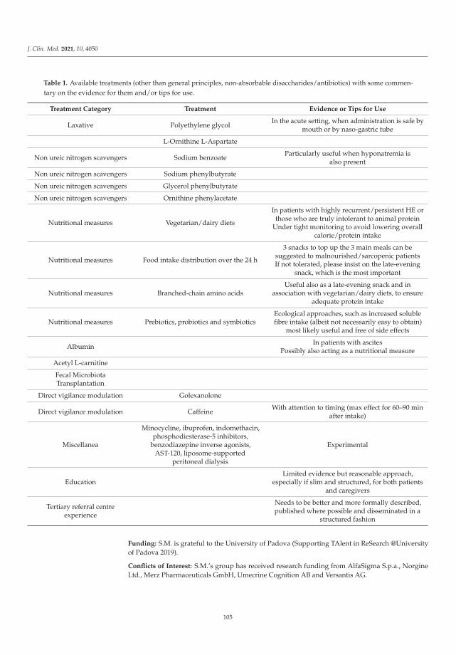
J. Clin. Med. 2021, 10, 4050
Table 1. Available treatments (other than general principles, non-absorbable disaccharides/antibiotics) with some commen-tary on the evidence for them and/or tips for use.
Treatment Category Treatment Evidence or Tips for Use
Laxative Polyethylene glycol In the acute setting, when administration is safe bymouth or by naso-gastric tube
L-Ornithine L-Aspartate
Non ureic nitrogen scavengers Sodium benzoate Particularly useful when hyponatremia isalso present
Non ureic nitrogen scavengers Sodium phenylbutyrate
Non ureic nitrogen scavengers Glycerol phenylbutyrate
Non ureic nitrogen scavengers Ornithine phenylacetate
Nutritional measures Vegetarian/dairy diets
In patients with highly recurrent/persistent HE orthose who are truly intolerant to animal protein
Under tight monitoring to avoid lowering overallcalorie/protein intake
Nutritional measures Food intake distribution over the 24 h
3 snacks to top up the 3 main meals can besuggested to malnourished/sarcopenic patientsIf not tolerated, please insist on the late-evening
snack, which is the most important
Nutritional measures Branched-chain amino acidsUseful also as a late-evening snack and in
association with vegetarian/dairy diets, to ensureadequate protein intake
Nutritional measures Prebiotics, probiotics and symbioticsEcological approaches, such as increased solublefibre intake (albeit not necessarily easy to obtain)
most likely useful and free of side effects
Albumin In patients with ascitesPossibly also acting as a nutritional measure
Acetyl L-carnitine
Fecal MicrobiotaTransplantation
Direct vigilance modulation Golexanolone
Direct vigilance modulation Caffeine With attention to timing (max effect for 60–90 minafter intake)
Miscellanea
Minocycline, ibuprofen, indomethacin,phosphodiesterase-5 inhibitors,
benzodiazepine inverse agonists,AST-120, liposome-supported
peritoneal dialysis
Experimental
EducationLimited evidence but reasonable approach,
especially if slim and structured, for both patientsand caregivers
Tertiary referral centreexperience
Needs to be better and more formally described,published where possible and disseminated in a
structured fashion
Funding: S.M. is grateful to the University of Padova (Supporting TAlent in ReSearch @Universityof Padova 2019).
Conflicts of Interest: S.M.’s group has received research funding from AlfaSigma S.p.a., NorgineLtd., Merz Pharmaceuticals GmbH, Umecrine Cognition AB and Versantis AG.
105

J. Clin. Med. 2021, 10, 4050
References
1. Vilstrup, H.; Amodio, P.; Bajaj, J.; Cordoba, J.; Ferenci, P.; Mullen, K.D.; Weissenborn, K.; Wong, P. Hepatic encephalopathy inchronic liver disease: 2014 Practice Guideline by the American Association for the Study of Liver Diseases and the EuropeanAssociation for the Study of the Liver. Hepatology 2014, 60, 715–735. [CrossRef] [PubMed]
2. Strauss, E.; Tramote, R.; Silva, E.P.; Caly, W.R.; Honain, N.Z.; Maffei, R.A. Doubleblind randomized clinical trial comparingneomycin and placebo in the treatment of exogenous hepatic encephalopathy. Hepatogastroenterology 1992, 39, 542–545.
3. Mullen, K.D.; Sanyal, A.J.; Bass, N.M.; Poordad, F.F.; Sheikh, M.Y.; Frederick, R.T.; Bortey, E.; Forbes, W.P. Rifaximin is safe andwell tolerated for long-term maintenance of remission from overt hepatic encephalopathy. Clin. Gastroenterol. Hepatol. 2014, 12,1390–1397.e2. [CrossRef]
4. Bajaj, J.S.; Barrett, A.C.; Bortey, E.; Paterson, C.; Forbes, W.P. Prolonged remission from hepatic encephalopathy with rifaximin:Results of a placebo crossover analysis. Aliment. Pharmacol. Ther. 2015, 41, 39–45. [CrossRef] [PubMed]
5. Sharma, P.; Agrawal, A.; Sharma, B.C.; Sarin, S.K. Prophylaxis of hepatic encephalopathy in acute variceal bleed: A randomizedcontrolled trial of lactulose versus no lactulose. J. Gastroenterol. Hepatol. 2011, 26, 996–1003. [CrossRef] [PubMed]
6. Tromm, A.; Griga, T.; Greving, I.; Hilden, H.; Huppe, D.; Schwegler, U.; Micklefield, G.H.; May, B. Orthograde whole gut irrigationwith mannite versus paromomycine + lactulose as prophylaxis of hepatic encephalopathy in patients with cirrhosis and uppergastrointestinal bleeding: Results of a controlled randomized trial. Hepatogastroenterology 2000, 47, 473–477.
7. Di Pascoli, M.; Ceranto, E.; De Nardi, P.; Donato, D.; Gatta, A.; Angeli, P. Hospitalizations due to cirrhosis: Clinical aspects in alarge cohort of Italian patients and cost analysis report. Dig. Dis. 2017, 35, 433–438. [CrossRef]
8. Sharma, B.C.; Sharma, P.; Agrawal, A.; Sarin, S.K. Secondary prophylaxis of hepatic encephalopathy: An open-label randomizedcontrolled trial of lactulose versus placebo. Gastroenterology 2009, 137, 885–891. [CrossRef]
9. Agrawal, A.; Sharma, B.C.; Sharma, P.; Sarin, S.K. Secondary prophylaxis of hepatic encephalopathy in cirrhosis: An open-label,randomized controlled trial of lactulose, probiotics, and no therapy. Am. J. Gastroenterol. 2012, 107, 1043–1050. [CrossRef]
10. Bass, N.M.; Mullen, K.D.; Sanyal, A.; Poordad, F.; Neff, G.; Leevy, C.B.; Sigal, S.; Sheikh, M.Y.; Beavers, K.; Frederick, T.; et al.Rifaximin treatment in hepatic encephalopathy. N. Engl. J. Med. 2010, 362, 1071–1081. [CrossRef]
11. Amodio, P. Hepatic encephalopathy: Diagnosis and management. Liver Int. 2018, 38, 966–975. [CrossRef] [PubMed]12. Riggio, O.; Efrati, C.; Catalano, C.; Pediconi, F.; Mecarelli, O.; Accornero, N.; Nicolao, F.; Angeloni, S.; Masini, A.; Ridola, L.; et al.
High prevalence of spontaneous portal-systemic shunts in persistent hepatic encephalopathy: A case-control study. Hepatology2005, 42, 1158–1165. [CrossRef] [PubMed]
13. Kochar, N.; Tripathi, D.; Ireland, H.; Redhead, D.N.; Hayes, P.C. Transjugular intrahepatic portosystemic stent shunt (TIPSS)modification in the management of post-TIPSS refractory hepatic encephalopathy. Gut 2006, 55, 1617–1623. [CrossRef] [PubMed]
14. Riggio, O.; Angeloni, S.; Salvatori, F.M.; De Santis, A.; Cerini, F.; Farcomeni, A.; Attili, A.F.; Merli, M. Incidence, natural history, andrisk factors of hepatic encephalopathy after transjugular intrahepatic portosystemic shunt with polytetrafluoroethylenecoveredstent grafts. Am. J. Gastroenterol. 2008, 3, 2738–2746. [CrossRef] [PubMed]
15. Schepis, F.; Vizzutti, F.; Garcia-Tsao, G.; Marzocchi, G.; Rega, L.; De Maria, N.; Di Maira, T.; Gitto, S.; Caporali, C.; Colopi, S.; et al.Under-dilated TIPS Associate With Efficacy and Reduced Encephalopathy in a Prospective, Non-randomized Study of PatientsWith Cirrhosis. Clin. Gastroenterol. Hepatol. 2018, 16, 1153–1162.e7. [CrossRef]
16. Montagnese, S.; Russo, F.P.; Amodio, P.; Burra, P.; Gasbarrini, A.; Loguercio, C.; Marchesini, G.; Merli, M.; Ponziani, F.R.; Riggio,O.; et al. Hepatic encephalopathy 2018: A clinical practice guideline by the Italian Association for the Study of the Liver (AISF).Dig. Liver Dis. 2019, 51, 190–205. [CrossRef] [PubMed]
17. Weissenborn, K.; Tietge, U.J.; Bokemeyer, M.; Mohammadi, B.; Bode, U.; Manns, M.P.; Caselitz, M. Liver transplantation improveshepatic myelopathy: Evidence by three cases. Gastroenterology 2003, 124, 346–351. [CrossRef] [PubMed]
18. Rahimi, R.S.; Singal, A.G.; Cuthbert, J.A.; Rockey, D.C. Lactulose vs. polyethylene glycol 3350—Electrolyte solution for treatmentof overt hepatic encephalopathy: The HELP randomized clinical trial. JAMA Intern. Med. 2014, 174, 1727–1733. [CrossRef]
19. Naderian, M.; Akbari, H.; Saeedi, M.; Sohrabpour, A.A. Polyethylene glycol and lactulose versus lactulose alone in the treatmentof hepatic encephalopathy in patients with cirrhosis: A non-inferiority randomized controlled trial. Middle East J. Dig. Dis. 2017,9, 12–29. [CrossRef]
20. Hoilat, G.J.; Ayas, M.F.; Hoilat, J.N.; Abu-Zaid, A.; Durer, C.; Durer, S.; Adhami, T.; John, S. Polyethylene glycol versus lactulose inthe treatment of hepatic encephalopathy: A systematic review and meta-analysis. BMJ Open Gastro. 2021, 8, e000648. [CrossRef]
21. Rose, C.; Michalak, A.; Rao, K.V.; Quack, G.; Kircheis, G.; Butterworth, R.F. L-ornithine-L-aspartate lowers plasma and cere-brospinal fluid ammonia and prevents brain edema in rats with acute liver failure. Hepatology 1999, 30, 636–640. [CrossRef]
22. Bai, M.; Yang, Z.; Qi, X.; Fan, D.; Han, G. L-ornithine-l-aspartate for hepatic encephalopathy in patients with cirrhosis: Ameta-analysis of randomized controlled trials. J. Gastroenterol. Hepatol. 2013, 28, 783–792. [CrossRef]
23. Poo, J.L.; Gongora, J.; Sanchez-Avila, F.; Aguilar-Castillo, S.; Garcia-Ramos, G.; Fernandez-Zertuche, M.; Rodríguez-Fragoso, L.;Uribe, M. Efficacy of oral L-ornithine-L-aspartate in cirrhotic patients with hyperammonemic hepatic encephalopathy. Results ofa randomized, lactulose-controlled study. Ann. Hepatol. 2006, 5, 281–288. [CrossRef]
24. Mittal, V.V.; Sharma, B.C.; Sharma, P.; Sarin, S.K. A randomized controlled trial comparing lactulose, probiotics, and L-ornithineL-aspartate in treatment of minimal hepatic encephalopathy. Eur. J. Gastroenterol. Hepatol. 2011, 23, 725–732. [CrossRef] [PubMed]
106
J. Clin. Med. 2021, 10, 4050
25. Varakanahalli, S.; Sharma, B.C.; Srivastava, S.; Sachdeva, S.; Dahale, A.S. Secondary prophylaxis of hepatic encephalopathy incirrhosis of liver: A double-blind randomized controlled trial of L-ornithine L-aspartate versus placebo. Eur. J. Gastroenterol.Hepatol. 2018, 30, 951–958. [CrossRef]
26. Sidhu, S.S.; Sharma, B.C.; Goyal, O.; Kishore, H.; Kaur, N. L-ornithine L-aspartate in bouts of overt hepatic encephalopathy.Hepatology 2018, 67, 700–710. [CrossRef]
27. Hadjihambi, A.; Arias, N.; Sheikh, M.; Jalan, R. Hepatic encephalopathy: A critical current review. Hepatol. Int. 2018, 12(Suppl. S1), 135–147. [CrossRef] [PubMed]
28. Goh, E.T.; Stokes, C.S.; Sidhu, S.S.; Vilstrup, H.; Gluud, L.L.; Morgan, M.Y. L-ornithine Laspartate for prevention and treatment ofhepatic encephalopathy in people with cirrhosis. Cochrane Database Syst. Rev. 2018, 5, CD012410.
29. Butterworth, R.F.; McPhail, M.J.W. L-Ornithine L-Aspartate (LOLA) for Hepatic Encephalopathy in Cirrhosis: Results ofRandomized Controlled Trials and Meta-Analyses. Drugs 2019, 79, S31–S37. [CrossRef]
30. Sushma, S.; Dasarathy, S.; Tandon, R.K.; Jain, S.; Gupta, S.; Bhist, M.S. Sodium benzoate inthe treatment of acute hepaticencephalopathy: A double-blind randomized trial. Hepatology 1992, 16, 138–144. [CrossRef]
31. Efrati, C.; Masini, A.; Merli, M.; Valeriano, V.; Riggio, O. Effect of sodium benzoate on blood ammonia response to oral glutaminechallenge in cirrhotic patients: A note of caution. Am. J. Gastroenterol. 2000, 95, 3574–3578. [CrossRef] [PubMed]
32. Zacharias, H.D.; Zacharias, A.P.; Gluud, L.L.; Morgan, M.Y. Pharmacotherapies that specifically target ammonia for the preventionand treatment of hepatic encephalopathy in adults with cirrhosis. Cochrane Database Syst. Rev. 2019, 6, CD012334. [CrossRef]
33. Amodio, P.; Bemeur, C.; Butterworth, R.; Cordoba, J.; Kato, A.; Montagnese, S.; Uribe, M.; Vilstrup, H.; Morgan, M.Y. Thenutritional management of hepatic encephalopathy in patients with cirrhosis: International Society for Hepatic Encephalopathyand Nitrogen Metabolism Consensus. Hepatology 2013, 58, 325–336. [CrossRef]
34. Weiss, N.; Tripon, S.; Lodey, M.; Guiller, E.; Junot, H.; Monneret, D.; Mayaux, J.; Brisson, H.; Mallet, M.; Rudler, M.; et al. Treatinghepatic encephalopathy in cirrhotic patients admitted to ICU with sodium phenylbutyrate: A preliminary study. Fundam. Clin.Pharmacol. 2018, 32, 209–215. [CrossRef] [PubMed]
35. Rockey, D.C.; Vierling, J.M.; Mantry, P.; Ghabril, M.; Brown, R.S., Jr.; Alexeeva, O.; Zupanets, I.A.; Grinevich, V.; Baranovsky, A.;Dudar, L.; et al. Randomized, double-blind, controlled study of glycerol phenylbutyrate in hepatic encephalopathy. Hepatology2014, 59, 1073–1083. [CrossRef]
36. Jover-Cobos, M.; Noiret, L.; Sharifi, Y.; Jalan, R. Ornithine phenylacetate revisited. Metab. Brain Dis. 2013, 28, 327–331. [CrossRef][PubMed]
37. Ventura-Cots, M.; Concepcion, M.; Arranz, J.A.; Simon-Talero, M.; Torrens, M.; Blanco-Grau, A.; Fuentes, I.; Suñé, P.; Alvarado-Tapias, E.; Gely, C.; et al. Impact of ornithine phenylacetate (OCR-002) in lowering plasma ammonia after upper gastrointestinalbleeding in cirrhotic patients. Therap. Adv. Gastroenterol. 2016, 9, 823–835. [CrossRef] [PubMed]
38. Rahimi, R.S.; Safadi, R.; Thabut, D.; Bhamidimarri, K.R.; Pyrsopoulos, N.; Potthoff, A.; Bukofzer, S.; Bajaj, J.S. Efficacy and Safetyof Ornithine Phenylacetate for Treating Overt Hepatic Encephalopathy in a Randomized Trial. Clin. Gastroenterol. Hepatol. 2020,in press. [CrossRef] [PubMed]
39. Merli, M.; Riggio, O.; Dally, L. Does malnutrition affect survival in cirrhosis? PINC (Policentrica italiana nutrizione cirrosi).Hepatology 1996, 23, 1041–1046. [CrossRef]
40. Merli, M.; Giusto, M.; Lucidi, C.; Giannelli, V.; Pentassuglio, I.; Di Gregorio, V.; Lattanzi, B.; Riggio, O. Muscle depletion increasesthe risk of overt and minimal hepatic encephalopathy: Results of a prospective study. Metab. Brain Dis. 2013, 28, 281–284.[CrossRef]
41. European Association for the Study of the Liver. EASL Clinical Practice Guidelines on nutrition in chronic liver disease. J. Hepatol.2019, 70, 172–193. [CrossRef]
42. Cordoba, J.; Lopez-Hellin, J.; Planas, M.; Sabin, P.; Sanpedro, F.; Castro, F.; Esteban, R.; Guardia, J. Normal protein diet for episodichepatic encephalopathy: Results of a randomized study. J. Hepatol. 2004, 41, 38–43. [CrossRef] [PubMed]
43. Maharshi, S.; Sharma, B.C.; Sachdeva, S.; Srivastava, S.; Sharma, P. Efficacy of Nutritional Therapy for Patients With Cirrhosis andMinimal Hepatic Encephalopathy in a Randomized Trial. Clin. Gastroenterol. Hepatol. 2016, 14, 454–460. [CrossRef] [PubMed]
44. Tsien, C.D.; McCullough, A.J.; Dasarathy, S. Late evening snack: Exploiting a period of anabolic opportunity in cirrhosis. J.Gastroenterol. Hepatol. 2012, 27, 430–441. [CrossRef]
45. Tandon, P.; Ismond, K.P.; Riess, K.; Duarte-Rojo, A.; Al-Judaibi, B.; Dunn, M.A.; Holman, J.; Howes, N.; Haykowsky, M.J.F.;Josbeno, D.A.; et al. Exercise in cirrhosis: Translating evidence and experience to practice. J. Hepatol. 2018, 69, 1164–1177.[CrossRef]
46. Chavez-Tapia, N.C.; Cesar-Arce, A.; Barrientos-Gutierrez, T.; Villegas-Lopez, F.A.; Mendez-Sanchez, N.; Uribe, M. A systematicreview and meta-analysis of the use of oral zinc in the treatment of hepatic encephalopathy. Nutr. J. 2013, 12, 74. [CrossRef]
47. Bai, Z.; Bernardi, M.; Yoshida, E.M.; Li, H.; Guo, X.; Méndez-Sánchez, N.; Li, Y.; Wang, R.; Deng, J.; Qi, X. Albumin infusionmay decrease the incidence and severity of overt hepatic encephalopathy in liver cirrhosis. Aging 2019, 8, 8502–8525. [CrossRef][PubMed]
48. Teh, K.B.; Loo, J.H.; Tam, Y.C.; Wong, Y.J. Efficacy and safety of albumin infusion for overt hepatic encephalopathy: A systematicreview and meta-analysis. Dig. Liver Dis. 2021, 53, 817–823. [CrossRef]
49. Caraceni, P.; Pavesi, M.; Baldassarre, M.; Bernardi, M.; Arroyo, V. The use of human albumin in patients with cirrhosis: AEuropean survey. Expert Rev. Gastroenterol. Hepatol. 2018, 12, 625–632. [CrossRef]
107
J. Clin. Med. 2021, 10, 4050
50. Gluud, L.L.; Dam, G.; Les, I.; Marchesini, G.; Borre, M.; Aagaard, N.K.; Vilstrup, H. Branched-chain amino acids for people withhepatic encephalopathy. Cochrane Database Syst. Rev. 2017, 5, CD001939. [CrossRef]
51. Park, J.G.; Tak, W.Y.; Park, S.Y.; Kweon, Y.O.; Chung, W.J.; Jang, B.K.; Bae, S.H.; Lee, H.J.; Jang, J.Y.; Suk, K.T.; et al. Effects ofBranched-Chain Amino Acid (BCAA) Supplementation on the Progression of Advanced Liver Disease: A Korean Nationwide,Multicenter, Prospective, Observational, Cohort Study. Nutrients 2020, 12, 1429. [CrossRef] [PubMed]
52. Gomes Romeiro, F.; do Val Ietsugu, M.; de Campos Franzoni, L.; Augusti, L.; Alvarez, M.; Alves Amaral Santos, L.; Bazeia Lima,T.; Hiromoto Koga, K.; Marta Moriguchi, S.; Antonio Caramori, C.; et al. Which of the branched-chain amino acids increasescerebral blood flow in hepatic ncephalopathy? A double-blind randomized trial. NeuroImage 2018, 19, 302–310. [CrossRef]
53. Marchesini, G.; Bianchi, G.; Merli, M.; Amodio, P.; Panella, C.; Loguercio, C.; Rossi Fanelli, F.; Abbiati, R. Italian BCAA StudyGroup. Nutritional supplementation with branched-chain amino acids in advanced cirrhosis: A double-blind, randomized trial.Gastroenterology 2003, 124, 1792–1801. [CrossRef]
54. Malaguarnera, M. Acetyl-L-carnitine in hepatic encephalopathy. Metab. Brain Dis. 2013, 28, 193–199. [CrossRef] [PubMed]55. Jiang, Q.; Jiang, G.; Shi, K.Q.; Cai, H.; Wang, Y.X.; Zheng, M.H. Oral acetyl-L-carnitine treatment in hepatic encephalopathy: View
of evidence-based medicine. Ann. Hepatol. 2013, 12, 803–809. [CrossRef]56. Martí-Carvajal, A.J.; Gluud, C.; Arevalo-Rodriguez, I.; Martí-Amarista, C.E. Acetyl-L-carnitine for patients with hepatic en-
cephalopathy. Cochrane Database Syst. Rev. 2019, 1, CD011451. [CrossRef]57. Viramontes Horner, D.; Avery, A.; Stow, R. The effects of probiotics and symbiotics on risk factors for hepatic encephalopathy: A
systematic review. J. Clin. Gastroenterol. 2017, 51, 312–323. [CrossRef]58. Dalal, R.; McGee, R.G.; Riordan, S.M.; Webster, A.C. Probiotics for people with hepatic encephalopathy. Cochrane Database Syst.
Rev. 2017, 2, CD008716. [CrossRef]59. Cao, Q.; Yu, C.B.; Yang, S.G.; Cao, H.C.; Chen, P.; Deng, M.; Li, L.J. Effect of probiotic treatment on cirrhotic patients with minimal
hepatic encephalopathy: A meta-analysis. Hepatob. Pancreat. Dis. Int. 2018, 17, 9–16. [CrossRef]60. Liu, Q.; Duan, Z.P.; Ha, D.K.; Bengmark, S.; Kurtovic, J.; Riordan, S.M. Synbiotic modulation of gut flora: Effect on minimal
hepatic encephalopathy in patients with cirrhosis. Hepatology 2004, 39, 1441–1449. [CrossRef]61. Bajaj, J.S.; Fagan, A.; Gavis, E.A.; Kassam, Z.; Sikaroodi, M.; Gillevet, P.M. Long-term Outcomes of Fecal Microbiota Transplanta-
tion in Patients With Cirrhosis. Gastroenterology 2019, 156, 1921–1923. [CrossRef] [PubMed]62. Goh, E.T.; Andersen, M.L.; Morgan, M.Y.; Gluud, L.L. Flumazenil versus placebo or no intervention for people with cirrhosis and
hepatic encephalopathy. Cochrane Database Syst. Rev. 2017, 8, CD002798. [PubMed]63. Johansson, M.; Agusti, A.; Llansola, M.; Montoliu, C.; Strömberg, J.; Malinina, E.; Ragagnin, G.; Doverskog, M.; Bäckström, T.;
Felipo, V. GR3027 antagonizes GABAA receptor-potentiating neurosteroids and restores spatial learning and motor coordinationin rats with chronic hyperammonemia and hepatic encephalopathy. Am. J. Physiol. Gastrointest. Liver Physiol. 2015, 309,G400–G409. [CrossRef] [PubMed]
64. Johansson, M.; Strömberg, J.; Ragagnin, G.; Doverskog, M.; Bäckström, T. GABAA receptor modulating steroid antagonists(GAMSA) are functional in vivo. J. Steroid Biochem. Mol. Biol. 2016, 160, 98–105. [CrossRef]
65. Montagnese, S.; Lauridsen, M.; Vilstrup, H.; Zarantonello, L.; Lakner, G.; Fitilev, S.; Zupanets, I.; Kozlova, I.; Bunkova, E.;Tomasiewicz, K.; et al. A pilot study of golexanolone, a new GABA-A receptor-modulating steroid antagonist, in patients withcovert hepatic encephalopathy. J. Hepatol. 2021, 75, 98–107. [CrossRef]
66. Garrido, M.; Skorucak, J.; Raduazzo, D.; Turco, M.; Spinelli, G.; Angeli, P.; Amodio, P.; Achermann, P.; Montagnese, S. Vigilanceand wake EEG architecture in simulated hyperammonaemia: A pilot study on the effects of L-Ornithine-L-Aspartate (LOLA) andcaffeine. Metab. Brain Dis. 2016, 31, 965–974. [CrossRef] [PubMed]
67. De Rui, M.; Schiff, S.; Aprile, D.; Angeli, P.; Bombonato, G.; Bolognesi, M.; Sacerdoti, D.; Gatta, A.; Merkel, C.; Amodio, P.; et al.Excessive daytime sleepiness and hepatic encephalopathy: It is worth asking. Metab Brain. Dis. 2013, 28, 245–248. [CrossRef]
68. Garrido, M.; Turco, M.; Formentin, C.; Corrias, M.; De Rui, M.; Montagnese, S.; Amodio, P. An educational tool for the prophylaxisof hepatic encephalopathy. BMJ Open Gastro. 2017, 4, e000161. [CrossRef]
69. Gamal, M.; Abdel Wahab, Z.; Eshra, M.; Rashed, L.; Sharawy, N. Comparative Neuroprotective effects of dexamethasone andminocycline during hepatic encephalopathy. Neurol. Res. Int. 2014, 2014, 254683. [CrossRef]
70. Cauli, O.; Rodrigo, R.; Piedrafita, B.; Boix, J.; Felipo, V. Inflammation and hepatic encephalopathy: Ibuprofen restores learningability in rats with portacaval shunts. Hepatology 2007, 46, 514–519. [CrossRef]
71. Ahboucha, S.; Jiang, W.; Chatauret, N.; Mamer, O.; Baker, G.B.; Butterworth, R.F. Indomethacin improves locomotor deficit andreduces brain concentrations of neuroinhibitory steroids in rats following portacaval anastomosis. Neurogastroenterol. Motil. 2008,20, 949–957. [CrossRef] [PubMed]
72. Agusti, A.; Hernandez-Rabaza, V.; Balzano, T.; Taoro-Gonzalez, L.; Ibanez-Grau, A.; Cabrera-Pastor, A.; Fustero, S.; Llansola,M.; Montoliu, C.; Felipo, V. Sildenafil reduces neuroinflammation in cerebellum, restores GABAergic tone, and improves motorin-coordination in rats with hepatic encephalopathy. CNS Neurosci. Ther. 2017, 23, 386–394. [CrossRef]
73. Steindl, P.; Puspok, A.; Druml, W.; Ferenci, P. Beneficial effect of pharmacological modulation of the GABAA-benzodiazepinereceptor on hepatic encephalopathy in the rat: Comparison with uremic encephalopathy. Hepatology 1991, 14, 963–968. [CrossRef][PubMed]
74. Bosoi, C.R.; Parent-Robitaille, C.; Anderson, K.; Tremblay, M.; Rose, C.F. AST-120 (spherical carbon adsorbent) lowers ammonialevels and attenuates brain edema in bile duct-ligated rats. Hepatology 2011, 53, 1995–2002. [CrossRef]
108
J. Clin. Med. 2021, 10, 4050
75. Matoori, S.; Forster, V.; Agostoni, V.; Bettschart-Wolfensberger, R.; Bektas, R.N.; Thöny, B.; Häberle, J.; Leroux, J.C.; Kabbaj, M.Preclinical evaluation of liposome-supported peritoneal dialysis for the treatment of hyperammonemic crises. J. Control. Release2020, 328, 503–513. [CrossRef] [PubMed]
76. Formentin, C.; Zarantonello, L.; Mangini, C.; Frigo, A.C.; Montagnese, S.; Merkel, C. Clinical, neuropsychological and neurophys-iological indices and predictors of hepatic encephalopathy (HE). Liver Int. 2021, 41, 1070–1082. [CrossRef] [PubMed]
77. Montagnese, S.; Schiff, S.; Amodio, P. Quick diagnosis of hepatic encephalopathy: Fact or fiction? Hepatology 2015, 61, 405–406.[CrossRef]
78. Montagnese, S.; De Rui, M.; Angeli, P.; Amodio, P. Neuropsychiatric performance in patients with cirrhosis: Who is “normal”? J.Hepatol. 2017, 66, 825–835. [CrossRef]
79. Bersagliere, A.; Raduazzo, I.D.; Nardi, M.; Schiff, S.; Gatta, A.; Amodio, P.; Achermann, P.; Montagnese, S. Induced hyper-ammonemia may compromise the ability to generate restful sleep in patients with cirrhosis. Hepatology 2012, 55, 869–878.[CrossRef]
109

Journal of
Clinical Medicine
Review
Acute-on-Chronic Liver Failure in Cirrhosis
Carmine Gambino, Salvatore Piano * and Paolo Angeli
���������������
Citation: Gambino, C.; Piano, S.;
Angeli, P. Acute-on-Chronic Liver
Failure in Cirrhosis. J. Clin. Med. 2021,
10, 4406. https://doi.org/10.3390/
jcm10194406
Academic Editors: Pierluigi Toniutto
and Hiroki Nishikawa
Received: 31 August 2021
Accepted: 22 September 2021
Published: 26 September 2021
Publisher’s Note: MDPI stays neutral
with regard to jurisdictional claims in
published maps and institutional affil-
iations.
Copyright: © 2021 by the authors.
Licensee MDPI, Basel, Switzerland.
This article is an open access article
distributed under the terms and
conditions of the Creative Commons
Attribution (CC BY) license (https://
creativecommons.org/licenses/by/
4.0/).
Unit of Internal Medicine and Hepatology (UIMH), Department of Medicine (DIMED), University of Padova,35128 Padova, Italy; [email protected] (C.G.); [email protected] (P.A.)* Correspondence: [email protected]
Abstract: Acute-on-chronic liver failure (ACLF) is a syndrome that develops in patients with acutelydecompensated chronic liver disease. It is characterised by high 28-day mortality, the presence of oneor more organ failures (OFs) and a variable but severe grade of systemic inflammation. Despite thepeculiarity of each one, every definition proposed for ACLF recognizes it as a proper clinical entity.In this paper, we provide an overview of the diagnostic criteria proposed by the different scientificsocieties and the clinical characteristics of the syndrome. Established and experimental treatmentsare also described. Among the former, the most relevant are directed to support organ failures, treatprecipitating factors and carry out early assessment for liver transplantation (LT). Further studies areneeded to better clarify pathophysiology of the syndrome and discover new therapies.
Keywords: acute-on-chronic liver failure (ACLF); cirrhosis; liver transplantation (LT)
1. Definition of Acute-on-Chronic Liver Failure
Acute decompensation (AD) of cirrhosis refers to the development of ascites, gastroin-testinal haemorrhage, hepatic encephalopathy or any combination of these, which leads tohospital admission [1]. Acute-on-chronic liver failure (ACLF) is a distinct syndrome thatdevelops in patients with acutely decompensated chronic liver disease and is characterisedby high 28-day mortality. Other major features of ACLF are the strong association withone or more precipitating factor(s), the development of single- or multiple organ failures(OFs) and a severe degree of systemic inflammation [2–4]. International scientific societieshave proposed different definitions of ACLF in recent years; they differ from each othermainly in the type of precipitant (hepatic or extrahepatic), the stage of underlying liverdisease (chronic hepatitis or cirrhosis) and the inclusion or not of extra-hepatic OFs. Inspite of these differences, each of them recognizes ACLF as a definite clinical entity. Table 1summarizes definitions, diagnostic criteria and stratification of ACLF used by the fourmajor international consortia [2,5–9].
The definition proposed by the European Association for the Study of the Liver—Chronic Liver Failure (EASL-CLIF) Consortium is based on the results of the CANONICstudy, a multi-center prospective investigation in which 1343 patients non-electively hos-pitalized for AD of cirrhosis were enrolled, irrespective of prior episode(s) of AD [2].This definition considers both hepatic and extra-hepatic precipitants and both liver andextra-hepatic OFs. The diagnosis of OFs is based on a modified Sequential Organ FailureAssessment (SOFA) score, called CLIF-C organ failure (CLIF-C OF), which considers thefunction of six organ systems (liver, kidney, brain, coagulation, circulation and respira-tion) [2]. According to the number of OFs, patients with ACLF were stratified into threegroups: (I) patients with a single kidney failure or another single OF if associated withbrain or kidney disfunction (ACLF grade 1); (II) patients with two OFs (ACLF grade 2);(III) patients with three or more OFs (ACLF grade 3) [2]. We contributed to the develop-ment of this definition, which nowadays is the most studied. Thus, we currently use it inour center.
J. Clin. Med. 2021, 10, 4406. https://doi.org/10.3390/jcm10194406 https://www.mdpi.com/journal/jcm
111
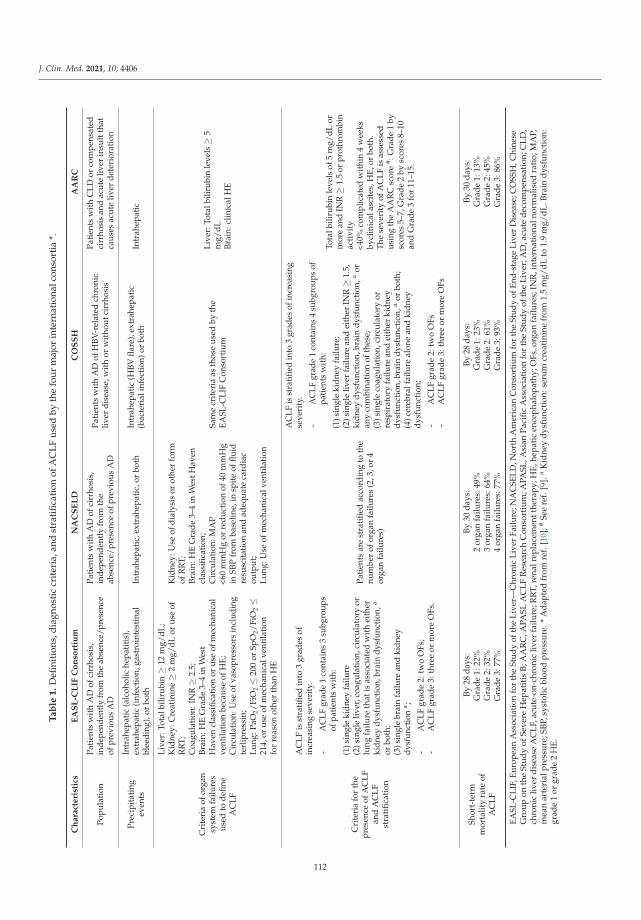
J. Clin. Med. 2021, 10, 4406
Ta
ble
1.
Defi
niti
ons,
diag
nost
iccr
iter
ia,a
ndst
rati
ficat
ion
ofA
CLF
used
byth
efo
urm
ajor
inte
rnat
iona
lcon
sort
ia*.
Ch
ara
cte
rist
ics
EA
SL
-CL
IFC
on
sort
ium
NA
CS
EL
DC
OS
SH
AA
RC
Popu
lati
onPa
tien
tsw
ith
AD
ofci
rrho
sis,
inde
pend
ently
from
the
abse
nce/
pres
ence
ofpr
evio
usA
D
Pati
ents
wit
hA
Dof
cirr
hosi
s,in
depe
nden
tly
from
the
abse
nce/
pres
ence
ofpr
evio
usA
D
Pati
ents
wit
hA
Dof
HBV
-rel
ated
chro
nic
liver
dise
ase,
wit
hor
wit
hout
cirr
hosi
s
Pati
ents
wit
hC
LDor
com
pens
ated
cirr
hosi
san
dac
ute
liver
insu
ltth
atca
uses
acut
eliv
erde
teri
orat
ion
Prec
ipit
atin
gev
ents
Intr
ahep
atic
(alc
ohol
iche
pati
tis)
,ex
trah
epat
ic(i
nfec
tion
,gas
troi
ntes
tina
lbl
eedi
ng),
orbo
thIn
trah
epat
ic,e
xtra
hepa
tic,
orbo
thIn
trah
epat
ic(H
BVfla
re),
extr
ahep
atic
(bac
teri
alin
fect
ion)
orbo
thIn
trah
epat
ic
Cri
teri
aof
orga
nsy
stem
failu
res
used
tode
fine
AC
LF
Live
r:To
talb
iliru
bin≥
12m
g/dL
;K
idne
y:C
reat
inin
e≥
2m
g/dL
orus
eof
RR
T;C
oagu
lati
on:I
NR≥
2.5;
Brai
n:H
EG
rade
3–4
inW
est
Hav
encl
assi
ficat
ion
orus
eof
mec
hani
cal
vent
ilati
onbe
caus
eof
HE;
Cir
cula
tion:
Use
ofva
sopr
esso
rsin
clud
ing
terl
ipre
ssin
;Lu
ng:P
aO2/
FiO
2≤
200
orSp
O2/
FiO
2≤
214,
orus
eof
mec
hani
calv
enti
lati
onfo
rre
ason
othe
rth
anH
E
Kid
ney:
Use
ofdi
alys
isor
othe
rfo
rmof
RR
T;Br
ain:
HE
Gra
de3–
4in
Wes
tHav
encl
assi
ficat
ion;
Cir
cula
tion
:MA
P<6
0m
mH
gor
redu
ctio
nof
40m
mH
gin
SBP
from
base
line,
insp
ite
offlu
idre
susc
itat
ion
and
adeq
uate
card
iac
outp
ut;
Lung
:Use
ofm
echa
nica
lven
tila
tion
Sam
ecr
iter
iaas
thos
eus
edby
the
EASL
-CLI
FC
onso
rtiu
m
Live
r:To
talb
iliru
bin
leve
ls≥
5m
g/dL
Brai
n:cl
inic
alH
E
Cri
teri
afo
rth
epr
esen
ceof
AC
LFan
dA
CLF
stra
tific
atio
n
AC
LFis
stra
tifie
din
to3
grad
esof
incr
easi
ngse
veri
ty.
-A
CLF
grad
e1
cont
ains
3su
bgro
ups
ofpa
tien
tsw
ith:
(1)s
ingl
eki
dney
failu
re(2
)sin
gle
liver
,coa
gula
tion
,cir
cula
tory
orlu
ngfa
ilure
that
isas
soci
ated
wit
hei
ther
kidn
eydy
sfun
ctio
n,br
ain
dysf
unct
ion,
a
orbo
th;
(3)s
ingl
ebr
ain
failu
rean
dki
dney
dysf
unct
ion
a ;
-A
CLF
grad
e2:
two
OFs
;-
AC
LFgr
ade
3:th
ree
orm
ore
OFs
.
Pati
ents
are
stra
tifie
dac
cord
ing
toth
enu
mbe
rof
orga
nfa
ilure
s(2
,3,o
r4
orga
nfa
ilure
s)
AC
LFis
stra
tified
into
3gr
ades
ofin
crea
sing
seve
rity
.
-A
CLF
grad
e1
cont
ains
4su
bgro
ups
ofpa
tien
tsw
ith:
(1)s
ingl
eki
dney
failu
re;
(2)s
ingl
eliv
erfa
ilure
and
eith
erIN
R≥
1.5,
kidn
eydy
sfun
ctio
n,br
ain
dysf
unct
ion,
aor
any
com
bina
tion
ofth
ese;
(3)s
ingl
eco
agul
atio
n,ci
rcul
ator
yor
resp
irat
ory
failu
rean
dei
ther
kidn
eydy
sfun
ctio
n,br
ain
dysf
unct
ion,
aor
both
;(4
)cer
ebra
lfai
lure
alon
ean
dki
dney
dysf
unct
ion;
-A
CLF
grad
e2:
two
OFs
-A
CLF
grad
e3:
thre
eor
mor
eO
Fs
Tota
lbili
rubi
nle
vels
of5
mg/
dLor
mor
ean
dIN
R≥
1.5
orpr
othr
ombi
nac
tivi
ty<4
0%co
mpl
icat
edw
ithi
n4
wee
ksby
clin
ical
asci
tes,
HE,
orbo
th.
The
seve
rity
ofA
CLF
isas
sess
edus
ing
the
AA
RC
scor
e# :G
rade
1by
scor
es5–
7,G
rade
2by
scor
es8–
10an
dG
rade
3fo
r11
–15.
Shor
t-te
rmm
orta
lity
rate
ofA
CLF
By28
days
:G
rade
1:22
%G
rade
2:32
%G
rade
3:77
%
By30
days
:2
orga
nfa
ilure
s:49
%3
orga
nfa
ilure
s:64
%4
orga
nfa
ilure
s:77
%
By28
days
:G
rade
1:23
%G
rade
2:61
%G
rade
3:93
%
By30
days
:G
rade
1:13
%G
rade
2:45
%G
rade
3:86
%
EASL
-CLI
F,Eu
rope
anA
ssoc
iatio
nfo
rth
eSt
udy
ofth
eLi
ver—
Chr
onic
Live
rFa
ilure
;NA
CSE
LD,N
orth
Am
eric
anC
onso
rtiu
mfo
rth
eSt
udy
ofEn
d-st
age
Live
rD
isea
se;C
OSS
H,C
hine
seG
roup
onth
eSt
udy
ofSe
vere
Hep
atit
isB
;AA
RC
,APA
SLA
CL
FR
esea
rch
Con
sort
ium
;APA
SL,A
sian
Paci
ficA
ssoc
iati
onfo
rth
eSt
udy
ofth
eL
iver
;AD
,acu
ted
ecom
pens
atio
n;C
LD
,ch
roni
cliv
erd
isea
seA
CL
F,ac
ute-
on-c
hron
icliv
erfa
ilure
;RR
T,re
nalr
epla
cem
entt
hera
py;H
E,h
epat
icen
ceph
alop
athy
;OFs
,org
anfa
ilure
s;IN
R,i
nter
nati
onal
norm
alis
edra
tio;
MA
P,m
ean
arte
rial
pres
sure
;SB
P,sy
stol
icbl
ood
pres
sure
.*A
dap
ted
from
ref.
[10]
;#Se
ere
f.[9
].a
Kid
ney
dys
func
tion
:ser
um
crea
tini
nefr
om1.
5m
g/d
Lto
1.9
mg/
dL
.Bra
ind
ysfu
ncti
on:
grad
e1
orgr
ade
2H
E.
112

J. Clin. Med. 2021, 10, 4406
The definition proposed by the North American Consortium for the Study of End-stage Liver Disease (NACSELD) is based on an investigation involving 507 patients withAD of cirrhosis non electively hospitalised for infection [5]. Like the European one, theNorth American definition considers extra-hepatic OFs as part of the syndrome but doesnot include liver and coagulation. It defines ACLF by the presence of two or more OFsamong kidney, brain, circulation and respiration and stratifies patients according to thenumber of organ failures [5]. The Chinese Group on the Study of Severe Hepatitis B(COSSH) developed a definition for hepatitis B virus (HBV)-related ACLF by using datafrom a large cohort of 1202 patients with HBV-related AD, with or without cirrhosis. TheCLIF-C OF scoring system was used to define OFs; so, this definition and the consequentstratification of patients are quite similar to the European ones. The only difference isthat, in the Chinese classification, a patient with single liver failure with INR ≥ 1.5 isconsidered as having ACLF grade 1 [6]. The Asian Pacific Association for the Studyof the Liver (APASL) proposed a definition of ACLF in 2009 which was based on anexpert opinion. This definition was updated by the APASL ACLF Research Consortium(AARC) in 2014 and then in 2019, using the results of the AARC database (5228 patientscollected at that time) [7–9]. Unlike the above definitions, AARC investigators considerextra-hepatic OFs as manifestations but not as components of the syndrome, and extra-hepatic insults (for example, bacterial infections) as complications, but not triggers, ofACLF. So, ACLF is considered as an acute hepatic insult (for example, HBV reactivation oracute alcoholic hepatitis), manifested as jaundice (total bilirubin levels ≥ 5 mg/dL) andcoagulation failure (INR ≥ 1.5 or prothrombin activity < 40%) and complicated by clinicalascites, encephalopathy or both within 4 weeks in patients with chronic liver disease orcompensated cirrhosis without prior decompensation and with no AD [9]. Thus, AARCinvestigators consider ACLF to be totally distinct from acutely decompensated cirrhosis.The severity of ACLF is assessed using a grading system based on the AARC score [9].
2. Clinical Features
ACLF has typical clinical features based on the definition used, on which its prevalencealso depends. In the European cohort, the prevalence of the syndrome was 23% amongpatients with AD of cirrhosis at admission and 8.3% of patients developed it duringhospitalization within a period of days (maximum of two weeks). In outpatients withcirrhosis, the incidence of the syndrome is about 40% at 10 years [11]. As confirmed bythe PREDICT (PREDICTing Acute-on-Chronic Liver Failure) study, another large-scaleEuropean prospective investigation designed to identify predictors of this syndrome,patients with ACLF were younger, showed higher levels of white blood cells and C-reactiveprotein (CRP) and had a greater prevalence of bacterial infections, severe alcoholic hepatitis,variceal bleeding, drug-induced encephalopathy as precipitants, with respect to patientswithout ACLF [2,12,13]. Moreover, the PREDICT study demonstrated that the clinicalcourse of AD that leads to ACLF is distinct from the other forms of AD of cirrhosis [12].The 28-day mortality significantly rises with the increase in the number of OFs, rangingfrom 4.7% for patients without ACLF to 22%, 32% and 77% for patients with ACLF grade 1,2 and 3, respectively [2].
In a validation study of NACSELD definition of ACLF, in which 2675 patients withAD of cirrhosis related or not to infection were included, the prevalence of ACLF was 10%and 30-day mortality rate was significantly different between patients with or without thesyndrome (41% vs. 7%, respectively) [14]. In a recent study, the NACSELD criteria weredemonstrated to be less sensitive compared to EASL-CLIF criteria in diagnosing ACLF [15].
When using NACSELD criteria, only about 40% of patients with a diagnosis of ACLFbased on EASL-CLIF criteria were classified as affected by the syndrome, probably becausethe NACSELD definition considers only more severe patients and because it could beinfluenced by the medical strategies available in the different centers (renal replacementtherapy (RRT), mechanical ventilation, use of vasopressors) [10,16].
113

J. Clin. Med. 2021, 10, 4406
In a cohort of patients with HBV-related AD of cirrhosis, the prevalence of ACLF was30.2% according to the COSSH ACLF definition. As in the European cohort, patients withACLF were younger, had a more severe grade of systemic inflammation (as demonstratedby higher levels of white blood cells and CRP) and more frequently had a bacterial infection(associated or not with HBV reactivation) as precipitant compared to those without ACLF,with a significantly higher short-term mortality (52.1% vs. 4.3%) [6]. Although EASL-CLIFand COSSH definitions of ACLF are very similar, clinical characteristics of patients arequite different, because of the higher prevalence of intra-hepatic precipitants in the Chinesecohort (most often HBV reactivation) with respect to the European cohort [16], with liverand coagulation failure being more frequent in the former and kidney and brain failuremore frequent in the latter [4]. As expected, a flare of HBV infection was the most frequenttrigger of ACLF in studies using AARC criteria [9,17]. In a study using AARC ACLFcriteria which enrolled patients with HBV-related ACLF, about 32% had a bacterial orfungal infection as a complication. The 28-day mortality rate was 27.8% [18].
3. Pathophysiology
The pathophysiology of ACLF is yet to be fully understood. To date, ACLF is con-sidered the extreme expression of systemic inflammation that drives AD of cirrhosis [19].Systemic inflammation is characterised by activation of the immune system that leadsto increased circulating levels of inflammatory mediators and, if severe, proliferation ofneutrophils, monocytes and dendritic cells [20]. The mechanism of systemic inflammationdepends on the precipitant of ACLF [3]. The recognition of pathogen-associated molecularpatterns (PAMPs) activates the innate immune system by pattern-recognition receptors(PRR) in case of bacterial infection or translocation of viable bacteria and bacterial productsthrough the intestinal wall [19,21]. Exceeding inflammation can cause direct tissue damageand necrotic cell death, resulting in the release of damage-associated molecular patterns(DAMPs) that perpetuate inflammation acting on PRR [21]. DAMPs are also releasedwhen an injury acts directly on the liver, as in case of alcoholic hepatitis or ischemia dueto variceal haemorrhage [22,23]. This overactivation of the immune cells requires a largeamount of energy sustained by reallocation of nutrients. This causes a reduced availabilityof substrates for other organ systems that leads to OFs by severe mitochondrial dysfunctionand impaired energy production [19]. Moreover, recent findings suggest that systemic in-flammation can explain and act with the traditionally accepted organ-specific mechanismsof AD (portal hypertension, hyperammonaemia, endogenous vasoconstrictors system andarterial blood volume) in determining OFs [19]. Blood metabolomics offers a new insightinto the pathophysiology of systemic inflammation in patients with ACLF and could bean intriguing starting point to uncover new potential therapeutic targets [24]. Figure 1summarizes the pathophysiology of ACLF.
114
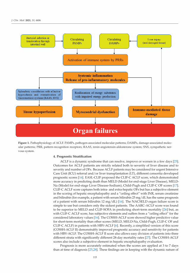
J. Clin. Med. 2021, 10, 4406
Figure 1. Pathophysiology of ACLF. PAMPs, pathogen-associated molecular patterns; DAMPs, damage-associated molec-ular patterns; PRR, pattern-recognition receptors; RAAS, renin-angiotensin-aldosterone system; SNS, sympathetic ner-vous system.
4. Prognostic Stratification
ACLF is a dynamic syndrome that can resolve, improve or worsen in a few days [25].Outcomes for ACLF patients are strictly related both to severity of liver disease and toseverity and number of OFs. Because ACLF patients may be considered for urgent IntensiveCare Unit (ICU) referral and/or liver transplantation (LT), different consortia developedprognostic scores [16]. EASL-CLIF proposed the CLIF-C ACLF score, which demonstratedmore accuracy in predicting death than MELD (Model for end-stage Liver Disease), MELD-Na (Model for end-stage Liver Disease-Sodium), Child-Pugh and CLIF-C OF scores [17].CLIF-C ACLF score captures both intra- and extra-hepatic OFs but has a subjective elementin the scoring of hepatic encephalopathy and a “ceiling effect” with INR, serum creatinineand bilirubin (for example, a patient with serum bilirubin 25 mg/dL has the same prognosisof a patient with serum bilirubin 12 mg/dL) [16]. The NACSELD organ failure score issimple to use but considers only the sickest patients. The AARC-ACLF score was foundto be superior to MELD and CLIF-SOFA in predicting short-term mortality [26] but, aswith CLIF-C ACLF score, has subjective elements and suffers from a “ceiling effect” for theconsidered laboratory values [16]. The COSSH-ACLF score showed higher predictive valuefor short-term mortality than other scores (MELD, MELD-Na, Child-Pugh, CLIF-C OF andCLIF-C ACLF) in patients with HBV-ACLF [6]. Recently, a simplified version of this score(COSSH-ACLF II) demonstrably improved prognostic accuracy and sensitivity for patientswith HBV-ACLF. The COSSH-ACLF II score also allows easy division of patients into threedifferent strata with significantly different 28-day mortality rates [27]. The COSSH-ACLFscores also include a subjective element in hepatic encephalopathy evaluation.
Prognosis is more accurately estimated when the scores are applied at 3 to 7 daysthan at time of diagnosis [25,28]. These findings are in keeping with the dynamic nature of
115
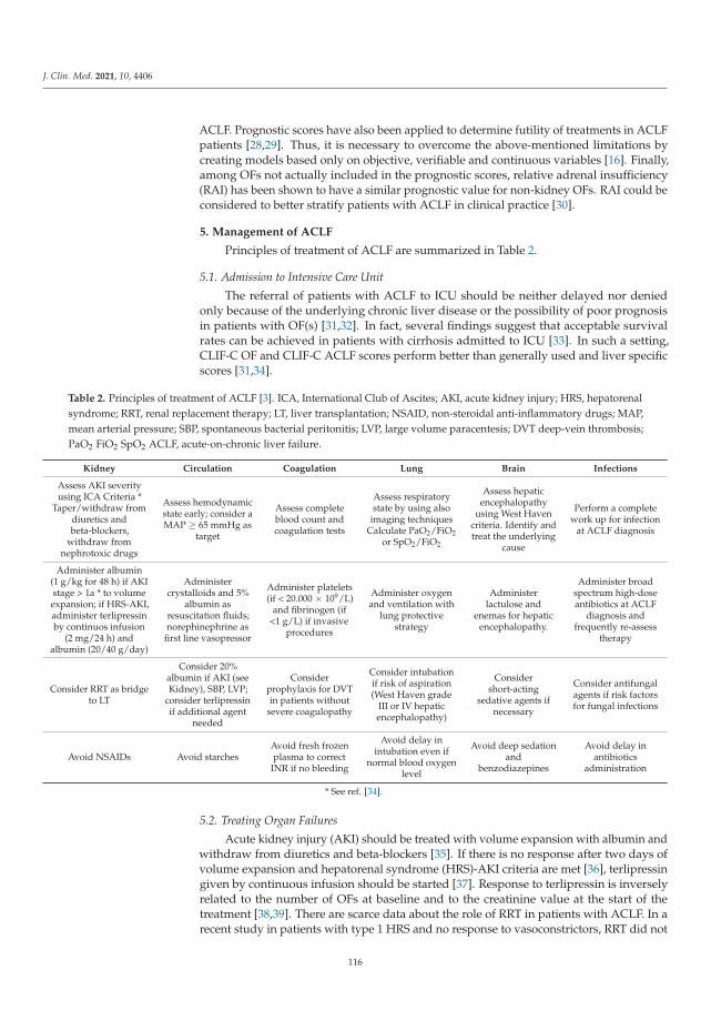
J. Clin. Med. 2021, 10, 4406
ACLF. Prognostic scores have also been applied to determine futility of treatments in ACLFpatients [28,29]. Thus, it is necessary to overcome the above-mentioned limitations bycreating models based only on objective, verifiable and continuous variables [16]. Finally,among OFs not actually included in the prognostic scores, relative adrenal insufficiency(RAI) has been shown to have a similar prognostic value for non-kidney OFs. RAI could beconsidered to better stratify patients with ACLF in clinical practice [30].
5. Management of ACLF
Principles of treatment of ACLF are summarized in Table 2.
5.1. Admission to Intensive Care Unit
The referral of patients with ACLF to ICU should be neither delayed nor deniedonly because of the underlying chronic liver disease or the possibility of poor prognosisin patients with OF(s) [31,32]. In fact, several findings suggest that acceptable survivalrates can be achieved in patients with cirrhosis admitted to ICU [33]. In such a setting,CLIF-C OF and CLIF-C ACLF scores perform better than generally used and liver specificscores [31,34].
Table 2. Principles of treatment of ACLF [3]. ICA, International Club of Ascites; AKI, acute kidney injury; HRS, hepatorenalsyndrome; RRT, renal replacement therapy; LT, liver transplantation; NSAID, non-steroidal anti-inflammatory drugs; MAP,mean arterial pressure; SBP, spontaneous bacterial peritonitis; LVP, large volume paracentesis; DVT deep-vein thrombosis;PaO2 FiO2 SpO2 ACLF, acute-on-chronic liver failure.
Kidney Circulation Coagulation Lung Brain Infections
Assess AKI severityusing ICA Criteria *
Taper/withdraw fromdiuretics andbeta-blockers,
withdraw fromnephrotoxic drugs
Assess hemodynamicstate early; consider aMAP ≥ 65 mmHg as
target
Assess completeblood count andcoagulation tests
Assess respiratorystate by using also
imaging techniquesCalculate PaO2/FiO2
or SpO2/FiO2
Assess hepaticencephalopathy
using West Havencriteria. Identify andtreat the underlying
cause
Perform a completework up for infection
at ACLF diagnosis
Administer albumin(1 g/kg for 48 h) if AKIstage > 1a * to volumeexpansion; if HRS-AKI,administer terlipressinby continuos infusion
(2 mg/24 h) andalbumin (20/40 g/day)
Administercrystalloids and 5%
albumin asresuscitation fluids;norephinephrine as
first line vasopressor
Administer platelets(if < 20.000 × 10⁹/L)
and fibrinogen (if<1 g/L) if invasive
procedures
Administer oxygenand ventilation with
lung protectivestrategy
Administerlactulose and
enemas for hepaticencephalopathy.
Administer broadspectrum high-doseantibiotics at ACLF
diagnosis andfrequently re-assess
therapy
Consider RRT as bridgeto LT
Consider 20%albumin if AKI (seeKidney), SBP, LVP;
consider terlipressinif additional agent
needed
Considerprophylaxis for DVTin patients without
severe coagulopathy
Consider intubationif risk of aspiration(West Haven grade
III or IV hepaticencephalopathy)
Considershort-acting
sedative agents ifnecessary
Consider antifungalagents if risk factorsfor fungal infections
Avoid NSAIDs Avoid starchesAvoid fresh frozenplasma to correct
INR if no bleeding
Avoid delay inintubation even if
normal blood oxygenlevel
Avoid deep sedationand
benzodiazepines
Avoid delay inantibiotics
administration
* See ref. [34].
5.2. Treating Organ Failures
Acute kidney injury (AKI) should be treated with volume expansion with albumin andwithdraw from diuretics and beta-blockers [35]. If there is no response after two days ofvolume expansion and hepatorenal syndrome (HRS)-AKI criteria are met [36], terlipressingiven by continuous infusion should be started [37]. Response to terlipressin is inverselyrelated to the number of OFs at baseline and to the creatinine value at the start of thetreatment [38,39]. There are scarce data about the role of RRT in patients with ACLF. In arecent study in patients with type 1 HRS and no response to vasoconstrictors, RRT did not
116

J. Clin. Med. 2021, 10, 4406
improve survival at 30 and 180 days [40]. To date, RRT should be considered as a bridge to LTin selected patients. A target of mean arterial pressure ≥ 65 mmHg should be reached withinthe first hours in patients with circulatory failure. Crystalloids and 5% albumin solutionshould be preferred over saline solutions as resuscitation fluids. Starches formulationsshould be avoided [4]. Norephinephrine is the first-line vasopressor agent [41]. Terlipressindemonstrated a better alternative in one study in patients with cirrhosis and septic shock [42].Infusion of blood products should be considered only if clinically significant bleeding orinvasive procedures in patients with coagulation failure. Respiratory failure should betreated with oxygen supplementation and ventilation, if needed. Intubation should beconsidered to prevent aspiration pneumonia in patients with severe hepatic encephalopathyby using short-acting sedative agents. Other measures include lactulose and enemas to clearthe bowel and the treatment of the underlying cause [4,35].
6. Treating the Precipitating Event
6.1. Bacterial or Fungal Infection
The prevalence of infections in patients with ACLF, either as precipitants or complica-tions of the syndrome, is about 50% and rises to 70% in patients with three or more OFs [43].Bacterial infections are more frequent than fungal ones, being multidrug-resistant (MDR)pathogens involved in one-third of cases with different prevalence related to geographicalregion [43,44]. A complete work up for infection, including microbiological and imagingexaminations, should be performed in all patients at diagnosis of ACLF before startinghigh-dose broad-spectrum antimicrobial therapy. The broad spectrum antibiotic treatmentshould be started as soon as possible. An effective antibiotic treatment is strongly associ-ated with an improvement in survival in patients with ACLF [45,46]. Antifungal agentsshould be considered in patients with risk factors for fungal infections (e.g., nosocomialinfections, previous antibiotic treatment, diabetes, AKI, recent endoscopy) [47,48].
6.2. Alcoholic Hepatitis
Corticosteroids are the first-line treatment for severe alcoholic hepatitis. The Lille scoreis used to identify response to treatment. The probability of response to corticosteroids islower in patients with ACLF respect to those without (38% and 77%, respectively) and isnegatively correlated with the number of OFs at diagnosis [49].
6.3. Acute Variceal Haemorrhage
Standard medical treatment for this life-threatening precipitant is made by a vasocon-strictor (terlipressin, somatostatin or analogues such as octreotide) and endoscopic therapy(preferably variceal ligation) plus a short-term antibiotic prophylaxis with ceftriaxone [50].In a recent multicenter international study which enrolled patients with acute varicealbleeding and ACLF, the syndrome was identified as an independent risk factor for rebleed-ing and short-term mortality. Pre-emptive TIPS may improve survival in this cluster ofpatients, but further studies are needed before recommending its routinary use [51].
6.4. Hepatitis B Virus Reactivation
All patients with hepatitis B virus infection at presentation should be treated with anucleoside or nucleotide analogue. Tenofovir, tenofovir alafenamide or entecavir shouldbe used [9].
6.5. Liver Transplantation
Several studies showed that LT improved survival in patients with ACLF [52,53]. Ina recent multi-center European investigation, one-year post-LT survival was >than 80%independently from ACLF grade [54]. Despite these findings, prioritization for LT ofpatients with ACLF remains complicated. Commonly used scores for listing patients withcirrhosis were demonstrated not to be accurate enough to predict survival in patients withOFs. Mortality of patients with ACLF of grade 3 and a MELD score < 25 was shown to
117
J. Clin. Med. 2021, 10, 4406
be higher than in patients with a MELD score > 35 but without ACLF [52]. MELD-Nascore underestimates mortality at 90-days in patients with ACLF, especially in those withMELD-Na < 30 [55]. Moreover, patients with ACLF grade 3 had a greater waitlist 14-daymortality than patients listed as status 1a, independent of MELD-Na score [56]. Thesefindings emphasize the importance of early discussion for LT and consideration of priorityfor patients with ACLF, irrespective of traditional listing scores. Recently, a novel scorewhich incorporates MELD score and ACLF grade demonstrably performs better thantraditional scores by giving a higher impact to ACLF grade at lower MELD listing [57].
The Spanish Society of Liver Transplantation (SETH) proposed a consensus statementin which expedited organ allocation is recommended to allow ACLF patients to be trans-planted [58]. SETH recommends the use of CLIF-C ACLF score instead of MELD to assessprognosis and suggests prioritisation of these patients because of their poor short-termprognosis [58]. NHS Blood and Transplant recently set the ACLF Liver TransplantationTier (ACLFLTT) which gives a priority below that of super-urgent listed patients to thosewith cirrhosis and liver failure (as manifested by jaundice and coagulopathy) who stay onICU for organ support and have risk of 28-day mortality of >50%. These patients usuallyfulfill EASL-CLIF criteria for ACLF of grade 2 or 3 [59].
An optimal selection of candidates for LT is equally important to avoid futile LT.Factors independently associated with poor post-LT survival were found to be lactatelevels > 4 mmol/L, need for RRT at LT, older age of recipient, use of marginal organs andinfections with MDROs while on the waiting list [52,54,60].
6.6. Extracorporeal Liver Support
Two large randomized clinical trials demonstrated no improvement in short-term sur-vival in ACLF patients treated with albumin dialysis versus standard medical therapy [61,62].Other two randomized trials are currently assessing plasma exchange (APACHE trial; Clin-icalTrials.gov number, NCT03702920) and albumin exchange with endotoxin removal(DIALIVE trial, NCT03065699).
6.7. Granulocyte-Colony Stimulating Factor
Two small single-center studies reported improved survival and reduced rate ofbacterial infections in ACLF patients treated with Granulocyte-Colony Stimulating Factor(G-CSF) [63,64]. This result was not confirmed by the recent large multicenter randomizedtrial (GRAFT study), which failed to demonstrate the superiority of G-CSF over standardmedical treatment and reported serious drug-related adverse events [65].
6.8. Human Allogeneic Liver-Derived Progenitor Cells
Low doses of human allogeneic liver-derived progenitor cells (HALPC) appeared tobe safe in a clinical phase II study which involved 24 patients [66]. Further studies areneeded to confirm safety and assess efficacy of this medicinal product.
7. Conclusions
ACLF is a distinct syndrome without a universally accepted definition and is charac-terized by high short-term mortality due to OFs. Patients with ACLF should access ICUwithout delay if necessary. LT has good outcomes and should be considered irrespective oftraditionally used scores for waiting list allocation. Prioritization of ACLF for LT should beimproved using proper scores for ACLF patients. Further studies are needed in order tobetter clarify the pathophysiology of the syndrome and to develop treatments other thansupportive measures for OFs.
Author Contributions: Writing—original draft, C.G., S.P. and P.A.; Writing—review & editing, C.G.,S.P. and P.A. All authors have read and agreed to the published version of the manuscript.
Funding: This research received no external funding.
Conflicts of Interest: C.G.: None; S.P.: Mallinckrodt advisory board; P.A.: None.
118

J. Clin. Med. 2021, 10, 4406
References
1. D’Amico, G.; Garcia-Tsao, G.; Pagliaro, L. Natural history and prognostic indicators of survival in cirrhosis: A systematic reviewof 118 studies. J. Hepatol. 2006, 44, 217–231. [CrossRef]
2. Moreau, R.; Jalan, R.; Gines, P.; Pavesi, M.; Angeli, P.; Cordoba, J.; Durand, F.; Gustot, T.; Saliba, F.; Domenicali, M.; et al. Acute-on-chronic liver failure is a distinct syndrome that develops in patients with acute decompensation of cirrhosis. Gastroenterology2013, 144, 1426–1437.e9. [CrossRef]
3. Arroyo, V.; Moreau, R.; Jalan, R. Acute-on-chronic liver failure. N. Engl. J. Med. 2020, 382, 2137–2145. [CrossRef] [PubMed]4. Zaccherini, G.; Weiss, E.; Moreau, R. Acute-on-chronic liver failure: Definitions, pathophysiology and principles of treatment.
JHEP Rep. 2020, 3, 100176. [CrossRef]5. Bajaj, J.S.; O’Leary, J.G.; Reddy, K.R.; Wong, F.; Biggins, S.W.; Patton, H.; Fallon, M.B.; Garcia-Tsao, G.; Maliakkal, B.; Malik, R.;
et al. Survival in infection-related acute-on-chronic liver failure is defined by extrahepatic organ failures. Hepatology 2014, 60,250–256. [CrossRef] [PubMed]
6. Wu, T.; Li, J.; Shao, L.; Xin, J.; Jiang, L.; Zhou, Q.; Shi, D.; Jiang, J.; Sun, S.; Jin, L.; et al. Development of diagnostic criteria and aprognostic score for hepatitis B virus-related acute-on-chronic liver failure. Gut 2017, 67, 2181–2191. [CrossRef]
7. Sarin, S.K.; Kumar, A.; Almeida, J.A.; Chawla, Y.K.; Fan, S.T.; Garg, H.; De Silva, H.J.; Hamid, S.S.; Jalan, R.; Komolmit, P.; et al.Acute-on-chronic liver failure: Consensus recommendations of the Asian Pacific Association for the study of the liver (APASL).Hepatol. Int. 2009, 3, 269–282. [CrossRef] [PubMed]
8. Sarin, S.K.; Party, T.A.A.W.; Kedarisetty, C.K.; Abbas, Z.; Amarapurkar, D.; Bihari, C.; Chan, A.C.; Chawla, Y.K.; Dokmeci, A.K.;Garg, H.; et al. Acute-on-chronic liver failure: Consensus recommendations of the Asian Pacific Association for the Study of theLiver (APASL) 2014. Hepatol. Int. 2014, 8, 453–471. [CrossRef]
9. Sarin, S.K.; APASL ACLF Research Consortium (AARC) for APASL ACLF working Party; Choudhury, A.; Sharma, M.K.; Maiwall,R.; Al Mahtab, M.; Rahman, S.; Saigal, S.; Saraf, N.; Soin, A.S.; et al. Acute-on-chronic liver failure: Consensus recommendationsof the Asian Pacific association for the study of the liver (APASL): An update. Hepatol. Int. 2019, 13, 353–390. [CrossRef]
10. Hernaez, R.; Kramer, J.R.; Liu, Y.; Tansel, A.; Natarajan, Y.; Hussain, K.B.; Ginès, P.; Solà, E.; Moreau, R.; Gerbes, A.; et al.Prevalence and short-term mortality of acute-on-chronic liver failure: A national cohort study from the USA. J. Hepatol. 2019, 70,639–647. [CrossRef]
11. Piano, S.; Tonon, M.; Vettore, E.; Stanco, M.; Pilutti, C.; Romano, A.; Mareso, S.; Gambino, C.; Brocca, A.; Sticca, A.; et al. Incidence,predictors and outcomes of acute-on-chronic liver failure in outpatients with cirrhosis. J. Hepatol. 2017, 67, 1177–1184. [CrossRef][PubMed]
12. Trebicka, J.; Fernández, J.; Papp, M.; Caraceni, P.; Laleman, W.; Gambino, C.; Giovo, I.; Uschner, F.E.; Jimenez, C.; Mookerjee, R.;et al. The PREDICT study uncovers three clinical courses of acutely decompensated cirrhosis that have distinct pathophysiology.J. Hepatol. 2020, 73, 842–854. [CrossRef] [PubMed]
13. Trebicka, J.; Fernandez, J.; Papp, M.; Caraceni, P.; Laleman, W.; Gambino, C.; Giovo, I.; Uschner, F.E.; Jansen, C.; Jimenez, C.; et al.PREDICT identifies precipitating events associated with the clinical course of acutely decompensated cirrhosis. J. Hepatol. 2020,74, 1097–1108. [CrossRef]
14. O’Leary, J.G.; Reddy, K.R.; Garcia-Tsao, G.; Biggins, S.W.; Wong, F.; Fallon, M.B.; Subramanian, R.M.; Kamath, P.S.; Thuluvath, P.;Vargas, H.E.; et al. NACSELD acute-on-chronic liver failure (NACSELD-ACLF) score predicts 30-day survival in hospitalizedpatients with cirrhosis. Hepatology 2018, 67, 2367–2374. [CrossRef]
15. Li, F.; Thuluvath, P.J. EASL-CLIF criteria outperform NACSELD criteria for diagnosis and prognostication in ACLF. J. Hepatol.2021, in press. [CrossRef]
16. Moreau, R.; Gao, B.; Papp, M.; Bañares, R.; Kamath, P.S. Acute-on-chronic liver failure: A distinct clinical syndrome. J. Hepatol.2021, 75, S27–S35. [CrossRef]
17. Jalan, R.; Saliba, F.; Pavesi, M.; Amoros, A.; Moreau, R.; Ginès, P.; Levesque, E.; Durand, F.; Angeli, P.; Caraceni, P.; et al.Development and validation of a prognostic score to predict mortality in patients with acute-on-chronic liver failure. J. Hepatol.2014, 61, 1038–1047. [CrossRef]
18. Chen, T.; Yang, Z.; Choudhury, A.K.; Al Mahtab, M.; Li, J.; Chen, Y.; Tan, S.-S.; Han, T.; Hu, J.; Hamid, S.S.; et al. Complicationsconstitute a major risk factor for mortality in hepatitis B virus-related acute-on-chronic liver failure patients: A multi-nationalstudy from the Asia–Pacific region. Hepatol. Int. 2019, 13, 695–705. [CrossRef]
19. Arroyo, V.; Angeli, P.; Moreau, R.; Jalan, R.; Clària, J.; Trebicka, J.; Fernández, J.; Gustot, T.; Caraceni, P.; Bernardi, M. The systemicinflammation hypothesis: Towards a new paradigm of acute decompensation and multiorgan failure in cirrhosis. J. Hepatol. 2020,74, 670–685. [CrossRef]
20. Jalan, R.; D’Amico, G.; Trebicka, J.; Moreau, R.; Angeli, P.; Arroyo, V. New clinical and pathophysiological perspectives definingthe trajectory of cirrhosis. J. Hepatol. 2021, 75, S14–S26. [CrossRef]
21. Angus, D.C.; van der Poll, T. Severe sepsis and septic shock. N. Engl. J. Med. 2013, 369, 840–851. [CrossRef]22. Lucey, M.R.; Mathurin, P.; Morgan, T.R. Alcoholic hepatitis. N. Engl. J. Med. 2009, 360, 2758–2769. [CrossRef]23. Cárdenas, A.; Ginès, P.; Uriz, J.; Bessa, X.; Salmerón, J.M.; Mas, A.; Ortega, R.; Calahorra, B.; Heras, D.D.L.; Bosch, J.; et al. Renal
failure after upper gastrointestinal bleeding in cirrhosis: Incidence, clinical course, predictive factors, and short-term prognosis.Hepatology 2001, 34, 671–676. [CrossRef] [PubMed]
119

J. Clin. Med. 2021, 10, 4406
24. Moreau, R.; Clària, J.; Aguilar, F.; Fenaille, F.; Lozano, J.J.; Junot, C.; Colsch, B.; Caraceni, P.; Trebicka, J.; Pavesi, M.; et al. Bloodmetabolomics uncovers inflamma-tion-associated mitochondrial dysfunction as a potential mechanism underlying ACLF. JHepatol. 2020, 72, 688–701. [CrossRef] [PubMed]
25. Gustot, T.; Fernandez, J.; Garcia, E.; Morando, F.; Caraceni, P.; Alessandria, C.; Laleman, W.; Trebicka, J.; Elkrief, L.; Hopf, C.; et al.Clinical Course of acute-on-chronic liver failure syndrome and effects on prognosis. Hepatology 2015, 62, 243–252. [CrossRef]
26. Choudhury, A.; Party, A.A.W.; Jindal, A.; Maiwall, R.; Sharma, M.K.; Sharma, B.C.; Pamecha, V.; Mahtab, M.; Rahman, S.; Chawla,Y.K.; et al. Liver failure determines the outcome in patients of acute-on-chronic liver failure (ACLF): Comparison of APASL ACLFresearch consortium (AARC) and CLIF-SOFA models. Hepatol. Int. 2017, 11, 461–471. [CrossRef]
27. Li, J.; Liang, X.; You, S.; Feng, T.; Zhou, X.; Zhu, B.; Luo, J.; Xin, J.; Jiang, J.; Shi, D.; et al. Development and validation of a newprognostic score for hepatitis B virus-related acute-on-chronic liver failure. J. Hepatol. 2021, in press. [CrossRef] [PubMed]
28. Verma, N.; Dhiman, R.K.; Singh, V.; Duseja, A.; Taneja, S.; Choudhury, A.; Sharma, M.K.; Eapen, C.E.; Devarbhavi, H.; AlMahtab, M.; et al. Comparative accuracy of prognostic models for short-term mortality in acute-on-chronic liver failure patients:CAP-ACLF. Hepatol. Int. 2021, 15, 753–765. [CrossRef] [PubMed]
29. Cao, Z.; Liu, Y.; Cai, M.; Xu, Y.; Xiang, X.; Zhao, G.; Cai, W.; Wang, H.; Wang, W.; Xie, Q. The Use of NACSELD and EASL-CLIFClassification systems of ACLF in the prediction of prognosis in hospitalized patients with cirrhosis. Am. J. Gastroenterol. 2020,115, 2026–2035. [CrossRef]
30. Piano, S.; Favaretto, E.; Tonon, M.; Antonelli, G.; Brocca, A.; Sticca, A.; Mareso, S.; Gringeri, E.; Scaroni, C.; Plebani, M.; et al.Including relative adrenal insufficiency in definition and classification of acute-on-chronic liver failure. Clin. Gastroenterol. Hepatol.2020, 18, 1188–1196.e3. [CrossRef]
31. Karvellas, C.J.; Garcia-Lopez, E.; Fernandez, J.; Saliba, F.; Sy, E.; Jalan, R.; Pavesi, M.; Gustot, T.; Ronco, J.; Arroyo, V.; et al.Dynamic prognostication in critically ill cirrhotic pa-tients with multiorgan failure in ICUs in Europe and North America: Amulticenter analysis. Crit. Care. Med. 2018, 46, 1783–1791. [CrossRef] [PubMed]
32. Karvellas, C.; Bagshaw, S.M. Advances in management and prognostication in critically ill cirrhotic patients. Curr. Opin. Crit.Care 2014, 20, 210–217. [CrossRef] [PubMed]
33. Durand, F.; Roux, O.; Weiss, E.; Francoz, C. Acute-on-chronic liver failure: Where do we stand? Liver Int. 2021, 41, 128–136.[CrossRef] [PubMed]
34. Weil, D.; METAREACIR Group; Levesque, E.; McPhail, M.; Cavallazzi, R.; Theocharidou, E.; Cholongitas, E.; Galbois, A.; Pan,H.C.; Karvellas, C.J.; et al. Prognosis of cirrhotic patients admitted to intensive care unit: A meta-analysis. Ann. Intensiv. Care2017, 7, 33. [CrossRef] [PubMed]
35. European Association for the Study of the Liver. EASL clinical practice guidelines for the management of patients withde-compensated cirrhosis. J. Hepatol. 2018, 69, 406–460. [CrossRef]
36. Angeli, P.; Ginès, P.; Wong, F.; Bernardi, M.; Boyer, T.D.; Gerbes, A.; Moreau, R.; Jalan, R.; Sarin, S.K.; Piano, S.; et al. Diagnosisand management of acute kidney injury in patients with cirrhosis: Revised consensus recommendations of the International Clubof Ascites. J. Hepatol. 2015, 62, 968–974. [CrossRef]
37. Cavallin, M.; Kamath, P.S.; Merli, M.; Fasolato, S.; Toniutto, P.; Salerno, F.; Bernardi, M.; Romanelli, R.G.; Colletta, C.; Salinas, F.;et al. Terlipressin plus albumin versus midodrine and oc-treotide plus albumin in the treatment of hepatorenal syndrome: Arandomized trial. Hepatology 2015, 62, 567–574. [CrossRef]
38. Piano, S.; Schmidt, H.H.; Ariza, X.; Amoros, A.; Romano, A.; Hüsing-Kabar, A.; Solà, E.; Gerbes, A.; Bernardi, M.; Alessandria,C.; et al. Association between grade of acute on chronic liver failure and response to terlipressin and albumin in patients withhepatorenal syndrome. Clin. Gastroenterol. Hepatol. 2018, 16, 1792–1800.e3. [CrossRef]
39. Boyer, T.D.; Sanyal, A.J.; Garcia-Tsao, G.; Blei, A.; Carl, D.; Bexon, A.S.; Terlipressin Study Group. Predictors of response toterlipressin plus albumin in hepatorenal syndrome (HRS) type 1: Relationship of serum creatinine to hemodynamics. J Hepatol.2011, 55, 315–321. [CrossRef]
40. Zhang, Z.; Maddukuri, G.; Jaipaul, N.; Cai, C.X. Role of renal replacement therapy in patients with type 1 hepatorenal syndromereceiving combination treatment of vasoconstrictor plus albumin. J. Crit. Care 2015, 30, 969–974. [CrossRef]
41. Nadim, M.K.; Durand, F.; Kellum, J.A.; Levitsky, J.; O’Leary, J.G.; Karvellas, C.J.; Bajaj, J.S.; Davenport, A.; Jalan, R.; Angeli, P.; et al.Management of the critically ill patient with cirrhosis: A multidisciplinary perspective. J. Hepatol. 2015, 64, 717–735. [CrossRef]
42. Choudhury, A.; Kedarisetty, C.K.; Vashishtha, C.; Saini, D.; Kumar, S.; Maiwall, R.; Sharma, M.K.; Bhadoria, A.S.; Kumar, G.;Joshi, Y.K.; et al. A randomized trial comparing terlipressin and noradrenaline in patients with cirrhosis and septic shock. LiverInt. 2016, 37, 552–561. [CrossRef]
43. Fernández, J.; Acevedo, J.; Wiest, R.; Gustot, T.; Amoros, A.; Deulofeu, C.; Reverter, E.; Martínez, J.; Saliba, F.; Jalan, R.; et al.Bacterial and fungal infections in acute-on-chronic liver failure: Prevalence, characteristics and impact on prognosis. Gut 2017, 67,1870–1880. [CrossRef]
44. Piano, S.; Singh, V.; Caraceni, P.; Maiwall, R.; Alessandria, C.; Fernandez, J.; Soares, E.C.; Kim, D.J.; Kim, S.E.; Marino, M.; et al.Epidemiology and effects of bacterial infections in patients with cirrhosis worldwide. Gastroenterology 2019, 156, 1368–1380.e10.[CrossRef]
45. Fernández, J.; Prado, V.; Trebicka, J.; Amoros, A.; Gustot, T.; Wiest, R.; Deulofeu, C.; Garcia, E.; Acevedo, J.; Fuhrmann, V.; et al.Multidrug-resistant bacterial infections in patients with decompensated cirrhosis and with acute-on-chronic liver failure inEurope. J. Hepatol. 2018, 70, 398–411. [CrossRef] [PubMed]
120
J. Clin. Med. 2021, 10, 4406
46. Wong, F.; Piano, S.; Singh, V.; Bartoletti, M.; Maiwall, R.; Alessandria, C.; Fernandez, J.; Soares, E.C.; Kim, D.J.; Kim, S.E.; et al.Clinical features and evolution of bacterial infection-related acute-on-chronic liver failure. J. Hepatol. 2020, 74, 330–339. [CrossRef][PubMed]
47. Bajaj, J.S.; Reddy, R.K.; Tandon, P.; Wong, F.; Kamath, P.S.; Biggins, S.W.; Garcia-Tsao, G.; Fallon, M.; Maliakkal, B.; Lai, J.; et al.Prediction of fungal infection development and their impact on survival using the NACSELD cohort. Am. J. Gastroenterol. 2018,113, 556–563. [CrossRef] [PubMed]
48. Bartoletti, M.; Rinaldi, M.; Pasquini, Z.; Scudeller, L.; Piano, S.; Giacobbe, D.R.; Maraolo, A.E.; Bussini, L.; Del Puente, F.; Incicco,S.; et al. Risk factors for Candidaemia in hospitalized patients with liver cirrhosis: A multicentre case–control–control study. Clin.Microbiol. Infect. 2020, 27, 276–282. [CrossRef]
49. Sersté, T.; Cornillie, A.; Njimi, H.; Pavesi, M.; Arroyo, V.; Putignano, A.; Weichselbaum, L.; Deltenre, P.; Degré, D.; Trépo, E.; et al.The prognostic value of acute-on-chronic liver failure during the course of severe alcoholic hepatitis. J. Hepatol. 2018, 69, 318–324.[CrossRef] [PubMed]
50. Garcia-Tsao, G.; Bosch, J. Management of varices and variceal hemorrhage in cirrhosis. N. Engl. J. Med. 2010, 362, 823–832.[CrossRef]
51. Trebicka, J.; Gu, W.; Ibáñez-Samaniego, L.; Hernández-Gea, V.; Pitarch, C.; Garcia, E.; Procopet, B.; Giráldez, Á.; Amitrano, L.;Villanueva, C.; et al. Rebleeding and mortality risk are increased by ACLF but reduced by pre-emptive TIPS. J. Hepatol. 2020, 73,1082–1091. [CrossRef] [PubMed]
52. Sundaram, V.; Jalan, R.; Wu, T.; Volk, M.L.; Asrani, S.K.; Klein, A.S.; Wong, R.J. Factors associated with survival of patients withsevere acute-on-chronic liver failure before and after liver transplantation. Gastroenterology 2019, 156, 1381–1391.e3. [CrossRef][PubMed]
53. Artru, F.; Louvet, A.; Ruiz, I.; Levesque, E.; Labreuche, J.; Ursic-Bedoya, J.; Lassailly, G.; Dharancy, S.; Boleslawski, E.; Lebuffe, G.;et al. Liver transplantation in the most severely ill cirrhotic patients: A multicenter study in acute-on-chronic liver failure grade 3.J. Hepatol. 2017, 67, 708–715. [CrossRef] [PubMed]
54. Belli, L.S.; Duvoux, C.; Artzner, T.; Bernal, W.; Conti, S.; Cortesi, P.A.; Sacleux, S.-C.; Pageaux, G.-P.; Radenne, S.; Trebicka, J.;et al. Liver transplantation for patients with acute-on-chronic liver failure (ACLF) in Europe: Results of the ELITA/EF-CLIFcollaborative study (ECLIS). J. Hepatol. 2021, 75, 610–622. [CrossRef] [PubMed]
55. Hernaez, R.; Liu, Y.; Kramer, J.R.; Rana, A.; El-Serag, H.B.; Kanwal, F. Model for end-stage liver disease-sodium underestimates90-day mortality risk in patients with acute-on-chronic liver failure. J. Hepatol. 2020, 73, 1425–1433. [CrossRef]
56. Sundaram, V.; Shah, P.; Wong, R.J.; Karvellas, C.J.; Fortune, B.E.; Mahmud, N.; Kuo, A.; Jalan, R. Patients with acute on chronicliver failure grade 3 have greater 14-day waitlist mortality than status-1a patients. Hepatology 2019, 70, 334–345. [CrossRef]
57. Abdallah, M.A.; Kuo, Y.-F.; Asrani, S.; Wong, R.J.; Ahmed, A.; Kwo, P.; Terrault, N.; Kamath, P.S.; Jalan, R.; Singal, A.K. Validatinga novel score based on interaction between ACLF grade and MELD score to predict waitlist mortality. J. Hepatol. 2020, 74,1355–1361. [CrossRef]
58. Rodríguez-Perálvarez, M.; Gómez-Bravo, M.; Sánchez-Antolín, G.; De la Rosa, G.; Bilbao, I.; Colmenero, J. Expanding Indicationsof Liver Transplantation in Spain: Consensus Statement and Recommendations by the Spanish Society of Liver Transplantation.Transplantation 2020, 105, 602–607. [CrossRef]
59. Jalan, R.; Gustot, T.; Fernandez, J.; Bernal, W. ‘Equity’ and ‘Justice’ for patients with acute-on chronic liver failure: A call to action.J. Hepatol. 2021. Online ahead of print. [CrossRef]
60. Artzner, T.; Michard, B.; Weiss, E.; Barbier, L.; Noorah, Z.; Merle, J.; Paugam-Burtz, C.; Francoz, C.; Durand, F.; Soubrane, O.; et al.Liver transplantation for critically ill cirrhotic patients: Stratifying utility based on pretransplant factors. Arab. Archaeol. Epigr.2020, 20, 2437–2448. [CrossRef]
61. Bañares, R.; Nevens, F.; Larsen, F.S.; Jalan, R.; Albillos, A.; Dollinger, M.; Saliba, F.; Sauerbruch, T.; Klammt, S.; Ockenga, J.; et al.Extracorporeal albumin dialysis with the molecular adsorbent recirculating system in acute-on-chronic liver failure: The RELIEFtrial. Hepatology 2012, 57, 1153–1162. [CrossRef] [PubMed]
62. Kribben, A.; Gerken, G.; Haag, S.; Herget–Rosenthal, S.; Treichel, U.; Betz, C.; Sarrazin, C.; Hoste, E.; Van Vlierberghe, H.;Escorsell, À.; et al. Effects of fractionated plasma separation and adsorption on survival in patients with acute-on-chronic liverfailure. Gastroenterology 2012, 142, 782–789.e3. [CrossRef] [PubMed]
63. Garg, V.; Garg, H.; Khan, A.; Trehanpati, N.; Kumar, A.; Sharma, B.C.; Sakhuja, P.; Sarin, S.K. Granulocyte colony–stimulatingfactor mobilizes CD34+ cells and improves survival of patients with acute-on-chronic liver failure. Gastroenterology 2012, 142,505–512.e1. [CrossRef] [PubMed]
64. Duan, X.-Z.; Liu, F.-F.; Tong, J.-J.; Yang, H.-Z.; Chen, J.; Liu, X.-Y.; Mao, Y.-L.; Xin, S.-J.; Hu, J.-H. Granulocyte-colony stimulatingfactor therapy improves survival in patients with hepatitis B virus-associated acute-on-chronic liver failure. World J. Gastroenterol.2013, 19, 1104–1110. [CrossRef]
65. Engelmann, C.; Herber, A.; Franke, A.; Bruns, T.; Schiefke, I.; Zipprich, A.; Zeuzem, S.; Goeser, T.; Canbay, A.; Berg, C.; et al.Granulocyte-colony stimulating factor (G-CSF) to treat acuteon- chronic liver failure, a multicenter randomized trial (GRAFTstudy). J. Hepatol. 2021, 5. Online ahead of print.
66. Nevens, F.; Gustot, T.; Laterre, P.-F.; Lasser, L.L.; Haralampiev, L.E.; Vargas, V.; Lyubomirova, D.; Albillos, A.; Najimi, M.; Michel,S.; et al. A phase II study of human allogeneic liver-derived progenitor cell therapy for acute-on-chronic liver failure and acutedecompensation. JHEP Rep. 2021, 3, 100291. [CrossRef] [PubMed]
121


Journal of
Clinical Medicine
Review
HBV and HDV: New Treatments on the Horizon
Valentina Zuccaro 1,*, Erika Asperges 1, Marta Colaneri 1, Lea Nadia Marvulli 1,2 and Raffaele Bruno 1,2
Citation: Zuccaro, V.; Asperges, E.;
Colaneri, M.; Marvulli, L.N.; Bruno, R.
HBV and HDV: New Treatments on
the Horizon. J. Clin. Med. 2021, 10,
4054. https://doi.org/10.3390/
jcm10184054
Academic Editors: Pierluigi Toniutto
and Tatsuo Kanda
Received: 8 August 2021
Accepted: 6 September 2021
Published: 8 September 2021
Publisher’s Note: MDPI stays neutral
with regard to jurisdictional claims in
published maps and institutional affil-
iations.
Copyright: © 2021 by the authors.
Licensee MDPI, Basel, Switzerland.
This article is an open access article
distributed under the terms and
conditions of the Creative Commons
Attribution (CC BY) license (https://
creativecommons.org/licenses/by/
4.0/).
1 U.O.C. Malattie Infettive I Fondazione IRCCS Policlinico San Matteo–Università di Pavia, 27100 Pavia, Italy;[email protected] (E.A.); [email protected] (M.C.);[email protected] (L.N.M.); [email protected] (R.B.)
2 Dipartimento di Scienze Clinico-Chirurgiche, Diagnostiche e Pediatriche–Università di Pavia,27100 Pavia, Italy
* Correspondence: [email protected]; Tel.: +39-0382502660
Abstract: Despite the accumulating knowledge, chronic hepatitis B (CHB) and HDV infection rep-resent a global health problem, and there are still several critical issues, which frequently remainuncovered. In this paper, we provided an overview of the current therapeutic options and summa-rized the investigational therapies in the pipeline. Furthermore, we discussed some critical issuessuch as a “functional cure” approach, the futility of long-term NA therapy and the relevance ofunderstanding drug actions and safety of antivirals, especially in special populations.
Keywords: HBV; HDV; antivirals; functional cure; pharmacology
1. Introduction
Viral hepatitis has been recognized as a health and development priority only re-cently [1]. Most countries have implemented neonatal vaccination programs against hepati-tis B virus (HBV) and reached a reduction in HBV prevalence among children; despite this,the burden this infection places, especially on the adult population, is still huge. In 2015,an estimated 257 million people were living with chronic HBV infection (CHB) worldwide,and its complications (especially long-term consequences, i.e., cirrhosis and hepatocellularcarcinoma) were responsible for the 66% of the deaths caused by viral hepatitis; futureperspectives are worrying, with 17 million deaths attributable to CHB in 2030 [2,3]. It isestimated that 5% of HBV-infected persons are also coinfected with hepatitis Delta virus(HDV) and have a more severe liver disease; however, there is substantial uncertainty, as inmany countries, HDV infection is not tested [2].
Currently, the recommended treatment of choice for CHB regardless of the severityof the liver disease is the long-term administration of a nucleos(t)ide analog (NA) with ahigh barrier to resistance, such as entecavir, tenofovir disoproxil fumarate and telbivudine;alternatively, for patients with mild to moderate CHB, a 48 week-therapy with peginterferonalfa (PegIFNa) can be considered [4]. The main endpoint of all current treatment strategiesis suppression of HBV DNA levels, as it is strongly associated with disease progression;however, this does not translate to an effective and complete cure of the HBV infection.Among the several barriers to cure, the most worrying one is the covalently closed circularDNA (cccDNA), which allows the virus to permanently persist in hepatocytes and againstwhich NAs have little effect [5]. Moreover, NAs rarely achieve the so-called “functionalcure”, which was defined by clinical guidelines as seroclearance of hepatitis B surfaceantigen (HBsAg) with or without anti-HBs. Therefore, treatment is often lifelong andoften leading to the selection of resistant mutants or causing side effects [4,6]. As for HDVinfection, the ideal goal of treatment is the clearance of HBsAg plus a sustained HDVvirological response at least 6 months after stopping the treatment, and the attainment ofboth the aforementioned aims is truly challenging. Pertaining to chronic HDV infection(CHD), the treatment of choice is a one year-course of PegIFNa, usually leading to areduction of the HDV RNA viral load, but this may prove useless if not associated with
J. Clin. Med. 2021, 10, 4054. https://doi.org/10.3390/jcm10184054 https://www.mdpi.com/journal/jcm
123
J. Clin. Med. 2021, 10, 4054
a clearance of the HBsAg [7]. When compared to other viral chronic hepatitis, there arecertainly fewer data on the PegIFNa efficacy for chronic hepatitis D. To date, the largestavailable trial includes a total of 38 participants. Treatment success was achieved in onlyeight patients (21%) after 24 weeks of follow-up (all patients were maintained on PegINFafor 48 weeks) [8]. A higher virological response rate (43%) after a 12-month-follow-up wasinstead found in a subsequent trial. Nevertheless, it was carried out in a restricted groupof 14 patients [9]. The European Association for the Study of the Liver (EASL) guidelinessuggest tenofovir or entecavir treatment for those patients not eligible for interferon-basedtherapy with detectable HBV DNA levels in order to block residual HBV replication, mainlyin patients with decompensated liver disease [4]. Unsurprisingly, rather than an ineffectivedrug with a well-known toxicity, we support the search for new molecules.
Although there are multiple comprehensive literature reviews on chronic hepatitis Band D treatments, there are still several critical issues, which frequently remain uncovered.
As CHB is associated with aging population, individuals often have co-morbid healthconcerns. Although current and investigational therapies do not carry high risks of toxici-ties, attention should be paid to subsets of the population called special populations, suchas HIV coinfected patients, children, pregnant women, immunosuppressed patients, andpatients undergoing chemotherapy and dialysis. Moreover, the futility of long-term NAtherapy has become a very interesting topic, and the approach of finite NA treatment is notcompletely uniform.
The purpose of our study is not only to overview the different therapeutic options forchronic hepatitis B and D but to focus on those critical issues especially.
2. Overview of the Drug Pipeline
To succeed in the cure of the chronic infection, the prevailing theory at the momentis that the combination of two different strategies is required [3,10]. On the one hand,the recent progress in understanding the structure and life cycle of the virus allowed thedevelopment of novel antivirals directly targeting multiple steps in the virus replication,preventing the synthesis of new cccDNA. On the other hand, immunomodulators are alsoneeded to subvert the state of tolerance found in the chronic phase of the disease andconsequently promote the death of infected hepatocytes and neutralization of circulatingvirions [5]. According to the latest update, more than 50 compounds are currently beingtested for CHB, and the majority of the studies are in a preclinical phase [11].
Adding to an existing class of drugs, new nucleotides analogs in development includebesifovir, metacavir and two prodrugs of tenofovir (tenofovir exalidex, tenofovir disoproxilorotate). However, it is now widely recognized that an efficacious therapy should targetmore than one step of the virus replication cycle. For this reason, other drugs currently indevelopment include attachment/entry inhibitors, such as bulevirtide.
Myrcludex-B (Myr), also known as bulevirtide, acts upon the sodium taurocholatecotransporting polypeptide (NTCP), a receptor of both HBV and HDV. Therefore, thisnew drug might block HBV and HDV entry, and it was approved in the European Unionin July 2020 as the first effective drug for the treatment of chronic HDV in patients withcompensated liver disease [11,12]. In a phase 2a trial, patients were treated with Myr for72 weeks, and a follow-up was planned 6 months after the end of treatment. The estimationof efficacy parameters was planned to be performed after 24 and 48 weeks of therapy andafter the end of follow-up. The results, though, were published as interim findings at week24 and showed that all patients with measurable HDV RNA experienced a decline of HDVRNA under Myr monotherapy, while, remarkably, the combination of Myr with PegIFNα-2a profoundly enhanced this antiviral effect, achieving a decline >1 log in HDV RNA in allthe subjects. Finally, ALT levels significantly declined in six of the eight patients of the Myrcohort [13]. Regarding the reduction of >0.5 log HBsAg, which we already described asan alternative therapeutic target, none of the patients achieved this endpoint. Similarly,Wedemeyer et al. showed a HDV RNA declined by 2 log and a normalization of ALT levelsin patients treated with Myr and tenofovir, but regrettably, HDV RNA replication relapsed
124

J. Clin. Med. 2021, 10, 4054
after the end of treatment in most of the patients and HBsAg remained unaffected [14].Moreover, recent studies showed that the effect on HBsAg seemed to be more pronouncedin the HDV patients receiving lower doses of Myr in combination with IFNa, rather thanhigher. The reason for this observation is not currently known.
Lonafarnib (LNF) is a farnesyl transferase inhibitor, which blocks assembly andsecretion of virions in the cell (IC50: 36 nM) through the hepatitis delta antigen prenylation.LNF has been more extensively studied because of its potential activity in cancer patientsand its proven efficacy in Hutchinson–Gilford progeria syndrome. In a phase 2a study,14 patients were randomly assigned to receive LNF 100 mg or 200 mg twice daily for28 days with greater decline in HDV RNA [15]. In a subsequent study, Yurdaydin et al.explored different potential LNF regimens: different doses of LNF, LNF plus ritonavir(RTV), LNF plus PEG-INF. A better antiviral response was achieved with the additionof RTV supporting the key role of the cytochrome P450 3A4 inhibitor and the need ofexploration of boosting combinations [7].
Recently, nucleic acid polymers (NAPs), such REP 2139, have also been widely studied,showing promising results, as after a follow-up of 1 year, 7 and 5 of the 12 evaluated patientswere HDV RNA and HBsAg negative, respectively. Asymptomatic and transient elevationof liver enzymes have been also reported [16]. Similarly, PEG-IFN-lambda was associatedwith improved or similar rates of virologic response with fewer adverse events than IFNa.The primary end point was once again a reduction of >2 log or negative HDV RNA at theend of 48-week-treatment and following a 24-week observation period [17]. We underlinehere that therapeutic targets remain generally similar in the older and newer studiesassessing the efficacy of HBV or HDV treatment. In any case, depending on the treatmentaim (HBV DNA or HBsAg decline, HDV-RNA long-term suppression, ALT normalization,etc.) and the degree of hepatic impairment, these novel regimens might potentially besuccessful, and with additional strategies, such as drug combinations, they might workeven better.
Other molecules working with different mechanisms include:
• siRNAs (small interfering RNA that interfere with viral mRNA to prevent synthesis ofviral antigens): GalNAc-siRNA, VIR-2218, DCR-HBVS, JNJ-3989, ARB-1467. Mostlynow in phase 1 or 2 studies.
• Antisense nucleotides: GSK3389404, RO7062931, GSK3228836. In phase 2 studies.• RNase H targeting (prevents degradation of pre-genomic RNA and synthesis of DNA):
a-hydrocytropolones, N-hydroxyisoquinolinediones, N-hydroxylpyridinediones.These are the chemical class of molecules now under investigation, no single moleculehas been developed yet.
• Capsid inhibitors (interfere with the formation of the HBC core protein): GLS4, JNJ56136379, JNJ 56136379 (alone or in combination with JNJ 73763989), ABI-H0731,ABI-H2158, QL-007, RO7049389, EDP-514, AB-423, and JNJ-6379. Mainly in phase II,some in phase I or in vitro studies, alone or in combination with nucleotide inhibitors.A study of a capsid inhibitor in combination with Toll-like receptor 7 agonist is also inprogram (RO7049389 + RO7020531).
• HBsAg release inhibitors (prevent the assembly and secretion of HBV subviral parti-cles: REP 2139 (also in combination with REP 2165), REP-2055, REP 301, REP 301-LTF,REP 401, REP 102. Some of these have been studied in combination with Peg-IFN andTDF. Now in phase II.
• cccDNA formation inhibitors: ccc_R08. Now in animal studies [5].
In the immunotherapy side of HBV treatment, mechanisms and molecules understudy include:
• Toll-like receptor agonists (activation of innate immune system with production ofIFN): GS-9620, GS9688, TQ-A3334, RO6864017. As explained above, there is alsoRO7020531 in combination with a capsid inhibitor (RO7049389 + RO7020531). Theyare mostly in phase II.
125
J. Clin. Med. 2021, 10, 4054
• Retinoic acid-inducible gene-1 agonist (lead to production of IFN and other cytokinesthat activate antiviral immunity): Inarigivir, SB-9200.
• Agonists of IFN genes stimulators (IFN production). Now in animal studies.• Checkpoint inhibitors (restore T-cell functionality): CTLA-4, CD244/2B4, Tim-3, LAG-3,
HLX10, cemiplimab, nivolumab in combination with a therapeutic vaccine.• Therapeutic vaccines: ABX-203, INO-1800 (with or without INO-9112), HB-110 (with
adefovir), GS-4774, TG-1050, JNJ-64300535, FP-02.2, DV-601, HBV0003, T101, GC1102.Mostly in phase I.
• Apoptosis inducers: APG-1387• Ciclophilin inhibitor: CRV-31• Transfer of genetically engineered T cells or CAR (chimeric antigen receptor) T cells [5].
3. Pharmacology and Safety of Current and Investigational Therapies of Hepatitis Band D
3.1. Myrcludex B (Myr)
Myr targets the hepatocytes exclusively, and this might allow subcutaneous admin-istration of low drug doses [18]. Phase III clinical trials have established a subcutaneousinjection of 10 mg as the optimal dose to reach more than 80% saturation of the NTCPreceptor for at least 15 h [19]. The raised concern that NTCP blockage might cause anelevated plasma bile acid levels-related adverse reaction [20] is now insubstantial because,while the inhibition of HBV/HDV infection is reached with an inhibitory concentration(IC) 50 of 80 pM [21], the increase in bile acid transportation is impaired with an IC 50 of47 nM, therefore significantly higher. Hence, Myr effectively inhibits HBV/HDV infectionat concentrations where the NTCP-mediated transport of substrates is not yet affected.However, whether NTCP inhibition can also affect drug exposure is unknown. Conversely,plasma bile acid levels might work as the drug’s marker. A study by Blank et al. recentlyinvestigated the pharmacokinetic data of Myr, and its effects on TDF 300 mg in 12 healthyvolunteers after administration of a 10 mg SC dose. The authors noted that the steady-stateAUC and the Cmax were significantly higher compared with those following the firstdose, thus indicating an accumulation [19]. A further major consideration for clinicalpractice concerns the drug’s excretion, and renal excretion resulted as a negligible route ofelimination of Myr [22].
A critical issue is definitely the combination of antiviral therapies for hepatitis Binfection. The reason behind it is precisely to achieve the HBsAg loss, acting at differentstages of the disease, and simultaneously decrease HBV attachment and entry, ccc DNAformation, nucleocapsid and core assembly. For example, although Myr blocks viralentry, HDV and HBV can still propagate undisturbed through cell division, which is,conversely, efficiently restricted by IFN. IFN-α inhibits HBV transcription and replicationin cell culture and in humanized mice by targeting the epigenetic regulation of the nuclearcccDNA minichromosome [23,24]. In the published first results of a phase 2 trial, a benefitof the antiviral combination of PegIFNα-2a and myrcludex was definitely observed [13].However, follow-up showed a viral rebound after treatment cessation. Myr may thusbe combined with current HBV drugs to improve HBV or HBV-HDV infected patientoutcome; however, despite a decrease in HDV-RNA in a dose-dependent manner, only10% of patients treated with Myr showed a definite virological response (defined as a2 log10 reduction in HDV-RNA). The optimal duration of treatment to clear HDV RNApermanently is still unknown, since studies of 2 to 3-year duration are being planned,while the suggestion of potential benefit of a higher dose of Myr has been investigated byLoglio et al. [25].
126

J. Clin. Med. 2021, 10, 4054
3.2. Lonafarnib
Lonafarnib (LNF) is a farnesyl transferase inhibitor, which blocks assembly andsecretion of virions in the cell (IC50: 36 nM) through the hepatitis delta antigen prenyla-tion [15,26]. LNF has dose and time-dependent pharmacokinetics with an insignificantrenal excretion [27]. Moreover, this drug notoriously has some adverse events, mostlyin the multiple-dose rather than once-daily administration, mainly reported as minorgastrointestinal disorders, which significantly decreased with food intake [28]. Althoughthe recommended dose is 200 mg bid [29], a recent PK and PD study showed that a highLNF dose of 610 mg bid would achieve 99% efficacy. However, such a high dose mightcause several adverse effects [30]. Therefore, the authors provided an already exploredsuggestion regarding the use of a ritonavir booster to potentially optimize both the LNFtolerability and its antiviral effect [31]. The true ramifications of this option will need to beextensively investigated. Finally, the work of Lempp and colleagues indicates that, besidesthe suppression of viral secretion, LFN led to an intracellular accumulation of a hepatitisdelta antigen [26].
3.3. JNJ-56136379
JNJ-5613379 (JNJ-6379) is an oral drug, which has at least two mechanisms of actionon HBV infection. First, it interferes with the HBV capsid assembly, and second, it preventscccDNA formation during de novo infection. Recently, Vandenbossche et al. demonstrateda dose-proportional increase in plasma concentration and AUC of the drug administered tohealthy subjects [32]. However, this is true for dosages up to 300 mg, while with a doubledose of 600 mg, the clearance decreased, determining a less than dose-proportional increasein the drug. Moreover, the drug showed a very long half-life of 120–140 h. Significantly,the drug clearance also decreased with lower weight [32]. No clear information regardingthe metabolism of this drug is currently available. However, since a renal excretion of18% has been recently reported, a certain share of hepatic metabolism probably exists.How this might potentially result in a drug–drug interaction (DDI) is still unclear [33].Importantly, no severe adverse reactions were reported in the first in vivo single andmultiple dose trial in healthy volunteers, and there was no dose limiting toxicity [34]. Onlyone patient experienced an elevation of ALT and AST during the treatment, but it was notpossible to link it with any certainty to therapy [35]. A phase 2, randomized, open labelstudy is currently ongoing to evaluate efficacy, pharmacokinetics, safety and tolerability ofresponse-guided treatment with this drug combined with NA and Pegylated InterferonAlpha-2 [36].
3.4. ABI-H0731
ABI-H0731 is an orally administered, HBV core protein inhibitor, which also blocksseveral other steps in the HBV life cycle, including the HBV DNA synthesis and cccDNAformation. For this very reason, core inhibitors might have a more profound inhibitoryeffect on overall HBV replication than nucleoside analogs alone [37,38]. In a recent ran-domized, placebo-controlled, first-in-human study by Man-Fung Yuen et al., ABI-H0731pharmacokinetics were assessed in healthy volunteers and HBV chronic patients. Overall,the authors’ aim was to identify a safe and effective dosing schedule for phase 2 clini-cal studies [39]. Interestingly, this study showed that ABI-H0731 has dose-proportionalpharmacokinetics, since steady-state Cmax, Cmin, and AUC increased when a higherdose of the drug was administered. The drug was rapidly absorbed, with mean time tomaximum plasma concentration (Tmax) values of 2 × 50–4 × 17 h, and inter-individuals’variability in pharmacokinetic parameters was low. Furthermore, a moderate-fat mealintake has a significant impact on absorption, causing an approximately 45% increasein AUC. These findings are supportive of once-daily dosing of this drug. Nevertheless,it is interesting to note that chronic HBV patients experienced a higher exposure to thesame dosages of ABI-H0731 than healthy individuals, suggesting a currently unexploredhepatic metabolism of the drug and hanging question marks over cirrhotic patients on the
127

J. Clin. Med. 2021, 10, 4054
one hand, and potential drug–drug interactions on the other. Regarding the efficacy ofABI-H0731 in chronic HBV, when administered as monotherapy for 28 days (and 28 daysof follow-up), the drug exhibited a dose-related antiviral activity, with mean maximalHBV DNA decline from baseline of 1 × 7 log10 IU/mL at 100 mg to 2 × 8 log10 IU/mLat 300 mg after 28 days, for both HBeAg positive and negative participants. To furtherconfirm that a combination therapy is preferable, a more profound HBV DNA decline oftreatment was seen when patients were treated with both an NA and ABI-H0731, comparedwith the placebo [40]. Therefore, the combination might not only maximize the antiviralpotency but also avoid treatment-emergent resistance. Regarding the safety data, whilea macular/maculopapular rash should be considered during the treatment, since somemoderate cases occurred, the treatment was well tolerated overall [39].
3.5. REP-2139
REP-2139 is a nucleic acid polymer (NAP), which acts as a secretion inhibitor. The cur-rently available studies investigated its role as monotherapy and in combination with NAor IFNa for 24–48 weeks, either IV or SC [16,41]. The suggested role of this compound is theremoval of HBsAg from the blood, unmasking the anti-HBs response, and finally allowingthe HBV clearance. Moreover, leading to a favorable immunological activation in theabsence of HBsAg, this drug would potentially enhance the effect of IFNa and TDF [16,41].Even considering all chance-related uncertainties, and due to the lack of pharmacokineticdata on REP-2139, its relative resemblance to other compounds under current use fordifferent conditions, such as mipomersen for homozygous familial hypercholesterolemia,might lead us to consider similarities in terms of absorption, distribution, metabolismand elimination. Hence, it showed a dose-dependent maximum plasma concentration atthe end of a 2-h IV infusion or SC administration, while the time of peak concentrations(t max) were typically observed 3–4 h after SC dosing and the half-time was quite long,with post-distribution-phase plasma concentrations well predicting tissue concentrationsand pharmacological activity [16]. Regarding REP-2139, some data on safety are currentlyavailable. Administration-related side effects, including fever and chills, were commonlyexperienced but generally did not require specific therapy. As in all oligonucleotides, animproved tolerability was then attributed to the neutralization of the chelation of calciumor magnesium. Importantly, significant elevation flares of ALT and AST (>10X ULN) werefrequently observed during REP-2139-Ca monotherapy in HBV/HDV patients, treatedeither with monotherapy or combination therapy [42]. This phenomenon, though, wasself-limited and so did not require any dose adjustment and/or interruption of treatment(Table 1).
Table 1. Summary of pharmacology and safety of current and investigational therapies of hepatitis B and D.
Myrcludex B (Myr)• Injections by SC route• Recommended dose is 10 mg qd• Insignificant renal excretion
Lonafarnib (LNF)
• Oral drug• Recommended dose is 200 mg bid• Insignificant renal excretion• Gastrointestinal disorders, which significantly decrease with food intake
JNJ-6379• Oral drug• Long half-life of 120–140• Renal excretion and hepatic metabolism may exist (potential DDI)
ABI-H0731• Oral drug• A moderate-fat meal intake has a significant impact on absorption, supportive of once-daily dosing• Hepatic metabolism (potential DDI)
REP-2139• Injections IV or SC• Side effects reported, including fever and chills
128
J. Clin. Med. 2021, 10, 4054
4. Evaluating the Response to the Hepatitis D Treatment
The only recognized available and effective drug against HDV is interferon alfa (IFNa),and it is recommended for patients with detectable HDV RNA and active liver disease(elevated serum aminotransferase and/or chronic hepatitis on liver biopsy) [43]. Thetreatment of chronic hepatitis D remains unsatisfactory and the eradication of HDV andHBV and prevention of the long-term sequelae of chronic hepatitis, such as cirrhosis, liverdecompensation, and HCC are still not commonly achieved. Since the primary endpoint oftreatment is suppression of HDV RNA, the standard of therapeutic success was defined asnegative HDV RNA at 6 months (24 weeks) or more after treatment, known as a sustainedvirological response (SVR) [9,44]. Detectable HDV RNA at 6 months of treatment mightbe a predictor for a failed virological response [45]. Although achieving a negative HDVviremia is still considered a hallmark of treatment efficacy, several studies have shown thatthe only robust endpoint might differently be the clearance of the HBsAg [46,47]. It finallyseems that the ideal goal of HDV treatment should be both the clearance of HBsAg and thesustained HDV virological response at least 6 months after stopping the treatment, and theattainment of both the aforementioned aims is truly challenging [48]. Moreover, togetherwith a negative HDV viremia, a successful treatment is also associated with ameliorationof necroinflammatory activity, defined as a sustained biochemical response (normalizationof ALT and/or AST levels at six months or more after treatment) or histological response(improvement of inflammatory activity confirmed by liver biopsy). These goals are com-monly considered as secondary outcomes measures in the available trials [45]. Taking theseinto account, Yurdaydin et al. recently proposed the evaluation of HDV treatment successbased on the improvement of liver function rather than the virologic response. Relyingon this unorthodox method, a decline of two or more logs of HDV RNA even withoutachieving a negative HDV RNA test might be sufficient, if ALT are normalized [7].
Therapeutic targets remain generally similar in the older and newer studies assessingthe efficacy of HDV treatment. In any case, depending on the treatment aim (HDV-RNAlong-term suppression, ALT normalization, etc.) and the degree of hepatic impairment,these novel regimens might potentially be successful, even more if additional strategies, asthe combination of drugs, are implemented. Our expert opinion is that the primary goalwould be, first of all, the functional cure of HBV.
5. Finite Nucleos(t)ide Analog Therapy
Long-term therapy with NA is effective in achieving viral suppression; however, thisis not indicative of HBV eradication [49]. As highlighted by Papatheodoridi et al., severalreasons have driven the emergent proposal to stop long-term NA therapy: the futilityof continuing a therapy that does not offer any further benefit; the unknown safety of alifetime NA therapy; the cumulative cost; the undoubted risk to occur through a decline oftreatment adherence [50]. Despite the lack of a well-defined endpoint for HBV treatments,international guidelines unanimously consider HBsAg loss as the most important one [6].However, Dusheiko et al. reported that HBsAg loss rates were <1% per year during NAtreatment [6,51]. With this in mind, over the past 5–10 years, the finite NA treatmentbecame a very interesting topic [6], and since 2016, the international guidelines have begunaccepting finite NA therapy as an option in a specific subset of patients.
Although the approach of finite NA treatment is not completely uniform, there isa consistent agreement among different guidelines: finite NA therapy was suggestedin not-cirrhotic patients with undetectable levels of HBV DNA(on three separate occa-sions, 6 months apart) after 12–18 months from HBeAb seroconversion (consolidationtherapy) [52]. The Asian guidelines, in contrast to EASL and AASLD, consider finiteNA treatment also in patients with cirrhosis [4,53–55]. After discontinuation, virologicalrelapse is quite common; however, not all patients necessarily have a biochemical relapse,which means that not all patients require retreatment [53]. Although several studies havefocused on identifying factors that might predict relapse after treatment discontinuation,at present, there is no reliable marker able to predict such a response [53]. Liu Y et al.
129

J. Clin. Med. 2021, 10, 4054
performed a meta-analysis to evaluate those factors and data showed that older age, highlevels of quantitative HBsAg (>1000 IU/mL) at baseline and at the end of treatment, andshorter duration of consolidation therapy result as factors predictive of relapse after NAdiscontinuation in HBeAg-negative CHB patients [56]. Certain viral markers have gainedinterest, such as the hepatitis B core-related antigen (HBcrAg), which seemed to serve as auseful marker on patients who are planning finite NA therapy. In particular, a decreasein HBcrAg levels was reported during NA therapy; Matsumoto et al. reported an expe-rience from CHB patients treated with lamivudine where HbcrAg levels > 4.9 log U/mLat the time of NA discontinuation were correlated to clinical relapse. In contrast, HbcrAglevels < 3.4 log U/mL were the only independent predictive factor without relapse afterNA cessation [57].
Moreover, immunological studies have highlighted the role of the host immuneresponse as a pathobiological basis to facilitate HBsAg decline towards HBsAg loss [58].Considering that, investigators have progressed to exploring immune biomarkers [53],such as soluble isoform of growth stimulation expressed gene 2 (ST2), which belongs tothe Toll-like/interleukin-1 receptor superfamily. Xie et al. showed that ST2 was correlatedwith HBsAg, HBV DNA, ALT and anti-HBc levels. Although baseline levels of ST2 werenot associated to clinical relapse, after 12 weeks after NA cessation, the level of ST2 wasable to predict the clinical relapse [59].
5.1. When and Whom to Stop Long-Term NA Therapy?
After reviewing the current literature, we suggest that NAs should be discontinued:
• After HBsAg loss; [4]• In patients with HBV DNA undetectability after 12–18 months from HBeAb serocon-
version (consolidation therapy): [4];• In HBsAg-positive patients without liver cirrhosis who achieved stable HBV DNA unde-
tectability on three separate occasions, 6 months apart after treatment for 2–3 years [4,6].
Conversely, NAs discontinuation should be avoided in:
• Cirrhotic patients;• Patients who are not motivated to adhere to close monitoring;• Patients with HIV or HDV coinfection [6].
As already pointed above, predictors of post-NA relapse are lacking; however, Pap-atheodorid et al. showed an overview of biomarkers able to identify non-cirrhotic CHBpatients who can safely discontinue NAs before HBsAg loss; HBsAg serum levels at NAdiscontinuation seem to be able to predict the clinical relapse, as already emerged from themeta-analysis of Liu Y et al. [53,56] (Table 2).
Table 2. Patients that should stop long-term NA therapy and when.
NAs Should Be Discontinued NAs Discontinuation Should Be Avoided
• HBsAg loss • Cirrhotic patients
• After 12–18 months from HBeAb seroconversion withHBV DNA undetectability • Patients who are not motivated to adhere to close monitoring
• HBV DNA undetectability on three separate occasions,6 months apart after treatment for 2–3 years
• Patients with HIV or HDV coinfection
5.2. Management of Patients after NA Cessation
Concerning the management of patients after NAs cessation, liver function tests (serumALT/AST, bilirubin and prothrombin time) should be monitored at week 6, week 12,week 18, week 24, and 3 monthly thereafter for the first 2 years. Weekly or biweekly testsare recommended in the case of elevation of ALT or AST > 5X ULN.
HBV DNA and HBsAg should be monitored every 3 months in the first year or in caseof virologic relapse or clinical relapse and every 6–12 months afterward [6,58].
130

J. Clin. Med. 2021, 10, 4054
6. Special Populations
Current guidelines for special populations in HBV-infected patients include HIV-HBVcoinfected patients, children, pregnant women, immunosuppressed patients and patientsundergoing chemotherapy and dialysis. In most cases, practice points are well defined:HIV patients should follow a TAF/TAF based regimen; safety and efficacy profiles forinterferon, TDF and entecavir in children are similar to adults, allowing for easy treatmentwhen warranted; HBsAg-positive patients undergoing immunosuppressive therapy shouldundergo prophylaxis according to their reactivation risk; renal failure/dialysis patientsshould be administered entecavir or TAF as treatment/prophylaxis. Studies regarding newdrugs, are, however, still missing. A search on clinicaltrial.gov reveals that active studiesregarding bulevirtide either exclude special populations or do not plan sub-analysis forthem. Similarly, Pubmed searches for “bulevirtide + HIV”, “bulevirtide + pregnancy”,“bulevirtide + children” and similar or related keywords yields no results. Lonafarnib hasbeen more extensively studied (though evidence is still scarce) because of its potential ac-tivity in cancer patients and its proven efficacy in Hutchinson–Gilford progeria syndrome.Studies on solid tumors and hematological patients include the following pathologies:myelodysplastic syndrome, chronic myelomonocytic and myelogenous leukemia, lym-phoma, breast, central nervous system, gastrointestinal, genitourinary, head and neck,lung and liver cancer and soft tissue sarcomas. Most of these studies are still ongoingwith no published results; however, some phase II studies that used standard lonafarnibdosage (100 to 200 mg twice daily) reported mainly grade 1 and 2 side effects, with noexcess of hematological side effects when compared to standard therapy [60–65]. A casereport in three chronic myelomonocytic leukemia patients describes hyperleukocytosisassociated with respiratory distress that has not been observed in other patients [27,66–71].Thus, we can conclude that in terms of side effects, cancer patients and patients under-going chemotherapy require no special attention. A few studies also examined potentialdrug–drug interactions, finding no interactions with gemcitabine, imatinib, paclitaxel. Con-cerning children, a phase I study on pediatric cancer patients determined an optimal bodysurface area-dependent dose (yielding good serum levels with grade 1 or 2 side effects)similar to adults, and higher doses resulted in the same side effects. It also noted that myelo-suppression occurred only with higher doses, as it happened in adults [72]. This mightalso make this drug ideal for immunosuppressed patients. Studies on Hutchinson–Gilfordprogeria syndrome were obviously directed at children, and showed pharmacologicalproperties and side effects profiles similar to the ones observed in adults [73,74]. Obviously,children with Hutchinson–Gilford progeria syndrome represent a nearly unique categorygiven the rarity of the disease, and this, plus the minuscule number of participants involvedin these studies, do not guarantee reliability of the findings on pharmacodynamics-kineticsand uncommon side effects. Other drugs in the pipeline for HBV treatment are still toonew in their development phases to have data about special populations.
7. Conclusions
In this paper, we provided an overview of the different therapeutic options for chronichepatitis B and D. As discussed above, despite the accumulating knowledge, although HBVand HDV infections represent a global health problem, unmet clinical needs still remain.Firstly, the chance of a cure with the currently available antiviral drugs is very low. Secondly,as infections are associated with an aging population, individuals often have co-morbidhealth concerns. Although current and investigational therapies do not carry high risks oftoxicities, attention should be paid in a subset of the population called a special population,such as HIV coinfected patients, children, pregnant women, immunosuppressed patients,and patients undergoing chemotherapy and dialysis.
In view of this, a basic understanding of actions and safety of current and inves-tigational therapies should be useful to guide clinicians toward the correct therapeuticchoice. Further studies will focus on the development of combination strategies targeting
131
J. Clin. Med. 2021, 10, 4054
different signaling pathways towards a functional cure, most probably with a combinationof multiple drugs.
Author Contributions: E.A.: data curation, writing—review and editing; M.C.: data curation, writing—review and editing; L.N.M.: data curation, writing—review and editing; V.Z.: writing—original draftpreparation, conceptualization, methodology; R.B.: conceptualization, validation, writing—review andediting. All authors have read and agreed to the published version of the manuscript.
Funding: No funding to declare.
Institutional Review Board Statement: Not applicable.
Informed Consent Statement: Not applicable.
Conflicts of Interest: The authors declare no conflict of interest.
References
1. World Health Organization. Global Health Sector Strategy on Viral Hepatitis 2016–2021; World Health Organization: Geneva,Switzerland, 2016.
2. World Health Organization. Global Hepatitis Report; World Health Organization: Geneva, Switzerland, 2017.3. Nguyen, M.H.; Wong, G.; Gane, E.; Kao, J.-H.; Dusheiko, G. Hepatitis B virus: Advances in prevention, diagnosis, and therapy.
Clin. Microbiol. Rev. 2020, 33, e00046-19. [CrossRef] [PubMed]4. Lampertico, P.; Agarwal, K.; Berg, T.; Buti, M.; Janssen, H.L.A.; Papatheordoridis, G.; Zoulim, F. EASL 2017 Clinical practice
guidelines on the management of Hepatitis B virus infection. J. Hepatol. 2017, 67, 370–398. [CrossRef] [PubMed]5. Alexopoulou, A.; Vasilieva, L.; Karayiannis, P. New approaches to the treatment of chronic Hepatitis B. J. Clin. Med. 2020, 9, 3187.
[CrossRef]6. Hall, S.; Howell, J.; Visvanathan, K.; Thompson, A. The Yin and the Yang of treatment for chronic Hepatitis B—When to start,
when to stop nucleos(t)ide analogue therapy. Viruses 2020, 12, 934. [CrossRef]7. Yurdaydin, C.; Abbas, Z.; Buti, M.; Cornberg, M.; Esteban, R.; Etzion, O.; Gane, E.J.; Gish, R.G.; Glenn, J.S.; Hamid, S.; et al.
Treating chronic hepatitis delta: The need for surrogate markers of treatment efficacy. J. Hepatol. 2019, 70, 1008–1015. [CrossRef][PubMed]
8. Niro, G.A.; Ciancio, A.; Battista Gaeta, G.; Smedile, A.; Marrone, A.; Olivero, A.; Stanzione, M.; David, E.; Brancaccio, G.; Fontana,R.; et al. Pegylated interferon alpha-2b as monotherapy or in combination with ribavirin in chronic hepatitis delta. Hepatology2006, 44, 713–720. [CrossRef] [PubMed]
9. Castelnau, C.; Le Gal, F.; Ripault, M.-P.; Gordien, E.; Martinot-Peignoux, M.; Boyer, N.; Pham, B.-N.; Maylin, S.; Bedossa, P.; Dény,P.; et al. Efficacy of peginterferon alpha-2b in chronic hepatitis delta: Relevance of quantitative RT-PCR for follow-up. Hepatology2006, 44, 728–735. [CrossRef] [PubMed]
10. Kim, W.R. Emerging therapies toward a functional cure for Hepatitis B virus infection. Gastroenterol. Hepatol. 2018, 14, 439–442.11. Kang, C.; Syed, Y.Y. Bulevirtide: First approval. Drugs 2020, 80, 1601–1605. [CrossRef]12. European Medicines Agency. Hepcludex; European Medicines Agency: Brussels, Belgium, 2021.13. Bogomolov, P.; Alexandrov, A.; Voronkova, N.; Macievich, M.; Kokina, K.; Petrachenkova, M.; Lehr, T.; Lempp, F.A.; Wedemeyer,
H.; Haag, M.; et al. Treatment of chronic Hepatitis D with the entry inhibitor myrcludex B: First results of a phase Ib/IIa study. J.Hepatol. 2016, 65, 490–498. [CrossRef]
14. Wedemeyer, H.; Schöneweis, K.; Bogomolov, P.; Voronka, N.; Chulanov, V.; Stepanova, T.; Bremer, B.; Lehmann, P.; Raupach, R.;Allweis, L.; et al. Final results of a multicenter, open-label phase 2b clinical trial to assess safety and efficacy of Myrcludex B incombination with Tenofovir in patients with chronic HBV/HDV co-infection. J. Hepatol. 2018, 68, S3. [CrossRef]
15. Koh, C.; Canini, L.; Dahari, H.; Zhano, X.; Uprichard, S.L.; Haynes-Williams, V.; Winters, M.A.; Subramanya, G.; Cooper, S.L.;Pinto, P.; et al. Oral prenylation inhibition with lonafarnib in chronic hepatitis D infection: A proof-of-concept randomised,double-blind, placebo-controlled phase 2A trial. Lancet Infect. Dis. 2015, 15, 1167–1174. [CrossRef]
16. Bazinet, M.; Pântea, V.; Cebotarescu, V.; Cojuhari, L.; Jimberi, P.; Albrecht, J.; Schmid, P.; Le Gal, F.; Gordien, E.; Krawczyk, A.;et al. Safety and efficacy of REP 2139 and pegylated interferon alfa-2a for treatment-naive patients with chronic hepatitis B virusand hepatitis D virus co-infection (REP 301 and REP 301-LTF): A non-randomised, open-label, phase 2 trial. Lancet Gastroenterol.Hepatol. 2017, 2, 877–889. [CrossRef]
17. Etzion, O.; Hamid, S.S.; Lurie, Y.; Gane, E.; Bader, N.; Yardeni, D.; Nevo-Shor, A.; Channa, S.; Mawani, M.; Parkash, O.; et al.PS-052-End of study results from LIMT HDV study: 36% durable virologic response at 24 weeks post-treatment with pegylatedinterferon lambda monotherapy in patients with chronic hepatitis delta virus infection. J. Hepatol. 2019, 70, e32. [CrossRef]
18. Schieck, A.; Schulze, A.; Gähler, C.; Müller, T.; Haberkorn, U.; Alexandrov, A.; Urban, S.; Mier, W. Hepatitis B virus hepatotropismis mediated by specific receptor recognition in the liver and not restricted to susceptible hosts. Hepatology 2013, 58, 43–53.[CrossRef]
132

J. Clin. Med. 2021, 10, 4054
19. Blank, A.; Eidam, A.; Haag, M.; Hohmann, N.; Burhenne, J.; Schwab, M.; van de Graaf, S.; Meyer, M.R.; Meier, K.; Weiss, J.; et al.The NTCP-inhibitor Myrcludex B: Effects on bile acid disposition and tenofovir pharmacokinetics. Clin. Pharmacol. Ther. 2018,103, 341–348. [CrossRef] [PubMed]
20. Haag, M.; Hofmann, U.; Mürdter, T.E.; Heinkele, G.; Leuthold, P.; Blank, A.; Haefeli, W.E.; Alexandrov, A.; Urban, S.; Schwab, M.Quantitative bile acid profiling by liquid chromatography quadrupole time-of-flight mass spectrometry: Monitoring hepatitis Btherapy by a novel Na+-taurocholate cotransporting polypeptide inhibitor. Anal. Bioanal. Chem. 2015, 407, 6815–6825. [CrossRef]
21. Schulze, A.; Schieck, A.; Ni, Y.; Mier, W.; Urban, S. Fine mapping of pre-S sequence requirements for Hepatitis B virus largeenvelope protein-mediated receptor interaction. J. Virol. 2010, 84, 1989–2000. [CrossRef]
22. Smolders, E.J.; Burger, D.M.; Feld, J.J.; Kiser, J.J. Review article: Clinical pharmacology of current and investigational hepatitis Bvirus therapies. Aliment. Pharmacol. Ther. 2020, 51, 231–243. [CrossRef]
23. Belloni, L.; Allweiss, L.; Guerrieri, F.; Pediconi, N.; Volz, T.; Pollicino, T.; Petersen, J.; Raimondo, G.; Dandri, M.; Levrero, M. IFN-αinhibits HBV transcription and replication in cell culture and in humanized mice by targeting the epigenetic regulation of thenuclear cccDNA minichromosome. J. Clin. Investig. 2012, 122, 529–537. [CrossRef]
24. Zhang, Z.; Walther, T.; Lempp, F.A.; Ni, Y.; Urban, S. Synergistic Suppression of HDV Persistence In Vitro by Cotreatment withInvestigational Drugs Targeting both Extracellular and Cell Division Mediated Spreading Pathways. In Proceedings of the LiverMeeting® 2019–AASLD, Boston, MA, USA, 8–12 November 2019.
25. Loglio, A.; Ferenci, P.; Uceda Renteria, S.C.; Tham, C.Y.L.; van Bömmel, F.; Borghi, M.; Holzmann, H.; Perbellini, R.; Trombetta, E.;Giovanelli, S.; et al. Excellent safety and effectiveness of high-dose myrcludex-B monotherapy administered for 48 weeks inHDV-related compensated cirrhosis: A case report of 3 patients. J. Hepatol. 2019, 71, 834–839. [CrossRef]
26. Lempp, F.A.; Schlund, F.; Rieble, L.; Nussbaum, L.; Link, C.; Zhang, Z.; Ni, Y.; Urban, S. Recapitulation of HDV infection in a fullypermissive hepatoma cell line allows efficient drug evaluation. Nat. Commun. 2019, 10, 2265. [CrossRef]
27. Castaneda, C.; Meadows, K.L.; Truax, R.; Morse, M.A.; Kaufmann, S.H.; Petros, W.P.; Zhu, Y.; Statkevich, P.; Cutler, D.L.; Hurwitz,H.I. Phase I and pharmacokinetic study of lonafarnib, SCH 66336, using a 2-week on, 2-week off schedule in patients withadvanced solid tumors. Cancer Chemother. Pharmacol. 2011, 67, 455–463. [CrossRef] [PubMed]
28. Zhu, Y.; Statkevich, P.; Cutler, D.L. Effect of food on the pharmacokinetics of lonafarnib (SCH 66336) following single and multipledoses. Int. J. Clin. Pharmacol. Ther. 2007, 45, 539–547. [CrossRef] [PubMed]
29. Chow, L.Q.M.; Eckhardt, S.G.; O’Bryant, C.L.; Schultz, M.K.; Morrow, M.; Grolnic, S.; Basche, M.; Gore, L. A phase I safety,pharmacological, and biological study of the farnesyl protein transferase inhibitor, lonafarnib (SCH 663366), in combination withcisplatin and gemcitabine in patients with advanced solid tumors. Cancer Chemother. Pharmacol. 2008, 62, 631–646. [CrossRef]
30. Canini, L.; Koh, C.; Cotler, S.J.; Uprichard, S.L.; Winters, M.A.; Han, M.A.T.; Kleiner, D.E.; Idilman, R.; Yurdaydin, C.; Glenn, J.S.;et al. Pharmacokinetics and pharmacodynamics modeling of lonafarnib in patients with chronic hepatitis delta virus infection.Hepatol. Commun. 2017, 1, 288–292. [CrossRef] [PubMed]
31. Treatment of Chronic Delta Hepatitis with Lonafarnib and Ritonavir. ClinicalTrials.gov Identifier: NCT02511431. Availableonline: ClinicalTrials.gov (accessed on 5 September 2021).
32. Vandenbossche, J.; Zoulim, F.; Lenz, O.; Talloen, W.; Moscalu, I.; Rosmawati, M.; Streinu-Cercel, A.; Chuang, W.-L.; Bourgeois,S.; Yang, S.-S.; et al. Safety, Antiviral Activity, and Pharmacokinetics of a Novel Hepatitis B Virus Capsid Assembly Modulator,JNJ-56136379, in Asian and Caucasian Patients with Chronic Hepatitis B (FRI-217). In Proceedings of the International LiverCongress EASL-European Association for the Study of the Liver 2019, Vienna, Austria, 10–14 April 2019.
33. Yogaratnam, J.Z.; Vandenbossche, J.; Lenz, O.; Gogate, J.; Verpoorten, N.; Biewenga, J.; Snoeys, J.; Blatt, L.; Fry, J. Safety, Tolerabilityand Pharmacokinetics of Single Ascending Doses of JNJ-56136379, a Novel HBV Capsid Assembly Modulator, in Japanese AdultHealthy Volunteers. In Proceedings of the APASL-27th Annual Conference of the Asian Pacific Association for the Study of theLiver, New Delhi, India, 14–18 March 2018.
34. Lenz, O.; Verbinnen, T.; Hodari, M.; Talloen, W.; Vandenbossche, J.; Shukla, U.; Berby, F.; Scholtes, C.; Blue, D.; Yogaratnam, J.;et al. FRI-189-HBcrAg decline in JNJ-56136379-treated chronic hepatitis B patients: Results of a phase 1 study. J. Hepatol. 2019, 70,e473–e474. [CrossRef]
35. Zoulim, F.; Yogaratnam, J.Z.; Vandenbossche, J.; Moscalu, I.; Streinu-Cercel, A.; Lenz, O.; Bourgeois, S.; Talloen, M.; Crespo, J.;Pascasio, J.M.; et al. Safety, Pharmacokinetics and Antiviral Activity of Novel HBV Capsid Assembly Modulator, JNJ-56136379, inPatients with Chronic Hepatitis B. In Proceedings of the Liver Meeting (AASLD), San Francisco, CA, USA, 9–13 November 2018.
36. ClinicalTrials.gov. A Study of JNJ-73763989 + JNJ-56136379 + Nucleos(t)Ide Analog (NA) Regimen with or without Pegylated InterferonAlpha-2a (PegIFN-α2a) in Treatment-Naive Participants with Hepatitis B e Antigen (HBeAg) Positive Chronic Hepatitis B Virus (HBV)Infection and Normal Alanine Aminotransferase (ALT); National Medicine Library of USA: Bethesda, MA, USA, 2019.
37. Huang, Q.; Mercier, A.; Zhou, Y.; Zong, Y.; Guo, L.; Mercier, A.; Zhou, Y.; Tang, A.; Henne, K.; Colonno, R. Preclinicalcharacterization of potent core protein assembly modifiers for the treatment of chronic hepatitis B. Antimicrob. Agents Chemother.2020, 64, e01463-20. [CrossRef]
38. Huang, Q.; Turner, W.W.; Haydar, S.; Li, L.; Rai, R.; Cai, D.; Yan, R.; Zhou, Y.; Zhou, X.; Zong, Y.; et al. Preclinical Profile of PotentSecond Generation CpAMs Capable of Inhibiting the Generation of HBsAg, HBeAg, pgRNA and cccDNA in HBV-Infected Cells.In Proceedings of the Liver Meeting, Washington, DC, USA, 20–24 October 2017.
133

J. Clin. Med. 2021, 10, 4054
39. Yuen, M.-F.; Agarwal, K.; Gane, E.J.; Schwabe, C.; Ahn, S.-H.; Kim, D.-J.; Lim, Y.-S.; Cheng, W.; Sievert, W.; Visvanathan, K.; et al.Safety, pharmacokinetics, and antiviral effects of ABI-H0731, a hepatitis B virus core inhibitor: A randomised, placebo-controlledphase 1 trial. Lancet Gastroenterol. Hepatol. 2020, 5, 152–166. [CrossRef]
40. Xiaoli, M.A.; Lalezari, J.; Nguyen, T.; Bae, H.; Schiff, E.R.; Fung, S.; Yuen, M.-F.; Hassanein, T.; Hann, H.-W.; Elkhashab, M.; et al.Interim Safety and Efficacy Results of the ABI-H0731 Phase 2a Program Exploring the Combination of ABI-H0731 with NucTherapy in Treatment-Naïve and Treatment-Suppressed Chronic Hepatitis B Patients. In Proceedings of the International LiverCongress EASL-European Association for the Study of the Liver, Vienna, Austria, 10–14 April 2019.
41. Al-Mahtab, M.; Bazinet, M.; Vaillant, A. Safety and efficacy of nucleic acid polymers in monotherapy and combined with im-munotherapy in treatment-naive bangladeshi patients with HBeAg + chronic Hepatitis B infection. PLoS ONE 2016, 11, e0156667.[CrossRef] [PubMed]
42. Bazinet, M.; Pântea, V.; Placinta, G.; Moscalu, I.; Cebotarescu, V.; Cojuhari, L.; Jimbei, P.; Iarovoi, L.; Smesnoi, V.; Mustetea, T.; et al.Safety and efficacy of 48 weeks REP 2139 or REP 2165, tenofovir disoproxil, and pegylated interferon alfa-2a in patients withchronic HBV infection naïve to nucleos(t)ide therapy. Gastroenterology 2020, 158, 2180–2194. [CrossRef] [PubMed]
43. Farci, P.; Mandas, A.; Coiana, A.; Lai, M.E.; Desmet, V.; Van Eyken, P.; Gibo, Y.; Caruso, L.; Scaccabarozzi, S.; Criscuolo, D.; et al.Treatment of chronic Hepatitis D with interferon alfa-2a. N. Engl. J. Med. 1994, 330, 88–94. [CrossRef] [PubMed]
44. Erhardt, A.; Gerlich, W.; Starke, C.; Wend, U.; Donner, A.; Sagir, A.; Heintges, T.; Haussinger, D. Treatment of chronic hepatitisdelta with pegylated interferon-α2b. Liver Int. 2006, 26, 805–810. [CrossRef] [PubMed]
45. Abbas, Z.; Khan, M.A.; Salih, M.; Jafri, W. Interferon alpha for chronic hepatitis D. Cochr. Database Syst. Rev. 2011, 2011, CD006002.[CrossRef] [PubMed]
46. Triantos, C.; Kalafateli, M.; Nikolopoulou, V.; Burroughs, A. Meta-analysis: Antiviral treatment for hepatitis D. Aliment. Pharmacol.Ther. 2012, 35, 663–673. [CrossRef]
47. Ouzan, D.; Pénaranda, G.; Joly, H.; Halfon, P. Optimized HBsAg titer monitoring improves interferon therapy in patients withchronic hepatitis delta. J. Hepatol. 2013, 58, 1258–1259. [CrossRef]
48. Niro, G.A.; Smedile, A.; Fontana, R.; Olivero, A.; Ciancio, A.; Valvano, M.R.; Pittaluga, F.; Coppola, N.; Wedemeyer, H.; Zachou, K.;et al. HBsAg kinetics in chronic hepatitis D during interferon therapy: On-treatment prediction of response. Aliment. Pharmacol.Ther. 2016, 44, 620–628. [CrossRef]
49. Sbarigia, U.; Vincken, T.; Wigfield, P.; Hashim, M.; Heeg, B.; Postma, M. A comparative network meta-analysis of standard of caretreatments in treatment-naïve chronic hepatitis B patients. J. Comp. Eff. Res. 2020, 9, 1051–1065. [CrossRef]
50. Papatheodoridi, M.; Papatheodoridis, G. Can we stop nucleoside analogues before HB sAg loss? J. Viral Hepat. 2019, 26, 936–941.[CrossRef]
51. Dusheiko, G. Will we need novel combinations to cure HBV infection? Liver Int. 2020, 40, 35–42. [CrossRef]52. Wang, C.H.; Chang, K.K.; Lin, R.C.; Kuo, M.J.; Yang, C.C.; Tseng, Y.T. Consolidation period of 18 months no better at promoting
off-treatment durability in HBeAg-positive chronic hepatitis B patients with tenofovir disoproxil fumarate treatment than a12-month period: A prospective randomized cohort study. Medicine 2020, 99, e19907. [CrossRef]
53. Papatheodoridi, M.; Papatheodoridis, G. Emerging diagnostic tools to decide when to discontinue nucleos(t)ide analogues inchronic Hepatitis, B. Cells 2020, 9, 493. [CrossRef] [PubMed]
54. Terrault, N.A.; Lok, A.S.F.; McMahon, B.J.; Chang, K.M.; Hwang, J.P.; Jonas, M.M.; Brown, R.S., Jr.; Bzowej, N.H.; Wong, J.B.Update on prevention, diagnosis, and treatment of chronic Hepatitis B: AASLD 2018 Hepatitis B guidance. Clin. Liver Dis. 2018,12, 33–34. [CrossRef] [PubMed]
55. Sarin, S.K.; Kumar, M.; Lau, G.K.; Abbas, Z.; Chan, H.L.; Chen, C.J.; Chen, D.S.; Chen, H.L.; Chen, P.J.; Chien, R.N.; et al.Asian-Pacific clinical practice guidelines on the management of Hepatitis B: A 2015 update. Hepatol. Int. 2016, 10, 1–98. [CrossRef][PubMed]
56. Liu, Y.; Jia, M.; Wu, S.; Jiang, W.; Feng, Y. Predictors of relapse after cessation of nucleos(t)ide analog treatment in HBeAg-negativechronic hepatitis B patients: A meta-analysis. Int. J. Infect. Dis. 2019, 86, 201–207. [CrossRef] [PubMed]
57. Matsumoto, A.; Tanaka, E.; Minami, M.; Okanoue, T.; Yatsuhashi, H.; Nagaoka, S.; Suzuki, F.; Kobayashi, M.; Chayama, K.;Imamura, M.; et al. Low serum level of hepatitis B core-related antigen indicates unlikely reactivation of hepatitis after cessationof lamivudine therapy. Hepatol. Res. 2007, 37, 661–666. [CrossRef] [PubMed]
58. Liaw, Y.-F. Finite nucleos(t)ide analog therapy in HBeAg-negative chronic Hepatitis B: An emerging paradigm shift. Hepatol. Int.2019, 13, 665–673. [CrossRef]
59. Xie, L.; Liao, G.; Chen, H.; Xia, M.; Huang, X.; Fan, R.; Peng, J.; Zhang, X.; Liu, H. Elevated expression of serum soluble ST2 inclinical relapse after stopping long-term Nucleos(t)ide analogue therapy for chronic hepatitis B. BMC Infect. Dis. 2019, 19, 640.[CrossRef]
60. Hanrahan, E.O.; Kies, M.S.; Glisson, B.S.; Khuri, F.R.; Feng, L.; Tran, H.T.; Ginsberg, L.E.; Truong, M.T.; Hong, W.K.; Kim, E.S. APhase II study of lonafarnib (SCH66336) in patients with chemorefractory, Advanced squamous cell carcinoma of the head andneck. Am. J. Clin. Oncol. 2009, 32, 274–279. [CrossRef] [PubMed]
61. Meier, W.; du Bois, A.; Rau, J.; Gropp-Meier, M.; Baumann, K.; Huober, J.; Wollschlaeger, K.; Kreienberg, R.; Canzler, U.;Schmalfeldt, B.; et al. Randomized phase II trial of carboplatin and paclitaxel with or without lonafarnib in first-line treatment ofepithelial ovarian cancer stage IIB–IV. Gynecol. Oncol. 2012, 126, 236–240. [CrossRef]
134
J. Clin. Med. 2021, 10, 4054
62. Kim, E.S.; Kies, M.S.; Fossella, F.V.; Glisson, B.S.; Zaknoen, S.; Statkevich, P.; Munden, R.F.; Summey, C.; Pisters, K.M.; Pa-padimitrakopoulou, V.; et al. Phase II study of the farnesyltransferase inhibitor lonafarnib with paclitaxel in patients withtaxane-refractory/resistant nonsmall cell lung carcinoma. Cancer 2005, 104, 561–569. [CrossRef]
63. Feldman, E.J.; Cortes, J.; DeAngelo, D.J.; Holyoake, T.; Simonsson, B.; O’Brien, S.G.; Reiffers, J.; Turner, A.R.; Roboz, G.J.; Lipton,J.H.; et al. On the use of lonafarnib in myelodysplastic syndrome and chronic myelomonocytic leukemia. Leukemia 2008, 22,1707–1711. [CrossRef]
64. Sharma, S.; Kemeny, N.; Kelsen, D.P.; Ilson, D.; O’Reilly, E.; Zaknoen, S.; Baum, C.; Statkevich, P.; Hollywood, E.; Zhu, Y. A phaseII trial of farnesyl protein transferase inhibitor SCH 66336, given by twice-daily oral administration, in patients with metastaticcolorectal cancer refractory to 5-fluorouracil and irinotecan. Ann. Oncol. 2002, 13, 1067–1071. [CrossRef] [PubMed]
65. Winquist, E.; Moore, M.J.; Chi, K.N.; Ernst, D.S.; Hirte, H.; North, S.; Powers, J.; Walsh, W.; Boucher, T.; Patton, R.; et al. Amultinomial phase II study of lonafarnib (SCH 66336) in patients with refractory urothelial cancer. Urol. Oncol. Semin. Orig.Investig. 2005, 23, 143–149. [CrossRef] [PubMed]
66. Buresh, A.; Perentesis, J.; Rimsza, L.; Kurtin, S.; Heaton, R.; Sugrue, M.; List, A. Hyperleukocytosis complicating lonafarnibtreatment in patients with chronic myelomonocytic leukemia. Leukemia 2005, 19, 308–310. [CrossRef]
67. Eskens, F.A.; Awada, A.; Cutler, D.L.; de Jonge, M.J.; Luyten, G.P.; Faber, M.N.; Statkevich, P.; Sparreboom, A.; Verweij, J.;Hanauske, A.R.; et al. Phase I and pharmacokinetic study of the oral farnesyl transferase inhibitor SCH 66336 given twice daily topatients with advanced solid tumors. J. Clin. Oncol. 2001, 19, 1167–1175. [CrossRef]
68. Awada, A.; Eskens, F.A.; Piccart, M.; Cutler, D.L.; van der Gaast, A.; Bleiberg, H.; Wanders, J.; Faber, M.N.; Statkevich, P.;Fumoleau, P.; et al. Phase I and pharmacological study of the oral farnesyltransferase inhibitor SCH 66336 given once daily topatients with advanced solid tumours. Eur. J. Cancer 2002, 38, 2272–2278. [CrossRef]
69. Wong, N.S.; Meadows, K.L.; Rosen, L.S.; Adjei, A.A.; Kaufmann, S.H.; Morse, M.A.; Petros, W.P.; Zhu, Y.; Statkevich, P.; Cutler,D.L.; et al. A phase I multicenter study of continuous oral administration of lonafarnib (SCH 66336) and intravenous gemcitabinein patients with advanced cancer. Cancer Investig. 2011, 29, 617–625. [CrossRef] [PubMed]
70. Cortes, J.; Jabbour, E.; Daley, G.Q.; O’Brien, S.; Verstovsek, S.; Ferrajoli, A.; Koller, C.; Zhu, Y.; Statkevich, P.; Kantarjian, H.Phase 1 study of lonafarnib (SCH 66336) and imatinib mesylate in patients with chronic myeloid leukemia who have failed priorsingle-agent therapy with imatinib. Cancer 2007, 110, 1295–1302. [CrossRef] [PubMed]
71. Khuri, F.R.; Glisson, B.S.; Kim, E.S.; Statkevich, P.; Thall, P.F.; Meyers, M.L.; Herbst, R.S.; Munden, R.F.; Tendler, C.; Zhu, Y.; et al.Phase I study of the farnesyltransferase inhibitor lonafarnib with paclitaxel in solid tumors. Clin. Cancer Res. 2004, 10, 2968–2976.[CrossRef] [PubMed]
72. Kieran, M.W.; Packer, R.J.; Onar, A.; Blaney, S.M.; Phillips, P.; Pollack, I.F.; Geyer, J.R.; Gururangan, S.; Banerjee, A.; Goldman, S.;et al. Phase I and pharmacokinetic study of the oral farnesyltransferase inhibitor lonafarnib administered twice daily to pediatricpatients with advanced central nervous system tumors using a modified continuous reassessment method: A pediatric braintumor consortium study. J. Clin. Oncol. 2007, 25, 3137–3143. [PubMed]
73. Gordon, L.B.; Kleinman, M.E.; Miller, D.T.; Neuberg, D.S.; Giobbie-Hurder, A.; Gerhard-Herman, M.; Smoot, L.B.; Gordon, C.M.;Cleveland, R.; Snyder, B.D.; et al. Clinical trial of a farnesyltransferase inhibitor in children with Hutchinson-Gilford progeriasyndrome. Proc. Natl. Acad. Sci. USA 2012, 109, 16666–16671. [CrossRef] [PubMed]
74. Gordon, L.B.; Massaro, J.; D’Agostino Sr, R.B.; Campbell, S.E.; Brazier, J.; Brown, W.T.; Kleinman, M.E.; Kieran, M.W.; ProgeriaClinical Trials Collaborative. Impact of farnesylation inhibitors on survival in hutchinson-gilford progeria syndrome. Circulation2014, 130, 27–34. [CrossRef] [PubMed]
135

Journal of
Clinical Medicine
Review
Emerging Therapies for Advanced Cholangiocarcinoma:An Updated Literature Review
Anthony Vignone 1,*,†, Francesca Biancaniello 1,*,†, Marco Casadio 1, Ludovica Pesci 1, Vincenzo Cardinale 2,
Lorenzo Ridola 1 and Domenico Alvaro 1
Citation: Vignone, A.; Biancaniello,
F.; Casadio, M.; Pesci, L.; Cardinale,
V.; Ridola, L.; Alvaro, D. Emerging
Therapies for Advanced
Cholangiocarcinoma: An Updated
Literature Review. J. Clin. Med. 2021,
10, 4901. https://doi.org/10.3390/
jcm10214901
Academic Editor: Pierluigi Toniutto
Received: 28 September 2021
Accepted: 22 October 2021
Published: 24 October 2021
Publisher’s Note: MDPI stays neutral
with regard to jurisdictional claims in
published maps and institutional affil-
iations.
Copyright: © 2021 by the authors.
Licensee MDPI, Basel, Switzerland.
This article is an open access article
distributed under the terms and
conditions of the Creative Commons
Attribution (CC BY) license (https://
creativecommons.org/licenses/by/
4.0/).
1 Department of Translational and Precision Medicine, Sapienza University of Rome, Viale dell’Università 37,00185 Rome, Italy; [email protected] (M.C.); [email protected] (L.P.);[email protected] (L.R.); [email protected] (D.A.)
2 Department of Medical-Surgical and Biotechnologies Sciences, Polo Pontino, Sapienza University of Rome,Corso della Repubblica 79, 04100 Latina, Italy; [email protected]
* Correspondence: [email protected] (A.V.); [email protected] (F.B.)† These authors contributed equally to this paper.
Abstract: Cholangiocarcinoma is a group of malignancies with poor prognosis. Treatments for themanagement of advanced-stage cholangiocarcinoma are limited, and the 5-year survival rate isestimated to be approximately 5–15%, considering all tumor stages. There is a significant unmetneed for effective new treatment approaches. The present review is provided with the aim of sum-marizing the current evidence and future perspectives concerning new therapeutic strategies forcholangiocarcinoma. The role of targeted therapies and immunotherapies is currently investigationalin cholangiocarcinoma. These therapeutic options might improve survival outcomes, as shown bythe promising results of several clinical trials illustrated in the present review. The co-presence ofdriver mutations and markers of susceptibility to immunotherapy may lead to rational combinationstrategies and clinical trial development. A better understanding of immunologically based ther-apeutic weapons is needed, which will lead to a form of a precision medicine strategy capable ofalleviating the clinical aggressiveness and to improve the prognosis of cholangiocarcinoma.
Keywords: cholangiocarcinoma; targeted therapy; immunotherapy
1. Introduction
Cholangiocarcinoma (CCA) is a rare malignant tumor that develops from the ep-ithelium of the bile ducts or peribiliary glands (PBGs). Although CCA is considered arare tumor in Western countries, it represents 3% of all gastrointestinal malignant tumorsworldwide and the second most common primary liver cancer [1]. In Eastern countries,the incidence is higher than in Western ones, where it is estimated to be lower than4 cases/100,000 people/year [2]. Northeast Thailand has the highest CCA rate in theworld (90 cases/100,000 people/year) [3]. The highest incidence rate is in the seventhdecade, with a slight prevalence in males. Due to classification coding (four differentICD-10 sub-codes) and variable terminology, CCA burden has been underestimated. CCAis the first cause of metastasis of unknown origin, and this further highlights how we stilldo not know the real burden of CCA [4]. While a reduction of the mortality rate fromother cancers, including breast, lung, and colon cancer, has been observed in 1990–2009(USA data), the mortality rate for liver and bile ducts tumors increased by more than 40%and 60% in females and males, respectively. While the mortality rate from hepatocellularcarcinoma (HCC) has become more uniform across Europe, intrahepatic CCA mortalityhas substantially increased [5].
Anatomically, three types of cholangiocarcinoma can be distinguished: intrahepatic(iCCA), perihilar (pCCA) and distal (dCCA). Histologically, these are different kinds oftumors, considering cholangiocarcinogenesis as a process that starts from several cells of
J. Clin. Med. 2021, 10, 4901. https://doi.org/10.3390/jcm10214901 https://www.mdpi.com/journal/jcm
137

J. Clin. Med. 2021, 10, 4901
origin. In particular, pCCA and dCCA are mainly mucinous adenocarcinomas, while iCCAis highly heterogeneous, since it could resemble conventional mucinous adenocarcinomas(large-duct type iCCA), similar to p/dCCA, or transformed interlobular bile ducts (small-duct type iCCA).
Currently, surgical resection with negative margins represents the best potentiallycurative therapy of CCA. Therapeutic options for the management of advanced-stageCCA are limited, and the 5-year survival rate is estimated to be approximately 5–15%,considering all tumor stages [6]. Cisplatin plus gemcitabine (GEMCIS) represents thefirst-line treatment for these patients, as established by the phase II BT22 trial and the phaseIII ABC-02 trial [7,8].
Few studies have enrolled specifically iCCA patients or have reported the anatomicsubtypes of CCA (iCCA, pCCA, and dCCA). Many studies reviewed here concernedbiliary tract cancers (BTCs), enrolling together CCA and gallbladder cancer (GBC) patients.Neglecting CCA heterogeneity in the study design, in terms of anatomical, histological, andmolecular subtypes, might represent a strong limitation in patients’ allocation to clinicaltrials. Moreover, given the possibilities shown by the development of targeted therapies,molecular profiling and efficient biomarkers would be needed to select the best therapeuticoption for each patient [9].
The present review aims at summarizing the current evidence and future perspectiveswith regards to new therapeutic strategies for advanced CCA. Most drugs summarized inthe following paragraphs are already used in the management of some oncological diseases,such as PD-L1 inhibitors (Pembrolizumab) that represent the first-line monotherapy foradvanced non-small cell lung cancer (NSCLC) with a programmed death ligand 1 tumorproportion score of 50% or greater and without EGFR/ALK aberrations, based on theresults of the phase III trial KEYNOTE 024 [10].
2. Targeted Therapy
2.1. FGFR2 Inhibitors
Approximately 15–20% of iCCAs have been observed to have FGRF2 transloca-tions [11] (fusion or rearrangements), implicated in promoting cell proliferation and an-giogenesis. These mutations are almost absent in extrahepatic cholangiocarcinomas. Onthis basis, several FGFR 1–3 inhibitors have been tested in advanced cholangiocarcinomaspatients, showing good antitumor activity and safety. Particularly, the European MedicinesAgency (EMA) approved in April 2021 the use of Pemigatinib for previously treated ad-vanced cholangiocarcinomas showing FGFR2 fusion or rearrangement. Furthermore, aphase III study (FIGHT-302) [12] is currently ongoing to test the efficacy of Pemigatinib asa first-line treatment versus chemotherapy in patients with advanced cholangiocarcinomawith FGFR2 mutations (Table 1). The efficacy of Infigratinib (BGJ398), a reversible selectiveFGFR 1–3 inhibitor, is also under evaluation (NCT03773302) as a first-line treatment for pa-tients with locally advanced or metastatic cholangiocarcinoma harboring FGFR2 mutations(Table 1).
However, point mutations of the FGFR 2 domain have been found capable of con-ferring resistance to FGFR inhibitors in previously treated patients [13]. In this categoryof patients, Futibatinib, a selective and irreversible FGFR inhibitor, has shown inhibitoryactivity and partial response, and a phase III study (Table 1) is underway to test its efficacyas a first-line treatment in patients with advanced CCA (FOENIX-CCA3 and NCT04093362).Another reversible ATP competitive inhibitor, Erdafitinib, showed promising result in aphase I–II study [14].
138

J. Clin. Med. 2021, 10, 4901
Table 1. Phase III targeted-therapy trials for BTC.
NCT PhaseCondition or
DiseaseN. Patients Regimen Line of Therapy Results
NCT02989857ClarIDHy III Advanced and
Metastatic CCA 187 Ivosidenib II OS: 8–10 monthsMedian PFS: 2–7 months
NCT01149122 III Advanced BTC 103 GEMOX +Erlotinib I
ORR: 48%Median PFS: 7.3 months
OS: 10.7 months
NCT03093870 II/III BTC 151 Varlitinib +Capecitabine I ORR: 9.4%
Median PFS: 2.8 months
NCT03345303 III iCCA 50 Bortezomib II -
NCT03656536Fight302 III Advanced,
CCA 432 Pemigatinib I ORR: 35.5%Median PFS: 6.93 months
NCT03773302 III Advanced CCA 384 Infigratinib I -
NCT04093362 III Advanced CCA 216 Futibatinib I -
2.2. Metabolic Regulator (IDH Inhibitors)
Reprogramming of cancer cells’ metabolism has been defined as one of the hallmarksof cancer [15] and represents a possible target for precision medicine. Genomic and tran-scriptomic studies [16] have demonstrated that isocitrate dehydrogenase 1 and 2 (IDH1,IDH2) mutations occur in 13–25% of iCCA. These enzymes are involved in tricarboxylicacid cycle (TCA), β-oxidation of unsaturated fatty acids, response to oxidative stress, andexpression of chromatin remodelers. In IDH1/2-mutated cells, the oncometabolite D-2-dihydroxyglutarate (2-HG) accumulates, leading to metabolic and epigenetic changes,enhanced proliferation, and susceptibility to DNA damage. This pathway may be ham-pered by inhibitors of IDH1 (AG120) and IDH2 (AG221), such as ivosidenib and enasidinib(NCT02273739), with encouraging results in randomized control trials (RCTs). Patientswith IDH1-mutated iCCA who had progressed on previous therapy [17] showed a sig-nificant response to ivosidenib when compared to placebo-administered patients in theClarIDHy phase III double-blind clinical trial (Table 1), in terms of both progression-freesurvival (2–7 vs. 1–4 months) and overall survival (10–8 vs. 9–7 months). Based onthese results, ivosidenib has been recently approved by the FDA for locally advancedand metastatic cholangiocarcinoma with IDH1 mutations. IDH1 inhibitors are currentlyunder investigation also in combination with other treatments. A phase Ib/II basket trial isevaluating Olutasidenib (FT-2102) alone, in combination with azacitidine, nivolumab, orgemcitabine and cisplatin in 200 patients with different solid tumors harboring the sameIDH1 mutations (NCT03684811).
2.3. Tyrosine Kinase Inhibitors
Mutations of epidermal growth factor receptors play a pivotal role in different can-cers [18], and several drugs are already approved for specific subsets of malignancies, i.e.,EGFR-mutated non-small cell lung cancer [19] and colorectal cancer [20]. Nevertheless,convincing evidence of their efficacy in CCA is still lacking.
In the PiCCA phase II randomized clinical trial [21], panitumumab, a monoclonalanti-EGFR1 antibody, was administered in combination with gemcitabine and cisplatin inKRAS-wild-type patients versus gemcitabine and cisplatin alone, but it failed to improveORR, PFS, and OS. Similar results were obtained in a phase II study in chemotherapy-naivepatients with advanced BTC, treated with panitumumab and GEMOX and GEMOX alone.Despite the attempt of selecting patients by IHC, PCR, and Sanger sequencing for KRAS,BRAF, and PI3KCA, no significant survival differences were observed. Nevertheless, itneeds to be underlined that the cohorts of these two studies were not specifically testedfor enrichment in EGFR alterations [22]. In addition, a phase II clinical trial studied the
139
J. Clin. Med. 2021, 10, 4901
efficacy of cetuximab combined with GEMOX vs. GEMOX alone in advanced BTC patients;KRAS, NRAS, and BRAF mutations and EGFR expression, were the criteria selected tostratify these patients. Despite a significant difference in progression-free survival, thestudy did not reach the primary endpoint (ORR) nor demonstrated a higher OS in thecetuximab arm. However, other genetic alterations involved in the EGFR pathway, i.e.,ROS1, ALK, or c-MET [23], were not specifically investigated and might have a role inexplaining anti-EGFR resistance.
The EGFR inhibitor erlotinib (Table 1) was studied in combination with chemotherapyregimens [24] and bevacizumab [25], but no clear survival benefits were observed whencompared to current standard of care. Varlitinib, a competitive inhibitor of the tyrosinekinases EGRF and HER 2–4, is currently under investigation in monotherapy (phase II,NCT02609958) and in combination with capecitabine in advanced BTC patients (phaseII/III, NCT03093870) (Table 1).
As far as the HER family is concerned, molecular profiling studies [26] have underlinedthe frequency of ERRB2 aberrations in p/dCCA, but evidence about the efficacy of anti-HER2 drugs in CCA has not supported their use in clinical practice so far [1]. On thesebases, the feasibility of this treatment has already been demonstrated [27], and severalphase II clinical trials are currently evaluating the efficacy of combination treatments withtrastuzumab and tucatinib (NCT04579380) and with chemotherapy (NCT04430738).
Combination treatments with bevacizumab and gemcitabine or capecitabine have beentested in a multicenter phase II trial, given the high prevalence of VEGF overexpression inCCA [28]. Nevertheless, the patients were not selected based on their mutational profile,and this may be responsible for the poor outcome of the study.
The lack of patients’ stratification may have also affected the results of differentclinical trials that evaluated the multikinase inhibitor sorafenib, also targeting VEGFR2 and3 [29]. Adding sorafenib to GEM–CIS in biliary tract cancer showed increased treatmenttoxicity without simultaneous clinical benefits in a phase II RCT [30] including biliaryadenocarcinomas of all subtypes without taking into account histological and moleculardifferences. Sun et al. [31] have shown that regorafenib improved PFS of (15.6 weeks) andOS (31.8 weeks) in advanced BTC patients with disease progression after first-line therapy.Targeting neurotrophic tyrosine kinase receptor (NTKR) fusions has seemed promising,too [32]. Two phase II basket trials have investigated entrectinib [33] and larotrectinib [34].FDA and EMA have approved larotrectinib and entrectinib as “wildcard” drugs that canbe used in every kind of malignancy harboring this genetic alteration, regardless of theanatomical origin. Unfortunately, NTKR fusions are rarely detected in CCA [35].
2.4. Proteasome Inhibitors
Mutations/deletions of the PTEN gene were observed in approximately 5% of iCCAsassociated with poor prognosis [6]. It was also observed that PTEN mutation/deletion isalso associated with increased activity of proteasomes in iCCAs. On these bases, a phaseIII study (Table 1) is actually evaluating the efficacy of Bortezomib, a proteasome inhibitor,in patients with advanced iCCA who have progressed after at least two cycles of systemicchemotherapy (NCT03345303).
3. Immunotherapy
Since 2010, immunotherapy has been one of the most important strategies in the treat-ment of malignancies, together with surgery, chemotherapy, radiotherapy, and targetedtherapy, even if its efficacy is very variable, and only a percentage of patients obtain adurable response [36]. The mechanism of immunotherapy is to enhance the anti-tumorimmune response, including both adaptative cells (B and T cells) and innate cells suchas macrophages, neutrophils, natural killers. Immunotherapy includes immune check-point inhibitors (ICIs) targeting programmed death 1 (PD-1), programmed death-ligand 1(PD-L1), and cytotoxic T lymphocyte antigen-4 (CTLA-4), cancer vaccines, and adoptivecell transfer (ACT). Several factors can influence the effect of immunotherapy-based treat-
140
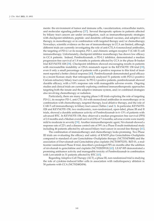
J. Clin. Med. 2021, 10, 4901
ments: the environment of tumor and immune cells, vascularization, extracellular matrix,and molecular signaling pathway [37]. Several therapeutic options in patients affectedby biliary tract cancers are under investigation, such as immunotherapeutic strategieswith checkpoint inhibitors, peptide- and dendritic cell-based vaccines, and adoptive celltherapy, in monotherapy or in combination with targeted therapy and/or chemotherapy.Nowadays, scientific evidence on the use of immunotherapy in CCA are limited, althoughdifferent trials are currently investigating the role of anti-CTLA-4 monoclonal antibodies,the targeting of PD-L1 or its receptor, PD-1, and chimeric antigen receptor T (CAR-T) cellimmunotherapy. Unfortunately, checkpoint inhibitor monotherapy has shown low efficacyin CCA patients. Indeed, Pembrolizumab, a PD-L1 inhibitor, demonstrated a medianprogression-free survival of 1.8 months in patients affected by CCA in the phase Ib baskettrial KEYNOTE 028 [38]. Checkpoint inhibitors showed encouraging results in patientswith microsatellite instability or DNA mismatch repair in the KEYNOTE 158 trial [39],even if only a small percentage of patients with a positive response to this kind of treat-ment reported a better clinical response [40]. Pembrolizumab demonstrated good efficacyin a recent Korean study that retrospectively analyzed 51 patients with PD-L1-positiveCisGem-refractory biliary tract cancer. In PD-L1-positive patients, pembrolizumab showeddurable efficacy, with a 9.8% response rate with manageable adverse events. Ongoingstudies and clinical trials are currently exploring combined immunotherapeutic approachestargeting both the innate and the adaptive immune system, and/or combined strategiesalso involving chemotherapy or radiation.
Particularly, there are many ongoing phase I–III trials exploring the role of targetingPD-L1, its receptor PD-1, anti CTL-A4 with monoclonal antibodies in monotherapy or incombination with chemotherapy, targeted therapy, local ablative therapy, and the role ofCAR-T cell immunotherapy in biliary tract cancer (Tables 2 and 3). In particular, KEYNOTE-028 and KEYNOTE-158, two multicentric, non-randomized, open-label, phase IB and IItrials, showed a durable antitumor activity of Pembrolizumab in 6–13% of patients withadvanced BTC. In KEYNOTE-158, they observed a median progression free survival (PFS)of 2.0 months and a Median overall survival (OS) of 7.4 months; adverse events were mainlymild to moderate in severity [39]. Another immunotherapeutic agent, Nivolumab showed aresponse rate of 22% and a disease control rate of 59% in a Phase II multi-institutional studyincluding 46 patients affected by advanced biliary tract cancer in second-line therapy [41].
The combination of immunotherapy and chemotherapy looks promising. Two PhaseIII trials are evaluating the efficacy and safety of KN035 plus Gemcitabine–Oxaliplatincompared to standard of care Gemcitabine–Oxaliplatin therapy (NCT03478488) and theassociation of Durvalumab and Gemcitabine plus cisplatin (NCT03875235). BilT-01, a mul-ticenter randomized Phase II trial, described a prolonged PFS six months after the additionof nivolumab to gemcitabine and cisplatin (NCT02829918) [42]. LEAP 005 demonstrated apromising antitumor activity and manageable toxicity of Pembrolizumab in combinationwith Lenvatinib in 31 patients affected by BTC [43].
Regarding Adoptive Cell Therapy (ACT), a phase III, non-randomized trial is studyingthe role of cytokine-induced killer cells in association with radiofrequency ablation in50 patients with CCA (NCT02482454).
141

J. Clin. Med. 2021, 10, 4901
Table 2. Ongoing immunotherapy trials of biliary tract cancers.
NCT Phase Condition or DiseaseNumber of
PatientsRegimen Status
ICI MONOTHERAPY
NCT03110328 II Advanced or refractory BTC 33 Pemrolizumab Recruiting
NCT02054806KEYNOTE-28 IB
Incurable advanced PD-L1positive cancers, including
BTC477 Pembrolizumab Completed
NCT02628067KEYNOTE-158 IIA Advanced, refractory solid
cancer including BTC 1595 Pemrolizumab Recruiting
NCT02829918 II Advanced refractory BTC 54 Nivolumab Active, notrecruiting
NCT03867370 IB-II Operable HCC o iCC 40 Toripalimab Recruiting
DUAL ICI
NCT03101566 II BTC 75 Nivolumab+ Ipilimumab Active, notrecruiting
ICI IN COMBINATION WITH CHEMOTHERAPY
NCT03473574 II Naïve BTC 128Durvalumab + tremelimumab
+ GEM or GEMCIS vs.GEMCIS chemotherapy
Active, notrecruiting
NCT03046862 II Unresectable, untreated BTC 31Durvalumab +
Tremelimumab + GEMCISchemotherapy
Recruiting
NCT03704480 II Advanced BTC 106 Durvalumab + tremelimumab+ paclitaxel Recruiting
NCT03875235 III Advanced BTC 757 Durvalumab + GEMCIS vsGEMCIS + chemotherapy Recruiting
NCT03257761 IbUnresecable, refractory HCC,
PDAC, BTC excludingampullary
90 Durvalumab + guadecitabine Recruiting
NCT03111732 II Unresecable, refractory BTC 11 Pemrolizumab + Oxaliplatine+ Capecitabine
Active, notrecruiting
NCT03260712 II Unresecable, untreated BTC 50 Pemrolizumab + GEMCIS Recruiting
NCT03796429 II Advanced BTC 40 Gemcitabine + Toripalimab Recruiting
NCT03101566 II Unresecable, untreatable BTC 75 Nivolumab + Ipilimumab vsGEMCIS + Nivolumab
Active, notrecruiting
NCT03785873 I/II Unresecable, refractory BTC 40 Nivolumab + nal-irinotecan +5-fluorouracil + leucovorin Recruiting
NCT03478488 III Unresecable, untreatable BTC 480 KN035 + GEMOX vs. GEMOX+ chemotherapy Recruiting
ICI IN COMBINATION WITH TARGETED THERAPY
NCT03797326 II Advanced, refractory solidtumours, including BTC 590 Lenvatinib + pembrolizumab Recruiting
NCT02393248 I/II Advanced solid tumourmalignancy, including CCA Pembrolizumab +pemigatinib Recruiting
142
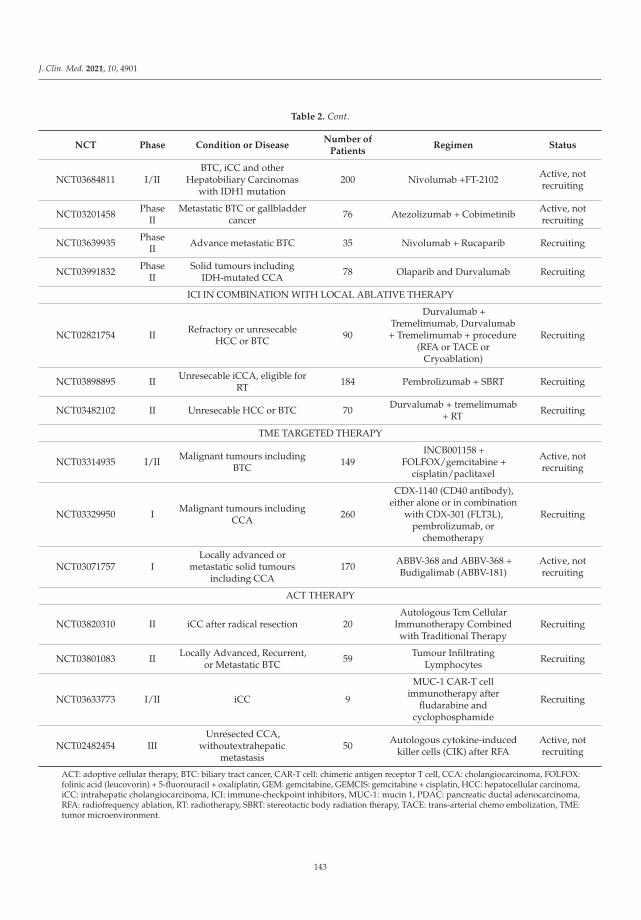
J. Clin. Med. 2021, 10, 4901
Table 2. Cont.
NCT Phase Condition or DiseaseNumber of
PatientsRegimen Status
NCT03684811 I/IIBTC, iCC and other
Hepatobiliary Carcinomaswith IDH1 mutation
200 Nivolumab +FT-2102 Active, notrecruiting
NCT03201458 PhaseII
Metastatic BTC or gallbladdercancer 76 Atezolizumab + Cobimetinib Active, not
recruiting
NCT03639935 PhaseII Advance metastatic BTC 35 Nivolumab + Rucaparib Recruiting
NCT03991832 PhaseII
Solid tumours includingIDH-mutated CCA 78 Olaparib and Durvalumab Recruiting
ICI IN COMBINATION WITH LOCAL ABLATIVE THERAPY
NCT02821754 II Refractory or unresecableHCC or BTC 90
Durvalumab +Tremelimumab, Durvalumab+ Tremelimumab + procedure
(RFA or TACE orCryoablation)
Recruiting
NCT03898895 II Unresecable iCCA, eligible forRT 184 Pembrolizumab + SBRT Recruiting
NCT03482102 II Unresecable HCC or BTC 70 Durvalumab + tremelimumab+ RT Recruiting
TME TARGETED THERAPY
NCT03314935 I/II Malignant tumours includingBTC 149
INCB001158 +FOLFOX/gemcitabine +
cisplatin/paclitaxel
Active, notrecruiting
NCT03329950 I Malignant tumours includingCCA 260
CDX-1140 (CD40 antibody),either alone or in combination
with CDX-301 (FLT3L),pembrolizumab, or
chemotherapy
Recruiting
NCT03071757 ILocally advanced or
metastatic solid tumoursincluding CCA
170 ABBV-368 and ABBV-368 +Budigalimab (ABBV-181)
Active, notrecruiting
ACT THERAPY
NCT03820310 II iCC after radical resection 20Autologous Tcm Cellular
Immunotherapy Combinedwith Traditional Therapy
Recruiting
NCT03801083 II Locally Advanced, Recurrent,or Metastatic BTC 59 Tumour Infiltrating
Lymphocytes Recruiting
NCT03633773 I/II iCC 9
MUC-1 CAR-T cellimmunotherapy after
fludarabine andcyclophosphamide
Recruiting
NCT02482454 IIIUnresected CCA,
withoutextrahepaticmetastasis
50 Autologous cytokine-inducedkiller cells (CIK) after RFA
Active, notrecruiting
ACT: adoptive cellular therapy, BTC: biliary tract cancer, CAR-T cell: chimeric antigen receptor T cell, CCA: cholangiocarcinoma, FOLFOX:folinic acid (leucovorin) + 5-fluorouracil + oxaliplatin, GEM: gemcitabine, GEMCIS: gemcitabine + cisplatin, HCC: hepatocellular carcinoma,iCC: intrahepatic cholangiocarcinoma, ICI: immune-checkpoint inhibitors, MUC-1: mucin 1, PDAC: pancreatic ductal adenocarcinoma,RFA: radiofrequency ablation, RT: radiotherapy, SBRT: stereotactic body radiation therapy, TACE: trans-arterial chemo embolization, TME:tumor microenvironment.
143
J. Clin. Med. 2021, 10, 4901
Table 3. Ongoing immunotherapy trials for BTC with preliminary results.
NCT Phase Condition or Disease N. Patients Regimen Results
NCT02054806KEYNOTE-28 IB
Incurable advancedPD-L1 positive cancers,
including BTC477 Pembrolizumab ORR: 13%
Median PFS: 2 months
NCT02628067KEYNOTE-158 IIA
Advanced, refractorysolid cancer including
BTC1595 Pemrolizumab ORR: 5.8%
Median PFS: 1.8 months
NCT02829918 II Advanced refractoryBTC 54 Nivolumab ORR: 22%
Median PFS: 3.8 monthd
NCT03797326 IIAdvanced, refractory
solid tumours, includingBTC
590 Lenvatinib +pembrolizumab ORR: 16%
4. Clinical-Pathological and Radiomic Monotherapy Susceptibility in Patients withCholangiocarcinoma
Within the CCA clinical-pathological spectrum, the pattern of tumor growth has beencorrelated with specific histological features, e.g., small-bile duct iCCAs and cholangiolocar-cinoma (CLC) showed a mass-forming growth pattern, while large-bile duct iCCAs showedboth a mass-forming growth pattern and a combination of a mass-forming growth patternwith a periductal infiltrative growth pattern, the latter being the typical pattern of growthof pCCA [44]. Mass-forming iCCAs showed more heterogeneous clinical-pathologicalcharacteristics than other gross types [45]. Radiologically, at dynamic contrast-enhancedimaging, all large-bile duct iCCAs showed concentric filling at the venous phase, whereassmall-bile duct iCCAs/CLCs showed washout in various patterns, in a clinical-pathologicalstudy including correlates with magnetic resonance imaging [44].
The USA Food and Drug Administration approved the use of pembrolizumab for pa-tients with advanced solid tumors lacking the expression of mismatch repair (MMR)proteins (MLH1, MSH2, MSH6, and PMS2) or having high microsatellite instability(MSI-H) [46]. MMR proteins can be inactivated through somatic or germline mutationsor they can be silenced through promoter hypermethylation, e.g., of the MLH1 gene [47].These alterations culminate to hypermutation during DNA replication (MSI) and maylead to the development of malignancies [48]. Interestingly, such molecular alterationspredispose to an increase of the neoantigen load of the tumor, promoting susceptibilityto immunotherapies targeting the PD-1 pathway because of the increased inflammationsurrounding these tumors [40].
Given the potential for immunotherapy in patients with CCA, authors studied theexpression of PD-L1/PD-1 and evaluated the presence of associated genetic alterations.For example, in 652 biliary tract cancers that comprised 77 p/dCCA, 372 iCCA, and 203gallbladder cancer (GBC), 8.6% tumors were PD-L1-positive [GBC 12.3% (25/203), iCCA7.3% (27/372), and p/dCCA 5.2% (4/77)]. Interestingly, there was an increase in BRAF,BRCA2, RNF43, and TP53 mutations in the PD-L1-positive group with respect to the PD-L1-negative one. Furthermore, there was an association between PD-L1 expression and certainbiomarkers (TOP2A, TMB high, MSI-H). As noted by the authors, the aforementioned com-binations of molecular alterations might direct the use of rational combination strategiesand clinical trial development [49]. On the same line, Ju et al. analyzed 96 cases of CCAfor morphology using H&E staining and for mutations of MMR genes using immunohis-tochemical staining. The authors found that 6% of the samples showed MMR deficiency(MMR-d). Divided by location, 10% (3 of 31) of iCCA and 5% (3 of 65) of p/dCCA wereMMR-d. The best predictive factor for MMR-d was a nontypical infiltrating pattern ofinvasion [50].
The increasing awareness of CCA heterogeneity at the morphological and molecularlevels, together with the advent of radiomic, artificial intelligence (AI), and machine
144
J. Clin. Med. 2021, 10, 4901
learning, has revitalized the study of radiological correlates. For example, it has been shownthat the magnetic resonance imaging texture signature, including three wavelets and one3D feature, has the ability to discriminate inflamed from non-inflamed immunophenotypesbased on the density of CD8+ T cells. This may be a surrogate of the response to immunecheckpoint blockade [51]. The preoperative prediction of PD-1/PD-L1 expression andoutcome in iCCA patients using magnetic resonance biomarkers and a machine learningapproach has been attempted [52]. Utilizing qualitative and quantitative imaging traits,reasonable accuracy in predicting tumor grade and higher AJCC stage in iCCA has beenshown [53].
5. Conclusions
The role of targeted therapy and immunotherapy in the treatment of CCA is currentlyunder investigation. These options might improve survival outcomes (OS and PFS), asshown by the promising results of several clinical trials illustrated in the present review.This is even more important considering the poor therapeutic options in the management ofCCA. The co-presence of driver mutations and markers of susceptibility to immunotherapymay lead to rational therapeutic combination strategies and clinical trial development.The combination of new therapeutic strategies, such as targeted therapy and immunother-apy, with conventional chemotherapy and/or locoregional treatments could be the nextfrontier for the treatment of advanced CCA. The evaluation of innovative strategies forthe prediction of immunotherapy susceptibility, such as multi omics, preferably withinlongitudinal clinical trials, and the use of systems of data analysis based on the preceptsof AI, may circumvent the lack of therapeutic biomarkers for immunotherapy. A betterunderstanding of immunological-based therapeutic weapons is needed, which will lead toa form of a precision medicine strategy capable of alleviating the clinical aggressivenessand to improve the prognosis of CCA.
Author Contributions: All authors: writing—review and editing. All authors have read and agreedto the published version of the manuscript.
Funding: This research received no external funding.
Conflicts of Interest: The authors declare no conflict of interest.
References
1. Banales, J.M.; Marin, J.J.G.; Lamarca, A.; Rodrigues, P.M.; Khan, S.A.; Roberts, L.R.; Cardinale, V.; Carpino, G.; Andersen, J.B.;Braconi, C.; et al. Cholangiocarcinoma 2020: The next horizon in mechanisms and management. Nat. Rev. Gastroenterol. Hepatol.2020, 17, 577–588. [CrossRef]
2. Khan, S.A.; Tavolari, S.; Brandi, G. Cholangiocarcinoma: Epidemiology and risk factors. Liver Int. 2019, 39, 19–31. [CrossRef]3. Shin, H.-R.; Oh, J.-K.; Masuyer, E.; Curado, M.-P.; Bouvard, V.; Fang, Y.; Wiangnon, S.; Sripa, B.; Hong, S.-T. Comparison of
incidence of intrahepatic and extrahepatic cholangiocarcinoma–focus on East and South-Eastern Asia. Asian Pac. J. Cancer Prev.2010, 11, 1159–1166.
4. Varadhachary, G.R.; Raber, M.N. Cancer of unknown primary site. N. Engl. J. Med. 2014, 371, 757–765. [CrossRef] [PubMed]5. Bertuccio, P.; Bosetti, C.; Levi, F.; Decarli, A.; Negri, E.; La Vecchia, C. A comparison of trends in mortality from primary liver
cancer and intrahepatic cholangiocarcinoma in Europe. Ann. Oncol. 2013, 24, 1667–1674. [CrossRef] [PubMed]6. Lamarca, A.; Barriuso, J.; McNamara, M.G.; Valle, J.W. Molecular targeted therapies: Ready for ‘prime time’ in biliary tract cancer.
J. Hepatol. 2020, 73, 170–185. [CrossRef]7. Valle, J.; Wasan, H.; Palmer, D.H.; Cunningham, D.; Anthoney, A.; Maraveyas, A.; Madhusudan, S.; Iveson, T.; Hughes, S.;
Pereira, S.P.; et al. Cisplatin plus Gemcitabine versus Gemcitabine for Biliary Tract Cancer. N. Engl. J. Med. 2010, 362, 1273–1281.[CrossRef] [PubMed]
8. Okusaka, T.; Nakachi, K.; Fukutomi, A.; Mizuno, N.; Ohkawa, S.; Funakoshi, A.; Nagino, M.; Kondo, S.; Nagaoka, S.; Funai, J.;et al. Gemcitabine alone or in combination with cisplatin in patients with biliary tract cancer: A comparative multicentre study inJapan. Br. J. Cancer 2010, 103, 469–474. [CrossRef]
9. Nault, J.; Villanueva, A. Biomarkers for Hepatobiliary Cancers. Hepatology 2021, 73, 115–127. [CrossRef]10. Reck, M.; Rodríguez-Abreu, D.; Robinson, A.G.; Hui, R.; Csoszi, T.; Fülöp, A.; Gottfried, M.; Peled, N.; Tafreshi, A.; Cuffe, S.; et al.
Pembrolizumab versus Chemotherapy for PD-L1–Positive Non–Small-Cell Lung Cancer. N. Engl. J. Med. 2016, 375, 1823–1833.[CrossRef] [PubMed]
145
J. Clin. Med. 2021, 10, 4901
11. Valle, J.W.; Lamarca, A.; Goyal, L.; Barriuso, J.; Zhu, A.X. REVIEW|New horizons for precision medicine in biliary tract cancers.Cancer Discov. 2017, 9, 943–962.
12. Bekaii-Saab, T.S.; Valle, J.W.; Van Cutsem, E.; Rimassa, L.; Furuse, J.; Ioka, T.; Melisi, D.; Macarulla, T.; Bridgewater, J.; Wasan,H.; et al. FIGHT-302: First-line pemigatinib vs gemcitabine plus cisplatin for advanced cholangiocarcinoma with FGFR2rearrangements. Futur. Oncol. 2020, 16, 2385–2399. [CrossRef]
13. Krook, M.A.; Bonneville, R.; Chen, H.-Z.; Reeser, J.W.; Wing, M.R.; Martin, D.M.; Smith, A.M.; Dao, T.; Samorodnitsky, E.;Paruchuri, A.; et al. Tumor heterogeneity and acquired drug resistance in FGFR2-fusion-positive cholangiocarcinoma throughrapid research autopsy. Mol. Case Stud. 2019, 5, a004002. [CrossRef] [PubMed]
14. Bahleda, R.; Italiano, A.; Hierro, C.; Mita, A.; Cervantes, A.; Chan, N.; Awad, M.; Calvo, E.; Moreno, V.; Govindan, R.; et al.Multicenter Phase I Study of Erdafitinib (JNJ-42756493), Oral Pan-Fibroblast Growth Factor Receptor Inhibitor, in Patients withAdvanced or Refractory Solid Tumors. Clin. Cancer Res. 2019, 25, 4888–4897. [CrossRef] [PubMed]
15. Hanahan, D.; Weinberg, R.A. Hallmarks of Cancer: The Next Generation. Cell 2011, 144, 646–674. [CrossRef] [PubMed]16. Nepal, C.; O’Rourke, C.J.; Oliveira, D.N.P.; Taranta, A.; Shema, S.; Gautam, P.; Calderaro, J.; Barbour, A.; Raggi, C.; Wennerberg,
K.; et al. Genomic perturbations reveal distinct regulatory networks in intrahepatic cholangiocarcinoma. Hepatology 2018, 68,949–963. [CrossRef] [PubMed]
17. Abou-Alfa, G.K.; Macarulla, T.; Javle, M.M.; Kelley, R.K.; Lubner, S.J.; Adeva, J.; Cleary, J.M.; Catenacci, D.V.; Borad, M.J.;Bridgewater, J.; et al. Ivosidenib in IDH1-mutant, chemotherapy-refractory cholangiocarcinoma (ClarIDHy): A multicentre,randomised, double-blind, placebo-controlled, phase 3 study. Lancet Oncol. 2020, 21, 796–807. [CrossRef]
18. Uribe, M.; Marrocco, I.; Yarden, Y. EGFR in Cancer: Signaling Mechanisms, Drugs, and Acquired Resistance. Cancers 2021, 13,2748. [CrossRef] [PubMed]
19. FDA. Approval Summary: Osimertinib for Adjuvant Treatment of Surgically Resected Non-Small Cell Lung Cancer, a Collabo-rative Project Orbis review|Clinical Cancer Research. 2021. Available online: https://clincancerres.aacrjournals.org/content/early/2021/07/22/1078-0432.CCR-21-1034 (accessed on 28 July 2021).
20. Xie, Y.-H.; Chen, Y.-X.; Fang, J.-Y. Comprehensive review of targeted therapy for colorectal cancer. Signal Transduct. Target. Ther.2020, 5, 22. [CrossRef]
21. Vogel, A.; Kasper, S.; Bitzer, M.; Block, A.; Sinn, M.; Schulze-Bergkamen, H.; Moehler, M.; Pfarr, N.; Endris, V.; Goeppert, B.; et al.PICCA study: Panitumumab in combination with cisplatin/gemcitabine chemotherapy in KRAS wild-type patients with biliarycancer—a randomised biomarker-driven clinical phase II AIO study. Eur. J. Cancer 2018, 92, 11–19. [CrossRef] [PubMed]
22. Leone, F.; Marino, D.; Cereda, S.; Filippi, R.; Belli, C.; Spadi, R.; Nasti, G.; Montano, M.; Amatu, A.; Aprile, G.; et al. Panitumumabin combination with gemcitabine and oxaliplatin does not prolong survival in wild-type KRAS advanced biliary tract cancer: Arandomized phase 2 trial (Vecti-BIL study). Cancer 2016, 122, 574–581.
23. Chiang, N.-J.; Hsu, C.; Chen, J.-S.; Tsou, H.-H.; Shen, Y.-Y.; Chao, Y.; Chen, M.-H.; Yeh, T.-S.; Shan, Y.-S.; Huang, S.-F.; et al.Expression levels of ROS1/ALK/c-MET and therapeutic efficacy of cetuximab plus chemotherapy in advanced biliary tractcancer. Sci. Rep. 2016, 6, 25369. [CrossRef]
24. Lee, J.; Park, S.H.; Chang, H.M.; Kim, J.S.; Choi, H.J.; Lee, M.A.; Jang, J.S.; Jeung, H.C.; Kang, J.H.; Lee, H.W.; et al. Gemcitabineand oxaliplatin with or without erlotinib in advanced biliary-tract cancer: A multicentre, open-label, randomised, phase 3 study.Lancet Oncol. 2012, 13, 181–188. [CrossRef]
25. Lubner, S.J.; Mahoney, M.R.; Kolesar, J.L.; Loconte, N.K.; Kim, G.P.; Pitot, H.C.; Philip, P.A.; Picus, J.; Yong, W.-P.; Horvath, L.;et al. Report of a multicenter phase II trial testing a combination of biweekly bevacizumab and daily erlotinib in patients withunresectable biliary cancer: A phase II Consortium study. J. Clin. Oncol. 2010, 28, 3491–3497. [CrossRef]
26. Jusakul, A.; Cutcutache, I.; Yong, C.H.; Lim, J.Q.; Ni Huang, M.; Padmanabhan, N.; Nellore, V.; Kongpetch, S.; Ng, A.W.T.; Ng,L.M.; et al. Whole-Genome and Epigenomic Landscapes of Etiologically Distinct Subtypes of Cholangiocarcinoma. Cancer Discov.2017, 7, 1116–1135. [CrossRef]
27. Jeong, H.; Jeong, J.H.; Kim, K.-P.; Lee, S.S.; Oh, D.W.; Park, D.H.; Song, T.J.; Park, Y.; Hong, S.-M.; Ryoo, B.-Y.; et al. Feasibility ofHER2-Targeted Therapy in Advanced Biliary Tract Cancer: A Prospective Pilot Study of Trastuzumab Biosimilar in Combinationwith Gemcitabine Plus Cisplatin. Cancers 2021, 13, 161. [CrossRef]
28. Iyer, R.V.; Pokuri, V.K.; Groman, A.; Ma, W.W.; Malhotra, U.; Iancu, D.M.; Grande, C.; Saab, T.B. A Multicenter Phase II Study ofGemcitabine, Capecitabine, and Bevacizumab for Locally Advanced or Metastatic Biliary Tract Cancer. Am. J. Clin. Oncol. 2018,41, 649–655. [CrossRef]
29. El-Khoueiry, A.B.; Rankin, C.J.; Ben-Josef, E.; Lenz, H.-J.; Gold, P.J.; Hamilton, R.D.; Govindarajan, R.; Eng, C.; Blanke, C.D. SWOG0514: A phase II study of sorafenib in patients with unresectable or metastatic gallbladder carcinoma and cholangiocarcinoma.Investig. New Drugs 2012, 30, 1646–1651. [CrossRef]
30. Lee, J.K.; Capanu, M.; O’Reilly, E.M.; Ma, J.; Chou, J.F.; Shia, J.; Katz, S.; Gansukh, B.; Reidylagunes, D.; Segal, N.H.; et al. A phaseII study of gemcitabine and cisplatin plus sorafenib in patients with advanced biliary adenocarcinomas. Br. J. Cancer 2013, 109,915–919. [CrossRef] [PubMed]
31. Sun, W.; Patel, A.; Normolle, D.; Patel, K.; Ohr, J.; Lee, J.J.; Bahary, N.; Chu, E.; Streeter, N.; Drummond, S. A phase 2 trial ofregorafenib as a single agent in patients with chemotherapy—refractory, advanced, and metastatic biliary tract adenocarcinoma.Cancer 2019, 125, 902–909. [CrossRef]
146
J. Clin. Med. 2021, 10, 4901
32. Kam, A.E.; Masood, A.; Shroff, R.T. Current and emerging therapies for advanced biliary tract cancers. Lancet Gastroenterol.Hepatol. 2021, 6, 956–969. [CrossRef]
33. Doebele, R.C.; Drilon, A.; Paz-Ares, L.; Siena, S.; Shaw, A.T.; Farago, A.F.; Blakely, C.M.; Seto, T.; Cho, B.C.; Tosi, D.; et al.Entrectinib in patients with advanced or metastatic NTRK fusion-positive solid tumours: Integrated analysis of three phase 1–2trials. Lancet Oncol. 2020, 21, 271–282. [CrossRef]
34. Drilon, A.; Laetsch, T.W.; Kummar, S.; DuBois, S.G.; Lassen, U.N.; Demetri, G.D.; Nathenson, M.; Doebele, R.C.; Farago, A.F.;Pappo, A.S.; et al. Efficacy of Larotrectinib in TRK Fusion-Positive Cancers in Adults and Children. N. Engl. J. Med. 2018, 378,731–739. [CrossRef]
35. Boilève, A.; Verlingue, L.; Hollebecque, A.; Boige, V.; Ducreux, M.; Malka, D. Rare cancer, rare alteration: The case of NTRKfusions in biliary tract cancers. Expert Opin. Investig. Drugs 2021, 30, 401–409. [CrossRef]
36. Hellmann, M.D.; Friedman, C.F.; Wolchok, J.D. Combinatorial Cancer Immunotherapies. Adv. Immunol. 2016, 130, 251–277.[CrossRef] [PubMed]
37. Carpino, G.; Cardinale, V.; Renzi, A.; Hov, J.R.; Berloco, P.B.; Rossi, M.; Karlsen, T.H.; Alvaro, D.; Gaudio, E. Activation of biliarytree stem cells within peribiliary glands in primary sclerosing cholangitis. J. Hepatol. 2015, 63, 1220–1228. [CrossRef] [PubMed]
38. Piha-Paul, S.A.; Oh, D.; Ueno, M.; Malka, D.; Chung, H.C.; Nagrial, A.; Kelley, R.K.; Ros, W.; Italiano, A.; Nakagawa, K.;et al. Efficacy and safety of pembrolizumab for the treatment of advanced biliary cancer: Results from the KEYNOTE -158 andKEYNOTE -028 studies. Int. J. Cancer 2020, 147, 2190–2198. [CrossRef]
39. Marabelle, A.; Le, D.T.; Ascierto, P.A.; Di Giacomo, A.M.; De Jesus-Acosta, A.; Delord, J.-P.; Geva, R.; Gottfried, M.; Penel,N.; Hansen, A.R.; et al. Efficacy of Pembrolizumab in Patients With Noncolorectal High Microsatellite Instability/MismatchRepair–Deficient Cancer: Results From the Phase II KEYNOTE-158 Study. J. Clin. Oncol. 2020, 38, 1–10. [CrossRef]
40. Le, D.T.; Uram, J.N.; Wang, H.; Bartlett, B.R.; Kemberling, H.; Eyring, A.D.; Skora, A.D.; Luber, B.S.; Azad, N.S.; Laheru, D.; et al.PD-1 Blockade in Tumors with Mismatch-Repair Deficiency. N. Engl. J. Med. 2015, 372, 2509–2520. [CrossRef] [PubMed]
41. A Phase 2 Multi-Institutional Study of Nivolumab for Patients with Advanced Refractory Biliary Tract Cancer|Gastroenterology|JAMA Oncology|JAMA Network. 2021. Available online: https://jamanetwork.com/journals/jamaoncology/fullarticle/2765293 (accessed on 28 July 2021).
42. Sahai, V.; Griffith, K.A.; Beg, M.S.; Shaib, W.L.; Mahalingam, D.; Zhen, D.B.; Deming, D.A.; Dey, S.; Mendiratta-Lala, M.; Zalupski,M. A multicenter randomized phase II study of nivolumab in combination with gemcitabine/cisplatin or ipilimumab as first-linetherapy for patients with advanced unresectable biliary tract cancer (BilT-01). J. Clin. Oncol. 2020, 38, 4582. [CrossRef]
43. Lwin, Z.; Gomez-Roca, C.; Saada-Bouzid, E.; Yanez, E.; Muñoz, F.L.; Im, S.-A.; Castanon, E.; Senellart, H.; Graham, D.; Voss, M.;et al. LBA41 LEAP-005: Phase II study of lenvatinib (len) plus pembrolizumab (pembro) in patients (pts) with previously treatedadvanced solid tumours. Ann. Oncol. 2020, 31, S1170. [CrossRef]
44. Komuta, M.; Govaere, O.; Vandecaveye, V.; Akiba, J.; Van Steenbergen, W.; Verslype, C.; Laleman, W.; Pirenne, J.; Aerts, R.; Yano,H.; et al. Histological diversity in cholangiocellular carcinoma reflects the different cholangiocyte phenotypes. Hepatology 2012,55, 1876–1888. [CrossRef]
45. Chung, T.; Rhee, H.; Nahm, J.H.; Jeon, Y.; Yoo, J.E.; Kim, Y.-J.; Han, D.H.; Park, Y.N. Clinicopathological characteristics ofintrahepatic cholangiocarcinoma according to gross morphologic type: Cholangiolocellular differentiation traits and inflammation-and proliferation-phenotypes. HPB 2020, 22, 864–873. [CrossRef]
46. Boyiadzis, M.M.; Kirkwood, J.M.; Marshall, J.L.; Pritchard, C.C.; Azad, N.S.; Gulley, J.L. Significance and implications of FDAapproval of pembrolizumab for biomarker-defined disease. J. Immunother. Cancer 2018, 6, 35. [CrossRef] [PubMed]
47. Gong, J.; Chehrazi-Raffle, A.; Reddi, S.; Salgia, R. Development of PD-1 and PD-L1 inhibitors as a form of cancer immunotherapy:A comprehensive review of registration trials and future considerations. J. Immunother. Cancer 2018, 6, 8. [CrossRef] [PubMed]
48. Tiwari, A.; Roy, H.; Lynch, H. Lynch syndrome in the 21st century: Clinical perspectives. Qjm Int. J. Med. 2016, 109, 151–158.[CrossRef]
49. Patterns and Genomic Correlates of PD-L1 Expression in Patients with Biliary Tract Cancers—Mody. J. Gastrointest. Oncol. 2021.Available online: https://jgo.amegroups.com/article/view/31932/html (accessed on 28 July 2021).
50. Ju, J.Y.; Dibbern, M.E.; Mahadevan, M.S.; Fan, J.; Kunk, P.R.; Stelow, E.B. Mismatch Repair Protein Deficiency/MicrosatelliteInstability Is Rare in Cholangiocarcinomas and Associated With Distinctive Morphologies. Am. J. Clin. Pathol. 2020, 153, 598–604.[CrossRef] [PubMed]
51. Zhang, J.; Wu, Z.; Zhao, J.; Liu, S.; Zhang, X.; Yuan, F.; Shi, Y.; Song, B. Intrahepatic cholangiocarcinoma: MRI texture signature aspredictive biomarkers of immunophenotyping and survival. Eur. Radiol. 2020, 31, 3661–3672. [CrossRef] [PubMed]
52. Rhee, H.; Kim, M.-J.; Park, Y.N.; An, C. A proposal of imaging classification of intrahepatic mass-forming cholangiocarcinomainto ductal and parenchymal types: Clinicopathologic significance. Eur. Radiol. 2018, 29, 3111–3121. [CrossRef]
53. Yoo, J.; Kim, J.H.; Bae, J.S.; Kang, H.-J. Prediction of prognosis and resectability using MR imaging, clinical, and histopathologicalfindings in patients with perihilar cholangiocarcinoma. Abdom. Radiol. 2021, 46, 4159–4169. [CrossRef] [PubMed]
147

Journal of
Clinical Medicine
Review
Treatment of Hepatocellular Carcinoma with ImmuneCheckpoint Inhibitors and Applicability of First-LineAtezolizumab/Bevacizumab in a Real-Life Setting
Maria Corina Plaz Torres 1, Quirino Lai 2, Fabio Piscaglia 3, Eugenio Caturelli 4, Giuseppe Cabibbo 5,
Elisabetta Biasini 6, Filippo Pelizzaro 7, Fabio Marra 8, Franco Trevisani 9 and Edoardo G. Giannini 1,*
Citation: Plaz Torres, M.C.; Lai, Q.;
Piscaglia, F.; Caturelli, E.; Cabibbo, G.;
Biasini, E.; Pelizzaro, F.; Marra, F.;
Trevisani, F.; Giannini, E.G. Treatment
of Hepatocellular Carcinoma with
Immune Checkpoint Inhibitors and
Applicability of First-Line
Atezolizumab/Bevacizumab in a
Real-Life Setting. J. Clin. Med. 2021,
10, 3201. https://doi.org/10.3390/
jcm10153201
Academic Editors: Pierluigi Toniutto
and Hidekazu Suzuki
Received: 1 June 2021
Accepted: 19 July 2021
Published: 21 July 2021
Publisher’s Note: MDPI stays neutral
with regard to jurisdictional claims in
published maps and institutional affil-
iations.
Copyright: © 2021 by the authors.
Licensee MDPI, Basel, Switzerland.
This article is an open access article
distributed under the terms and
conditions of the Creative Commons
Attribution (CC BY) license (https://
creativecommons.org/licenses/by/
4.0/).
1 Gastroenterology Unit, Department of Internal Medicine, IRCCS—Ospedale Policlinico San Martino,University of Genoa, 16132 Genoa, Italy; [email protected]
2 Hepatobiliary and Organ Transplantation Unit, Umberto I Polyclinic of Rome, Sapienza University of Rome,00185 Rome, Italy; [email protected]
3 Internal Medicine Unit, Department of Medical and Surgical Sciences,IRCCS Azienda Ospedaliero—Universitaria di Bologna, 40138 Bologna, Italy; [email protected]
4 Gastroenterology Unit, Belcolle Hospital, 01100 Viterbo, Italy; [email protected] Gastroenterology and Hepatology Unit, Department of Health Promotion, Mother and Child Care,
Internal Medicine and Medical Specialties (PROMISE), University of Palermo, 90133 Palermo, Italy;[email protected]
6 Infectious Diseases and Hepatology Unit, Azienda Ospedaliero—Universitaria di Parma, 43126 Parma, Italy;[email protected]
7 Gastroenterology Unit, Department of Surgery, Oncology and Gastroenterology, University of Padova,35128 Padova, Italy; [email protected]
8 Internal Medicine and Hepatology Unit, Department of Experimental and Clinical Medicine,University of Firenze, 50134 Firenze, Italy; [email protected]
9 Medical Semeiotics Unit, Department of Medical and Surgical Sciences,IRCCS Azienda Ospedaliero—Universitaria di Bologna, 40138 Bologna, Italy; [email protected]
* Correspondence: [email protected]; Tel.: +39-010-353-7950; Fax: +39-010-353-8638
Abstract: Immune checkpoint inhibitors (ICIs) are the new frontier for the treatment of advancedhepatocellular carcinoma (HCC). Since the first trial with tremelimumab, a cytotoxic T-lymphocyte-associated protein 4 inhibitor, increasing evidence has confirmed that these drugs can significantlyextend the survival of patients with advanced hepatocellular carcinoma (HCC). As a matter of fact,the overall survival and objective response rates reported in patients with advanced HCC treatedwith ICIs are the highest ever reported in the second-line setting and, most recently, the combinationof the anti-programmed death ligand protein-1 atezolizumab with bevacizumab—an anti-vascularendothelial growth factor monoclonal antibody—demonstrated superiority to sorafenib in a Phase IIIrandomized clinical trial. Therefore, this regimen has been approved in several countries as first-linetreatment for advanced HCC and is soon expected to be widely used in clinical practice. However,despite the promising results of trials exploring ICIs alone or in combination with other agents, thereare still some critical issues to deal with to optimize the prognosis of advanced HCC patients. Forinstance, the actual proportion of patients who are deemed eligible for ICIs in the real-life rangesfrom 10% to 20% in the first-line setting, and is even lower in the second-line scenario. Moreover,long-term data regarding the safety of ICIs in the population of patients with cirrhosis and impairedliver function are lacking. Lastly, no biomarkers have been identified to predict response, and thusto help clinicians to individually tailor treatment. This review aimed to summarize the state of theart immunotherapy in HCC and, by analyzing a large, multicenter cohort of Italian patients withHCC, to assess the potential applicability of the combination of atezolizumab/bevacizumab in thereal-life setting.
Keywords: liver cancer; systemic treatment; immunotherapy; real-world; unresectable hepatocel-lular carcinoma
J. Clin. Med. 2021, 10, 3201. https://doi.org/10.3390/jcm10153201 https://www.mdpi.com/journal/jcm
149

J. Clin. Med. 2021, 10, 3201
1. Introduction
Hepatocellular carcinoma (HCC) is one of the leading causes of cancer-related mor-tality worldwide, with approximately 800,000 deaths per year and an estimated increaseto more than 1 million deaths by 2030 [1]. HCC arises predominantly in the context ofliver cirrhosis, but also can be diagnosed in a not negligible proportion of patients withoutcirrhosis suffering from non-alcoholic steato-hepatitis who carry additional metabolic andgenetic risk factors [2–5]. In the past decades, the armamentarium for the systemic treatmentof advanced HCC was limited to the anti-vascular endothelial growth factor (VEGFR),multi-target-tyrosine kinase inhibitor (TKI) sorafenib. This drug determined a significant—though modest—survival benefit in two Phase III trials and remained the sole first-linetreatment option for about 10 years, during which neither an alternative drug nor effectivesecond-line therapies became available for patients who progressed during—or were intol-erant to—sorafenib [6,7]. As a fact, lenvatinib (a TKI targeting VEGFR) became an effectivealternative to sorafenib as first-line therapy for HCC in 2018, while regorafenib, cabozan-tinib, and ramucirumab only recently have been approved in the second-line setting [8].With the advent of second-line treatments, the survival of patients with advanced HCChas significantly improved, with a proportion (approximately 20%) of patients reachingsurvival times of about 2 years with the sequential use of sorafenib-regorafenib [9]. Thesepatients, however, belong to a small subgroup of patients who, maintaining an optimalliver function, are eligible for sequential treatment and tolerate the adverse effects of theanti-neoplastic agents [9].
In this scenario, immunotherapy has emerged as an additional promising approachpotentially able to obtain even longer survival times. Research in this field is steadilyincreasing, also fueled by the positive results obtained in other cancer types and by theevidence of efficacy demonstrated in both first- and second-line settings [10–12]. Themost recent Phase I/II trials have shown a clinically meaningful survival increase in thesecond-line setting for the programmed cell death protein 1 (PD-1) inhibitors nivolumaband pembrolizumab [12]. Hence, these agents have been granted accelerated conditionalapproval for sorafenib-experienced patients in the US, while the European MedicinesAgency (EMA) maintains a more cautious attitude in approving these ICIs for the treatmentof HCC. Indeed, subsequent Phase III trials testing nivolumab versus sorafenib as first-line treatment, and pembrolizumab versus placebo in second-line treatment, failed tomeet their primary survival endpoints [13,14]. This notwithstanding, the results fromtrials testing the combination of immune checkpoint inhibitors (ICIs) with other agents,among which VEGFR-targeted therapies obtained very encouraging results, so that thecombinations of pembrolizumab plus lenvatinib as well as atezolizumab (monoclonalantibody against PD-L1) plus bevacizumab (monoclonal antibody against VEGF) have bothreceived breakthrough therapy designation from the US Food and Drug Administration(FDA). Actually, in a recent Phase III trial, the latter overperformed compared to sorafenibas first-line treatment of advanced HCC in terms of both overall (OS) and progression-freesurvival (PFS) [15]. Therefore, atezolizumab plus bevacizumab has been approved asthe first-line treatment option for advanced HCC, thus becoming the standard of care forthese patients.
Overall, the results from the trials testing ICIs alone or in combination, or combinedwith other agents, suggest that ICIs alone are not the best option for the treatment ofHCC, while combined treatments are safe and highly effective. As such, immunotherapy-based treatments will probably soon change the landscape of advanced HCC therapy. Inthis review, we summarize the state of the art immunotherapy in advanced HCC, with aparticular focus on the combination of atezolizumab plus bevacizumab, by assessing in alarge cohort of Italian patients with HCC the potential applicability of this regimen to thereal-life setting.
150

J. Clin. Med. 2021, 10, 3201
2. Approved Treatments for HCC before the “Era” of Immune Checkpoint Inhibitors
Until the approval of sorafenib in 2008, no systemic treatment was available foradvanced HCC [6]. Sorafenib, an orally active multi-target TKI targeting different cellsurface tyrosine kinases (e.g., VEGFR-1, -2, and -3 and platelet-derived growth factor(PDGFR)-β), at the dose of 400 mg twice daily, significantly improved OS in patientswith HCC not amenable to surgery and locoregional procedures, who had well-preservedliver function (97% Child–Pugh A) and Eastern Cooperative Oncology Group (ECOG)performance status (PS) ≤ 2 [6]. The median OS was 10.7 months in the sorafenib groupand 7.9 months in the placebo group (p < 0.001), whereas the median time to radiologicprogression (TTP) was 5.5 months in the sorafenib arm versus 2.8 months in the placeboarm (p < 0.001). Of note, the median OS of patients with the Barcelona Clinic LiverCancer (BCLC) staging system stage B HCC treated with sorafenib was 15–20 months, afinding confirmed by subsequent post-marketing studies [16,17]. In the following years,several drugs were tested against sorafenib in the first-line setting, failing to demonstratesuperiority to this drug, so that sorafenib remained the sole effective systemic treatmentavailable for HCC until 2018, when lenvatinib, an oral TKI with a biologic action similar tosorafenib, showed non-inferior OS as compared to sorafenib in the REFLECT trial, and wastherefore approved as an alternative to this drug in the first-line setting [8]. Again, patientsincluded in this trial belonged to a selected group of subjects with well-preserved liverfunction (Child–Pugh class A) and ECOG PS ≤ 1, while those with extensive tumor burden(≥50% of the liver), bile duct invasion, or invasion of the main portal vein were excluded.Forest plots for OS revealed that lenvatinib was more effective than sorafenib in patientswith baseline AFP ≥ 200 ng/mL (Hazard ratio (HR), 0.78; 95% confidence interval (95%CI),0.63–0.98) and less effective in patients without macrovascular invasion/extrahepaticspread and those enrolled in the Western area. Secondary endpoints (PFS, TTP, objectiveresponse rate (ORR)) were significantly and remarkably better with lenvatinib, suggestingthat these surrogate endpoints poorly predict OS in HCC patients treated with these drugs.
As far as the second-line setting is concerned, regorafenib, an oral TKI targetingVEGFR-2, VEGFR-3, TIE-2, PDGFR, fibroblast growth factor receptor (FGFR)-1, and themutant oncogenic kinases KIT, RET, and B-RAF, was the first agent able to provide a signif-icant survival benefit in patients with tumor progression on sorafenib [18]. Compared withplacebo, regorafenib improved OS with a HR of 0.63 (95%CI, 0.50–0.79; p < 0.0001). It has tobe emphasized that this study enrolled patients who progressed on sorafenib but toleratedthe drug (≥400 mg/day for ≥20 of last 28 days of treatment) and had Child–Pugh classA liver function. Median survival was 10.6 months (95%CI, 9.1–12.1) for the regorafenibgroup versus 7.8 months (95%CI, 6.3–8.8) for the placebo group [18]. Interestingly, thetreatment sequence of the sorafenib-regorafenib group was able to determine an OS of26 months from the start of sorafenib treatment versus 19.2 months in the sorafenib-placebogroup [18]. This survival time is comparable with that of patients with intermediate stageHCC undergoing trans-arterial chemo-embolization (TACE), suggesting that in a well-selected subgroup of patients the sequential treatment with TKIs may significantly improveprognosis as compared to the standard of care [9].
Other drugs that have shown efficacy in placebo-controlled trials and have conse-quently been approved as second-line treatment options for HCC are cabozantinib andramucirumab [19]. Cabozantinib is an oral TKI targeting MET in addition to VEGFR2. TheCELESTIAL trial was a global Phase III trial testing cabozantinib in patients with HCCprogression on sorafenib [20]. It also included patients who had received up to two priortherapies for advanced-stage HCC. The study was stopped after a second interim analysis,which revealed a median OS of 10.2 months in the cabozantinib versus 8.0 months in theplacebo group (HR, 0.76; 95% CI 0.63–0.92; p = 0.0049). Approximately 72% of patients hadreceived only prior sorafenib treatment and, in this subpopulation, median OS was evenlonger, being 11.3 months in patients in the cabozantinib group versus 7.2 months in theplacebo group (HR, 0.70; 95% CI, 0.55–0.88) [20].
151
J. Clin. Med. 2021, 10, 3201
Ramucirumab is an anti-VEGFR2 monoclonal antibody, and its utility in subjectswith advanced HCC emerged from the double-blind, Phase III REACH-2 trial comparingramucirumab versus placebo as second-line treatment in patients progressing on sorafeniband with baseline AFP ≥ 400 ng/mL [19]. This study was designed on the basis ofthe results of the REACH trial that failed to demonstrate an OS advantage with ramu-cirumab as compared to the placebo, but in a post-hoc analysis showed a benefit of thedrug—albeit small—in prolonging OS (8.5 months with ramucirumab versus 7.3 monthswith placebo (HR, 0.71; 95% CI, 0.53–0.95; p = 0.0199)) among patients with baselineAFP ≥ 400 ng/mL [21]. Ramucirumab is therefore the first agent with a biomarker-drivenuse for patients with HCC progression on sorafenib [22].
In summary, sorafenib and lenvatinib are the TKIs that have long been in use for thefront-line treatment of advanced HCC, providing a median extension of survival of about3 months compared to the placebo. The survival benefit for patients eligible for second-line treatment with regorafenib/cabozantinib or ramucirumab, although significant, stillremains modest. Hence, novel treatments targeting different tumorigenic pathways havebeen studied and others are still under investigation with the aim of further improving theoutcomes of these patients. In this context, ICIs have gained excellent results.
3. The Advent of Immune Checkpoint Inhibitors
Despite the benefit in OS with sequential TKI treatment, the prognosis of patients withadvanced HCC remains poor [9,23]. The reasons for this include, besides the sub-optimaltumoricidal activity of these drugs, the progression of the underlying liver disease, theadvanced median age of this cohort of patients (approximately 70 years), and the presenceof substantial comorbidities, which are very frequent in these subjects and, overall, makethem a particularly vulnerable cohort [24].
In this context, ICIs have increasingly been investigated in the last years, with ex-tremely encouraging results both in the first- and second-line setting, further boosting arising number of clinical trials using ICIs alone or combined with other anti-tumoral drugsor with locoregional treatment. The rationale for the use of ICIs in HCC relies on the fact thatHCC arises in a context of chronic inflammation and an altered tumor microenvironment,with the presence of tumor-infiltrating lymphocytes expressing PD1, which is a recognizedkey enabling factor beyond tumor cell-intrinsic molecular aberrations [25–27]. Moreover,the presence of PD1-expressing lymphocytes in HCC samples has been correlated with thisoutcome [28,29]. In this regard, Sia et al. have recently proposed a novel HCC classificationbased upon the tumor immune status: according to this classification, about 30% of HCCscould be categorized into an ‘immune class’, with high levels of immune cell infiltration,expression of PD-1 and/or PD-L1, activation of interferon-γ signaling, and markers ofcytolytic activity [30]. Within this class, two distinct subclasses have been identified: the‘active immune’ and the ‘exhausted immune’ classes, characterized by markers of an adap-tive T-cell response or of an exhausted immune response, respectively [30]. The lattersubclass is the ideal target of immunotherapy. The in-depth description of the molecularmechanisms involved in the tumor microenvironment of HCC is beyond the aim of thisarticle, but it is worth pointing out that interactions between cancer cell antigens and theantigen-presenting cells lead to a priming of T-cells and their eventual migration into thetumor microenvironment. Physiologically, the T-lymphocytes’ recognition of neoplasticantigens is followed by a T-cell-mediated killing of cancer cells [31]. This process is finelymodulated at a local and general level by several mechanisms including immune check-points, which play a pivotal role in such modulation, as they suppress T-cell activity toinhibit eventual over-activation of the immune system and maintain self-tolerance. Thus,immune checkpoints physiologically prevent hyperimmune responses leading to tissuedamage. Malignancies exploit these molecular mechanisms (immune checkpoints) toescape from the immune system recognition. In other words, ICIs act as anti-neoplasticagents by inhibiting negative feedback pathways of the immune system that mediateimmune escape.
152

J. Clin. Med. 2021, 10, 3201
The most largely studied immune checkpoints are PD-1 and cytotoxic T-lymphocyte-associated protein 4 (CTLA-4). The pathological activation of PD-1 by its ligands, in partic-ular PD-L1, expressed by cancer cells, can result in the immune escape of the tumor [32,33].CTLA-4, which is mainly expressed on T-cells, regulates T-cell activity in physiologicalconditions, preventing an excess in T-cell responses and a hyperactivation of the immuneresponse. Inversely, in pathological (neoplastic) conditions, CTLA-4 activation inhibits inthe activation, proliferation, and production of tumor antigen-activated T-cells in the tumormicroenvironment [32,33]. In the HCC tumor microenvironment, T-regulators (T-regs)express both CTLA-4 and PD-1 [28,32].
4. Immune Checkpoint Inhibitors in HCC
Tremelimumab, a CTLA-4-blocking monoclonal antibody, was the first ICI showingbenefits in the treatment of HCC. This agent was tested in 2013 by Sangro et al. in a PhaseII open-label trial that enrolled 21 patients with advanced HCC who were either sorafenib-naïve (76.2%) or -experienced, and a significant proportion of them were classified asChild–Pugh class B (43%) [10]. The positive results in terms of both safety and anti-tumor activity (partial response rate (PRR) 17.6%; disease control rate (DCR) 76.4%; TTP6.48 months (95%CI, 3.95–9.14)), were instrumental in stimulating the research in immunecheckpoint blockade in both first- and second-line treatment of HCC. In the last years,the effects of ICIs in HCC have been tested alone or in combination with other ICIs orcombined with agents targeting the VEGFR. Currently available immunotherapy-basedregimens and those under Phase III clinical investigation are summarized in Figure 1.
Figure 1. Possible HCC treatments with ICIs. Atezolizumab plus bevacizumab has been approved as a first-line treatment,whereas nivolumab with or without ipilimumab and pembrolizumab gained FDA approval as second-line treatments.Selected Phase III trials (orange squares) are testing ICIs alone or in combination or combined with other agents in the firstand second-line setting, and in the adjuvant and neo-adjuvant setting as well.
4.1. Immune Checkpoint Inhibitors in Monotherapy
Following the encouraging results of the Phase II tremelimumab study, nivolumab,a monoclonal antibody targeting PD1, demonstrated a single-agent activity in the PhaseIb/II open-label, non-comparative, Checkmate 040 trial [11]. The initial trial included262 sorafenib-naïve and -experienced patients assigned to a dose-escalation (48 subjects) orto a dose-expansion (214 subjects) phase. In the dose-expansion phase, the investigator-
153
J. Clin. Med. 2021, 10, 3201
assessed overall ORR was 20%, with 3 complete responses (CR) and 39 partial responses(PR). Particularly, ORR was 22.5% for sorafenib-naive and 18.7% for sorafenib-experiencedpatients. Median OS was 29 months for sorafenib-naïve group and 15 months for thesorafenib-experienced group. The most impressive was the duration of response of9.9 months amongst patients who had an objective response, which led the US FDAto grant accelerated approval to nivolumab as second-line therapy for patients withadvanced-stage HCC previously treated with sorafenib [11]. In this subgroup, the ORRconfirmed by blinded independent central review was 14.3% by Response EvaluationCriteria In Solid Tumors (RECIST) 1.1 and 18.2% by modified RECIST (mRECIST) criteria.Of note, the median duration of response was the longest ever seen in a second-line setting:16.6 months [34,35]. However, the expectations raised by the results of this study were dis-appointed in a subsequent Phase III randomized trial (CheckMate-459) testing nivolumabversus sorafenib, as the anti-PD1 agent failed to demonstrate superiority as comparedto the TKI [14]. Still, the study results confirmed clinically meaningful improvementsin OS (16.4 versus 14.7 months), ORR (15% for nivolumab versus 7% for sorafenib), andCR (14 versus 5 patients). Moreover, nivolumab demonstrated a favorable safety profile,consistent with previous reports and, of particular interest, the quality of life was better inthe nivolumab treatment arm [14]. The long survival of the sorafenib arm (median OS ofabout 15 months) was an unexpected outcome that negatively impacted the study resultsand that probably reflects the improved tailored management of patients with advancedHCC in the last decades, as well as physicians’ familiarity with the TKI.
Another ICI that has been tested with favorable outcomes in monotherapy for advancedHCC is pembrolizumab, a monoclonal antibody targeting PD-1. Promising results came fromthe Phase II trial KEYNOTE 224, which showed good responses (ORR 17%, DCR 61%) and agood safety profile of pembrolizumab in patients who were intolerant to, or progressedunder, sorafenib [12]. These results prompted Finn et al. to conduct the KEYNOTE-240 trialenrolling 413 patients who failed sorafenib and who were randomized 2:1 to pembrolizumabor placebo [13]. The survival in the pembrolizumab arm was among the highest everreached in the second-line setting, being approximately 14 months (95%CI, 11.6–16.0)for pembrolizumab versus 10 months (95%CI, 8.3–13.5) for placebo (HR, 0.781; 95%CI,0.611–0.998; p = 0.0238). Nevertheless, even this study failed to reach statistical significancedue to the long survival of the control arm, reflecting once more the advances in the clinicalmanagement of advanced HCC. The safety profile of the drug was good, confirming thepositive results of the Phase II study and the previous experience with nivolumab.
Despite the apparently “negative” results of these studies, likely due to issues relatedto their design requesting an overwhelming superiority of the tested ICIs over sorafenib,several positive aspects capturing the attention of researchers and clinicians were theoverall objective response to nivolumab and pembrolizumab in 15–20% of cases, the durableantitumor responses, and the long-term OS in responding patients. Based on these peculiarresults, the FDA granted conditional approval for these ICIs in the second-line setting.
Currently, results from the ongoing Phase III non-inferiority trial testing tislelizumab,a monoclonal antibody targeting PD-1, versus sorafenib (RATIONALE-301 trial) andthose of the Phase III HIMALAYA study, testing durvalumab—an anti-PD-L1 monoclonalantibody—alone or in combination with tremelimumab versus sorafenib, are eagerlyawaited [36].
As far as the safety profile of ICIs is concerned, the results of the pilot study by Sangroet al. on tremelimumab and those of the CheckMate and Keynote trials showed reassuringsafety profiles for these agents, coherent with previous reports testing the use of these drugsin other cancer types [10–12,37]. As compared with the standard of care (i.e., sorafenib andlenvatinib), ICIs are generally better tolerated and have comparable or even lower rates oftoxicity. The pathophysiology of adverse events (AEs) occurring during immunotherapyis related to their mechanism of action as the inhibition of physiological immune check-points may trigger immune-related AEs (irAEs) targeting the skin, gut, thyroid, adrenalglands, lung, and the liver itself, which may be a particularly worrisome complication in
154
J. Clin. Med. 2021, 10, 3201
a population with an already impaired liver function [38,39]. Most frequent any gradeAEs in patients treated with ICIs for other cancer types are skin AEs (rash and pruritus),colitis, hyper- or hypothyroidism, hepatitis, and pneumonitis. Skin AEs occur in about13–35% of cases, being grade > 3 only in a minority of cases (<3%) [38,40]. Grade 1 and2 skin AEs are usually easily managed with emollients, oral anti-histamines, and topicalsteroids, whereas grade ≥ 3 reactions require oral corticosteroids administration and thediscontinuation of the immunotherapy until the skin AE has reverted to grade 1 [38].Thyroid dysfunction has been reported in a variable proportion of cases (5–20%), butthese events are rarely severe and rarely require treatment discontinuation or hormonalreplacement treatment or corticosteroids administration [38]. The frequency of colitisranges from 2% to 22% [38,40], being more frequent and severe in patients treated withanti-CTLA4 agents [38,40]. Again, the incidence of high-grade colitis is very low, beingaround 1–2% [40]. Patients with non-severe diarrhea should be treated with anti-diarrheal,fluid replacement, and electrolytes; conversely, patients with grade ≥3 diarrhea or persis-tent grade 2 diarrhea should discontinue ICIs and receive intravenous (i.v.) corticosteroids.In case of lack of response to corticosteroids, infliximab should be prescribed [38]. Pneu-monitis occurs in 2–4% of patients, with grade ≥ 3 events representing only 1% to 2% ofcases [38,40], and the frequency of fatal pneumonitis and that of treatment discontinua-tion (due to this AE) are extremely low (0.2% and 0.2–4%, respectively) [38]. In the caseof documented or high suspicion of immune-related pneumonitis, immunosuppressivetreatment should be started immediately. In grade 1 to 2 pneumonitis, treatment consistsof oral steroids (prednisone 1 mg/kg daily), whilst patients with grade 3 to 4 pneumonitisshould be hospitalized and treatment should consist of high-dose i.v. corticosteroids. Inthese severe cases, immunotherapy should be permanently discontinued. With regardsto the occurrence of treatment-related hepatitis, which occurs in a proportion of 5% to10% of patients (among which 1–2% are grade 3) [38,40], in the presence of grade ≤ 2transaminases elevation, checkpoint inhibitor therapy should be withheld and transami-nases and bilirubin should be measured twice weekly. Persistent grade 2 elevation lastinglonger than 2 weeks, after having ruled out other causes, should be treated with corticos-teroids at a dose of 1 mg/kg/day (methyl)prednisolone or equivalent. Upon improvement,re-challenge with ICIs may be attempted after corticosteroid tapering. In the absence ofimprovement despite the initiation of corticosteroids, the dose should be increased to2 mg/kg/day of (methyl)prednisolone or equivalent and checkpoint inhibitor therapyshould permanently be discontinued [38]. In the instance of grade 3 or 4 transaminase ortotal bilirubin elevation, checkpoint inhibitor therapy should be permanently discontinued,and corticosteroids started at 1–2 mg/kg/day (methyl)prednisolone or equivalent. If theabsence of response to corticosteroids within 2–3 days, mycophenolate mofetil should beadded at 1000 mg twice daily. If no improvement is seen, liver biopsy should be consid-ered. However, ICI-related hepatitis usually resolves within 4–6 weeks with appropriatetreatment; therefore, if no improvement is detected in this time frame, other contributorycauses should be reconsidered and the initial diagnostic work-up should be repeated.
Overall, the available evidence suggests that, although common, irAEs can be eas-ily managed in most cases by delaying the subsequent scheduled administrations, andwith the administration of corticosteroids in severe cases [39]. In HCC studies, approxi-mately 90–98% of patients experienced any AE during treatment, with up to 50% of thembeing grade 3 or higher [10–14]. However, similar rates of AEs have been recorded inrandomized controlled trials in the respective placebo arms as well [13]. With regardsto treatment-related AEs, grade ≥ 3 AEs have been reported in approximately 20% ofcases for nivolumab and pembrolizumab monotherapy [13,14]; among them, the mostfrequent AE in the Keynote-240 and CheckMate-040 studies was aminotransferase in-crease (about 4–5% and 6–10%, respectively) [11,13]. This event is of particular concernin patients with cirrhosis due to the potential deterioration of liver function and to thepeculiar risk of corticosteroid-related AEs in these subjects. However, current data showthat ICIs are safe in well-selected cohorts of patients with cirrhosis and preserved liver
155

J. Clin. Med. 2021, 10, 3201
function (Child–Pugh class A), with no safety alerts as compared with patients withoutcirrhosis treated with ICIs for other cancer types [9,41]. The available evidence thus sug-gests that cirrhotic patients with HCC should not be at increased risk of liver irAEs, butclose monitoring of liver function tests should be performed in cirrhotic patients treatedwith ICIs. Treatment-related serious AEs such as pneumonitis and colitis occurred in aminority of patients (<1%), as reported in the literature for immunotherapy in other cancertypes [11,12]. Definite data on the safety and tolerability of ICIs in Child–Pugh class Bpatients, which represent a significant proportion of advanced HCC patients, are lacking.However, those from the CheckMate-040 trial are reassuring, since only 4 out of 49 patientswith Child–Pugh class B reported treatment-related hepatic events, and only 2 of themneeded treatment discontinuation [38]. Moreover, similar results regarding the safety ofnivolumab and pembrolizumab in patients with Child–Pugh class B have been observedby Scheiner et al. in a real-life cohort of HCC patients [41]. Taken together, the availableevidence suggests the safety profile of ICIs in the HCC population is good in selected caseswith well-preserved liver function and that ICIs may be safely administered in Child–Pughclass B patients as well.
4.2. Dual Immune Chechpoint Blockade
Based on the hypothesis that anti-PD-1 and anti-CTLA4 agents may have a syner-gistic effect by inhibiting two different steps of the immune checkpoint system, combi-nations of anti-PD1 and anti-CTLA4 are underway. A Phase III trial with dual treatmentwith nivolumab plus ipilimumab, a CTLA-4 monoclonal antibody, in the first-line setting(CheckMate 9DW, NCT04039607) is underway. This trial was supported by the positiveresults observed in the cohort 4 (nivolumab plus ipilimumab) of the Checkmate-040 trialin the second-line setting [42]. In this study, patients were randomized 1:1:1 to eithernivolumab 1 mg/kg plus ipilimumab 3 mg/kg, administered every 3 weeks (4 doses),followed by nivolumab 240 mg every 2 weeks (arm A); nivolumab 3 mg/kg plus ipili-mumab 1 mg/kg, administered every 3 weeks (4 doses), followed by nivolumab 240 mgevery 2 weeks (arm B); or nivolumab 3 mg/kg every 2 weeks plus ipilimumab 1 mg/kgevery 6 weeks (arm C). Treatment combination had manageable safety, promising ORR,and durable responses. The arm A regimen showed the greatest benefits in terms ofORR (32% versus 27% and 29% in arms B and C, respectively) and OS (22.8 months(95%CI, 9.4—not reached) in arm A versus 12.5 months (95%CI, 7.6–16.4) in arm B and12.7 months in arm C (95%CI, 7.4–33.0) [43]. Any grade treatment-related AE occurred in94% of cases in arm A, 71% in arm B, and 79% of cases in arm C. Among them, 53% ofpatients in arm A, 29% of patients in arm B, and 31% of patients in arm C had grade 3 or 4treatment-related AEs. Arm A also had higher rates of irAEs and irAEs leading to treat-ment discontinuation (18%), as compared with arms B and C (6% and 4%, respectively).Consequently, in arm A, 16% of patients stopped treatment: 6% of them due to treatment-related hepatitis, 6% due to pneumonitis, and 4% due to diarrhea/colitis [43]. However,most cases of patients presenting AEs continued treatment and the AEs resolved withstandard management, while only 1 treatment-related death due to pneumonitis was re-ported (0.6%) [43]. Importantly, among patients who were re-challenged with nivolumabor ipilimumab after experiencing an irAE in any category, no patients experienced an eventrecurrence after the re-challenge [43]. Considering the outstanding OS and ORR obtainedin arm A, these results suggest that nivolumab plus ipilimumab may provide improvedefficacy in terms of ORR, and, potentially, of survival with an acceptable safety profile.Based on this evidence, this dual treatment received accelerated approval in the US assecond-line treatment for HCC.
In the first-line setting, a Phase III trial (HIMALAYA) is testing the PD-L1 inhibitordurvalumab alone and in combination with tremelimumab, compared with sorafenib. Thisstudy was designed on the basis of the findings from a Phase I/II, randomized, open-labelstudy that included patients progressing under, intolerant to, or refusing sorafenib [44].Patients were randomized 1:2 to different tremelimumab plus durvalumab combinations,
156

J. Clin. Med. 2021, 10, 3201
and safety was the primary endpoint. Patients assigned to the high-dose tremelimumabarm (i.e., tremelimumab 300 mg plus durvalumab 1500 mg 1 dose followed by durvalumabevery 4 weeks) had the highest confirmed ORR (duration of response not reached) andlongest OS (18.7 months (10.8—not reached)) [44]. Grade 3 or 4 treatment-related AEs rateswere comparable to those occurring in the nivolumab plus ipilimumab trials, being 35% inthe high-dose (300 mg) tremelimumab arm and 25% in the low-dose (75 mg) tremelimumabarm. Discontinuation of the study drug due to AEs was 10.8% and 6% in the high- andlow-dose arm, respectively, but no deaths were attributed to treatment.
In summary, dual checkpoint blockade may improve OS in HCC patients, but consis-tent evidence is still scarce. As might have been expected, the trials testing ICIs in dualtreatment reported higher rates of AEs in comparison with ICIs used in monotherapy, butin most cases, the safety profile was consistent in presentation and management with thatof monotherapy. Taking into consideration the poor prognosis of patients with advancedHCC, the benefit/risk ratio may still favor the dual treatment strategy. Current trials withdual checkpoint blockade are reported in Table 1.
Table 1. Ongoing clinical trials with immune checkpoint inhibitors, alone or in combination with other agents, in HCC.
TrialName
PhaseLine of
TreatmentDesign
PatientsEnrolled
Endpoints ClinicalTrial.gov Company Status
GO30140 I First-line
Atezolizumab +Bevacizumab (arm A)
Atezolizumab +Bevacizumab (arm F1)Atezolizumab (arm F2)
430 Safety, efficacy,pharmacokinetics NCT02715531 Hoffmann-La
RocheActive, notrecruiting
- I No restriction Ramucirumab +MEDI4736 [HCC] (arm C) 114 DLTs NCT02572687 Eli Lilly &
Co/Astra ZenecaActive, notrecruiting
NUANCE I Second-line Nivolumab +bevacizumab 1 Safety and
tolerability NCT03382886 University of Utah Terminated
- I Neo-adjuvant Nivolumab +cabozantinib 15 Safety and
tolerability NCT 03299946
Sidney KimmelCompehensive
Cancer Center atJohn Hopkins
Active, notrecruiting
- Ib First-line Regorafenib +pembrolizumab 57 Safety and
tolerability NCT03347292 Bayer Recruiting
- Ib First-line Pembrolizumab +lenvatinib 104 Safety and
tolerability NCT 03006926 Eisai Co., Ltd. Active, notrecruiting
- Ib First-line Nivolumab + lenvatinib 30 Safety andtolerability NCT03418922 Eisai Co., Ltd. Active, not
recruiting
- Ib Second-line Sintilimab + IBI305 47 AEs/ORR NCT04401813 Innovent Biologics(Suzhou) Co., Ltd. Recruiting
- I/IIa First-lineNivolumab +
Pexastimogenedevacirepvec
Safety andtolerability NCT03071094 Transgene Active, not
recruiting
CheckMate040 I/II Second-line
Cohort 4: Nivolumab +ipilimumab
Cohort 6: Nivolumab +cabozantinib
148 Safety andtolerability NCT01658878
Bristol-MyersSquibb/Ono
PharmaceuticalCo., Ltd.
Active, notrecruiting
- I/II Second-line SHR-1210 + apatinib 60 OS NCT02942329
The AffiliatedHospital of the
Chinese Academyof Military Medical
Sciences
Unknown
- Ib/II First-line Pembrolizumab +talimogene laherarepvec 244 ORR/DLTs NCT02509507 Amgen Recruiting
- II First-line andSecond-line
Durvalumab +tremelimmumab
[regimen 1] (arm A)Durvalumab (arm B)
Tremelimumab (arm C)Durvalumab +tremelimumab
[regimen 2] (arm D)Durvalumab +
bevacizumab (arm E)
545 Safety andtolerability NCT02519348 MedImmune, LLC Active, not
recruiting
157
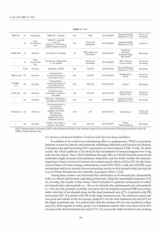
J. Clin. Med. 2021, 10, 3201
Table 1. Cont.
RESCUE II Second-line SHR-1210 + apatinib 190 ORR NCT03463876 Jiangsu HengRuiMedicine Co., Ltd.
Active, notrecruiting
- IIFirst-
line/Second-line
SHR1210 + apatinib(arm A)
SHR1210 + FOLFOX4 orGEMOX regimen (arm B)
152 Safety andtolerability NCT03092895 Jiangsu HengRui
Medicine Co., Ltd. Unknown
IMMUNIB II First-line Nivolumab + lenvatinib 50 ORR/safety andtolerability NCT03841201
Institut furKlinische
KrebsforschungIKF GmbH
Recruiting
- IIFirst-
line/Second-line
Nivolumab + Ipilimumabvs. nivolumab
Safety andtolerability NCT03222076 MD Anderson
Cancer CenterActive, notrecruiting
- II/III First-line Sintilimab + IBI305 566 OS/PFS NCT03794440 Innovent Biologics(Suzhou) Co., Ltd. Recruiting
IMbrave150 III First-lineAtezolizumab +
bevacizumab (arm A)Sorafenib (arm B)
480 OS/PFS NCT03434379 Hoffmann-LaRoche
Active, notrecruiting
COSMIC-312 III First-line
Cabozantinib +atezolizumab (arm A)
Sorafenib (arm B)Cabozantinib (arm C)
740 PFS/OS NCT03755791 Exelixis Recruiting
LEAP-002 III First-linePembrolizumab +
Lenvatinib vs. placebo +lenvatinib
750 PFS/OS NCT03713593 Merck Sharp &Dohme Corp.
Active, notrecruiting
- III First-line SHR-1210 + FOLFOX4 vs.sorafenib or FOLFOX4 448 OS NCT03605706 Jiangsu HengRui
Medicine Co., Ltd. Recruiting
HIMALAYA III First-line
Durvalumab (arm A)Durvalumab +tremelimumab
[regimen 1] (arm B)Durvalumab +tremelimumab
[regimen 2] (arm C)Sorafenib (arm D)
1310 OS NCT03298451 AstraZeneca Active, notrecruiting
- III First-line CS1003 + lenvatinib vs.placebo + lenvatinib 525 PFS/OS NCT04194775 CStone
Pharmaceuticals Recruiting
HCC, hepatocellular carcinoma; DLTs, dose-limiting toxicities; AEs, adverse events; OS, overall survival; PFS progression-free survival;ORR, overall response rate.
4.3. Immune Checkpoint Inhibitors Combined with Tyrosine Kinase Inhibitors
In addition to its well-known stimulating effect on angiogenesis, VEGF can promoteimmune evasion by directly and indirectly inhibiting infiltration and function of cytotoxicT-lymphocytes and increasing PD-1 expression on intra-tumoral CD8+ T-cells. In otherwords, the VEGF pathway is involved in the recruitment of immunosuppressive T-regcells into the tumor. Thus, VEGF inhibition through TKIs or VEGFR-directed monoclonalantibodies might increase local antitumor immunity and favorably modify the immuno-suppressive tumor microenvironment, thus enhancing the effects of ICIs [45]. On this basis,several Phase I/II trials testing combinations of anti-PD1/PD-L1 with anti-VEGFRs wereundertaken and have already shown promising results in this research field, paving theway for Phase III trials that are currently in progress (Table 1) [46].
Among these studies, one trial tested the combination of nivolumab plus cabozantinib,with or without ipilimumab, reporting preliminary clinically meaningful responses [47].As of today, the results of this study, which included 71 patients randomized to eithernivolumab plus cabozantinib (n = 36) or nivolumab plus ipilimumab and cabozantinib(n = 35), are only partially available, and show that investigator-assessed ORR was compa-rable with that of nivolumab alone for the dual treatment arm (17%, 6 patients with PR)but reached 26% (9 patients with PR) in the triple treatment arm. The diseased control ratewas good and similar in the two groups, being 81% for the dual treatment arm and 83% forthe triple treatment arm. It is noteworthy that the median OS was not reached in eitherarm [47]. With regards to safety, grade 3 or 4 treatment-related AEs were observed in 42%of cases in the dual treatment arm and in 71% of cases in the triple treatment arm, leading
158

J. Clin. Med. 2021, 10, 3201
to treatment discontinuation in 3% and 20% of patients, respectively. However, no newsafety signals were observed in either arm. Based on these promising findings, completeand updated results of this trial are eagerly awaited.
Another combination that is currently under investigation in patients with advancedHCC is that of pembrolizumab plus lenvatinib, which, in a Phase Ib study, showed goodresults with a median OS of 22 months and a 46% confirmed ORR [48]. Hence, this com-bination has been granted a breakthrough therapy designation by the FDA for advancedHCC patients who are not amenable to locoregional treatment, and it is currently beingtested in a Phase III, international, multicenter clinical study (LEAP-002).
4.4. Immune Checkpoint Inhibitors Combined with Anti-VEGFR Agents
Recently, Finn et al. tested the combination of atezolizumab, a monoclonal antibodytargeting PD-L1, plus bevacizumab, an anti-VEGF monoclonal antibody, as a front-linetreatment of advanced HCC. The trial (IMbrave-150) showed a clear superiority of thedual therapy over sorafenib [15]. The intention-to-treat population included 336 patientsin the atezolizumab plus bevacizumab group and 165 patients in the sorafenib group. Atthe time of the primary interim analysis, the HR for death with atezolizumab plus beva-cizumab as compared with sorafenib was 0.58 (95%CI, 0.42–0.79; p < 0.001). The reported12-month OS was 67.2% (95%CI, 61.3–73.1) with atezolizumab plus bevacizumab versus54.6% (95%CI, 45.2–64.0) with sorafenib. Median PFS was 6.8 months (95%CI, 5.7 to 8.3)and 4.3 months (95%CI, 4.0–5.6) in the respective groups (HR for disease progressionor death: 0.59; 95%CI, 0.47–0.76; p < 0.001) [15]. Hypertension, proteinuria, and fatiguewere the top three treatment-related AEs in the combination arm. Upper gastrointestinalbleeding, a known AE of bevacizumab and a main concern in patients with cirrhosis,occurred in 7% of patients in this group, which is well within the range of previous stud-ies evaluating the use of bevacizumab in HCC [49,50]. Esophageal varices hemorrhageoccurred in 2.4% of cases, but only 1.8% were grade ≥3 and less than 1% of cases neededtreatment discontinuation. Of note, in this study, causality was reported only in <1% ofpatients [15]. In this respect, it is important to emphasize that patients intended to receivethe combination of atezolizumab plus bevacizumab had undergone endoscopic varicealscreening, as per the study protocol. Given the increased bleeding risk associated withbevacizumab, patients with gastro-esophageal varices at risk of bleeding received ade-quate prophylactic treatment, as must be done in standard care of cirrhotic patients withesophageal varices [51,52]. Increases in aminotransferases and pruritus were other commonAEs attributable to atezolizumab but, again, only a few patients (0.6% of cases) needed tostop treatment and developed immune-mediated liver damage. The proportion of patientswho discontinued any treatment component because of AEs was 15.5% in the atezolizumabplus bevacizumab group (7% discontinued both components) and 10.3% in the sorafenibgroup [15]. Overall, AEs leading to dose modification or interruption occurred in 49.5% ofpatients who received atezolizumab plus bevacizumab and in 60.9% of those who receivedsorafenib. Therefore, this study provided the first and strong—evidence of the benefitprovided by combining an ICI and a VEGFR inhibitor for patients with advanced HCC,and its superiority over sorafenib has undoubtedly already changed the standard of carefor these patients, where it has substituted sorafenib as first-line treatment in most cases.Nevertheless, as only patients with Child–Pugh class A were included in this study, whichis standard practice in HCC trials, so no consistent data are available regarding efficacy andsafety of this combination in patients with a greater impairment in liver function. To date,only one study has reported the outcomes for four Child–Pugh class B patients treatedwith atezolizumab plus bevacizumab in a Japanese cohort of patients [53]. Among thesepatients, all patients could be treated without the development of severe AEs until tumorprogression and efficacy was comparable to that of Child–Pugh class A patients. These re-sults are undoubtedly important, but further research in larger cohorts of patients is neededbefore a recommendation can be made for the use of this immunotherapy in patients withChild–Pugh class B liver function. However, we could argue that well selected patients
159

J. Clin. Med. 2021, 10, 3201
with Child–Pugh class B7 liver function may be treated safely with atezolizumab plusbevacizumab but close monitoring of biochemistry and close clinical monitoring shouldbe performed and patients should be informed that the benefit of this treatment in theChild–Pugh class B population still has to be determined.
The role of sorafenib and that of lenvatinib and, more in general, the treatmentalgorithms for the systemic treatment of HCC, will soon need to be reviewed in order to beoptimized. Whether TKIs are going to be part of the second-line treatment alternatives,alone or in combination with other agents, is still unknown and extensive research isongoing to try to adequately frame treatment sequences.
4.5. Immune Checkpoint Inhibitors Combined with Locoregional Treatments for HCC
To date, no systemic treatment tested in combination with locoregional treatments forHCC has demonstrated benefit in terms of OS. Conversely, ICIs might revolutionize thetherapeutic panorama of early and intermediate stage HCC, thus achieving a role not onlyin the setting of palliative treatment, but also in the curative one. The rationale for their usein combination with radiofrequency ablation (RFA) and TACE relies on the fact that ablativeand intra-arterial techniques indirectly induce a peripheral immune response that canenhance the effect of ICIs [54,55] (Figure 2). Namely, the RFA- and TACE-induced necrosisof tumor cells favors the release of tumor antigens and the activation of immune-mediateddeath of tumor cells, which, in turn, stimulate a peripheral systemic immune response thatcan potentially be amplified by immune checkpoint blockade [56–61]. Arayu et al. showedthat alpha-fetoprotein-specific CD4+ T-cell responses to three immune-dominant epitopesin HCC patients were significantly expanded during and after embolization (p < 0.002).Specifically, the development of alpha-fetoprotein-specific CD4+ T-cells after treatmentwas significantly associated with the induction of >50% necrosis of tumor and an improvedclinical outcome (p < 0.007) [57]. Similarly, Mizokushiet al, evaluating T-cell responses inpatients with HCC undergoing RFA, observed immune responses to antigens for which noT-cell response was detected before RFA [60]. Interestingly, the number of tumor-specificT-cells after RFA correlated with the prevention of HCC recurrence in patients treatedwith curative intent [60]. Moreover, RFA ablation not only provides activating signals forT-cell responses against HCC, but also augments the pool of circulating natural killer (NK)lymphocytes and enhances preferential expression of NK cells’ activating receptors andNK cells’ cytotoxicity, and all these effects are seen as soon as one week after treatment [61].
Although very limited data exist in patients with very early or early HCC (BCLC 0 orBCLC A stage) and intermediate HCC (BCLC B stage) treated with ICIs in the adjuvant andneo-adjuvant setting, preliminary data are promising. With regards to the neo-adjuvantsetting, a recent pilot randomized, Phase II trial showed that dual treatment with nivolumabplus ipilimumab prior to surgery leads to a complete pathological response rate in 33.3%of cases [62]. An increase in T-cell infiltration and upregulation of cytotoxic and effectormemory cell markers in tissue after treatment was also seen, as compared with beforetreatment [62]. Two other small studies investigated tumor-specific immune responsesafter combined TACE and RFA treatment, or after each individual treatment, confirmingthat ablative therapies induce tumor-specific T-cell responses in individual patients uponablative therapies [59,63].
Combined ICIs plus TACE or RFA are not the only treatments under investigation, assome reports regarding the combination of trans-arterial Y90-radioembolization (TARE)and immune checkpoint blockade have been presented at recent oncological meetings withpromising results. In particular, Tai et al. reported the results of a Phase II, open-label,single-center, non-randomized trial regarding the effects of a combined therapy with TAREand nivolumab for advanced HCC in an Asian cohort. Their results showed that thiscombination had a synergistic effect, with an ORR of 30.5% and with good safety andtolerability profiles [64].
160

J. Clin. Med. 2021, 10, 3201
Figure 2. Locoregional treatments applied to hepatocellular carcinoma (HCC) induce immunological effects in the tumormicroenvironment, which can be amplified by immune checkpoint inhibitors. After radiofrequency ablation (RFA) ortrans-arterial chemo-embolization (TACE) or radio-embolization (TARE), necrosis of tumor cells induces increased tumor-antigen release, thus facilitating the recruitment and activation of cytotoxic T-cells and dendritic cells. These effects can beexploited by administering immune checkpoint inhibitors (ICIs) to transform an immunosuppressive microenvironment inan immune-supportive one, in which systemic therapies might be more effective.
Based on these findings, several trials are ongoing to test the efficacy of combined ICIsand locoregional treatments in HCC. This strategy might significantly decrease recurrencerates after treatment with ablative techniques, thus ameliorating long-term prognosis ofpatients with very early/early HCC. Similarly, ICIs may potentially enhance responses aftertrans-arterial treatments; this implicates that patients with intermediate stage HCC maybe effectively down-staged and might therefore become qualified for curative treatments.Hence, if ongoing studies in this field obtain good results in terms of safety and efficacy,ICIs would not only play a role in the setting of advanced HCC, but would also become afundamental component of the management of the earlier stages of this tumor.
5. Amenability to Atezolizumab Plus Bevacizumab in Real-Life Setting
Given the expected upcoming change in the standard of care for the treatment ofpatients with advanced HCC, with a preferential use of the combination of atezolizumabplus bevacizumab as first-line treatment, we aimed to explore the actual estimates ofthe potential applicability in clinical practice of this dual treatment in the Western HCCpopulation. In order to do so, we applied the inclusion and exclusion criteria of theatezolizumab plus bevacizumab IMBrave-150 study to the HCC population recorded in theItalian Liver Cancer (ITA.LI.CA) database. We used this database as it is representative ofthe real-life setting of HCC patients in Italy: the ITA.LI.CA database, indeed, includes morethan 10,000 patients with newly diagnosed or recurrent HCC, with various underlyingliver disease etiologies at all stages, who are managed in a large number of Italian centerswith different levels of expertise (secondary and tertiary referral centers). Thus, it providesa reliable insight into the characteristics of HCC patients in Western regions and allowsfor predicting figures of the potential utilization of newly available HCC drugs in real-lifeclinical practice [39].
In order to carry this out, within the ITA.LI.CA database, we excluded patientsdiagnosed before 2008—that is the year of availability of sorafenib in clinical practice inItaly—and we applied the inclusion and exclusion criteria, listed in Table 2, set forth inthe Phase III IMbrave-150 trial in patients with advanced HCC. In the studied period
161
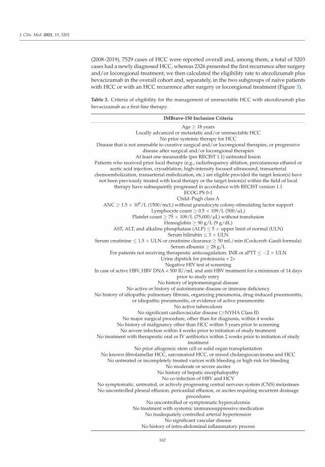
J. Clin. Med. 2021, 10, 3201
(2008–2019), 7529 cases of HCC were reported overall and, among them, a total of 5203cases had a newly diagnosed HCC, whereas 2326 presented the first recurrence after surgeryand/or locoregional treatment; we then calculated the eligibility rate to atezolizumab plusbevacizumab in the overall cohort and, separately, in the two subgroups of naïve patientswith HCC or with an HCC recurrence after surgery or locoregional treatment (Figure 3).
Table 2. Criteria of eligibility for the management of unresectable HCC with atezolizumab plusbevacizumab as a first-line therapy.
IMBrave-150 Inclusion Criteria
Age ≥ 18 yearsLocally advanced or metastatic and/or unresectable HCC
No prior systemic therapy for HCCDisease that is not amenable to curative surgical and/or locoregional therapies, or progressive
disease after surgical and/or locoregional therapiesAt least one measurable (per RECIST 1.1) untreated lesion
Patients who received prior local therapy (e.g., radiofrequency ablation, percutaneous ethanol oracetic acid injection, cryoablation, high-intensity focused ultrasound, transarterial
chemoembolization, transarterial embolization, etc.) are eligible provided the target lesion(s) havenot been previously treated with local therapy or the target lesion(s) within the field of local
therapy have subsequently progressed in accordance with RECIST version 1.1ECOG PS 0-1
Child–Pugh class AANC ≥ 1.5 × 10⁹/L (1500/mcL) without granulocyte colony-stimulating factor support
Lymphocyte count ≥ 0.5 × 10⁹/L (500/μL)Platelet count ≥ 75 × 109/L (75,000/μL) without transfusion
Hemoglobin ≥ 90 g/L (9 g/dL)AST, ALT, and alkaline phosphatase (ALP) ≤ 5 × upper limit of normal (ULN)
Serum bilirubin ≤ 3 × ULNSerum creatinine ≤ 1.5 × ULN or creatinine clearance ≥ 50 mL/min (Cockcroft–Gault formula)
Serum albumin ≥ 28 g/LFor patients not receiving therapeutic anticoagulation: INR or aPTT ≤ −2 × ULN
Urine dipstick for proteinuria < 2+Negative HIV test at screening
In case of active HBV, HBV DNA < 500 IU/mL and anti-HBV treatment for a minimum of 14 daysprior to study entry
No history of leptomeningeal diseaseNo active or history of autoimmune disease or immune deficiency
No history of idiopathic pulmonary fibrosis, organizing pneumonia, drug-induced pneumonitis,or idiopathic pneumonitis, or evidence of active pneumonitis
No active tuberculosisNo significant cardiovascular disease (≥NYHA Class II)
No major surgical procedure, other than for diagnosis, within 4 weeksNo history of malignancy other than HCC within 5 years prior to screening
No severe infection within 4 weeks prior to initiation of study treatmentNo treatment with therapeutic oral or IV antibiotics within 2 weeks prior to initiation of study
treatmentNo prior allogeneic stem cell or solid organ transplantation
No known fibrolamellar HCC, sarcomatoid HCC, or mixed cholangiocarcinoma and HCCNo untreated or incompletely treated varices with bleeding or high risk for bleeding
No moderate or severe ascitesNo history of hepatic encephalopathy
No co-infection of HBV and HCVNo symptomatic, untreated, or actively progressing central nervous system (CNS) metastasesNo uncontrolled pleural effusion, pericardial effusion, or ascites requiring recurrent drainage
proceduresNo uncontrolled or symptomatic hypercalcemia
No treatment with systemic immunosuppressive medicationNo inadequately controlled arterial hypertension
No significant vascular diseaseNo history of intra-abdominal inflammatory process
162
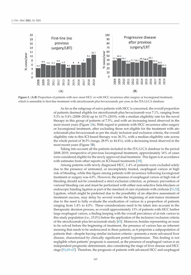
J. Clin. Med. 2021, 10, 3201
(A) (B)
Figure 3. (A,B) Proportion of patients with new onset HCC or with HCC recurrence after surgery or locoregional treatment,which is amenable to first-line treatment with atezolizumab plus bevacizumab, per year, in the ITA.LI.CA database.
As far as the subgroup of naive patients with HCC is concerned, the overall proportionof patients deemed eligible for atezolizumab plus bevacizumab was 7.1%, ranging from5.3% to 5.4% (2008–2014) up to 10.7% (2019), with a median eligibility rate for the noveltherapy in this group of patients of 7.5%, and with an increasing trend observed in themost recent years (Figure 3A). With regard to patients with HCC recurrence after surgeryor locoregional treatment, after excluding those not eligible for the treatment with ate-zolizumab plus bevacizumab as per the study inclusion and exclusion criteria, the overalleligibility rate to this ICI-based therapy was 36.3%, with a median eligibility rate acrossthe whole period of 36.5% (range, 28.9% to 44.4%), with a decreasing trend observed in themost recent years (Figure 3B).
Taking into account all the patients included in the ITA.LI.CA database in the period2008–2019, irrespective of previous locoregional treatment, approximately 16% of caseswere considered eligible for the newly approved dual treatment. This figure is in accordancewith estimates from other reports on ICI-based treatments [39].
Among patients with newly diagnosed HCC, 1.4% of patients were excluded solelydue to the presence of untreated, or incompletely treated, esophageal varices at highrisk of bleeding, while this figure among patients with recurrence following locoregionaltreatment or surgery was 4.0%. However, the presence of esophageal varices at high risk ofbleeding should not be considered a strict exclusion criterion, as primary prevention ofvariceal bleeding can and must be performed with either non-selective beta-blockers orendoscopic banding ligation as part of the standard of care of patients with cirrhosis [51,52].Ligation, which might be preferred due to the possibility of an objective assessment oftreatment success, may delay by several weeks the beginning of anti-tumor treatmentdue to the need to fully evaluate the eradication of varices in a proportion of patientsranging from 1.4% to 4.0%. These considerations need to be taken into account in thetherapeutic decision process, as overall approximately 13% of patients with HCC harborlarge esophageal varices, a finding keeping with the overall prevalence of at-risk varices inthis study population (i.e., 15.0%) before the application of the inclusion/exclusion criteriaof the atezolizumab plus bevacizumab study [65]. Moreover, besides representing an issueto be solved before the beginning of treatment, the presence of varices has an inherentmeaning that needs to be underscored in these patients, as it pinpoints a subpopulation ofpatients that—despite having similar inclusion criteria—presents a more advanced liverdisease, characterized by clinically significant portal hypertension. This finding is notnegligible when patients’ prognosis is assessed, as the presence of esophageal varices is anindependent prognostic determinant, also considering the stage of liver disease and HCCstage [51,65–67]. Therefore, the prognosis of patients with advanced HCC and esophageal
163

J. Clin. Med. 2021, 10, 3201
varices will be poorer than that of patients without varices, regardless of the efficacy ofthe anti-tumoral drug (Figure 4) [65]; as such, screening and treatment (either with bandligation or beta-blockers, selected on a case by case basis) is strongly recommended andmust be performed in all patients with HCC, independently from the tumor stage andprior to the initiation of any anti-tumoral treatment.
Figure 4. Overall survival of patients with advanced hepatocellular carcinoma, subdivided according to the presence ofesophageal varices. Reprinted with permission from ref. [65]. Copyright 2006 American Gastroenterological Association.
6. Conclusions
Immunotherapy certainly represents a new, exciting frontier in the treatment of ad-vanced, unresectable HCC, and might play a role as an adjuvant or neo-adjuvant treatmentof patients with early-stage HCC as well, giving them the chance to decrease the risk oftumor recurrence. New ICI-based treatment strategies with dual, or even triple, combina-tions of immune-targeting agents, or combinations of immunotherapy and TKIs or otheranti-neoplastic agents, will probably be available in the foreseeable future. Thus, it is cur-rently difficult to predict the future algorithm for the systemic treatment of advanced HCCand to state whether sorafenib and lenvatinib, as single agents, will still be listed among thefirst-line treatment options for this cancer. However, despite the understandable enthusi-asm for immunotherapy, some unmet needs remain and require further, extensive researchto be resolved. First, as many as 30–40% of patients with HCC do not respond to ICIs, andbiomarkers predicting treatment response are lacking. This is a particular challenging issueas data about histological or serological biomarkers related to the effectiveness of ICIs inHCC have not been clearly identified, and, even if a histological marker was identified,biopsy sampling of HCC is not standard clinical practice for this tumor, which is mostlydiagnosed on the basis of its radiological hallmarks; therefore, in the future, the role ofliver biopsy in HCC might need to be revisited [68]. Secondly, we have shown that inreal-life, also taking into consideration previous treatments, only approximately one-tenthto one-third of patients with HCC are eligible for the recently approved combination ofatezolizumab plus bevacizumab. Moreover, the safety and utility of immunotherapy inpatients with a greater impairment in liver function, such as Child–Pugh class B patients,still has to be demonstrated, as most trials have explored the safety of these drugs in
164

J. Clin. Med. 2021, 10, 3201
patients with well-preserved liver function (Child–Pugh class A) and, even though somereports have described an acceptable safety profile of some ICIs in Child–Pugh class Bpatients, consistent data regarding this topic are lacking, so that no strong recommendationcan be made in this regard for the time being. Finally, ICIs are highly expensive drugs andthis may represent a serious threat to the worldwide treatment implementation in clinicalpractice, since a large share of patients with HCC are diagnosed in developing countries,where available economic resources cannot support their use [69].
Taken together, the available evidence clearly shows that ICIs are going to play apivotal role in the treatment of HCC and will improve the prognosis of patients withadvanced HCC and, presumably, of those at earlier stages of the disease as well. Wecan assume that in the foreseeable future the current treatment algorithms will needrevisions based on the most recent evidence. However, considering that in real-life settingsa high proportion of patients will probably not be eligible for ICI-based regimens, mucheffort is still needed in order to optimize treatment strategies for patients with advanced,unresectable HCC.
Funding: This research received no external funding.
Institutional Review Board Statement: Not applicable.
Informed Consent Statement: Not applicable.
Data Availability Statement: Data available on request due to restrictions. The data presented inthis study are available on request from the corresponding author. The data are not publicly availabledue to the rules of the ITA.LI.CA consortium.
Acknowledgments: We acknowledged Italian Liver Cancer (ITA.LI.CA) Group and the Italian Asso-ciation for the Study of the Liver—Hepatocellular Carcinoma Special Interest Group (AISF HCC-SIG).Others members of the ITA.LI.CA group: Maurizio Biselli, Laura Bucci, Paolo Caraceni, Annag-
iulia Gramenzi, Lorenzo Lani, Davide Rampoldi, Nicola Reggidori, Valentina Santi, Benedetta
Stefanini: Semeiotics Unit, Department of Medical and Surgical Sciences, IRCCS Azienda Ospedaliero-Universitaria di Bologna, University of Bologna, Bologna, Italy. Donatella Magalotti, Marco Zoli: Medic-ina Interna, Malattie neurovascolari e epatometaboliche, IRCCS Azienda Ospedaliero-Universitaria di Bologna,University of Bologna, Bologna, Italy. Alessandro Granito, Luca Muratori, Francesco Tovoli: Inter-nal Medicine, Department of Medical and Surgical Sciences, IRCCS Azienda Ospedaliero-Universitaria diBologna, University of Bologna, Bologna, Italy. Francesco Azzaroli, Elton Dajti, Federico Ravaioli: Gas-troenterology Unit, Department of Surgical and Medical Sciences, IRCCS Azienda Ospedaliero-Universitariadi Bologna, University of Bologna, Bologna, Italy. Alberta Cappelli, Rita Golfieri, Cristina Mosconi,
Matteo Renzulli: Radiology Unit, Department of Specialist, Diagnostic and Experimental Medicine, IRCCSAzienda Ospedaliero-Universitaria di Bologna, University of Bologna, Bologna, Italy. Fabio Farinati, Bar-
bara Penzo: Gastroenterology Unit, Department of Surgery, Oncology and Gastroenterology, Universityof Padova, Padova, Italy. Rodolfo Sacco, Ester Marina Cela, Antonio Facciorusso: Gastroenterol-ogy and Digestive Endoscopy Unit, Foggia University Hospital, Foggia, Italy. Giulia Pieri, Edoardo
Casagrande, Mattia Vanello. Gastroenterology Unit, Department of Internal Medicine, IRCCS OspedalePoliclinico San Martino, University of Genova, Genova, Italy. Antonio Gasbarrini, Gianludovico Rapac-
cini, Francesca Romana Ponziani, Nicoletta de Matthaeis: Gastroenterology Unit, IRCCS FondazionePoliclinico Universitario A. Gemelli, Roma, Italy. Gianluca Svegliati Baroni, Gloria Allegrini: LiverInjury and Transplant Unit, Polytechnic University of Marche, Ancona, Italy. Valentina Lauria, Giorgia
Ghittoni, Giorgio Pelecca: Gastroenterology Unit, Belcolle Hospital, Viterbo, Italy. Fabrizio Chegai,
Fabio Coratella, Mariano Ortenzi: Vascular and Interventional Radiology Unit, Belcolle Hospital, Viterbo,Italy. Serena Dell’Isola: Infectious Disease Unit, Belcolle Hospital, Viterbo, Italy. Gabriele Missale,
Elisabetta Biasini, Andrea Olivani: Infectious Diseases and Hepatology Unit, Department of Medicineand Surgery, University of Parma and Azienda Ospedaliero-Universitaria of Parma, Parma, Italy. Alberto
Masotto, Alessandro Inno, Fabiana Marchetti: Gastroenterology Unit, IRCCS Sacro Cuore Don CalabriaHospital, Negrar, Italy. Maria Di Marco: Medicine Unit, Bolognini Hospital, Seriate, Italy. Andrea Mega:
Gastroenterology Unit, Bolzano Regional Hospital, Bolzano, Italy. Ciro Celsa, Mauro Grova, Caterina
Stornello, Anita Busacca, Calogero Cammà, Giacomo Emanuele Maria Rizzo: Gastroenterology andHepatology Unit, Department of Health Promotion, Mother and Child Care, Internal Medicine and MedicalSpecialties, PROMISE, University of Palermo, Palermo, Italy. Maria Stella Franzè, Giovanni Raimondo,
165

J. Clin. Med. 2021, 10, 3201
Carlo Saitta: Clinical and Molecular Hepatology Unit, Department of Clinical and Experimental Medicine,University of Messina, Messina, Italy. Gianpaolo Vidili, Assunta Sauchella: Department of Medical,Surgical and Experimental Sciences, Azienda Ospedaliero-Universitaria of Sassari, Sassari, Italy. Francesco
Giuseppe Foschi, Lucia Napoli, Vittoria Bevilacqua, Dante Berardinelli, Alberto Borghi, Andrea
Casadei Gardini, Fabio Conti, Alessandro Cucchetti, Anna Chiara Dall’Aglio, Giorgio Ercolani:Department of Internal Medicine, Ospedale per gli Infermi di Faenza, Faenza, Italy. Claudia Campani,
Chiara Di Bonaventura, Stefano Gitto, Valentina Adotti: Internal Medicine and Hepatology Unit,Department of Experimental and Clinical Medicine, University of Firenze, Firenze, Italy. Gerardo Nardone,
Pietro Coccoli, Antonio Malerba: Hepato-Gastroenterology Unit, Department of Clinical Medicine andSurgery, University of Napoli "Federico II", Napoli, Italy. Filomena Morisco, Maria Guarino, Mario
Capasso: Gastroenterology Unit, Department of Clinical Medicine and Surgery, University of Napoli "Fed-erico II", Napoli, Italy. Mauriza Rossana Brunetto, Filippo Oliveri, Veronica Romagnoli: Hepatologyand Liver Physiopathology Laboratory, Department of Clinical and Experimental Medicine, University Hos-pital of Pisa, Pisa, Italy. Members of the Associazione Italiana per lo Studio del Fegato Special
Interest Group on Hepatocellular Carcinoma (AISF SIG-HCC): Aglitti A., Baccarani U.,Bhoori S.,Borzio M., Brancaccio G., Burra P., Carrai P., Conti F., Cozzolongo R, Cucchetti A., D’Ambrosio R.,Dell’Unto C., De Matthaeis N., Di Costanzo G.G., Di Sandro S., Famularo S, Fucilli F., Galati G.,Gambato M., Giuliante F., Ghinolfi D., Grieco A., Gruttadauria S., Iavarone M., Kostandini A.,Lenci I., Levi Sandri G.V., Losito F., Lupo L.G., Marasco G, Manzia T.M., Mazzocato S., Masarone M.,Melandro F., Mescoli C., Miele L., Muley M., Nicolini D., Pagano D, Persico M., Pompili M.,Pravisani R., Rendina M., Renzulli M., Romano F, Rossi M., Rreka E., Russo F.P., Sangiovanni A.,Sessa A., Simonetti N., Sposito C., Tortora R., Viganò L., Viganò M., Villa E., Vincenzi V., Violi P.,Vitale A.
Conflicts of Interest: The authors declare no conflict of interest.
References
1. Villanueva, A. Hepatocellular Carcinoma. N. Engl. J. Med. 2019, 380, 1450–1462. [CrossRef]2. Mittal, S.; El-Serag, H.B.; Sada, Y.H.; Kanwal, F.; Duan, Z.; Temple, S.; May, S.; Kramer, J.R.; Richardson, P.A.; Davila, J.A.
Hepatocellular Carcinoma in the Absence of Cirrhosis in United States Veterans Is Associated with Nonalcoholic Fatty LiverDisease. Clin. Gastroenterol. Hepatol. 2016, 14, 124–131.e1. [CrossRef]
3. Kanwal, F.; Kramer, J.R.; Mapakshi, S.; Natarajan, Y.; Chayanupatkul, M.; Richardson, P.A.; Li, L.; Desiderio, R.; Thrift, A.P.;Asch, S.M.; et al. Risk of Hepatocellular Cancer in Patients with Non-Alcoholic Fatty Liver Disease. Gastroenterology 2018, 155,1828–1837.e2. [CrossRef] [PubMed]
4. Torres, M.C.P.; Bodini, G.; Furnari, M.; Marabotto, E.; Zentilin, P.; Strazzabosco, M.; Giannini, E.G. Surveillance for HepatocellularCarcinoma in Patients with Non-Alcoholic Fatty Liver Disease: Universal or Selective? Cancers 2020, 12, 1422. [CrossRef]
5. Torres, M.C.P.; Aghemo, A.; Lleo, A.; Bodini, G.; Furnari, M.; Marabotto, E.; Miele, L.; Giannini, E.G. Mediterranean Diet andNAFLD: What We Know and Questions That Still Need to Be Answered. Nutrients 2019, 11, 2971. [CrossRef]
6. Llovet, J.M.; Ricci, S.; Mazzaferro, V.; Hilgard, P.; Gane, E.; Blanc, J.F.; De Oliveira, A.C.; Santoro, A.; Raoul, J.L.; Forner, A.; et al.Sorafenib in Advanced Hepatocellular Carcinoma. N. Engl. J. Med. 2008, 359, 378–390. [CrossRef]
7. Cheng, A.-L.; Kang, Y.-K.; Chen, Z.; Tsao, C.-J.; Qin, S.; Kim, J.S.; Luo, R.; Feng, J.; Ye, S.; Yang, T.-S.; et al. Efficacy and safety ofsorafenib in patients in the Asia-Pacific region with advanced hepatocellular carcinoma: A phase III randomised, double-blind,placebo-controlled trial. Lancet Oncol. 2009, 10, 25–34. [CrossRef]
8. Cheng, A.-L.; Finn, R.S.; Qin, S.; Han, K.-H.; Ikeda, K.; Piscaglia, F.; Baron, A.D.; Park, J.-W.; Han, G.; Jassem, J.; et al. Phase IIItrial of lenvatinib (LEN) vs sorafenib (SOR) in first-line treatment of patients (pts) with unresectable hepatocellular carcinoma(uHCC). J. Clin. Oncol. 2017, 35, 4001. [CrossRef]
9. Kirstein, M.M.; Scheiner, B.; Marwede, T.; Wolf, C.; Voigtländer, T.; Semmler, G.; Wacker, F.; Manns, M.P.; Hinrichs, J.B.; Pinter,M.; et al. Sequential systemic treatment in patients with hepatocellular carcinoma. Aliment. Pharmacol. Ther. 2020, 52, 205–212.[CrossRef] [PubMed]
10. Sangro, B.; Gomez-Martin, C.; de la Mata, M.; Iñarrairaegui, M.; Garralda, E.; Barrera, P.; Riezu-Boj, J.-I.; Larrea, E.; Alfaro, C.;Sarobe, P.; et al. A clinical trial of CTLA-4 blockade with tremelimumab in patients with hepatocellular carcinoma and chronichepatitis C. J. Hepatol. 2013, 59, 81–88. [CrossRef]
11. El-Khoueiry, A.B.; Sangro, B.; Yau, T.C.C.; Crocenzi, T.S.; Kudo, M.; Hsu, C.; Kim, T.-Y.; Choo, S.-P.; Trojan, J.; Welling, T.H.; et al.Nivolumab in patients with advanced hepatocellular carcinoma (CheckMate 040): An open-label, non-comparative, phase 1/2dose escalation and expansion trial. Lancet 2017, 389, 2492–2502. [CrossRef]
12. Zhu, A.X.; Finn, R.S.; Edeline, J.; Cattan, S.; Ogasawara, S.; Palmer, D.; Verslype, C.; Zagonel, V.; Fartoux, L.; Vogel, A.;et al. Pembrolizumab in patients with advanced hepatocellular carcinoma previously treated with sorafenib (KEYNOTE-224):A non-randomised, open-label phase 2 trial. Lancet Oncol. 2018, 19, 940–952. [CrossRef]
166

J. Clin. Med. 2021, 10, 3201
13. Finn, R.S.; Ryoo, B.-Y.; Merle, P.; Kudo, M.; Bouattour, M.; Lim, H.Y.; Breder, V.; Edeline, J.; Chao, Y.; Ogasawara, S.; et al.Pembrolizumab As Second-Line Therapy in Patients with Advanced Hepatocellular Carcinoma in KEYNOTE-240: A Randomized,Double-Blind, Phase III Trial. J. Clin. Oncol. 2020, 38, 193–202. [CrossRef] [PubMed]
14. Yau, T.; Park, J.W.; Finn, R.S.; Cheng, A.L.; Mathurin, P.; Edeline, J.; Kudo, M.; Han, K.H.; Harding, J.J.; Merle, P.; et al. CheckMate459: A randomized, multi-center phase III study of nivolumab (NIVO) vs sorafenib (SOR) as first-line (1L) treatment in patients(pts) with advanced hepatocellular carcinoma (aHCC). Ann. Oncol. 2019, 30, v874–v875. [CrossRef]
15. Finn, R.S.; Qin, S.; Ikeda, M.; Galle, P.R.; Ducreux, M.; Kim, T.-Y.; Kudo, M.; Breder, V.; Merle, P.; Kaseb, A.O. Atezolizumab plusBevacizumab in Unresectable Hepatocellular Carcinoma. N. Engl. J. Med. 2020, 382, 1894–1905. [CrossRef]
16. Iavarone, M.; Cabibbo, G.; Piscaglia, F.; Zavaglia, C.; Grieco, A.; Villa, E.; Camma’, C.; Colombo, M.; on behalf of the SOFIA(SOraFenib Italian Assessment) study group. Field-practice study of sorafenib therapy for hepatocellular carcinoma: A prospectivemulticenter study in Italy. Hepatology 2011, 54, 2055–2063. [CrossRef] [PubMed]
17. Ganten, T.M.; Stauber, R.E.; Schott, E.; Malfertheiner, P.; Buder, R.; Galle, P.R.; Göhler, T.; Walther, M.; Koschny, R.; Gerken, G.Sorafenib in Patients with Hepatocellular Carcinoma—Results of the Observational INSIGHT Study. Clin. Cancer Res. 2017, 23,5720–5728. [CrossRef]
18. Bruix, J.; Qin, S.; Merle, P.; Granito, A.; Huang, Y.-H.; Bodoky, G.; Pracht, M.; Yokosuka, O.; Rosmorduc, O.; Breder, V.; et al.Regorafenib for patients with hepatocellular carcinoma who progressed on sorafenib treatment (RESORCE): A randomised,double-blind, placebo-controlled, phase 3 trial. Lancet 2017, 389, 56–66. [CrossRef]
19. Zhu, A.X.; Kang, Y.-K.; Yen, C.-J.; Finn, R.S.; Galle, P.R.; Llovet, J.M.; Assenat, E.; Brandi, G.; Lim, H.Y.; Pracht, M.; et al. REACH-2:A randomized, double-blind, placebo-controlled phase 3 study of ramucirumab versus placebo as second-line treatment inpatients with advanced hepatocellular carcinoma (HCC) and elevated baseline alpha-fetoprotein (AFP) following first-linesorafenib. J. Clin. Oncol. 2018, 36, 4003. [CrossRef]
20. Abou-Alfa, G.K.; Meyer, T.; Cheng, A.-L.; El-Khoueiry, A.B.; Rimassa, L.; Ryoo, B.-Y.; Cicin, I.; Merle, P.; Chen, Y.; Park, J.-W.; et al.Cabozantinib in Patients with Advanced and Progressing Hepatocellular Carcinoma. N. Engl. J. Med. 2018, 379, 54–63. [CrossRef]
21. Zhu, A.X.; Park, J.O.; Ryoo, B.-Y.; Yen, C.-J.; Poon, R.; Pastorelli, D.; Blanc, J.-F.; Chung, H.; Baron, A.D.; Pfiffer, T.E.F.; et al.Ramucirumab versus placebo as second-line treatment in patients with advanced hepatocellular carcinoma following first-linetherapy with sorafenib (REACH): A randomised, double-blind, multicentre, phase 3 trial. Lancet Oncol. 2015, 16, 859–870.[CrossRef]
22. Giannini, E.G.; Trevisani, F. Ramucirumab as a second-line treatment for hepatocellular carcinoma: Reaching out further topatients with elevated alpha-fetoprotein. Hepatobiliary Surg. Nutr. 2019, 8, 515–518. [CrossRef]
23. Lai, E.; Astara, G.; Ziranu, P.; Pretta, A.; Migliari, M.; Dubois, M.; Donisi, C.; Mariani, S.; Liscia, N.; Impera, V.; et al. Introducingimmunotherapy for advanced hepatocellular carcinoma patients: Too early or too fast? Crit. Rev. Oncol. 2021, 157, 103167.[CrossRef]
24. Garuti, F.; Neri, A.; Avanzato, F.; Gramenzi, A.; Rampoldi, D.; Rucci, P.; Farinati, F.; Giannini, E.G.; Piscaglia, F.; Rapaccini, G.L.;et al. The changing scenario of hepatocellular carcinoma in Italy: An update. Liver Int. 2021, 41, 585–597. [CrossRef]
25. Hoshida, Y.; Villanueva, A.; Kobayashi, M.; Peix, J.; Chiang, D.Y.; Camargo, A.; Gupta, S.; Moore, J.; Wrobel, M.J.; Lerner, J.; et al.Gene Expression in Fixed Tissues and Outcome in Hepatocellular Carcinoma. N. Engl. J. Med. 2008, 359, 1995–2004. [CrossRef]
26. Pikarsky, E.; Porat, R.M.; Stein, I.; Abramovitch, R.; Amit, S.; Kasem, S.; Gutkovich-Pyest, E.; Urieli-Shoval, S.; Galun, E.;Ben-Neriah, Y. NF-κB functions as a tumour promoter in inflammation-associated cancer. Nature 2004, 431, 461–466. [CrossRef]
27. Llovet, J.M.; Montal, R.; Sia, D.; Finn, R.S. Molecular therapies and precision medicine for hepatocellular carcinoma. Nat. Rev.Clin. Oncol. 2018, 15, 599–616. [CrossRef] [PubMed]
28. Prieto, J.; Melero, I.; Sangro, B. Immunological landscape and immunotherapy of hepatocellular carcinoma. Nat. Rev. Gastroenterol.Hepatol. 2015, 12, 681–700. [CrossRef] [PubMed]
29. Shi, F.; Shi, M.; Zeng, Z.; Qi, R.-Z.; Liu, Z.-W.; Zhang, J.-Y.; Yang, Y.-P.; Tien, P.; Wang, F.-S. PD-1 and PD-L1 upregulation promotesCD8+ T-cell apoptosis and postoperative recurrence in hepatocellular carcinoma patients. Int. J. Cancer 2010, 128, 887–896.[CrossRef] [PubMed]
30. Sia, D.; Jiao, Y.; Martinez-Quetglas, I.; Kuchuk, O.; Villacorta-Martin, C.; de Moura, M.C.; Putra, J.; Campreciós, G.; Bassaganyas,L.; Akers, N.; et al. Identification of an Immune-specific Class of Hepatocellular Carcinoma, Based on Molecular Features.Gastroenterology 2017, 153, 812–826. [CrossRef] [PubMed]
31. Chen, D.S.; Mellman, I. Oncology Meets Immunology: The Cancer-Immunity Cycle. Immunity 2013, 39, 1–10. [CrossRef]32. Ringelhan, M.; Pfister, D.; O’Connor, T.; Pikarsky, E.; Heikenwalder, M. The immunology of hepatocellular carcinoma. Nat. Immunol.
2018, 19, 222–232. [CrossRef]33. Postow, M.A.; Callahan, M.K.; Wolchok, J.D. Immune Checkpoint Blockade in Cancer Therapy. J. Clin. Oncol. 2015, 33, 1974–1982.
[CrossRef] [PubMed]34. El-Khoueiry, A.B.; Melero, I.; Yau, T.C.; Crocenzi, T.S.; Kudo, M.; Hsu, C.; Choo, S.; Trojan, J.; Welling, T.; Meyer, T.; et al. Impact
of antitumor activity on survival outcomes, and nonconventional benefit, with nivolumab (NIVO) in patients with advancedhepatocellular carcinoma (aHCC): Subanalyses of CheckMate-040. J. Clin. Oncol. 2018, 36, 475. [CrossRef]
35. Crocenzi, T.S.; El-Khoueiry, A.B.; Yau, T.C.; Melero, I.; Sangro, B.; Kudo, M.; Hsu, C.; Trojan, J.; Kim, T.-Y.; Choo, S.-P.; et al.Nivolumab (nivo) in sorafenib (sor)-naive and -experienced pts with advanced hepatocellular carcinoma (HCC): CheckMate 040study. J. Clin. Oncol. 2017, 35, 4013. [CrossRef]
167

J. Clin. Med. 2021, 10, 3201
36. Abou-Alfa, G.K.; Chan, S.; Furuse, J.; Galle, P.R.; Kelley, R.K.; Qin, S.; Armstrong, J.; Darilay, A.; Vlahovic, G.; Negro, A.; et al.A randomized, multicenter phase 3 study of durvalumab (D) and tremelimumab (T) as first-line treatment in patients withunresectable hepatocellular carcinoma (HCC): HIMALAYA study. J. Clin. Oncol. 2018, 36, TPS4144. [CrossRef]
37. Kudo, M.; Matilla, A.; Santoro, A.; Melero, I.; Gracian, A.C.; Acosta-Rivera, M.; Choo, S.P.; El-Khoueiry, A.B.; Kuromatsu, R.;El-Rayes, B.F.; et al. Checkmate-040: Nivolumab (NIVO) in patients (pts) with advanced hepatocellular carcinoma (aHCC) andChild-Pugh B (CPB) status. J. Clin. Oncol. 2019, 37, 327. [CrossRef]
38. Haanen, J.; Carbonnel, F.; Robert, C.; Kerr, K.; Peters, S.; Larkin, J.; Jordan, K. Management of toxicities from immunotherapy:ESMO Clinical Practice Guidelines for diagnosis, treatment and follow-up. Ann. Oncol. 2017, 28, iv119–iv142. [CrossRef][PubMed]
39. Giannini, E.G.; Aglitti, A.; Borzio, M.; Gambato, M.; Guarino, M.; Iavarone, M.; Lai, Q.; Sandri, G.B.L.; Melandro, F.; Morisco, F.;et al. Overview of Immune Checkpoint Inhibitors Therapy for Hepatocellular Carcinoma, and The ITA.LI.CA Cohort DerivedEstimate of Amenability Rate to Immune Checkpoint Inhibitors in Clinical Practice. Cancers 2019, 11, 1689. [CrossRef]
40. De Velasco, G.; Je, Y.; Bossé, D.; Awad, M.M.; Ott, P.A.; Moreira, R.B.; Schutz, F.; Bellmunt, J.; Sonpavde, G.P.; Hodi, F.S.; et al.Comprehensive Meta-analysis of Key Immune-Related Adverse Events from CTLA-4 and PD-1/PD-L1 Inhibitors in CancerPatients. Cancer Immunol. Res. 2017, 5, 312–318. [CrossRef]
41. Scheiner, B.; Kirstein, M.M.; Hucke, F.; Finkelmeier, F.; Schulze, K.; Von Felden, J.; Koch, S.; Schwabl, P.; Hinrichs, J.B.; Waneck, F.;et al. Programmed cell death protein-1 (PD-1)-targeted immunotherapy in advanced hepatocellular carcinoma: Efficacy andsafety data from an international multicentre real-world cohort. Aliment. Pharmacol. Ther. 2019, 49, 1323–1333. [CrossRef]
42. Yau, T.; Kang, Y.-K.; Kim, T.-Y.; El-Khoueiry, A.B.; Santoro, A.; Sangro, B.; Melero, I.; Kudo, M.; Hou, M.-M.; Matilla, A.; et al.Nivolumab (NIVO) + ipilimumab (IPI) combination therapy in patients (pts) with advanced hepatocellular carcinoma (aHCC):Results from CheckMate. J. Clin. Oncol. 2019, 37 (Suppl. S15), 4012. [CrossRef]
43. Yau, T.; Kang, Y.-K.; Kim, T.-Y.; El-Khoueiry, A.B.; Santoro, A.; Sangro, B.; Melero, I.; Kudo, M.; Hou, M.-M.; Matilla, A.; et al.Efficacy and Safety of Nivolumab Plus Ipilimumab in Patients with Advanced Hepatocellular Carcinoma Previously Treatedwith Sorafenib. JAMA Oncol. 2020, 6, e204564. [CrossRef] [PubMed]
44. Kelley, R.K.; Sangro, B.; Harris, W.P.; Ikeda, M.; Okusaka, T.; Kang, Y.-K.; Qin, S.; Tai, W.M.D.; Lim, H.Y.; Yau, T.; et al. Efficacy,tolerability, and biologic activity of a novel regimen of tremelimumab (T) in combination with durvalumab (D) for patients (pts)with advanced hepatocellular carcinoma (aHCC). J. Clin. Oncol. 2020, 38, 4508. [CrossRef]
45. Tai, D.; Choo, S.P.; Chew, V. Rationale of Immunotherapy in Hepatocellular Carcinoma and Its Potential Biomarkers. Cancers2019, 11, 1926. [CrossRef]
46. Liu, L.; Qin, S.; Zhang, Y. The Evolving Landscape of Checkpoint Inhibitor Combination Therapy in the Treatment of AdvancedHepatocellular Carcinoma. Target. Oncol. 2021, 16, 153–163. [CrossRef]
47. Yau, T.; Zagonel, V.; Santoro, A.; Acosta-Rivera, M.; Choo, S.P.; Matilla, A.; He, A.R.; Gracián, A.C.; El-Khoueiry, A.B.; Sangro,B.; et al. Nivolumab (NIVO) + ipilimumab (IPI) + cabozantinib (CABO) combination therapy in patients (pts) with advancedhepatocellular carcinoma (aHCC): Results from CheckMate 040. J. Clin. Oncol. 2020, 38, 478. [CrossRef]
48. Finn, R.S.; Ikeda, M.; Zhu, A.X.; Sung, M.W.; Baron, A.D.; Kudo, M.; Okusaka, T.; Kobayashi, M.; Kumada, H.; Kaneko, S.; et al.Phase Ib Study of Lenvatinib Plus Pembrolizumab in Patients with Unresectable Hepatocellular Carcinoma. J. Clin. Oncol. 2020,38, 2960–2970. [CrossRef] [PubMed]
49. Pinter, M.; Ulbrich, G.; Sieghart, W.; Kölblinger, C.; Reiberger, T.; Li, S.; Ferlitsch, A.; Müller, C.; Lammer, J.; Peck-Radosavljevic,M. Hepatocellular Carcinoma: A Phase II Randomized Controlled Double-Blind Trial of Transarterial Chemoembolization inCombination with Biweekly Intravenous Administration of Bevacizumab or a Placebo. Radiology 2015, 277, 903–912. [CrossRef][PubMed]
50. Siegel, A.B.; Cohen, E.I.; Ocean, A.; Lehrer, D.; Goldenberg, A.; Knox, J.J.; Chen, H.; Clark-Garvey, S.; Weinberg, A.; Mandeli, J.;et al. Phase II Trial Evaluating the Clinical and Biologic Effects of Bevacizumab in Unresectable Hepatocellular Carcinoma. J. Clin.Oncol. 2008, 26, 2992–2998. [CrossRef]
51. Angeli, P.; Bernardi, M.; Villanueva, C.; Francoz, C.; Mookerjee, R.; Trebicka, J.; Krag, A.; Laleman, W.; Gines, P. EASL ClinicalPractice Guidelines for the management of patients with decompensated cirrhosis. J. Hepatol. 2018, 69, 406–460. [CrossRef]
52. Giannini, E.G.; Trevisani, F. Improving survival of cirrhosis patients with hepatocellular carcinoma through application ofstandard of care. Hepatology 2014, 60, 1446–1447. [CrossRef]
53. Iwamoto, H.; Shimose, S.; Noda, Y.; Shirono, T.; Niizeki, T.; Nakano, M.; Okamura, S.; Kamachi, N.; Suzuki, H.; Sakai, M.; et al.Initial Experience of Atezolizumab Plus Bevacizumab for Unresectable Hepatocellular Carcinoma in Real-World Clinical Practice.Cancers 2021, 13, 2786. [CrossRef] [PubMed]
54. Greten, T.F.; Duffy, A.G.; Korangy, F. Hepatocellular Carcinoma from an Immunologic Perspective. Clin. Cancer Res. 2013, 19,6678–6685. [CrossRef] [PubMed]
55. Singh, P.; Toom, S.; Avula, A.; Kumar, V.; E Rahma, O. The Immune Modulation Effect of Locoregional Therapies and Its PotentialSynergy with Immunotherapy in Hepatocellular Carcinoma. J. Hepatocell. Carcinoma 2020, 7, 11–17. [CrossRef] [PubMed]
56. Duffy, A.G.; Ulahannan, S.; Makorova-Rusher, O.; Rahma, O.; Wedemeyer, H.; Pratt, D.; Davis, J.L.; Hughes, M.S.; Heller, T.;ElGindi, M.; et al. Tremelimumab in combination with ablation in patients with advanced hepatocellular carcinoma. J. Hepatol.2017, 66, 545–551. [CrossRef] [PubMed]
168

J. Clin. Med. 2021, 10, 3201
57. Ayaru, L.; Pereira, S.; Alisa, A.; Pathan, A.A.; Williams, R.; Davidson, B.; Burroughs, A.K.; Meyer, T.; Behboudi, S. Unmasking ofα-Fetoprotein-Specific CD4+ T Cell Responses in Hepatocellular Carcinoma Patients Undergoing Embolization. J. Immunol. 2007,178, 1914–1922. [CrossRef]
58. Hänsler, J.; Hä, J.; Nsler, T.; Wissniowski, D.S.U.T. Activation and dramatically increased cytolytic activity of tumor specific Tlymphocytes after radio-frequency ablation in patients with hepatocellular carcinoma and colorectal liver metastases. World J.Gastroenterol. 2006, 12, 3716–3721. [CrossRef] [PubMed]
59. Hiroishi, K.; Eguchi, J.; Baba, T.; Shimazaki, T.; Ishii, S.; Hiraide, A.; Sakaki, M.; Doi, H.; Uozumi, S.; Omori, R.; et al. Strong CD8+T-cell responses against tumor-associated antigens prolong the recurrence-free interval after tumor treatment in patients withhepatocellular carcinoma. J. Gastroenterol. 2009, 45, 451–458. [CrossRef]
60. Mizukoshi, E.; Nakamoto, Y.; Arai, K.; Yamashita, T.; Sakai, A.; Sakai, Y.; Kagaya, T.; Yamashita, T.; Honda, M.; Kaneko, S.Comparative analysis of various tumor-associated antigen-specific t-cell responses in patients with hepatocellular carcinoma.Hepatology 2011, 53, 1206–1216. [CrossRef]
61. Zerbini, A.; Pilli, M.; Laccabue, D.; Pelosi, G.; Molinari, A.; Negri, E.; Cerioni, S.; Fagnoni, F.; Soliani, P.; Ferrari, C.; et al.Radiofrequency Thermal Ablation for Hepatocellular Carcinoma Stimulates Autologous NK-Cell Response. Gastroenterology2010, 138, 1931–1942.e2. [CrossRef]
62. Kaseb, A.O.; Vence, L.; Blando, J.; Yadav, S.S.; Ikoma, N.; Pestana, R.C.; Vauthey, J.N.; Allison, J.P.; Sharma, P. ImmunologicCorrelates of Pathologic Complete Response to Preoperative Immunotherapy in Hepatocellular Carcinoma. Cancer Immunol. Res.2019, 7, 1390–1395. [CrossRef] [PubMed]
63. Nakatsura, T.; Nobuoka, D.; Motomura, Y.; Shirakawa, H.; Yoshikawa, T.; Kuronuma, T.; Takahashi, M.; Nakachi, K.; Ishii,H.; Furuse, J.; et al. Radiofrequency ablation for hepatocellular carcinoma induces glypican-3 peptide-specific cytotoxic Tlymphocytes. Int. J. Oncol. 2011, 40, 63–70. [CrossRef] [PubMed]
64. Tai, W.M.D.; Loke, K.S.H.; Gogna, A.; Tan, S.H.; Ng, D.C.E.; Hennedige, T.P.; Irani, F.; Lee, J.J.X.; Too, C.W.; Ng, M.C.; et al.A phase II open-label, single-center, nonrandomized trial of Y90-radioembolization in combination with nivolumab in Asianpatients with advanced hepatocellular carcinoma: CA 209-678. J Clin. Oncol. 2020, 38, 4590. [CrossRef]
65. Giannini, E.G.; Risso, D.; Testa, R.; Trevisani, F.; Di Nolfo, M.A.; Del Poggio, P.; Benvegnù, L.; Rapaccini, G.L.; Farinati, F.; Zoli, M.;et al. Prevalence and Prognostic Significance of the Presence of Esophageal Varices in Patients with Hepatocellular Carcinoma.Clin. Gastroenterol. Hepatol. 2006, 4, 1378–1384. [CrossRef] [PubMed]
66. European Association for the Study of the Liver. EASL Clinical Practice Guidelines: Management of hepatocellular carcinoma.J. Hepatol. 2018, 69, 182–236. [CrossRef]
67. D’Amico, G.; Pasta, L.; Morabito, A.; Caltagirone, M.; Malizia, G.; Tinè, F.; Giannuoli, G.; Traina, M.; Vizzini, G.; Politi, F.; et al.Competing risks and prognostic stages of cirrhosis: A 25-year inception cohort study of 494 patients. Aliment. Pharmacol. Ther.2014, 39, 1180–1193. [CrossRef]
68. Russo, F.P.; Imondi, A.; Lynch, E.N.; Farinati, F. When and how should we perform a biopsy for HCC in patients with livercirrhosis in 2018? A review. Dig. Liver Dis. 2018, 50, 640–646. [CrossRef]
69. Dimitroulis, D.; Damaskos, C.; Valsami, S.; Davakis, S.; Garmpis, N.; Spartalis, E.; Athanasiou, A.; Moris, D.; Sakellariou, S.;Kykalos, S.; et al. From diagnosis to treatment of hepatocellular carcinoma: An epidemic problem for both developed anddeveloping world. World J. Gastroenterol. 2017, 23, 5282–5294. [CrossRef]
169


Journal of
Clinical Medicine
Review
Liver Transplantation in Patients with HepatocellularCarcinoma beyond the Milan Criteria:A Comprehensive Review
Pierluigi Toniutto *, Elisa Fumolo, Ezio Fornasiere and Davide Bitetto
Citation: Toniutto, P.; Fumolo, E.;
Fornasiere, E.; Bitetto, D. Liver
Transplantation in Patients with
Hepatocellular Carcinoma beyond
the Milan Criteria: A Comprehensive
Review. J. Clin. Med. 2021, 10, 3932.
https://doi.org/10.3390/jcm10173932
Academic Editor: Tatsuo Kanda
Received: 24 July 2021
Accepted: 29 August 2021
Published: 31 August 2021
Publisher’s Note: MDPI stays neutral
with regard to jurisdictional claims in
published maps and institutional affil-
iations.
Copyright: © 2021 by the authors.
Licensee MDPI, Basel, Switzerland.
This article is an open access article
distributed under the terms and
conditions of the Creative Commons
Attribution (CC BY) license (https://
creativecommons.org/licenses/by/
4.0/).
Hepatology and Liver Transplantation Unit, Azienda Sanitaria Universitaria Integrata, 33100 Udine, Italy;[email protected] (E.F.); [email protected] (E.F.);[email protected] (D.B.)* Correspondence: [email protected]; Tel.: +39-0432-552636; Fax: +39-0432-559487
Abstract: The Milan criteria (MC) were developed more than 20 years ago and are still considered thebenchmark for liver transplantation (LT) in patients with hepatocellular carcinoma (HCC). However,the strict application of MC might exclude some patients who may receive a clinical benefit of LT.Several expanded criteria have been proposed. Some of these consider pretransplant morphologicaland biological variables of the tumor, others consider post-LT variables such as the histology ofthe tumor, and others combine pre- and post-LT variables. More recently, the HCC response tolocoregional treatments before transplantation emerged as a surrogate marker of the biologicalaggressiveness of the tumor to be used as a better selection criterion for LT in patients beyond the MCat presentation. This essential review aims to present the current data on the pretransplant selectioncriteria for LT in patients with HCC exceeding the MC at presentation based on morphologicaland histological characteristics of the tumor and to critically discuss those that have been validatedin clinical practice. Moreover, the role of HCC biological markers and the tumor response todownstaging procedures as new tools for selecting patients with a tumor burden outside of the MCfor LT is evaluated.
Keywords: hepatocellular carcinoma; liver transplantation; Milan criteria; alpha-fetoprotein
1. Introduction
Hepatocellular carcinoma (HCC) is the fifth most common cancer and the third mostcommon cause of cancer-related mortality [1]. The incidence of HCC has progressivelyincreased during recent decades due to the increased number of patients with liver cirrhosiscaused by chronic hepatitis C (HCV), hepatitis B (HBV), and alcohol abuse, as well aspatients with metabolic syndrome-induced liver disease [2]. Fewer than half of HCCcases are diagnosed at an early stage, which allows them to receive curative treatmentssuch as surgical resection, locoregional ablative therapy, and liver transplantation (LT) [3].LT is considered the best treatment option for HCC because removal of the native liversimultaneously eliminates the tumor and the underlying liver disease [4].
In the second half of the 1960s, LT programs were developed in humans with the aim ofoffering radical treatment of unresectable liver tumors [5]. However, the initial enthusiasmfor these experiences quickly dwindled, as it appeared evident that post-transplant survivalwas unsatisfactory due to the unsustainable tumor recurrence rates in the transplantedliver, which predicted poor survival [6]. These unsatisfactory results stemmed from a lackof precise selection criteria for patients to undergo transplantation, which were based onthe tumor type and on the intrahepatic burden of the neoplastic disease [7]. Thus, thedramatic change that allowed improvement of the post-transplant survival in patientswith HCC was the introduction of more accurate selection criteria. In 1996, Mazzaferroet al. published for the first time the Milan criteria (MC), which are still largely used as
J. Clin. Med. 2021, 10, 3932. https://doi.org/10.3390/jcm10173932 https://www.mdpi.com/journal/jcm
171
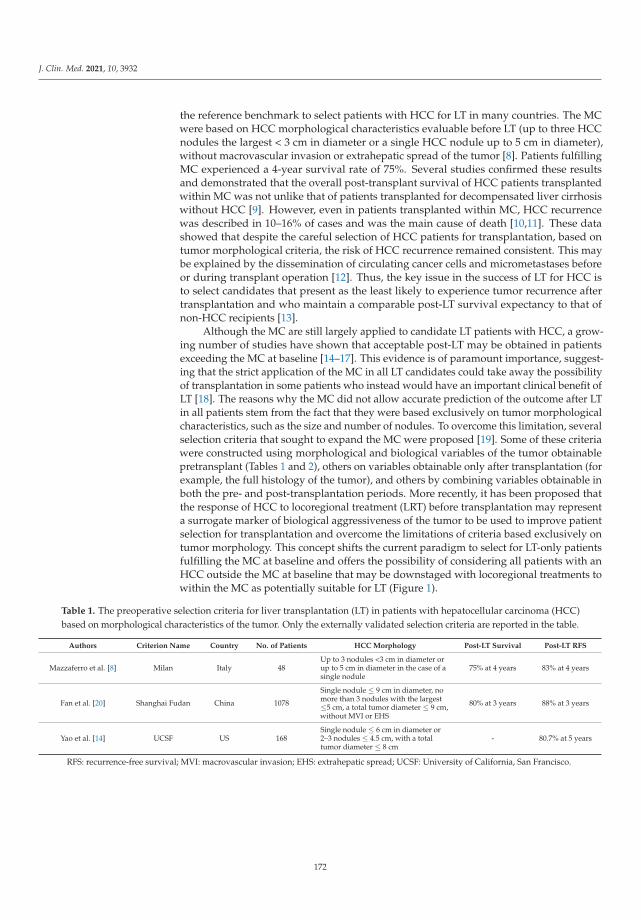
J. Clin. Med. 2021, 10, 3932
the reference benchmark to select patients with HCC for LT in many countries. The MCwere based on HCC morphological characteristics evaluable before LT (up to three HCCnodules the largest < 3 cm in diameter or a single HCC nodule up to 5 cm in diameter),without macrovascular invasion or extrahepatic spread of the tumor [8]. Patients fulfillingMC experienced a 4-year survival rate of 75%. Several studies confirmed these resultsand demonstrated that the overall post-transplant survival of HCC patients transplantedwithin MC was not unlike that of patients transplanted for decompensated liver cirrhosiswithout HCC [9]. However, even in patients transplanted within MC, HCC recurrencewas described in 10–16% of cases and was the main cause of death [10,11]. These datashowed that despite the careful selection of HCC patients for transplantation, based ontumor morphological criteria, the risk of HCC recurrence remained consistent. This maybe explained by the dissemination of circulating cancer cells and micrometastases beforeor during transplant operation [12]. Thus, the key issue in the success of LT for HCC isto select candidates that present as the least likely to experience tumor recurrence aftertransplantation and who maintain a comparable post-LT survival expectancy to that ofnon-HCC recipients [13].
Although the MC are still largely applied to candidate LT patients with HCC, a grow-ing number of studies have shown that acceptable post-LT may be obtained in patientsexceeding the MC at baseline [14–17]. This evidence is of paramount importance, suggest-ing that the strict application of the MC in all LT candidates could take away the possibilityof transplantation in some patients who instead would have an important clinical benefit ofLT [18]. The reasons why the MC did not allow accurate prediction of the outcome after LTin all patients stem from the fact that they were based exclusively on tumor morphologicalcharacteristics, such as the size and number of nodules. To overcome this limitation, severalselection criteria that sought to expand the MC were proposed [19]. Some of these criteriawere constructed using morphological and biological variables of the tumor obtainablepretransplant (Tables 1 and 2), others on variables obtainable only after transplantation (forexample, the full histology of the tumor), and others by combining variables obtainable inboth the pre- and post-transplantation periods. More recently, it has been proposed thatthe response of HCC to locoregional treatment (LRT) before transplantation may representa surrogate marker of biological aggressiveness of the tumor to be used to improve patientselection for transplantation and overcome the limitations of criteria based exclusively ontumor morphology. This concept shifts the current paradigm to select for LT-only patientsfulfilling the MC at baseline and offers the possibility of considering all patients with anHCC outside the MC at baseline that may be downstaged with locoregional treatments towithin the MC as potentially suitable for LT (Figure 1).
Table 1. The preoperative selection criteria for liver transplantation (LT) in patients with hepatocellular carcinoma (HCC)based on morphological characteristics of the tumor. Only the externally validated selection criteria are reported in the table.
Authors Criterion Name Country No. of Patients HCC Morphology Post-LT Survival Post-LT RFS
Mazzaferro et al. [8] Milan Italy 48Up to 3 nodules <3 cm in diameter orup to 5 cm in diameter in the case of asingle nodule
75% at 4 years 83% at 4 years
Fan et al. [20] Shanghai Fudan China 1078
Single nodule ≤ 9 cm in diameter, nomore than 3 nodules with the largest≤5 cm, a total tumor diameter ≤ 9 cm,without MVI or EHS
80% at 3 years 88% at 3 years
Yao et al. [14] UCSF US 168Single nodule ≤ 6 cm in diameter or2–3 nodules ≤ 4.5 cm, with a totaltumor diameter ≤ 8 cm
- 80.7% at 5 years
RFS: recurrence-free survival; MVI: macrovascular invasion; EHS: extrahepatic spread; UCSF: University of California, San Francisco.
172
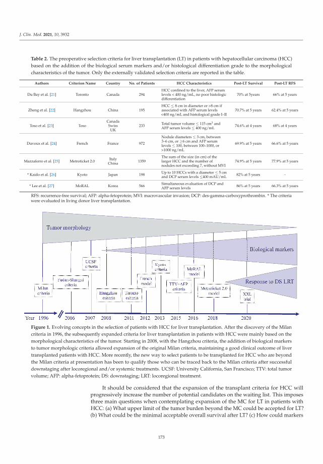
J. Clin. Med. 2021, 10, 3932
Table 2. The preoperative selection criteria for liver transplantation (LT) in patients with hepatocellular carcinoma (HCC)based on the addition of the biological serum markers and/or histological differentiation grade to the morphologicalcharacteristics of the tumor. Only the externally validated selection criteria are reported in the table.
Authors Criterion Name Country No. of Patients HCC Characteristics Post-LT Survival Post-LT RFS
Du Bay et al. [21] Toronto Canada 294HCC confined to the liver, AFP serumlevels < 400 ng/mL, no poor histologicdifferentiation
70% at 5years 66% at 5 years
Zheng et al. [22] Hangzhou China 195HCC ≤ 8 cm in diameter or >8 cm ifassociated with AFP serum levels<400 ng/mL and histological grade I–II
70.7% at 5 years 62.4% at 5 years
Toso et al. [23] TosoCanadaSwissUK
233 Total tumor volume ≤ 115 cm3 andAFP serum levels ≤ 400 ng/mL 74.6% at 4 years 68% at 4 years
Duvoux et al. [24] French France 972
Nodule diameters ≤ 3 cm, between3–6 cm, or ≥6 cm and AFP serumlevels ≤ 100, between 100–1000, or>1000 ng/mL
69.9% at 5 years 66.6% at 5 years
Mazzaferro et al. [25] Metroticket 2.0 ItalyChina 1359
The sum of the size (in cm) of thelarger HCC and the number ofnodules not exceeding 7, without MVI
74.9% at 5 years 77.9% at 5 years
* Kaido et al. [26] Kyoto Japan 198 Up to 10 HCCs with a diameter ≤ 5 cmand DCP serum levels ≤400 mAU/mL 82% at 5 years -
* Lee et al. [27] MoRAL Korea 566 Simultaneous evaluation of DCP andAFP serum levels 86% at 5 years 66.3% at 5 years
RFS: recurrence-free survival; AFP: alpha-fetoprotein; MVI: macrovascular invasion; DCP: des-gamma-carboxyprothrombin. * The criteriawere evaluated in living donor liver transplantation.
Figure 1. Evolving concepts in the selection of patients with HCC for liver transplantation. After the discovery of the Milancriteria in 1996, the subsequently expanded criteria for liver transplantation in patients with HCC were mainly based on themorphological characteristics of the tumor. Starting in 2008, with the Hangzhou criteria, the addition of biological markersto tumor morphologic criteria allowed expansion of the original Milan criteria, maintaining a good clinical outcome of livertransplanted patients with HCC. More recently, the new way to select patients to be transplanted for HCC who are beyondthe Milan criteria at presentation has been to qualify those who can be traced back to the Milan criteria after successfuldownstaging after locoregional and/or systemic treatments. UCSF: University California, San Francisco; TTV: total tumorvolume; AFP: alpha-fetoprotein; DS: downstaging; LRT: locoregional treatment.
It should be considered that the expansion of the transplant criteria for HCC willprogressively increase the number of potential candidates on the waiting list. This imposesthree main questions when contemplating expansion of the MC for LT in patients withHCC: (a) What upper limit of the tumor burden beyond the MC could be accepted for LT?(b) What could be the minimal acceptable overall survival after LT? (c) How could markers
173

J. Clin. Med. 2021, 10, 3932
of tumor biology, in addition to the tumor burden, be incorporated to better select patientsfor LT?
This essential review aims to present the current data on the pretransplant selectioncriteria for LT in patients with HCC exceeding the MC at presentation based on morpho-logical and histological characteristics of the tumor and to critically discuss those that havebeen validated in clinical practice. Moreover, the role of HCC biological markers and thetumor response to downstaging procedures as new tools for selecting patients for LT witha baseline tumor burden outside of the MC will be evaluated.
2. Selection Criteria for LT Based on HCC Morphological Characteristics
A common theme in the morphological criteria adopted to select patients with HCCwas to exclude those patients presenting HCC with macrovascular invasion and/or ex-trahepatic spread. The main reason why morphological pre-LT criteria improved theaccuracy in the selection of patients with HCC for LT derives from the demonstration thatboth the number and the size of HCC nodules can be considered surrogate markers ofhistologic microvascular invasion (MVI) and/or poor tumor differentiation [28,29], whichare the main determinants of HCC recurrence and death after LT [30]. In addition to MC,two pretransplant expansive morphological criteria were adopted and validated in cohortsof patients of various geographical origins. The Shanghai Fudan criteria were originallydeveloped in patients with chronic HBV infection. Patients transplanted with a singleHCC nodule ≤ 9 cm in diameter or with up to three lesions (the largest ≤ 5 cm) but with amaximum tumor diameter of ≤9 cm experienced 1- and 3-year post-LT survival and HCCrecurrence-free survival comparable with those transplanted within the MC [20]. Thesecriteria were subsequently validated in more than 1000 patients enrolled in liver transplantcenters in Shanghai [31]. In the United States, a further criterion aimed at expanding theoriginal MC was developed at the University of California, San Francisco (UCSF) [14]. TheUCSF criteria were developed after the observation that patients transplanted with a singlenodule of HCC ≤ 6.5 cm or with up to 2–3 lesions ≤ 4.5 cm each, maintaining the totaltumor diameter ≤ 8 cm, experienced a 5-year post-LT recurrence-free survival of 80.7%,which was not significantly different than that obtained in patients transplanted withinthe MC.
Both new criteria increased, albeit slightly, the size and/or the number of HCC nod-ules to select patients for LT compared to what was originally established by the MC. Thecertainty of always being able to carry out an accurate and reproducible measurement ofthe size and number of nodules to capture millimeter differences with respect to the MCprobably represents the greatest limitation of the new criteria based exclusively on the mor-phological characteristics of HCC. There is great heterogeneity and different accuracies ofliver imaging techniques applied to detect liver nodules and to properly characterize them,such as contrast-enhanced computed tomography (CT) or magnetic resonance imaging(MRI) [19]. Several reports have indicated that as many as 20–25% of patients undergoingLT for HCC have been inaccurately staged when only imaging techniques were used [32,33].To overcome this important drawback, the American College of Radiology created theLiver Imaging Reporting and Data System (LI-RADS), aimed at standardizing the processof acquisition, interpretation, reporting, and data collection for liver imaging [34]. LI-RADSstratified the characteristics of liver lesions into five categories starting from LR-1 (definitelybenign) to LR-5 (definitely HCC). This system is adopted in many countries to allow astandardized differential diagnosis of liver nodules in patients at high risk of HCC. Despitethis improvement in the radiological categorization of liver nodules, a recent meta-analysisshowed that LI-RADS was only 67% sensitive and 92% specific in diagnosing HCC [35].
3. Selection Criteria for LT Based on HCC Histological Characteristics
In patients transplanted beyond MC, a greater incidence of MVI, which is associatedwith higher post-LT tumor recurrence and death, has been demonstrated [36]. When theexplanted livers of patients within and outside the MC were compared, MVI was present
174

J. Clin. Med. 2021, 10, 3932
in 11% of the former group and in 42% of the latter group [37]. A potential tool to detectthe presence of MVI of HCC before LT is to perform liver biopsy of the nodules. Liverbiopsy of confirmed HCC in the explanted liver was performed before LT in a series of155 patients [33]. MVI was significantly more frequent in patients with larger nodules orwith multinodular HCC; furthermore, 68% of patients who experienced HCC recurrenceafter LT were positive for MVI. In addition to the detection of MVI, another potential toolemploying liver biopsy of HCC is to evaluate tumor histological grading. It has beendemonstrated that well-differentiated HCCs may be successfully transplanted, assuringa recipient 5-year post-LT survival of 75% despite approximately 30% of these patientsbeing beyond MC at the explant [38]. Although the clinical usefulness of pre-LT histologicassessment of tumor grading and MVI may be important, routine biopsy of HCC is notoften feasible, mainly due to the presence of multiple nodules or the risk of promotingseeding of cancer cells [39].
The current challenging question is whether the presence of MVI may be predictedby means of noninvasive methods. To answer this question, several authors propose theuse of imaging techniques such 18F-FDG PET/CT or gadoxetic acid enhanced dynamicMRI as noninvasive methods to predict the presence of MVI [40,41]. The rationale forusing 18F-FDG PET/CT in detecting MVI derives from the observation that the HCCgrowth rate and the activity of glycolytic enzymes are related [42]. This explains whypoorly differentiated HCC shows low glucose-6 phosphatase activity and high 18F-FDGuptake [43]. It has been proposed that good cutoff values for the SUVmax of HCC (SUVmaxT) and SUVmax of the normal liver (SUVmax L) in predicting the presence of MVI may be3.80 and 1.49, respectively [44]. The SUVmax T/SUVmean L ratio ≥ 1.2 was demonstratedto be significantly associated with the presence of MVI [45]. Compared with 18F-FDGPET/CT alone, the addition of gadoxetic acid enhanced dynamic MRI is a promisingtechnology that may further improve the sensitivity and specificity of MVI detection. Thiswas confirmed by applying the combination of MRI and PET/CT, as the sensitivity andspecificity in predicting the presence of MVI were 78.6% and 80%, respectively [40]. Theseobservations have been confirmed in clinical studies conducted in Asia in living donorliver transplantation. In Japan, 182 living donor liver transplanted patients with HCCwere studied by means of 18F-FDG PET/CT and the serum levels of alpha-fetoprotein(AFP) before LT. In recipients transplanted beyond MC who presented negative 18F-FDG PET/CT and AFP serum levels < 115 ng/mL, the 5-year HCC recurrence rate wascomparable to those transplanted within MC [46]. Very similar results were obtained in aKorean study also conducted in patients transplanted outside the Milan and UCSF criteriausing living donors. The authors demonstrated that these two groups of patients, if theypresented a negative 18F-FDG PET/CT, experienced 5-year post-LT HCC-free survivalrates of 73.3% and 72.8%, respectively [47]. Although these results are encouraging, it isnecessary to confirm them in populations of different ethnic groups, where the causesof LT are often different from those present in Asia, as well as in patients transplantedusing deceased donors. At the present time, the combined use of 18F-FDG PET/CT andgadoxetic acid enhanced MRI could be applied in addition to the morphological andbiological characteristics of HCC to stratify the risk of MVI.
4. Selection Criteria Based on Serum Biological Marker Measurements
The measurement of serum markers referring to biological tumor characteristicsand/or to host immune system reactivity has been considered a fascinating approachto overcome the limits of morphological criteria. Three main categories of measurablebiological markers are currently available: (a) serum markers related to the biological char-acteristics of HCC, such as AFP and des-gamma-carboxyprothrombin (DCP); (b) markersreflecting systemic host inflammation (neutrophil-to-lymphocyte ratio and platelet-to-lymphocyte ratio); and (c) molecular biomarkers that may be measured both in liver tissueand in serum (genetic mutations, enzymes, and microRNAs). In relation to the clinical
175

J. Clin. Med. 2021, 10, 3932
purpose of this review, only serum markers will be discussed, as there is still no solidevidence justifying the use of inflammation and molecular biomarkers in clinical practice.
5. Selection Criteria Based on the Addition of AFP and/or DCP Serum LevelMeasurements to HCC Morphology
AFP is considered a marker of HCC differentiation and vascular invasion; thus, themeasurement of its serum levels pre-LT has been proposed as a potential tool to identifyHCC patients with a higher risk of tumor recurrence and poor post-LT survival who shouldbe excluded from transplantation [48]. The limitations of this approach became imme-diately evident, since it was very difficult to apply AFP measurement in a standardizedand reproducible timeframe. For example, in deceased donor liver transplantation, thedate of the transplant is never predictable. Thus, the optimal time interval between trans-plantation and AFP measurement that would make it a predictive marker of recurrencewas unclear [19]. Instead of adopting cutoff values of AFP, an interesting approach is toconsider multiple AFP measurements to calculate a trend in the increase or decrease in itsserum levels. Patients experiencing an increase in serum AFP > 15 ng/mL/month had ahigher frequency of waitlist dropout or significantly worse post-LT survival than those witha lower (≤15 ng/mL/month) increase in AFP (54% vs. 94%) [49]. These results suggestthat the variations in AFP serum levels, rather than the last AFP level available before LT,may be more accurate in predicting post-LT outcome. [50]. This concept is incorporated toincrease the accuracy of the assessment of tumor downstaging before LT. For example, arapid increase in AFP serum levels after a presumed successful downstaging, assessed byradiologic imaging, should be considered a predictor of poor post-transplant outcome.
A very attractive way to select in patients beyond MC at baseline those with a higherrisk of HCC recurrence is to add AFP serum levels to the morphologic characteristicsof HCC [51]. The Toronto criteria [21] were developed assuming that all patients withHCC may have acceptable post-LT survival independent of the number and/or size ofthe nodules if HCC was confined to the liver, well differentiated at histology, and withoutmacrovascular invasion. These criteria identified AFP serum levels > 400 IU/mL at thetime of transplant as an independent predictor of worse 5-year disease-free survival. TheToronto criteria were subsequently validated, confirming that AFP serum levels before LTwere strongly associated with post-LT survival and HCC recurrence [52].
Similar results, combining the morphology and histology of HCC with AFP serum lev-els, have been obtained in China, where the Hangzhou criteria were developed [22]. Thesecriteria selected HCC patients for LT in the absence of portal vein tumor invasion and witheither HCC ≤ 8 cm in diameter or with HCC ≥ 8 cm in diameter but with concurrent AFPserum levels < 400 ng/mL and histological grade I or II. Patients fulfilling the Hangzhoucriteria experienced 1- and 3-year survival rates very similar to those reported in patientstransplanted within the MC. The combination of AFP serum levels and morphologic char-acteristics of the tumor also inspired European authors to expand the MC for selectingpatients with HCC for LT. Toso et al. [53] evaluated a large cohort of 6000 European LTpatients and demonstrated that the subgroup of recipients who presented a total tumorvolume (TTV) of ≤115 cm3 and AFP serum levels ≤ 400 ng/mL experienced a lower risk ofHCC recurrence and better survival after LT. It should be highlighted that these expandedcriteria were more effective than both the Milan and UCSF criteria in selecting patients witha low risk of HCC recurrence for LT. The TTV-AFP criteria were subsequently validated incohorts of patients in countries outside Europe and in Canada [23]. A further model thatcombined AFP serum levels and morphologic characteristics of HCC, known as the AFPmodel, was proposed by the Liver Transplantation French Group [24]. The AFP modelmerges AFP serum levels and the size and the number of nodules, attributing differentscores for each variable. Tumor sizes of 0, 1, or 4 points were assigned if the largest tumordiameter was ≤3 cm, 3–6 cm, or >6 cm, respectively. Moreover, 0 or 2 points were assignedif the number of nodules was ≤3 or ≥4. Regarding the AFP serum levels, 0, 2, or 3 pointswere assigned if AFP serum levels were ≤100, 100–1000, or >1000 ng/mL, respectively. Themaximum score sum of the AFP model is 9. Patients may be divided into low risk of HCC
176

J. Clin. Med. 2021, 10, 3932
recurrence if the final score is up to 2 points and high risk of HCC recurrence if the finalscore is ≥3. A very innovative observation of the study was that in patients presentingAFP serum levels > 1000 ng/mL, 3 points are attributed, irrespective of the number or sizeof nodules; thus, they may be immediately considered at high risk of HCC recurrence. TheAFP model was validated in different countries and in living donor liver transplantation,confirming its clinical utility in stratifying the recurrence risk of HCC after LT in a betterway than MC [54–57]. The Metroticket 2.0 model [25], developed in Italy, was based on themeasurement of the sum of the number and size of nodules and the log10 AFP level. Recip-ients with AFP levels < 200 ng/mL and with the sum of the number and size of tumors(in centimeters) not exceeding 7 presented a post-LT survival probability of 70%, whichwas comparable to that observed in patients transplanted within MC. To maintain thisexcellent clinical outcome in the presence of AFP levels of 200–400 ng/mL, the sum of thenumber and size of tumors should be reduced to ≤5, and if AFP levels are 400–1000 ng/mL,the sum of the number and size of tumors should be further reduced to ≤4. This modeloutperformed the original MC, UCSF, and AFP French models in identifying patients withexcellent 5-year post-LT survival.
The results obtained by combining the morphological characteristics of the tumorand the AFP values made it possible to develop selection criteria for LT that definitivelyexceeded those of Milan. These models introduced many innovations to more accuratelyselect patients with HCC for LT. First, these selection criteria made it possible to offer LTto many patients with HCC who would have been excluded by application of the MC,assuring excellent post-transplant survival. Second, the calculation of both the size andnumber of nodules and the AFP serum levels appears simple and available in every context,making these models applicable in different geographical contexts and with all types ofpatients. Third, these models may be used “dynamically”, in addition to the assessment ofthe HCC response to neoadjuvant treatments, to more accurately select patients who willundergo tumor downstaging before LT.
The interest in measuring DCP serum levels was derived from the observation thatsome HCCs expressed normal levels of AFP but increased levels of DCP. These subtypes ofHCCs present a poor grade of differentiation and frequent MVI [58,59]. The combinationof morphological characteristics of HCC (up to 10 nodules ≤ 5 cm in diameter) and DCPserum levels (≤400 mAU/mL) are the key elements included in the Kyoto criteria [26,60,61].Patients beyond MC who fulfilled the Kyoto criteria at the time of LT had similar post-LTprognoses in terms of survival and HCC recurrence compared to those within MC [60]. Inthe context of living donor liver transplantation, the Kyushu criteria [62] that are consideredsuitable for LT patients with any number of HCC < 5 cm in diameter and DCP serum levels< 300 mAU/mL were developed. These criteria appeared more accurate for predictingHCC recurrence than both the Kyoto and UCSF criteria but only when living donation wasconsidered [63,64].
Another interesting way to construct a prediction model of post-LT clinical outcomein patients with HCC beyond MC is to combine DCP and AFP serum levels. Startingfrom this assumption, the MoRAL model was developed in living donor liver transplantpatients exceeding the MC [27]. Both AFP and DCP serum levels were significantlyassociated with the time elapsed from transplantation to HCC recurrence. In the group ofrecipients exceeding the MC, a MoRAL score ≤ 314.8 was predictive to select patients withsignificantly longer (66.3%) 5-year recurrence-free and overall (82.1%) survival. In contrast,the group of recipients fulfilling the MC but with a MoRAL score > 314.8 showed a higherrisk of HCC recurrence and lower post-LT survival than patients beyond MC with a lowMoRAL score. A retrospective study evaluating the combination of DCP and AFP serumlevels in predicting clinical outcome in liver transplant patients outside the MC was alsoconducted in the United States [65]. In this study, AFP and DCP serum levels ≥ 250 ng/mLand ≥7.5 ng/mL, respectively, were predictive of more frequent HCC recurrence. WhenAFP and DCP were combined with MC, the hazard ratio of HCC recurrence risk increasedfrom 2.6 for patients beyond MC to 8.6 when AFP serum levels were ≥250 ng/mL and to
177
J. Clin. Med. 2021, 10, 3932
7.2 when DCP serum levels were ≥7.5 ng/mL. The encouraging results deriving from theuse of DCP as post-transplant survival as well as post-transplant HCC recurrence predictorshould be analyzed with caution. It should be emphasized that more than 90% of thepublished papers related to DCP have been produced in Asian countries and refer to livingdonor liver transplanted patients for liver diseases mainly related to HBV infection [66].Thus, solid data on the role of DCP in conditioning the clinical outcome of patients withHCC transplanted with deceased donors and with liver diseases due to nonviral etiologiesare still lacking. Considering these limitations, among all biological markers that havebeen studied, AFP remains the only one that has proven useful in predicting the clinicaloutcome in patients transplanted for HCC. The prognostic models of post-transplantsurvival that incorporate pre-LT AFP serum levels and HCC morphological characteristicsremain the most widely used to accurately select patients beyond MC for LT. In addition,the evaluation of AFP serum level variations induced by locoregional therapies and HCCdownstaging modalities is becoming the most promising strategy to select patients to betransplanted with HCC beyond the MC with even greater accuracy.
6. Selection Criteria Based on the Response of HCC to Bridging andDownstaging Treatments
A very innovative approach to select patients for LT presenting at baseline beyond theMC is to evaluate the characteristics of tumor response after LRT and consider it a surrogatemarker of biological HCC aggressiveness and of the risk of recurrence [19]. When LRT isused to control tumor growth with the aim of reducing the risk of waiting list dropout,it may be considered a “bridge” treatment to LT. The efficacy of LRT in reducing waitinglist dropout has been demonstrated if the waiting time for LT is at least 6 months [67].Patients whose tumor progression occurs despite LRT within 6 months have a worse post-LT outcome than those who achieved treatment response or the stability of HCC followingLRT [49,68,69]. These observations suggest that the response to LRT might be influencednot only by the treatment modality but also, more importantly, by the biological behaviorof the tumor.
The term “downstaging” is used by attributing the possibility of LRT decreasing thebaseline tumor burden until it meets the criteria for LT (ideally within MC) and to assureacceptable post-LT outcomes [36]. This concept was derived from some studies suggestingthat post-LT outcomes in patients successfully downstaged to the MC were comparablewith those observed in transplanting patients with MC at presentation [70,71]. The po-tential explanation of these findings is that a close correlation exists between successfuldownstaging and better explant histologic characteristics of the tumor [70–72].
As indicated by the Barcelona Clinic Liver Cancer (BCLC) staging system [73], amongthe LRT modalities that may be employed to perform downstaging of HCC, transarterialchemoembolization (TACE) is the most commonly utilized. Transarterial radioembolization(TARE) and ablative techniques may be often proposed [74].
It should be emphasized that there are important potential safety concerns of TACEand TARE in generating hepatic decompensation. In accordance with the guidelines forTACE [3], it has been strongly suggested that only patients with preserved liver function(Child–Pugh score A/B and bilirubin ≤ 3 mg/dL) should be considered for downstagingprocedures [75]. Although TACE is the recommended first-line treatment for downstagingobjectives in most studies [75–77], TARE may be considered an alternative treatment toTACE, particularly in larger HCCs, where the results are encouraging. However, thistreatment modality requires further studies to confirm its real utility as a downstaging pro-cedure [78]. The major limitation of downstaging protocols is that they can be applied onlyin a subgroup of patients who present simultaneously compensated cirrhosis complicatedwith HCC but cannot be applied in patients with decompensated cirrhosis with HCC.
A very challenging issue will soon be the potential role of neoadjuvant treatmentscombining systemic drugs such tyrosine kinase and checkpoint inhibitors in downstagingprotocols [79]. In a recent clinical trial, the combination of atezolizumab plus bevacizumabin the treatment of unresectable HCC was able to induce a complete and partial response
178
J. Clin. Med. 2021, 10, 3932
to therapy in 18% and 71% of treated patients, respectively [80]. These results openthe critical question of whether systemic treatments may be adopted in combinationand/or sequentially with traditional LRT to increase the chances of obtaining successfulHCC downstaging.
A further critical element that must be considered in the application of downstagingprocedures is the objective measurement of the treatment response. The modified ResponseEvaluation Criteria in Solid Tumors (mRECIST) were developed for the assessment oftreatment response by measuring tumor shrinkage. These criteria divided the rate ofresponse to treatment into four categories: (1) complete response (CR—disappearance ofarterial enhancement in tumor(s), (2) partial response (PR—a minimum 30% reduction inthe sum of diameters of viable tumors compared with baseline), (3) stable disease (SD—notmeeting PR or progressive disease), and (4) progressive disease (PD—an increase of at least20% in the sum of diameters of viable tumors compared with baseline or the appearance ofnew lesions) [81]. The usefulness of the mRECIST criteria in the evaluation of response toLRT after downstaging protocols in patients with HCC has been confirmed both in thosewithin and beyond the MC. In a small series of 33 patients presenting HCC outside theMC who underwent LT after downstaging performed by TACE, the 5-year survival wassignificantly higher in those who achieved CR (94.4%) than in those who had PR (45.4%)and SD (50%). These significant differences were explained by a progressive increasein HCC recurrence rates from patients with CR (15.5%) to those with SD (50%) to thosewith PR (53.3%) [82]. Similar results were obtained in the study performed by Kim et al.,which evaluated HCC recipients within and beyond MC after the TACE procedure [83].The 5-year HCC recurrence rate was 5.3% in patients who achieved CR or PR after TACEcompared with 17.6% in those who achieved SD or PD. It should be noted that althoughthe mRECIST criteria are sufficiently detailed, they may not be systematically adoptedamong different transplant centers, such that the results obtained by LRT may not becomparable [19].
In addition to the quality of the response to LRT, the duration of response is increas-ingly used as a surrogate marker to identify HCC with more aggressive behavior. Startingfrom this assumption, many liver transplant centers adopted the strategy to “ablate andwait” to assess the type and duration of response to LRT [9]. It has been suggested that thesuccess of downstaging should be assessed, demonstrating the absence of tumor progres-sion during an observation period of at least 3 months after the procedure. A successfuldownstaging procedure allows the selection of candidates with more favorable tumor biol-ogy and better post-LT survival [84]. This strategy avoided early post-LT recurrences, asdemonstrated in patients with HCC beyond MC transplanted after short waiting times [85].Thus, the guidelines of both the American and European associations for the study ofthe liver are concordant in recommending the adoption of LRT in patients with HCCbeyond MC and the consideration of those who achieved successful downstaging for atleast 3–6 months as suitable candidates for LT [3–86].
The question at this time is what the baseline and the final burdens of HCC obtainedafter successful downstaging that may be considered sufficient to perform LT should be. Inthe United States, the UCSF downstaging protocol [70] has recently been employed as anational policy both for the graduation of urgency to transplantation and to try to answerthis question. In this protocol, the baseline selection criteria for patients with HCC who maybenefit from LRT downstaging procedures before LT were as follows: a single HCC ≤ 8 cmin diameter; up to three lesions < 5 cm in diameter; or up to five nodules, all of them <3 cmin diameter, but in any case with a total tumor diameter < 8 cm. Retrospectively analyzingthe UNOS database, the 3819 liver transplanted patients with HCC were divided as alwayswithin the MC or achieved UNOS downstaging criteria (UNOS-DS). The 3-year post-LTsurvival was 83.2% in patients always within MC and 79.1% in those fulfilling UNOS-DS.Moreover, the 3-year HCC recurrence rate was 6.9% in recipients who were always withinMC and 12.8% for those within UNOS-DS. A very interesting issue that emerged from this
179
J. Clin. Med. 2021, 10, 3932
study was that AFP serum levels ≥ 100 ng/mL were the only independent predictor ofpost-LT HCC recurrence in downstaged groups [87].
To date, only one randomized clinical trial (the XXL trial) has evaluated the clinicaloutcome of patients presenting at baseline with HCC beyond MC who were successfullydownstaged by means of LRT and subsequently transplanted compared with those whoreceived only LRT [88]. This study was conducted in Italy and enrolled 74 patients withHCC beyond the MC, without macrovascular invasion or extrahepatic spread, with a5-year expected post-LT survival of at least 50% (estimated by Metroticket calculator [37])and preserved liver function (Child–Pugh classes A5–B7). All patients initially underwenttumor downstaging with LRT or systemic therapies, according to a multidisciplinarydecision. After an observation period of 3 months, during which treatment with sorafenibwas allowed, patients presenting CR or PR, assessed by means of the mRECIST criteria,were randomly assigned (1:1) to LT or to continue LRT or systemic treatments (controlgroup). Of note, in patients with baseline AFP values ≥ 400 ng/mL, a radiological tumorresponse was confirmed only in case of a parallel percentage decrease in AFP concentrations.In contrast, in patients with AFP serum levels < 400 ng/mL at recruitment, an increase inAFP concentrations above that cutoff value at the end of the downstaging phase or duringthe observation period was considered tumor progression, independent of radiologicalassessment. The primary endpoints of the study were the evaluation of 5-year tumor-event-free survival and overall survival. The first result of the study was that 29/74 (39.1%) ofthe patients dropped out before randomization; thus, only 45 (60.9%) patients were finallyincluded in the study (23 underwent LT, and 22 maintained LRT or systemic treatments).Despite the high rate of dropout, after a median follow-up of 71 months, a significantlyhigher 5-year tumor-event-free survival was observed in the LT group (76.8%) than inthe control group (18.3%). Regarding the 5-year overall survival, the figures were quitesimilar, indicating a better overall survival in the LT group (77.5%) than in the controlgroup (31.2%). Tumor progression was the main cause of death in both groups, while in theLT group, HCC recurrence was detected in 22% of cases. The results of this study provideadditional evidence to previous results demonstrating comparable post-LT outcomes inpatients with HCC beyond the MC who underwent successful downstaging within the MCand in those who underwent LT fulfilling the MC at presentation. A further very importantmessage from this study is that different schedules of LRT and systemic treatments maybe employed in patients with HCC beyond the MC to achieve successful and durabledownstaging to permit them to be suitable for LT.
7. Conclusions
LT must be considered the best treatment option for patients with unresectableHCC [89]. Since the number of donors is insufficient to satisfy all requests for trans-plantation for HCC, it is essential to perform careful selection of transplant candidates. Forapproximately 25 years, MC have been the benchmark for offering patients with HCC theopportunity for transplantation, but as recently demonstrated, they excluded a subset ofpatients who could have benefited from LT. For this reason, several other more extendedselection criteria to offer LT to an increasing number of patients with HCC have been eval-uated. In the beginning, many of the expanded criteria evaluable in the pre-LT period werebased on the morphological characteristics of the tumor as the original strategy adopted forconstructing the MC. Subsequently, the addition of biological markers, predominantly AFPserum levels, to the morphological characteristics of the tumor emerged as the more solidand reproducible criteria for patient selection beyond the MC for LT, assuring excellentpost-LT outcomes. Downstaging HCC to MC by means of LRT and/or systemic therapiesis becoming a valid and increasingly utilized method of patient selection for LT. Adoptingthis approach, the surrogates of tumor biology can be assessed, such as the response rateto LRT and its maintenance for a sufficient time during the waiting list before transplant.The measurement of AFP serum levels or AFP slope during or after downstaging protocolscan be considered a further important option to identify those patients at higher risk of
180

J. Clin. Med. 2021, 10, 3932
HCC recurrence that should be excluded from LT. It appears clear that the risk of failureof successful downstaging is related to both the tumor burden and to AFP serum levelsat baseline.
It is important to highlight that the selection of HCC patients for LT by means ofthe expanded criteria may be difficult to adopt in areas of the world with severe organshortages. In these areas, the selection criteria based on the utility principle that assuresthe maximum post-transplant survival, such as the MC, will remain preponderant, ratherthan expanded criteria that could reduce access to LT for patients with better post-LTprognosis [67–90]. This justified that not all liver transplant centers around the world adoptthe same criteria to select patients with HCC for LT [3–74]. Each country developed, basedon its scientific experiences, some selection criteria for transplanting patients with HCCbeyond MC. A common thread that links the various selection criteria adopted in differentcountries is to consider the morphological characteristics of the tumor (number and size ofnodules) and the values of some biological markers, mainly AFP, as main determinants ofthe selection process. In the United States and in Europe, the concept to not necessarilyset a baseline HCC limit size to consider patients potentially transplantable, except for thepresence of macrovascular invasion or extrahepatic spread, appears to be more accepted.Thus, the downstaging process will probably become the main selection tool for LT, en-abling clinicians to postpone the transplantation decision from tumor presentation to theassessment of final response to LRT [91]. The effectiveness of downstaging proceduresshould be considered as having brought the tumor back within the MC for a period of atleast 3–6 months before enlisting [84].
In summary, to try to answer the key questions reported in the introduction, the fol-lowing proposals can be suggested: (a) it seems proven that in the absence of macrovascularinvasion and extrahepatic spread, no upper limit of tumor burden beyond MC should beestablished “a priori” to determine transplant eligibility for HCC; (b) to justify a policy totransplant patients with HCC beyond MC at presentation, the minimal expected 5-yearpost-LT survival probability, estimated by Metroticket calculator, should be at least 50% [37];(c) the response to neoadjuvant LRT and/or systemic treatments in addition to the dynamicevaluation of AFP serum levels are expected to replace conventional morphological criteriafor selecting patients with HCC for LT in the near future.
Author Contributions: Conceptualization, P.T.; methodology, E.F. (Elisa Fumolo); data curation, E.F. (EzioFornasiere), D.B.; writing—original draft preparation, P.T., E.F. (Elisa Fumolo), E.F. (Ezio For-nasiere), andD.B.; writing—review and editing, P.T. All authors have read and agreed to the published version ofthe manuscript.
Funding: This research received no external funding.
Institutional Review Board Statement: Ethical review and approval were waived for this study dueto the fact that it refers to a review of the scientific literature in the field.
Informed Consent Statement: Not applicable.
Conflicts of Interest: The authors declare no conflict of interest.
References
1. White, D.L.; Thrift, A.P.; Kanwal, F.; Davila, J.; El-Serag, H.B. Incidence of Hepatocellular Carcinoma in All 50 United States, from2000 through 2012. Gastroenterology 2017, 152, 812–820.e5. [CrossRef] [PubMed]
2. Kulik, L.; El-Serag, H.B. Epidemiology and Management of Hepatocellular Carcinoma. Gastroenterology 2019, 156, 477–491.[CrossRef] [PubMed]
3. European Association for the Study of the Liver. EASL Clinical Practice Guidelines: Management of hepatocellular carcinoma. J.Hepatol. 2018, 69, 182–236. [CrossRef]
4. Vibert, E.; Schwartz, M.; Olthoff, K.M. Advances in resection and transplantation for hepatocellular carcinoma. J. Hepatol. 2020,72, 262–276. [CrossRef]
5. Starzl, T.E.; Marchioro, T.L.; Vonkaulla, K.N.; Hermann, G.; Brittain RSWaddell, W.R. Homotransplantation of the Liver inHumans. Surg. Gynecol. Obs. 1963, 117, 659–676.
181

J. Clin. Med. 2021, 10, 3932
6. Starzl, T.E.; Groth, C.G.; Brettschneider, L.; Penn, I.; Fulginiti, V.A.; Moon, J.B.; Blanchard, H.; Martin, A.J., Jr.; Porter, K.A.Orthotopic homotransplantation of the human liver. Ann. Surg. 1968, 168, 392–415. [CrossRef]
7. Iwatsuki, S.; Gordon, R.D.; Shaw, B.W., Jr.; Starzl, T.E. Role of liver transplantation in cancer therapy. Ann. Surg. 1985, 202,401–407. [CrossRef]
8. Mazzaferro, V.; Regalia, E.; Doci, R.; Andreola, S.; Pulvirenti, A.; Bozzetti, F.; Montalto, F.; Ammatuna, M.; Morabito, A.;Gennari, L. Liver transplantation for the treatment of small hepatocellular carcinomas in patients with cirrhosis. N. Engl. J. Med.1996, 334, 693–699. [CrossRef]
9. Lingiah, V.A.; Niazi, M.; Olivo, R.; Paterno, F.; Guarrera, J.V.; Pyrsopoulos, N.T. Liver Transplantation Beyond Milan Criteria. J.Clin. Transl. Hepatol. 2020, 8, 69–75. [CrossRef] [PubMed]
10. Plessier, A.; Codes, L.; Consigny, Y.; Sommacale, D.; Dondero, F.; Cortes, A.; Degos, F.; Brillet, P.Y.; Vilgrain, V.; Paradis, V.; et al.Underestimation of the influence of satellite nodules as a risk factor for post-transplantation recurrence in patients with smallhepatocellular carcinoma. Liver Transplant. 2004, 10, S86–S90. [CrossRef] [PubMed]
11. Escartin, A.; Sapisochin, G.; Bilbao, I.; Vilallonga, R.; Bueno, J.; Castells, L.; Dopazo, C.; Castro, E.; Caralt, M.; Balsells, J. Recurrenceof hepatocellular carcinoma after liver transplantation. Transplant. Proc. 2007, 39, 2308–2310. [CrossRef] [PubMed]
12. Roayaie, S.; Schwartz, J.D.; Sung, M.W.; Emre, S.H.; Miller, C.M.; Gondolesi, G.E.; Krieger, N.R.; Schwartz, M.E. Recurrence ofhepatocellular carcinoma after liver transplant: Patterns and prognosis. Liver Transplant. 2004, 10, 534–540. [CrossRef]
13. Bruix, J.; Fuster, J.; Llovet, J.M. Liver transplantation for hepatocellular carcinoma: Foucault pendulum versus evidence-baseddecision. Liver Transplant. 2003, 9, 700–702. [CrossRef] [PubMed]
14. Yao, F.Y.; Xiao, L.; Bass, N.M.; Kerlan, R.; Ascher, N.L.; Roberts, J.P. Liver transplantation for hepatocellular carcinoma: Validationof the UCSF-expanded criteria based on preoperative imaging. Am. J. Transplant. 2007, 7, 2587–2596. [CrossRef]
15. Herrero, J.I.; Sangro, B.; Pardo, F.; Quiroga, J.; Inarrairaegui, M.; Rotellar, F.; Montiel, C.; Alegre, F.; Prieto, J. Liver transplantationin patients with hepatocellular carcinoma across Milan criteria. Liver Transplant. 2008, 14, 272–278. [CrossRef]
16. Silva, M.; Moya, A.; Berenguer, M.; Sanjuan, F.; Lopez-Andujar, R.; Pareja, E.; Torres-Quevedo, R.; Aguilera, V.; Montalva, E.;De Juan, M.; et al. Expanded criteria for liver transplantation in patients with cirrhosis and hepatocellular carcinoma. LiverTransplant. 2008, 14, 1449–1460. [CrossRef]
17. Guiteau, J.J.; Cotton, R.T.; Washburn, W.K.; Harper, A.; O’Mahony, C.A.; Sebastian, A.; Cheng, S.; Klintmalm, G.; Ghobrial, M.;Halff, G.; et al. An early regional experience with expansion of Milan Criteria for liver transplant recipients. Am. J. Transplant.2010, 10, 2092–2098. [CrossRef]
18. Costentin, C.E.; Bababekov, Y.J.; Zhu, A.X.; Yeh, H. Is It Time to Reconsider the Milan Criteria for Selecting Patients WithHepatocellular Carcinoma for Deceased-Donor Liver Transplantation? Hepatology 2019, 69, 1324–1336. [CrossRef]
19. Toniutto, P.; Fornasiere, E.; Fumolo, E.; Bitetto, D. Risk factors for hepatocellular carcinoma recurrence after liver transplantation.Hepatoma Res. 2020, 6, 50–71. [CrossRef]
20. Fan, J.; Zhou, J.; Xu, Y.; Qiu, S.J.; Wu, Z.Q.; Yu, Y.; Huang, X.W.; Tang, Z.Y.; Wang, Y.Q. Indication of liver transplantation forhepatocellular carcinoma: Shanghai Fudan Criteria. Zhonghua Yi Xue Za Zhi 2006, 86, 1227–1231.
21. DuBay, D.; Sandroussi, C.; Sandhu, L.; Cleary, S.; Guba, M.; Cattral, M.S.; McGilvray, I.; Ghanekar, A.; Selzner, M.; Greig, P.D.; et al.Liver transplantation for advanced hepatocellular carcinoma using poor tumor differentiation on biopsy as an exclusion criterion.Ann. Surg. 2011, 253, 166–172. [CrossRef]
22. Zheng, S.S.; Xu, X.; Wu, J.; Chen, J.; Wang, W.L.; Zhang, M.; Liang, T.B.; Wu, L.M. Liver transplantation for hepatocellularcarcinoma: Hangzhou experiences. Transplantation 2008, 85, 1726–1732. [CrossRef]
23. Toso, C.; Meeberg, G.; Hernandez-Alejandro, R.; Dufour, J.F.; Marotta, P.; Majno, P.; Kneteman, N.M. Total tumor volume andalpha-fetoprotein for selection of transplant candidates with hepatocellular carcinoma: A prospective validation. Hepatology 2015,62, 158–165. [CrossRef]
24. Duvoux, C.; Roudot-Thoraval, F.; Decaens, T.; Pessione, F.; Badran, H.; Piardi, T.; Francoz, C.; Compagnon, P.; Vanlemmens,C.; Dumortier, J.; et al. Liver transplantation for hepatocellular carcinoma: A model including alpha-fetoprotein improves theperformance of Milan criteria. Gastroenterology 2012, 143, 986–994 e3. [CrossRef]
25. Mazzaferro, V.; Sposito, C.; Zhou, J.; Pinna, A.D.; De Carlis, L.; Fan, J.; Cescon, M.; Di Sandro, S.; Yi-Feng, H.; Lauterio, A.; et al.Metroticket 2.0 Model for Analysis of Competing Risks of Death After Liver Transplantation for Hepatocellular Carcinoma.Gastroenterology 2018, 154, 128–139. [CrossRef]
26. Kaido, T.; Ogawa, K.; Mori, A.; Fujimoto, Y.; Ito, T.; Tomiyama, K.; Takada, Y.; Uemoto, S. Usefulness of the Kyoto criteria asexpanded selection criteria for liver transplantation for hepatocellular carcinoma. Surgery 2013, 154, 1053–1060. [CrossRef]
27. Lee, J.H.; Cho, Y.; Kim, H.Y.; Cho, E.J.; Lee, D.H.; Yu, S.J.; Lee, J.W.; Yi, N.J.; Lee, K.W.; Kim, S.H.; et al. Serum Tumor MarkersProvide Refined Prognostication in Selecting Liver Transplantation Candidate for Hepatocellular Carcinoma Patients Beyond theMilan Criteria. Ann. Surg. 2016, 263, 842–850. [CrossRef]
28. Shah, S.A.; Tan, J.C.; McGilvray, I.D.; Cattral, M.S.; Levy, G.A.; Greig, P.D.; Grant, D.R. Does microvascular invasion affectoutcomes after liver transplantation for HCC? A histopathological analysis of 155 consecutive explants. J. Gastrointest. Surg. 2007,11, 464–471. [CrossRef]
29. Zhang, X.; Li, J.; Shen, F.; Lau, W.Y. Significance of presence of microvascular invasion in specimens obtained after surgicaltreatment of hepatocellular carcinoma. J. Gastroenterol. Hepatol. 2018, 33, 347–354. [CrossRef] [PubMed]
182

J. Clin. Med. 2021, 10, 3932
30. Jonas, S.; Bechstein, W.O.; Steinmuller, T.; Herrmann, M.; Radke, C.; Berg, T.; Settmacher, U.; Neuhaus, P. Vascular invasion andhistopathologic grading determine outcome after liver transplantation for hepatocellular carcinoma in cirrhosis. Hepatology 2001,33, 1080–1086. [CrossRef]
31. Fan, J.; Yang, G.S.; Fu, Z.R.; Peng, Z.H.; Xia, Q.; Peng, C.H.; Qian, J.M.; Zhou, J.; Xu, Y.; Qiu, S.J.; et al. Liver transplantationoutcomes in 1,078 hepatocellular carcinoma patients: A multi-center experience in Shanghai, China. J. Cancer Res. Clin. Oncol.2009, 135, 1403–1412. [CrossRef]
32. Sotiropoulos, G.C.; Malago, M.; Molmenti, E.; Paul, A.; Nadalin, S.; Brokalaki, E.; Kuhl, H.; Dirsch, O.; Lang, H.; Broelsch, C.E.Liver transplantation for hepatocellular carcinoma in cirrhosis: Is clinical tumor classification before transplantation realistic?Transplantation 2005, 79, 483–487. [CrossRef]
33. Shah, S.A.; Tan, J.C.; McGilvray, I.D.; Cattral, M.S.; Cleary, S.P.; Levy, G.A.; Greig, P.D.; Grant, D.R. Accuracy of staging as apredictor for recurrence after liver transplantation for hepatocellular carcinoma. Transplantation 2006, 81, 1633–1639. [CrossRef]
34. Kierans, A.S.; Song, C.; Gavlin, A.; Roudenko, A.; Lu, L.; Askin, G.; Hecht, E.M. Diagnostic Performance of LI-RADS Version 2018,LI-RADS Version 2017, and OPTN Criteria for Hepatocellular Carcinoma. AJR Am. J. Roentgenol. 2020, 215, 1085–1092. [CrossRef]
35. Lee, S.; Kim, S.S.; Roh, Y.H.; Choi, J.Y.; Park, M.S.; Kim, M.J. Diagnostic Performance of CT/MRI Liver Imaging Reportingand Data System v2017 for Hepatocellular Carcinoma: A Systematic Review and Meta-Analysis. Liver Int. 2020, 40, 1488–1497.[CrossRef]
36. Rudnick, S.R.; Russo, M.W. Liver transplantation beyond or downstaging within the Milan criteria for hepatocellular carcinoma.Expert Rev. Gastroenterol. Hepatol. 2018, 12, 265–275. [CrossRef]
37. Mazzaferro, V.; Llovet, J.M.; Miceli, R.; Bhoori, S.; Schiavo, M.; Mariani, L.; Camerini, T.; Roayaie, S.; Schwartz, M.E.;Grazi, G.L.; et al. Predicting survival after liver transplantation in patients with hepatocellular carcinoma beyond the Milancriteria: A retrospective, exploratory analysis. Lancet Oncol. 2009, 10, 35–43. [CrossRef]
38. Cillo, U.; Vitale, A.; Bassanello, M.; Boccagni, P.; Brolese, A.; Zanus, G.; Burra, P.; Fagiuoli, S.; Farinati, F.; Rugge, M.; et al. Livertransplantation for the treatment of moderately or well-differentiated hepatocellular carcinoma. Ann. Surg. 2004, 239, 150–159.[CrossRef]
39. Cuccurullo, V.; Di Stasio, G.D.; Mazzarella, G.; Cascini, G.L. Microvascular Invasion in HCC: The Molecular Imaging Perspective.Contrast Media Mol. Imaging 2018, 2018, 9487938. [CrossRef]
40. Yaprak, O.; Acar, S.; Ertugrul, G.; Dayangac, M. Role of pre-transplant 18F-FDG PET/CT in predicting hepatocellular carcinomarecurrence after liver transplantation. World J. Gastrointest. Oncol. 2018, 10, 336–343. [CrossRef] [PubMed]
41. Choi, S.H.; Byun, J.H.; Kwon, H.J.; Ha, H.I.; Lee, S.J.; Kim, S.Y.; Won, H.J.; Kim, P.N. The usefulness of gadoxetic acid-enhanceddynamic magnetic resonance imaging in hepatocellular carcinoma: Toward improved staging. Ann. Surg. Oncol. 2015, 22,819–825. [CrossRef]
42. Sweeney, M.J.; Ashmore, J.; Morris, H.P.; Weber, G. Comparative Biochemistry Hepatomas. Iv. Isotope Studies of Glucose andFructose Metabolism in Liver Tumors of Different Growth Rates. Cancer Res. 1963, 23, 995–1002. [PubMed]
43. Torizuka, T.; Tamaki, N.; Inokuma, T.; Magata, Y.; Sasayama, S.; Yonekura, Y.; Tanaka, A.; Yamaoka, Y.; Yamamoto, K.; Konishi, J.In vivo assessment of glucose metabolism in hepatocellular carcinoma with FDG-PET. J. Nucl. Med. 1995, 36, 1811–1817. [PubMed]
44. Lin, C.Y.; Liao, C.W.; Chu, L.Y.; Yen, K.Y.; Jeng, L.B.; Hsu, C.N.; Lin, C.L.; Kao, C.H. Predictive Value of 18F-FDG PET/CT forVascular Invasion in Patients With Hepatocellular Carcinoma Before Liver Transplantation. Clin. Nucl. Med. 2017, 42, e183–e187.[CrossRef]
45. Ahn, S.Y.; Lee, J.M.; Joo, I.; Lee, E.S.; Lee, S.J.; Cheon, G.J.; Han, J.K.; Choi, B.I. Prediction of microvascular invasion ofhepatocellular carcinoma using gadoxetic acid-enhanced MR and (18)F-FDG PET/CT. Abdom. Imaging 2015, 40, 843–851.[CrossRef] [PubMed]
46. Takada, Y.; Kaido, T.; Shirabe, K.; Nagano, H.; Egawa, H.; Sugawara, Y.; Taketomi, A.; Takahara, T.; Wakabayashi, G.;Nakanishi, C.; et al. Significance of preoperative fluorodeoxyglucose-positron emission tomography in prediction of tumorrecurrence after liver transplantation for hepatocellular carcinoma patients: A Japanese multicenter study. J. Hepatobiliary Pancreat.Sci. 2017, 24, 49–57. [CrossRef]
47. Lee, S.D.; Kim, S.H.; Kim, S.K.; Kim, Y.K.; Park, S.J. Clinical Impact of 18F-Fluorodeoxyglucose Positron Emission Tomogra-phy/Computed Tomography in Living Donor Liver Transplantation for Advanced Hepatocellular Carcinoma. Transplantation2015, 99, 2142–2149. [CrossRef]
48. Hakeem, A.R.; Young, R.S.; Marangoni, G.; Lodge, J.P.; Prasad, K.R. Systematic review: The prognostic role of alpha-fetoproteinfollowing liver transplantation for hepatocellular carcinoma. Aliment. Pharmacol. Ther. 2012, 35, 987–999. [CrossRef]
49. Lai, Q.; Avolio, A.W.; Graziadei, I.; Otto, G.; Rossi, M.; Tisone, G.; Goffette, P.; Vogel, W.; Pitton, M.B.; Lerut, J.; et al. Alpha-fetoprotein and modified response evaluation criteria in solid tumors progression after locoregional therapy as predictors ofhepatocellular cancer recurrence and death after transplantation. Liver Transplant. 2013, 19, 1108–1118. [CrossRef]
50. Dumitra, T.C.; Dumitra, S.; Metrakos, P.P.; Barkun, J.S.; Chaudhury, P.; Deschenes, M.; Paraskevas, S.; Hassanain, M.;Tchervenkov, J.I. Pretransplantation alpha-fetoprotein slope and milan criteria: Strong predictors of hepatocellular carcinomarecurrence after transplantation. Transplantation 2013, 95, 228–233. [CrossRef]
51. Burra, P.; Giannini, E.G.; Caraceni, P.; Ginanni Corradini, S.; Rendina, M.; Volpes, R.; Toniutto, P. Specific issues concerning themanagement of patients on the waiting list and after liver transplantation. Liver Int. 2018, 38, 1338–1362. [CrossRef] [PubMed]
183
J. Clin. Med. 2021, 10, 3932
52. Sapisochin, G.; Goldaracena, N.; Laurence, J.M.; Dib, M.; Barbas, A.; Ghanekar, A.; Cleary, S.P.; Lilly, L.; Cattral, M.S.;Marquez, M.; et al. The extended Toronto criteria for liver transplantation in patients with hepatocellular carcinoma: A prospec-tive validation study. Hepatology 2016, 64, 2077–2088. [CrossRef]
53. Toso, C.; Asthana, S.; Bigam, D.L.; Shapiro, A.M.; Kneteman, N.M. Reassessing selection criteria prior to liver transplantationfor hepatocellular carcinoma utilizing the Scientific Registry of Transplant Recipients database. Hepatology 2009, 49, 832–838.[CrossRef]
54. Notarpaolo, A.; Layese, R.; Magistri, P.; Gambato, M.; Colledan, M.; Magini, G.; Miglioresi, L.; Vitale, A.; Vennarecci, G.;Ambrosio, C.D.; et al. Validation of the AFP model as a predictor of HCC recurrence in patients with viral hepatitis-relatedcirrhosis who had received a liver transplant for HCC. J. Hepatol. 2017, 66, 552–559. [CrossRef] [PubMed]
55. Varona, M.A.; Soriano, A.; Aguirre-Jaime, A.; Garrido, S.; Oton, E.; Diaz, D.; Portero, J.; Bravo, P.; Barrera, M.A.; Perera, A.Risk factors of hepatocellular carcinoma recurrence after liver transplantation: Accuracy of the alpha-fetoprotein model in asingle-center experience. Transplant. Proc. 2015, 47, 84–89. [CrossRef]
56. Pinero, F.; Tisi Bana, M.; de Ataide, E.C.; Hoyos Duque, S.; Marciano, S.; Varon, A.; Anders, M.; Zerega, A.; Menendez, J.;Zapata, R.; et al. Liver transplantation for hepatocellular carcinoma: Evaluation of the alpha-fetoprotein model in a multicentercohort from Latin America. Liver Int. 2016, 36, 1657–1667. [CrossRef]
57. Rhu, J.; Kim, J.M.; Choi, G.S.; Kwon, C.H.D.; Joh, J.W. Validation of the alpha-fetoprotein Model for Hepatocellular CarcinomaRecurrence After Transplantation in an Asian Population. Transplantation 2018, 102, 1316–1322. [CrossRef] [PubMed]
58. Okuda, H.; Nakanishi, T.; Takatsu, K.; Saito, A.; Hayashi, N.; Yamamoto, M.; Takasaki, K.; Nakano, M. Comparison of clinico-pathological features of patients with hepatocellular carcinoma seropositive for alpha-fetoprotein alone and those seropositive fordes-gamma-carboxy prothrombin alone. J. Gastroenterol. Hepatol. 2001, 16, 1290–1296. [CrossRef]
59. Hong, Y.M.; Cho, M.; Yoon, K.T.; Chu, C.W.; Yang, K.H.; Park, Y.M.; Rhu, J.H. Risk factors of early recurrence after curativehepatectomy in hepatocellular carcinoma. Tumour Biol. 2017, 39, 1010428317720863. [CrossRef]
60. Takada, Y.; Ito, T.; Ueda, M.; Sakamoto, S.; Haga, H.; Maetani, Y.; Ogawa, K.; Ogura, Y.; Oike, F.; Egawa, H.; et al. Living donorliver transplantation for patients with HCC exceeding the Milan criteria: A proposal of expanded criteria. Dig. Dis. 2007, 25,299–302. [CrossRef] [PubMed]
61. Fujiki, M.; Takada, Y.; Ogura, Y.; Oike, F.; Kaido, T.; Teramukai, S.; Uemoto, S. Significance of des-gamma-carboxy prothrombinin selection criteria for living donor liver transplantation for hepatocellular carcinoma. Am. J. Transplant. 2009, 9, 2362–2371.[CrossRef]
62. Soejima, Y.; Taketomi, A.; Yoshizumi, T.; Uchiyama, H.; Aishima, S.; Terashi, T.; Shimada, M.; Maehara, Y. Extended indication forliving donor liver transplantation in patients with hepatocellular carcinoma. Transplantation 2007, 83, 893–899. [CrossRef]
63. Shirabe, K.; Itoh, S.; Yoshizumi, T.; Soejima, Y.; Taketomi, A.; Aishima, S.; Maehara, Y. The predictors of microvascular invasionin candidates for liver transplantation with hepatocellular carcinoma-with special reference to the serum levels of des-gamma-carboxy prothrombin. J. Surg. Oncol. 2007, 95, 235–240. [CrossRef]
64. Shirabe, K.; Taketomi, A.; Morita, K.; Soejima, Y.; Uchiyama, H.; Kayashima, H.; Ninomiya, M.; Toshima, T.; Maehara, Y.Comparative evaluation of expanded criteria for patients with hepatocellular carcinoma beyond the Milan criteria undergoingliving-related donor liver transplantation. Clin. Transplant. 2011, 25, E491–E498. [CrossRef]
65. Chaiteerakij, R.; Zhang, X.; Addissie, B.D.; Mohamed, E.A.; Harmsen, W.S.; Theobald, P.J.; Peters, B.E.; Balsanek, J.G.; Ward, M.M.;Giama, N.H.; et al. Combinations of biomarkers and Milan criteria for predicting hepatocellular carcinoma recurrence after livertransplantation. Liver Transplant. 2015, 21, 599–606. [CrossRef]
66. Lai, Q.; Iesari, S.; Levi Sandri, G.B.; Lerut, J. Des-gamma-carboxy prothrombin in hepatocellular cancer patients waiting for livertransplant: A systematic review and meta-analysis. Int. J. Biol. Markers 2017, 32, e370–e374. [CrossRef]
67. Clavien, P.A.; Lesurtel, M.; Bossuyt, P.M.; Gores, G.J.; Langer, B.; Perrier, A.; The OLT for HCC Consensus Group. Recommenda-tions for liver transplantation for hepatocellular carcinoma: An international consensus conference report. Lancet Oncol. 2012, 13,e11–e22. [CrossRef]
68. Otto, G.; Herber, S.; Heise, M.; Lohse, A.W.; Monch, C.; Bittinger, F.; Hoppe-Lotichius, M.; Schuchmann, M.; Victor, A.; Pitton, M.Response to transarterial chemoembolization as a biological selection criterion for liver transplantation in hepatocellularcarcinoma. Liver Transplant. 2006, 12, 1260–1267. [CrossRef]
69. Millonig, G.; Graziadei, I.W.; Freund, M.C.; Jaschke, W.; Stadlmann, S.; Ladurner, R.; Margreiter, R.; Vogel, W. Response topreoperative chemoembolization correlates with outcome after liver transplantation in patients with hepatocellular carcinoma.Liver Transplant. 2007, 13, 272–279. [CrossRef] [PubMed]
70. Yao, F.Y.; Mehta, N.; Flemming, J.; Dodge, J.; Hameed, B.; Fix, O.; Hirose, R.; Fidelman, N.; Kerlan, R.K., Jr.; Roberts, J.P.Downstaging of hepatocellular cancer before liver transplant: Long-term outcome compared to tumors within Milan criteria.Hepatology 2015, 61, 1968–1977. [CrossRef] [PubMed]
71. Ravaioli, M.; Grazi, G.L.; Piscaglia, F.; Trevisani, F.; Cescon, M.; Ercolani, G.; Vivarelli, M.; Golfieri, R.; D’Errico Grigioni, A.;Panzini, I.; et al. Liver transplantation for hepatocellular carcinoma: Results of down-staging in patients initially outside theMilan selection criteria. Am. J. Transplant. 2008, 8, 2547–2557. [CrossRef]
72. Mehta, N.; Guy, J.; Frenette, C.T.; Dodge, J.L.; Osorio, R.W.; Minteer, W.B.; Roberts, J.P.; Yao, F.Y. Excellent Outcomes of LiverTransplantation Following Down-Staging of Hepatocellular Carcinoma to Within Milan Criteria: A Multicenter Study. Clin.Gastroenterol. Hepatol. 2018, 16, 955–964. [CrossRef]
184
J. Clin. Med. 2021, 10, 3932
73. Bruix, J.; Reig, M.; Sherman, M. Evidence-Based Diagnosis, Staging, and Treatment of Patients With Hepatocellular Carcinoma.Gastroenterology 2016, 150, 835–853. [CrossRef]
74. Marrero, J.A.; Kulik, L.M.; Sirlin, C.B.; Zhu, A.X.; Finn, R.S.; Abecassis, M.M.; Roberts, L.R.; Heimbach, J.K. Diagnosis, Staging,and Management of Hepatocellular Carcinoma: 2018 Practice Guidance by the American Association for the Study of LiverDiseases. Hepatology 2018, 68, 723–750. [CrossRef]
75. Yao, F.Y.; Fidelman, N. Reassessing the boundaries of liver transplantation for hepatocellular carcinoma: Where do we stand withtumor down-staging? Hepatology 2016, 63, 1014–1025. [CrossRef] [PubMed]
76. Parikh, N.D.; Waljee, A.K.; Singal, A.G. Downstaging hepatocellular carcinoma: A systematic review and pooled analysis. LiverTransplant. 2015, 21, 1142–1152. [CrossRef]
77. Mehta, N.; Bhangui, P.; Yao, F.Y.; Mazzaferro, V.; Toso, C.; Akamatsu, N.; Durand, F.; Ijzermans, J.; Polak, W.; Zheng, S.; et al.Liver Transplantation for Hepatocellular Carcinoma. Working Group Report from the ILTS Transplant Oncology ConsensusConference. Transplantation 2020, 104, 1136–1142. [CrossRef]
78. Lewandowski, R.J.; Kulik, L.M.; Riaz, A.; Senthilnathan, S.; Mulcahy, M.F.; Ryu, R.K.; Ibrahim, S.M.; Sato, K.T.; Baker, T.;Miller, F.H.; et al. A comparative analysis of transarterial downstaging for hepatocellular carcinoma: Chemoembolization versusradioembolization. Am. J. Transplant. 2009, 9, 1920–1928. [CrossRef] [PubMed]
79. Sonbol, M.B.; Riaz, I.B.; Naqvi, S.A.A.; Almquist, D.R.; Mina, S.; Almasri, J.; Shah, S.; Almader-Douglas, D.; Uson Junior, P.L.S.;Mahipal, A.; et al. Systemic Therapy and Sequencing Options in Advanced Hepatocellular Carcinoma: A Systematic Review andNetwork Meta-analysis. JAMA Oncol. 2020, 6, e204930. [CrossRef] [PubMed]
80. Finn, R.S.; Qin, S.; Ikeda, M.; Galle, P.R.; Ducreux, M.; Kim, T.Y.; Kudo, M.; Breder, V.; Merle, P.; Kaseb, A.O.; et al. Atezolizumabplus Bevacizumab in Unresectable Hepatocellular Carcinoma. N. Engl. J. Med. 2020, 382, 1894–1905. [CrossRef]
81. Lencioni, R.; Llovet, J.M. Modified RECIST (mRECIST) assessment for hepatocellular carcinoma. Semin. Liver Dis. 2010, 30, 52–60.[CrossRef]
82. Bargellini, I.; Vignali, C.; Cioni, R.; Petruzzi, P.; Cicorelli, A.; Campani, D.; De Simone, P.; Filipponi, F.; Bartolozzi, C. Hepatocellularcarcinoma: CT for tumor response after transarterial chemoembolization in patients exceeding Milan criteria–selection parameterfor liver transplantation. Radiology 2010, 255, 289–300. [CrossRef] [PubMed]
83. Kim, D.J.; Clark, P.J.; Heimbach, J.; Rosen, C.; Sanchez, W.; Watt, K.; Charlton, M.R. Recurrence of hepatocellular carcinoma:Importance of mRECIST response to chemoembolization and tumor size. Am. J. Transplant. 2014, 14, 1383–1390. [CrossRef][PubMed]
84. Roberts, J.P.; Venook, A.; Kerlan, R.; Yao, F. Hepatocellular carcinoma: Ablate and wait versus rapid transplantation. LiverTransplant. 2010, 16, 925–929. [CrossRef] [PubMed]
85. Halazun, K.J.; Patzer, R.E.; Rana, A.A.; Verna, E.C.; Griesemer, A.D.; Parsons, R.F.; Samstein, B.; Guarrera, J.V.; Kato, T.;Brown, R.S., Jr.; et al. Standing the test of time: Outcomes of a decade of prioritizing patients with hepatocellular carcinoma,results of the UNOS natural geographic experiment. Hepatology 2014, 60, 1957–1962. [CrossRef] [PubMed]
86. Heimbach, J.K.; Kulik, L.M.; Finn, R.S.; Sirlin, C.B.; Abecassis, M.M.; Roberts, L.R.; Zhu, A.X.; Murad, M.H.; Marrero, J.A. AASLDguidelines for the treatment of hepatocellular carcinoma. Hepatology 2018, 67, 358–380. [CrossRef] [PubMed]
87. Mehta, N.; Dodge, J.L.; Grab, J.D.; Yao, F.Y. National Experience on Down-Staging of Hepatocellular Carcinoma Before LiverTransplant: Influence of Tumor Burden, Alpha-Fetoprotein, and Wait Time. Hepatology 2020, 71, 943–954. [CrossRef] [PubMed]
88. Mazzaferro, V.; Citterio, D.; Bhoori, S.; Bongini, M.; Miceli, R.; De Carlis, L.; Colledan, M.; Salizzoni, M.; Romagnoli, R.;Antonelli, B.; et al. Liver transplantation in hepatocellular carcinoma after tumour downstaging (XXL): A randomised, controlled,phase 2b/3 trial. Lancet Oncol. 2020, 21, 947–956. [CrossRef]
89. Forner, A.; Reig, M.; Bruix, J. Hepatocellular carcinoma. Lancet 2018, 391, 1301–1314. [CrossRef]90. Volk, M.L.; Vijan, S.; Marrero, J.A. A novel model measuring the harm of transplanting hepatocellular carcinoma exceeding Milan
criteria. Am. J. Transplant. 2008, 8, 839–846. [CrossRef] [PubMed]91. Mazzaferro, V. Squaring the circle of selection and allocation in liver transplantation for HCC: An adaptive approach. Hepatology
2016, 63, 1707–1717. [CrossRef]
185


Journal of
Clinical Medicine
Review
COVID-19 in Liver Transplant Recipients: A Systematic Review
Chiara Becchetti 1,2,*, Sarah Gabriela Gschwend 1, Jean-François Dufour 1,2 and Vanessa Banz 1
Citation: Becchetti, C.; Gschwend,
S.G.; Dufour, J.-F.; Banz, V. COVID-19
in Liver Transplant Recipients: A
Systematic Review. J. Clin. Med. 2021,
10, 4015. https://doi.org/
10.3390/jcm10174015
Academic Editor: Pierluigi Toniutto
Received: 31 July 2021
Accepted: 1 September 2021
Published: 5 September 2021
Publisher’s Note: MDPI stays neutral
with regard to jurisdictional claims in
published maps and institutional affil-
iations.
Copyright: © 2021 by the authors.
Licensee MDPI, Basel, Switzerland.
This article is an open access article
distributed under the terms and
conditions of the Creative Commons
Attribution (CC BY) license (https://
creativecommons.org/licenses/by/
4.0/).
1 University Clinic for Visceral Surgery and Medicine, Inselspital, Bern University Hospital, University of Bern,3010 Bern, Switzerland; [email protected] (S.G.G.); [email protected] (J.-F.D.);[email protected] (V.B.)
2 Hepatology, Department of Biomedical Research, University of Bern, 3010 Bern, Switzerland* Correspondence: [email protected]
Abstract: Liver transplant (LT) recipients are considered a vulnerable population amidst the COVID-19 pandemic. To date, available data have been heterogeneous and scarce. Therefore, we conducteda systematic literature review identifying English-language articles published in PubMed betweenNovember 2019 and 30 May 2021. We aimed to explore three areas: (1) outcome and clinicalcourse; (2) immunological response after COVID-19 in LT recipients; and (3) vaccination response.After systematic selection, 35, 4, and 5 articles, respectively, were considered suitable for each area ofanalysis. Despite the heterogeneity of the reports included in this study, we found that gastrointestinalsymptoms were common in LT recipients. The outcome of the LT population was not per se worsecompared to the general population, although careful management of immunosuppressive therapyis required. While a complete therapy discontinuation is not encouraged, caution needs to be takenwith use of mycophenolate mofetil (MMF), favoring tacrolimus (TAC) use. Although data conflictedabout acquired immunity after SARS-CoV-2 infection, vaccine immunogenicity appeared to be low,suggesting that the level of surveillance should be kept high in this population.
Keywords: solid organ transplantation; liver injury; immunosuppressant; SARS-CoV-2; humoralresponse; vaccination
1. Introduction
Severe acute respiratory syndrome coronavirus 2 (SARS-CoV-2) has first been identi-fied in Wuhan city, Hubei Province, China as the pathogen responsible for several casesof severe pneumonia during November 2019, subsequently defined by the World HealthOrganization (WHO) as Coronavirus disease 2019 (COVID-19). Typical symptoms ofCOVID-19 include fever, cough, dyspnea, fatigue, myalgia, gastrointestinal manifestations,and impairment of smell and/or taste [1–3]. The course of the disease ranges from asymp-tomatic or mild [4] to severe manifestations, mainly with respiratory features, leading torespiratory insufficiency, acute respiratory distress syndrome (ARDS), and in some casesto death. Age, male gender, and comorbidities have been established as risk factors for amore severe course of the disease and for mortality [5,6].
Since March 2020, COVID-19 has spread worldwide, has been declared a pandemicby the WHO, and has rapidly become a public health matter with several unmet issues. Asof 16 July 2021, there were over 188 million confirmed cases and over 4 million reporteddeaths worldwide [7].
While knowledge on disease evolution, risk factors, clinical manifestations, and opti-mal management of affected individuals is progressively increasing, treatment guidelinesare difficult to standardize when taking into account specific categories of patients. Inthis regard, solid organ transplant (SOT) recipients, and among them, liver transplant (LT)patients, may represent a potentially high-risk population. Concerns have been raisedregarding immunosuppression therapy, including SARS-CoV-2-associated liver injury [8]and a possible impairment of the immunological response.
J. Clin. Med. 2021, 10, 4015. https://doi.org/10.3390/jcm10174015 https://www.mdpi.com/journal/jcm
187
J. Clin. Med. 2021, 10, 4015
In December 2020, encouraging results on the safety and efficacy profile of the first anti-SARS-CoV-2 vaccines were published [9,10], paving the way for a large-scale vaccinationcampaign. However, most special populations were excluded from the pivotal studies ofthese vaccines, and therefore, real-life observations on efficacy and safety are necessary.
Data regarding the management of immunosuppression therapy in LT recipientsaffected by COVID-19, as well as information on the course of the disease, outcome, andimmunological response both to the infection and vaccination, remain scarce.
The aim of this review was therefore to analyze and summarize the published literatureconcerning LT recipients with COVID-19.
2. Materials and Methods
A systematic literature review was conducted identifying PubMed English-languagearticles published between November 2019 and 30 May 2021.
We structured our search on three areas, using different MeSH terms. First, we aimedto analyze outcome and clinical course in LT recipients; second, we aimed to analyzeimmunological response after COVID-19 in LT recipients; and third, we aimed to analyzevaccination response.
For the first purpose, the MeSH terms used were “COVID-19” (and related terms: 2019novel coronavirus, SARS-CoV-2 infection, 2019-nCoV infection) AND “liver transplant”(and related terms: orthotopic liver transplant (OLT), hepatic transplant, liver transplanta-tion, solid organ transplant).
For the second purpose, the MeSH terms used were “COVID-19” (and related terms)AND “liver transplant” (and related terms) AND “humoral response” (and related terms:serology, immune response, T-cell response).
For the third purpose, the MeSH terms used were “liver transplant” (and relatedterms) AND “COVID-19 vaccines”.
Original articles, case reports, case series, commentaries, letters to the editor, andreview articles were considered. Additional articles were considered on the basis of thereference lists of the included studies. Two reviewers independently evaluated titles andabstracts for inclusion. Only well-characterized adult transplant recipients were included.Articles with known duplications were excluded. When feasible, information on LT recipi-ents summarized in mixed cohorts of SOT patients were extracted and analyzed. Systematicselection was performed according to the Preferred Reporting Items for Systematic Reviewsand Meta-Analyses (PRISMA) [11]. Data extraction was conducted independently by tworesearchers (SGG and CB), using the text, tables, and figures of the original publishedarticles. Independently, the overall quality was also evaluated and graded according tothe Newcastle–Ottawa scale (NOS) for assessing the quality of the observational studiesand converting the results to the Agency for Health Care Research and Quality (AHRQ)standards (good, fair, and poor) where applicable. When disagreement was present, anopen discussion led to a final consensus. All the reported patients’ demographic andclinical characteristics, baseline immunosuppressant medications and modifications duringthe course of the infection, need for intensive care unit (ICU) and/or mechanical ventilation(MV), and outcome were collected. A meta-analysis to investigate association betweenbaseline characteristics, immunosuppression, and outcomes was not performed because ofthe lack of sufficient data and the high heterogeneity between the different studies. Forthe second and third aims, we collected the data regarding the type of assay used to assessimmunity and the type of vaccines applied. The principal measures used were the median,mean, standard deviation, and incidence as pooled results.
3. Results
3.1. Study Selection
For the first aim, 820 papers met the research criteria applied, of which 76 articleswere considered suitable for evaluation. Preliminary reports subsequently published asextended analyses were considered duplications and therefore not included in the final
188

J. Clin. Med. 2021, 10, 4015
analysis. In addition, data duplication for survey-based studies could not be completelyruled out. Therefore, we restricted our final selection to 35 articles, including a totalof 1076 patients. No randomized control trials were found, and only two studies wereprospectively designed; the remaining 33 articles were retrospective studies, case reports orcase series, editorials, or letters to the editor. Five studies reached “good quality” accordingto the NOS converted in the AHRQ standards. One study reached “fair quality”, and theother studies were rated as “poor quality”. The selection process followed the PRISMAguidelines and is summarized in Figure 1.
Figure 1. Flow diagram of the systematic literature search according to the PRISMA statement.
For the second and the third aims, 18 and 19 papers met the research criteria applied,respectively. Of these, four and five articles respectively were considered suitable forevaluation. No randomized control trials were found. Only two studies were prospectivelydesigned; the remaining articles were retrospective studies, case reports or case series,editorials, or letters to the editor.
3.2. Study Population Characteristics, Clinical Course, and Management of Immunosuppression
Overall, 1076 patients were pooled. Mean age was 54.5 ± 12.1 years, with male genderbeing prevalent (n = 553, 66.8%). Extensive information on comorbidities was availablein 30 papers. Diabetes mellitus type 2, arterial hypertension, and obesity were presentin 38.6%, 43.5%, and 16.0% of patients, respectively. A history of previous neoplasia wasdescribed in three reports, identifying 23 out of 832 patients (2.8%). In the majority ofpatients, infection with SARS-CoV-2 occurred 79.7 months after LT. The demographiccharacteristics and main outcomes are summarized in Table 1.
189
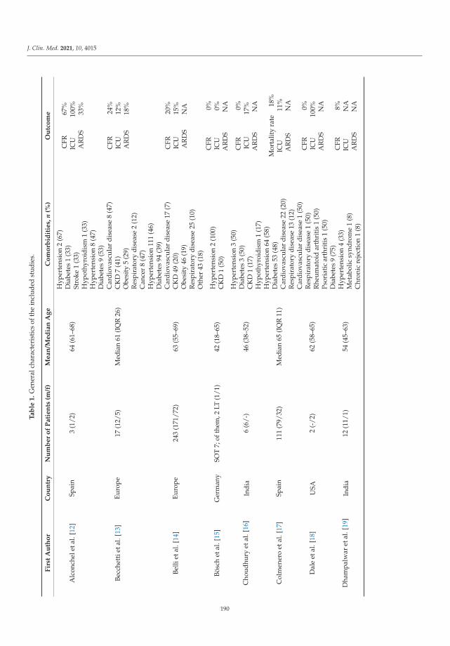
J. Clin. Med. 2021, 10, 4015
Ta
ble
1.
Gen
eral
char
acte
rist
ics
ofth
ein
clud
edst
udie
s.
Fir
stA
uth
or
Co
un
try
Nu
mb
er
of
Pa
tie
nts
(m/f
)M
ea
n/M
ed
ian
Ag
eC
om
orb
idit
ies,
n(%
)O
utc
om
e
Alc
onch
elet
al.[
12]
Spai
n3
(1/2
)64
(61–
68)
Hyp
erte
nsio
n2
(67)
CFR
67%
ICU
100%
AR
DS
33%
Dia
bete
s1
(33)
Stro
ke1
(33)
Hyp
othy
roid
ism
1(3
3)
Becc
hett
ieta
l.[1
3]Eu
rope
17(1
2/5)
Med
ian
61(I
QR
26)
Hyp
erte
nsio
n8
(47)
CFR
24%
ICU
12%
AR
DS
18%
Dia
bete
s9
(53)
Car
diov
ascu
lar
dise
ase
8(4
7)C
KD
7(4
1)O
besi
ty5
(29)
Res
pira
tory
dise
ase
2(1
2)C
ance
r8
(47)
Belli
etal
.[14
]Eu
rope
243
(171
/72)
63(5
5–69
)
Hyp
erte
nsio
n11
1(4
6)
CFR
20%
ICU
15%
AR
DS
NA
Dia
bete
s94
(39)
Car
diov
ascu
lar
dise
ase
17(7
)C
KD
49(2
0)O
besi
ty46
(19)
Res
pira
tory
dise
ase
25(1
0)O
ther
43(1
8)
Bösc
het
al.[
15]
Ger
man
ySO
T7;
ofth
em,2
LT(1
/1)
42(1
8–65
)H
yper
tens
ion
2(1
00)
CK
D1
(50)
CFR
0%IC
U0%
AR
DS
NA
Cho
udhu
ryet
al.[
16]
Indi
a6
(6/-
)46
(38–
52)
Hyp
erte
nsio
n3
(50)
CFR
0%IC
U17
%A
RD
SN
A
Dia
bete
s3
(50)
CK
D1
(17)
Hyp
othy
roid
ism
1(1
7)
Col
men
ero
etal
.[17
]Sp
ain
111
(79/
32)
Med
ian
65(I
QR
11)
Hyp
erte
nsio
n64
(58)
Mor
talit
yra
te18
%IC
U11
%A
RD
SN
A
Dia
bete
s53
(48)
Car
diov
ascu
lar
dise
ase
22(2
0)R
espi
rato
rydi
seas
e13
(12)
Dal
eet
al.[
18]
USA
2(-
/2)
62(5
8–65
)
Car
diov
ascu
lar
dise
ase
1(5
0)C
FR0%
ICU
100%
AR
DS
NA
Res
pira
tory
dise
ase
1(5
0)R
heum
atoi
dar
thri
tis
1(5
0)Ps
oria
tic
arth
riti
s1
(50)
Dha
mpa
lwar
etal
.[19
]In
dia
12(1
1/1)
54(4
5–63
)
Dia
bete
s9
(75)
CFR
8%IC
UN
AA
RD
SN
A
Hyp
erte
nsio
n4
(33)
Met
abol
icsy
ndro
me
1(8
)C
hron
icre
ject
ion
1(8
)
190
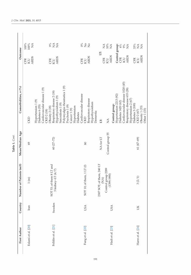
J. Clin. Med. 2021, 10, 4015
Ta
ble
1.
Con
t.
Fir
stA
uth
or
Co
un
try
Nu
mb
er
of
Pa
tie
nts
(m/f
)M
ea
n/M
ed
ian
Ag
eC
om
orb
idit
ies,
n(%
)O
utc
om
e
Esla
mie
tal.
[20]
Iran
1(m
)69
CK
DC
FR10
0%IC
U10
0%A
RD
SN
A
Felld
inet
al.[
21]
Swed
enSO
T53
;oft
hem
8LT
and
3ki
dney
–LT
(4/7
)60
(27–
72)
Hyp
erte
nsio
n1
(9)
CFR
9%IC
UN
AA
RD
SN
A
Dia
bete
s6
(55)
Car
diov
ascu
lar
dise
ase
1(9
)C
KD
2(1
8)O
besi
ty2
(18)
Res
pira
tory
dise
ase
2(1
8)H
ypot
hyro
idis
m1
(9)
Sarc
oido
sis
1(9
)Po
lym
yalg
iarh
eum
atic
a1
(9)
Psyc
hosi
s1
(9)
Can
cer
1(9
)
Fung
etal
.[22
]U
SASO
T10
;oft
hem
,1LT
(f)
80
Hyp
erte
nsio
n
CFR
0%IC
UN
oA
RD
SN
o
Dia
bete
sC
ardi
ovas
cula
rdi
seas
eC
KD
Res
pira
tory
dise
ase
Hyp
othy
roid
ism
Dem
enti
a
Had
ieta
l.[2
3]U
SA
2307
SOT;
ofth
em,2
40LT
(NA
)N
Afo
rLT
LT
:L
T:
CFR
NA
ICU
10%
AR
DS
NA
Con
trol
grou
p22
89(1
399/
890)
Con
trol
grou
p55
NA
Co
ntr
ol
gro
up
:H
yper
tens
ion
2213
(92)
Co
ntr
ol
gro
up
:C
FR6%
ICU
9%A
RD
SN
A
Dia
bete
s14
20(6
2)C
ardi
ovas
cula
rdi
seas
e10
20(4
5)R
espi
rato
rydi
seas
e63
3(2
8)
Han
net
al.[
24]
UK
3(2
/1)
61(4
7–69
)
Hyp
erte
nsio
n2
(67)
CFR
33%
ICU
NA
AR
DS
NA
Dia
bete
s3
(100
)C
KD
2(6
7)O
besi
ty1
(33)
Oth
er1
(33)
191
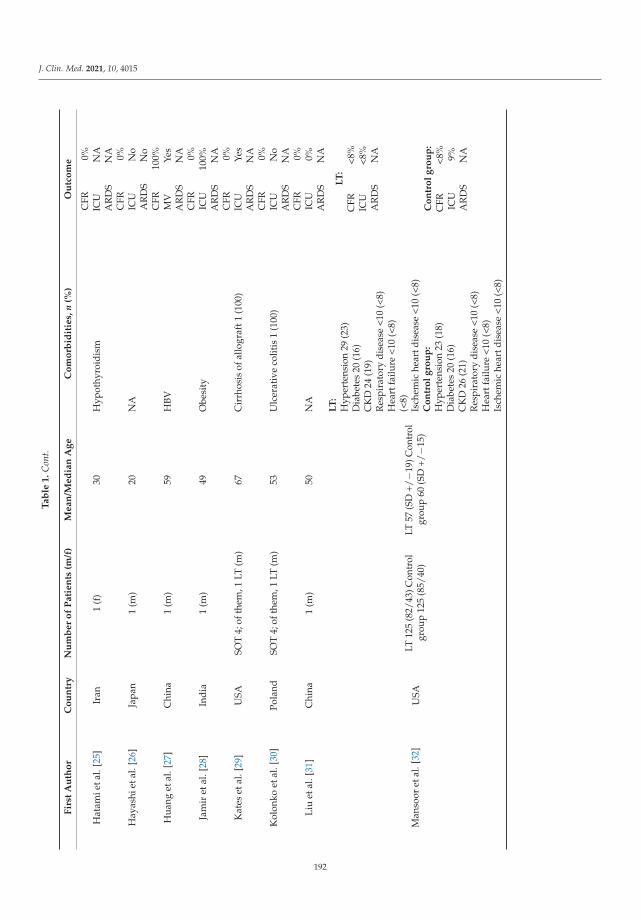
J. Clin. Med. 2021, 10, 4015
Ta
ble
1.
Con
t.
Fir
stA
uth
or
Co
un
try
Nu
mb
er
of
Pa
tie
nts
(m/f
)M
ea
n/M
ed
ian
Ag
eC
om
orb
idit
ies,
n(%
)O
utc
om
e
Hat
amie
tal.
[25]
Iran
1(f
)30
Hyp
othy
roid
ism
CFR
0%IC
UN
AA
RD
SN
A
Hay
ashi
etal
.[26
]Ja
pan
1(m
)20
NA
CFR
0%IC
UN
oA
RD
SN
o
Hua
nget
al.[
27]
Chi
na1
(m)
59H
BVC
FR10
0%M
VYe
sA
RD
SN
A
Jam
iret
al.[
28]
Indi
a1
(m)
49O
besi
tyC
FR0%
ICU
100%
AR
DS
NA
Kat
eset
al.[
29]
USA
SOT
4;of
them
,1LT
(m)
67C
irrh
osis
ofal
logr
aft1
(100
)C
FR0%
ICU
Yes
AR
DS
NA
Kol
onko
etal
.[30
]Po
land
SOT
4;of
them
,1LT
(m)
53U
lcer
ativ
eco
litis
1(1
00)
CFR
0%IC
UN
oA
RD
SN
A
Liu
etal
.[31
]C
hina
1(m
)50
NA
CFR
0%IC
U0%
AR
DS
NA
Man
soor
etal
.[32
]U
SALT
125
(82/
43)C
ontr
olgr
oup
125
(85/
40)
LT57
(SD
+/−1
9)C
ontr
olgr
oup
60(S
D+/
−15)
LT
:L
T:
CFR
<8%
ICU
<8%
AR
DS
NA
Hyp
erte
nsio
n29
(23)
Dia
bete
s20
(16)
CK
D24
(19)
Res
pira
tory
dise
ase
<10
(<8)
Hea
rtfa
ilure
<10
(<8)
Co
ntr
ol
gro
up
:C
FR<8
%IC
U9%
AR
DS
NA
(<8)
Isch
emic
hear
tdis
ease
<10
(<8)
Co
ntr
ol
gro
up
:H
yper
tens
ion
23(1
8)D
iabe
tes
20(1
6)C
KD
26(2
1)R
espi
rato
rydi
seas
e<1
0(<
8)H
eart
failu
re<1
0(<
8)Is
chem
iche
artd
isea
se<1
0(<
8)
192
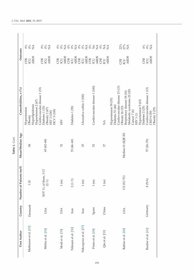
J. Clin. Med. 2021, 10, 4015
Ta
ble
1.
Con
t.
Fir
stA
uth
or
Co
un
try
Nu
mb
er
of
Pa
tie
nts
(m/f
)M
ea
n/M
ed
ian
Ag
eC
om
orb
idit
ies,
n(%
)O
utc
om
e
Mat
hias
enet
al.[
33]
Den
mar
k1
(f)
58H
yper
tens
ion
CFR
0%IC
U0%
AR
DS
NA
Obe
sity
Hyp
erlip
idem
ia
Meh
taet
al.[
34]
USA
SOT
11;o
fthe
m,3
LT(2
/1)
65(6
2–68
)
Hyp
erte
nsio
n2
(67)
CFR
0%IC
U0%
AR
DS
NA
Car
diov
ascu
lar
dise
ase
1(3
3)D
iabe
tes
1(3
3)O
besi
ty2
(67)
HIV
3(1
00)
HC
V3
(100
)
Mod
ieta
l.[3
5]U
SA1
(m)
32H
IVC
FR0%
ICU
0%A
RD
SN
A
Nik
nam
etal
.[36
]Ir
an2
(1/1
)53
(46–
60)
Dia
bete
s1
(50)
CFR
0%IC
U0%
AR
DS
NA
Nik
oupo
uret
al.[
37]
Iran
1(m
)35
Ulc
erat
ive
colit
is1
(100
)C
FR0%
ICU
0%A
RD
SN
A
Prie
toet
al.[
38]
Spai
n1
(m)
52C
ardi
ovas
cula
rdi
seas
e1
(100
)C
FR0%
ICU
No
AR
DS
NA
Qin
etal
.[39
]C
hina
1(m
)37
NA
CFR
0%IC
UN
AA
RD
SN
A
Rab
iee
etal
.[40
]U
SA11
2(6
1/51
)M
edia
n61
(IQ
R20
)
Hyp
erte
nsio
n59
(53)
CFR
22%
ICU
27%
AR
DS
NA
Dia
bete
s51
(46)
Car
diov
ascu
lar
dise
ase
15(1
3)O
besi
ty26
(23)
Res
pira
tory
dise
ase
18(1
6)M
etab
olic
synd
rom
e22
(20)
Can
cer
7(6
)H
IV1
(1)
Rau
ber
etal
.[41
]G
erm
any
8(N
A)
57(2
6–70
)
Hyp
erte
nsio
n5
(63)
CFR
0%IC
U0%
AR
DS
NA
Dia
bete
s2
(25)
Car
diov
ascu
lar
dise
ase
1(1
3)C
KD
4(5
0)O
besi
ty2
(25)
193

J. Clin. Med. 2021, 10, 4015
Ta
ble
1.
Con
t.
Fir
stA
uth
or
Co
un
try
Nu
mb
er
of
Pa
tie
nts
(m/f
)M
ea
n/M
ed
ian
Ag
eC
om
orb
idit
ies,
n(%
)O
utc
om
e
Sess
aet
al.[
42]
Fran
ce1
(m)
58H
yper
tens
ion
CFR
0%IC
U0%
AR
DS
NA
Terr
abui
oet
al.[
43]
Braz
il4
(1/3
)52
(42–
62)
Hyp
erte
nsio
n1
(25)
CFR
0%IC
U0%
AR
DS
NA
Dia
bete
s1
(25)
Obe
sity
2(5
0)C
hron
icre
ject
ion
1(2
5)C
rohn
’sdi
seas
e1
(25)
Wai
sber
get
al.[
44]
Braz
il5
(4/1
)60
(34–
69)
Hyp
erte
nsio
n3
(60)
CFR
40%
ICU
NA
AR
DS
NA
Dia
bete
s1
(20)
Car
diov
ascu
lar
dise
ase
1(2
0)O
besi
ty2
(40)
Res
pira
tory
dise
ase
1(2
0)
Web
bet
al.[
45]
Inte
rnat
iona
lLT
151
(102
/49)
Con
trol
grou
p62
7(3
29/2
98)
LTM
edia
n60
(IQ
R47
–66)
Con
trol
grou
p73
(IQ
R55
–84)
LT
:L
T:
CFR
19%
ICU
28%
AR
DS
NA
Hyp
erte
nsio
n63
(42)
Dia
bete
s65
(43)
Car
diov
ascu
lar
dise
ase
22(1
5)O
besi
ty44
(29)
Res
pira
tory
dise
ase
8(5
)
Co
ntr
ol
gro
up
:C
FR27
%IC
U8%
AR
DS
NA
Can
cer
8(5
)C
on
tro
lg
rou
p:
Hyp
erte
nsio
n24
1(3
8)D
iabe
tes
144
(23)
Car
diov
ascu
lar
dise
ase
202
(32)
Obe
sity
158
(25)
Res
pira
tory
dise
ase
160
(26)
Can
cer
92(1
5)St
roke
73(1
2)
Wei
etal
.[46
]C
hina
1(m
)61
NA
CFR
0%IC
UN
AA
RD
SN
A
Abb
revi
atio
ns:I
CU
:int
ensi
veca
reun
it;A
RD
S:ac
ute
resp
irat
ory
dist
ress
synd
rom
e;H
BV:h
epat
itis
Bvi
rus;
HIV
:hum
anim
mun
odefi
cien
cyvi
rus;
HC
V:h
epat
itis
Cvi
rus;
SOT:
solid
orga
ntr
ansp
lant
atio
n;LT
:liv
ertr
ansp
lant
;NA
:not
appl
icab
le;C
KD
:chr
onic
kidn
eyin
suffi
cien
cy;I
QR
:int
erqu
arti
lera
nge;
CFR
:cas
efa
talit
yra
te.
194

J. Clin. Med. 2021, 10, 4015
Regarding the incidence of COVID-19 infections in LT recipients, only the SETHcohort provided data, showing that the incidence of COVID-19 in liver transplant recipientscompared to the general population (837.41 cases/ 105 patients vs. 311.93 cases/ 105 people)was almost double [17].
On the other hand, the COVID-LT cohort recorded 57 confirmed SARS-CoV-2 infec-tions out of 11,790 patients in regular follow-ups, resulting in an incidence of 483.46 cases/105 patients [13]. Another report from Germany documented, using either serology orPCR-swab test, present or past SARS-CoV-2 infection in 3.7% of their LT recipients duringthe study period (May and August 2020) [41].
The most frequently described clinical presentation was fever (61.4%), followed bycough (58.6%) and dyspnea (36.2%). Webb et al. [45] reported general “respiratory symp-toms”, which were experienced by the 77% of the LT recipients included in the study.Gastrointestinal symptoms including vomiting, diarrhea, nausea, and abdominal painwere strongly represented (159/569 patients, 27.9%). In the aforementioned study, theproportion of patients with gastrointestinal symptoms was higher among LT recipientscompared to the nontransplant cohort (30% vs. 12%, p < 0.0001), whereas no significantdifference was observed with respect to respiratory symptoms. On the same line, Belliet al. [14] found diarrhea as the presenting symptom in 55 LT recipients, corresponding to22.6%.
Concerning immunosuppression therapy, data on basal immunosuppression (IS)therapy and on subsequent management during the course of infection was available for33 and 29 studies, respectively. The data are summarized in Table 2.
195

J. Clin. Med. 2021, 10, 4015
Ta
ble
2.
Info
rmat
ion
rega
rdin
gim
mun
osup
pres
sant
regi
men
and
its
mod
ifica
tion
indu
ring
CO
VID
-19.
Fir
stA
uth
or
Nu
mb
er
of
Pa
tie
nts
Ba
seli
ne
IS,n
(%)
Mo
difi
cati
on
IST
yp
eo
fM
od
ifica
tio
n
Alc
onch
elet
al.[
12]
3TA
C+
ster
oid,
1(3
3)TA
C+
MM
F+
ster
oid,
2(6
7)Ye
sTA
Cre
duct
ion
33%
MM
Fw
ithd
raw
alan
dTA
Cre
duct
ion
33%
MM
Fw
ithd
raw
al33
%
Becc
hett
ieta
l.[1
3]17
Sin
gle
ag
en
tC
ys,1
(6)
TAC
,4(2
4)Ev
erol
imus
,1(6
)M
MF,
2(1
2)C
om
bin
ati
on
CN
Is+
MM
F,7
(41)
CN
Is+
ster
oids
,2(1
2)
Yes
Red
ucti
on29
%W
ithd
raw
al18
%
Belli
etal
.[14
]24
3
Sin
gle
ag
en
tC
ys,1
3(5
)C
ys+
MM
F,9
(4)
Cys
+st
eroi
d,3
(1)
Cys
+M
MF
+st
eroi
d,4
(2)
TAC
,54
(22)
TAC
+M
MF,
52(2
1)TA
C+
mTO
Ri,
12(5
)TA
C+
ster
oid,
27(1
1)TA
C+
MM
F+
ster
oid,
15(6
)M
MF,
24(1
0)M
MF
+m
TOR
i,10
(4)
MM
F+
ster
oid,
3(1
)m
TOR
i,11
(5)
mTO
Ri+
ster
oid,
2(1
)St
eroi
d,2
(1)
Yes
CN
Isw
ithd
raw
al7%
CN
Isre
duct
ion
16%
Ant
imet
abol
ites
wit
hdra
wal
14%
mTO
Riw
ithd
raw
al4%
Oth
er2%
Bösc
het
al.[
15]
SOT
7;of
them
,2LT
Ever
olim
us+
MM
F,1
(50)
MM
F,1
(50)
Yes
MM
Fw
ithd
raw
al50
%
Cho
udhu
ryet
al.[
16]
6
TAC
,2(3
3)TA
C+
MM
F,2
(33)
Ster
oid,
1(1
7)Ev
erol
imus
,1(1
7)
Yes
TAC
and
MM
Fw
ithd
raw
alan
dst
arts
tero
ids
(17)
TAC
+and
MM
Fw
ithd
raw
alan
dst
arts
tero
ids
(17)
TAC
wit
hdra
wal
and
star
tste
roid
s(3
3)Se
vera
ladj
ustm
ents
(17)
196
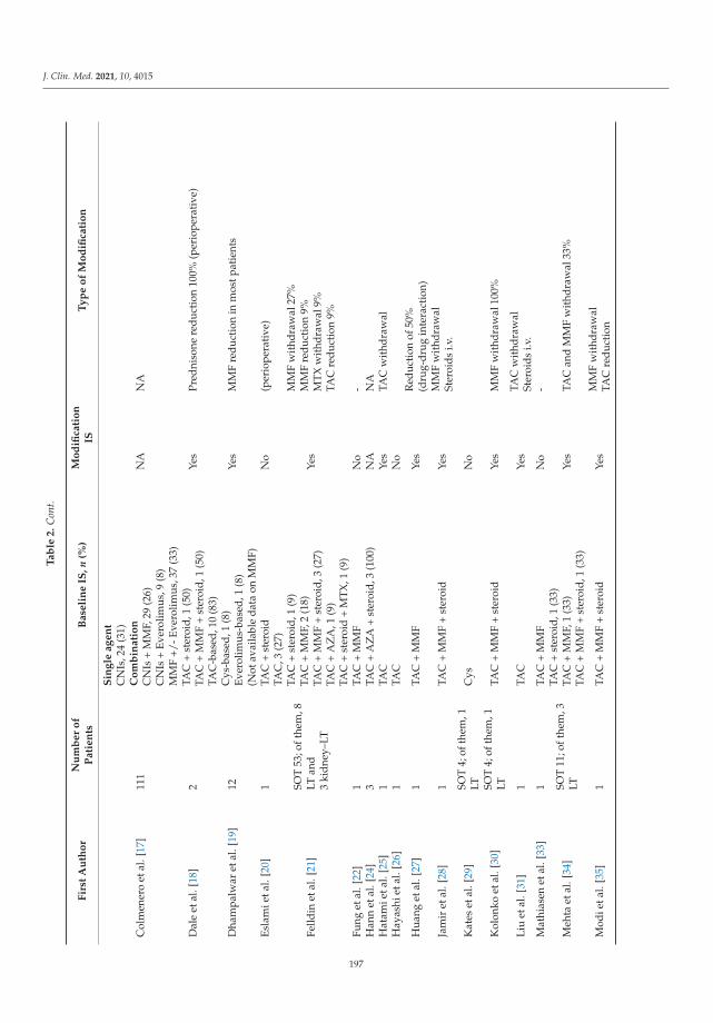
J. Clin. Med. 2021, 10, 4015
Ta
ble
2.
Con
t.
Fir
stA
uth
or
Nu
mb
er
of
Pa
tie
nts
Ba
seli
ne
IS,n
(%)
Mo
difi
cati
on
IST
yp
eo
fM
od
ifica
tio
n
Col
men
ero
etal
.[17
]11
1
Sin
gle
ag
en
tC
NIs
,24
(31)
Co
mb
ina
tio
nC
NIs
+M
MF,
29(2
6)C
NIs
+Ev
erol
imus
,9(8
)M
MF
+/-E
vero
limus
,37
(33)
NA
NA
Dal
eet
al.[
18]
2TA
C+
ster
oid,
1(5
0)TA
C+
MM
F+
ster
oid,
1(5
0)Ye
sPr
edni
sone
redu
ctio
n10
0%(p
erio
pera
tive
)
Dha
mpa
lwar
etal
.[19
]12
TAC
-bas
ed,1
0(8
3)C
ys-b
ased
,1(8
)Ev
erol
imus
-bas
ed,1
(8)
(Not
avai
labl
eda
taon
MM
F)
Yes
MM
Fre
duct
ion
inm
ostp
atie
nts
Esla
mie
tal.
[20]
1TA
C+
ster
oid
No
(per
iope
rati
ve)
Felld
inet
al.[
21]
SOT
53;o
fthe
m,8
LTan
d3
kidn
ey–L
T
TAC
,3(2
7)TA
C+
ster
oid,
1(9
)TA
C+
MM
F,2
(18)
TAC
+M
MF
+st
eroi
d,3
(27)
TAC
+A
ZA
,1(9
)TA
C+
ster
oid
+M
TX,1
(9)
Yes
MM
Fw
ithd
raw
al27
%M
MF
redu
ctio
n9%
MT
Xw
ithd
raw
al9%
TAC
redu
ctio
n9%
Fung
etal
.[22
]1
TAC
+M
MF
No
-H
ann
etal
.[24
]3
TAC
+A
ZA
+st
eroi
d,3
(100
)N
AN
AH
atam
ieta
l.[2
5]1
TAC
Yes
TAC
wit
hdra
wal
Hay
ashi
etal
.[26
]1
TAC
No
Hua
nget
al.[
27]
1TA
C+
MM
FYe
sR
educ
tion
of50
%(d
rug-
drug
inte
ract
ion)
Jam
iret
al.[
28]
1TA
C+
MM
F+
ster
oid
Yes
MM
Fw
ithd
raw
alSt
eroi
dsi.v
.
Kat
eset
al.[
29]
SOT
4;of
them
,1LT
Cys
No
Kol
onko
etal
.[30
]SO
T4;
ofth
em,1
LTTA
C+
MM
F+
ster
oid
Yes
MM
Fw
ithd
raw
al10
0%
Liu
etal
.[31
]1
TAC
Yes
TAC
wit
hdra
wal
Ster
oids
i.v.
Mat
hias
enet
al.[
33]
1TA
C+
MM
FN
o-
Meh
taet
al.[
34]
SOT
11;o
fthe
m,3
LT
TAC
+st
eroi
d,1
(33)
TAC
+M
MF,
1(3
3)TA
C+
MM
F+
ster
oid,
1(3
3)Ye
sTA
Can
dM
MF
wit
hdra
wal
33%
Mod
ieta
l.[3
5]1
TAC
+M
MF
+st
eroi
dYe
sM
MF
wit
hdra
wal
TAC
redu
ctio
n
197
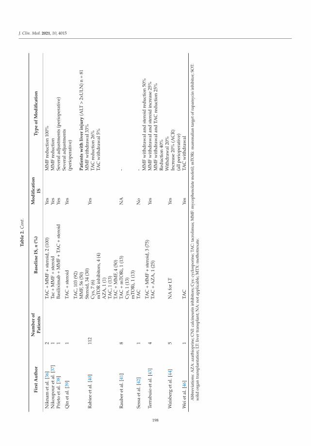
J. Clin. Med. 2021, 10, 4015
Ta
ble
2.
Con
t.
Fir
stA
uth
or
Nu
mb
er
of
Pa
tie
nts
Ba
seli
ne
IS,n
(%)
Mo
difi
cati
on
IST
yp
eo
fM
od
ifica
tio
n
Nik
nam
etal
.[36
]2
TAC
+M
MF
+st
eroi
d,2
(100
)Ye
sM
MF
redu
ctio
n10
0%N
ikou
pour
etal
.[37
]1
Tac
+M
MF
+st
eroi
dYe
sM
MF
redu
ctio
nPr
ieto
etal
.[38
]1
Basi
lixim
ab+
MM
F+
TAC
+st
eroi
dYe
sSe
vera
ladj
ustm
ents
(per
iope
rati
ve)
Qin
etal
.[39
]1
TAC
+st
eroi
dYe
sSe
vera
ladj
ustm
ents
(per
iope
rati
ve)
Rab
iee
etal
.[40
]11
2
TAC
,103
(92)
MM
F,56
(50)
Ster
oid,
34(3
0)C
ys,7
(6)
mTO
Rin
hibi
tors
,4(4
)A
ZA
,1(1
)
Yes
Pa
tie
nts
wit
hli
ve
rin
jury
(ALT
>2x
ULN
)n=
81M
MF
wit
hdra
wal
33%
TAC
redu
ctio
n26
%TA
Cw
ithd
raw
al5%
Rau
ber
etal
.[41
]8
TAC
,1(1
3)TA
C+
MM
F,4
(50)
TAC
+m
TOR
i,1
(13)
Cys
,1(1
3)m
TOR
i,1
(13)
NA
-
Sess
aet
al.[
42]
1TA
CN
o-
Terr
abui
oet
al.[
43]
4TA
C+
MM
F+
ster
oid,
3(7
5)TA
C+
AZ
A,1
(25)
Yes
MM
Fw
ithd
raw
alan
dst
eroi
dre
duct
ion
50%
MM
Fw
ithd
raw
alan
dst
eroi
din
crea
se25
%M
MF
wit
hdra
wal
and
TAC
redu
ctio
n25
%
Wai
sber
get
al.[
44]
5N
Afo
rLT
Yes
Red
ucti
on40
%W
ithd
raw
al20
%In
crea
se20
%(A
CR
)(a
llpe
riop
erat
ive)
Wei
etal
.[46
]1
TAC
Yes
TAC
wit
hdra
wal
Abb
revi
atio
ns:A
ZA
:aza
thio
prin
e;C
NI:
calc
ineu
rin
inhi
bito
rs;C
ys:c
yclo
spor
ine;
TAC
:tac
rolim
us;M
MF:
myc
ophe
nola
tem
ofet
il;m
TOR
i:m
amm
alia
nta
rget
ofra
pam
ycin
inhi
bito
r;SO
T:so
lidor
gan
tran
spla
ntat
ion;
LT:l
iver
tran
spla
nt;N
A:n
otap
plic
able
;MTX
:met
hotr
exat
e.
198

J. Clin. Med. 2021, 10, 4015
In the study by Colmenero et al. [17] patients receiving MMF or in whom an attemptwas made to completely withdraw immunosuppression were more prevalent in the severeCOVID-19 group (p = 0.014, and p = 0.016 respectively). Conversely, tacrolimus-basedimmunosuppression was more frequent in the nonsevere COVID-19 group, albeit withoutstatistical significance (p = 0.113). Similar findings regarding calcineurin inhibitor (CNIs)-based regimens were observed in the COVID-LT study, where the continuation of CNIstherapy after COVID-19 diagnosis was higher among survivors (64% vs. 42.8%) [47].Indeed, in the study of Belli et al. [14], after multivariable analysis, the use of TAC wasconfirmed to be independently associated with a reduced mortality risk (HR, 0.55; 95%CI, 0.31–0.99). Additionally, in the Spanish cohort, survival curves illustrated the negativeprognostic impact of MMF, particularly at doses higher than 1000 mg/day. In agreementwith this finding, in patients receiving full-dose of MMF at baseline (i.e., 2000 mg/day),complete drug withdrawal showed a trend towards reduced severe COVID-19 (41.7% vs.69.2%, p = 0.16) [17].
Overall, 375 out of 1064 (35.2%) patients were managed in an outpatient setting,whereas 64.8% were hospitalized. Of the hospitalized patients, 158/689 (22.9%) wereadmitted to an ICU. Death was reported in 135 cases. In the COVID-LT study, casefatality was estimated at 12% (95% CI 5–24%), which increased to 17% (95% CI 7–32%)among hospitalized patients [13], whereas Rabiee and coauthors found a 22.3% case fatalityrate [40]. In the study by Webb and coauthors [45], case fatality was 19% (vs. the 27%reported for the comparison cohort, p = 0.046), with the propensity-score-matched analysisshowing that LT did not significantly increase the risk of death in patients with SARS-CoV-2infection (absolute risk difference 1.4% (95% CI 7.7–10.4)). Colmenero et al. [17] describeda mortality rate of 18% among LT patients.
In Webb et al., multivariate analyses showed that factors significantly associated withdeath were: increased age (OR 1.06 (95% CI 1.01–1.11) per 1 year increase, p = 0.031),presence of nonliver cancer (OR 18.30 (1.96–170.75); p = 0.011), and higher baseline serumcreatinine (OR 1.57 (1.05–2.36) per 1 mg/dL increase) p = 0.028) [45]. Results derived fromthe multivariate analysis performed within the SETH cohort study identified the followingindependent predictors: Charlson comorbidity index (relative risk (RR) = 1.28 (95% CI1.05–1.56), male gender (RR = 2.49; 95% CI 1.14–5.41), dyspnea at diagnosis (RR = 7.25;95% CI 2.95–17.82), and baseline immunosuppression containing MMF (RR = 3.94; 95% CI1.59–9.74) [17]. Belli et al. reported risk factors associated with worse prognosis includingadvanced age (>70 vs. <60 years, HR 4.16; 95% CI 1.78–9.73) and the use of TAC [14].
Despite theoretically higher levels of immunosuppression, only the report by Belliet al. [14] mentioned time since LT as an independent factor associated with poor outcomein univariate analysis. On the other hand, Colmenero et al. [17] showed that the time fromLT had no impact on the risk of suffering from severe COVID, a finding that was confirmedby Webb et al. [45], who reported no association between death and time since LT.
Lastly, Rabiee et al. showed that the incidence of acute liver injury (defined byALT 2-5x ULN) was not higher in LT recipients when compared to age- and gender-matched nontransplant patients with chronic liver disease and COVID-19 (47.5% vs. 34.6%;p = 0.037). The presence of liver injury during COVID-19 in LT recipients was significantlyassociated with mortality (OR 6.91 (95% CI: 1.68–28.48), p = 0.007) and ICU admission(OR 7.93 (95% CI: 1.75–35.69), p = 0.007) [40]. In the US study of Hadi et al., consideringonly LT recipients, only 18 patients (7.5%) experienced the composite outcome includingmechanical ventilation and death at 30 days. This rate was lower when compared to thatfor recipients of other organ transplants [23].
3.3. Immunological Response after COVID-19 in LT Recipients
Regarding the immunological response after SARS-CoV-2 infection in LT patients,only four studies were considered, including a total of 91 LT recipients. However, all ofthese studies examined different types of tests/assays directed toward different targets,
199
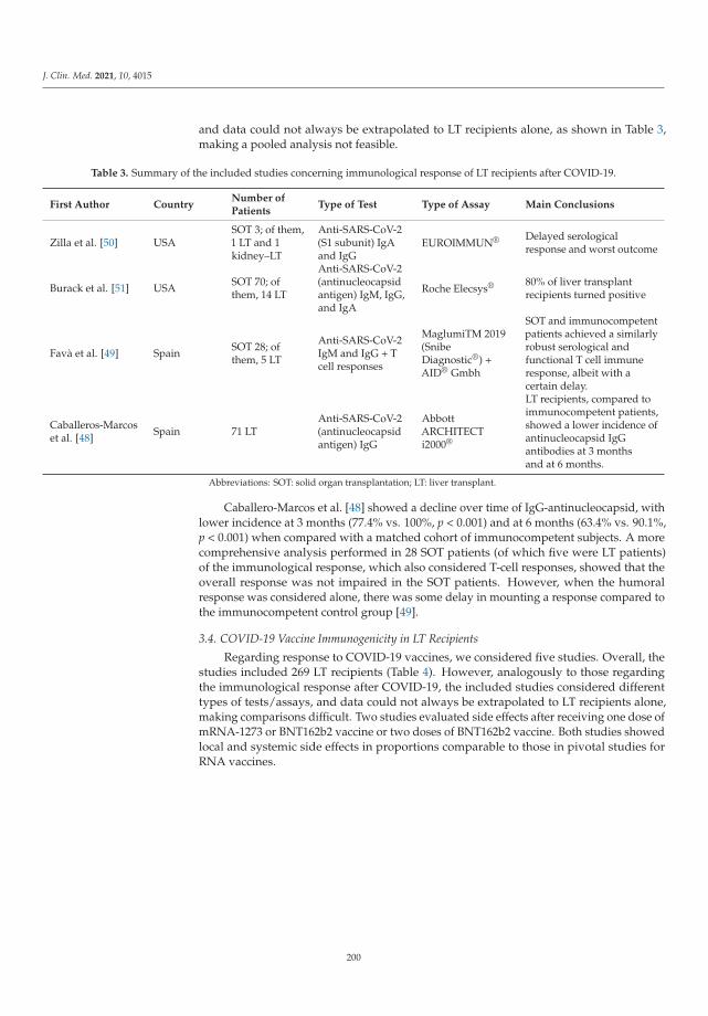
J. Clin. Med. 2021, 10, 4015
and data could not always be extrapolated to LT recipients alone, as shown in Table 3,making a pooled analysis not feasible.
Table 3. Summary of the included studies concerning immunological response of LT recipients after COVID-19.
First Author CountryNumber ofPatients
Type of Test Type of Assay Main Conclusions
Zilla et al. [50] USASOT 3; of them,1 LT and 1kidney–LT
Anti-SARS-CoV-2(S1 subunit) IgAand IgG
EUROIMMUN® Delayed serologicalresponse and worst outcome
Burack et al. [51] USA SOT 70; ofthem, 14 LT
Anti-SARS-CoV-2(antinucleocapsidantigen) IgM, IgG,and IgA
Roche Elecsys® 80% of liver transplantrecipients turned positive
Favà et al. [49] Spain SOT 28; ofthem, 5 LT
Anti-SARS-CoV-2IgM and IgG + Tcell responses
MaglumiTM 2019(SnibeDiagnostic®) +AID® Gmbh
SOT and immunocompetentpatients achieved a similarlyrobust serological andfunctional T cell immuneresponse, albeit with acertain delay.
Caballeros-Marcoset al. [48] Spain 71 LT
Anti-SARS-CoV-2(antinucleocapsidantigen) IgG
AbbottARCHITECTi2000®
LT recipients, compared toimmunocompetent patients,showed a lower incidence ofantinucleocapsid IgGantibodies at 3 monthsand at 6 months.
Abbreviations: SOT: solid organ transplantation; LT: liver transplant.
Caballero-Marcos et al. [48] showed a decline over time of IgG-antinucleocapsid, withlower incidence at 3 months (77.4% vs. 100%, p < 0.001) and at 6 months (63.4% vs. 90.1%,p < 0.001) when compared with a matched cohort of immunocompetent subjects. A morecomprehensive analysis performed in 28 SOT patients (of which five were LT patients)of the immunological response, which also considered T-cell responses, showed that theoverall response was not impaired in the SOT patients. However, when the humoralresponse was considered alone, there was some delay in mounting a response compared tothe immunocompetent control group [49].
3.4. COVID-19 Vaccine Immunogenicity in LT Recipients
Regarding response to COVID-19 vaccines, we considered five studies. Overall, thestudies included 269 LT recipients (Table 4). However, analogously to those regardingthe immunological response after COVID-19, the included studies considered differenttypes of tests/assays, and data could not always be extrapolated to LT recipients alone,making comparisons difficult. Two studies evaluated side effects after receiving one dose ofmRNA-1273 or BNT162b2 vaccine or two doses of BNT162b2 vaccine. Both studies showedlocal and systemic side effects in proportions comparable to those in pivotal studies forRNA vaccines.
200
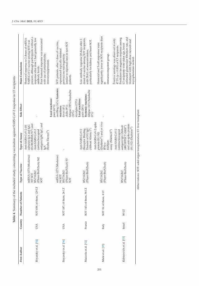
J. Clin. Med. 2021, 10, 4015
Ta
ble
4.
Sum
mar
yof
the
incl
uded
stud
yco
ncer
ning
vacc
inat
ion
agai
nstS
AR
S-C
oV-2
resp
onse
inLT
reci
pien
ts.
Fir
stA
uth
or
Co
un
try
Nu
mb
er
of
Pa
tie
nts
Ty
pe
of
Va
ccin
eT
yp
eo
fA
ssa
yS
ide
Eff
ect
Ma
inC
on
clu
sio
ns
Boya
rsky
etal
.[54
]U
SASO
T65
8;of
them
,129
LT
mR
NA
-127
3(M
oder
na)
307
SOT
BNT1
62b2
(Pfiz
er-B
ioN
Tech
)342
SOT
Ant
i-SA
RS-
CoV
-2(S
1su
buni
t)Ig
Aan
dIg
G(E
UR
OIM
MU
N®
)and
Ant
i-SA
RS-
CoV
-2(a
ntin
ucle
ocap
sid
anti
gen)
IgM
,IgG
,and
IgA
(Roc
heEl
ecsy
s®)
-
Hum
oral
resp
onse
to2
dose
sof
mR
NA
SAR
S-C
oV-2
vacc
ine
amon
gSO
Tw
aspr
esen
t,al
thou
ghpa
rtic
ipan
tsw
itho
uta
resp
onse
afte
rdo
se1
had
gene
rally
low
anti
body
leve
ls.
Poor
hum
oral
resp
onse
was
asso
ciat
edw
ith
use
ofan
tim
etab
olit
eim
mun
osup
pres
sion
.
Boya
rsky
etal
.[56
]U
SASO
T18
7;of
them
,26
LT
mR
NA
-127
3(M
oder
na)
94SO
TBN
T162
b2(P
fizer
-Bio
NTe
ch)9
3SO
T
-
Lo
cal
rea
ctio
ns:
pain
(61%
)sw
ellin
g(1
6%)S
yst
em
icre
act
ion
s:fe
ver
(4%
)ch
ills
(9%
)fa
tigu
e(3
8%)h
eada
che
(32%
)m
yalg
ias
(15%
)
SOT
pati
ents
,aft
er1
dose
ofva
ccin
e,ex
peri
ence
dty
pica
llym
inim
alpe
riva
ccin
ere
acto
geni
city
sim
ilar
tore
port
edra
tes
inno
n-SO
Tpa
tien
ts.
Maz
zola
etal
.[52
]Fr
ance
SOT
143;
ofth
em,5
6LT
BNT1
62b2
(Pfiz
er-B
ioN
Tech
)
Ant
i-SA
RS-
CoV
-2(R
ecep
tor
Bind
ing
Dom
ain
ofS1
)IgG
(Abb
ottA
linit
yi®
)
Lo
cal
rea
ctio
ns:
pain
(26%
)S
yst
em
icre
act
ion
s:fa
tigu
e(1
4%)h
eada
che
(6%
)
Low
anti
body
resp
onse
28da
ysaf
ter
2do
ses
ofva
ccin
eam
ong
SOT
reci
pien
ts,
wit
h28
.6%
ofse
roco
nver
sion
,pa
rtic
ular
lyfo
rki
dney
and
hear
tSO
T.
Mie
leet
al.[
55]
Ital
ySO
T16
;oft
hem
,4LT
BNT1
62b2
(Pfiz
er-B
ioN
Tech
)
Ant
i-SA
RS-
CoV
-2sp
ike
prot
ein
(S1/
S2)
(Dia
Sori
n®)a
ndex
vivo
IFN
-γ-
ELIS
pot
assa
y(M
abte
ch®
)
-
Hum
oral
and
T-ce
llre
spon
ses
wer
esi
gnifi
cant
lylo
wer
inSO
Tre
cipi
ents
than
in imm
unoc
ompe
tent
grou
p.
Rab
inow
ich
etal
.[53
]Is
rael
80LT
BNT1
62b2
(Pfiz
er-B
ioN
Tech
)
Ant
i-SA
RS-
CoV
-2(a
ntin
ucle
ocap
sid
anti
gen)
IgG
(Abb
ott
AR
CH
ITEC
Ti2
000®
)an
dan
ti-s
pike
prot
ein
(S1/
S2)(
Dia
Sori
n®)
-
Posi
tive
sero
logy
was
obse
rved
inon
ly47
.5%
(p<
0.00
1)of
LTre
cipi
ents
.Pr
edic
tors
for
nega
tive
resp
onse
amon
gLT
reci
pien
tsw
ere
olde
rag
e,lo
wer
esti
mat
edgl
omer
ular
filtr
atio
nra
te,a
ndtr
eatm
entw
ith
high
-dos
est
eroi
dsan
dm
ycop
heno
late
mof
etil.
Abb
revi
atio
ns:S
OT:
solid
orga
ntr
ansp
lant
atio
n;LT
:liv
ertr
ansp
lant
.
201
J. Clin. Med. 2021, 10, 4015
Concerning immunogenicity, four studies, although using different assays, evaluatedthe humoral response recording seroconversion rates between 29 and 50% [52,53]. Twostudies emphasized that the use of antimetabolites as immunosuppression was a risk factorfor reduced serum conversion rates [53,54]. Only one study also analyzed T-cell response,which was described as reduced [55], and only one of the included studies considered onlyLT recipients [53].
4. Discussion
At the beginning of the COVID-19 pandemic, SOT recipients, including LT recipients,were considered a vulnerable population, raising the question as to whether they wouldbe at particular risk for severe disease and graft injury given their immunocompromisedstate and high prevalence of metabolic comorbidities. The aim of our study was to sys-tematically pool all the available literature on this topic. We found that middle-agedmen with metabolic comorbidities were the main target for the infection. Even thoughrespiratory problems represented the main clinical feature in LT patients, a high percent-age of gastrointestinal symptoms were also reported. Approximately 70% of LT patientswith SARS-CoV-2 infection were hospitalized. Modification/reduction of IS was common,particularly for MMF, although complete withdrawal of all IS was rarely observed. Withrespect to outcome, a case fatality rate ranging between 12 and 22% was described in themajor reports accessible for this analysis. Interestingly, when compared to the controlpopulation, outcomes were not worse in the LT recipient group.
There seems to be a difference in the immune response to the SARS-CoV-2 infectionand the immune response as acquired by vaccination. In the first case, the data conflict,but if we consider the response mediating the neutralizing activity (anti-spike proteinIgG and T-cell mediated), it seems that there is a similar response, albeit probably slightlydelayed, in LT recipients compared to immunocompetent subjects. On the other hand,vaccine-induced immunogenicity seems to be defective.
Although a considerable number of patients were included in the present study, thequality of the manuscripts analyzed makes it difficult to consolidate associations andpredictive factors regarding COVID-19 and the LT population.
The epidemiological distribution of the disease is superimposable to that of the generalpopulation [2,57], with COVID-19 being mainly prevalent in middle-aged males.
Recently, new findings have highlighted how obesity [58], diabetes type II [59], andarterial hypertension [60] are associated with a more severe course of COVID-19 andhence a poorer outcome. Despite the high prevalence of these metabolic conditions in LTrecipients [61,62] and more specifically in the present cohort, this did not seem to negativelyaffect the prognosis of the current study population.
Interestingly, the presence of gastrointestinal complaints (28%) was considerablyhigher among LT recipients. A recent review on gastrointestinal symptoms in COVID-19 [63] showed a high heterogeneity in incidence (ranging between 3 and 79%), withother large cohorts of patients reporting rates of gastrointestinal symptoms between 5 and15% [60,64]. It is widely accepted that SARS-CoV-2 enters host mucosal cells via the cellreceptor angiotensin-converting enzyme-2 (ACE-2) and the transmembrane serine protease2 (TMPRSS2), which are also highly expressed in the absorptive enterocytes from the ileumand colon [65]. Once the virus enters the enterocytes, it can start replication and its cyto-pathic effect [66]. The gut microbiome can be significantly altered by SARS-CoV-2 throughseveral mechanisms (e.g., proinflammatory cytokines, perturbation in the gut–lung axis,medications, changing ratio of pathogenic organisms) leading to clinical manifestationssuch as diarrhea and vomiting [67]. LT patients are also known to have an extremelyvulnerable gut microbiomes [68,69], and immunosuppressive fluctuation in trough levelcan interfere with gut flora stability [70]. Furthermore, it was observed that patients withdigestive symptoms, probably not recognized from the outset as symptoms associated withCOVID-19, had a significantly longer time from onset to admission than patients withoutdigestive symptoms (9.0 days vs. 7.3 days, p = 0.013) [71]. Interestingly, a recent study aim-
202

J. Clin. Med. 2021, 10, 4015
ing to analyze the gut inflammatory response in immunocompetent subjects infected withSARS-CoV-2 highlighted the absence of a proinflammatory response in the gastrointestinaltract despite detection of SARS-CoV-2. Additionally, this study showed reduced mortalityin patients with COVID-19 presenting with GI symptoms. Therefore, the authors specu-lated on a potential role of the gastrointestinal tract in attenuating SARS-CoV-2-associatedinflammation [72].
In the current cohort, the mortality rate and case-fatality rate did not seem to exceedthose expected in the general population. Indeed, the hypothesis that LT is a possibleassociated factor for a pejorative outcome could not be confirmed. However, of note wasthe high rate of hospitalization, with 64.8% of LT recipients being admitted to a ward and22.9% of such patients requiring intensive care. This may of course reflect a certain selectionbias, with more LT recipients being hospitalized per se. Additionally, one must keep inmind that during the first wave, there were many logistical difficulties, which probably ledto an underdiagnosis of asymptomatic or paucisymptomatic cases [73].
With regard to the management of immunosuppression, not all the information canbe extrapolated, as most studies have had descriptive designs. The different nature ofthe immunosuppressive regimens adopted, often multiple, and the modification of theseregimens during the course of infection make it difficult to provide any clear guidelinesto this respect. However, it seems that a complete discontinuation of IS therapy was veryrare and limited to extremely severe cases. Unfortunately, complete cessation of IS was notassociated with improved prognosis [17] and should therefore only be considered as a lastresort in selected cases.
In the Spanish cohort, patients receiving MMF were more prevalent in the severeCOVID-19 group. Baseline immunosuppression containing MMF was identified as anindependent predictor of mortality, whereas the withdrawal of IS was not. However, dataon modifications in immunosuppressive therapy during the infection were not extensivelyavailable in this study [17] and reduction or discontinuation of MMF was recommended bythe guidelines shared by experts at the beginning of the pandemic [74]. Therefore, whetherthe impact on outcome is attributable to the MMF itself or to its reduction/discontinuationremains objects of further investigation. In a preclinical setting, MMF showed promisingresults against Middle East respiratory syndrome (MERS); however, in vivo studies sug-gested that its use is likely to cause more harm than benefit against coronavirus (CoV)infection [75]. Bearing in mind that MMF acts on activated lymphocytes with a cytostaticeffect [76] and that SARS-CoV-2 has a cytotoxic effect on the same target [77], these syn-ergistic effects may represent an additional risk factor and worsen the prognosis of LTpatients taking MMF.
In Belli et al. [14], TAC was found to protect against worse outcomes in COVID-19 LT recipients. In vitro experiments have shown that TAC, and CNIs in general, arecapable of inhibiting human CoV growth, mainly by acting on the cyclophilin pathway [78].Additionally, by modulating T-cell activation, CNIs may act on reducing the deleteriouseffect of the COVID-19 late inflammatory phase [79].
Concerning the immunological response to SARS-CoV-2 infection, contrasting resultswere seen. However, most of the studies considered analyzed only the humoral responseand in particular used an assay, the antinucleocapsid test, more suitable to evaluate preex-isting exposure to the virus than to assess protective efficacy. Indeed, these antibodies havelow or no neutralizing activity [80]. On the other hand, the analysis of the T-cell-mediatedresponse in LT recipients showed similar results compared to that for immunocompetentsubjects. Further studies are therefore necessary that take into account the complexityof the immune response in vivo and of the interplay among native, humoral, and T-cellimmunity.
In contrast, all studies evaluating vaccine immunity have demonstrated reducedimmunogenicity of LT recipients, and SOT recipients in general. It is possible, as pointedout by Mazzola et al. [52], that this reduced response is more evident in other SOT recipients,such as those for kidney or heart SOTs, than in LT recipients. In this line, the serum
203

J. Clin. Med. 2021, 10, 4015
conversion data of the only cohort that included only LT recipients were actually higherthan the average of the other studies, which included mixed cohorts. A deterrent role couldbe played again by more sustained immunosuppression (with dual or triple regimens) andby the use of antimetabolites [53]. Reduced immunogenicity in LT recipients has alreadybeen demonstrated with other respiratory virus (e.g., influenza) vaccines [81], suggestingthat more than the standard dose may be needed to achieve protective immunogenicity [82].
Several limitations affected the current study, mainly because of the high heterogeneityand quality of the majority of the studies considered, leading in several cases to incompleteinformation. Therefore, more than a few research questions remain open and will needfuture investigation (Figure 2).
Figure 2. Graphical representation of the key unresolved issues for SARS-CoV-2 infection in LT recipients.
5. Conclusions
Despite the heterogeneity of the reports included in this study, we are able to show thatthe middle-aged man with metabolic comorbidities represents the main target of COVID-19among LT recipients, as is the case in nontransplanted patients. Gastrointestinal symptomsare very common in LT recipients with COVID-19, and particular attention should be paidto these complaints as a surrogate marker for COVID-19, even in the absence of fever orrespiratory problems. The outcome of the LT population is nevertheless similar to that ofthe general population. This is, in part, also likely due to a more cautious managementof immunosuppressive therapy, paying particular attention to the use of MMF and TACwhile discouraging the complete discontinuation of all immunosuppression. LT recipientsshould be vaccinated, and great attention to protective measures should be maintained inthese individuals even after a regular course of vaccination.
Author Contributions: C.B. contributed to this paper with conception, literature review, statisticalanalysis, writing the manuscript, critical revision, and editing. S.G.G. participated in drafting andliterature review. V.B. and J.-F.D. contributed with critical revision and editing. All authors have readand agreed to the published version of the manuscript.
Funding: Chiara Becchetti received financial support from the Stiftung für Leberkrankheiten.
204
J. Clin. Med. 2021, 10, 4015
Data Availability Statement: Not applicable.
Conflicts of Interest: The funders had no role in the design of the study; in the collection, analyses,or interpretation of data; in the writing of the manuscript, or in the decision to publish the results.
References
1. Zhou, P.; Yang, X.-L.; Wang, X.-G.; Hu, B.; Zhang, L.; Zhang, W.; Si, H.-R.; Zhu, Y.; Li, B.; Huang, C.-L.; et al. A pneumoniaoutbreak associated with a new coronavirus of probable bat origin. Nature 2020, 579, 270–273. [CrossRef]
2. Suleyman, G.; Fadel, R.A.; Malette, K.M.; Hammond, C.; Abdulla, H.; Entz, A.; Demertzis, Z.; Hanna, Z.; Failla, A.; Dagher, C.;et al. Clinical Characteristics and Morbidity Associated with Coronavirus Disease 2019 in a Series of Patients in MetropolitanDetroit. JAMA Netw. Open 2020, 3, e2012270. [CrossRef] [PubMed]
3. Mao, L.; Jin, H.; Wang, M.; Hu, Y.; Chen, S.; He, Q.; Chang, J.; Hong, C.; Zhou, Y.; Wang, D.; et al. Neurologic Manifestations ofHospitalized Patients with Coronavirus Disease 2019 in Wuhan, China. JAMA Neurol. 2020, 77, 683–690. [CrossRef] [PubMed]
4. Hu, Z.; Song, C.; Xu, C.; Jin, G.; Chen, Y.; Xu, X.; Ma, H.; Chen, W.; Lin, Y.; Zheng, Y.; et al. Clinical characteristics of 24asymptomatic infections with COVID-19 screened among close contacts in Nanjing, China. Sci. China Life Sci. 2020, 63, 706–711.[CrossRef] [PubMed]
5. Loomba, R.S.; Aggarwal, G.; Aggarwal, S.; Flores, S.; Villarreal, E.G.; Farias, J.S.; Lavie, C.J. Disparities in case frequency andmortality of coronavirus disease 2019 (COVID-19) among various states in the United States. Ann. Med. 2021, 53, 151–159.[CrossRef]
6. Palaiodimos, L.; Kokkinidis, D.G.; Li, W.; Karamanis, D.; Ognibene, J.; Arora, S.; Southern, W.N.; Mantzoros, C.S. Severe obesity,increasing age and male sex are independently associated with worse in-hospital outcomes, and higher in-hospital mortality, in acohort of patients with COVID-19 in the Bronx, New York. Metabolism 2020, 108, 154262. [CrossRef]
7. Available online: https://www.who.int (accessed on 1 June 2021).8. Bertolini, A.; Van De Peppel, I.P.; Bodewes, F.A.; Moshage, H.; Fantin, A.; Farinati, F.; Fiorotto, R.; Jonker, J.W.; Strazzabosco, M.;
Verkade, H.J.; et al. Abnormal Liver Function Tests in Patients with COVID-19: Relevance and Potential Pathogenesis. Hepatology2020, 72, 1864–1872. [CrossRef]
9. Polack, F.P.; Thomas, S.J.; Kitchin, N.; Absalon, J.; Gurtman, A.; Lockhart, S.; Perez, J.L.; Marc, G.P.; Moreira, E.D.; Zerbini, C.; et al.Safety and Efficacy of the BNT162b2 mRNA Covid-19 Vaccine. N. Engl. J. Med. 2020, 383, 2603–2615. [CrossRef]
10. Sadoff, J.; Le Gars, M.; Shukarev, G.; Heerwegh, D.; Truyers, C.; de Groot, A.M.; Stoop, J.; Tete, S.; Van Damme, W.; Leroux-Roels,I.; et al. Interim Results of a Phase 1–2a Trial of Ad26.COV2.S Covid-19 Vaccine. N. Engl. J. Med. 2021, 384, 1824–1835. [CrossRef]
11. Shamseer, L.; Moher, D.; Clarke, M.; Ghersi, D.; Liberati, A.; Petticrew, M.; Shekelle, P.; Stewart, L.A.; the PRISMA-P Group.Preferred reporting items for systematic review and meta-analysis protocols (PRISMA-P) 2015: Elaboration and explanation. BMJ2015, 349, g7647. [CrossRef]
12. Alconchel, F.; Cascales-Campos, P.A.; Pons, J.A.; Martínez, M.; Valiente-Campos, J.; Gajownik, U.; Ortiz, M.L.; Martínez-Alarcón,L.; Parrilla, P.; Robles, R.; et al. Severe COVID-19 after liver transplantation, surviving the pitfalls of learning on-the-go: Threecase reports. World J. Hepatol. 2020, 12, 870–879. [CrossRef] [PubMed]
13. Becchetti, C.; Zambelli, M.F.; Pasulo, L.; Donato, M.F.; Invernizzi, F.; Detry, O.; Dahlqvist, G.; Ciccarelli, O.; Morelli, M.C.; Fraga,M.; et al. COVID-19 in an international European liver transplant recipient cohort. Gut 2020, 69, 1832–1840. [CrossRef]
14. Belli, L.S.; Fondevila, C.; Cortesi, P.A.; Conti, S.; Karam, V.; Adam, R.; Coilly, A.; Ericzon, B.G.; Loinaz, C.; Cuervas-Mons, V.; et al.Protective Role of Tacrolimus, Deleterious Role of Age and Comorbidities in Liver Transplant Recipients with Covid-19: ResultsFrom the ELITA/ELTR Multi-center European Study. Gastroenterology 2021, 160, 1151–1163.e3. [CrossRef] [PubMed]
15. Bösch, F.; Börner, N.; Kemmner, S.; Lampert, C.; Jacob, S.; Koliogiannis, D.; Stangl, M.; Michel, S.; Kneidinger, N.; Schneider,C.; et al. Attenuated early inflammatory response in solid organ recipients with COVID-19. Clin. Transplant. 2020, 34, e14027.[CrossRef]
16. Choudhury, A.; Reddy, G.S.; Venishetty, S.; Pamecha, V.; Shasthry, S.M.; Tomar, A.; Mitra, L.G.; Prasad, V.S.T.; Mathur, R.P.;Bhattacharya, D.; et al. COVID-19 in Liver Transplant Recipients—A Series with Successful Recovery. J. Clin. Transl. Hepatol.2020, 8, 1–7. [CrossRef] [PubMed]
17. Colmenero, J.; Rodríguez-Perálvarez, M.; Salcedo, M.; Arias-Milla, A.; Muñoz-Serrano, A.; Graus, J.; Nuño, J.; Gastaca, M.;Bustamante-Schneider, J.; Cachero, A.; et al. Epidemiological pattern, incidence, and outcomes of COVID-19 in liver transplantpatients. J. Hepatol. 2021, 74, 148–155. [CrossRef]
18. Dale, M.; Sogawa, H.; Seyedsaadat, S.M.; Wolf, D.C.; Bodin, R.; Partiula, B.; Nog, R.; Latifi, R.; John, D.; Veillette, G.; et al.Successful Management of COVID-19 Infection in 2 Early Post-Liver Transplant Recipients. Transplant. Proc. 2021, 53, 1175–1179.[CrossRef]
19. Dhampalwar, S.; Saigal, S.; Choudhary, N.; Saraf, N.; Bhangui, P.; Rastogi, A.; Thiagrajan, S.; Soin, A.S. Outcomes of CoronavirusDisease 2019 in Living Donor Liver Transplant Recipients. Liver Transplant. 2020, 26, 1665–1666. [CrossRef]
20. Eslami, P.; Moradi, M.; Moghadam, A.D.; Pirsalehi, A.; Lateef, S.A.; Hadaegh, A.; Rezai, B.; Sadeghi, A.; Aghdaei, H.A.; Zali, M.R.Lethal outcome of Covid-19 pneumonia in a new liver recipient with neurological manifestation. Gastroenterol. Hepatol. Bed Bench2020, 13, 405–409. Available online: http://www.ncbi.nlm.nih.gov/pubmed/33244386 (accessed on 1 May 2021).
205

J. Clin. Med. 2021, 10, 4015
21. Felldin, M.; Søfteland, J.M.; Magnusson, J.; Ekberg, J.; Karason, K.; Schult, A.; Larsson, H.; Oltean, M.; Friman, V. Initial ReportFrom a Swedish High-volume Transplant Center After the First Wave of the COVID-19 Pandemic. Transplantation 2021, 105,108–114. [CrossRef]
22. Fung, M.; Chiu, C.Y.; DeVoe, C.; Doernberg, S.B.; Schwartz, B.S.; Langelier, C.; Henrich, T.J.; Yokoe, D.; Davis, J.; Hays, S.R.; et al.Clinical outcomes and serologic response in solid organ transplant recipients with COVID-19: A case series from the UnitedStates. Arab. Archaeol. Epigr. 2020, 20, 3225–3233. [CrossRef]
23. Hadi, Y.B.; Naqvi, S.F.; Kupec, J.T.; Sofka, S.; Sarwari, A. Outcomes of Coronavirus Infectious Disease -19 (COVID-19) in SolidOrgan Transplant Recipients: A Propensity-matched Analysis of a Large Research Network. Transplantation 2021, 105, 1365–1371.[CrossRef]
24. Hann, A.J.; Lembach, H.; McKay, S.C.; Perrin, M.; Isaac, J.; Oo, Y.H.; Mutimer, D.; Mirza, D.F.; Hartog, H.; Perera, T. Controversiesregarding shielding and susceptibility to COVID-19 disease in liver transplant recipients in the United Kingdom. Transpl. Infect.Dis. 2020, 22, 13352. [CrossRef]
25. Hatami, B.; Moghadam, P.K.; Zali, M. Presentation of COVID-19 in a liver transplant recipient. Gastroenterol. Hepatol. Bed Bench2020, 13, 396–399. Available online: http://www.ncbi.nlm.nih.gov/pubmed/33244384 (accessed on 1 May 2021). [PubMed]
26. Hayashi, K.; Ito, Y.; Yamane, R.; Yoshizaki, M.; Matsushita, K.; Kajikawa, G.; Kozawa, T.; Mizutani, T.; Shimizu, Y.; Nagano, K.;et al. The case of a liver-transplant recipient with severe acute respiratory syndrome coronavirus 2 infection who had a favorableoutcome. Clin. J. Gastroenterol. 2021, 14, 842–845. [CrossRef] [PubMed]
27. Huang, J.; Zheng, K.I.; George, J.; Gao, H.; Wei, R.; Yan, H.; Zheng, M. Fatal outcome in a liver transplant recipient with COVID-19.Arab. Archaeol. Epigr. 2020, 20, 1907–1910. [CrossRef] [PubMed]
28. Jamir, I.; Lohia, P.; Pande, R.K.; Setia, R.; Singhal, A.K.; Chaudhary, A.A. Convalescent plasma therapy and remdesivir duosuccessfully salvaged an early liver transplant recipient with severe COVID-19 pneumonia. Ann. Hepato-Biliary-Pancreat. Surg.2020, 24, 526–532. [CrossRef]
29. Kates, O.S.; Fisher, C.E.; Stankiewicz-Karita, H.C.; Shepherd, A.K.; Church, E.C.; Kapnadak, S.G.; Lease, E.D.; Riedo, F.X.; Rakita,R.M.; Limaye, A.P. Earliest cases of coronavirus disease 2019 (COVID-19) identified in solid organ transplant recipients in theUnited States. Arab. Archaeol. Epigr. 2020, 20, 1885–1890. [CrossRef]
30. Kolonko, A.; Dudzicz, S.; Wiecek, A.; Król, R. COVID-19 infection in solid organ transplant recipients: A single-center experiencewith patients immediately after transplantation. Transpl. Infect. Dis. 2021, 23, e13381. [CrossRef] [PubMed]
31. Liu, B.; Wang, Y.; Zhao, Y.; Shi, H.; Zeng, F.; Chen, Z. Successful treatment of severe COVID-19 pneumonia in a liver transplantrecipient. Arab. Archaeol. Epigr. 2020, 20, 1891–1895. [CrossRef]
32. Mansoor, E.; Perez, A.; Abou-Saleh, M.; Sclair, S.N.; Cohen, S.; Cooper, G.S.; Mills, A.; Schlick, K.; Khan, A. Clinical Characteristics,Hospitalization, and Mortality Rates of Coronavirus Disease 2019 Among Liver Transplant Patients in the United States: AMulticenter Research Network Study. Gastroenterology 2021, 160, 459–462.e1. [CrossRef] [PubMed]
33. Mathiasen, V.D.; Oversoe, S.K.; Ott, P.; Jensen-Fangel, S.; Leth, S. Recovery of Moderate Coronavirus Disease 2019 in a LiverTransplant Recipient on Continued Immunosuppression: A Case Report. Transplant. Proc. 2020, 52, 2703–2706. [CrossRef]
34. Mehta, S.A.; Rana, M.M.; Motter, J.D.; Small, C.B.; Pereira, M.R.; Stosor, V.; Elias, N.; Haydel, B.; Florman, S.; Odim, J.; et al.Incidence and Outcomes of COVID-19 in Kidney and Liver Transplant Recipients With HIV: Report from the National HOPE inAction Consortium. Transplantation 2021, 105, 216–224. [CrossRef]
35. Modi, A.R.; Koval, C.E.; Taege, A.J.; Esfeh, J.M.; Eghtesad, B.; Menon, K.N.; Quintini, C.; Miller, C. Coronavirus disease 2019 in anorthotopic liver transplant recipient living with human immunodeficiency virus. Transpl. Infect. Dis. 2020, 22, e13351. [CrossRef][PubMed]
36. Niknam, R.; Malek-Hosseini, S.A.; Hashemieh, S.S.; Dehghani, M. COVID-19 in Liver Transplant Patients: Report of 2 Cases andReview of the Literature. Int. Med Case Rep. J. 2020, 13, 317–321. [CrossRef]
37. Nikoupour, H.; Arasteh, P.; Gholami, S.; Nikeghbalian, S. Liver transplantation and COVID-19: A case report and crosscomparison between two identical twins with COVID-19. BMC Surg. 2020, 20, 1–3. [CrossRef]
38. Prieto, M.; Gastaca, M.; Ruiz, P.; Ventoso, A.; Palomares, I.; Íguez-Álvarez, R.J.R.; Salvador, P.; Bustamante, J.; Valdivieso, A.A.A case of COVID-19 immediately after liver transplantation: Not only bad news. Ann. Hepato-Biliary-Pancreat. Surg. 2020, 24,314–318. [CrossRef] [PubMed]
39. Qin, J.; Wang, H.; Qin, X.; Zhang, P.; Zhu, L.; Cai, J.; Yuan, Y.; Li, H. Perioperative Presentation of COVID-19 Disease in a LiverTransplant Recipient. Hepatology 2020, 72, 1491–1493. [CrossRef] [PubMed]
40. Rabiee, A.; Sadowski, B.; Adeniji, N.; Perumalswami, P.V.; Nguyen, V.; Moghe, A.; Latt, N.L.; Kumar, S.; Aloman, C.; Catana,A.M.; et al. Liver Injury in Liver Transplant Recipients with Coronavirus Disease 2019 (COVID-19): U.S. Multicenter Experience.Hepatology 2020, 72, 1900–1911. [CrossRef]
41. Rauber, C.; Tiwari-Heckler, S.; Pfeiffenberger, J.; Mehrabi, A.; Lund, F.; Gath, P.; Mieth, M.; Merle, U.; Rupp, C. SARS-CoV-2Seroprevalence and Clinical Features of COVID-19 in a German Liver Transplant Recipient Cohort: A Prospective SerosurveyStudy. Transplant. Proc. 2021, 53, 1112–1117. [CrossRef] [PubMed]
42. Sessa, A.; Mazzola, A.; Lim, C.; Atif, M.; Pappatella, J.; Pourcher, V.; Scatton, O.; Conti, F. COVID-19 in a liver transplantrecipient: Could iatrogenic immunosuppression have prevented severe pneumonia? A case report. World J. Gastroenterol. 2020,26, 7076–7084. [CrossRef]
206
J. Clin. Med. 2021, 10, 4015
43. Terrabuio, D.R.B.; Haddad, L.; Ducatti, L.; Gouveia, L.; Rocha-Santos, V.; Ferreira, R.M.T.; Darce, G.F.; Cardoso, A.J.A.; Carrilho,F.J.; Andraus, W.; et al. Insights in the approach of long-term liver transplant recipients with COVID-19. Transpl. Infect. Dis. 2021,23, e13424. [CrossRef] [PubMed]
44. Waisberg, D.R.; Abdala, E.; Nacif, L.S.; Haddad, L.B.; Ducatti, L.; Santos, V.R.; Gouveia, L.N.; Lazari, C.S.; Martino, R.B.; Pinheiro,R.S.; et al. Liver transplant recipients infected with SARS-CoV-2 in the early postoperative period: Lessons from a single center inthe epicenter of the pandemic. Transpl. Infect. Dis. 2020, e13418. [CrossRef]
45. Webb, G.J.; Marjot, T.; Cook, J.A.; Aloman, C.; Armstrong, M.J.; Brenner, E.J.; Catana, M.-A.; Cargill, T.; Dhanasekaran, R.;García-Juárez, I.; et al. Outcomes following SARS-CoV-2 infection in liver transplant recipients: An international registry study.Lancet Gastroenterol. Hepatol. 2020, 5, 1008–1016. [CrossRef]
46. Wei, L.; Liu, B.; Zhao, Y.; Chen, Z. Prolonged shedding of SARS-CoV-2 in an elderly liver transplant patient infected by COVID-19:A case report. Ann. Palliat. Med. 2021, 10, 7003–7007. [CrossRef] [PubMed]
47. Rodriguez-Peralvarez, M.; Salcedo, M.; Colmenero, J.; Pons, J.A. Modulating immunosuppression in liver transplant patientswith COVID-19. Gut 2021, 70, 1412–1414. [CrossRef] [PubMed]
48. Caballero-Marcos, A.; Salcedo, M.; Alonso-Fernández, R.; Rodríguez-Perálvarez, M.; Olmedo, M.; Morales, J.G.; Cuervas-Mons,V.; Cachero, A.; Loinaz-Segurola, C.; Iñarrairaegui, M.; et al. Changes in humoral immune response after SARS-CoV-2 infection inliver transplant recipients compared to immunocompetent patients. Arab. Archaeol. Epigr. 2021, 21, 2876–2884. [CrossRef]
49. Favà, A.; Donadeu, L.; Sabé, N.; Pernin, V.; González-Costello, J.; Lladó, L.; Meneghini, M.; Charmetant, X.; García-Romero, E.;Cachero, A.; et al. SARS-CoV-2-specific serological and functional T cell immune responses during acute and early COVID-19convalescence in solid organ transplant patients. Arab. Archaeol. Epigr. 2021, 21, 2749–2761. [CrossRef]
50. Zilla, M.L.; Keetch, C.; Mitchell, G.; McBreen, J.; Shurin, M.R.; E Wheeler, S. SARS-CoV-2 Serologic Immune Response inExogenously Immunosuppressed Patients. J. Appl. Lab. Med. 2021, 6, 486–490. [CrossRef]
51. Burack, D.; Pereira, M.R.; Tsapepas, D.S.; Harren, P.; Farr, M.A.; Arcasoy, S.; Cohen, D.J.; Mohan, S.; Emond, J.C.; Hod, E.A.; et al.Prevalence and predictors of SARS-CoV-2 antibodies among solid organ transplant recipients with confirmed infection. Arab.Archaeol. Epigr. 2021, 21, 2254–2261. [CrossRef]
52. Mazzola, A.; Todesco, E.; Drouin, S.; Hazan, F.; Marot, S.; Thabut, D.; Varnous, S.; Soulié, C.; Barrou, B.; Marcelin, A.-G.; et al. PoorAntibody Response After Two Doses of Severe Acute Respiratory Syndrome Coronavirus 2 (SARS-CoV-2) Vaccine in TransplantRecipients. Clin. Infect. Dis. 2021. [CrossRef]
53. Rabinowich, L.; Grupper, A.; Baruch, R.; Ben-Yehoyada, M.; Halperin, T.; Turner, D.; Katchman, E.; Levi, S.; Houri, I.; Lubezky, N.;et al. Low immunogenicity to SARS-CoV-2 vaccination among liver transplant recipients. J. Hepatol. 2021, 75, 435–438. [CrossRef][PubMed]
54. Boyarsky, B.J.; Werbel, W.A.; Avery, R.K.; Tobian, A.A.R.; Massie, A.B.; Segev, D.L.; Garonzik-Wang, J.M. Antibody Responseto 2-Dose SARS-CoV-2 mRNA Vaccine Series in Solid Organ Transplant Recipients. JAMA 2021, 325, 2204–2206. [CrossRef][PubMed]
55. Miele, M.; Busà, R.; Russelli, G.; Sorrentino, M.C.; Di Bella, M.; Timoneri, F.; Mularoni, A.; Panarello, G.; Vitulo, P.; Conaldi, P.G.;et al. Impaired anti-SARS-CoV-2 humoral and cellular immune response induced by Pfizer-BioNTech BNT162b2 mRNA vaccinein solid organ transplanted patients. Arab. Archaeol. Epigr. 2021, 21, 2919–2921. [CrossRef]
56. Boyarsky, B.J.; Ou, M.T.; Greenberg, R.S.; Teles, A.T.; Werbel, W.A.; Avery, R.K.; Massie, A.B.; Segev, D.L.; Garonzik-Wang, J.M.Safety of the First Dose of SARS-CoV-2 Vaccination in Solid Organ Transplant Recipients. Transplantation 2021, 105, e56–e57.[CrossRef] [PubMed]
57. Li, L.-Q.; Huang, T.; Wang, Y.-Q.; Wang, Z.-P.; Liang, Y.; Huang, T.-B.; Zhang, H.-Y.; Sun, W.; Wang, Y. COVID-19 patients’ clinicalcharacteristics, discharge rate, and fatality rate of meta-analysis. J. Med. Virol. 2020, 92, 577–583. [CrossRef] [PubMed]
58. Hussain, A.; Mahawar, K.; Xia, Z.; Yang, W.; El-Hasani, S. RETRACTED: Obesity and mortality of COVID-19. Meta-analysis.Obes. Res. Clin. Pract. 2020, 14, 295–300. [CrossRef]
59. Kumar, A.; Arora, A.; Sharma, P.; Anikhindi, S.A.; Bansal, N.; Singla, V.; Khare, S.; Srivastava, A. Is diabetes mellitus associatedwith mortality and severity of COVID-19? A meta-analysis. Diabetes Metab. Syndr. Clin. Res. Rev. 2020, 14, 535–545. [CrossRef]
60. Li, X.; Xu, S.; Yu, M.; Wang, K.; Tao, Y.; Zhou, Y.; Shi, J.; Zhou, M.; Wu, B.; Yang, Z.; et al. Risk factors for severity and mortality inadult COVID-19 inpatients in Wuhan. J. Allergy Clin. Immunol. 2020, 146, 110–118. [CrossRef] [PubMed]
61. Watt, K.D.; Charlton, M.R. Metabolic syndrome and liver transplantation: A review and guide to management. J. Hepatol. 2010,53, 199–206. [CrossRef]
62. Becchetti, C.; Dirchwolf, M.; Banz, V.; Dufour, J.-F. Medical management of metabolic and cardiovascular complications after livertransplantation. World J. Gastroenterol. 2020, 26, 2138–2154. [CrossRef]
63. Tian, Y.; Rong, L.; Nian, W.; He, Y. Review article: Gastrointestinal features in COVID-19 and the possibility of faecal transmission.Aliment. Pharmacol. Ther. 2020, 51, 843–851. [CrossRef]
64. Mao, R.; Qiu, Y.; He, J.-S.; Tan, J.-Y.; Li, X.-H.; Liang, J.; Shen, J.; Zhu, L.-R.; Chen, Y.; Iacucci, M.; et al. Manifestations andprognosis of gastrointestinal and liver involvement in patients with COVID-19: A systematic review and meta-analysis. LancetGastroenterol. Hepatol. 2020, 5, 667–678. [CrossRef]
65. Zhang, H.; Kang, Z.; Gong, H.; Xu, D.; Wang, J.; Li, Z.; Li, Z.; Cui, X.; Xiao, J.; Zhan, J.; et al. Digestive system is a potentialroute of COVID-19: An analysis of single-cell coexpression pattern of key proteins in viral entry process. Gut 2020, 69, 1010–1018.[CrossRef]
207
J. Clin. Med. 2021, 10, 4015
66. Xiao, F.; Tang, M.; Zheng, X.; Liu, Y.; Li, X.; Shan, H. Evidence for Gastrointestinal Infection of SARS-CoV-2. Gastroenterology 2020,158, 1831–1833.e3. [CrossRef]
67. Perisetti, A.; Goyal, H.; Gajendran, M.; Boregowda, U.; Mann, R.; Sharma, N. Prevalence, Mechanisms, and Implications ofGastrointestinal Symptoms in COVID-19. Front. Med. 2020, 7, 588711. [CrossRef] [PubMed]
68. Bajaj, J.S.; Kakiyama, G.; Cox, I.J.; Nittono, H.; Takei, H.; White, M.; Fagan, A.; Gavis, E.A.; Heuman, D.M.; Gilles, H.C.; et al.Alterations in gut microbial function following liver transplant. Liver Transplant. 2018, 24, 752–761. [CrossRef]
69. Sun, L.; Yang, Y.-S.; Qu, W.; Zhu, Z.-J.; Wei, L.; Ye, Z.-S.; Zhang, J.-R.; Sun, X.-Y.; Zeng, Z.-G. Gut microbiota of liver transplantationrecipients. Sci. Rep. 2017, 7, 3762. [CrossRef]
70. Jiang, J.-W.; Ren, Z.-G.; Lu, H.-F.; Zhang, H.; Li, A.; Cui, G.-Y.; Jia, J.-J.; Xie, H.-Y.; Chen, X.-H.; He, Y.; et al. Optimalimmunosuppressor induces stable gut microbiota after liver transplantation. World J. Gastroenterol. 2018, 24, 3871–3883. [CrossRef][PubMed]
71. Pan, L.; Mu, M.; Yang, P.; Sun, Y.; Wang, R.; Yan, J.; Li, P.; Hu, B.; Wang, J.; Hu, C.; et al. Clinical Characteristics of COVID-19Patients With Digestive Symptoms in Hubei, China: A Descriptive, Cross-Sectional, Multicenter Study. Am. J. Gastroenterol. 2020,115, 766–773. [CrossRef]
72. Livanos, A.E.; Jha, D.; Cossarini, F.; Gonzalez-Reiche, A.S.; Tokuyama, M.; Aydillo, T.; Parigi, T.L.; Ladinsky, M.S.; Ramos, I.;Dunleavy, K.; et al. Intestinal Host Response to SARS-CoV-2 Infection and COVID-19 Outcomes in Patients with GastrointestinalSymptoms. Gastroenterology 2021, 160, 2435–2450.e34. [CrossRef]
73. Krantz, S.G.; Rao, A.S.S. Level of underreporting including underdiagnosis before the first peak of COVID-19 in various countries:Preliminary retrospective results based on wavelets and deterministic modeling. Infect. Control. Hosp. Epidemiol. 2020, 41, 857–859.[CrossRef]
74. Fix, O.K.; Hameed, B.; Fontana, R.J.; Kwok, R.M.; McGuire, B.M.; Mulligan, D.C.; Pratt, D.S.; Russo, M.W.; Schilsky, M.L.; Verna,E.C.; et al. Clinical Best Practice Advice for Hepatology and Liver Transplant Providers during the COVID-19 Pandemic: AASLDExpert Panel Consensus Statement. Hepatology 2020, 72, 287–304. [CrossRef]
75. Russell, B.; Moss, C.; George, G.; Santaolalla, A.; Cope, A.; Papa, S.; Van Hemelrijck, M. Associations between immune-suppressiveand stimulating drugs and novel COVID-19—A systematic review of current evidence. Ecancermedicalscience 2020, 14, 1022.[CrossRef] [PubMed]
76. Allison, A.C.; Eugui, E.M. Mycophenolate mofetil and its mechanisms of action. Immunopharmacology 2000, 47, 85–118. [CrossRef]77. Wang, F.; Nie, J.; Wang, H.; Zhao, Q.; Xiong, Y.; Deng, L.; Song, S.; Ma, Z.; Mo, P.; Zhang, Y. Characteristics of Peripheral
Lymphocyte Subset Alteration in COVID-19 Pneumonia. J. Infect. Dis. 2020, 221, 1762–1769. [CrossRef] [PubMed]78. Ma-Lauer, Y.; Zheng, Y.; Malesevic, M.; Von Brunn, B.; Fischer, G.; Von Brunn, A. Influences of cyclosporin A and non-
immunosuppressive derivatives on cellular cyclophilins and viral nucleocapsid protein during human coronavirus 229E replica-tion. Antivir. Res. 2020, 173, 104620. [CrossRef] [PubMed]
79. Willicombe, M.; Thomas, D.; McAdoo, S. COVID-19 and Calcineurin Inhibitors: Should They Get Left Out in the Storm? J. Am.Soc. Nephrol. 2020, 31, 1145–1146. [CrossRef] [PubMed]
80. Galipeau, Y.; Greig, M.; Liu, G.; Driedger, M.; Langlois, M.-A. Humoral Responses and Serological Assays in SARS-CoV-2Infections. Front. Immunol. 2020, 11, 610688. [CrossRef]
81. Natori, Y.; Shiotsuka, M.; Slomovic, J.; Hoschler, K.; Ferreira, V.; Ashton, P.; Rotstein, C.; Lilly, L.; Schiff, J.; Singer, L.; et al. ADouble-Blind, Randomized Trial of High-Dose vs Standard-Dose Influenza Vaccine in Adult Solid-Organ Transplant Recipients.Clin. Infect. Dis. 2017, 66, 1698–1704. [CrossRef] [PubMed]
82. Kamar, N.; Abravanel, F.; Marion, O.; Couat, C.; Izopet, J.; Del Bello, A. Three Doses of an mRNA Covid-19 Vaccine in Solid-OrganTransplant Recipients. N. Engl. J. Med. 2021, 385, 661–662. [CrossRef] [PubMed]
208

Journal of
Clinical Medicine
Review
New Indications for Liver Transplantation
Alberto Zanetto, Sarah Shalaby, Martina Gambato, Giacomo Germani, Marco Senzolo, Debora Bizzaro , Francesco
Paolo Russo and Patrizia Burra *
Citation: Zanetto, A.; Shalaby, S.;
Gambato, M.; Germani, G.; Senzolo,
M.; Bizzaro, D.; Russo, F.P.; Burra, P.
New Indications for Liver
Transplantation. J. Clin. Med. 2021, 10,
3867. https://doi.org/10.3390/
jcm10173867
Academic Editors: Pierluigi Toniutto
and Hidekazu Suzuki
Received: 30 July 2021
Accepted: 27 August 2021
Published: 28 August 2021
Publisher’s Note: MDPI stays neutral
with regard to jurisdictional claims in
published maps and institutional affil-
iations.
Copyright: © 2021 by the authors.
Licensee MDPI, Basel, Switzerland.
This article is an open access article
distributed under the terms and
conditions of the Creative Commons
Attribution (CC BY) license (https://
creativecommons.org/licenses/by/
4.0/).
Multivisceral Transplant Unit, Department of Surgery Oncology and Gastroenterology, University of Padova,Via Giustiniani 2, 35128 Padova, Italy; [email protected] (A.Z.); [email protected] (S.S.);[email protected] (M.G.); [email protected] (G.G.); [email protected] (M.S.);[email protected] (D.B.); [email protected] (F.P.R.)* Correspondence: [email protected]
Abstract: Liver transplantation (LT) is an important therapeutic option for the treatment of severalliver diseases. Modern LT is characterized by remarkable improvements in post-transplant patientsurvival, graft survival, and quality of life. Thanks to these great improvements, indications for LTare expanding. Nowadays, clinical conditions historically considered exclusion criteria for LT, havebeen considered new indications for LT, showing survival advantages for patients. In this review,we provide an updated overview of the principal newer indications for LT, with particular attentionto alcoholic hepatitis, acute-on-chronic liver failure (ACLF), cholangiocarcinoma and colorectalcancer metastases.
Keywords: alcoholic hepatitis; acute-on-chronic liver failure; cholangiocarcinoma; colorectal can-cer metastases
1. Introduction
Since the first procedure performed in 1963, liver transplantation (LT) has become animportant therapeutic option for the treatment of inborn metabolic disorders, acute liverfailure, end-stage chronic liver disease, and primary hepatic cancers [1].
Over the past several decades LT has continued to grow and evolve with huge im-provements in surgical techniques, organ preservation and procurement, and immuno-suppression. Therefore, the modern LT is characterized by remarkable improvementsin post-transplant patient survival, graft survival, and quality of life. Thanks to theseever-increasing improvements in overall survival, with one-year graft and patient survivalnowadays around 90% [2], indications for LT are expanding, also as a result of a betterunderstanding of liver diseases and innovative therapies.
Nowadays, clinical conditions historically considered exclusion criteria for LT, suchas severe alcoholic hepatitis (AH), acute-on-chronic liver failure (ACLF), colorectal cancermetastases and cholangiocarcinoma are now considered new indications for LT, showingsurvival advantages for patients. In this review, we provide an updated overview of thesenewer indications for LT (Figure 1).
J. Clin. Med. 2021, 10, 3867. https://doi.org/10.3390/jcm10173867 https://www.mdpi.com/journal/jcm
209
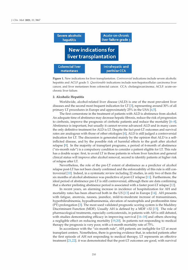
J. Clin. Med. 2021, 10, 3867
Figure 1. New indications for liver transplantation. Controversial indications include severe alcoholichepatitis and ACLF grade 3. Questionable indications include non-hepatocellular carcinoma livercancer, and liver metastases from colorectal cancer. CCA: cholangiocarcinoma; ACLF: acute-on-chronic liver failure.
2. Alcoholic Hepatitis
Worldwide, alcohol-related liver disease (ALD) is one of the most prevalent liverdiseases and the second most frequent indication for LT [3], representing around 30% of allprimary LT procedures in Europe and approximately 25% in the USA [4,5].
The first cornerstone in the treatment of patients with ALD is abstinence from alcohol.An adequate time of abstinence may decrease hepatic fibrosis, reduce the risk of progressionto cirrhosis, improve the prognosis of cirrhotic patients and reduce the mortality [6–8].Abstinence is important, but usually it cannot reverse advanced ALD and in many casesthe only definitive treatment for ALD is LT. Despite the fact post-LT outcomes and survivalrates are analogous with those of other etiologies [4], ALD is still judged a controversialindication for LT. The discussion is generated mainly by the opinion that ALD is a self-inflicted disease, and by the possible risk of harmful effects to the graft after alcoholrelapse [9]. In the majority of transplant programs, a period of 6-month of abstinence(“six-month rule”) is a compulsory condition to consider a patient eligible for LT. This rulehas a double scope: first, to avoid LT in those patients in whom liver function and generalclinical status will improve after alcohol removal, second to identify patients at higher riskof relapse after LT.
Nevertheless, the role of the pre-LT extent of abstinence as a predictor of alcoholrelapse post-LT has not been clearly confirmed and the enforceability of this rule is still con-troversial [10]. Indeed, in a systematic review including 22 studies, in only two of them thesix months of alcohol abstinence was predictive of post-LT relapse [11]. Furthermore, theideal period of abstinence pre-LT is still controversial, although there are data confirmingthat a shorter prelisting abstinence period is associated with a faster post-LT relapse [12].
In recent years, an alarming increase in incidence of hospitalization for AH andmortality rates has been observed both in the US [13] and in Europe [14]. AH presentswith fatigue, anorexia, nausea, jaundice, mild-to-moderate increase of transaminases,hyperbilirubinemia, hypoalbuminemia, elevation of neutrophils and prothrombin time(PT) prolongation [3]. The most used validated prognostic scoring system is the MaddreyDiscriminant Function (MDF). Usually AH is defined by a MDF >32 [15]. The role ofpharmacological treatments, especially corticosteroids, in patients with AH is still debated,with studies demonstrating efficacy in improving survival [16–18] and others showinga negligible effect on reducing mortality [19,20]. In patients not responding to medicaltherapy the prognosis is very poor, with a 6-month mortality rate of 75%.
In accordance with the “six-month rule”, AH patients are ineligible for LT at mosttransplant centers. Nonetheless, there is growing evidence that, in selected patients afterthe first episode of AH not responding to medical therapy, LT represents an effectivetreatment [21,22]. it was demonstrated that the post-LT outcomes are good, with survival
210
J. Clin. Med. 2021, 10, 3867
rates significantly higher compared to not transplanted patients with AH not respondingto steroid therapy [23–25].
Like with ALD cirrhotic LT recipients some ethical and social concerns remain. Theseare mostly originated by the public opinion that a graft is afforded to patients who wereactively consuming alcohol immediately previous to admission on the waiting list, withhigher risk of post-LT alcohol relapse. However, the existing data on LT in these pa-tients demonstrate that relapse rates are analogous to those observed in patients withALD that respected the “6-month rule”, if a rigorous and appropriate selection process isapplied [23–25].
Multiple arguments for either “tight” or “loose” selection criteria have been proposedfor LT in AH [26]. One major argument for tight selection is that current models forpredicting survival without LT are not adequately precise for use in an individual patient,which implies not only that some patients will be subjected to LT unnecessarily, but alsothat others will be denied a potentially lifesaving LT. Additionally, most criteria for listingrely on clinical judgment, which may vary across different centers, thus leading to inequityof access to LT. On the other hand, real-life patients with AH undergoing LT often presentwith ACLF and a high risk of short-term mortality, thus making unnecessary LT very rare.Furthermore, a careful selection of patients at the first event of liver decompensation hasrepeatedly yielded excellent outcomes and low risk of relapse [26].
Like other indications for LT, further refinement of selection criteria is expected toevolve gradually over time. However, without the establishment of national and inter-national agreement on criteria for admitting in waiting list and transplanting patientswith AH, a high variability persists in terms of admittance to LT for those patients, withdisparities that are manifest also at a national level with a potential inequality amongpatients with the same clinical conditions [27].
Data reporting good outcomes of early LT in selected patients were published in thelast years. In the study by Mathurin et al. [23], 26 patients with AH with no response tocorticosteroids were subjected to early LT as rescue therapy, after a strict multidisciplinaryselection process. Survival after 6 and 24 months post-LT were significantly higher than inmatched not transplanted controls (77% vs. 23%). Alcohol relapse was detected up to threeyears after LT in about 10% of patients.
A US study, published by Im et al. [28] confirmed the good outcomes of early LTin 94 patients with AH, in whom the 6-month survival rate was higher compared withmatched not transplanted patients (89% vs. 11%). Alcohol relapse was diagnosed in onlyone recipient at 180 days after LT. Similarly, in a retrospective study published by Leeet al. [29], cumulative patient survival percentages after LT for AH were 94% and 84%at 1 year and 3 years, respectively After LT, 72% were abstinent, 18% had occasionallyrelapses, and 11% had sustained alcohol intake.
In Italy, Germani et al. coordinated the first Italian experience in a pilot study on earlyLT for AH from four different LT centers. Among those centers, the coordinating centeris the Multivisceral Transplant Unit of Padua University Hospital. The inclusion criteriawere AH, as a first episode of decompensation in chronic liver disease and no responses tomedical therapies, but more importantly, the patient should have been socially integratedand have supportive family members, with psychiatric assessment and addiction profileand no comorbidities [30]. Preliminary data coming from Padua Liver Transplant centerdemonstrated a significantly higher survival rate amongst patients who underwent earlyLT compared to non-responding patients who were denied early LT.
The Spanish Society of Liver Transplantation has recently published a consensus state-ment on the potential expansion of indications for LT including patients with a first episodeof severe AH not responding to medical therapy [31], whereas no specific guidelines orposition statements have been published with this regards in Germany. In UK a pilot pro-gram for LT in patients with severe AH was developed. Over a 3-year period 20 patientsaged between 18 and 40 years were evaluated, but none underwent LT, mainly due to the
211

J. Clin. Med. 2021, 10, 3867
extremely stringent criteria for listing and the need for unanimity among members of thetransplant panel [27].
The most significant concern in patients actively drinking before admission, is thepost-LT risk of relapse. In the already mentioned landmark paper [23], about 10% ofpatients had a relapse up to three years after LT. This could be important not only fromthe “single-patient” perspective, but also for the possible negative effects on donation rate.Nevertheless, a recent multicenter survey suggests that organ donation was not negativelyinfluenced by the early LT for AH [32]. Given the complexity of the selection and manage-ment of patients with AH, a multidisciplinary approach, involving various stakeholdersincluding transplant hepatologists and transplant surgeons, but also psychiatrists, psy-chologists, and addiction specialists is becoming compulsory to accurately evaluate LTcandidates [33,34]. The SALT prognostic score, developed including four objective pre-LTvariables, was proposed in order to foresee the risk of sustained alcohol intake after earlyLT for AH assisting in the selection of patient candidates for early LT or in advising controlspost-LT [35].
The psychosocial assessment of LT candidates and the evaluation of social background,including the presence of an active and effective support by the family, are essential partsof the pre-transplant evaluation process. In fact, the transplant outcome is undoubt-edly influenced also by psychosocial and behavioral issues along with the usual medicalfactors [36]. This concept is even more important in the context of early LT where the psy-chosocial assessment is essential for the establishment of the real probability of long-termabstinence. Indeed, alcohol abuse is frequently associated with depression, personalitydisorders and other psychiatric disease, that can affect the post-transplant outcome of thesepatients [37,38].
To ensure to the LT candidates for AH the best long-term outcomes, globally acceptedclinical and psychosocial selection criteria should be identified [39]. Very strict criteriashould be explored for the early LT in this setting, as indicated in an Italian positionstatement [34]. Notably, a transparent and direct interaction between clinicians and society,based on the concept of no “a priori” exclusion to the evaluation for LT in the case of AHis essential.
3. Acute-On-Chronic Liver Failure (ACLF)
ACLF is a clinical syndrome characterized by acute decompensation (AD) of chronicliver disease, development of organ failures and systemic inflammation, and high risk ofshort-term mortality (>15% at 28 day) [40–43]. Development of ACLF in patients withchronic liver disease results from various precipitating factors that vary according to geo-graphical regions: alcoholic hepatitis and bacterial infections in the West and relapse ofchronic HBV infection in the East [44]. In approximately 40–50% of the cases, however,development of ACLF is not associated with identifiable triggers and current hypothesis inthese cases is that metabolites from gut bacteria or translocation of DAMPs from leaky gut(or a combination of both) may be involved and trigger inflammation which in turn leadsto organ dysfunction/failure [44]. There is no specific treatment for patients with ACLF,and current management include treatment of associated complications/precipitatingfactors and organ support. In patients with ACLF due to one or more specific factors (i.e.,bacterial infections, alcoholic hepatitis, bleeding events, drug-induced liver injury), earlyidentification of trigger factor(s) and specific treatments are important though it’s unclearwhether this can really prevent worsening of ACLF [40]. All patients with ACLF shouldbe preferably managed in a tertiary care center and by a multidisciplinary team includingtransplant hepatologists, ICU doctors, and transplant surgeons. General management ofpatients with ACLF and their complications should follow current guidelines for manage-ment of critically ill patients with cirrhosis [45]. Patients should be monitored frequentlyand evaluated serially for potential transfer to intensive care unit. Each organ dysfunctionshall be treated specifically in order to prevent a stage in which multiorgan failure occursand all treatments eventually become futile [45].
212

J. Clin. Med. 2021, 10, 3867
Per current consensus, severity of ACLF is defined by number of organ failures, andis not surprising that patients with three or more organ failures have increased risk ofmortality compared with those with one or two organ failures. Specifically, patients with≥3 organ failures have grade 3 ACLF (ACLF 3), and 28-day mortality in these patientsapproaches 80% [40]. Given such a high risk of death and the lack of alternative medicaltreatments, LT may be the only viable option in certain patients with ACLF [46,47]. Yet,selection of patients suitable for LT, prioritization of candidates during wait-list time,and best timing for LT in ACLF remain problematic [48]. Here, we discuss the currentknowledge and the main open issues regarding LT in patients with ACLF, particularlythose with ACLF 3.
3.1. Wait-List Priority in Patients with ACLF Awaiting LT: Beyond MELD-Based Allocation
ACLF is a rapidly progressive syndrome with a variable course [49,50]. On the onehand, it is important to identify patients with potential for full recovery, in whom LT wouldbe unnecessary and not beneficial; on the other, one has to identify those at higher riskfor progression, in whom development of either sepsis or irreversible organ failures cancompromise eligibility for LT and post-transplant outcomes [41]. In these patients, thetherapeutic window for LT is significantly narrow and unexpected clinical deteriorationmay determine removal of candidates from the waiting list [51].
Unfortunately, the discrimination between these groups remains unclear. The Modelfor End-Stage Liver Disease (MELD) score, that is used to estimate wait-list mortality andguide organ allocation in patients with cirrhosis awaiting LT, is not appropriate to predictsurvival in candidates with ACLF [52]. In fact, MELD does not reflect severity of hepaticencephalopathy and respiratory/circulatory failures, which are major drivers of mortalityin ACLF [40–42]. Also, it does not include biomarkers of systemic inflammation, suchas white blood cells and levels of C reactive protein, which reflect severity of ACLF andcorrelate with survival [53].
In a retrospective analysis based on the United Network for Organ Sharing (UNOS)database and including approximately 100,000 patients, Sundaram and coworkers demon-strated that the risk of death in ACLF 3 was 44% even if their MELD score was <25, and wasgreater than that in advanced patients as defined by a MELD >35 but without ACLF [53]. Inan independent cohort including 71,894 veterans with decompensated cirrhosis, Hernaezet al. found that in those with ACLF the probability of 90-day mortality was significantlyhigher than the one predicted by MELD-Na alone [54]. In a third, large retrospectiveanalysis including patients from the UNOS registry between 2002 and 2014, those withACLF 3 (n = 5099) had a significantly higher risk of death at 14 days than those listed foracute liver failure (n = 3377), regardless of MELD-Na [55]. Taken together, these studiesindicate that in ACLF patients, particularly grade 3 ACLF, an early discussion about LTshould be initiated independent of their MELD status.
Whether a combination of MELD score and grade of ACLF could be the optimalstrategy to assess wait-list priority in patients with ACLF has not yet been thoroughlyinvestigated. To this end, in a large, retrospective study including 18,416 candidates withACLF from UNOS registry, Abdallah showed that the severity of ACLF and MELD scoreinteracted synergistically in anticipating the risk of mortality at 90-day, and that the effectof ACLF grade was relatively more relevant at lower (i.e., ≤25) levels of MELD [56]. Anew prognostic tool integrating MELD score and grade of ACLF was therefore proposedto mitigate disparities regarding organ allocation in ACLF, particularly for candidateswith a MELD score ≤ 25 [56]. If these results will be confirmed in prospective cohorts,it is plausible that a combination of MELD score and ACLF grade will become the nextstandard to assess priority of candidates with ACLF.
In conclusion, there is a strong need to improve the MELD-based allocation to mitigatewait-list mortality in candidates with ACLF [47,57]. Innovative scores are supposed tocapture recipient factors (number and severity of organ failures), global patient’s status andperformance (sarcopenia and frailty), and chronic associated conditions (comorbidities).
213
J. Clin. Med. 2021, 10, 3867
This could lead to a more personalized approach regarding management of wait-list priorityin ACLF and would ultimately improve patient’s survival and LT outcomes [58].
3.2. Benefit of Liver Transplantation in Patients with ALCF Grade 3
Patients with ACLF 3 have a 28-day LT-free survival of 20% [40–42]. Consideringsuch a high risk of death, LT is a potentially life-saving treatment for the vast majority ofthese patients. Preliminary data, however, suggested that post-transplant survival in ACLF3 might be lower than that in recipients transplanted for decompensated cirrhosis [59].Hence, the question rose as to which all patients with ACLF, independent of ACLF grade,could be considered for transplantation [57].
Results from other studies, on the other hand, indicated good survival 1-year afterLT [53,60–62]. Sundaram et al. retrospectively analyzed the UNOS database for the years2004–2017 [63]. In total, 56,801 patients received LT and 54.6% had no ACLF, 15.4% hadACLF 1, 15.9% had ACLF 2, and 14.1% had ACLF 3. Interestingly, survival at 1-yearpost-LT was comparable between patients with ACLF 3 and those with grade 0–2, andthat was above 80% in all groups [63]. More interestingly, although patients with ACLF 3had lower long-term survival compared with those with grade 0–2, 68% of patients whoreceived LT for ACLF 3 were alive 5 years after transplantation, which would justify boththe transplant benefit in these patients, set at >50% 5-year after LT [64], and the acceptableutility of donor grafts. Comparable findings were reported by multicenter cohort studiesfrom Europe [49,60,65] and by one recent study from Asia that first evaluated the outcomesof living donor LT for the treatment of ACLF [66]. In this study, including 321 candidateswith high MELD score who underwent living donor LT, survival at 5-year was comparablebetween patients with and without ACLF (72% versus 81.82%), and 1 year-survival inACLF 3 was comparable with that of grade 1 and 2 (76% vs. 85% vs. 93%; p = 0.2) [66].Taken together, these studies indicate that ACLF grade 3 is not an absolute contraindicationfor LT, and that satisfactory outcomes can be achieved provided there is a good selectionof candidates.
To this end, new methods to improve assessment of LT eligibility in these patientsare eagerly awaited, that is to evaluate whether an individual patient has become “toosick to be transplanted” [58,67]. The fact that no patient with ACLF and severe respiratoryfailure in the CANONIC trial underwent LT indicated that this condition was consideredan absolute contraindication for LT in ACLF [49]. Two large multicenter studies fromUS [53,68] indicated the following factors to be associated with higher risk of death posttransplantation: need for mechanical ventilation at transplant, levels of lactate >4 mmol/Lbefore transplantation, pre-transplant white blood cells count within normal limits, olderage of recipient, and transplantation of marginal-grafts. The combination of three differentorgan supports (dialysis, vasoactive drugs, and mechanical ventilation) has been proposedas a potential criterion to withhold LT [51], however it may also prevent transplantation ina significant number of subjects with potentially favourable outcome. In fact, other factorsmay affect severity of organ failures and therefore the chance to perform a successfulLT. This includes indications for organ support (i.e., ventilation for severe respiratorydysfunction vs. grade IV encephalopathy) and intensity (dose of vasoactives) and/orduration (i.e., 2–3 vs. >7 days) of organs support.
Given the increasing number of LTs performed in patients with ACLF worldwide, abetter understanding of how to define too-sick-to transplant patients and thereby avoid“futile” transplantations is urgently needed [58]. Not only it is important to confirmwhether LT in ACLF 3 confers a significant survival benefit, but also whether is associatedwith an improved quality of life [69]. For example, pre-transplant acute kidney injury(AKI), that is commonly observed in cirrhosis and ACLF [70–72], is a major predictor ofpost-LT chronic kidney disease (CKD) and need for replacement therapy [73]. Although theburden of CKD after LT in ACLF 1 may not be substantially increased [74], more specificresults regarding patients with ACLF 3 are lacking. Long-term data regarding quality of life
214

J. Clin. Med. 2021, 10, 3867
after LT for ACLF are awaited and may help to improve selection criteria and managementof candidates with ACLF both before and after LT [75,76].
3.3. Timing of LT: The Earlier the Better?
One major challenge in candidates with ACLF is to assess the appropriate timing forLT. Given their high risk of death, one would expect that “as soon as possible” could leadto the greatest transplant benefit. In support of this assumption, in a landmark analysisof UNOS database, Sundaram and Jalan demonstrated that patients with ACLF whoreceived LT within 30 days within listing had higher survival at 1-year than those whowere transplanted thereafter (83% vs. 79%, respectively; p = 0.03) [53]. The same studydemonstrated that LT within 30 days could significantly improve survival in patients whounderwent LT on machinal ventilation (77% vs. 72%; p = 0.03). Comparable results weredescribed in the CANONIC trial where survival at 6 months in patients with ACLF 2 and3 who underwent LT within 28 days was 81% compared with 10% in those treated withmedical therapy [49].
Yet, other evidence suggested that the benefits of early LT have to be balancedagainst benefits conferred by resolution of organ failures. In a retrospective trial including98 candidates with ACLF who received LT, the 37 who had improvement of ACLF gradeprior to transplantation had a significantly better survival compared with controls withno improvement [61]. Similar results were reported by Sundaram in a larger study fromUNOS data [77]. The authors included 3636 candidates with ACLF 3 who received LTwithin 28 days of listing. Of these patients, 24.5% recovered to either no ACLF or grade 1or 2 ACLF, whereas 75.5% remained with ACLF 3 at time of surgery. Interestingly, survivalat 1 year was 82% in patients who underwent LT with ACLF 3 and 88% in patients recov-ering to ACLF 0–2 (p < 0.001). Furthermore, the probability of survival of ACLF 0–2 whoworsened to ACLF 3 was significantly lower than in patients who remained at ACLF 0–2(84% vs. 90%; p < 0.001). However, <25% of candidates with ACLF 3 at enlisting were ableto achieve a lower grade of ACLF [77]. Hence, while in principle it would be optimal toundergo LT upon recovering of organ failures, this appears not doable in the major part ofpatients with ACLF 3.
In summary, in candidates awaiting LT for ACLF 3, two major variables need to bebalanced to assess the best timing for transplantation, that is on one hand the individualpatient’s risk of progression and death on the waiting list, on the other whether there is anychance to postpone transplantation with the goal of waiting for ALCF to improve prior toLT [43,58].
4. Colorectal Liver Metastases and Liver Transplantation
Colorectal carcinoma (CRC) shows an incidence of about 700 per million populationin Western countries and the liver is involved in approximately 70% of patients withcolorectal metastases [78]. Currently, the only potentially curative treatment is representedby surgical resection of metastases [79], with a median 5-year survival of 30–40% comparedto only 5% in those non-resected [79,80]. Despite the recent advancements in surgicaltechniques, only ~20% of patients are resectable at diagnosis [81] and the disease oftenrecurs within 3 years of resection [82]. Percutaneous radiological treatments such asablative treatments (radiofrequency, microwave and cryosurgical ablation, transcatheterintra-arterial therapy), hepatic arterial infusion chemotherapy, transarterial embolizationand chemoembolization, and radioembolization with yttrium 90 can be applied to achievetumor resectability. Even though it has been demonstrated that interventional radiologycontributes to the improvement of overall survival rates [83,84], its role in the curativeintent is still marginal. Palliative chemotherapy remains the main option for patients noncandidable for surgery. Even though the initiation of first-line chemotherapy in selectedpatients with good performance status, no KRAS or BRAF mutations, and left-sided tumorsprolongs median overall survival [85–89], prognosis remains poor and only ~10% of themsurvive up to 5 years [86,90]. Interestingly, recent clinical trials show an improvement of
215
J. Clin. Med. 2021, 10, 3867
median survival with modern chemotherapy including the use of bevacizumab/EGFRantibodies, from 6 months to 2 years [85,91,92].
In this context, LT has been explored as an option to remove all viable disease in thosepatients with disease spread limited to the liver, which are not elegible for resection dueto the low remnant liver volume [93,94]. In the past, several attempts were performedobtaining 5-year overall survivals <20% [95]. Due to these poor results and organ shortages,active colorectal liver metastasis remained a contraindication for LT thereafter. Nonetheless,the majority of these patients were disease-free at death, which was due to transplant-related complications instead. A prospective study from Norway a few years ago (theSECA-I study) has renewed focus for this potentially curative option. Patients included inthis study had completed surgical resection of the primary tumor, had a good performancestatus, and received LT after at least 6 weeks of chemotherapy. The estimated 1, 3 and 5-yearoverall survival after LT were 95%, 68% and 60%, respectively. The median follow-up was27 months (range 8–60 months) and disease-free survival was 35% at 1 year. The candidateswith the best prognosis were those with presence of colorectal liver metastasis at diagnosis,pN0, pretransplant maximal CCR diameter <5.5 cm, levels of carcinoembryonic antigen(CEA) <80 mg/L, response or stable disease on chemotherapy, and >2 years from diagnosisto LT [93]. The 5-year overall survival rate of these patients with favorable prognosticfactors was similar to that of patients transplanted for hepatocellular carcinoma (HCC)following the Milan criteria [96,97]. Interestingly overall survival was much longer whencompared to disease-free survival. As a matter of facts, most recurrences in CRC patientswere slowly-growing lung metastases amenable to treatment, regardless of immunosup-pression [98,99], which is not the case for HCC-recurrence after LT. As a reinforcementto these results, LT was demonstrated to produce longer 5-year overall survival rateswhen compared to chemotherapy in patients non amenable to surgical resection of livermetastastases when data from the SECA-I study were compared to those of the NORDICVII trial (56% vs. 9%, respectively) [88].
Following the SECA-I study, Toso et al. reported the results of 12 patients withcolorectal liver metastases undergoing LT, confirming 1-, 3- and 5-year overall survivalrates of 83%, 62%, and 50%, respectively, and disease-free survivals of 56%, 38% and 38%at 1, 3 and 5 years, respectively [94]. The time from diagnosis to LT appeared to have ahigh impact on survival rates, suggesting a natural selection of those tumors with morefavorable characteristics. The authors suggested that a minimum of 12–24 months shouldbe applied as a selection criterion during the evaluation for LT of these patients. In therecently published open label randomized controlled SECA-II trial, the application ofmore strict selection criteria led to a significant raise of overall survival after LT (1 and5 years were 100% and 83%, respectively), with a median follow-up of 36 months (range5–60 months) [100]. As a matter of fact, patients included in the SECA-II study showedbetter pre-LT prognostic factors and more favorable tumor biology (lower number ofmetastatic lesions, size of largest liver lesions, CEA levels, and recurrence risk scores) thanSECA-I patients. Nevertheless, the burden of the disease was considerable at the time ofLT in both cohorts.
Even though patients that were included had different tumoral characteristics and un-derwent different treatments before LT, it is becoming clear that it represents a valid optionfor curative intent in the context of colorectal liver metastases, offering the possibility oflong-term overall survival to highly-selected patients with extensive disease. Data and ex-perience are still limited, however several clinical trials are coming through (NCT 04161092,NCT 03494946, NCT 04616495, NCT 04874259). The major aim is to refine selection criteriain order to raise overall survival rates close to those of patients undergoing LT as a standardof care. Moreover, one multicentre Italian trial based in Milan (NCT 03803436) is aiming toassess the efficacy of LT compared with a matched cohort of patients included in anothertrial involving chemotherapy plus anti-EGFR. Among others, another element which needsto be evaluated is the possibility to add adjuvant chemotherapy after LT, consideringpossible graft toxicity. In this regard, the TRANSMET Trial from France (NCT 02597348) is
216
J. Clin. Med. 2021, 10, 3867
currently comparing the survival rates between standard of care chemotherapy and LT plusadjuvant chemotherapy [101]. Another major challenge is related to the paucity of livergrafts, challenging the approval of new oncological indications. The recruitment of patientsin LT trials is currently limited to a minimum percentage of the total amount of transplantsper center, aiming to not impact the waiting time for other waitlisted patients. One of thesuggested strategies is the use of marginal grafts, as patients with CRC rarely present atLT with either portal hypetension, end stage liver disease or deterioration of other organsfunctionality and thus could be more easily matched with these donors [100]. Anotheralternative to increase the donor pool is represented by the RAPID-protocol, which involvesa two-stage hepatectomy followed by LT using a left-lateral split graft and delayed totalhepatectomy [102]. Three trials, one in our center (NCT04865471), one in Oslo, Norway(NCT 02215889) and one in Jena, Germany (NCT 03488953) are currently evaluating thisoption. Additionally, a protocol started in Toronto (Canada), is also evaluating the optionof living donor LT in patients with CRC (NCT 02864485).
5. Cholangiocarcinoma and Liver Transplantation
Cholangiocarcinoma (CCA) is one of the most frequent primary liver cancers, secondonly to HCC. It can be classified in three subtypes: intrahepatic (iCCA), perihilar (pCCA)and distal extrahepatic (eCCA) cholangiocarcinoma. In all three subtypes the current goldstandard for treatment is surgical resection [103]. Extensive surgery protocols have beenestablished [104], however radical surgical resection can be achieved in <50% of patientsdue to insufficient remnant liver and difficulties in vascular reconstructions [104]. However,in many cases, complete surgical resection cannot be achieved [105]. Moreover, CCA fre-quently presents with local vascular infiltration, and primary sclerosing cholangitis (PSC)associated CCA is often considered as unresectable due to the underlying liver disease andpredisposition to skip lesions [106]. Radiological treatments, such as intra-arterial therapies,ablation, radioembolization and brachytherapy (iodine-125 seed implantation) are used aslocoregional oncological, palliative, and bridging to surgery in patients with unresectableor recurrent CCA after hepatectomy [107,108], however with no curative potential. Withinthis frame, [106]. LT has been suggested as a potential curative treatment for those patientspresenting with non-resectable pCCA and more recently for patients with “very early”iCCA, since it ideally allows radical resection and eradication underlying PSC when associ-ated. Patients included in clinical trials were free from extrahepatic metastases, vascularor lymphnodes invasion. Even though the initial experiences discouraged many centersto pursue this goal [109], a few groups kept on offering LT to these patients, improvingselection criteria and treatment protocols. Eventually some impressive survival data wereproduced leading to internationally re-evaluate LT as a curative option [109] as a treatmentfor nonresectable CCA, particularly in those countries suffering from graft shortages.
5.1. Intrahepatic Cholangiocarcinoma (iCCA)
ICCA is a subtype of CCA that arises from the intrahepatic biliary tract, which can bedivided into mass-forming, periductal-infiltrating, intraductal, and undefined subtypes,depending on macroscopic growth patterns. The incidence of iCCA has been increasingin the last decades, particularly among cirrhotic patients [110]. Despite surgical advances,long-term outcomes of liver resections remain poor, with a 5-year overall survival of40% and very high prevalence of postoperative morbidity [111–113]. Additionally, in>50% of patients the disease recurs, typically within 24 months after resection [114]. Eventhough iCCA is still widely considered a contraindication to LT, there is still a quitehigh percentage of grafts showing incidental tumors at explant pathology (1–3.3% ofall LT) [115,116], as it still represents a diagnostical challenge. Retrospective data fromthese accidentally transplanted iCCA demonstrated an acceptable 5-year overall- andrecurrence free-survivals in cirrhotic patients with “very early” iCCA (<2 cm), [117–119].This led to reconsider LT as a potentially curative option in this context. Moreover, the2 cm cut-off has been challenged by De Martin et al. who showed comparable survival
217

J. Clin. Med. 2021, 10, 3867
rates after LT for iCCA of <2 cm and those of 2.1–3 cm [120]. As a matter of fact, inthis study the only independent variable associated with tumoral recurrence was itsdifferentiation, which, when available, reduces the impact of tumor size for prognosis.On the other hand, iCCA features are still often underestimated during pre-LT diagnosticevaluation, leading to higher recurrence rates and worse post-LT survival when comparedto HCC [121]. Thus, careful consideration of potential higher aggressiveness of the tumorneeds to be born in mind, especially in the context of PSC. Independent predictors ofpost-LT recurrence and survival include microvascular, perineural or lymphovascularinvasion, multifocality, poor differentiation, infiltrative subtype, lack of neo- or adjuvanttreatments [120,122]. In the noncirrhotic population, Lunsford et al. recently showed a 50%recurrence-free survival, and 83% 5-year overall survival following LT in six patients withlocally advanced, unresectable iCCA [123]. Inclusion criteria were solitary tumor >2 cm ormultifocal disease confined to the liver without evidence of macrovascular or lymph nodeinvolvement and sustained response to neoadjuvant gemcitabine-based chemotherapy.Neither tumor volume nor multifocality affected the incidence of disease recurrence after LT.This neoadjuvant protocol has also been proven successful in downstaging iCCA to surgicalresections, which still remains the gold standard of treatment for iCCA. In this scenario,LT could be kept as an option to those remaining non-resectable despite the neoadjuvanttreatment [124]. Validation of these findings in ongoing clinical trials may change thecurrent exclusion of patients with iCCA from transplant programs, and the identificationof the best selection criteria could further implement long term results (NCT 04195503,NCT 02878473, NCT04556214). For now, iCCA remains a contraindication for LT outsideof clinical trials. Ongoing studies are evaluating the role of mutations in KRAS, fibroblastgrowth factor receptor and VEGF expression and dysregulated immune checkpoints [124].Therefore it has been proposed to sequence the whole genome to facilitate the individuationof new therapeutic targets.
5.2. Perihilar Cholangiocarcinoma (pCCA)
PCCA is a subtype of CCA that arises anywhere from the second-order biliary ductsto above the site of cystic duct origin; it can have exophytic (mass-forming) and intraductalgrowth patterns. PCCA is one of the current challenges for hepatic surgery, which stillrepresents the first-line treatment for this malignancy in case of localized disease [125].Achievement of curative intent often requires implementation of surgery with neo- or adju-vant chemo and radiotherapies [126,127]. Still <20% of pCCA are amenable of surgery atdiagnosis, due to the innate propensity of this tumor for the invasion of the adjacent vessels.For non-resectable patients, chemotherapy offers a minimal extension of survival which, inany case, remains <1 year [128,129], with a progression-free survival of 5 months [130,131].Additionally, frequently pCCA arises from underlying PSC, which limits the possibility ofresection [132]. LT has been considered as it can theoretically maximize resection marginsand remove the underlying parenchymal liver disease when present. The first reportsin the 1990s gave disappointing 28% 5-year survival and a 51% recurrence rate with LTalone, which withheld transplant centers from accepting this as [132]. LT can theoreticallymaximize resection margins and cure the underlying parenchymal liver disease. The earlyexperience in the 1990s with LT alone gave disappointing 28% 5-year survival and a 51%recurrence rate for pCCA, which deterred transplant centers from accepting this oncologi-cal indication [122]. However, in these studies no selection criteria was imposed, treatmentarms did not distinguish between pCCA and iCCA and neo- or adjuvant treatment wherenot included in the treatment protocols. Therefore, despite these dismal initial results, theUniversity of Nebraska and the Mayo Clinic kept on working on this program until theyestablished a successful multimodality protocol for unresectable pCCA preliminary toLT [133–135]. The so called “Mayo Clinic Protocol” includes external beam radiation, com-bined with intravenous 5-fluorouracil, followed by intraluminal brachytherapy and oralcapecitabine. After this a routine exploratory laparoscopic staging is performed to confirmthe absence of extrahepatic disease lymphonodal localizations prior to LT. By adopting this
218

J. Clin. Med. 2021, 10, 3867
protocol, Heimbach et al. were able to obtain a 5-year survival exceeding 80% after LT inthose patients with solitary tumors including nonresectable ones <3 cm in radial diameternot extending below the cystic duct, without evidence of lymph node metastases, and inthose pCCA associated to PSC [135]. Similar results were confirmed in 12 large-volumetransplant centers in the US, which reported a median 5-years disease-free survival ratesof 65% [135]. Similar results were confirmed in 12 large-volume transplant centers in theUS, which reported 5-years disease-free survival rates of 65% [136]. During the years itbecame evident that those patients with early disease had improved outcomes followingLT [137–139], and that neoadjuvant chemoradiotherapy followed by LT offered the best out-comes for patients selected following the Mayo Clinic criteria [140–142]. Moreover, Mandelet al. demonstrated that patients selected for LT with these criteria reach a significant bettersurvival compared to those not respecting them (59% versus 21% at 5-years) [143]. Thus,the “Mayo Clinic Protocol” has been gradually adopted, confirming 5-year survival ratesof approximately 65–70% across different transplant centers [136,144–146]. These results,similar to those obtained in patients transplanted for HCC, led the United Network forOrgan Sharing (UNOS) to allow the assignment of a Model for End-Stage Liver Disease(MELD) score exception to patients with unresectable pCCA or arising in the setting ofPSC, for accessing LT [136,147]. However, concerns regarding organ allocation, waitingtimes, and the intensity of the neoadjuvant protocol have been limiting the spread of thisindication in clinical practice. As for now, the guidelines of the European Associationfor the Study of the Liver recommend LT for pCCA to be limited to centers with clinicalresearch protocols employing strict selection of patients and adjuvant or neoadjuvanttherapy [10]. Still several patients drop out from the waiting list due to tumoral progressionand its related complications, positive laparoscopy or inability to tolerate chemotherapyprior to LT. As demonstrated by a recent observational study, the estimated 82% 5-yearsurvival rate precipitated to 58% on intention to treat analysis, since 46% of patients initiallyincluded did not access LT due to neoplastic progression [148]. The Mayo Clinic groupreported that the risk factors for dropout of the LT waiting list due to disease progressionwere: CA 19.9 ≥500 U/mL, a mass ≥3 cm, malignant brushing or biopsy and a MELDscore ≥20. Likewise, predictors of recurrence after LT were elevated CA 19.9, invasion ofportal vein and evidence of residual tumor at explant [149]. Furthermore, the time intervalbetween neoadjuvant therapy and LT was found to be inversely proportional to recurrencerates, which in turn correlates with tumor biology [150]. However, waiting time needs to bebalanced with more pronounced fibrosis induced by prolonged radiotheraphy, hamperingboth staging laparoscopy and LT. Living donor LT could theoretically avoid timing issues.However ethical concerns toward living donation need to be considered as well at thisstage of evidence. Ongoing and future studies will probably better address these issues andfurther refine treatment protocols [151]. A group at Washington University are currentlyrecruiting for a prospective study which aims to assess whether highly selected patientsstill require neoadjuvant chemoradiation (NCT 00301379). Moreover, the use of sirolimuswill be explored in a pilot trial (NCT 01888302). The rationale of giving sirolimus with gem-citabine and cisplatin is that it may be useful for patients with high risk of CCA recurrenceafter LT or either surgery. The results of these and other ongoing trials (NCT01549795,NCT02178280, NCT 04378023, NCT02232932) will be of great interest.
6. Conclusions
The LT scenario is undoubtedly evolving rapidly, with a plethora of new indicationsthat could give hope for a better life for a large number of patients. However, these newerindications increase the pressure in an already difficult context of organ shortage. Strategiesare therefore needed to increase the pool of transplantable organs that aim to ensure thebalance between new indications and available resources. Moreover, it is mandatory tooptimize the patients’ selection criteria to guarantee transplant advantages and achieveadequate patient and graft survival. Specialized surgeons, oncologists, hepatologistsand radiologists should collaborate in a multi-disciplinary transplant team to ensure
219
J. Clin. Med. 2021, 10, 3867
proper work-up and minimize the risks for these patients. Finally, the new scenario oftransplants makes it essential to review and standardize the allocation systems and ethicalconsiderations across countries to ensure the same treatment options for all patients.
Author Contributions: A.Z., S.S., M.G., G.G., M.S., D.B. writing—original draft preparation, F.P.R.,P.B. writing—review and editing. All authors have read and agreed to the published version ofthe manuscript.
Funding: This research received no external funding.
Acknowledgments: The Authors are grateful to the Marina Minnaja Foundation for co-founding theresearch grant of A.Z., S.S. and D.B.
Conflicts of Interest: The authors declare no conflict of interest.
References
1. Starzl, T.E.; Marchioro, T.L.; Porter, K.A.; Brettschneider, L. Homotransplantation of the liver. Transplantation 1967, 5, 790–803.[CrossRef]
2. Adam, R.; Karam, V.; Cailliez, V.; Grady, J.G.O.; Mirza, D.; Cherqui, D.; Klempnauer, J.; Salizzoni, M.; Pratschke, J.; Jamieson,N.; et al. 2018 Annual Report of the European Liver Transplant Registry (ELTR)—50-Year Evolution of Liver Transplantation.Transplant. Int. 2018, 31, 1293–1317. [CrossRef]
3. Mathurin, P.; Bataller, R. Trends in the management and burden of alcoholic liver disease. J. Hepatol. 2015, 62, S38–S46. [CrossRef]4. Burra, P.; Senzolo, M.; Adam, R.; Delvart, V.; Karam, V.; Germani, G.; Neuberger, J. Liver transplantation for alcoholic liver
disease in Europe: A study from the ELTR (European Liver Transplant Registry). Am. J. Transplant. 2010, 10, 138–148. [CrossRef][PubMed]
5. Singal, A.K.; Guturu, P.; Hmoud, B.; Kuo, Y.F.; Salameh, H.; Wiesner, R.H. Evolving frequency and outcomes of liver transplanta-tion based on etiology of liver disease. Transplantation 2013, 95, 755–760. [CrossRef] [PubMed]
6. Mehta, G.; Macdonald, S.; Cronberg, A.; Rosselli, M.; Khera-Butler, T.; Sumpter, C.; Al-Khatib, S.; Jain, A.; Maurice, J.; Char-alambous, C.; et al. Short-term abstinence from alcohol and changes in cardiovascular risk factors, liver function tests andcancer-related growth factors: A prospective observational study. BMJ Open 2018, 8, e020673. [CrossRef] [PubMed]
7. Lackner, C.; Spindelboeck, W.; Haybaeck, J.; Douschan, P.; Rainer, F.; Terracciano, L.; Haas, J.; Berghold, A.; Bataller, R.; Stauber,R.E. Histological parameters and alcohol abstinence determine long-term prognosis in patients with alcoholic liver disease.J. Hepatol. 2017, 66, 610–618. [CrossRef] [PubMed]
8. Kirpich, I.A.; McClain, C.J.; Vatsalya, V.; Schwandt, M.; Phillips, M.; Falkner, K.C.; Zhang, L.; Harwell, C.; George, D.T.; Umhau,J.C. Liver Injury and Endotoxemia in Male and Female Alcohol-Dependent Individuals Admitted to an Alcohol TreatmentProgram. Alcohol. Clin. Exp. Res. 2017, 41, 747–757. [CrossRef]
9. Neuberger, J. Transplantation for alcoholic liver disease: A perspective from Europe. Liver Transpl. Surg. 1998, 4, S51–S57.10. European Association for the Study of the Liver. EASL Clinical Practice Guidelines: Liver transplantation. J. Hepatol. 2016, 64,
433–485. [CrossRef]11. McCallum, S.; Masterton, G. Liver transplantation for alcoholic liver disease: A systematic review of psychosocial selection
criteria. Alcohol Alcohol. 2006, 41, 358–363. [CrossRef]12. Varma, V.; Webb, K.; Mirza, D.F. Liver transplantation for alcoholic liver disease. World J. Gastroenterol. 2010, 16, 4377–4393.
[CrossRef]13. Liangpunsakul, S. Clinical characteristics and mortality of hospitalized alcoholic hepatitis patients in the United States. J. Clin.
Gastroenterol. 2011, 45, 714–719. [CrossRef]14. Sandahl, T.D.; Jepsen, P.; Thomsen, K.L.; Vilstrup, H. Incidence and mortality of alcoholic hepatitis in Denmark 1999–2008: A
nationwide population based cohort study. J. Hepatol. 2011, 54, 760–764. [CrossRef] [PubMed]15. Maddrey, W.C.; Boitnott, J.K.; Bedine, M.S.; Weber, F.L., Jr.; Mezey, E.; White, R.I., Jr. Corticosteroid therapy of alcoholic hepatitis.
Gastroenterology 1978, 75, 193–199. [CrossRef]16. Mathurin, P.; Duchatelle, V.; Ramond, M.J.; Degott, C.; Bedossa, P.; Erlinger, S.; Benhamou, J.P.; Chaput, J.C.; Rueff, B.; Poynard, T.
Survival and prognostic factors in patients with severe alcoholic hepatitis treated with prednisolone. Gastroenterology 1996, 110,1847–1853. [CrossRef] [PubMed]
17. Mathurin, P.; Mendenhall, C.L.; Carithers, R.L.; Ramond, M.J.; Maddrey, W.C.; Garstide, P.; Rueff, B.; Naveau, S.; Chaput, J.C.;Poynard, T. Corticosteroids improve short-term survival in patients with severe alcoholic hepatitis (AH): Individual data analysisof the last three randomized placebo controlled double blind trials of corticosteroids in severe AH. J. Hepatol. 2002, 36, 480–487.[CrossRef]
18. Ramond, M.J.; Poynard, T.; Rueff, B.; Mathurin, P.; Theodore, C.; Chaput, J.C.; Benhamou, J.P. A randomized trial of prednisolonein patients with severe alcoholic hepatitis. N. Engl. J. Med. 1992, 326, 507–512. [CrossRef]
220

J. Clin. Med. 2021, 10, 3867
19. Rambaldi, A.; Saconato, H.H.; Christensen, E.; Thorlund, K.; Wetterslev, J.; Gluud, C. Systematic review: Glucocorticosteroids foralcoholic hepatitis—A Cochrane Hepato-Biliary Group systematic review with meta-analyses and trial sequential analyses ofrandomized clinical trials. Aliment. Pharmacol. Ther. 2008, 27, 1167–1178. [CrossRef]
20. Thursz, M.R.; Richardson, P.; Allison, M.; Austin, A.; Bowers, M.; Day, C.P.; Downs, N.; Gleeson, D.; MacGilchrist, A.; Grant, A.;et al. Prednisolone or pentoxifylline for alcoholic hepatitis. N. Engl. J. Med. 2015, 372, 1619–1628. [CrossRef]
21. Burra, P.; Germani, G. Transplantation for acute alcoholic hepatitis. Clin. Liver Dis. 2017, 9, 141–143. [CrossRef]22. Burra, P.; Bizzaro, D.; Forza, G.; Feltrin, A.; Volpe, B.; Ronzan, A.; Feltrin, G.; Carretta, G.; D’Amico, F.; Cillo, U.; et al. Severe
acute alcoholic hepatitis: Can we offer early liver transplantation? Minerva Gastroenterol. 2021, 67, 23–25. [CrossRef] [PubMed]23. Mathurin, P.; Moreno, C.; Samuel, D.; Dumortier, J.; Salleron, J.; Durand, F.; Castel, H.; Duhamel, A.; Pageaux, G.P.; Leroy, V.; et al.
Early Liver Transplantation for Severe Alcoholic Hepatitis. N. Engl. J. Med. 2011, 365, 1790–1800. [CrossRef]24. Weeks, S.R.; Sun, Z.; McCaul, M.E.; Zhu, H.; Anders, R.A.; Philosophe, B.; Ottmann, S.E.; Garonzik Wang, J.M.; Gurakar, A.O.;
Cameron, A.M. Liver Transplantation for Severe Alcoholic Hepatitis, Updated Lessons from the World’s Largest Series. J. Am.Coll. Surg. 2018, 226, 549–557. [CrossRef] [PubMed]
25. Lee, B.P.; Mehta, N.; Platt, L.; Gurakar, A.; Rice, J.P.; Lucey, M.R.; Im, G.Y.; Therapondos, G.; Han, H.; Victor, D.W.; et al. Outcomesof Early Liver Transplantation for Patients With Severe Alcoholic Hepatitis. Gastroenterology 2018, 155, 422–430. [CrossRef][PubMed]
26. Im, G.Y.; Neuberger, J. Debate on Selection Criteria for Liver Transplantation for Alcoholic Hepatitis: Tighten or Loosen? LiverTranspl. 2020, 26, 916–921. [CrossRef]
27. Thursz, M.; Allison, M. Liver transplantation for alcoholic hepatitis: Being consistent about where to set the bar. Liver Transpl.2018, 24, 733–734. [CrossRef]
28. Im, G.Y.; Kim-Schluger, L.; Shenoy, A.; Schubert, E.; Goel, A.; Friedman, S.L.; Florman, S.; Schiano, T.D. Early Liver Transplantationfor Severe Alcoholic Hepatitis in the United States—A Single-Center Experience. Am. J. Transplant. 2016, 16, 841–849. [CrossRef]
29. Lee, B.P.; Chen, P.H.; Haugen, C.; Hernaez, R.; Gurakar, A.; Philosophe, B.; Dagher, N.; Moore, S.A.; Li, Z.P.; Cameron, A.M.Three-year Results of a Pilot Program in Early Liver Transplantation for Severe Alcoholic Hepatitis. Ann. Surg. 2017, 265, 20–27.[CrossRef]
30. Germani, G.; Angrisani, D.; Addolorato, G.; Merli, M.; Mazzarelli, C.; Tarli, C. Liver transplantation for severe alcoholic hepatitis:amulticentre Italian study. Am. J. Transplant. 2021. submitted.
31. Rodríguez-Perálvarez, M.; Gómez-Bravo, M.; Sánchez-Antolín, G.; De la Rosa, G.; Bilbao, I.; Colmenero, J. Expanding Indicationsof Liver Transplantation in Spain: Consensus Statement and Recommendations by the Spanish Society of Liver Transplantation.Transplantation 2021, 105, 602–607. [CrossRef] [PubMed]
32. Stroh, G.; Rosell, T.; Dong, F.; Forster, J. Early Liver Transplantation for Patients With Acute Alcoholic Hepatitis: Public Views andthe Effects on Organ Donation. Am. J. Transplant. 2015, 15, 1598–1604. [CrossRef] [PubMed]
33. EASL Clinical Practice Guidelines: Management of alcohol-related liver disease. J. Hepatol. 2018, 69, 154–181. [CrossRef]34. Burra, P.; Belli, L.S.; Corradini, S.G.; Volpes, R.; Marzioni, M.; Giannini, E.; Toniutto, P. Common issues in the management of
patients in the waiting list and after liver transplantation. Dig. Liver Dis. 2017, 49, 241–253. [CrossRef] [PubMed]35. Lee, B.P.; Vittinghoff, E.; Hsu, C.; Han, H.S.; Therapondos, G.; Fix, O.K.; Victor, D.W.; Dronamraju, D.; Im, G.Y.; Voigt, M.D.; et al.
Predicting Low Risk for Sustained Alcohol Use After Early Liver Transplant for Acute Alcoholic Hepatitis: The Sustained AlcoholUse Post-Liver Transplant Score. Hepatology 2019, 69, 1477–1487. [CrossRef]
36. Maldonado, J.R. Why It is Important to Consider Social Support When Assessing Organ Transplant Candidates? Am. J. Bioeth.2019, 19, 1–8. [CrossRef]
37. Walter, M.; Scholler, G.; Moyzes, D.; Hildebrandt, M.; Neuhaus, R.; Danzer, G.; Klapp, B.F. Psychosocial prediction of abstinencefrom ethanol in alcoholic recipients following liver transplantation. Transplant. Proc. 2002, 34, 1239–1241. [CrossRef]
38. Bottesi, G.; Granziol, U.; Forza, G.; Volpe, B.; Feltrin, A.; Battermann, F.; Cavalli, C.; Cillo, U.; Gerosa, G.; Fraiese, A.; et al. ThePsychosocial Assessment of Transplant Candidates: Inter-Rater Reliability and Predictive Value of the Italian Stanford IntegratedPsychosocial Assessment for Transplantation (SIPAT). Psychosomatics 2020, 61, 127–134. [CrossRef] [PubMed]
39. Donckier, V.; Lucidi, V.; Gustot, T.; Moreno, C. Ethical considerations regarding early liver transplantation in patients with severealcoholic hepatitis not responding to medical therapy. J. Hepatol. 2014, 60, 866–871. [CrossRef]
40. Moreau, R.; Jalan, R.; Gines, P.; Pavesi, M.; Angeli, P.; Cordoba, J.; Durand, F.; Gustot, T.; Saliba, F.; Domenicali, M.; et al. Acute-on-chronic liver failure is a distinct syndrome that develops in patients with acute decompensation of cirrhosis. Gastroenterology2013, 144, 1426–1437. [CrossRef]
41. O’Leary, J.G.; Reddy, K.R.; Garcia-Tsao, G.; Biggins, S.W.; Wong, F.; Fallon, M.B.; Subramanian, R.M.; Kamath, P.S.; Thuluvath, P.;Vargas, H.E.; et al. NACSELD acute-on-chronic liver failure (NACSELD-ACLF) score predicts 30-day survival in hospitalizedpatients with cirrhosis. Hepatology 2018, 67, 2367–2374. [CrossRef] [PubMed]
42. Sarin, S.K.; Choudhury, A.; Sharma, M.K.; Maiwall, R.; Al Mahtab, M.; Rahman, S.; Saigal, S.; Saraf, N.; Soin, A.S.; Devarbhavi,H.; et al. Acute-on-chronic liver failure: Consensus recommendations of the Asian Pacific association for the study of the liver(APASL): An update. Hepatol. Int. 2019, 13, 353–390. [CrossRef]
43. Ferrarese, A.; Feltracco, P.; Barbieri, S.; Cillo, U.; Burra, P.; Senzolo, M. Outcome of critically ill cirrhotic patients admitted to theICU: The role of ACLF. J. Hepatol. 2019, 70, 801–803. [CrossRef]
44. Hernaez, R.; Solà, E.; Moreau, R.; Ginès, P. Acute-on-chronic liver failure: An update. Gut 2017, 66, 541–553. [CrossRef]
221
J. Clin. Med. 2021, 10, 3867
45. Nadim, M.K.; Durand, F.; Kellum, J.A.; Levitsky, J.; O’Leary, J.G.; Karvellas, C.J.; Bajaj, J.S.; Davenport, A.; Jalan, R.; Angeli,P.; et al. Management of the critically ill patient with cirrhosis: A multidisciplinary perspective. J. Hepatol. 2016, 64, 717–735.[CrossRef] [PubMed]
46. Abdallah, M.A.; Waleed, M.; Bell, M.G.; Nelson, M.; Wong, R.; Sundaram, V.; Singal, A.K. Systematic review with meta-analysis:Liver transplant provides survival benefit in patients with acute on chronic liver failure. Aliment. Pharmacol. Ther. 2020, 52,222–232. [CrossRef]
47. Trebicka, J.; Sundaram, V.; Moreau, R.; Jalan, R.; Arroyo, V. Liver Transplantation for Acute-on-Chronic Liver Failure: Science orFiction? Liver Transpl. 2020, 26, 906–915. [CrossRef] [PubMed]
48. Toniutto, P.; Zanetto, A.; Ferrarese, A.; Burra, P. Current challenges and future directions for liver transplantation. Liver Int. 2017,37, 317–327. [CrossRef]
49. Gustot, T.; Fernandez, J.; Garcia, E.; Morando, F.; Caraceni, P.; Alessandria, C.; Laleman, W.; Trebicka, J.; Elkrief, L.; Hopf, C.; et al.Clinical Course of acute-on-chronic liver failure syndrome and effects on prognosis. Hepatology 2015, 62, 243–252. [CrossRef][PubMed]
50. Piano, S.; Tonon, M.; Vettore, E.; Stanco, M.; Pilutti, C.; Romano, A.; Mareso, S.; Gambino, C.; Brocca, A.; Sticca, A.; et al. Incidence,predictors and outcomes of acute-on-chronic liver failure in outpatients with cirrhosis. J. Hepatol. 2017, 67, 1177–1184. [CrossRef][PubMed]
51. Putignano, A.; Gustot, T. New concepts in acute-on-chronic liver failure: Implications for liver transplantation. Liver Transpl.2017, 23, 234–243. [CrossRef]
52. Mookerjee, R.P. Prognosis and Biomarkers in Acute-on-Chronic Liver Failure. Semin. Liver Dis. 2016, 36, 127–132. [CrossRef][PubMed]
53. Sundaram, V.; Jalan, R.; Wu, T.; Volk, M.L.; Asrani, S.K.; Klein, A.S.; Wong, R.J. Factors Associated with Survival of PatientsWith Severe Acute-On-Chronic Liver Failure Before and After Liver Transplantation. Gastroenterology 2019, 156, 1381–1391.e3.[CrossRef] [PubMed]
54. Hernaez, R.; Liu, Y.; Kramer, J.R.; Rana, A.; El-Serag, H.B.; Kanwal, F. Model for end-stage liver disease-sodium underestimates90-day mortality risk in patients with acute-on-chronic liver failure. J. Hepatol. 2020, 73, 1425–1433. [CrossRef] [PubMed]
55. Sundaram, V.; Shah, P.; Wong, R.J.; Karvellas, C.J.; Fortune, B.E.; Mahmud, N.; Kuo, A.; Jalan, R. Patients With Acute on ChronicLiver Failure Grade 3 Have Greater 14-Day Waitlist Mortality Than Status-1a Patients. Hepatology 2019, 70, 334–345. [CrossRef]
56. Abdallah, M.A.; Kuo, Y.F.; Asrani, S.; Wong, R.J.; Ahmed, A.; Kwo, P.; Terrault, N.; Kamath, P.S.; Jalan, R.; Singal, A.K. Validatinga novel score based on interaction between ACLF grade and MELD score to predict waitlist mortality. J. Hepatol. 2021, 74,1355–1361. [CrossRef]
57. Zaccherini, G.; Weiss, E.; Moreau, R. Acute-on-chronic liver failure: Definitions, pathophysiology and principles of treatment.JHEP Rep. 2021, 3, 100176. [CrossRef]
58. Burra, P.; Samuel, D.; Sundaram, V.; Duvoux, C.; Petrowsky, H.; Terrault, N.; Jalan, R. Limitations of current liver donor allocationsystems and the impact of newer indications for liver transplantation. J. Hepatol. 2021, 75, S178–S190. [CrossRef] [PubMed]
59. Levesque, E.; Winter, A.; Noorah, Z.; Daures, J.P.; Landais, P.; Feray, C.; Azoulay, D. Impact of acute-on-chronic liver failure on90-day mortality following a first liver transplantation. Liver Int. 2017, 37, 684–693. [CrossRef]
60. Artru, F.; Louvet, A.; Ruiz, I.; Levesque, E.; Labreuche, J.; Ursic-Bedoya, J.; Lassailly, G.; Dharancy, S.; Boleslawski, E.; Lebuffe, G.;et al. Liver transplantation in the most severely ill cirrhotic patients: A multicenter study in acute-on-chronic liver failure grade 3.J. Hepatol. 2017, 67, 708–715. [CrossRef] [PubMed]
61. Huebener, P.; Sterneck, M.R.; Bangert, K.; Drolz, A.; Lohse, A.W.; Kluge, S.; Fischer, L.; Fuhrmann, V. Stabilisation of acute-on-chronic liver failure patients before liver transplantation predicts post-transplant survival. Aliment. Pharmacol. Ther. 2018, 47,1502–1510. [CrossRef]
62. Thuluvath, P.J.; Thuluvath, A.J.; Hanish, S.; Savva, Y. Liver transplantation in patients with multiple organ failures: Feasibilityand outcomes. J. Hepatol. 2018, 69, 1047–1056. [CrossRef] [PubMed]
63. Sundaram, V.; Mahmud, N.; Perricone, G.; Katarey, D.; Wong, R.J.; Karvellas, C.J.; Fortune, B.E.; Rahimi, R.S.; Maddur, H.; Jou,J.H.; et al. Longterm Outcomes of Patients Undergoing Liver Transplantation for Acute-on-Chronic Liver Failure. Liver Transpl.2020, 26, 1594–1602. [CrossRef] [PubMed]
64. Linecker, M.; Krones, T.; Berg, T.; Niemann, C.U.; Steadman, R.H.; Dutkowski, P.; Clavien, P.A.; Busuttil, R.W.; Truog, R.D.;Petrowsky, H. Potentially inappropriate liver transplantation in the era of the “sickest first” policy—A search for the upper limits.J. Hepatol. 2018, 68, 798–813. [CrossRef]
65. Finkenstedt, A.; Nachbaur, K.; Zoller, H.; Joannidis, M.; Pratschke, J.; Graziadei, I.W.; Vogel, W. Acute-on-chronic liver failure:Excellent outcomes after liver transplantation but high mortality on the wait list. Liver Transpl. 2013, 19, 879–886. [CrossRef]
66. Moon, D.B.; Lee, S.G.; Kang, W.H.; Song, G.W.; Jung, D.H.; Park, G.C.; Cho, H.D.; Jwa, E.K.; Kim, W.J.; Ha, T.Y.; et al. Adult LivingDonor Liver Transplantation for Acute-on-Chronic Liver Failure in High-Model for End-Stage Liver Disease Score Patients. Am. J.Transplant. 2017, 17, 1833–1842. [CrossRef] [PubMed]
67. Weiss, E.; Saner, F.; Asrani, S.K.; Biancofiore, G.; Blasi, A.; Lerut, J.; Durand, F.; Fernandez, J.; Findlay, J.Y.; Fondevila, C.; et al.When Is a Critically Ill Cirrhotic Patient Too Sick to Transplant? Development of Consensus Criteria by a Multidisciplinary Panelof 35 International Experts. Transplantation 2021, 105, 561–568. [CrossRef] [PubMed]
222
J. Clin. Med. 2021, 10, 3867
68. Artzner, T.; Michard, B.; Weiss, E.; Barbier, L.; Noorah, Z.; Merle, J.C.; Paugam-Burtz, C.; Francoz, C.; Durand, F.; Soubrane, O.;et al. Liver transplantation for critically ill cirrhotic patients: Stratifying utility based on pretransplant factors. Am. J. Transplant.2020, 20, 2437–2448. [CrossRef]
69. Goosmann, L.; Buchholz, A.; Bangert, K.; Fuhrmann, V.; Kluge, S.; Lohse, A.W.; Huber, S.; Fischer, L.; Sterneck, M.R.; Huebener, P.Liver transplantation for acute-on-chronic liver failure predicts post-transplant mortality and impaired long-term quality of life.Liver Int. 2020, 41, 574–584. [CrossRef]
70. Arora, V.; Maiwall, R.; Rajan, V.; Jindal, A.; Muralikrishna Shasthry, S.; Kumar, G.; Jain, P.; Sarin, S.K. Terlipressin Is Superiorto Noradrenaline in the Management of Acute Kidney Injury in Acute on Chronic Liver Failure. Hepatology 2020, 71, 600–610.[CrossRef]
71. Campello, E.; Zanetto, A.; Radu, C.M.; Bulato, C.; Truma, A.; Spiezia, L.; Senzolo, M.; Garcia-Tsao, G.; Simioni, P. Acute kidneyinjury is associated with increased levels of circulating microvesicles in patients with decompensated cirrhosis. Dig. Liver Dis.2021. [CrossRef]
72. Zanetto, A.; Rinder, H.M.; Campello, E.; Saggiorato, G.; Deng, Y.; Ciarleglio, M.; Wilson, F.P.; Senzolo, M.; Gavasso, S.; Bulato, C.;et al. Acute Kidney Injury in Decompensated Cirrhosis Is Associated With Both Hypo-coagulable and Hyper-coagulable Features.Hepatology 2020, 72, 1327–1340. [CrossRef] [PubMed]
73. Durand, F.; Francoz, C.; Asrani, S.K.; Khemichian, S.; Pham, T.A.; Sung, R.S.; Genyk, Y.S.; Nadim, M.K. Acute Kidney Injury AfterLiver Transplantation. Transplantation 2018, 102, 1636–1649. [CrossRef] [PubMed]
74. Marciano, S.; Mauro, E.; Giunta, D.; Torres, M.C.; Diaz, J.M.; Bermudez, C.; Gutierrez-Acevedo, M.N.; Narvaez, A.; Ortiz,J.; Dirchwolf, M.; et al. Impact of acute-on-chronic liver failure on post-transplant survival and on kidney outcomes. Eur. J.Gastroenterol. Hepatol. 2019, 31, 1157–1164. [CrossRef]
75. Burra, P.; De Bona, M. Quality of life following organ transplantation. Transpl. Int. 2007, 20, 397–409. [CrossRef]76. Burra, P.; Ferrarese, A.; Feltrin, G. Quality of life and adherence in liver transplant recipients. Minerva Gastroenterol. Dietol. 2018,
64, 180–186. [CrossRef]77. Sundaram, V.; Kogachi, S.; Wong, R.J.; Karvellas, C.J.; Fortune, B.E.; Mahmud, N.; Levitsky, J.; Rahimi, R.S.; Jalan, R. Effect of
the clinical course of acute-on-chronic liver failure prior to liver transplantation on post-transplant survival. J. Hepatol. 2020, 72,481–488. [CrossRef] [PubMed]
78. Riihimäki, M.; Hemminki, A.; Sundquist, J.; Hemminki, K. Patterns of metastasis in colon and rectal cancer. Sci. Rep. 2016, 6,29765. [CrossRef]
79. Hackl, C.; Gerken, M.; Loss, M.; Klinkhammer-Schalke, M.; Piso, P.; Schlitt, H.J. A population-based analysis on the rate andsurgical management of colorectal liver metastases in Southern Germany. Int. J. Colorectal Dis. 2011, 26, 1475–1481. [CrossRef]
80. Kanas, G.P.; Taylor, A.; Primrose, J.N.; Langeberg, W.J.; Kelsh, M.A.; Mowat, F.S.; Alexander, D.D.; Choti, M.A.; Poston, G.Survival after liver resection in metastatic colorectal cancer: Review and meta-analysis of prognostic factors. Clin. Epidemiol. 2012,4, 283–301. [CrossRef]
81. Kopetz, S.; Chang, G.J.; Overman, M.J.; Eng, C.; Sargent, D.J.; Larson, D.W.; Grothey, A.; Vauthey, J.N.; Nagorney, D.M.;McWilliams, R.R. Improved survival in metastatic colorectal cancer is associated with adoption of hepatic resection and improvedchemotherapy. J. Clin. Oncol. 2009, 27, 3677–3683. [CrossRef]
82. Nordlinger, B.; Sorbye, H.; Glimelius, B.; Poston, G.J.; Schlag, P.M.; Rougier, P.; Bechstein, W.O.; Primrose, J.N.; Walpole, E.T.;Finch-Jones, M.; et al. Perioperative FOLFOX4 chemotherapy and surgery versus surgery alone for resectable liver metastasesfrom colorectal cancer (EORTC 40983): Long-term results of a randomised, controlled, phase 3 trial. Lancet Oncol. 2013, 14,1208–1215. [CrossRef]
83. Kallini, J.R.; Gabr, A.; Abouchaleh, N.; Ali, R.; Riaz, A.; Lewandowski, R.J.; Salem, R. New Developments in InterventionalOncology Liver Metastases From Colorectal Cancer. Cancer J. 2016, 22, 373–380. [CrossRef] [PubMed]
84. Andres, A.; Majno, P.; Terraz, S.; Morel, P.; Roth, A.; Rubbia-Brandt, L.; Schiffer, E.; Ris, F.; Toso, C. Management of patients withcolorectal liver metastasis in eleven questions and answers. Expert Rev. Anticancer Ther. 2016, 16, 1277–1290. [CrossRef] [PubMed]
85. Van Cutsem, E.; Köhne, C.H.; Láng, I.; Folprecht, G.; Nowacki, M.P.; Cascinu, S.; Shchepotin, I.; Maurel, J.; Cunningham, D.;Tejpar, S.; et al. Cetuximab plus irinotecan, fluorouracil, and leucovorin as first-line treatment for metastatic colorectal cancer:Updated analysis of overall survival according to tumor KRAS and BRAF mutation status. J. Clin. Oncol. 2011, 29, 2011–2019.[CrossRef]
86. Masi, G.; Vasile, E.; Loupakis, F.; Cupini, S.; Fornaro, L.; Baldi, G.; Salvatore, L.; Cremolini, C.; Stasi, I.; Brunetti, I.; et al.Randomized trial of two induction chemotherapy regimens in metastatic colorectal cancer: An updated analysis. J. Natl. CancerInst. 2011, 103, 21–30. [CrossRef] [PubMed]
87. Douillard, J.Y.; Siena, S.; Cassidy, J.; Tabernero, J.; Burkes, R.; Barugel, M.; Humblet, Y.; Bodoky, G.; Cunningham, D.; Jassem, J.;et al. Randomized, phase III trial of panitumumab with infusional fluorouracil, leucovorin, and oxaliplatin (FOLFOX4) versusFOLFOX4 alone as first-line treatment in patients with previously untreated metastatic colorectal cancer: The PRIME study. J.Clin. Oncol. 2010, 28, 4697–4705. [CrossRef]
88. Dueland, S.; Guren, T.K.; Hagness, M.; Glimelius, B.; Line, P.D.; Pfeiffer, P.; Foss, A.; Tveit, K.M. Chemotherapy or livertransplantation for nonresectable liver metastases from colorectal cancer? Ann. Surg. 2015, 261, 956–960. [CrossRef]
223

J. Clin. Med. 2021, 10, 3867
89. Boeckx, N.; Koukakis, R.; Op de Beeck, K.; Rolfo, C.; Van Camp, G.; Siena, S.; Tabernero, J.; Douillard, J.Y.; André, T.; Peeters, M.Primary tumor sidedness has an impact on prognosis and treatment outcome in metastatic colorectal cancer: Results from tworandomized first-line panitumumab studies. Ann. Oncol. 2017, 28, 1862–1868. [CrossRef]
90. Van Cutsem, E.; Köhne, C.H.; Hitre, E.; Zaluski, J.; Chang Chien, C.R.; Makhson, A.; D’Haens, G.; Pintér, T.; Lim, R.; Bodoky, G.;et al. Cetuximab and chemotherapy as initial treatment for metastatic colorectal cancer. N. Engl. J. Med. 2009, 360, 1408–1417.[CrossRef]
91. Glimelius, B.; Cavalli-Björkman, N. Metastatic colorectal cancer: Current treatment and future options for improved survival.Medical approach–present status. Scand. J. Gastroenterol. 2012, 47, 296–314. [CrossRef]
92. Quaranta, M.; Micelli, G.; Coviello, M.; Donadeo, A.; Lozupone, A.; Schittulli, F. Clinical usefulness of CA M26 and CA M29 inbreast carcinoma. J. Nucl. Med. Allied Sci. 1990, 34, 35–38. [PubMed]
93. Hagness, M.; Foss, A.; Line, P.D.; Scholz, T.; Jørgensen, P.F.; Fosby, B.; Boberg, K.M.; Mathisen, O.; Gladhaug, I.P.; Egge, T.S.;et al. Liver transplantation for nonresectable liver metastases from colorectal cancer. Ann. Surg. 2013, 257, 800–806. [CrossRef][PubMed]
94. Toso, C.; Pinto Marques, H.; Andres, A.; Castro Sousa, F.; Adam, R.; Kalil, A.; Clavien, P.A.; Furtado, E.; Barroso, E.; Bismuth,H. Liver transplantation for colorectal liver metastasis: Survival without recurrence can be achieved. Liver Transpl. 2017, 23,1073–1076. [CrossRef] [PubMed]
95. Hoti, E.; Adam, R. Liver transplantation for primary and metastatic liver cancers. Transpl. Int. 2008, 21, 1107–1117. [CrossRef][PubMed]
96. Pavel, M.C.; Fuster, J. Expansion of the hepatocellular carcinoma Milan criteria in liver transplantation: Future directions. World J.Gastroenterol. 2018, 24, 3626–3636. [CrossRef]
97. Dueland, S.; Foss, A.; Solheim, J.M.; Hagness, M.; Line, P.D. Survival following liver transplantation for liver-only colorectalmetastases compared with hepatocellular carcinoma. Br. J. Surg. 2018, 105, 736–742. [CrossRef]
98. Hagness, M.; Foss, A.; Egge, T.S.; Dueland, S. Patterns of recurrence after liver transplantation for nonresectable liver metastasesfrom colorectal cancer. Ann. Surg. Oncol. 2014, 21, 1323–1329. [CrossRef]
99. Grut, H.; Solberg, S.; Seierstad, T.; Revheim, M.E.; Egge, T.S.; Larsen, S.G.; Line, P.D.; Dueland, S. Growth rates of pulmonarymetastases after liver transplantation for unresectable colorectal liver metastases. Br. J. Surg. 2018, 105, 295–301. [CrossRef]
100. Dueland, S.; Syversveen, T.; Solheim, J.M.; Solberg, S.; Grut, H.; Bjørnbeth, B.A.; Hagness, M.; Line, P.D. Survival Following LiverTransplantation for Patients With Nonresectable Liver-only Colorectal Metastases. Ann. Surg. 2020, 271, 212–218. [CrossRef][PubMed]
101. Gorgen, A.; Muaddi, H.; Zhang, W.; McGilvray, I.; Gallinger, S.; Sapisochin, G. The New Era of Transplant Oncology: LiverTransplantation for Nonresectable Colorectal Cancer Liver Metastases. Can. J. Gastroenterol. Hepatol. 2018, 2018, 9531925.[CrossRef]
102. Line, P.D.; Hagness, M.; Berstad, A.E.; Foss, A.; Dueland, S. A Novel Concept for Partial Liver Transplantation in NonresectableColorectal Liver Metastases: The RAPID Concept. Ann. Surg. 2015, 262, e5–e9. [CrossRef] [PubMed]
103. Waisberg, D.R.; Pinheiro, R.S.; Nacif, L.S.; Rocha-Santos, V.; Martino, R.B.; Arantes, R.M.; Ducatti, L.; Lai, Q.; Andraus, W.;D’Albuquerque, L.C. Resection for intrahepatic cholangiocellular cancer: New advances. Transl. Gastroenterol. Hepatol. 2018, 3, 60.[CrossRef]
104. Laurent, S.; Verhelst, X.; Geerts, A.; Geboes, K.; De Man, M.; Troisi, R.; Vanlander, A.; Rogiers, X.; Berrevoet, F.; Van Vlierberghe,H. Update on liver transplantation for cholangiocarcinoma: A review of the recent literature. Acta Gastroenterol. Belg. 2019, 82,417–420.
105. Squires, M.H.; Cloyd, J.M.; Dillhoff, M.; Schmidt, C.; Pawlik, T.M. Challenges of surgical management of intrahepatic cholangio-carcinoma. Expert Rev. Gastroenterol. Hepatol. 2018, 12, 671–681. [CrossRef] [PubMed]
106. Fung, B.M.; Tabibian, J.H. Cholangiocarcinoma in patients with primary sclerosing cholangitis. Curr. Opin. Gastroenterol. 2020, 36,77–84. [CrossRef]
107. Li, H.; Chen, L.; Zhu, G.Y.; Yao, X.; Dong, R.; Guo, J.H. Interventional Treatment for Cholangiocarcinoma. Front. Oncol. 2021, 11,671327. [CrossRef] [PubMed]
108. Mosconi, C.; Calandri, M.; Javle, M.; Odisio, B.C. Interventional radiology approaches for intra-hepatic cholangiocarcinoma. Chin.Clin. Oncol. 2020, 9, 8. [CrossRef]
109. Schmeding, M.; Neumann, U.P. Liver Transplant for Cholangiocarcinoma: A Comeback? Exp. Clin. Transplant. 2015, 13, 301–308.[PubMed]
110. Wu, L.; Tsilimigras, D.I.; Paredes, A.Z.; Mehta, R.; Hyer, J.M.; Merath, K.; Sahara, K.; Bagante, F.; Beal, E.W.; Shen, F.; et al.Trends in the Incidence, Treatment and Outcomes of Patients with Intrahepatic Cholangiocarcinoma in the USA: Facility Type isAssociated with Margin Status, Use of Lymphadenectomy and Overall Survival. World J. Surg. 2019, 43, 1777–1787. [CrossRef][PubMed]
111. Katayose, Y.; Rikiyama, T.; Motoi, F.; Yamamoto, K.; Yoshida, H.; Morikawa, T.; Hayashi, H.; Kanno, A.; Hirota, M.; Satoh, K.; et al.Phase I trial of neoadjuvant chemoradiation with gemcitabine and surgical resection for cholangiocarcinoma patients (NACRACstudy). Hepatogastroenterology 2011, 58, 1866–1872. [CrossRef]
224

J. Clin. Med. 2021, 10, 3867
112. Endo, I.; Gonen, M.; Yopp, A.C.; Dalal, K.M.; Zhou, Q.; Klimstra, D.; D’Angelica, M.; DeMatteo, R.P.; Fong, Y.; Schwartz, L.; et al.Intrahepatic cholangiocarcinoma: Rising frequency, improved survival, and determinants of outcome after resection. Ann. Surg.2008, 248, 84–96. [CrossRef] [PubMed]
113. Merath, K.; Chen, Q.; Bagante, F.; Alexandrescu, S.; Marques, H.P.; Aldrighetti, L.; Maithel, S.K.; Pulitano, C.; Weiss, M.J.; Bauer,T.W.; et al. A Multi-institutional International Analysis of Textbook Outcomes Among Patients Undergoing Curative-IntentResection of Intrahepatic Cholangiocarcinoma. JAMA Surg. 2019, 154, e190571. [CrossRef] [PubMed]
114. Zhang, X.F.; Beal, E.W.; Bagante, F.; Chakedis, J.; Weiss, M.; Popescu, I.; Marques, H.P.; Aldrighetti, L.; Maithel, S.K.; Pulitano, C.;et al. Early versus late recurrence of intrahepatic cholangiocarcinoma after resection with curative intent. Br. J. Surg. 2018, 105,848–856. [CrossRef]
115. Takahashi, K.; Obeid, J.; Burmeister, C.S.; Bruno, D.A.; Kazimi, M.M.; Yoshida, A.; Abouljoud, M.S.; Schnickel, G.T. IntrahepaticCholangiocarcinoma in the Liver Explant After Liver Transplantation: Histological Differentiation and Prognosis. Ann. Transplant.2016, 21, 208–215. [CrossRef]
116. Sapisochín, G.; Fernández de Sevilla, E.; Echeverri, J.; Charco, R. Liver transplantation for cholangiocarcinoma: Current statusand new insights. World J. Hepatol. 2015, 7, 2396–2403. [CrossRef] [PubMed]
117. Sapisochin, G.; Rodríguez de Lope, C.; Gastaca, M.; Ortiz de Urbina, J.; Suarez, M.A.; Santoyo, J.; Castroagudín, J.F.; Varo,E.; López-Andujar, R.; Palacios, F.; et al. “Very early” intrahepatic cholangiocarcinoma in cirrhotic patients: Should livertransplantation be reconsidered in these patients? Am. J. Transplant. 2014, 14, 660–667. [CrossRef] [PubMed]
118. Sapisochin, G.; de Lope, C.R.; Gastaca, M.; de Urbina, J.O.; López-Andujar, R.; Palacios, F.; Ramos, E.; Fabregat, J.; Castroagudín,J.F.; Varo, E.; et al. Intrahepatic cholangiocarcinoma or mixed hepatocellular-cholangiocarcinoma in patients undergoing livertransplantation: A Spanish matched cohort multicenter study. Ann. Surg. 2014, 259, 944–952. [CrossRef]
119. Sapisochin, G.; Facciuto, M.; Rubbia-Brandt, L.; Marti, J.; Mehta, N.; Yao, F.Y.; Vibert, E.; Cherqui, D.; Grant, D.R.; Hernandez-Alejandro, R.; et al. Liver transplantation for “very early” intrahepatic cholangiocarcinoma: International retrospective studysupporting a prospective assessment. Hepatology 2016, 64, 1178–1188. [CrossRef]
120. De Martin, E.; Rayar, M.; Golse, N.; Dupeux, M.; Gelli, M.; Gnemmi, V.; Allard, M.A.; Cherqui, D.; Sa Cunha, A.; Adam, R.; et al.Analysis of Liver Resection Versus Liver Transplantation on Outcome of Small Intrahepatic Cholangiocarcinoma and CombinedHepatocellular-Cholangiocarcinoma in the Setting of Cirrhosis. Liver Transpl. 2020, 26, 785–798. [CrossRef]
121. Lee, D.D.; Croome, K.P.; Musto, K.R.; Melendez, J.; Tranesh, G.; Nakhleh, R.; Taner, C.B.; Nguyen, J.H.; Patel, T.; Harnois, D.M.Liver transplantation for intrahepatic cholangiocarcinoma. Liver Transpl. 2018, 24, 634–644. [CrossRef] [PubMed]
122. Hand, F.; Hoti, E. Contemporary role of liver transplantation for the treatment of cholangiocarcinoma. Expert Rev. Gastroenterol.Hepatol. 2020, 14, 475–481. [CrossRef] [PubMed]
123. Lunsford, K.E.; Javle, M.; Heyne, K.; Shroff, R.T.; Abdel-Wahab, R.; Gupta, N.; Mobley, C.M.; Saharia, A.; Victor, D.W.; Nguyen,D.T.; et al. Liver transplantation for locally advanced intrahepatic cholangiocarcinoma treated with neoadjuvant therapy: Aprospective case-series. Lancet Gastroenterol. Hepatol. 2018, 3, 337–348. [CrossRef]
124. Sapisochin, G.; Javle, M.; Lerut, J.; Ohtsuka, M.; Ghobrial, M.; Hibi, T.; Kwan, N.M.; Heimbach, J. Liver Transplantation forCholangiocarcinoma and Mixed Hepatocellular Cholangiocarcinoma: Working Group Report From the ILTS Transplant OncologyConsensus Conference. Transplantation 2020, 104, 1125–1130. [CrossRef] [PubMed]
125. Mansour, J.C.; Aloia, T.A.; Crane, C.H.; Heimbach, J.K.; Nagino, M.; Vauthey, J.N. Hilar cholangiocarcinoma: Expert consensusstatement. HPB 2015, 17, 691–699. [CrossRef] [PubMed]
126. McMasters, K.M.; Tuttle, T.M.; Leach, S.D.; Rich, T.; Cleary, K.R.; Evans, D.B.; Curley, S.A. Neoadjuvant chemoradiation forextrahepatic cholangiocarcinoma. Am. J. Surg. 1997, 174, 605–608; discussion 608–609. [CrossRef]
127. Burke, E.C.; Jarnagin, W.R.; Hochwald, S.N.; Pisters, P.W.; Fong, Y.; Blumgart, L.H. Hilar Cholangiocarcinoma: Patterns of spread,the importance of hepatic resection for curative operation, and a presurgical clinical staging system. Ann. Surg. 1998, 228, 385–394.[CrossRef]
128. Weigt, J.; Malfertheiner, P. Cisplatin plus gemcitabine versus gemcitabine for biliary tract cancer. Expert Rev. Gastroenterol. Hepatol.2010, 4, 395–397. [CrossRef]
129. Matsuo, K.; Rocha, F.G.; Ito, K.; D’Angelica, M.I.; Allen, P.J.; Fong, Y.; Dematteo, R.P.; Gonen, M.; Endo, I.; Jarnagin, W.R. TheBlumgart preoperative staging system for hilar cholangiocarcinoma: Analysis of resectability and outcomes in 380 patients. J. Am.Coll. Surg. 2012, 215, 343–355. [CrossRef]
130. Hu, J.H.; Tang, J.H.; Lin, C.H.; Chu, Y.Y.; Liu, N.J. Preoperative staging of cholangiocarcinoma and biliary carcinoma using18F-fluorodeoxyglucose positron emission tomography: A meta-analysis. J. Investig. Med. 2018, 66, 52–61. [CrossRef]
131. Eckel, F.; Schmid, R.M. Chemotherapy and targeted therapy in advanced biliary tract carcinoma: A pooled analysis of clinicaltrials. Chemotherapy 2014, 60, 13–23. [CrossRef] [PubMed]
132. Rosen, C.B.; Nagorney, D.M. Cholangiocarcinoma complicating primary sclerosing cholangitis. Semin. Liver Dis. 1991, 11, 26–30.[CrossRef]
133. Sudan, D.; DeRoover, A.; Chinnakotla, S.; Fox, I.; Shaw, B., Jr.; McCashland, T.; Sorrell, M.; Tempero, M.; Langnas, A. Ra-diochemotherapy and transplantation allow long-term survival for nonresectable hilar cholangiocarcinoma. Am. J. Transplant.2002, 2, 774–779. [CrossRef]
225
J. Clin. Med. 2021, 10, 3867
134. De Vreede, I.; Steers, J.L.; Burch, P.A.; Rosen, C.B.; Gunderson, L.L.; Haddock, M.G.; Burgart, L.; Gores, G.J. Prolonged disease-freesurvival after orthotopic liver transplantation plus adjuvant chemoirradiation for cholangiocarcinoma. Liver Transpl. 2000, 6,309–316. [CrossRef]
135. Heimbach, J.K.; Gores, G.J.; Haddock, M.G.; Alberts, S.R.; Nyberg, S.L.; Ishitani, M.B.; Rosen, C.B. Liver transplantation forunresectable perihilar cholangiocarcinoma. Semin. Liver Dis. 2004, 24, 201–207. [CrossRef] [PubMed]
136. Darwish Murad, S.; Kim, W.R.; Harnois, D.M.; Douglas, D.D.; Burton, J.; Kulik, L.M.; Botha, J.F.; Mezrich, J.D.; Chapman, W.C.;Schwartz, J.J.; et al. Efficacy of neoadjuvant chemoradiation, followed by liver transplantation, for perihilar cholangiocarcinomaat 12 US centers. Gastroenterology 2012, 143, 88–98. [CrossRef] [PubMed]
137. Meyer, C.G.; Penn, I.; James, L. Liver transplantation for cholangiocarcinoma: Results in 207 patients. Transplantation 2000, 69,1633–1637. [CrossRef] [PubMed]
138. Robles, R.; Figueras, J.; Turrión, V.S.; Margarit, C.; Moya, A.; Varo, E.; Calleja, J.; Valdivieso, A.; Valdecasas, J.C.; López, P.;et al. Spanish experience in liver transplantation for hilar and peripheral cholangiocarcinoma. Ann. Surg. 2004, 239, 265–271.[CrossRef]
139. Rizvi, S.; Khan, S.A.; Hallemeier, C.L.; Kelley, R.K.; Gores, G.J. Cholangiocarcinoma—Evolving concepts and therapeutic strategies.Nat. Rev. Clin. Oncol. 2018, 15, 95–111. [CrossRef]
140. Blechacz, B. Cholangiocarcinoma: Current Knowledge and New Developments. Gut Liver 2017, 11, 13–26. [CrossRef]141. Welling, T.H.; Feng, M.; Wan, S.; Hwang, S.Y.; Volk, M.L.; Lawrence, T.S.; Zalupski, M.M.; Sonnenday, C.J. Neoadjuvant
stereotactic body radiation therapy, capecitabine, and liver transplantation for unresectable hilar cholangiocarcinoma. LiverTranspl. 2014, 20, 81–88. [CrossRef]
142. Duignan, S.; Maguire, D.; Ravichand, C.S.; Geoghegan, J.; Hoti, E.; Fennelly, D.; Armstrong, J.; Rock, K.; Mohan, H.; Traynor, O.Neoadjuvant chemoradiotherapy followed by liver transplantation for unresectable cholangiocarcinoma: A single-centre nationalexperience. HPB 2014, 16, 91–98. [CrossRef]
143. Mantel, H.T.; Westerkamp, A.C.; Adam, R.; Bennet, W.F.; Seehofer, D.; Settmacher, U.; Sánchez-Bueno, F.; Fabregat Prous, J.;Boleslawski, E.; Friman, S.; et al. Strict Selection Alone of Patients Undergoing Liver Transplantation for Hilar CholangiocarcinomaIs Associated with Improved Survival. PLoS ONE 2016, 11, e0156127. [CrossRef] [PubMed]
144. Goldaracena, N.; Gorgen, A.; Sapisochin, G. Current status of liver transplantation for cholangiocarcinoma. Liver Transpl. 2018,24, 294–303. [CrossRef] [PubMed]
145. Zilbert, N.; Sapisochin, G. Time to reconsider liver transplantation for intrahepatic cholangiocarcinoma? Lancet Gastroenterol.Hepatol. 2018, 3, 294–295. [CrossRef]
146. DeOliveira, M.L.; Kambakamba, P.; Clavien, P.A. Advances in liver surgery for cholangiocarcinoma. Curr. Opin. Gastroenterol.2013, 29, 293–298. [CrossRef]
147. Gores, G.J.; Gish, R.G.; Sudan, D.; Rosen, C.B. Model for end-stage liver disease (MELD) exception for cholangiocarcinoma orbiliary dysplasia. Liver Transpl. 2006, 12, S95–S97. [CrossRef]
148. Rea, D.J.; Heimbach, J.K.; Rosen, C.B.; Haddock, M.G.; Alberts, S.R.; Kremers, W.K.; Gores, G.J.; Nagorney, D.M. Livertransplantation with neoadjuvant chemoradiation is more effective than resection for hilar cholangiocarcinoma. Ann. Surg. 2005,242, 451–458; discussion 458–461. [CrossRef]
149. Darwish Murad, S.; Kim, W.R.; Therneau, T.; Gores, G.J.; Rosen, C.B.; Martenson, J.A.; Alberts, S.R.; Heimbach, J.K. Predictors ofpretransplant dropout and posttransplant recurrence in patients with perihilar cholangiocarcinoma. Hepatology 2012, 56, 972–981.[CrossRef]
150. Heimbach, J.K.; Gores, G.J.; Haddock, M.G.; Alberts, S.R.; Pedersen, R.; Kremers, W.; Nyberg, S.L.; Ishitani, M.B.; Rosen, C.B.Predictors of disease recurrence following neoadjuvant chemoradiotherapy and liver transplantation for unresectable perihilarcholangiocarcinoma. Transplantation 2006, 82, 1703–1707. [CrossRef] [PubMed]
151. Tan, E.K.; Taner, T.; Heimbach, J.K.; Gores, G.J.; Rosen, C.B. Liver Transplantation for Peri-hilar Cholangiocarcinoma. J. Gastrointest.Surg. 2020, 24, 2679–2685. [CrossRef] [PubMed]
226
MDPISt. Alban-Anlage 66
4052 BaselSwitzerland
Tel. +41 61 683 77 34Fax +41 61 302 89 18
www.mdpi.com
Journal of Clinical Medicine Editorial OfficeE-mail: [email protected]
www.mdpi.com/journal/jcm


MDPI
St. Alban-Anlage 66
4052 Basel
Switzerland
Tel: +41 61 683 77 34
Fax: +41 61 302 89 18
www.mdpi.com ISBN 978-3-0365-3860-0
Copyright © 2022 FDOKUMEN