110. Microbiota, immune ....Pharmacological Research 2012
28
This article appeared in a journal published by Elsevier. The attached copy is furnished to the author for internal non-commercial research and education use, including for instruction at the authors institution and sharing with colleagues. Other uses, including reproduction and distribution, or selling or licensing copies, or posting to personal, institutional or third party websites are prohibited. In most cases authors are permitted to post their version of the article (e.g. in Word or Tex form) to their personal website or institutional repository. Authors requiring further information regarding Elsevier’s archiving and manuscript policies are encouraged to visit: http://www.elsevier.com/copyright
Transcript of 110. Microbiota, immune ....Pharmacological Research 2012

This article appeared in a journal published by Elsevier. The attachedcopy is furnished to the author for internal non-commercial researchand education use, including for instruction at the authors institution
and sharing with colleagues.
Other uses, including reproduction and distribution, or selling orlicensing copies, or posting to personal, institutional or third party
websites are prohibited.
In most cases authors are permitted to post their version of thearticle (e.g. in Word or Tex form) to their personal website orinstitutional repository. Authors requiring further information
regarding Elsevier’s archiving and manuscript policies areencouraged to visit:
http://www.elsevier.com/copyright
Author's personal copy
Pharmacological Research 69 (2013) 87– 113
Contents lists available at SciVerse ScienceDirect
Pharmacological Research
jo ur n al hom epage: www.elsev ier .com/ locate /yphrs
Invited review
Gut microbiota, immune development and function
Stig Bengmark ∗
Division of Surgery & Interventional Science, University College London, 4th floor, 74 Huntley Street, London WC1E 6AU, United Kingdom
a r t i c l e i n f o
Article history:Received 29 August 2012Accepted 1 September 2012
Keywords:MicrobiotaMicrobiomeMicrobial translocationProbiotic bacteriaLactobacillusLactobacillus plantarumLactobacillus paracaseiMicrobial translocationInflammationInfectionToll-likeNeutrophilsPharmaceuticalsBiologicalEco-biologicalsNutraceticalsCurcuminResveratrolAntibioticsChemotherapeuticsBarriersLeakageGutAirwaysOral cavitySkinVaginaPlacentaAmnionBlood–brain barrierGrowthReplicationApoptosisMucosaEndotheliumPlaquesCytokinesIL1NF-kB
a b s t r a c t
The microbiota of Westerners is significantly reduced in comparison to rural individuals living a similarlifestyle to our Paleolithic forefathers but also to that of other free-living primates such as the chim-panzee. The great majority of ingredients in the industrially produced foods consumed in the West areabsorbed in the upper part of small intestine and thus of limited benefit to the microbiota. Lack of propernutrition for microbiota is a major factor under-pinning dysfunctional microbiota, dysbiosis, chronicallyelevated inflammation, and the production and leakage of endotoxins through the various tissue barriers.Furthermore, the over-comsumption of insulinogenic foods and proteotoxins, such as advanced glyca-tion and lipoxidation molecules, gluten and zein, and a reduced intake of fruit and vegetables, are keyfactors behind the commonly observed elevated inflammation and the endemic of obesity and chronicdiseases, factors which are also likely to be detrimental to microbiota. As a consequence of this lifestyleand the associated eating habits, most barriers, including the gut, the airways, the skin, the oral cavity, thevagina, the placenta, the blood–brain barrier, etc., are increasingly permeable. Attempts to reconditionthese barriers through the use of so called ‘probiotics’, normally applied to the gut, are rarely success-ful, and sometimes fail, as they are usually applied as adjunctive treatments, e.g. in parallel with heavypharmaceutical treatment, not rarely consisting in antibiotics and chemotherapy.
It is increasingly observed that the majority of pharmaceutical drugs, even those believed to haveminimal adverse effects, such as proton pump inhibitors and anti-hypertensives, in fact adversely affectimmune development and functions and are most likely also deleterious to microbiota. Equally, it appearsthat probiotic treatment is not compatible with pharmacological treatments. Eco-biological treatments,with plant-derived substances, or phytochemicals, e.g. curcumin and resveratrol, and pre-, pro- and syn-biotics offers similar effects as use of biologicals, although milder but also free from adverse effects. Suchtreatments should be tried as alternative therapies; mainly, to begin with, for disease prevention but alsoin early cases of chronic diseases. Pharmaceutical treatment has, thus far, failed to inhibit the tsunamiof endemic diseases spreading around the world, and no new tools are in sight. Dramatic alterations, indirection of a paleolithic-like lifestyle and food habits, seem to be the only alternatives with the potential
∗ Correspondence address: 185 Barrier Point Road, Pontoon Docks, London E16 2SE, United Kingdom. Tel.: +44 20 7511 6842; fax: +44 20 7511 6842.E-mail address: [email protected]: http://www.bengmark.com.
1043-6618/$ – see front matter © 2012 Elsevier Ltd. All rights reserved.http://dx.doi.org/10.1016/j.phrs.2012.09.002

Author's personal copy
88 S. Bengmark / Pharmacological Research 69 (2013) 87– 113
TNFGrowth factorsInsulinogenicIGF-1PrebioticsPlant fibersGreensFruitsVegetablesMineralsFat dietRefined carbohydrate dietAdvanced glycation end products(AGEs)Advanced lipoxidation end products(ALEs)EndotoxinLPSProteotoxinsCaseinGlutenZeinWestern lifestylePaleolithicSchimpanzeeADHDAIDSAllergyALSAlzheimerArteriosclerosisAtheromaAutoimmuneAutismBipolarCancerCeliac diseaseCOPDCoronary Heart DiseaseChronic Fatigue SyndromeChronic Renal DiseaseCognitiveDiabetesHIV-1EncephalopathyIrritable Bowel DiseaseInflammatory Bowel DiseaseLiver cirrhosisLiver steatosisObesityOsteoarthritisOsteoporosisPancreatitisParadontosis ;ParkinsonPolycystic Ovary DiseaseRheumatoid DiseaseSchizophreniaStressStrokeUveitis
to control the present escalating crisis. The present review focuses on human studies, especiallythose of clinical relevance.
© 2012 Elsevier Ltd. All rights reserved.
Contents
1. An epidemic of obesity and chronic diseases . . . . . . . . . . . . . . . . . . . . . . . . . . . . . . . . . . . . . . . . . . . . . . . . . . . . . . . . . . . . . . . . . . . . . . . . . . . . . . . . . . . . . . . . . . . . . . . . . . . . . . . . 892. A consequence of large consumption of insulinotrophic foods? . . . . . . . . . . . . . . . . . . . . . . . . . . . . . . . . . . . . . . . . . . . . . . . . . . . . . . . . . . . . . . . . . . . . . . . . . . . . . . . . . . . . 893. Western food and its effects on microbiota and disease . . . . . . . . . . . . . . . . . . . . . . . . . . . . . . . . . . . . . . . . . . . . . . . . . . . . . . . . . . . . . . . . . . . . . . . . . . . . . . . . . . . . . . . . . . . . 904. Proteotoxins induce and enhance inflammation . . . . . . . . . . . . . . . . . . . . . . . . . . . . . . . . . . . . . . . . . . . . . . . . . . . . . . . . . . . . . . . . . . . . . . . . . . . . . . . . . . . . . . . . . . . . . . . . . . . . 905. Gluten-sensitivity a common and “new” disorder . . . . . . . . . . . . . . . . . . . . . . . . . . . . . . . . . . . . . . . . . . . . . . . . . . . . . . . . . . . . . . . . . . . . . . . . . . . . . . . . . . . . . . . . . . . . . . . . . . 916. Proteotoxin-induced low threshold for immune response . . . . . . . . . . . . . . . . . . . . . . . . . . . . . . . . . . . . . . . . . . . . . . . . . . . . . . . . . . . . . . . . . . . . . . . . . . . . . . . . . . . . . . . . . 917. Heat- and storage induced inflammation-inducing proteins . . . . . . . . . . . . . . . . . . . . . . . . . . . . . . . . . . . . . . . . . . . . . . . . . . . . . . . . . . . . . . . . . . . . . . . . . . . . . . . . . . . . . . . 918. Each body surface has its own typical microbiome . . . . . . . . . . . . . . . . . . . . . . . . . . . . . . . . . . . . . . . . . . . . . . . . . . . . . . . . . . . . . . . . . . . . . . . . . . . . . . . . . . . . . . . . . . . . . . . . . 929. Numerous mechanisms to control intestinal homeostasis . . . . . . . . . . . . . . . . . . . . . . . . . . . . . . . . . . . . . . . . . . . . . . . . . . . . . . . . . . . . . . . . . . . . . . . . . . . . . . . . . . . . . . . . . . 9210. Great differences in microbiota between rural and urban areas. . . . . . . . . . . . . . . . . . . . . . . . . . . . . . . . . . . . . . . . . . . . . . . . . . . . . . . . . . . . . . . . . . . . . . . . . . . . . . . . . . . 93

Author's personal copy
S. Bengmark / Pharmacological Research 69 (2013) 87– 113 89
11. Clear association between level of fiber intake and obesity and obesity-related diseases . . . . . . . . . . . . . . . . . . . . . . . . . . . . . . . . . . . . . . . . . . . . . . . . . . . . . . . . 9312. Vitamin D, physical exercise and other factors of importance . . . . . . . . . . . . . . . . . . . . . . . . . . . . . . . . . . . . . . . . . . . . . . . . . . . . . . . . . . . . . . . . . . . . . . . . . . . . . . . . . . . . . 9413. Dysbiosis and leaky barriers . . . . . . . . . . . . . . . . . . . . . . . . . . . . . . . . . . . . . . . . . . . . . . . . . . . . . . . . . . . . . . . . . . . . . . . . . . . . . . . . . . . . . . . . . . . . . . . . . . . . . . . . . . . . . . . . . . . . . . . . 9414. Effect of foods on microbiota and leaky barriers . . . . . . . . . . . . . . . . . . . . . . . . . . . . . . . . . . . . . . . . . . . . . . . . . . . . . . . . . . . . . . . . . . . . . . . . . . . . . . . . . . . . . . . . . . . . . . . . . . . 9715. Effect of pharmaceutical drugs on microbiota and leaky barriers . . . . . . . . . . . . . . . . . . . . . . . . . . . . . . . . . . . . . . . . . . . . . . . . . . . . . . . . . . . . . . . . . . . . . . . . . . . . . . . . . 9816. Over-reacting neutrophils . . . . . . . . . . . . . . . . . . . . . . . . . . . . . . . . . . . . . . . . . . . . . . . . . . . . . . . . . . . . . . . . . . . . . . . . . . . . . . . . . . . . . . . . . . . . . . . . . . . . . . . . . . . . . . . . . . . . . . . . . . 9817. Bioecological reduction of inflammation, neutrophil infiltration and tissue destruction . . . . . . . . . . . . . . . . . . . . . . . . . . . . . . . . . . . . . . . . . . . . . . . . . . . . . . . . . 9918. Personal experience with pro- and synbiotics . . . . . . . . . . . . . . . . . . . . . . . . . . . . . . . . . . . . . . . . . . . . . . . . . . . . . . . . . . . . . . . . . . . . . . . . . . . . . . . . . . . . . . . . . . . . . . . . . . . . . 99
18.1. Perioperative prophylaxis in elective surgery . . . . . . . . . . . . . . . . . . . . . . . . . . . . . . . . . . . . . . . . . . . . . . . . . . . . . . . . . . . . . . . . . . . . . . . . . . . . . . . . . . . . . . . . . . . . . . 10018.2. Perioperative prophylaxis in liver transplantation . . . . . . . . . . . . . . . . . . . . . . . . . . . . . . . . . . . . . . . . . . . . . . . . . . . . . . . . . . . . . . . . . . . . . . . . . . . . . . . . . . . . . . . . . 10118.3. Early treatment in major trauma . . . . . . . . . . . . . . . . . . . . . . . . . . . . . . . . . . . . . . . . . . . . . . . . . . . . . . . . . . . . . . . . . . . . . . . . . . . . . . . . . . . . . . . . . . . . . . . . . . . . . . . . . . . 10118.4. Early treatment in severe acute pancreatitis . . . . . . . . . . . . . . . . . . . . . . . . . . . . . . . . . . . . . . . . . . . . . . . . . . . . . . . . . . . . . . . . . . . . . . . . . . . . . . . . . . . . . . . . . . . . . . . 10118.5. Effects on “mind clarity” – encephalopathy . . . . . . . . . . . . . . . . . . . . . . . . . . . . . . . . . . . . . . . . . . . . . . . . . . . . . . . . . . . . . . . . . . . . . . . . . . . . . . . . . . . . . . . . . . . . . . . . 10218.6. Effects in HIV . . . . . . . . . . . . . . . . . . . . . . . . . . . . . . . . . . . . . . . . . . . . . . . . . . . . . . . . . . . . . . . . . . . . . . . . . . . . . . . . . . . . . . . . . . . . . . . . . . . . . . . . . . . . . . . . . . . . . . . . . . . . . . . . 102
19. Life-threatening systemic inflammation . . . . . . . . . . . . . . . . . . . . . . . . . . . . . . . . . . . . . . . . . . . . . . . . . . . . . . . . . . . . . . . . . . . . . . . . . . . . . . . . . . . . . . . . . . . . . . . . . . . . . . . . . . . 10320. Studies with no or adverse effects . . . . . . . . . . . . . . . . . . . . . . . . . . . . . . . . . . . . . . . . . . . . . . . . . . . . . . . . . . . . . . . . . . . . . . . . . . . . . . . . . . . . . . . . . . . . . . . . . . . . . . . . . . . . . . . . . . 103
20.1. Ecologic 641TM . . . . . . . . . . . . . . . . . . . . . . . . . . . . . . . . . . . . . . . . . . . . . . . . . . . . . . . . . . . . . . . . . . . . . . . . . . . . . . . . . . . . . . . . . . . . . . . . . . . . . . . . . . . . . . . . . . . . . . . . . . . . . . 10320.2. Lactobacillus plantarum 299TM – ProVivaTM . . . . . . . . . . . . . . . . . . . . . . . . . . . . . . . . . . . . . . . . . . . . . . . . . . . . . . . . . . . . . . . . . . . . . . . . . . . . . . . . . . . . . . . . . . . . . . . . . 10320.3. Lactobacillus rhamnosus GGTM . . . . . . . . . . . . . . . . . . . . . . . . . . . . . . . . . . . . . . . . . . . . . . . . . . . . . . . . . . . . . . . . . . . . . . . . . . . . . . . . . . . . . . . . . . . . . . . . . . . . . . . . . . . . . . 10320.4. Synbiotic 2000TM/Synbiotic 2000 ForteTM . . . . . . . . . . . . . . . . . . . . . . . . . . . . . . . . . . . . . . . . . . . . . . . . . . . . . . . . . . . . . . . . . . . . . . . . . . . . . . . . . . . . . . . . . . . . . . . . . . 10320.5. TrevisTM . . . . . . . . . . . . . . . . . . . . . . . . . . . . . . . . . . . . . . . . . . . . . . . . . . . . . . . . . . . . . . . . . . . . . . . . . . . . . . . . . . . . . . . . . . . . . . . . . . . . . . . . . . . . . . . . . . . . . . . . . . . . . . . . . . . . . 10420.6. VSL#3TM . . . . . . . . . . . . . . . . . . . . . . . . . . . . . . . . . . . . . . . . . . . . . . . . . . . . . . . . . . . . . . . . . . . . . . . . . . . . . . . . . . . . . . . . . . . . . . . . . . . . . . . . . . . . . . . . . . . . . . . . . . . . . . . . . . . . . 104
21. Why do studies fail? . . . . . . . . . . . . . . . . . . . . . . . . . . . . . . . . . . . . . . . . . . . . . . . . . . . . . . . . . . . . . . . . . . . . . . . . . . . . . . . . . . . . . . . . . . . . . . . . . . . . . . . . . . . . . . . . . . . . . . . . . . . . . . . . 10422. Choice of lactic acid bacteria as probiotics . . . . . . . . . . . . . . . . . . . . . . . . . . . . . . . . . . . . . . . . . . . . . . . . . . . . . . . . . . . . . . . . . . . . . . . . . . . . . . . . . . . . . . . . . . . . . . . . . . . . . . . . . 10423. Molecular gene targeting – the future? . . . . . . . . . . . . . . . . . . . . . . . . . . . . . . . . . . . . . . . . . . . . . . . . . . . . . . . . . . . . . . . . . . . . . . . . . . . . . . . . . . . . . . . . . . . . . . . . . . . . . . . . . . . . 10524. It is all about inflammation . . . . . . . . . . . . . . . . . . . . . . . . . . . . . . . . . . . . . . . . . . . . . . . . . . . . . . . . . . . . . . . . . . . . . . . . . . . . . . . . . . . . . . . . . . . . . . . . . . . . . . . . . . . . . . . . . . . . . . . . . 10525. Cytokine-inhibition, pharma and/or probiotics? . . . . . . . . . . . . . . . . . . . . . . . . . . . . . . . . . . . . . . . . . . . . . . . . . . . . . . . . . . . . . . . . . . . . . . . . . . . . . . . . . . . . . . . . . . . . . . . . . . . 10626. Single target or multitarget treatment? . . . . . . . . . . . . . . . . . . . . . . . . . . . . . . . . . . . . . . . . . . . . . . . . . . . . . . . . . . . . . . . . . . . . . . . . . . . . . . . . . . . . . . . . . . . . . . . . . . . . . . . . . . . . 10627. Final remarks . . . . . . . . . . . . . . . . . . . . . . . . . . . . . . . . . . . . . . . . . . . . . . . . . . . . . . . . . . . . . . . . . . . . . . . . . . . . . . . . . . . . . . . . . . . . . . . . . . . . . . . . . . . . . . . . . . . . . . . . . . . . . . . . . . . . . . . 106
References . . . . . . . . . . . . . . . . . . . . . . . . . . . . . . . . . . . . . . . . . . . . . . . . . . . . . . . . . . . . . . . . . . . . . . . . . . . . . . . . . . . . . . . . . . . . . . . . . . . . . . . . . . . . . . . . . . . . . . . . . . . . . . . . . . . . . . . . . . 107
1. An epidemic of obesity and chronic diseases
The global incidence of obesity and various endemic chronicdiseases from ADHD, Alzheimer, and autism to osteoarthritis andstroke is rapidly increasing. For some decades the epidemic wasmainly a problem of the Western world, with its modern agri-cultural techniques, mass production and easy access to andlarge consumption of agricultural foods, including those frequentlyindustrially manipulated and processed such as meat, dairy andwheat [2–4]. However, similar development is now observed alsoin other parts of the world, largely in parallel to the adoption ofa “modern”/Western lifestyle. Seemingly this epidemic of obesityand associated diseases has its epicenter in Southern United States[1], states like Alabama, Louisiana and Mississippi having the high-est incidence of obesity and chronic diseases in the US and theworld. These diseases spread, with a pattern similar to a tsunami,across the world; to the West to New Zeeland and Australia, to theNorth to Canada, to the East to Western Europe & the Arab worldand to the South, particularly Brazil.
Recent studies forecast by the year of 2050 a doubling of theincidence of diabetes [5] and a tripling of the incidence of ADHD,Alzheimer disease [6] and cancer [7] in most countries, includingthe US. A most interesting recently published study looked at the USand UK, together representing approximately 5% of the world’s pop-ulation [8], two countries which already have the highest rates ofobesity and chronic diseases. The study suggest that these countriescombined will, by the year of 2030, see another 76 m obese adults,additional 6–8.5 m cases of diabetes, 6–7 m cases of cardiovascu-lar disease, 492,000–669,000 cases of cancer, leading to loss of26–55 m quality-adjusted life years and a dramatic increase in costsof care (calculated to be $50–68b/year) [8]. Other studies suggestthat the increase will continue beyond 2030, if dramatic preven-tive measures are not instituted. While predicting future disease,especially cancer, might be fraught with uncertainty; predictionsare necessary aids to health planners and others and must be done.
The general experience is that the statistical models have, over theyears, been refined and today these models are capable of providingaccurate predictions.
2. A consequence of large consumption of insulinotrophicfoods?
Although sharing an almost identical genome, difference oflifestyle and food habits between modern man and our forefathersliving some 200,000 years ago, are huge. These individuals con-sumed only a small fraction of the insulinotrophic food consumedby modern man, especially those living in the Western World. Theso-called Paleolithic diet was almost identical to the food of thewild chimpanzee of today, with which we share about 99.4 of thegenome. The Neolithic Revolution, and introduction of agriculturesome 10 000 years ago, has provided increasing access food toinsulinotropic and IGF-1-raising foods including sugar, dairy prod-ucts and grains, a process significantly augmented by the IndustrialRevolution, i.e. during the last 150 years. Many agree that thehuman genome has not, and may never, adapt to the high levelsof insulin/IGF-1 signalling (IIS) that drives the Western diet, whichsupports the argument that modern man should attempt to developa more Paleolithic-type diet [9].
The association between high intake of high IIS foods andthe development of chronic diseases is strongly supported, espe-cially by more recent observations in individuals with congenitaldeficiency in IGF-I (Laron syndrome, GH gene deletion, GHRHreceptor defects and IGF-I resistance), who demonstrate a dra-matic reduction in pro-aging signalling [10], rate of cancer [10–12],diabetes [10] and other chronic diseases. It is most interestingto note that these individuals, despite their dwarfism, markedobesity, and severely impaired metabolism (>50% of the individ-uals suffer nonalcoholic fatty liver disease (NAFLD)) experiencelongevity, the greater majority being alive at the ages of 75–78years, some reaching even more advanced ages. Studies in both

Author's personal copy
90 S. Bengmark / Pharmacological Research 69 (2013) 87– 113
invertebrates (C-elegans flies, Drosphila) and rodents (mice andrats) with induced IGF1 deficiency (the IGF1 gene or the GH recep-tor being inactivated), demonstrate a significant prolongation oflifespan, particularly in females [13].
Reducing IGF signalling is presently regarded as a most promis-ing strategy to reduce so-called proteo-toxicity and to delay/inhibitthe development of chronic diseases similar to Alzheimer’s disease,as recently demonstrated in a mouse study [14], and supportedby ‘calorie restriction’ studies in humans [15,16]. However, manyother factors seem to contribute to development of obesity andchronic diseases, among them vitamin D in serum deficiency andaccumulation in the body of high temperature-induced, stronglyproinflammatory products, known as ‘advanced glycation endproducts’ (AGEs) [2–4]. Both Vitamin D-deficiency and accumu-lated AGEs in the body are factors highly suspected to potentiatethe processes leading to obesity and chronic diseases. It is a fact thatIGF1 plays a major role in childhood growth, has profound anaboliceffects in adults, and promotes alterations in aging later in life. Indi-viduals eating Western type food will normally show higher levelsof IGF1 in serum, induced by large consumption of high ‘glycemicindex’ food; in Western countries > half of the consumed caloriesconsist in such foods, which constitute strong inducers of liversynthesis of IGF. Meanwhile, other foods provide a significant addi-tional source of the peptide, dairy foods being especially rich inIGF1. A recent study in young boys fed casein, demonstrated a sig-nificant 15% (P < 0.0001) increase IGF1/s but no changes in fastinginsulin (P = 0.36), while boys fed whey instead had a 21% (P = 0.006)increased fasting insulin, and no change in IGF-1 (P = 0.27) [16].
3. Western food and its effects on microbiota and disease
A major aim of this chapter is to review documented effects ofWestern lifestyle on the microbiota, its diversity and numbers ofstrains but also to investigate the role of Western foods on induc-tion of inflammation and the role of dysbiosis in the pathogenesisof obesity and chronic diseases. The gut microbiota of individualsconsuming a Western diet are likely to be reduced as the lowerdigestive tract is seriously depleted of metabolic fuels, probablyleading to a sub-optimal gut microbial profile. The most obviousand harmful consequences of reduced microbiota are malfunction,dysbiosis, and the often observed high levels of endotoxin in plasma(endotoxemia), which in both experimental and clinical studiesis strongly associated with inflammation and risk of obesity andchronic diseases. Endotoxins are integral components of the outermembrane of Gram-negative bacteria like Enterobacteriaceae andPseudomonadaceae, composed of proteins, lipids, and lipopolysac-charides (LPS). LPS, which is responsible for most of the biologicalproperties of bacterial endotoxins, is known to have exception-ally strong ability to induce inflammation via the so called Toll-likereceptors 2 (TLR2) and 4 (TLR4).
Volunteers living for 1 month on a Western-style diet demon-strated, in a crossover study, a 71% increase in plasma levels ofendotoxin activity (endotoxemia) when compared to those con-suming what the authors called a prudent-style diet, who, in turn,demonstrated a 31% reduced level of endotoxin(s) [17]. A posi-tive correlation between sedentary lifestyle and higher levels ofendotoxin levels and a negative correlation to the degree of phys-ical exercise has also been reported [18]. High fat content of food,rather than high carbohydrate content correlates with high lev-els of endotoxemia. Fat in foods is likely to negatively influencemicroflora and its replication, but the most pronounced effectsare expected from their translocation-facilitating ability i.e. toserve as vehicle for translocation of endotoxin, embedded in fat,though the mucosa and into the circulation, a process referred toas transcellular transportation. Mice fed a high-energy diet (either
high-fat diet or high-carbohydrate diet) demonstrate a significantincrease in plasma LPS, however, again, a high fat diet correlatesto a higher degree than a high carbohydrate diet [19]. Strongcorrelations between plasma levels of endotoxin and numbers ofparameters of metabolic syndrome and between persistant levelsof high endotoxin/plasma and ‘prospect of life’ have been noted –large differences have been reported between the first and fourthquartiles [18]. Among the diseases associated with increased endo-toxin/plasma are, particularly; Alzheimer’s disease [20,21] andcognitive impairment [22], arterio-coronary disease [22–24] andstroke [25], diabetes 1 [26] and 2 [27,28] and cancer [29] but alsoallergy [30], ALS [31], autism [32], autoimmune diseases [33], bipo-lar disease [34], chronic fatigue syndrome [35], COPD [36], minimalencephalopathy [37,38], fibromyalgia [39], HIV [40], liver cirrho-sis [37,38], macular degeneration [41], nephropathies [42], obesity[43,44], osteoarthritis [45], paradontosis [46], Parkinson’s disease[47], rheumatoid disease [48], schizophrenia [49], stress [50] anduveitis [51].
4. Proteotoxins induce and enhance inflammation
Humans are known to be extremely sensitive to endotoxin expo-sure and reported to show signs of inflammation at a dose of LPSthat is at least 250-fold lower than that required in, for example,mice [52]. This is important as modern humans, as an unfortunateconsequence of modern living, much more than other primates, areexposed to LPS both outdoors and indoors, to a large extent throughthe dust inhaled at the home, workplace and at school. Agriculture,textile and wood industries are especially recognized for their badenvironment with extremely high levels of endotoxin exposure.Tobacco smoking is also recognized as a major source of LPS. Oftenneglected is the fact that the food we eat often contains unaccept-ably high levels of endotoxin. Cooking makes little difference asLPS is heat-resistant while both LPS and dead bacteria remain capa-ble of inducing inflammation. The majority of fresh and raw wholevegetables should normally contain only minimal or undetectablelevels of stimulants of TLR2 or TLR4 [53]. However, certain rawand minimally processed vegetables (MPVs) are very sensitive tostorage and might occasionally contain large quantities of bacteriaand endotoxins; among these are bean sprouts, diced onions, andchopped root vegetables such as carrots and onions [54]. Beef, porkand turkey increase their content of TLR2- and TLR4-stimulantswithin a few days, even when stored at 5 ◦C, and especially ifexposed to air [55]. The accumulation of TLR2- and TLR4-stimulantsis minimized by storage of meat in its intact rather than in mincedforms, and when stored under a modified atmosphere, rather thanexposed to air [55]. Very little data exists about the health hazardsof game meat, which despite its favourable nutritional profile whencompared to farmed meat, is often kept hanging for weeks and, asa consequence, is especially rich in endotoxins.
Different food ingredients and particularly proteins mayenhance or diminish the inflammatory properties of meat diets.Many peptides, to which modern humans are daily exposed,possess the ability to induce inflammation, activate TGF-� andToll-like receptors (TLRs). Among these are various lectins, espe-cially glutenoids, and caseins. Molecules induced by heating offood or longer storage at room temperature are molecules col-lectively called Maillard products e.g. the ‘advanced glycation endproducts’ (AGEs) and ‘advanced lipoxidation end products’ (ALEs).The invention of fire increased dramatically the possibilities forthese food products to be produced, and the introduction of gluten-containing grains, which occurred about 10,000 years ago with theadvent of agriculture, further increased exposure to dysfunction-ing proteotoxins – pro-inflammatory molecules – developments,which, in a way, might be regarded as unfortunate “mistakes of

Author's personal copy
S. Bengmark / Pharmacological Research 69 (2013) 87– 113 91
evolution”. The main reason for the large exposure to glutensmight have been that it was mainly the members of the Triticeaegrass tribe (wheat, rye, barley) and the Pooideae subfamily (includ-ing even oats) that grew well at higher latitudes. Modern plantbreeding technology exacerbated the situation as modern breadcontains 15–20 times more gluten, when compared to bread fromthe past. As an unexpected consequence, modern man suffers aseries of highly unwanted human disorders that relate to exposureto glutenoids, particularly gluten (wheat), but also secalins (rye)and hordeins (barley), which all seem to induce inflammation andincrease intestinal permeability [54]. Oats, on the other hand, aremore distantly related to wheat, rye and barley and its active pep-tides, the avenins, are rarely reported to give stronger reactions ofinflammation and allergy [55].
5. Gluten-sensitivity a common and “new” disorder
!t has become increasingly apparent that “classic” celiac dis-ease (CD) represents only “the tip of the iceberg” of an overall largeglutenoid-associated disease burden. We are increasingly awarethat there frequently exists, besides those who suffer celiac dis-ease (CD) and classical wheat allergy, many individuals who oftensuffer discrete reactions to glutenoids. In these individuals, neitherallergic nor autoimmune mechanisms seem to be involved, par-ticularly when exposed to wheat, but also to rye and barley [56].This phenomenon, many times more common than classical CD, isdefined as gluten sensitivity (GS) [56]. Although LA-DQ8 is presentin almost all CD patients, these genes are only present in about halfof patients with GS. Some of the individuals with GS may sufferwell-defined chronic diseases, others ‘only’ more ill-defined dis-tresses; fatigue, depression, encephalopathy/‘foggy mind’, lack ofenergy, diffuse abdominal pain, bloating, diarrhea, eczema and/orrash, undefined headache, numbness in the legs, arms or fingers,joint pain and many other manifestations. More or less all report,when turning to gluten-free diets, increased well-being and fre-quently also improved clinical signs and symptoms. Experienceslike these have made the gluten-free diet the number one healthtrend in the US, growing faster than both low carbohydrate dietsand “fat-free” diets while fueling a market for gluten-free productsapproaching $2.5 billion (US) in global sales in 2010 [57]. Amongthe alternatives flours used for bread-baking are ancient grains,several of them known to grow particularly, if not exclusively,well in Africa; amaranth, arrowroot, brown rice, buckwheat, chia,chickpea, corn, hemp, maize, millet, oat, potato, quinoa, sesame,sorghum, soya, tapioca, teff and white rice, of which sorghum isthe 5th commonest grain in world and especially attractive due toits extremely high content of antioxidants, low content of energy[58], and ability to resist heat-induced protein glycation [59], butalso due to its high versatility and cost-effectiveness.
6. Proteotoxin-induced low threshold for immuneresponse
Glutenoids, which demonstrate endotoxin-mimicking abilities,are capable of lowering the threshold for immune responses,attracting leukocytes and increasing their reactive state, in similarmanner to that of endotoxin e.g. 10 �g/ml of wheat gluten inducesthe equivalent effects of 1 ng/ml LPS [60], while increasing dendriticcell maturation and chemokine secretion [60]. A study investigat-ing fecal samples from 76 symptom-free, non-celiac, first degreeCD relatives and compared to samples from 91 aged-matchedhealthy controls reported significantly lower level of acetic acidand total SCFAs as well as significantly increased level of i-butyricacid and fecal tryptic activity in the asymptomatic CD-relatives[61]. The information that removal of gluten from the diet of the
non-obese diabetic mouse could attenuate the intensity of autoim-munity and reduce the incidence of diabetes led to a cross-overstudy, where 17 first-degree relatives were kept on a gluten-freediet for the first 6 months followed by another 6 months on astandard gluten-containing diet [62]. The acute insulin responseto iv glucose tolerance test rose significantly in 12/14 subjectsafter the first 6 months of gluten-free diet when complying withthe diet (P = 0.04) and decreased again in 10/13 during the follow-ing 6-month period on a standard gluten-contaning diet (P = 0.07)[62]. A similar outcome was recently reported from a crossoverstudy in which 100 individuals suffering ADHD, aged 4–8 years,were randomly assigned to 5 weeks on a restricted eliminationdiet including a restricted consumtion of gluten-containing bread(at the most twice a week) and compared to what was referredto as a healthy diet, followed by another 5 weeks on the alterna-tive diet. All parameters’ total score, inattention, hyperactivity andabbreviated Connor scale scores (ACS) improved significantly onthe restricted diet but deteriorated significantly during the subse-quent period on normal, although supposedly healthy diet [63].Another study focused on thirty-four individuals with irritablebowel syndrome syndrome, 56% of them with human leukocyteantigen (HLA)-DQ2 and/or HLA-DQ8 genes, during 6 weeks exposedto a gluten-free diet [64]. Statistically significant improvements,compared to controls, were reported in the gluten-free group in3/6 parameters studied: abdominal pain (P = 0.02), satisfaction withstool consistency (P = 0.03), and tiredness (P = 0.001); no improv-ment was observed in overall symptoms (P = 0.15), wind (P = 0.08),and nausea (P = 0.69), and no differences observed between indi-viduals with or without DQ2/DQ8 genes [64].
Inclusion of industrially made foods ingredients and particu-larly those of a protein nature might both enhance and diminishthe inflammatory properties of the diet. This is also true for variouslectins and frequently observed with various bovine milk-derivedproteins, and particularly with powdered milk. The synthesis in thebrain of serotonin (5HT) and melatonin is dependent on access totheir precursor amino acid, the essential amino acid l-tryptophan(TRP), normally released in the gut by microbial fermentation ofplants rich in this amino acid [65]. As it is believed that some foodproteins might block such a release, an animal study was under-taken to compare the effects of five different such proteins: zein(corn), gluten (wheat), soy protein isolate, casein, lactalbumin or noprotein. An 8-fold variation was observed in levels of cortex trypto-phan: a marked decline followed zein ingestion, modest reductionsafter casein or gluten, which were paralleled by reductions in cor-tical and hippocampal hypothalamic 5-hydroxytryptophan (5HTP)[65]. A recent publication reports disappearance of signs of malab-sorption and intractable therapy-resistent seizures in three younggirls (ages 18, 19, 23) when placed on gluten free diet [66]. Sim-ilarly individuals with similar symptoms as in neuropsychiatricdiseases; Alzheimer’s disease/cognitive decline [67], autism [68]and schizophrenia [69] are also reported in the literature.
7. Heat- and storage induced inflammation-inducingproteins
Heat- and storage-induced dysfunctioning proteins are of spe-cial interest due to their strong ability to induce inflammation[70,71]. Among foods rich in AGEs and ALEs are dairy products,especially powdered milk (frequently used in enteral nutrition andbaby formulas, as well as in numerous industrially produced foods),in high-temperature produced fried and grilled meat and poultry,but also fish (especially deep-fried and oven-fried), drinks includingcoffee and colas, Asian sauces, such as Chinese soy sauces, bal-samic products and smoked and cured foods in general [72–74].The consumption of such foods, often main constituents of fast
Author's personal copy
92 S. Bengmark / Pharmacological Research 69 (2013) 87– 113
foods, have increased dramatically in recent decades, much in par-allel to the endemic of chronic diseases. Higher levels of AGEs suchas methylglyoxal derivatives in serum (sMG) and/or other AGEs,are strongly associated with a faster rate of cognitive decline inelderly individuals [75], neuro-degenerative diseases [72], prema-ture aging and cognitice decline [73], diabetes type 1 and 2 [74],diabetic nephropathy [75], obesity [76] and liver disease, particu-larly liver steatosis and liver fibrosis [77], lung disease, particularlyCOPD [78] and various cancers including breast cancer [79], colo-rectal cancer [80], esophageal [81], gastric [82], lung [81], ovarian[81], pancreatic [83], prostatic [84], renal [85], and leukemia [86].
A wide range of pro-inflammatory mediators, including TNF-�, IL-1b, IL-6, IL-8 and the nuclear protein high mobility groupbox-1 (HMGB1), are implicated in the pathogenesis of the above-mentioned chronic diseases. HMGB1 is one of the importantmediators known to signal by way of the advanced glycation endproducts, particularly RAGEs, and through the Toll-like receptorsTLR2 and TLR4. Activation of these receptors will ultimately resultin activation of NF-kB, known to induce up-regulation of leukocyteadhesion molecules and production of pro-inflammatory cytokinesand angiogenic factors in both hematopoietic and endothelial cells,thereby promoting inflammation [87]. A recent review suggeststhat particularly HMGB1, TLR and RAGE constitutes a functionaltripod with high ability to promote inflammation [88]. How-ever, many other molecules are also involved in the extremelycomplex processes, which are behind the development of bothinfectious and sterile inflammation. Among them are moleculessuch as heat shock proteins (HSPs), S100s, and hyaluronan, whichplay important roles, all known to trigger immune responses.For practical purpose it has been suggested that these, togetherwith other mediators such as defensins, cathelicidin, eosinophilic-derived neurotoxin (EDN) and several others should be groupedtogether under the name of alarmins [89]. Recently recognizedsuch alarmins are Activin A, a member of the TGF-� superfamilyas well as its binding protein follistatin (FS), which during acuteand chronic inflammatory processes are released by various celltypes in the body [90]. The importance of these proteins to theinflammatory processes has, although known to biomedical sci-ence since the 1980s, only emerged recently [91]. The rapid releaseduring the acute phases of inflammation into the circulation ofActivin A is particularly noteworthy, placing it as one of the ear-liest factors in the systemic cascade of inflammatory events. But itis equally involved in the pathogenesis of chronic diseases, espe-cially in rheumatoid arthritis, in IBD and in other diseases knownfor their association to pathological fibrotic events [91]. It also con-tributes to the proinflammatory macrophage polarization triggeredby granulocyte-macrophage colony-stimulating factor (GM-CSF)while limiting the acquisition of the anti-inflammatory phenotypein a Smad2-dependent manner, skewing macrophage polarizationtowards a proinflammatory phenotype [92].
The triggering effects of several, if not all, of the alarmins are pro-moted by deficiency in vitamin D. A significant negative correlationhas been observed between vitamin D levels and high-sensitivityC-reactive protein, NF�B activity, and TLR4 expression (P < 0.05),while monocytes, when preincubated with vitamin D are shownto significantly decrease lipopolysaccharide-activated TLR4 expres-sion and also cytokine levels (P < 0.05) [93]. A recent study lookedat seasonality of vitamin D status in healthy individuals and itsrelation to TLR-4-mediated cytokines [94]. Circulating concentra-tions of 25(OH)D(3) and 1,25(OH)(2) D(3) were, as expected, higherduring summer (P < 0.05) and also significantly associated with adown-regulation of the TLR-4-mediated cytokines, IL-1�, IL-6, TNF-�, interferon (IFN)-� and IL-10 more in summer than during winter(P < 0.05). The variation in cytokine response upon TLR-2 (Pam3Cys)stimulation was, compared to TLr-4, moderate throughout all thefour seasons [94].
8. Each body surface has its own typical microbiome
Virtually every surface of the human body exposed to the envi-ronment; mouth, hair, nose, ears, vagina, lungs, skin, eyes, etc. hasits own unique, specific and very complex microbial assemblageconstituted by very different microbial species each with their dis-tinct functions, collectively referred to as microbiota or microbiome[95]. The genomic pool of human microbiota is claimed to be atleast 150 times larger than the eukaryotic human nuclear genome,together harbouring more than nine million specific genes [96],and contributing to the enrichment and modulation of numer-ous human functions. The microbiome is extremely sensitive toexternal influences and easily deranged. This is well demonstratedby the catastrophic changes induced on intestinal homeostasis byantibiotic treatment, according to a recent study, affecting over 87%of all metabolites detected, and deranging most metabolic path-ways of critical importance to host physiology, including bile acidmetabolism and eicosanoid and steroid hormone synthesis [97].
Studies on the largest microbiome of the human body – that ofthe gastrointestinal tract, and particularly that of the lower diges-tive tract – has until now received most the scientific interest, whilethe microbiome at other sites of the body remain largely unex-plored. About 60% of the luminal content of lower GI tract consistsin commensal bacteria, which has been described as “an organwithin an organ” and also as “a virtual super-organ” weighing upto 2 kg. The commensal bacteria play a key role in preservationof intact integrity of the mucosal barrier function at all surfaces,and particularly at that of the lower part of the digestive tract.Impaired microbiome function, dysbiosis, has inevitably seriousconsequences for health, and is, sooner or later, associated withsevere pathological implications. The gut microbiota also plays amajor role in the modulation of both the intestinal and generalimmune system and is essential for preservation of functions suchas maturation of gut-associated lymphatic tissue (GALT), secretionof IgA and production of important antimicrobial peptides. The gutmicrobiota exerts important trophic and developmental functionson the intestinal mucosa. More than anything, the enteric micro-biome functions as a potent bioreactor, which controls numerousmetabolic functions, of which many still remain unrecognized [98],while producing thousands of important and unique substances ofthe greatest benefits to the body, as indigestible food substancesare converted by fermentation to simple sugars, short-chain fattyacids, various nutrients, antioxidants and vitamins.
9. Numerous mechanisms to control intestinal homeostasis
Dysbiosis and impaired barrier functions are associated withseveral negative consequences; translocation of lipopolysaccha-rides (LPS) and whole microbial cells, accumulation of endotoxinin the body (endotoxemia) and hyperactivation of the immunesystem. The microbiota controls intestinal homeostasis throughnumerous mechanisms in which substances such as lipopolysac-carides, flagellins, peptidoglycans, and formylated peptides areinvolved. It interacts with intestinal cell receptors such as Toll-likereceptors and activates important intracellular signalling pathwayswith ability to modulate processes such as cell survival, replicationand apoptosis as well as inflammatory response. Among the chal-lenging molecules are NF-�B, caspases, mitogen-activated proteinkinases. The host immune system controls microbial composi-tion through release of molecules such as �-defensins, cryptidins,lectins, angiogenin 4, reactive oxygen species, IgA and so calledbacteriocins, which effectively limits the expansion of variouspathogenic microorganisms (see further [99,100]).
The enteric flora is mostly represented by strict anaerobes(70–90%), which predominate over facultative anaerobes and
Author's personal copy
S. Bengmark / Pharmacological Research 69 (2013) 87– 113 93
aerobes (10–30%) [100]. Recent studies suggest that the gut micro-biota might be classified as belonging to one of three principalvariants, or “enterotypes,” defined by a dominant presence ofBacteroides, Prevotella, or Ruminococcus species [101]. However,increasing evidence suggests that these enterotypes are moremicrobial gradients than, although discrete, defined microbial com-munities as most of the observed differences are largely explainedon the basis of long-term dietary intake [102,103]. Diet is themost powerful influence on gut microbial communities in healthyhuman subjects [104–106]. A study of human subjects and 59 othermammals revealed clusters in which the effects of diet (carniv-orous, omnivorous, or herbivorous almost always outweigh hostphylogeny [104]. Bacteroides species are prevalent with long-termprotein and animal fat diets, whereas Prevotella species are asso-ciated with long-term carbohydrate diets [106]. 45,000 of thepresently identified >800,000 rDNA sequences (microbial species)and about 5 of the about 50 bacterial phyla identified are foundin the lower GI tract [100]. Two of these phyla are totally dominat-ing: Firmicutes (65–80% of the clones) and Bacteroidetes (about 23%),while Actinobacteria (about 3%), Proteobacteria (1%) and Verrumicro-bia (0.1%) exists only in smaller amounts [100,107,108]. Of specialinterest is that Actinobacteria and Firmicutes, to which the genusLactobacillus belongs, are almost exclusively Gram-positive, whileBacteroidetes and Proteobacteria are mainly Gram-negative (see fur-ther [109]). Recent attempts to study the microbiota at other siteswithin the digestive tract report that the mouth harbours the great-est phylogenetic diversity, the stomach the lowest, and diversity toincrease from stomach to the stool [110].
10. Great differences in microbiota between rural andurban areas
As lifestyle, and particularly food intake, has a profound influ-ence on the composition of the microbiota it should be of thegreatest interest to understand more about the Paleolithic micro-biome, to which humans have been adapted during millions ofyears. A recent study compared the fecal microbiota of European(Italian) children (EU) with that of children from a rural Africanvillage of Burkina Faso (BF) in Central Africa. In this environment,the high fibre diet is, in most respects, the closest we can getto that of early human settlements at the time of the birth ofagriculture. Significant differences in both biodiversity and rich-ness of microbiota to the favour of BF children (P < 0.01) werea general and most characteristic observation [111]. BF childrenshowed a significant enrichment in Bacteroidetes and depletion inFirmicutes (P < 0.001), with a unique abundance of bacteria fromthe genus Prevotella and Xylanibacter, genuses known to containa set of bacterial genes for cellulose and xylan hydrolysis. Thesegenuses were completely lacking in the EU children [111]. In addi-tion, significantly higher levels of short-chain fatty acids (P < 0.001)were observed in BF than in EU children. Also Enterobacteriaceae(Shigella and Escherichia) were significantly under-represented inBF compared to EU children (P < 0.05) [111]. Of somewhat greatersurprise was the observation, that Gram-negative bacteria (mainlyBacteroidetes) were more abundant (58.5%) than Gram-positivebacteria (37.4%) in the BF population, whereas Gram-positive(mainly Firmicutes) were more abundant than Gram-negativebacteria (70.4% vs 29.1% respectively) in the EU population, result-ing in a Gram-positive to Gram negative ratio of 37 to 59 in theBF population compared to 70 to 29 in the EU population [111].A further study compared the fecal microbiota of monozygotic(MZ) and dizygotic (DZ) twin pairs living in South Korea and theUnited States; thirty-one MZ (n = 62) and 23 DZ (n = 46) European-and African-ancestry twin pairs from the Missouri AdolescentFemale Twin Study, and 9.5 MZ (n = 19) twin pairs from the Korea
Twin Family Cohort [112]. Alpha diversity (within-sample) mea-surements of the fecal microbiota did not show any significantoverall difference between the Korean and U.S. cohorts, but agreater inter-individual separation between American and Koreansubjects was observed in the lean sub-population than in the obesesub-population as well as in the total population. Furthermore, thediversity in obese US twins was found to be significantly smallerthan in lean US twins; a similar trend was observed in the muchsmaller Korean sample, which consequently did not reach statis-tical significance. No significant differences were found betweenthose of African or European origin in the American lean population.Finally, it was observed within both Korean and the US popula-tions that the differences in fecal microbiota were significantlygreater between individuals from different families than betweenthose of the same family. The family-level taxa that discrimi-nated between the Korean and US cohorts included Bacteroidaceae,Enterococcaceae, Lactobacillaceae, Leuconostocaceae, Prevotellaceae,Rikenellaceae, Ruminococaceae, Streptococcaceae, and Veillonellaceae[112].
11. Clear association between level of fiber intake andobesity and obesity-related diseases
It is an old observation that some individuals, despite similarintake of calories and nutrients, and comparable levels of dailyactivity, are more susceptible to weight gain than others. Thisobservation is usually explained on the basis of real, althoughdiscrete, differences in content of dietary fibers in the foods.Equally though, it could the result of differences in composition ofmicrobiota, and consequently due to differences in production ofnutrients and calories to be absorbed [113]. A study demonstratesthat the small intestine of dogs fed fermentable fiber has a 28%greater surface area, a 37% larger mucosal mass, is 35% heavier, andhas 95% higher capacity for glucose uptake than that of dogs fed adiet without access to fermentable fibers (in the study given onlynon-fermentable cellulose) [114]. Furthermore, it was observedthat the anatomic differences were most pronounced in the proxi-mal portion of the small intestine, where salvage of up to 10% moreenergy from the eaten food could occur [114].
However, it is not unlikely that as much or even more calorieswill be produced through the colonic “bioreactor” – i.e. producedby the fermentation of otherwise indigestible components of thediet, e.g. fermentable fibers, a process referred to as “energy har-vest” [115]. As a matter of fact, it has been observed that the cecalconcentrations of just short-chain fatty acids (SCFA) – importantenergy sources for the host – could account for as much as 10% ofdaily energy intake [116]. It has also been noted that productionof SCFAs is significantly higher in obese than in lean animals [117],which correlates well with the pronounced phylum-level bacte-rial changes observed, which includes decreased Bacteroidetes andincreased Firmicutes levels, in subjects on a weight-reduction diet[118]. The wide spectrum of prebiotic fibres possess varying influ-ences on microbiota, gastrointestinal function and health. Somesuch fibers are reported to increase Firmicutes and decrease Bac-teroidetes, a profile often associated with a leaner phenotype butalso with positive effects on energy intake, blood glucose, insulinrelease, satiety hormones, and hepatic cholesterol and triglycerideaccumulation [119]. Some Bifidobacteria and Lactobacillus speciesseem, though, to remain within the very obese, where they canexist in normal and sometimes increased numbers. A group of obesepatients were recently reported to have low levels of Bacteroidetes,but also Firmicutes compared to their lean controls, but still anabundance of Lactobacillus species within the Firmicutes seemedcharacteristic of obesity [120].
Author's personal copy
94 S. Bengmark / Pharmacological Research 69 (2013) 87– 113
A study in obese adolescents (average age 15) undergoinglifestyle intervention (reduced food intake and regular physicalexercise) found definite changes in gut bacteria and in asso-ciated IgA production, which clearly related to the success inbody weight reductions. This supports the concept of interac-tions between diet, gut microbiota, host metabolism and immunity[121,122]. Reductions in Clostridium histolyticum and E. rectale-C. coccoides correlated significantly with weight reductions inthe whole adolescent population. Proportions of C. histolyticum,C. lituseburense and E. rectale-C. coccoides dropped significantly,whereas the Bacteroides-Prevotella group increased significantlyafter the intervention in the adolescents who lost more than 4 kg.The total fecal energy was nearly significantly reduced in this groupof adolescents, but not at all in the group that lost less than 2.5 kg.Proportions of IgA-coating bacteria decreased most significantly inthose who, during the intervention, lost more than 6 kg significantlyin parallel to reductions in the C. histolyticum and E. rectale-C. coc-coides populations [121,122]. Twenty 4–5 year old overweight orobese children were compared to twenty children of the same agebut with normal body mass index [123]. The concentration of theGram-negative family Enterobacteriaceae was significantly higherin the obese/overweight children and the levels of Desulfovib-rio and Akkermansia muciniphila-like bacteria were significantlylower in the obese/overweight children. No significant differenceswere found in content of Lactobacillus, Bifidobacterium or the Bac-teroides fragilis group. It was also observed that the diversity of thedominating bacterial community tended to be less diverse in theobese/overweight group, although the difference was not statisti-cally significant [123].
12. Vitamin D, physical exercise and other factors ofimportance
Although lifestyle and dietary habits seem to have a dominantinfluence on the composition of the microbiota, immune develop-ment, immune functions and numerous other factors associatedwith inflammation seem to play important roles for the microbiotato grow and function well. Among key participants are vitamin Dand its receptor (VDR), as well as the level of physical activity of theindividual.
Vitamin D deficiency is increasingly recognized as associatedwith early-life wheeze, reduced asthma control [125–128] andallergic diseases [124–126] and most chronic diseases. Mice thatlack the VDR receptor show signs of a chronic, low-grade inflam-mation, especially affecting the gastrointestinal tract, but also signsof decreased homing of T cells in the gut and low levels of IL-10 andincreased inflammatory response to normally harmless commen-sal flora [127,128]. Commensal as well as pathogenic bacteria aredemonstrated in vivo to directly regulate colonic epithelial VDRexpression and enhance bacterial-induced activation of intestinalNF-�B and attenuate the response to microbial infection [129].
Increasing evidence suggest that voluntary regular physicalexercise lowers the risk of diseases such as colon cancer, diver-ticular disease, cholelithiasis as well as constipation [130]. Ratswho voluntarily exercised in a wheel an average of a distanceof 3530 + 950 m/day (corresponds if related to body weight aboutimpossible 70 km/day for an human weighing 70 kg) demonstratedafter 5 weeks, when compared to sedentary controls, not onlylower body weight (318 g vs 364 g), but also significantly largercaecum, increased cecal weight (0.21 g vs 0.17 g) and signifi-cantly higher concentrations of caecal n-butyrate (8.14 mmol/gvs 4.87 mmol/g caecal content) and a significantly altered caecalmicrobiota [131,132]. No human study investigating exclusivelythe effects of physical exercise on microbiota has thus far been pub-lished, all efforts to date have concentrated on the combined effects
of exercise and controlled food intake (see for example [121,122]).Increased systemic inflammation is almost, if not always, a sign ofdysbiosis and increased translocation of toxins of bacterial origin,such as endotoxin [130]. A number of observational and inter-ventional trials have demonstrated significant positive effects ofphysical exercise on parameters of inflammation, such as C-reactiveprotein (CRP), TNF-alpha, IL-1 alpha, IL-1 beta, IL-4, IL-10, IL-6and transforming growth factor-beta-1 (TGF-�1), which drive thecytokine balance to an “anti-inflammatory” state,” [132], paralleledby significant signs of improved health; reduction in triglyceridesand apolipoprotein B, increased high-density lipoprotein, alteredlow-density lipoprotein particle size, increase in tissue plasmino-gen activator activity, and decrease in coronary artery calcium[133]. Brisk walking is a form of exercise which fits most middle-aged and elderly individuals, demonstrated to have seeminglymiraculous effects on health. A recent study reports that men, whowalk briskly for 3 h/wk or more, demonstrate a 57% lower rate ofprogression of prostatic cancer compared to those who walkedat an easy pace for less than 3 h/wk [134]. The positive effects ofbrisk walking observed in breast cancer patients include: reducedrisk of breast cancer [135], significant reductions in insulin-likegrowth factor-I (IGF-I) and its binding protein (IGFBP-3) [136],decreased body fat, increased lean mass and maintained bone min-eral density (BMD) [137]. Similar positive effects are reported in,for example, common diseases such as Alzheimer’s disease [138],cardiovascular disease [139], diabetes [140] and obesity [141], alldiseases associated with endotoxemia and consequently also withderanged microbiota and dysbiosis [31,43]. Similar improvementsare reported in less frequent conditions such as sleep apnea [142]and polycystic ovary syndrome [143].
13. Dysbiosis and leaky barriers
Most interest has, thus far, focused on translocation from thelower gastrointestinal tract. However, increasing evidence suggeststhat leakage from other barriers; the oral cavity, the upper GI tract,the airways, the skin, the vagina and female reproductive tract, theplacenta, the eye cavity, etc., but also the blood–brain barrier, mightbe of equal importance in the pathogenesis of disease.
Leaky gut: [loss of gut barrier integrity] The gut meets the exte-rior world across a surface suggested be approximately 7–8000 m2
– equivalent to the size of a soccer field. This surface is the objectof extreme challenges with at least half, if not more, of individualsliving a Western-type lifestyle suggested to suffer impaired micro-biota and more or less permanent leaky gut. Increased translocationof toxic or infectious molecules and even whole microorganismsis a frequent phenomenon in a comprehensive series of diseases.The transfer of these elements and other occur paracellularly e.g.through the intercellular space referred to as ‘tight junctions’,but also trans-cellularly, and then encapsulated in fat moleculesfrom the consumed foods. The tight junctions, once regarded asstatic structures, are now known to be extremely dynamic andready to adapt to a variety of developmental, physiological, andpathological circumstances, and regulated by several moleculesincluding the interesting endogenous modulators named zonulins[144,145]. The tightness of the GI mucosa is largely dependenton consumed foods and its effects on intestinal microflora and isthus strongly associated with dysbiosis and subsequent inflamma-tion. Life-style factors such as physical activity,intake of alcoholand cigarette smoking play important roles but the dominantregulatory factors are processed and refined food, sugars and con-tent of insulinotrophic molecules, proteotoxic and dysfunctiningmolecules such as AGEs and ALEs, molecules especially commonin modern food/industrially produced foods – all disadvanta-geous to barrier integrity. High temperature-produced foods are

Author's personal copy
S. Bengmark / Pharmacological Research 69 (2013) 87– 113 95
prevalent in the Western world, commonly produced in pro-cesses such as bread baking, and the preparation of fast foodsdependent on frying and grilling [2–4,146]. Storage of foods forlonger periods, even at room temperature, as well as flavouringof foods, is known enhance the availability of these molecules inthe foods [2–4,146]. These molecules play important roles in thepathogenesis of diseases including Alzheimer’s disease [147], car-diovascular diseases [148,149], chronic liver diseases [150–152],chronic kidney disease [153,154], chronic obstructive pulmonarydiseases (COPD)[155], diabetes [156], inflammatory bowel diseases(IBD) [157,158], irritable bowel syndrome (IBS) [159], paradon-tal diseases such as paradontosis [160,161] and polycystic ovarysyndrome (PCOS) [161]. Leaky gut is also seen in a large vari-ety of other conditions, such as alcoholism [162], autoimmunediseases [163], chronic encephalopathy [164], chronic fatigue syn-drome [35,39], mental depression [165,166] and other, idiopathic,conditions, which are mainly observed in the Western world. Notonly translocated endotoxin, but also viruses [167,168] live bacte-ria [169,170] and debris of bacteria not only translocate, but canremain intracellularly in various cell types; these may be partic-ularly observed in the adipocytes in obesity, where they seem toenhance inflammation and further storage of fats.
Leaky oral cavity: The oral cavity comprises different mucosalsites, anaerobic pockets, and teeth, each harbouring a uniqueand diverse microbial assemblage. Great interpersonal variationin pattern of microbiota exists; some oral communities are domi-nated by Streptococcus species and others by Prevotella, Neisseria,Haemophilus, or Veillonella species. Accumulation of pathogensand inflammatory cells in the vascular wall and the subsequentrelease of pro-inflammatory cytokines are thought to exacerbateatherogenic processes. Studies published over the last two decadessuggest that coronary artery disease may be due to an infection-induced inflammation, but also that the impact of infection onatherogenesis relates to numbers of aggregated pathogens withinthe endothelial walls/plaques, a concept referred to as “pathogenburden” [171]. Several studies published thereafter confirm anoral source of bacteria associated with atherosclerotic plaques[172–174]. A recently published study of 15 individuals identifiedChryseomonas in all atherosclerotic plaque samples studied, andVeillonella and Streptococcus in the majority of them [175]. The com-bined abundances of Veillonella and Streptococcus in atheroscleroticplaques correlated well with their abundance in the oral cavity. Sev-eral additional bacterial phylotypes in the same individual werecommon both to the atherosclerotic plaque and oral or gut sam-ples. Interestingly, several bacterial taxa in the oral cavity and thegut also correlated with levels of plasma cholesterol [175].
Special interest has been paid to Chlamydia pneumonia, the firstbacteria to have been identified in atherosclerotic lesions [176],a species known to possess the ability to promote lipid body for-mation in human macrophages [177]. Recently a diverse range ofbacteria have been identified in human atheroma (181), the mostfrequently observed being Gram-negative, including Acenetobac-ter baumannii, Escherichia coli, Klebsiella pneumonia, Pseudomonasaeruginosa, Pseudomonas diminutive and Proteus vulgaris and Gram-positive; Staphylococcus aureus, Staphylococcus epidermidis, andStreptococcus salivarius [177]. Each of these bacteria, even whenheat-killed, in common with many other bacteria, is known tostimulate Toll-like receptors and have demonstrated ability to, in adose-dependent manner, induce lipid body formation and choles-terol ester accumulation. Microbial debris in atheroma, in the pastlargely considered harmless, might well play a major role in theformation of lipid bodies in the arterial wall but also in the con-tinuous progress of the artherioscerotic disease [177]. It is not yetfully verified if the translocation occurs predominantly in the oralcavity or further down the GI tract. The present belief is, how-ever, that it occurs directly through the gingiva and that brisk
tooth-brushing, [with eventual smaller bleeding], might enhancethe process.
Leaky airways: The surface of adult human airway is, after thegut, the second largest in the body, thought to cover up to 200 m2
(size of a tennis court). Exposure of sensitive individuals to antigenscan induce allergic responses, mainly apparent in the respiratorytract but also in the skin and eyes, manifesting as vasodilata-tion, plasma leakage, leukocyte influx, and bronchoconstriction.Endothelial gaps have been identified through which leakage ofplasma and inflammatory mediators occur [178], accompanied byleukocyte influx and accumulation of plasma proteins in the airwaymucosa. Far less interest has been paid to the process of leak-age from the airways through the airway epithelium and into thecirculation, despite the fact that such leakage is a very common phe-nomenon, probably as frequent as leaky gut. Such leakage is knownto influence expression of pattern recognition receptors that detectenvironmental stimuli and secrete endogenous danger signals, acti-vate dendritic cells and innate and adaptive immunity [178].
For some reason healthy airways have, until recently, beenregarded as sterile but now we know that it has both a richand diverse microbiota. Most recent studies of microbiota havetended to focus on microbiota in individuals with airway diseases,such those with asthma [179,180], cystic fibrosis (CF) [181,182],obstructive lung disease (COPD) [183,184], mechanically venti-lated preterm infants [185], whith less information being availableregarding normal microbiota in healthy nose and lungs. In CF forexample, in addition to previously recognized pathogens typicalfor the disease, such as Pseudomonas aeruginosa and Staphylococcusaureus, another 460 phylogenetically diverse bacterial genera, notpreviously associated with the disease, have now been reported[181]. However, much as in the gut, the airway microbiota ofpatients with CF are not only polymicrobial but also spatially het-erogeneous, few taxa being common to all microbial communitiesin the different anatomical regions of the airways [182]; conse-quently treatment based only on cultivation of sputum might notalways be adequate. Future studies will most probably try to furtherexplore the microbiota of different microbial communities in theairways in healthy individuals, as well as the mechanisms behindleaky airways, and the extent and consequences of such leakage forhealth, not only associated to the airways, but to the whole body.
Leaky skin: The skin, compared to the gut and the airways,is a quite modest surface area – less than 2 m2, correspond-ing to approximately half a table tennis board. Non-invasivetechniques to study the barrier function of the skin have longbeen available. It is well known that a number of human skinconditions and disorders are associated with defects in skinpermeability. Most of the skin barrier function resides in thecornified layer, while most immune cells, especially the den-dritic cells/Langerhans cells. are located slightly below. The humanskin harbours a myriad bacteria, fungi, and viruses, these micro-bial communities intricately linked to human health and disease.Recent findings suggest that a dysfunctional epidermal barrier ispathologically involved in a variety of common, antigen-drivenskin diseases, allergic diseases such as atopic dermatitis (AD) aswell as psoriasis [186], and probably contributes to several gen-eral health disorders. Genomic approaches reveal a great diversityof organisms predominantly within the four main phyla: Acti-nobacteria, Firmicutes, Bacteroidetes and Proteobacteria [187]. Greatdifferences in the pattern of microbiota are observed betweenindividuals and also between different anatomical regions of theskin, largely associated with differences in structure and phys-iology of the various skin sites but also depending on factorsincluding hygiene and character of the skin with moist, dryor sebaceous microenvironments [187,188]; Staphylococcus andCorynebacterium spp. being the dominant colonizing organismsof moist areas. The greatest diversity of microbes is, however,

Author's personal copy
96 S. Bengmark / Pharmacological Research 69 (2013) 87– 113
found in the dry areas with a mixed representation from all fourphyla [187]. It is most interesting that Gram-negative organisms,previously thought to rarely colonize the skin, are found in abun-dance in the dry areas, an observation which might have greatimplications for disease development not only within the skin butin the whole body [187].
The transfer of chemicals through the skin is so effectiveand reliable that it is increasingly used for drug delivery ofanalgesics, such as Buprenorphine, Caisapsin, Fentanyl and Lido-caine, hormones, such as estradiol, progesterone and testosterone,drugs against motion sickness and nausea, such as Scopolamineand Granisetron, anti-inflammatory drugs, such as Ketopro-fen, Piroxicam, Piclofenac, antihypertensives, including Clonidine,Rivastigmine, and Rotigotine to be used in Alzheimer’s and Parkin-son’s diseases, Selegiline for mental depression, Oxybutynin forhyperactive bladder, and antihypertensives like Clonidine andMethylphenidate prescribed for ADHD [189], in total some 40 prod-ucts as registered in 2010 [189]. The fact that at least half ofthe drugs are meant to target the central nervous system meansthat they not only have ability to transfer through the skin butalso through other barriers, including the blood–brain barrier. Ifthese chemicals can easily pass the skin barriers, and also theblood–brain, it is most likely that other chemicals, such cosmeticswill do the same.
Translocation of chemicals and microbes in individuals withintact skin occurs mainly through the hair follicles. In burn patients,however, where the protective layer has been eliminated, it occursdirectly through the skin. Microbial translocation, sepsis and even-tually multiple organ failure (MOF) was for long time thought tohappen via a leaky gut. Increasing evidence suggest, however, thatto a large extent, such translocation occurs directly through theburned skin surfaces, especially as cultivations from blood andseptic skin areas are dominated by pathogens typical for skin. Arecent study looked at the microbial pattern in blood and at burnsurfaces in a group of 338 patients with thermic injuries. Themicrobes most commonly simultaneously cultivated in both bloodand at the burned skin surfaces were Acinetobacter baumannii (47%)and Pseudomonas aeruginosa (37%) [190]; other frequently isolatedmicroorganisms identified in this study were the Gram-positiveStaphylococcus epidermidis MRSE (20%) and Staphylococcus aureusMRSA (19%) [190].
Leaky vagina (incl. the whole female reproductive tract): Thevaginal microbiota provide a vital and highly effective defensemechanism against a whole range of microbial infections [104].The predominant phyla of bacteria identified in the vaginabelong to Firmicutes, Bacteroidetes, Actinobacteria and Fusobacte-ria [191]. No single bacterium has been identified as a specificmarker for healthy over diseased conditions, but three phyla– Bacteroidetes, Actinobacteria and Fusobacteria, and eight gen-era including Gardnerella, Atopobium, Megasphaera, Eggerthella,Aerococcus, Leptotrichia/Sneathia, Prevotella and Papillibacter arestrongly associated with bacterial vaginosis (BV) (P < 0.05) [191].The vaginal bacterial communities of 396 asymptomatic NorthAmerican women, representing four ethnic groups (white, black,Hispanic, and Asian), were recently characterized by pyrosequenc-ing of barcoded 16S rRNA genes [192]. The communities clusteredinto five groups: four dominated by Lactobacillus iners, L. crispatus,L. gasseri, or L. jensenii. The proportions of each community groupvaried significantly among the four ethnic groups (P < 0.0001).Moreover, the vaginal pH of women in different ethnic groupsalso differed being higher in Hispanic (pH 5.0 ± 0.59) and black(pH 4.7 ± 1.04) women than in Asian (pH 4.4 ± 0.59) and white (pH4.2 ± 0.3) women [192].
The tight junction protein, occludin, is to a large extent undercontrol of estrogens and the tightness of the vaginal mucosa willfor that reason vary significantly with age [193], as well as with
the menstrual cycle. Not only the vagina but the whole femalereproductive tract (FRT) has unique structures for the regulationof immune protection, especially as it must deal not only with sex-ually transmitted pathogens, but also with allogeneic spermatozoa,and the immunologically very different fetus. To meet these chal-lenges, the FRT has evolved unique immune mechanisms to protectagainst potential pathogens without compromising fetal survival ormaternal health [194].
More than twenty pathogens are transmissible through sexualintercourse, and an estimated 340 million new cases of sexuallytransmitted infections (STI) are reported each year; bacteria suchas (group B streptococcus, Neisseria gonorrhoeae, Chlamydiatra-chomatis, Treponema pallidum), parasites (Trichomonas vaginalis),and viruses (HerpesSimplex, Human Papilloma, Human Immu-nodeficiency) are commonly identified [194]. The epithelial cellstructures of vagina and FRT possess intracellular and extracellularpathogen recognition receptors (TLR, NOD, RIG, MDA-5, etc.), andhave the abilitie to secrete chemokines and cytokines that initiate,regulate and link together innate and adaptive immune responses,present antigens to T cells, produce polymeric immunoglobulinreceptors for transporting mucosal IgA antibodies from tissuesinto luminal secretions, and produce intracellular and secretedanti-microbial factors aimed to kill invading microbes – see further[194].
Leaky blood brain barrier (BBB) (and the blood–cerebrospinalfluid barrier (BCSFB): These two barriers constitute a tight sealbetween the circulating blood/cerebrospinal fluid and the cen-tral nervous system (CNS), both consisting of brain microvascularendothelial cells surrounded by basement membranes, astrocyticendfeet, and pericytes. The brain microvascular endothelium ischaracterized by the presence of tight junctions (TJs) and a lack offenestrae, meant to limit the entry of plasma components, as wellas red blood cells and leukocytes, into the CNS. These anatomicalstructures confer a low paracellular permeability and high electricalresistance to the deposition of molecules such as amyloid beta (Ab)into leptomeningeal and cortical brain vasculature, characteristicof Alzheimer’s disease.
Interplay between dozens of connecting transmembrane pro-teins (occludin and claudins) are as essential to these barriers, intheir tight junction formation and function, as they are to all otherbarriers in the body, and demonstrated to malfunction when leak-age occur. Clearly dysfunction of these barriers and their efflux andinflux transporters constitute a major factor in the pathogenesisof degenerative neuronal disorders. Complex interactions betweenAGEs, advanced glycation end products (AGEs), advanced lipoxi-dation end products (ALEs), the receptor for advanced glycationend products (RAGE), oxidative stress, inflammatory mediators,common proinflammatory pathways and amyloid-beta (A beta)peptide contribute to BBB dysfunction in a series of degenerativedisorders [2–4,195,196]. Malfunction of other transporters such asthe organic anion transporter (OAT) 3 and organic cation trans-porter (OCT) 3 are essential to leakage of toxic injurious material[197]. Endotoxins, originating from a leaky gut, induce disruptionin tight junction (TJ) functions, increase paracellular permeabilityand alters the functions of the TJ proteins occludin ZO-1, and ZO-2,and thereby increase transcellular leakage as observed in sepsis-induced barrier leakage [198,199], and also in encephalopathies ingeneral – see further below.
Leaky placenta: For the last two decades it has been knownthat not all babies are born in sterile conditions [200,201]. Morerecent studies describe an association between infection, withinthe amniotic cavity, and low birth weight. These studies revealthe presence of various opportunistic pathogens in the amnioticcavity, most of them from outside the genitourinary tract, often oforal origin but also sometimes coming from other body sites of themother, such as the gut, which are all thought to contribute to the

Author's personal copy
S. Bengmark / Pharmacological Research 69 (2013) 87– 113 97
development premature labour and birth. A number of bacteriahave been cultured in amniotic infections including Fusobacteriumnucleatum, a common oral species, being the most frequentlyisolated species from amniotic fluid, but also Fusobacterium nuclea-tum, Peptostreptococcus spp., Porphyromonas and Prevotella spp.Eubacterium spp. and Eikenella corrodens are sometimes found theamniotic fluid of women with preterm labour [201,202]. Whenumbilical cord blood was cultivated from healthy neonates bornby cesarean section, a shocking 9/20 (45%) demonstrated positivegrowth and the following species identified on 16S rDNA sequenc-ing: Enterococcus faecium, Propionibacterium acnes, Staphylococcusepidermidis, and Streptococcus sanguinis [203].
Chorioamnionitis is a new entity, defined as the inflamma-tory response of the membranes, placenta and amniotic fluid inresponse to a microbial invasion of the amniotic cavity, frequentlyseen and associated with a greatly enhanced risk of adverse neona-tal outcome [204,205]. It is likely, although the pathogenesis of thiscondition is yet not fully investigated and understood, that in accor-dance with what we know about leakage of other membranes, thatthese conditions are associated with Western lifestyle, and espe-cially with Western food habits. Chorioamnionitis is most oftenclinically silent or diagnosed in the presence of signs of inflam-matory reactions of the mother, often very early in pregnancyand more or less always associated with microbial invasion of theamniotic cavity, as documented by microbial cultures of amnioticfluid and histologic analysis of the placenta and its membranes[204–206].
Barker et al., in their classical series of studies, presented mainlybetween 1996 and 2004, provide evidence of a link between intra-uterine programming of the immune system of the infant andlater in life chronic diseases, particularly cardiovascular events[207–212]. Since then, numerous clinical and experimental stud-ies have confirmed the early developmental influences, with andwithout alterations in birth weight, on not only later in lifecardiovascular but also pulmonary, metabolic, and psychologi-cal diseases. The intrauterine environment is dramatically andalmost exclusively impacted by the overall maternal lifestyle andhealth. Both premature birth and low or high birth weights aremost often associated with maternal conditions including alco-hol, drug and pharmaceutical consumption, use of tobacco or othertoxic substances, over- as well as under-nutrition, dys-nutrition ofother reasons, metabolic conditions including obesity, diabetes andhypercholesterolemia, chronic maternal stresses, infections andinflammations of other reasons [213]. A recent study suggests thatthe beta-cell adaptive growth, which normally occurs during ges-tation, does not, under the above-mentioned conditions, take placein the offspring, with risk of gestational diabetes and propagation ofdiabetes to the new generation [214]. Women with high degree ofsystemic inflammation, such as seen in psoriasis [215], are reportedto suffer a two-fold risk of chronic diseases. It is most likely thatleaky placenta and subsequent chorioamniotis will provide a sat-isfactory explanation for such a development, occurring at a timewhen the immune system of the fetus is in its most sensitive phaseof “calibration” – the third trimester of pregnancy.
14. Effect of foods on microbiota and leaky barriers
It is almost half a century since Burkitt reported an up to 90%decrease in the intake of plant fibers to have occurred in Westernsocieties between 1880 and 1970, paralleled by an approximatelyfour-fold increase in intake of calories derived from animal fatand refined sugars [216], a dramatic deterioration of eating habits,which seemingly has continued during the years up until today.More than 50% of today’s diet is made up of refined carbohydrates,e.g. foods which are absorbed in the upper GI tract, which will not
reach and benefit the microbiota in the lower GI tract. Another app25% of the diet is comprised of meat and refined oils, also not idealfoods for microbiota; less than 20% of the foods consumed containplant fibers e.g. fruits, vegetables and greens. Burkitt also reportedan up to 5-fold increase in GI transit time (app 100 vs 20 h) and 10-fold reduction in stool weight when comparing rural Africans withEuropeans (600 g/day vs app 60/day) [216]. Another study at thesame time, undertaken in British geriatric patients, reported transittimes of a shocking >14 days in >half of geriatric patients [217].
Burkitt emphasized the association of low intake of fiber, highGI transit times, and low weight of stool not only with increasingproblem of constipation, but also with the endemic of various acuteand particularly chronic diseases in Western societies; includingappendicitis, coronary heart disease and some cancers, particularlycolorectal cancers, diverticulosis/diverticulitis and gallbladder dis-eases [218], as well, as was later shown, with obesity and diabetes.The dysbiosis induced by Western food habits is strongly associ-ated with a dramatic reduction in both total numbers and diversityof bacteria at body surfaces, particularly in the gut, in compari-son to individuals who live in rural areas and who most likely havelifestyle and eating habits closer to our Paleolithic forefathers. Sim-ilar differences as observed between Westerners and rural Africansare also observed when comparing microbiota sequenced fromchimpanzees in the wild and in captivity, where there is a far greaterpresence of plant polymers and clostridia, ruminococci, and eubac-teria being described in the stools of wild chimpanzees [219].
Finegold reported in 1983 that Lb plantarum, a lactic acid bacte-ria (LAB) always present in rural stools, were found in only 25% ofhealthy omnivorous Americans but in 65% of healthy vegetarianAmericans [220], A similar study, performed in healthy Scandina-vians and published some 15 years later, reported a significant lackof common LAB normally found in rural stools; L. plantarum 52%,L rhamnosus 26%, and L paracasei ssp. paracasei 17% [221], while arecent study reported significant reductions in LAB in obese Euro-peans compared to individuals with normal weight, in fact, no Lb.plantarum at all was found in the obese compared to 18% in leancontrols (P = 0.0004) and Lb. paracasei in only 14.7% of the obesevs 38% in lean controls (P = 0.004) [222]. L Reuteri was associatedwith obesity (P = 0.04) and Bifidobacterium animalis (B. animalis,P = 0.056) and Methanobrevibacter smithii (M. smithii, P = 0.03) withnormal weight, no differences observed in L rhamnosis, L ruminisand L salivarius [222].]
An in vitro study published 50 years ago demonstrated signifi-cant inhibition of lactobacillus growth in the presence of purifiedcasein or wheat gluten with some LAB growing poorly and oth-ers not at all [223]. More recent observations suggest that bothdiets rich in protein or fat are detrimental not only to microbiotabut also to long-term health. Both high protein and moderate car-bohydrate diet (HPMC) and high protein and low carbohydrate(HPLC) diets increase proportions of branched-chain fatty acidsbut also the concentrations of phenylacetic acid and N-nitrosocompounds [224]. The low carbohydrate diet version (HPLC) in par-ticular resulted in significant decrease in proportions of butyrate infecal short-chain fatty acid concentrations and in a reduction inthe Roseburia/Eubacterium rectal group of bacteria, in parallel togreatly reduced concentrations of fiber-derived antioxidant phe-nolic acids, such as ferulate and its derivatives [224]. Cani andDelzenne have, in a series of studies, demonstrated that feedinghigh-fat diets changes the gut microbiota profile and that partic-ularly the levels of Bifidobacterium spp. and E rectale/Cl Coccoidesgroup are significantly reduced in animals fed a high fat diet whencompared to animals receiving a standard high carbohydrate diet[225]. It is noteworthy that, as demonstrated in rodents, that Bifi-dobacterium spp. possesses unique abilities to reduce the levels ofintestinal endotoxin, and thereby improve or fully restore mucosalbarrier function [226,227].
Author's personal copy
98 S. Bengmark / Pharmacological Research 69 (2013) 87– 113
Minerals, especially magnesium (Mg), are important forimmune functions, for cellular replication, and also for microbes,particularly the Gram-positives. Mg is involved in >300 biochemicalprocesses and subclinical hypomagnesemia is known to increasethe severity of the systemic inflammatory response, worsen thesystemic response to endotoxins, increase the levels and the effectsof endotoxemia and increase insulin resistance, thereby promotingthe development of the organ injuries commonly seen in crit-ical illness but also in various chronic diseases. Mice deprivedof dietary magnesium demonstrate, within just two days, anincreased systemic and intestinal inflammation, which after 4 daysis accompanied by significant reduction in gut bifidobacteria con-tent (21.5 log), a 36–50% lower mRNA content of factors knownto control gut barrier function, particularly in the ileum (zonulaoccludens-1, occludin, proglucagon), and increased mRNA con-tent (app 2-fold) in both the liver and intestine of tumor necrosisfactor-� (TNF-�), interleukin-6 (IL-6), CCAAT/enhancer bindingprotein homologous protein, and activating transcription factor 4,all reflecting inflammatory and cellular stress [228]. Magnesiumdeficiency, clinical as well as subclinical, is commonly observed inhumans as well as in farm animals. In man it is associated with com-mon disorders such as obesity, body aches, muscle twitches, legcramps, headaches and migraines, fatigue or low energy, restlesssleep, premenstrual syndrome, chronic bowel problems, insulinresistance, hypertension, heart disease, stroke, type 2 diabetes andosteoporosis. It is worth observing that typical Western foods con-tain only small amounts of Mg (cheese 35, French fries 35, bread24, hamburgers 20, milk 15, cream 14, butter 3 mg/100 g food)in contrast to foods more commonly consumed in rural culturessimilar to Paleolithic foods (e.g. pumpkin and squash seeds each540, cacao 520, wheat bran 355, sesame seeds 350, wheat germs290, almonds 280, soya beans 265, cashew nuts 260, rosehip 240,peanuts 190, beans 190 and peas 150 mg/100 g food), foods, whichseemingly constitute a better substrate to enhance the growth andfunction of microbiota. Iron-deficient rats have significantly lowerconcentrations of cecal butyrate (−87%) and propionate (−72%),shown to be accompanied by significant modifications of the domi-nant microbial species including greater numbers of lactobacilli andEnterobacteriaceae but also a significant decrease of the Roseburiaspp./E. rectale group, known as major butyrate producers. Reple-tion with 20 mg FeSO4 kg diet−1 did not only significantly increasececal butyrate concentrations, it also, at least partly, restored nor-mal bacterial populations [229]. However, substitution of iron mustbe done with care as a recent study in aenemic African chil-dren observed that to heavy iron fortication can be accompaniedby increased risk of inducing a more pathogenic profile to gutmicrobiota, characterized by significant increase in the numberof enterobacteria (P < 0.005), decrease in lactobacilli (P < 0.0001)and increase in fecal calprotectin concentration (P < 0.01), changesknown to be associated with increased gut inflammation [230].
15. Effect of pharmaceutical drugs on microbiota and leakybarriers
It is becoming increasingly obvious that almost all pharmaceu-tical drugs have a negative influence on immune development andfunctions and probably on the microbiota, too. As discussed above,antibiotic treatment will dramatically destroy intestinal homeosta-sis and introduce changes that affect almost 90% of the functionsof microbiota, including critical metabolic functions such as bileacid metabolism and eicosanoid and steroid hormone synthesis[97]. Similar negative effects on microbiota have been reported inassociation with chemotherapy treatment for cancer; for decadesthis has been known to significantly damage the rapidly gen-erating GI mucosal cells, disrupts the ecological balance, induce
dysbiosis, and allow pathogens such as Clostridium difficile to grow[231], a bacterium found to be the causative agent in at least20% of antibiotic-associated diarrhea (AAD) cases [232]. Duringchemotherapy treatment, as observed in a pediatric patient mate-rial, the total number of bacteria in fecal samples is reduced to only109 per gram of dry weight feces, which is 100-fold lower thannormally seen in healthy individuals, and on fluorescent in situhybridization analysis shown to consist in an up to 10,000-folddecrease in anaerobic bacteria and a 100-fold increase in potentiallypathogenic enterococci [233].
The negative effects of pharmaceutical drugs on microbiotais not only limited to antibiotics and chemotherapeutics. Nega-tive effects on microbiota also occur with other drugs includingthose that, in the past, have been assumed to have no or limitedside effects, such as proton pump inhibitors and anti-hypertensivedrugs. As examples, the offspring of mothers consuming protonpump inhibitors during pregnancy have a significantly increasedrisk of acquiring asthma later in life [234], while users of hyper-tensive drugs suffer not only significantly reduced salivation andsevere mouth dryness (xerostomia) but also a documented pro-found oral dysbiosis [235].
New information concerning intimate cross-talk between theintestinal microbiota and the host immune system has openednew avenues. Alterations in the microbiota are known to imme-diately induce increased translocation of bacterial antigens anddramatically alter the host immune reaction, leading to a chronicinflammatory state and impaired metabolic function, includinginsulin resistance, hepatic fat deposition, insulin unresponsiveness,and excessive adipose tissue development [236]. Consequently,each decision to use pharmacological treatment may, in thefuture, need to be based on weighing the need of pharmacolog-ical treatment against the importance of maintaining microbiotahomeostasis and preventing leakage at body surfaces. Clearly, theimpact of newly developed pharmaceuticals on microbiota andimmune functions, neglected in the past, should be fully investi-gated before products are licensed for public use.
It is very unfortunate that pharmacological treatment and bioe-cological treatments are in general not compatible. It is a frequentlyobserved that pre-, pro- and synbiotic treatments are more suc-cessful in experimental animals than in man. Until today most, ifnot all, clinical trials using probiotic treatment have had to acceptbeing applied merely as adjunctive interventions, i.e. in parallel toexisting pharmaceutical treatment, and never having the chance tobe tried as a truly alternative treatment. Particularly in critically illpatients, trials involving probiotics have always been influenced,and most likely, strongly handicapped by a parallel application ofheavy antibiotic, chemotherapeutic and other similar regimens. Inmany, if not most, of the supplied probiotics have been dramaticallycompromised before reaching their target organs, which could wellexplain the absence of positive results observed, especially in thecritically ill.
16. Over-reacting neutrophils
Dysbiosis frequently occurs in severe trauma, major surgeryand severe sepsis, often in parallel with a significant decrease inlymphocytes, a significant, sometimes disproportionate, increasein circulating and tissue neutrophils, and a persistent decline inT-4 helper lymphocytes and elevation of T-8 suppressor lym-phocytes [237]. It is suggested that a T-4/T-8 lymphocyte cellratio of <1 is a sign of severe immunosuppression and predic-tion of poor outcome in conditions such as multiple and severetrauma, multiple organ dysfunction syndrome, severe acute pan-creatitis but also in myocardial infarction, and in chemotherapeutictreatments, especially with oncology patients [238]. A large early
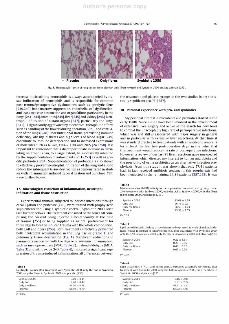
Author's personal copy
S. Bengmark / Pharmacological Research 69 (2013) 87– 113 99
Fig. 1. Hematoxylin–eosin of lung tissues from placebo, only fibers-treated and Synbiotic 2000-treated animals [255].
increase in circulating neutrophils is always accompanied by tis-sue infiltration of neutrophils and is responsible for commonpost-trauma/postoperative dysfunctions such as paralytic ileus[239,240], bone marrow suppression, endothelial cell dysfunction,and leads to tissue destruction and organ failure, particularly in thelungs [241–244], intestines [244], liver [245] and kidney [246]. Neu-trophil infiltration of distant organs [247], particularly the lungs[241], is significantly aggravated by mechanical therapeutic effortssuch as handling of the bowels during operation [239], and ventila-tion of the lungs [248]. Poor nutritional status, preexisting immunedeficiency, obesity, diabetes and high levels of blood sugar [249]contribute to immune deterioration and to increased expressionsof molecules such as NF-�B, COX-2, LOX and iNOS [249,250]. It isimportant to remember that a disproportionate increase in circu-lating neutrophils can, to a large extent, be successfully inhibitedby the supplementation of antioxidants [251–253] as well as spe-cific probiotics [254]. Supplementation of probiotics is also shownto effectively prevent neutrophil infiltration of the lung and also toreduce the subsequent tissue destruction as demonstrated in stud-ies with inflammation induced by cecal ligation and puncture (CLP)– see further below.
17. Bioecological reduction of inflammation, neutrophilinfiltration and tissue destruction
Experimental animals, subjected to induced infections throughcecal ligation and puncture (CLP), were treated with prophylacticsupplementation using a synbiotic cocktail, Synbiotic 2000 Forte(see further below). The treatment consisted of the four LAB com-prising the cocktail being injected subcutaneously at the timeof trauma [255] or being supplied as an oral pretreatment forthree days before the induced trauma with the whole composition,both LAB and fibers [256]. Both treatments effectively preventedboth neutrophil accumulation in the lung tissues (Table 1) andpulmonary tissue destruction (Fig. 1). Significant reductions inparameters associated with the degree of systemic inflammation,such as myeloperoxidase (MPO, Table 2), malondialdehyde (MDA,Table 3) and nitric oxide (NO, Table 4), indicated a significant sup-pression of trauma-induced inflammation, all differences between
Table 1Neutrophil counts after treatment with Synbiotic 2000, only the LAB in Synbiotic2000, only the fibers in Synbiotic 2000 and placebo [255].
Synbiotic 2000 9.00 ± 0.44Only LAB 8.40 ± 0.42Only the fibers 31.20 ± 0.98Placebo 51.10 ± 0.70
P < 0.05.
the treatment and placebo groups in the two studies being statis-tically significant (<0.05) [257].
18. Personal experience with pro- and synbiotics
My personal interest in microbiota and probiotics started in theearly 1980s. Since 1963 I have been involved in the developmentof extensive liver surgery and active in the search for new toolsto combat the unacceptably high rate of peri-operative infections,which was and still is associated with major surgery in generaland in particular with extensive liver resections. At that time itwas standard practice to treat patients with an antibiotic umbrellafor at least the first five post-operative days, in the belief thatthis treatment would reduce the rate of post-operative infections.However, a review of our last 81 liver resections gave unexpectedinformation, which directed my interest to human microbiota andthe possibility of using probiotics as an alternative infection pro-phylaxis. From this study it was shown that only 57/81 patientshad, in fact, received antibiotic treatment; this prophylaxis hadbeen neglected in the remaining 24/81 patients [257,258]. It was
Table 2Myeloperoxidase (MPO) activity in the supernatant presented as U/g lung tissue,after treatment with Synbiotic 2000, only the LAB in Synbiotic 2000, only the fibersin Synbiotic 2000 and placebo [255].
Synbiotic 2000 25.62 ± 2.19Only LAB 26.75 ± 2.61Only the fibres 56.59 ± 1.73Placebo 145.53 ± 7.53
P < 0.05.
Table 3Lipid peroxidation in the lung tissue determined expressed as levels of malondialde-hyde (MDA), measured in nmol/mg protein, after treatment with Synbiotic 2000,only the LAB in Synbiotic 2000, only the fibers in Synbiotic 2000 and placebo [255].
Synbiotic 2000 0.22 ± 1.31Only LAB 0.28 ± 3.55Only the fibres 0.48 ± 5.32Placebo 0.67 ± 2.94
P < 0.05.
Table 4Lung tissue nitrite (NO2) and nitrate (NO3), expressed as �mol/g wet tissue, aftertreatment with Synbiotic 2000, only the LAB in Synbiotic 2000, only the fibers inSynbiotic 2000 and placebo [255].
Synbiotic 2000 17.16 ± 2.03Only LAB 8.91 ± 2.24Only the fibres 47.71 ± 3.20Placebo 66.22 ± 5.92
P < 0.05.

Author's personal copy
100 S. Bengmark / Pharmacological Research 69 (2013) 87– 113
surprising that there were no cases of sepsis in the group of patients,who had not received prophylactic antibiotics with sepsis incidenceconfined to the antibiotic-treated patients. There was at that timea growing awareness of the importance of human microbiota [259]and to contemporaneous published studies that had attempted torecondition the gut through the supply of lactobacilli [260]. Therewas also at that time a growing understanding that not only diseasebut lifestyle and prescribed chemicals and pharmaceuticals, couldimpair microbiota immune defense. The use of probiotic treatment,as an alternative means of preventing unwanted infections in dis-ease in general but particularly in surgical and medical critically illpatients, appeared an attractive option. This was the reason whyI established collaborative efforts with experts in microbiology,chemistry, nutrition and experimental and clinical science to seek,develop and test probiotics both experimental and clinically, whichcould be expected to constitue powerful tools to prevent sepsis ofvarious kinds.
Interdisciplinary collaboration in the early 1990s lead to theidentification of some L. plantarum strains that demonstratedstrong anti-inflammatory capacities. L. plantarum 299, later usedtogether with oatmeal in a synbiotic composition [261–263], isproduced and marketed by Probi AB, Lund, Sweden. I participatedheavily in this program until 1999, when I decided to re-direct myinterest towards development and studies of a more complex syn-biotic composition, designed not only to supplement four newlyidentified bioactive LABs in combination but also four different pre-biotic fibers, already known for their strong bioactivity. Our aimwas to provide this composition in much larger doses than was thepractice at that time. Furthermore, knowing that most of the impor-tant LABs rarely exist in the microbiota of Westerners encouragedus to seek potent probiotic bacteria normally growing on plantsinstead of selecting bacteria normally found in human microbiota.
Since 1999, all my efforts in this field have concentrated ona four LAB/four fiber composition, consisting of either a mixtureof 4 × 10¹⁰ (40 billion LAB, Standard version – Synbiotic 2000TM)or a mixture of 1011 (400 billion Forte version – Synbiotic 2000ForteTM) based on the following four LAB: Pediococcus pentosaceus5-33:3, Leuconostoc mesenteroides 32-77:1, Lactobacillus paracaseisubsp paracasei 19, and Lactobacillus plantarum 2362 in combina-tion with 4 × 2.5 g of each of the following four fermentable fibres:betaglucan, inulin, pectin and resistant starch, in total 10 gr of prebi-otic fibers per dose [264,265], a formula that is currently producedand studied by Synbiotic AB, Höganäs, Sweden.
18.1. Perioperative prophylaxis in elective surgery
L. plantarum 299 in a dose of 109 plus a total of 15 gram of oat andinulin fibers was tried, under research condition, in patients under-going extensive abdominal surgical operations. The patient weremainly derived from those undergoing liver, pancreatic and gastricresections, equally distributed between three groups and supple-mented with either: 1. live LAB and fiber, 2. heat-inactivated LABand fiber, and 3. standard enteral nutrition [267]. Each group com-prised 30 patients. The 30-day sepsis rate was 10% (3/30 patients)in the two groups receiving either live or heat-inactivated LAB,compared to 30% (9/30 patients) in the group on standard enteralnutrition (P = 0.01) [266]. The largest difference was observed inincidence of pneumonia: Group 1 – 2 patients; Group 2 – 1 patient;Group 3 – 6 patients. The beneficial effects of treatment were seem-ingly most pronounced in gastric and pancreatic resections; thesepsis rate being: Group 1 – 7%, Group 2 – 17% and Group 3 – 50%.The same pattern was observed for non-infectious complications:Group 1 – 13% (4/30) Group 2 – 17% (5/30); Group 3 – 30% (9/30).The supply of antibiotics to Group 1 was significantly less (P = 0.04)than to the other two groups, with the mean length of antibiotic
Table 5Pathogens isolated from patients undergoing pancreatectomy treated with Synbi-otic 2000 and only the fibers in Synbiotic 2000 resp. [267].
Isolated microorganisms Synbiotic 2000 Fibers only
Enterobacter cloacae 2 8Enterococcus faecalis/faecium 1 7Escherichia coli 0 7Klebsiella pneumoniae 2 2Proteus mirabilis 1 1Staphylococcus aureus 0 2
(Total 6) (Total 27)
treatment also considerably shorter: Group 1 – 4 ± 3.7 days; Group2 – 7 ± 5.2 days; Group 3 – 8 ± 6.5 days.
In a prospective, randomized, double-blind trial 80 patientsundergoing pylorus-preserving pancreatoduodenectomy (PPPD)received, twice daily, either Synbiotic 2000TM (2 × 40 billion LAB,i.e. 80 billion LAB per day) or only the fibers in composition from theday before surgery and during the first seven postoperative days[267]. A highly significant difference in infection rate (P = 0.005)was observed as only 5/40 patients (12.5%) in the Synbiotic 2000-treated group suffered infections (4 wound and one urinary tractinfection) vs 16/40 (40%) in the fiber-only group (6 wound infec-tions, 5 peritonitis, 4 chest infections, 2 sepsis, and one of eachof urinary tract infection, cholangitis and empyema). The numberof infecting microorganisms were also statistically and significantreduced – see Table 5. Statistically significant differences betweenthe groups were also observed regarding the use of antibiotics(mean: Synbiotic 2000; 2 ± 5 days, Only-fibers; 10 ± 14 days) [267].
In another randomized controlled study 45 patients undergo-ing major surgery for abdominal cancer were divided into threetreatment groups: 1. enteral nutrition (EN) supplemented withSynbiotic 2000 (LEN), 2. EN supplemented with only the fibers inthe same amounts (20 g) (20 g) as in Synbiotic 2000TM (FEN) and3. standard parenteral nutrition (PN). All treatments lasted for 2preoperative and 7 days postoperative days. The incidence of post-operative bacterial infections was 47% with PN, 20% with FEN and6.7% with LEN (P < 0.05). The numbers of infecting microorganismswere also statistically and significantly reduced – see Table 6. Signif-icant improvements were also observed in prealbumin (LEN, FEN),C-reactive protein (LEN, FEN), serum cholesterol (LEN, FEN), whitecell blood count (LEN), serum endotoxin (LEN, FEN) and IgA (LEN)(Han Chun Mao, personal information).
Table 6Pathogens recovered from patients undergoing surgery for abdominal cancertreated with Synbiotic 2000 and only the fibers in Synbiotic 2000 resp. (Han et al.personal communication.)
Isolated microorganisms Synbiotic 2000 Fibers only
Pseudomonas aeruginosa 17 24Staphylococcus aureus 8 11Staphylococcus epidermidis 1 1Staphylococcus faecalis – 1Enterobacter cloacae 4 –Acinetobacter spp. 2 3Staphylococcus haemolyticus – 1Serratia spp. – 2Klebsiella spp. – 1Proteus mirabilis – 2Candida albicans 2 6Aspergillus spp. – –Bacillus subtilis – 1Klebsiella spp. – 1
(Total 34) (Total 54)

Author's personal copy
S. Bengmark / Pharmacological Research 69 (2013) 87– 113 101
Table 7Pathogens isolated from patients undergoing liver transplantation treated with Syn-biotic 2000 and only the fibers in Synbiotic 2000 resp [268].
Isolated bacteria Synbiotic 2000 Fibers only
Enterococcus faecalis 1 11Escherichia coli 0 3Enterobacter cloacae 0 2Pseudomonas aeruginosa 0 2Staphylococcus aureus 0 1
(Total 1) (Total 18)
18.2. Perioperative prophylaxis in liver transplantation
A prospective, randomized, study in 95 liver transplant patientssupplemented L. plantarum 299 in a dose of 109 plus 15 gram ofoat and inulin fiber [268]. Three groups of patients were stud-ied: 1. selective digestive tract decontamination (SDD) four timesdaily for six weeks, 2. L. plantarum 299 (LLP) in a dose of 109 plus15 g of oat and inulin fibres supplied postoperatively for 12 days,and 3. identical to group 2 but with heat-killed L. plantarum 299(HLP). Identical enteral nutrition was supplied to all patients fromthe second postoperative day. The numbers of postoperative infec-tions were SDD 23, LLP 4 and HLP 17. Signs of infections occurredin SDD 48% (15/32), in LLP 13% (4/31), P = 0.017 and HLP 34%(11/32) respectively. The most dominant infections were cholangi-tis (which occurred: SDD 10, LLP in 2, and HLP in 8) and pneumonia(which occurred: SDD in 6, in LLP in 1, and HLP in 4). There wasa statistically significant reduction in the numbers of infectingmicroorganisms, the most often isolated microbes being Entero-cocci and Staphylococci. Patients requiring haemodialysis were SDD:8; LLP: 2 and HLP: 4 and the number of re-operations SDD: 6; LLP: 4and HLP: 2 respectively. There were no deaths. The stay in ICU, thehospital stay and length on antibiotic therapy was shorter in theLLP group, but did not reach statistical significance. The CD4/CD8ratio was alsohigher in the LLP group compared to the other twogroups (P = 0.06).
In a subsequent study, 66 human orthotopic liver transplantpatients were randomized to either receive Synbiotic 2000 or onlythe fibers in Synbiotic 2000. The treatment was started on theday before surgery and continued for 14 days after surgery. Dur-ing the first postoperative month only one patient in the Synbiotic2000-treated group (3%) show signs of infection (urinary infection)compared to 17/33 (51%) patients in those supplemented with onlythe four fibers [269]. Only one infecting organism was cultivated inthe Synbiotic-treated group, which was shown to be Enterococcusfecalis, in contrast to seventeen organisms in the fiber- only treatedgroup – see Table 7. The use of antibiotics was on average 0.1 ± 0.1 din the Synbiotic-treated patients and 3.8 ± 0.9 d in the fiber- onlytreated group [269].
18.3. Early treatment in major trauma
Two prospective randomized trials with Synbiotic 2000 andSynbiotic 2000 Forte respectively were undertaken. The first studycompared in patients with acute extensive trauma four types oftreatment: 1. Enteral nutrition formula Alitraq (Abbott-Ross), 2.Enteral nutrition formula Nova Source (Novartis) 3. Enteral nutri-tion formula Nutricomp peptide (Braun) and 4. Enteral nutritionformula Nutricomp (Braun), complemented with Synbiotic 2000.The total number of infections as well as the number of chest infec-tions were studied [269,270] – see further Table 8.
In the other study 65 polytrauma patients were randomized toreceive once daily, for 15 days following major trauma, either Syn-biotic 2000 Forte (400 billion LAB + 10 gram of fibers, see above)or maltodextrine, as placebo. Significant reductions were observed
Table 8Total number of infections and total number of chest infections observed in severetrauma patients when treated with various commercial enteral nutrition formulas,includ-ing one with Synbiotic 2000 [269,270].
Total number of infections:Alitraq Abbott-Ross (glut + arg) 16/32 50%Nova Source Novartis (+guargum) 17/29 58%Nutricomp peptide Braun (+peptide) 13/26 50%Nutricomp standard (+Synbiotic 2000) 4/26 15%
Number of chest infectionsAlitraq Abbott-Ross (glut + arg) 11/32 34%Nova Source Novartis (+guargum) 12/29 41%Nutricomp Braun (peptide) 11/26 42%Nutricomp standard (+Synbiotic 2000) 5/26 19%
between the groups in the number of deaths (5/35 vs 9/30, P < 0.02),severe sepsis (6/35 vs 13/30, P < 0.02), chest infections (19/35 vs24/30, P < 0.03), central line infections (13/32 vs 20/30, P < 0.02),and ventilation days (average 15 vs 26 days [65]) [271]. A totalof 54 pathogenic microorganisms were cultivated in the Synbiotictreated group compared to 103 in the maltodextrine group – seeTable 9 [271–273]. Repeat analyses also revealed that serum levelsof endotoxin (LPS) were decreased and ‘time to bloodstream infec-tion’ significantly prolonged in patients treated with Synbiotic 2000Forte.
18.4. Early treatment in severe acute pancreatitis
In a further study, patients with severe acute pancreatitis wererandomized to receive either a freeze-dried preparation contain-ing live L. plantarum 299 in a dose of 109 together with a substrateof oat fiber or a similar preparation but heat-inactivated, admin-istered daily through a nasojejunal tube for seven days [274]. Thestudy was concluded when, on repeat statistical analysis, signifi-cant differences in favour of one of the two groups were obtained.This occurred when a total of 45 patients had entered the study.22 patients had, at that time, received treatment with live, and 23with the heat-killed, L. plantarum 299. Infected pancreatic necrosisand abscesses were seen in 1/22 (4.5%) in the live LAB group vs 7/23(30%) in the heat-inactivated group (P = 0.023). The only patient inthe lactobacillus group, who developed infection, a urinary infec-tion, did so on the fifteenth day, i.e. at a time when he had notreceived treatment for eight days. The length of stay was also con-siderably shorter in the live LAB group (13.7 days vs 21.4 days) butthe limited size of the material menat that statistical significancewas not reached [274].
Sixty-two patients with severe acute pancreatitis (SAP) (ApacheII scores: Synbiotic 2000-treated 11.7 ± 1.9, controls 10.4 ± 1.5)were given either two sachets/day of Synbiotic 2000TM (2 × 40 bil-lion LAB/day and totally 20 g fibers) or the same amounts of fibers(20 g) as in Synbiotic 2000TM during the first 14 days after arrival at
Table 9Pathogens isolated from patients with polytrauma treated with Synbiotic 2000 andonly the fibers in Synbiotic 2000 resp. [271].
Isolated microorganisms Synbiotic 2000 Fibers only
Acinetobacter baumanni 21 35Candida albicans 7 17Pseudomonas aeruginosa 15 14Staphylococcus epidermidis 2 10Staphylococcus aureus 4 7Staphylococcus hominis – 2Enterobacter aerogenes – 2Staphylococcus haemolyticus – 1Serratia spp. – 2Klebsiella spp. 5 12Proteus spp. – 1
(Total 54) (Total 103)
Author's personal copy
102 S. Bengmark / Pharmacological Research 69 (2013) 87– 113
Table 10Pathogens isolated from patients with acute pancreatis treated with synbiotic vsreceiving only fibers [275].
Isolated microorganisms Synbiotic 2000 Fibers only
Pseudomonas aeruginosa 1 4Enterococcus faecalis 1 2Enterobacter spp. 1 1Streptococcus spp. 2 –Staphylococcus aureus 1 1Enterococcus faecium 1 –Candida spp. – 2Staphylococcus haemolyticus – 1Serratia spp. – 2Klebsiella spp. – 1Escherichia coli – 1Stenotrophomonas maltophilia – 1Citrobacter freundii – 1
(Total 7) (Total 17)
the hospital [275]. 9/33 patients (27%) in the Synbiotic 2000-treatedgroup and 15/29 patients (52%) in the fiber-only treated groupdeveloped subsequent infections. 8/33 (24%) of the Synbiotic 2000-treated and 14/29 (48%) of the fiber-only treated patients developedSIRS, MOF or both (P < 0.005). A total of seven pathogenic microor-ganisms were cultivated in the Synbiotic-treated group comparedto seventeen in the fiber-only group – see Table 10. Another, asyet unpublished, study in patients with severe acute pancreati-tis compared 32 patients treated with Synbiotic 2000 Forte with30 control patients. Eight patients in the treated group sufferedseptic episodes compared to 21 in the control group (Pupelis G,personal information). Late MODS occurred in 1 patient vs 9 inthe control group, the mortality was 0% vs 17%, hospital stay23 vs 36 days, and stay in ICU 8 vs 16 days. Three patients inthe treated group vs 12 in the control group underwent surgicaloperations.
18.5. Effects on “mind clarity” – encephalopathy
Patients with critical illness, as well as patients with chronicdisorders such as liver cirrhosis and diabetes, frequently suf-fer a mild but sometimes severe confusion, which often has itsorigin in the gut [276]. Increasing evidence suggest that pro-biotics, alone but also in combination with plant antioxidantsand fibers, possess strong neuro-endocrine modulatory effectsand can alleviate the effects of physical and mental stressors[277,278]. We undertook some studies to explore the effects ofSynbiotic in patients with liver cirrhosis and minimal encephalo-pathy (MHE) [37]. Fifty-five patients with MHE were randomizedto receive for 30 days: 1. Synbiotic 2000 (n = 20), 2. the fibersin the composition alone (n = 20), or 3. a placebo (n = 15). Allcirrhotic patients with MHE were found to have severe derange-ments of the gut micro-ecology and significant overgrowth ofpotentially pathogenic Escherichia coli and Staphylococcal species.Synbiotic treatment significantly increased the fecal content ofnon-urease-producing Lactobacillus species and reduced the num-bers of potentially pathogenic micro-organisms. The treatment wasalso associated with a significant reduction in endotoxemia and inblood ammonia levels. A documented reversal of MHE was obtainedin half of the treated patients, while the Child-Turcotte-Pugh func-tional class improved in about 50% of cases [37]. Treatment withfermentable fibers alone also demonstrated substantial benefits ina proportion of patients.
In a second study, 30 cirrhotic patients were randomized toreceive either Synbiotic 2000 or placebo for only 7 days [279].Viable fecal counts of Lactobacillus species, Child-Pugh class,plasma retention rate of indocyanine green (ICGR15), whole blood
tumour necrosis factor alpha (TNF-a) mRNA and interleukin-6(IL-6) mRNA, serum TNF-a, soluble TNF receptor (sTNFR)I, sTNFRIIand IL-6 and plasma endotoxin levels were measured, pre- andpost-treatment. The treatment with Synbiotic 2000 was associatedwith significantly increased fecal lactobacilli counts and signifi-cant improvements in ICGR15 and Child-Pugh class. Significantincreases in whole blood TNF-a mRNA and IL-6 mRNA, along withserum levels of sTNFRI and sTNFRII, were also observed and TNF-aand IL-6 levels correlated significantly, both at baseline and post-Synbiotic treatment. Synbiotic-related improvement in ICGR15was accompanied by significant changes in IL-6, both at mRNA andprotein levels, but this was unrelated to levels of plasma endotoxin.No significant changes in any parameter were observed followingplacebo treatment. This study concluded that even short-term syn-biotic treatment significantly modulated gut flora and improvedliver function in patients with cirrhosis [279]. Minimal encephalo-pathy is common not only in liver cirrhosis but is also seen in otherchronic diseases such as diabetes. The observations in patients withliver cirrhosis gives hope that Synbiotic treatment may also beeffective in other chronic diseases.
18.6. Effects in HIV
It is well documented that disturbance of the microbiota occurearly in HIV-1 infection, which leads to greater dominance of poten-tial pathogens, reduced levels of bifidobacteria and lactobacillusspecies and increasing mucosal inflammation. Current and emerg-ing studies support the concept that probiotic bacteria can providespecific benefit in HIV-1 infection. It was not until Brenchley et al.in 2006 identified translocation of microbes or microbial productswithout overt bacteremia, as a major cause of systemic immuneactivation in HIV-1 and SIV infection [280], that a greater interestin bio-ecological treatment emerged.
Impairment of the GI tract in HIV-positive patients is alreadypresent in the early phases of HIV disease and is associated withelevated levels of intestinal inflammatory parameters and definitealterations in the gut commensal microbiota, confirming a possi-ble correlation between intestinal microbial alteration, GI mucosaldamage, and immune activation status, further confirming thatalterations at the GI-tract level are a key factor in the pathogen-esis of chronic HIV infection [281]. The findings, in a recent study,of fairly mild changes in microbiota of HIV-infected individuals,before intiation of pharmacological treatment, might suggest thatthe later observed more profound alterations in microbiota couldbe pharma-induced, as only a trend to a greater proportion ofEnterobacteriales compared to control subjects (P = 0.099) wereobserved, despite the significant negative correlations betweentotal bacterial load and duodenal CD4+ and CD8+ T-cell activationlevels [282]. As pointed out in a recent review, current and emerg-ing studies appear to support the concept that probiotic bacteriacan provide specific benefit in HIV-1 infection. Probiotic bacteriahave proven active against bacterial vaginosis in HIV-1 positivewomen and have enhanced growth in infants with congenital HIV-1 infection [283]. Probiotic bacteria may also stabilize CD4+ T cellnumbers in HIV-1 infected children and are likely to have protec-tive effects against inflammation and chronic immune activation ofthe gastrointestinal immune system [283].
Recent studies at least partly support the assumption that Lrhamnosis GR-1 and L Reuteri RC-14 tend to increase the probabilityof a normal vaginal flora (odds ratio 2.4; P = 0.1) and significantlyincrease the probability of a beneficial vaginal pH (odds ratio 3.8;P = 0.02) at follow-up [284,285]. However, later attempts using pro-biotic yoghurts have proven less successful [286]. In a recent pilotstudy 38 women with HIV, taking highly active antiretroviral ther-apy (HAART), were supplemented with Synbiotic 2000 Forte orallyfor 4 weeks [287]. In a surprising and very encouraging observation,
Author's personal copy
S. Bengmark / Pharmacological Research 69 (2013) 87– 113 103
the supplemented formula showed ability, despite heavy pharma-ceutical treatment, to survive during the passage through the GItract, and also the ability to colonize the gut and contribute to asignificantly elevated level in the stool of the supplemented LABgroup. The T-cell activation phenotype was altered by exposureto the Synbiotic formula and was accompanied by a slightly ele-vated HLA-DR expression of a minor population of CD4+ T-cells,which normally lack expression of HLA-DR or PD-1. These sig-nificant changes occurred in the context of unaltered microbialtranslocation, as measured by plasma bacterial 16S ribosomal DNA[287]. It is especially encouraging that the LAB supplemented withSynbiotic 2000, despite heavy medication/highly active antiretro-viral therapy (HAART), were able to colonize the gut and seemingly,at least slightly, improve immune functions. Hopefully, signifi-cantly more pronounced positive effects will be obtained the daywe are ready to try eco-biological treatment, not only as com-plementary treatment but as an alternative to pharmaceuticaltreatment.
19. Life-threatening systemic inflammation
A study of patients in intensive care suffering life-threateningextreme systemic inflammation – a systemic inflammationresponse syndrome (SIRS) – and its relation to gut microbiotawas recently published. Twenty-five patients with severe SIRSand a serum C-reactive protein level >10 mg/dl were studied[288]. Analysis of gut microbiota revealed markedly lower totalanaerobic bacterial counts, particularly of the beneficial Bifidobac-terium and Lactobacillus and higher counts of total facultativeanaerobes such as Staphylococcus and Pseudomonas comparedto healthy volunteers. In patients with bacterial translocation,Gram-negative facultative anaerobes were the most commonlyidentified microbial organisms in mesenteric lymph nodes andserosal scrapings at laparotomy. Gastrointestinal complicationswere strongly associated with a significantly reduced numberof total obligate anaerobes and highly increased numbers ofStaphylococcus and Enterococcus and significantly decreased num-bers of total obligate anaerobes and total facultative anaerobes[288].
A more recent study in 63 similar patients suggests impairedgastrointestinal motility as a significant marker of poor outcome[289]. Patients with ≥300 ml per day reflux from nasal gastric feed-ing tube demonstrated significantly lower numbers of total obligateanaerobes including Bacteroidaceae and Bifidobacterium, highernumbers of Staphylococcus, lower concentrations of acetic acid andpropionic acid, and higher concentrations of succinic acid and lacticacid (P ≤ 0.05), accompanied by dramatically higher incidences ofbacteremia (86% vs 18%) and mortality (64% vs 20%) than patientswithout gastric detension (P ≤ 0.05) [289]. Furthermore, in 29 sim-ilar patients treatment with a synbiotic composition, consisting ofBifidobacterium breve and Lactobacillus casei, in combination withgalactooligosaccharides, was attempted. Higher levels of Bifidobac-teria and Lactobacillus, but also total organic acids, particularlyshort-chain fatty acids, were reported and the incidence, comparedto historical controls, of infectious complications such as enteritis,pneumonia, and bacteremia, observed to be significantly lower inthe treated group [290].
20. Studies with no or adverse effects
20.1. Ecologic 641TM
In a multicenter randomized, double-blind, placebo-controlledtrial, 298 patients with predicted severe acute pancreatitis andwith APACHE II score > or =8, Imrie score >3, or C-reactive pro-tein >150 mg/l) were, within 72 h of onset of symptoms, randomly
assigned to receive either a multi-species synbiotic composition(n = 153) or a placebo (n = 145), administered enterally twice dailyfor 28 days [291]. The supplemented synbiotic composition, Eco-logic 641 (Winclove Bio Industries, Amsterdam, Netherlands),consists of 1010 of each of six different strains of freeze-dried,viable bacteria: Lactobacillus acidophilus, Lactobacillus casei, Lacto-bacillus salivarius, Lactococcus lactis, Bifidobacterium bifidum, andBifidobacterium lactis plus cornstarch and maltodextrins. Infec-tious complications occurred in 46 (30%) of patients in the treatedgroup and in 41 (28%) in the placebo group (relative risk 1.06,95% CI 0.75–1.51). Twenty-five (16%) patients in the synbioticgroup died, compared to nine (6%) in the placebo group (relativerisk 2.53, 95% CI 1.22–5.25) [291]. Furthermore, nine patients inthe synbiotics group developed bowel ischemia, of which eighthad fatal outcomes, compared to none in the placebo group(P = 0.004).
20.2. Lactobacillus plantarum 299TM – ProVivaTM
One hundred and three critically ill patients were randomizedto receive 1. an oral preparation containing L. plantarum 299v, Pro-Viva, a fruit drink containing 5% of LAB-fermented oat and liveLactobacillusb plantarum 299v with a density of 5 × 10⁷ (n = 52)or 2, conventional nutrition therapy alone (n = 51). The treatmentdemonstrated no identifiable effect in terms of bacterial translo-cation (12% vs 12%; P = 0.82), gastric colonization with entericorganisms (11% vs 17%, P = 0.42), or septic morbidity (13% vs 15%;P = 074), serum CRP levels or mortality [292]. In another study, 11patients undergoing elective abdominal surgery receive L. plan-tarum 299 v, (ProViva) for a median time of 9 days (range 5–18days) to a total average amount of 3250 ml (range 2100–9000 ml)and were then compared to 11 control patients. The authors foundno significant differences between the L. plantarum 299v group andthe control group in terms of concentrations of plasma cells, IgApositive cells or IgM positive cells in the lamina propria [293]. Asignificantly higher concentration of IgM at the mucosal surfacewas observed in the control group (P = 0.02, Fishers Exact test midP) but no difference in terms of IgA.
20.3. Lactobacillus rhamnosus GGTM
Sixty-one patients in a pediatric ICU study were randomizedwith 31 patients receiving treatment with one capsule of Lacto-bacillus rhamnosus strain GG in a dose of 10 × 109 and 30 receivingone capsule of inulin daily (control group) [292]. No differencesin rate of infections were observed between the groups; the meannumber of infections in the treatment and control groups was 1.83and 1.33, respectively. 9/31 patients in the probiotic-treated groupdeveloped in total 15 nosocomial infections: 6 bloodstream infec-tions (40%), 5 tracheo-bronchitis (33%), 2 pneumonia (13%), and 2UTI (13%). There were six deaths in total during the study period;four in the placebo group and two in the treatment group. No casesof Lactobacillus bacteremia or other serious adverse effects wereobserved [294].
20.4. Synbiotic 2000TM/Synbiotic 2000 ForteTM
Two hundred and fifty nine enterally fed critically ill patients,expected to require mechanical ventilation for 48 h or more wereenrolled in a study; 130 patients received Synbiotic 2000 Forte®
(twice a day) and 129 patients, a cellulose-based placebo for amaximum of 28 days [226]. The oropharyngeal microbial flora andcolonization rates were unaffected by the synbiotic treatment. Theoverall incidence of ventilator associated pneumonia (VAP) waslower than anticipated (11.2%) and no statistical difference wasdemonstrated between the groups receiving synbiotic or placebo;

Author's personal copy
104 S. Bengmark / Pharmacological Research 69 (2013) 87– 113
incidence of VAP (9 and 13%, P = 0.42), VAP rate per 1000 ventila-tor days (13 and 14.6, P = 0.91) or hospital mortality (27 and 33%,P = 0.39), respectively. No negative effects of the treatment wereobserved [295].
20.5. TrevisTM
A total of 90 patients admitted to an ICU were randomized toreceive either a synbiotic or placebo (45 into each group) [296].The synbiotic treatment consisted of the supply of a capsule ofTrevisTM (Chr Hansen Biosystem, Denmark) three times a day, con-taining 4 × 10⁹ colony forming units of each of L. acidophilus La5(La5), B. lactis Bb-12 (Bb-12), S. thermophilus and L. bulgaricus. Inaddition, the prebiotic oligofructose (7.5 g of RaftiloseTM powder,Orafti Active Food Ingredients, Belgium) was administered twice aday. The patients in the synbiotic group demonstrated, after 1 weekof therapy, significantly lower incidence of potentially pathogenicbacteria (43% vs 75%, P = 0.05) and multiple organisms (39% vs 75%,P = 0.01) in their nasogastric aspirates, than the controls. However,there were no significant differences between the groups in termsof intestinal permeability, septic complications or mortality [296].
20.6. VSL#3TM
Twenty-eight patients critically ill patients were enrolled andrandomly assigned to one of 3 treatment groups: 1. placebo (n = 9) 2.viable probiotics – 2 sachets daily of VSL#3TM (n = 10) or 3. bacterialsonicates – non viable VSL#3 bacteria (n = 9) [297]. Each sachet ofthe supplemented probiotic, VSL#3 (VSL Pharmaceuticals, Ft Laud-erdale, FL) contained 900 billion viable lyophilized bacteria of 4strains of Lactobacillus (L. casei, L. plantarum, L. acidophilus, andL. delbrueckii subsp. Bulgaricus) plus 3 strains of Bifidobacterium(B. longum, B. breve, and B. infantis), plus Streptococcus salivariussubsp. Thermophiles, totally eight strains. Intestinal permeabilitydecreased in all treatment groups. The rate of severe sepsis andMODS were not significantly affected by the treatment, althougha significantly larger increase in systemic IgA and IgG concentra-tions were observed in the group supplied live bacteria than in thepatients who received placebo or sonicated bacteria (P 0.05) [297].
21. Why do studies fail?
Critical care units are generally highly artificial environmentsand the burden of environment-induced physical and mental stressand subsequent status of systemic hyper-inflammation on thepatient, enormous. Patients treated under these conditions are inmany ways dys-functional; the whole microbiota has often, moreor less, disappeared and probiotic bacteria supplied will usuallybe extinct before they reach one of their targets – the lower gas-trointestinal tract. This artificiality seems to vary from country tocountry and sometimes also from hospital to hospital, an observa-tion that might explain the great variation in outcome from studiesundertaken in different countries and regions.
As discussed above, probiotic treatment has never been giventhe chance as a ‘stand alone’ alternative. It has, thus far, only beentried as an adjunctive treatment to heavy multi-drug pharmaceu-tical treatments. Many of the numerous drugs used in the ICU,including antibiotics are known to derange not only the microbiotabut to dramatically derange the majority of immune functions.Some 25 years ago the use of an antibiotic, Mezlocillin (Bayer,150 mg/kg body weight) was demonstrated to significantly sup-press essential macrophage functions, derange chemiluminescenceresponse, chemotactic motility, bactericidal and cytostatic abilityand impair lymphocyte proliferation, impair macrophage functionsand bactericidal efficacy as well as production and secretion ofcytokines [298].
Artificial nutrition, both enteral and parenteral, is an impor-tant contributor to ICU-associated sepsis; catheter-related sepsisis reported to occur in about 25% of patients fed via intravenousfeeding-tubes [299]. Other common perioperative practices, e.g.use of artificial feeding regimens, preoperative antibiotics [300],and mechanical bowel preparation [301,302] will, instead ofpreventing expected infections, contribute to increased rates oftreatment-associated infections. Other measures in the ICU such asmechanical ventilation [303], treatment with various pharmaqceu-tical drugs, including antibiotics [304,305], chemical solutions forclinical nutrition and many others promote super-inflammationand, indirectly, infection.
Enteral nutrition formulas, most likely deleterious to micro-biota, are known to induce loss of intestinal barrier function,promote bacterial translocation, and impair host immune defense[306], a phenomenon, observed in humans but also extensivelyelucidated in animal studies. In such studies the incidence ofbacterial translocation to the mesenteric lymph node was signif-icantly increased when the animals were fed nutrition formulassuch as Vivonex (53%), Criticare (67%), or Ensure (60%) (P < 0.05)[304–307]. Dramatic elevations in pro-inflammatory cytokineshave been observed in patients, when fed a standard enteral nutri-tion solution (Nutrison) following pancreat-duodenectomy, e.g.IL-1beta day 7 (P < 0.001); day 14 (P = 0.022), TNF-alpha- day 3(P = 0.006); day 7 (P < 0.001) [308]. Of special interest are the obser-vations that such changes are not observed when the standardnutrition is replaced with a formula whichis claimed to haveimmune-modulatory effects (Stresson). Instead anti-inflammatorycytokines were seen to be significantly elevated: IL-1ra/s: day 7(P < 0.001); IL-6: day 10 (P = 0.017); IL-8: day 1 (P = 0.011) days3, 7, 10, and 14 (P < 0.001), and IL-10: days 3 and 10 (P < 0.001)[308].
22. Choice of lactic acid bacteria as probiotics
The choice of bacteria for probiotic purposes is critical. Onlya few LAB strains have demonstrated an ability to influence theimmune system, reduce inflammation and/or eliminate or reduceunwanted pro-inflammatory molecules from foods. Even strainsthat carry the same name can have different and even sometimesopposite effects. A recent study selected 46 strains of Lactococ-cus lactis from about 2,600 LAB and compared their ability toinduce cytokines. It was demonstrated that the inter-strain differ-ences in ability to produce pro- and anti-inflammatory cytokineswas great [309], an observation that underlines the importance ofextensive animal and preclinical studies before a LAB or combina-tion of LAB is chosen as a probiotic. Strains that improve immunefunction by increasing the number of IgA-producing plasma cells,improve phagocytosis, and influence the proportion of Th1 cellsand NK cells [310] are particularly desirable for probiotic purposes.Popular probiotic species that are usually reliable and availablecommercially are L. paracasei, L. rhamnosus, L. acidophilus, L. john-sonii, L. fermentum, L. reuteri, L. plantarum, Bifidobacterium longumand Bifidobacterium animalis [109]. Among the strains with doc-umented stronger anti-inflammatory functions are Lactobacillusparacasei subsp paracasei, Lactobacillus plantarum, and Pediococ-cus pentosaceus. Lactobacillus paracasei, in particular, seems tohave a solid record; it has been shown to induce cellular immu-nity and stimulate production of suppressive cytokines such asTGF� and Il-10 and to suppress Th2 activity and CD4 T-cells[311,312], suppress splenocyte proliferation [313] and decreaseantigen-specific IgE and IgG1 [314]. When more than one hun-dred LAB strains were compared Lactobacillus paracasei was shownto be the strongest inducer of Th1 and repressor of Th2 cytokines[315].

Author's personal copy
S. Bengmark / Pharmacological Research 69 (2013) 87– 113 105
A recent study using rats compared the ability of four differentstrains: Lactobacillus paracasei, Lactobacillus johnsonii, Bifidobac-terium longum, or Bifidobacterium lactis to control Trichinellaspiralis-induced infection. Lactobacillus paracasei, alone was able toreduce infection-associated Th2 response, muscle levels of TGF-�,COX-2 and PGE2 and to attenuate infection-induced muscle hyper-contractility [316]. Another study compared the ability to reducestress-induced changes in gut permeability and sensitivity to colo-rectal distension of three probiotic strains: Bifidobacterium lactisNCC362, Lactobacillus johnsonii NCC533, and Lactobacillus paraca-sei NCC2461. Lactobacillus paracasei, alone restored normal gutpermeability, reduced visceral hyperalgesia and reduced visceralpain [317]. Several other important clinical effects of Lactobacil-lus paracasei subsp paracasei are summarized in a recent review;a strain called NTU 101 and its fermented products demonstratingthe ability to reduce blood cholesterol, blood pressure, and pre-vent allergies, osteoporosis and inhibit accumulation of fat tissue[318]. Lactobacillus plantarum also has an excellent record. Whenthe ability of fifty different LAB to control twenty-three differ-ent Clostridium difficile (C diff) strains were studied, Lactobacillusparacasei and Lactobacillus plantarum seemed to be equally efficientand the only strains of the fifty tried, to demonstrate the ability toeffectively eliminate all C diff strains – more than half of the triedLAB strains were totally ineffective, and some effective only againsta few [319].
Some LAB seem to be potentiated in their efficacyby simultaneous supply of prebiotic fibers (probi-otics + prebiotics ⇒ synbiotics). However, there are greatdifferences in the ability of different strains to ferment andutilize plant fibers, especially when it comes to semi-fermentablefibers such as oligofructans. Only a handful of 712 LAB strainstested demonstrated an ability to ferment inulin and phlein,namely: L. plantarum (several strains), L paracasei subsp. paracasei,L brevis & Pediococcus pentosaceus [320].
23. Molecular gene targeting – the future?
Ingredients specific to certain plants are known to exert pro-fund effects on specific genes. Among these agents are curcumin(turmeric), resveratrol (red grapes, peanuts and berries), genis-tein (soybean), diallyl sulfide (allium), S-allyl cysteine (allium),allicin (garlic), lycopene (tomato), capsaicin (red chilli), diosgenin(fenugreek), 6-gingerol (ginger), ellagic acid (pomegranate), ursolicacid (apple, pears, prunes), silymarin (milk thistle), anethol (anise,camphor, and fennel), catechins (green tea), eugenol (cloves),indole-3-carbinol (cruciferous vegetables), limonene (citrus fruits),beta carotene (carrots), and dietary fiber [321–324].
Curcumin, for example, has demonstrated a profound ability toinhibit a whole series of cell-signalling pathways, including NF-kB, AP-1, STAT3, Akt, Bcl-2, Bcl-X(L), caspases, PARP, IKK, EGFR,HER2, JNK, MAPK, COX2, and 5-LOX [251,321–324]; supplementedprobiotics are likely to exert similar effects. Gene expression ofhuman duodenal mucosa cells were studied after exposure to oneof the following four lactic acid bacteria; Lactobacillus plantarumWCFS1 [325], Lactobacillus acidophilus L10, Lactobacillus casei CRL-431 and L. rhamnosus GG [326], administered in a cross-over studyto healthy volunteers in a dose of 1010. Mucosal biopsies were takenfrom duodenum after 6 h and compared to control biopsies. Theinterventions did not impair immune and metabolic homeostasisbut a fascinating and most distinct influence on the expression ofseveral hundred genes (transcriptome) was reported after adminis-tration of each of the studied LAB. This is possibly the first time thatdifferent probiotic lactobacilli have been reported to induce moreor less strain-specific and markedly different expression profiles,very similar to what is known to occur with ingestion of variousfoods, especially plant ingredients [321–324] and in many respects
similar tp what is observed after supply of certain pharmaceuticals.L. plantarum was observed to specifically modulate overt adaptiveimmune responses [321,322], L. acidophilus to suppress inflamma-tion, L. casei to stimulate Th1 response and improve the Th1–Th2balance and L. rhamnosis to influence cellular growth and prolifera-tion [323,324]. These effects were suggested to resemble, althoughin a distinctly milder form, those obtained by specific pharmaceut-icals:
• L. acidophilus – antagonists of �-receptor activity, guanine antag-onists, synthetic corticosteroids and flavonoids,
• L casei – modulators of GABA receptors, cholinergic blockingagents, antagonists of �-adrenergic receptors,
• L. rhamnosus – glycoside steroids, alkaloids, protein synthesisinhibitors and protein kinase C inhibitors.
The responsiveness to ingestion of various LAB seems to bestrongly influenced, not only by eventual genetic background andexisting resident microbiota, but also by lifestyle, and particularlyby diet, which might explain the differences in person-to-personresponse, as observed in the above studies but also the differencesin outcome, often encountered in clinical probiotic studies, andespecially when tried with critically ill patients (see above).
24. It is all about inflammation
Inflammation, an essential component of immune-mediatedprotection against pathogens and tissue damage, and uncontrolledimmune responses, will commonly, especially in Westerners, insti-tute a state of chronic inflammation, which will occur whenimmune response are activated despite the absence of ‘danger’signals, fail to fully turn-off despite elimination of danger signalsand/or fail to completely clear such signals. Numerous factors, inaddition to genetic predisposition, trauma and various stress fac-tors (physical and emotional) are known to contribute to increaseddiscrete and long-lasting inflammation, among them age, diet andmedications.
Studies of human gene-related inflammation suggest that, of theapproximately 25,000 human genes, approximately 5%, or some1200 genes, are involved in inflammation [325–327]. It is increas-ingly understood that the human genome in itself will only explaina minority of chronic diseases, far less than changes in lifestyle,food habits and social behaviour, factors which seem to have adominating impact on human health. Clearly, the molecular mecha-nisms linking environmental factors and genetic susceptibility wasfirst envisioned after the recent exploration of the, until recentlyhidden, source of genomic diversity, i.e. the metagenome with itsmore than 3 million genes [328]. Although the mechanisms behindthe metagenome-associated low-grade inflammation and the cor-responding immune response are not yet fully understood, thereis no doubt that the metagenome has a dominating influence onaltered body functions such as adipose tissue plasticity and diseasessuch as hepatic steatosis, insulin resistance and cardiovascular dis-eases, but also on disorders such as autoimmune diseases includingrheumatoid arthritis, gastrointestinal and neuropsychiatric dis-eases and on development and progress of a number of cancers[97], as well as many other chronic disorders. When disease exacer-bations occur, in trauma or in critical illness, the normally silent ordiscrete inflammation turn into a storm [329] as experienced in sys-temic inflammatory response syndrome (SIRS) and multiple organfailure (MOF) [330]. In many severe conditions like MOF and SIRScomponents of cytokine-induced injury might be more damagingthan the initial cause/trauma/early invasion of micro-organismsin themselves. Inflammatory cytokines, such as TNF alpha and IL-1�, released by these events will destabilize endothelial cell-cell
Author's personal copy
106 S. Bengmark / Pharmacological Research 69 (2013) 87– 113
interactions and cripple vascular barrier function, producingcapillary leakage, tissue edema, organ failure, and sometimes death[330].
25. Cytokine-inhibition, pharma and/or probiotics?
Inflammation is, as discussed above, extraordinarily complex.In rheumatoid arthritis (RA) for example, the joints are richin cytokine-secreting cells containing a wide range of effectormolecules including pro-inflammatory cytokines such as IL-1�, IL-6, TNF-� and IL-18, chemokines such as IL-8, IP-10, MCP-1, MIP-1and RANTES, MMPs such as MMP-1, -3, -9 and -13 and metabolicproteins such as Cox-1, Cox-2 and iNOS, which interact with oneanother in a complex manner that is thought to cause a viciouscycle of pro-inflammatory signals resulting in chronic and per-sistent inflammation [331,332]. NF-�B is increasingly suggestedto the master regulators of inflammatory cytokine production inRA. These mediators are also involved, although not in an identi-cal manner, in other autoimmune disorders such as inflammatorybowel diseases [333]. This knowledge has led to developmentto a new generation of pharmaceutical drugs, generally referredto as biological, designed to inhibit the crucial mediators ofpro-inflammatory signals and subsequent abnormal immuneresponse. A whole series of revolutionary new drugs such as anti-TNF-�, anti-IL-1�, anti-HER2, etc. are already successfully triedand new drugs such as antibodies targeting IL-12/IL-23 pathways,IFN-�, IL-17A, IL-2 and IL-6, and also inhibitors of NF-�B moreor less extensively tried in a variety of chronic inflammatory andautoimmune diseases. Some of these have already demonstratedinitially promising result, while other treatments such as admin-istration of the regulatory cytokines IL-10 and IL-11 have failedto induce reproducible clinical effects [333]. Significant benefits inquality of life and tissue/organ healing are encountered in at leastsomething over 50%. These drugs are generally tolerated well, butadverse events such as infections including reactivating tuberculo-sis, tumours such as lymphomas and demyelinating diseases andinfusion reactions are sometimes evident. These changes must beregarded as acceptable as long as they are used in diseases that haveproven to be refractory to all other treatments but may be an issuewhen, as increasingly suggested, they are tried in early stages ofdiseases, as happened after widening indications for statins [334].
26. Single target or multitarget treatment?
Most biologicals are desinged to target single molecules, thoseregarded as mainly responsible for the etiology of disease, even ifthey in reality actually affect several other molecules. There are alsoindications that sometimes the results of selective targeting maybe short-lasting and that the inflammation sooner or later will findother pathways and the disease consequently continue to progress.Most diseases involve a large variety of molecular abnormali-
Table 11Comparison between biologicals and eco-biologicals.
Biologicals made to target singlegenes; anti-TNF-�, anti-IL-1�,anti-HER2, IL-12/IL-23, IFN-�,IL-17A, IL-2 and IL-6, andinhibitor of NF-�B
Eco-biologicals; utilizes theantiinflammatory effects ofplants and microbes to supportmicrobiota and reduce systemicinflammation
Uni-targetting Multi-targettingImmediate powerful effects Slower and weaker effectsLimited by toxicity GRAS – e.g. no toxicityNegative to microbiota Support microbiotaSometimes short-lasting effects For-ever lasting effectsSubstansial adverse effects No adverse effectsIndicated-aggressive diseases Indicated – prevention and early
disease
ties; for these broad-spectrum anti-inflammatory (eco-biological)treatment, using plant fibers and pro- and synbiotics might offer agood, and sometimes better, solution to treatment of the disease.
It is unfortunate that no studies thus far have addressed theeffects of the biologicals on microbiota and leaky barriers. Untildone, one must assume that these drugs have the same devastatingeffects on microbiota and barrier efficacy as other drugs. Plant-derived mediators, or phytochemicals, such curcumin, resveratrol,genistein, etc., (see further above) and plant fibers, particularlyprebiotic fibers, and probiotic bacteria, which may be termed ‘eco-biologicals’ can be expected, alone or in combination, to have thesame molecular functions as biologicals – although much weaker– but also without known adverse effects. Compounds officiallyclassified as GRAS, generally considered as safe, should be consid-ered where the main indications are prevention, early in diseasetreatment but also when used as palliative treatment particularly inchildren and the elderly. Table 11 is an attempt to summarize sim-ilarities and dissimilarities between biological and eco-biologicaltreatments.
Several population-based studies indicate that people in South-east Asian countries have a much lower risk of developingcolon, gastrointestinal, prostate, breast, and other cancers than dotheir Western counterparts. They also have significantly reducedincidence of other chronic diseases such as coronary heart diseases,neuro-degenerative diseases, diabetes, inflammatory bowel dis-eases, etc. It is likely that their frequent use of dietary constituentscontaining chemopreventive molecules, as is the case with garlic,ginger, soybeans, curcumin, onion, tomatoes, cruciferous vegeta-bles, chilies, and green tea and many others may play an importantrole in protection from these cancers, and other diseases, especiallyas these dietary agents might suppress transformative, hyperpro-liferative, and inflammatory processes [335].
27. Final remarks
An individual who wants to live in line with the present knowl-edge obtained from extensive research in recent years might want,in addition regular physical exercise, good sleep and spiritual har-mony to consider:
1. Minimizing intake of insulinogenic foods such as refined car-bohydrates; cereals, bread, sweats, cookies, rice, pasta, cookedtubers incl. potatoes, foods, which are absorbed high in thesmall intestine and of minimal benefit to microbiota.
2. Keeping a daily intake of fructose below 25 g a day.3. Minimizing their intake of dairy products especially butter,
cheese and milk powder, rich in saturated fats, hormones andgrowth factors such as IGF1, and to reduce meat intake, espe-cially inflammation-inducing processed and cured meat suchas bacon and sausages, this far though only fat demonstratedto being detrimental to microbiota.
4. Dramatically increasing the intake of fresh and raw greens,fresh spices and vegetables, rich in antioxidants, fibers, min-erals and nutrients, but also inflammation-controlling factorssuch as curcumin, resveratrol – some of which most likelyare of great importance for diversity, replication, growth andfunctions of the microbiota and for immune development andimmune functions of the body.
5. Minimizing intake of foods, which are heated above 100 ◦Cknown to be rich in the inflammation-inducing molecules AGEsand ALEs, foods heated above 130 ◦C, which with increase intemperature becomes increasingly rich in pro-inflammatoryand carcinogenic substances such as acrylamide and hetero-cyclic amines. This means avoiding fried and grilled foods butalso toasted and high-temperature baked breads.

Author's personal copy
S. Bengmark / Pharmacological Research 69 (2013) 87– 113 107
6. Minimizing exposure to microbe-derived highly inflammation-inducing endotoxin, especially rich in meat hung for severaldays, hard cheeses, pork and ice-creams.
7. Eliminating/minimizing intake of foods rich in proteotoxinssuch as casein, gluten and zein.
8. Seeking out and consuming ancient anti-oxidant-rich, highfiber, low-calorie containing grains such as buckwheat, ama-ranth, chia, lupin, millet, quinoa, sorghum, taro, teff, etc., andalso increasing the intake of beans, peas, chickpeas, lentils, nutsand almonds – all extraordinary rich in nutrients and minerals– all prepared for eating by low-temperature cooking – all mostlikely of importance for maintenance of a rich microbiota.
9. Restricting intake of chemicals including pharmaceutical drugsto only what is absolutely nessessary as most likely most chem-icals are detrimental to microbiota.
10. Supplement of large doses of vitamin D and omega fatty acids,both important in control of inflammation and for function ofmicrobiota.
References
[1] http://www.cdc.gov/obesity/data/trends.html and http://www.cdc.gov/diabetes/data/trends.html
[2] Bengmark S. Advanced glycation and lipoxidation end products – amplifiersof inflammation: the role of food. Journal of Parenteral and Enteral Nutrition2007;31:430–40.
[3] Bengmark S. AGE, ALE RAGE and disease – a foods perspective. In: ChoSS, Finocchiaro T, editors. Handbook of prebiotic and probiotic ingredients:health benefits and food applications. Boca Raton: CRC Press, Taylor and Fran-cis Group; 2010. p. 139–62.
[4] Bengmark S. Modified amino acid-based molecules: accumulation and healthimplications. In: Mello JFD, editor. Amino acids in human nutrition and health.UK: CABI Wallingford; 2011. p. 382–405.
[5] Boyle JP, Thompson TJ, Gregg EW, Barker LE, Williamson DF. Projection of theyear 2050 burden of diabetes in the US adult population: dynamic modeling ofincidence, mortality, and prediabetes prevalence. Population Health Metrics2010;8:29.
[6] Hebert LE, Scherr PA, Bienias JL, Bennett DA, Evans DA. Alzheimer disease inthe US population: prevalence estimates using the 2000 census. Archives ofNeurology 2003;60(August):1119–22.
[7] Bray F, Møller B. Predicting the future burden of cancer. Nature ReviewsCancer 2006;6:63–74.
[8] Wang YC, McPherson K, Marsh T, Gortmaker SL, Brown M. Health and eco-nomic burden of the projected obesity trends in the USA and the UK. Lancet2011;378:815–25.
[9] Carrera-Bastos P, Fontes-Villalba M, O’Keefe JH, Lindeberg S, Cordain L. Thewestern diet and lifestyle and diseases of civilization. Research Reports inClinical Cardiology 2011;2:15–35.
[10] Guevara-Aguirre J, Balasubramanian P, Guevara-Aguirre M, Wei M, MadiaF, Cheng CW, et al. Growth hormone receptor deficiency is associated witha major reduction in pro-aging signaling, cancer, and diabetes in humans.Science Translational Medicine 2011;3:1–9.
[11] Shevah O, Laron Z. Patients with congenital deficiency of IGF-I seem pro-tected from the development of malignancies: a preliminary report. GrowthHormone and IGF Research 2007;17:54–7.
[12] Steuerman R, Shevah O, Laron Z. Congenital IGF1 deficiency tends toconfer protection against post-natal development of malignancies. Euro-pean Journal of Endocrinology/European Federation of Endocrine Societies2011;164:485–9.
[13] Laron Z. The GH-IGF1 axis and longevity. The paradigm of IGF1 deficiency.Hormones 2008;7:24–7.
[14] Cohen E, Paulsson JF, Blinder P, Burstyn-Cohen T, Du D, Estepa G, et al.Reduced IGF-1 signaling delays age-associated proteotoxicity in mice. Cell2009;139:1157–69.
[15] Moore T, Checkley LA, DiGiovanni J. Dietary energy balance modulation ofepithelial carcinogenesis: a role for IGF-1 receptor signaling and crosstalk.Annals of the New York Academy of Sciences 2011;1229:7–17.
[16] Zemva J, Schubert M. Central insulin and insulin-like growth factor-1signaling: implications for diabetes associated dementia. Current DiabetesReviews 2011;7:356–66.
[17] Pendyala S, Walker JM, Holt PR. A high-fat diet is associated with endotoxemiathat originates from the gut. Gastroenterology 2012;142:1100–1.
[18] Lira FS, Rosa JC, Pimentel GD, Souza HA, Caperuto EC, Carnevali Jr LC, et al.Endotoxin levels correlate positively with a sedentary lifestyle and negativelywith highly trained subjects. Lipids in Health and Disease 2010;9(August):82.
[19] Pussinen PJ, Havulinna AS, Lehto M, Sundvall J, Salomaa V. Endotoxemiais associated with an increased risk of incident diabetes. Diabetes Care2011;34:392–7.
[20] Fassbender K, Walter S, Kühl S, Landmann R, Ishii K, Bertsch T, et al. TheLPS receptor (CD14) links innate immunity with Alzheimer’s disease. FASEBJournal 2004;18:203–5.
[21] Jaeger LB, Dohgu S, Sultana R, Lynch JL, Owen JB, Erickson MA, et al.Lipopolysaccharide alters the blood–brain barrier transport of amyloid betaprotein: a mechanism for inflammation in the progression of Alzheimer’sdisease. Brain, Behavior, and Immunity 2009;23:507–17.
[22] Wiedermann CJ, Kiechl S, Dunzendorfer S, Schratzberger P, Egger G, Oberhol-lenzer F, et al. Association of endotoxemia with carotid atherosclerosis andcardiovascular disease: prospective results from the Bruneck Study. Journalof the American College of Cardiology 1999;34:1975–81.
[23] Mathew JP, Grocott HP, Phillips-Bute B, Stafford-Smith M, Laskowitz DT,Rossignol D, et al. Lower endotoxin immunity predicts increased cognitivedysfunction in elderly patients after cardiac surgery. Stroke 2003;34:508–13.
[24] Wrigley BJ, Lip GY, Shantsila E. The role of monocytes and inflammationin the pathophysiology of heart failure. European Journal of Heart Failure2011;13:1161–71.
[25] Risley P, Jerrard-Dunne P, Sitzer M, Buehler A, von Kegler S, Markus HS, et al.Promoter polymorphism in the endotoxin receptor (CD14) is associated withincreased carotid atherosclerosis only in smokers: the Carotid AtherosclerosisProgression Study (CAPS). Stroke 2003;34:600–4.
[26] Beyan H, Goodier MR, Nawroly NS, Hawa MI, Bustin SA, Ogunkolade WB, et al.Altered monocyte cyclooxygenase response to lipopolysaccharide in type 1diabetes. Diabetes 2006;55(December):3439–45.
[27] Nymark M, Pussinen PJ, Tuomainen AM, Forsblom C, Groop PH, et al. Serumlipopolysaccharide activity is associated with the progression of kidney dis-ease in finnish patients with type 1 diabetes. Diabetes Care 2009;32:1689–93.
[28] Andreasen AS, Kelly M, Berg RM, Møller K, Pedersen BK. Type 2 diabetesis associated with altered NF-�B DNA binding activity, JNK phosphoryla-tion, and AMPK phosphorylation in skeletal muscle after LPS. PLoS One2011;6(9):e23999.
[29] Hsu RY, Chan CH, Spicer JD, Rousseau MC, Giannias B, Rousseau S, et al.LPS-induced TLR4 signaling in human colorectal cancer cells increasesbeta1 integrin-mediated cell adhesion and liver metastasis. Cancer Research2011;71(March):1989–98.
[30] Peden DB. The role of oxidative stress and innate immunity in O(3) andendotoxin-induced human allergic airway disease. Immunological Reviews2011;242:91–105.
[31] Zhang R, Miller RG, Gascon R, Champion S, Katz J, Lancero M, et al. Circulatingendotoxin and systemic immune activation in sporadic amyotrophic lateralsclerosis (sALS). Journal of Neuroimmunology 2009;206:121–4.
[32] Emanuele E, Orsi P, Boso M, Broglia D, Brondino N, Barale F, et al. Low-grade endotoxemia in patients with severe autism. Neuroscience Letters2010;471:162–5.
[33] Rabin RL, Levinson AI. The nexus between atopic disease and autoimmu-nity: a review of the epidemiological and mechanistic literature. Clinical andExperimental Immunology 2008;153:19–30.
[34] DellaGioia N, Hannestad J. A critical review of human endotoxin admin-istration as an experimental paradigm of depression. Neuroscience andBiobehavioral Reviews 2010;34:130–43.
[35] Maes M, Coucke F, Leunis JC. Normalization of the increased translocationof endotoxin from gram negative enterobacteria (leaky gut) is accompa-nied by a remission of chronic fatigue syndrome. Neuroendocrinology Letters2007;28:739–44.
[36] Bengoechea JA, Ito K. Chronic obstructive pulmonary disease Th1 cells displayimpaired response to endotoxin. American Journal of Respiratory and CriticalCare Medicine 2011;183:148–50.
[37] Liu Q, Duan ZP, Ha DK, Bengmark S, Kurtovic J, Riordan SM. Synbiotic modu-lation of gut flora: effect on minimal hepatic encephalopathy in patients withcirrhosis. Hepatology 2004;39:1441–9.
[38] Bengmark S. Bio-ecological control of chronic liver disease and encephalo-pathy. Metabolic Brain Disease 2009;24:223–36.
[39] Maes M, Leunis JC. Normalization of leaky gut in chronic fatigue syndrome(CFS) is accompanied by a clinical improvement: effects of age, duration ofillness and the translocation of LPS from gram-negative bacteria. Neuroen-docrinology Letters 2008;29:902–10.
[40] Dohgu S, Fleegal-DeMotta MA, Banks WA. Lipopolysaccharide-enhancedtranscellular transport of HIV-1 across the blood–brain barrier is mediatedby luminal microvessel IL-6 and GM-CSF. Journal of Neuroinflammation2011;8:167.
[41] Leung KW, Barnstable CJ, Tombran-Tink J. Bacterial endotoxin activates reti-nal pigment epithelial cells and induces their degeneration through IL-6 andIL-8 autocrine signaling. Molecular Immunology 2009;46:1374–86.
[42] McIntyre CW, Harrison LE, Eldehni MT, Jefferies HJ, Szeto CC, John SG, et al.Circulating endotoxemia: a novel factor in systemic inflammation and cardio-vascular disease in chronic kidney disease. Clinical Journal of the AmericanSociety of Nephrology 2011;6(January (1)):133–41.
[43] Lassenius MI, Pietiläinen KH, Kaartinen K, Pussinen PJ, Syrjänen J, ForsblomC, et al. Bacterial endotoxin activity in human serum is associated with dysli-pidemia, insulin resistance, obesity, and chronic inflammation. Diabetes Care2011;34:1809–15.
[44] Caesar R, Fåk F, Bäckhed F. Effects of gut microbiota on obesity and atheroscle-rosis via modulation of inflammation and lipid metabolism. Journal of InternalMedicine 2010;268:320–8.
[45] Schwager J, Hoeller U, Wolfram S, Richard N. Rose hip and its con-stituent galactolipids confer cartilage protection by modulating cytokine,
Author's personal copy
108 S. Bengmark / Pharmacological Research 69 (2013) 87– 113
and chemokine expression. BMC Complementary and Alternative Medicine2011;11:105.
[46] Shaddox LM, Wiedey J, Calderon NL, Magnusson I, Bimstein E, Bidwell JA, et al.Local inflammatory markers and systemic endotoxin in aggressive periodon-titis. Journal of Dental Research 2011;90:1140–4.
[47] Lange JH, Buja A, Mastrangelo G. Endotoxin, a possible agent in the causationof Parkinson’s disease. Journal of Occupational and Environmental Medicine2006;48:655.
[48] Lange JH. Endotoxin as a factor for joint pain and rheumatoid arthritis. ClinicalRheumatology 2004;23:566.
[49] Jenkins TA, Harte KM, Stenson G, Reynolds GP. Neonatal lipopolysaccharideinduces pathological changes in parvalbumin immunoreactivity in the hip-pocampus of the rat. Behavioural Brain Research 2009;205:355–9.
[50] Huang CJ, Stewart JK, Franco RL, Evans RK, Lee ZP, Cruz TD, et al. LPS-stimulated tumor necrosis factor-alpha and interleukin-6 mRNA and cytokineresponses following acute psychological stress. Psychoneuroendocrinology2011;36:1553–61.
[51] Misiuk-Hojło M, Miedzybrodzki R, Grzybowski A, Tugowski C, NiedzielaT, Turno-Krecicka A, et al. Elevated levels of anti-endotoxin antibodies inpatients with bilateral idiopathic acute anterior uveitis. Acta Ophthalmolog-ica 2011;89:e283–8.
[52] Copeland S, Warren HS, Lowry SF, Calvano SE, Remick D. Acute inflammatoryresponse to endotoxin in mice and humans. Clinical and Diagnostic Labora-tory Immunology 2005;12:60–7.
[53] Erridge C. Food accumulation of stimulants of Toll-like receptor (TLR)-2 andTLR4 in meat products stored at 5 ◦C. Journal of Food Science 2011;76:72–9.
[54] Arrieta MC, Bistritz L, Meddings JB. Alterations in intestinal permeability. Gut2006;55:1512–20.
[55] Kagnoff MF. Celiac disease: pathogenesis of a model immunogenetic disease.Journal of Clinical Investigation 2007;117:41–9.
[56] Sapone A, Lammers KM, Casolaro V, Cammarota M, Giuliano MT, De RosaM. Divergence of gut permeability and mucosal immune gene expression intwo gluten-associated conditions: celiac disease and gluten sensitivity. BMCMedicine 2011;9:23.
[57] Sapone A, Bai J, Ciacci C, Dolinsek J, Green PHR, Hadjivassiliou, et al. Spectrumof gluten-related disorders: consensus on new nomenclature and classifica-tion. BMC Medicine 2012;10:13.
[58] Dykes L, Rooney LW. Phenolic compounds in cereal grains and their healthbenefits. Cereal Foods World 2007;52:105–11.
[59] Farrar JL, Hartle DK, Hargrove JL, Greenspan P. A novel nutraceutical propertyof select sorghum (Sorghum bicolor) brans: inhibition of protein glycation.Phytotherapy Research 2008;22:1052–6.
[60] Nikulina M, Habich C, Flohe SB, Scott FW, Kolb H. Wheat gluten causesdendritic cell maturation and chemokine secretion. Journal of Immunology2004;173:1925–33.
[61] Tjellström B, Stenhammar L, Högberg L, Fälth-Magnusson K, Magnusson KE,Midtvedt T, et al. Gut microflora associated characteristics in first-degree rela-tives of children with celiac disease. Scandinavian Journal of Gastroenterology2007;42:1204–8.
[62] Pastore MR, Bazzigaluppi E, Belloni C, Arcovio C, Bonifacio E, Bosi E. Sixmonths of gluten-free diet do not influence autoantibody titers, but improveinsulin secretion in subjects at high risk for type 1 diabetes. Journal of ClinicalEndocrinology and Metabolism 2003;88:162–5.
[63] Pelsser LM, Frankena K, Toorman J, Savelkoul HF, Dubois AE, Pereira RR,et al. Effects of a restricted elimination diet on the behaviour of childrenwith attention-deficit hyperactivity disorder (INCA study): a randomised con-trolled trial. Lancet 2011;377:494–503.
[64] Biesiekierski JR, Newnham ED, Irving PM, Barrett JS, Haines M, Doecke JD,et al. Gluten causes gastrointestinal symptoms in subjects without celiac dis-ease: a double-blind randomized placebo-controlled trial. American Journalof Gastroenterology 2011;106:508–14.
[65] Choi SJ, DiSilvio B, Fernstrom MH, Fernstrom JD. Meal ingestion, amino acidsand brain neurotransmitters: effects of dietary protein source on serotoninand catecholamine synthesis rates. Physiology and Behavior 2009;98:156–62.
[66] Maniar VP, Yadav SS, Gokhale YA. Intractable seizures and metabolic bonedisease secondary to celiac disease. Journal of the Association of Physiciansof India 2010;58:512–5.
[67] Lurie Y, Landau DA, Pfeffer P, Oren R. Celiac disease diagnosed in the elderly.Journal of Clinical Gastroenterology 2008;42:59–61.
[68] Genuis SJ, Bouchard TP. Celiac disease presenting as autism. Journal of ChildNeurology 2010;25:114–9.
[69] Dickerson F, Stallings C, Origoni A, Vaughan C, Khushalani S, Leister F, et al.Markers of gluten sensitivity and celiac disease in recent-onset psychosis andmulti-episode schizophrenia. Biological Psychiatry 2010;68:100–4.
[70] Uribarri J, Woodruff S, Goodman S, Cai W, Chen X, Pyzik R, et al. Advancedglycation end products in foods and a practical guide to their reduction in thediet. Journal of the American Dietetic Association 2010;110:911–6.
[71] Beeri MS, Moshier E, Schmeidler J, Godbold J, Uribarri J, Reddy S, et al. Serumconcentration of an inflammatory glycotoxin, methylglyoxal, is associatedwith increased cognitive decline in elderly individuals. Mechanisms of Ageingand Development 2011;132:583–7.
[72] Williams WM, Weinberg A, Smith MA. Protein modification by dicarbonylmolecular species in neurodegenerative diseases. Journal of Amino Acids2011;46:12–6.
[73] Luevano-Contreras C, Chapman-Novakofski K. Dietary advanced glycationend products and aging. Nutrients 2010;2:1247–65.
[74] Guerin-Dubourg A, Catan A, Bourdon E, Rondeau P. Structural modificationsof human albumin in diabetes. Diabetes and Metabolism 2012;38:171–8.
[75] Coughlan MT, Patel SK, Jerums G, Penfold SA, Nguyen TV, Sourris KC, et al.Advanced glycation urinary protein-bound biomarkers and severity of dia-betic nephropathy in man. American Journal of Nephrology 2011;34:347–55.
[76] Matafome P, Santos-Silva D, Crisóstomo J, Rodrigues T, Rodrigues L, Sena CM,et al. Methylglyoxal causes structural and functional alterations in adipose tis-sue independently of obesity. Archives of Physiology and Biochemistry 2012[Epub].
[77] Basta G, Navarra T, De Simone P, Del Turco S, Gastaldelli A, Filipponi F. Whatis the role of the receptor for advanced glycation end products-ligand axis inliver injury? Liver Transplantation 2011;17:633–40.
[78] Wu L, Ma L, Nicholson LF, Black PN. Advanced glycation end products andits receptor (RAGE) are increased in patients with COPD. Advanced glycationend products and its receptor (RAGE) are increased in patients with COPD.Respiratory Medicine 2011;105:329–36.
[79] Tesarová P, Kalousová M, Jáchymová M, Mestek O, Petruzelka L, Zima T. Recep-tor for advanced glycation end products (RAGE)—soluble form (sRAGE) andgene polymorphisms in patients with breast cancer. Cancer Investigation2007;25:720–5.
[80] Fuentes MK, Nigavekar SS, Arumugam T, Logsdon CD, Schmidt AM, ParkJC, et al. RAGE activation by S100P in colon cancer stimulates growth,migration, and cell signaling pathways. Diseases of the Colon and Rectum2007;50:1230–40.
[81] Jing RR, Cui M, Sun BL, Yu J, Wang HM. Tissue-specific expression profil-ing of receptor for advanced glycation end products and its soluble formsin esophageal and lung cancer. Genetic Testing and Molecular Biomarkers2010;14:355–61.
[82] Kuniyasu H, Oue N, Wakikawa A, Shigeishi H, Matsutani N, Kuraoka K, et al.Expression of receptors for advanced glycation end-products (RAGE) is closelyassociated with the invasive and metastatic activity of gastric cancer. Journalof Pathology 2002;196:163–70.
[83] Jiao L, Weinstein SJ, Albanes D, Taylor PR, Graubard BI, Virtamo J, et al. Evi-dence that serum levels of the soluble receptor for advanced glycation endproducts are inversely associated with pancreatic cancer risk: a prospectivestudy. Cancer Research 2011;71:3582–9.
[84] Elangovan I, Thirugnanam S, Chen A, Zheng G, Bosland MC, Kajdacsy-Balla A, et al. Targeting receptor for advanced glycation end prod-ucts (RAGE) expression induces apoptosis and inhibits prostate tumorgrowth. Biochemical and Biophysical Research Communications 2012;417:1133–8.
[85] Lin L, Zhong K, Sun Z, Wu G, Ding G. Receptor for advanced glycationend products (RAGE) partially mediates HMGB1-ERKs activation in clearcell renal cell carcinoma. Journal of Cancer Research and Clinical Oncology2012;138:11–22.
[86] Klune JR, Dhupar R, Cardinal J, Billiar TR, Tsung A. HMGB1: endogenous dangersignaling. Molecular Medicine 2008;14:476–84.
[87] Gangemi S, Allegra A, Aguennouz M, Alonci A, Speciale A, Cannavò A, et al.Relationship between advanced oxidation protein products, advanced glyca-tion end products, and S-nitrosylated proteins with biological risk and MDR-1polymorphisms in patients affected by B-chronic lymphocytic leukemia. Can-cer Investigation 2012;30:20–6.
[88] Nogueira-Machado JA, Volpe CM, Veloso CA, Chaves MM. HMGB1, TLR andRAGE: a functional tripod that leads to diabetic inflammation. Expert Opinionon Therapeutic Targets 2011;15:1023–35.
[89] Oppenheim JJ, Yang D. Alarmins: chemotactic activators of immuneresponses. Current Opinion in Immunology 2005;17:359–65.
[90] Ebert S, Nau R, Michel U. Role of activin in bacterial infections: apotential target for immunointervention? Role of activin in bacterial infec-tions: a potential target for immunointervention? Immunotherapy 2010;2:673–84.
[91] Phillips DJ, de Kretser DM, Hedger MP. Activin and related proteins in inflam-mation: not just interested bystanders. Cytokine and Growth Factor Reviews2009;20:153–64.
[92] Sierra-Filardi E, Puig-Kröger A, Blanco FJ, Nieto C, Bragado R, PalomeroMI, et al. Activin A skews macrophage polarization by promoting a proin-flammatory phenotype and inhibiting the acquisition of anti-inflammatorymacrophage markers. Blood 2011;117:5092–101.
[93] Devaraj S, Yun JM, Duncan-Staley CR, Jialal I. Low vitamin D levels corre-late with the proinflammatory state in type 1 diabetic subjects with andwithout microvascular complications. American Journal of Clinical Pathology2011;135:429–33.
[94] Khoo AL, Chai LY, Koenen HJ, Sweep FC, Joosten I, Netea MG, et al. Regulation ofcytokine responses by seasonality of vitamin D status in healthy individuals.Clinical and Experimental Immunology 2011;164:72–9.
[95] Dominguez-Bello MG, Costello EK, Contreras M, Magris M, Hidalgo G, FiererN, et al. Delivery mode shapes the acquisition and structure of the initialmicrobiota across multiple body habitats in newborns. Proceedings of theNational Academy of Sciences of the United States of America 2010;107:11971–5.
[96] Zoetendal EG, Rajilic-Stojanovic M, de Vos WM. High-throughput diver-sity and functionality analysis of the gastrointestinal tract microbiota. Gut2008;57:1605–15.
[97] Caetano LL, Antunes M, Han J, Ferreira RBR, Lolicı P, Borchers CH, et al. Effectof antibiotic treatment on the intestinal metabolome. Antimicrobial Agentsand Chemotherapy 2011;55:1494–503.
Author's personal copy
S. Bengmark / Pharmacological Research 69 (2013) 87– 113 109
[98] Festi D, Schiumerini R, Birtolo C, Marzi L, Montrone L, Scaioli E, et al. Gutmicrobiota and its pathophysiology in disease paradigms. Digestive Diseases2011;29:518–24.
[99] Neish A. Microbes in gastrointestinal health and disease. Gastroenterology2009;136:65–80.
[100] Frank DN, Pace NR. Gastrointestinal microbiology enters the metagenomicsera. Current Opinion in Gastroenterology 2008;24(January):4–10.
[101] Arumugam M, Raes J, Pelletier E, Le Paslier D, Yamada T, Mende DR, et al.Enterotypes of the human gut microbiome. Nature 2011;473:174–80.
[102] Ursell LK, Clemente JC, Rideout JR, Gevers D, Caporaso JG, Knight R. The inter-personal and intrapersonal diversity of human-associated microbiota in keybody sites. Journal of Allergy and Clinical Immunology 2012;129:1204–8.
[103] Clemente JC, Ursell LK, Parfrey LW, Knight R. The impact of the gut microbiotaon human health: an integrative view. Cell 2012;148:1258–70.
[104] Ley RE, Hamady M, Lozupone C, Turnbaugh PJ, Ramey RR, Bircher JS,et al. Evolution of mammals and their gut microbes. Science 2008;320:1647–51.
[105] Muegge BD, Kuczynski J, Knights D, Clemente JC, Gonzalez A, Fontana L, et al.Diet drives convergence in gut microbiome functions across mammalian phy-logeny and within humans. Science 2011;332:970–4.
[106] Wu GD, Chen J, Hoffmann C, Bittinger K, Chen YY, Keilbaugh SA, et al.Linking long-term dietary patterns with gut microbial enterotypes. Science2011;334:105–8.
[107] Sekirov I, Russel SL, Antunes CM, Finlay BB. Gut microbiota in health anddisease. Physiological Reviews 2010;90:859–904.
[108] Tap J, Mondot S, Levenez F, Pelletier E, Caron C, Furet JP, et al. Towards thehuman intestinal microbiota phylogenetic core. Environmental Microbiology2009;11:2574–84.
[109] Håkansson Å, Molin G. Gut microbiota and inflammation. Nutrients2011;3:637–82.
[110] Stearns JC, Lynch MDJ, Senadheera DB, Tenenbaum HC, Goldberg MB,Cvitkovitch DG, et al. Bacterial biogeography of the human digestive tract.Science Reports 2011;1:170.
[111] De Filippo C, Cavalieri D, Di Paola M, Ramazzotti M, Poullet JB, Massart S, et al.Impact of diet in shaping gut microbiota revealed by a comparative study inchildren from Europe and rural Africa. Proceedings of the National Academyof Sciences of the United States of America 2010;107:14691–6.
[112] Lee S, Sung J, Lee J, Ko G. Comparison of the gut microbiotas of healthy adulttwins living in South Korea and the United States. Applied and EnvironmentMicrobiology 2011;77:7433–7.
[113] Tappy L. Metabolic consequences of overfeeding in humans. Current Opinionin Clinical Nutrition and Metabolic Care 2004;7:623–8.
[114] Buddington RK, Buddington KK, Sunvold GD. Influence of fermentable fiberon small intestinal dimensions and transport of glucose and proline in dogs.American Journal of Veterinary Research 1999;60:354–8.
[115] Flint HJ. Obesity and the gut microbiota. Journal of Clinical Gastroenterology2011;45(Suppl.):S128–32.
[116] Macfarlane GT, Gibson GR. Carbohydrate fermentation, energy transductionand gas metabolism in the human large intestine. In: Mackie RI, White BA,editors. Gastrointestinal microbiology, vol. 1: Gastrointestinal ecosystemsand fermentations. New York: Chapman and Hall; 1997. p. 269–318.
[117] Turnbaugh PJ, Ley RE, Mahowald MA, Magrini V, Mardis ER, Gordon JI. Anobesity-associated gut microbiome withincreased capacity for energy har-vest. Nature 2006;444(7122):1027–31.
[118] Ley RE, Turnbaugh PJ, Klein S, Gordon JI. Microbial ecology: human gutmicrobes associated with obesity. Nature 2006;444:1022–3.
[119] Conterno L, Fava F, Viola R, Tuohy KM. Obesity and the gut microbiota: doesup-regulating colonic fermentation protect against obesity and metabolicdisease? Genes & Nutrition 2011;6:241–60.
[120] Armougom F, Henry M, Vialettes B, Raccah D, Raoult D. Monitoring bacterialcommunity of human gut microbiota reveals an increase in Lactobacil-lus in obese patients and methanogens in anorexic patients. PLoS One2009;4:e7125.
[121] Nadal I, Santacruz A, Marcos A, Warnberg J, Garagorri M, Moreno LA, et al.Shifts in clostridia, bacteroides and immunoglobulin-coating fecal bacteriaassociated with weight loss in obese adolescents. International Journal ofObesity 2009;33:758–67.
[122] Santacruz A, Marcos A, Wärnberg J, Martí A, Martin-Matillas M, Campoy C,et al. Interplay between weight loss and gut microbiota composition in over-weight adolescents. Obesity 2009;17:1906–15.
[123] Karlsson CL, Onnerfält J, Xu J, Molin G, Ahrné S, Thorngren-Jerneck K. Themicrobiota of the gut in preschool children with normal and excessive bodyweight. Obesity 2012 [Epub].
[124] Brehm JM, Celedon JC, Soto-Quiros ME, Avila L, Hunninghake GM, Forno E,et al. Serum vitamin D levels and markers of severity of childhood asthmain Costa Rica. American Journal of Respiratory and Critical Care Medicine2009;179:765–71.
[125] Brehm JM, Schuemann B, Fuhlbrigge AL, Hollis BW, Strunk RC, Zeiger RS,et al. Serum vitamin D levels and severe asthma exacerbations in the Child-hood Asthma Management Program study. Journal of Allergy and ClinicalImmunology 2010;126:52–8.
[126] Ly NP, Litonjua A, Gold DR, Celedón JC. Gut microbiota, probiotics, and vitaminD: interrelated exposures influencing allergy, asthma, and obesity? Journal ofAllergy and Clinical Immunology 2011;127:1087–94.
[127] Yu S, Bruce D, Froicu M, Weaver V, Cantorna MT. Failure of Tcell homing,reduced CD4/CD8alphaalpha intraepithelial lymphocytes, and
inflammation in the gut of vitamin D receptor KO mice. Proceedingsof the National Academy of Sciences of the United States of America2008;105:20834–9.
[128] Peters HP, De Vries WR, Vanberge-Henegouwen GP, Akkermans LM. Potentialbenefits and hazards of physical activity and exercise on the gastrointestinaltract. Gut 2001:48435–9.
[129] Wu S, Liao AP, Xia Y, Li YC, Li JD, Sartor RB, et al. Vitamin D receptornegatively regulates bacterial-stimulated NF-kappaB activity in intestine.American Journal of Pathology 2010;177:686–97.
[130] Lam YY, Mitchell AJ, Holmes AJ, Denyer GS, Gummesson A, Caterson ID, et al.Role of the gut in visceral fat inflammation and metabolic disorders. Obesity2011;19:2113–20.
[131] Matsumoto M, Inoue R, Tsukahara T, Ushida K, Chiji H, Matsubara N, et al.Voluntary running exercise alters microbiota composition and increases n-butyrate concentration in the rat cecum. Bioscience, Biotechnology, andBiochemistry 2008;72:572–6.
[132] Kasapis C, Thompson PD. The effects of physical activity on serum C-reactiveprotein and inflammatory markers a systematic review. Journal of the Amer-ican College of Cardiology 2005;45:1563–9.
[133] Ahmed HM, Blaha MJ, Nasir K, Rivera JJ, Blumenthal RS. Effects of phys-ical activity on cardiovascular disease. American Journal of Cardiology2012;109:288–95.
[134] Richman EL, Kenfield SA, Stampfer MJ, Paciorek A, Carroll PR, Chan JM. Physicalactivity after diagnosis and risk of prostate cancer progression: data from thecancer of the prostate strategic urologic research endeavor. Cancer Research2011;71:3889–95.
[135] Eliassen AH, Hankinson SE, Rosner B, Holmes MD, Willett WC. Physical activityand risk of breast cancer among postmenopausal women. Archives of InternalMedicine 2010;170:1758–64.
[136] Irwin ML, Varma K, Alvarez-Reeves M, Cadmus L, Wiley A, Chung GG, et al.Randomized controlled trial of aerobic exercise on insulin and insulin-likegrowth factors in breast cancer survivors: the Yale Exercise and Survivorshipstudy. Cancer Epidemiology, Biomarkers and Prevention 2009;18:306–13.
[137] Irwin ML, Alvarez-Reeves M, Cadmus L, Mierzejewski E, Mayne ST, Yu H,et al. Exercise improves body fat, lean mass, and bone mass in breast cancersurvivors. Obesity 2009;17:1534–41.
[138] Baker LD, Frank LL, Foster-Schubert K, Green PS, Wilkinson CW, McTiernan A,et al. Aerobic exercise improves cognition for older adults with glucose intol-erance, a risk factor for Alzheimer’s disease. Journal of Alzheimer’s Disease2010;22:569–79.
[139] Eguchi E, Iso H, Tanabe N, Wada Y, Yatsuya H, Kikuchi S, et al. Healthylifestyle behaviours and cardiovascular mortality among Japanese menand women: the Japan collaborative cohort study. European Heart Journal2012;33:467–77.
[140] Zorba E, Cengiz T, Karacabey K. Exercise training improves body composition,blood lipid profile and serum insulin levels in obese children. Journal of SportsMedicine and Physical Fitness 2011;51:664–9.
[141] Lambourne K, Donnelly JE. The role of physical activity in pediatric obesity.Pediatric Clinics of North America 2011;58:1481–91.
[142] Kline CE, Crowley EP, Ewing GB, Burch JB, Blair SN, Durstine JL, et al. Theeffect of exercise training on obstructive sleep apnea and sleep quality: arandomized controlled trial. Sleep 2011;34:1631–40.
[143] Nybacka Å, Carlström K, Ståhle A, Nyrén S, Hellström PM, Hirschberg AL. Ran-domized comparison of the influence of dietary management and/or physicalexercise on ovarian function and metabolic parameters in overweightwomen with polycystic ovary syndrome. Fertility and Sterility 2011;96:1508–13.
[144] Wang W, Uzzau S, Goldblum SE, Fasano A. Human zonulin, a potential modu-lator of intestinal tight junctions. Journal of Cell Science 2000;113:4435–40.
[145] Fasano A. Zonulin and its regulation of intestinal barrier function: the biolog-ical door to inflammation, autoimmunity, and cancer. Physiological Reviews2011;91:151–75.
[146] Rapin JR, Wiernsperger N. Possible links between intestinal permeablityand food processing: a potential therapeutic niche for glutamine. Clinics2010;65:635–43.
[147] Rahmadi A, Steiner N, Münch G. Advanced glycation endproducts asgerontotoxins and biomarkers for carbonyl-based degenerative processesin Alzheimer’s disease. Clinical Chemistry and Laboratory Medicine2011;49:385–91.
[148] Hegab Z, Gibbons S, Neyses L, Mamas MA. Role of advanced glyca-tion end products in cardiovascular disease. World Journal of Cardiology2012;4:90–102.
[149] Krack A, Sharma R, Figulla HR, Anker SD. The importance of the gastroin-testinal system in the pathogenesis of heart failure. European Heart Journal2005;26:2368–74.
[150] D’Adamo E, Giannini C, Chiavaroli V, de Giorgis T, Verrotti A, Chiarelli F, et al.What is the significance of soluble and endogenous secretory receptor foradvanced glycation end products in liver steatosis in obese prepubertal chil-dren? Antioxidants and Redox Signalling 2011;14:1167–72.
[151] Fukui H. How leaky gut and endotoxemia induce bacterial infection in cir-rhosis and gastrointestinal hemorrhage? Journal of Gastroenterology andHepatology 2011;26:423–5.
[152] Tang Y, Forsyth CB, Farhadi A, Rangan J, Jakate S, Shaikh M, et al. Nitricoxide-mediated intestinal injury is required for alcohol-induced gut leak-iness and liver damage. Alcoholism, Clinical and Experimental Research2009;33:1220–30.
Author's personal copy
110 S. Bengmark / Pharmacological Research 69 (2013) 87– 113
[153] Mallipattu SK, He JC, Uribarri J. Role of advanced glycation endproducts andpotential therapeutic interventions in dialysis patients. Seminars in Dialysis2012 [E-pub].
[154] Ramasamy R, Yan SF, Schmidt AM. Receptor for AGE (RAGE): signaling mech-anisms in the pathogenesis of diabetes and its complications. Annals of theNew York Academy of Sciences 2011;1243:88–102.
[155] Wu L, Ma L, Nicholson LF, Black PN. Advanced glycation end products and itsreceptor (RAGE) are increased in patients with COPD. Respiratory Medicine2011;105:329–36.
[156] de Kort S, Keszthelyi D, Masclee AA. Leaky gut and diabetes mellitus: what isthe link? Obesity Reviews 2011;12:449–58.
[157] Kato S, Itoh K, Ochiai M, Iwai A, Park Y, Hata S, et al. Increased pentosidine, anadvanced glycation end-product, in urine and tissue reflects disease activityin inflammatory bowel diseases. Journal of Gastroenterology and Hepatology2008;23(Suppl. 2):S140–5.
[158] Foell D, Wittkowski H, Ren Z, Turton J, Pang G, Daebritz J, et al. Phagocyte-specific S100 proteins are released from affected mucosa and promoteimmune responses during inflammatory bowel disease. Journal of Pathology2008;216:183–92.
[159] Gecse K, Róka R, Séra T, Rosztóczy A, Annaházi A, Izbéki F, et al. Leaky gut inpatients with diarrhea-predominant irritable bowel syndrome and inactiveulcerative colitis. Digestion 2012;85:40–6.
[160] Pietropaoli D, Monaco A, Del Pinto R, Cifone MG, Marzo G, Giannoni M.Advanced glycation end products: possible link between metabolic syndromeand periodontal diseases. International Journal of Immunopathology andPharmacology 2012;25:9–17.
[161] Diamanti-Kandarakis E, Katsikis I, Piperi C, Kandaraki E, Piouka A, Papavassil-iou AG, et al. Increased serum advanced glycation end-products is a distinctfinding in lean women with polycystic ovary syndrome (PCOS). ClinicalEndocrinology 2008;69:634–41.
[162] Leclercq S, Cani PD, Neyrinck AM, Stärkel P, Jamar F, Mikolajczak M, et al. Roleof intestinal permeability and inflammation in the biological and behavioralcontrol of alcohol-dependent subjects. Brain, Behavior, and Immunity 2012[E-pub].
[163] Fasano A. Leaky gut and autoimmune diseases. Clinical Reviews in Allergyand Immunology 2012;42:71–8.
[164] Bajaj JS, Ridlon JM, Hylemon PB, Thacker LR, Heuman DM, Smith S,et al. Linkage of gut microbiome with cognition in hepatic encephalo-pathy. American Journal of Physiology: Gastrointestinal and Liver Physiology2012;302:G168–75.
[165] Maes M, Kubera M, Obuchowiczwa E, Goehler L, Brzeszcz J. Depression’smultiple comorbidities explained by (neuro)inflammatory and oxidative &nitrosative stress pathways. Neuroendocrinology Letters 2011;32:7–24.
[166] Maes M, Kubera M, Leunis JC, Berk M. Increased IgA and IgM responses againstgut commensals in chronic depression: further evidence for increased bac-terial translocation or leaky gut. Journal of Affective Disorders 2012 [E-pubMarch 11].
[167] Bouwman JJ, Diepersloot RJ, Visseren FL. Intracellular infections enhanceinterleukin-6 and plasminogen activator inhibitor 1 production by cocul-tivated human adipocytes and THP-1 monocytes. Clinical and VaccineImmunology 2009;16:1222–7.
[168] Na HN, Nam JH. Adenovirus 36 as an obesity agent maintains the obesitystate by increasing MCP-1 and inducing inflammation. Journal of InfectiousDiseases 2012;205:914–22.
[169] Kim JS, Ryu MJ, Byun EH, Kim WS, Whang J, Min KN, et al. Differential immuneresponse of adipocytes to virulent and attenuated Mycobacterium tuberculosis.Microbes and Infection 2011;13:1242–51.
[170] Hanses F, Kopp A, Bala M, Buechler C, Falk W, Salzberger B, et al. Intracellu-lar survival of Staphylococcus aureus in adipocyte-like differentiated 3T3-L1cells is glucose dependent and alters cytokine, chemokine, and adipokinesecretion. Endocrinology 2011;152:4148–57.
[171] Epstein SE, Zhu J, Burnett MS, Zhou YF, Vercellotti G, Hajjar D. Infectionand atherosclerosis: potential roles of pathogen burden and molecu-lar mimicry. Arteriosclerosis, Thrombosis, and Vascular Biology 2000;20:1417–20.
[172] Haraszthy VI, Zambon JJ, Trevisan M, Zeid M, Genco RJ. Identification ofperiodontal pathogens in atheromatous plaques. Journal of Periodontology2000;71:1554–60.
[173] Stelzel M, Conrads G, Pankuweit S, Maisch B, Vogt S, Moosdorf R, et al. Detec-tion of Porphyromonas gingivalis DNA in aortic tissue by PCR. Journal ofPeriodontology 2002;73:868–70.
[174] Gaetti-Jardim Jr E, Marcelino SL, Feitosa AC, Romito GA, Avila-CamposMJ. Quantitative detection of periodontopathic bacteria in atheroscle-rotic plaques fromcoronary arteries. Journal of Medical Microbiology2009;58:1568–75.
[175] Koren O, Spor A, Felin J, Fåk F, Stombaugh J, Tremaroli V, et al. Humanoral, gut, and plaque microbiota in patients with atherosclerosis. Proceedingsof the National Academy of Sciences of the United States of America2011;108(Suppl. 1):4592–8.
[176] Mayr M, Kiechl S, Willeit J, Wick G, Xu Q. Infections, immunity, and atheroscle-rosis: associations of antibodies to Chlamydia pneumoniae, Helicobacter pylori,and cytomegalovirus with immune reactions to heat-shock protein 60 andcarotid or femoral atherosclerosis. Circulation 2000;102:833–9.
[177] Nicolaou G, Goodall AH, Erridge C. Diverse bacteria promote macrophagefoam cell formation via Toll-like receptor-dependent lipid body biosynthesis.Journal of Atherosclerosis and Thrombosis 2012;19:137–48.
[178] Baluk P, Bolton P, Hirata A, Thurston G, McDonald DM. Endothelial gaps andadherent leukocytes in allergen-induced early- and late-phase plasma leak-age in rat airways. American Journal of Pathology 1998;152:1463–76.
[179] Lambrecht BN, Hammad H. The airway epithelium in asthma. NatureMedicine 2012;18:684–92.
[180] Brar T, Nagaraj S, Mohapatra S. Microbes and asthma: the missing cellular andmolecular links. Current Opinion in Pulmonary Medicine 2012;18:14–22.
[181] Guss AM, Roeselers G, Newton IL, Young CR, Klepac-Ceraj V, Lory S, et al. Phy-logenetic and metabolic diversity of bacteria associated with cystic fibrosis.ISME Journal 2011;5:20–9.
[182] Willner D, Haynes MR, Furlan M, Schmieder R, Lim YW, Rainey PB, et al. Spatialdistribution of microbial communities in the cystic fibrosis lung. ISME Journal2012;6:471–4.
[183] Han MK, Huang YJ, Lipuma JJ, Boushey HA, Boucher RC, Cookson WO,et al. Significance of the microbiome in obstructive lung disease. Thorax2012;67:456–63.
[184] Sze MA, Dimitriu PA, Hayashi S, Elliott WM, McDonough JE, Gosselink JV,et al. The lung tissue microbiome in chronic obstructive pulmonary dis-ease. American Journal of Respiratory and Critical Care Medicine 2012;185:1073–80.
[185] Mourani PM, Harris JK, Sontag MK, Robertson CE, Abman SH. Molecular iden-tification of bacteria in tracheal aspirate fluid from mechanically ventilatedpreterm infants. PLoS One 2011;6:e25959.
[186] De Benedetto A, Kubo A, Beck LA. J Skin barrier disruption: a require-ment for allergen sensitization? Journal of Investigative Dermatology2012;132:949–63.
[187] Kong HH, Segre JA. Skin microbiome: looking back to move forward. Journalof Investigative Dermatology 2012;132:933–9.
[188] Costello EK, Lauber CL, Hamady M, Fierer N, Gordon JI, Knight R. Bacterialcommunity variation in human body habitats across space and time. Science2009;326:1694–7.
[189] Subedi RK, Oh SY, Chun MK, Choi HK. Recent advances in transdermal drugdelivery. Archives of Pharmacal Research 2010;33:339–51.
[190] Glik J, Kawecki M, Gazdzik T, Nowak M. The impact of the types of microor-ganisms isolated from blood and wounds on the results of treatment in burnpatients with sepsis. Polski Przeglad Chirurgiczny 2012;84:6–16.
[191] Ling Z, Kong J, Liu F, Zhu H, Chen X, Wang Y, et al. Molecular analysis ofthe diversity of vaginal microbiota associated with bacterial vaginosis. BMCGenomics 2010;11:488.
[192] Ravel J, Gajer P, Abdo Z, Schneider GM, Koenig SS, McCulle SL, et al. Vagi-nal microbiome of reproductive-age women. Proceedings of the NationalAcademy of Sciences of the United States of America 2011;108(Suppl.1):4680–7.
[193] Gorodeski GI. Estrogen modulation of epithelial permeability in cervical-vaginal cells of premenopausal and postmenopausal women. Menopause2007;14:1012–9.
[194] Wira CR, Ghosh M, Smith JM, Shen L, Connor RI, Sundstrom P, et al. Epithelialcell secretions from the human female reproductive tract inhibit sexuallytransmitted pathogens and Candida albicans but not Lactobacillus. MucosalImmunol 2011;4:335–42.
[195] Schmidt AM, Sahagan B, Nelson RB, Selmer J, Rothlein R, Bell JM. The role ofRAGE in amyloid-beta peptide-mediated pathology in Alzheimer’s disease.Current Opinion in Investigational Drugs 2009;10:672–80.
[196] Ueno M, Nakagawa T, Wu B, Onodera M, Huang CL, Kusaka T, et al.Transporters in the brain endothelial barrier. Current Medicinal Chemistry2010;17:1125–38.
[197] Hosoya K, Tachikawa M. Roles of organic anion/cation transporters at theblood–brain and blood–cerebrospinal fluid barriers involving uremic toxins.Clinical and Experimental Nephrology 2011;15:478–85.
[198] Esen F, Senturk E, Ozcan PE, Ahishali B, Arican N, Orhan N, et al. Intravenousimmunoglobulins prevent the breakdown of the blood–brain barrier in exper-imentally induced sepsis. Critical Care Medicine 2012;40:1214–20.
[199] Banks WA, Erickson MA. The blood–brain barrier and immune function anddysfunction. Neurobiology of Disease 2010;37:26–32.
[200] Hill GB. Investigating the source of amniotic fluid isolates of Fusobacteria.Clinical Infectious Diseases 1993;16:423–4.
[201] Hill GB. Preterm birth: associations with genital and possibly oral microflora.Annals of Periodontology 1998;3:222–31.
[202] Bearfield C, Davenport ES, Sivapathasundaram V, Allaker RP. Possible asso-ciation between amniotic fluid micro-organism infection and microflorain the mouth. British Journal of Obstetrics and Gynaecology 2002;109:527–33.
[203] Jiménez E, Fernández L, Marín ML, Martín R, Odriozola JM, et al. Isolation ofcommensal bacteria from umbilical cord blood of healthy neonates born bycesarean section. Current Microbiology 2005;51:270–4.
[204] Thomas W, Speer CP. Chorioamnionitis: important risk factor or innocentbystander for neonatal outcome? Neonatology 2010;99:177–87.
[205] Kramer BW. Chorioamnionitis – new ideas from experimental models. Neona-tology 2011;99:320–5.
[206] Gantert M, Been JV, Gavilanes AW, Garnier Y, Zimmermann LJ, Kramer BW.Chorioamnionitis: a multiorgan disease of the fetus? Journal of Perinatology2010;30(Suppl.):S21–30.
[207] Barker DJ. The fetal origins of coronary heart disease. European Heart Journal1997;18:883–4.
[208] Barker DJ. Intra-uterine programming of the adult cardiovascular system.Current Opinion in Nephrology and Hypertension 1997;6:106–10.

Author's personal copy
S. Bengmark / Pharmacological Research 69 (2013) 87– 113 111
[209] Barker DJ. The developmental origins of adult disease. Journal of the AmericanCollege of Nutrition 2004;23(Suppl. 6):588S–95S.
[210] Barker DJ. Developmental origins of adult health and disease. Journal of Epi-demiology and Community Health 2004;58:114–5.
[211] Barker DJ, Osmond C. Infant mortality, childhood nutrition, and ischaemicheart disease in England and Wales. Lancet 1986;1(8489):1077–81.
[212] Barker DJ, Winter PD, Osmond C, Margetts B, Simmonds SJ. Weight in infancyand death from ischaemic heart disease. Lancet 1989;2(8663):577–80.
[213] Rogers LK, Velten M. Maternal inflammation, growth retardation, andpreterm birth: insights into adult cardiovascular disease. Life Sciences2011;89:417–21.
[214] Portha B, Chavey A, Movassat J. Early-life origins of type 2 diabetes:fetal programming of the beta-cell mass. Experimental Diabetes Research2011;2011:105076.
[215] Lima XT, Janakiraman V, Hughes MD, Kimball AB. The impact of psoria-sis on pregnancy outcomes. Journal of Investigative Dermatology 2012;132:85–91.
[216] Burkitt DP, Walker ARP, Painter NS. Effect of dietary fibre on stoolsand transit-times, and its role in the causation of disease. The Lancet1972;300(7792):1408–11.
[217] Brocklehurst JC, Khan MY. A study of faecal stasis in old age and the use of“dorbanex” in its prevention. Gerontologia Clinica 1969;11:293–300.
[218] Burkitt DP, Trowell HC. Nutritional intake, adiposity, and diabetes. BritishMedical Journal 1979;1(6170):1083–4.
[219] Uenishi G, Fujita S, Ohashi G, Kato A, Yamauchi S, Matsuzawa T, et al. Molec-ular analyses of the intestinal microbiota of chimpanzees in the wild and incaptivity. American Journal of Primatology 2007;69:367–76.
[220] Finegold SM, Sutler VL, Mathisen GE. Normal indigenous intestinal flora. In:Hentges DL, editor. Human intestinal microflora in health and disease. Lon-don, UK: Academic Press; 1983. p. 3–31.
[221] Ahrné S, Nobaek S, Jeppsson B, Adlerberth I, Wold AE, Molin G. The normal Lac-tobacillus flora of healthy human rectal and oral mucosa. Journal of AppliedMicrobiology 1998;85:88–94.
[222] Million M, Maraninchi M, Henry M, Armougom F, Richet H, Carrieri P, et al.Obesity-associated gut microbiota is enriched in Lactobacillus reuteri anddepleted in Bifidobacterium animalis and Methanobrevibacter smithii. Inter-national Journal of Obesity (London) 2011 [E-pub August 9].
[223] Dubos RJ, Schaedler RW. The effect of diet on the fecal bacterial flora ofmice and on their resistance to infection. Journal of Experimental Medicine1962;115:1161–72.
[224] Russell WR, Gratz SW, Duncan SH, Holtrop G, Ince J, Scobbie L, et al. High-protein, reduced-carbohydrate weight-loss diets promote metabolite profileslikely to be detrimental to colonic health. American Journal of Clinical Nutri-tion 2011;93:1062–72.
[225] Cani PD, Amar J, Iglesias MA, Poggi M, Knauf C, Bastelica D, et al.Metabolic endotoxemia initiates obesity and insulin resistance. Diabetes2007;56:1761–72.
[226] Griffiths EA, Duffy LC, Schanbacher FL, Qiao H, Dryja D, Leavens A, et al.In vivo effects of bifidobacteria and lactoferrin on gut endotoxin concentra-tion and mucosal immunity in Balb/c mice. Digestive Diseases and Sciences2004;49:579–89.
[227] Wang Z, Xiao G, Yao Y, Guo S, Lu K, Sheng Z. The role of bifidobacte-ria in gut barrier function after thermal injury in rats. Journal of Trauma2006;61:650–7.
[228] Pachikian BD, Neyrinck AM, Deldicque L, De Backer FC, Catry E, DewulfEM, et al. Changes in intestinal bifidobacteria levels are associated with theinflammatory response in magnesium-deficient mice. Journal of Nutrition2010;140:509–14.
[229] Dostal A, Chassard C, Hilty FM, Zimmermann MB, Jaeggi T, Rossi S, et al. Irondepletion and repletion with ferrous sulfate or electrolytic iron modifies thecomposition and metabolic activity of the gut microbiota in rats. Journal ofNutrition 2012;142:271–7.
[230] Zimmermann MB, Chassard C, Rohner F, N’goran EK, Nindjin C, Dostal A, et al.The effects of iron fortification on the gut microbiota in African children:a randomized controlled trial in Cote d’Ivoire. American Journal of ClinicalNutrition 2010;92:1406–15.
[231] Zwielehner J, Lassl C, Hippe B, Pointner A, Switzeny OJ, Remely M, et al.Changes in human fecal microbiota due to chemotherapy analyzed byTaqMan-PCR, 454 sequencing and PCR-DGGE fingerprinting. PLoS One2011;6(12):e28654.
[232] Koning CJ, Jonkers DM, Stobberingh EE, Mulder L, Rombouts FM, et al. Theeffect of a multispecies probiotic on the intestinal microbiota and bowelmovements in healthy volunteers taking the antibiotic amoxycillin. AmericanJournal of Gastroenterology 2008;103:178–89.
[233] van Vliet MJ, Tissing WJ, Dun CA, Meessen NE, Kamps WA, de Bont ES, et al.Chemotherapy treatment in pediatric patients with acute myeloid leukemiareceiving antimicrobial prophylaxis leads to a relative increase of colonizationwith potentially pathogenic bacteria in the gut. Clinical Infectious Diseases2009;49:262–70.
[234] Andersen AB, Erichsen R, Farkas DK, Mehnert F, Ehrenstein V, SørensenHT. Prenatal exposure to acid-suppressive drugs and the risk of childhoodasthma: a population-based Danish cohort study. Alimentary Pharmacologyand Therapeutics 2012;35:1190–8.
[235] Nonzee V, Manopatanakul S, Khovidhunkit SO. Xerostomia, hyposalivationand oral microbiota in patients using antihypertensive medications. Journalof the Medical Association of Thailand 2012;95:96–104.
[236] Burcelin R, Garidou L, Pomié C. Immuno-microbiota cross and talk: the newparadigm of metabolic diseases. Seminars in Immunology 2012;24:67–74.
[237] Menges T, Engel J, Welters I, Wagner RM, Little S, Ruwoldt R, et al. Changes inblood lymphocyte populations after multiple trauma. Critical Care Medicine1999;27:733–40.
[238] Zahorec R. Ratio of neutrophil to lymphocyte counts – rapid and simpleparameter of systemic inflammation and stress in critically ill. BratislavskeLekarske Listy 2001;102:5–14.
[239] Kalff C, Carlos TM, Schraut WH, Billiar TR, Simmons RL, Bauer AJ. Surgicallyinduced leukocytic infiltrates within the rat intestinal muscularis mediatepostoperative ileus. Gastroenterology 1999;117:378–87.
[240] De Jonge WJ, Van den Wungaard RM, The FO, Ter Bek ML, Bennink RJ,Tytgat GNJ. Postoperative ileus is maintained by intestinal immune infil-trates that activate inhibitory neural pathways in mice. Gastroenterology2003;125:1137–47.
[241] Steinberg KP, Milberg JA, Martin TA, Maunder RJ, Cockrill BA, Hudson LD.Evolution of bronchoalveolar cell populations in the adult respiratory dis-tress syndrome. American Journal of Respiratory and Critical Care Medicine1994;150:113–22.
[242] Sookhai S, Wang JH, McCourt M, Di Wu Q, Kirwan Hayes D, Redmond HP. Anovel mechanism for attenuating neutrophil-mediated lung injury in vivo.Surgical Forum 1999:50205–8.
[243] Wei L, Wei H, Frenkel K. Sensitivity to tumor promotion of SENCAR andC57BL/6J mice correlates with oxidative events and DNA damage. Carcino-genesis 1993;14:841–7.
[244] Kubes P, Hunter J, Granger DN. Ischemia/reperfusion induced feline intesti-nal dysfunction: importance of granulocyte recruitment. Gastroenterology1992;103:807–12.
[245] Ho JS, Buchweitz JP, Roth RA, Ganey PE. Identification of factors from rat neu-trophil responsible for cytotoxicity to isolated hepatocytes. Leukocyte Biology1996;59:716–24.
[246] Lowell CA, Bertin G. Resistance to endotoxic shock and reduced neutrophilmigration in mice deficient for the Src-family kinases Hck and Fgr. PNAS:Proceedings of the National Academy of Sciences of the United States ofAmerica 1998;95:7580–4.
[247] Goris RJ, Boekholtz WK, van Bebber IP, Nuytinck JK, Schillings PH. Multiple-organ failure and sepsis without bacteria. An experimental model. Archivesof Surgery 1986;121:897–901.
[248] Wilson MR, Choudhury S, Takata M. Pulmonary inflammation induced byhigh-stretch ventilation is mediated by tumor necrosis factor signaling inmice. American Journal of Physiology: Lung Cellular and Molecular Physiology2005;288:L599–607.
[249] Rassias AJ, Marrin CAS, Arruda J, Whalen PK, Beach M, Yeager MP. Insulininfusion improves neutrophil function in diabetic cardiac surgery patients.Anesthesia & Analgesia 1999;88:1011–6.
[250] O’Brien G, Shields CJ, Winter DC, Dillin JP, Kirwan WO, Redmont HP.Cyclooxygenase-2 plays a central role in the genesis of pancreatitis andassociated lung injury. Hepatobiliary & Pancreatic Diseases International2005;4:126–9.
[251] Bengmark S. Control of systemic inflammation and chronic disease – the useof turmeric and curcumenoids. In: Mine Y, Miyashita K, Shahidi F, editors.Nutrigenomics and proteonomics in health and disease. Food factors and geneinteraction. Chichester, West Sussex, UK: Wiley-Blackwell; 2009. p. 161–80.
[252] Lee JC, Kinniry PA, Arguiri E, Serota M, Kanterakis S, Chatterjee S, et al. Dietarycurcumin increases antioxidant defenses in lung, ameliorates radiation-induced pulmonary fibrosis, and improves survival in mice. RadiationResearch 2010;173:590–601.
[253] Landi-Librandi AP, Caleiro Seixas Azzolini AE, de Oliveira CA, Lucisano-ValimYM. Inhibitory activity of liposomal flavonoids during oxidative metabolismof human neutrophils upon stimulation with immune complexes and phorbolester. Drug Delivery 2012 [E-pub April 25].
[254] Bengmark S. Synbiotics in human medicine. In: Versalovic J, Wilson M, editors.Therapeutic microbiology: probiotics and related strategies. Washington, DC,USA: ASM Press; 2008. p. 307–21.
[255] Ilkgul O, Aydede H, Erhan Y, Surucuoglu S, Gazi H, Vatansever S, et al. Sub-cutaneous administration of live lactobacillus prevents sepsis-induced lungorgan failure in rats. British Journal of Intensive Care 2005;15:52–7.
[256] Tok D, Ilkgul O, Bengmark S, Aydede H, Erhan Y, Taneli F, et al. Pretreatmentwith pro- and synbiotics reduces peritonitis-induced acute lung injury in rats.Journal of Trauma 2007;62:880–5.
[257] Ekberg H. Colorectal liver cancer, resection and regional chemotherapy, vol.61. Lund University: Bulletin from Department of Surgery; 1986. p. 1–76.
[258] Ekberg H, Tranberg KG, Andersson R, Jeppsson B, Bengmark S. Major liverresection: perioperative course and management. Surgery 1986;100:1–8.
[259] Gustafsson BE. The physiological importance of the colonic microflora. Scan-dinavian Journal of Gastroenterology 1982;77(Suppl.):117–31.
[260] Gilliland SE, Speck ML. Antagonistic action of Lactobacillus acidophilus towardsintestinal and food-borne pathogens in associative cultures. Journal of FoodProtection 1977;40:820–3.
[261] Molin G, Andersson R, Ahrné S, Lönner C, Marklinder I, Johansson ML, et al.Effect of fermented oatmeal soup on the cholesterol level and the Lacto-bacillus colonization of rat intestinal mucosa. Antonie Van Leeuwenhoek1992;61:167–73.
[262] Molin G, Jeppsson B, Johansson ML, Ahrné S, Nobaek S, Ståhl, et al. Numericaltaxonomy of Lactobacillus spp. associated with healthy and diseased mucosaof the human intestines. Journal of Applied Bacteriology 1993;74:314–23.
Author's personal copy
112 S. Bengmark / Pharmacological Research 69 (2013) 87– 113
[263] Johansson ML, Molin G, Jeppsson B, Nobaek S, Ahrné S, BengmarkS. Administration of different Lactobacillus strains in fermented oat-meal soup: in vivo colonization of human intestinal mucosa and effecton the indigenous flora. Applied and Environment Microbiology 1993;59:15.
[264] Ljungh Å, Lan JG, Yamagisawa N. Isolation, selection and characteristics ofLactobacillus paracasei ssp paracasei isolate F19. Microbial Ecology in Healthand Disease 2002;Suppl. 3:4–6.
[265] Kruszewska K, Lan J, Lorca G, Yanagisawa N, Marklinder I, Ljungh Å. Selectionof lactic acid bacteria as probiotic strains by in vitro tests. Microecology andTherapy 2002;29:37–51.
[266] Rayes N, Hansen S, Seehöfer D, Müller AR, Serke S, Bengmark S, et al.Early enteral supply of fiber and Lactobacilli versus conventional nutri-tion: a controlled trial in patients with major abdominal surgery. Nutrition2002;18:609–15.
[267] Rayes N, Seehöfer D, Theruvath T, Mogl M, Langrehr JM, Nüssler NC, et al.Effect of enteral nutrition and synbiotics on bacterial infection rates afterpylorus-preserving pancreatoduodenectomy: a randomized, double-blindtrial. Annals of Surgery 2007;246:36–41.
[268] Rayes N, Seehöfer D, Hansen S, Boucsein K, Müller AR, Serke S, et al. Earlyenteral supply of lactobacillus and fiber versus selective bowel decontam-ination: a controlled trial in liver transplant recipients. Transplantation2002;74:123–7.
[269] Rayes N, Seehöfer D, Theruvath T, Schiller RA, Langrehr JM, Jonas S, et al.Combined perioperative enteral supply of bioactive pre- and probiotics abol-ishes postoperative bacterial infections in human liver transplantation – arandomised, double blind clinical trial. American Journal of Transplantation2005;5:125–30.
[270] Spindler-Vesel A, Bengmark S, Vovk I, Cerovic O, Kompan L. Synbiotics,prebiotics, glutamine, or peptide in early enteral nutrition: a randomizedstudy in trauma patients. JPEN: Journal of Parenteral and Enteral Nutrition2007;31:119–26.
[271] Kotzampassi K, Giamerellos-Bourboulis EJ, Voudouris A, Kazamias P, Eleft-heriadis E. Benefits of Synbiotic 2000 Forte in critically ill trauma patients– early results of a randomized controlled trial. World Journal of Surgery2006;30:1848–55.
[272] Giamarellos-Bourboulis EJ, Bengmark S, Kanellakopoulou K, Kotzampassi K.Pro- and synbiotics to control inflammation and infection in patients withmultiple injuries. Journal of Trauma 2009;67:815–21.
[273] Koutelidakis IM, Bezirtzoglou E, Giamarellos-Bourboulis EJ, Grosomanidis V,Kotzampassi K. Impact of synbiotics on the intestinal flora of critically illpatients with multiple injuries. International Journal of Antimicrobial Agents2010;36:90–1.
[274] Oláh A, Belágyi T, Issekutz Á, Gamal ME, Bengmark S. Early enteral nutritionwith specific lactobacillus and fibre reduces sepsis in severe acute pancreati-tis. British Journal of Surgery 2002;89:1103–7.
[275] Oláh A, Belágyi T, Pótó L, Romics Jr L, Bengmark S. Synbiotic control of inflam-mation and infection in severe acute pancreatitis, a randomized double blindstudy. Hepato-Gastroenterology 2007;54:36–41.
[276] Butterworth RF. Hepatic encephalopathy: a central neuroinflammatory dis-order? Hepatology 2011;53:1372–6.
[277] Eutamene H, Bueno L. Role of probiotics in correcting abnormalities of colonicflora induced by stress. Gut 2007;56:1495–7.
[278] Ait-Belgnaoui A, Durand H, Cartier C, Chaumaz G, Eutamene H, Ferrier L, et al.Prevention of gut leakiness by a probiotic treatment leads to attenuated HPAresponse to an acute psychological stress in rats. Psychoneuroendocrinology2012 [E-pub April 25].
[279] Riordan SM, Skinner NA, McIver CJ, Liu Q, Bengmark S, Bihari D, et al.Synbiotic-associated improvement in liver function in cirrhotic patients: rela-tion to changes in circulating cytokine messenger RNA and protein levels.Microbial Ecology in Health and Disease 2007;19:7–16.
[280] Brenchley JM, Price DA, Schacker TW, Asher TE, Silvestri G, Rao S, et al. Micro-bial translocation is a cause of systemic immune activation in chronic HIVinfection. Nature Medicine 2006;12:1365–71.
[281] Gori A, Tincati C, Rizzardini G, Torti C, Quirino T, Haarman M, et al. Earlyimpairment of gut function and gut flora supporting a role for alterationof gastrointestinal mucosa in human immunodeficiency virus pathogenesis.Journal of Clinical Microbiology 2008;46:757.
[282] Ellis CL, Ma CM, Mann SK, Li SC, Wu J, Knight TH, et al. Molecular char-acterization of stool microbiota in HIV-infected subjects by panbacterialand order-level 16S Ribosomal DNA (rDNA) quantification and correlationswith immune activation. Journal of Acquired Immune Deficiency Syndromes2011;57:363–70.
[283] Cunningham-Rundles S, Ahrné S, Johann-Liang R, Abuav R, Dunn-Navarra AM,Grassey C, et al. Effect of probiotic bacteria on microbial host defense, growth,and immune function in human immunodeficiency virus type-1 infection.Nutrients 2011;3:1042–70.
[284] Hummelen R, Changalucha J, Butamanya NL, Cook A, Habbema JD, Reid G.Lactobacillus rhamnosus GR-1 and L. reuteri RC-14 to prevent or cure bacterialvaginosis among women with HIV. International Journal of Gynaecology andObstetrics 2010;111:245–8.
[285] Hummelen R, Changalucha J, Butamanya NL, Koyama TE, Cook A, HabbemaJD, et al. Effect of 25 weeks probiotic supplementation on immune functionof HIV patients. Gut Microbes 2011;2:80–5.
[286] Hummelen R, Hemsworth J, Changalucha J, Butamanya NL, Hekmat S,Habbema JD, et al. Effect of micronutrient and probiotic fortified yogurt on
immune-function of anti-retroviral therapy naive HIV patients. Nutrients2011;3:897–909.
[287] Schunter M, Chu H, Hayes TL, McConnell DL, Crawford SS, Luciw PA, et al.Randomized pilot trial of a synbiotic dietary supplement in chronic HIV-1infection. BMC Complementary and Alternative Medicine 2012;12:84.
[288] Shimizu K, Ogura H, Goto M, Asahara T, Nomoto K, Morotomi M, et al. Alteredgut flora and environment in patients with severe SIRS. Journal of Trauma2006;60:126–33.
[289] Shimizu K, Ogura H, Asahara T, Nomoto K, Morotomi M, Nakahori Y, et al.Gastrointestinal dysmotility is associated with altered gut flora and septicmortality in patients with severe systemic inflammatory response syn-drome: a preliminary study. Neurogastroenterology and Motility 2011;23:330–5.
[290] Shimizu K, Ogura H, Goto M, Asahara T, Nomoto K, Morotomi M, et al. Synbi-otics decrease the incidence of septic complications in patients with severeSIRS: a preliminary report. Digestive Diseases and Sciences 2009:541071–8.
[291] Besselink MG, van Santvoort C, Buskens E, Boermeester MA, van Goor H,Timmerman HM, et al. Probiotic prophylaxis in predicted severe acutepancreatitis: a randomised, double-blind, placebo-controlled trial. Lancet2008;371:651–9.
[292] McNaught CE, Woodcock NP, Anderson AD, MacFie J. A prospective ran-domised trial of probiotics in critically ill patients. Clinical Nutrition2005;24:211–9.
[293] Woodcock NP, McNaught CE, Morgan DR, Gregg KL, MacFie J. An investigationinto the effect of a probiotic on gut immune function in surgical patients.Clinical Nutrition 2004;23:1069–73.
[294] Honeycutt TC, El Khashab M, Wardrop III RM, McNeal-Trice K, Honeycutt AL,Christy CG, et al. Probiotic administration and the incidence of nosocomialinfection in pediatric intensive care: a randomized placebo-controlled trial.Pediatric Critical Care Medicine 2007:452–8.
[295] Knight D, Girling K, Banks A, Snape S, Weston W, Bengmark S. The effectof enteral synbiotics on the incidence of ventilator associated pneumonia inmechanically ventilated critically ill patients. British Journal of Anaesthesia2004;92:P307–8 [Abstract].
[296] Jain PK, McNaught CE, Anderson AD, MacFie J, Mitchell CJ. Influence of syn-biotic containing Lactobacillus acidophilus La5, Bifidobacterium lactis Bb 12,Streptococcus thermophilus, Lactobacillus bulgaricus and oligofructose ongut barrier function and sepsis in critically ill patients: a randomised con-trolled trial. Clinical Nutrition 2004;23:467–75.
[297] Alberda C, Gramlich L, Meddings J, Field C, McCargar L, Kutsogiannis D,et al. Effects of probiotic therapy in critically ill patients: a randomized,double-blind, placebo-controlled trial. American Journal of Clinical Nutrition2007;85:816–23.
[298] Roszkowski K, Ko KL, Beuth J, Ohshima Y, Roszkowski W, et al. Intestinalmicroflora of BALB/c-mice and function of local immune cells. Zeitschr Bak-teriol Hygien 1988;270:270–9.
[299] Beghetto MG, Victorino J, Teixeira L, de Azevedo MJ. Parenteral nutrition asa risk factor or central venous catheter-related infection. JPEN: Journal ofParenteral and Enteral Nutrition 2005;29:367–73.
[300] Wren SM, Ahmed N, Jamal A, Safadi BY. Preoperative oral antibiotics in colo-rectal surgery increase the rate of Clostridium difficile colitis. Archives ofSurgery 2005;140:752–6.
[301] Bucher P, Gervaz P, Soravia C, Mermillod B, Erne M, Morel P. Random-ized clinical trial of mechanical bowel preparation versus no preparationbefore elective left-sided colorectal surgery. British Journal of Surgery2005;92:409–14.
[302] Bucher P, Gervaz P, Egger JF, Soravia C, Morel P. Morphologic alterations asso-ciated with mechanical bowel preparation before elective colorectal surgery:a randomized trial. Diseases of the Colon and Rectum 2006;49:109–12.
[303] Wunsch H, Linde-Zwirble WT, Angus DC, Hartman ME, Milbrandt EB, Kahn JM.The epidemiology of mechanical ventilation use in the United States. CriticalCare Medicine 2010;38:1947–53.
[304] Deitch E, Xu D, Naruhn MB, Deitch DC, Lu Q, et al. Elemental diet and IV-TPN-induced bacterial translocation is associated with loss of intestinal mucosalbarrier function against bacteria. Annals of Surgery 1995;221:299–307.
[305] Haskel Y, Xu D, Lu Q, Deitch E. Elemental diet-induced bacterial translocationcan be hormonally modulated. Annals of Surgery 1993;217:634–42.
[306] Haskel Y, Xu D, Lu Q, Deitch E. Bombesin protects against bacte-rial translocation induced by three commercially available liquid enteraldiets: a prospective, randomized, multigroup trial. Critical Care Medicine1994;22:108–13.
[307] Haskel Y, Xu D, Lu Q, Deitch E. The modulatory role of gut hormones inelemental diet and intravenous total parenteral nutrition-induced bacte-rial translocation in rats. JPEN: Journal of Parenteral and Enteral Nutrition1994;18:159–66.
[308] Slotwinski R, Olszewski WL, Slotkowski M, Lech G, Zaleska M, Slotwin-ska S, et al. Can the interleukin-1 receptor antagonist (IL-1ra) be a markerof anti-inflammatory response to enteral immunonutrition in malnour-ished patients after pancreaticoduodenectomy? JOP: Journal of the Pancreas2007;8:759–69.
[309] Suzuki C, Kimoto-Nira H, Kobayashi M, Nomura M, Sasaki K, Mizumachi K.Immunomodulatory and cytotoxic effects of various Lactococcus strains onthe murine macrophage cell line J774.1. International Journal of Food Micro-biology 2008;123:159–65.
[310] Ouwehand AC, Salminen S, Isolauri E. Probiotics: an overview of beneficialeffects. Antonie Van Leeuwenhoek 2002;82:279–89.
Author's personal copy
S. Bengmark / Pharmacological Research 69 (2013) 87– 113 113
[311] Von der Weid T, Bulliard C, Sciffrin EJ. Induction by a lactic acid bacteriumof a population of CD4(+) T cells with low proliferative capacity that producetransforming growth factor beta and interleukin-10. Clinical and DiagnosticLaboratory Immunology 2001;8:695–701.
[312] Ibnou-Zekri N, Blum S, Schiffrin EJ, von der Weid T. Divergent patterns ofcolonization and immune response elicited from two intestinal Lactobacil-lus strains that display similar properties in vitro. Infection and Immunity2003;71:428–36.
[313] Nagler-Andersson C. Tolerance and immunity in the intestinal immune sys-tem. Critical Reviews in Immunology 2000;20:103–20.
[314] Prioul G, Fliss I, Pecquet S. Effect of probiotic bacteria on induction and main-tenance of oral tolerance to beta-lactoglobulin in gnotobiotic mice. Clinicaland Diagnostic Laboratory Immunology 2003;10:787–92.
[315] Fujiwara D, Inoue S, Wakabayashi H, Fujii T. The anti-allergic effects oflactic acid bacteria are strain dependent and mediated by effects on bothTh1/Th2 cytokine expression and balance. International Archives of Allergyand Immunology 2004;135:205–15.
[316] Verdú EF, Bercík P, Bergonzelli GE, Huang XX, Blennerhasset P, Rochat F,et al. Lactobacillus paracasei normalizes muscle hypercontractility in a murinemodel of postinfective gut dysfunction. Gastroenterology 2004;127:826–37.
[317] Eutamene H, Lamine F, Chabo C, Theodorou V, Rochat F, Bergonzelli GE. Syn-ergy between Lactobacillus paracasei and its bacterial products to counteractstress-induced gut permeability and sensitivity increase in rats. Journal ofNutrition 2007;137:1901–7.
[318] Chiang SS, Pan TM. Beneficial effects of Lactobacillus paracasei subsp. paracaseiNTU 101 and its fermented products. Applied Microbiology and Biotechnol-ogy 2012;93:903–16.
[319] Naaber P, Smidt I, Stsepetova J, Brilene T, Annuk H, Mikelsaar M. Inhibitionof Clostridium difficile strains by intestinal Lactobacillus species. Journal ofMedical Microbiology 2004;53:551–4.
[320] Müller M, Lier D. Fermentation of fructans by epiphytic lactic acid bacteria.Journal of Applied Bacteriology 1994;76:406–11.
[321] Aggarwal BB, Shishodia S. Molecular targets of dietary agents for preventionand therapy of cancer. Biochemical Pharmacology 2006;71:1397–421.
[322] van Baarlen P, Troost FJ, van Hemert S, van der Meer C, de Vos WM, de GrootPJ. Differential NF-kappaB pathways induction by Lactobacillus plantarum inthe duodenum of healthy humans correlating with immune tolerance. PNAS– Proceedings of the National Academy of Sciences of the United States ofAmerica 2009;106:2371–6.
[323] Aggarwal BB, Van Kuiken ME, Iyer LH, Harikumar KB, Sung B. Molecu-lar targets of nutraceuticals derived from dietary spices: potential role insuppression of inflammation and tumorigenesis. Experimental Biology andMedicine (Maywood) 2009;234:825–49.
[324] van Baarlen P, Troost F, van der Meer C, Hooiveld G, Boekschoten M, BrummerRJ, et al. Human mucosal in vivo transcriptome responses to three lacto-bacilli indicate how probiotics may modulate human cellular pathways. PNAS– Proceedings of the National Academy of Sciences of the United States ofAmerica 2011;108(Suppl. 1):4562–9.
[325] Calvano SE, Xiao W, Richards DR, Felciano RM, Baker HV, Cho RJ, et al.A network-based analysis of systemic inflammation in humans. Nature2005;13437(7061):1032–7.
[326] Loza MJ, McCall CE, Li L, Isaacs WB, Xu J, Chang BL. Assembly of inflammation-related genes for pathway-focused genetic analysis. PLoS One 2007;2:e1035.
[327] Seok J, Xiao W, Moldawer LL, Davis RW, Covert MW. A dynamic net-work of transcription in LPS-treated human subjects. BMC Systems Biology2009;3:78.
[328] Burcelin R, Serino M, Chabo C, Blasco-Baque V, Amar J. Gut microbiota anddiabetes: from pathogenesis to therapeutic perspective. Acta Diabetologica2011;48:257–73.
[329] Leavy O, Inflammation:. Trauma kicks up a storm. Nature Reviews Immunol-ogy 2011;12:3.
[330] London NR, Zhu W, Bozza FA, Smith MC, Greif DM, Sorensen LK, et al. TargetingRobo4-dependent Slit signaling to survive the cytokine storm in sepsis andinfluenza. Science Translational Medicine 2010;2:23ra19.
[331] Muller-Ladner U, Pap T, Gay RE, Neidhart M, Gay S. Mechanisms of disease:the molecular and cellular basis of joint destruction in rheumatoid arthritis.Nature Clinical Practice Rheumatology 2005;1:102–10.
[332] Simmonds RE, Foxwell BM. Signalling, inflammation and arthritis NF-jB andits relevance to arthritis and inflammation. Rheumatology 2008;47:584–90.
[333] Perrier C, Rutgeerts P. Cytokine blockade in inflammatory bowel diseases.Immunotherapy 2011;3:1341–52.
[334] Mancini GB, Baker S, Bergeron J, Fitchett D, Frohlich J, Genest J, et al. Diag-nosis, prevention, and management of statin adverse effects and intolerance:proceedings of a Canadian Working Group Consensus Conference. CanadianJournal of Cardiology 2011;27:635–62.
[335] Aggarwal BB, Sethi G, Baladandayuthapani V, Krishnan S, Shishodia S. Target-ing cell signaling pathways for drug discovery: an old lock needs a new key.Journal of Cellular Biochemistry 2007;102:580–92.