Human microbiota research
24
Human microbiota research June 2019 www.nature.com/collections/microbiota-milestone Produced by: Nature, Nature Microbiology, Nature Reviews Microbiology and Nature Medicine With support from: Yakult
-
Upload
khangminh22 -
Category
Documents
-
view
1 -
download
0
Transcript of Human microbiota research

Human microbiota research
June 2019
www.nature.com/collections/microbiota-milestone
Produced by: Nature, Nature Microbiology,
Nature Reviews Microbiology and
Nature Medicine
With support from: Yakult

A field is born (FOREWORD)
1944 Culturing anaerobes (MILESTONE 1)
1958 Faecal microbiota transplantation for Clostridioides difficile infection (MILESTONE 2)
1965 Gut microbiota transfer experiments in germ-free animals (MILESTONE 3)
1972 The microbiota influences metabolism of host-directed drugs (MILESTONE 4)
1981 Microbiota succession in early life (MILESTONE 5)
1996 Sequence-based identification of human associated microbiota (MILESTONE 6)
1998 Stability and individuality of adult microbiota (MILESTONE 7)
2003 Beyond bacteria: studies of other host-associated microorganisms (MILESTONE 8)
2004 Regulation of mucosal immunity by the microbiota (MILESTONE 9)
2005 The importance of adequately feeding your microbiota (MILESTONE 10)
2006 Transfer of host phenotypes through microbiota transplantation (MILESTONE 11)
2006 Impact of diet–microbiota interactions on human metabolism (MILESTONE 12)
2007 Mechanisms of colonization resistance (MILESTONE 13)
2007 Functional human microbiota analyses in vivo using ’omics technologies (MILESTONE 14)
2010 Antibiotic effects on microbiota composition and host health (MILESTONE 15)
2010 Bioinformatics tools enable the analysis of microbiome sequencing data (MILESTONE 16)
2010 Microbiome analyses in large human populations (MILESTONE 17)
2011 The microbiota–gut–brain axis (MILESTONE 18)
2012 Modern culturing efforts expand the culturable microbiota (MILESTONE 19)
2012 Global human microbiome (MILESTONE 20)
2013 Microbially-produced short-chain fatty acids induce regulatory T cell production
(MILESTONE 21)
2014 Production of antibiotics by the human microbiota (MILESTONE 22)
2015 Host-targeted drugs affect microbiota populations (MILESTONE 23)
2018 Human microbiota affects response to cancer therapy (MILESTONE 24)
2019 Metagenome-assembled genomes provide unprecedented characterization of human-associated microbiota (MILESTONE 25)
M I L E S TO N E S I N H U M A N M I C R O B I OTA R E S E A R C H
S2 | JUNE 2019 www.nature.com/collections/microbiota-milestone

“I then most always saw, with
great wonder, that in the said
matter there were many very little
living animalcules, very prettily
a-moving.”
— Antonie van Leeuwenhoek.
Despite being considered by many as a relatively modern field of research, the first descriptions of human- associated microbiota date back to the 1670s–1680s, when Antonie van Leeuwenhoek started using his newly developed, handcrafted microscopes. In a letter written to the Royal Society of London in 1683, he described and illustrated five different kinds
of bacteria (although he called them animalcules at the time) present in his own mouth and that of others, and subsequently also compared his own oral and faecal microbiota, determining that there are differences between body sites as well as between health and disease. Some of the first direct observations of bacteria were of human-associated microbiota.
Fast-forward a couple of cen-turies and, in 1853, Joseph Leidy published a book entitled A Flora
and Fauna within Living Animals, which some consider to be the origin of microbiota research. Then, the work of Pasteur, Metchnikoff,
Koch, Escherich, Kendall and a few others, laid the foundations of how we understand host–microrganism interactions. Pasteur developed the germ theory of disease, but also thought that non-pathogenic micro-organisms might have an important role in normal human physiology; Metchnikoff believed that microbiota composition and its interactions with the host was essential for health; and Escherich was convinced that understanding the endogenous flora was essential for understanding the physiology of digestion and the pathology and therapy of intestinal disease. Sound familiar? The themes we explore in these ‘Milestones in human microbiota research’ largely brought to bear the hypotheses and early work of these microbiology giants, on the shoulders of which the field stands today.
F O R E WO R D
A field is bornC
redi
t: T
etra
Imag
es /
Ala
my
Stoc
k Ph
oto
MILESTONES
NATURE MILESTONES | HUMAN MICROBIOTA RESEARCH JUNE 2019 | S3

25 milestones, we want to highlight particular areas of research — both established and burgeoning — that have contributed to a better under-standing of our microbial selves, as well as methodological advances that have propelled the field for-ward. We also want to highlight important but lesser known aspects of the field, such as the fact that our microbiota is not just composed of bacteria; that human-associated, health-promoting microbial com-munities exist on all bodily surfaces, not only our gut; and, importantly, that to have a complete picture of the functional capacity of our microbiota and its roles in human health, we need to look beyond the gut of white, Western populations.
We thank the many researchers from all corners of the field who have advised on the different aspects of this project, as well as those who have participated in the podcasts. It is, of course, impossible to cover everything in a field as broad and diverse as this one, but we hope to have captured the major steps for-ward. In our attempt to summarise almost 350 years of research, we will have unavoidably missed impor-tant contributions and sincerely apologize for any unintended over-sights. Although we have focussed these milestones on the study of human-associated microbiota, other vibrant research communities are trying to understand plant- and animal-associated, as well as envi-ronmental, microbial communities. We hope that this journey through history will be inspirational and we look forward to the exciting developments that are sure to come, ultimately aiming to harness our understanding of microbial commu-nities to improve not only human health, but that of plants, animals and ecosystems.
Nonia Pariente, Nature Microbiology
FURTHER READING Savage, D. C. Microbial biota of the human intestine: a tribute to some pioneering scientists. Curr. Issues Intest. Microbiol. 2, 1–15 (2001) | Finegold, S. M. A century of anaerobes: a look backward and a call to arms. Clin. Infect. Dis. 16, 453–457 (1993) | Falk, P. G., Hooper, L. V., Midtvedt, T. & Gordon, J. I. Creating and maintaining the gastrointestinal ecosystem: what we know and need to know from gnotobiology. Microbiol. Mol. Biol. Rev. 62, 1157–1170 (1998) | Leidy, J. A Flora and Fauna Within Living Animals
(Smithsonian Institution, 1853).
In 1890, Koch published his famous postulates, four criteria designed to establish a causative rela-tionship between a microorganism and a disease, and during the first half of the twentieth century, micro-biology became more focused on the identification of etiological agents of disease. This was also likely due to the fact that most bacterial pathogens can grow in the presence of oxygen, whereas most members of the gut microbiota cannot and thus could not be studied at the time. Alfred Nissle, a German physician, isolated the Escherichia coli Nissle 1917 strain — which remains a commonly used probiotic — in 1917. During World War I, when the first gut eukaryotic microorganisms and bacteriophages were also described, Nissle noticed that one soldier did not succumb to dysentery and thought he might have a protective microorganism in his gut. He isolated the strain and later showed that it antagonized other pathogens, establishing the concept of colonization resistance, whereby human-associated microorganisms prevent the establishment of patho-gens in the same niche.
Despite these early insights, the field only took off in earnest once methods to culture anaerobic organ-isms were discovered in the 1940s and 1950s, when members of the microbiota were grown and studied in the laboratory. This is where we have chosen to start our timeline of milestones, as increasing numbers of researchers became interested in understanding the composition and function of the microbial communi-ties that live on our different surfaces and how they change throughout our lives. The realization that much of the normal physiology of conven-tional laboratory mice was missing in germ-free mice, and could be reconstituted through colonization with bacteria obtained from faeces, enabled the first in vivo experiments. Comparisons of germ-free and colonised animals in the 1960s led to observations that predicted much of what has since been discovered using methodologies that enable more in-depth analyses. Despite advances in culturing microorganisms, it soon became apparent that there were gross discrepancies between the
numbers of existing cells and how many could grow in the lab, what became known as the ‘great plate count anomaly’. This key observa-tion motivated the development of sequencing-based approaches to identify unculturable micro-organisms, which were pioneered by Woese, Pace, Fox and others to study environmental microorganisms and subsequently adapted to the analysis of human-associated communities, providing an unprecedented view into their composition. A key step in popularising microbiota research, which got it into the mainstream news and made it a household con-cept, was the finding by the Gordon group, in 2006, that reconstituting mice with the microbial communities associated with a human disease state could transplant the phenotype to the animals. This opened the door to research trying to establish causal relationships between altered micro-bial communities and disease, which has become a cornerstone of the field.
Although the first use of faecal microbiota transplantation (FMT) in Western medicine was published in 1958 by Ben Eiseman and colleagues, who successfully treated four people suffering from pseudomembranous colitis (before Clostridioides difficile was the known cause), FMT was already used in ancient Chinese medicine. Fourth-century Chinese medical literature mentions its use, by Ge Hong among others, to treat food poisoning and severe diarrhoea. In the sixteenth century, Li Shizhen used oral administration of a ‘soup’ containing fresh, dry or fermented stool to treat abdominal diseases. In seventeenth century Europe, the Italian Fabrizio and the German Paullini documented the use of FMT, and the American microbiologist Stan Falkow candidly recalled his role in preparing first-generation poop pills to reconstitute the gut communities of surgical patients a year before Eiseman and colleagues published their work.
We recognize that an enormous body of work precedes each milestone that we have selected to highlight progress in this field. This foreword aims to pay homage to some of these microbiota pioneers. With this project, divided into
the field [...]
took off in
earnest once
methods
to culture
anaerobic
organisms were
discovered in
the 1940s and
1950s, when
members of
the microbiota
were grown
and studied in
the laboratory
MILESTONES
S4 | JUNE 2019 www.nature.com/collections/microbiota-milestone

H2 + CO
2
(80 : 20)
Agar
Colony
Gas impermeablestopper
Gas impermeablestopper
Cre
dit:
S. F
enw
ick
/ Spr
inge
r Nat
ure
Lim
ited
Understanding the role of our micro-biota in health and disease has long been hampered by the strict growth requirements of many of its constitu-ent members. Underpinning modern day investigations into the vast com-plexity and functions of the human microbiota are fundamental method-ologies to culture anaerobic bacteria outside their natural environment.
From the rudimentary oxy-gen-free culture methods in the era of Pasteur, and subsequent advances in surface culture in the early twen-tieth century, the mid-1900s saw a substantial expansion and refinement of anaerobic culture techniques, largely due to the pioneering work of Robert. E. Hungate. In a 1944 study of cellulose-degrading microorganisms in the bovine rumen, his revolution-ary roll-tube approach enabled the successful culture of Clostridium cello-
bioparus and, in 1950, he published a complete description of his technique. The protocol used rubber-stoppered tubes of boiled culture medium with cellulose agar, through which anoxic gas was bubbled to remove any remaining oxygen. Firstly, passing this gas through a column of hot, reduced copper wire excluded any oxygen from the gas itself, and the subsequent addition of a reducing agent to the medium removed residual traces of oxygen. Rolling tubes under cold water produced a thin layer of solid agarose medium, and for the first time, anaerobiosis was maintained throughout manipulations using a constant flow of anoxic gas. The method, now known as ‘the Hungate technique’, is still in use to this day.
Several modifications later emerged, such as the VPI (Virginia Polytech Institute) method for larger- scale culture introduced by Moore in 1966, using prereduced medium and prehardened roll tubes. Hungate also made adaptations to culture meth-anogens, the strictest of anaerobes, reported in 1969. Others, such as Spears and Freter in 1967, similarly recognised the importance of con-tinuously avoiding any exposure to
oxygen, yet the Hungate technique was still more efficient and enabled a wealth of anaerobes that had not grown previously in surface cultures to be isolated for further study.
Alternative approaches used today were also launched in the mid-late 1960s, namely the GasPak and the anaerobic glove-box. The former, a self-contained combustion jar system, quickly made surface culture of anaerobic microorgansims accessible to more laboratories. The glove-box, a sealed chamber with attached gloves, filled with anoxic gases, was also a popular choice, simplifying equip-ment and procedures for oxygen-free culture.
As well as apparatus to create an oxygen-free environment, culturing anaerobes requires appropriate media, which must have a low oxidation-reduction potential, as well as the substrates obtained by microorganisms in their natural habitat. Many researchers working on Bacteroides species were instrumental in determining the requirements of specific anaerobic microorganisms, and a recent breakthrough in media composition (the inclusion of antioxi-dants) has since permitted the aerobic growth of anaerobic bacteria.
Moving into the twenty-first century, the advent of metagenomics
revealed that the majority of envi-ronmental microbial biodiversity remained uncultured, inspiring a rebirth of culture techniques. Recent culture-dependent efforts to characterize the human micro-biota (see MILESTONE 19) utilised dilution culturing and culminated in the development of culturomics; a high-throughput methodology using hundreds of different culture conditions, prolonged incubations, and matrix-assisted laser desorption/ionization–time of flight (MALDI–TOF) spectrometry, combined with 16S ribosomal RNA gene sequencing for the rapid identification of a great number of previously uncultured gut bacteria.
With a large proportion of the human microbiota requiring oxy-gen-free growth conditions, early breakthroughs in anaerobic culture were crucial in enabling more of our microbiota to be isolated and classified, and for their metabolism, distribution and roles within the microbiota to be studied. Initial methodologies paved the way for higher-throughput technologies that provide vital insights about the functions of the bacteria inhabiting the human body, and their effects on the human host. Now, with our understanding of the importance of the gut microbiota in human health advancing by the day, we are even more indebted to these early research-ers and their innovations enabling the culture of anaerobes.
Hannah Clark, Nature Protocols
M I L E S TO N E 1
Culturing anaerobes
ORIGINAL ARTICLE Hungate, R. E. Studies on cellulose fermentation: I. The culture and physiology of an anaerobic cellulose-digesting bacterium. J. Bacteriol. 48, 499–513 (1944).FURTHER READING Hall, I. C. Practical methods in the purification of obligate anaerobes. J. Infect. Dis. 27, 576–590 (1920) | Hall, I. C. Differentiation and identification of the sporulating anaerobes. J. Infect. Dis. 30, 445–504 (1922) | Hungate, R. E. The anaerobic mesophilic cellulolytic bacteria. Bacteriol. Rev. 14, 1–49 (1950) | Bryant, M. P. & Doetsch, R. N. Factors necessary for the growth of Bacteroides succinogenes in the volatile acid fraction of rumen fluid. Science 120, 944–945 (1954) | Moore W. E. C. Techniques for routine culture of fastidious anaerobes. Intern. J. Syst. Bacteriol. 16, 173–190 (1966) | Brewer, J. H. & Allgeier, D. L. Safe self-contained carbon dioxide-hydrogen anaerobic system. Appl. Microbiol. 14, 985–988 (1966) | Spears R. W. & Freter, R. Improved isolation of anaerobic bacteria from the mouse cecum by maintaining continuous strict anaerobiosis. Proc. Soc. Exp. Biol. Med. 124, 903–909 (1967) | Drasar, B. S. Cultivation of anaerobic intestinal bacteria. J. Pathol. Bacteriol. 94, 417–427 (1967) | Savage, D. C., Dubos, R. & Schaedler, R. W. The gastrointestinal epithelium and its autochthonous bacterial flora. J. Exp. Med. 127, 67–76 (1968) | Aranki, A. et al. Isolation of anaerobic bacteria from human gingiva and mouse cecum by means of a simplified glove box procedure. Appl. Microbiol. 17, 568–576 (1969) | Hungate R. E. Chapter IV: A roll tube method for cultivation of strict anaerobes. Method. Microbiol. 3, 117–132 (1969) | Sutter, V. L. & Finegold, S. M. Antibiotic disc susceptibility tests for rapid presumptive identification of gram-negative anaerobic bacteria. Appl. Microbiol. 21, 13–20 (1970) | Sonnenwirth, A. C. Evolution of anaerobic methodology. Am. J. Clin. Nutr. 25, 1295–1298 (1972) | Holdeman, L. V. & Moore, W. E. C. Roll-tube techniques for anaerobic bacteria. Am. J. Clin. Nutr. 25, 1314–1317 (1972) | Salyers, A. A. Energy sources of major intestinal fermentative anaerobes. Am. J. Clin. Nutr. 32, 158–163 (1979) | Goodman, A. L. et al. Extensive personal human gut microbiota culture collections characterized and manipulated in gnotobiotic mice. Proc. Natl Acad. Sci. USA 108, 6252–6257 (2011) | Lagier, J. C. et al. Microbial culturomics: paradigm shift in the human gut microbiome study. Clin. Microbiol. Infect. 18, 1185–1193 (2012) | Dione, N. et al. A quasi-universal medium to break the aerobic/anaerobic bacterial culture dichotomy in clinical microbiology. Clin. Microbiol. Infect. 22, 53–58 (2016) | Lagier, J. C. et al. Culturing the human microbiota and culturomics. Nat. Rev. Micro. 16, 540–550 (2018).
the Hungate
technique
[…] enabled
a wealth of
anaerobes that
had not grown
previously
in surface
cultures to be
isolated for
further study.
MILESTONES
NATURE MILESTONES | HUMAN MICROBIOTA RESEARCH JUNE 2019 | S5
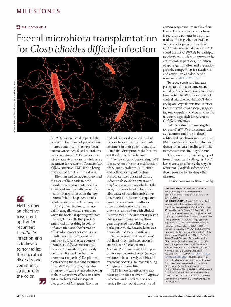
In 1958, Eiseman et al. reported the successful treatment of pseudomem-branous enterocolitis using a faecal enema. Since then, faecal microbiota transplantation (FMT) has become widely accepted as a successful rescue treatment for recurrent Clostridioides
difficile infection. FMT is also being investigated for other indications.
Eiseman and colleagues presented the cases of four patients with pseudomembranous enterocolitis. They used enemas with faeces from healthy donors after other therapy options failed. The patients had a rapid recovery from their symptoms.
C. difficile infection can cause debilitating diarrhoeal symptoms when the bacterial spores germinate into vegetative cells that produce enterotoxins, resulting in colonic inflammation and the formation of ‘pseudomembranes’ consisting of inflammatory cells, dead cells and debris. Over the past couple of decades, C. difficile infection has increased in incidence, morbidity and mortality, and has become known as a ‘superbug’. Despite anti-biotics being the standard treatment for C. difficile infection, they also often are the cause of infection owing to their suppressive effects on native gut microbiota and subsequent overgrowth of C. difficile. Eiseman
and colleagues also noted this link to prior broad-spectrum antibiotic treatment in their patients and spec-ulated that disruption of the ‘healthy gut flora’ underlies infection.
The intention of performing FMT is restoration of the normal function of the gut microbiota. In Eiseman and colleagues’ report, culture of stool samples obtained during infection showed the presence of Staphylococcus aureus, which, at the time, was considered to be a pos-sible cause of pseudomembranous enterocolitis. S. aureus disappeared from the stool sample cultures after administration of a faecal enema in association with clinical improvement. The authors suggested that normal colonic non-patho-gens displaced the colitis-causing pathogen, which, decades later, was demonstrated to be C. difficile.
Since Eiseman and co-workers’ publication, others have reported success using faecal enemas, Lactobacillus rhamnosus GG (a pro-biotic) and bacteriotherapy (using a mixture of facultatively aerobic and anaerobic bacteria) to treat relapsing C. difficile enterocolitis.
FMT is now an effective treat-ment option for recurrent C. difficile infection and is believed to nor-malize the microbial diversity and
community structure in the colon. Currently, a research consortium is recruiting patients to a clinical trial examining whether FMT is safe, and can prevent recurrent C. difficile-associated disease. FMT could inhibit C. difficile by multiple mechanisms, such as suppression by antimicrobial peptides, inhibition of spore germination and vegetative growth, competition for nutrients, and activation of colonization resistance (MILESTONE 13).
To reduce costs and increase patient and clinician convenience, oral delivery of faecal microbiota has been tested. In 2017, a randomized clinical trial showed that FMT deliv-ery by oral capsule was non-inferior to delivery via colonoscopy, suggest-ing oral capsules could be an effective treatment approach for recurrent C. difficile infection.
FMT has also been investigated for non-C. difficile indications, such as ulcerative and drug-induced colitis, and has shown some promise. FMT from lean donors has also been shown to increase insulin sensitivity in men with metabolic syndrome.
Thus, since the pioneering report from Eiseman and colleagues, FMT has become an effective therapy for recurrent C. difficile infection and shows promise for treating other diseases.
Louise Stone, Nature Reviews Urology
M I L E S TO N E 2
Faecal microbiota transplantation for Clostridioides difficile infection
ORIGINAL ARTICLE Eiseman B. et al. Fecal enema as an adjunct in the treatment of pseudomembranous enterocolitis. Surgery 44, 854–859 (1958).FURTHER READING Khoruts A. & Sadowsky M. J. Understanding the mechanisms of faecal microbiota transplantation. Nat. Rev. Gastro. Hep. 13, 508–516 (2016) | Pamer, E. G. Fecal microbiota transplantation: effectiveness, complexities, and lingering concerns. Mucosal Immunol. 7, 210–2014 (2014) | Schwan A. et al. Relapsing Clostridium difficile enterocolitis cured by rectal infusion of homologous faeces. Lancet 322, 845 (1983) | Gorbach S. L., Chang T. W. & Goldin B. Successful treatment of relapsing Clostridium difficile colitis with Lactobacillus GG. Lancet 330, 1519 (1987) | Tvede M, Rask-Madsen J. Bacteriotherapy for Clostridium difficile diarrhoea. Lancet 1, 1156–1160 (1989) | US National Library of Medicine. Microbial Restoration for Individuals With One or More Recurrences of Clostridium Difficile Associated Disease (CDAD) https://ClinicalTrials.gov/show/NCT03548051 (2018) | Kao, D. et al. Effect of oral capsule- vs. colonoscopy-delivered fecal microbiota transplantation on recurrent Clostridium difficile infection: a randomized clinical trial. JAMA 318, 1985–1993 (2017) | Vrieze, A. et al. Transfer of intestinal microbiota from lean donors increases insulin sensitivity in individuals with metabolic syndrome. Gastroenterology 143, 913–916 (2012).
FMT is now
an effective
treatment
option for
recurrent
C. difficile
infection and
is believed
to normalize
the microbial
diversity and
community
structure in
the colon
Cre
dit:
Sci
ence
Pho
to L
ibra
ry /
Ala
my
Stoc
k Ph
oto
MILESTONES
S6 | JUNE 2019 www.nature.com/collections/microbiota-milestone

Germ-free animals are raised under sterile conditions to prevent coloni-zation with bacteria and other micro-organisms. These animals (mostly mice and rats, but also guinea pigs and chicks) have a life span similar to that of conventional, normally col-onized animals, although they seem to have a slower or impaired growth as well as some anatomical and physiological differences (such as an enlarged cecum). By the 1960s, germ-free animals were a well-established tool in nutritional studies aiming to understand the contribution of the intestinal microbiota to host dietary requirements (for example, with regard to the synthesis of vitamins).
In 1965, Schaedler and colleagues introduced a new use for GF animals: the transfer of bacterial cultures to germ-free mice. Such transfer experi-ments have been essential in studying the effects of the gut microbiota on the host ever since. In their pivotal study, Schaedler and colleagues reported the results of feeding bacterial cultures isolated from the gut of Nelson–Collins–Swiss (NCS) mice (a colony of albino mice that are free of ordinary mouse pathogens as well as intestinal Escherichia coli and Proteus spp.) to germ-free mice. The germ-free mice were fed food inoculated with individual bacterial
cultures of several anaerobic isolates. After one week, the numbers and localization of these bacterial strains in the gastrointestinal tract were comparable to those observed in the NCS mice and remained stable for several months, confirming the feasibility of microbiota transfer experiments and their usefulness for studying bacterial gut colonization. Importantly, transfer of a Bacteroides strain partially reduced the cecum enlargement typical of germ-free mice, and the offspring of germ-free mice that had been colonized with a mixture of strains inherited those strains and subsequently showed normal cecum size and structure. These results directly showed the important and profound effect of the gut microbiota on host development and physiology.
This landmark study paved the way for further research on the effects of the gut microbiota on the host and on the interactions
between different species of the gut microbiota. For example, one strain of Lactobacillaceae that could 7α-dehydroxylate bile acids in vitro did not have the same catabolic activity when transferred to germ-free rats until additional bacterial strains were introduced. Another study in germ-free rats also showed the met-abolic capacity of the gut microbiota, specifically the reduction of bilirubin to urobilins, which had been assumed by some to be produced by the liver. These initial studies laid the ground for detailed work that explored the links between microbial and host metabolism (MILESTONE 12).
In addition to exploring the met-abolic capacity of the gut microbiota, GF animals were also essential for elucidating the close links between bacteria and the host that determine tissue homeostasis and immune system development. One striking example is determination of the role of segmented filamentous bacteria (SFB), which had previously been shown to closely interact with the intestinal epithelium. Experiments that led to monoassociation of germ-free mice with SFB showed that these bacteria are key determinants of intestinal lymphocyte numbers and phenotype in mice. Subsequent studies have identified many links between specific microbial taxa and/or molecules and host immune function (MILESTONE 9).
Germ-free animals have been, and still are, indispensable tools for studying functional relationships between the microbiota and the host — although, as always with animal studies, the comparability and applicability of the results to humans need to be verified. Nevertheless, the early studies using these models have inspired several avenues of micro-biota research and highlighted the important effects the gut microbiota have on their host.
Lucia Brunello, Nature Reviews Disease Primers
M I L E S TO N E 3
Gut microbiota transfer experiments in germ-free animals
ORIGINAL ARTICLES Schaedler, R.W., Dubos, R. & Costello, R. Association of germfree mice with bacteria isolated from normal mice. J. Exp. Med. 122, 77–82 (1965) | Gustafsson, B.E., Midtvedt, T. & Norman. A. Metabolism of cholic acid in germfree animals after the establishment in the intestinal tract of deconjugating and 7α-dehydroxylating bacteria. Acta Pathol. Microbiol. Scand. 72, 433–443 (1968) | Gustafsson, B.E. & Sewander Lanke, L. Bilirubin and urobilins in germfree, ex-germfree and conventional rats. J. Ex. Med. 112, 975–981 (1960) | Umesaki, Y. et al. Segmented filamentous
bacteria are indigenous intestinal bacteria that activate intraepithelial lymphocytes and induce MHC class II molecules and fucosyl asialo GM1 glycolipids on the small intestinal epithelial cells in the ex-germ-free mouse. Microbiol. Immunol. 39, 555–562 (1995).FURTHER READING Johansson, K.R. & Sarles, W.B. Some considerations of the biological importance of intestinal microörganisms. Bacteriol. Rev. 13, 25–45 (1949) | Sommer F. & Bäckhed, F. The gut microbiota — masters of host development and physiology. Nat. Rev. Microbiol. 11, 227–238 (2013).
In 1965,
Schaedler and
colleagues
introduced
a new use
for germ-
free animals:
the transfer
of bacterial
cultures to
germ-free
mice […] have
been essential
to study the
effects of the
gut microbiota
on the host
ever since
Laboratory mice housed under germ-free conditions. Photograph courtesy of Taren M. Thron, California Institute of Technology.
MILESTONES
NATURE MILESTONES | HUMAN MICROBIOTA RESEARCH JUNE 2019 | S7

Early life experiences have complex and long-lasting effects that can reach into adulthood — the same can be said of the acquisition and succession of our microbiota during the first years of life. The culmination of years of investigation from many laboratories has led to an in-depth characterization of postnatal microbial acquisition and maturation during the first years of life, and has led to the realisation that this represents a crucial window in our long-term development.
Early studies, dating as far back as 1900, described various aspects of bacterial succession in infants, but in 1981, three studies were reported that set out to quantitatively characterize early acquisition of gut commensals and to study how feeding shapes our initial microbiota. In one study, development of the bacterial community was investigated in infants in Sheffield, England, by culturing specimens taken from the meconium (a baby’s first faeces), faeces, mouth and umbilicus in the first six days of life. In another study, the faecal bacterial community was compared between infant cohorts
Peppercorn and Goldman demonstrated that
the anti-inflammatory drug, salicylazosulfapyri-
dine, could be degraded in conventional rats and
when cultured with human gut bacteria, but not
in germ-free rats, indicating a role for the gut
microbiota in drug transformations. An increasing
number of studies have confirmed the role of
the microbiota, not limited to the gut, in drug
metabolism and highlighted the implications for
drug inactivation, efficacy and toxicity.
in France that were either bottle-fed or breastfed; and in the third study, faecal bacterial communities from breastfed infants, weaned children and adults born in urban England and rural Nigeria, were compared. These studies provided quantitative measurements of specific bacterial taxa in early life, giving insight into the pioneer species that colonize the infant gut. This paved the way for future high-resolution studies of microbial succession in infants.
With the advent of ‘omics’ technologies in the following decades, our understanding of when the majority of our microbiota are acquired, and of what species are there, has heightened and the importance of host–microbiota–environment interactions during early life has become realised. The infant gut microbiota undergoes a period of massive change in the first years of life. The initial microbiota adapts over time and is shaped by the availability of different nutrients. As the infant consumes increasingly more complex dietary substrates, there are shifts in composition and an enrichment of bacterial functions related to
carbohydrate metabolism and the biosynthesis of amino acids and vitamins. By 2–3 years of age, a stable microbiota develops that resembles that of the adults in the infant’s community (see MILESTONE 7).
When colonization first occurs is an open question; however, most scientists think that the foetus develops in a sterile environment and that we acquire the bulk of our initial microbiota during and immediately after birth. Recently, a few studies have found traces of bacterial DNA in the placenta, in the amniotic fluid that surrounds the foetus and in the meconium — suggesting prenatal colonization. However, many scientists think these findings could be the result of contamination and the debate is ongoing. Regardless of possible exposure to microorganisms in utero, the foetus is exposed to microbial molecules that cross the placenta from the mother.
The first major exposure to microorganisms happens during delivery, and is highly dependent on the mode of delivery. The microbiota of neonates that are born vaginally are enriched in bacteria that resemble
M I L E S TO N E 5
Microbiota succession in early life
M I L E S TO N E 4
The microbiota influences metabolism of host-directed drugs
ORIGINAL ARTICLE Peppercorn, M. A. & Goldman, P. The role of intestinal bacteria in the metabolism of salicylazosulfapyridine. J. Pharmacol. Exp. Ther. 181, 555–562 (1972).FURTHER READING Clayton, T. A. et al. Pharmacometabonomic identification of a significant host-microbiome metabolic interaction affecting human drug metabolism. Proc. Natl Acad. Sci. USA 106, 14728–14733 (2009) | Lindenbaum, J., Rund, D. G., Butler, V. P. J., Tse-Eng, D. & Saha, J. R. Inactivation of digoxin by the gut flora: reversal by antibiotic therapy. N. Eng. J. Med. 305, 789–794 (2010) | Wallace, B. D. et al. Alleviating cancer drug toxicity by inhibiting a bacterial enzyme. Science 330, 831–835 (2010) | Haiser, H. J. et al. Predicting and manipulating cardiac drug inactivation by the human gut bacterium Eggerthella lenta. Science 341, 295–298
(2013) | Liang, X. et al. Bidirectional interactions between indomethacin and the murine intestinal microbiota. eLife 4, e08973 (2015) | Klatt, N. R. et al. Vaginal bacteria modify HIV tenofovir microbicide efficacy in African women. Science 356, 938–945 (2017) | Zimmermann, M., Zimmermann-Kogadeeva, M., Wegmann, R. & Goodman, A. L. Separating host and microbiome contributions to drug pharmacokinetics and toxicity. Science 363, eaat9931 (2019) | Spanogiannopoulos, P., Bess, E. N., Carmody, R. N. & Turnbaugh, P. J. The microbial pharmacists within us: a metagenomic view of xenobiotic metabolism. Nat. Rev. Microbiol. 14, 273–287 (2016) | Koppel, N., Maini Rekdal, V. & Balskus, E. P. Chemical transformation of xenobiotics by the human gut microbiota. Science
356, eaag2770 (2017).
The infant gut
microbiota
undergoes
a period
of massive
change in the
first years
of life
Cre
dit:
G. M
arsh
all /
Spr
inge
r Nat
ure
Lim
ited
MILESTONES
S8 | JUNE 2019 www.nature.com/collections/microbiota-milestone

ORIGINAL ARTICLES Rotimi, V. O. & Duerden, B. I. The
development of the bacterial flora in normal neonates. J. Med.
Microbiol. 14, 51–62 (1981). | Tompkins, A.M. et al. Diet and the
faecal microflora of infants, children and adults in rural Nigeria
and urban U.K. J. Hyg. 86, 285–293 (1981). | Daoulas Le
Bourdelles, F., Avril, J. L. & Ghnassia, J. C. Quantitative study of the
faecal flora of breast- or bottle-fed neonates (transl.). Arch. Fr.
Pediatr. 38, 35–39 (1981).
FURTHER READING Tamburini, S., Shen, N., Wu, H. C. &
Clemente, J. C. The microbiome in early life: implications for
health outcomes. Nat. Med. 22, 713–722 (2016) | Robertson, R. C.,
Manges, A. R., Finlay, B. B. & Prendergast, A. J. The Human
microbiome and child growth: first 1,000 days and beyond. Trends
Microbiol. 27, 131–147 (2019) | Cooperstock, M. S. & Zedd, A. J. in
Human Intestinal Microflora in Health and Disease (ed. Hentges,
D. J.) Ch. 4 (Elsevier, 1983) | Tissier, H. Recherches sur la flore
intestinale des nourrissons (état normal et pathologique) (Carre, G.
& C. Naud, C., Paris, 1900) | Long, S. S. & Swenson, R. M.
Development of anaerobic faecal flora in healthy newborn
infants. J. Pediatr. 91, 298–301 (1977) | Simhon, A., Douglas, J. R.,
Drasar, B.S. & Soothill, J. F. Effect of feeding on infants’ faecal flora.
Arch. Dis. Child 57, 54–58 (1982) | Stark, P. L. & Lee, A. The microbial
ecology of the large bowel of breast-fed and formula-fed infants
during the first year of life. J. Med. Microbiol. 15, 189–203 (1982) |
Satokari, R., Vaughan, E. E., Favier, C., Edwards, C. & de Vos, W. M.
Diversity of Bifidobacteria and Lactobacillus spp. in breast-fed and
formula fed infants as assessed by 16S rDNA sequence
differences. Microb. Ecol. Health Dis. 14, 97–105 (2002) | Palmer, C.,
Bik, E. M., DiGiulio, D. B., Relman, D. A. & Brown, P. O. Development
of the human infant intestinal microbiota. PLoS Biol. 5, e177 (2007) |
Bennet, R. & Nord, C. E. Development of the fecal anaerobic
microflora after cesarean section and treatment with antibiotics
in newborn infants. Infection 15, 332–336 (1987) | Dominguez-
Bello, M.G. et al. Delivery mode shapes the acquisition and
structure of the initial microbiota across multiple body habitats in
newborns. Proc. Natl Acad. Sci. USA 107, 11971–11975 (2010) |
Bäckhed, F. et al. Dynamics and stabilization of the human gut
microbiome during the first year of life. Cell Host Microbe 17,
690–703 (2015) | Dominguez-Bello, M. G. et al. Partial restoration
of the microbiota of cesarean-born infants via vaginal microbial
transfer. Nat. Med. 22, 250–253 (2016) | Jakobsson, H.E. et al.
Decreased gut microbiota diversity, delayed Bacteroidetes
colonization and reduced TH1 responses in infants delivered by
cesarean section. Gut 63, 559–566 (2014) | Koenig, J. E. et al.
Succession of microbial consortia in the developing infant gut
microbiome. Proc. Natl Acad. Sci. USA 108, 4578–4585 (2011) | Lim,
E. S. et al. Early-life dynamics of the human gut virome and
bacterial microbiome in infants. Nat. Med. 21, 1228–1234 (2015) |
Yatsunenko, T. et al. Human gut microbiome viewed across age
and geography. Nature 486, 222–227 (2012) | Bokulich, N. A. et al.
Antibiotics, birth mode, and diet shape microbiome maturation
during early life. Sci. Transl Med. 8, 343ra82 (2016) | Chu, D. M. et al.
Maturation of the infant microbiome community structure and
function across multiple body sites and in relation to mode of
delivery. Nat. Med. 23, 314–326 (2017) | Wampach, L. et al.
Colonization and succession within the human gut microbiome by
archaea, bacteria and microeukaryotes during the first year of life.
Front. Microbiol. 8, 738 (2017) | Stewart, C. J. et al. Temporal
development of the gut microbiome in early childhood from the
TEDDY study. Nature 562, 583–588 (2018) | Vatanen, T. et al.
Genomic variation and strain-specific functional adaptation in the
human gut microbiome during early life. Nat. Microbiol. 4, 470–479
(2018) | Yassour, M. et al. Strain-level analysis of mother-to-child
bacterial transmission during the first few months of life. Cell Host
Microbe 24, 146–154 (2018) | Ferretti, P. et al. Mother-to-infant
microbial transmission from different body sites shapes the
developing infant gut microbiome. Cell Host Microbe 24, 133–145
(2018) | Wampach, L. et al. Birth mode is associated with earliest
strain-conferred gut microbiome functions and
immunostimulatory potential. Nat. Commun. 9, 5091 (2018) |
Aagaard, K. et al. The placenta harbors a unique microbiome. Sci.
Transl. Med. 6, 237ra265 (2014) | Kliman, H.J. Comment on “The
placenta harbors a unique microbiome”. Sci. Transl. Med. 6, 254le4
(2014) | Subramanian, S. Persistent gut microbiota immaturity in
malnourished Bangladeshi children. Nature 510, 417–421 (2014).
Cre
dit:
S. B
radb
rook
/ Sp
ring
er N
atur
e Li
mit
ed
the maternal vaginal microbiota (for example, Lactobacillus species), whereas neonates delivered by cae-sarean (C-) section lack these species and are instead enriched in skin commensals such as Staphylococcus, Streptococcus and Propionibacterium species. Over time, these differences gradually reduce between vaginally and C-section-born infants; however, in one study, bacteria associated with C-section remained associated with C-section-delivered infants up to two years of age, showing that birth mode could have long-term impacts on the microbiota.
Postnatal factors further configure the microbiota in early
life. Breastmilk contains a complex community of bacteria that may help seed the infant gut microbiota, and in breastfed infants the gut microbiota is dominated by species that metabolise human milk oligosaccharides. Overall, diet has been found to be a major determinant of the infant gut microbiota. Studies of malnourished infants have shown that maturation of the gut microbiota does not occur in a similar manner to healthy infants, even after dietary intervention, and it has been proposed that an ‘undernourished’ microbiome in infancy can perpetuate growth impairments later in life.
The environment and people that surround an infant are also a source of microorganisms that can colonize various body sites. Genetically unrelated parents and even pets share a high proportion of their microbiota with infants. Genetics also has a role in determining our microbiota make-up, as evidenced by associations between the heritability of specific taxa and host genes.
The use of antimicrobials, which is essential for preserving life when infants acquire a serious bacterial infection, can impact the ecological succession of the infant microbiota. Antibiotics can impair the diversity and stability of the developing microbiota in infants, with abundances of specific taxa remaining reduced for years after treatment. The impact of antibiotics on the infant microbiota could have long-lasting health implications and their use in early life has been linked to an increased risk of several diseases, including asthma, inflammatory bowel disease and allergies (see MILESTONE 9). More research is required to uncover the underlying mechanisms; however, what is clear is that the microbiota has a vital role in immune, endocrine, metabolic and a variety of other developmental pathways in infants, and without it we would not be here today.
Ashley York, Nature Reviews Microbiology
MILESTONES
NATURE MILESTONES | HUMAN MICROBIOTA RESEARCH JUNE 2019 | S9

Over the past twenty years, advances in DNA sequencing technology have deepened our knowledge in every niche of biology, from annotation of the human genome to sequencing the human-associated microbial metagenome, the genetic material of the microorganisms that inhabit almost every surface of the human body.
Study of the human microbiota histori-cally relied on culture-dependent methods to isolate and grow bacterial colonies in a prede-termined medium. The inability to cultivate a large portion of microorganisms, however, substantially underestimated the biodiversity of human-associated microbial communities.
An early milestone in this field was the adoption of a technology, previously pioneered by Carl Woese, Norman Pace and others to identify environmental bacteria, that was based on sequencing small subunit ribosomal RNA genes (16S rRNA). Using this approach, Wilson and Blitchington compared the diversity of cultivated and noncultivated bacteria within a human faecal sample in 1996. Since then, sequencing of 16S rRNA genes from complex communities has become a powerful tool for assessing microbial diversity in the human microbiota. In 2005, Eckburg et al. analysed samples not only from faeces, but also from multiple colonic mucosal sites. They sequenced >13,000 16S rRNA genes, which constituted a substantial increase in scope over previous work, and discovered significant inter-subject variability and greater differences between stool and mucosal community composition than previously described.
Sequencing of marker genes (such as 16S rRNA) is traditionally associated with Sanger sequencing, which requires a labour-intensive
cloning step and is prohibitively expensive for large-scale microbiome studies. The advent of next generation sequencing (NGS) offered a cost-effective method that eliminated the cloning step by amplifying 16S rRNA genes using primers containing sequencing adapters and barcodes. The massive parallel sequencing throughput offered by NGS has significantly increased the sequence depth of 16S rRNA genes, allowing for taxonomic and phylogenetic analyses of complex microbial communities. Yet, 16S rRNA sequencing can-not always resolve closely related species and may miss the intra-species diversity. To better capture the full picture, shotgun sequencing was developed for direct sequencing of DNA. The advantage is its capability to recover the underrepresented microorganisms that were often masked by high-abundance species. Shotgun sequencing can use short-read (such as Illumina) or long-read (for example,
Oxford Nanopore MinION and Pacific Bioscience Sequel) platforms. The short-read approach typically demands extensive com-putational support for assembling short reads into meaningful sequences. However, an
accurate and complete assembly of genome sequences is still hampered by the difficulty in resolving long repetitive regions. Thus, ref-erence-based sequencing methods emerged.
Concerted sequencing efforts have been made to construct microbial reference sequences. In 2007, the National Institutes of Health launched the Human Microbiome Project and, five years later, published the first reference data for microorganisms collected from 242 healthy United States volunteers, covering a number of anatomical sites such as mouth, nose, skin, lower intes-tine and vagina (MILESTONE 17). The valuable genome references that were generated by this consortium allow reliable identification of individual microbial species, but fall short when new genomes are present in the community.
Long-read sequencing offers an alternative solution for mapping challenging repetitive regions. For example, single molecule, real-time (SMRT) DNA sequencing, in combina-tion with a short-read shotgun DNA library, allows for de novo microbial genome assem-blies, although it suffers from high error rates.
Access to genome sequencing has transformed human microbiome research from focusing on identity characterizations, to metagenomics approaches that not only reveal microbial species but also how micro-bial metabolic activities correlate with human health and disease.
In addition to metagenomics, meta-transcriptomic analysis enabled by RNA sequencing offers a way to detect active members present in a microbial community (MILESTONE 14).
The construction of metagenome-assem-bled genomes, an approach also pioneered in environmental microbiology and recently applied to human-associated communi-ties, will provide a new, unprecedented opportunity for deep characterization of the functional potential of the human microbial ecosystem.
Lei Tang, Nature Methods
M I L E S TO N E 6
Sequence-based identification of human-associated microbiota
ORIGINAL ARTICLES Wilson, K. H. & Blitchington, R. B. Human colonic biota studied by ribosomal DNA sequence analysis. Appl.
Environ. Microbiol. 62, 2273–2278 (1996) | Suau, A. et al. Direct analysis of genes encoding 16S rRNA from complex communities reveals many novel molecular species within the human gut. Appl. Environ. Microbiol. 65, 4799–4807 (1999) | Kroes, I., Lepp, P. W. & Relman, D. A. Bacterial diversity within the human subgingival crevice. Proc. Natl Acad. Sci. USA 96, 14547–14552 (1999) | Eckburg, P. B. et al. Diversity of the human intestinal microbial flora. Science 308, 1635–1638 (2005) | Gill, S. R. et al. Metagenomic analysis of the human distal gut microbiome. Science 312, 1355–1359 (2006).FURTHER READING Relman, D. A., Schmidt, T. M., MacDermott, R. P. & Falkow, S. Identification of the uncultured bacillus of Whipple’s disease. N. Engl. J. Med. 327, 293–301 (1992) | Hold, G. L., Pryde, S. E., Russell, V. J., Furrie, E. & Flint, H. J. Assessment of microbial diversity in human colonic samples
by 16S rDNA sequence analysis. FEMS Microbiol. Ecol. 39, 33–39 (2002) | Pei, Z. et al. Bacterial biota in the human distal esophagus. Proc. Natl Acad. Sci. USA 101, 4250–4255 (2004) | Bik, E. M. et al. Molecular analysis of the bacterial microbiota in the human stomach. Proc. Natl Adad. Sci. USA 103, 732–737 (2006) | Fredricks, D. N. Molecular identification of bacteria associated with bacterial vaginosis. N. Engl. J. Med. 353, 1899–1911 (2005) | Hyman, R. W. et al. Microbes on the human vaginal epithelium. Proc. Natl Acad. Sci. USA 102, 7952–7957 (2005) | Gao, Z., Pei, Z., Tseng, C.-H. & Blaser, M. J. Molecular analysis of human forearm superficial skin bacterial biota. Proc. Natl Acad. Sci. USA 104, 2927–2932 (2007) | Grice, E. A. et al. Topographical and temporal diversity of the human skin microbiome. Science 324, 1190–1192 (2009) | Human Microbiome Jumpstart Reference Strains Consortium et al. A catalog of reference genomes from the human microbiome. Science 328, 994–999 (2010).
Cre
dit:
Sea
n Pr
ior /
Ala
my
Stoc
k Ph
oto
MILESTONES
S10 | JUNE 2019 www.nature.com/collections/microbiota-milestone
A plethora of studies conducted over the past
few decades have revealed strong associations
between a disrupted microbiota and diseases, for
example, inflammatory bowel disease. However,
the key to understanding the role of a disrupted
microbiota in human diseases is first to answer
the question: what is a ‘normal’ microbiota?
This question still frustrates many
microbiologists, even with the advent of
high-throughput sequencing and ’omics
techniques. Prior to the availability of
these technologies, however, several
studies were instrumental in answering similar
questions to help define ‘normal’, such as how
much microbial variation is there between and
within adults, and is the microbiota stable?
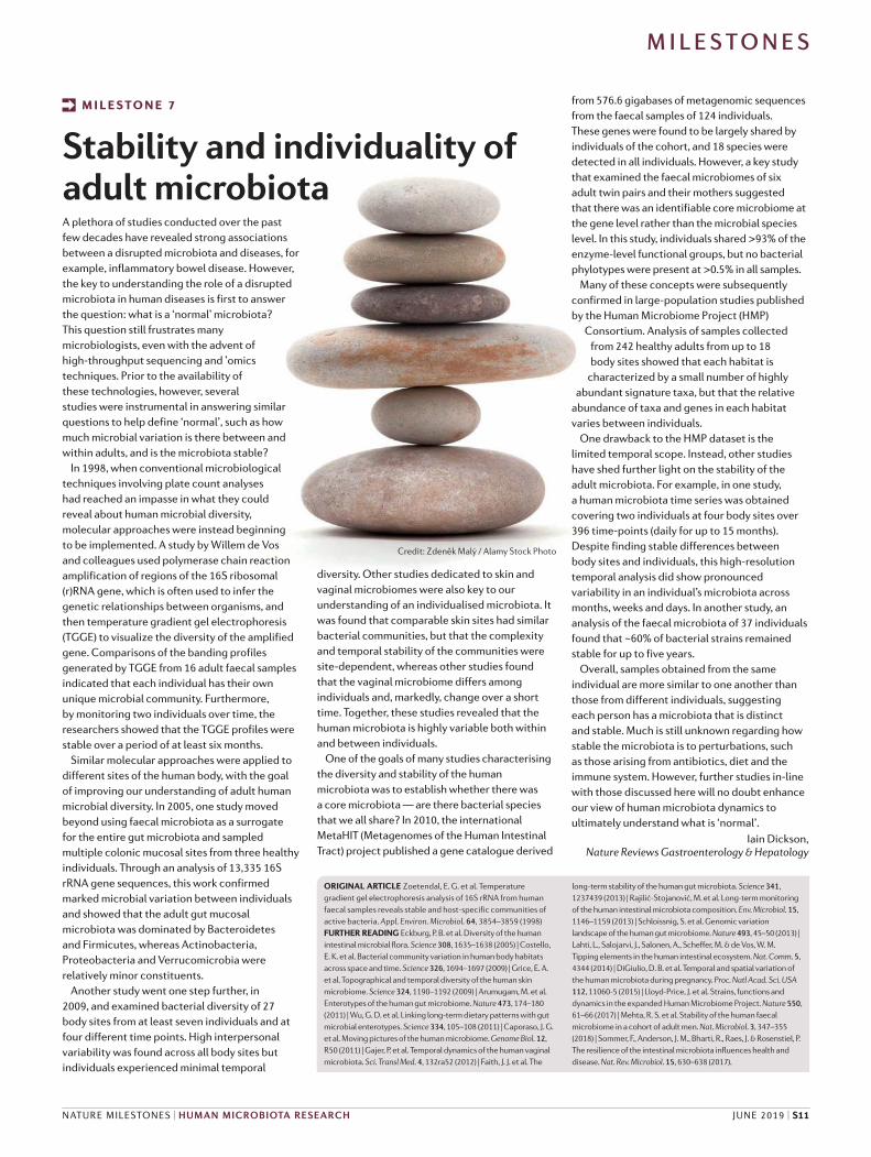
In 1998, when conventional microbiological
techniques involving plate count analyses
had reached an impasse in what they could
reveal about human microbial diversity,
molecular approaches were instead beginning
to be implemented. A study by Willem de Vos
and colleagues used polymerase chain reaction
amplification of regions of the 16S ribosomal
(r)RNA gene, which is often used to infer the
genetic relationships between organisms, and
then temperature gradient gel electrophoresis
(TGGE) to visualize the diversity of the amplified
gene. Comparisons of the banding profiles
generated by TGGE from 16 adult faecal samples
indicated that each individual has their own
unique microbial community. Furthermore,
by monitoring two individuals over time, the
researchers showed that the TGGE profiles were
stable over a period of at least six months.
Similar molecular approaches were applied to
different sites of the human body, with the goal
of improving our understanding of adult human
microbial diversity. In 2005, one study moved
beyond using faecal microbiota as a surrogate
for the entire gut microbiota and sampled
multiple colonic mucosal sites from three healthy
individuals. Through an analysis of 13,335 16S
rRNA gene sequences, this work confirmed
marked microbial variation between individuals
and showed that the adult gut mucosal
microbiota was dominated by Bacteroidetes
and Firmicutes, whereas Actinobacteria,
Proteobacteria and Verrucomicrobia were
relatively minor constituents.
Another study went one step further, in
2009, and examined bacterial diversity of 27 body sites from at least seven individuals and at
four different time points. High interpersonal
variability was found across all body sites but
individuals experienced minimal temporal
diversity. Other studies dedicated to skin and
vaginal microbiomes were also key to our
understanding of an individualised microbiota. It
was found that comparable skin sites had similar
bacterial communities, but that the complexity
and temporal stability of the communities were
site-dependent, whereas other studies found
that the vaginal microbiome differs among
individuals and, markedly, change over a short
time. Together, these studies revealed that the
human microbiota is highly variable both within
and between individuals.
One of the goals of many studies characterising
the diversity and stability of the human
microbiota was to establish whether there was
a core microbiota — are there bacterial species
that we all share? In 2010, the international
MetaHIT (Metagenomes of the Human Intestinal
Tract) project published a gene catalogue derived
from 576.6 gigabases of metagenomic sequences from the faecal samples of 124 individuals.
These genes were found to be largely shared by
individuals of the cohort, and 18 species were
detected in all individuals. However, a key study
that examined the faecal microbiomes of six
adult twin pairs and their mothers suggested
that there was an identifiable core microbiome at
the gene level rather than the microbial species
level. In this study, individuals shared >93% of the
enzyme-level functional groups, but no bacterial
phylotypes were present at >0.5% in all samples.
Many of these concepts were subsequently
confirmed in large-population studies published
by the Human Microbiome Project (HMP)
Consortium. Analysis of samples collected
from 242 healthy adults from up to 18
body sites showed that each habitat is
characterized by a small number of highly
abundant signature taxa, but that the relative
abundance of taxa and genes in each habitat
varies between individuals.
One drawback to the HMP dataset is the
limited temporal scope. Instead, other studies
have shed further light on the stability of the
adult microbiota. For example, in one study,
a human microbiota time series was obtained
covering two individuals at four body sites over
396 time-points (daily for up to 15 months).
Despite finding stable differences between
body sites and individuals, this high-resolution
temporal analysis did show pronounced
variability in an individual’s microbiota across
months, weeks and days. In another study, an
analysis of the faecal microbiota of 37 individuals found that ~60% of bacterial strains remained
stable for up to five years.Overall, samples obtained from the same
individual are more similar to one another than
those from different individuals, suggesting
each person has a microbiota that is distinct
and stable. Much is still unknown regarding how
stable the microbiota is to perturbations, such
as those arising from antibiotics, diet and the
immune system. However, further studies in-line
with those discussed here will no doubt enhance
our view of human microbiota dynamics to
ultimately understand what is ‘normal’.
Iain Dickson, Nature Reviews Gastroenterology & Hepatology
M I L E S TO N E 7
Stability and individuality of adult microbiota
ORIGINAL ARTICLE Zoetendal, E. G. et al. Temperature gradient gel electrophoresis analysis of 16S rRNA from human faecal samples reveals stable and host-specific communities of active bacteria. Appl. Environ. Microbiol. 64, 3854–3859 (1998)FURTHER READING Eckburg, P. B. et al. Diversity of the human intestinal microbial flora. Science 308, 1635–1638 (2005) | Costello, E. K. et al. Bacterial community variation in human body habitats across space and time. Science 326, 1694–1697 (2009) | Grice, E. A. et al. Topographical and temporal diversity of the human skin microbiome. Science 324, 1190–1192 (2009) | Arumugam, M. et al. Enterotypes of the human gut microbiome. Nature 473, 174–180 (2011) | Wu, G. D. et al. Linking long-term dietary patterns with gut microbial enterotypes. Science 334, 105–108 (2011) | Caporaso, J. G. et al. Moving pictures of the human microbiome. Genome Biol. 12, R50 (2011) | Gajer, P. et al. Temporal dynamics of the human vaginal microbiota. Sci. Transl Med. 4, 132ra52 (2012) | Faith, J. J. et al. The
long-term stability of the human gut microbiota. Science 341, 1237439 (2013) | Rajilić-Stojanović, M. et al. Long-term monitoring of the human intestinal microbiota composition. Env. Microbiol. 15, 1146–1159 (2013) | Schloissnig, S. et al. Genomic variation landscape of the human gut microbiome. Nature 493, 45–50 (2013) | Lahti, L., Salojarvi, J., Salonen, A., Scheffer, M. & de Vos, W. M. Tipping elements in the human intestinal ecosystem. Nat. Comm. 5, 4344 (2014) | DiGiulio, D. B. et al. Temporal and spatial variation of the human microbiota during pregnancy. Proc. Natl Acad. Sci. USA
112, 11060-5 (2015) | Lloyd-Price, J. et al. Strains, functions and dynamics in the expanded Human Microbiome Project. Nature 550, 61–66 (2017) | Mehta, R. S. et al. Stability of the human faecal microbiome in a cohort of adult men. Nat. Microbiol. 3, 347–355 (2018) | Sommer, F., Anderson, J. M., Bharti, R., Raes, J. & Rosenstiel, P. The resilience of the intestinal microbiota influences health and disease. Nat. Rev. Microbiol. 15, 630–638 (2017).
Credit: Zdeněk Malý / Alamy Stock Photo
MILESTONES
NATURE MILESTONES | HUMAN MICROBIOTA RESEARCH JUNE 2019 | S11

While bacteria are a major component of the
human microbiota, viruses, fungi and archaea
are also important members of the community,
with potential effects on human health. The
development of 16S ribosomal RNA gene
sequencing transformed the field of microbiome
research by enabling bacterial and archaea
phylogenetic analyses without the need for
culturing (MILESTONE 6). Fungi can also be
identified and classified by sequencing a common
nuclear ribosomal internal transcribed spacer
region. Viruses, however, are far more challenging
to isolate and sequence due to the necessity
of a eukaryotic or prokaryotic host and the
absence of conserved genes. The modern era of
human-associated viral metagenomics actually
started in the ocean. In 2001, the research group
of Forest Rowher, a marine microbial ecologist
at San Diego State University, published
a randomized shotgun library sequencing
method to analyse genomic DNA from a single
bacteriophage. This was rapid, reasonably
unbiased and required very little DNA input,
a crucial limiting variable for sequencing. The
research team further demonstrated the utility of
this technique beyond single phage analysis by
characterizing the more complex viral make-up
of seawater and marine sediment, paving the
way for analyses of human viromes and a deeper
characterisation of the human microbiome.
By 2003, bacteriophages that infected individual
bacterial species had been identified from
human faecal waste, but the diversity and relative
abundance of different phages remained unknown,
as existing approaches were biased towards
bacteria already known to be infected by phages.
By using the linker-amplified shotgun library
approach, Rowher’s research group provided the
first quantitative description of the composition of
the uncultured virome in human faeces collected
from a single healthy adult. The majority of phage
sequence matches were from temperate phages,
which commonly integrate into the host bacterial
genome. The faecal virome was also dominated by
phages known to infect Gram-positive bacteria, in
keeping with prior data on faecal bacterial content.
Viral metagenomics has continued to advance,
with higher-throughput techniques enabling
more rapid virus discovery and classification in
both healthy and diseased tissues, and helping
us to understand the role of commensal and
pathogenic viruses in the context of the wider
microbiome.
In a study published in 2010, by Jeffrey Gordon
and Rowher, faecal viromes of healthy adult
female twins and their mothers were shown to be
individually distinct and stable over the course
of a year, in agreement with faecal bacterial data
from this same cohort. Other studies have begun
to elucidate the effects of disease and diet on
the gut virome, finding that phage composition
became increasingly similar between individuals
on the same diet, and that significant expansion
of one phage order is associated with Crohn’s
disease and ulcerative colitis. Viral microbiome
signatures may therefore be associated with
environmental influences as well as disease
progression, including in tissues beyond the gut.
Shotgun metagenomics has facilitated a more
functional and interconnected perspective
of the microbiome, with the contributions of
fungi and archaea also becoming increasingly
understood. The human mycobiome (the
fungal community of humans) has also been
fruitfully studied. A number of recent studies
have highlighted the crucial roles of fungi in
both healthy and disease states. For example,
enteric commensal bacteria and fungi may have
redundant protective and tolerizing functions in
the context of regulating the immune response,
suggesting that gut homeostasis can be retained
through the mycobiome even in the absence of
‘good’ bacteria. In inflammatory bowel diseases,
however, mycobiome dysbiosis contributes to
disease progression, with fungi enriched at the
expense of bacteria. These data, along with other
studies, suggest a complex interplay between
fungi and bacteria.
Recently, the human archaeome (the
community of human-associated archaea)
has attracted interest due to a number of
observations, including recognition of the human
gut archaeon Methanosphaera stadtmanae by the
immune system and the discovery of previously
undetected human-associated archaea.
Methanogenic archaea are amongst the most
abundant microorganisms in the human gut and
sometimes outnumber even the most abundant
bacterial species. In 2004, methanogenic archaea
were found to be associated with the onset
of periodontal disease, identifying the first
link between archaea and human disease. As
with bacteria, viruses and fungi, archaea have
been isolated from various human anatomical
sites, including the gut, skin, vagina and oral
cavity. Yet, owing to fundamental differences in
biology, they have often remained undetected
in microbiome surveys, warranting further
investigation of the human archaeome and the
role of archaea in human health and disease.
As identification and characterization of the
human virome, mycobiome and archaeome in
various tissues and disease states continues
to improve, it will be increasingly important
to delineate function and how these other
‘omes’ interact as a community to preserve or
dysregulate human health.
Saheli Sadanand, Nature Medicine
M I L E S TO N E 8
Beyond bacteria: studies of other host-associated microorganisms
ORIGINAL ARTICLE Breitbart, M. et al. Metagenomic analyses of an uncultured viral community from human feces. J. Bacteriol. 185, 6220–6223 (2003).FURTHER READING Schoch, C. L. et al. Nuclear ribosomal internal transcribed spacer (ITS) region as a universal DNA barcode marker for Fungi. Proc. Natl Acad. Sci. USA 109, 6241–6246 (2012) | Rowher, F. et al. Production of shotgun libraries using random amplification. Biotechniques 31, 108–118 (2001) | Reyes et al. Viruses in the faecal microbiota of monozygotic twins and their mothers. Nature 466, 334–338 (2010) | Norman, J. M. et al. Disease-specific alterations in the enteric virome in inflammatory bowel disease. Cell 160, 447–460 (2015) | Minot, S. et al. The human gut virome: inter-individual variation and dynamic response to diet. Genome Res. 21, 1616–1625 (2011) | Jiang, T. T. et al. Commensal fungi recapitulate the protective benefits of intestinal bacteria. Cell Host Microbe 22, 809–816 (2017) | Shao, T. Y. et al. Commensal Candida albicans positively calibrates systemic Th17 immunological responses. Cell Host Microbe 25, 404–417 (2019) | Iliev, I. D. et al. Interactions between commensal fungi and the C-type lectin receptor Dectin-1 influence colitis. Science 336, 1314–1317 (2012) | Hoarau, G. et al. Bacteriome and mycobiome interactions underscore microbial dysbiosis in familial Crohn’s
disease. mBio 7, e01250-16 (2016) | Sokol, H. et al. Fungal microbiota dysbiosis in IBD. Gut 66, 1039–1048 (2017) | Lepp, P. W. et al. Methanogenic Archaea and human periodontal disease. Proc. Natl Acad. Sci. USA 101, 6176–6181 (2004) | Koskinen, K. et al. First insights into the diverse human archaeome: specific detection of archaea in the gastrointestinal tract, lung, and nose and on skin. mBio 8, e00824-17 (2017) | Vierbuchen, T. et al. The human-associated archaeon Methanosphaera stadtmanae is recognized through its RNA and induces TLR8-dependent NLRP3 inflammasome activation. Front. Immunol. 8, 1535 (2017) | Borrel, G. et al. Genomics and metagenomics of trimethylamine-utilizing Archaea in the human gut microbiome. ISME J. 11, 2059–2074 (2017) | Adam, P. S. et al. The growing tree of Archaea: new perspectives on their diversity, evolution and ecology. ISME J. 11, 2407 (2017) | Nottingham, P. M. et al. Isolation of methanogenic bacteria from feces of man. J. Bacteriol. 96, 2178–2179 (1968) | Breitbart, M. et al. Viral diversity and dynamics in an infant gut. Res. Microbiol. 159, 367–373 (2008) | Ghannoum, M. A. et al. Characterization of the oral fungal microbiome (mycobiome) in healthy individuals. PLoS Pathog. 6, e1000713 (2010) | Yang, A. M. et al. Intestinal fungi contribute to development of alcoholic liver disease. J. Clin. Invest. 127, 2829–2841 (2017).
Cre
dit:
P. P
aten
all /
Spr
inge
r Nat
ure
Lim
ited
MILESTONES
S12 | JUNE 2019 www.nature.com/collections/microbiota-milestone

The relationship between the immune system and microorganisms was long viewed as a war rather than a union, as it was mostly studied in the context of host defence against pathogens. Thus, when deficiency in lym-phoid organ development and immune cell activity was reported in germ-free animals in the 1960s, this first evidence that microbiota shape immune homeostasis was interpreted as education of the immune system by infections.
This concept laid the foundation for the ‘hygiene hypothesis’, formalized by David Strachan in 1989 in his influential study reporting lower incidence of hay fever and eczema in children with older siblings. He proposed that infections in early childhood prevent atopy later in life, and that increased allergy prevalence in developed countries may be caused by high standards of personal hygiene. Further developments of the hygiene hypothesis correlated pathogen exposure to decreased allergy risk in humans, and established a role for microbiota in oral tolerance in mice, presumably in shifting an immune set point from T helper 2 (Th2) to Th1 cell responses.
In parallel, fundamental principles of microorganism recognition by the immune system unfolded. In 1989, Charles Janeway proposed that immune responses are initiated by genome-encoded pattern recognition receptors (PRR) on immune cells, which sense conserved microbial molecules. Over the next decade, PRRs specific to various bacterial, fungal and viral components were discovered. However, it became evident that PRRs are not specific to pathogens, posing the question of how commensals, which colonize mucosal surfaces, coexist with the immune system.
The prevailing explanation was that com-mensals and immune cells are separated by epithelial barriers. This view was challenged in the early 2000s by accumulating examples of immune responses to commensals in healthy mice. Among these, Andrew Macpherson, Rolf Zinkernagel and colleagues found that in mice, commensals are recognized and compartmentalized by gut lumen-secreted IgA. Lora Hooper, Jeffrey Gordon and cow-orkers reported that a commensal bacterium stimulates antimicrobial peptide production by Paneth cells.
In 2004, Seth Rakoff-Nahoum and Ruslan Medzhitov provided evidence that the immune system senses commensals through PRRs under normal conditions and that this sensing is crucial for tissue repair. This finding opened a new perspective on immune response to microorganisms not as host defence, but as a symbiotic physiological process.
Realization of beneficial roles of immune–microbial interactions prompted a revision of the hygiene hypothesis to postulate that protection from allergic diseases is mediated by early-life exposure to commensals rather than pathogens (MILESTONE 5). The refined hypothesis suggested that the rise of allergies in industrialized societies is caused by loss of commensals. A link between lifestyle, microbiota and allergic diseases has been now confirmed in a large number of human observational studies, and expanded to implicate microbiota in other chronic inflam-matory and autoimmune pathologies, as well as metabolic and neurological disease.
Dissecting the underlying cellular and molecular complexity of microbiota–immune interactions has occupied microbiologists and immunologists to this day. Pioneering works by Sarkis Mazmanian, Dennis Kasper and colleagues showed that microbiota-guided maturation of the mouse immune system can be recapitulated by a polysaccharide produced by a symbiotic bacterium, and delineated that its uptake by dendritic cells promotes antigen presentation, inducing expansion and differentiation of CD4+ T cells into Th1 and regulatory T cell lineages. Ivaylo Ivanov,
Kenya Honda, Dan Littman and colleagues uncovered how specific commensal bac-teria direct Th17 cell differentiation in the small intestine. In 2013–2014, four groups independently discovered a mechanism of immune tolerance mediated by metabolites of intestinal commensals (MILESTONE 21).
Our intimate companion in sickness and in health, microbiota impacts our physiol-ogy to a large extent through interactions with the immune system at mucosal sites. How these interactions, ranging from host defence to active tolerance to symbiosis, integrate into physiological outcomes remains an exciting avenue to explore.
Tanya Bondar, Nature Communications
M I L E S TO N E 9
Regulation of mucosal immunity by the microbiota
ORIGINAL ARTICLES Strachan, D. P. Hay fever, hygiene, and household size. BMJ 299, 1259–1260 (1989) | Rakoff-Nahum, S. et al. Recognition of commensal microflora by toll-like receptors is required for intestinal homeostasis. Cell 118, 229–241 (2004) | Mazmanian, S. K. et al. An immunomodulatory molecule of symbiotic bacteria directs maturation of the host immune system. Cell 122, 107–118 (2005).FURTHER READING Sudo, N. et al. The requirement of intestinal bacterial flora for the development of an IgE production system fully susceptible to oral tolerance induction. J. Immunol. 159, 1739–1745 (1997) | Macpherson A. J. et al. A primitive T cell-independent mechanism of intestinal mucosal IgA responses to commensal bacteria. Science 288, 2222–2226 (2000) | Hooper, L. et al. Angiogenins: a new class of microbicidal proteins involved in innate immunity. Nat. Immunol. 4, 269–273 (2003) | Bashir, M. E. et al. Toll-like receptor 4 signaling by intestinal microbes influences susceptibility to food allergy. J. Immunol. 172, 6978–6987 (2004) | Mazmanian, S. K. et al. An immunomodulatory molecule of symbiotic bacteria directs maturation of the host immune system. Cell 122, 107–118 (2005) | Cash, H. L., Whitham, C. V., Behrendt, C. L. & Hooper, L. V.
Symbiotic bacteria direct expression of an intestinal bactericidal lectin. Science 313, 1126–1130 (2006) | Mazmanian, S. K., Round, J. L., & Kasper, D. L. A microbial symbiosis factor prevents intestinal inflammatory disease. Nature 453, 620–625 (2008) | Ivanov, I. I. et al. Specific microbiota direct the differentiation of IL-17-producing T-helper cells in the mucosa of the small intestine. Cell Host Microbe 4, 337–349 (2008) | Wen, L. et al. Innate immunity and intestinal microbiota in the development of Type 1 diabetes. Nature 455, 1109–1113 (2008) | Hall, J. A. et al. Commensal DNA limits regulatory T cell conversion and is a natural adjuvant of intestinal immune responses. Immunity 29, 637–649 (2008) | O’Mahony, C. et al. Commensal-induced regulatory T cells mediate protection against pathogen-stimulated NF-kappaB activation. PLoS Pathog. 4, e1000112 (2008) | Ivanov, I. I. et al. Induction of intestinal Th17 cells by segmented filamentous bacteria. Cell 139, 485–498 (2009) | Wu, H.-J. et al. Gut-residing segmented filamentous bacteria drive autoimmune arthritis via T helper 17 cells. Immunity 32, 815–827 (2010) | Naik, S. et al. Compartmentalized control of skin immunity by resident commensals. Science 337, 1115–1119 (2012).
Cre
dit:
S. B
radb
rook
/ Sp
ring
er N
atur
e Li
mit
ed
MILESTONES
NATURE MILESTONES | HUMAN MICROBIOTA RESEARCH JUNE 2019 | S13

Most complex plant polysaccharides are not digested by humans and enter the colon as a potential food source for the microbiota. But, in the late 1970s, the extent to which gut bacte-ria could metabolize this dietary fibre was largely unknown.
To bridge this gap, Abigail Salyers and colleagues tested the ability of a wide range of anaerobic bacterial spe-cies resident in the human colon to ferment plant polysaccharides as well as intestinal mucins (glycosylated proteins that line the gut epithelium). They found that the bacterial strains had a diverse and inducible ability to break down different substrates, with the largest variety of polysaccharides fermented by Bifidobacterium and Bacteroides species. The researchers proposed that by altering availability of preferred bacterial food sources in the host diet — such as limiting fibre intake — could trigger induction of enzymes capable of degrading the intestinal mucin layer, affecting human health and even colon cancer.
But it was not until 2005 that Jeffrey Gordon’s group demonstrated that a change in diet in a mammal could alter the degradative activity of the colonic microbiota in vivo. By colonizing the gut of germ-free mice with Bacteroides thetaiotaomicron
— which Salyers and colleagues had identified as a human symbiont capable of fermenting a wide range of glycan substrates — the researchers tested, in a physiologically relevant setting, the effects of different diets on expression of bacterial genes.
In mice fed a fibre-rich diet, B. thetaiotaomicron genes involved in polysaccharide metabolism were significantly upregulated compared to their expression in bacteria grown in a minimal medium. By contrast, in mice fed a diet devoid of complex poly saccharides, the most highly upregulated bacterial genes were those involved in host glycan degradation. The work confirmed in a vertebrate the differential expression of bacterial
enzymes dependent on food source, exposing a flexible network of genes for harvesting glycans based on their availability in the host.
Subsequent work on B. thetaiotaomicron elucidated many of the gene clusters and pathways involved in polysaccharide metabolism that enable bacteria to use the diversity of glycans provided by a mammalian diet and the host itself. This ability to harvest host gly-cans, such as during fluctuations of a host’s diet, was shown to critically affect survival of B. thetaiotamicron in mono-colonized mice fed a fibre-free diet. Such findings, as well as later studies, underscore the impor-tant interplay of host diet and glycan metabolism for gut colonization of human commensals and their persis-tence in populations over time.
We now know that gut micro-organisms harbour thousands of genes involved in catabolism of carbohydrates, but how this enzy-matic breadth was acquired remains unclear. One potential source of genetic diversity is horizontal transfer of genes from environmental micro-organisms to gut bacteria. In a search for enzymes expressed by a marine Bacteroidetes that degrade sulfated polysaccharides found in edible seaweed (such as nori), porphyranases were identified that had a homologue in a gut bacterium, Bacteroides ple-
beius. Additional porphyranases and agarases were subsequently identified in the microbiomes of Japanese but not North American individuals. The findings suggest that transfer of genes from marine bacteria on nori was the likely origin of enzymes in the human gut that diversify the ability of bacteria to harvest energy from food sources, such as algal polysaccharides in seaweed, a major dietary component in Japan.
But do the nutrient foraging properties of gut microbiota have a role in human health? Results in
mice suggest they may. A fibre-free diet in mice reduced the thickness of the colonic mucus layer, as predicted, and increased susceptibility to dis-ease caused by a mouse enteric path-ogen. In another study, the microbial production of short chain fatty acids from dietary fibre influenced mouse lung disease and immune responses. And microbial metabolism of other components of the human diet, such as L-carnitine in meat, has been linked to atherosclerosis.
Much remains to be understood of the impact of diet and the micro-biota on human health and disease. But these studies shed light on the dynamic effect of altering host diet on the makeup and behaviour of microorganisms in the gut and other organs, with potential implications for disease modification and treatment.
Alison Farrell, Nature Medicine
M I L E S TO N E 1 0
The importance of feeding your microbiota
ORIGINAL ARTICLE Sonnenburg, J. L. et al. Glycan foraging in vivo by an intestine-adapted bacterial symbiont. Science 307, 1955–1959 (2005).FURTHER READING Salyers, A. A. et al. Fermentation of mucin and plant polysaccharides by strains of Bacteroides from the human colon. Appl. Environ. Microbiol. 33, 319–322 (1977) | Salyers, A. A. Fermentation of mucins and plant polysaccharides by anaerobic bacteria from the human colon. Appl. Environ. Microbiol. 34, 529–533 (1977) | Salyers A. A. et al. Degradation of polysaccharides by intestinal bacterial enzymes. Am. J. Clin. Nutr. 31, 128–130 (1978) | Martens, E. C. et al. Mucosal foraging enhances fitness and transmission of a saccharolytic human gut bacterial symbiont. Cell Host Microbe 4, 447–457 (2008) | Sonnenburg, E. D. et al. Diet-induced extinctions in the gut microbiota compound over generations. Nature 529, 212–215 (2016) | Hehemann, J. H. et al. Transfer of carbohydrate-active enzymes from marine bacteria to Japanese gut microbiota. Nature 464, 908–912 (2010) | Desai, M. S. et al. A dietary fiber-deprived gut microbiota degrades the colonic mucus barrier an enhances pathogen susceptibility. Cell 167, 1339–1353 (2016) | Trompette, A. et al. Gut microbiota metabolism of dietary fiber influences allergic airway disease and hematopoiesis, Nature Med. 20, 159–166 (2014) | Koeth, R. A. et al. Intestinal microbiota metabolism of L-carnitine, a nutrient in red meat, promotes atherosclerosis. Nature Med. 19, 576–585 (2013) | Maldonado- Gómez, M. X. et al. Stable engraftment of Bifidobacterium longum AH1206 in the human gut depends on individualized features of the resident microbiome. Cell Host Microbe 20, 515–526 (2016) | Shepherd, E. S. et al. An exclusive metabolic niche enables strain engraftment in the gut microbiota. Nature 557, 434–438 (2018).
Such findings
[…] underscore
the important
interplay
of host diet
and glycan
metabolism
for gut
colonization
of human
commensals
Cre
dit:
P. P
aten
all /
Sp
ring
er N
atur
e Li
mit
ed
MILESTONES
S14 | JUNE 2019 www.nature.com/collections/microbiota-milestone

Chronic inflammatory conditions, such as obesity, diabetes, heart dis-ease, autoimmune disorders and can-cer, have long been associated with a Westernized diet. Environmental factors also play a role, with the gut microbiota taking centre stage in the past two decades. Indeed, high-fat diets (HFDs) regulate microbial communities in drastic fashion. The microbiota is now acknowledged as having direct, even causative roles in mediating connections between the environment, food intake and chronic disease.
Early studies using germ-free mice showed that body fat content and insulin resistance are transferable from obese to lean mice through exposure to faecal material. In a pioneering paper, researchers found that the microbiota of obese mice are more efficient at extracting energy from the host diet compared to the microbiota of lean ones and that increased adiposity is transferable. When the microbiota from the cecum of obese mice, which had a higher Firmicutes/Bacteroidetes ratio than lean donors, were transplanted into germ-free recipients, there was a greater increase in body fat than in recipients of microbiota from lean mice. Notably, the structure of the established colonizing community in the recipient was highly similar to that of the original donor, suggesting that the composition of the donor microbiota is critical for development of the obese phenotype.
Subsequent studies using gnobiotic mice (with a defined micro-biota) showed that a single endo-toxin-producing Enterobacter species isolated from an obese human’s gut was sufficient to induce obesity, insulin-resistant phenotypes and systemic inflammation in mice fed a HFD. The results, derived from the first gnobiotic mouse obesity model, also represent the first demonstration of a causative role of the microbiota in human obesity.
These findings have raised the possibility of developing therapies based on modulating the microbiota. One such proof-of-principle study found that small intestinal infusion of the gut microbiota from a lean donor restored insulin sensitivity and increased the microbial diversity (with concomitant short-chain fatty acid metabolism) in obese human subjects with untreated metabolic syndrome. This study also described a role for butyrate from gut micro-bial metabolism in improving insulin sensitivity, a first step in defining specific species and mechanisms used by gut microorganisms to modulate host physiology. Extending these findings beyond adiposity and insulin resistance, host phenotypes were also found to be transferable in mice by co-housing or through vertical transmission. For instance, a colitis phenotype (resembling human ulcerative colitis) can be induced by genetic mutation or can be transferred, vertically to wildtype progeny or horizontally to wildtype cage-mates, through co-housing — a process that is blocked with antibiotics.
More recently, the intestinal micro-biota has been shown to modulate neurological and psychiatric diseases, as well as cancer, adding to those phenotypes that can be transferred by faecal material or mother-to-child transmission. Intestinal tumours can be induced in mice harbouring an oncogenic mutation in Kras with a HFD, independent of obesity, and this effect can be transferred to healthy adult, normal-diet, Kras mutant mice via the dysbiotic gut microbial com-munity found in the faecal material of HFD mice. Inflammation-induced colorectal cancer is transferrable to co-housed mice, an effect that is blocked by antibiotic treatment of donor mice. The impact of gut micro-organisms on promoting obesity and liver cancer induced by a Westernized diet is trans-generational — it can be seen in the progeny as well as the grandchildren of mother mice consuming the diet, even though the mothers themselves are unaffected by these conditions. Finally, the intestinal microbiota influences brain development and function in mice. For instance, distinct anxiety levels of different mouse strains are linked to distinct microbiota compositions and the phenotypes are transferrable via faecal matter.
There is a large body of anecdotal and direct evidence suggesting that the microbiota has a role in the health of many human functional systems. Being able to transmit phenotypes via whole microbiota, individual microorganisms or, even-tually, through individual microor-ganism-derived metabolites, should prove revolutionary for treatment of disease and maintenance of a healthy human condition.
Mirella Bucci, Nature Chemical Biology
M I L E S TO N E 1 1
Transfer of host phenotypes through microbiota transplantation
ORIGINAL ARTICLE Turnbaugh, P.J. et al. An obesity-associated gut microbiome with increased capacity for energy harvest. Nature 444, 1027–1031 (2006).FURTHER READING Bäckhed, F. et al. The gut microbiota as an environmental factor that regulates fat storage. Proc. Natl Acad. Sci. USA 101 15718–15723 (2004) | Garrett, W.S. et al. Communicable ulcerative colitis induced by T-bet deficiency in the innate immune system. Cell 131, 33–45 (2007) | Vrieze, A. et al. Transfer of intestinal microbiota from lean donors increases insulin sensitivity in individuals with metabolic syndrome. Gastroenterology 143, 913–916 (2012) | Fei, N. et al. An opportunistic pathogen from the gut of an obese human causes obesity in germfree mice. ISME J. 7, 880–884 (2013) | Schulz, M.D. et al. High-fat-diet-mediated dysbiosis promotes intestinal carcinogenesis independently of obesity. Nature 514, 508–512 (2014) | Poutahidis, T. et al. Dietary microbes modulate transgenerational cancer risk. Cancer Res. 75, 1197–1204 (2015) | Collins, S. M. et al. The adoptive transfer of behavioural phenotype via the intestinal microbiota: experimental evidence and clinical implications. Curr. Opin. Microbiol. 16, 240–245 (2013) | Hu, B. et al. Microbiota-induced activation of epithelial IL-6 signaling links inflammasome-driven inflammation with transmissible cancer. Proc. Natl Acad. Sci. USA 110, 9862–9867 (2013).
These findings
have raised
the possibility
of developing
therapies
based on
modulating
the microbiota
Credit: Gl0ck / Alamy Stock Photo
MILESTONES
NATURE MILESTONES | HUMAN MICROBIOTA RESEARCH JUNE 2019 | S15

While studies had made it clear that
our gut microbiota could metabolize
dietary components (MILESTONE 10),
it was yet to be confirmed whether
these diet–microbiota interactions had
implications for human health.
Jeffrey Gordon’s group jump started
research into the links between the gut
microbiota and obesity with a series of
mouse studies. In 2004, this group found
that germ-free mice had reduced body
fat compared to conventional mice,
even though they consumed less food.
A year later it was shown that a mouse
model of obesity had an altered ratio of
the two main phyla present in the gut;
the Bacteroidetes and the Firmicutes. A
functional analysis of these microbiomes
revealed that an obesity-associated
microbiota had an increased capacity
for energy harvest, and this phenotype
could be transferred through faecal
microbiota transplant (MILESTONE 11).
Members of the Gordon lab continued
this line of research, but this time in
humans. In 2006, Ley et al. found that obese individuals had a reduction in the
relative abundance of Bacteroidetes
compared to lean individuals, and
that this could be reversed using
diet. This triggered numerous studies
of the microbiota in the context of
obesity and malnutrition. While many
were in agreement and identified an
obesity-associated microbiota, others
identified no trend, the opposite trend
or found that diet was in fact the main
driver, rather than the obese state.
Given the conflicting results, three
reanalysis papers were published, using
publicly available datasets, in an attempt
to uncover conserved microbiota
signatures of obesity. The overarching
results were that phylum-level signatures
were not generalizable, especially at
the population level, however Shannon
diversity and evenness, the number of
operational taxonomic units, and obesity
status did have significant associations,
albeit relatively weak.
However, diet was found to
consistently alter the gut microbiota. For
example, the high-fibre diets that are
typically consumed by rural communities
were associated with increased microbial
diversity, an enrichment of Prevotella
species, higher concentrations of
health-promoting short-chain fatty acids
and a reduction in metabolic disease.
Studies on a range of other metabolic
diseases in relation to the microbiota
and diet, such as type 2 diabetes and cardiovascular disease, also emerged.
A clear link between the ability of the
gut microbiota to metabolise dietary
phosphatidylcholine into trimethyl-
amine-N-oxide and the development of
cardiovascular disease was confirmed in
a series of papers by Stanley Hazen and
colleagues.
Given the substantial impact of diet
on the microbiota, numerous research
groups have attempted to harness
this power in order to modulate the
gut microbiota to alleviate metabolic
disease. In 2015, Zeevi et al. used gut
microbiota data, together with blood
parameters and metadata, to develop
a machine-learning algorithm that
could predict an individual’s glycemic
response to a particular meal, resulting
in a personalized diet that could lower
post-meal glucose. A more general
dietary intervention was recently used for
the treatment of type 2 diabetes mellitus. In 2018, Zhao et al. used a high-fibre diet to promote colonization by short-chain
fatty acid producers and to improve
haemoglobin A1c levels, which was used
as a readout of type 2 diabetes status.These studies highlight the crucial
impact that diet can have on the gut
microbiota and host metabolism, the
resulting implications for human health,
and how we can use our knowledge
of these interactions to develop
nutrition-based treatments.
Emily White, Nature Microbiology
M I L E S TO N E 1 2
Impact of diet–microbiota interactions on human metabolism
ORIGINAL ARTICLE Ley, R. E. et al. Human gut microbes associated with obesity. Nature 444, 1022–1023 (2006).FURTHER READING Bäckhed, F. et al. The gut microbiota as an environmental factor that regulates fat storage. Proc. Natl Acad. Sci. USA 101, 15718–157123 (2004) | Ley, R. E. et al. Obesity alters gut microbial ecology. Proc. Natl Acad. Sci. USA 102, 11070 (2005) | Cani, P. D. et al. Metabolic endotoxemia initiates obesity and insulin resistance. Diabetes 56, 1761–1772 (2007) | Turnbaugh, P. J. et al. A core gut microbiome in obese and lean twins. Nature 457, 480–484 (2009) | Hildebrandt, M. A. et al. High-fat diet determines the composition of the murine gut microbiome independently of obesity. Gastroenterol. 137, 1716–1724 (2009) | De Filippo, C. et al. Impact of diet in shaping gut microbiota revealed by a comparative study in children from Europe and rural Africa. Proc. Natl Acad. Sci. USA 107, 14691–14696 (2010) | Wang, Z. et al. Gut flora metabolism of phosphatidylcholine promotes cardiovascular disease. Nature 472, 57–63 (2011) | Wu, G. D. et al. Linking long-term dietary patterns with gut microbial enterotypes. Science 334, 105–108 (2011) | Ridaura, V. K. et al. Gut microbiota from twins discordant for obesity modulate metabolism in mice. Science
341, 1241214 (2013) | Cotillard, A. et al. Dietary intervention impact on gut microbial gene richness. Nature 500, 585–588 (2013) |
Le Chatelier, E. et al. Richness of human gut microbiome correlates with metabolic markers. Nature 500, 541–546 (2013) | Smith, M. I. et al. Gut microbiomes of Malawian twin pairs discordant for kwashiorkor. Science 339, 548–554 (2013) | Tang, W. H. et al. Intestinal microbial metabolism of phosphatidylcholine and cardiovascular risk. N. Engl. J. Med. 368, 1575–1584 (2013) | David, L. et al. Diet rapidly and reproducibly alters the human gut microbiome. Nature 505, 559–563 (2014) | Subramanian, S. et al. Persistent gut microbiota immaturity in malnourished Bangladeshi children. Nature 510, 417–421 (2014) | Zeevi, D. et al. Personalized nutrition by prediction of glycemic responses. Cell 163, 1079–1094 (2015) | Thaiss, C. A. et al. Persistent microbiome alterations modulate the rate of post-dieting weight regain. Nature 540, 544–551 (2016) | Zhao, L. et. al. Gut bacteria selectively promoted by dietary fibers alleviate type 2 diabetes. Science 359, 1151–1156 (2018) | Sze, M. A. & Schloss, P. D. Looking for a signal in the noise: revisiting obesity and the microbiome. mBio 7, e01018-16 (2016) | Walters, W. A., Xu, Z. & Knight, R. Meta-analyses of human gut microbes associated with obesity and IBD. FEBS Lett. 588, 4223–4233 (2014) | Finucane, M. M., Sharpton, T. J., Laurent, T. J. & Pollard, K. S. A taxonomic signature of obesity in the microbiome? Getting to the guts of the matter. PLoS ONE 9, e84689 (2014).
These studies
highlight the
crucial impact
that diet can
have on the
gut microbiota
and host
metabolism
Cre
dit:
S. B
radb
rook
/ Sp
ring
er N
atur
e Li
mit
edMILESTONES
S16 | JUNE 2019 www.nature.com/collections/microbiota-milestone

In 1917, as war was tearing its way across Europe, a fascinating scientific observation was being made. The German physician Alfred Nissle had been looking for novel therapies to tackle enteric infections, which in this pre-anti-biotic era represented an enormous burden on troops. He noted that one soldier in particular, who had participated in a military campaign in the Balkans, proved stubbornly resistant to dysentery when many of his comrades had been laid low by the disease. Speculating that a component of this soldier’s intestinal microbiota might be responsible for this resistance, Nissle acquired stool samples and was able to isolate a strain of bacteria that came to be known as Escherichia coli Nissle 1917. Laboratory testing, as well as some self-experimentation on the part of Nissle, showed that this novel strain of E. coli was indeed able to antagonise pathogenic bacteria and it soon entered clinical practice. Although his identity is lost to history, the soldier’s donation of his
unique E. coli strain is still used to this day as the active component of the probiotic Mutaflor.
In many ways, the findings of Nissle were built on earlier concepts articulated by the ‘father’ of cellular immunology Élie Metchnikoff, who in a monograph in 1910 had lauded the consumption of soured milk (rich in bacteria) as a means to stave-off infectious disease and enhance human longevity. Indeed, peasants from the Balkans and Caucasus had long-been famous not only for their centenarians but also for millennia-old traditions of yoghurt-making. However, while it seemed that certain strains of bacteria could have beneficial prop-erties on their host, perhaps in part through their direct antagonism of enteric pathogens, the mechanistic basis of these remarkable effects were almost wholly unknown. Arguably, the first in-roads into this question were made in the mid-1920s, in Belgium, by an often-overlooked early pioneer of microbiology — André Gratia.
Along with his colleagues, most notably Sarah Dath, Gratia observed antagonism between co-cultures of different strains of E. coli. This effect was attributed to a secreted factor, which came to be known as ‘colicin’. This protein now represents the first described member of an unrelated family of narrow-spectrum, bacteri-ally-produced antibiotics known as ‘bacteriocins’.
Another important step in understanding the role played by the host’s microbiota in resistance to enteropathogenic bacteria was made in the 1950s, by Marjorie Bohnhoff and colleagues at the University of Chicago, and subsequently in the early 1970s by Dirk van Waaij and colleagues in the Netherlands. Secondary infections are a common occurrence following a course of antibiotics, suggesting that some perturbation of the microbiota might be responsible. In order to model this phenomenon, these studies found that mice that had their microbiota heavily-depleted by antibiotics were drastically more susceptible to oral challenge with even mildly pathogenic strains of Salmonella or E. coli. A 1971 paper by van Waaij and colleagues was especially important for its coining of the term ‘colonization resistance’ and placing it into a quantitative framework.
The host’s microbiota can manifest colonization resistance through a number of potential mechanisms, for example, ‘passively’ by out-competing bacteria for space and trophic resources, or more actively by the generation of bacteriocidal factors. Three key papers in 2007 illuminated different aspects of colonization resistance. The probiotic strain Lactobacillus
salivarius UCC118 is known to pro-duce the bacteriocin Apb118. Conor Gahan and colleagues observed that this probiotic strain could protect mice against infection with Listeria
monocytogenes and this effect was wholly dependent on the production of Apb118. However, L. salivarius UCC118 also conferred protection against a strain of Salmonella resistant to Apb118, suggesting that colonization resistance by this probiotic is more multi-faceted than
M I L E S TO N E 1 3
Mechanisms of colonization resistance
Cre
dit:
Sci
ence
Pho
to L
ibra
ry /
Ala
my
Stoc
k Ph
oto
The host’s
microbiota
can manifest
colonization
resistance
through a
number of
potential
mechanisms
MILESTONES
NATURE MILESTONES | HUMAN MICROBIOTA RESEARCH JUNE 2019 | S17

Eline Klaassens and colleagues applied a
metaproteomics approach to uncultured faecal
microbiota, providing the first insights beyond
taxonomic identification. This was followed by
numerous studies using ’omics methods, such as
metabolomics and metatranscriptomics, as well as
the development of multi-omics pipelines; methods
that are still uncovering the functions of the
microbiota today.
M I L E S TO N E 1 4
Functional human microbiota analyses in vivo using ’omics technologies
ORIGINAL ARTICLE Klaassens, E. S., de Vos, W. M. & Vaughan, E. E. Metaproteomics approach to study the functionality of the microbiota in the human infant gastrointestinal tract. Appl.
Environ. Microbiol. 73, 1388–1392 (2007).FURTHER READING Verberkmoes, N. C. et al. Shotgun metaproteomics of the human distal gut microbiota. ISME J. 3, 179–189 (2008) | Jansson, J. et al. Metabolomics reveals
metabolic biomarkers of Crohn’s disease. PLoS ONE 4, e6386 (2009) | Martin, F. P. et al. Topographical variation in murine intestinal metabolic profiles in relation to microbiome speciation and functional ecological activity. J. Proteome Res. 8, 3464–3474 (2009) | Franzosa, E. A. et al. Relating the metatranscriptome and metagenome of the human gut. Proc. Natl Acad. Sci. USA 111, 2329–2338 (2014) | Bouslimani, A. et al.
Molecular cartography of the human skin surface in 3D. Proc. Natl Acad. Sci. USA 112, 2120–2129 (2015) | Heintz-Buschart, A. et al. Integrated multi-omics of the human gut microbiome in a case study of familial type 1 diabetes. Nat. Microbiol. 2, 16180 (2016) | Franzosa, E. A. et al. Gut microbiome structure and metabolic activity in inflammatory bowel disease. Nat. Microbiol. 4, 293–305 (2019).
Cre
dit:
Ant
lii /
Ala
my
Stoc
k Ph
oto
ORIGINAL ARTICLES Corr, S. et al. Bacteriocin production as a mechanism for the anti-infective activity of Lactobacillus salivarius UCC118. Proc. Natl Acad. Sci. USA 104, 7617–7621 (2007) | Lupp, C. et al. Host-mediated inflammation disrupts the intestinal microbiota and promotes the overgrowth of Enterobacteriaceae. Cell Host Microbe 16, 119–129 (2007) | Stecher, B. et al. Salmonella enterica serovar Typhimurium exploits inflammation to compete with the intestinal microbiota. PLoS Biol. 5, 2177–2189 (2007).FURTHER READING Bohnhoff, M. et al. Effect of streptomycin on susceptibility of intestinal tract to experimental Salmonella infection. Proc. Soc. Exp. Biol. Med. 86, 132–137 (1954) | van der Waaij, D. et al. Colonization resistance of the digestive tract in conventional and antibiotic-treated mice. J. Hyg. 69, 405–411 | Freter, R. Experimental enteric Shigella and Vibrio infections in mice and guinea pigs. J. Exp. Med. 104, 411–418 (1956) | Bohnhoff, M., Miller, C. P. & Martin, W. R. Resistance of the mouse’s intestinal tract to experimental Salmonella infection: factors responsible for its loss following streptomycin treatment. J. Exp. Med. 120, 817–828 (1964) | Yamazaki, S., Kamimura, H., Momose, H., Kawashima, T. & Ueda K. Protective effect of bifidobacterium monoassociation against lethal activity of Escherichia coli. Bifidobacteria Microflora 1, 55–60 (1964) | O’Mahony, C. et al. Commensal-induced regulatory T cells mediate protection against pathogen-stimulated NF-κB activation. PLoS Pathog. 4, e1000112 (2008) | Winter, S. E. et al. Gut inflammation provides a respiratory electron acceptor for Salmonella. Nature 467, 426–429 (2010) | Thiennimitr, P. et al. Intestinal inflammation allows Salmonella to use ethanolamine to compete with the microbiota.
Proc. Natl Acad. Sci. USA 108, 17480–17485 (2011) | Fukuda, S. et al. Bifidobacteria can protect from enteropathogenic infection through production of acetate. Nature 469, 543–547 (2011) | Kamada, N. et al. Regulated virulence controls the ability of a pathogen to compete with the gut microbiota. Science 336, 1325–1329 (2012) | Buffie, C. G. et al. Precision microbiome reconstitution restores bile acid mediated resistance to Clostridium difficile. Nature 517, 205–208 (2015) | Sassone-Corsi, M. et al. Microcins mediate competition among Enterobacteriaceae in the inflamed gut. Nature 540, 280–283 (2016) | Rivera-Chávez, F. et al. Depletion of butyrate-producing Clostridia from the gut microbiota drives an aerobic luminal expansion of Salmonella. Cell Host Microbe 19, 443–454 (2016) | Faber, F. et al. Host-mediated sugar oxidation promotes post-antibiotic pathogen expansion. Nature 534, 697–699 (2016) | Byndloss, M. X. et al. Microbiota-activated PPAR-γ signaling inhibits dysbiotic Enterobacteriaceae expansion. Science
357, 570–575 (2017) | Becattini, S. et al. Commensal microbes provide first line defense against Listeria
monocytogenes infection. J. Exp. Med. 214, 1973–1989 (2017) | Caballero, S. et al. Cooperating commensals restore colonization resistance to vancomycin-resistant enterococcus faecium. Cell Host Microbe 21, 592–602 (2017) | Zhu, W. et al. Precision editing of the gut microbiota ameliorates colitis. Nature 553, 208–211 (2018) | Zmora, N. et al. Personalized gut mucosal colonization resistance to empiric probiotics is associated with unique host and microbiome features. Cell 174, 1388–1405 (2018) | Litvak, Y. et al. Commensal Enterobacteriaceae protect against Salmonella colonization through oxygen competition. Cell Host Microbe 25, 128–139 (2019).
simply the production of a bacte-riocin. A second pair of unrelated papers set out to understand how enteropathogens could overcome colonization resistance. In separate mouse studies and using different enteropathogens (Citrobacter roden-
tium or Salmonella), it was shown that intestinal inflammation altered the composition of the host’s micro-biota and made them susceptible to colonization by the invading bacte-ria. In both cases the bacteria needed to be able to elicit gut inflammation in order to establish themselves — in other words, this appeared to be a case of the enteropathogen co-opting the host’s immune response to its advantage.
Colonization resistance has proved to be a useful model for understanding the dynamics of microbial communities in the gut and other barrier surfaces, such as the skin, however in one sense it is strikingly similar to the much earlier ecological concept of ‘allelopathy’. Initially outlined in the 1930s to describe interactions between certain plant species, allelopathy was later broadened to describe the sup-pression of any competitor organism
by another through the generation of biologically active factors.
As we teeter towards the dangers a post-antibiotic era, further insights
from the study of colonization resist-ance could offer the hope of novel antimicrobial therapies.
Zoltan Fehervari, Nature Immunology
MILESTONES
S18 | JUNE 2019 www.nature.com/collections/microbiota-milestone

Antibiotics not only act on bacteria that cause infections but also affect the resident microbiota. Although this side effect has long been appreci-ated, advances in sequencing technol-ogies enabled detailed study of how antibiotics alter the gut microbiome.
Although the composition of the gut microbiota varies between individuals, the community in each individual is relatively stable over time (MILESTONE 7). In 2008, Relman and colleagues studied three healthy individuals and showed that treatment with ciprofloxacin influenced the abundance of about one third of bacterial taxa in faecal samples. These changes decreased the taxonomic richness, diversity and evenness of the community. Although most bacterial groups recovered after treatment, several taxa did not (even after six months) and the level of reconstitu-tion varied between the individuals. A follow-up study showed that a second course of ciprofloxacin had similar effects. There was no correlation between the magnitude of the micro-biome shift in the first and second treatments within any individual; each treatment was another ‘roll of the dice’.
Owing to the close links between the resident microbiota and the host, such disturbance of the microbiota by antibiotics can be expected to affect host physiology and potentially host health. Indeed, one study found that administra-tion of antibiotics to young mice increased adiposity and levels of metabolic hormones in the blood, and faecal transplantation from antibiotic-treated mice to germ-free mice transferred the metabolic phenotype. Besides effects on metabolism, the gut microbiota also closely interacts with and influences the host immune system, and thus microbiome disturbances have the potential to affect the development of several autoimmune, inflammatory and allergic diseases.
Antibiotic treatment of mice from birth also alters the expression of genes and the number of cells that regulate immune responses. Pulsed
antibiotic treatment increased the incidence of type-1 diabetes in sus-ceptible mice, and the relative levels of anti-inflammatory T cells were lower in these mice (prior to the onset of disease) than in untreated mice. Neonatal treatment with antibiotics increased the susceptibility of adult mice to imiquimod-induced psoriasis.
Similar observations have been made for asthma. Children who are exposed to antibiotics in their first year of life had a slightly increased risk of developing asthma, and the risk increased with the number of antibiotic courses. Regression analysis from children with asthma identified that early exposure to antibiotics is a risk factor. The abundance of the genera Faecalibacterium, Lachnospira, Veillonella and Rothia (FLVR) were decreased in children at three months of age who had been treated with anti-biotics and later developed asthma. In faecal transfer experiments from one of these children, the addition of FLVR decreased disease severity in an
asthma mouse model. The changes in community composition were accom-panied by changes in metabolites such as acetate, a short chain fatty acid that is known to influence host metabo-lism and immune function. In another study, early life exposure to the anti-biotic vancomycin indeed increased immunoglobulin E and decreased regulatory T cell levels in mice.
A study looking at antibiotic perturbation of the gut microbiota and the risk of developing inflam-matory bowel disease, also showed that transfer of the disturbed gut microbiota from mouse mothers to their newborn pups, promoted and accelerated the development of gut inflammation in the offspring.
Certain interventions could help restore the gut microbial community to its original state if antibiotics are needed. For example, Elinav and colleagues recently found that the administration of certain probiotics hindered, and autologous faecal transplantation helped restore, the microbiome.
The observation that the gut microbiome can be permanently perturbed even by short-term or low-dose antibiotic treatment, and that this change can have long-term effects on health, cautions against widespread and potentially unneces-sary use of antibiotics, particularly in young children and pregnant women, and illustrates that antibiotics should not be considered harmless. However, it also raises hopes for microbiome modulation as a therapeutic treatment for immune conditions.
Megan Cully, Nature Reviews Drug Discovery
M I L E S TO N E 1 5
Antibiotics alter the gut microbiome and host health
ORIGINAL ARTICLES Dethlefsen, L. et al. The pervasive effects of an antibiotic on the human gut microbiota, as revealed by deep 16S rRNA sequencing. PLoS Biol. 6, e280 (2008) | Dethlefsen, L, & Relman, D. A. Incomplete recovery and individualized responses of the human distal gut microbiota to repeated antibiotic perturbation. Proc. Natl Acad. Sci. USA 108, 4554–4561 (2011) | Cho, I. et al. Antibiotics in early life alter the murine colonic microbiome and adiposity. Nature 488, 621–626 (2012) | Cox, L. M. et al. Altering the intestinal microbiota during a critical developmental window has lasting metabolic consequences. Cell 158, 705–721 (2014) | Zanvit, P. N. et al. Antibiotics in neonatal life increase murine susceptibility to experimental psoriasis. Nat. Commun. 6, 8424 (2015) | Marra, F. et al. Antibiotic use in children is associated with increased risk of asthma. Pediatrics 123, 1003–1010 (2009) | Arrieta, M. C. et al. Early infancy microbial and
metabolic alterations affect risk of childhood asthma. Sci. Transl. Med. 7, 307ra152 (2015) | Russell, S. L. et al. Perinatal antibiotic treatment affects murine microbiota, immune responses and allergic asthma. Gut Microbes 4, 158–164 (2013) | Livanos, A. E. et al. Antibiotic-mediated gut microbiome perturbation accelerates development of type 1 diabetes in mice. Nat. Microbiol. 1, 16140 (2016) | Schulfer, A.F. et al. Intergenerational transfer of antibiotic-perturbed microbiota enhances colitis in susceptible mice. Nat. Microbiol. 3, 234–242 (2018) | Suez, J. et al. Post-antibiotic gut mucosal microbiome reconstitution is impaired by probiotics and improved by autologous FMT. Cell 174, 1406–1423 (2018)FURTHER READING Willing, C. P. et al. Shifting the balance: antibiotic effects on host-microbiota mutualism. Nat. Rev. Microbiol. 9, 233–243 (2011)
Treatment with
ciprofloxacin
influenced the
abundance
of about
one third
of bacterial
taxa [and]
decreased the
taxonomic
richness,
diversity and
evenness of
the community
Credit: yulia Petrova / Alamy Stock Photo
MILESTONES
NATURE MILESTONES | HUMAN MICROBIOTA RESEARCH JUNE 2019 | S19

CALL FOR PAPERS
nature.com/naturemicrobiology
Nature Microbiology covers all aspects
of microorganisms be it their evolution,
physiology and cell biology, their interactions
with each other, with a host, with an
environment, or their societal significance.
Submit your research today
A35845
The software pipeline QIIME, which stands for
‘quantitative insights into microbial ecology’,
enables the analysis and interpretation of
the increasingly large datasets generated by
microbiome sequencing.
M I L E S TO N E 1 6
Bioinformatics tools facilitate the analysis of microbiome sequencing data
Cre
dit:
N. S
mit
h / S
prin
ger N
atur
e Li
mit
ed
ORIGINAL ARTICLE Caporaso, J. G. et al. QIIME allows analysis of high-throughput community sequencing data. Nat. Methods
7, 335–336 (2010).FURTHER READING Knight, R. et al. Best practices for analysing microbiomes. Nat. Rev. Microbiol. 16, 410–422 (2018).
MILESTONES
S20 | JUNE 2019 www.nature.com/collections/microbiota-milestone

Microbiome composition and func-tion have been implicated in various diseases. However, understanding and exploiting the interactions of the microbiome with the human host is tempered by the huge diversity of the microbiome within and between individuals. Advances in metagenom-ics and high-throughput sequencing in the early 2000s, inspired projects aimed at capturing microbiome diversity in large human populations. For example, one of the first such pro-jects, Metagenomics of the Human Intestinal Tract, studied faecal sam-ples from 124 individuals, generating 576.7 gigabases of sequence data. The authors noted that this is “almost 200 times more than in all previous studies” and “provides a broad view of the functions important for bacterial life in the gut”.
Besides the scientific achievement of this and similar projects, such as the Human Microbiome Project, Belgian Flemish Gut Flora Project, Dutch LifeLines-DEEP study and others, one should note and acknowl-edge the fruitful collaboration of these large scientific consortia, lately even including citizen scientists in American Gut. Furthermore, dedi-cated support from several funders has been essential.
As mentioned above, initial stud-ies looked at around 100 individuals;
by contrast, today’s studies can con-tain samples from several thousand participants. Larger numbers, as well as refinement and standardisation of protocols and pipelines, and the availability of larger reference data sets — all of which have profited hugely from early microbiome population studies — add to the robustness of results. Sometimes it has been difficult to compare studies and there have been studies with contradictory results. Nevertheless, large population studies have greatly advanced our understanding of what constitutes a ‘normal’ human microbiome, although this statement should be qualified by the fact that North Americans and Europeans are the best-studied populations. Projects are underway to ameliorate this bias.
In general, the microbiome differs not only between healthy individ-uals and those with diseases, even between healthy people there is a large diversity — there is no uniform ‘healthy’ microbiome. Some general measures, such as high taxonomic and functional richness, which usually correlate with a diverse, fibre-rich diet, seem to be beneficial.
Population studies have also identified factors that shape the microbiome, and have helped quan-tify their impact, including body site, diet, drugs, host genetics and others.
Furthermore, it has been possible to identify disease-specific microbiome signals, for example, for type-1 diabe-tes mellitus (MILESTONE 9), metabolic syndrome, obesity (MILESTONE 12), inflammatory bowel disease and oth-ers. Notably, the second phase of the Human Microbiome Project used an integrative approach that combines several ’omics techniques to study the role of the microbiome in preterm birth, the development of type-2 diabetes mellitus and inflammatory bowel disease over time. Identifying such signals is a first step towards understanding how the microbiome might contribute to disease develop-ment, and towards the development of preventative and therapeutic applications.
For example, individual microbi-ome differences are associated with the response to cancer treatment (MILESTONE 24). This finding and other studies have inspired plans to exploit microbiome differences for individualized therapies and interventions.
In summary, large population studies have greatly advanced our understanding of gut microbiome diversity and have identified numerous potential links to health and disease, inspiring many new research avenues. They have also made essential con-tributions to establishing methods and standards on which future work can build. Finally, these studies have highlighted the importance of the microbiome, not only for scientists but also for the general public.
Ursula Hofer, Nature Reviews Microbiology
M I L E S TO N E 1 7
Microbiome analyses in large human populations
ORIGINAL ARTICLE Qin, J. N. et al. A human gut microbial gene catalogue established by metagenomic sequencing. Nature 464, 59–65 (2010)FURTHER READING The Human Microbiome Project Consortium. Structure, function and diversity of the healthy human microbiome. Nature 486, 207–214 (2012) | The Human Microbiome Project Consortium. A framework for human microbiome research. Nature 486, 215–221 (2012) | Goodrich, J. K. et al. Human genetics shape the gut microbiome. Cell 159, 789–799 (2014) | Ding, T. & Schloss, P. D. Dynamics and associations of microbial community types across the human body. Nature 509, 357–360 (2014) | Falony, G. et al. Population-level analysis of gut microbiome variation. Science 352, 560–564 (2016) | Zhernakova, A. et al. Population-based metagenomics analysis reveals markers for gut microbiome composition and diversity. Science 352, 365–369 (2016) | Rothschild, D. et al. Environment dominates over host genetics in shaping human gut microbiota. Nature 555, 210–215 (2018) | McDonald, D. et al. Amercian gut: an open platform for citizen science microbiome research. mSystems 3, e00031-18 (2018) | He, Y. et al. Regional variation limits applications of healthy gut microbiome references and disease models. Nat. Med. 24, 1532–1535 (2018). | Fettweis, J. M. et al. The vaginal microbiome and preterm birth. Nat. Med. https://doi.org/10.1038/s41591-019-0450-2 (2019) | Zhou, W. et al. Complex host-microbial dynamics in prediabetes revealed through longitudinal multi-omics profiling. Nature 569, 663–671 (2019) | Lloyd-Price, J. et al. Multi-omics of the gut microbial ecosystem in inflammatory bowel diseases. Nature
569, 655–662 (2019) | Integrative HMP (iHMP) Research Network Consortium. The Integrative Human Microbiome Project. Nature 569, 641–648 (2019)
Besides the
scientific
achievement
[…], one should
note and
acknowledge
the fruitful
collaboration
of these large
scientific
consortia
Cre
dit:
Pan
ther
Med
ia G
mbH
/ A
lam
y St
ock
Phot
o
MILESTONES
NATURE MILESTONES | HUMAN MICROBIOTA RESEARCH JUNE 2019 | S21

A link between the gut microbiota and the brain
has long been surmised, but in recent decades,
studies have started to report causal effects of
the gut microbiota on our brains and behaviour,
and the underlying molecular mechanisms have
begun to be elucidated.
Several early studies in animal models provided
evidence that stress can perturb the composition
of the gut microbiota and that enteric pathogens
can affect host behaviour. In 2004, a study showed
that germ-free (GF) mice exhibit an upregulated
hormonal response to stress induced by physical
restraint, implying that the microbiota influences
the neuroendocrine hypothalamic–pituitary–
adrenal (HPA) axis, the central stress response
system. However, the effects of the microbiota
— or the absence thereof — on behaviour
remained unclear. Seven years later, in 2011, several experimental findings in mice shed light
on how a lack of conventional microbiota affects
behaviour, gene expression in the brain and the
development of the nervous system.
Studies revealed that GF and antibiotic-treated
mice displayed reduced anxiety-like behaviour
compared with specific pathogen-free (SPF)
controls. For example, GF mice were found
to spend more time on the open arms of the
elevated plus maze (EPM), and in the illuminated
compartment of the light–dark box, than their
SPF counterparts. The offspring of GF mice that
had been conventionalized with SPF microbiota,
but not GF mice conventionalized as adults,
showed behaviour similar to SPF controls,
suggesting that the microbiota may influence the
brain during a ‘critical period’ of development.
Related work showed an effect of differences
in gut microbiota on behaviour. Mice treated
with a mixture of antimicrobials (ATM) showed
more exploratory behaviour, and GF BALB/c
mice (which are typically timid) colonized with
microbiota from another mouse strain exhibited
more exploratory behaviour than those receiving
BALB/c microbiota, and vice versa. Furthermore,
it was found that treatment of SPF mice with the
probiotic Lactobacillus rhamnosus (JB-1) reduced
anxiety- and depression-like behaviour.
As well as behavioural differences, the brains
of animals with altered or absent gut microbiota
displayed various molecular differences. These
included brain-region-specific changes in
levels of brain-derived neurotrophic factor
(BDNF; which is known to be modulated in
anxiety and depression), differences in the
expression of various neurotransmitter receptors
and alterations in the turnover of certain
neurotransmitters, including serotonin.
Indeed, much research since has focused on
serotonin as a node of gut microbiota–brain
interactions. Spore-forming gut bacteria were
found to drive the production of serotonin by
enterochromaffin cells in the mouse colon,
although exactly how this may affect the brain has
not been clear. Moreover, male (but not female) GF
mice show higher levels of hippocampal serotonin
and plasma levels of a serotonin precursor,
suggesting that certain influences of the gut
microbiota on the brain may be sex-specific.
How the gut microbiota signal to the brain has
been the focus of much research. Evidence from
models of multiple sclerosis and stroke suggested
that changes in the gut microbiota may indirectly
influence the central nervous system via effects
on immune homeostasis and immune responses.
In support of a vagus-nerve mediated route for
gut-derived signals, severing the vagus nerve
below the diaphragm blocked the anxiolytic and
gene expression effects of L. rhamnosus (JB-1). By
contrast, ablating the vagus nerve or sympathetic
nerves did not prevent the effects of ATM on
anxiety-like behaviour, and ATM-treated mice
showed no overt signs of gut inflammation or
alterations in enteric neurotransmitter levels,
indicating that some gut–brain communication
routes might be independent of the immune and
nervous systems.
In fact, later research has started to uncover
other means of gut–brain communication — in
particular, microorganism-derived products
that can directly or indirectly signal to the
nervous system. For example, the offspring of
immune-challenged mice showed gut dysbiosis,
disrupted intestinal integrity and behavioural
abnormalities (including anxiety-like behaviour),
as well as high serum levels of a microbial
metabolite that, when injected into wild-type
mice, induced anxiety-like behaviour. Similarly,
in a model of Parkinson disease (a neurological
disorder associated with α-synuclein
aggregation in the brain) the presence of gut
microbiota or microbially produced short-chain
fatty acids promoted neuroinflammation, motor
impairments and α-synuclein pathology.
Nearly all of the work in this field to date
has been carried out in animal models, and
establishing whether those findings translate
to humans will be crucial yet challenging. As
an example of such an endeavour, a study
investigated the link between faecal microbiota
composition and quality of life using data from
more than 1,000 people. As well as identifying
bacterial genera associated with higher
quality of life or depression, they carried out
metagenomic analyses that indicated that
the potential of microorganisms to synthesize
certain neuroactive metabolites may also
correlate with mental wellbeing.
Together, the studies described above have
laid the foundations for our understanding of the
effects of the gut microbiota on the brain and
behaviour, and the mechanisms that underlie
them, and represent initial efforts to explore the
relevance of animal-model findings for humans.
Natasha Bray, Nature Reviews Neuroscience
M I L E S TO N E 1 8
The microbiota–gut–brain axis
ORIGINAL ARTICLES Diaz Heijtz, R. et al. Normal gut microbiota modulates brain development and behavior. Proc. Natl Acad. Sci. USA 108, 3047–3052 (2011) | Neufeld, K. M. et al. Reduced anxiety-like behavior and central neurochemical change in germ-free mice. Neurogastroenterol. Motil. 23, 255–264 (2011) | Bercik, P. et al. The intestinal microbiota affect central levels of brain-derived neurotropic factor and behavior in mice. Gastroenterol. 141, 599–609 (2011) | Bravo, J. A. et al. Ingestion of Lactobacillus strain regulates emotional behavior and central GABA receptor expression in a mouse via the vagus nerve. Proc. Natl Acad. Sci. USA 108, 16050–16055 (2011).FURTHER READING Sudo, Y. et al. Postnatal microbial colonization programs the hypothalamic–pituitary–adrenal system for stress response in mice. J. Physiol. 1, 263–275 (2004) | Yano, J. et al. Indigenous bacteria from the gut microbiota regulate host serotonin biosynthesis. Cell 161, 264–276 (2015) | Clarke, G. et al. The microbiome-gut-brain axis during early life regulates the hippocampal serotonergic system in a sex-dependent manner. Mol. Psychiatry 18, 666–673 (2013) | Hsiao, E. Y. et al. Microbiota modulate behavioral and physiological abnormalities associated with neurodevelopmental disorders. Cell 155, 1451−1463 (2013) |
Sampson, T. R. et al. Gut microbiota regulate motor deficits and neuroinflammation in a model of Parkinson’s disease. Cell 167, 1469−1480 (2016) | Valles-Colomer, M. et al. The neuroactive potential of the human gut microbiota in quality of life and depression. Nat. Microbiol. 4, 623–632 (2019) | Desbonnet, L. et al. Microbiota is essential for social development in the mouse. Mol. Psychiatry 19, 146–148 (2014) | De Vedder, F. et al. Microbiota-generated metabolites promote metabolic benefits via gut-brain neural circuits. Cell 156, 84–96 (2014) | Olson, C. A. et al. The gut microbiota mediates the anti-seizure effects of the ketogenic diet. Cell 173, 1728–1741 (2018) | Buffington, S. A. et al. Microbial reconstitution reverses maternal diet-induced social and synaptic deficits in offspring. Cell 165, 1762–1775 (2016) | Kim, S. et al. Maternal gut bacteria promote neurodevelopmental abnormalities in mouse offspring. Nature
549, 528–532 (2017) | Schretter, C. E. et al. A gut microbial factor modulates locomotor behaviour in Drosophila. Nature 563, 402–406 (2018) | Ochoa-Repáraz, J. et al. Role of gut commensal microflora in the development of experimental autoimmune encephalomyelitis. J. Immunol. 183, 6041–6050 (2009) | Singh, V. et al. Microbiota dysbiosis controls the neuroinflammatory response after stroke. J. Neurosci. 36, 7428–7440 (2016).
Credit: K. Lee / Springer Nature Limited
MILESTONES
S22 | JUNE 2019 www.nature.com/collections/microbiota-milestone

High-throughput anaerobic culturing enabled the
recovery of a large part of the diverse human gut
microbiota and the creation of individual culture
collections.
Genetic variation occurs between human
populations living in different places,
but little was known about variation
in microbiomes. To investigate how
gut microbiomes differ among human
populations, Yatsunenko et al. characterized
bacterial species in faecal samples from
cohorts living in different regions, including
the Amazonas of Venezuela, rural Malawi
and US metropolitan areas. The authors
found pronounced differences in the
composition and functions in the gut
microbiomes between these geographically
distinct cohorts.
Regulatory T cells (Tregs) are crucial in maintenance of immune homeostasis. In 2013, three studies
found that microbiota-derived short-chain fatty
acids promote the expansion and differentiation
of Tregs, revealing a form of chemical-mediated
communication between the commensal microbiota
and the immune system that affects immune
mechanisms.
M I L E S TO N E 1 9
Modern culturing efforts expand the culturable microbiota
M I L E S TO N E 2 0
Global human microbiome
M I L E S TO N E 2 1
Microbially-produced
short-chain fatty acids induce regulatory T cell production
ORIGINAL ARTICLE Goodman, A. L. et al. Extensive personal human gut microbiota culture collections characterized and manipulated in gnotobiotic mice. Proc. Natl Acad. Sci. USA 108, 6252–6257 (2011).FURTHER READING Lagier, J. C. et al. Culturing the human microbiota and culturomics. Nat. Rev. Microbiol. 1, 540–550 (2018).
ORIGINAL ARTICLE Yatsunenko, T. et al. Human gut microbiome viewed across age and geography. Nature
486, 222–227 (2012).FURTHER READING Schnorr, S. L. et al. Gut microbiome of the Hadza hunter-gatherers. Nat. Commun. 5, 3654 (2014) | O’Keefe, S. J. D. et al. Fat, fibre and cancer risk in African Americans and rural Africans. Nat. Commun. 6, 6342 (2014) | Obregon-Tito, A. J. et al. Subsistence strategies in traditional societies distinguish gut microbiomes. Nat. Commun. 6, 6505 (2015) | Nishijima, S. et al. The gut microbiome of healthy Japanese and its
microbial and functional uniqueness. DNA Res. 23, 125–133 (2016) | Das, B. et al. Analysis of the gut microbiome of rural and urban healthy Indians living in sea level and high-altitude areas. Sci. Rep. 8, 10104 (2018) | Pasolli, E. et al. Extensive unexplored human microbiome diversity revealed by over 150,000 genomes from metagenomes spanning age, geography, and lifestyle. Cell 176, 649–662 (2019) | Nayfach, S., Shi, Z. J., Seshadri, R., Pollard, K. S. & Kyrpides, N. Novel insights from uncultivated genomes of the global human gut microbiome. Nature https://doi.org/10.1038/s41586-019-1058-x (2019).
ORIGINAL ARTICLES Smith, P.M. et al. The microbial metabolites, short-chain fatty acids, regulate colonic Treg cell homeostasis. Science 341, 569–573 (2013) | Atarashi, K. et al. Treg induction by a rationally selected mixture of Clostridia strains from the human microbiota. Nature 500, 232–236 (2013) | Arpaia, N. et al. Metabolites produced by commensal bacteria promote peripheral regulatory T-cell generation. Nature 504, 451–455 (2013).FURTHER READING Tanoue, T., Atarashi, K. & Honda, K. Development and maintenance of intestinal regulatory
T cells. Nat. Rev. Immunol. 16, 295–309 (2016) | Round, J. L. & Mazmanian, S. K. Inducible Foxp3+ regulatory T-cell development by a commensal bacterium of the intestinal microbiota. Proc. Natl Acad. Sci. USA 107, 12204–12209 (2010) | Geuking, M. B. et al. Intestinal bacterial colonization induces mutualistic regulatory T cell responses. Immunity
34, 794–806 (2011) | Lathrop, S. K. et al. Peripheral education of the immune system by colonic commensal microbiota. Nature 478, 250–254 (2011).
Cre
dit:
Rid
van
Ard
a / A
lam
y St
ock
Phot
o
Cre
dit:
N. W
allin
gton
/ Sp
ring
er N
atur
e Li
mit
ed
Cre
dit:
S. B
radb
rook
/ Sp
ring
er N
atur
e Li
mit
ed
MILESTONES
NATURE MILESTONES | HUMAN MICROBIOTA RESEARCH JUNE 2019 | S23

Following earlier studies in mouse models, gut microbiota composition was shown to affect the
response of melanoma patients, and those suffering from advanced lung or kidney cancer, to
immune checkpoint therapy, as well as tumour control.
Identification of biosynthetic
gene clusters for antibiotics
in the genomes of the
human microbiota,
suggests new sources of
antimicrobial drugs whose
species-specific production
has the potential to modulate
the local microbial community
structure.
Advances in computational methods,
recently pioneered in the environmental
microbiology field, enable the reconstruction
of bacterial genomes from metagenomic
datasets. This approach was used to identify
thousands of new uncultured candidate
bacterial species from the gut and other body
sites, of global populations from rural and
urban settings, substantially expanding the
known phylogenetic diversity and improving
classification of understudied, non-Western
populations.
Commonly used medications affect gastrointestinal microbial
abundances and bacterial gene expression, which may both positively
and negatively
contribute to the
effects on human
health associated
with drug
treatment.
M I L E S TO N E 2 4
Human microbiota affects response to cancer therapy
M I L E S TO N E 2 2
Production of antibiotics by the human microbiota
M I L E S TO N E 2 5
Metagenome-assembled genomes provide unprecedented characterization of human-associated microbiota
M I L E S TO N E 2 3
Host-targeted drugs affect microbiota populations
ORIGINAL ARTICLES Routy, B. et al. Gut microbiome influences efficacy of PD-1-based immunotherapy against epithelial tumors. Science 359, 91–97 (2018) | Gopalakrishnan, V. et al. Gut microbiome modulates response to anti-PD-1 immunotherapy in melanoma patients. Science 359, 97–103 (2018) | Matson, V. et al. The commensal microbiome is associated with anti-PD-1 efficacy in metastatic melanoma patients. Science 359, 104–108 (2018).FURTHER READING Tanoue, T. et al. A defined commensal consortium elicits CD8 T cells and anti-cancer immunity. Nature
565, 600–605 (2019) | Iida, N. et al. Commensal bacteria control
cancer response to therapy by modulating the tumor microenvironment. Science 342, 967–970 (2013) | Viaud, S. et al. The intestinal microbiota modulates the anticancer immune effects of cyclophosphamide. Science 342, 971–976 (2013) | Taur, Y. et al. The effects of intestinal tract bacterial diversity on mortality following allogeneic hematopoietic stem cell transplantation. Blood 124, 1174–1182 (2014) | Sivan, A. et al. Commensal Bifidobacterium promotes antitumor immunity and facilitates anti-PD-L1 efficacy. Science 350, 1084–1089 (2015) | Vétizou, M. et al. Anticancer immunotherapy by CTLA-4 blockade relies on the gut microbiota. Science 350, 1079–1084 (2015).
ORIGINAL ARTICLE Donia, M. S. et al. A systematic analysis of biosynthetic gene clusters in the human microbiome reveals a common family of antibiotics. Cell 158, 1402–1414 (2014).FURTHER READING Zipperer, A. et al. Human commensals producing a novel antibiotic impair pathogen colonization. Nature 535, 511–516 (2016).
ORIGINAL ARTICLES Pasolli, E. et al. Extensive unexplored human microbiome diversity revealed by over 150,000 genomes from metagenomes spanning age, geography, and lifestyle. Cell 176, 649–662 (2019) | Almeida, A. et al. A new genomic blueprint of the human gut microbiota. Nature https://doi.org/10.1038/s41586-019-0965-1 (2019) | Nayfach, S. et al. New insights from uncultivated genomes of the global human gut microbiome. Nature https://doi.org/10.1038/s41586-019-1058-x (2019).
ORIGINAL ARTICLES Tsuda A et al. Influence of proton-pump inhibitors on the luminal microbiota in the gastrointestinal tract. Clin. Transl. Gastroenterol. 6, e89 (2015) | Freedberg, D. E. et al. Proton pump inhibitors alter specific taxa in the human gastrointestinal microbiome: a crossover trial. Gastroenterology 149, 883–885 (2015) | Forslund, K. et al. Disentangling type 2 diabetes and metformin treatment signatures in the human gut microbiota. Nature 528, 262–266 (2015).FURTHER READING Maurice, C. F., Haiser, H. J. & Turnbaugh, P. J. Xenobiotics shape the physiology and gene expression of the active human gut microbiome. Cell 152, 39–50 (2013) | Maier L. et al. Extensive impact of non-antibiotic drugs on human gut bacteria. Nature 555, 623–628 (2018) | Zimmermann, M. et al. Separating host and microbiome contributions to drug pharmacokinetics and toxicity. Science 363, eaat9931 (2019).
Cre
dit:
Imag
eSou
rce
Cre
dit:
N. W
allin
gton
/ Sp
ring
er N
atur
e Li
mit
ed
Cre
dit:
Sci
ence
Pho
to
Libr
ary
/ Ala
my
Stoc
k Ph
oto
Credit: V. Summersby / Sprin
ger Nat
ure
Lim
ited
MILESTONES
S24 | JUNE 2019 www.nature.com/collections/microbiota-milestone