Treponema species enrich the gut microbiota of traditional ...
Upload
khangminh22Category
view
0download
0
Elise Orvedal Leiten
e airway microbiota of stableCOPD Association with exacerbation frequency and the risks associated withbronchoscopic data collection
2022
Thesis for the degree of Philosophiae Doctor (PhD)University of Bergen, Norway

at the University of Bergen
Avhandling for graden philosophiae doctor (ph.d )
ved Universitetet i Bergen
.
2017
Dato for disputas: 1111
Elise Orvedal Leiten
The airway microbiota ofstable COPD
Association with exacerbation frequency and the risksassociated with bronchoscopic data collection
Thesis for the degree of Philosophiae Doctor (PhD)
Date of defense: 25.03.2022

The material in this publication is covered by the provisions of the Copyright Act.
Print: Skipnes Kommunikasjon / University of Bergen
© Copyright Elise Orvedal Leiten
Name: Elise Orvedal Leiten
Title: The airway microbiota of stable COPD
Year: 2022

3
Table of contents
Terms and abbreviations ........................................................................................ 7
Scientific environment.......................................................................................... 11
Acknowledgements .............................................................................................. 12
Abstract ............................................................................................................... 15
List of Publications ............................................................................................... 17
Introduction ......................................................................................................... 18
Chronic Obstructive Pulmonary Disease ............................................................................................................ 18
Risk factors .................................................................................................................................................... 18
Pathophysiology ............................................................................................................................................ 19
Symptoms ..................................................................................................................................................... 20
Obstructiveness............................................................................................................................................. 20
Treatment ..................................................................................................................................................... 20
Grading COPD................................................................................................................................................ 21
Phenotypes ................................................................................................................................................... 22
Acute Exacerbations of COPD ............................................................................................................................ 22
Microbiome research ......................................................................................................................................... 24
Defining the key terms .................................................................................................................................. 24
Sequencing technology ................................................................................................................................. 25
Bioinformatic pre-processing ........................................................................................................................ 26
Microbiome data ........................................................................................................................................... 28
Microbiome analysis ..................................................................................................................................... 29
The airway microbiome ..................................................................................................................................... 30
Bronchoscopy ..................................................................................................................................................... 41
History ........................................................................................................................................................... 41
Indications ..................................................................................................................................................... 42
Sampling techniques ..................................................................................................................................... 43
Brush sampling ......................................................................................................................................... 43

4
Bronchoalveolar lavage ............................................................................................................................ 43
Bronchial wash ......................................................................................................................................... 44
Biopsy....................................................................................................................................................... 44
Sedation and anaesthesia ............................................................................................................................. 44
Contraindications .......................................................................................................................................... 45
Patient monitoring ........................................................................................................................................ 45
A note on research bronchoscopy ................................................................................................................ 45
Safety of participants in clinical research .......................................................................................................... 46
The Helsinki declaration ................................................................................................................................ 46
Monitoring, assessing and documenting risks in research ........................................................................... 47
CONSORT – a guideline for randomised controlled trials ...................................................................... 47
Adverse events assessment tools .............................................................................................................. 48
Objectives ............................................................................................................ 51
Materials and Methods ........................................................................................ 52
Systematic literature review .............................................................................................................................. 52
The systematic search ................................................................................................................................... 52
Search filtering ......................................................................................................................................... 52
Evaluation of papers................................................................................................................................. 53
The Bergen COPD Microbiome Study (MicroCOPD) ........................................................................................... 54
Study design and study population............................................................................................................... 54
Pre-bronchoscopy data collection ................................................................................................................ 56
Bronchoscopy procedure .............................................................................................................................. 57
Microbial samples ......................................................................................................................................... 58
Measures of complications and discomfort ................................................................................................. 61
Data management and quality control of information from the data collection forms .............................. 62
Follow-up of exacerbations........................................................................................................................... 63
Laboratory processing ................................................................................................................................... 64
Bioinformatic analyses .................................................................................................................................. 64
Pre-processing of airway samples in MicroCOPD .................................................................................... 64
Bioinformatic analyses for paper III ......................................................................................................... 65
Visualisations of microbiome data ........................................................................................................... 66
Statistics ............................................................................................................................................................. 67
Analysis of Composition of Microbes (ANCOM) ........................................................................................... 68
Balance trees (gneiss) ................................................................................................................................... 69
ANOVA-like differential expression analysis 2 (ALDEx2) .............................................................................. 69

5
Differential distribution analysis (DDA) ........................................................................................................ 69
Summary of papers .............................................................................................. 70
Paper I: ............................................................................................................................................................... 70
Paper II: .............................................................................................................................................................. 71
Paper III: ............................................................................................................................................................. 72
Methodological considerations ................................................................................. 73
Reliability, validity, bias and confounding ......................................................................................................... 73
Study design, data collection and analysis – paper I ......................................................................................... 74
Asking the right question .............................................................................................................................. 75
Searching the right places ............................................................................................................................. 76
To MeSH or not to MeSH .............................................................................................................................. 76
Inclusion and exclusion ................................................................................................................................. 77
Publishing the protocol ................................................................................................................................. 78
Study design – The MicroCOPD study – papers II and III ................................................................................... 79
Data collection – The MicroCOPD study – papers II and III ............................................................................... 79
Study population ........................................................................................................................................... 80
Participant interviews ................................................................................................................................... 81
Bronchoscopy procedure .............................................................................................................................. 81
Microbial samples ......................................................................................................................................... 82
Measures of complications and discomfort ................................................................................................. 83
Follow-up of exacerbations........................................................................................................................... 86
Laboratory processing – paper III ...................................................................................................................... 87
DNA extraction .............................................................................................................................................. 87
PCR ................................................................................................................................................................ 88
Sequencing of the 16S rRNA gene ................................................................................................................ 88
Avoiding batch effect by run ......................................................................................................................... 89
Wanted: Dead or alive .................................................................................................................................. 90
Do unquantifiable findings qualify? .............................................................................................................. 91
Bioinformatic analyses – paper III...................................................................................................................... 92
Sequence quality control and feature table construction ............................................................................ 92
Decontamination .......................................................................................................................................... 93
Diversity analysis ........................................................................................................................................... 94
Visualisations of microbiome data................................................................................................................ 95

6
Statistics – papers II and III ................................................................................................................................ 96
Calculation of power and sample size .......................................................................................................... 96
Multiple testing ............................................................................................................................................. 97
Differential abundance testing ..................................................................................................................... 97
Discussion of results ............................................................................................. 99
Complications and discomfort of bronchoscopy – papers I and II ..................................................................... 99
Bronchoscopy complications and their rates ............................................................................................... 99
Self-reported measures: Fever and discomfort .......................................................................................... 101
Predictors of complications and discomfort ............................................................................................... 102
Bronchoscopy in COPD................................................................................................................................ 104
Airway microbiota and exacerbations of COPD – paper III ............................................................................. 105
Exacerbations and participant characteristics ............................................................................................ 105
Taxonomy and differential abundance testing ........................................................................................... 109
Streptococcus ......................................................................................................................................... 109
Pseudomonas ......................................................................................................................................... 110
Moraxella................................................................................................................................................ 110
Diversity ...................................................................................................................................................... 112
If not bacteria, what about fungi? .............................................................................................................. 112
Conclusions ........................................................................................................ 119
Implications and future perspectives .................................................................. 120
Errata ................................................................................................................. 122
Paper I: ............................................................................................................................................................. 122
References ......................................................................................................... 123
Appendices and papers ...................................................................................... 138

7
Terms and abbreviations
16S rRNA RNA component of the 30S subunit of a prokaryotic
ribosome
AECOPD Acute Exacerbations of COPD
Adonis Implementation of PERMANOVA in R
AERIS study Acute Exacerbation and Respiratory InfectionS in COPD
study
ALDEx2 ANOVA-Like Differential Expression Analysis, a DA-test
Alpha diversity The level of diversity found within a single sample
ANCOM Analysis of composition of microbes, a DA-test
ANCOM-BC ANCOM with bias correction
ASV Amplicon sequence variant
BAL
Bronchoalveolar lavage
BAL2
Second fraction of BAL
Beta diversity The level of diversity or dissimilarity found between
samples. Used to examine whether samples within a group
are more similar to each other than those in another group.
BMI Body Mass Index
BTS British Thoracic Society
CAT
COPD Assessment Test
Clavien-Dindo
A complication assessment tool
CONSORT
Consolidated Standards of Reporting Trials
COPD
Chronic Obstructive Pulmonary Disease
COPDMAP study
Chronic Obstructive Pulmonary Disease Medical Research
Council/Association of the British Pharmaceutical Industry
study
CSS
Cumulative sum scaling
CTCAE
Common Terminology Criteria for Adverse Events, a
complication assessment tool

8
DA-tests Differential abundance tests, statistical tests developed for
identification of features that significantly differ in
abundance (or is otherwise differentially expressed)
between groups of interest
DADA2
Divisive Amplicon Denoising Algorithm version 2, a
denoising algorithm
DDA Differential Distribution Analysis, a DA-test
Deblur
A denoising algorithm
Decontam A bioinformatic tool for contaminant removal
Diversity The richness and/or distribution of taxa in a sample and
similarity/dissimilarity of taxonomic composition between
samples
EBB Endobronchial biopsies
ECG Electrocardiogram
FastTree2 A tool that infers approximately-maximum-likelihood
phylogenetic trees from alignments of nucleotide or
protein sequences
FB Flexible bronchoscopy
FDR False discovery rate (Benjamini-Hochberg method)
FEV1 Forced Expiratory Volume in 1 second
FVC Forced Vital Capacity
Gneiss
Balance test, a DA-test
GOLD Global Initiative for Chronic Obstructive Lung Disease
HOMD
Human Oral Microbiome Database
HUH
Haukeland University Hospital
ICS
Inhaled corticosteroids
ITS1
internal transcribed spacer 1
LABA
Long-acting beta2 agonist
LAMA
Long-acting muscarine antagonist
LEfSe
Linear discriminant analysis effect size
LLN
Lower limit of normal

9
Mafft Multiple Alignment using Fast Fourier Transform, is a
high speed multiple sequence alignment program
MeSH
Medical Subject Heading
MicrobiomeDDA
R package, see DDA
MicroCOPD
Bergen COPD Microbiome Study
MicroDecon
A bioinformatic tool for contaminant removal
MicroILD
The Microbiome in Interstitial Lung Disease study
mMRC
Modified Medical Research Council scale
MSRP
Medical Student Research Programme (Forskerlinjen)
NCS
Negative control sample
NCBI BLAST
National Center for Biotechnology Information Basic
Local Alignment Search Tool
OTU
Operational Taxonomic Unit
OW
Oral wash
PaCO2
Partial pressure of carbon dioxide
PaO2
Partial pressure of oxygen
pBAL
Protected BAL
PBS
Phosphate-buffered saline
PCoA
Principal Coordinates Analysis
PCR
Polymerase chain reaction
PERMANOVA
Permutational multivariate analysis of variance
PICO
Population - Intervention - Comparison - Outcome
PRISMA
Preferred Reporting Items for Systematic reviews and
Meta-Analyses
PROSPERO
International Prospective Register of Systematic Reviews
q2
QIIME2, abbreviation in plugins
QIIME2
Quantitative Insights Into Microbial Ecology 2
qPCR
Quantitative PCR
Rarefaction
A process which subsamples each sample to a given
sequencing depth without replacement. Samples with a
sequence count below the given value is discarded.

10
RCT
Randomised Controlled Trial
Relative abundance
The proportion of that feature in relation to the sum of
features in that sample
rPSB
Protected Sterile Brushes from the right lung
SAFTEE
Systematic Assessment for Treatment of Emergent Events,
a complication assessment tool
SD
Standard deviation
SPIROMICS
Study of COPD Subgroups and Biomarkers
SPO2
Peripheral capillary oxygen saturation
spp Species, plural
SVC
Superior vena cava syndrome
SVL
Small volume lavage
Taxon
A taxonomic group of any rank, such as a species, family,
or class.
TBB, TBLB
Transbronchial biopsies, transbronchial lung biopsies
Trimmomatic
A read trimming tool for Illumina NGS data
V3-V4
Variable regions 3 and 4 of the 16S rRNA gene
VSEARCH Vectorized search, a bioinformatic tool
Zero-inflation, sparsity Refers to the very large number of zeros in the feature
table, which could be caused by both under-sampling and
true biological differences

11
Scientific environment
This thesis is anchored in the Bergen COPD Microbiome study (MicroCOPD). The
study was conducted at the Department of Thoracic Medicine at Haukeland University
Hospital, by the Bergen Respiratory Research Group. The group is led by professor
Tomas Mikal Lind Eagan.
I have been affiliated with the Department of Clinical Science, Faculty of Medicine,
University of Bergen. The work with this thesis started as a project within the Medical
Student Research Programme (MSRP, Norwegian: Forskerlinjen). MSRP includes one
year of full-time research and later part-time research in addition to the ordinary
medical degree programme. I became a PhD candidate in 2018. The PhD work,
including the MSRP, was funded by the University of Bergen.
During MSRP, my main supervisor was Rune Nielsen, associate professor, MD at the
Department of Clinical Science, Faculty of Medicine, University of Bergen.
As a PhD candidate, my main supervisor has been Tomas Mikal Lind Eagan,
professor, MD at the Department of Clinical Science, Faculty of Medicine, University
of Bergen.
Co-supervisors have been associate professor Rune Nielsen and professor Harald
Gotten Wiker, both at the Department of Clinical Science, Faculty of Medicine,
University of Bergen.

12
Acknowledgements
In my research group, this section often depicts the story of how the PhD candidate
was tricked into doing research. Typically, the person responsible for deceiving these
young and aspiring medical doctors is professor Per Bakke. In my case, the story goes
a bit differently. As the MicroCOPD study was being planned, Per decided that the
project should involve a student in the Medical Student Research Programme (MSRP).
Other involved researchers were, understandably, rather sceptical towards incompetent
second-year medical students. Per insisted. And for that, Per, I am grateful! Anyways,
so it happened, that post doc Rune Nielsen went, perhaps somewhat unwillingly, to the
MSRP information meeting to recruit a student…
Rune, you can be very convincing. Your persuasive powers are the reason why the
MicroCOPD study ended up having not just one, but two useless medical students on
board. As supervisor for Einar Marius and me throughout the MSRP and medical
school you’ve been an inspiration. You are kind, smart, funny and ambitious (also on
behalf of others). Some might say you talk too much, but I think you are excused. It
does require a great deal of talk if one wants to delve into research related topics in
addition to solving world peace. Thank you for all your help and guidance!
Dear Tomas. I feel very lucky to have you as my supervisor. You have an astonishing
work capacity, and you are extremely structured, thorough and knowledgeable. This is
in part what makes you such a great researcher and teacher. I have also admired and
appreciated how you always maintain a calm and reassuring attitude, even when faced
with a rather hysterical PhD student. Thank you for interesting discussions, valuable
comments, and prompt email replies – all delivered with a ton of patience!
Harald, my third supervisor and go-to microbiologist. Thank you for introducing me to
laboratory work, for your contribution to paper III and for your feedback on this thesis.

13
Einar Marius. One of the purposes of a PhD is to make the student an independent
researcher. Doing more or less everything together, I guess we’ve been as independent
as the parties of an old married couple. This whole research process has taught me a
lot of different things and given me some useful skills and many precious experiences.
But, if I had to choose one favourite result of my research, it is the friend that I got in
you! Thank you for everything!
Thanks to the Thoracic Department for facilitating clinical research. The Bergen
Respiratory Research Group has provided a safe and including working environment,
and I would like to thank everyone in it! Eli deserves a special thanks. I’ve always
been welcome in your office, and I guess I have taken advantage of that. Thank you
for having the answers to everything! Ane, thank you for interesting discussions and
for social meetings, even in maternity leave! Thanks also to Anders, Andreas,
Bahareh, Bente, Bernt, Birger, Christina, Christina, Christine, Einar, Fabian, Gunnar,
Hilda, Hildegunn, Ingvild, Jon, Kristel, Kristina, Lise, Lucia, Margrethe, Marianne,
Marit, Marta, Maryia, Rajesh, Randi, Randi, Solveig, Stine, Sverre, Tharmini,
Themina, Tiina, Tove, Tuyen, Øistein and Øystein. You have contributed to collection
and processing of data in the MicroCOPD study, provided feedback on manuscripts
and presentations, taken part in fruitful discussions and social meetings, or all of the
above.
There are a lot of people to thank also outside the research group. Obviously, the
participants in the MicroCOPD study deserves a big thank you! Thanks also to the
University of Bergen, and especially thanks to the MSRP! The opportunity to start my
research early has really been an advantage! It has also been of great value to be part
of this environment of students. You didn’t always make the choice of doing research
alongside medical school seem any more reasonable, but at least I’ve felt reassured
knowing that we were all in the same boat. A special thanks to Jian Hao. We’ve been
friends ever since we started med school, and in a way, I feel like we have “grown-up”
together. You are good at everything you decide upon doing, so logically I was very
pleased when you decided to start dancing. I hope you’ll dance with me at my defence

14
party!
The Institute of Clinical Science has a policy of randomly allocating their employees
all across campus. I think it serves a public health purpose; it forces everyone to a
minimum exercise when climbing stairs or walking the long underground corridors of
the hospital in order to meet their colleagues. As a PhD candidate I’ve been seated at
the Centre for Nutrition(!), and I would like to thank everyone there. You make up a
great workplace! A special thanks to my office mate Ester. When I am stressed out,
your comforting words always lower my blood pressure.
Thanks to my dear family and friends outside of work! You’ve been a support and a
distraction. Some of you should be mentioned in particular.
Hanna. You’ve often said that you could never have done a PhD yourself, but I think
you would have made an excellent researcher. Thank you for always being there. You
are a great listener, but also have the ability to talk for hours when I really don’t want
to talk, or think, about my own issues.
Dear Nello. Thank you for your enthusiastic attempt at teaching me R and for helping
me out with fancy scripts. It has indeed been very useful for this work. More
important is this: You are the love of my life and you make me happy! I deeply
appreciate how you always ask what my day was like and what I did, although I, in
times of slow writing processes, might respond negatively. There are a thousand more
things I would like to thank you for, and I will. But as you agree, and have pointed out
many times, this thesis is already too long…
My dear little Arnfinn. By coming into this world, you delayed my work a little bit.
Thank you for postponing the submission and defence of this thesis until the covid
related restrictions were lifted. Also: You are my favourite person. You turned my life
up-side-down and are the best thing that ever happened to me.

15
Abstract
Background
Acute exacerbations of COPD are an important cause of mortality and morbidity in
patients with COPD. It is incompletely understood why some COPD patients
experience frequent exacerbations, while others rarely or never exacerbate. Studies
have suggested that the microbiome of the lungs is different in patients with different
exacerbation frequencies. Most studies use sputum samples prone to contamination
from the upper airway. Bronchoscopic sampling could improve the quality of the
samples, but is a more invasive approach.
Aims
The overall aim of the PhD project is to investigate if the airway microbiota in subjects
with stable COPD is associated with exacerbation frequency and to assess the
complications and discomforts (including rates and predictors) associated with
bronchoscopic data collection in participants with and without COPD.
Materials and Methods
For the first paper, we performed a systematic literature search on complications and
discomfort of non-therapeutic bronchoscopy in PubMed. Titles and abstracts of
retrieved search hits were sorted according to inclusion and exclusion criteria.
The second and third paper uses data collected in the Bergen COPD Microbiome
Study (MicroCOPD). Individuals with and without COPD underwent bronchoscopy
including protected bronchoalveolar lavage (BAL) (in participants with FEV1 >30% of
predicted), protected specimen brushes (PSB), small volume lavage, and in 1/3 of
bronchoscopies, endobronchial biopsies. In addition to bronchoscopic sampling,
participants provided oral wash samples. For each bronchoscopic procedure, there was
one negative control sample of the phosphate-buffered-saline used for the microbial
samples. Some participants underwent more than one bronchoscopy. Light sedation
with alfentanil was offered to participants. Immediate complications, defined as any
event requiring an unplanned intervention or early termination of the procedure, were

16
recorded. Participants were interviewed after a week regarding discomfort, respiratory
symptoms and fever sensation. Participants with COPD were followed with telephone
interviews every three months for one year regarding exacerbations. Microbial
samples and negative controls went through laboratory processing including DNA
extraction, PCR and sequencing of the 16S rRNA gene. Extensive bioinformatic
processing of sequencing data and microbiota analyses were performed using QIIME2
and R. Pre-processing included bioinformatic identification and removal of
contaminant sequences. We them compared bacterial taxonomy and alpha and beta
diversity in individuals with and without COPD exacerbations in the follow-up.
Results
Bronchoscopy is generally a safe procedure with low mortality and few severe
complications, but the literature shows a wide range of specific complication rates, and
it was not possible to conclude on discomfort or predictors.
In MicroCOPD, 239 participants underwent bronchoscopy once, 61 underwent more
than one bronchoscopy. Complications occurred in 25.9% of first bronchoscopies. The
rate of potentially severe complications was 1.3%. Participants with COPD
experienced more dyspnoea than participants without lung disease. Sedation and lower
age were associated with less complications. 47.7% reported fever. Discomfort was
associated with fever, dread of bronchoscopy, high COPD Assessment Test score, and
never-smoking. Complications and fever in a first bronchoscopy were often predictive
for complications and fever in a second bronchoscopy. We found no difference in
alpha and beta diversity between participants with and without COPD exacerbations,
and no ASV or genus was found to be consistently differentially abundant or
distributed between the groups.
Conclusions
Bronchoscopy is a generally safe procedure, even in research into COPD, but is not
free of risk. Bronchoscopy was associated with frequent need for unplanned
interventions, discomfort and fever sensation in MicroCOPD. We found no association
between the lung microbiota at stable state and exacerbations of COPD.

17
List of Publications
Paper I
Leiten EO, Martinsen EM, Bakke PS, Eagan TM, Grønseth R.
Complications and discomfort of bronchoscopy: a systematic review.
Eur Clin Respir J. 2016 Nov 11;3:33324.
Paper II
Leiten EO, Eagan TML, Martinsen EMH, Nordeide E, Husebø GR, Knudsen KS,
Lehmann S, Svanes Ø, Bakke PS, Nielsen R.
Complications and discomfort after research bronchoscopy in the MicroCOPD study.
BMJ Open Respir Res. 2020 Mar;7(1):e000449.
Paper III
Leiten EO, Nielsen R, Wiker HG, Bakke PS, Martinsen EMH, Drengenes C, Tangedal
S, Husebø GR, Eagan TML.
The airway microbiota and exacerbations of COPD.
ERJ Open Res. 2020 Aug 31;6(3):00168-2020.

18
Introduction
The thesis is part of the Bergen COPD Microbiome Study (MicroCOPD), and is based
on three papers covering, broadly, two main topics: 1) safety of bronchoscopy, the
sampling methodology chosen in MicroCOPD, and 2) a potential association between
airway microbiota and exacerbations of chronic obstructive pulmonary disease
(COPD).
In this chapter, I provide some background information intended to help the reader
understand the objectives, methods, results and discussion of the thesis.
Chronic Obstructive Pulmonary Disease
According to the Global Initiative for Chronic Obstructive Pulmonary Disease
(GOLD), COPD should be diagnosed based on findings of both obstructed airflow and
persistent respiratory symptoms (1). In this chapter, I present some known risk factors,
the pathophysiology, symptoms, diagnostic criteria for airflow obstruction, treatment,
disease grading and phenotype classification.
Risk factors
Tobacco smoking is the most important and preventable risk factor; however, indoor
or occupational air pollution are also contributors to COPD (2). The risk of developing
COPD increases with advancing age (3). In addition, individuals have different
susceptibility towards developing the disease. Such a susceptibility can be caused by
for instance genetic risk factors (especially alpha-1 antitrypsin deficiency) (4), asthma
(5) and airway hyper-responsiveness (6), and disturbances in early lung growth and
development (7).

19
Pathophysiology
The airflow limitation and symptoms of COPD are attributable to abnormalities in the
bronchi, bronchioles and alveoli usually caused by exposure to noxious particles or
gases. Exposure to such irritants leads to an inflammatory response, which is normal.
In patients who develop COPD, this response is modified and the response turns into a
chronic inflammation of the lung. Many factors might impact this inflammatory
response and the course of COPD disease progression: the amount of oxidative stress
(from inhaled noxious gases), the levels of proteases and anti-proteases, inflammatory
cells, inflammatory mediators (growth factors, cytokines), exposure to infectious
agents, as well as treatment drugs (8). The inflammatory response present in COPD is
often characterised by infiltration and activation of neutrophils, macrophages and
lymphocytes (9), somewhat similar to the response associated with bacterial
pneumonia (10). Some patients have a more eosinophilic pattern (9). Inflammation
followed by fibrosis development and mucus excretion directly leads to narrowing and
destruction of the airways which gives limited airflow. The inflammation also induces
damage to the parenchymal tissue, breaking down alveolar walls, resulting in the
condition known as emphysema. Emphysema contributes to airflow limitation, and
reduces gas exchange leading to hypoxaemia and hypercapnia. COPD is associated
with hyperinflated lungs, a result of air being trapped from exiting on expiration. The
pathological processes described above cause dyspnoea in COPD patients. In addition,
hypersecretion of mucus leads to productive cough (1), which is the ground for
diagnosing chronic bronchitis.
Importantly, COPD does not only affect the lung, but is indeed a systemic disease.
Changes in respiration and ventilation affect the heart and circulatory system. The
inflammation also has potential systemic effects. COPD patients frequently suffer
from a wide range of comorbidities including cardiac disease, hypertension, anaemia,
musculoskeletal dysfunction, diabetes, osteoporosis, cancer and psychiatric illness (11,
12).

20
Symptoms
Although many population-based studies rely on obstructed airflow alone to define
COPD, a diagnosis of COPD requires the presence of persistent respiratory symptoms.
Typical symptoms are dyspnoea, cough and the production of sputum. Symptoms are
often progressive. Many patients with COPD experience exacerbations, episodes of
worsened respiratory symptoms (see Acute exacerbations of COPD) (1).
Obstructiveness
Obstructive airflow in the airways is diagnosed and quantified by spirometry. Forced
expiratory volume after 1 second (FEV1) and forced vital capacity (FVC) is measured.
The ratio between these measurements, FEV1/FVC, can be used to determine if the
respiration is obstructed. The cut-off separating obstructed airflow from normal
airflow is set to a ratio of 0.70 (1). Of note, this fixed ratio criterion is debated, as it
results in a considerable prevalence of obstruction in healthy individuals (especially
elderly), as well as also being less sensitive in detecting early signs of airway disease
in others (13, 14). Therefore, it has been suggested to use reference values from a
general population to estimate the cut-off for an individual. Using the lower limit of
normal (LLN) would mean that the 5% lowest values are defined as abnormal (14).
Treatment
There is no cure against COPD. However, many measures can be taken to prevent the
disease from progressing and to relieve symptoms (1). For treatment of COPD
exacerbations, see “Acute exacerbations of COPD”. For maintenance therapy, both
non-pharmacological and pharmacological treatment should be considered. All COPD
patients who smoke should be advised and helped to quit, and other ongoing harmful
exposures should be identified and eliminated. Specific recommendations for
pharmacological interventions depend on disease severity. Most patients with COPD
receive inhalation bronchodilators, usually a long-acting muscarinic antagonist

21
(LAMA) or a long-acting beta2 agonists (LABA), or combination therapy. Inhaled
corticosteroid (ICS) treatment is indicated in those with many exacerbations, high
eosinophilic blood count and asthma. In addition to inhalation therapy, COPD
treatment can include nutritional support, exercise, pulmonary rehabilitation,
supplemental oxygen, non-invasive positive pressure ventilation, pneumococcal and
influenza vaccinations, phosphodiesterase-4 inhibitors, oral glucocorticoids,
mucolytics, theophylline and continuous antibiotic treatment (macrolides) (1).
Grading COPD
Severity of obstruction in COPD is graded into four categories based on spirometry
(FEV1, in % of expected value). The grades are (from least to most severe airway
obstruction) GOLD 1 (FEV1 80%), GOLD 2 (FEV1 50-79%), GOLD 3 (FEV1 30-
49%) and GOLD 4 (FEV1 <30%). Since airway obstruction level alone does not
necessarily reflect the disease severity, COPD patients can also be grouped according
to a combination of symptom scores and frequency of moderate to severe
exacerbations in the preceding year (1). The symptom scores being used are COPD
Assessment Test (CAT) (15) and the modified Medical Research Council scale
(mMRC) (16). There are four groups: A, B, C and D in which COPD patients are
assigned to (1), like shown in Table 1.
Table 1: Groups A-D for assessment of COPD symptoms and exacerbation risk.
Exacerbations
2 moderate, or 1 severe C D
0-1 moderate (no hospitalisation) A B
Symptom score mMRC 0-1
CAT < 10
mMRC 2
CAT 10

22
GOLD grade 1-4 and group A-D are used in combination. For instance, a COPD
patient with a FEV1 of 60 %, one exacerbation that did not require hospitalisation and
high symptom scores will be classified as GOLD 2, group B.
Phenotypes
Unlike COPD GOLD grade and group, COPD phenotype is not a standardised term. In
the literature, COPD phenotypes may refer to many different alternative
categorisations of COPD patients. Examples of characteristics that are used to define
COPD phenotypes include inflammation type (eosinophilia/neutrophilia in blood or
sputum) (17, 18), response to bronchodilator treatment (19), predominance of
emphysema or chronic bronchitis (19, 20), body composition (21), sex (22),
comorbidities (23, 24), rapid lung function decline (25, 26) and whether or not the
patients experience (frequent) exacerbations (27, 28). In this thesis, the frequent vs
infrequent exacerbation phenotype is investigated. Often, the cut-off between frequent
and infrequent exacerbator is set at 2 in the preceding year (29-35), but the cut-off can
also be set at 1 (36, 37) or 3 (38).
Acute Exacerbations of COPD
Persons with COPD may experience periods of worsened respiratory symptoms that
necessitates additional therapy. These events are usually called acute exacerbations of
COPD (AECOPD); often (and in this thesis) referred to simply as exacerbations.
Exacerbations contribute substantially to reduced health related quality of life (39) and
mortality (40) in COPD patients and are important drivers of disease progression (41).
Exacerbations lead to increased health care usage and are the main driver of economic
cost in COPD (42). Exacerbations vary in duration (typically days-weeks) (43). The
respiratory symptoms typically seen in exacerbations include increased dyspnoea,
increased volume and purulence of sputum, increased cough and wheezing (44). In
severe cases, exacerbations can lead to respiratory failure (45).

23
The most important risk factor for developing an exacerbation is having experienced a
previous exacerbation (27, 46-48), supporting the idea of an exacerbator phenotype, as
described above. It is not entirely known why some patients with COPD are more
susceptible to exacerbations. The exacerbation state is associated with increased
inflammation (49). Exacerbations are considered to be caused or triggered mainly by
respiratory viral infections, such as the common cold (rhinovirus) (50). Often, signs of
bacterial infection or bacterial overgrowth is present. Bacterial infections could as well
potentially trigger exacerbations, or represent secondary infection of an established
exacerbation. Bacteria known to be associated with COPD exacerbations include
Streptococcus pneumoniae, Haemophilus influenzae and Moraxella catarrhalis (51).
An exacerbation can be categorised as mild, moderate or severe; mild if it can be
handled with patient-managed symptom treatment alone (increased usage of a short-
acting bronchodilator), moderate if it requires additional treatment with antibiotics or
systemic steroids and severe, if it leads to hospitalisation (1).
Systemic glucocorticoids are given to reduce the duration of an exacerbation and
improve lung function. GOLD recommends 40 mg oral prednisolone a day for 5 days
(1). Clinical experience dictates a sometimes longer course. Antibiotic treatment for 5-
7 days is recommended with the presentation of both purulent sputum and either
increased dyspnoea or increased sputum volume. It should also be considered for
severe exacerbations requiring mechanical ventilation (1).
In summary, COPD exacerbations are inflammatory and often infectious states that for
unknown reasons more often affect one part of the COPD population. Moderate and
severe exacerbations are treated with antibiotic and anti-inflammatory drugs that have
systemic consequences and significant side-effects. In addition, frequent antimicrobial
treatment in the COPD population bears the potential of antimicrobial resistance. An
improved understanding of what causes susceptibility towards COPD exacerbations
and the frequent exacerbator phenotype could optimise the prevention and treatment of
COPD exacerbations and potentially reduce antibiotic usage. It has been suggested

24
that airway bacteria present during stable phase COPD may play a role in the
development of COPD exacerbations.
Microbiome research
Due to recent advances in medicine, biology and bioinformatics, we have an
understanding of the interaction between microorganisms and humans that is quite
different from the one we had just a few decades ago. Microbes, including bacteria,
viruses and fungi, are not viewed merely as commensals or pathogens of the human
body anymore. Microorganisms live in symbiosis with their host and with each other.
It is assumed that these communities within us impact our health. This even applies to
diseases and conditions not traditionally considered to be of infectious origin, such as
psoriasis (52), inflammatory bowel disease (53), irritable bowel syndrome (54), type 2
diabetes (55) and multiple sclerosis (56). Instead of focusing on single pathogens as
causes of infectious disease, researchers now examine “healthy” microbial patterns in
health, and discover disturbances in the community composition of microbes (a
dysbiosis) in disease. Best studied are bacterial communities of the gut.
Distinguishing between normal and disease-associated bacterial compositions could be
a step in the direction of establishing new, improved and targeted treatment for a series
of conditions.
Defining the key terms
The vocabulary used in microbial community research has grown alongside with the
rapid evolution of the field, resulting in confusing use (and misuse) of some common
terms, including those used in this thesis. An editorial in the journal Microbiome
attempted to provide clear definitions:

25
The authors defined the microbiome as “the entire habitat, including the
microorganisms (bacteria, archaea, lower and higher eukaryotes, and viruses), their
genomes (i.e., genes), and the surrounding environmental conditions.”
The microbiota was defined as “the assemblage of microorganisms present in a
defined environment.” (57)
Sequencing technology
A key driver of development in this field has been the advancement of, and subsequent
cost reduction of, the sequencing technology. Sequencing enables reading of genetic
material (usually DNA) and has to a great extent replaced culture-dependent
techniques. There are several different sequencing platforms (providers of sequencing
technology), for instance Illumina, 454 and Ion Torrent. The different technologies
vary when it comes to price and quality (different read-lengths, accuracy and
throughput) (58).
The most common sequencing method in microbiota research is marker gene amplicon
sequencing (also called marker gene sequencing or amplicon sequencing), in which
only a specific target gene is PCR amplified and sequenced. For this to work, the
marker gene needs to be present in all the organisms that we want to identify. The
gene must be similar enough across all organisms that it can be identified as this
particular gene in order for all organisms to be detected (conserved regions). At the
same time, the marker gene has to include variable regions with alterations that allow
for separation and classification of the different organisms. In bacteria, the marker
gene for 16S rRNA is used. All bacteria share this gene that codes for the small-
subunit ribosomal RNA locus, and the genetic information in the nine variable regions
is different in different types of bacteria (59), although it cannot be used to classify
bacteria beyond the genus level with certainty (60). Usually, only a part of the gene
(including one-two variable regions) is amplified and sequenced (61).

26
An alternative to marker gene sequencing is shotgun sequencing, in which all DNA
from all organisms in a sample is sequenced. Shotgun sequencing provides high
resolution (taxonomic classification at species and strain level) and in addition
functional profiling. Although there are continued cost reductions also in this
technology, shotgun sequencing is still often prohibitively expensive, and the
bioinformatic management and further analysis and interpretation of shotgun
sequencing data is more challenging (62).
Although many of the principles explained in the following sections are valid for all
types of sequencing data, it should be noted that this thesis is based on data and
literature from studies using 16S rRNA gene sequencing, and that the introduction to
data management in “Bioinformatic pre-processing” and “Microbiota analysis”
reflects that.
Bioinformatic pre-processing
No sequencing machine provides clean, straight-forward sequencing data suited for
direct analysis of taxonomy. The output requires bioinformatic handling for technical
reasons and for quality control. For instance, the sequencing data has to be organised
into separate features for later identification of different organisms. Some optional and
required processing steps are listed in Table 2.

27
Table 2: Examples of bioinformatic processing steps in the management of
microbial sequencing data.
Processing step Short description
Demultiplexing Removal of barcodes/indexes used during sequencing and
splitting of sequencing information into separate files for each
sample. Is often performed by the sequencing facility.
Sequence quality control and feature table
construction
Removes sequencing “noise”, e.g., chimeras (artifact
sequences formed by incorrect union of two or more biological
sequences). Divides similar/identical sequences into feature
entities, for instance amplicon sequence variants or operational
taxonomic units and formats data into a table for further
analysis. Different algorithms/software can be applied for this
purpose, for instance DADA2(63) or Deblur(64).
Filtering Feature tables can be filtered:
- As part of quality control: Removing features that only
appear in few samples or that have few sequences, or
features identified as contaminant sequences
- Down to only features or only samples of specific
interest
Construction of a phylogenetic tree Using the genetic information in the sequences, a tree relating
the features to one another can be constructed and gives
information on genetic similarity between features.
Assignment of taxonomy Taxonomic identification of features can be performed using
classifiers trained on specific taxonomic databases.
Normalisation(65) Diversity analyses require an equal number of sequences in
samples being compared. See also “Microbiome data”. In
principle, there are two different normalisation approaches:
- Scaling (count normalisation): There are several
methods that multiply the feature table counts by fixed
values or proportions, commonly a quantile of the
data.
- Rarefaction: Random sequences are drawn from each
sample, so that every sample has the same number of
total counts. Samples with total counts below the set
threshold (rarefaction depth) are excluded.
28
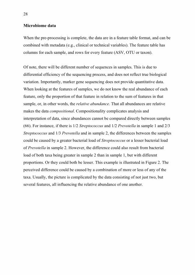
Microbiome data
When the pre-processing is complete, the data are in a feature table format, and can be
combined with metadata (e.g., clinical or technical variables). The feature table has
columns for each sample, and rows for every feature (ASV, OTU or taxon).
Of note, there will be different number of sequences in samples. This is due to
differential efficiency of the sequencing process, and does not reflect true biological
variation. Importantly, marker gene sequencing does not provide quantitative data.
When looking at the features of samples, we do not know the real abundance of each
feature, only the proportion of that feature in relation to the sum of features in that
sample, or, in other words, the relative abundance. That all abundances are relative
makes the data compositional. Compositionality complicates analysis and
interpretation of data, since abundances cannot be compared directly between samples
(66). For instance, if there is 1/2 Streptococcus and 1/2 Prevotella in sample 1 and 2/3
Streptococcus and 1/3 Prevotella and in sample 2, the differences between the samples
could be caused by a greater bacterial load of Streptococcus or a lesser bacterial load
of Prevotella in sample 2. However, the difference could also result from bacterial
load of both taxa being greater in sample 2 than in sample 1, but with different
proportions. Or they could both be lesser. This example is illustrated in Figure 2. The
perceived difference could be caused by a combination of more or less of any of the
taxa. Usually, the picture is complicated by the data consisting of not just two, but
several features, all influencing the relative abundance of one another.

29
Figure 1. Illustration of four scenarios of absolute abundances that correspond to
the relative abundances of Streptococcus and Prevotella in samples 1 and 2, with
explanations.
Another common phenomenon of microbiome data is zero-inflation, or sparsity. This
refers to the very large number of zeros in the feature table, which could be caused by
both under-sampling and true biological differences. Both compositionality and
sparsity pose challenges, disallowing use of classical statistical tests and complicates
interpretation of microbiome analysis (67).
Microbiome analysis
Analysis and presentation of microbiome data often encompass descriptions of
taxonomy, results from diversity analyses and differential abundance testing. The
taxonomy gives information on the names of the specific organisms that have been
identified. Diversity analysis can provide information about how rich or diverse each
sample is (within-sample-diversity, alpha diversity) or about how different samples are
from one another (between-sample-diversity, beta diversity). There are several
different ways of measuring alpha and beta diversity (diversity metrices) (68). The
1 2
1 2 1 21 2 1 2
2-fold increase ofStreptococcus
Prevotella is 50 % reduced
Both genera increase, butStreptococcus increases more
Both genera decrease, butPrevotella decreases more
Streptococcus
Prevotella
Relative abundances
Potential corresponding absolute abundances

30
diversity metrices used in this thesis are presented in the methods chapter. Differential
abundance tests (DA-tests) are statistical tests developed for identification of features
that significantly differ in abundance (or is otherwise differentially expressed) between
groups of interest (69). Due to the challenges related to compositionality and sparsity,
standard statistical tests are not applicable (67). The DA-tests used in this thesis are
described in the methods chapter.
The airway microbiome
The lower airway microbiome has not been as thoroughly studied as other human
microbiomes such as those of the gut, mouth, vagina and skin. This is, in part, due to
the lungs being less accessible for sampling, but likely also because they, for a long
time, were not considered a site of particular interest. The healthy lungs have
historically been considered to be free from bacteria. The notion of lung sterility was
expressed in a paper in New England Journal of Medicine as late as in 2008 (70), and
in lectures for medical students at University of Bergen as late as in 2018 (personal
experience).
In 2013, at the time when data collection for the MicroCOPD study started, it had been
recognised that even healthy lungs do contain microorganisms (71), that the
microbiota of the lungs is different from oral microbiota, and that microbiota could
show regional differences within the lung (72). In studies of healthy bacterial
communities (71-73), the most common consistently observed phyla were
Bacteroidetes, Firmicutes and Proteobacteria, and dominant genera included
Prevotella, Veillonella, Streptococcus and Pseudomonas. However, the same
taxonomic groups were seen as dominant in COPD and other disease states (71, 72,
74-76), and there seemed to be a focus on reporting alterations in the relative
abundances of bacteria. In one study, Proteobacteria was found to be increased in
asthma and COPD compared to healthy controls (71). Fungal microbiota was found to
differ between health and disease states, but studies were few (77, 78). The literature
was inconclusive with regard to diversity analysis in diseased and healthy airways. In

31
COPD, the diversity was reported to be increased (74), decreased (72) and equal to
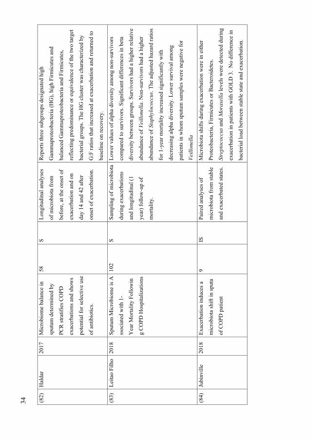
that of healthy individuals (75). An overview of the literature on the airway microbiota
of COPD in relation to exacerbations prior to the publication of paper III is presented
in Table 3.

32
Tab
le 3
: L
iter
atu
re t
ab
le d
epic
tin
g p
ap
ers
on
air
way m
icro
bio
ta o
f C
OP
D i
n r
ela
tion
to e
xa
cerb
ati
on
s p
ub
lish
ed p
rior
to t
he
pu
bli
cati
on
of
pap
er I
II.
Ref
1st
au
thor
Year
Tit
le
Nu
mb
er
of
CO
PD
pati
ents
Sam
ple
typ
e
Desi
gn
an
d o
utc
om
e M
ain
resu
lts
(79)
Huan
g
2014
Air
way
mic
robio
me
dynam
ics
in
exac
erbat
ions
of
chro
nic
obst
ruct
ive
pulm
onar
y
dis
ease
12
SS
In
ves
tigat
ed t
he
dynam
ics
of
the
airw
ay
bac
teri
al m
icro
bio
me
bef
ore
, at
the
onse
t of,
and a
fter
an
exac
erbat
ion.
Shif
ts i
n t
he
abundan
ce (
≥2
-fold
) of
man
y t
axa
at
exac
erbat
ion a
nd a
fter
tre
atm
ent.
Mic
robio
ta m
ember
s
that
wer
e in
crea
sed a
t ex
acer
bat
ion w
ere
pri
mar
ily o
f th
e
Pro
teobac
teri
a phylu
m, in
cludin
g n
onty
pic
al C
OP
D
pat
hogen
s. C
han
ges
in t
he
bac
teri
al c
om
posi
tion a
fter
trea
tmen
t fo
r an
exac
erbat
ion d
iffe
red s
ignif
ican
tly
among p
resc
ribed
ther
apy r
egim
ens.
Tre
atm
ent
wit
h
anti
bio
tics
alo
ne
pri
mar
ily d
ecre
ased
the
abundan
ce o
f
Pro
teobac
teri
a, w
ith t
he
pro
longed
suppre
ssio
n o
f so
me
mic
robio
ta m
ember
s bei
ng o
bse
rved
. In
contr
ast,
trea
tmen
t w
ith c
ort
icost
eroid
s al
one
led t
o e
nri
chm
ent
for
Pro
teobac
teri
a an
d m
ember
s of
oth
er p
hyla
.

33
(80)
Mil
lare
s 2015
Funct
ional
Met
agen
om
ics
of
the
Bro
nch
ial
Mic
robio
me
in
CO
PD
8
S
Mic
robio
ta a
nal
ysi
s in
CO
PD
pat
ients
wit
h
FE
V1<
50%
pre
d >
= 3
exac
erbat
ions
in t
he
pre
vio
us
yea
r in
stab
ilit
y a
nd
exac
erbat
ion s
tate
s to
iden
tify
the
funct
ional
chan
ges
in b
ronch
ial
mic
robio
ta.
Str
epto
cocc
us
and H
aem
ophil
us
most
abundan
t (>
50%
abundan
ce t
og
ether
). N
o c
han
ges
in c
om
posi
tion a
t
phylu
m o
r gen
us
level
fro
m s
table
to e
xac
erbat
ion. N
o
chan
ges
in a
lpha
or
bet
a div
ersi
ty.
(81)
Wan
g
2016
Lung m
icro
bio
me
dynam
ics
in C
OP
D
exac
erbat
ions.
87
S
Longit
udin
al a
nal
yse
s
of
mic
robio
ta f
rom
bef
ore
, duri
ng a
nd 6
wee
ks
afte
r
exac
erbat
ion.
Chan
ges
in m
icro
bio
ta a
ppea
red t
o b
e as
soci
ated
wit
h
exac
erbat
ion e
ven
ts a
nd i
ndic
ativ
e of
spec
ific
exac
erbat
ion p
hen
oty
pes
. A
nti
bio
tic
and s
tero
id
trea
tmen
ts a
ppea
r to
hav
e dif
fere
nti
al e
ffec
ts o
n t
he
lung
mic
robio
me.
In p
arti
cula
r H
aem
ophil
us
spp. im
pac
t th
e
over
all
mic
robia
l co
mm
unit
y s
truct
ure
. S
erum
and
sputu
m b
iom
arker
s co
rrel
ated
wit
h t
he
stru
cture
and
div
ersi
ty o
f th
e m
icro
bio
me.
34
(82)
Hal
dar
2017
Mic
robio
me
bal
ance
in
sputu
m d
eter
min
ed b
y
PC
R s
trat
ifie
s C
OP
D
exac
erbat
ions
and s
how
s
pote
nti
al f
or
sele
ctiv
e use
of
anti
bio
tics
.
58
S
Longit
udin
al a
nal
yse
s
of
mic
robio
ta f
rom
bef
ore
, at
the
onse
t of
exac
erbat
ion a
nd o
n
day
14 a
nd 4
2 a
fter
onse
t of
exac
erbat
ion.
Rep
ort
s th
ree
subgro
ups
des
ignat
ed h
igh
Gam
map
rote
obac
teri
a (H
G),
hig
h F
irm
icute
s an
d
bal
ance
d G
amm
apro
teo
bac
teri
a an
d F
irm
icute
s,
refl
ecti
ng p
redom
inan
ce o
r eq
uiv
alen
ce o
f th
e tw
o t
arget
bac
teri
al g
roups.
The
HG
clu
ster
was
char
acte
rize
d b
y
G:F
rat
ios
that
incr
ease
d a
t ex
acer
bat
ion a
nd r
eturn
ed t
o
bas
elin
e on r
ecover
y.
(83)
Leit
ao F
ilho
2018
Sputu
m M
icro
bio
me i
s A
ssoci
ated
wit
h 1
-
Yea
r M
ort
alit
y F
oll
ow
in
g C
OP
D H
osp
ital
izat
ions
102
S
Sam
pli
ng o
f m
icro
bio
ta
duri
ng e
xac
erbat
ions
and l
ongit
udin
al (
1
yea
r) f
oll
ow
-up o
f
mort
alit
y.
Low
er v
alues
of
alpha
div
ersi
ty a
mong n
on
-surv
ivors
com
par
ed t
o s
urv
ivors
. S
ignif
ican
t dif
fere
nce
s in
bet
a
div
ersi
ty b
etw
een g
roups.
Surv
ivors
had
a h
igher
rel
ativ
e
abundan
ce o
f V
eill
onel
la. N
on-s
urv
ivors
had
a h
igher
abundan
ce o
f Sta
phyl
oco
ccus.
The a
dju
sted
haz
ard r
atio
s
for
1-y
ear
mort
alit
y i
ncr
ease
d s
ignif
ican
tly w
ith
dec
reas
ing a
lpha
div
ersi
ty. L
ow
er s
urv
ival
am
ong
pat
ients
in w
hom
sputu
m s
ample
s w
ere
neg
ativ
e fo
r
Veil
lonel
la
(84)
Jubin
vil
le
2018
Exace
rbat
ion i
nduce
s a
mic
robio
ta s
hif
t in
sputa
of
CO
PD
pat
ient
9
IS
Pai
red a
nal
yse
s of
mic
robio
ta f
rom
sta
ble
and e
xac
erbat
ed s
tate
s.
Mic
robio
ta s
hif
ts d
uri
ng e
xac
erbat
ion w
ere
in e
ither
Pro
teobac
teri
a, F
irm
icute
s or
Bac
tero
idet
es.
Str
epto
cocc
us
and M
ora
xell
a l
evel
s w
ere
det
ecte
d d
uri
ng
exac
erbat
ion i
n p
atie
nts
wit
h G
OL
D 3
. N
o d
iffe
rence
in
bac
teri
al l
oad
bet
wee
n s
table
sta
te a
nd e
xac
erbat
ion.

35
(85)
May
hew
2018
Longit
udin
al p
rofi
ling o
f
the
lung m
icro
bio
me
in
the
AE
RIS
stu
dy
dem
onst
rate
s
repea
tabil
ity o
f bac
teri
al
and e
osi
nophil
ic C
OP
D
exac
erbat
ions.
101
S
Pai
red a
nal
yse
s of
mic
robio
ta f
rom
sta
ble
and e
xac
erbat
ed s
tate
s.
Inves
tigat
ion o
f
dif
fere
nt
exac
erbat
ion
phen
oty
pes
(bac
teri
al,
vir
al, eo
sinophil
ic).
Sta
bil
ity o
f m
icro
bio
me
over
tim
e w
as m
ore
lik
ely t
o b
e
dec
reas
ed i
n e
xac
erbat
ions
and w
ithin
indiv
idual
s w
ith
hig
her
exac
erbat
ion f
requen
cies
. B
acte
rial
and
eosi
nophil
ic e
xac
erbat
ions
were
more
lik
ely t
o b
e
repea
ted i
n s
ubse
quen
t ex
acer
bat
ions
wit
hin
a s
ubje
ct,
wher
eas
vir
al e
xac
erbat
ions
were
not
more
lik
ely t
o b
e
repea
ted. A
ssoci
atio
n o
f bac
teri
al g
ener
a,
incl
udin
g H
aem
ophil
us
and
Mora
xell
a, w
ith d
isea
se
sever
ity, ex
acer
bat
ion e
ven
ts a
nd b
ronch
iect
asis
.

36
(86)
Wan
g
2018
Sputu
m m
icro
bio
me
tem
pora
l var
iabil
ity a
nd
dysb
iosi
s in
chro
nic
obst
ruct
ive
pulm
onar
y
dis
ease
exac
erbat
ions:
an
anal
ysi
s of
the
CO
PD
MA
P s
tudy
281
S
Longit
udin
al c
han
ges
in
the
lung m
icro
bio
me
and t
hei
r re
lati
onsh
ip
wit
h a
ssoci
ated
CO
PD
outc
om
es (
phen
oty
pes
of
exac
erbat
ions)
wit
h
up t
o 2
-yea
r fo
llow
-up
The m
icro
bio
me
com
posi
tion w
as s
imil
ar a
mong c
entr
es
and b
etw
een s
table
and e
xac
erbat
ions
exce
pt
for
a sm
all
signif
ican
t dec
reas
e of
Vei
llonel
la a
t ex
acer
bat
ions.
The
abundan
ce o
f M
ora
xell
a w
as n
egat
ivel
y a
sso
ciat
ed w
ith
bac
teri
al a
lpha
div
ersi
ty. M
icro
bio
mes
wer
e dis
tinct
bet
wee
n e
xac
erbat
ions
asso
ciat
ed w
ith b
acte
ria
ver
sus
eosi
nophil
ic a
irw
ay i
nfl
amm
atio
n. D
ysb
iosi
s at
exac
erbat
ions,
mea
sure
d a
s si
gnif
ican
t w
ithin
subje
ct
dev
iati
on o
f m
icro
bia
l co
mposi
tion r
elat
ive
to b
asel
ine,
was
pre
sent
in 4
1%
of
exac
erbat
ions.
Dysb
iosi
s w
as
asso
ciat
ed w
ith i
ncr
ease
d e
xac
erbat
ion s
ever
ity i
ndic
ated
by a
gre
ater
fal
l in
forc
ed e
xpir
atory
volu
me
in o
ne
seco
nd, fo
rced
vit
al c
apac
ity a
nd a
gre
ater
incr
ease
in
CA
T s
core
, par
ticula
rly i
n e
xac
erbat
ions
wit
h c
oncu
rren
t
eosi
nophil
ic i
nfl
amm
atio
n. T
here
was
a s
ignif
ican
t
dif
fere
nce
of
tem
pora
l var
iabil
ity o
f m
icro
bia
l al
pha
and
bet
a div
ersi
ty a
mong c
entr
es. T
he
var
iati
on o
f bet
a
div
ersi
ty s
ignif
ican
tly d
ecre
ased
in t
hose
subje
cts
wit
h
freq
uen
t his
tori
cal
exac
erbat
ions.

37
(87)
Dic
ker
2018
Gen
etic
man
nose
bin
din
g
lect
in (
MB
L)
def
icie
ncy
is a
ssoci
ated
wit
h a
irw
ay
mic
robio
ta d
iver
sity
and
reduce
d e
xac
erbat
ion
freq
uen
cy i
n C
OP
D.
141
SS
C
om
par
es m
icro
bio
ta i
n
stab
le C
OP
D p
atie
nts
wit
h a
nd w
itout
gen
etic
MB
L d
efi
cien
cy
Pat
ients
wit
h M
BL
defi
cien
cy w
ere
signif
ican
tly l
ess
likel
y t
o h
ave
sever
e ex
acer
bat
ions.
MB
L d
efi
cien
cy d
id
not
affe
ct r
ate
of
FE
V1 d
ecli
ne
or
mort
alit
y. P
atie
nts
wit
h
MB
L d
efic
iency
had
a m
ore
div
erse
lung m
icro
bio
ta a
nd
wer
e le
ss l
ikel
y t
o b
e co
lonis
ed w
ith H
aem
ophil
us
spp.
There
wer
e lo
wer
lev
els
of
airw
ay i
nfl
amm
atio
n i
n
pat
ients
wit
h M
BL
defi
cien
cy.
(88)
Ren
2018
Tra
nsc
ripti
onal
ly A
ctiv
e
Lung M
icro
bio
me
and I
ts
Ass
oci
atio
n w
ith
Bac
teri
al B
iom
ass
and
Host
Infl
amm
atory
Sta
tus.
25
BA
L
Met
atra
nsc
ripto
me
and
mic
robio
ta a
nal
ysi
s in
pat
ients
under
goin
g
bro
nch
osc
opy a
s par
t of
clin
ical
man
agem
ent.
Ret
rosp
ecti
ve
foll
ow
-
up o
f ex
acer
bat
ions
four
yea
rs l
ater
.
Obta
ined
the
num
ber
of
exac
erbat
ions
for
21 i
ndiv
idual
s,
in t
ota
l 29 e
xac
erbat
ions.
Sugges
t th
at S
trep
toco
ccus
and
Roth
ia m
ight
be
pro
tect
ive
agai
nst
exac
erbat
ions,
and
Pse
udom
onas
mig
ht
be
har
mfu
l.
(36)
Pra
gm
an
2019
Chro
nic
obst
ruct
ive
pulm
onar
y d
isea
se u
pper
airw
ay m
icro
bio
ta a
lpha
div
ersi
ty i
s as
soci
ated
wit
h e
xac
erbat
ion
phen
oty
pe:
a c
ase-
contr
ol
obse
rvat
ional
stu
dy.
22
IS
Cro
ss-s
ecti
onal
cas
e
contr
ol
study o
f
mic
robio
ta i
n f
requen
t
and i
nfr
equen
t
exac
erbat
ors
(sa
mpli
ng
in s
table
sta
te).
Fre
quen
t ex
acer
bat
ors
had
low
er a
lpha
div
ersi
ty. G
roups
only
shar
ed 3
3/1
69 A
SV
s bet
wee
n t
hem
. C
opy n
um
ber
not
asso
ciat
ed w
ith p
hen
oty
pe.

38
(89)
Wan
g
2019
Air
way
host
-mic
robio
me
inte
ract
ions
in c
hro
nic
obst
ruct
ive
pulm
onar
y
dis
ease
43
S
Inves
tigat
ed
mic
robio
me,
tran
scri
pto
me
and
pro
teom
e in
sta
ble
and
exac
erbat
ed C
OP
D
(longit
udin
al a
nal
ysi
s).
Rep
ort
s 8
.6 %
Str
epto
cocc
us.
More
Mora
xell
a a
nd l
ow
er
alpha
div
ersi
ty i
n e
xac
erbat
ion s
ample
s, a
tre
nd w
hic
h
was
als
o r
ever
sed a
t post
-sam
ple
s, b
ut
this
was
not
signif
ican
t in
pai
red a
nal
yse
s. N
o s
ignif
ican
t in
crea
se i
n
bac
teri
al l
oad
(qP
CR
). I
n t
ransc
ripto
me
anal
yse
s,
inte
rleu
kin
-6 a
nd i
nte
rfer
on s
ignal
ing p
athw
ays
wer
e
upre
gula
ted i
n e
xac
erbat
ion. N
o d
iffe
rence
s in
pro
teom
e
wer
e se
en b
etw
een s
table
and e
xac
erbat
ed s
ample
s.
Haem
ophil
us
and M
ora
xell
a w
ere
most
ass
oci
ated
wit
h
imm
une
resp
onse
. M
egasp
her
a w
as a
ssoci
ated
wit
h
reduce
d e
xpre
ssio
n o
f host
infl
amm
atory
pat
hw
ays.
(35)
Mil
lare
s 2019
Rel
atio
nsh
ip b
etw
een t
he
resp
irat
ory
mic
robio
me
and t
he
sever
ity o
f
airf
low
lim
itat
ion, his
tory
of
exac
erbat
ions
and
circ
ula
ting e
osi
nophil
s in
CO
PD
pat
ients
72
SS
C
om
par
es m
icro
bio
ta
pro
file
s in
sta
ble
sta
te
and s
tudie
d r
elat
ion t
o
airf
low
lim
itat
ion,
pre
vio
us
exac
erbat
ions
and b
lood e
osi
nophil
ia.
Abundan
ce o
f Str
epto
cocc
us
1.9
2 %
. P
seudom
onas
incr
ease
d a
nd T
reponem
a d
ecre
ased
wit
h i
ncr
easi
ng
airf
low
lim
itat
ion. T
M7 l
ow
er i
n t
hose
wit
h 1
exac
erbat
ion p
revio
us
yea
r. T
M7 a
nd S
pir
och
etes
reduce
d i
n f
requen
t ex
acer
bat
ors
. In
crea
sed a
bundan
ces
of
Pse
udom
onas,
Sel
enom
onas,
Anaer
oco
ccus
in f
requen
t
exac
erbat
ors
.

39
(90)
Tanged
al
2019
Sputu
m m
icro
bio
ta a
nd
infl
amm
atio
n a
t st
able
stat
e an
d d
uri
ng
exac
erbat
ions
in a
cohort
of
chro
nic
obst
ruct
ive
pulm
onar
y d
isea
se
(CO
PD
) pat
ients
.
36
IS
Pai
red a
nal
yse
s of
mic
robio
ta f
rom
sta
ble
and e
xac
erbat
ed s
tate
s,
in a
ddit
ion t
o a
cas
e
pre
senta
tion w
ith m
ore
even
ts.
Level
s of
IP-1
0, M
IG, T
NF
-alp
ha a
nd A
MP
s dif
fere
d
bet
wee
n s
table
and e
xac
erbat
ion s
tate
. O
f 36 s
ample
pai
rs, 24 h
ad s
ignif
ican
t dif
fere
nce
s in
the
most
abundan
t
gen
era
in p
aire
d a
nal
yse
s, b
ut
no d
iffe
renti
ally
abundan
t
feat
ure
s w
hen tes
ted w
ith A
LD
Ex2. D
iver
sity
was
signif
ican
tly d
iffe
rent
in s
ever
al i
ndiv
idual
s, b
ut
not
when
dat
a w
as a
nal
yse
d o
n a
gro
up l
evel
. T
he p
atie
nt
case
stu
dy s
how
ed l
ongit
udin
al d
ynam
ics
in m
icro
bio
ta
unre
late
d t
o d
isea
se s
tate
.
(91)
Lòpez
Car
o
2019
Sputu
m M
icro
bio
me
Dynam
ics
in C
hro
nic
Obst
ruct
ive
Pulm
onar
y
Dis
ease
Pat
ients
duri
ng
an E
xac
erbat
ion E
vent
and P
ost
-Sta
bil
izat
ion
55
S
Pai
red m
icro
bio
ta
anal
ysi
s of
exac
erbat
ion
even
t and p
ost
-
stab
iliz
atio
n
The s
tudy f
ound a
sta
ble
mic
robio
me
com
posi
tion i
n t
he
post
-sta
bil
izat
ion s
ample
s an
d 4
addit
ional
mic
robio
mes
in s
ample
s ta
ken
duri
ng e
xac
erbat
ion, 3 o
f w
hic
h s
how
ed
a m
arked
dysb
iosi
s by H
aem
ophil
us,
Pse
udom
onas,
and
Ser
rati
a. T
he f
ourt
h e
xac
erbat
ion m
icro
bio
me
had
a v
ery
sim
ilar
com
posi
tion t
o p
ost
-sta
bil
izat
ion s
ample
s, b
ut
som
e pat
hogen
s su
ch a
s M
ora
xell
a a
nd r
espir
atory
vir
use
s w
ere
als
o f
ound.
Abbre
via
tions:
SS
; S
ponta
neo
us
sputu
m, IS
; In
duce
d s
putu
m;
S:
Sputu
m (
type
not
def
ined
, or
mix
ed),
HG
; H
igh G
amm
apro
teobac
teri
a, G
:F;
Gam
mab
acte
ria:
Fir
mic
ute
s, M
BL
; m
annose
bin
din
g l
ecti
n,
IP
-10;
Inte
rfer
on g
amm
a-i
nduce
d p
rote
in 1
0, M
IG;
monokin
e i
nduce
d b
y g
amm
a in
terf
eron
,
TN
F-a
lpha;
Tum
our
Nec
rosi
s F
acto
r-al
pha,
AM
Ps;
Anti
mic
robia
l pep
tides
.
40
Studies of the airway microbiome have presented with inconsistent findings, likely due
to different methods and small sample sizes. This, and a lack of longitudinal studies,
has left a series of questions unanswered. For instance: Beyond general knowledge
about which high-level taxonomic groups of bacteria that are dominant, what is to be
considered a normal/healthy bacterial airway microbiome? Is the composition of
microbes stable over time? (How) is the microbiome altered in respiratory diseases
such as COPD and asthma? What characterises fungal and viral communities in
healthy and diseased airways? And (how) does the airway microbiota relate to specific
aspects of disease or disease progression, such as frequency of COPD exacerbations?
These questions, among others, are central to the MicroCOPD study, which set out to
be the largest one-centre study of the airway microbiome (92). Studying lower airway
microbiota is, however, challenging. The main challenges are related to samples being
low-biomass. Since the total burden of microorganisms is low, the samples are prone
to contamination of bacteria, fungi or viruses from other sources. Presence of
contaminants can cause serious errors in analysis and interpretation of data, and can be
difficult to prevent, identify and account for. Contamination can stem from the
laboratory processing of samples, for instance from the sample medium, the reagents
in use, the researchers or research environment, or from other samples (cross-
contamination) (93). Another contamination source is that of the upper airways.
Sputum collection is a common airway sampling method both in microbiota research
and in clinical practice. Sputum is mucus from the lower airways (spontaneously
occurring, or induced for the purpose of sampling) that is coughed up. The sputum
sample passes through the respiratory tract and is delivered via the high biomass oral
cavity (94). Although useful for detecting pathogens in a clinical setting, sputum
unlikely provides researchers with a representative picture of lung microbiota due to
contamination of oral microbes. It is therefore recommended to sample the lungs
directly (95). This can be done with bronchoscopy.

41
Bronchoscopy
History
In 1897, German otorhinolaryngologist Gustav Killian (1860-1921) used an
oesophagoscope to remove a foreign body (an inhaled pork bone) from the right main
bronchus of a patient. The scope used was a simple rigid tube with an 8 mm lumen, a
proximal (external) light source, and a laryngoscope handle (96). The following year,
after having acquired some additional experience, he presented the method as “direct
bronchoscopy” (97) and has since been credited as the “father of bronchoscopy” (98).
In the years prior, Przemyslaw Pieniazek had safely performed investigations in and
removed objects from the lower trachea via tracheostomies. Rosenheim and Kirstein
had, by accident and intent respectively, passed the oesophagoscope into the trachea.
Kirstein did not dare to proceed below the level of carina. Killian became widely
known for bronchoscopic foreign body removal, and patients were referred from all
over Europe, and even from as far as Uruguay (99). His invention was made possible
by an important discovery in the former decade; the topical anaesthetic properties of
cocaine. Concurrent advances in medicine and technology contributed to the method
development. Within few years, Killian performed bronchoscopy on a number of
indications, including drainage of pulmonary abscesses, stenting of airways and
endoluminal brachyradiotherapy (100). Bronchoscopy soon became an established
medical procedure, mainly for therapeutic indications. The first textbook on
bronchoscopy, written by Chevalier Jackson in 1907, presents a mortality rate of 9.6%
and still states “To-day when endoscopy has reached such a high degree of perfection
(…)” (98). Despite its perceived perfection, the field of bronchoscopy continued to
progress. Alongside innovations in other endoscopic fields, new features were added
and continuously improved, including light sources, suction tubes, camera
technologies and different surgical instruments (100). In the 1960s, Shigeto Ikeda
developed and commercialised a thin flexible bronchoscope with glass fibre
illumination (101). His invention would revolutionise bronchoscopy. In contrast to the
rigid scope which limits investigations to proximal parts of the bronchial tree, Ikedas
42
bronchoscope has a mechanism that flexes or extends the distal end, which facilitates
insertion through arched airways. Although Killian had performed his first rigid
bronchoscopies with topical anaesthesia only(!) (97), sedation or general anaesthesia
was usually necessary (98). In the beginning, Ikeda’s bronchoscope had to be inserted
through an endotracheal tube or used in combination with a rigid scope (101). As
improvements was made to the instrument, bronchoscopy was soon to be less invasive,
and could be performed without sedation. It was also possible to insert the new
bronchoscope via the nose (102). Modern flexible bronchoscopes come with advanced
video and illumination technology and an increasing number of procedures can be
performed using specialised equipment through the working channel. Although rigid
bronchoscopy still has a number of indications, the flexible scope can be applied for
most procedures (103). Flexible bronchoscopy is described in more detail below, and
is hereafter referred to simply as bronchoscopy.
Indications
Bronchoscopy can be applied in most situations where access to the bronchi for
visualisation, sampling or direct treatment is desired, and hence has a wide range of
indications in diagnostics, therapy and research. Diagnostic indications include
assessment of, for example, pneumonia, pulmonary infiltrates, haemoptysis, persistent
cough, stridor, (potentially) malignant tissue, inhalation and burn injuries, fistulae,
tracheobronchomalacia, lung transplants and foreign bodies. Therapeutic indications
include for instance mucus suction, foreign body removal, endotracheal tube
placement in difficult airways, tumour debulking and removal, abscess drainage,
asthma refractory to medical treatment (bronchial thermoplasty), and insertion of
valves, coils and stents, for instance in treating emphysema (104).
Bronchoscopy, with its diagnostic and therapeutic applications, is continuously being
subject to research. However, bronchoscopy can also be performed for pure research
purposes where obtaining material from the lungs is desired (see A note on research
bronchoscopy). Sampling techniques commonly used in diagnostic work-ups and
research are described below.

43
Sampling techniques
Brush sampling
Protected specimen brushes (PSB), also referred to as endobronchial brushes, is a
technique where specimens can be obtained using a sterile, single-use brush enclosed
within a catheter sheath and protected (until advanced) by a resorbable wax-plug. The
tip of the bronchoscope is placed in the desired area before the catheter sheath (with
the brush inside) is advanced through the working channel. The brush can be pushed
out of the sheath and rubbed against the bronchial wall, in which small parts of the
endothelium will be released onto the brush. Brushes come in different lengths, widths
and with different stiffness. The catheter sheath prevents contamination of the brush
and protects the working channel of the bronchoscope from damage by the brush
(105).
Bronchoalveolar lavage
Bronchoalveolar lavage (BAL) is a procedure in which a fluid (saline) is installed into
a subsegment of the lung (with the bronchoscope in a wedged position, preventing
fluid from escaping the segment), and thereafter recollected by suction into a sterile
trap, or aspiration into a sterile syringe. Since the installed fluid is quickly distributed
throughout the chosen segment, beyond the reach of the tip of the bronchoscope, BAL
enables sampling that includes contents from the alveolar milieu in addition to that of
the bronchial lumen. The amount of installed fluid is not standardised, but a total of
100-300 ml in aliquots of 20-60 ml is common (106, 107). Bronchoalveolar lavages of
with smaller volumes can be referred to as small volume lavage (SVL). BAL sampling
can be performed with different techniques. A method using a sterile catheter inside
the working channel of the scope, preventing contamination of the sample, can be
referred to as protected BAL (pBAL) (95). In this thesis, protected BAL is simply
referred to as BAL.
44
Bronchial wash
Washing the bronchi with smaller volumes of saline can also be performed without the
wedging of the scope and with smaller volumes compared to BAL (107).
Biopsy
Bronchoscopy can be used to obtain both endobronchial biopsies (EBB) and
transbronchial biopsies (TBB) (also referred to as transbronchial lung biopsies
(TBLB)). EBB can be taken using forceps in the working channel under direct
visualisation of the bronchoscopist, are useful in diagnosing endobronchial lesions or
in research and are associated with less bleeding compared to TBB (108). TBB are
useful for sampling the lung parenchyma, and can be done blindly or guided by
fluoroscopy, ultrasound or other navigation technologies (105).
Sedation and anaesthesia
The British Thoracic Society guideline for diagnostic flexible bronchoscopy in adults
(BTS guideline) recommends that patients undergoing bronchoscopy should be offered
intravenous sedation, preferably with the benzodiazepine midazolam, titrated to a level
in which verbal contact is possible at all times (109). This light level of sedation is
commonly referred to as “conscious sedation”, although anaesthesiologist argue
against the term since consciousness usually is altered (110). Other sedatives
commonly used include Propofol (should be administered by anaesthetist) and,
recommended in combination with midazolam; the short-acting opioids fentanyl and
alfentanil (109). At Haukeland University Hospital, light sedation with alfentanil alone
is routinely used for bronchoscopy, and additional midazolam is given to anxious
patients.
Unless contraindicated, lidocaine is used for topical anaesthesia of the upper and lower
airways. Lidocaine gel is recommended for anaesthesia of nostrils/nasopharynx (in the
case of nasal insertion) and lidocaine spray for oropharynx prior to scope insertion.
Lidocaine solution can be administered to the larynx and trachea through a

45
cricothyroid puncture, or directly through the bronchoscope (spray-as-you-go
technique). It is recommended to note the total dosage of lidocaine given (109).
Contraindications
The BTS guideline states only active myocardial ischemia as an absolute
contraindication for bronchoscopy, in addition to situations where sedation and
anaesthesia are contraindicated (109). Specific procedures performed under
bronchoscopy have their own contraindications. For research bronchoscopies, more
cautious contraindications may be in place (see Methods).
Patient monitoring
The BTS guideline recommends to record the heart rate, respiratory rate, blood
pressure and oxygen saturation of patients undergoing bronchoscopy, before,
repeatedly during and after the procedure. In addition, it is recommended that
bronchoscopy units undertake periodic audit of the efficacy, safety and patient
satisfaction (109).
A note on research bronchoscopy
Bronchoscopy can be applied as a research tool in various settings. Most often,
sampling for a research purpose is conducted in patients who are already undergoing a
bronchoscopy for a clinical indication (111). When this is the case, it has been argued
that research bronchoscopy would be acceptable even in children (112).
Bronchoscopies are also performed for scientific reasons only. For instance,
bronchoscopy has been used to study lung microbiota in health and disease (71, 72,
113, 114), and drug or pollution effects on respiratory cells (115, 116). This use of
bronchoscopy might be more controversial, due to the potential harm for research
participants, as demonstrated in the following example: In 1996, 19-year-old Hoiyan

46
Wan participated as a healthy volunteer in a study on lung cell function at the
University of Rochester. As part of the study, she underwent bronchoscopy. To ease
her discomfort, considerable amounts of lidocaine was administered. Despite
complaints of chest discomfort, she was discharged after the one-hour observation
period. At home she quickly deteriorated. Upon arrival in the emergency department
about three hours after bronchoscopy, she was in cardiac arrest. She died two days
later. Her blood levels of lidocaine suggested that she had received more than twice
the maximum safe dosage (117, 118).
Safety of participants in clinical research
The Helsinki declaration
Throughout the history of science, potentially harmful (and undoubtedly dangerous)
experiments have been conducted on humans for the sake of new knowledge and
progress in research, many of which we today would consider highly unethical (119).
In 1964, The World Medical Association developed the Declaration of Helsinki, a
statement of ethical principles for medical research involving human subjects. Its main
purpose is to protect the rights of individuals taking part in research. The document
has been revised eight times (last update in 2013), and is universally regarded an
ethical norm for everyone who conducts medical research. Today, the Declaration of
Helsinki includes sections concerning the obligation of informed consent, the balance
between risks and benefits, the requirement for research ethics committees and
research protocols, considerations of research in vulnerable persons, the use of placebo
and more. Regarding risks, the declaration states:
“16. In medical practice and in medical research, most interventions involve risks and
burdens.
Medical research involving human subjects may only be conducted if the importance
of the objective outweighs the risks and burdens to the research subjects.
47
17. All medical research involving human subjects must be preceded by careful
assessment of predictable risks and burdens to the individuals and groups involved in
the research in comparison with foreseeable benefits to them and to other individuals
or groups affected by the condition under investigation.
Measures to minimise the risks must be implemented. The risks must be continuously
monitored, assessed and documented by the researcher.
18. Physicians may not be involved in a research study involving human subjects
unless they are confident that the risks have been adequately assessed and can be
satisfactorily managed.
When the risks are found to outweigh the potential benefits or when there is
conclusive proof of definitive outcomes, physicians must assess whether to continue,
modify or immediately stop the study.” (120)
Monitoring, assessing and documenting risks in research
Although promoting the documentation of risks in research, the Helsinki declaration
does not state how this should be performed.
CONSORT – a guideline for randomised controlled trials
Consolidated Standards of Reporting Trials (CONSORT) is a guideline for reporting
and documenting methods and results from randomised controlled trials (RCTs). Many
publishers require that authors of RCT publications adhere to the CONSORT
statement and checklist. Harm is one of the items on the checklist (121). An extension
regarding harm has been published, with a list of recommendations and discussions of
the terms adverse events, serious adverse events, adverse (drug) reaction, harms,
active and passive surveillance of harms, safety, risk-benefit ratio, toxicity and side
effects. Not all of these terms are defined by CONSORT. Researchers are

48
recommended to define adverse events and preferably report events using field
specific scales and validated measures (122).
Adverse events assessment tools
There are a number of published tools for assessment of adverse events in research and
in clinical practice, see Table 4 for examples.
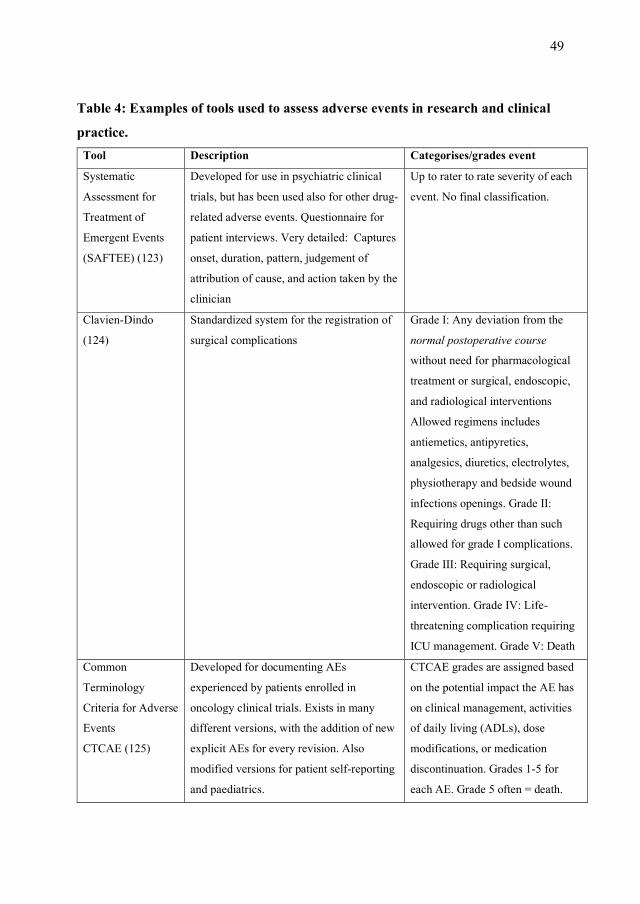
49
Table 4: Examples of tools used to assess adverse events in research and clinical
practice.
Tool Description Categorises/grades event
Systematic
Assessment for
Treatment of
Emergent Events
(SAFTEE) (123)
Developed for use in psychiatric clinical
trials, but has been used also for other drug-
related adverse events. Questionnaire for
patient interviews. Very detailed: Captures
onset, duration, pattern, judgement of
attribution of cause, and action taken by the
clinician
Up to rater to rate severity of each
event. No final classification.
Clavien-Dindo
(124)
Standardized system for the registration of
surgical complications
Grade I: Any deviation from the
normal postoperative course
without need for pharmacological
treatment or surgical, endoscopic,
and radiological interventions
Allowed regimens includes
antiemetics, antipyretics,
analgesics, diuretics, electrolytes,
physiotherapy and bedside wound
infections openings. Grade II:
Requiring drugs other than such
allowed for grade I complications.
Grade III: Requiring surgical,
endoscopic or radiological
intervention. Grade IV: Life-
threatening complication requiring
ICU management. Grade V: Death
Common
Terminology
Criteria for Adverse
Events
CTCAE (125)
Developed for documenting AEs
experienced by patients enrolled in
oncology clinical trials. Exists in many
different versions, with the addition of new
explicit AEs for every revision. Also
modified versions for patient self-reporting
and paediatrics.
CTCAE grades are assigned based
on the potential impact the AE has
on clinical management, activities
of daily living (ADLs), dose
modifications, or medication
discontinuation. Grades 1-5 for
each AE. Grade 5 often = death.
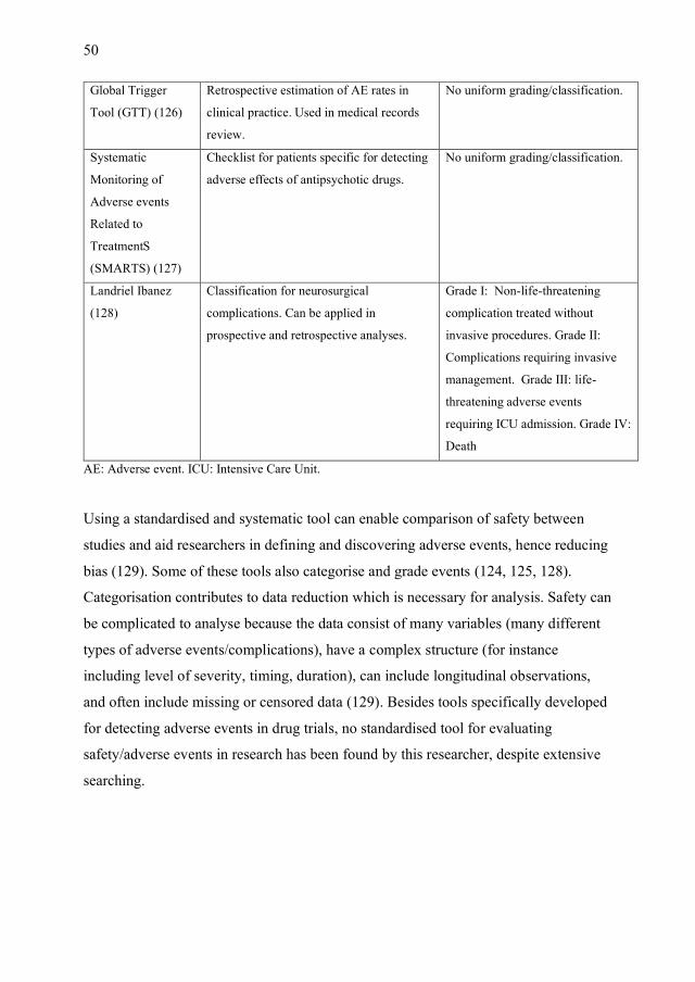
50
Global Trigger
Tool (GTT) (126)
Retrospective estimation of AE rates in
clinical practice. Used in medical records
review.
No uniform grading/classification.
Systematic
Monitoring of
Adverse events
Related to
TreatmentS
(SMARTS) (127)
Checklist for patients specific for detecting
adverse effects of antipsychotic drugs.
No uniform grading/classification.
Landriel Ibanez
(128)
Classification for neurosurgical
complications. Can be applied in
prospective and retrospective analyses.
Grade I: Non-life-threatening
complication treated without
invasive procedures. Grade II:
Complications requiring invasive
management. Grade III: life-
threatening adverse events
requiring ICU admission. Grade IV:
Death
AE: Adverse event. ICU: Intensive Care Unit.
Using a standardised and systematic tool can enable comparison of safety between
studies and aid researchers in defining and discovering adverse events, hence reducing
bias (129). Some of these tools also categorise and grade events (124, 125, 128).
Categorisation contributes to data reduction which is necessary for analysis. Safety can
be complicated to analyse because the data consist of many variables (many different
types of adverse events/complications), have a complex structure (for instance
including level of severity, timing, duration), can include longitudinal observations,
and often include missing or censored data (129). Besides tools specifically developed
for detecting adverse events in drug trials, no standardised tool for evaluating
safety/adverse events in research has been found by this researcher, despite extensive
searching.

51
Objectives
The objectives of this thesis were to:
Systematically review the literature to identify bronchoscopy-related complications
and discomfort, meaningful complication rates, and predictors. (Paper I)
Investigate if research bronchoscopy is less safe in subjects with than without
obstructive lung disease by evaluating complications and discomfort occurring
immediately, and within a week after bronchoscopy. (Paper II)
Investigate whether the compositionality of the lower airway microbiota in stable
COPD predicts later exacerbation risk in a cohort study. (Paper III)

52
Materials and Methods
Systematic literature review
A systematic literature review is a type of study design which uses standardised and
transparent methods for systematic searching, filtering, reviewing, evaluating and
reporting information from multiple studies in order to summarise and learn from the
entire available literature on the topic of interest (130).
The systematic search
For paper I, a modified population intervention comparison outcome (PICO) form was
created in order to include bronchoscopy, bronchoscopy related techniques and
associated procedures, and the outcomes of interest (paper I, Table 1). The search was
conducted in PubMed (Medline). Keywords were selected by combining existing
thesauruses (MeSH terms) and text words after examining the existing MeSH database
and the (MeSH) classification of a selection of known relevant papers. In addition, we
added text words describing complications known to the authors.
The final literature search for paper I was conducted on February 8th 2016. For the
discussion of this thesis, a new search was repeated on March 2nd 2021.
Search filtering
All available titles and abstracts from the search hits were read thoroughly.
Publications were excluded if they:
• Were not published in English, French or a Scandinavian language
• Described single case-reports or non-original research (letters, review articles,
guidelines etc.)
• Did not involve humans (animal studies)

53
• Reported findings solely based on interventional procedures or specialised
examination techniques
• Studied paediatric populations
• Studied severely ill patients exclusively (intubated, on mechanical ventilation,
general anaesthesia, in intensive care unit)
• Clearly did not cover the topic of complications or discomfort associated with
bronchoscopy. (Studies on bronchoscopes as a source of contamination were
considered outside the scope of the current review.)
Remaining papers were further classified as prospective or retrospective, and whether
investigation of complications and discomfort was considered an objective (primary,
secondary, not formalised). We also divided articles into three groups based on the
number of subjects in the study and identified studies on medication during or before
bronchoscopy.
Full review was only performed on papers where complications or discomfort was a
primary or secondary objective of the study, where the number of subjects exceeded
50, and where there was given a sufficient description of the sample and the sampling
methods (inclusion/exclusion criteria, definition of endpoints, and data collection).
Evaluation of papers
Remaining papers were subsequently reviewed with respect to subtopics: death,
bleeding, pneumothorax, bronchospasm, hypoxaemia, haemodynamic variations, fever
and infection, health care utilisation, coughing, other respiratory symptoms and signs,
and identified discomfort and pain.

54
The Bergen COPD Microbiome Study (MicroCOPD)
Study design and study population
The protocol of the Bergen COPD Microbiome Study (Short name "MicroCOPD") has
been published (113). MicroCOPD is a prospective observation study conducted at the
outpatient clinic at the Department of Thoracic Medicine, Haukeland University
Hospital (HUH). A study pilot with eight participants was conducted between April
19th, 2012 and December 3rd, 2012 as part of protocol development. The
bronchoscopies were subsequently performed between April 11th, 2013 and June 5th,
2015. The final date for collection of follow-up data was May 3rd, 2016.
Participants in the MicroCOPD study were mainly recruited from two other study
populations; the Bergen COPD Cohort Study (131) and the GenKOLS study (132). In
addition, MicroCOPD recruited participants from the outpatient clinic at HUH, a local
pulmonology clinic (Spesialistsenteret på Straume) and the general population through
local media attention.
The participants in MicroCOPD were volunteers with COPD, asthma or without
known respiratory disease. The COPD and asthma diagnoses were verified by
experienced pulmonologists based on spirometry (COPD: postbronchodilation forced
expiratory volume in 1 second/forced vital capacity (FEV1/FVC)<0.7, according to
Global Initiative for Chronic Obstructive Lung Disease (GOLD) guidelines (133)),
respiratory symptoms, disease history and other diagnostic modalities including CT
thorax. Participants were categorised as healthy if they did not have symptoms or lung
function tests compatible with a diagnosis of airways disease.
For the sake of our participants safety, participants were excluded if they were deemed
not fit for bronchoscopy. The exclusion criteria were:
• Increased bleeding risk (Double platelet inhibition, oral anticoagulant therapy,
treatment with clopidogrel or ticagrelor, low molecular weight heparin

55
treatment; total platelet count <75*109, International Normalized Ratio >2.0;
the presence of a known coagulopathy)
• Cardiac valve prosthesis
• Known severe pulmonary hypertension
• Acute coronary syndrome during the preceding 6 weeks
• Arterial CO2 tension (pCO2) >6.65 kPa
• Arterial O2 tension (pO2) <8.0 kPa or SpO2 <90% despite 3 litres/minute
oxygen supply through a nasal cannula
• Allergy to lidocaine or alfentanil
MicroCOPD investigated COPD during stable disease. Antibiotic treatment could
influence the samples. Inclusion was therefore postponed for participants with ongoing
symptoms of exacerbation or who had been treated for a COPD exacerbation or
received any antibiotic treatment in the last two weeks. The form used to interview
participants regarding exclusion criteria is included in the Appendices.
The reported number of participants in MicroCOPD publications has varied, due to
differences in sub-studies. For instance, some publications (134, 135) include data
from the pilot phase, which was not possible for the papers included in this thesis.
Method papers have included data from smaller samples (95, 136).
In a non-random selection, longitudinal data collection with repeated sampling was
conducted. Longitudinal follow-up of exacerbations was only relevant in participants
with COPD. In paper II, the bronchoscopy safety study, the complete MicroCOPD
cohort, with the exception of the eight pilot participants and two co-workers, was
included in the analyses. Paper III, the exacerbation study, included participants with
COPD only, and only sequencing and data from one timepoint. (Figure 2).

56
Figure 2: Study design. Selection of study populations within the MicroCOPD study
for papers II and III.
Pre-bronchoscopy data collection
Data collection for explanatory variables was mainly performed through a structured
interview (questionnaires) and examinations (blood samples, lung function testing,
blood pressure measurements, CT thorax) prior to microbial sampling. Blood gas
analysis was performed in all but three participants. All participants underwent
spirometry 15 minutes after inhalation of 0.4 mg salbutamol. Norwegian reference
values for FEV1 and FVC were used (137).
The information was registered in a paper form (for an English translation of the form
including questionnaires, see Appendix 1).
The interview included a COPD assessment test (CAT) (15), a modified Medical
Research Council dyspnoea scale (mMRC) (16), the Borg scale (138), questions to
identify contraindications for bronchoscopy in the study, number of exacerbations in
MicroCOPD:
130 participants with COPD ( 8 pilot phase participants)
103 healthy participants(2 co-workers)
16 participants with asthma
Paper II:Complication and discomfort
data for the
122 participants with COPD
101 healthy participants
16 participants with asthma
Paper III:Microbiota data from first
bronchoscopy and exacerbation follow-up for
the
122 participants withCOPD

57
the preceding year, smoking history, alcohol consumption, respiratory and other co-
morbidities, medication usage, time of menopause (for women), marital status, parity,
education, domestic animals, motivation for study participation and dread of
bronchoscopy (evaluated on a scale from 0 to 10).
Bronchoscopy procedure
The participant was in the supine position, and oral access was used.
Bronchoscopy was performed by one of six bronchoscopists, assisted by one of two
experienced study nurses. A team of study personnel managed and prepared the
samples as they were collected and partly assisted in observation and care of the
participant.
In addition to salbutamol administration prior to the preceding spirometry, participants
with asthma received 5 mg of nebulised salbutamol, and in some cases, per judgement
of the bronchoscopist, 0.5 mg of ipratropium bromide in as well. All participants
received topical anaesthesia with lidocaine by oral spray formulation (10 mg/dose)
prior to the procedure and through a catheter (20 mg/mL) in the bronchoscope’s
working channel during bronchoscopy. All participants were offered light conscious
sedation with intravenous alfentanil (0.25–1.0 mg, dosage varied per judgement of the
bronchoscopist). In those who received sedation, additional alfentanil was
administered during bronchoscopy if deemed necessary by the bronchoscopists. All
participants received supplemental oxygen by nasal cannula, 3 L/min.
Participants were monitored by three-lead ECG and pulse oximetry throughout the
procedure, and automatic non-invasive blood pressure was measured every five
minutes. Most of the bronchoscopy procedures (245/323) were captured on video
using the bronchoscopy camera enabling later sample location validation.
The procedure included a general inspection, protected specimen brushings, protected
BAL (in participants with FEV1 >30% of predicted), SVL, and in one third of
bronchoscopies; endobronchial biopsies. The biopsies were taken from carinas in the
right lower lobe using a sterile and disposable 1.8 mm cupped biopsy forceps after

58
installation of 5 mL of 0.1% epinephrine in the mucosa where the biopsies were
planned.
The mean procedure duration, from passing of the vocal cords to withdrawal of the
bronchoscope, was 14.2 minutes (SD 4.0).
Microbial samples
MicroCOPD is a large study with several methodological and clinical outcomes,
which is reflected by an array of microbial samples from each participant. Sampling
was not limited to the airways; a majority of participants also provided faecal samples.
The upper airways were sampled prior to bronchoscopy. All participants were asked to
gargle 10 mL of Phosphate-buffered saline (PBS) for an oral wash sample (OW).
Gingival (interdental) samples were included from January 14th, 2014.
Bronchoscopy was subsequently performed to collect samples from the lower airways.
Protected specimen brushes were taken from the right lower and left upper lobe, three
for each site. Brushes were cut off using sterile scissors and placed in an Eppendorf
tube with 1 mL of PBS. For protected BAL, two fractions each containing 50 mL of
PBS were installed through a Combicath sterile catheter with a sealed tip, in the right
middle lobe and aspirated through the same sterile catheter. Small volume lavage
(SVL) of 20 mL PBS was instilled through the working channel in the left upper lobe
and suctioned into the lavage trap.
For each participant, a sample of PBS was drawn directly from the bottle used for
airway samples in that particular participant, without entering the bronchoscope or
participant, to serve as a negative control sample. Pictures illustrating the sampling
equipment are included in Figures 3 and 4.

59
Fig
ure
3:
PS
Bs
wer
e obta
ined
usi
ng a
sp
ecia
lise
d d
isposa
ble
mic
robio
logy b
rush
(C
onM
ed, N
Y, U
SA
). T
he
bru
sh i
s pro
tecte
d w
ith
in
a sh
eath
and b
ehin
d a
wax
plu
g w
hic
h i
s ex
pel
led b
efore
sam
pli
ng. A
fter
sam
pli
ng, th
e b
rush
es w
ere
cut
off
usi
ng s
teri
le s
cis
sors
and
pla
ced i
n E
ppen
dorf
tubes
.

60
Fig
ure
4:
For
pro
tect
ed B
AL
, tw
o f
racti
ons
each
conta
inin
g 5
0 m
L o
f P
BS
wer
e in
stal
led t
hro
ugh
a C
om
bic
ath s
teri
le c
ath
eter
wit
h a
seal
ed t
ip (
wax
tip
rele
ased
in p
ictu
re).
BA
L f
luid
was
asp
irat
ed w
ith m
anual
sucti
on t
hro
ugh t
he
sam
e st
eril
e ca
thet
er u
sing t
he s
ame
ster
ile
50 m
L s
yri
nges
thro
ugh w
hic
h P
BS
buff
ere
d s
alin
e w
as i
nse
rted
.

61
All airway samples and the negative control samples from the MicroCOPD cohort
were included in the bioinformatic pre-processing. In paper III, the second fraction of
BAL, rPSB and OW were selected for analysis. We wanted to look at both upper and
lower respiratory microbiota, and more participants provided oral wash than gingiva
samples. Since protected bronchoscopic sampling is found to be more suitable for
distinguishing the lower airway microbiota from the oral microbiota (95), BAL and
brushes were chosen over SVL. Right brushes were chosen over left brushes to cover
the same geographical area as BAL. Not all first fractions of BAL were sequenced,
hence the second fraction was chosen. For more details on differences between BAL
and brushes, see the supplementary files of paper III.
Measures of complications and discomfort
Complications occurring during the procedure and a two-hour observation period was
observed and recorded by the bronchoscopist and other study personnel. A
complication was defined as any event that led to an unplanned intervention or
premature termination of the procedure. An unplanned intervention was defined as any
intervention that was not part of the prespecified bronchoscopy procedure, and deemed
necessary by the bronchoscopist during or immediately after bronchoscopy. Outcomes
of special interest were cough, dyspnoea, decrease in oxygen saturation,
haemodynamic changes (eg, pulse/blood pressure) and bleeding. Examples of
unplanned interventions included (but were not limited to) additional topical
anaesthesia or sedation in the case of cough, increase in oxygen delivery in the case of
desaturation, administration of (additional) epinephrine in the case of bleeding,
bronchodilators in the case of dyspnoea, intravenous fluids and/or naloxone in the case
of light-headedness or an observed reduction in blood pressure and antiemetics in the
case of nausea. The term “severe complication” referred to situations where a
participant received urgent healthcare attendance due to a threat to life or health.
Self-reported events and discomforts were recorded in structured interviews that took
place on-site immediately after bronchoscopy, after the two-hour observation period

62
and by telephone 1 week after. The interviews were mainly conducted by one out of
two medical students (including myself). Only responses from the last (1 week)
interview were included in the analyses in paper II. Discomfort was graded on a 10-
point scale, where 0 represented ‘no discomfort’ and 10 ‘worst discomfort
imaginable’. Participants were asked to grade their dyspnoea on the Borg scale (138),
which was not utilised after a quality check. Participants were also asked post-
procedure about their willingness to undergo a repeated procedure, and whether they
had experienced fever sensation (temperature was not measured), dyspnoea, sputum,
rhinitis, wheezing chest sounds, sore throat, cough, fatigue, haemoptysis and feeling of
influenza (muscle/joint ache, fever, headache, malaise). Respiratory symptom
exacerbations within the following week were defined according to modified
Anthonisen criteria for COPD exacerbations (44). All healthcare utilisations in the
week following bronchoscopy (medication use, exacerbation treatment and
hospitalisation) were recorded.
The forms used to record complications and self-reported events and discomfort were
published (in English translation) as supplementary material to paper II, and are
included in Appendix 1, together with other data collection forms.
Data management and quality control of information from the data collection
forms
Handwritten information from the forms (Appendix 1) was read and interpreted by the
two medical students (including the candidate) and typed into a hospital computer
using the software IBM SPSS Data Collection Data Entry. The complete dataset was
then transferred into a Stata file (.dta-format), and quality control with checks for
inconsistencies was performed. In many cases, missing information or evident typos
could be retrieved or corrected by going through the original paper form again. For
participants with more than one visit, some types of information could be collected
from another form. This was done with caution, and in most cases, missing

63
information was not possible to retrieve. The form collected large amounts of
information from participants that was not used in any published analyses.
The information for variables of complications and health care utilisation following
bronchoscopy was available in text only, and categorisation of events was conducted
after reading through the descriptions of all events, and made into categorical
variables.
Follow-up of exacerbations
For participants with COPD, MicroCOPD collected information on health-seeking
behaviour and use of antibiotics and oral steroids in structured, quarterly telephone
interviews for 12 months after the bronchoscopy. The interviews were conducted by
the two study nurses1. Participants were also offered a follow-up examination 1–1.5
years after inclusion where exacerbation history was collected again. The original
forms are presented in Appendix 2. Information from the interviews was extracted and
interpreted by the candidate in the autumn of 2018. Exacerbations were self-reported,
but only counted if they led to administration of antibiotics and/or oral corticosteroids,
or the participant had been admitted to hospital (moderate-to-severe exacerbations).
Dates reported in the telephone interviews and the physical follow-up examination
were compared in order to avoid counting the same exacerbation more than once.
When information on hospitalisation was contradicting, the reason for admittance was
unclear (for instance hospital admittance without administration of antibiotics), or the
time point for an exacerbation leading to hospitalisation was unknown, the digital
hospital records were consulted.
1 When the study nurses were not present, interviews were conducted by Eli Nordeide,
and in a few cases Tomas Eagan.

64
Laboratory processing
The detailed protocol used for laboratory processing in the MicroCOPD study is
published (139) on the repository protocols.io, and thus available on open access. It
described all steps included to prepare the samples for sequencing. In short, bacterial
DNA was extracted by enzymatic lysis and with the FastDNA Spin Kit (MP
biomedicals). The V3-V4 region of the 16S rRNA gene was PCR amplified using 45
cycles and prepared for a subsequent 8-cycle index PCR step using specific primers.
The samples were pooled and diluted before 2×300 cycles of paired-end sequencing
was performed using reagents from the MiSeq reagent kit v3 (Illumina Inc., San
Diego, CA, USA).
Samples from the MicroCOPD study was sequenced alongside mock community
controls, generous donor material, other procedural controls and samples from the
MicroILD study (140). The almost 2500 airway samples (including negative controls)
were distributed across 30 different sequencing runs. The set-up of the different runs
was planned in order to limit run batch effect and ensure that the data could be used to
answer a wide range of study questions. Therefore, samples belonging to the same
individual (but from different bronchoscopies) were placed on the same run, and each
run included samples from both participants with and without lung disease. In
addition, generous donor material (professor saliva) was distributed across 21 runs,
and mock community controls were included from run 21 (9 different runs)
Bioinformatic analyses
Pre-processing of airway samples in MicroCOPD
The sequencing data (airway samples and negative controls) were imported (each
Illumina run separately) into QIIME 2 (141) from Casava 1.8 paired-end
demultiplexed fastq format. The software Trimmomatic (142) was used to pre-trim the
sequences to ensure that the all runs were treated exactly the same regarding quality
requirements before the Divisive Amplicon Denoising Algorithm version 2 (DADA2)

65
software package (63) (via q2-dada2) was used to denoise the data. Sequences were
further processed with VSEARCH(143) (via q2-vsearch) for additional chimera
removal. The data was subsequently merged to one amplicon sequence variant (ASV)
table.
All ASVs representing <0.005% of reads (less than 3000 reads) were removed, since
they were deemed likely to represent sequencing noise (144). The ASV table was
exported from QIIME 2 into R where the package Decontam (145) was used to
identify and remove contaminant sequences.
The curation of data and handling of contaminants has been described in detail in the
supplementary text of paper III.
Bioinformatic analyses for paper III
The merged decontaminated table was filtered to only include the BAL2, rPSB, OW
and negative control samples from participants with COPD. Taxonomy was assigned
using a classifier trained on the Human Oral Microbiome Database (HOMD) (146).
All ASVs that were unassigned at the phylum level, were checked with the NCBI
BLAST tool (147). These sequences were identified as nonbacterial, and therefore
filtered out. Remaining ASVs were aligned with mafft (via q2-phylogeny) and a
phylogenetic tree was constructed with FastTree2 (148) (via q2-phylogeny).
Sequences were rarefied at a sampling depth of 1000 prior to alpha and beta diversity
analyses (via q2-diversity). When applying this plug-in in QIIME 2, several distance
metrices are calculated. All were initially evaluated, however only a selected few were
presented in the paper. These are briefly described below:
Shannon diversity index
In 1948, Claude Shannon proposed an index that quantified the entropy in strings of
text (letters) (149). It has since been widely used to calculate alpha diversity in
ecology. It takes into account both the abundance and evenness of the features (for

66
instance ASVs) present. Shannon diversity (H) can be calculated using a natural
logarithm and the proportions (p) of every feature (i)
𝐻 = ∑ 𝑝𝑖𝑙𝑛(𝑝𝑖).
𝑅
𝑖=1
Faith’s phylogenetic diversity
Faith's phylogenetic diversity is an alpha diversity metric that incorporates the
phylogenetic difference between the features by summarising the lengths of the
phylogenetic tree branches in each sample (150).
Bray-Curtis dissimilarity
The Bray-Curtis index is a non-phylogenetic beta diversity metric (151). It is similar to
the Sørensen index, but modified to take the abundances of features into account
(152). Bray-Curtis index can be calculated using the number of species (n), and the
relative frequencies of the species common to the samples 𝑝𝑖𝑎 𝑎𝑛𝑑 𝑝𝑖
𝑏.
∑ |𝑝𝑖𝑎 − 𝑝𝑖
𝑏|𝑛𝑖=1
∑ (𝑝𝑖𝑎 + 𝑝𝑖
𝑏)𝑛𝑖=1
Weighted UniFrac distance
Weighted UniFrac distance is a modified version of the phylogenetic beta diversity
metric unweighted UniFrac distance. Unweighted UniFrac distance is equal to the
fraction of total unshared branch lengths. In weighted UniFrac distance, abundances of
features are incorporated in the computation (153).
Visualisations of microbiome data
Taxonomy was presented in stacked taxonomic bar-plots (Paper III, Figures 2 and 3)

67
and in a heatmap (Paper III, Figure 4) with separate bars for each individual included
to illustrate the differences between individuals as well as differences between those
with and without exacerbations. For alpha diversity, box plots were used. Taxonomy
plots and box plots were generated in R using data extracted from QIIME 2. The
different beta diversity metrices were illustrated by principal coordinate analysis
(PCoA) plots with two dimensions. In such a plot, each sample, with its sequences, is
reduced to a dot. The physical distance between two dots indicates how different those
two samples are. Dots that are far apart illustrate that the samples they represent are
very different from one another, and two dots with close proximity have a similar
content. Dots are then coloured according to the metadata variable of interest, which in
the case of paper III is exacerbation frequency group, with those who had
exacerbations during follow-up coloured in blue, and those without in red. If there is a
true difference in the microbiota of those with and without exacerbations, one would
expect to be able to see one blue and one red cluster. PCoA plots were also used
extensively in the supplementary file of paper III to illustrate differences between
negative control samples and study samples and to show a potential effect of run
(batch effect). The PCoA plots were generated as qzv-files using QIIME 2 (Emperor
plots), downloaded as images and edited in Microsoft Powerpoint.
Statistics
The three papers are based on very different data; hence, several different statistical
tools were applied.
In paper I, no statistical analysis was performed.
In paper II, bivariate analyses of explanatory and outcome variables in COPD and
controls were performed using parametric (t-test, paired t-test) and non-parametric
tests (Chi-squared test, Fisher’s exact test, Cohen’s kappa, quantile regression). For
subjects undergoing more than one bronchoscopy, the outcomes of the first and second
bronchoscopy were compared. Data from asthma subjects were included in the

68
regression models and in the overall descriptive statistics. Due to the low number of
asthmatics included, no comparison between the group with asthma and the control
and COPD groups was made. A logistic regression model for the dichotomous
combined variable of unplanned intervention and/or premature termination of
bronchoscopy (complicated procedure yes or no) and a quantile (median) regression
model for the outcome of discomfort were fitted. Age and sex were included in both
multivariable regression models. Additional variables were added based on bivariate
effect size. Variables were kept for the final model if p<0.1 by a likelihood-ratio-test.
In paper III, clinical characteristics of the participants with and without exacerbations
were tested using t-tests and Chi-squared tests. We tested the main outcome, which
was differences in microbiota between the group with and without exacerbation using
diversity metrices and tests for differential abundance (DA-tests). We tested alpha
diversity difference with the Kruskal–Wallis test and beta diversity difference with the
ADONIS permutation-based test.
We applied four different DA-tests. These are described in short below.
Analysis of Composition of Microbes (ANCOM) (154)
We performed ANCOM (version 1), which is implemented in QIIME 2, in the plugin
q2-composition. ANCOM compares the log-ratios of abundances of the features
(ASVs, taxa.) to detect differences in mean abundance using a linear model (close to
an ANOVA test). The use of log-ratios accounts for different sequencing depths and
the compositionality of the data. To deal with sparsity, ANCOM adds a pseudo-count
of 1. ANCOM assumes that there are few features changing between the groups being
tested. The test output is the W statistic, which gives the number of times the null
hypothesis is rejected for a specific feature. In the output of the QIIME 2 plugin, the
features for which the W statistic is significant are listed, and all W values are plotted
in a volcano plot.

69
Balance trees (gneiss) (155)
We used balance trees for compositional data implemented in the QIIME 2 plugin q2-
gneiss. The plugin first adds a pseudo-count of 1, before it creates “trees” based on
correlation of features (recommended default, used in our analyses), numerical
metadata or phylogeny. Gneiss calculates balances between features that can be used
for further analysis. The regression used in this thesis is not supported from QIIME 2
version 2020.2 and later.
ANOVA-like differential expression analysis 2 (ALDEx2) (156)
ALDEx2 was performed using the QIIME 2 plugin q2-aldex2. In this method, the data
are modelled as a log-ratio transformed probability distribution rather than as counts.
ALDEx2 removes zeros from the data, and filters any samples with 0 reads. It takes
into account the dispersion (within-condition difference) of the data, plots it against
the difference and marks the differentially expressed features.
Differential distribution analysis (DDA) (157)
We performed DDA using the R package MicrobiomeDDA. The method tests for
differences in abundance, prevalence and dispersion, and uses a zero-inflated negative
binomial regression model. To account for outliers, DDA winsorises data.
Specifically, DDA replaces any observation exceeding the 0.97 quantile of scaled
counts for a given taxon with the 0.97 quantile of scaled counts for that taxon
(157).MicrobiomeDDA normalises the data by including a scale factor to account for
variable library sizes across samples. The scale factor is calculated using Geometric
Mean of Pairwise Ratios (158). The R package provides a recommended built-in
filtering step prior to analyses. The output is a list of differentially abundant, prevalent
and dispersed features with effect sizes and adjusted and unadjusted p-values.

70
Summary of papers
Paper I:
The literature search conducted on February 8th, 2016 yielded 1707 hits, of which 1435
were excluded after screening of titles and abstracts. The most common reasons for
exclusion were that the publications had no relevance to the research question (381
publications), were case studies (268 publications) or described non-original research
(214 publications). Of the remaining 272 publications, 94 publications reported
complications and discomfort (or equivalent) as their primary or secondary objective
and included 50 subjects or more. Among these, publications that did not define
outcomes, describe data collection or list criteria for inclusion and/or exclusion were
excluded. In addition, four papers describing surveys of health care personnel were
excluded. 45 papers remained eligible for full review.
In a study of 71 bone marrow transplant patients undergoing bronchoscopy to assess
pulmonary infiltrates, 2 patients died within 24 hours after procedural bleeding
following protected specimen brushing. All other studies reported a mortality rate of 0
%. Other complications rates showed considerable ranges: Bleeding (2.5-100%),
desaturation (0.7-76.3%), post-procedural fever rates (2-33%), cough (4.7-86.0%),
hypotension (2.9-28.9%), pneumothorax (0-4%), bronchospasm (0-12.3%) and
complications in need of health care utilisation (0-31%). Variation in rates was found
to be due to different study populations, different procedural aspects (such as sedation
regimen) and how outcomes were defined and measured.
Measures of patient discomfort differed considerably, and results were difficult to
compare between different study populations. Predictors of complications were often
not presented in the reviewed articles.

71
Paper II:
Complications were defined as any event requiring an unplanned intervention or early
termination of bronchoscopy, during bronchoscopy or in the two-hour observation
period. Complications occurred in 25.9% of participants undergoing their first
bronchoscopy. Altogether 6.3% of initial bronchoscopies were aborted early. With the
exception of dyspnoea, complications were not more common in participants with
COPD than in healthy participants. The most frequent complications during
bronchoscopy were cough (7.9%), desaturation (3.3%) and bleeding (2.9%). In the
observation period, common complications were dyspnoea (4.6%, 8.2 % in COPD
group, 0% in healthy group) and sedation side effects (nausea, light-headedness)
(4.2%). Three participants had potentially severe complications requiring immediate
healthcare attendance: Two syncopated, one experienced bronchospasm. They
recovered quickly without sequelae.
There were fewer complications in participants receiving alfentanil (OR 0.27, CI 0.11
to 0.66), and more in participants with higher age (OR 1.73, CI 1.13 to 2.63).
After one week, participants with COPD reported more dyspnoea (31.4 % vs 13.9%)
and increased wheezing sounds (24.8% vs 7.9%) compared to healthy individuals. For
other self-reported outcomes, such as discomfort, willingness to return for a second
research bronchoscopy (79.8%) sore throat/cough (55.6%) or sensation of fever
(47.7%), there was no difference between the COPD and healthy group.
Significant variables associated with higher discomfort were postprocedural fever,
dread of bronchoscopy and being a never-smoker.
In subjects undergoing more than one bronchoscopy, the initial bronchoscopy was
often predictive for complications and postprocedural fever related to the second
bronchoscopy.

72
Paper III:
105 out of 122 participants with COPD had complete follow-up of exacerbations for a
full year after bronchoscopic sampling. Participants who experienced one or more
exacerbations within follow-up had significantly lower lung function, higher CAT
score, more frequent exacerbations in the preceding year and were more often ICS
users. Median time to first exacerbation was 146 days. Exacerbations were evenly
distributed across seasons of the year.
In OW, BAL2 and rPSB samples, the most abundant phyla were Firmicutes,
Bacteroidetes, Proteobacteria and Fusobacteria. The most abundant genera were
Streptococcus, Veillonella, Prevotella and Gemella. However, the relative abundances
of different taxa showed a large variation within groups and between samples.
ANCOM, balance trees (gneiss) and ALDEx2 performed on BAL2 and rPSB did not
identify any differentially abundant ASVs or genera between the group with and
without exacerbations. Differential distribution analysis of rPSB samples identified
two differentially expressed ASVs. These were classified as Capnocytophaga
gingivitis (more abundant, less prevalent and less dispersed (adjusted p=0.011)) and
Prevotella pallens (less abundant, more prevalent and more dispersed (adjusted
p=0.041)) in the exacerbation group. The tests for differential abundance were also
performed for the comparison of those with two or more exacerbations to those with
zero or one, for the comparison of ICS users to non-ICS users, and to compare those
with and without reported exacerbations in the preceding year. Only differential
distribution analysis detected differentially expressed features. Overall, very few
differences were found.
Neither alpha nor beta diversity indices differed between those with and without
exacerbations in the follow-up.

73
Methodological considerations
Reliability, validity, bias and confounding
Reliability is the overall consistency and repeatability of a test or a measure. Different
types of reliability, such as test-retest reliability and inter-rater reliability deserves
mentioning. It is essential that the methods we use, for instance our questionnaires or
the sequencing technology produce a similar or identical outcome if the same
individual or sample is investigated in the same setting (test-retest reliability). For data
collection that depend on an observer, the reliability relates to how consistent different
observers are when measuring or rating the same situation (inter-rater reliability).
Reliability is an important characteristic of a measure. If the methods we use fail to
produce consistent results, we cannot trust that they measure what we intend to
measure. High reliability is however not sufficient, since a measure can produce
consistently false results. A reliable method does not necessarily result in valid
findings. Validity is the degree to which we measure what we aim to measure (159).
Validity is necessary to draw meaningful conclusions from studies. Internal validity
assesses a test or study’s ability to draw conclusions regarding its study population.
External validity is the generalisability of our findings to a larger population outside
the study population. Internal validity is a prerequisite for external validity, but the
external validity also depends on the relationship between the selected study
population and the population of interest (160).
To produce reliable and valid results, any error arising from data collection or analysis
should be avoided when possible. Avoiding random errors can be important, especially
with small sample sizes. However, a more critical threat towards validity is skewness
in the data that originate from systematic errors, known as bias (160). There are many
types of bias in research, and a categorisation of biases is not entirely agreed upon
(161). However, biases can broadly can be classified into two main categories:
74
Selection and information bias. Selection bias may refer to systematic differences
between those who participate in a study and those who do not (which may affect
external validity), or unintended differences between study groups that affects
comparison. Information bias is also referred to as misclassification bias. An example
is recall bias, which occurs when respondents’ recollections of prior events are
coloured by an outcome (160).
Confounding is sometimes referred to as a type of bias, although it is rather a
confusion of effects that occurs when data is interpreted. A confounder is a variable
that is associated with both the explanatory and outcome variable, leading to false
assumptions about cause and effect. Confounding can often be adjusted for, but not
when confounders are unknown or unmeasured. Confounding is a major problem in
observational studies like the MicroCOPD study (162).
In order to obtain valuable and meaningful results, the study design must be solid, the
data collection well thought-through and the analyses appropriate. In the remainder of
this chapter, I draw attention to methodological issues in the current thesis.
Study design, data collection and analysis – paper I
In literature studies, study design and data collection differ in how systematic the
search strategy is. In a narrative review, a non-systematic review, there is no
requirement of a systematic search, and the methodology in which the literature is
searched and reviewed, is often not explained. A non-systematic review can include
some systematic methodology. These reviews are often used for introducing broader
research topics, although such introductions also benefit from a more systematic
search. A description of a detailed search strategy helps assure the reader that
important studies have not been ignored. In order to answer more specific research
questions, systematic review is the recommended study design. A meta-analysis is a
statistical method used to combine and analyse results from multiple studies as if it
was one study. This method requires comparable study designs of included studies. A

75
meta-analysis particularly, but also any other literature review, is limited by the quality
of the studies included (163).
Asking the right question
The MicroCOPD study was approved and the bronchoscopic data collection had
started before paper I was planned. Research bronchoscopy of persons with and
without COPD was already deemed safe based on clinical experience as well as
selected sources in the literature, especially a study by Hattotuwa et al, reporting on
safety of research bronchoscopy of COPD patients (164). Still, the original intention of
conducting a systematic literature review was to gain a better overview of
complications and discomfort associated with research bronchoscopy performed in
persons with COPD as planned in the MicroCOPD study. An initial literature search
limited to bronchoscopy in COPD resulted in very few hits. Research bronchoscopy is
not a defined type of bronchoscopy, and could not easily be searched for. The research
question was therefore widened to include other types of patients and diagnostic (non-
interventional) bronchoscopy as well. The strategy was to include all potentially
relevant publications in the search, and manually exclude publications according to the
exclusion criteria. Such a wide research question results in inclusion of very
heterogeneous papers, complicating compilation. The search could have been
narrowed down to discomfort alone, to specific complications of interest, or to more
specific bronchoscopic procedures or sedation regimens. Increased similarity between
study methodology can enable more direct comparison of results, or even a meta-
analysis. Some efforts were made to exclude studies that were too different from the
MicroCOPD study in terms of patient population and bronchoscopy setting. Only
looking at pre-defined complications of interest, or excluding a large number of
diverse studies could, on the other hand, lead to a selection bias, potentially resulting
in false low complication rates, weakening the external validity of our results.

76
Searching the right places
To reduce the workload, we searched only one database. We assumed that searching
additional databases would not result in identification of additional relevant
publications, as PubMed (MEDLINE) was considered the largest and most relevant
medical database and non-PubMed indexed journals were regarded likely not to be
relevant. This assumption deserves questioning. In a study investigating optimal
database combinations for literature searches in biomedical systematic reviews, the
authors estimated that 60% of published systematic reviews fail to retrieve 95% of
available relevant references because they do not search important databases. They
conclude that the combination of Embase, MEDLINE, Web of Science, and Google
Scholar is a minimum requirement to guarantee adequate and efficient coverage (165).
To MeSH or not to MeSH
In PubMed (MEDLINE), publications are indexed using Medical Subheadings, also
known as MeSH terms. MeSH terms can aid researchers by making searches more
efficient and precise, enabling inclusion of publications that use alternative spellings
or synonyms (166). Their usage can come at a cost of lower sensitivity if not
combined with text words, as described in a study comparing subject searches to text
word searches (167). In paper I, the following method description section is
ambiguous:
“We used a modified Population - Intervention - Outcome comparison (PICO) form
(3) (Table 1) and performed a systematic literature search in PubMed (Medline).
Keywords were selected by combining existing thesauruses (MeSH terms) and text
words. We performed a review of the existing MeSH database and of the (MeSH)
classification of relevant papers that were already published. In addition, we added
text words considered relevant to describe complications known to the authors.”

77
Also from Table 1, which describes the modified PICO form, it is unclear which terms
were used as text word and which were MeSH terms. Bronchoscopy was used as a
MeSH term only, which restricted the search to papers that had received the MeSH
term Bronchoscopy. Of note, the search was updated and revised for this thesis, and a
more sensitive approach (including bronchoscopy as a text word) was employed, as a
result of seeing that paper II would not have been identified by the original approach.
Special consideration was given to publications which at the time could have been
included in paper I using the revised search strategy.
This alternative search strategy identified 11 additional papers that could have been
included in paper I; none of which presented important new complications or
unexpected complication rates compared to those found in the original search.
Additional information that should have been reviewed in paper I includes the reported
safety aspects of bronchoscopy in specific populations such as patients with
thrombocytopenia (168) and patients with and without pulmonary hypertension (169).
This supplementary evidence from two publications does not alter any conclusion
made in paper I. Also, the inclusion of these 11 papers notably would result in even
wider ranges of complication rates due to added definitions and measures, which
further supports the conclusion of paper I.
Inclusion and exclusion
To avoid exclusion of important publications and inclusion of inappropriate
publications, it is necessary to specify and apply clear inclusion and exclusion criteria
(170).
Language is a non-scientific exclusion criterion applied due to resource limitations,
and can introduce language bias. The impact of English-language restrictions was
assessed in a systematic review of systematic review-based meta-analyses from 2012.
The authors found no evidence of a systematic language bias in conventional
medicine. Yet, in order to minimise the risk of a biased summary effect, they
recommend that searches should include languages other than English (LOE) when
time and other resources are available (171). Given that paper I was a less specific

78
literature review that did not include a meta-analysis, and that some LOE were
included (of note, none of them were eligible for full review) the risk of language bias
is likely to be negligible.
Among the other exclusion criteria in paper I, some could undoubtedly benefit from a
more detailed description. Particularly “experimental or non-standard bronchoscopy
techniques” could be subject to interpretation. Even with clear definitions, it is
recommended to involve more than one reviewer (170), to counteract the impact of
interpretation. A common approach is to have two independent reviewers screen
publications. Disagreements can be resolved in a consensus meeting or by a third
reviewer. A study investigating systematic reviews showed that humans reviewers are
prone to error, with false inclusion and exclusion estimated to occur at a rate of
10.76% (172). For paper I, I screened all abstracts and extracted information
independently, and when in doubt, supervisor Rune Nielsen was consulted. Including a
second independent reviewer could have improved the quality of paper I and increased
the transparency of the research process.
Publishing the protocol
Another key to enhance transparency of systematic reviews is the publication of a
research protocol and prospective registration of the review in a database. Health-
related systematic reviews can be registered prior to information extraction in the
international prospective register of systematic reviews (PROSPERO) (173). Protocols
can also be considered for publication in various scientific journals. Writing and
publishing a protocol could furthermore promote adherence to standards and
recommendations for systematic reviews, such as the Preferred Reporting Items for
Systematic reviews and Meta-Analyses (PRISMA) (174). Our protocol was neither
registered nor published, and does not comply with PRISMA.

79
Study design – The MicroCOPD study – papers II and III
Clinical studies can be observational, like the MicroCOPD study, or experimental. In
an experimental study design, such as an RCT, the experiment is set up in a way that
allows for testing of a causal relationship between one explanatory variable and
outcomes of interest. Observational studies are, on the other hand, multifactorial.
There is no randomisation, hence no control of other explanatory factors. This can lead
to confounding, described in the beginning of this chapter. Observational studies can
further be cross-sectional or longitudinal. In a cross-sectional study, the study
population is investigated at one time point, which means that information for
potential predictor variables and outcome variables are collected at the same time. This
study design is unsuited for causality assessment. In longitudinal studies, the study
population is examined with repeated measurements over time. A longitudinal study
design can aid determination of the order in which events occurred, which is a
prerequisite to understand causality, however in itself is not enough to establish
causality. Longitudinal study designs require more time to conduct, which comes at a
higher cost, and with the risk of loss to follow-up (160). The MicroCOPD study and
the sub-studies of papers II and III had a mixed design, with data in part collected at
one time point only and longitudinal data for selected individuals and variables.
Data collection – The MicroCOPD study – papers II and III
In our research group, the MicroCOPD study is often referred to as the bronchoscopy
study, since the bronchoscopic data collection clearly distinguishes it from previous
studies on COPD. In this thesis, biological material from bronchoscopy was used for
paper III, and the collection of safety data on this data collection method was used for
paper II. Different questionnaires and forms were used to interview participants and
record participant information (see Appendices 1 and 2). These variables served both
as outcome and explanatory variables in paper II, and as metadata in the microbiota
paper (paper III).

80
Study population
Variables of particular importance include those which separate study groups of
special interest. In paper II, comparisons were often made between participants with
and without COPD. And in order to maintain external validity for paper III, it was
essential that all included participants had COPD. The inclusion and diagnostic criteria
for COPD (described in the Introduction and Methods) are therefore important.
Classifications of participants were done by experienced pulmonologists, using
spirometry results, in accordance with GOLD guidelines, and performed by
experienced pulmonologists. There are factors that still can influence categorization.
Part of the diagnostic work-up is based on self-reported symptoms. Most participants
had a smoking history. A few participants with COPD had reports of previous asthma.
And, as described in the introduction, the fixed ratio criterion is debated. It is possible
that we have categorised older individuals without disease as COPD, and that younger
individuals with early stage of disease may have been falsely identified as healthy. We
could have improved our confidence in the categories by creating a larger gap between
groups, for instance by including even healthier individuals with lung function
measurements above expected, or excluding GOLD 1, but this would lead to
recruitment difficulties, fewer included participants (less power), and weaken the
generalisability of our findings. Narrow inclusion criteria and too rigorous exclusion
criteria would in general limit the external validity of our findings, as described for
other studies on COPD populations (175, 176). We applied relatively few exclusion
criteria, except for safety related criteria with regard to the bronchoscopy procedure.
In paper III, information on exacerbations was essential for discriminating between
participant with frequent and non-frequent exacerbations. This is further discussed
under Follow-up of exacerbations.

81
Participant interviews
The questionnaires used for participant interviews in the MicroCOPD study can be
found in the Appendices. In part, the questionnaire form consisted of standardised and
validated tests, such as mMRC, Borg scale and CAT score. Due to lack of available
relevant and validated tools, the majority of questions in the structured interviews were
posed using self-designed forms which we applied without extensive prior validation.
Some variables were found to be error-prone in the quality assessment or in
preliminary analysis. This was, for instance, the case for the Borg scale. It could have
been used both as an outcome and explanatory variable in paper II, but it was clear that
the participants had not understood the score. Another example of a variable that had
to be discarded was self-reported information on education level, which several times
varied between visits for the same individual. The structured interviews applied in the
MicroCOPD study carry some risk of bias, including recall bias (since participants
were asked about events in the past) and interviewer bias (since interviews were
conducted by different study personnel). Uncertainty regarding the reliability of the
structured interview elements could limit the internal and external validity of our
findings.
Bronchoscopy procedure
The bronchoscopy procedure (described in the Methods) includes elements that could
influence the findings in papers II and III. The procedure was standardised, and the
safety of participants was of paramount importance. Therefore, BAL was only
performed in individuals with sufficient lung function. This left us without BAL
measurements in COPD patients in GOLD stage IV. The second most important
concern, was standardisation of the sampling in order to obtain adequate microbial
samples without contamination. Protected brushings and protected BAL were used,
sterile scissors for cutting the brushes, sterile gloves while handling scissors and
samples, and sterile PBS fluid used fresh daily are examples. However, as in most
real-life studies, some compromises must be made. In order to make it possible for

82
some subjects to participate, sedation was only recommended, not required. This
allowed some participants to drive home with their own car after the procedure.
Biopsies were introduced at the time when the necessary equipment and personnel for
biopsy handling became available; one year into the study. Both sedation and biopsy
taking could affect perception of procedural discomforts. Biopsies were actually taken
every other week, thus semi-random, but choice of sedation was by nature non-
random. Importantly, there was no difference in sedation preference between
participants with and without COPD.
Another point of consideration is that the MicroCOPD study had six different
bronchoscopists. This could have introduced variation in technical management of
procedural as well as human aspects. This could have led to systematic differences in
in the quality of samples, in complication profile and in the experience of the
participants. We checked the amount of BAL yield between the bronchoscopists and
found small differences and microbiota profile did not differ by bronchoscopist. We
did not see a difference in discomfort and complication rates between bronchoscopists.
Choice of bronchoscopist was based on availability. There is no indication of
systematic selection of participants based on bronchoscopist.
More emphasis could have been put on detailed monitoring and observation (see also
Measures of complications and discomfort), and the procedure could have been
planned in a way that would enable more systematic comparison of different safety
aspects. This could, on the other hand, have drawn attention away from tasks critical to
the main outcomes of the study and distort the standardisation of microbial sampling.
Microbial samples
Although many types of samples were available in the MicroCOPD study, we chose in
paper III to limit the analysis to one oral sample (oral wash) and protected lung
samples from the right lung. For the left lung, protected BAL was not performed,
although small-volume washings and brushes were taken. Advantages and
disadvantages of these protected sequencing techniques are described in detail in the

83
supplementary material of paper III. Dickson et al. have suggested that BAL of a
single lobe is sufficient to examine the healthy lung microbiome (114), but it has also
been reported that lungs of patients with severe COPD show more spatial bacterial
variation (72). We have performed analyses comparing all sample types (95) We felt
protected BAL was the superior method, and we felt the added complexity of the
analyses including all samples types did not justify inclusion of samples from the left
lung in paper III.
Measures of complications and discomfort
Safety related outcomes such as complication and discomfort are challenging to
define. The terms are used in everyday language, in clinical practice and research
publications, usually without explanation or clear definitions. During the review
process of paper II, this researcher experienced that the connotations associated with
the word complication made the term difficult to use. Despite complication being
defined as “any event that led to an unplanned intervention during the procedure or in
the two-hour observation period, or premature termination of the procedure”, review
comments included
“I still disagree with the authors that giving additional Alfentanyl be considered a
complication”
and
“almost every patient coughs at least once during a bronchoscopy”
The comments above well illustrate some of the difficulties in establishing a
meaningful term for this type of event in the research bronchoscopy setting. Perhaps
another, less severe-sounding term, would be more accurate for some of these events.
On the other hand, some events would have had significant consequences for research
participants if no intervention was put in place. Operating with two different terms and

84
definitions is complicated. Drawing the line between common minor events and “real
complications” when they all led to an unplanned intervention, would also require
subjective interpretation, which we intended to avoid as far as we could.
Interventions were decided upon by the bronchoscopist, carrying a risk of observer
bias. However, we could not detect any important differences between
bronchoscopists. The recordings were also aided by other personnel observing and
assisting the procedure and the post-procedure monitoring, which could be assumed to
increase sensitivity, but further complicates interpretation of the observations. We did
not attempt to investigate the inter-rater reliability in our study. In the introduction of
this thesis, a table presenting some tools for assessing adverse events in research and
clinical practice is shown (Table 4). None of these tools are well-suited for event
evaluation in MicroCOPD, either due to differences in expected outcomes or due to a
different threshold for what is to be considered a complication. In research with very
standardised procedures, and where the participant gains no benefit from participation,
the threshold for what is considered a complication event worthy of mentioning should
be lower than in clinical practice. Having employed for instance the CTCAE tool
(125) could have reduced observer bias, but would also have resulted in far lower
rates, as many events relevant in this research setting would have been overseen. And
importantly, even this verified tool (and others) contains elements requiring subjective
evaluation. The MicroCOPD study and paper II could undoubtedly benefit from
having a more detailed plan for observing complications. We could for instance have
had one competent observer exclusively dedicated to reviewing all procedures.
Including digital recordings of exact and continuous vital measurements could have
added more transparency to our dataset. For some specific complications, such as
bleeding, outcomes could have been subject to quality control using video recordings.
Having bronchoscopists and other study personnel discuss what should be done in case
of specific events prior to the study could have been one way of increasing the inter-
rater reliability. Still, complications represent unplanned, and therefore unexpected
events, which could often not have been anticipated prior to the study. The well-being
and health of the participants is, and have to be, the primary concern of the

85
bronchoscopist. Therefore, differences are likely inevitable. This reflects the real-life
situation of both the research setting that we wish to examine and that of clinical
practice.
Discomfort was assessed on a scale from 0-10, one week after bronchoscopy. This
measure is problematic, as different individuals can have different understanding of
how the scale is to be interpreted, even though the extreme points (no
discomfort/worst discomfort imaginable) were listed. Discomfort is a subjective
measure, differently perceived and interpreted by different individuals. We did not
attempt to define or explain any concept of discomfort to the participants. Another
more studied subjective measure, closely related to discomfort, is pain. The numeric
rating scale used to assess discomfort was developed for pain intensity assessment.
Pain perception and expression can be influenced by for instance genetic,
psychological and cultural factors (177). This is likely to be the case also for
discomfort. Also, we do not know if participants focused on the procedure itself or the
post-procedural side-effects when weighing their responses. It is plausible that those
having a very unpleasant bronchoscopy would focus on the procedure itself, whereas
those with more fever would have a tendency to focus on the time after bronchoscopy.
Including more questions focused on specific aspects of the research participation
would have been useful.
Another central measure was that of fever sensation. We did not instruct participant to
measure temperature, nor provide participants with thermometers as they returned
home, since the focus was on the experience of research bronchoscopy participation
and because we believe transient post-bronchoscopy fever to be harmless. Patients
were informed that fever was a potential side effect, which could have increased the
number of reported fever episodes.

86
Follow-up of exacerbations
In exacerbation follow-up, it is important to correctly classify events, reveal all
relevant events, and make sure that such events are not counted multiple times. This
can be challenging even with the reasonably clear and simple definition of
exacerbation that we applied. We depended on self-reported data on drug prescriptions
and admissions, and did not examine the participants ourselves nor investigate events
in real-time. Recall of exacerbations might be imperfect. In the SPIROMICS study, the
repeatability of exacerbation recall (exacerbation in the 12 months prior) was tested in
68 study participants with COPD. The researchers reported repeatability to be low
(kappa 0.42, 95% CI 0.23-0.61), but indicated that repeatability could be increased if
the exacerbations were associated with antibiotic treatment or hospital admissions
(178), which we employed. Their results suggest that obtaining exacerbation data
retrospectively from study participants is an unreliable method (179). However, in our
study, having a shorter recall period, in part cross-checking responses to information
from other sources (hospital records), and only including exacerbations with
associated treatment, the reliability was likely higher.
When comparing groups with few and frequent exacerbations to study a frequent
exacerbator phenotype, it is essential that the threshold between frequent and
infrequent exacerbations is meaningful. We chose to set the threshold at 1, which can
seem arbitrary. Making a larger distinction between groups, for instance only
comparing 0 to many (for instance more than 3) would limit power, as few participants
had very frequent exacerbations. Of note, paper III included analysis on several cut-
offs (see supplementary material of paper III), to confirm that the chosen threshold
was not of importance.

87
Laboratory processing – paper III
DNA extraction
The DNA extraction step is an important source of bias in studies of microbial
composition (180, 181). Since different bacteria have different cell wall structures,
they usually require different DNA extraction methods, which we employed.
Performing too extensive extraction might damage the DNA itself (182). In
MicroCOPD, the extraction protocol was designed with the intent of securing optimal
bacterial community representation. A recommended combination of three different
enzymes for bacterial lysis was used (183), in order to include bacteria with potential
resistance to any one enzyme. Isolated DNA was then removed before a last
mechanical lysis step was applied, as a measure to avoid shearing of DNA. Even
though particular thought went into the preparation of DNA, and all samples were
treated identically according to the protocol, there is still a possibility of DNA
extraction bias, as different bacteria are likely to have been extracted at unequal
efficiencies, thereby influencing the output proportions. A way to monitor the effect of
the DNA extraction step would be to include a mock community (a defined mixture of
microbial cells or nucleic acid molecules created in vitro, sometimes also referred to as
a positive control). In MicroCOPD, mock communities were included in method
assessments of the PCR and sequencing protocols (184), but mock community
analysis was not used to validate the DNA extraction step. The DNA extraction
process can also introduce contaminant sequences (185-188). In fact, the DNA
extraction kit (FastDNA Spin Kit) was found to be the main source of laboratory
contamination in the MicroCOPD study (136). A discussion on the handling of
contaminants in the MicroCOPD study is presented in the section Decontamination
below.

88
PCR
There is no standard protocol for PCR in 16S rRNA analyses. PCR conditions,
including initial DNA concentration, choice of primers and DNA polymerase,
amplication reaction time, reaction temperature and number of PCR cycles, vary. To
compare results from studies with different protocols is challenging as we do not know
the exact impact of each parameter. More importantly, these conditions have the
potential to introduce error. It has for instance been shown that a higher number of
PCR cycles leads to an increasing sequencing error rate and increased number of
chimeras (189). However, In studies of low biomass samples, more PCR cycles are
required to obtain adequate levels of DNA for sequencing coverage (190), although
the signal from contaminant sequences seem to increase with the number of PCR
cycles (188). Since we have studied low biomass samples, a high number of PCR
cycles is arguably called for. However, while other groups studying the airway
microbiota in COPD typically report 25-35 cycles (35, 79-81, 84-86, 89), we have
used 45. I am not aware of any other group studying the airway microbiota of COPD
that apply more than 40 PCR cycles. We studied the impact of PCR cycles in
MicroCOPD and found 45 cycles to be acceptable (136). We also mitigated potential
negative effects (chimera, contamination) during the bioinformatic processing
(Sequence quality control and feature table construction).
Sequencing of the 16S rRNA gene
As mentioned in the introduction, sequencing regions of the 16S rRNA gene does not
provide sufficient resolution to distinguish between different species and strains of
bacteria. Different regions have been shown to be biased in the bacterial taxa they are
able to identify. It has been argued that the targeting of sub-regions represents a
historical compromise, and that sequencing of the entire gene is not just preferrable,
but also possible (61). However, even when sequencing the whole gene, there are
limitations of using the 16S rRNA gene. Since the gene is essential in all bacteria,
there are relatively few differences. Those genetic differences that make up the

89
biological differences distinguishing different bacteria from one another are largely
located to other genes (191). In MicroCOPD, it would clearly be of interest to separate
the COPD exacerbation related pathogen Streptococcus pneumoniae from typical oral
commensals such as Streptococcus Mitis. This is not possible with 16S rRNA
sequencing (192).
Our chosen sequencing platform, Illumina Miseq, have an estimated median
sequencing error rate of 0.473 % (SD 0.938). The error rate is not significantly
different from other, more expensive, Illumina platforms (193). Such level of error can
be seen as negligible in comparison to other sources of error in the microbiota
pipeline, especially those introduced by DNA extraction and PCR (194). Sequencing
errors are still not random (195), and while we have attempted to mitigate other
sources of bias in the laboratory processing, we have not assessed the potential of
sequencing error in MicroCOPD.
Avoiding batch effect by run
A major challenge with performing a large microbiota study is that samples have to be
distributed over multiple sequencing runs, as one plate can only fit 96 samples. The
DNA extraction, PCR amplification and sequencing steps can introduce batch effects
per sequencing run. Sequencing batch effects can occur due to contamination of the
specific reagents on the sequencing plate, but can also be a result of well-to-well-
contamination where sequences “jump” from one well to wells in close proximity on
the plate (196). In MicroCOPD, the set-up of the different runs was planned in order to
limit run batch effect and ensure that the data could be used to answer a wide range of
study questions. By blocking samples across the plates, as explained in the methods
we tried to ensure that the well-to-well contamination only added noise and not bias to
our design. Particularly, we wanted all samples from the same individual to be run on
the same plate to avoid batch errors when comparing sampling in participants with
more than one bronchoscopy, and between sampling types within the same
individuals. However, this also meant that high biomass samples (for instance OW)
were processed in close proximity to low biomass samples (for instance BAL) from
90
the same individual. This could have caused a result where lung samples incorrectly
resemble the mouth samples.
Most runs included a generous donor sample and mock communities, which assist in
revealing such noise and also potentially biased runs. These positive controls were not
assessed in paper III, but have been subject to analyses in other publication (136, 184),
indicating that noise and run batch effects were of limited importance in MicroCOPD.
However, we attempted to look for signs of batch clustering in our results (see
supplementary material of paper III), and could not find evidence of such. Also,
exacerbation status in the follow-up (which was unknown at the time of laboratory
processing) was evenly distributed across runs. We still cannot conclude that our
approach for run setup were free of errors.
Wanted: Dead or alive
As mentioned in the introduction, genetic methods have largely replaced culture-
dependent techniques in microbiota research. Culture-independent techniques have
many advantages over standard culture. Most importantly, in-vitro conditions may not
allow the growth of many bacteria. For instance, only an estimated 50% of oral
bacteria have been cultured (197). Proposed reasons for unculturability include lack of
required nutrients in the medium, toxicity of the medium, inhibitory substances
produced by other organisms present, and disruption of in vivo bacterial signalling
(198). An argument against the use of more sensitive genetic methods is that they do
not differentiate between living and dead bacteria, leading to an overestimation of
bacterial burden and diversity. Dead bacteria do not replicate, colonise and infect, and
can therefore be assumed to be of less importance. However, there is always a
turnover of bacteria. Identification of DNA from dead bacteria indicates former
presence of those bacteria in viable form. Also, components of non-viable bacteria
have been shown to interact with different components of the immune system(199).
Methods for removing DNA deriving from dead bacteria prior to sequencing have
been proposed (200-202). Such methods have been applied in a few airway microbiota
studies (203-207). To the best of my knowledge, no study on airway microbiota and
91
COPD exacerbations have attempted to sequence only live bacteria in such a manner.
An alternative to removing dead bacteria is to include measures of functional output,
thereby providing information on the “active” microbiota, which has been attempted in
studies on COPD and exacerbations (80, 88). Not assessing the viability of bacteria is
a limitation of the MicroCOPD study which is largely shared with entire microbiome
field.
Do unquantifiable findings qualify?
As described in the introduction of this thesis and discussed in paper III, microbiota
data from marker gene sequencing only provide compositional data based on relative,
not absolute, abundances. In samples where we can expect the total bacterial load to be
similar, this approach is justifiable. However, we do not really know if that is the case
in our study. Could it be that bacterial load differs between persons with and without
COPD exacerbations, and that exacerbation frequency risk is explained entirely by the
number of bacteria present? Perhaps a certain threshold burden is necessary to trigger
relevant immune responses as well as interactions between different members of the
microbial community? Is this potential clinical impact of bacterial load taxa
dependent? Quantitative measurements of bacterial load would enable interesting
subgroup analysis and provide answers to some if the questions above. Statistical
analysis and data interpretation would benefit from data not being compositional.
There are many methods for (indirect/direct) quantification of microbiome data (208),
perhaps the most common being quantitative PCR (qPCR). Quantitative PCR has its
own methodological challenges and can be quite costly, especially in time employed.
In MicroCOPD, qPCR was performed on a subset of samples as part of a method
paper (136), but the extent of this analysis was not sufficient to evaluate whether
bacterial load differed between groups investigated in paper III. Our lack of
quantitative measures is arguably the most important shortcoming of paper III.

92
Bioinformatic analyses – paper III
Sequence quality control and feature table construction
Until recent years, errors in amplicon data were managed by quality filtering and
clustering of sequences into separate operating taxonomic units (OTUs) if they
differed by less than a set dissimilarity threshold. Typically, one would set OTU
similarity to 97%. There are many OTU picking methods available (209-213). When
using 97% OTUs, the rate at which errors are misinterpreted as biological variation is
reduced. However, OTU picking methods underexploits the quality of modern
sequencing by precluding detection of small variations that might be biologically
important (214). More modern methods may be referred to as denoising. Denoising
strategies attempt to correct sequencing errors, and group sequences into a unit where
all sequences share higher similarity, sometimes referred to as 100% OTU, sub-OTU,
exact sequence variant (ESV) or amplicon sequence variant (ASV) (215, 216). We
chose this approach, since we believe ASVs to be more precise and reproducible
(215). In our chosen pipeline, QIIME2, a denoising step resulting in an ASV table is
standard. Different denoising software packages are available. We considered Deblur
(64) and DADA2 (63) since they were both open source and available in the QIIME2
pipeline. Different denoising strategies have been found to produce overall similar
results, although there have been found differences with regard to unweighted beta
diversity metrices and alpha diversity. We chose DADA2, as DADA2 has a pooled-
sample workflow where all sequences are pooled together during the denoising
process. This allows for better account for batch errors across multi-run experiments.
Deblur runs sample-by-sample which reduces computational requirements, but at the
cost of its ability to correct batch effects (216). Of note, we chose to apply
trimmomatic prior to DADA2, although this is not part of the standard workflow in the
QIIME2 pipeline. We justify its use in the supplementary material of paper III.
DADA2 includes a chimera removal step. As our laboratory processing might be
particularly prone to chimera formation and sequencing noise, we chose to add

93
chimera removal with VSEARCH to the protocol and to filter out ASVs representing
<0.005% of reads. These steps did not appear to remove significant amounts of
sequences.
Decontamination
Handling contaminant sequences is essential in studies of low biomass samples. Since
laboratory contamination only can be limited, not avoided, bioinformatic strategies
should be applied. Decontam (145), a tool especially developed for contaminant
identification and removal in marker gene sequencing studies, is probably the best
available approach, as discussed in paper III. Decontam is based on two assumptions:
1) Contaminant sequences are likely to have frequencies that inversely correlate with
the total DNA concentration in the sample (there is less contamination in high biomass
samples). 2) Contaminant sequences are likely to have higher prevalence in negative
control samples than in biological samples (145). When using Decontam, one chooses
one of these assumptions or “either” as the basis for contaminant identification. We
chose the prevalence-based method, which is more suitable for low biomass samples
(145). The relationship between prevalence in NCS and biological samples is used to
create a score for each ASV. An ASV is classified as a contaminant if the score is
below a certain user-defined threshold. It is recommended to adjust the threshold
according to data characteristics, which we did. We also investigated the effect of
different thresholds. An advantage in our study design with regard to the prevalence
based Decontam method is the large amount of NCS included. However, here lies also
a potential that could not be fully exploited. Decontam does not link negative control
samples to specific biological samples, but pool all samples together. It would have
been interesting to see if a similar method could benefit from incorporating
information connecting NCS to other samples, for instance participant ID or day of
laboratory processing. A more recent tool for bioinformatic decontamination is
MicroDecon (217). MicroDecon works on the assumption that all samples receive
equal proportions of contamination from a common source. A “pure contaminant”
feature is used as a constant for calculating how many sequences in a sample
94
originated from contamination. According to the developers, this tool can accurately
decontaminate a single sample using a single negative control, suggesting that it could
be used for matching negative control samples with their respective samples.
However, it is not designed for this purpose and adaptation suited for the MicroCOPD
study would require highly resource-demanding extensive manual processing and
carry a risk of introducing error. Also, based on our limited knowledge of the airway
microbiota composition, it would be challenging to point to an adequate constant
contaminant. This constant would also differ between sets of samples. An option
would be to add a known contaminant to all samples to serve as a constant, which
arguably would complicate analyses.
Diversity analysis
We applied several different diversity metrics, which is recommended as there is no
standard for which to use and interpretation is subjective (218). In our selection of
metrices, we made sure to include both phylogenetic and non-phylogenetic, and both
weighted and unweighted, methods. We assessed more metrics than presented in the
paper, but did not consider methods not available in QIIME2, and can therefore not
guarantee that choice of metric could not have influenced the results, although it is
unlikely that there is a biological signal that none of our diversity metrics could
uncover.
Creation of diversity metrics is complicated by differences in sequencing depth across
samples. By rarefying the ASV table to a specified depth, all sample counts were
randomly subsampled to the given depth (1000). Samples with a total read count lower
than 1000 was consequently excluded. Higher values retain more information, but may
result in a bigger sample loss. The depth was chosen based on examination of the ASV
table and from alpha rarefaction plots in QIIME2, but arguably represent a subjective
evaluation. Rarefaction has been criticised for discarding information and for low
reproducibility (219). As touched upon in the Introduction, other normalisation
methods might be worthy of consideration (65). We chose rarefaction as
recommended by the QIIME2 developers, being the default option in their diversity

95
plugins. Since we were uncertain how this step influenced diversity analyses, we also
checked this processing step by testing cumulative sum scaling (CSS) (220) in
QIIME1 (221). CSS did not markedly influence the diversity results.
Visualisations of microbiome data
Visualisations are an important part of microbiota dissemination and should be used to
aid interpretation of the data. It is important that they do not mislead readers (or the
researchers).
We chose taxa bar plots with separate bars for each individual included to illustrate the
taxonomic differences between individuals as well as differences between those with
and without exacerbations. Others have merged taxonomy from different individuals
within a group for between-group comparison (222, 223), which disguises potentially
important information. Intraindividual variation may be significant, and outliers may
skew the overall taxonomy.
Distance metric visualisations can be made using different dimensionality reduction
tools, and different techniques may be applied to illustrate (potential) clustering
between dots. We chose PCoA plots as these were readily available as part of the
QIIME2 pipeline, thereby only colour coding our data points. We did not include any
supervised clustering techniques (no starburst patterns or confidence eclipses), as such
visualisations can cause the eye to perceive discrete clusters as stronger than they are
(224, 225). A much-discussed phenomenon in ordination plots, is that of the horseshoe
effect, which refers to a linear gradient that appears as a curve in ordination space. The
horseshoe effect has been seen as a mathematical artifact requiring modification of
data or disregard of results. This is not necessarily true for microbiome samples (226).
Since it is not clear how or if a horseshoe effect in the ordination plot should be
handled, we have not addressed the issue further. However, this effect represents a
potential for misinterpretation of our data.
Alpha diversity was originally illustrated using violin plots, since these provide more
information on the distribution of data, which can enrich interpretation (227). The
violin plots were replaced with box plots for the publication, per reviewer request.

96
Statistics – papers II and III
Calculation of power and sample size
Statistical testing is not just what researchers do with data after data collection; it is
also a fundamental part of planning clinical studies. A power analysis can determine
the sample size needed to ensure a high probability that the study correctly rejects the
null hypothesis (228). For example, we can calculate how many participants with and
without COPD we need to be certain that participants with COPD experience more
dyspnoea following bronchoscopy. Importantly, these calculations should be done
prior to data collection and statistical testing (229). If we trust the power calculation,
and use the recommended sample size, we strengthen the conclusion of the study.
Another reason to calculate sample size has to do with research ethics; ideally a study
should not include more participants than necessary, since research poses a potential
risk to participants and cost more money (that could be better spent). How power
calculations are performed depend on the outcome variable and the study group
design. Elements required for sample size calculation are the desired level of power
(commonly set to 0.8 or higher), significance level (commonly 0.05) and the effect
size. The effect size is here the difference that needs to be detected between groups in
order for it to be clinically relevant, or in other words, interesting (228). For instance,
if 50% of participants with COPD and 51% of participants without COPD experienced
fever following bronchoscopy, the difference in incidence would clearly not be
interesting, even if statistically significant.
No sample size calculation was performed for the MicroCOPD study, because there is
no consensus on what is considered a relevant effect size in microbiome studies or
how it should be calculated. Commonly applied power analysis methods usually make
assumptions that are not valid in the analysis of microbial communities (230). Some
methods have been suggested, for instance power analysis based on pairwise distances
of beta diversity metrics (like UniFrac) for PERMANOVA testing (231). This would
limit the power estimation to global measures of community structure. La Rosa et al.

97
previously suggested a method for power analysis prior to taxonomic based testing
(232), but to the extent of my knowledge, power calculation methods for commonly
used differential abundance tests are not developed. Although Kelly et al. suggest
ways of defining effect sizes with regard to beta diversity (231), challenges remain due
to limitations in the literature. We still do not know what is a relevant difference in
beta diversity between groups (or the relevance of different taxonomic distributions) in
the field of airway microbiota. Also the statistical integrity of paper II is affected by
the lack of a sample size calculation. The outcomes of paper II were secondary to the
overall outcomes of the MicroCOPD study, and not suited for sample size calculation.
Multiple testing
In microbiome studies, which are typically explorative in their nature, there are very
many variables tested (perhaps without a well-considered hypothesis). For instance,
each feature (for instance ASV) might be tested as a separate variable. The issue of
multiple testing is distinctly more pronounced in microbiome studies than in more
traditional observational studies. Still, it is worth considering the impact of multiple
testing also in paper II. There were many comparisons made between participants with
and without COPD (41 outcome variables). This relatively high number of hypothesis
tests carries a substantial risk of false positive test results (233). We did not perform
any correction for multiple testing in paper II, although it would probably have been
justified. One option would be to adjust the significance threshold. Of note, too
rigorous correction of multiple testing may lead to false negative results (233). In
paper III we did not perform correction for multiple testing where this was not
included by default by tests. However, with no positive test results, this was also not
called for.
Differential abundance testing
A plethora of methods for differential abundance testing exist. Different tests have
been shown to produce a highly variable number of significant ASVs within the same

98
microbiome datasets (234). There is no consensus on which test one should use. The
reasons for choosing these exact tests (ANCOM1, ALDEx2, gneiss and
MicrobiomeDDA) and this exact number of tests(four) in paper III, are arguable
somewhat arbitrary. For instance, the first version of ANCOM was chosen over
succeeding versions (ANCOM version 2 and ANCOM-BC), as it was readily available
as a plugin in QIIME2. MicrobiomeDDA is not commonly used in the field, but the
test’s ability to test for dispersion and distribution was appealing. However, all the
tests have strategies to address the compositionality of the data, and they have built-in
mechanisms that adjust for multiple testing or reduce the false discovery rate. The
different tests do however have different assumptions, and there is no simple way to
figure out if the assumptions have been violated. One cannot judge from the test result
if the test was adequate. Both negative and positive findings could simply be a result
of the test chosen. By presenting (a comparison) of multiple tests, we strengthen the
integrity of the results. Recently it has been recommended to use a consensus approach
based on multiple differential abundance tests to help ensure robust biological
interpretations (234).
Most of the limitations discussed in this thesis are widely shared with other studies on
the same topics. Important limitations are related to a missing standard or consensus,
both when it comes to defining complications and discomfort and in the microbiome
field. The MicroCOPD study importantly incorporated several method improvements
which strengthens the integrity of our results. The results of this thesis are discussed in
the next chapter.

99
Discussion of results
Complications and discomfort of bronchoscopy – papers I and II
Paper I was a literature review on this topic, which is closely related to that of paper II.
The results of these papers are discussed in relation to one another and to other
literature sources, mainly identified in the revised and updated literature search (see
Methodological considerations, To MeSH or not to MeSH).
Bronchoscopy complications and their rates
The complication rates for specific events in the MicroCOPD study (paper II) were all
within the ranges of the complication rates found in the literature review (paper I),
with the exception of post-procedural fever which in paper II was a self-reported
discomfort measure (discussed in the next section). Given the wide ranges of
complication rates in the literature, this was to be expected.
Severe complications are rare. As described in the introduction, the procedure-related
mortality has improved much since the early days of bronchoscopy, but still
sporadically occur, even in healthy individuals. Two papers in the updated and revised
search both reported a mortality rate of 0.2% (235, 236). Death was reported in one
patient who developed AECOPD following bronchoscopy (235). This finding is
particularly interesting with regard to our study population, and emphasises the value
of providing the participants with the physician’s phone number, questioning
participants about respiratory symptoms and illness after one week, as well as our
specific follow-up of exacerbations in the MicroCOPD study. In another paper, death
followed massive bleeding in a patient where warfarin treatment was recently initiated,
but not disclosed to the bronchoscopist (236). In the MicroCOPD study, anticoagulant
therapy was thoroughly enquired. More importantly, the biopsy technique used to
collect biopsies in MicroCOPD has a much lower bleeding risk than the transbronchial
needle aspiration performed in the mentioned study. Another paper reported no deaths
caused by bleeding, but other causes of death were not mentioned, which leaves the

100
overall mortality rate unknown (237). A mortality rate between 0 (in most studies) and
3% (in a single study of somewhat selected individuals) is imprecise, and unacceptably
high. A mortality rate of 0.2% is too high for bronchoscopy to be considered a safe
procedure in research, but not all bronchoscopies are similar, with some procedures
carrying a higher risk. The chance of inducing bronchospasm is possibly related to
length of procedure, but can in principle always occur. In MicroCOPD, all patients
received beta2 agonist inhalation prior to the bronchoscopy. And, in the bronchoscopy
lab where the study was performed a bilevel positive airway pressure machine is
always available for emergency use after the procedure if needed. This was never the
case in MicroCOPD.
The true mortality rate of bronchoscopy is lower than the studies suggest, and larger
populations (for instance with the help of a meta-analysis) are necessary to estimate
the accurate rate. Instead of only including studies reporting on mortality as a potential
outcome, we could have assumed that other studies on bronchoscopy safety would
have reported on death if it occurred. Paper II did not report on death, but I believe it is
clear to the reader that no participant died. We also did not specifically report on
another well-known rare complication; pneumothorax. In paper I, pneumothorax
occurred at a rate between 0 and 4%. In the updated literature search, the range is
between 0 (238-240) and 7.5% (241). There was no known pneumothorax in the
MicroCOPD study, which was also expected given the minimally invasive sampling
methods used. However, we did not screen for pneumothorax specifically.
Other, less severe complications had wide ranges in rates in paper I. The rates were
expanded by the new literature search that was done for this thesis. Rates should be
compared for each complication type separately in more homogenous populations with
regard to specific bronchoscopic techniques and sedation regimens. Some predictors of
complications are discussed below (Predictors of complications and discomfort). The
types of complications in the updated literature search were the same as to those
identified by papers I and II, with the addition of epistaxis (242-244) and vomiting
(242, 243, 245, 246), reported with rates ranges 0.2-12.9% (243, 244) and 1.0-2.1%
(242, 245), respectively. Use of oral access and strict fasting prior to the procedure
might explain why these events did not occur in the MicroCOPD study. Retching

101
during bronchoscopy and nausea following the procedure was not uncommon. Prompt
administration of antiemetics might have eliminated vomiting as a complication. The
updated literature also included two papers reporting on recovery time/profile as a
safety measure (247, 248). There were three papers that used the systematic
complication assessment tool CTCAE (249-251).
Self-reported measures: Fever and discomfort
Paper II relied on a self-reported sensation of fever, which likely explains the much
higher fever incidence in paper II compared to the fever rates found in paper I (2-33%)
and in the updated and revised literature search (0-16.2%) (240, 250). Interestingly,
Crawford et al. also reported on fever-like symptoms without temperature change, and
reported a rate as low as 0.7% (252). Another study reported separate rates for fever
(4.3%) and chills (3.5%) (246), the latter necessarily being self-reported. There is a
possibility that our high fever rate is influenced by other, unknown factors, as well. As
argued in Methodological considerations (Measures of complications and discomfort),
continuous discomfort measures are difficult to compare across studies, and are mainly
used for comparison between study groups for papers included in paper I. The most
comparable sign of acceptance/tolerance (or oppositely discomfort) of the procedure is
“willingness to return”, which was 79.8% in MicroCOPD, between 55.4 and 96.3% in
paper I, and between 49.1% (study subgroup) (253) and 96.8% (254) in the updated
literature search. Paper I did not reveal pain as a typical complaint of bronchoscopy,
and queries about pain was only included in more general questions like “have you
(…) experienced flu symptoms (fever, muscle/joint ache, headache, reduced general
condition)?”, which cannot be used to assess pain specifically. In a Polish
bronchoscopy study, 25% of patients reported post-procedural pain (246). Discomfort
is difficult to quantify and compare between studies. Perhaps the focus of paper I
could have been directed towards qualitative studies for discomfort related outcomes.
We could also have applied qualitative methods in MicroCOPD to gain a deeper
understanding of the discomfort related numbers presented in paper II, but including
all 249 participants would not be feasible, and deciding upon a selection would require

102
caution. A qualitative study addressed experiences of bronchoscopy with conscious
sedation and analgesia in high-risk respiratory patients (255). The authors concluded
that patients often are conscious during the procedure and that their experience may be
uncomfortable and distressing. Despite negative experiences, participants were
accepting of their experience, considering it a “necessary evil” to obtain a diagnosis
(255). This conclusion is neither very informative nor surprising, and adds little to my
impression from the MicroCOPD study. The COPD patients in the qualitative study
underwent diagnostic bronchoscopy for investigations into potentially malignant
disease. Whether research bronchoscopies can be compared to the “necessary evil” of
obtaining a cancer diagnosis is quite questionable, and highly depends on the
perceived scientific impact of the obtained samples and the altruistic motives of
research participants. Motivations for undergoing research bronchoscopy has been
investigated by our group (134, 256).
Predictors of complications and discomfort
Knowing what causes (and increases rates of) complications would be useful.
Predictors of complications could serve as contraindications for research
bronchoscopy. With the exception of bleeding predictors, paper I did not identify
predictors for complications and discomfort in the existing literature. In paper II,
potential predictors were analysed using regression analyses. Higher age and not
having received light sedation were variables predictive for complication. Having
experienced fever, being a current smoker, and dread of the procedure were predictive
for reports of higher discomfort. In the updated literature search, predictors for
combined safety outcomes, similar to the combined outcome in paper II, were reported
on. One study of hematopoietic stem cell recipients found an increased odds of a
bronchoscopy related complication if TBB was performed and where the patient had
undergone myeloablative conditioning, but found no association between the number
of biopsies and a complication (239). Another study reported BAL to be significantly
associated with complications (257).
Although testing for differences between participants with and without COPD for a

103
number of outcomes, we did not attempt to look for predictors of specific events like
for instance bleeding, due to low incidence and power.
The literature, however, can assist our understanding of bleeding predictors further.
Bleeding rates varied in studies with comparable definitions and study designs,
indicating that risk of bleeding also depends, and could potentially be stratified, on
type of bronchoscopic intervention or other factors. In paper I, superior vena cava
syndrome (SVC) and addition of EBB and TBB to TBNA were reported to be
predictors of bleeding. The finding of SVC as a predictor of bleeding was supported
by Muthu et al., who retrospectively identified predictors of severe bleeding during
endobronchial biopsies. The study also proposed central airway lesion and assessed
tumour vascularisation degree as predictors of more severe bleeding, whereas forceps
type did not predict bleeding severity (235). Another retrospective study of 80
participants suggested that bleeding risk could differ depending on which lobe the
biopsy was taken from, and reported bleeding to be more if biopsies were collected
using alligator forceps compared to cupped forceps, although this difference did not
reach statistical significance (241). Hou et al. reported no difference in bleeding rates
between groups that had biopsies taken before or after bronchial brushings (258).
Bleeding risk was not reported to be different among different groups of patients.
Grendelmeier et al. reported, like paper II, no difference in bleeding risk between
persons with and without COPD (254). Bleeding was also similar in COPD patients
with and without pulmonary hypertension (169). One study even reported that
bronchoscopy safely could be performed in patients with thrombocytopenia, with
platelet counts as low as 30*109/L (168). In MicroCOPD, thrombocytopenia (tpc
<75*109/L) was a contraindication for bronchoscopy. In light of this information, and
given the minimal invasiveness of the procedure, perhaps a normal platelet count is
not required for safe research bronchoscopy. The relevance to MicroCOPD is minimal,
as no participant was excluded based on this criterion. Another contraindication in
MicroCOPD was known severe pulmonary hypertension (not defined or evaluated in
MicroCOPD, but left to the judgement of the bronchoscopist who had access to the
participants’ electronic medical journal). A study reported that diagnostic

104
bronchoscopy could be safely performed in patients with mild, moderate and severe
pulmonary hypertension, although these patients experienced more hypoxaemia (169).
Paper I and the updated literature search did not identify studies on complication and
discomfort in repeated bronchoscopies besides from paper II. We report that a first
research bronchoscopy was predictive for complications, as well as self-reported fever.
This finding is plausible, but since the safety of repeated bronchoscopies is only
studied in the MicroCOPD study, its implication is uncertain.
Bronchoscopy in COPD
Paper I could not conclude on the safety of bronchoscopy in specific patient
populations, such as COPD. Paper II concluded that research bronchoscopy performed
in the MicroCOPD study was safe in both participants with and without COPD. That
clinical bronchoscopy safely can be performed in COPD is supported by two studies
from 2017 (254, 257). In 2019, Wells et al. published a paper on the safety and
tolerability of research bronchoscopy of participants with and without COPD in the
prospective SPIROMICS study (259), which also was a microbiota study sampling the
lungs of participants with and without COPD using bronchoscopy (260).
Clinically relevant adverse events in the SPIROMICS study were adverse events that
required the following interventions: Pharmacologic treatment (i.e., administration of
bronchodilators, antibiotics, and/or corticosteroids), a new supplemental oxygen
therapy requirement after bronchoscopy, or hospitalisation for any reason. It is not
clear if this definition was always used, as the paper seems to distinguish between
adverse events and adverse events requiring intervention. Of note, this distinction can
be confusing also in MicroCOPD, since tables in paper II included all events. In
SPIROMICS, adverse events requiring intervention occurred in 14 of 208 (6.7%) of
bronchoscopies, which is considerably less frequent than in the MicroCOPD study.
Importantly, adverse events were more common in participants with COPD (defined
as FEV1/FVC <0.7) than in those without COPD (11.8% vs. 2.6%). Conflicting
findings can result from differences between bronchoscopists and study investigators
in defining (the need for) interventions, reflected by the difference in the overall
105
complication rate. The criteria might have been stricter in the SPIROMICS study.
Specifically, there were no patients requiring reversal agents for anaesthesia or
intravenous fluid boluses for hypotension. Early termination of bronchoscopy was not
mentioned. Also, age is a potential confounder in the SPIROMICS study; the COPD
group in SPIROMICS was seven years older than the non-COPD group. In
MicroCOPD, higher age was associated with more complications.
Airway microbiota and exacerbations of COPD – paper III
Exacerbations and participant characteristics
The comparison of microbiota between those having frequent exacerbations and those
who did not, depended on separation of participants into groups in the follow-up and
on participants not being lost to follow-up in a systematic manner. We could not have
anticipated the occurrence of sufficient exacerbations and the separation of groups in
the study. In the beforementioned SPIROMICS study, a potential association between
lung microbiota and prospective exacerbation events was not possible to assess due to
limited number of events (260).
Table 1 of paper III shows MicroCOPD participant characteristics at baseline by
exacerbation status during follow-up. Participants who experienced one or more
exacerbations in the follow-up had significantly lower lung function, higher symptom
score, more frequent exacerbations in the preceding year and more use of inhaled
corticosteroids (ICSs) compared to those who did not. These differences were
expected and in line with known predictors of exacerbations (37), supporting our
phenotype classification. A total of 17 participants had to be taken out of our analyses
due to missing exacerbation status in the follow-up, resulting in less power. We do not
believe that these missing participants led to bias in our results. Although one could
speculate that these individuals were unavailable for follow-up due to disease, this is
unlikely. Most of them had some initial follow-up without exacerbations. If reporting
on frequent exacerbations early in the follow-up, they could have been included

106
without complete follow-up. Table 1 in paper III indicates that those with unknown
exacerbation status might also be healthier than the other groups, with regard to lung
function, symptom score, exacerbation frequency in previous year and ICS usage. We
did not perform extensive microbiota analyses on the participants with missing
exacerbation status. However, there were no obvious difference in taxonomy, as
illustrated in Figures 5 and 6.

107
Figure 5: Bacterial taxonomy at the phylum level in participants with COPD with
and without exacerbation and participants with missing status during follow-up.
Taxonomic groups in the legend are sorted in decreasing order based on the average
relative frequency of that group in all samples. Each bar represents one participant,
ordered in the same position horizontally for all three sample types according to
relative abundance of Firmicutes in rPSB samples. OW: oral wash; BAL2: second
fraction of bronchoalveolar lavage; rPSB: right protected specimen brushes.

108
Fig
ure
6. B
act
eria
l ta
xon
om
y a
t th
e gen
us
lev
el i
n p
art
icip
an
ts w
ith
CO
PD
wit
h a
nd
wit
hou
t ex
ace
rbati
on
du
rin
g f
oll
ow
-up
.
Tax
onom
ic g
roups
in t
he
legen
d a
re s
ort
ed i
n d
ecr
easi
ng o
rder
bas
ed o
n t
he
aver
age
rela
tive
freq
uen
cy o
f th
at g
roup i
n a
ll s
am
ple
s.
Eac
h b
ar r
epre
sen
ts o
ne
part
icip
ant,
ord
ered
in t
he
sam
e posi
tion h
ori
zon
tall
y f
or
all
thre
e sa
mp
le t
ypes
acc
ord
ing t
o r
elati
ve
abundan
ce o
f Str
epto
cocc
us
in r
PS
B s
amp
les.
OW
: ora
l w
ash
; B
AL
2:
seco
nd f
racti
on o
f bro
nch
oal
veo
lar
lavag
e;
rPS
B:
right
pro
tect
ed s
pec
imen
bru
shes.

109
Taxonomy and differential abundance testing
Unlike previous (35, 36, 88) and more recent (261-263) reports, paper III did not
identify any association between taxonomic distribution in stable COPD lung samples
and exacerbation frequency. Below I discuss three taxa of particular interest.
Streptococcus
As discussed in paper III, Ren et al. suggested a protective effect of Streptococcus
(88). Visual inspection of figure 3 in paper III (or figure Y above) could lead to this
conclusion as well, but importantly, none of the four differential abundance tests we
applied found Streptococcus to be differentially abundant. Contrary to this, a
longitudinal multicentre study investigating spontaneous sputum from 200 individuals
with COPD reported that presence or higher abundances of Streptococcus was
correlated with frequent exacerbations (261). Also, from an observational study of 253
stable COPD patients, Dicker et al. reported that more sputum microbiomes
characterized by Streptococcus dysbiosis (defined as Streptococcus making up >40%
of OTUs) belonged to GOLD group D (263). Another recent cross-sectional study
investigated induced sputum microbiota from 78 COPD patients classified into
categories of low-risk exacerbators (< 2 moderate exacerbations and no severe
exacerbations per year, n = 60) and high-risk exacerbators (≥ 2 moderate or severe
exacerbations or ≥ 1 hospitalizations for COPD exacerbation, n = 18) based on
retrospective exacerbation data. In this study, the Mann-Whitney test identified less
Streptococcus in the high-risk exacerbators, but this finding was not significant after
correction for multiple testing (Bonferroni) (262).
The inconsistent effects reported for Streptococcus could be a result of the different
methods applied. Results are difficult to compare across studies with different sample
types, laboratory handling, bioinformatic processing, and choice of statistical tests, as
already discussed. Inconsistencies in the literature support the finding that
Streptococcus is not differentially abundant or expressed based on exacerbator
phenotype. Of course, a negative, positive, or no correlation between Streptococcus
and the frequent exacerbator phenotype could all represent true biological results for

110
different patient populations. Importantly, different results for Streptococcus could
simply be an effect of low resolution of 16S rRNA gene sequencing for this specific
genus. The protective effect seen by Ren et al. could be driven by a specific species
not present in other studies, and that Streptococcus was correlated with frequent
exacerbations could potentially be caused by a greater relative abundance of S.
pneumoniae or another pathogenic species in other studies.
Pseudomonas
Two papers published prior to paper III point to an association between Pseudomonas
and exacerbation frequency (35, 88). A similar association has been presented also in
newer publications (261-263) (although in two papers the actual testing has been
performed at order (262) and phylum (263) level). Contrary to that of Streptococcus,
the effect of Pseudomonas does not show conflicting directions when mentioned in the
literature. In paper III, Pseudomonas was identified as a contaminant and removed
from analyses. In unfiltered data, Pseudomonas was found in high abundances in
samples both from participants with and without exacerbations in the follow-up, as
illustrated in Figure 7.
Moraxella
This known exacerbation-related pathogen was present in very high abundances in five
participants, four of which were in the frequent exacerbation group. Similar findings
were presented by Pragman et al. (36).
Although the presentation of Moraxella could not be a predictor for exacerbations in
the group as a whole, we speculated that this assumed microbiota alteration could have
a clinical impact on these selected individuals. Stable state Moraxella has been
reported to be associated with frequent exacerbations by others (261).

111
Fig
ure
7. B
act
eria
l ta
xon
om
y a
t th
e gen
us
lev
el i
n p
art
icip
an
ts w
ith
CO
PD
wit
h a
nd
wit
hou
t ex
ace
rbati
on
du
rin
g f
oll
ow
-up
.
Unfi
ltere
d d
ata.
Tax
ono
mic
gro
up
s in
the
legen
d a
re s
ort
ed i
n d
ecre
asin
g o
rder
bas
ed o
n t
he
aver
age
rela
tiv
e fr
equen
cy o
f th
at g
roup
in a
ll s
amp
les.
Eac
h b
ar r
epre
sen
ts o
ne
part
icip
ant,
ord
ered
in t
he
sam
e posi
tion h
ori
zon
tall
y f
or
all
fou
r sa
mp
le t
ypes
acco
rdin
g t
o
rela
tive
abundan
ce o
f Str
epto
cocc
us
in r
PS
B s
am
ple
s. O
W:
ora
l w
ash;
BA
L2:
seco
nd f
racti
on o
f bro
nch
oal
veo
lar
lavag
e; r
PS
B:
righ
t
pro
tect
ed s
pec
imen
bru
shes;
NC
S:
neg
ativ
e co
ntr
ol
sam
ple
s.

112
Diversity
In paper III, there was no difference in alpha or beta diversity between those with and
without COPD exacerbations. Other studies have reported inconsistent diversity
results when comparing groups with frequent and infrequent exacerbations. Before the
publication of paper III, Pragman et al. reported lower alpha diversity (Shannon and
Simpson) in frequent exacerbators (36), Millares et al. reported groups to be separated
by beta diversity (Bray-Curtis) (35), whereas Ren et al. did not mention differences in
diversity (88). Yang et al. reported no separation of groups based on unweighted
UniFrac, but found alpha diversity (measured by Chao-1 and number of OTUs) to be
lower in high-risk exacerbators. Alpha diversity measured by Shannon index was not
lower (262). Bouquet et al. oppositely reported Shannon index (but not number of
OTUs) to be lower in those with frequent exacerbations, and a clear separation of
groups by weighted and unweighted UniFrac (261). Like for taxonomy and differential
abundance, these conflicting results could underpin the negative findings of paper III.
They also highlight a need for clinically relevant and meaningful differences for
diversity analysis.
A new intriguing suggestion is that microbiota variability (variation in an individual’s
microbiota over time) in stable disease state, measured by diversity indices, is
associated with higher exacerbation frequency and frequent viral infections (261). Due
to our cross-sectional design of paper III, we were not able to assess this.
If not bacteria, what about fungi?
The term microbiota is in this thesis usually limited to investigations into bacterial
composition. Other organisms, such as viruses or fungi, could be of importance. In
MicroCOPD, we also performed sequencing of the internal transcribed spacer 1
(ITS1), a marker gene for fungi. Some of the results from our mycobiota analyses have
been published (264). Airway mycobiota is less studied than that of bacterial airway
microbiota. A potential association between the mycobiota of stable COPD lungs and
113
exacerbation frequency was first suggested in 2020: Tiew et al. conducted a
longitudinal multicentre study on sputum mycobiota in 337 participants with COPD
(265). They reported that very frequent COPD exacerbators (3 or more exacerbations
per year in follow-up, n=92) had airway mycobiota profiles discriminated by
Wickerhamomyces. They also reported that very frequent exacerbators had contrasting
beta-diversity compared with nonfrequent exacerbators, although there was no visual
clustering of samples based on exacerbator status in ordination space. The differences
in mycobiota profiles between the groups remained significant following
PERMANOVA testing adjusted for age, sex, smoking pack-year history, body mass
index, geographic origin and inhaled corticosteroid use. Total fungal burden, assessed
semi-quantitatively by PCR, were comparable between the groups.
In light of these recent findings, it was compelling, when working on this thesis, to
briefly check if mycobiota profiles differed between frequent and infrequent
exacerbators in the MicroCOPD study as well. Lung mycobiota could be compared in
BAL samples from 36 participants with, and 27 participations without, exacerbations
in the follow-up. Seven genera (Mucor, Symmetrospora, Elmerina, Filobasidium,
Cryptococcus, Auricularia, Uwebraunia) were found only in participants who
exacerbated, but each of these taxons occurred in only one or two (Cryptococcus)
individuals in total. By looking at taxonomic distribution (genus level depicted in
Figure 8), there was no clear differences between the study groups. As for bacteria,
differences between individuals within the groups were more apparent. In the paper by
Tiew et al., individual differences were masked by stacked bar-plots. Tiew et al.’s
finding of Wickerhamomyces in frequent exacerbators was based on Linear
discriminant analysis effect size (LEfSe) (266), a tool for differential abundance
testing that does not account for compositionality, nor incorporates correction for
multiple testing. Of note, Wickerhamomyces was not identified in any sample in the
MicrCOPD study. For analyses of diversity, a rarefaction depth of 1000 was used,
leaving only 34 samples for analyses. Bray-Curtis and Jaccard (another non-
phylogenetic beta diversity metric) did not support a separation of the two groups
(Figure 9). Alpha diversity was not found to be different in frequent and non-frequent
exacerbators by Tiew et al.. In MicroCOPD, higher evenness (Pielou’s evenness, a

114
non-phylogenetic alpha diversity metric) was found in samples from participants with
exacerbations in the follow-up (Figure 10), a tendency that appeared to be dependent
on number of exacerbations in the follow-up (Figure 11). Shannon Index appeared to
be higher in those with frequent exacerbations (Figure 12), and potentially increasing
with number of exacerbations, but the association was not statistically significant
(Figure 13)
Figure 8: Fungal taxonomy at the genus level in BAL samples from participants
with COPD with and without exacerbation during follow-up. Taxonomic groups in
the legend are sorted in decreasing order based on the average relative frequency of
that group in all samples. Each bar represents one participant, ordered according to
relative abundance of Candida in samples.

115
Fig
ure
9:
PC
oA
plo
ts o
f beta
div
ers
ity, m
easu
red b
y B
ray
-Curt
is d
issi
mil
ari
ty a
nd J
acca
rd i
ndex
in B
AL
sam
ple
s. E
ach s
am
ple
is
colo
ure
d a
ccord
ing t
o e
xac
erbati
on s
tatu
s (z
ero v
ersu
s one
or
more
) du
ring f
oll
ow
-up.
Bra
y-C
urt
is d
issi
mila
rity
Jacc
ard
ind
ex
Axi
s 2
(1
5.9
%)
Axi
s 2
(1
1.3
%)
Axi
s 1
(1
9.4
%)
Axi
s1
(1
8.3
%)
No
exa
cerb
atio
nEx
acer
bat
ion
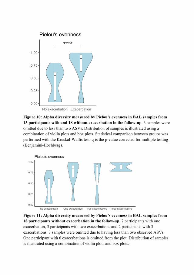
Figure 10: Alpha diversity measured by Pielou’s evenness in BAL samples from
13 participants with and 18 without exacerbation in the follow-up. 3 samples were
omitted due to less than two ASVs. Distribution of samples is illustrated using a
combination of violin plots and box plots. Statistical comparison between groups was
performed with the Kruskal-Wallis test. q is the p-value corrected for multiple testing
(Benjamini-Hochberg).
Figure 11: Alpha diversity measured by Pielou’s evenness in BAL samples from
18 participants without exacerbation in the follow-up. 7 participants with one
exacerbation, 3 participants with two exacerbations and 2 participants with 3
exacerbations. 3 samples were omitted due to having less than two observed ASVs.
One participant with 6 exacerbations is omitted from the plot. Distribution of samples
is illustrated using a combination of violin plots and box plots.

117
Figure 12: Alpha diversity measured by Shannon Index in BAL samples from 15
participants with and 19 without exacerbation in the follow-up. Distribution of
samples is illustrated using a combination of violin plots and box plots. Statistical
comparison between groups was performed with the Kruskal-Wallis test. q is the p-
value corrected for multiple testing (Benjamini-Hochberg).
Figure 13: Alpha diversity measured by Shannon Index in BAL samples from 19
participants without exacerbation in the follow-up. 8 participants with one
exacerbation, 3 participants with two exacerbations and 2 participants with 3
exacerbations. One participant with 4 exacerbations and one with 6 exacerbations were
omitted from the plot. Distribution of samples is illustrated using a combination of
violin plots and box plots.

118
This preliminary fungal analysis of samples from the lower airways, show results
conflicting to the study of Tiew et al. (265). The mycobiota data presented in this
thesis are based on few participants compared to that of the multicentre study, and we
could not address fungal burden. Methodological issues specific to DNA extraction,
sequencing, and bioinformatic analysis in studies of mycobiota are many (267), and
could contribute to differences in findings. However, the study by Tiew et al. has
limitations in methodology which are also common in studies of bacterial microbiota.
These includes relying on sputum samples (and not differentiating between induced
and spontaneous sputum, not including oral samples for comparison) and the use of
LEfSe for differential abundance analysis.
In summary, airway microbiota (and mycobiota) analysis in stable COPD did not show
an association to exacerbation frequency. Other studies have shown positive, but
conflicting findings. Different results can be explained by differences in methods, but
could also be due to geographical differences in study populations. To enable
comparison between studies, microbiome studies should be more standardised. In the
case of the frequent exacerbator phenotype, divergent results support the negative
findings in paper III.

119
Conclusions
1. Bronchoscopy is a safe procedure with a low risk of severe complications such
as mortality, pneumothorax, and bleeding that necessitate intervention. For
other complications, we could not find meaningful complication rates, as rates
varied substantially between the studies included. Discomfort was difficult to
compare between studies. We were not able to conclude on predictors. (Paper I)
2. 25.9% of participants experienced a complication leading to intervention during
or immediately following bronchoscopy. Only 1.3% of subjects had a
potentially serious complication, all of whom had no sequela, indicating that
bronchoscopy is a safe procedure in studies of patients with obstructive lung
disease. Sore throat, fever and flu-like symptoms each occurred in roughly half
of all subjects. Non-sedation and higher age were significantly associated with
more unplanned interventions during bronchoscopy. In participants undergoing
more than one bronchoscopy, complications and fever in the first bronchoscopy
was often predictive for complications and fever in the second. (Paper II)
3. Individual differences in the lower airway microbiota in persons with COPD far
outweigh group differences between frequent and nonfrequent COPD
exacerbators. Neither diversity metrices nor analyses of taxonomy in stable
state COPD could identify any predictors for frequent exacerbations. (Paper III)

120
Implications and future perspectives
The literature review included in this thesis pointed to the need for a well-powered,
prospective study with clear definitions of complications and discomfort. The safety
study in MicroCOPD was a step in the right direction, but has its limitations as
discussed in this thesis. For the topic of safety in COPD research bronchoscopy, there
were no papers that could be included at the time of review. The MicroCOPD study
implies that research bronchoscopy in COPD is safe, in accordance with the overall
conclusion of the SPIROMICS study (259). Although paper II can mainly answer
questions related to low-risk research bronchoscopy, the MicroCOPD study procedure
is similar to many diagnostic procedures performed at Haukeland University Hospital,
and study personnel are also involved in clinical work. Partially the study served as a
local quality assessment as well as an evaluation of the research procedure. The
specific result that around half of the participants experienced fever sensation
following bronchoscopy was surprising. As a direct result of the study, patients
undergoing bronchoscopy in the clinic now receive more information that emphasises
this side effect. In a way, the findings might be more transferrable to local practice
than to the research community as a whole, given the likely impact of the
bronchoscopist and local sedation practices. The impact of the MicroCOPD study with
regard to patient information is potentially greatest locally. An investigation into
specific aspects of diagnostic and therapeutic bronchoscopy at Haukeland University
hospital could be interesting, building on the experience from the work in this thesis.
The conclusions of paper III might not be a surprise to a reader who knows the field
and its weaknesses, hence the novelty might be questioned. However, all other studies
on the microbiome and exacerbations on COPD have reported positive findings. We
introduced several improvements to the methodology and presented negative results.
This could imply that the microbiota of stable COPD is not associated with frequency
of exacerbations, or that the current available and used methods are insufficient for
revealing this potentially complex relationship. Conducting additional studies on the
topic will be of minimal use unless the remaining methodological issues are resolved.

121
For future studies I would especially highlight the need for quantitative measures,
protected sampling, proper handling of contamination, longitudinal study designs,
integration of fungal and viral data, functional data and assessment of microbial
viability, and adequate statistical testing. The research community should seek to
agree upon standards for data collection and sample processing that enable comparison
between studies. That being said, the MicroCOPD study constitutes an important
contribution to the respiratory microbiome field with its rigorous focus on
methodology. For research into the specific relationship of bacterial microbiota and
exacerbation frequency, the MicroCOPD study has reached its potential. A number of
MicroCOPD papers exploring other methodological and clinical aspects have been
published (95, 134, 136, 184, 264). MicroCOPD has provided a considerable amount
of data which is subject to ongoing and future investigations.
The negative findings of paper III might seem disappointing both from a researcher’s
and participant’s perspective. Identifying an association between stable state
microbiota and exacerbation frequency could have been a step towards predicting,
preventing and improving treatment of exacerbations. In hindsight, knowing that we
did not come closer to this long-term goal, one could ask: Was a negative result worth
the risk and discomfort of research bronchoscopy? In the MicroCOPD study,
bronchoscopy was deemed safe, meaning that the benefit of falsifying previous reports
probably outweighs the risks for the individuals. However, if just a single a participant
had experienced a severe complication with sequelae, the answer might be opposite.
Future airway microbiota studies should absolutely make use of the advantages that
bronchoscopy offers, but make sure to assess and secure the safety of research
participants.

122
Errata
Paper I:
1. The highest rate bleeding rate was supposed to be 100 %, not 89.9%, as stated
in the paper. The rate of 89.9% refers to the report of minimal bleeding in the
cited reference, but this reported bleeding is in addition to 8.1% mild and 2.1%
moderate bleeding, which sums up to 100%.
2. The section on health care utilisation states “Nine prospective studies (57–59,
61) (65, 68) (70, 84) (86) reported complications that had to be handled by
increased health care utilisation”, and should have been “Ten prospective
studies (57-61, 65, 68, 70, 84, 86) reported complications that had to be handled
by increased health care utilisation”. The missing citation is however included
in the same section: “The incidence of health care contacts ranged from 0 to
31%, (59, 60)”. Reference numbers corresponding to those of paper I.
3. The missing reference mentioned above is also missing from Table 4
(supplementary table). Table 4 has been updated with a marked insertion (Table
4_corrected) in Appendix 3 of this thesis.

123
References
1. Global strategy for the diagnosis, management, and prevention of chronic obstructive
pulmonary disease 2021 Report. 2021.
2. Burney P, Patel J, Minelli C, Gnatiuc L, Amaral AFS, Kocabaş A, et al. Prevalence
and Population Attributable Risk for Chronic Airflow Obstruction in a Large Multinational
Study. Am J Respir Crit Care Med. 2020;203(11):1353-65.
3. MacNee W. Is Chronic Obstructive Pulmonary Disease an Accelerated Aging
Disease? Ann Am Thorac Soc. 2016;13 Suppl 5:S429-s37.
4. El-Zein RA, Young RP, Hopkins RJ, Etzel CJ. Genetic Predisposition to Chronic
Obstructive Pulmonary Disease and/or Lung Cancer: Important Considerations When
Evaluating Risk. Cancer Prevention Research. 2012;5(4):522-7.
5. Hayden LP, Cho MH, Raby BA, Beaty TH, Silverman EK, Hersh CP, et al. Childhood
asthma is associated with COPD and known asthma variants in COPDGene: a genome-wide
association study. Respir Res. 2018;19(1):209.
6. Brutsche MH, Downs SH, Schindler C, Gerbase MW, Schwartz J, Frey M, et al.
Bronchial hyperresponsiveness and the development of asthma and COPD in asymptomatic
individuals: SAPALDIA cohort study. Thorax. 2006;61(8):671-7.
7. Smith BM, Kirby M, Hoffman EA, Kronmal RA, Aaron SD, Allen NB, et al.
Association of Dysanapsis With Chronic Obstructive Pulmonary Disease Among Older
Adults. JAMA. 2020;323(22):2268-80.
8. Barnes PJ, Burney PGJ, Silverman EK, Celli BR, Vestbo J, Wedzicha JA, et al.
Chronic obstructive pulmonary disease. Nature Reviews Disease Primers. 2015;1(1):15076.
9. O’Donnell R, Breen D, Wilson S, Djukanovic R. Inflammatory cells in the airways in
COPD. Thorax. 2006;61(5):448.
10. Grudzinska FS, Brodlie M, Scholefield BR, Jackson T, Scott A, Thickett DR, et al.
Neutrophils in community-acquired pneumonia: parallels in dysfunction at the extremes of
age. Thorax. 2020;75(2):164.
11. Cavaillès A, Brinchault-Rabin G, Dixmier A, Goupil F, Gut-Gobert C, Marchand-
Adam S, et al. Comorbidities of COPD. European Respiratory Review. 2013;22(130):454.
12. Agusti À, Soriano JB. COPD as a Systemic Disease. COPD: Journal of Chronic
Obstructive Pulmonary Disease. 2008;5(2):133-8.
13. Roberts SD, Farber MO, Knox KS, Phillips GS, Bhatt NY, Mastronarde JG, et al.
FEV1/FVC Ratio of 70% Misclassifies Patients With Obstruction at the Extremes of Age.
Chest. 2006;130(1):200-6.
14. Hansen JE, Sun X-G, Wasserman K. Spirometric Criteria for Airway Obstruction: Use
Percentage of FEV1/FVC Ratio Below the Fifth Percentile, Not < 70%. Chest.
2007;131(2):349-55.
15. Jones PW, Harding G, Berry P, Wiklund I, Chen WH, Kline Leidy N. Development
and first validation of the COPD Assessment Test. Eur Respir J. 2009;34(3):648-54.
16. Bestall JC, Paul EA, Garrod R, Garnham R, Jones PW, Wedzicha JA. Usefulness of
the Medical Research Council (MRC) dyspnoea scale as a measure of disability in patients
with chronic obstructive pulmonary disease. Thorax. 1999;54(7):581-6.
17. Hegewald MJ, Horne BD, Trudo F, Kreindler JL, Chung Y, Rea S, et al. Blood
Eosinophil Count and Hospital Readmission in Patients with Acute Exacerbation of Chronic
Obstructive Pulmonary Disease. Int J Chron Obstruct Pulmon Dis. 2020;15:2629-41.
18. Singh D, Kolsum U, Brightling CE, Locantore N, Agusti A, Tal-Singer R.
Eosinophilic inflammation in COPD: prevalence and clinical characteristics. Eur Respir J.
2014;44(6):1697-700.

124
19. Ji Z, Hernández-Vázquez J, Domínguez-Zabaleta IM, Xia Z, Bellón-Cano JM, Gallo-
González V, et al. Influence of Comorbidities on the Survival of COPD Patients According to
Phenotypes. Int J Chron Obstruct Pulmon Dis. 2020;15:2759-67.
20. Castaldi PJ, Dy J, Ross J, Chang Y, Washko GR, Curran-Everett D, et al. Cluster
analysis in the COPDGene study identifies subtypes of smokers with distinct patterns of
airway disease and emphysema. Thorax. 2014;69(5):415-22.
21. Schneider LP, Sartori LG, Machado FVC, Dala Pola D, Rugila DF, Hirata RP, et al.
Physical activity and inactivity among different body composition phenotypes in individuals
with moderate to very severe chronic obstructive pulmonary disease. Brazilian Journal of
Physical Therapy. 2020.
22. Zahraei HN, Guissard F, Paulus V, Henket M, Donneau AF, Louis R. Comprehensive
Cluster Analysis for COPD Including Systemic and Airway Inflammatory Markers. Copd.
2020:1-12.
23. Chubachi S, Sato M, Kameyama N, Tsutsumi A, Sasaki M, Tateno H, et al.
Identification of five clusters of comorbidities in a longitudinal Japanese chronic obstructive
pulmonary disease cohort. Respir Med. 2016;117:272-9.
24. Vazquez Guillamet R, Ursu O, Iwamoto G, Moseley PL, Oprea T. Chronic obstructive
pulmonary disease phenotypes using cluster analysis of electronic medical records. Health
Informatics J. 2018;24(4):394-409.
25. Kim WJ, Gupta V, Nishimura M, Makita H, Idolor L, Roa C, et al. Identification of
chronic obstructive pulmonary disease subgroups in 13 Asian cities. Int J Tuberc Lung Dis.
2018;22(7):820-6.
26. Nishimura M, Makita H, Nagai K, Konno S, Nasuhara Y, Hasegawa M, et al. Annual
change in pulmonary function and clinical phenotype in chronic obstructive pulmonary
disease. Am J Respir Crit Care Med. 2012;185(1):44-52.
27. Hurst JR, Vestbo J, Anzueto A, Locantore N, Mullerova H, Tal-Singer R, et al.
Susceptibility to exacerbation in chronic obstructive pulmonary disease. N Engl J Med.
2010;363(12):1128-38.
28. Sadatsafavi M, McCormack J, Petkau J, Lynd LD, Lee TY, Sin DD. Should the
number of acute exacerbations in the previous year be used to guide treatments in COPD? Eur
Respir J. 2020.
29. Domenech A, Muñoz-Montiel A, García-Casares N, Rioja J, Ruiz-Esteban P,
Gutierrez-Castaño P, et al. High risk of subclinical atherosclerosis in COPD exacerbator
phenotype. Respir Med. 2018;141:165-71.
30. Beeh KM, Glaab T, Stowasser S, Schmidt H, Fabbri LM, Rabe KF, et al.
Characterisation of exacerbation risk and exacerbator phenotypes in the POET-COPD trial.
Respir Res. 2013;14(1):116-.
31. Tomioka R, Kawayama T, Suetomo M, Kinoshita T, Tokunaga Y, Imaoka H, et al.
"Frequent exacerbator" is a phenotype of poor prognosis in Japanese patients with chronic
obstructive pulmonary disease. Int J Chron Obstruct Pulmon Dis. 2016;11:207-16.
32. Sundh J, Johansson G, Larsson K, Lindén A, Löfdahl CG, Sandström T, et al. The
phenotype of concurrent chronic bronchitis and frequent exacerbations in patients with severe
COPD attending Swedish secondary care units. Int J Chron Obstruct Pulmon Dis.
2015;10:2327-34.
33. Fu JJ, McDonald VM, Baines KJ, Gibson PG. Airway IL-1β and Systemic
Inflammation as Predictors of Future Exacerbation Risk in Asthma and COPD. Chest.
2015;148(3):618-29.
34. Ahn JH, Chung JH, Shin KC, Choi EY, Jin HJ, Lee MS, et al. Critical Inhaler
Handling Error Is an Independent Risk Factor for Frequent Exacerbations of Chronic

125
Obstructive Pulmonary Disease: Interim Results of a Single Center Prospective Study. Int J
Chron Obstruct Pulmon Dis. 2019;14:2767-75.
35. Millares L, Pascual S, Monton C, Garcia-Nunez M, Lalmolda C, Faner R, et al.
Relationship between the respiratory microbiome and the severity of airflow limitation,
history of exacerbations and circulating eosinophils in COPD patients. BMC Pulm Med.
2019;19(1):112.
36. Pragman AA, Knutson KA, Gould TJ, Isaacson RE, Reilly CS, Wendt CH. Chronic
obstructive pulmonary disease upper airway microbiota alpha diversity is associated with
exacerbation phenotype: a case-control observational study. Respir Res. 2019;20(1):114.
37. Husebø GR, Bakke PS, Aanerud M, Hardie JA, Ueland T, Grønseth R, et al.
Predictors of exacerbations in chronic obstructive pulmonary disease--results from the Bergen
COPD cohort study. PLoS One. 2014;9(10):e109721-e.
38. Uzun S, Djamin RS, Kluytmans JA, Mulder PG, van't Veer NE, Ermens AA, et al.
Azithromycin maintenance treatment in patients with frequent exacerbations of chronic
obstructive pulmonary disease (COLUMBUS): a randomised, double-blind, placebo-
controlled trial. Lancet Respir Med. 2014;2(5):361-8.
39. Stöber A, Lutter JI, Schwarzkopf L, Kirsch F, Schramm A, Vogelmeier CF, et al.
Impact of Lung Function and Exacerbations on Health-Related Quality of Life in COPD
Patients Within One Year: Real-World Analysis Based on Claims Data. Int J Chron Obstruct
Pulmon Dis. 2021;16:2637-51.
40. Soler-Cataluña JJ, Martínez-García MA, Sánchez LS, Tordera MP, Sánchez PR.
Severe exacerbations and BODE index: two independent risk factors for death in male COPD
patients. Respir Med. 2009;103(5):692-9.
41. Anzueto A. Impact of exacerbations on COPD. European Respiratory Review.
2010;19(116):113.
42. Erdal M, Johannessen A, Bakke P, Gulsvik A, Eagan TM, Nielsen R. Incremental
costs of COPD exacerbations in GOLD stage 2+ COPD in ever-smokers of a general
population. Respiratory Medicine: X. 2020;2:100014.
43. Husebø G, Bakke P, Aanerud M, Hardie J, Grønseth R, Eagan T. How long does a
COPD exacerbation last? – Predictors for duration more than 3 weeks. Eur Respir J.
2014;44(Suppl 58):P1072.
44. Anthonisen NR, Manfreda J, Warren CP, Hershfield ES, Harding GK, Nelson NA.
Antibiotic therapy in exacerbations of chronic obstructive pulmonary disease. Ann Intern
Med. 1987;106(2):196-204.
45. Calverley PMA. Respiratory failure in chronic obstructive pulmonary disease. Eur
Respir J. 2003;22(47 suppl):26s.
46. Jenkins CR, Celli B, Anderson JA, Ferguson GT, Jones PW, Vestbo J, et al.
Seasonality and determinants of moderate and severe COPD exacerbations in the TORCH
study. Eur Respir J. 2012;39(1):38.
47. Bowler RP, Kim V, Regan E, Williams AAA, Santorico SA, Make BJ, et al.
Prediction of acute respiratory disease in current and former smokers with and without
COPD. Chest. 2014;146(4):941-50.
48. Husebø GR, Bakke PS, Aanerud M, Hardie JA, Ueland T, Grønseth R, et al.
Predictors of Exacerbations in Chronic Obstructive Pulmonary Disease - Results from the
Bergen COPD Cohort Study. PLoS One. 2014;9(10):e109721.
49. Koutsokera A, Kostikas K, Nicod LP, Fitting J-W. Pulmonary biomarkers in COPD
exacerbations: a systematic review. Respir Res. 2013;14(1):111.
50. George SN, Garcha DS, Mackay AJ, Patel ARC, Singh R, Sapsford RJ, et al. Human
rhinovirus infection during naturally occurring COPD exacerbations. Eur Respir J.
2014;44(1):87.

126
51. Sethi S. Bacteria in Exacerbations of Chronic Obstructive Pulmonary Disease. Proc
Am Thorac Soc. 2004;1(2):109-14.
52. Sikora M, Stec A, Chrabaszcz M, Knot A, Waskiel-Burnat A, Rakowska A, et al. Gut
Microbiome in Psoriasis: An Updated Review. Pathogens. 2020;9(6):463.
53. Glassner KL, Abraham BP, Quigley EMM. The microbiome and inflammatory bowel
disease. J Allergy Clin Immunol. 2020;145(1):16-27.
54. Menees S, Chey W. The gut microbiome and irritable bowel syndrome. F1000Res.
2018;7:F1000 Faculty Rev-29.
55. Gurung M, Li Z, You H, Rodrigues R, Jump DB, Morgun A, et al. Role of gut
microbiota in type 2 diabetes pathophysiology. EBioMedicine. 2020;51.
56. Jangi S, Gandhi R, Cox LM, Li N, von Glehn F, Yan R, et al. Alterations of the
human gut microbiome in multiple sclerosis. Nature Communications. 2016;7(1):12015.
57. Marchesi JR, Ravel J. The vocabulary of microbiome research: a proposal.
Microbiome. 2015;3:31.
58. Buermans HPJ, den Dunnen JT. Next generation sequencing technology: Advances
and applications. Biochimica et Biophysica Acta (BBA) - Molecular Basis of Disease.
2014;1842(10):1932-41.
59. Bharti R, Grimm DG. Current challenges and best-practice protocols for microbiome
analysis. Brief Bioinform. 2019;22(1):178-93.
60. Janda JM, Abbott SL. 16S rRNA gene sequencing for bacterial identification in the
diagnostic laboratory: pluses, perils, and pitfalls. J Clin Microbiol. 2007;45(9):2761-4.
61. Johnson JS, Spakowicz DJ, Hong B-Y, Petersen LM, Demkowicz P, Chen L, et al.
Evaluation of 16S rRNA gene sequencing for species and strain-level microbiome analysis.
Nature Communications. 2019;10(1):5029.
62. Pérez-Cobas AE, Gomez-Valero L, Buchrieser C. Metagenomic approaches in
microbial ecology: an update on whole-genome and marker gene sequencing analyses.
Microbial Genomics. 2020;6(8).
63. Callahan BJ, McMurdie PJ, Rosen MJ, Han AW, Johnson AJ, Holmes SP. DADA2:
High-resolution sample inference from Illumina amplicon data. Nat Methods.
2016;13(7):581-3.
64. Amir A, McDonald D, Navas-Molina Jose A, Kopylova E, Morton James T, Zech Xu
Z, et al. Deblur Rapidly Resolves Single-Nucleotide Community Sequence Patterns.
mSystems.2(2):e00191-16.
65. Weiss S, Xu ZZ, Peddada S, Amir A, Bittinger K, Gonzalez A, et al. Normalization
and microbial differential abundance strategies depend upon data characteristics. Microbiome.
2017;5(1):27.
66. Gloor GB, Macklaim JM, Pawlowsky-Glahn V, Egozcue JJ. Microbiome Datasets Are
Compositional: And This Is Not Optional. Front Microbiol. 2017;8(2224).
67. Bardenhorst SK, Berger T, Klawonn F, Vital M, Karch A, Rübsamen N, et al. Data
Analysis Strategies for Microbiome Studies in Human Populations—a Systematic
Review of Current Practice. mSystems. 2021;6(1):e01154-20.
68. Lozupone CA, Knight R. Species divergence and the measurement of microbial
diversity. FEMS Microbiol Rev. 2008;32(4):557-78.
69. Thorsen J, Brejnrod A, Mortensen M, Rasmussen MA, Stokholm J, Al-Soud WA, et
al. Large-scale benchmarking reveals false discoveries and count transformation sensitivity in
16S rRNA gene amplicon data analysis methods used in microbiome studies. Microbiome.
2016;4(1):62.
70. Sethi SMD, Murphy TFMD. Infection in the Pathogenesis and Course of Chronic
Obstructive Pulmonary DiseaseCurrent Concepts. The New England Journal of Medicine.
2008;359(22):2355-65.

127
71. Hilty M, Burke C, Pedro H, Cardenas P, Bush A, Bossley C, et al. Disordered
microbial communities in asthmatic airways. PLoS One. 2010;5(1):e8578.
72. Erb-Downward JR, Thompson DL, Han MK, Freeman CM, McCloskey L, Schmidt
LA, et al. Analysis of the lung microbiome in the "healthy" smoker and in COPD. PLoS One.
2011;6(2):e16384.
73. Charlson ES, Bittinger K, Haas AR, Fitzgerald AS, Frank I, Yadav A, et al.
Topographical continuity of bacterial populations in the healthy human respiratory tract. Am J
Respir Crit Care Med. 2011;184(8):957-63.
74. Pragman AA, Kim HB, Reilly CS, Wendt C, Isaacson RE. The lung microbiome in
moderate and severe chronic obstructive pulmonary disease. PLoS One. 2012;7(10):e47305.
75. Sze MA, Dimitriu PA, Hayashi S, Elliott WM, McDonough JE, Gosselink JV, et al.
The lung tissue microbiome in chronic obstructive pulmonary disease. Am J Respir Crit Care
Med. 2012;185(10):1073-80.
76. Huang YJ, Kim E, Cox MJ, Brodie EL, Brown R, Wiener-Kronish JP, et al. A
persistent and diverse airway microbiota present during chronic obstructive pulmonary
disease exacerbations. OMICS. 2010;14(1):9-59.
77. Charlson ES, Diamond JM, Bittinger K, Fitzgerald AS, Yadav A, Haas AR, et al.
Lung-enriched organisms and aberrant bacterial and fungal respiratory microbiota after lung
transplant. Am J Respir Crit Care Med. 2012;186(6):536-45.
78. van Woerden HC, Gregory C, Brown R, Marchesi JR, Hoogendoorn B, Matthews IP.
Differences in fungi present in induced sputum samples from asthma patients and non-atopic
controls: a community based case control study. BMC Infect Dis. 2013;13:69.
79. Huang YJ, Sethi S, Murphy T, Nariya S, Boushey HA, Lynch SV. Airway
microbiome dynamics in exacerbations of chronic obstructive pulmonary disease. J Clin
Microbiol. 2014;52(8):2813-23.
80. Millares L, Perez-Brocal V, Ferrari R, Gallego M, Pomares X, Garcia-Nunez M, et al.
Functional Metagenomics of the Bronchial Microbiome in COPD. PLoS One.
2015;10(12):e0144448.
81. Wang Z, Bafadhel M, Haldar K, Spivak A, Mayhew D, Miller BE, et al. Lung
microbiome dynamics in COPD exacerbations. Eur Respir J. 2016;47(4):1082-92.
82. Haldar K, Bafadhel M, Lau K, Berg A, Kwambana B, Kebadze T, et al. Microbiome
balance in sputum determined by PCR stratifies COPD exacerbations and shows potential for
selective use of antibiotics. PLoS One. 2017;12(8):e0182833.
83. Leitao Filho FS, Alotaibi NM, Ngan D, Tam S, Yang J, Hollander Z, et al. Sputum
Microbiome is Associated with 1-Year Mortality Following COPD Hospitalizations. Am J
Respir Crit Care Med. 2018.
84. Jubinville E, Veillette M, Milot J, Maltais F, Comeau AM, Levesque RC, et al.
Exacerbation induces a microbiota shift in sputa of COPD patients. PLoS One.
2018;13(3):e0194355.
85. Mayhew D, Devos N, Lambert C, Brown JR, Clarke SC, Kim VL, et al. Longitudinal
profiling of the lung microbiome in the AERIS study demonstrates repeatability of bacterial
and eosinophilic COPD exacerbations. Thorax. 2018;73(5):422-30.
86. Wang Z, Singh R, Miller BE, Tal-Singer R, Van Horn S, Tomsho L, et al. Sputum
microbiome temporal variability and dysbiosis in chronic obstructive pulmonary disease
exacerbations: an analysis of the COPDMAP study. Thorax. 2018;73(4):331-8.
87. Dicker AJ, Crichton ML, Cassidy AJ, Brady G, Hapca A, Tavendale R, et al. Genetic
mannose binding lectin deficiency is associated with airway microbiota diversity and reduced
exacerbation frequency in COPD. Thorax. 2018;73(6):510-8.

128
88. Ren L, Zhang R, Rao J, Xiao Y, Zhang Z, Yang B, et al. Transcriptionally Active
Lung Microbiome and Its Association with Bacterial Biomass and Host Inflammatory Status.
mSystems. 2018;3(5).
89. Wang Z, Maschera B, Lea S, Kolsum U, Michalovich D, Van Horn S, et al. Airway
host-microbiome interactions in chronic obstructive pulmonary disease. Respir Res.
2019;20(1):113.
90. Tangedal S, Nielsen R, Aanerud M, Persson LJ, Wiker HG, Bakke PS, et al. Sputum
microbiota and inflammation at stable state and during exacerbations in a cohort of chronic
obstructive pulmonary disease (COPD) patients. PLoS One. 2019;14(9):e0222449.
91. López Caro JC, Santibáñez M, García Rivero JL, Villanueva M, Sainz J, González
Astorqui P, et al. Sputum Microbiome Dynamics in Chronic Obstructive Pulmonary Disease
Patients during an Exacerbation Event and Post-Stabilization. Respiration. 2019;98(5):447-
54.
92. Grønseth R, Haaland I, Wiker HG, Martinsen EM, Leiten EO, Husebø G, et al. The
Bergen COPD microbiome study (MicroCOPD): rationale, design, and initial experiences.
Eur Clin Respir J. 2014;1.
93. Eisenhofer R, Minich JJ, Marotz C, Cooper A, Knight R, Weyrich LS. Contamination
in Low Microbial Biomass Microbiome Studies: Issues and Recommendations. Trends
Microbiol. 2019;27(2):105-17.
94. Ditz B, Christenson S, Rossen J, Brightling C, Kerstjens HAM, van den Berge M, et
al. Sputum microbiome profiling in COPD: beyond singular pathogen detection. Thorax.
2020;75(4):338.
95. Grønseth R, Drengenes C, Wiker HG, Tangedal S, Xue Y, Husebø GR, et al.
Protected sampling is preferable in bronchoscopic studies of the airway microbiome. ERJ
Open Res. 2017;3(3).
96. Kollofrath O. Entfernung eines knochenstucks aus dem Rechten Bronchus auf
Naturlichem Wege und unter Anwendung der direkten Laryngoskopie. Munch Med
Wochenschr. 1897;44(38):1038-9.
97. Killian G. Ueber directe Bronkoskopie. Munch Med Wochenschr. 1898;45(27):844-7.
98. Jackson C. Tracheo-bronchoscopy, esophagoscopy and gastroscopy. St. Louis: The
Laryngoscope Company; 1907.
99. Killian H. … bak oss står Herren: Av en kirurgs dagbok. Oslo: Lutherstiftselses
forlag; 1957.
100. Becker HD. A Short History of Bronchoscopy. In: Ernst A, editor. Introduction to
Bronchoscopy: Cambridge University Press; 2009.
101. Ikeda S, Yanai N, Ishikawa S. Flexible bronchofiberscope. Keio J Med. 1968;17(1):1-
16.
102. Harrell JH. Transnasal approach for fiberoptic bronchoscopy. Chest. 1978;73(5
Suppl):704-6.
103. Introduction to Bronchoscopy: Cambridge University Press; 2009.
104. Islam S. Flexible bronchoscopy in adults: Indications and contraindications. 2021
[cited 25.01.2021]. In: UpToDate [Internet]. Waltham, Massachusetts: UpToDate, [cited
25.01.2021]. Available from: https://www.uptodate.com/contents/flexible-bronchoscopy-in-
adults-indications-and-contraindications.
105. Islam S. Flexible bronchoscopy in adults: Associated diagnostic and therapeutic
procedures. 2021 [cited 25.01.2021]. In: UpToDate [Internet]. Waltham, Massachusetts:
UpToDate, [cited 25.01.2021]. Available from: https://www.uptodate.com/contents/flexible-
bronchoscopy-in-adults-associated-diagnostic-and-therapeutic-procedures.
106. Patel PH, Antoine M, Ullah S. Bronchoalveolar Lavage. StatPearls. Treasure Island
(FL): StatPearls Publishing

129
Copyright © 2020, StatPearls Publishing LLC.; 2020.
107. Lamb C. Bronchial washing, bronchoalveolar lavage, bronchial brush, and
endobronchial biopsy. In: Ernst A, editor. Introduction to Bronchoscopy: Cambridge
University Press; 2009.
108. Cordasco EM, Mehta AC, Ahmad M. Bronchoscopically Induced Bleeding: A
Summary of Nine Years' Cleveland Clinic Experience and Review of the Literature. Chest.
1991;100(4):1141-7.
109. Du Rand IA, Blaikley J, Booton R, Chaudhuri N, Gupta V, Khalid S, et al. British
Thoracic Society guideline for diagnostic flexible bronchoscopy in adults: accredited by
NICE. Thorax. 2013;68 Suppl 1:i1-i44.
110. Green SM, Krauss B. Procedural sedation terminology: Moving beyond “conscious
sedation”. Ann Emerg Med. 2002;39(4):433-5.
111. Fitzpatrick AM, Teague WG, Burwell L, Brown MS, Brown LA. Glutathione
oxidation is associated with airway macrophage functional impairment in children with severe
asthma. Pediatr Res. 2011;69(2):154-9.
112. Davies J, Payne D. Research applications of bronchoscopy. Paediatr Respir Rev.
2003;4(3):230-6.
113. Grønseth R, Haaland I, Wiker HG, Martinsen EM, Leiten EO, Husebo G, et al. The
Bergen COPD microbiome study (MicroCOPD): rationale, design, and initial experiences.
Eur Clin Respir J. 2014;1.
114. Dickson RP, Erb-Downward JR, Freeman CM, McCloskey L, Beck JM, Huffnagle
GB, et al. Spatial Variation in the Healthy Human Lung Microbiome and the Adapted Island
Model of Lung Biogeography. Ann Am Thorac Soc. 2015;12(6):821-30.
115. Elston WJ, Whittaker AJ, Khan LN, Flood-Page P, Ramsay C, Jeffery PK, et al.
Safety of research bronchoscopy, biopsy and bronchoalveolar lavage in asthma. Eur Respir J.
2004;24(3):375-7.
116. Fullerton DG, Jere K, Jambo K, Kulkarni NS, Zijlstra EE, Grigg J, et al. Domestic
smoke exposure is associated with alveolar macrophage particulate load. Trop Med Int
Health. 2009;14(3):349-54.
117. Day RO, Chalmers DR, Williams KM, Campbell TJ. The death of a healthy volunteer
in a human research project: implications for Australian clinical research. Med J Aust.
1998;168(9):449-51.
118. Steinbrook R. Improving Protection for Research Subjects. N Engl J Med.
2002;346(18):1425-30.
119. Horner JS. Retreat from Nuremberg: can we prevent unethical medical research?
Public Health. 1999;113(5):205-10.
120. World Medical Association Declaration of Helsinki: ethical principles for medical
research involving human subjects. JAMA. 2013;310(20):2191-4.
121. Schulz KF, Altman DG, Moher D. CONSORT 2010 statement: updated guidelines for
reporting parallel group randomised trials. BMJ. 2010;340:c332.
122. Ioannidis JP, Evans SJ, Gøtzsche PC, O'Neill RT, Altman DG, Schulz K, et al. Better
reporting of harms in randomized trials: an extension of the CONSORT statement. Ann Intern
Med. 2004;141(10):781-8.
123. Coates M, Spanos M, Parmar P, Chandrasekhar T, Sikich L. A Review of Methods for
Monitoring Adverse Events in Pediatric Psychopharmacology Clinical Trials. Drug Saf.
2018;41(5):465-71.
124. Dindo D, Demartines N, Clavien PA. Classification of surgical complications: a new
proposal with evaluation in a cohort of 6336 patients and results of a survey. Ann Surg.
2004;240(2):205-13.

130
125. Miller TP, Fisher BT, Getz KD, Sack L, Razzaghi H, Seif AE, et al. Unintended
consequences of evolution of the Common Terminology Criteria for Adverse Events. Pediatr
Blood Cancer. 2019;66(7):e27747.
126. Classen DC, Lloyd RC, Provost L, Griffin FA, Resar R. Development and Evaluation
of the Institute for Healthcare Improvement Global Trigger Tool. Journal of Patient Safety.
2008;4(3):169-77.
127. Haddad PM, Fleischhacker WW, Peuskens J, Cavallaro R, Lean ME, Morozova M, et
al. SMARTS (Systematic Monitoring of Adverse events Related to TreatmentS): The
development of a pragmatic patient-completed checklist to assess antipsychotic drug side
effects. Ther Adv Psychopharmacol. 2014;4(1):15-21.
128. Landriel Ibañez FA, Hem S, Ajler P, Vecchi E, Ciraolo C, Baccanelli M, et al. A new
classification of complications in neurosurgery. World Neurosurg. 2011;75(5-6):709-15;
discussion 604-11.
129. Vitiello B, Riddle MA, Walkup JT, Labellarte MJ, Varipatis E, Greenhill LL, et al.
How Can We Improve the Assessment of Safety in Child and Adolescent
Psychopharmacology? J Am Acad Child Adolesc Psychiatry. 2003;42(6):634-41.
130. Pati D, Lorusso LN. How to Write a Systematic Review of the Literature. Herd.
2018;11(1):15-30.
131. Eagan TM, Ueland T, Wagner PD, Hardie JA, Mollnes TE, Damas JK, et al. Systemic
inflammatory markers in COPD: results from the Bergen COPD Cohort Study. Eur Respir J.
2010;35(3):540-8.
132. Sorheim IC, Johannessen A, Grydeland TB, Omenaas ER, Gulsvik A, Bakke PS.
Case-control studies on risk factors for chronic obstructive pulmonary disease: how does the
sampling of the cases and controls affect the results? Clin Respir J. 2010;4(2):89-96.
133. Global Strategy for the Diagnosis, Management and Prevention of COPD, Global
Initiative for Chronic Obstructive Lung Disease (GOLD) 2017.
134. Martinsen EMH, Eagan TML, Leiten EO, Nordeide E, Bakke PS, Lehmann S, et al.
Motivation and response rates in bronchoscopy studies. Multidiscip Respir Med. 2019;14:14.
135. Nielsen R, Xue Y, Jonassen I, Haaland I, Kommedal Ø, Wiker HG, et al. Repeated
bronchoscopy examination of the airway microbiome. Eur Respir J. 2019;54(suppl
63):PA2344.
136. Drengenes C, Wiker HG, Kalananthan T, Nordeide E, Eagan TML, Nielsen R.
Laboratory contamination in airway microbiome studies. BMC Microbiol. 2019;19(1):187.
137. Johannessen A, Lehmann S, Omenaas ER, Eide GE, Bakke PS, Gulsvik A. Post-
bronchodilator spirometry reference values in adults and implications for disease
management. Am J Respir Crit Care Med. 2006;173(12):1316-25.
138. Borg GA. Psychophysical bases of perceived exertion. Med Sci Sports Exerc.
1982;14(5):377-81.
139. Hoang T, Wiker HG, Eagan T, Drengenes C. 16S Amplicon PCR for the V3-V4
region for the MicroCOPD samples 2019 [
140. Knudsen KS, Nielsen R, Haaland I, Lehmann S, Eagan TM. The lower airways
microbiome in patients with IPF and COPD. Eur Respir J. 2019;54(suppl 63):PA1326.
141. Bolyen E, Rideout JR, Dillon MR, Bokulich NA, Abnet CC, Al-Ghalith GA, et al.
Reproducible, interactive, scalable and extensible microbiome data science using QIIME 2.
Nat Biotechnol. 2019.
142. Bolger AM, Lohse M, Usadel B. Trimmomatic: a flexible trimmer for Illumina
sequence data. Bioinformatics. 2014;30(15):2114-20.
143. Rognes T, Flouri T, Nichols B, Quince C, Mahe F. VSEARCH: a versatile open
source tool for metagenomics. PeerJ. 2016;4:e2584.

131
144. Navas-Molina JA, Peralta-Sánchez JM, González A, McMurdie PJ, Vázquez-Baeza Y,
Xu Z, et al. Advancing our understanding of the human microbiome using QIIME. Methods
Enzymol. 2013;531:371-444.
145. Davis NM, Proctor DM, Holmes SP, Relman DA, Callahan BJ. Simple statistical
identification and removal of contaminant sequences in marker-gene and metagenomics data.
Microbiome. 2018;6(1):226.
146. Chen T, Yu WH, Izard J, Baranova OV, Lakshmanan A, Dewhirst FE. The Human
Oral Microbiome Database: a web accessible resource for investigating oral microbe
taxonomic and genomic information. Database (Oxford). 2010;2010:baq013.
147. Madden T. The BLAST Sequence Analysis Tool. In: McEntyre J, O’stell J, editors.
The NCBI Handbook. Bethesda (MD): National Center for Biotechnology Information; 2002.
148. Price MN, Dehal PS, Arkin AP. FastTree 2--approximately maximum-likelihood trees
for large alignments. PLoS One. 2010;5(3):e9490.
149. Shannon CE. A mathematical theory of communication. The Bell System Technical
Journal. 1948;27(4):623-56.
150. Faith DP. Conservation evaluation and phylogenetic diversity. Biol Conserv.
1992;61(1):1-10.
151. Bray JR, Curtis JT. An Ordination of the Upland Forest Communities of Southern
Wisconsin. Ecol Monogr. 1957;27(4):326-49.
152. Hao M, Corral-Rivas JJ, González-Elizondo MS, Ganeshaiah KN, Nava-Miranda MG,
Zhang C, et al. Assessing biological dissimilarities between five forest communities. Forest
Ecosystems. 2019;6(1):30.
153. Lozupone CA, Hamady M, Kelley ST, Knight R. Quantitative and qualitative beta
diversity measures lead to different insights into factors that structure microbial communities.
Appl Environ Microbiol. 2007;73(5):1576-85.
154. Mandal S, Van Treuren W, White RA, Eggesbo M, Knight R, Peddada SD. Analysis
of composition of microbiomes: a novel method for studying microbial composition. Microb
Ecol Health Dis. 2015;26:27663.
155. Morton JT, Sanders J, Quinn RA, McDonald D, Gonzalez A, Vazquez-Baeza Y, et al.
Balance Trees Reveal Microbial Niche Differentiation. mSystems. 2017;2(1).
156. Fernandes AD, Macklaim JM, Linn TG, Reid G, Gloor GB. ANOVA-Like
Differential Expression (ALDEx) Analysis for Mixed Population RNA-Seq. PLoS One.
2013;8(7):e67019.
157. Chen J, King E, Deek R, Wei Z, Yu Y, Grill D, et al. An omnibus test for differential
distribution analysis of microbiome sequencing data. Bioinformatics. 2018;34(4):643-51.
158. Chen L, Reeve J, Zhang L, Huang S, Wang X, Chen J. GMPR: A robust normalization
method for zero-inflated count data with application to microbiome sequencing data. PeerJ.
2018;6:e4600.
159. Roach KE. Measurement of Health Outcomes: Reliability, Validity and
Responsiveness. JPO: Journal of Prosthetics and Orthotics. 2006;18(6):P8-P12.
160. Benestad HB, Laake P. Forskning: metode og planlegging. In: Laake P, Olsen BR,
Benestad HB, editors. Forskning i medisin og biofag. 2 ed. Oslo: Gyldendal; 2008.
161. Staff A. Bias Oslo: De nasjonale forskningsetiske komiteene (The norwegian national
research ethics committees); 2015 [Available from:
https://www.forskningsetikk.no/ressurser/fbib/uavhengighet/bias/.
162. Vetter TR, Mascha EJ. Bias, Confounding, and Interaction: Lions and Tigers, and
Bears, Oh My! Anesth Analg. 2017;125(3):1042-8.
163. Haraldstad AB, Christophersen E. Litteratursøk og personlige referansedatabaser. In:
Laake P, Olsen BR, Benestad HB, editors. Forskning i medisin og biofag. 1. 2 ed. Oslo:
Gyldendal; 2008.

132
164. Hattotuwa K, Gamble EA, O'Shaughnessy T, Jeffery PK, Barnes NC. Safety of
bronchoscopy, biopsy, and BAL in research patients with COPD. Chest. 2002;122(6):1909-
12.
165. Bramer WM, Rethlefsen ML, Kleijnen J, Franco OH. Optimal database combinations
for literature searches in systematic reviews: a prospective exploratory study. Syst Rev.
2017;6(1):245.
166. Baumann N. How to use the medical subject headings (MeSH). Int J Clin Pract.
2016;70(2):171-4.
167. Jenuwine ES, Floyd JA. Comparison of Medical Subject Headings and text-word
searches in MEDLINE to retrieve studies on sleep in healthy individuals. J Med Libr Assoc.
2004;92(3):349-53.
168. Nandagopal L, Veeraputhiran M, Jain T, Soubani AO, Schiffer CA. Bronchoscopy can
be done safely in patients with thrombocytopenia. Transfusion. 2016;56(2):344-8.
169. Neuman Y, Koslow M, Matveychuk A, Bar-Sef A, Guber A, Shitrit D. Increased
hypoxemia in patients with COPD and pulmonary hypertension undergoing bronchoscopy
with biopsy. Int J Chron Obstruct Pulmon Dis. 2015;10:2627-32.
170. Stoll CRT, Izadi S, Fowler S, Green P, Suls J, Colditz GA. The value of a second
reviewer for study selection in systematic reviews. Research Synthesis Methods.
2019;10(4):539-45.
171. Morrison A, Polisena J, Husereau D, Moulton K, Clark M, Fiander M, et al. THE
EFFECT OF ENGLISH-LANGUAGE RESTRICTION ON SYSTEMATIC REVIEW-
BASED META-ANALYSES: A SYSTEMATIC REVIEW OF EMPIRICAL STUDIES. Int J
Technol Assess Health Care. 2012;28(2):138-44.
172. Wang Z, Nayfeh T, Tetzlaff J, O’Blenis P, Murad MH. Error rates of human reviewers
during abstract screening in systematic reviews. PLoS One. 2020;15(1):e0227742.
173. Page MJ, Shamseer L, Tricco AC. Registration of systematic reviews in PROSPERO:
30,000 records and counting. Syst Rev. 2018;7(1):32.
174. Page MJ, Moher D, Bossuyt PM, Boutron I, Hoffmann TC, Mulrow CD, et al.
PRISMA 2020 explanation and elaboration: updated guidance and exemplars for reporting
systematic reviews. BMJ. 2021;372:n160.
175. Pahus L, Burgel P-R, Roche N, Paillasseur J-L, Chanez P, on behalf of Initiatives Bsc.
Randomized controlled trials of pharmacological treatments to prevent COPD exacerbations:
applicability to real-life patients. BMC Pulm Med. 2019;19(1):127.
176. Scichilone N, Basile M, Battaglia S, Bellia V. What Proportion of Chronic Obstructive
Pulmonary Disease Outpatients Is Eligible for Inclusion in Randomized Clinical Trials?
Respiration. 2014;87(1):11-7.
177. McGrath PA. Psychological aspects of pain perception. Arch Oral Biol. 1994;39
Suppl:55s-62s.
178. Anderson WH, Ha JW, Couper DJ, O'Neal WK, Barr RG, Bleecker ER, et al.
Variability in objective and subjective measures affects baseline values in studies of patients
with COPD. PLoS One. 2017;12(9):e0184606.
179. McHugh ML. Interrater reliability: the kappa statistic. Biochem Med (Zagreb).
2012;22(3):276-82.
180. Brooks JP. Challenges for case-control studies with microbiome data. Ann Epidemiol.
2016;26(5):336-41.e1.
181. Teng F, Darveekaran Nair SS, Zhu P, Li S, Huang S, Li X, et al. Impact of DNA
extraction method and targeted 16S-rRNA hypervariable region on oral microbiota profiling.
Sci Rep. 2018;8(1):16321.

133
182. V. Wintzingerode F, Göbel UB, Stackebrandt E. Determination of microbial diversity
in environmental samples: pitfalls of PCR-based rRNA analysis. FEMS Microbiol Rev.
1997;21(3):213-29.
183. Yuan S, Cohen DB, Ravel J, Abdo Z, Forney LJ. Evaluation of Methods for the
Extraction and Purification of DNA from the Human Microbiome. PLoS One.
2012;7(3):e33865.
184. Drengenes C, Eagan TML, Haaland I, Wiker HG, Nielsen R. Exploring protocol bias
in airway microbiome studies: one versus two PCR steps and 16S rRNA gene region V3 V4
versus V4. BMC Genomics. 2021;22(1):3.
185. Peters RPH, Mohammadi T, Vandenbroucke-Grauls CMJE, Danner SA, van Agtmael
MA, Savelkoul PHM. Detection of bacterial DNA in blood samples from febrile patients:
underestimated infection or emerging contamination? FEMS Immunol Med Microbiol.
2004;42(2):249-53.
186. van der Zee A, Peeters M, de Jong C, Verbakel H, Crielaard JW, Claas EC, et al.
Qiagen DNA extraction kits for sample preparation for legionella PCR are not suitable for
diagnostic purposes. J Clin Microbiol. 2002;40(3):1126.
187. Evans GE, Murdoch DR, Anderson TP, Potter HC, George PM, Chambers ST.
Contamination of Qiagen DNA extraction kits with Legionella DNA. J Clin Microbiol.
2003;41(7):3452-3.
188. Salter SJ, Cox MJ, Turek EM, Calus ST, Cookson WO, Moffatt MF, et al. Reagent
and laboratory contamination can critically impact sequence-based microbiome analyses.
BMC Biol. 2014;12(1):87.
189. Sze MA, Schloss PD, McMahon K. The Impact of DNA Polymerase and Number of
Rounds of Amplification in PCR on 16S rRNA Gene Sequence Data. mSphere.
2019;4(3):e00163-19.
190. Witzke MC, Gullic A, Yang P, Bivens NJ, Adkins PRF, Ericsson AC. Influence of
PCR cycle number on 16S rRNA gene amplicon sequencing of low biomass samples. J
Microbiol Methods. 2020;176:106033.
191. Jaspers E, Overmann J. Ecological significance of microdiversity: identical 16S rRNA
gene sequences can be found in bacteria with highly divergent genomes and ecophysiologies.
Appl Environ Microbiol. 2004;70(8):4831-9.
192. Wade WG, Prosdocimi EM. Profiling of Oral Bacterial Communities. J Dent Res.
2020;99(6):621-9.
193. Stoler N, Nekrutenko A. Sequencing error profiles of Illumina sequencing
instruments. NAR Genomics and Bioinformatics. 2021;3(1).
194. Brooks JP, Edwards DJ, Harwich MD, Rivera MC, Fettweis JM, Serrano MG, et al.
The truth about metagenomics: quantifying and counteracting bias in 16S rRNA studies.
BMC Microbiol. 2015;15(1):66.
195. Nakamura K, Oshima T, Morimoto T, Ikeda S, Yoshikawa H, Shiwa Y, et al.
Sequence-specific error profile of Illumina sequencers. Nucleic Acids Res. 2011;39(13):e90.
196. Minich JJ, Sanders JG, Amir A, Humphrey G, Gilbert JA, Knight R, et al. Quantifying
and Understanding Well-to-Well Contamination in Microbiome Research. mSystems.
2019;4(4):e00186-19.
197. Aas JA, Paster BJ, Stokes LN, Olsen I, Dewhirst FE. Defining the normal bacterial
flora of the oral cavity. J Clin Microbiol. 2005;43(11):5721-32.
198. Wade W. Unculturable bacteria--the uncharacterized organisms that cause oral
infections. J R Soc Med. 2002;95(2):81-3.
199. Kearney SC, Dziekiewicz M, Feleszko W. Immunoregulatory and immunostimulatory
responses of bacterial lysates in respiratory infections and asthma. Ann Allergy Asthma
Immunol. 2015;114(5):364-9.

134
200. Elizaquível P, Sánchez G, Aznar R. Quantitative detection of viable foodborne E. coli
O157:H7, Listeria monocytogenes and Salmonella in fresh-cut vegetables combining
propidium monoazide and real-time PCR. Food Control. 2012;25(2):704-8.
201. Nogva HK, Drømtorp SM, Nissen H, Rudi K. Ethidium monoazide for DNA-based
differentiation of viable and dead bacteria by 5'-nuclease PCR. Biotechniques.
2003;34(4):804-8, 10, 12-3.
202. Mukhopadhyay UK, Mukhopadhyay A. A low-cost, rapid, sensitive and reliable PCR-
based alternative method for predicting the presence of possible live microbial contaminants
in food. Curr Sci. 2002;83(1):53-6.
203. Pezzulo AA, Kelly PH, Nassar BS, Rutland CJ, Gansemer ND, Dohrn CL, et al.
Abundant DNase I-sensitive bacterial DNA in healthy porcine lungs and its implications for
the lung microbiome. Appl Environ Microbiol. 2013;79(19):5936-41.
204. Willis AL, Calton JB, Carr TF, Chiu AG, Chang EH. Dead or alive:
Deoxyribonuclease I sensitive bacteria and implications for the sinus microbiome. Am J
Rhinol Allergy. 2016;30(2):94-8.
205. Nguyen LDN, Deschaght P, Merlin S, Loywick A, Audebert C, Van Daele S, et al.
Effects of Propidium Monoazide (PMA) Treatment on Mycobiome and Bacteriome Analysis
of Cystic Fibrosis Airways during Exacerbation. PLoS One. 2016;11(12):e0168860-e.
206. Rogers GB, Cuthbertson L, Hoffman LR, Wing PA, Pope C, Hooftman DA, et al.
Reducing bias in bacterial community analysis of lower respiratory infections. Isme j.
2013;7(4):697-706.
207. Mika M, Nita I, Morf L, Qi W, Beyeler S, Bernasconi E, et al. Microbial and host
immune factors as drivers of COPD. ERJ Open Res. 2018;4(3).
208. Wang X, Howe S, Deng F, Zhao J. Current Applications of Absolute Bacterial
Quantification in Microbiome Studies and Decision-Making Regarding Different Biological
Questions. Microorganisms. 2021;9(9):1797.
209. Edgar RC. UPARSE: highly accurate OTU sequences from microbial amplicon reads.
Nature Methods. 2013;10(10):996-8.
210. Eren AM, Morrison HG, Lescault PJ, Reveillaud J, Vineis JH, Sogin ML. Minimum
entropy decomposition: unsupervised oligotyping for sensitive partitioning of high-throughput
marker gene sequences. The ISME journal. 2015;9(4):968-79.
211. Schloss PD, Handelsman J. Introducing DOTUR, a computer program for defining
operational taxonomic units and estimating species richness. Appl Environ Microbiol.
2005;71(3):1501-6.
212. Edgar RC. Search and clustering orders of magnitude faster than BLAST.
Bioinformatics. 2010;26(19):2460-1.
213. Li W, Godzik A. Cd-hit: a fast program for clustering and comparing large sets of
protein or nucleotide sequences. Bioinformatics. 2006;22(13):1658-9.
214. Tikhonov M, Leach RW, Wingreen NS. Interpreting 16S metagenomic data without
clustering to achieve sub-OTU resolution. The ISME journal. 2015;9(1):68-80.
215. Callahan BJ, McMurdie PJ, Holmes SP. Exact sequence variants should replace
operational taxonomic units in marker-gene data analysis. The ISME Journal.
2017;11(12):2639-43.
216. Nearing JT, Douglas GM, Comeau AM, Langille MGI. Denoising the Denoisers: an
independent evaluation of microbiome sequence error-correction approaches. PeerJ.
2018;6:e5364-e.
217. McKnight DT, Huerlimann R, Bower DS, Schwarzkopf L, Alford RA, Zenger KR.
microDecon: A highly accurate read-subtraction tool for the post-sequencing removal of
contamination in metabarcoding studies. Environmental DNA. 2019;1(1):14-25.

135
218. Koren O, Knights D, Gonzalez A, Waldron L, Segata N, Knight R, et al. A guide to
enterotypes across the human body: meta-analysis of microbial community structures in
human microbiome datasets. PLoS Comput Biol. 2013;9(1):e1002863-e.
219. McMurdie PJ, Holmes S. Waste Not, Want Not: Why Rarefying Microbiome Data Is
Inadmissible. PLoS Comput Biol. 2014;10(4):e1003531.
220. Paulson JN, Stine OC, Bravo HC, Pop M. Differential abundance analysis for
microbial marker-gene surveys. Nature Methods. 2013;10(12):1200-2.
221. Caporaso JG, Kuczynski J, Stombaugh J, Bittinger K, Bushman FD, Costello EK, et
al. QIIME allows analysis of high-throughput community sequencing data. Nature Methods.
2010;7(5):335-6.
222. Ghebre MA, Pang PH, Diver S, Desai D, Bafadhel M, Haldar K, et al. Biological
exacerbation clusters demonstrate asthma and chronic obstructive pulmonary disease overlap
with distinct mediator and microbiome profiles. J Allergy Clin Immunol. 2018;141(6):2027-
36.e12.
223. Molyneaux PL, Mallia P, Cox MJ, Footitt J, Willis-Owen SA, Homola D, et al.
Outgrowth of the bacterial airway microbiome after rhinovirus exacerbation of chronic
obstructive pulmonary disease. Am J Respir Crit Care Med. 2013;188(10):1224-31.
224. Knights D, Ward TL, McKinlay CE, Miller H, Gonzalez A, McDonald D, et al.
Rethinking "enterotypes". Cell Host Microbe. 2014;16(4):433-7.
225. Goodrich JK, Di Rienzi SC, Poole AC, Koren O, Walters WA, Caporaso JG, et al.
Conducting a microbiome study. Cell. 2014;158(2):250-62.
226. Morton JT, Toran L, Edlund A, Metcalf JL, Lauber C, Knight R, et al. Uncovering the
Horseshoe Effect in Microbial Analyses. mSystems. 2017;2(1):e00166-16.
227. Szafir DA. The good, the bad, and the biased: five ways visualizations can mislead
(and how to fix them). interactions. 2018;25(4):26–33.
228. Bowers D. Medical Statistics from Scratch: An Introduction for Health Professionals.
3 ed: Wiley Blackwell; 2014.
229. Lydersen S. Statistisk styrke – før, men ikke etter. Tidsskr Nor Laegeforen. 2019.
230. Debelius J, Song SJ, Vazquez-Baeza Y, Xu ZZ, Gonzalez A, Knight R. Tiny
microbes, enormous impacts: what matters in gut microbiome studies? Genome Biol.
2016;17(1):217.
231. Kelly BJ, Gross R, Bittinger K, Sherrill-Mix S, Lewis JD, Collman RG, et al. Power
and sample-size estimation for microbiome studies using pairwise distances and
PERMANOVA. Bioinformatics (Oxford, England). 2015;31(15):2461-8.
232. La Rosa PS, Brooks JP, Deych E, Boone EL, Edwards DJ, Wang Q, et al. Hypothesis
testing and power calculations for taxonomic-based human microbiome data. PLoS One.
2012;7(12):e52078-e.
233. Groenwold RHH, Goeman JJ, Cessie SL, Dekkers OM. Multiple testing: when is
many too much? European Journal of Endocrinology. 2021;184(3):E11-E4.
234. Nearing JT, Douglas GM, Hayes M, MacDonald J, Desai D, Allward N, et al.
Microbiome differential abundance methods produce disturbingly different results across 38
datasets. bioRxiv. 2021:2021.05.10.443486.
235. Muthu V, Ram B, Sehgal IS, Dhooria S, Prasad KT, Bal A, et al. Predictors of Severe
Bleeding During Endobronchial Biopsy: Experience of 537 Cases. J Bronchology Interv
Pulmonol. 2019;26(4):273-9.
236. Zaman MK, Shrestha R. Major Complications Associated with Conventional
Transbronchial Needle Aspiration. South Med J. 2018;111(9):565-71.
237. Wang S, Hu X, Pan Y. Low-density lipoprotein cholesterol levels are positively
associated with the risk of endobronchial biopsy-induced refractory hemorrhage in patients
with lung cancer. Lipids Health Dis. 2019;18(1):190.

136
238. Ishiwata T, Tsushima K, Terada J, Fujie M, Abe M, Ikari J, et al. Efficacy of End-
Tidal Capnography Monitoring during Flexible Bronchoscopy in Nonintubated Patients under
Sedation: A Randomized Controlled Study. Respiration. 2018;96(4):355-62.
239. O'Dwyer DN, Duvall AS, Xia M, Hoffman TC, Bloye KS, Bulte CA, et al.
Transbronchial biopsy in the management of pulmonary complications of hematopoietic stem
cell transplantation. Bone Marrow Transplant. 2018;53(2):193-8.
240. Davis KL, Channick CL. Safety of topical tetracaine in patients undergoing flexible
bronchoscopy. J Bronchology Interv Pulmonol. 2009;16(2):95-8.
241. Almadani A, Ping MNY, Deenadayalu A, Saunders J, O'Brien A. The Effect of Using
Different Types of Forceps in the Efficacy of Transbronchial Lung Biopsy. Lung.
2019;197(1):61-6.
242. Fuehner T, Fuge J, Jungen M, Buck A, Suhling H, Welte T, et al. Topical Nasal
Anesthesia in Flexible Bronchoscopy--A Cross-Over Comparison between Two Devices.
PLoS One. 2016;11(3):e0150905.
243. Rademacher J, Suhling H, Greer M, Haverich A, Welte T, Warnecke G, et al. Safety
and efficacy of outpatient bronchoscopy in lung transplant recipients - a single centre analysis
of 3,197 procedures. Transplant Res. 2014;3:11.
244. Zha B, Wu Z, Xie P, Xiong H, Xu L, Wei H. Supraglottic jet oxygenation and
ventilation reduces desaturation during bronchoscopy under moderate to deep sedation with
propofol and remifentanil: A randomised controlled clinical trial. Eur J Anaesthesiol.
2021;38(3):294-301.
245. Pastis NJ, Yarmus LB, Schippers F, Ostroff R, Chen A, Akulian J, et al. Safety and
Efficacy of Remimazolam Compared With Placebo and Midazolam for Moderate Sedation
During Bronchoscopy. Chest. 2019;155(1):137-46.
246. Andrychiewicz A, Konarska K, Gorka K, Bartyzel S, Salek M, Biedron G, et al.
Evaluation of factors that influence anxiety and satisfaction in patients undergoing
bronchofiberoscopy with analgosedation. Clin Respir J. 2017;11(5):566-73.
247. Wu SH, Lu DV, Hsu CD, Lu IC. The Effectiveness of Low-dose Dexmedetomidine
Infusion in Sedative Flexible Bronchoscopy: A Retrospective Analysis. Medicina (Kaunas).
2020;56(4).
248. Lee H, Choe YH, Park S. Analgosedation during flexible fiberoptic bronchoscopy:
comparing the clinical effectiveness and safety of remifentanil versus midazolam/propofol.
BMC Pulm Med. 2019;19(1):240.
249. Choo R, Anantham D. Role of bronchoalveolar lavage in the management of
immunocompromised patients with pulmonary infiltrates. Ann Transl Med. 2019;7(3):49.
250. Takeda Y, Udagawa H, Nakamichi S, Yoneshima Y, Iikura M, Hirano S, et al.
Patient-oriented optimal depth of conscious sedation using midazolam during flexible
bronchoscopy: A prospective open-labeled single-arm trial. Respir Investig. 2018;56(4):349-
55.
251. Udagawa H, Kirita K, Naito T, Nomura S, Ishibashi M, Matsuzawa R, et al.
Feasibility and utility of transbronchial cryobiopsy in precision medicine for lung cancer:
Prospective single-arm study. Cancer Sci. 2020;111(7):2488-98.
252. Crawford EL, Levin A, Safi F, Lu M, Baugh A, Zhang X, et al. Lung cancer risk test
trial: study design, participant baseline characteristics, bronchoscopy safety, and
establishment of a biospecimen repository. BMC Pulm Med. 2016;16:16.
253. Dhooria S, Chaudhary S, Ram B, Sehgal IS, Muthu V, Prasad KT, et al. A
Randomized Trial of Nebulized Lignocaine, Lignocaine Spray, or Their Combination for
Topical Anesthesia During Diagnostic Flexible Bronchoscopy. Chest. 2020;157(1):198-204.

137
254. Grendelmeier P, Tamm M, Jahn K, Pflimlin E, Stolz D. Flexible bronchoscopy with
moderate sedation in COPD: a case-control study. Int J Chron Obstruct Pulmon Dis.
2017;12:177-87.
255. Saxon C, Fulbrook P, Fong KM, Ski CF. High-risk respiratory patients' experiences of
bronchoscopy with conscious sedation and analgesia: A qualitative study. J Clin Nurs.
2018;27(13-14):2740-51.
256. Martinsen EMH, Leiten EO, Bakke PS, Eagan TML, Grønseth R. Participation in
research bronchoscopy: a literature review. European Clinical Respiratory Journal.
2016;3(1):29511.
257. Towe CW, Ho VP, Kazakov J, Jackson T, Perry Y, Argote-Greene LM, et al. Severe
Chronic Obstructive Pulmonary Disease Is Not Associated With Complications After
Navigational Bronchoscopy Procedures. Ann Thorac Surg. 2017;104(1):290-5.
258. Hou G, Miao Y, Hu XJ, Wang W, Wang QY, Wu GP, et al. The optimal sequence for
bronchial brushing and forceps biopsy in lung cancer diagnosis: a random control study. J
Thorac Dis. 2016;8(3):520-6.
259. Wells JM, Arenberg DA, Barjaktarevic I, Bhatt SP, Bowler RP, Christenson SA, et al.
Safety and Tolerability of Comprehensive Research Bronchoscopy in Chronic Obstructive
Pulmonary Disease. Results from the SPIROMICS Bronchoscopy Substudy. Ann Am Thorac
Soc. 2019;16(4):439-46.
260. Opron K, Begley LA, Erb-Downward JR, Freeman C, Madapoosi S, Alexis NE, et al.
Lung microbiota associations with clinical features of COPD in the SPIROMICS cohort. NPJ
Biofilms Microbiomes. 2021;7(1):14-.
261. Bouquet J, Tabor DE, Silver JS, Nair V, Tovchigrechko A, Griffin MP, et al.
Microbial burden and viral exacerbations in a longitudinal multicenter COPD cohort. Respir
Res. 2020;21(1):77.
262. Yang CY, Li SW, Chin CY, Hsu CW, Lee CC, Yeh YM, et al. Association of
exacerbation phenotype with the sputum microbiome in chronic obstructive pulmonary
disease patients during the clinically stable state. J Transl Med. 2021;19(1):121.
263. Dicker AJ, Huang JTJ, Lonergan M, Keir HR, Fong CJ, Tan B, et al. The sputum
microbiome, airway inflammation, and mortality in chronic obstructive pulmonary disease. J
Allergy Clin Immunol. 2021;147(1):158-67.
264. Martinsen EMH, Eagan TML, Leiten EO, Haaland I, Husebø GR, Knudsen KS, et al.
The pulmonary mycobiome-A study of subjects with and without chronic obstructive
pulmonary disease. PLoS One. 2021;16(4):e0248967.
265. Tiew PY, Dicker AJ, Keir HR, Poh ME, Pang SL, Mac Aogáin M, et al. A high-risk
airway mycobiome is associated with frequent exacerbation and mortality in COPD. Eur
Respir J. 2021;57(3).
266. Segata N, Izard J, Waldron L, Gevers D, Miropolsky L, Garrett WS, et al.
Metagenomic biomarker discovery and explanation. Genome Biol. 2011;12(6):R60.
267. Halwachs B, Madhusudhan N, Krause R, Nilsson RH, Moissl-Eichinger C, Högenauer
C, et al. Critical Issues in Mycobiota Analysis. Front Microbiol. 2017;8:180-.

138
Appendices and papers
Appendix 1: Questionnaires and data collection forms at day of bronchoscopy in
English translation.
Appendix 2: Questionnaire for exacerbation follow-up.
Appendix 3: Paper I Supplementary Table 4 with correction.
Paper I
Paper II
Paper III

Appendix 1


Participantpapers,MicroCOPD 29.08.14 IDnumber,MicroCOPD:_________________
Version 2.16, September 2014. English translation February 2021 Page1outof13
Recruitment form and personal information. MicroCOPD. Firstname,lastname 1.1 Sex 1.1a
Norwegiannationalidentitynumber 1.2a,1.2b
IDnumberBergenCOPD
1.3
GeneCOPDID 1.3a
ID,MicroCOPD
1.4
Recruitmentsource 1.5
Address,postcodeandplace 1.6a,1.6b,1.6c
Telephonenumber1.7a,1.7b,1.7c,1.7d
Re-bronchoscopy (tick): 1.8
Participant group (tick): COPD Control,never-
smokeControl,smoke
Asthma Other
1.9
Critical information regarding recruitment Informedaboutfast
1.10
Informedaboutnon-abilitytodriveifsedationisgiven1.11
Questionedaboutanticoagulants,dualantiplatelettherapy1.12
Questionedaboutartificialheartvalve1.13
Questionedaboutantibioticusage,steroidsandexacerbation1.14
Bronchoscopicsamplingcompleted?Sample Tickifcompleted Notewhatismissing,andreason
Brushes*21.32 1.28
SVL1.33 1.29
BAL1.34 1.30
Biopsies1.35 1.31
Plottedandcontrolled,dateandsignature: ________________________________1.27a,1.27b

Participantpapers,MicroCOPD 29.08.14 IDnumber,MicroCOPD:_________________
Version 2.16, September 2014. English translation February 2021 Page2outof13
Contraindications. Yes No
BLEEDINGRISK
Knownhaemophilia 2.1
Bloodsamples 2.2
tpc<75*109 2.3
INR>2,0 2.4
Anticoagulants(Marevan,Pradaxa,Xarelto,Eliquis) 2.5
Dualantiplatelettherapy,orclopidogrel/plavix/ticagrelor/briliquelast5days.
2.6
Lowmolecularweightheparinlast24hours. 2.7
OTHER Artificialheartvalve
2.8Myocardialinfarction,acutecoronarysyndrome/unstableanginalast6weeks
2.9Knownseverepulmonaryhypertension
2.10SpO2below90%,withsupplementaloxygen
2.11
STABLECOPD? HospitalisedforCOPDlast2weeks 2.12 Antibioticusagelast2weeks 2.13 Oralglucocorticoidslast2weeks 2.14 Ongoingexacerbation–2majoror1major+1minor,2 subsequentdays(relative)
MAJ:Increaseddyspnoea 2.15
MAJ:Increasedsputum 2.16
MAJ:Colourchangesputum 2.17 MIN:Stuffed/runnynose 2.18 MIN:Increasedcoughorsorethroat 2.19 MIN:Asthenia
2.20 MIN:Increasedwheezingsoundsfromthechest 2.21THEPATIENTINTENDSTODRIVEHOMEAFTERTHEPROCEDURE 2.22FASTNOTCOMPLETED 2.23Inthecaseofany“yes”,theprojectphysicianiscontactedforindividualconsideration.Iftheprocedureisconducteddespite“yes”,thereasonisdocumentedhere: 2.24
__________________________________________________________________________

Participantpapers,MicroCOPD
29.08.14
IDnum
ber,MicroCOPD:_________________
Version 2.16, September 2014. English translation February 2021
Page3outof13
Med
icatio
n an
d vaccination:
Notealldrugsthepatientuses,bothas-n
eededandregularmedication.Nomedication:3.0.
Med
icatio
n name
Administratio
n form
Do
se (u
nit)
Dosage (n
umber o
f do
ses p
er 24 ho
urs, or B fo
r
as-needed)
Last dosage given (time in
last 24h
) Start d
ate if cure
3.1a
3.1b
3.1c, 3.1d
3.1e
3.1f
3.1g
3.2a
3.2b
3.2c, 3.2d
3.2e
3.2f
3.2g
3.3a
3.3b
3.3c, 3.3d
3.3e
3.3f
3.3g
3.4a
3.4b
3.4c, 3.4d
3.4e
3.4f
3.4g
3.5a
3.5b
3.5c, 3.5d
3.5e
3.5f
3.5g
3.6a
3.6b
3.6c, 3.6d
3.6e
3.6f
3.6g
3.7a
3.7b
3.7c, 3.7d
3.7e
3.7f
3.7g
3.8a
3.8b
3.8c, 3.8d
3.8e
3.8f
3.8g
3.9a
3.9b
3.9c, 3.9d
3.9e
3.9f
3.9g
3.10
a 3.10
b 3.10
c, 3.10d
3.10
e 3.10
f 3.10
g
3.11
a 3.11
b 3.11
c, 3.11d
3.11
e 3.11
f 3.11
g
Approximatemonth/year
Whendidyoulastreceiveaflushot?
3.12
Whendidyoulastreceiveantibiotictreatment?
3.13
Whenwasyourlatestcortisone/prednisolonecure?
3.14

Participantpapers,MicroCOPD 29.08.14 IDnumber,MicroCOPD:_________________
Version 2.16, September 2014. English translation February 2021 Page4outof13
Conditions/diseases (active treatment, current symptoms, sequelae etc) Disease Yes No
Diagnosis When diagnosed (years since)
4.1 Chronic obstructive pulmonary disease (COPD) 4.1a
4.2 Emphysema 4.2a
4.3 Chronic bronchitis 4.3a
4.4 Asthma 4.4a
4.5 Lung fibrosis 4.5a
4.6 Cystic fibrosis 4.6a
4.7 Sarcoidosis 4.7a
4.8 Lung cancer 4.8a
4.9 Tuberculosis
4.9a
Conditions in airways are to be verified through medical history taking/spirometry/medical records and are regarded as diagnoses given/verified at the time of examination. CONDITION Yes No CONDITION Yes NoDiabetesmellitus
4.10Depressionwithregularuseofmedication
4.33Myocardialinfarction
4.11Otherpsychiatricillness
4.34
Angina 4.12
which? 4.35
Intermittentclaudication 4.13
Musclediseasewithregularuseofmedication
4.36
Heartvalvecondition 4.14
which? 4.37
Heartfailure 4.15
Activeknowncancer(diagnosed/treatedlast5years)
Cerebralinfarctionorbleeding 4.16
Lungcancer 4.38
Otherknownneurologicaldisease 4.17
GIcancer 4.39
which? 4.18 Breastcancer 4.40
Gastriculcer 4.19
Endometrialcancer(NOTdysplasiaonly)
4.41
Hepaticdisease 4.20
Canceringonads (testes/ovaries) 4.42
which? 4.21 Prostatecancer 4.43
Kidneydisease 4.22
Bloodcancer,leukaemia 4.44
which? 4.23 Lymphoma 4.45
Highbloodpressure,treatedhypertension 4.24
Skincancer(notincludingtreatedbasalioma)
4.46
Othertypeofcancer 4.47
Inflammatorydiseasesinneedoftherapy which 4.49
Rheumatoidarthritis 4.26
Otherdiseases(activetreatment,physician-givendiagnose)–writehere:
Psoriasisarthritis 4.27
4.50
Systemiclupuserythematosus 4.28
4.51
Polymyalgiarheumatica 4.29
4.52
Ulcerouscolitis/MbCrohn 4.30
4.53
Diseaseinskeletonorjointswithregularuseofmedication,includingosteoporosis
4.31
4.54
Which? 4.32 4.55
Comorbiditiesshould,tothegreatestextentpossible,beverifiedthroughmedicalhistorytakingormedicalrecordreview.

Participantpapers,MicroCOPD 29.08.14 IDnumber,MicroCOPD:_________________
Version 2.16, September 2014. English translation February 2021 Page5outof13
Marital status, children, education, menopause, domestic animals 1.Areyou(onetick): Married/Registeredpartner 5.1a Widow/widower 5.1b Cohabitant 5.1c Divorced,livealone 5.1d Unmarried/single 5.1e2.Ifyouhavechildren,howmany? number
5.2
3.Whicheducationlevelbestsuitsyou? Compulsoryeducation 5.3a Highschool/vocationaltraining5.3b 3yearsofhighereducation/university5.3c ³4yearsofhighereducation/university5.3d4.Forwomen,doyoustillexperienceregularperiods? Yes No 5.44.a.Ifno,whendidyoureachmenopause?
yearsago
5.5
5.Doyoukeepdomesticanimalsorbirds? Yes No 5.6
5.b.Whichdomesticanimal(s)/bird(s)doyouhave?5.6a1–5.6a4
5.c.Haveyoukeptdomesticanimals/birdsathomebefore? Yes No 5.6b
5.dWhichdomesticanimal(s)/bird(s)didyouhavebefore?5.6c1-5.6c4
Arterial blood gas and pulse oximetry
Yes No
Doesthepatientreceivecontinuousoxygen 5.7
Oxygensuppliedinthe30minutespriortopuncture(litres/min) 5.8
Bloodgasresults
FiO2,oxygenfraction,roomair=0,215.9
pH5.10
Oxygentension(PaO2,kPa)5.11
Carbonacidtension(PaCO2,kPa)5.12
arterialsaturation(%)5.13
bicarbonate,mmol/l5.14
carbonmonoxide,%5.15
haemoglobin,g/dl5.16
Bloodgasnotperformedbecause(performpulseoximetry,noteinbronchoscopyform)
Refuses 5.17
>6attempts 5.18
Apparatusfailure 5.19

Participantpapers,MicroCOPD 29.08.14 IDnumber,MicroCOPD:_________________
Version 2.16, September 2014. English translation February 2021 Page6outof13
Lung function testing, height, weight 1.Chosenspirometer:________________________________________________6.1
2.Technician(four-charactercode):□□□□ 6.2
3.Weightofparticipant(kg):□□□.□6.3
4.Heightofparticipant(inwholecm)□□□ 6.45.GivenVentolin? Yes No6.5
5.1Time:□□:□□ 6.66.Timeteststart:□□:□□ 6.77.aBestFEV1_____________________(litres)6.8 7b.BestFEV1%ofpredicted_________6.98.aBestFVC_______________________(litres)6.10 8b.BestFVC%ofpredicted_________6.119.Whichreferencevalueswereapplied?______________________________________________6.12

Participantpapers,MicroCOPD 29.08.14 IDnumber,MicroCOPD:_________________
Version 2.16, September 2014. English translation February 2021 Page7outof13
COPD exacerbations, smoking habits, alcohol 1.Numberofexacerbationsinthelast12monthsrequiringantibiotics/steroidsorhospitaladmission?
_________________________(numberofexacerbations)7.1
Ifyes,answerquestion2,ifnot,moveontoquestion3.
2.Ifoneormoretherapyrequiringexacerbationsinthelast12months–howmanyofthemrequiredacutehospitalisation? __________________________(numberofadmissions)7.2
3a
Doyousmokedailynow?Ifyes,answer3b,ifnomoveto3d
Yes No7.3
3b Doyousmokecigarettesdaily?(roll-your-ownormanufactured)?Ifyes,moveto3f,ifnomoveto3c
Yes No7.4
3c
Whatdoyouusetosmoketobacco?Moveto3f
pipecigar
7.5
3d Haveyousmokeddailybefore?Ifyesmoveto3e,ifnomoveto3i
Yes No
7.63e Howlongsinceyouquit? Lessthanthreemonths
BetweenthreemonthsandoneyearOnetofiveyearsMorethanfiveyears 7.7
3f Howmanyyearshaveyousmokeddaily? ëûëû7.8 Numberofyears
3g Howmanycigarettesdoyousmokeordidyousmokedaily?(givethenumberperday,bothroll-your-ownandmanufactured)Movetoquestion4.
ëûëû7.9 Numberofunits
3i Doyousmokecigarettesonceinawhile,orhaveyousmokedcigarettesonceinawhilebefore?Ifyes,moveto3jand3k,ifnotmovetoquestion4.
Yes Before No
7.133j Forhowlonghaveyousmokedonceinawhile? ëûëû7.14 Numberofyears
3k Howmanycigarettesdo/didyousmokeinthecourseofaregularweek?
ëûëû7.15 Numberofunits
4.Numberofcigarettesortobaccounits(notsnus)inthelast24hours_______numberofunits.7.105.Numberofhourssinceyousmokedorusedtobacco___________(numberofhours)7.116.Howmanyunitsofalcoholdoyouconsumeinthecourseofanaverageweek?
___________________(numberofalcoholunits)7.12

Participantpapers,MicroCOPD 29.08.14 IDnumber,MicroCOPD:_________________
Version 2.16, September 2014. English translation February 2021 Page8outof13
CAT COPD assessment test (copyright GSK): For each item below, place a mark (X) in the box that best describes your current situation. Please ensure that you only select one response for each question Example:Iamveryhappy 0 1 2 3 4 5 Iamverysad
SCORE
Inevercough 0 1 2 3 4 5 Icoughallthetime 8.1
Ihavenophlegm(mucus)inmychestatall
0 1 2 3 4 5 Mychestisfullofphlegm(mucus)
8.2
Mychestdoesnotfeeltightatall
0 1 2 3 4 5 Mychestfeelsverytight 8.3
WhenIwalkupahilloraflightofstairs,Iamnotoutofbreath
0 1 2 3 4 5 WhenIwalkupahilloraflightofstairs,Iamcompletelyoutofbreath 8.4
Iamnotlimitedtodoinganyactivitiesathome
0 1 2 3 4 5 Iamcompletelylimitedtodoingallactivitiesathome 8.5
Iamconfidentleavingmyhomedespitemylungcondition
0 1 2 3 4 5 Iamnotconfidentleavingmyhomeatallbecauseofmylungcondition 8.6
Isleepsoundly 0 1 2 3 4 5 Idonotsleepsoundlybecauseofmylungcondition 8.7
Ihavelotsofenergy 0 1 2 3 4 5 Ihavenoenergyatall 8.8
TOTALSCORE 8.9
Motivation for participation Whydidyouwishtotakepartinthisproject(openquestion,answerinfree-formtext)?
8.10
Expectations Onascalefromzeroto10,where10istheworstyoucanimagine,and0isnothing.Howmuchdoyoudreadthisexamination?(wholenumbers,nocomment)
8.11

Participantpapers,MicroCOPD 29.08.14 IDnumber,MicroCOPD:_________________
Version 2.16, September 2014. English translation February 2021 Page9outof13
Assessment of chronic dyspnoea (mMRC scale) FirstAreyourestrictedfromwalkingduetootherconditionthanbreathlessness?
Yes ☐ No☐ 9.0
Ifyes,movepassthenextquestion,ifno:Givetheoneanswerthatiscorrectforyou(markonlyone)9.1
☐ IamtoobreathlesstoleavethehouseorIambreathlesswhendressing.☐ Istopforbreathafterwalkingabout100metresorafterafewminutesonlevelground☐ Onlevelground,Iwalkslowerthanpeopleofthesameagebecauseofbreathlessness,orhavetostopforbreathwhenwalkingatmyownpace.☐ Igetshortofbreathwhenhurryingonlevelgroundorwalkingupaslighthill☐Ionlygetbreathlesswithstrenuousexercise
Assessment of dyspnoea before bronchoscopy (Borg scale) 9.2 Howsevereisyourbreathlessness? Tick0 Nothingatall ”Nointensity” 0,3 0,5 Very,veryslight Justnoticeable 0,7 1 Veryslight 1,5 2 Slight Light 2,5 3 Moderate 4 5 Severe Heavy 6 7 Verysevere 8 9 10 Extremelysevere ”Strongestintensity” 11 * Absolutemaximum Highestpossible BorgCR10scale. CopyrightGunnarBorg,1982,1998

Participantpapers,MicroCOPD 29.08.14 IDnumber,MicroCOPD:_________________
Version 2.16, September 2014. English translation February 2021 Page10outof13
Bronchoscopy date: ____/____/20___ 10.0 Safety
Contraindicationschecked 10.1
Allergies,asked* 10.2
Bloodsamplesdate(safety) 10.2a
Tpc*10910.3 Hb,mg/l 10.4
INR10.5
SpO2WITHOUTsupplementaloxygen(beforestart,%) 10.7b
SpO2WITHsupplementaloxygen(beforestart,%) 10.6
O2supplied(l/min
10.7BPbeforeanaesthesia 10.8
BPafteranaesthesia10.9
Operators,equipment
Operator1,four-charactercode 10.10
Nurse1,four-charactercode 10.11
Bronchoscope110.12
Rack10.13
Drugs
Lidocaine10mg/spray,numberofapplications 10.14
Bronchodilationpriortoprocedure
Drug(s) Amount+unit Indication
Fillinhereifgiven: 10.15 10.16,10.16a 10.17Alfentanilpreop.,mg 10.18
Supplementalalfentanilperop,mg 10.19
Midazolampreop,mg 10.20
Supplementalmidazolamperop,mg 10.21
Appliedlidocaineduringprocedure,inmillilitre(20mg/ml) 10.22
Appliedadrenaline,0,1mg/ml,place,amount
10.23Procedurestartandend
Time,start(passingofvocalcords)
10.24
Timeend(scopewithdrawn) 10.25

Participantpapers,MicroCOPD 29.08.14 IDnumber,MicroCOPD:_________________
Version 2.16, September 2014. English translation February 2021 Page11outof13
Sampling
Negativecontrolsoffluid?
11.1
Inspectioncompleted**
11.2
Normal(notnorm.nextpage)
11.3
Gingivalsamplestaken. 11.3a
Commentgingiva 11.3b
Oralwash,type&fluidamount 11.4a,11.4b,
Oralwashreturn(ml) 11.5
Brushes
Order Lobe Segment Numberofbrushes
Right
11.6a 11.6 11.7 11.8
Left11.9a 11.9 11.10 11.11
Lavage
Order Lobe Segm. Typeoffluid
BALorSVL?
Installed(ml)
Return(ml)
Right11.18a 11.18 11.19 11.20 11.21 11.22 11.23
Left11.12a 11.12 11.13 11.14 11.15 11.16 11.17
Endobrochialbiopsy
Place(lobe,carinalevelandsegment)
Typeofforceps
Sentto(GMA,freezer,mitoc.,other)
number111.24 11a 11a1
number211.27 11b 11b1
number311.30 11c 11c1
number411.33 11d 11d1
number511.36 11e 11e1
number611.39 11f 11f1
number711.41a 11g 11g1
number811.41c 11h 11h1
Termination
Complicated
11.42,11.43
Mustfilloutcomplicationform
Whencanthepatienteat(time)
11.44
Observationtimecompleted,time,four-charactercode 11.45 11.46
*anaesthesia,sedation**vocalcords,carina,inspectionofalllobesandsegmentalostia

Participantpapers,MicroCOPD 29.08.14 IDnumber,MicroCOPD:_________________
Version 2.16, September 2014. English translation February 2021 Page12outof13
Bronchoscopy event form Study personnel present: _____________________________________________________ 12.1a - h
Event: Time, duration, assumed cause and intervention
No event (mark)
12.2
Cough
12.3, 12.3a
Dyspnoea
12.4, 12.4a
Oxygen desaturation >4 % or to <90 %
12.5, 12.5a
Change in BP/Heart rate
12.6, 12.6a
Bleeding
12.7
Other serious complication
12.8
Other events
12.9
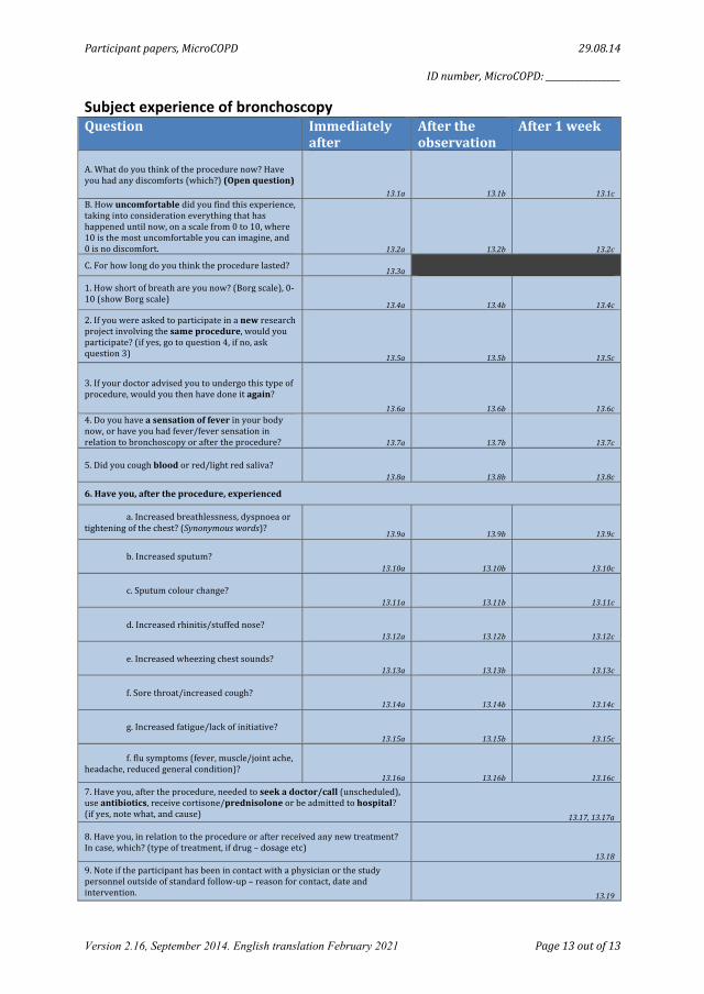
Participantpapers,MicroCOPD 29.08.14 IDnumber,MicroCOPD:_________________
Version 2.16, September 2014. English translation February 2021 Page13outof13
Subject experience of bronchoscopy Question Immediately
afterAftertheobservation
After1week
A.Whatdoyouthinkoftheprocedurenow?Haveyouhadanydiscomforts(which?)(Openquestion)
13.1a 13.1b 13.1cB.Howuncomfortabledidyoufindthisexperience,takingintoconsiderationeverythingthathashappeneduntilnow,onascalefrom0to10,where10isthemostuncomfortableyoucanimagine,and0isnodiscomfort. 13.2a 13.2b 13.2c
C.Forhowlongdoyouthinktheprocedurelasted? 13.3a
1.Howshortofbreathareyounow?(Borgscale),0-10(showBorgscale)
13.4a 13.4b 13.4c
2.Ifyouwereaskedtoparticipateinanewresearchprojectinvolvingthesameprocedure,wouldyouparticipate?(ifyes,gotoquestion4,ifno,askquestion3) 13.5a 13.5b 13.5c
3.Ifyourdoctoradvisedyoutoundergothistypeofprocedure,wouldyouthenhavedoneitagain?
13.6a 13.6b 13.6c4.Doyouhaveasensationoffeverinyourbodynow,orhaveyouhadfever/feversensationinrelationtobronchoscopyoraftertheprocedure? 13.7a 13.7b 13.7c
5.Didyoucoughbloodorred/lightredsaliva?13.8a 13.8b 13.8c
6.Haveyou,aftertheprocedure,experienced a.Increasedbreathlessness,dyspnoeaortighteningofthechest?(Synonymouswords)?
13.9a 13.9b 13.9c
b.Increasedsputum?13.10a 13.10b 13.10c
c.Sputumcolourchange?13.11a 13.11b 13.11c
d.Increasedrhinitis/stuffednose?13.12a 13.12b 13.12c
e.Increasedwheezingchestsounds?13.13a 13.13b 13.13c
f.Sorethroat/increasedcough?13.14a 13.14b 13.14c
g.Increasedfatigue/lackofinitiative?13.15a 13.15b 13.15c
f.flusymptoms(fever,muscle/jointache,headache,reducedgeneralcondition)?
13.16a 13.16b 13.16c
7.Haveyou,aftertheprocedure,neededtoseekadoctor/call(unscheduled),useantibiotics,receivecortisone/prednisoloneorbeadmittedtohospital?(ifyes,notewhat,andcause) 13.17,13.17a8.Haveyou,inrelationtotheprocedureorafterreceivedanynewtreatment?Incase,which?(typeoftreatment,ifdrug–dosageetc)
13.18
9.Noteiftheparticipanthasbeenincontactwithaphysicianorthestudypersonneloutsideofstandardfollow-up–reasonforcontact,dateandintervention. 13.19


Appendix 2


Ringeskjema OLS forverrelser for pasienter i studien MikroKOLS
Pasient status: ☐ KOLS ☐ Astma Dato for første bronkoskopi (dd-MMM-åå): _______________________ evt Dato for andre bronkoskopi (dd-MMM-åå): _______________________ Planlagt ca dato for første telefon: (dd-MMM-åå): ______________________ Planlagt ca dato for andre telefon: (dd-MMM-åå): ______________________ Planlagt ca dato for tredje telefon: (dd-MMM-åå): ______________________ Planlagt ca dato for siste telefon: (dd-MMM-åå): ______________________ Faste medisiner for obstruktiv lungesykdom (fylles ut av bronkoskopør): Navn (x: Seretide diskus) Styrke (x: 50/500ug/d) Dosering (x: 1x2) _____________________________________________________________________________________________ _____________________________________________________________________________________________ _____________________________________________________________________________________________ _____________________________________________________________________________________________ _____________________________________________________________________________________________

Dato for forrige kontakt (dd-MMM-åå): _______________________
Dato for oppnådd telefonkontakt (dd-MMM-åå):_______________________ 1) INNLEGGELSER "Har du siden forrige telefonkontakt vært innlagt på sykehus?" ☐ Nei ☐ Ja Hvis ja: "Hvor mange ganger siden forrige telefonkontakt har du vært innlagt?": ______ For første innleggelse: "Var du innlagt for en forverrelse av din KOLS/astma?" ☐ Nei ☐ Ja Hvis Nei: "Hva var årsaken til innleggelsen?":__________________________________________________ "Husker du omtrent hvilken dato du ble innlagt?" (ca dd-MMM-åå):_______________ "Husker du om du fikk antiobiotika (Som Amoxicillin eller lignende) eller prednisolon under eller etter innleggelsen?"
☐ Begge deler ☐ Kun Prednisolon ☐ Kun antiobiotika For evt andre innleggelse: "Var du innlagt for en forverrelse av din KOLS/astma?" ☐ Nei ☐ Ja Hvis Nei: "Hva var årsaken til innleggelsen?":__________________________________________________ "Husker du omtrent hvilken dato du ble innlagt?" (ca dd-MMM-åå):_______________ "Husker du om du fikk antiobiotika (Som Amoxicillin eller lignende) eller prednisolon under eller etter innleggelsen?"
☐ Begge deler ☐ Kun Prednisolon ☐ Kun antiobiotika For evt tredje innleggelse: "Var du innlagt for en forverrelse av din KOLS/astma?" ☐ Nei ☐ Ja Hvis Nei: "Hva var årsaken til innleggelsen?":__________________________________________________ "Husker du omtrent hvilken dato du ble innlagt?" (ca dd-MMM-åå):_______________ "Husker du om du fikk antiobiotika (Som Amoxicillin eller lignende) eller prednisolon under eller etter innleggelsen?"
☐ Begge deler ☐ Kun Prednisolon ☐ Kun antiobiotika 2) BESØK I AKUTTMOTTAK MEN IKKE INNLAGT (som regel definert som under 6 timer i mottak)

"Har du vært innom sykehusets akuttmottak grunnet forverrelse av din KOLS/astma, men ikke hatt behov for innleggelse?" ☐ Nei ☐ Ja Hvis ja: "Hvor mange ganger?": ___________ For første hendelse: "Husker du omtrent hvilken dato du var i akuttmottaket?": ___________________ "Husker du om du fikk antiobiotika (Som Amoxicillin eller lignende) eller prednisolon under eller etter besøket?"
☐ Begge deler ☐ Kun Prednisolon ☐ Kun antiobiotika For evt andre hendelse: "Husker du omtrent hvilken dato du var i akuttmottaket?": ___________________ "Husker du om du fikk antiobiotika (Som Amoxicillin eller lignende) eller prednisolon under eller etter besøket?"
☐ Begge deler ☐ Kun Prednisolon ☐ Kun antiobiotika For evt tredje hendelse: "Husker du omtrent hvilken dato du var i akuttmottaket?": ___________________ "Husker du om du fikk antiobiotika (Som Amoxicillin eller lignende) eller prednisolon under eller etter besøket?"
☐ Begge deler ☐ Kun Prednisolon ☐ Kun antiobiotika 3) BESØK HOS FASTLEGEN (eller tilsvarende på fastlegekontoret) "Har du siden forrige telefonkontakt vært hos fastlegen og fått behandling for en forverrelse av din KOLS/astma?" ☐ Nei ☐ Ja Hvis ja: "Hvor mange ganger siden forrige telefonkontakt har du vært hos fastlegen?": ________ For første hendelse: "Husker du omtrent hvilken dato du var hos fastlegen?": ___________________ "Husker du om du fikk antiobiotika (Som Amoxicillin eller lignende) eller prednisolon etter besøket?"
☐ Begge deler ☐ Kun Prednisolon ☐ Kun antiobiotika For evt andre hendelse: "Husker du omtrent hvilken dato du var hos fastlegen?": ___________________ "Husker du om du fikk antiobiotika (Som Amoxicillin eller lignende) eller prednisolon etter besøket?"
☐ Begge deler ☐ Kun Prednisolon ☐ Kun antiobiotika

For evt tredje hendelse: "Husker du omtrent hvilken dato du var hos fastlegen?": ___________________ "Husker du om du fikk antiobiotika (Som Amoxicillin eller lignende) eller prednisolon etter besøket?"
☐ Begge deler ☐ Kun Prednisolon ☐ Kun antiobiotika "Har du siden forrige telefonkontakt vært i telefonkontakt med fastlegen og fått behandling for en forverrelse av din KOLS/astma?" ☐ Nei ☐ Ja Hvis ja: "Hvor mange ganger?": _____________ For første hendelse: "Husker du omtrent hvilken dato du ringte fastlegen?": ___________________ "Husker du om du fikk antiobiotika (Som Amoxicillin eller lignende) eller prednisolon etter telefonkontakten?"
☐ Begge deler ☐ Kun Prednisolon ☐ Kun antiobiotika For evt andre hendelse: "Husker du omtrent hvilken dato du ringte fastlegen?": ___________________ "Husker du om du fikk antiobiotika (Som Amoxicillin eller lignende) eller prednisolon etter telefonkontakten?"
☐ Begge deler ☐ Kun Prednisolon ☐ Kun antiobiotika For evt tredje hendelse: "Husker du omtrent hvilken dato du ringte fastlegen?": ___________________ "Husker du om du fikk antiobiotika (Som Amoxicillin eller lignende) eller prednisolon etter telefonkontakten?"
☐ Begge deler ☐ Kun Prednisolon ☐ Kun antiobiotika 4) BESØK PÅ AKUTT LEGEVAKT "Har du siden forrige telefonkontakt vært på akutt legevakten og fått
behandling for en forverrelse av din KOLS/astma?" ☐ Nei ☐ Ja Hvis ja: "Hvor mange ganger siden forrige telefonkontakt har du vært på legevakten?": ________ For første hendelse: "Husker du omtrent hvilken dato du var på legevakten?": ___________________

"Husker du om du fikk antiobiotika (Som Amoxicillin eller lignende) eller prednisolon etter besøket?"
☐ Begge deler ☐ Kun Prednisolon ☐ Kun antiobiotika For evt andre hendelse: "Husker du omtrent hvilken dato du var på legevakten?": ___________________ "Husker du om du fikk antiobiotika (Som Amoxicillin eller lignende) eller prednisolon etter besøket?"
☐ Begge deler ☐ Kun Prednisolon ☐ Kun antiobiotika For evt tredje hendelse: "Husker du omtrent hvilken dato du var på legevakten?": ___________________ "Husker du om du fikk antiobiotika (Som Amoxicillin eller lignende) eller prednisolon etter besøket?"
☐ Begge deler ☐ Kun Prednisolon ☐ Kun antiobiotika "1000 takk for hjelpen, vi ringer deg tilbake om ca 3 måneder!"


Appendix 3


Supplementary Table 4 with marked correction
Complication type
ref #
First author
Journal
Year published
Population
Exclusion/inclusion criteria
Sample size
Mean complication
prevalence
Remarks
Death
53
Dunagan
Chest
1997
Bone marrow
recipients
Excluded: Patients whose
clinical assessment indicated
that oxygen and haemodynamic
status might be compromised
by the procedure.
71
2.8%
24-hour mortality. The
patients who died
experienced extensive
bleeding during the
procedure.
54
Shannon
Bone Marrow
Transplant
2010
Bone marrow
recipients
Included: Patients whose SaO2
exceeded 90% on room air or
supplemental O2. For TBB
with BAL: platelet count > 80
x106 /mcgL.
501
0.0%
24-hour mortality. States
no procedure-related
deaths.
55
D'Ippolito
Monaldi Archives
for Chest Disease
2007
Elderly patients
Excluded: Platelet count <50
000, APTT >=50 s or PTT
=<75%, and haemodynamic
instability.
301
0.0%
56
Diaz-Guzman
Respiration
2009
Pulmonary
hypertension
patients
Included: Patients with a
diagnosis of PH defined as (1)
mean PAP >25 mmHg
measured by right heart
catheterization or (2) right
ventricular systolic pressure
>40 mm Hg estimated by
Doppler echocardiography and
clinical evidence of heart
failure. Controls: Patients that
did not meet criteria for PH +
underwent bronchoscopy by the
same physician within 48 hours
of the study patients.
90
0.0%

57
Dang
Internal Medicine
Journal
2012
Consecutive
patients referred
to bronchoscopy
Excluded: Patients with
hypersensitivity or allergies to
anaesthetic medications,
opioids or benzodiazepines,
with brady- or tachycardia
(resting heart rate <60 or
>100 bpm), or systolic blood
pressure of <100 or
>180 mmHg), concurrent
psychological disorders, those
already on high doses of
opioids or benzodiazepines,
and oxygen requirements of
>4 L/min O2 at rest.
539
0.0%
58
Jain
Chest
2004
Immunocompromi
sed patients with
lung infiltrates
Included:
Immunocompromised patients
with the presence of either a
focal or diffuse pulmonary
infiltrate, referred and giving
informed consent to
bronchoscopy. Excluded: HIV
positive patients, lung
transplant recipients.
104
0.0%
59
Grendelmeier
Swiss Medical
Weekly
2011
Elective
bronchoscopy
patients
Excluded: Patients who were
intubated and patients with
known propofol
allergy/intolerance.
440
0.0%
60
Schlatter
The European
respiratory journal
2011
Elective
bronchoscopy
patients
Excluded: Intubated patients,
patients receiving
interventional bronchoscopy
(e.g. laser or stent), patients
with known hydrocodone or
propofol allergy/intolerance.
300
0.0%

61
Grendelmeier
The European
respiratory journal
2014
Patients
undergoing non-
emergency
bronchoscopy
Excluded: ICU/emergency
bronchoscopy, patients aged
<18 years, intubated or isolated
patients, patients with known
propofol allergy/intolerance,
pregnant or breastfeeding
patients, and patients with a
mental disorder preventing
appropriate judgment
concerning study participation.
702
0.0%
Bleeding
58
Jain
Chest
2004
Immunocompromi
sed patients with
lung infiltrates
Included:
Immunocompromised patients
with the presence of either a
focal or diffuse pulmonary
infiltrate, referred and giving
informed consent to
bronchoscopy. Excluded: HIV
positive patients, lung
transplant recipients.
104
13.5%
59
Grendelmeier
Swiss Medical
Weekly
2011
Elective
bronchoscopy
patients
Excluded: Patients who were
intubated and patients with
known propofol
allergy/intolerance.
440
2.5%
Minor bleeding. Not
defined.
62
Carr
Respiration
2012
Patients with low
risk of bleeding
Excluded: Patients with active
pulmonary bleeding
(haemoptysis), personal/family
history or physical evidence of
bleeding tendencies (petechiae,
purpura, ecchymosis,
haematoma), ever having anti-
platelet therapy (aspirin,
clopidogrel) or anti-coagulation
therapy (warfarin, heparin),
history or physical evidence of
liver disease (hepatitis,
cirrhosis, hepatomegaly,
ascites, jaundice), platelet count
<20 *10^3.
234
89.7%-8.1%-2.1%
Minimal - mild -
moderate

63
Ni
Chang Gung
medical journal
2010
Patients
undergoing
diagnostic
bronchoscopy
Excluded: patients aged <18
years, ASA IV or V, neurologic
disorders or other conditions
contributing to difficulty in
assessing a conscious response,
FVC < 15 ml/kg body weight,
FEV1 < 1000 ml, or
FEV1/FVC < 35%, patients
with known allergy to study
drugs, and patients with
glaucoma.
88
26.1%
In need of adrenalin
spray.
64
Herth
Chest
2002
Patients aged > 40
undergoing TBB
Excluded: Patients with
warfarin/heparin use last 2
weeks, known bleeding
disorder, or TPK<80
000cells/mcL.
1217
4.7%
65
von Bartheld
JAMA
2013
Patients with
sarcoidosis
Excluded: Patients with
obvious organ involvement of
sarcoidosis with the possibility
to confirm granulomas with a
minimally invasive diagnostic
procedure (e.g., biopsy of skin
lesions or superficial lymph
nodes), patients with Lofgren
syndrome, inability to undergo
endoscopy, pregnancy, or
inability to consent.
304*
8.0%
Percentage of bleeding
in patients undergoing
bronchoscopy. *149
undergoing
bronchoscopy.
66
Bilaceroglu
Monaldi Archives
for Chest Disease
1997
Endobronchial
lung cancer
patients
Included: Visible cauliflower-
like or infiltrative
endobronchial tumours were
enrolled. Excluded: Patients
with only extrinsic
compression, extrabronchial
tumour or
submucosal/peribronchial
involvement.
151
7.0%-17.0% -13.0%
EBNA-forceps biopsy-
bronchial brushing.

67
Diette
Chest
1999
Lung transplant
recipients (LTRs),
and patients
referred to
bronchoscopy
Excluded: Patients who were
mechanically ventilated, non-
English speaking or had
communication deficits that
precluded answering questions,
and who died within 48 hours
of the procedure.
697
44.5% - 17.5%
LTRs - other patients.
68
Hetzel
The European
respiratory journal
2012
Patients with
suspected
endobronchial
tumours
Included: Patients with
suspected endobronchial lesion
based on clinical signs and
radiological images, aged >18
years, who signed informed
consent. Excluded: Patients
with a bleeding diathesis,
patients on anticoagulants,
patients with SaO2<90%
(under delivery of 2L O2/min),
or unstable angina pectoris,
myocardial infarction in the
past month, or decompensated
heart failure.
593
69.4% (17.8%)
(Requiring intervention).
69
Khan
Respiration
2010
Patients with
visible
endobronchial
lesions.
Excluded: Patients aged <18
years, patients with
anticoagulantia use, known
bleeding diathesis, blood
pressure >140/90 mmHg,
pacemaker or defibrillator
implants, poor
cardiopulmonary reserve or
marked hypoxaemia at rest,
patients unable to cooperate,
and patients enrolled previously
in the study.
160
84.4%- 15.6%-0.0%
Mild-moderate-severe.
70
Williams
Chest
1998
Consecutive
patients referred
to bronchoscopy
Excluded: Patients with
tracheostomy, cerebral
secondaries, supraventricular
tachycardia, haemoptysis,
unstable angina and mechanical
ventilation.
100
42.0% - 48.0% -
10.0%
Without need for wash -
needed saline wash -
needed adrenalin.

71
Choi
The International
journal of
tuberculosis and
lung disease
2005
Adult, Korean
patients, referred
to bronchoscopy
Excluded: Patients subject to
follow-up after lung resection
surgery, patients receiving
midazolam, therapeutic
bronchoscopy, patients without
an interview due to early
discharge, patients
experiencing lidocain toxicity,
patients unable to speak
Korean, and patients who
refused to enroll in the study.
307
43.0-35.6%
Nasal - oral insertion of
scope.
Pneumothorax
55
D'Ippolito
Monaldi Archives
for Chest Disease
2007
Elderly patients
Excluded: Platelet count <50
000, APTT >=50 s or PTT
=<75%, and haemodynamic
instability.
301
0.3%
Occurred in one patient
without a biopsy
procedure
57
Dang
Internal medicine
journal
2012
Consecutive
patients referred
to bronchoscopy
Excluded: Patients with
hypersensitivity or allergies to
anaesthetic medications,
opioids or benzodiazepines,
with brady- or tachycardia
(resting heart rate <60 or
>100 bpm), or systolic blood
pressure of <100 or
>180 mmHg), concurrent
psychological disorders, those
already on high doses of
opioids or benzodiazepines,
and oxygen requirements of
>4 L/min O2 at rest.
539
0.5% - 1.6%
Direct consequence of
FFB - percentage of
TBB.
58
Jain
Chest
2004
Immunocompromi
sed patients with
lung infiltrates
Included:
Immunocompromised patients
with the presence of either a
focal or diffuse pulmonary
infiltrate, referred and giving
informed consent to
bronchoscopy. Excluded: HIV
positive patients, lung
transplant recipients.
104
4 %

64
Herth
Chest
2002
Patients aged > 40
undergoing TBB
Excluded: Patients with
warfarin/heparin use last 2
weeks, known bleeding
disorder, or TPK<80
000cells/mcL.
1217
2.1%
65
von Bartheld
JAMA
2013
Patients with
sarcoidosis
Excluded: Patients with
obvious organ involvement of
sarcoidosis with the possibility
to confirm granulomas with a
minimally invasive diagnostic
procedure (biopsy of skin
lesions or superficial lymph
nodes), patients with Lofgren
syndrome, inability to undergo
endoscopy, pregnancy, or
inability to consent.
304*
0.7%
One patient, after TBB. *
149 undergoing
bronchoscopy.
66
Bilaceroglu
Monaldi Archives
for Chest Disease
1997
Endobronchial
lung cancer
patients
Included: Visible cauliflower-
like or infiltrative
endobronchial tumour patients.
Excluded: Patients with only
extrinsic compression,
extrabronchial tumour or
submucosal/peribronchial
involvement.
151
0.0%
72
Hirose
Respirology
2008
Japanese, adult
patients
undergoing
diagnostic
bronchoscopy
Excluded: Patients with known
confusion (cerebral metastases,
degenerative dementia, or other
conditions), or patients judged
too ill to participate, intubation,
aged >80 years, patients unable
to speak Japanese.
129
0.0%
73
Tukey
Respiratory
medicine
2012
Population based
Excluded: Patients who had
undergone invasive procedures
associated with complications
during the same visit.
-
0.97%
Biopsy procedures
complicated by
pneumothorax.
Bronchospasm
55
D'Ippolito
Monaldi Archives
for Chest Disease
2007
Elderly patients
Excluded: Platelet count <50
000, APTT >=50 s or PTT
=<75%, and haemodynamic
instability.
301
1.1%

57
Dang
Internal medicine
journal
2012
Consecutive
patients referred
to bronchoscopy
Excluded: Patients with
hypersensitivity or allergies to
anaesthetic medications,
opioids or benzodiazepines,
with brady- or tachycardia
(resting heart rate <60 or
>100 bpm), or systolic blood
pressure of <100 or
>180 mmHg), concurrent
psychological disorders, those
already on high doses of
opioids or benzodiazepines,
and oxygen requirements of
>4 L/min O2 at rest.
539
0.0%
74
Kaur
Chest
2015
Consecutive
patients referred
to bronchoscopy
Included: Patients with
indication for flexible
bronchoscopy, aged 12 to 90
years, who had systolic BP 100
- 180 mm Hg. Excluded:
Pregnant patients, SaO2<92%
with FiO2 <0.3, patients
undergoing TBNA or other
interventions, patients failing to
provide informed consent.
500
0.0%
Bronchospasm.
75
Tapanainen
Respiratory
medicine
2002
Asthmatics
Included: Non-smoking 18-60
year-old asthmatics[ATS
criteria (5)] FEV1 from 60-
100% of predicted and a
moderate or severe bronchial
hyperreactivity [provocative
dose of histamine inducing a
fall of 15% in FEV1 less than
0,4 and 0,1 mg respectively.
57
12.0%
Cough/Bronchospasm
that interfered with
procedure.
Hypoxaemia
59
Grendelmeier
Swiss Medical
Weekly
2011
Elective
bronchoscopy
patients
Excluded: Patients who were
intubated and patients with
known propofol
allergy/intolerance.
440
16.4%
Fall in SaO2 to <90%.

61
Grendelmeier
The European
respiratory journal
2014
Patients
undergoing non-
emergency
bronchoscopy
Excluded: ICU/emergency
bronchoscopy, patients aged
<18 years, intubated or isolated
patients, patients with known
propofol allergy/intolerance,
pregnant or breastfeeding
patients, and patients with a
mental disorder preventing
appropriate judgment
concerning study participation.
702
28.9%
Fall in SaO2 to <90%.
63
Ni
Chang Gung
medical journal
2010
Patients
undergoing
diagnostic
bronchoscopy
Excluded: patients aged <18
years, ASA IV or V, neurologic
disorders or other conditions
contributing to difficulty in
assessing a conscious response,
FVC < 15 ml/kg body weight,
FEV1 < 1000 ml, or
FEV1/FVC < 35%, patients
with known allergy to study
drugs, and patients with
glaucoma.
88
4.5% - 31.8%
Fall in SaO2 to <90%.
Unsedated - Sedated
patients.
76
Lo
PLoS One
2011
Elective
bronchoscopy
patients
Exluded: Patients aged <18
years, ASA IV or V, neurologic
disorders, FVC < 15 ml/kg,
FEV1 < 1L, FEV1/FVC <
35%, Mallampati score of 4.
492
37.8%
Fall in SaO2 to <90%.

77
Ogawa
Respiratory
investigation
2014
Consecutive
patients ≥ 20
years undergoing
diagnostic
bronchoscopy
Included: Patients able to
answer a simple questionnaire
without assistance; adequate
hepatic function
(AST≤100 IU/L,
ALT≤100 IU/L, and total
bilirubin≤1.5 mg/dL); adequate
renal function (serum
creatinine≤1.5 mg/dL); and
adequate respiratory function
(PaO2≥60 mmHg or
SaO2≥90% while breathing
room air). Excluded: Patients
with type II chronic respiratory
failure requiring LTOT; sleep
apnea syndrome requiring
CPAP; history of severe drug
allergy; neuromuscular
disorders; acute narrow-angle
glaucoma; myocardial
infarction onset within 6
weeks; and pregnant or
lactating women.
204
75.5%
Percentage in need of
supplemental oxygen to
prevent SpO2 from
declining to <90%.
78
Fruchter
Respiration
2014
Elective
bronchoscopy
patients
Excluded: Patients unable to
give or who refused to provide
informed consent, patients aged
<18 years, patients with
endotracheal tube or
tracheostomy, and patients with
propofol allergy.
81
74.0%
Patients who required
supplemental oxygen by
100% O2 mask due to
hypoxaemia
(SaO2<90%).
79
Ryu
British journal of
anaesthesia
2012
Elective
bronchoscopy
patients
Included: Patients aged 18-70
years, ASA I-III. Excluded:
Patients with SaO2 <90%,
FEV1 < 1.0L, HR < 60 bpm,
systolic pressure <100 mmHg,
asthmatics, COPD patients, and
pregnant patients.
72
28.6% - 2.9%
Oxygen desaturation
(<90% for >10s):
propofol–
dexmedetomidine group
- propofol–remifentanil
group.

80
Rosell
Respiratory
medicine
2006
Consecutive, adult
patients referred
to bronchoscopy
with BAL
Contraindications for BAL:
Lack of patient cooperation,
hypercapnia/hypoxaemia
uncorrected by supplemental
O2 administration, serious
cardiac arrhythmias, recent
acute myocardial infarction or
recent asthma attack,
uncorrected bleeding diathesis
and haemodynamic instability.
295
3.4%
Fall in SaO2 to <90%.
81
Gibson
Australian and
New Zealand
journal of
medicine
1990
Patients
undergoing BAL
Data was only included from
patients whose transcutaneous
PO2 was within + 5mmHg of
the arterial PaO2 value.
88
25.0% - 75.0%
Patients receiving
oxygen - patients who
did not receive oxygen.
82
Stolz
Chest
2005
Patients
undergoing
diagnostic
bronchoscopy
Excluded: Intubated patients,
those receiving propofol (e.g.
IV drug abuse), and patients
requiring endobronchial
ultrasound examination.
150
4.0% - 8.0%
Nebulised lidocain -
nebulised placebo, fall in
SaO2 to <90%.
Haemodynamic
variations
59
Grendelmeier
Swiss Medical
Weekly
2011
Elective
bronchoscopy
patients
Excluded: Patients who were
intubated and patients with
known propofol
allergy/intolerance.
440
37.0% - 15.4%
Drop in SBP >20 mmHg
– Hypotension.
61
Grendelmeier
The European
respiratory journal
2014
Patients
undergoing non-
emergency
bronchoscopy
Excluded: ICU/emergency
bronchoscopy, patients aged
<18 years, intubated or isolated
patients, patients with known
propofol allergy/intolerance,
pregnant or breastfeeding
patients, and patients with a
mental disorder preventing
appropriate judgment
concerning study participation.
702
28.9%
Hypotension.

63
Ni
Chang Gung
medical journal
2010
Patients
undergoing
diagnostic
bronchoscopy
Excluded: patients aged <18
years, ASA IV or V, neurologic
disorders or other conditions
contributing to difficulty in
assessing a conscious response,
FVC < 15 ml/kg body weight,
FEV1 < 1000 ml, or
FEV1/FVC < 35%, patients
with known allergy to study
drugs, and patients with
glaucoma.
88
4.5% - 47.7% vs
20.5%
Hypotension-
Hypertension in non-
sedated vs sedated
patients.
70
Williams
Chest
1998
Consecutive
patients referred
to bronchoscopy
Excluded: Patients with
tracheostomy, cerebral
secondaries, supraventricular
tachycardia, haemoptysis,
unstable angina and mechanical
ventilation.
100
74
Kaur
Chest
2015
Consecutive
patients referred
to bronchoscopy
Included: Patients with
indication for flexible
bronchoscopy, aged 12 to 90
years, who had systolic BP 100
- 180 mm Hg. Excluded:
Pregnant patients, SaO2<92%
with FiO2 <0.3, patients
undergoing TBNA or other
interventions, patients failing to
provide informed consent.
500
0 %
Arrhythmias. Post-
procedural increase in
HR, RR, and BP (rates
not provided).
76
Lo
PLoS One
2011
Elective
bronchoscopy
patients
Exluded: Patients aged <18
years, ASA IV or V, neurologic
disorders, FVC < 15 ml/kg,
FEV1 <1L, FEV1/FVC < 35%,
Mallampati score of 4.
492
3.0% - 5.9%
MAP<60mmHg - SBP
<90 mmHg.
78
Fruchter
Respiration
2014
Elective
bronchoscopy
patients
Excluded: Patients unable to
give or who refused to provide
informed consent, patients aged
<18 years, patients with
endotracheal tube or
tracheostomy, and patients with
propofol allergy.
81
16.0%- 6.2%
Hypotension -
Hypotension requiring
phenylephrine.

79
Ryu
British journal of
anaesthesia
2012
Elective
bronchoscopy
patients
Included: Patients aged 18-70
years, ASA I-III. Excluded:
Patients with SaO2 <90%,
FEV1 < 1.0L, HR < 60 bpm,
systolic pressure <100 mmHg,
asthmatics, COPD patients, and
pregnant patients.
72
10.0%- 11.0% - 2.9% -
25.7% - 0.0%
Arrhythmias -
Hypertension -
Hypotension -
Tachycardia –
Bradycardia.
83
Silvestri
Chest
2009
Patients
undergoing
bronchoscopy and
meeting RCT
criteria
Included: Patients aged ≥ 18
years, ASA I-IV, non-pregnant.
Excluded: Patients with
hypersensitivity to any
anaesthetic or opioid; failure to
meet non per os status; an
abnormal, clinically significant
ECG finding; participation in
an investigational drug study
within the previous month;
patients with Mallampati score
of IV, or Mallampati score of
III and a thyromental distance
of ≤ 4 cm, or a difficult airway
for any other reason as judged
by the clinician.
252
3.2% - 0.0%
Hypotension -
Bradycardia
Fever and infection
55
D'Ippolito
Monaldi Archives
for Chest Disease
2007
Elderly patients
Excluded: Platelet count <50
000, APTT >=50 s or PTT
=<75%, and haemodynamic
instability.
301
10.0%
Fever (axillary body
temperature >=38degr.
C).
57
Dang
Internal medicine
journal
2012
Consecutive
patients referred
to bronchoscopy
Excluded: Patients with
hypersensitivity or allergies to
anaesthetic medications,
opioids or benzodiazepines,
with brady- or tachycardia
(resting heart rate <60 or
>100 bpm), or systolic blood
pressure of <100 or
>180 mmHg), concurrent
psychological disorders, those
already on high doses of
opioids or benzodiazepines,
and oxygen requirements of
>4 L/min O2 at rest.
539
5.7%
Perceived fever within
48 hours.
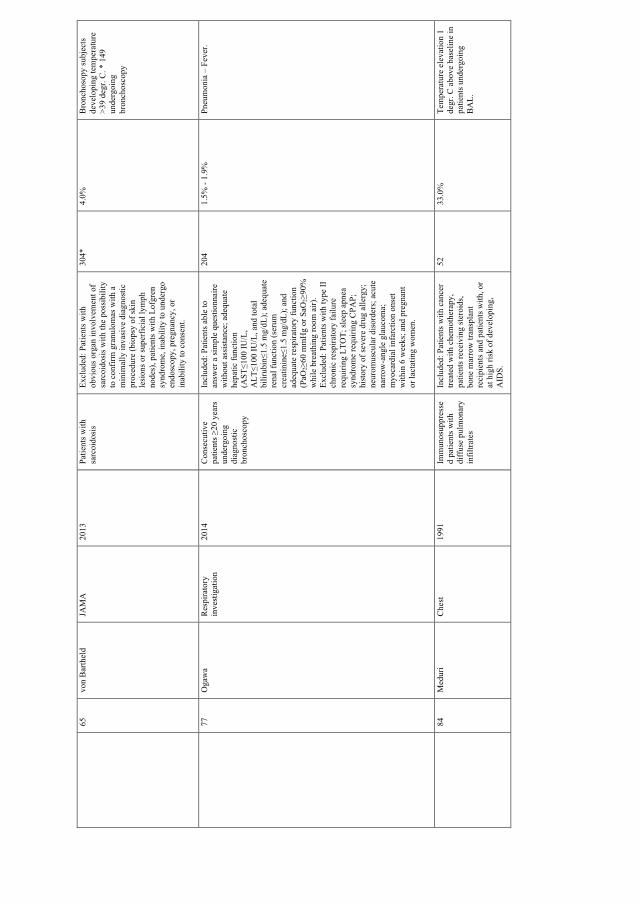
65
von Bartheld
JAMA
2013
Patients with
sarcoidosis
Excluded: Patients with
obvious organ involvement of
sarcoidosis with the possibility
to confirm granulomas with a
minimally invasive diagnostic
procedure (biopsy of skin
lesions or superficial lymph
nodes), patients with Lofgren
syndrome, inability to undergo
endoscopy, pregnancy, or
inability to consent.
304*
4.0%
Bronchosopy subjects
developing temperature
>39 degr. C. * 149
undergoing
bronchoscopy
77
Ogawa
Respiratory
investigation
2014
Consecutive
patients ≥20 years
undergoing
diagnostic
bronchoscopy
Included: Patients able to
answer a simple questionnaire
without assistance; adequate
hepatic function
(AST≤100 IU/L,
ALT≤100 IU/L, and total
bilirubin≤1.5 mg/dL); adequate
renal function (serum
creatinine≤1.5 mg/dL); and
adequate respiratory function
(PaO
2≥60 mmHg or SaO
2≥90%
while breathing room air).
Excluded: Patients with type II
chronic respiratory failure
requiring LTOT; sleep apnea
syndrome requiring CPAP;
history of severe drug allergy;
neuromuscular disorders; acute
narrow-angle glaucoma;
myocardial infarction onset
within 6 weeks; and pregnant
or lactating women.
204
1.5% - 1.9%
Pneumonia – Fever.
84
Meduri
Chest
1991
Immunosuppresse
d patients with
diffuse pulmonary
infiltrates
Included: Patients with cancer
treated with chemotherapy,
patients receiving steroids,
bone marrow transplant
recipients and patients with, or
at high risk of developing,
AIDS.
52
33.0%
Temperature elevation 1
degr. C above baseline in
patients undergoing
BAL.

85
Krause
American Journal
of Respiratory and
Critial Care
Medicine
1997
Consecutive
patients referred
to bronchoscopy
Excluded: Patients with fever
or with infectious pneumonia
and patients receiving more
than 10 mg prednisolone
equivalent per day.
50
24.0%
Body temperature above
38 degr.C. No difference
between BAL and non-
BAL groups.
86
Pereira
American Review
of Respiratory
Disease
1975
Hospitalised
patients with
indication for
bronchoscopy
Excluded: Patients who had
fever during the week before
endoscopy; 5 procedures
performed in 5 patients were
excluded because of incomplete
data.
95
6.3% - 9.5%
Fever with-without
infiltration on chest X-
ray.
87
Gonzalez Aguirre
The International
journal of
Tuberculosis and
Lung Disease
2015
Hospitalised
patients with
indication for
bronchoscopy
Included: Patients aged>18
years, with an indication for
bronchoscopy. Excluded:
Patients with tracheostomy,
endotracheal intubation or
contraindication for
bronchoscopy
(contraindications not listed).
63
65.1%
Increase in
symptomatology, mainly
due to fever. Details not
given.
88
Yigla
The European
respiratory journal
1999
Consecutive
patients referred
to bronchoscopy
Excluded: Patients with current
respiratory tract infection or
febrile illnesses and patients
receiving antibiotic therapy
within a week prior to the
bronchoscopy.
200
6.5%
Bacteriaemia rate.
Health care utilization
53
Dunagan
Chest
1997
Bone marrow
recipients
Excluded: Patients whose
clinical assessment indicated
that oxygen and haemodynamic
status might be compromised
by the procedure.
71
4.2% - 1.4%
Needed intubation -
needed pressor
treatment.
54
Shannon
Bone Marrow
Transplantation
2010
Bone marrow
recipients
Included: Patients whose SaO2
exceeded 90% on room air or
supplemental O2. For TBB
with BAL: platelet count > 80
x106 /mcgL.
501
0.6%
Percentage of patients
who needed high
inspired oxygen.
55
D'Ippolito
Monaldi Archives
for Chest Disease
2007
Elderly patients
Excluded: Platelet count <50
000, APTT >=50 s or PTT
=<75%, and haemodynamic
instability.
301
0.0%
Reports no need for
mechanical ventilation
and no procedure
interrupted by major
complications.

56
Diaz-Guzman
Respiration
2009
Patients with
pulmonary
hypertension
Included: Patients with a
diagnosis of PH defined as (1)
mean PAP >25 mmHg
measured by right heart
catheterization or (2) right
ventricular systolic pressure
>40 mm Hg estimated by
Doppler echocardiography and
clinical evidence of heart
failure. Controls: Patients that
did not meet criteria for PH +
underwent bronchoscopy by the
same physician within 48 h of
the study patients.
90
0.0%
Reports no moderate or
severe haemorrhage,
refractory hypotension,
haemodynamic
instability, death,
hospitalisation or
respiratory failure.
57
Dang
Internal medicine
journal
2012
Consecutive
patients referred
to bronchoscopy
Excluded: Patients with
hypersensitivity or allergies to
anaesthetic medications,
opioids or benzodiazepines,
with brady- or tachycardia
(resting heart rate <60 or
>100 bpm), or systolic blood
pressure of <100 or
>180 mmHg), concurrent
psychological disorders, those
already on high doses of
opioids or benzodiazepines,
and oxygen requirements of
>4 L/min O2 at rest.
539
0.4%
Two patients:
Pseudoseizure in need of
neurological
consultation, and one
ventricular tachycardia
in pre-existing severe
cardiomyopathy.
58
Jain
Chest
2004
Immunocompromi
sed patients with
lung infiltrates
Included:
Immunocompromised patients
with the presence of either a
focal or diffuse pulmonary
infiltrate, referred and giving
informed consent to
bronchoscopy. Excluded: HIV
positive patients, lung
transplant recipients.
104
2.9%
Short-term ventilatory
support after FFB
(respiratory failure) in
two patients. One patient
required transfusion with
packed RBCs.
59
Grendelmeier
Swiss Medical
Weekly
2011
Elective
bronchoscopy
patients
Excluded: Patients who were
intubated and patients with
known propofol
allergy/intolerance.
440
0.0%
Defined adverse events.

60
Schlatter
The European
respiratory
journal
2011
Elective
bronchoscopy
patients
Excluded: Intubated patients,
patients receiving
interventional bronchoscopy
(e.g. laser or stent), patients
with known hydrocodone or
propofol allergy/intolerance.
300
31 %
Complications in need
of intervention.
61
Grendelmeier
The European
respiratory journal
2014
Patients
undergoing non-
emergency
bronchoscopy
Excluded: ICU/emergency
bronchoscopy, patients aged
<18 years, intubated or isolated
patients, patients with known
propofol allergy/intolerance,
pregnant or breastfeeding
patients, and patients with a
mental disorder preventing
appropriate judgment
concerning study participation.
702
0.3% - 0.3% - 6.3%
Patients transferred to
ICU - intubated -
receiving oro-
/nasopharyngeal airway.
65
von Bartheld
JAMA
2013
Patients with
sarcoidosis
Excluded: Patients with
obvious organ involvement of
sarcoidosis with the possibility
to confirm granulomas with a
minimally invasive diagnostic
procedure (biopsy of skin
lesions or superficial lymph
nodes), patients with Lofgren
syndrome, inability to undergo
endoscopy, pregnancy, or
inability to consent.
304*
1.3%
Pneumothorax after TBB
requiring chest tube
drainage in one patient.
Non-invasive ventilation
(<12 hours) after general
anaesthesia in one
patient.
68
Hetzel
The European
respiratory journal
2012
Patients with
suspected
endobronchial
tumours
Included: Patients with
suspected endobronchial lesion
based on clinical signs and
radiological images, aged >18
years, who signed informed
consent. Excluded: Patients
with a bleeding diathesis,
patients on anticoagulants,
patients with SaO2<90%
(under delivery of 2L O2/min),
or unstable angina pectoris,
myocardial infarction in the
past month, or decompensated
heart failure.
593
17.8%
All interventions related
to bleeding.

70
Williams
Chest
1998
Consecutive
patients referred
to bronchoscopy
Excluded: Patients with
tracheostomy, cerebral
secondaries, supraventricular
tachycardia, haemoptysis,
unstable angina and mechanical
ventilation.
100
0.0%
No admissions of
outpatients.
73
Tukey
Respiratory
medicine
2012
Population based
Excluded: Patients who had
undergone invasive procedures
associated with complications
during the same visit.
-
0.55%
Pneumothorax after
transbronchial biopsy
requiring chest tube
placement.
84
Meduri
Chest
1991
Immunosuppresse
d patients with
diffuse pulmonary
infiltrates
Included: Patients with cancer
treated with chemotherapy,
patients receiving steroids,
bone marrow transplant
recipients and patients with, or
at high risk of developing,
AIDS.
52
3.9%
Two patients
experienced respiratory
failure and were treated
with mechanical
ventilation.
86
Pereira
American Review
of Respiratory
Disease
1975
Hospitalised
patients with
indication for
bronchoscopy
Excluded: Patients who had
fever during the week before
endoscopy; 5 procedures
performed in 5 patients were
excluded because of incomplete
data.
95
1.0%
One patient needed
antimicrobial therapy.
Coughing
57
Dang
Internal medicine
journal
2012
Consecutive
patients referred
to bronchoscopy
Excluded: Patients with
hypersensitivity or allergies to
anaesthetic medications,
opioids or benzodiazepines,
with brady- or tachycardia
(resting heart rate <60 or
>100 bpm), or systolic blood
pressure of <100 or
>180 mmHg), concurrent
psychological disorders, those
already on high doses of
opioids or benzodiazepines,
and oxygen requirements of
>4 L/min O2 at rest.
539
10.8%
New cough after
bronchoscopy.

63
Ni
Chang Gung
medical journal
2010
Patients
undergoing
diagnostic
bronchoscopy
Excluded: patients aged <18
years, ASA IV or V, neurologic
disorders or other conditions
contributing to difficulty in
assessing a conscious response,
FVC < 15 ml/kg body weight,
FEV1 < 1000 ml, or
FEV1/FVC < 35%, patients
with known allergy to study
drugs, and patients with
glaucoma.
88
31.1%-55.7%
procedures interfered by
cough -patients with
post-procedural cough
65
von Bartheld
JAMA
2013
Patients with
sarcoidosis
Excluded: Patients with
obvious organ involvement of
sarcoidosis with the possibility
to confirm granulomas with a
minimally invasive diagnostic
procedure (biopsy of skin
lesions or superficial lymph
nodes), patients with Lofgren
syndrome, inability to undergo
endoscopy, pregnancy, or
inability to consent.
304*
4.7%
Intolerable cough during
bronchoscopy. *149
undergoing
bronchoscopy.
71
Choi
The International
journal of
tuberculosis and
lung disease
2005
Adult, Korean
patients, referred
to bronchoscopy
Excluded: Patients subject to
follow-up after lung resection
surgery, patients receiving
midazolam, therapeutic
bronchoscopy, patients without
an interview due to early
discharge, patients
experiencing lidocain toxicity,
patients unable to speak
Korean, and patients who
refused to enroll in the study.
307
44.3%-33.6%
Nasal - oral insertion of
scope. Patient-reported
72
Hirose
Respirology
2008
Japanese, adult
patients
undergoing
diagnostic
bronchoscopy
Excluded: Patients with known
confusion (cerebral metastases,
degenerative dementia, or other
conditions), or patients judged
too ill to participate, intubation,
aged > 80 years, patients
unable to speak Japanese.
129
86.0%
Percentage of patients
who reported to be
"bothered by coughing"
during bronchoscopy.

75
Tapanainen
Respiratory
medicine
2002
Asthma patients
Included: Non-smoking 18-60
year old asthmatics (ATS
criteria (5)). FEV1 from 60-
100% of predicted and a
moderate or severe bronchial
hyperreactivity (provocative
dose of histamine inducing a
fall of 15% in FEV1 less than
0,4 and 0,1 mg respectively).
57
52%-40%-12%
Cough-cough of no
significance-
cough/bronchospasm
that interfered with
procedure.
Other respiratory
symptoms and signs
57
Dang
Internal medicine
journal
2012
Consecutive
patients referred
to bronchoscopy
Excluded: Patients with
hypersensitivity or allergies to
anaesthetic medications,
opioids or benzodiazepines,
with brady- or tachycardia
(resting heart rate <60 or
>100 bpm), or systolic blood
pressure of <100 or
>180 mmHg), concurrent
psychological disorders, those
already on high doses of
opioids or benzodiazepines,
and oxygen requirements of
>4 L/min O2 at rest.
539
5.7%
Dyspnoea
61
Grendelmeier
The European
respiratory journal
2014
Patients
undergoing non-
emergency
bronchoscopy
Excluded: ICU/emergency
bronchoscopy, patients aged
<18 years, intubated or isolated
patients, patients with known
propofol allergy/intolerance,
pregnant or breastfeeding
patients, and patients with a
mental disorder preventing
appropriate judgment
concerning study participation.
702
0.1%
One patient experienced
respiratory failure.

71
Choi
The International
journal of
tuberculosis and
lung disease
2005
Adult, Korean
patients, referred
to bronchoscopy
Excluded: Patients subject to
follow-up after lung resection
surgery, patients receiving
midazolam, therapeutic
bronchoscopy, patients without
an interview due to early
discharge, patients
experiencing lidocain toxicity,
patients unable to speak
Korean, and patients who
refused to enroll in the study.
307
38.2% - 30.9%
Complained about
dyspnoea: Nasal - oral
insertion of scope.
75
Tapanainen
Respiratory
medicine
2002
Asthmatics
Included: Non-smoking 18-60
year-old asthmatics[ATS
criteria (5)] FEV1 from 60-
100% of predicted and a
moderate or severe bronchial
hyperreactivity [provocative
dose of histamine inducing a
fall of 15% in FEV1 less than
0,4 and 0,1 mg respectively.
57
3.5% - 5.2%
Dyspnoea - Increase in
asthma symptomatology
92
Humbert
Thorax
1996
Asthma patients
and controls
Excluded: Patients taking oral
corticosteroids in the last three
months, current smokers or ex-
smokers with more than 5 pack
years. Study 1: Excluded
inhaled corticosteroids for the
last three months. Study 2:
Excluded inhaled
corticosteroids for the last two
weeks, patients aged <18 or
>55 years, FEV1<50% of
predicted, acute or chronic
infection, pregnancy, breast
feeding, or any other significant
medical condition.
77
4.7% - 2.9%
Experienced mild
symptomatic wheeze
after the bronchoscopic
examination, which was
rapidly reversed with an
additional dose of
salbutamol by nebulizer:
Study 1 - study 2. No
significant changes in
daily asthma symptoms,
rescue salbutamol
therapy, and PEFR for
two weeks before and
after bronchoscopy.
Identified discomfort
and pain
89
Webb
Thorax
1990
Patients
undergoing
diagnostic
bronchoscopy
Excluded: Patients undergoing
bronchoscopy for haemoptysis
with a normal chest radiograph,
stridor, abnormal coagulation,
HIV positive.
62
No difference in
discomfort between
groups receiving topical
anaesthesia with two
different techniques.

90
Watts
Respiratory
medicine
2005
Elderly patients
Excluded: Patients taking any
benzodiazepine/opioid, patients
with a resting SaO2 <90% on
room air, an abbreviated
Mental Test Score < 7/10 or
those unable to perform a VAS.
50
Patient derived VAS
scores of discomfort and
willingness to return
were significantly lower
(better) in the group
treated with oral
temazepam plus
nebulised lidocaine than
in the alfentanyl group.
91
Stolz
Thorax
2004
Patients
undergoing
diagnostic
bronchoscopy
Excluded: Intubated patients.
110
The patients’ tolerance
was significantly better
in the midazolam +
hydrocodon group
compared to the
midazolam + placebo
group.
93
Diette
Chest
2003
Consecutive
patients referred
to bronchoscopy
Included: Patients aged >18
years undergoing
bronchoscopy. Excluded: Non-
English-speakers, patients with
encephalopathy or other
significant alteration of mental
status, sensory deficits that
precluded the use of a visual
and auditory aid, or contact
isolation.
80
94
Putinati
Chest
1999
Patients
undergoing a
routine diagnostic
FFB for the first
time
Excluded: Patients undergoing
BAL and TBB, as well as
intubated patients.
100
The tolerance score
attributed by the patients
was better in the
diazepam sedation group
than in the non-sedation
group (p<0.05). FFB was
better tolerated in male
patients (p< 0.05), in
patients with less pre-
FFB anxiety score, and
in the diazepam sedation
group (p< 0.01).

95
Yoon
Acta
anaesthesiologica
Scandinavica
2011
Elective
bronchoscopy
patients
Excluded: Patients aged >75
years, patients with asthma or
FEV1<1.0 L, patients who had
resting hypoxaemia and needed
supplementary oxygen in the
resting state, patients with a
history of alcohol abuse or
current use of any psychiatric
medication, and patients
refusing to give informed
consent.
64
There were no
differences in the
satisfaction VAS scores
between the propofol
and propofol + alfentanil
group.
96
Hwang
Acta
anaesthesiologica
Scandinavica
2005
Patients
undergoing
diagnostic
bronchoscopy
Excluded: Patients allergic to
study medication, those judged
unable to use the Patient-
controlled-sedation system, and
patients who had an
endotracheal tube or
tracheostomy.
264
Patients in the propofol
+ ketamine group were
more satisfied with the
degree of sedation than
those in the propofol +
alfentanyl group (9.5 (6–
10) vs. 9.0 (6–
10), P < 0.05). The
satisfaction score
attributed by the
bronchoscopists was not
significantly different in
the two groups.
97
Schwarz
Respiration
2007
Consecutive
patients referred
to bronchoscopy
Excluded: Patients aged <18
years; pregnant patients;
nursing women; patients
allergic to opioids, patients
with usage of opioids, sedatives
or centrally acting drugs
(central nervous system
depressants or antidepressants)
during 12 hours prior to
bronchoscopy.
59
The dextromethorphan
sedation group had lower
pain scores (p<0.00005),
and the difference
continued for 2 h post
bronchoscopy (p<0.05).
They had a significantly
lower preoperative
emotion score
(p<0.00001), a
significant reduction in
the score of complaints
after the procedure
(p<0.0001), in the
patients’ evaluation of
the stress level during
the procedure (p<0.005),
and in the level of fear of
the procedure
(p<0.0005), and felt that
the procedure was much
less unpleasant than they

had expected
(p<0.00005).

Paper I


REVIEW ARTICLE
Complications and discomfort of bronchoscopy:a systematic review
Elise Orvedal Leiten1*, Einar Marius Hjellestad Martinsen1,Per Sigvald Bakke1, Tomas Mikal Lind Eagan1,2 and Rune Grønseth2
1Department of Clinical Science, University of Bergen, Bergen, Norway; 2Department of Thoracic Medicine,Haukeland University Hospital, Bergen, Norway
Objective: To identify bronchoscopy-related complications and discomfort, meaningful complication rates,
and predictors.
Method: We conducted a systematic literature search in PubMed on 8 February 2016, using a search strategy
including the PICO model, on complications and discomfort related to bronchoscopy and related sampling
techniques.
Results: The search yielded 1,707 hits, of which 45 publications were eligible for full review. Rates of mortality
and severe complications were low. Other complications, for instance, hypoxaemia, bleeding, pneumothorax,
and fever, were usually not related to patient characteristics or aspects of the procedure, and complication
rates showed considerable ranges. Measures of patient discomfort differed considerably, and results were
difficult to compare between different study populations.
Conclusion: More research on safety aspects of bronchoscopy is needed to conclude on complication rates and
patient- and procedure-related predictors of complications and discomfort.
Keywords: diagnostic bronchoscopy; safety; adverse events; patient satisfaction; informed consent
*Correspondence to: Elise Orvedal Leiten, Department of Thoracic Medicine, Haukeland University
Hospital, NO-5021 Bergen, Norway, Email: [email protected]
To access the supplementary material for this article, please see Supplementary files under ‘Article Tools’
Received: 31 August 2016; Accepted in revised form: 20 October 2016; Published: 11 November 2016
Flexible bronchoscopy (FB) was introduced in 1968,
and today it is an essential procedure in respiratory
medicine. There are numerous indications for bron-
choscopy, and it is frequently used for diagnostic and
therapeutic purposes in both inpatients and outpatients.
White light FB is commonly used in diagnostics, as it
enables visualisation of the lower airways and sampling
techniques such as bronchial brushings (BB), bronchial
washings (BW), bronchoalveolar lavage (BAL), endobron-
chial biopsies (EBB), transbronchial biopsies (TBB), and
transbronchial needle aspiration (TBNA) (1).
Bronchoscopy is generally considered safe (2). How-
ever, whether performed with anaesthesia or only light
sedation, pre-procedural medications are routinely admi-
nistered and may have side effects. Diagnostic sampling
may lead to immediate, although rare, complications,
such as intrabronchial bleeding, bronchospasm, and
pneumothorax. In addition, some discomfort may be
felt in the days after the procedure, such as fever, sore
throat, cough, or reactions to the medications used (1).
Events occurring after the observation period may not be
detected by the bronchoscopist. To ensure that both the
bronchoscopy team and the patient are adequately pre-
pared for the procedure, a realistic picture of the potential
for complications and discomfort is imperative.
To the best of our knowledge, there is no recent sys-
tematic review of complications and discomfort associa-
ted with bronchoscopy. The 2013 British Thoracic Society
Guidelines (2) includes a comprehensive overview of
complications, but only presents a few selected references
without discussing potential weaknesses of the included
studies.
Thus, we set out to conduct a systematic review of
complications and patient discomfort associated with non-
interventional bronchoscopy, and the frequency and
predictors of these in patients and research subjects.
MethodsWe used a modified Population - Intervention - Outcome
comparison (PICO) form (3) (Table 1) and performed
E U R O P E A N
C L I N I C A L R E S P I R A T O R YJ O U R N A L
�
European Clinical Respiratory Journal 2016. # 2016 Elise Orvedal Leiten et al. This is an Open Access article distributed under the terms of the Creative CommonsAttribution 4.0 International License (http://creativecommons.org/licenses/by/4.0/), allowing third parties to copy and redistribute thematerial in anymediumor format andto remix, transform, and build upon the material for any purpose, even commercially, provided the original work is properly cited and states its license.
1
Citation: European Clinical Respiratory Journal 2016, 3: 33324 - http://dx.doi.org/10.3402/ecrj.v3.33324
(page number not for citation purpose)

a systematic literature search in PubMed (Medline). Key-
words were selected by combining existing thesauruses
(MeSH terms) and text words. We performed a review of
the existing MeSH database and of the (MeSH) classifica-
tion of relevant papers that were already published. In
addition, we added text words considered relevant to
describe complications known to the authors.
The search in PubMed was conducted on 8 February
2016.
We included publications in English, Norwegian, Swed-
ish, Danish, and French. Case reports, non-original
research (letters, review articles, guidelines, etc.), animal
studies, studies solely based on interventional procedures
and specialised examination techniques, studies on pae-
diatric populations as well as studies of intubated patients,
patients on mechanical ventilation, under general anaes-
thesia or in an intensive care unit (ICU), were excluded,
along with publications that did not cover the topic on
complications or discomfort associated with bronchoscopy.
Studies on bronchoscopes as a source of contamination
were considered outside the scope of the current review.
Papers were classified as prospective or retrospective,
and whether investigation of complications and discom-
fort was considered an objective (primary, secondary, not
formalised). We also divided articles into three groups
based on the number of subjects in the study and identi-
fied studies on medication during or before bronchoscopy.
Full review was only performed on papers where
complications or discomfort was a primary or secondary
objective of the study, where the number of subjects
exceeded 50, and where there was given a sufficient
description of the sample and the sampling methods
(inclusion/exclusion criteria, definition of endpoints, and
data collection). We chose to exclude papers based on less
than 50 subjects since the statistical power of these
studies in detecting rare complications is bound to be low.
ResultsThe initial literature search yielded 1,707 papers, of
which 1,435 were excluded (Table 2). In total, 94 papers
reported complications and discomfort as their primary or
Table 1. Search word combinations, in a modified PICO
form for a systematic literature search on complications and
discomfort related to bronchoscopy
We are interested in
a procedure called
(Intervention 1)
Where . . . is
performed.
(Intervention 2)
Will it lead to . . .?
(Outcome)
Bronchoscopy Bronchoalveolar
lavage
Complicationa
BAL Discomfort
Brusha Cougha
Transbronchial biopsy Saturation decrease
Endobronchial biopsy Adverse events
Bronchial biopsy Adverse effects
Conscious sedation Bronchospasm
Lidocaine Death
Pneumothorax
Shortness of breath
Dyspnoea
Bleeding
Haemorrhage
Fever
Vasovagal syncope
Cardiac arrest
Contraindication
Safety
Patient experience
Adverse symptoms
Anxiety
Pain
Hospitalisation
aTruncation. The content of columns was combined with OR.
Different columns were combined with AND.
Table 2. Yield of a PubMed � literature search on discomfort
and complications related to bronchoscopy (8 February 2016)
Number of
articles
Total in search 1,707
Type of publications
Excluded, non-original 214
Excluded, language 183
Excluded, case studies 268
Excluded, not human 37
Type of bronchoscopy
Excluded, provocation test 24
Excluded, interventional bronchoscopy 26
Excluded, general anaesthesia/intubated/
mechanical ventilation/ICU
149
Excluded, endobronchial ultrasound (EBUS) 32
Excluded, experimental or non-standard
bronchoscopy techniques
7
General
Excluded, no relevance/does not address
complications nor patient experience
381
Excluded, children 110
Excluded, disease outbreak study 3
Excluded, did not report according to
objectivea1
Publications excluded, total 1,435
Publications remaining, total 272
aOne study did not report complications, despite the objective
‘(. . .) to document any complications’.
Elise Orvedal Leiten et al.
2(page number not for citation purpose)
Citation: European Clinical Respiratory Journal 2016, 3: 33324 - http://dx.doi.org/10.3402/ecrj.v3.33324

secondary objective in procedures on more than 50 subjects
(Table 3). Of these papers, 15 did not define outcomes
sufficiently (4�18), five papers did not give information
on the data collection (6, 7, 13, 14, 19), four papers were
based on surveys of health care suppliers (20�23), and
inclusion or exclusion criteria were not specified in 37
papers (8, 9, 12, 17, 20�52). Thus, further review was
performed on the remaining 45 publications. The articles
are subsequently reviewed with respect to the subtopics:
death, bleeding, pneumothorax, bronchospasm, hypoxae-
mia, haemodynamic variations, fever and infection, health
care utilisation, coughing, other respiratory symptoms and
signs, and identified discomfort and pain The publications
are further described in the Supplementary file.
DeathNine papers specified death as a potential outcome
(53�61). The studies comprised 71�702 subjects (53,
61). All studies, except Grendelmeier et al. (59, 61),
were conducted on selected populations (mostly immu-
nocompromised individuals). As in all but one study (53),
Grendelmeier et al. report a mortality rate of 0% (59, 61).
BleedingBleeding rates varied between 2.5 and 89.9% in the
prospective studies and drug studies (59, 62). The studies
comprised 88�1,217 subjects (63, 64). Some studies graded
severity of bleeding according to volume (58, 62, 65�67),
whereas others graded in terms of required intervention
(63, 64, 68�70). Three studies did not define bleeding (59,
61, 71). Carr et al. aimed to investigate actual blood loss in
234 patients with low risk of bleeding. They categorised
bleeding as minimal (B5 ml), mild (5�20 ml), moderate
(20�100 ml), and severe (�100 ml) and found that 89.7%
had minimal bleeding, 8.1% had mild bleeding, and 2.1%
had moderate bleeding. No patients had severe bleeding.
Superior vena cava syndrome and addition of EBB and
TBB to TBNA predicted bleeding (62).
PneumothoraxSix prospective studies (57, 58, 64�66, 72) and two retros-
pective studies (55, 73) listed pneumothorax as a poten-
tial outcome, with rates ranging from 0 to 4% (58, 72).
Two studies reported no pneumothoraces in various
bioptic techniques that included TBB (66, 72). Jain et al.
reported 4% pneumothorax but did not relate complica-
tions to the specific procedure (58). Dang et al. reported
that pneumothorax occurred in three patients at a rate
of 1.6% when expressed as a percentage of TBB. One
pneumothorax required intervention (57). Herth et al.
conducted a study on 1,217 patients going through TBB
and found that 26 of them (2.1%) developed pneu-
mothoraces, of which 14 were treated with tube thora-
costomy, and the remaining 12 required no intervention
(64). There were no prospective studies reporting pneu-
mothorax as a result of other sampling procedures, such
as brush sampling or lavage.
A large, retrospective population-based register study
found that 0.97% (95% confidence interval (CI): 0.94�1.01%)
of transbronchial lung biopsies were complicated by a
pneumothorax that required chest tube placement (73).
BronchospasmThree prospective studies (57, 74, 75) and one retro-
spective study with prospective recordings of bronchos-
pasm (55) reported on bronchospasm. Bronchospasm
occurred at a rate between 0 and 12.3% (57, 75). The rate
of 12.3% was found in a study including asthma patients
exclusively (75).
HypoxaemiaTen studies provided information on hypoxaemia in un-
selected, elective patients (59, 61, 63, 76�82). The studies
Table 3. Quantitative overview of articles from a systematic literature search on complications and discomfort of bronchoscopy,
divided into groups based on study design characteristics, number of subjects investigated, and relevance to the topic of
complications and discomfort
Subjects Primary objective Secondary objective Reports complication Claims ‘no complications’ Total
Prospective studies n�200 14a 3a 4 0 21
n 50�200 31a 2a 26 4 63
nB50 28 4 31 11 74
Retrospective studies n�200 15a 4a 12 3 34
n 50�200 3a 2a 13 2 20
nB50 8 7 7 7 29
Medication studies n�200 5a 0 0 0 5
n 50�200 15a 0 0 0 15
nB50 8 0 3 0 11
Total 127 22 96 27 272
aIn total, 94 articles reported complications and discomfort as their primary or secondary objective in procedures on more than50 subjects.
Complications and discomfort of bronchoscopy
Citation: European Clinical Respiratory Journal 2016, 3: 33324 - http://dx.doi.org/10.3402/ecrj.v3.33324 3(page number not for citation purpose)

comprised 73�702 subjects (61, 79). The majority of these
prospective studies and drug studies defined hypoxaemia,
or desaturation, as an oxygen saturation 590% (59, 61, 63,
76�80, 82) or as a drop in pO2 to B60 mmHg at varying
time points (81). The papers reported desaturation rates
between 0.7 and 76.3% (80, 81). Rates around 75.0%
(duration not defined) were observed in both subjects
with (78) and without supplemental oxygen (77, 81).
Fruchter et al. aimed at conscious sedation (propofol),
which is defined as being able to rouse the patient by mild
prodding or shaking (78). Grendelmeier et al. reported
more mid-range results, with desaturation less than 90%
in 16.4% of 440 patients going through bronchoscopy
with propofol sedation, with conscious sedation defined
as onset ptosis (59). Of note is that only two out of
10 studies specified a level of hypoxaemia at which they
considered bronchoscopy contra-indicated (77, 79).
Haemodynamic variationsEight drug studies (61, 70, 74, 76, 78, 79, 82, 83) and two
prospective studies (59, 63) reported haemodynamic
complications. The studies comprised 72�702 subjects
(61, 79). In six papers, hypotension was regarded as a
systolic blood pressure (SBP) of B90 mmHg (59, 61, 63,
76, 78, 79) that required intervention (83). Hypotension
ranged from 2.9%, in patients sedated with propofol and
dexmedetomidine (79), to 28.9% in propofol sedation (61).
Two papers reported that 1�16% of participants needed
fluid resuscitation due to hypotension (78, 83). No paper
reported clinical outcome associated with hypotension.
Only two studies defined hypertension: one as SBP�
180 mmHg or diastolic BP�90 mmHg (63) and the other
as BP�140/90 (79). Bradycardia was defined in three
studies, B60/min (79), B55/min (70), and B50/min, and
required intervention (83). All reported the incidence of
bradycardia to be 0. Two drug studies defined tachycardia,
�100/min (79) and �130/min (70), and reported inci-
dence rates of 25.7% (79) and 8.0% (70), respectively. Ryu
reported 10.0% arrhythmias (79). Information regarding
the need of anti-arrhythmic therapy was not given in any of
these studies (70, 79).
Fever and infectionElevation of body temperature was reported in seven
prospective studies (57, 65, 77, 84�87) and one retro-
spective study with prospective recordings of temperature
(55). The studies comprised 50�539 subjects (57, 85). The
range in incidence was 2�33% (77, 84). No studies used
comparable definitions of ‘fever’ or ‘temperature change’.
Krause et al. defined fever as a rise in body temperature
to �388C. Axillary body temperature was measured in
the morning prior to bronchoscopy and 3, 6, 12, and 24 h
after examination. In 20 patients, BAL was performed;
30 patients were examined by bronchoscopy only; 12
patients (24%) developed fever. There was no difference
between the BAL and non-BAL groups (85). Gonzalez
Aguirre et al. reported an increased symptomatology in
65.1% post-FB and stated that this was mainly due to
fever. The number of patients experiencing fever was not
reported (87). Other signs, symptoms, and findings rela-
ted to infection were reported in six prospective studies
(75, 77, 85�88). Yigla et al. studied 200 patients without
pre-procedural pulmonary infection and found a 6.5% of
bacteraemia rate following bronchoscopy (88). In a study
of asthma patients, 7% experienced respiratory infection
during the 2 weeks following bronchoscopy, but anti-
biotic treatment or other required intervention was not
reported (75). Krause et al. found flu-like symptoms in
8 out of 12 patients with fever, and two with chills and
severe constitutional symptoms, all of whom responded
well to Non Steroid Antiinflammatory Drugs (NSAIDs)
and subsided within 24 h (85). Pereira et al. reported that
one patient with protracted fever had a progressive
pneumonitis with a fatal outcome following broncho-
scopy despite antimicrobial drug therapy. All other cases
of fever subsided without antimicrobial treatment (86).
Health care utilisationNine prospective studies (57�59, 61, 65, 68, 70, 84, 86)
reported complications that had to be handled by
increased health care utilisation. Similarly, five retro-
spective studies (53�56, 73) reported events of increased
healthcare utilisation. Tukey and Wiener used health care
registers to identify pneumothoraces and haemorrhages
coded as iatrogenic and subsequently attributed them to
bronchoscopic procedures (73). The remaining studies
comprised 71�702 subjects (53, 61). The incidence of
health care contacts ranged from 0 to 31%, (59, 60) but
was difficult to compare across different studies and
designs. We were not able to conclude regarding admis-
sion rates, prolonged observation after bronchoscopy, or
regarding assistance from outpatient/emergency room
services after the initial in-hospital observation.
CoughingIn some papers, coughing was referred to as a complica-
tion or adverse event (57, 59, 63, 65, 71, 75, 87, 89), and
in others it was simply a measure of discomfort (61, 72,
74, 82, 90, 91). Six prospective studies, comprising
57�539 subjects (57, 75), reported cough by giving the
proportion of patients who experienced or were bothered
by coughing (57, 63, 65, 71, 72, 75). In these studies, the
rate ranged from 4.7 to 86.0% (65, 72). Procedural cough
was investigated in five articles (63, 65, 71, 72, 75). Post-
procedural cough was investigated in two papers, with an
incidence of 10.8% (57) and 55.7% (63). None of the
above-mentioned papers reported on the duration of
cough. Visual analogue scale (VAS), numeric rating scale
(NRS), and cough counting were the main rating tools of
cough in the drug trials; however, results were difficult to
compare as they investigated different drug regimens and
Elise Orvedal Leiten et al.
4(page number not for citation purpose)
Citation: European Clinical Respiratory Journal 2016, 3: 33324 - http://dx.doi.org/10.3402/ecrj.v3.33324

primarily reported differences in cough related to seda-
tion or topical anaesthesia in subgroups within the trial.
Other respiratory symptoms and signsPapers reporting on respiratory symptoms besides cough
and bronchospasm included five prospective studies (57,
61, 71, 75, 92). Two papers investigated change in asthma
symptom scores in a 2-week period following broncho-
scopy. Humbert et al. found no change in asthma score
(92), whereas Tapanainen et al. found that 5.3% had an
increase in asthma symptoms (75). Two papers reported
rates of dyspnoea between 3.5% (75) and 5.7% (57) as
observed by the researchers. In a study by Choi et al.,
self-reported shortness of breath was 38.2 and 30.9% in
subgroups of nasal and oral insertion of bronchoscope,
respectively (71). In other studies investigating patient-
reported dyspnoea, rates were not possible to extract as
only the ratios between subgroups were given in the
papers (53�59, 61, 65, 68, 70, 73, 84, 86).
Identified discomfort and painEight prospective studies (63, 71, 72, 77, 87, 89, 93, 94) and
12 drug studies (60, 61, 74, 76, 79, 82, 83, 90, 91, 95�97)
reported subjective measures of patient satisfaction or
discomfort related to bronchoscopy. Numeric rating scales
(NRS), verbal analogue scales (VAS) and visual analogue
scales (VAS) were the most common assesment tools.
Several different scales were employed: verbal analogue
scales from 0 to 10 (63, 76): 10-point Likert scale (1�10)
(83); VAS 0�10 cm (with opposite orientations) (60, 82, 87,
90, 91, 96, 97); VAS 0�100 mm (with opposite orientation)
(72, 89, 95); NRS 0�100 (79); faces pain rating scale (0�5)
(74); and grading distress as no, some, or extreme distress
(77). Drug studies and studies evaluating different clinical
interventions used these scales to compare the patient
satisfaction between the intervention groups (60, 63, 74,
76, 79, 82, 83, 87, 89�91)(94�97). The only measure of
satisfaction that was comparable between studies was
‘willingness to return’, which was used in six studies (61,
63, 71, 72, 79, 83) ranging between 55.4 and 96.3% (61, 71).
DiscussionWe have presented a systematic review on complications
and discomfort of FB. Severe complications were rare;
pneumothorax requiring intervention was reported in
0�2.1% of patients who had undergone TBB (64, 72).
Mortality rate was low, but it was difficult to compare
between studies that were performed on more or less
selected populations. The willingness to repeat broncho-
scopy was well above 50%.
Rates of specific complications ranged considerably, as
in the case of oxygen desaturation [0.7�76.3% of patients
(80, 81)] and bleeding (2.5�89.9% of patients) (59, 62).
There are several potential reasons for this: the wide range
of definitions (discussed below), different schemes for
data collection, differences in equipment and techniques,
differences between patient populations, and possibly
time-dependent inter-study differences, as there are more
than 40 years of gap between the publications included
in this review. We argue that the considerable variability
in complication rates can be attributed to a lack of con-
sensus on how to define and measure complications
and that many of the presented studies have a modest
sample size.
Patient tolerance was difficult to assess as all studies
utilised different measures of discomfort. VAS and NRS
were mostly used to compare subgroups receiving differ-
ent drug regimens, and it was unclear whether the results
of these studies were representative for clinical practice.
Furthermore, absolute scale values were rarely pre-
sented in result sections, as relative comparison between
subgroups was preferred.
The closest we got to a mortality rate that is repre-
sentative of routine clinical practice was in one of the
excluded studies. Facciolongo and co-workers reported a
mortality rate of 0.02% in a large prospective study in 19
centres conducting diagnostic and therapeutic broncho-
scopy. All deaths were somehow related to patients with a
scheduled bronchoscopic laser treatment. This report was
excluded from our main review because the authors did
not specify how patients were selected for inclusion, and
with regard to other complications they reported an un-
usual low number of incidents (1.08% of procedures) (24).
That we had to resolve to referring an excluded article
when discussing a major outcome such as mortality
illustrated one potential weakness of our approach � we
might have applied much rigorous exclusion criteria.
However, the informed reader needs to evaluate the
external validity of the included studies, and we considered
a comprehensive description of the sampling process
as imperative for this purpose. We have also chosen to
exclude more specific procedures such as bronchoscopy in
the ICU, endobronchial ultrasound (EBUS), and inter-
ventional ultrasound, which should be topics of separate,
future reviews.
Although bronchoscopy appeared to be a safe proce-
dure in terms of mortality, bleeding, and pneumothoraces,
it was difficult to conclude regarding the frequency of
other specific complications. The inter-study variation in
definitions of specific complications was considerable
if the outcomes were defined at all. In particular, this
could be exemplified by the variation in desaturation and
bleeding rates, as well as cough, health care utilisation,
and discomfort. The variation in definitions of ‘complica-
tions’ can have several reasons, but it is likely due to the
researchers’ and clinicians’ perception of what can be
considered significant complications, and which adverse
events are relevant for a specific patient group. Definitions
may also vary due to available tools for recording adverse
events. We also observed a lack of studies addressing
Complications and discomfort of bronchoscopy
Citation: European Clinical Respiratory Journal 2016, 3: 33324 - http://dx.doi.org/10.3402/ecrj.v3.33324 5(page number not for citation purpose)

complications and discomfort related to specific sampling
techniques, sedation, duration of the procedure, and
experience of the bronchoscopist. Similarly, there were
few articles that reported patient characteristics related to
safety and discomfort, such as indication for broncho-
scopy, comorbidities, age, and pre-procedural anxiety.
In the case of hypoxaemia, only two of the studies that
provided desaturation rates specified a pre-procedural
minimum resting/room air saturation of the participants
(�90%) (77, 79). Few subtopics in our article present
predictors of complications, and we cannot, finally, con-
clude on predictors of complication. This is mainly due to
predictors not being presented in the reviewed articles,
which could result from insufficient statistical power.
ConclusionTo conclude, bronchoscopy is a safe procedure in terms
of complications such as mortality, pneumothorax, and
bleeding that necessitate intervention. However, we
should be able to inform patients in less broad strokes,
with details concerning risk of both complications and
what clinicians would characterise as discomforts. To
provide this information, we need a sufficiently powered,
prospective study on a well-described sample with clear
definitions of complications that at least include mortal-
ity, pneumothorax, desaturation, bleeding, hypotension,
arrhythmia, fever, and ‘willingness to return’. Character-
istics of participants and procedures should be related to
the outcomes in order to identify high-risk procedures. In
addition, all complications should be characterised in
terms of necessary intervention.
Authors’ contributionsEOL, EMHM, TMLE, PSB, and RG took part in
developing the research question. EOL, EMHM, and
RG took part in the development of the search strategy,
and EOL and RG systematised the publications and wrote
the first draft. EOL, EMHM, TMLE, PSB, and RG
critically revised the article and approved the final draft.
Conflict of interest and funding
The authors have read and understood the International
Committee of Medical Journal Editors (ICMJE) policy
on declaration of interests and declare the following
interests: within the last 3 years, both EOL and EMHM
have received a travel grant from GlaxoSmithKline; RG
has received travel grants from the Norwegian Respira-
tory Society, a grant for the MicroCOPD study from
GlaxoSmithKline, and speaker fees from AstraZeneca
and Boehringer Ingelheim; TMLE has received travel
grants from InterMune for the AIR conferences, a grant
for the MicroILD study from Boehringer Ingelheim, and
speaker fees from AstraZeneca and Boehringer Ingelheim;
and PSB has acted as an advisory board member for
Boehringer-Ingelheim, Mundipharma, AstraZeneca. This
research received no specific grant from any funding
agency in the public, commercial, or not-for-profit sectors.
References
1. Introduction to bronchoscopy. Cambridge University Press;
2009.
2. Du Rand IA, Blaikley J, Booton R, Chaudhuri N, Gupta V,
Khalid S, et al. British thoracic society guideline for diagnostic
flexible bronchoscopy in adults: accredited by NICE. Thorax.
2013; 68(Suppl 1): i1�44.
3. Wikipedia_contributors. PICO process: wikipedia, the free ency-
clopedia. 2016 [updated 9 January 2016 13:56 UTC]. Available
from: https://en.wikipedia.org/w/index.php?title�PICO_process&
oldid�698981503 [cited 5 June 2016].
4. Kanemoto K, Satoh H, Ishikawa H, Ishikawa S, Ohtsuka M,
Sekizawa K. Prospective study of fever and pneumonia after
flexible fiberoptic bronchoscopy in older people. J Am Geriatr
Soc. 2006; 54: 827�30.
5. Elston WJ, Whittaker AJ, Khan LN, Flood-Page P, Ramsay C,
Jeffery PK, et al. Safety of research bronchoscopy, biopsy and
bronchoalveolar lavage in asthma. Eur Respir J. 2004; 24: 375�7.
6. Hattotuwa K, Gamble EA, O’Shaughnessy T, Jeffery PK, Barnes
NC. Safety of bronchoscopy, biopsy, and BAL in research patients
with COPD. Chest. 2002; 122: 1909�12.
7. de Fenoyl O, Capron F, Lebeau B, Rochemaure J. Transbronchial
biopsy without fluoroscopy: a five year experience in outpatients.
Thorax. 1989; 44: 956�9.
8. Alzeer AH, Al-Otair HA, Al-Hajjaj MS. Yield and complica-
tions of flexible fiberoptic bronchoscopy in a teaching hospital.
Saudi Med J. 2008; 29: 55�9.
9. Brimacombe J, Tucker P, Simons S. The laryngeal mask airway
for awake diagnostic bronchoscopy. A retrospective study of 200
consecutive patients. Eur J Anaesthesiol. 1995; 12: 357�61.
10. Pue CA, Pacht ER. Complications of fiberoptic bronchoscopy
at a university hospital. Chest. 1995; 107: 430�2.
11. Milam MG, Evins AE, Sahn SA. Immediate chest roentgenogra-
phy following fiberoptic bronchoscopy. Chest. 1989; 96: 477�9.
12. Ahmad M, Livingston DR, Golish JA, Mehta AC, Wiedemann
HP. The safety of outpatient transbronchial biopsy. Chest. 1986;
90: 403�5.
13. Hummel M, Rudert S, Hof H, Hehlmann R, Buchheidt D.
Diagnostic yield of bronchoscopy with bronchoalveolar lavage in
febrile patients with hematologic malignancies and pulmonary
infiltrates. Ann Hematol. 2008; 87: 291�7.
14. Cazzadori A, Di Perri G, Todeschini G, Luzzati R, Boschiero L,
Perona G, et al. Transbronchial biopsy in the diagnosis of
pulmonary infiltrates in immunocompromised patients. Chest.
1995; 107: 101�6.
15. Fruchter O, Fridel L, El Raouf BA, Abdel-Rahman N,
Rosengarten D, Kramer MR. Histological diagnosis of inter-
stitial lung diseases by cryo-transbronchial biopsy. Respirology.
2014; 19(5): 683�8.
16. Kumar R, Gupta N. Role of bronchoscopy in evaluation of
cases with sputum smear negative pulmonary tuberculosis,
interstitial lung disease and lung malignancy: a retrospective
study of 712 cases. Indian J Tuberc. 2015; 62: 36�42.
17. Thornton JA. Recent advances in local analgesia. Clinical
experience with prilocaine. Proc R Soc Med. 1965; 58: 418�20.
18. Maranetra N, Pushpakom R, Bovornkitti S. Oxygen desatura-
tion during fibreoptic bronchoscopy. J Med Assoc Thai. 1990;
73: 258�63.
Elise Orvedal Leiten et al.
6(page number not for citation purpose)
Citation: European Clinical Respiratory Journal 2016, 3: 33324 - http://dx.doi.org/10.3402/ecrj.v3.33324

19. Van Vyve T, Chanez P, Bousquet J, Lacoste JY, Michel FB,
Godard P. Safety of bronchoalveolar lavage and bronchial
biopsies in patients with asthma of variable severity. Am Rev
Respir Dis. 1992; 146: 116�21.
20. Niwa H, Tanahashi M, Kondo T, Ohsaki Y, Okada Y, Sato S,
et al. Bronchoscopy in Japan: a survey by the Japan Society for
Respiratory Endoscopy in 2006. Respirology. 2009; 14: 282�9.
21. Simpson FG, Arnold AG, Purvis A, Belfield PW, Muers MF,
Cooke NJ. Postal survey of bronchoscopic practice by physicians
in the United Kingdom. Thorax. 1986; 41: 311�7.
22. Suratt PM, Smiddy JF, Gruber B. Deaths and complications
associated with fiberoptic bronchoscopy. Chest. 1976; 69: 747�51.
23. Herman PG, Hessel SJ. The diagnostic accuracy and complica-
tions of closed lung biopsies. Radiology. 1977; 125: 11�4.
24. Facciolongo N, Patelli M, Gasparini S, Lazzari Agli L, Salio M,
Simonassi C, et al. Incidence of complications in bronchos-
copy. Multicentre prospective study of 20,986 bronchoscopies.
Monaldi Arch Chest Dis. 2009; 71: 8�14.
25. McWilliams TJ, Williams TJ, Whitford HM, Snell GI. Surveillance
bronchoscopy in lung transplant recipients: risk versus benefit.
J Heart Lung Transplant. 2008; 27: 1203�9.
26. Izbicki G, Shitrit D, Yarmolovsky A, Bendayan D, Miller G,
Fink G, et al. Is routine chest radiography after transbronchial
biopsy necessary?: a prospective study of 350 cases. Chest. 2006;
129: 1561�4.
27. Chhajed PN, Aboyoun C, Malouf MA, Hopkins PM, Plit ML,
Glanville AR. Risk factors and management of bleeding
associated with transbronchial lung biopsy in lung transplant
recipients. J Heart Lung Transplant. 2003; 22: 195�7.
28. Mtunthama N, Malamba R, French N, Molyneux ME, Zijlstra
EE, Gordon SB. Malawians permit research bronchoscopy due
to perceived need for healthcare. J Med Ethics. 2008; 34: 303�7.
29. Fang WF, Chen YC, Chung YH, Woon WT, Tseng CC, Chang
HW, et al. Predictors of oxygen desaturation in patients under-
going diagnostic bronchoscopy. Chang Gung Med J. 2006; 29:
306�12.
30. Mitsumune T, Senoh E, Adachi M. Prediction of patient discom-
fort during fibreoptic bronchoscopy. Respirology. 2005; 10: 92�6.
31. White P, Bonacum JT, Miller CB. Utility of fiberoptic broncho-
scopy in bone marrow transplant patients. Bone Marrow
Transplant. 1997; 20: 681�7.
32. Dhillon DP, Haslam PL, Townsend PJ, Primett Z, Collins JV,
Turner-Warwick M. Bronchoalveolar lavage in patients with
interstitial lung diseases: side effects and factors affecting fluid
recovery. Eur J Respir Dis. 1986; 68: 342�50.
33. Straja AM, Munro DD, Gilbert RG. Bronchoscopy with the aid
of diazepam. Can Anaesth Soc J. 1969; 16: 241�8.
34. Shrader DL, Lakshminarayan S. The effect of fiberoptic
bronchoscopy on cardiac rhythm. Chest. 1978; 73: 821�4.
35. Hernandez Blasco L, Sanchez Hernandez IM, Villena Garrido
V, de Miguel Poch E, Nunez Delgado M, Alfaro Abreu J. Safety
of the transbronchial biopsy in outpatients. Chest. 1991; 99:
562�5.
36. Afsar S, Choudhri AN, Talib A, Farooqi T, Pasha MJ. Oxygen
desaturation during fiberoptic bronchoscopy. J Pak Med Assoc.
1992; 42: 263�5.
37. Graham DR, Hay JG, Clague J, Nisar M, Earis JE. Compari-
son of three different methods used to achieve local anesthesia
for fiberoptic bronchoscopy. Chest. 1992; 102: 704�7.
38. Stather DR, MacEachern P, Chee A, Dumoulin E, Tremblay A.
Trainee impact on procedural complications: an analysis of 967
consecutive flexible bronchoscopy procedures in an interven-
tional pulmonology practice. Respiration. 2013; 85: 422�8.
39. Joos L, Patuto N, Chhajed PN, Tamm M. Diagnostic yield of
flexible bronchoscopy in current clinical practice. Swiss Med
Wkly. 2006; 136: 155�9.
40. Frazier WD, Pope TL Jr, Findley LJ. Pneumothorax following
transbronchial biopsy. Low diagnostic yield with routine chest
roentgenograms. Chest. 1990; 97: 539�40.
41. Anders GT, Johnson JE, Bush BA, Matthews JI. Transbron-
chial biopsy without fluoroscopy. A seven-year perspective.
Chest. 1988; 94: 557�60.
42. Huang L, Hecht FM, Stansell JD, Montanti R, Hadley WK,
Hopewell PC. Suspected Pneumocystis carinii pneumonia with
a negative induced sputum examination. Is early bronchoscopy
useful? Am J Respir Crit Care Med. 1995; 151: 1866�71.
43. Mainland PA, Kong AS, Chung DC, Chan CH, Lai CK.
Absorption of lidocaine during aspiration anesthesia of the
airway. J Clin Anesth. 2001; 13: 440�6.
44. Zainudin BM, Rafia MH, Sufarlan AW. Topical nasal anaes-
thesia for fibreoptic bronchoscopy: lignocaine spray or gel?
Singapore Med J. 1993; 34: 148�9.
45. Greig JH, Cooper SM, Kasimbazi HJ, Monie RD, Fennerty
AG, Watson B. Sedation for fibre optic bronchoscopy. Respir
Med. 1995; 89: 53�6.
46. Teale C, Gomes PJ, Muers MF, Pearson SB. Local anaesthesia
for fibreoptic bronchoscopy: comparison between intratracheal
cocaine and lignocaine. Respir Med. 1990; 84: 407�8.
47. Keane D, McNicholas WT. Comparison of nebulized and
sprayed topical anaesthesia for fibreoptic bronchoscopy. Eur
Respir J. 1992; 5: 1123�5.
48. Hamad S, Al-Alawi M, Devaney N, Subramaniam A, Lane S.
Evaluation of the efficacy of transcricoid lignocaine as adjunc-
tive local anaesthesia for fiberoptic bronchoscopy. Ir J Med Sci.
2015; 184: 273�6.
49. Griff S, Schonfeld N, Ammenwerth W, Blum TG, Grah C,
Bauer TT, et al. Diagnostic yield of transbronchial cryobiopsy
in non-neoplastic lung disease: a retrospective case series. BMC
Pulm Med. 2014; 14: 171.
50. Gilbert CR, Lerner A, Baram M, Awsare BK. Utility of flexible
bronchoscopy in the evaluation of pulmonary infiltrates in the
hematopoietic stem cell transplant population � a single center
fourteen year experience. Arch Bronconeumol. 2013; 49: 189�95.
51. Middleton RM, Shah A, Kirkpatrick MB. Topical nasal anesthe-
sia for flexible bronchoscopy. A comparison of four methods in
normal subjects and in patients undergoing transnasal broncho-
scopy. Chest. 1991; 99: 1093�6.
52. Charalampidou S, Harris E, Chummun K, Hawksworth R,
Cullen JP, Lane SJ. Evaluation of the efficacy of nebulised
lignocaine as adjunctive local anaesthesia for fibreoptic broncho-
scopy: a randomised, placebo-controlled study. Ir Med J. 2006;
99: 8�10.
53. Dunagan DP, Baker AM, Hurd DD, Haponik EF. Broncho-
scopic evaluation of pulmonary infiltrates following bone
marrow transplantation. Chest. 1997; 111: 135�41.
54. Shannon VR, Andersson BS, Lei X, Champlin RE, Kontoyiannis
DP. Utility of early versus late fiberoptic bronchoscopy in the
evaluation of new pulmonary infiltrates following hematopoietic
stem cell transplantation. Bone Marrow Transplant. 2010; 45:
647�55.
55. D’Ippolito R, Foresi A, Castagnetti C, Gesualdi S, Castagnaro
A, Marangio E, et al. Indications for flexible fiberoptic
bronchoscopy and its safety in the very elderly. Monaldi Arch
Chest Dis. 2007; 67: 23�9.
56. Diaz-Guzman E, Vadi S, Minai OA, Gildea TR, Mehta AC.
Safety of diagnostic bronchoscopy in patients with pulmonary
hypertension. Respiration. 2009; 77: 292�7.
57. Dang D, Robinson PC, Winnicki S, Jersmann HP. The
safety of flexible fibre-optic bronchoscopy and proceduralist-
administered sedation: a tertiary referral centre experience.
Intern Med J. 2012; 42: 300�5.
Complications and discomfort of bronchoscopy
Citation: European Clinical Respiratory Journal 2016, 3: 33324 - http://dx.doi.org/10.3402/ecrj.v3.33324 7(page number not for citation purpose)

58. Jain P, Sandur S, Meli Y, Arroliga AC, Stoller JK, Mehta AC.
Role of flexible bronchoscopy in immunocompromised patients
with lung infiltrates. Chest. 2004; 125: 712�22.
59. Grendelmeier P, Kurer G, Pflimlin E, Tamm M, Stolz D.
Feasibility and safety of propofol sedation in flexible broncho-
scopy. Swiss Med Wkly. 2011; 141: w13248.
60. Schlatter L, Pflimlin E, Fehrke B, Meyer A, Tamm M, Stolz D.
Propofol versus propofol plus hydrocodone for flexible broncho-
scopy: a randomised study. Eur Respir J. 2011; 38: 529�37.
61. Grendelmeier P, Tamm M, Pflimlin E, Stolz D. Propofol seda-
tion for flexible bronchoscopy: a randomised, noninferiority
trial. Eur Respir J. 2014; 43: 591�601.
62. Carr IM, Koegelenberg CF, von Groote-Bidlingmaier F,
Mowlana A, Silos K, Haverman T, et al. Blood loss during flex-
ible bronchoscopy: a prospective observational study. Respiration.
2012; 84: 312�8.
63. Ni YL, Lo YL, Lin TY, Fang YF, Kuo HP. Conscious sedation
reduces patient discomfort and improves satisfaction in flexible
bronchoscopy. Chang Gung Med J. 2010; 33: 443�52.
64. Herth FJ, Becker HD, Ernst A. Aspirin does not increase
bleeding complications after transbronchial biopsy. Chest. 2002;
122: 1461�4.
65. von Bartheld MB, Dekkers OM, Szlubowski A, Eberhardt R,
Herth FJ, in ’t Veen JC, et al. Endosonography vs conventional
bronchoscopy for the diagnosis of sarcoidosis: the GRANULOMA
randomized clinical trial. JAMA. 2013; 309: 2457�64.
66. Bilaceroglu S, Gunel O, Cagirici U, Perim K. Comparison of
endobronchial needle aspiration with forceps and brush biopsies
in the diagnosis of endobronchial lung cancer. Monaldi Arch
Chest Dis. 1997; 52: 13�7.
67. Diette GB, Wiener CM, White P Jr. The higher risk of bleeding
in lung transplant recipients from bronchoscopy is independent
of traditional bleeding risks: results of a prospective cohort
study. Chest. 1999; 115: 397�402.
68. Hetzel J, Eberhardt R, Herth FJ, Petermann C, Reichle G,
Freitag L, et al. Cryobiopsy increases the diagnostic yield of
endobronchial biopsy: a multicentre trial. Eur Respir J. 2012;
39: 685�90.
69. Khan A, Aggarwal AN, Agarwal R, Bal A, Gupta D. A
randomized controlled trial of electrocoagulation-enabled
biopsy versus conventional biopsy in the diagnosis of endo-
bronchial lesions. Respiration. 2011; 81: 129�33.
70. Williams T, Brooks T, Ward C. The role of atropine premedica-
tion in fiberoptic bronchoscopy using intravenous midazolam
sedation. Chest. 1998; 113: 1394�8.
71. Choi CM, Yoon HI, Lee SM, Yoo CG, Kim YW, Han SK, et al.
Oral insertion of a flexible bronchoscope is associated with less
discomfort than nasal insertion for Korean patients. Int J
Tuberc Lung Dis. 2005; 9: 344�8.
72. Hirose T, Okuda K, Ishida H, Sugiyama T, Kusumoto S,
Nakashima M, et al. Patient satisfaction with sedation for
flexible bronchoscopy. Respirology. 2008; 13: 722�7.
73. Tukey MH, Wiener RS. Population-based estimates of trans-
bronchial lung biopsy utilization and complications. Respir
Med. 2012; 106: 1559�65.
74. Kaur H, Dhooria S, Aggarwal AN, Gupta D, Behera D,
Agarwal R. A randomized trial of 1% vs 2% lignocaine by the
spray-as-you-go technique for topical anesthesia during flexible
bronchoscopy. Chest. 2015; 148: 739�45.
75. Tapanainen L, Lindqvist A, Halme M, Laitinen LA. Investiga-
tive bronchoscopy and endobronchial biopsy is well tolerated in
hyperreactive asthma patients. Respir Med. 2002; 96: 466�8.
76. Lo YL, Lin TY, Fang YF, Wang TY, Chen HC, Chou CL, et al.
Feasibility of bispectral index-guided propofol infusion for
flexible bronchoscopy sedation: a randomized controlled trial.
PLoS One. 2011; 6: e27769.
77. Ogawa T, Imaizumi K, Hashimoto I, Shindo Y, Imai N, Uozu S,
et al. Prospective analysis of efficacy and safety of an indi-
vidualized-midazolam-dosing protocol for sedation during
prolonged bronchoscopy. Respir Investig. 2014; 52: 153�9.
78. Fruchter O, Tirosh M, Carmi U, Rosengarten D, Kramer MR.
Prospective randomized trial of bispectral index monitoring of
sedation depth during flexible bronchoscopy. Respiration. 2014;
87: 388�93.
79. Ryu JH, Lee SW, Lee JH, Lee EH, Do SH, Kim CS.
Randomized double-blind study of remifentanil and dexmede-
tomidine for flexible bronchoscopy. Br J Anaesth. 2012; 108:
503�11.
80. Rosell A, Xaubet A, Agusti C, Castella J, Puzo C, Curull V,
et al. A new BAL fluid instillation and aspiration technique: a
multicenter randomized study. Respir Med. 2006; 100: 529�35.
81. Gibson PG, Breit SN, Bryant DH. Hypoxia during bronchoal-
veolar lavage. Aust N Z J Med. 1990; 20: 39�43.
82. Stolz D, Chhajed PN, Leuppi J, Pflimlin E, Tamm M. Nebulized
lidocaine for flexible bronchoscopy: a randomized, double-
blind, placebo-controlled trial. Chest. 2005; 128: 1756�60.
83. Silvestri GA, Vincent BD, Wahidi MM, Robinette E,
Hansbrough JR, Downie GH. A phase 3, randomized, double-
blind study to assess the efficacy and safety of fospropofol
disodium injection for moderate sedation in patients undergoing
flexible bronchoscopy. Chest. 2009; 135: 41�7.
84. Meduri GU, Stover DE, Greeno RA, Nash T, Zaman MB.
Bilateral bronchoalveolar lavage in the diagnosis of opportu-
nistic pulmonary infections. Chest. 1991; 100: 1272�6.
85. Krause A, Hohberg B, Heine F, John M, Burmester GR, Witt
C. Cytokines derived from alveolar macrophages induce fever
after bronchoscopy and bronchoalveolar lavage. Am J Respir
Crit Care Med. 1997; 155: 1793�7.
86. Pereira W, Kovnat DM, Khan MA, Iacovino JR, Spivack ML,
Snider GL. Fever and pneumonia after flexible fiberoptic
bronchoscopy. Am Rev Respir Dis. 1975; 112: 59�64.
87. Gonzalez Aguirre JE, Chavarria Martinez U, Rodriguez Mier
D, Acosta Moreno M, Mercado Longoria R. Bronchoscope
insertion route and patient comfort during flexible broncho-
scopy. Int J Tuberc Lung Dis. 2015; 19: 356�61.
88. Yigla M, Oren I, Bentur L, Solomonov A, Elias N, Altshuler R,
et al. Incidence of bacteraemia following fibreoptic broncho-
scopy. Eur Respir J. 1999; 14: 789�91.
89. Webb AR, Fernando SS, Dalton HR, Arrowsmith JE, Woodhead
MA, Cummin AR. Local anaesthesia for fibreoptic broncho-
scopy: transcricoid injection or the ‘spray as you go’ technique?
Thorax. 1990; 45: 474�7.
90. Watts MR, Geraghty R, Moore A, Saunders J, Swift CG.
Premedication for bronchoscopy in older patients: a double-blind
comparison of two regimens. Respir Med. 2005; 99: 220�6.
91. Stolz D, Chhajed PN, Leuppi JD, Brutsche M, Pflimlin E,
Tamm M. Cough suppression during flexible bronchoscopy
using combined sedation with midazolam and hydrocodone: a
randomised, double blind, placebo controlled trial. Thorax.
2004; 59: 773�6.
92. Humbert M, Robinson DS, Assoufi B, Kay AB, Durham SR.
Safety of fibreoptic bronchoscopy in asthmatic and control
subjects and effect on asthma control over two weeks. Thorax.
1996; 51: 664�9.
93. Diette GB, Lechtzin N, Haponik E, Devrotes A, Rubin HR.
Distraction therapy with nature sights and sounds reduces pain
during flexible bronchoscopy: a complementary approach to
routine analgesia. Chest. 2003; 123: 941�8.
94. Putinati S, Ballerin L, Corbetta L, Trevisani L, Potena A.
Patient satisfaction with conscious sedation for bronchoscopy.
Chest. 1999; 115: 1437�40.
Elise Orvedal Leiten et al.
8(page number not for citation purpose)
Citation: European Clinical Respiratory Journal 2016, 3: 33324 - http://dx.doi.org/10.3402/ecrj.v3.33324

95. Yoon HI, Kim JH, Lee JH, Park S, Lee CT, Hwang JY, et al.
Comparison of propofol and the combination of propofol and
alfentanil during bronchoscopy: a randomized study. Acta
Anaesthesiol Scand. 2011; 55: 104�9.
96. Hwang J, Jeon Y, Park HP, Lim YJ, Oh YS. Comparison of
alfetanil and ketamine in combination with propofol for
patient-controlled sedation during fiberoptic bronchoscopy.
Acta Anaesthesiol Scand. 2005; 49: 1334�8.
97. Schwarz Y, Greif J, Lurie O, Tarrasch R, Weinbroum AA.
Dextromethorphan premedication reduces midazolam require-
ment: objective and subjective parameters in peribronchoscopy.
Respiration. 2007; 74: 314�9.
Complications and discomfort of bronchoscopy
Citation: European Clinical Respiratory Journal 2016, 3: 33324 - http://dx.doi.org/10.3402/ecrj.v3.33324 9(page number not for citation purpose)


Paper II


1Leiten EO, et al. BMJ Open Resp Res 2020;7:e000449. doi:10.1136/bmjresp-2019-000449
To cite: Leiten EO, Eagan TML, Martinsen EMH, et al. Complications and discomfort after research bronchoscopy in the MicroCOPD study. BMJ Open Resp Res 2020;7:e000449. doi:10.1136/bmjresp-2019-000449
► Additional material is published online only. To view please visit the journal online (http:// dx. doi. org/ 10. 1136/ bmjresp- 2019- 000449).
An early version of this work was presented in poster form September 12th 2017 at the European Respiratory Society International Congress in Milan.
Received 22 May 2019Revised 7 January 2020Accepted 27 January 2020
1Department of Clinical Science, University of Bergen, Bergen, Norway2Department of Thoracic Medicine, Haukeland University Hospital, Bergen, Norway
Correspondence toDr Elise Orvedal Leiten; elise. leiten@ uib. no
Complications and discomfort after research bronchoscopy in the MicroCOPD study
Elise Orvedal Leiten ,1 Tomas Mikal Lind Eagan,1,2 Einar Marius Hjellestad Martinsen,1 Eli Nordeide,2 Gunnar Reksten Husebø,1,2 Kristel Svalland Knudsen,2 Sverre Lehmann,1,2 Øistein Svanes,1,2 Per Sigvald Bakke,1 Rune Nielsen1,2
Respiratory research
© Author(s) (or their employer(s)) 2020. Re- use permitted under CC BY- NC. No commercial re- use. See rights and permissions. Published by BMJ.
AbstrActbackground Data on discomfort and complications from research bronchoscopy in chronic obstructive pulmonary disease (COPD) and asthma is limited. We present complications and discomfort occurring within a week after bronchoscopy, and investigate personal and procedural risk factors.Methods 239 subjects with COPD, asthma or without lung disease underwent research bronchoscopies as part of a microbiome study of the lower airways (the MicroCOPD study). Bronchoscopy was done in the supine position with oral scope insertion with the option of light conscious alfentanil sedation. Sampling consisted of protected specimen brushes, bronchoalveolar lavage, small volume lavage and for some, endobronchial biopsies. Bleeding, desaturation, cough, haemodynamic changes, dyspnoea and other events that required an unplanned intervention or early termination of bronchoscopy were prospectively recorded. Follow- up consisted of a telephone interview where subjects rated discomfort and answered questions about fever sensation and respiratory symptoms in the week following bronchoscopy.results An unplanned intervention or early termination of bronchoscopy was required in 25.9% of bronchoscopies. Three subjects (1.3%) experienced potentially severe complications, of which all recovered without sequelae. COPD subjects experienced more dyspnoea than controls. Sedation and lower age was associated with less unplanned intervention or premature termination. About half of the subjects (47.7%) reported fever. Discomfort was associated with postprocedural fever, dread of bronchoscopy, higher score on the COPD Assessment Test and never- smoking. In subjects undergoing more than one bronchoscopy, the first bronchoscopy was often predictive for complications and postprocedural fever in the repeated bronchoscopy.conclusion Research bronchoscopies were not associated with more complications or discomfort in COPD subjects. 47.7% experienced postbronchoscopy fever sensation, which was associated with discomfort.
IntroductIonBronchoscopy is a standard diagnostic proce-dure in lung cancer and interstitial lung disease. In addition, many patients with
obstructive lung disease undergo bronchos-copy as part of differential diagnostics or for microbial sampling.
The reported complication rates of bron-choscopy vary considerably. For instance bleeding varies from 2.5% to 100% of proce-dures1 2 and desaturation from 0.7% to 76.3% of procedures.3 4 Fever, perhaps more of a discomfort than a complication, occurs in 2%–33% of bronchoscopies.5 6 This variation in reported rates can be attributed to a lack of sufficiently powered studies with clearly defined outcomes, and to a heterogeneity in study populations and local practices. The paucity of information about specific procedure- related and patient- related factors, also applies to bronchoscopy in high- prevalent illnesses such as chronic obstructive pulmonary disease (COPD).7 Accurate knowl-edge would serve to better prepare patients and prime bronchoscopists’ awareness of possible discomforts and complications for patients undergoing bronchoscopy.
In the Bergen COPD microbiome study (MicroCOPD)8 we performed more than 300 research bronchoscopies in subjects with and without obstructive pulmonary disease. The current analysis investigates if research bron-choscopy is less safe in subjects with obstruc-tive lung disease by evaluating complications and discomfort occurring immediately, and within a week after bronchoscopy.
MAterIAls And Methodsstudy populationThe MicroCOPD study included COPD and asthma patients as well as subjects without lung or airways disease (‘controls’).8 Partici-pants were recruited from the Bergen COPD Cohort Study9 and the GenKOLS Study,10 in addition to volunteers from the outpa-tient clinic at the Department of Thoracic
Protected by copyright.
on October 5, 2021 at U
niversity of Bergen.
http://bmjopenrespres.bm
j.com/
BM
J Open R
esp Res: first published as 10.1136/bm
jresp-2019-000449 on 9 March 2020. D
ownloaded from

2 Leiten EO, et al. BMJ Open Resp Res 2020;7:e000449. doi:10.1136/bmjresp-2019-000449
Open access
Medicine, Haukeland University Hospital and asthma patients from a local pulmonology clinic. COPD and control subjects were 40 years or older. The COPD and asthma diagnoses were verified by experienced pulmo-nologists based on spirometry (COPD: postbronchodila-tion forced expiratory volume in 1 second/forced vital capacity (FEV1/FVC)<0.7, according to Global Initiative for Chronic Obstructive Lung Disease (GOLD) guide-lines11), respiratory symptoms, disease history and other diagnostic modalities such as CT of the lungs. No bronch-oprovocation challenge was conducted. Control subjects were subjects that did not have symptoms or lung func-tion tests compatible with a diagnosis of airways disease. A pilot study was conducted with eight COPD subjects before starting the main study, as part of protocol devel-opment. All participants provided written informed consent prior to inclusion.
selection for bronchoscopyParticipation was postponed in subjects that had been treated for a COPD exacerbation within the last 2 weeks, or who had ongoing respiratory symptoms. Bronchos-copy was not performed in subjects that were hypoxemic despite oxygen supplementation (O2 saturation <90%), hypercapnic, at increased risk of bleeding, had known allergy towards the premedication, or had cardiac risk factors as specified in the protocol.8
bronchoscopy procedureBronchoscopy was performed by one of six bronchosco-pists with the subject in the supine position, through oral access and either with or without light conscious seda-tion according to the subjects’ preference, with intrave-nous alfentanil (0.25–1.0 mg). In addition to salbutamol administration related to the preceding spirometry, asth-matics received 5 mg of nebulised salbutamol and in some cases also 0.5 mg of ipratropium bromide (per judge-ment of the bronchoscopist). All participants received topical anaesthesia (lidocaine) by oral spray formula-tion (10 mg/dose) prior to the procedure and through a catheter (20 mg/mL) in the bronchoscope’s working channel during bronchoscopy. Additional alfentanil was administered during bronchoscopy, if deemed necessary. All participants received supplemental oxygen by nasal cannula, 3 L/min. The procedure included a general inspection, sampling with protective specimen brushes, bronchoalveolar lavage (BAL) of 50 mL +50 mL if FEV1 >30% of predicted, small volume lavage (20 mL), and in one third of bronchoscopies; endobronchial biopsies. The biopsies, up to six in total, were taken from carinas in the right lower lobe after installation of 5 mL of 0.1% epinephrine. A disposable 1.8 mm cupped biopsy forceps was used. Subjects were monitored by three- lead ECG and pulse oximetry throughout the procedure. After bron-choscopy, the subjects were observed by trained nurses in our outpatient clinic for 2 hours. After discharge, the participants received a direct telephone number to the
physician that performed their bronchoscopy in case of illness or worries following the procedure.
Predictors and outcomesInformation about subject- related explanatory variables was collected prior to bronchoscopy. All subjects were evaluated by the COPD Assessment Test (CAT),12 utilised as a binary (CAT ≥10) variable. COPD and asthma subjects reported the number of exacerbations in the preceding year. Partial oxygen pressure (PaO2) at rest was measured. All subjects underwent spirometry after inhalation of 0.4 mg salbutamol. Norwegian reference values for FEV1 and FVC were used.13 Subjects were cate-gorised as ex- smokers, current- smokers or never- smokers. Subjects rated dread of the upcoming bronchoscopy on a scale from 0 to 10, with 0 being ‘not at all’ and 10 being ‘worst imaginable’. The six bronchoscopists were divided into a binary more- or- less experienced variable, based on experience level. The two most experienced bronchos-copists were all certified pulmonologists, senior consult-ants, and with more than 400 bronchoscopies, whereas the four least experienced failed to fulfil one or more of the above criteria.
Procedure- related explanatory variables included premedication with alfentanil and whether biopsies or BAL was performed.
Complications occurring during the procedure and observation period was recorded (online supplemen-tary appendix 1). The main outcome was complications leading to unplanned intervention or premature termi-nation of the procedure. An unplanned intervention was defined as any intervention that was not part of the prespecified bronchoscopy procedure, and deemed necessary by the bronchoscopist during or immediately after bronchoscopy. All supplementary administration of medications, included increased oxygen delivery, was regarded an unplanned intervention. Outcomes of special interest were observed cough, dyspnoea, decrease in oxygen saturation, haemodynamic changes (eg, pulse/blood pressure) and bleeding. To some degree these events are side- effects of the procedure, rather than complications. So, to be considered a complication, the event had to lead to an unplanned intervention. Exam-ples of unplanned interventions included (but were not limited to) additional topical anaesthesia or sedation in the case of cough, increase in oxygen delivery in the case of desaturation, administration of (additional) epineph-rine in the case of bleeding, bronchodilators in the case of dyspnoea, intravenous fluids and/or naloxone in the case of light- headedness or an observed reduction in blood pressure and antiemetics in the case of nausea. Severe complications are in this study limited to situ-ations where a participant received urgent healthcare attendance due to a threat to life or health.
Self- reported events and discomfort were recorded in structured interviews that took place on- site after bronchoscopy, and by telephone 1 week after (online
Protected by copyright.
on October 5, 2021 at U
niversity of Bergen.
http://bmjopenrespres.bm
j.com/
BM
J Open R
esp Res: first published as 10.1136/bm
jresp-2019-000449 on 9 March 2020. D
ownloaded from

Leiten EO, et al. BMJ Open Resp Res 2020;7:e000449. doi:10.1136/bmjresp-2019-000449 3
Open access
Figure 1 Study design. COPD, chronic obstructive pulmonary disease; MicroCOPD, Bergen COPD Microbiome Study.
supplementary appendix 2). Discomfort was graded on a 10- point scale, where 0 represented ‘no discomfort’ and 10 ‘worst discomfort imaginable’. Participants were asked about willingness to repeat the procedure, and whether they had experienced fever sensation (temperature was not measured), dyspnoea, sputum, rhinitis, wheezing chest sounds, sore throat, cough, fatigue, haemoptysis and feeling of influenza (muscle/joint ache, fever, headache, malaise). Respiratory symptom exacerbations within the following week were defined according to modified Anthonisen criteria.14 All healthcare utilisations in the week following bronchoscopy (medication use, exacerba-tion treatment and hospitalisation) was recorded.
repeated bronchoscopiesIn a non- random selection, some participants were invited to undergo a repeated second, and in a few cases, third bronchoscopy. For each repeated bronchoscopy procedure, all information on the subject and the bron-choscopy procedure was recorded again.
statisticsBivariate analyses of explanatory and outcome variables in COPD and controls were performed using parametric (t- test, paired t- test) and non- parametric tests (χ2, Fish-er’s exact test, Cohen’s kappa, quantile regression). For subjects undergoing more than one bronchoscopy, the outcomes of the first and second bronchoscopy were compared. Data from asthma subjects were included in the regression models and in the overall descriptive statistics. However, comparison between asthmatics and the COPD and control groups was not performed due to the low number of asthmatics included. A logistic
regression model for the dichotomous combined variable of unplanned intervention and/or premature termina-tion of bronchoscopy and a quantile (median) regression model for the outcome of discomfort were fitted. In the multivariate regression models, age and sex were always included, with additional variables added based on bivar-iate effect size. Predictors were kept for the final model if p<0.1 by a likelihood- ratio- test. Analyses were performed using R V.3.4.3 and V.3.6.1 and Stata V.14 for Windows and Stata V.15 for Mac.
Patient and public involvementUser involvement in the MicroCOPD study has been represented through informal contacts between our bronchoscopists/nurses and their patients, as well as regular meetings between the Department of Thoracic medicine and patient interest organisations such as The Norwegian Association of Heart and Lung Patients and The Norwegian Asthma and Allergy Association.
resultsFive bronchoscopies were interrupted before broncho-scopic sampling started, and were excluded from further analyses. In one case, the cause of interruption was unre-ported. In four of these cases, interruption was due to a choking sensation when accessing the larynx and thereby difficult passage of the scope. Three out of these four subjects had received 0.5 mg alfentanil. The eight partic-ipants from the pilot study and two volunteer co- workers were also excluded. The current analyses are thus based on 239 subjects (122 COPD, 16 asthma, 101 controls) undergoing bronchoscopy, of which 61 underwent two bronchoscopies and 11 underwent a third. Study design
Protected by copyright.
on October 5, 2021 at U
niversity of Bergen.
http://bmjopenrespres.bm
j.com/
BM
J Open R
esp Res: first published as 10.1136/bm
jresp-2019-000449 on 9 March 2020. D
ownloaded from
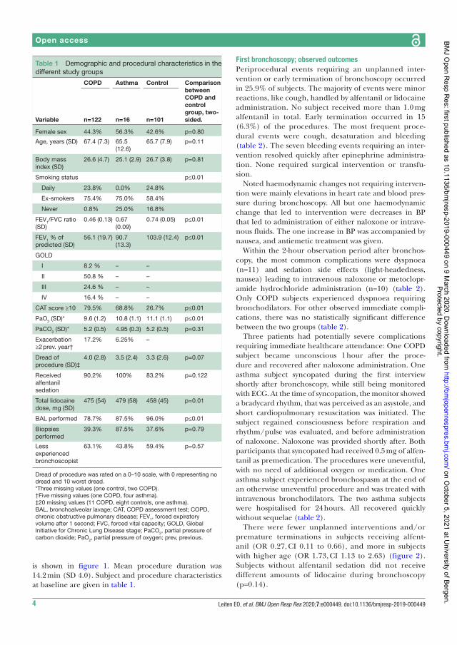
4 Leiten EO, et al. BMJ Open Resp Res 2020;7:e000449. doi:10.1136/bmjresp-2019-000449
Open access
Table 1 Demographic and procedural characteristics in the different study groups
Variable
COPD Asthma Control Comparison between COPD and control group, two- sided.n=122 n=16 n=101
Female sex 44.3% 56.3% 42.6% p=0.80
Age, years (SD) 67.4 (7.3) 65.5 (12.6)
65.7 (7.9) p=0.11
Body mass index (SD)
26.6 (4.7) 25.1 (2.9) 26.7 (3.8) p=0.81
Smoking status p≤0.01
Daily 23.8% 0.0% 24.8%
Ex- smokers 75.4% 75.0% 58.4%
Never 0.8% 25.0% 16.8%
FEV1/FVC ratio (SD)
0.46 (0.13) 0.67 (0.09)
0.74 (0.05) p≤0.01
FEV1 % of predicted (SD)
56.1 (19.7) 90.7 (13.3)
103.9 (12.4) p≤0.01
GOLD
I 8.2 % – –
II 50.8 % – –
III 24.6 % – –
IV 16.4 % – –
CAT score ≥10 79.5% 68.8% 26.7% p≤0.01
PaO2 (SD)* 9.6 (1.2) 10.8 (1.1) 11.1 (1.1) p≤0.01
PaCO2 (SD)* 5.2 (0.5) 4.95 (0.3) 5.2 (0.5) p=0.31
Exacerbation ≥2 prev. year†
17.2% 6.25% –
Dread of procedure (SD)‡
4.0 (2.8) 3.5 (2.4) 3.3 (2.6) p=0.07
Received alfentanil sedation
90.2% 100% 83.2% p=0.122
Total lidocaine dose, mg (SD)
475 (54) 479 (58) 458 (45) p=0.01
BAL performed 78.7% 87.5% 96.0% p≤0.01
Biopsies performed
39.3% 87.5% 37.6% p=0.79
Less experienced bronchoscopist
63.1% 43.8% 59.4% p=0.57
Dread of procedure was rated on a 0–10 scale, with 0 representing no dread and 10 worst dread.*Three missing values (one control, two COPD).†Five missing values (one COPD, four asthma).‡20 missing values (11 COPD, eight controls, one asthma).BAL, bronchoalveolar lavage; CAT, COPD assessment test; COPD, chronic obstructive pulmonary disease; FEV1, forced expiratory volume after 1 second; FVC, forced vital capacity; GOLD, Global Initiative for Chronic Lung Disease stage; PaCO2, partial pressure of carbon dioxide; PaO2, partial pressure of oxygen; prev, previous.
is shown in figure 1. Mean procedure duration was 14.2 min (SD 4.0). Subject and procedure characteristics at baseline are given in table 1.
First bronchoscopy; observed outcomesPeriprocedural events requiring an unplanned inter-vention or early termination of bronchoscopy occurred in 25.9% of subjects. The majority of events were minor reactions, like cough, handled by alfentanil or lidocaine administration. No subject received more than 1.0 mg alfentanil in total. Early termination occurred in 15 (6.3%) of the procedures. The most frequent proce-dural events were cough, desaturation and bleeding (table 2). The seven bleeding events requiring an inter-vention resolved quickly after epinephrine administra-tion. None required surgical intervention or transfu-sion.
Noted haemodynamic changes not requiring interven-tion were mainly elevations in heart rate and blood pres-sure during bronchoscopy. All but one haemodynamic change that led to intervention were decreases in BP that led to administration of either naloxone or intrave-nous fluids. The one increase in BP was accompanied by nausea, and antiemetic treatment was given.
Within the 2- hour observation period after bronchos-copy, the most common complications were dyspnoea (n=11) and sedation side effects (light- headedness, nausea) leading to intravenous naloxone or metoclopr-amide hydrochloride administration (n=10) (table 2). Only COPD subjects experienced dyspnoea requiring bronchodilators. For other observed immediate compli-cations, there was no statistically significant difference between the two groups (table 2).
Three patients had potentially severe complications requiring immediate healthcare attendance: One COPD subject became unconscious 1 hour after the proce-dure and recovered after naloxone administration. One asthma subject syncopated during the first interview shortly after bronchoscopy, while still being monitored with ECG. At the time of syncopation, the monitor showed a bradycard rhythm, that was perceived as an asystole, and short cardiopulmonary resuscitation was initiated. The subject regained consciousness before respiration and rhythm/pulse was evaluated, and before administration of naloxone. Naloxone was provided shortly after. Both participants that syncopated had received 0.5 mg of alfen-tanil as premedication. The procedures were uneventful, with no need of additional oxygen or medication. One asthma subject experienced bronchospasm at the end of an otherwise uneventful procedure and was treated with intravenous bronchodilators. The two asthma subjects were hospitalised for 24 hours. All recovered quickly without sequelae (table 2).
There were fewer unplanned interventions and/or premature terminations in subjects receiving alfent-anil (OR 0.27, CI 0.11 to 0.66), and more in subjects with higher age (OR 1.73, CI 1.13 to 2.63) (figure 2). Subjects without alfentanil sedation did not receive different amounts of lidocaine during bronchoscopy (p=0.14).
Protected by copyright.
on October 5, 2021 at U
niversity of Bergen.
http://bmjopenrespres.bm
j.com/
BM
J Open R
esp Res: first published as 10.1136/bm
jresp-2019-000449 on 9 March 2020. D
ownloaded from
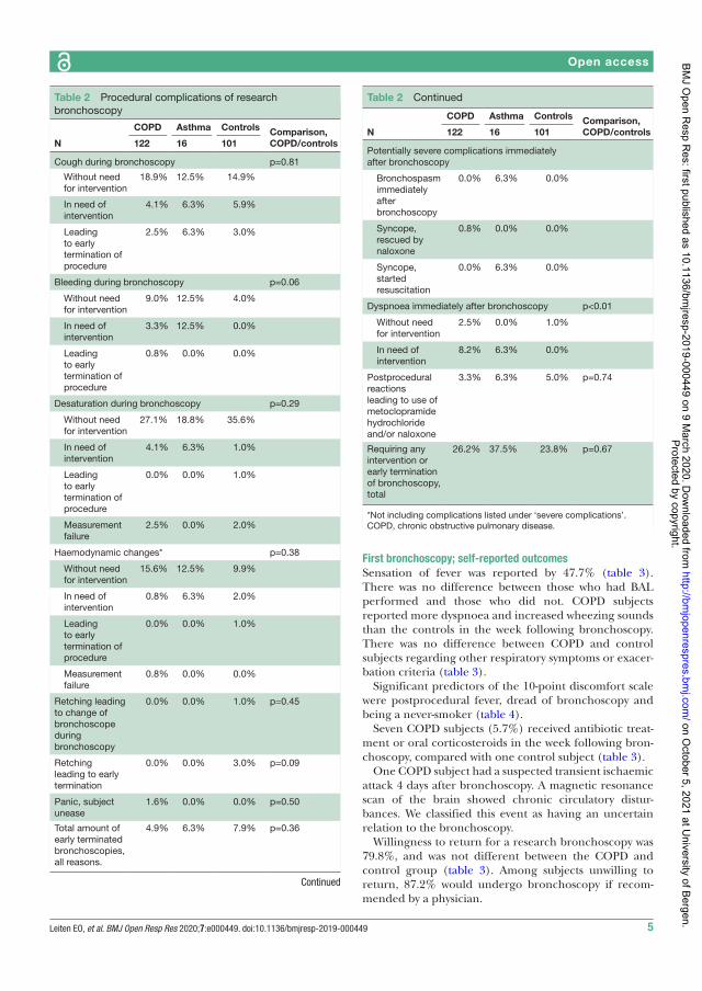
Leiten EO, et al. BMJ Open Resp Res 2020;7:e000449. doi:10.1136/bmjresp-2019-000449 5
Open access
Table 2 Procedural complications of research bronchoscopy
N
COPD Asthma Controls Comparison, COPD/controls122 16 101
Cough during bronchoscopy p=0.81
Without need for intervention
18.9% 12.5% 14.9%
In need of intervention
4.1% 6.3% 5.9%
Leading to early termination of procedure
2.5% 6.3% 3.0%
Bleeding during bronchoscopy p=0.06
Without need for intervention
9.0% 12.5% 4.0%
In need of intervention
3.3% 12.5% 0.0%
Leading to early termination of procedure
0.8% 0.0% 0.0%
Desaturation during bronchoscopy p=0.29
Without need for intervention
27.1% 18.8% 35.6%
In need of intervention
4.1% 6.3% 1.0%
Leading to early termination of procedure
0.0% 0.0% 1.0%
Measurement failure
2.5% 0.0% 2.0%
Haemodynamic changes* p=0.38
Without need for intervention
15.6% 12.5% 9.9%
In need of intervention
0.8% 6.3% 2.0%
Leading to early termination of procedure
0.0% 0.0% 1.0%
Measurement failure
0.8% 0.0% 0.0%
Retching leading to change of bronchoscope during bronchoscopy
0.0% 0.0% 1.0% p=0.45
Retching leading to early termination
0.0% 0.0% 3.0% p=0.09
Panic, subject unease
1.6% 0.0% 0.0% p=0.50
Total amount of early terminated bronchoscopies, all reasons.
4.9% 6.3% 7.9% p=0.36
Continued
N
COPD Asthma Controls Comparison, COPD/controls122 16 101
Potentially severe complications immediately after bronchoscopy
Bronchospasm immediately after bronchoscopy
0.0% 6.3% 0.0%
Syncope, rescued by naloxone
0.8% 0.0% 0.0%
Syncope, started resuscitation
0.0% 6.3% 0.0%
Dyspnoea immediately after bronchoscopy p<0.01
Without need for intervention
2.5% 0.0% 1.0%
In need of intervention
8.2% 6.3% 0.0%
Postprocedural reactions leading to use of metoclopramide hydrochloride and/or naloxone
3.3% 6.3% 5.0% p=0.74
Requiring any intervention or early termination of bronchoscopy, total
26.2% 37.5% 23.8% p=0.67
*Not including complications listed under ‘severe complications’.COPD, chronic obstructive pulmonary disease.
Table 2 Continued
First bronchoscopy; self-reported outcomesSensation of fever was reported by 47.7% (table 3). There was no difference between those who had BAL performed and those who did not. COPD subjects reported more dyspnoea and increased wheezing sounds than the controls in the week following bronchoscopy. There was no difference between COPD and control subjects regarding other respiratory symptoms or exacer-bation criteria (table 3).
Significant predictors of the 10- point discomfort scale were postprocedural fever, dread of bronchoscopy and being a never- smoker (table 4).
Seven COPD subjects (5.7%) received antibiotic treat-ment or oral corticosteroids in the week following bron-choscopy, compared with one control subject (table 3).
One COPD subject had a suspected transient ischaemic attack 4 days after bronchoscopy. A magnetic resonance scan of the brain showed chronic circulatory distur-bances. We classified this event as having an uncertain relation to the bronchoscopy.
Willingness to return for a research bronchoscopy was 79.8%, and was not different between the COPD and control group (table 3). Among subjects unwilling to return, 87.2% would undergo bronchoscopy if recom-mended by a physician.
Protected by copyright.
on October 5, 2021 at U
niversity of Bergen.
http://bmjopenrespres.bm
j.com/
BM
J Open R
esp Res: first published as 10.1136/bm
jresp-2019-000449 on 9 March 2020. D
ownloaded from

6 Leiten EO, et al. BMJ Open Resp Res 2020;7:e000449. doi:10.1136/bmjresp-2019-000449
Open access
Figure 2 Logistic regression was used to evaluate the combined outcome of unplanned intervention or premature termination of bronchoscopy. Total number of observations in this model was 236, as three observations were omitted due to missing values of oxygen. BAL, bronchoalveolar lavage; BMI, body mass index; CAT, COPD assessment test; COPD, chronic obstructive pulmonary disease; FEV1, forced expiratory volume in 1 second; FVC, forced vital capacity; PaO2, partial oxygen pressure.
Table 3 Self- reported outcomes of research bronchoscopy after 1 week
COPD Asthma Controls Comparison, COPD/controlsn=122 n=16 n=101
Willingness to return for research bronchoscopy* 76.2% 68.8% 84.2% p=0.213
Fever sensation† 45.9% 37.5% 51.5% p=0.440
Increased dyspnoea† 31.4% 25.0% 13.9% p=0.002
Increased sputum† 26.2% 25.0% 22.8% p=0.540
Change in sputum colour† 20.7% 25.0% 12.9% p=0.125
Increased rhinitis† 31.4% 25.0% 31.7% p=0.965
Increased wheezing respiration† 24.8% 12.5% 7.9% p=0.001
Sore throat or coughing† 54.1% 56.3% 57.4% p=0.601
Increased asthenia‡ 37.2% 31.3% 27.0% p=0.108
Flu- like symptoms, including fever, muscle/joint pain, headache, reduced general condition*
41.7% 31.3% 50.5% p=0.189
Discomfort graded from 0 to 10. Mean (SD)* 4.2 (2.6) 3.8 (2.8) 4.2 (1.9) p=0.364
Exacerbation criteria fulfilled, total‡ 45.5% 37.5% 33.0% p=0.169
Hospitalisation related to bronchoscopy§ 0.8% 12.5% 1.0% p=1.000
Received treatment as if exacerbation (prednisolone/antibiotics)§ 5.7% 0.0% 1.0% p=0.076
*Two missing values (two COPD).†One missing value (one COPD),‡Two missing values (one COPD, one control).§One missing value (one control).COPD, chronic obstructive pulmonary disease.
second bronchoscopyAmong the 61 subjects who underwent a second bronchoscopy, the total complication rate of the first
bronchoscopy was 20%. Of those with a complicated first bronchoscopy, 42% had a complicated second bronchoscopy. In the group with no event in the first
Protected by copyright.
on October 5, 2021 at U
niversity of Bergen.
http://bmjopenrespres.bm
j.com/
BM
J Open R
esp Res: first published as 10.1136/bm
jresp-2019-000449 on 9 March 2020. D
ownloaded from

Leiten EO, et al. BMJ Open Resp Res 2020;7:e000449. doi:10.1136/bmjresp-2019-000449 7
Open access
Table 4 Predictors of perceived discomfort during and after bronchoscopy, estimated from a quantile regression analysis
Variable Coef.
CIs
Type* P valueLower Upper
Sex, male −1 −1.91 −0.08 Bivariate 0.03
Age/10 −0.60 −1.11 −0.09 Bivariate 0.22
Body mass index 0 −0.10 0.10 Bivariate 1.00
Smoking status
Ex- smoker 0 −2.06 2.06 Bivariate 1.00
Current smoker 0 −2.37 2.37 Bivariate 1.00
FEV1/FVC 0 −0.03 0.03 Bivariate 1.00
CAT score ≥10 1.5 0.48 2.52 Bivariate <0.01
PaO2
PaO2 8–9 kPa 0 −1.79 1.79 Bivariate 1.00
PaO2 <8 kPa 1 −3.42 5.42 Bivariate 0.66
Dread of procedure 0.34 0.16 0.50 Bivariate <0.01
Alfentanil sedation 0 −1.28 1.28 Bivariate 1.00
BAL performed 0 −1.74 1.74 Bivariate 1.00
Biopsies performed 0 −0.91 0.91 Bivariate 1.00
Less experienced bronchoscopist 1 −0.09 2.09 Bivariate 0.07
Fever sensation 1.5 0.28 2.72 Bivariate 0.02
Complication 0.5 0.83 1.83 Bivariate 0.46
Sex, male −0.08 −0.88 0.73 Multivariate 0.83
Age/10 −0.36 −0.84 0.11 Multivariate 0.14
CAT score ≥10 0.62 −0.17 1.41 Multivariate 0.12
Fever 0.87 0.09 1.65 Multivariate 0.03
Dread of procedure 0.30 0.16 0.44 Multivariate <0.01
Smoking status
Ex- smoker −1.35 −2.34 −0.35 Multivariate 0.01
Current smoker −2.05 −3.38 −0.67 Multivariate <0.01
Discomfort was rated on a 0–10 scale, with 0 representing no discomfort and 10 worst imaginable discomfort.*In the multivariate model, age and sex were included and additional variables were added based on bivariate effect size. Predictors were kept for the final model if p<0.1 by a likelihood- ratio- test.BAL, bronchoalveolar lavage; CAT, COPD assessment test; Coef, Coefficient; FEV1, forced expiratory volume after 1 second; FVC, forced vital capacity; PaO2, partial pressure of oxygen.
bronchoscopy, 12% had a complicated second bron-choscopy (p=0.01).
Especially sensation of fever after the first bronchos-copy was associated with similar reports after a second bronchoscopy (p<0.01). Among subjects undergoing a second bronchoscopy, 45% reported fever after the first bronchoscopy. Of these, 63% experienced fever after the second bronchoscopy. Of those who did not report fever in their first bronchoscopy, only 27% reported fever after the second procedure.
dIscussIonIn our single- centre bronchoscopy study we found that only 1.3% of 239 participants experienced serious compli-cations, all of whom had a diagnosis of COPD or asthma. No complication had long- term consequences. Of first bronchoscopies, 6.3% were prematurely terminated.
As it can be unclear what constitutes a complication or an expected discomfort, we chose to define a compli-cation as an observed event that led to an unplanned intervention, and we chose to let subjects report overall discomfort during the week after the procedure. The most frequent complications were cough, dyspnoea and other discomforts leading to administration of naloxone or metoclopramide hydrochloride. The most common discomforts reported after 1 week were sore throat, fever and flu- like symptoms.
Although one fourth of the subjects required some form of unplanned intervention, it is important to point out that our definition of unplanned intervention was made quite wide to capture as many events as may be of any significance. However, many events will regu-larly happen during a routine bronchoscopy, like cough or light bleeding, being routinely handled by extra
Protected by copyright.
on October 5, 2021 at U
niversity of Bergen.
http://bmjopenrespres.bm
j.com/
BM
J Open R
esp Res: first published as 10.1136/bm
jresp-2019-000449 on 9 March 2020. D
ownloaded from

8 Leiten EO, et al. BMJ Open Resp Res 2020;7:e000449. doi:10.1136/bmjresp-2019-000449
Open access
medication without any harm to the person undergoing the procedure.
The only statistically significant difference between COPD subjects and controls was more postprocedural dyspnoea in COPD subjects. These findings are in accordance with a recent study that reported similar safety profiles in patients with and without COPD,15 and previous findings of more respiratory complications in COPD patients following bronchoscopy.16 Predictors of unplanned intervention or early procedure termina-tion were lack of sedation and higher age. Predictors of reported discomfort were smoking habits, dread of bron-choscopy and postprocedural fever, but effect sizes were small. The reason why ever smokers reported less discom-fort is unknown, but one possibility is a higher tolerance for respiratory symptoms.
Aside from more dyspnoea, bronchoscopy of COPD patients was not associated with more complications, even when FEV1 was below 30% of predicted. However, we did not perform BAL in subjects with the most reduced FEV1. Less dyspnoea in the asthma group than in the COPD group could be explained by a low number of asthma patients, but also by preprocedural bronchodilation. The British Thoracic Society guideline for diagnostic flex-ible bronchoscopy in adults states that nebulised bron-chodilators should be considered before bronchoscopy in patients with asthma,17 whereas no benefit of inhaled salbutamol has been identified in COPD patients.18
The low number of asthma subjects makes it impossible to draw conclusions based on complication rates in the asthma group. That the two subjects who were hospital-ised directly following bronchoscopy both had asthma could indicate that subjects with asthma are more prone to complications.
Alfentanil reduced overall need for unplanned inter-vention or early termination of bronchoscopy, even though ten cases of drug- induced complications were included in the analyses. There is no commonly accepted best practice regarding choice of sedative agent for bron-choscopy.19 Bronchoscopy sedation with alfentanil has only recently been compared with placebo or dexme-detomidine in a relatively small randomised controlled trial. The authors reported more events of hypoxaemia and heart rate changes in the alfentanil group, but present few clinically significant differences between the groups.20 Older studies comparing alfentanil and midaz-olam sedation have shown that alfentanil sedation results in less cough, but not necessarily less discomfort or improved ease of the procedure.21 22 The trial comparing alfentanil to placebo did not find a statistically signif-icant difference in cough, and did not address subject discomfort.20 Sedation was not randomised in our study, but offered to all. We were unable to find differences in reported discomfort in subjects with and without seda-tion. A non- recorded observation was that some partic-ipants declined sedation to be able to drive a motor vehicle after bronchoscopy. Midazolam is not routinely used at our institution, although sometimes preferred
in patients with manifest anxiety for instance. Patients receiving midazolam may relax more, which may have preferable effects, however we cannot assess what impact midazolam may have on procedural discomfort from the current study.
Thus, alfentanil appears to provide clear benefits for the majority of recipients, but does come with the risk of serious events, especially related to depression of respira-tion. Using a standardised sedation protocol and having an experienced team performing the bronchoscopies is likely beneficial in maximising benefit and minimising risk.
The postbronchoscopy fever rate was in accordance with a paediatric study.23 Others have reported lower rates (0%–38%).24–30 The causes of this wide range in rates are unclear, but the studies vary greatly in patient population and design. A possible reason for our rela-tively high fever rate is that we asked for a self- reported fever, which also included a ‘fever sensation’ instead of measuring body temperature. The abovementioned paediatric study found more fever in cases where BAL had been performed. In the current study, fever was not associated with BAL. However, BAL was not done in a randomised manner, and was not performed in patients with very severe obstruction.
Postbronchoscopy fever itself is harmless, but is associ-ated with subject discomfort. COPD patients may inter-pret fever sensation as an early sign of exacerbations. Although subjects reported relatively high scores of discomfort and many had airway exacerbation symptoms, 79.8% would undergo research bronchoscopy again. Our findings on willingness to return fall within the range of previously reported rates,15 31–34 but might be influenced by response bias.
Of participants undergoing a second bronchoscopy, the first bronchoscopy appears to be predictive for both immediate complications and fever. This indicates that the fever is at least partially subject- related and not procedural- related. To the best of our knowledge, there are no other studies on the complication or discomfort rate in repeated bronchoscopies.
Our analyses suggest that exacerbation symptoms may increase after bronchoscopy. Thus, patients might profit from an increase in their bronchodilators or inhaled steroids before and after the procedure. Dread of bron-choscopy before the procedure predicted discomfort, suggesting an anxiety relieving effect of information. Knowledge about factors influencing discomfort may help bronchoscopists improve their preprocedural information.
There are reasons why caution should be used when comparing our results to the clinical bronchoscopy setting. First, the patients undergoing bronchoscopy may be healthier than patients undergoing bronchoscopy for a clinical indication. Despite setting no upper age- limit (the oldest individual was 82 years old) and including COPD GOLD stage 4, the frailest subjects were excluded. Second, clinical bronchoscopies often have longer
Protected by copyright.
on October 5, 2021 at U
niversity of Bergen.
http://bmjopenrespres.bm
j.com/
BM
J Open R
esp Res: first published as 10.1136/bm
jresp-2019-000449 on 9 March 2020. D
ownloaded from

Leiten EO, et al. BMJ Open Resp Res 2020;7:e000449. doi:10.1136/bmjresp-2019-000449 9
Open access
durations and include more invasive techniques than those applied and accepted in our research setting. Diag-nostic bronchoscopies investigating potential malignant disease obtains larger and often transbronchial biopsies. Therapeutic bronchoscopies, such as endobronchial coil therapy, is associated with more severe complica-tions (eg, pneumothorax and death).35 Third, volunteers are possibly more positive towards the procedure than patients in a clinical setting. This could cause them to have an overall more positive experience, resulting in less perceived discomfort. However, they could also report more complications as a result of a greater discrepancy between what they expected and the actual procedure. Fourth, the procedure was standardised. Hence, the bronchoscopist had to make fewer decisions during bronchoscopy, which may lower complication rates of research bronchoscopy.
This descriptive study has some limitations. For many of our descriptive and outcome variables, we used self- reported outcomes. Recording of complications such as dyspnoea, cough, or bleeding will by nature be subjec-tive. Therefore, we reported events that had a conse-quence in the form of intervention or termination. The perceived need to intervene is however also subjective and at the discretion of each bronchoscopist. Impor-tantly, we did not observe differences in complication or discomfort rates between the bronchoscopists in our study. All subjects were monitored with ECG, pulse oxim-eter and blood pressure measurements, however these parameters were only noted in the case of an observed event. If these measurements had been systematically collected, they could have been implemented in quality control or further analyses. This could have improved the study and potentially aided in defining cutoffs for future investigations.
conclusIonOnly 1.3% of subjects had a potentially serious complica-tion, all of whom had no sequela, indicating that bron-choscopy applying invasive techniques such as BAL and mucosal biopsies is a safe procedure in studies of patients with obstructive lung disease. Overall, a sizeable number of subjects perceived some discomfort or less serious complications, but these were minor and to a large degree to be expected from the procedure. Sore throat, fever and flu- like symptoms each occurred in roughly half of all subjects. Non- sedation and higher age were signif-icantly associated with more unplanned interventions during bronchoscopy, indicating that sedation improves tolerability of the procedure and is advised. Information regarding expected discomfort should be given prior to bronchoscopy.
Acknowledgements The authors wish to thank Kristina Apalseth, Marit Aardal, Hildegunn Bakke Fleten, Lise Østgård Monsen, Ane Aamli Gagnat and Ingvild Haaland for their contributions in the data collection, and Marianne Aanerud and Øystein Fløtten for constructive criticism of the manuscript.
contributors EOL had full access to all of the data in the study and takes responsibility for the integrity of the data and the accuracy of the data analysis. EOL, TMLE, EMHM, EN, GRH, KSK, SL, ØS and RN participated in different aspects of data collection. TMLE, GRH, KSK, SL, ØS and RN performed the bronchoscopies. EOL, TMLE, EMHM, EN, GRH, KSK, SL, ØS, PSB and RN contributed substantially to the study design, data analysis and interpretation, and the writing of the manuscript.
Funding The MicroCOPD study was funded by unrestricted grants and fellowships from Helse Vest, Bergen Medical Research Foundation, the Endowment of Timber Merchant A. Delphin and Wife through the Norwegian Medical Association and GlaxoSmithKline through the Norwegian Respiratory Society.
competing interests EOL, EMHM, EN, KSK, ØS and PSB declare no competing interests. TMLE has for the last three years received lecture fees from Boehringer Ingelheim, Roche and Astra Zeneca. GRH has for the last three years received lecture fees from Novartis and Boehringer Ingelheim. RN reports grants from GlaxoSmithKline, during the conduct of the study; grants from Boehringer Ingelheim, grants and personal fees from AstraZeneca, grants from Novartis, personal fees from GlaxoSmithKline, outside the submitted work. SL has for the last three years received lecture fees from Philips, Novartis and Astra Zeneca, and advisory board fees (paid to employer) from Novartis and Astra Zeneca, all outside the submitted work.
Patient consent for publication Not required.
ethics approval The Regional Committee for Medical and Health Research Ethics approved the project (REK VEST 2011/1307). All participants provided written informed consent.
Provenance and peer review Not commissioned; externally peer reviewed.
data availability statement The data and command lines necessary for running the analyses presented in this article are available upon reasonable request.
open access This is an open access article distributed in accordance with the Creative Commons Attribution Non Commercial (CC BY- NC 4.0) license, which permits others to distribute, remix, adapt, build upon this work non- commercially, and license their derivative works on different terms, provided the original work is properly cited, appropriate credit is given, any changes made indicated, and the use is non- commercial. See: http:// creativecommons. org/ licenses/ by- nc/ 4. 0/.
orcId idElise Orvedal Leiten http:// orcid. org/ 0000- 0002- 5588- 9076
RefeRenCes 1 Grendelmeier P, Kurer G, Pflimlin E, et al. Feasibility and safety
of propofol sedation in flexible bronchoscopy. Swiss Med Wkly 2011;141:w13248.
2 Carr IM, Koegelenberg CFN, von Groote- Bidlingmaier F, et al. Blood loss during flexible bronchoscopy: a prospective observational study. Respiration 2012;84:312–8.
3 Rosell A, Xaubet A, Agustí C, et al. A new BAL fluid instillation and aspiration technique: a multicenter randomized study. Respir Med 2006;100:529–35.
4 Gibson PG, Breit SN, Bryant DH. Hypoxia during bronchoalveolar lavage. Aust N Z J Med 1990;20:39–43.
5 Ogawa T, Imaizumi K, Hashimoto I, et al. Prospective analysis of efficacy and safety of an individualized- midazolam- dosing protocol for sedation during prolonged bronchoscopy. Respir Investig 2014;52:153–9.
6 Meduri GU, Stover DE, Greeno RA, et al. Bilateral bronchoalveolar lavage in the diagnosis of opportunistic pulmonary infections. Chest 1991;100:1272–6.
7 Leiten EO, Martinsen EMH, Bakke PS, et al. Complications and discomfort of bronchoscopy: a systematic review. Eur Clin Respir J 2016;3:33324.
8 Grønseth R, Haaland I, Wiker HG, et al. The Bergen COPD microbiome study (MicroCOPD): rationale, design, and initial experiences. Eur Clin Respir J2014:1.
9 Eagan TML, Ueland T, Wagner PD, et al. Systemic inflammatory markers in COPD: results from the Bergen COPD cohort study. Eur Respir J 2010;35:540–8.
10 Sørheim I- C, Johannessen A, Grydeland TB, et al. Case- Control studies on risk factors for chronic obstructive pulmonary disease: how does the sampling of the cases and controls affect the results? Clin Respir J 2010;4:89–96.
11 The HMW. 2017 global initiative for chronic obstructive lung disease report and practice implications for the respiratory therapist.. Respir Care 2017;62:1492–500.
Protected by copyright.
on October 5, 2021 at U
niversity of Bergen.
http://bmjopenrespres.bm
j.com/
BM
J Open R
esp Res: first published as 10.1136/bm
jresp-2019-000449 on 9 March 2020. D
ownloaded from

10 Leiten EO, et al. BMJ Open Resp Res 2020;7:e000449. doi:10.1136/bmjresp-2019-000449
Open access
12 Jones PW, Harding G, Berry P, et al. Development and first validation of the COPD assessment test. Eur Respir J 2009;34:648–54.
13 Johannessen A, Lehmann S, Omenaas ER, et al. Post- Bronchodilator spirometry reference values in adults and implications for disease management. Am J Respir Crit Care Med 2006;173:1316–25.
14 Anthonisen NRet al. Antibiotic therapy in exacerbations of chronic obstructive pulmonary disease. Ann Intern Med 1987;106:196–204.
15 Grendelmeier P, Tamm M, Jahn K, et al. Flexible bronchoscopy with moderate sedation in COPD: a case- control study. Int J Chron Obstruct Pulmon Dis 2017;12:177–87.
16 Bellinger CR, Khan I, Chatterjee AB, et al. Bronchoscopy safety in patients with chronic obstructive lung disease. J Bronchology Interv Pulmonol 2017;24:98–103.
17 Du Rand IA, Blaikley J, Booton R, et al. British thoracic Society guideline for diagnostic flexible bronchoscopy in adults: accredited by NICE. Thorax 2013;68:i1–44.
18 Stolz D, Pollak V, Chhajed PN, et al. A randomized, placebo- controlled trial of bronchodilators for bronchoscopy in patients with COPD. Chest 2007;131:765–72.
19 José RJ, Shaefi S, Navani N. Sedation for flexible bronchoscopy: current and emerging evidence. Eur Respir Rev 2013;22:106–16.
20 Riachy M, Khayat G, Ibrahim I, et al. A randomized double- blind controlled trial comparing three sedation regimens during flexible bronchoscopy: dexmedetomidine, alfentanil and lidocaine. Clin Respir J 2018;12:1407–15.
21 Greig JH, Cooper SM, Kasimbazi HJN, et al. Sedation for fibre optic bronchoscopy. Respir Med 1995;89:53–6.
22 Houghton CM, Raghuram A, Sullivan PJ, et al. Pre- medication for bronchoscopy: a randomised double blind trial comparing alfentanil with midazolam. Respir Med 2004;98:1102–7.
23 Picard E, Schwartz S, Goldberg S, et al. A prospective study of fever and bacteremia after flexible fiberoptic bronchoscopy in children. Chest 2000;117:573–7.
24 Hemmers T, Nüßlein T, Teig N, et al. Prospective study of fever after bronchoalveolar lavage in children. Klin Padiatr 2006;218:74–8.
25 Mohallem Fonseca MT, Camargos PAM, Abou Taam R, et al. Incidence rate and factors related to post- bronchoalveolar lavage fever in children. Respiration 2007;74:653–8.
26 Schellhase DE, Tamez JR, Menendez AA, et al. High fever after flexible bronchoscopy and bronchoalveolar lavage in noncritically ill immunocompetent children. Pediatr Pulmonol 1999;28:139–44.
27 Georgiades G, Myrianthefs P, Venetsanou K, et al. Temperature and serum proinflammatory cytokine changes in patients with NSCLC after BAL. Lung 2003;181:35–47.
28 Kane RC, Cohen MH, Fossieck BE, et al. Absence of bacteremia after fiberoptic bronchoscopy. Am Rev Respir Dis 1975;111:102–4.
29 Strumpf IJ, Feld MK, Cornelius MJ, et al. Safety of fiberoptic bronchoalveolar lavage in evaluation of interstitial lung disease. Chest 1981;80:268–71.
30 Kanemoto K, Satoh H, Ishikawa H, et al. Prospective study of fever and pneumonia after flexible fiberoptic bronchoscopy in older people. J Am Geriatr Soc 2006;54:827–30.
31 Hehn BT, Haponik E, Rubin HR, et al. The relationship between age and process of care and patient tolerance of bronchoscopy. J Am Geriatr Soc 2003;51:917–22.
32 Hirose T, Okuda K, Ishida H, et al. Patient satisfaction with sedation for flexible bronchoscopy. Respirology 2008;13:722–7.
33 Lechtzin N, Rubin HR, White P, et al. Patient satisfaction with bronchoscopy. Am J Respir Crit Care Med 2002;166:1326–31.
34 YL N, YL L, Lin TY, et al. Conscious sedation reduces patient discomfort and improves satisfaction in flexible bronchoscopy. Chang Gung Med J 2010;33:443–52.
35 Sciurba FC, Criner GJ, Strange C, et al. Effect of endobronchial coils vs usual care on exercise tolerance in patients with severe emphysema: the RENEW randomized clinical trial. JAMA 2016;315:2178–89.
Protected by copyright.
on October 5, 2021 at U
niversity of Bergen.
http://bmjopenrespres.bm
j.com/
BM
J Open R
esp Res: first published as 10.1136/bm
jresp-2019-000449 on 9 March 2020. D
ownloaded from

Paper III


The airway microbiota and exacerbationsof COPD
Elise Orvedal Leiten 1, Rune Nielsen1,2, Harald Gotten Wiker1,3,Per Sigvald Bakke1, Einar Marius Hjellestad Martinsen 1,Christine Drengenes1,2, Solveig Tangedal1,2, Gunnar Reksten Husebø1,2 andTomas Mikal Lind Eagan1,2
Affiliations: 1Dept of Clinical Science, University of Bergen, Bergen, Norway. 2Dept of Thoracic Medicine,Haukeland University Hospital, Bergen, Norway. 3Dept of Microbiology, Haukeland University Hospital,Bergen, Norway.
Correspondence: Elise Orvedal Leiten, Dept of Clinical Science, University of Bergen, Haukeland UniversityHospital, N-5021 Bergen. E-mail: [email protected]
ABSTRACTAim: The aim of this study was to investigate whether the compositionality of the lower airway microbiotapredicts later exacerbation risk in persons with COPD in a cohort study.Materials and methods: We collected lower airways microbiota samples by bronchoalveolar lavage andprotected specimen brushes, and oral wash samples from 122 participants with COPD. Bacterial DNA wasextracted from all samples, before we sequenced the V3-V4 region of the 16S RNA gene. The frequency ofmoderate and severe COPD exacerbations was surveyed in telephone interviews and in a follow-up visit.Compositional taxonomy and α and β diversity were compared between participants with and withoutlater exacerbations.Results: The four most abundant phyla were Firmicutes, Bacteroidetes, Proteobacteria and Fusobacteria inboth groups, and the four most abundant genera were Streptococcus, Veillonella, Prevotella and Gemella.The relative abundances of different taxa showed a large variation between samples and individuals, andno statistically significant difference of either compositional taxonomy, or α or β diversity could be foundbetween participants with and without COPD exacerbations within follow-up.Conclusion: The findings from the current study indicate that individual differences in the lower airwaymicrobiota in persons with COPD far outweigh group differences between frequent and nonfrequentCOPD exacerbators, and that the compositionality of the microbiota is so complex as to present largechallenges for use as a biomarker of later exacerbations.
@ERSpublicationsContrary to previous reports, in this study there were no significant associations between thelung microbiota in stable COPD and COPD exacerbation frequency https://bit.ly/2ZVcNdG
Cite this article as: Leiten EO, Nielsen R, Wiker HG, et al. The airway microbiota and exacerbationsof COPD. ERJ Open Res 2020; 6: 00168-2020 [https://doi.org/10.1183/23120541.00168-2020].
Copyright ©ERS 2020. This article is open access and distributed under the terms of the Creative Commons AttributionNon-Commercial Licence 4.0.
This article has supplementary material available from openres.ersjournals.com
The sequencing data and relevant metadata are available at the DRYAD data repository from https://doi.org/10.5061/dryad.tqjq2bvw3
Received: 3 April 2020 | Accepted after revision: 27 May 2020
https://doi.org/10.1183/23120541.00168-2020 ERJ Open Res 2020; 6: 00168-2020
ORIGINAL ARTICLECOPD

IntroductionCOPD exacerbations are often caused by bacterial and viral respiratory infections with common pathogenssuch as Haemophilus influenzae, Streptococcus pneumoniae and Moraxella catarrhalis [1]. Beforenext-generation sequencing was made available, these microbes were detected by growth in cultures andlater PCR. PCR studies suggested the aetiology could be multi-microbial, with frequent detection ofmultiple bacteria and/or viruses [2–4]. However, not all COPD exacerbations show clinical or microbialsigns of infection. Exacerbation-associated bacteria have been cultured from sputum obtained in clinicallystable COPD, giving rise to a still unresolved debate on whether colonisation is a pathogenic factor forlater exacerbation [5]. The finding of a diverse lung microbiome has led to the hypothesis thatexacerbations are caused by a dysbiosis in the airway microbiome, and that differences in baselinemicrobiota might help explain the difference between the frequent and infrequent exacerbator type [6].
Some previous studies comparing sputum microbiota between stable and exacerbated state in COPDindicate there are differences between disease states, but the findings are inconsistent [7–12]. And, twoprevious retrospective studies indicated that the sputum microbiota in the stable state was different inpatients prone to exacerbations compared to patients who do not exacerbate [13, 14]. However, sputum isinvariably fraught by contamination of microbes from the upper airways, which is both difficult to adjustfor and potentially interferes with our interpretations of the lower airway microbiota.
To address the question of whether the stable-state lower airway microbiota is predictive of later COPDexacerbation, a study should include both COPD patients known to exacerbate and not, sample the lowerairways directly, and include a prospective longitudinal follow-up of exacerbation events. The current studyis to our knowledge the first such study with this methodology.
MethodsStudy populationCOPD participants in the Bergen COPD Microbiome (MicroCOPD) study were recruited among previousparticipants in the Bergen COPD Cohort Study [15] and the GenKOLS Study [16], in addition tovolunteers from the outpatient clinic at the Department of Thoracic Medicine, Haukeland UniversityHospital. The protocol for the MicroCOPD study is previously published [17].
To be included, all participants had to be >40 years of age. In the current study sample the youngestparticipant was 49.5 years upon inclusion. The COPD diagnosis was verified by experiencedpulmonologists based on spirometry, respiratory symptoms and disease history. Participation waspostponed in individuals that had been treated for a respiratory exacerbation within the last 2 weeks or ifthey had ongoing symptoms of exacerbation according to modified Anthonisen criteria [18]. We excludedindividuals deemed not fit for bronchoscopy, namely those who had an O2 saturation <90% despiteoxygen supplementation, CO2 tension >6.65 kPa in arterial blood gas analyses, increased risk of bleeding,known allergy towards the premedication or cardiac risk factors as specified in the protocol [17].Participants were asked about their current medication, smoking history, number of exacerbations treatedwith systemic steroids or antibiotics and exacerbation-related hospital admissions in the preceding year. Allparticipants underwent spirometry after inhalation of 0.4 mg salbutamol, followed by bronchoscopy. In theMicroCOPD study, a total of 130 participants with COPD were included. For the current analyses, onlythe 122 participants from the post-pilot phase of the MicroCOPD study were included.
The MicroCOPD study was approved by the regional ethical committee (REK Vest case number 2011/1307). All participants provided oral and written consent.
Microbial samplingPrior to bronchoscopy, all participants were asked to gargle 10 mL of PBS for oral wash (OW) sampling.Bronchoscopy was performed through oral access. Topical anaesthesia (lidocaine) was delivered by oralspray formulation prior to the procedure and per-operatively through a catheter within the bronchoscope’sworking channel. Three protective specimen brushes were collected in the right lower lobe (rPSB). Thethree brushes were cut off using sterile scissors and placed in an Eppendorf tube with 1 mL of PBS.Protected bronchoalveolar lavage (BAL) was performed in all participants with FEV1>30% of predicted:two fractions each containing 50 mL of PBS were installed through a Combicath sterile catheter with asealed tip, in the right middle lobe and aspirated through the same sterile catheter. The median retrievalwas 33 mL in the group that had no exacerbations and 26 mL in the group that had exacerbations(p=0.04, Mann–Whitney U-test). For the current study we used the second fraction of BAL (BAL2), whichusually had the highest yield. All samples from one participant (OW, BAL2, rPSB) used the same bottle ofPBS, and for each participant we analysed one negative control sample from this bottle. A more detailedexplanation of handling of negative control samples is presented in the supplementary text.
https://doi.org/10.1183/23120541.00168-2020 2
COPD | E.O. LEITEN ET AL.

DNA extraction and 16S sequencingThe detailed protocol for laboratory processing is published [19]. Briefly, bacterial DNA was extracted byenzymatic lysis followed by processing though the FastDNA Spin Kit. Sequencing of the V3-V4 region ofthe 16S rRNA gene was carried out according to the Illumina 16S Metagenomic Sequencing LibraryPreparation guide. The V3-V4 region was PCR amplified using 45 cycles and prepared for a subsequent8-cycle index PCR step using specific primers. The samples were pooled and diluted before 2×300 cyclesof paired-end sequencing was performed using reagents from the MiSeq reagent kit v3 (Illumina Inc., SanDiego, CA, USA).
Exacerbation follow-upWe collected information on health-seeking behaviour, use of antibiotics and oral steroids in structured,quarterly telephone interviews for 12 months after the bronchoscopy. All participants were also offered afollow-up examination 1–1.5 years after inclusion where we also collected exacerbation history.Exacerbations were self-reported, but only counted if the exacerbation led to any administration ofantibiotics and/or oral corticosteroids or hospitalisations (moderate-to-severe exacerbations). Datesreported in the telephone interviews and the physical follow-up examination were used to confirm that theevent had not already been recorded and to separate one exacerbation from another. When informationon hospitalisation was contradicting, the reason for admittance was unclear (for instance hospitaladmittance without administration of antibiotics), or the time point for an exacerbation leading tohospitalisation was unknown, the digital hospital records were consulted.
Bioinformatic analysesBioinformatic analyses were performed with QIIME 2 [20] (versions 2018.8, 2018.11, 2019.4, 2019.7,2020.2) and R (versions 3.5.0 and 3.6.0). Sequencing data from the entire MicroCOPD study (includingsamples not included in the current analyses) were imported batched by Illumina runs into QIIME 2(version 2018.8) from Casava 1.8 paired-end demultiplexed fastq format. Denoising was performed usingthe Divisive Amplicon Denoising Algorithm version 2 (DADA2) software package [21] (via q2-dada2),and sequences were further processed with VSEARCH [22] (via q2-vsearch) for additional chimeraremoval and then merged to one amplicon sequence variant (ASV) table (QIIME 2, version 2018.8).
All ASVs representing <0.005% of reads (fewer than 3000 reads) were removed, as they were deemed likelyto represent sequencing noise [23]. The ASV table was exported from QIIME 2 into R for bioinformaticidentification and removal of contaminants with the package Decontam [24], where we used theprevalence-based method with a threshold of 0.3. A more detailed explanation of the data curation andhandling of contaminants are given in the supplementary text.
TABLE 1 Participant characteristics at baseline by exacerbation status during follow-up
Variable Exacerbationgroup
No exacerbationgroup
Exacerbationunknown
Comparison
Subjects n 46 59 17 NSFemale 45.7% 47.5% 29.4% NSAge years 67.2±7.2 68.5±7.3 64.3±7.4 NSSmoking status NSDaily 19.6% 27.1% 23.5%Ex-smoker 78.3% 72.9% 76.5%Never 2.2% 0% 0%
FEV1 % pred 46.1±17.0 60.4±17.3 68.3±22.6 p<0.01FEV1/FVC 0.40±0.12 0.49±0.11 0.45±0.12 p<0.01CAT-score# 19.4±8.1 15.5±7.5 12.5±6.4 p=0.01PaO2
kPa¶ 9.4±1.2 9.8±1.2 9.6±1.2 NSExacerbation ⩾2 previousyear
32.6% 10.2% 5.9% p<0.01
Daily use of inhaledcorticosteroids
69.6% 39.0% 35.3% p⩽0.01
Daily use of oral steroids 6.5% 3.4% 5.9% NS
Data are presented as % or mean±SD, unless otherwise stated. The comparisons were between theexacerbation and no exacerbation groups only, and were tested using t-tests and Chi-squared tests. NS:not significant; FEV1: forced expiratory volume in 1 s; FVC: forced vital capacity; CAT: COPD AssessmentTest; PaO2
: arterial oxygen tension. #: Two missing values; ¶: three missing values.
https://doi.org/10.1183/23120541.00168-2020 3
COPD | E.O. LEITEN ET AL.

The ASV table was then filtered to contain only samples included in the present study. Taxonomy wasassigned using a classifier trained on the Human Oral Microbiome Database [25]. All ASVs that wereunassigned to phylum level, were checked with the NCBI BLAST tool [26], and all nonbacterial ASVswere removed. Remaining ASVs were aligned with mafft (via q2-phylogeny) and a phylogenetic tree wasconstructed with fasttree2 [27] (via q2-phylogeny). Sequences were rarefied at a sampling depth of 1000prior to α (within sample) and β (between samples) diversity analyses to ensure equal sampling depths(via q2-diversity).
StatisticsDifferentially abundant features (i.e. genera and ASVs) and differences in compositionality were analysedusing Analysis of Composition of Microbes (ANCOM) [28] (via q2-composition), balance trees [29] (viaq2-gneiss), ANOVA-like differential expression analysis 2 (ALDEx2) [30] (via q2-aldex2) and differentialdistribution analysis [31] (with R package MicrobiomeDDA). The α diversity difference, measured withthe Shannon index and Faith phylogenetic diversity, was tested with the Kruskal–Wallis test. The βdiversity difference, measured with Bray–Curtis and weighted UniFrac distance, was tested with theADONIS permutation-based test using the vegan package in R [32].
ResultsOf the 122 participants, 105 had complete follow-up of exacerbations for a full year after bronchoscopy.Participants who experienced one or more exacerbations within follow-up had significantly lower lungfunction, higher symptom score, more frequent exacerbations in the preceding year and more use ofinhaled corticosteroids (ICSs) (table 1). Median time to first exacerbation was 146 days. Exacerbations wereevenly distributed across seasons of the year.
For the current analyses, we selected the 327 samples of BAL2, OW and rPSB from the 122 participants,of which three samples were judged as only containing nonbacterial taxa and filtered out. In addition, weremoved the samples from the 17 participants with unknown exacerbation status. For diversity analyses,rarefied data included 235 samples. Figure 1 shows the flow chart for the sample processing.
Taxonomy and differential abundance testingTaxonomy at the phylum and genus levels for the three sample types by exacerbation status are shown infigures 2 and 3. Overall, the most abundant phyla were Firmicutes, Bacteroidetes, Proteobacteria andFusobacteria. The most abundant genera were Streptococcus, Veillonella, Prevotella and Gemella. However,the relative abundances of different taxa showed a large variation between samples. Four participants withone or more later exacerbations and one participant without later exacerbations presented with highabundances of the Moraxella genus in rPSB samples, which was also reflected by detection of Moraxella inBAL2 for those participants who had a lavage sample. When we sorted rPSB samples by relativeabundance of the most common taxon, the visual pattern differed from that in BAL2 and OW (figures 2and 3).
The visual impression from both figure 2 (phylum level) and figure 3 (genus level) was that participantswithout later exacerbations had more Firmicutes (figure 2), and more streptococci (figure 3), thanparticipants with later exacerbations. However, when testing for statistical significance in difference ofabundance between the two groups, few were found. ANCOM, balance trees (gneiss) and ALDEx2performed on BAL2 and rPSB did not identify any differentially abundant ASVs or genera between thegroup with and without exacerbations. Differential distribution analysis identified two ASVs asdifferentially expressed between the groups in rPSB samples (supplementary table 1). These two ASVswere classified as Capnocytophaga gingivitis (more abundant, less prevalent and less dispersed in groupwith exacerbations (adjusted p=0.011)) and Prevotella pallens (less abundant, more prevalent and moredispersed in group with exacerbations (adjusted p=0.041)).
To see whether patients who had two or more exacerbations during follow-up differed from those whohad zero or one, we repeated the above analyses on this outcome (supplementary table 2). Again,ANCOM, balance trees (gneiss) and ALDEx2 performed on BAL2 and rPSB did not identify anydifferentially abundant ASVs or genera between the groups. However, differential distribution analysisidentified two ASVs (Capnocytophaga leadbetteri, adjusted p=0.008, and Prevotella oris, adjusted p=0.045)and one genus (Moraxella, adjusted p=0.026) as differentially expressed between the groups in BALsamples.
Next, we checked whether the taxonomy by sample type and exacerbation status varied by use of ICSs.The bacterial composition did not appear altered by ICS use, as illustrated in a heat map of genera inBAL2 (figure 4). This impression was strengthened by differential abundance testing by the same four tests
https://doi.org/10.1183/23120541.00168-2020 4
COPD | E.O. LEITEN ET AL.

as above, revealing no significant differences in taxa between COPD patients who used ICSs and COPDpatients who did not (supplementary table 3).
Finally, we examined whether taxonomy differed by previous exacerbations the last year beforebronchoscopy (supplemental table 4). Again, only the differential distribution analysis yielded a potentialdifference, with Actinomyces graevenitzii being less abundant and more prevalent in patients havingprevious exacerbations (adjusted p=0.011).
Overall the main message from the differential abundance tests was that very few differences were found.
DiversityThe α diversity in BAL 2 and rPSB, measured by Faith phylogenetic diversity and Shannon diversityindex, did not significantly differ between the group with and without later exacerbations. Box plots of theα diversity are presented in the supplementary material (supplementary figures 5 and 6).
Neither β diversity, measured by Bray–Curtis distance (nonphylogenetic) and weighted UniFrac distance(phylogenetic), differed by exacerbation status (supplementary figures 7 and 8). The four rPSB samples
DADA2 on each run
Merging of denoised
runs into one ASV
table
VSEARCH for extra
chimaera removal
2511 samples
24 091 ASVs
Sequences: 62 706 647
2477 samples
21 034 ASVs
Sequences: 60 344 169
2474 samples
891 ASVs
Sequences: 56 645 782
2274 samples
611 ASVs
Sequences: 29 220 383
327 samples
572 ASVs
Sequences: 5 246 789
324 samples, 122 subjects
463 ASVs
Sequences: 5 037 612
90 samples
375 ASVs
Sequences: 1 247 440
119 samples
441 ASVs
Sequences: 2 627 758
115 samples
370 ASVs
Sequences: 1 162 414
75 samples
358 ASVs
Sequences: 917 549
60 samples 102 samples 73 samples
Removal on non-
bacterial taxa
3 rPSB lost
Removal of samples with
unknown exacerbation status102 samples
430 ASVs
Sequences: 2 178 449
98 samples
332 ASVs
Sequences: 916 888
BAL2 OW rPSB
Filtering of ASVs with
<3000 sequences
Identification and
removal of
contaminants with
Decontam
Selecting only BAL2, OW
and rPSB for COPD subjects
Assigning taxomony
using HOMD classifier
Phylogenetic tree
Splitting the data into
subsets of sample type
Rarafaction at sampling depth
1000 for diversity analyses
Samples left for diversity analyses
Run 1 Run 2 Run 3 Run 4 Run 5 Run 6 Run 7 Run 8 Run 9 Run 10
Run 11 Run 12 Run 13 Run 14 Run 15 Run 16 Run 17 Run 18 Run 19 Run 20
Run 21 Run 22 Run 23 Run 24 Run 25 Run 26 Run 27 Run 28 Run 29 Run 30
FIGURE 1 Sample flow chart of data processing. DADA2: Divisive Amplicon Denoising Algorithm 2; ASV: amplicon sequence variant; BAL2: secondfraction of bronchoalveolar lavage; OW: oral wash; rPSB: protected specimen brushes from the right lung; HOMD: Human oral microbiomedatabase.
https://doi.org/10.1183/23120541.00168-2020 5
COPD | E.O. LEITEN ET AL.

and three BAL2 samples that clustered separately (based on weighted UniFrac distance, supplementaryfigure 9) were the samples with high abundances of Moraxella (figure 3). Both unadjusted and adjusted(age, sex, FEV1, ICS use) PERMANOVA analyses showed nonsignificant differences in weighted UniFracdistances between patients with and without later exacerbations (supplementary tables 5 and 6 andsupplementary figure 9).
DiscussionThis study did not show any statistically significant characteristics of the lung microbiota in stable COPD,neither in differential abundance of taxonomy, nor α or β diversity, that could predict whether theparticipants would experience moderate or severe exacerbations in the subsequent follow-up. Thesefindings challenge the few existing previous reports.
PRAGMAN et al. [14] reported lower α diversity in sputum from patients with frequent exacerbations, and asignificant difference in ASV counts between frequent (n=11) and infrequent (n=11) exacerbators, whereActinomyces was found more abundant in patients with infrequent exacerbations. In another recent study,there was an increased abundance of Pseudomonas, Selenomonas and Anaerococcus, in sputum frompatients with frequent exacerbations (n=31) compared with patients without exacerbations (n=23). Therewas also a significant difference in β diversity between patients with frequent and infrequent exacerbations[13]. A study of the microbiota in BAL from patients with stable COPD retrospectively recorded theexacerbation history of 21 patients and suggested that Streptococcus and Rothia species might be protectiveand that Pseudomonas might be predictive of exacerbations [33].
The current study has superior methodology with a 2–5 times larger sample size than previous studies,prospective follow-up of exacerbations and protected sampling of the lower airways. However, otherpotential causes of the differences in results between our study and previous studies are worthy of
FIGURE 2 Bacterial taxonomy at thephylum level in participants withCOPD with and withoutexacerbation during follow-up.Taxonomic groups in the legend aresorted in decreasing order basedon the average relative frequency ofthat group in all samples. Each barrepresents one participant, orderedin the same position horizontallyfor all three sample typesaccording to relative abundance ofFirmicutes in rPSB samples. OW:oral wash; BAL2: second fraction ofbronchoalveolar lavage; rPSB: rightprotected specimen brushes.
1.00
No exacerbation Exacerbation
OW
BA
L 2
rPS
B
0.75
0.50
0.25
0.00
1.00
0.75
0.50
0.25
0.00
Re
lati
ve a
bu
nd
an
ce1.00
0.75
0.50
0.25
0.00
Participant
Phyla
Firmicutes ActinobacteriaSaccharibacteria_(TM7)
Absconditabacteria_(SR1)
Spirochaetes and Synergistetes
Bacteroidetes
Proteobacteria
Fusobacteria
https://doi.org/10.1183/23120541.00168-2020 6
COPD | E.O. LEITEN ET AL.

consideration. Two of the previous studies suggest that Pseudomonas is associated with more frequentexacerbations, whereas we did not report Pseudomonas in any of our samples. Some Pseudomonas speciesare known colonisers in patients with severe disease or impaired immunity, but Pseudomonas is ubiquitousin the environment, and there is a high potential for Pseudomonas to be a contaminant. The differencesbetween our study and the two previous reports could reflect differences between study populations, but ismore likely caused by Pseudomonas being identified as a contaminant by the Decontam algorithm in ourstudy, and therefore removed. In the filtered data, Pseudomonas was completely removed. In unfiltered
1.00
No excerbation Exacerbation
0.75
0.50
0.25
0.00
1.00
0.75
0.50
0.25
Re
lati
ve a
bu
nd
an
ce
0.00
1.00
0.75
0.50
0.25
0.00
Participant
Genus
StreptococcusVeillonellaPrevotellaGemellaRothia
MoraxellaGranulicatellaNeisseriaHaemophilusFusobacterium
PorphyromonasLeptotrichiaActinomycesSaccharibacteria_(TM7)_[G-1]Capnocytophaga
MegasphaeraOribacteriumCampylobacterAtopobiumSelenomonas
Others
OW
BA
L 2
rPS
B
FIGURE 3 Bacterial taxonomy at the genus level in participants with COPD with and without exacerbation during follow-up. Taxonomic groups inthe legend are sorted in decreasing order based on the average relative frequency of that group in all samples. Each bar represents oneparticipant, ordered in the same position horizontally for all three sample types according to relative abundance of Streptococcus in rPSBsamples. OW: oral wash; BAL2: second fraction of bronchoalveolar lavage; rPSB: right protected specimen brushes.
Oribacterium
Exacerbation
ICS
No exacerbation
ICS
No exacerbation
No ICS
Relative
abundance
0.1000.075
0.050
0.025
0.000
Exacerbation
No ICS
Atopobium
Neisseria
Capnocytophaga
Porphyromonas
Campylobacter
Megasphaera
Selenomonas
Actinomyces
Haemophilus
Fusobacterium
Ge
nu
s
Leptotrichia
Moraxella
Granulicatella
Rothia
Gemella
Prevotella
Veillonella
Streptococcus
Saccharibacteria_(TM7)_[G-1]
FIGURE 4 Taxonomy in bronchoalveolar lavage. Heat map of 20 most common taxa at genus level. Divided into exacerbation and inhaledcorticosteroid categories. All features with a relative abundance above 10% are adjusted white in order to better visualise the less commonfeatures. Within each category, samples are ordered according to increasing abundance of Streptococcus.
https://doi.org/10.1183/23120541.00168-2020 7
COPD | E.O. LEITEN ET AL.

data, Pseudomonas was identified in high abundances in samples from participants both with and withoutlater exacerbations.
The overall abundance of Streptococcus in our study appeared by visual inspection higher in thoseparticipants without later exacerbation (figures 2 and 3), perhaps supporting the suggestion of this taxabeing protective as suggested by REN et al. [33]. However, none of the four differential abundance tests weperformed found Streptococcus to be differentially abundant. The compositionality of data, the lack ofquantitative measures, and the low taxonomic resolution of 16S sequencing for Streptococcus arechallenges that may explain why no association was detected, in addition to the obvious possibility that noassociation actually exists. We also found that the differences in abundance within groups were bigger thanbetween groups. To date, there is no consensus on what is to be considered a true clinically significantdifference in compositionality. For instance, it may be that a select few pathogens have a large influence insome individuals, but not in others. This may be undetected when examining differences on group levels.
An intriguing finding in our study was that Moraxella, a pathogen known to be involved in COPDexacerbations, was present in high abundance in stable-state COPD in five participants. Four of theseparticipants were in the exacerbation group. This was also reflected by differences in β diversity (weightedUniFrac distance) separating these samples from the remaining samples. The participant presenting withMoraxella without exacerbation in the follow-up was not part of the diversity analyses due to low samplingdepth (below 1000). Although the presentation of Moraxella is not useful for predicting exacerbations inthe group as a whole, we might speculate that this alteration in the microbiota can have a clinical impacton these individuals.
Overall, our results challenge previous findings of alterations in the stable microbiota in patients withdifferent exacerbation frequencies. However, results are difficult to compare across studies due to variationsin sampling technique (induced or spontaneous sputum or bronchoscopically sampled BAL or brushes),DNA extraction, choice of primers and hypervariable region, number of PCR cycles, management ofnegative controls, bioinformatic processing and statistical analysis. Each step of the microbiome analysisworkflow can introduce bias, for instance by over-identifying certain bacterial taxa. This limits comparisonof results from different protocols, and even misrepresents the true distribution within a study [34].
There are some methodological limitations in our study as well: 1) We have relied on self-reportedinformation regarding exacerbations and medication use. Even though participants were called four timesthroughout the 1-year follow-up period, this can be prone to recall bias. 2) We do not present quantitativeresults. Quantitative PCR was performed on a subset of samples in MicroCOPD as part of a method paperon laboratory contamination [35], but the extent of this analysis was not sufficient to evaluate whetherbacterial load was different between groups in this analysis. PRAGMAN et al. [14] found no difference inbacterial load in groups of different exacerbation frequencies. However, it is biologically plausible thatdifferences in bacterial load by itself impacts the immune response, and our study cannot assess this. 3)Our analyses are based on one sampling of the microbiota, at one time point for each participant. Eachsample’s representativeness for an individual’s microbiota is uncertain, as we do not know how stable themicrobiota is. MAYHEW et al. [8] reported that stability of sputum microbiota over time was more likely tobe decreased in individuals with higher exacerbation frequencies. Due to the cross-sectional sampling, thiswas not possible to assess in the current study. 4) The MicroCOPD study included many samples from alarge number of participants. Although handled by only three well-trained laboratory technicians at ourcentre, samples had to be run in different batches. We looked for batch effects by examiningprincipal-component analysis plots of different β diversity metrics, which showed no clustering based onrun (see supplementary material). Samples from participants with exacerbations versus without were notunevenly distributed across runs.
The mentioned shortcomings are widely shared with published studies in the field. However, we have alsoapplied some methods that strengthen the integrity of our results. First, MicroCOPD is the largestbronchoscopy study on the COPD microbiota. Second, we performed prospective follow-up ofexacerbations. Third, we argue that protected sampling by bronchoscopy is superior to sputum collection[36]. Sputum is likely contaminated as it passes through the mouth. In addition, we present OW samplesin this analysis. Fourth, we sequenced negative control samples from every examination and then used thevalidated Decontam method [24] to identify contaminants. A paper describing the use and implications ofDecontam in the MicroCOPD study is recently published [35]. We consider this statistical approach animprovement compared to other strategies in this field such as removal of all ASVs found (consistently) innegative controls [37], removal based on lists of “known” contaminants [10, 38, 39], figure presentation ofnegative control taxa [33], or ruling out contamination if negative samples yield no band on gelelectrophoresis [40]. Other publications do not address contamination [11, 12].
https://doi.org/10.1183/23120541.00168-2020 8
COPD | E.O. LEITEN ET AL.

Thus, in the largest study to date of the potential predictive value of the lower airway microbiota, asignificant prognostic factor for later exacerbations was not found. This study highlights the difficulties inexamining microbial dysbiosis of the low biomass airway microbiota in participants where thebetween-individual variation is potentially much larger than the between-clinical group variation. Newstatistical methods, or much larger sample sizes, are likely needed to overcome this problem.
Acknowledgements: The authors wish to thank Eli Nordeide, Hildegunn Bakke Fleten and Lise Østgård Monsen(Haukeland University Hospital, Bergen, Norway) for conducting the telephone interviews, and Ingvild Haaland(University of Bergen/Haukeland University Hospital, Bergen, Norway) and Tharmini Kalananthan (HaukelandUniversity Hospital, Bergen, Norway) for preparation of bronchoalveolar lavage samples.
Author contributions: E.O. Leiten, R. Nielsen, H.G. Wiker, P.S. Bakke, S. Tangedal and T.M.L. Eagan participated inplanning of the study. R. Nielsen, G.R. Husebø, E.O. Leiten, E.M.H. Martinsen and T.M.L. Eagan participated in thedata collection. H.G. Wiker and C. Drengenes planned and performed the sequencing analyses. T.M.L. Eagan andR. Nielsen performed the bioinformatic pre-processing. E.O. Leiten, E.M.H. Martinsen and T.M.L. Eagan performed thebioinformatic and statistical analyses. E.O. Leiten and T.M.L. Eagan drafted the manuscript. All authors participated inthe revision of the manuscript and approved the final version.
Conflict of interest: E.O. Leiten has nothing to disclose. R. Nielsen reports grants from the Timber Merchant DelphinsEndowment (The Norwegian Medical Association), GlaxoSmithKline and Boehringer Ingelheim, during the conduct ofthe study; grants and personal fees from AstraZeneca, personal fees from GlaxoSmithKline, grants and personal feesfrom Boehringer Ingelheim, and grants from Novartis, outside the submitted work. H.G. Wiker has nothing to disclose.P.S. Bakke reports an advisory board fee from GlaxoSmithKline, and advisory board and lecture fees from Chiesi,AstraZeneca and Boehringer Ingelheim, outside the submitted work. E.M.H. Martinsen has nothing to disclose.C. Drengenes has nothing to disclose. S. Tangedal has nothing to disclose. G.R. Husebø has nothing to disclose. T.M.L.Eagan reports a grant for data collection in the MicroCOPD study from Helse Vest (regional health authority researchboard) during the conduct of the study and lecture fee from Boehringer Ingelheim outside the submitted work.
Support statement: The MicroCOPD study was funded by unrestricted grants and fellowships from Helse Vest, BergenMedical Research Foundation, the Endowment of Timber Merchant A. Delphin and Wife through the NorwegianMedical Association and GlaxoSmithKline through the Norwegian Respiratory Society. Funding information for thisarticle has been deposited with the Crossref Funder Registry.
References1 Ko FW, Chan KP, Hui DS, et al. Acute exacerbation of COPD. Respirology 2016; 21: 1152–1165.2 Shimizu K, Yoshii Y, Morozumi M, et al. Pathogens in COPD exacerbations identified by comprehensive real-time
PCR plus older methods. Int J Chron Obstruct Pulmon Dis 2015; 10: 2009–2016.3 Curran T, Coyle PV, McManus TE, et al. Evaluation of real-time PCR for the detection and quantification of
bacteria in chronic obstructive pulmonary disease. FEMS Immunol Med Microbiol 2007; 50: 112–118.4 Perotin JM, Dury S, Renois F, et al. Detection of multiple viral and bacterial infections in acute exacerbation of
chronic obstructive pulmonary disease: a pilot prospective study. J Med Virol 2013; 85: 866–873.5 Lopez-Campos JL, Agusti A. Heterogeneity of chronic obstructive pulmonary disease exacerbations: a two-axes
classification proposal. Lancet Respir Med 2015; 3: 729–734.6 Dickson RP, Martinez FJ, Huffnagle GB. The role of the microbiome in exacerbations of chronic lung diseases.
Lancet 2014; 384: 691–702.7 Tangedal S, Nielsen R, Aanerud M, et al. Sputum microbiota and inflammation at stable state and during
exacerbations in a cohort of chronic obstructive pulmonary disease (COPD) patients. PLoS One 2019; 14:e0222449.
8 Mayhew D, Devos N, Lambert C, et al. Longitudinal profiling of the lung microbiome in the AERIS studydemonstrates repeatability of bacterial and eosinophilic COPD exacerbations. Thorax 2018; 73: 422–430.
9 Jubinville E, Veillette M, Milot J, et al. Exacerbation induces a microbiota shift in sputa of COPD patients. PLoSOne 2018; 13: e0194355.
10 Wang Z, Bafadhel M, Haldar K, et al. Lung microbiome dynamics in COPD exacerbations. Eur Respir J 2016; 47:1082–1092.
11 Haldar K, Bafadhel M, Lau K, et al. Microbiome balance in sputum determined by PCR stratifies COPDexacerbations and shows potential for selective use of antibiotics. PLoS ONE 2017; 12: e0182833.
12 Huang YJ, Sethi S, Murphy T, et al. Airway microbiome dynamics in exacerbations of chronic obstructivepulmonary disease. J Clin Microbiol 2014; 52: 2813–2823.
13 Millares L, Pascual S, Monton C, et al. Relationship between the respiratory microbiome and the severity ofairflow limitation, history of exacerbations and circulating eosinophils in COPD patients. BMC Pulm Med 2019;19: 112.
14 Pragman AA, Knutson KA, Gould TJ, et al. Chronic obstructive pulmonary disease upper airway microbiota alphadiversity is associated with exacerbation phenotype: a case–control observational study. Respir Res 2019; 20: 114.
15 Eagan TM, Ueland T, Wagner PD, et al. Systemic inflammatory markers in COPD: results from the Bergen COPDCohort Study. Eur Respir J 2010; 35: 540–548.
16 Sorheim IC, Johannessen A, Grydeland TB, et al. Case-control studies on risk factors for chronic obstructivepulmonary disease: how does the sampling of the cases and controls affect the results? Clin Respir J 2010; 4:89–96.
17 Grønseth R, Haaland I, Wiker HG, et al. The Bergen COPD microbiome study (MicroCOPD): rationale, design,and initial experiences. Eur Clin Respir J 2014; 1: 10.3402/ecrj.v1.26196.
18 Anthonisen NR, Manfreda J, Warren CP, et al. Antibiotic therapy in exacerbations of chronic obstructivepulmonary disease. Ann Intern Med 1987; 106: 196–204.
https://doi.org/10.1183/23120541.00168-2020 9
COPD | E.O. LEITEN ET AL.

19 Hoang T, Wiker HG, Eagan T, et al. 16S Amplicon PCR for the V3-V4 region for the MicroCOPD samples.Protocols.io 2019; protocol [https://dx.doi.org/10.17504/protocols.io.2sygefw].
20 Bolyen E, Rideout JR, Dillon MR, et al. Reproducible, interactive, scalable and extensible microbiome data scienceusing QIIME 2. Nat Biotechnol 2019; 37: 852–857.
21 Callahan BJ, McMurdie PJ, Rosen MJ, et al. DADA2: High-resolution sample inference from Illumina amplicondata. Nat Methods 2016; 13: 581–583.
22 Rognes T, Flouri T, Nichols B, et al. VSEARCH: a versatile open source tool for metagenomics. PeerJ 2016; 4:e2584.
23 Navas-Molina JA, Peralta-Sánchez JM, González A, et al. Advancing our understanding of the human microbiomeusing QIIME. Meth Enzymol 2013; 531: 371–444.
24 Davis NM, Proctor DM, Holmes SP, et al. Simple statistical identification and removal of contaminant sequencesin marker-gene and metagenomics data. Microbiome 2018; 6: 226.
25 Chen T, Yu WH, Izard J, et al. The Human Oral Microbiome Database: a web accessible resource for investigatingoral microbe taxonomic and genomic information. Database (Oxford) 2010; 2010: baq013.
26 Madden T. The BLAST sequence analysis tool. In: McEntyre J, O’stell J, eds. The NCBI Handbook. Bethesda,National Center for Biotechnology Information, 2002.
27 Price MN, Dehal PS, Arkin AP. FastTree 2--approximately maximum-likelihood trees for large alignments. PLoSONE 2010; 5: e9490.
28 Mandal S, Van Treuren W, White RA, et al. Analysis of composition of microbiomes: a novel method forstudying microbial composition. Microb Ecol Health Dis 2015; 26: 27663.
29 Morton JT, Sanders J, Quinn RA, et al. Balance trees reveal microbial niche differentiation. mSystems 2017; 2:e00162-16.
30 Fernandes AD, Reid JN, Macklaim JM, et al. Unifying the analysis of high-throughput sequencing datasets:characterizing RNA-seq, 16S rRNA gene sequencing and selective growth experiments by compositional dataanalysis. Microbiome 2014; 2: 15.
31 Chen J, King E, Deek R, et al. An omnibus test for differential distribution analysis of microbiome sequencingdata. Bioinformatics 2018; 34: 643–651.
32 Oksanen J, Blanchet FG, Friendly M, et al. vegan: Community Ecology Package. R package version 2.5-6. www.CRAN.R-project.org/package=vegan Date last accessed: March 30, 2020. Date last updated: September 01, 2019.2019.
33 Ren L, Zhang R, Rao J, et al. Transcriptionally active lung microbiome and its association with bacterial biomassand host inflammatory status. mSystems 2018; 3: e00199-18.
34 Brooks JP. Challenges for case–control studies with microbiome data. Ann Epidemiol 2016; 26: 336–341.e331.35 Drengenes C, Wiker HG, Kalananthan T, et al. Laboratory contamination in airway microbiome studies. BMC
Microbiol 2019; 19: 187.36 Grønseth R, Drengenes C, Wiker HG, et al. Protected sampling is preferable in bronchoscopic studies of the
airway microbiome. ERJ Open Res 2017; 3: 00019-2017.37 Leitao Filho FS, Alotaibi NM, Ngan D, et al. Sputum microbiome is associated with 1-year mortality following
COPD hospitalizations. Am J Respir Crit Care Med 2018; 199: 1205–1213.38 Wang Z, Singh R, Miller BE, et al. Sputum microbiome temporal variability and dysbiosis in chronic obstructive
pulmonary disease exacerbations: an analysis of the COPDMAP study. Thorax 2018; 73: 331–338.39 Wang Z, Maschera B, Lea S, et al. Airway host-microbiome interactions in chronic obstructive pulmonary disease.
Respir Res 2019; 20: 113.40 Millares L, Perez-Brocal V, Ferrari R, et al. Functional metagenomics of the bronchial microbiome in COPD.
PLoS ONE 2015; 10: e0144448.
https://doi.org/10.1183/23120541.00168-2020 10
COPD | E.O. LEITEN ET AL.

1
Supplementary material
The airway microbiota and exacerbations of COPD
Elise Orvedal Leiten1, Rune Nielsen1,2, Harald Gotten Wiker1,3, Per Sigvald Bakke1, Einar
Marius Hjellestad Martinsen1, Christine Drengenes1,2, Solveig Tangedal1,2, Gunnar Reksten
Husebø1,2, Tomas Mikal Lind Eagan1,2
Affiliations:
1Department of Clinical Science, University of Bergen 2Department of Thoracic Medicine, Haukeland University Hospital, Bergen 3Department of Microbiology, Haukeland University Hospital, Bergen

2
Methodological considerations
Collection of the microbial samples.
In the current study, we explore the lower airways microbiome by two different sampling
methods, sterile brushes and protected broncheoalveolar lavage (BAL). Both methods have
advantages and weaknesses, and therefore may enhance each other when both are
employed.
When a bronchoscope is inserted through the upper airways, invariably the tip of the
bronchoscope will come into contact with the upper airways' microbiome. We employed no
suction prior to placement with the bronchoscope below the vocal cords, yet even though
there is no 100% avoidance of contaminating the working channel from the microbiome that
may have attached itself on the tip of the bronchoscope during the passage through the
upper airways.
However, the protected specimen brushes will be truly sterile if handled correctly, since they
have a wax sealed tip which can by released once the brush is visually in safe distance from
the bronchoscope tip, and then be applied by visual inspection in the bronchi. The brush
shall then be retracted to safe distance within their plastic sheet covering by visual guidance
before pulling out, and the brush tip must be cut by sterile scissors into a sterile tube, as was
done in the current study, and rarely reported upon in detail by others. The main problem
with the brushes is that the area sampled is distinct but small, and the biomass per one
brush tends to be very small. To mitigate this, we used three brushes in each patient at each
sampling site, and each brush was brushed ten times back and forth without excessive force,
at each sampling site.

3
Sampling BAL offers the advantage of obtaining microbial biomass from a much larger
geographical area of a bronchial tree, and also, unrelated to the current analyses, the
potential for fluid for biomarker analyses and differential cell counts.
However, BAL have contamination issues, especially if one merely installs the fluid through
the working channel, likely contaminated by previous suctioning, and not usually fully sterile
by regular scope washing procedures. We used a sterile (Combicath) inner catheter with a
sealed tip to install the fluid. Even though, our BAL procedure can still not be 100% sterile by
design, since when the fluid is instilled, some minute amounts of fluid will touch the tip of
the bronchoscope, and thus possibly be contaminated. Another potential issue with BAL is
yield. Although a controlled similar amount of fluid may be installed, yield will vary between
patients. Especially in patients with a large degree of emphysema, much more fluid is "lost"
to the periphery, and not returned. How to correct for this is uncertain, as the effects on the
concentration of microbial mass is uncertain. Like other researchers before us, we choose
not to attempt any adjustment for yield. This potential problem is presumably absent with
the use of brushes, however there is invariably variation of force and length of each
brushing, for each patient and each bronchoscopist, illustrating that no sampling method
can be 100% standardized.
Control samples
Each bronchoscopy day, we opened a new, sealed, sterile 500mL Phosphate Buffered Saline
(PBS) bottle. From this bottle, PBS was drawn with sterile syringes, used for BAL sampling,
the fluid used for Oral wash sampling, and also the fluid in which the sterile brushes were
immersed in sterile tubes. Also, we collected small amounts of this fluid in sterile tubes and
froze them. These latter samples were our negative control samples. Thus, the negative

4
controls are PBS fluid - which all other samples will have been in contact with - but clean
from the sterile bottle - the very same bottle all sample fluids were drawn from. For every
single procedure in each examined participant, we thus have a negative control sample
available for microbial sequencing. Sequencing the negative controls enabled us to assess
sequences which must be considered contaminants, since they either must have been
acquired from the PBS bottle, or from the laboratory handling. These negative control
samples were extremely useful for the Decontam algorithm, allowing the use of the
prevalence-based method. We have previously examined in detail the possible
contamination in the MicroCOPD study, showing the usefulness of these negative control
samples [1].
Further details on data curation
All samples in our prediction of exacerbation analysis belong to the larger MicroCOPD study,
collected with an identical protocol, and thus likely to see the same sampling and laboratory
related contaminants, in addition to other study-wide phenomena such as index bleeding.
The samples were analysed on one MiSeq instrument, however over different 30 runs due to
the large number of samples (>2500). Since the quality scores will differ for the different
runs, and the Divisive Amplicon Denoising Algorithm version 2 (DADA2) algorithm uses the
quality scores for the denoising, DADA2 requires that each run is imported and denoised
separately before merging. In addition to specifying the minimum phred score per base, the
DADA2 relies on visual inspection of the quality plot of the forward and reverse reads, and
then deciding where to cut the forward and reverse reads per run. Invariably, this is
subjective to the eye of the beholder, and somewhat arbitrary over 30 runs.

5
Thus, we chose to pre-trim the sequences by the software tool Trimmomatic (v0.39) [2],
using the following parameters:
-phred33, and algorithm: HEADCROP:17:300:21:300 LEADING:20 TRAILING:25 SLIDINGWINDOW:4:20 MINLEN:220.
The HEADCROP:17:300:21:300 ensures the primers which were 17 bp forward and 21 bp
reverse were removed. Since quality was regularly lower for the very first as well as the tail
end of the reads, LEADING and TRAILING were set to 20 and 25 respectively. SLIDING
WINDOW represents the possibility not present in DADA2, effectively picking out low quality
reads within the middle range of the reads. However, the main advantage to using
Trimmomatic in our study was to ensure all runs were treated exactly the same regarding
quality requirements.
After this trimming, each of the 30 runs were imported into QIIME 2 (v2018.8) and denoised
with DADA2, using the command:
qiime dada2 denoise-paired --i-demultiplexed-seqs /Volumes/.../RUNX_workingfiles/RUNX_importdemux/RUNXimport-trimmed-demux.qza --output-dir /Volumes/LaCie/RUN6_workingfiles/RUNX_denoisedpaired --p-trunc-len-f 0 --p-trunc-len-r 0 --p-trim-left-f 0 --p-trim-left-r 0 [already trimmed by trimmomatic] --p-chimera-method consensus
And after that the files from the 30 runss were merged, before another round of chimera
removal using VSEARCH.
Handling of contamination using information from the control samples
In order to do effective contamination removal, taking into account all samples in the
relevant runs, we kept all samples as long as possible in the up-stream processing. After the

6
steps described above, the ASV table was exported for use in R (v3.4.1) with the Decontam
algorithm (v1.1.2) [3].
We chose the prevalence method which is adviced for low-biomass samples such as ours,
and where the contaminants are identified by increased prevalence in negative controls
relative to the true biological samples. A key parameter is the threshold parameter, which is
the probability threshold below which a contaminant should be rejected in favor of a non-
contaminant. After running the algorithm, frequency distribution plots can be run to assess
how well the contaminants and non-contaminants are distinguished between each other.
We ran Decontam with thresholds 0.3, 0.4 and 0.5, and found that a threshold of 0.3 struck
the best balance between distinguishing likely contaminants, and retaining likeli non-
contaminant reads. With threshold = 0.3, we identified 280 ASVs deemed likely
contaminants.
To see how the negative controls differed from the biological samples, we provide principal
coordinate analysis (PCoA) plots with Bray-Curtis, weighted and unweighted UniFrac
distances, showing the clustering of samples before the Decontam step (Supplemetary
figures 1-3).

7
Supplementary figure 1: Bray-Curtis distances between airway samples and negative control
samples. Samples in this figure are from the participants in this study, before the Decontam step and
rarefied to a sampling depth of 1000 before diversity analyses. OW: Oral wash, rPSB: Right protected
specimen brushes, BAL 2: Second fraction of bronchoalveolar lavage, NCS: Negative control samples.
OW samples
Axis 2 (9.73 %)
Axis 1 (29.67 %)
Axis 2 (13.52 %)
Axis 1 (16.40 %)
rPSB samples
Axis 2 (13.92 %)
Axis 1 (19.73 %)
BAL 2 samples NCS samples
Bray-Curtis distance

8
Supplementary figure 2: Weighted UniFrac distances between airway samples and negative control
samples. Samples in this figure are from the participants in this study, before the Decontam step and
rarefied to a sampling depth of 1000 before diversity analyses. OW: Oral wash, rPSB: Right protected
specimen brushes, BAL 2: Second fraction of bronchoalveolar lavage, NCS: Negative control samples.
OW samples
Axis 2 (11.33 %)
Axis 1 (51.92 %)
Axis 2 (18.20 %)
Axis 1 (35.94 %)
rPSB samples
Axis 2 (22.39 %)
Axis 1 (34.05 %)
BAL 2 samples NCS samples
Weighted UniFrac distance

9
Supplementary figure 3: Unweighted UniFrac distances between airway samples and negative
control samples. Samples in this figure are from the participants in this study, before the Decontam
step and rarefied to a sampling depth of 1000 before diversity analyses. OW: Oral wash, rPSB: Right
protected specimen brushes, BAL 2: Second fraction of bronchoalveolar lavage, NCS: Negative
control samples.
When collapsed to the genus level (not all could be assigned taxonomy at the genus level),
the number of separate taxa was 82. These 82 taxa are listed below. Taxa are ordered
according to their relative abundance among identified contaminants across all samples, in
increasing order:
g__Neisseriaceae_[G-1] g__Veillonellaceae_[G-1] p__Bacteroidetes;__;__;__;__ g__Clostridiales_[F-3][G-1] g__Lactococcus g__Caulobacter g__Bdellovibrio g__Mitsuokella f__Comamonadaceae;__ p__Firmicutes;__;__;__;__ g__Defluvibacter g__Enhydrobacter g__Comamonas g__Dialister c__Alphaproteobacteria;__;__;__ g__Bosea g__Lactobacillus
OW samples
Axis 2 (7.25 %)
Axis 1 (44.39 %)
Axis 2 (9.93 %)
Axis 1 (31.63 %)
rPSB samples
Axis 2 (12.91 %)
Axis 1 (29.26 %)
BAL 2 samples NCS samples
Unweighted UniFrac distance
10
g__Alloprevotella p__Proteobacteria;__;__;__;__ f__Rhizobiaceae;__ o__Burkholderiales;__;__ g__Campylobacter g__Achromobacter g__Ochrobactrum g__Paracoccus g__Corynebacterium g__Bifidobacterium g__Moraxella g__Microbacterium g__Bacillus f__Neisseriaceae;__ g__Saccharibacteria_(TM7)_[G-6] g__Bacteroidales_[G-2] g__Butyrivibrio g__Arthrospira g__Lachnospiraceae_[G-2] g__Acinetobacter g__Megasphaera g__Cupriavidus g__Micrococcus g__Leptothrix g__Peptostreptococcaceae_[XI][G-9] g__Lysinibacillus g__Cutibacterium g__Gemella g__Leptotrichia c__Betaproteobacteria;__;__;__ g__Kingella g__Roseomonas g__Pedobacter c__Gammaproteobacteria;__;__;__ g__Sphingomonas g__Actinomyces g__Stenotrophomonas g__Mesorhizobium g__Capnocytophaga g__Selenomonas o__Rhizobiales;__;__ f__Bradyrhizobiaceae;__ f__Enterobacteriaceae;__ g__Fusobacterium k__Bacteria;__;__;__;__;__ g__Brevundimonas g__Staphylococcus g__Porphyromonas c__Actinobacteria;__;__;__ g__Burkholderia g__Agrobacterium g__Haemophilus f__Burkholderiaceae;__ g__Bergeyella g__Segetibacter g__Prevotella g__Veillonella

11
g__Streptococcus g__Rothia g__Pseudomonas g__Neisseria g__Delftia c__Negativicutes;__;__;__ g__Klebsiella g__Ralstonia
g = genus, c = class, f= family, o = order, p = phylum, k = kingdom
Examining potential batch effects
As mentioned above, data curation and quality criteria were similar for all 30 RUNs.
After merging and curation was finalized, we created a PCoA plot of all samples per patient
to see if some samples clustered by sequencing RUN. We were unable to see clustering
according to sequencing RUN (Supplementary figure 4).

12
Supplementary figure 4: PCoA plots of three different beta diversity metrices (Bray-Curtis, weighted
and unweighted UniFrac distances) for all samples from the 105 participants with exacerbation
follow-up after rarefaction to a sampling depth of 1000, coloured according to sequencing run.
Bray-Curtis Unweighted UniFracWeighted UniFracAxis 28.8%
Axis 1 15.1 %
Axis 2 19.9%
Axis 1 31.7 %
Axis 2 7.6%
Axis 1 20.6 %
Different sequencing runs

13
Additional results
Differential abundance testing
Supplementary table 1. Results from 4 different tests (ANCOM [4], gneiss [5], ALDEx2 [6], MicrobiomeDDA [7]) of differences in abundance of taxa, either at the ASV or genus level, between patients who did and did not experience one or more later COPD exacerbations.
Test Sample type
Level Taxonomic annotation of differentially expressed features:
Details
ANCOM BAL ASV No differentially abundant features
Genus No differentially abundant genera
rPSB ASV No differentially abundant features
Genus No differentially abundant genera
gneiss BAL ASV No significant balances
rPSB ASV No significant balances
ALDEx2 BAL ASV No differentially abundant features
Genus No differentially abundant genera
rPSB ASV No differentially abundant features
Genus No differentially abundant genera
MicrobiomeDDA BAL ASV No differentially expressed features
Genus No differentially expressed genera
rPSB ASV Capnocytophaga gingivitis (more abundant, less prevalent and less dispersed in group with exacerbations) Prevotella pallens (less abundant, more prevalent and more dispersed in group with exacerbations)
abund.LFC: 1.693, prev.change: -0.054 disp.LFC: -3.301 statistic: 20.035 P.adj: 0.011 abund.LFC: -0.601 prev.change: 0.060 disp.LFC: 28.006 statistic: 15.627 P.adj: 0.041
Genus No differentially abundant genera
Abund.LFC: log2-fold change in fitted mean abundance parameter between exacerbation and no-exacerbation group. Prev.change: linear difference in prevalence between exacerbation and no-exacerbation group. Disp.LFC: log2-fold change in fitted dispersion parameter between exacerbation and no-exacerbation group. Statistic: Value of likelihood ratio test statistic. Padj: p-value adjusted for multiple comparisons according to the FDR/BH method.

14
Supplementary table 2. Results from 4 different tests (ANCOM [4], gneiss [5], ALDEx2 [6], MicrobiomeDDA [7]) of differences in abundance of taxa, either at the ASV or genus level, between patients who had none or one and patients who experienced two or more later COPD exacerbations.
Test Sample type
Level Taxonomic annotation of differentially expressed features:
Details
ANCOM BAL ASV No differentially abundant features
Genus No differentially abundant genera
rPSB ASV No differentially abundant features
Genus No differentially abundant genera
gneiss BAL ASV No significant balances
rPSB ASV No significant balances
ALDEx2 BAL ASV No differentially abundant features
Genus No differentially abundant genera
rPSB ASV No differentially abundant features
Genus No differentially abundant genera
MicrobiomeDDA BAL ASV Capnocytophaga leadbetteri (less abundant, more prevalent and less dispersed in group with two or more exacerbations) Prevotella oris (less abundant, more prevalent and more dispersed in group with two or more exacerbations)
abund.LFC: -2.462 prev.change: 0.052 disp.LFC: -2.155 statistic: 24.360 P.adj: 0.008 abund.LFC: -3.424 prev.change: 0.0004 disp.LFC: 1.826 statistic: 19.169 P.adj: 0.045
Genus Moraxella (less abundant, more prevalent and less dispersed in group with two or more exacerbations)
abund.LFC: 4.076 prev.change: 0.175 disp.LFC: 27.53 statistic: 18.215 P.adj: 0.026
rPSB ASV No differentially expressed features
Genus No differentially expressed genera
Abund.LFC: log2-fold change in fitted mean abundance parameter between exacerbation and no-exacerbation group. Prev.change: linear difference in prevalence between exacerbation and no-exacerbation group. Disp.LFC: log2-fold change in fitted dispersion parameter between exacerbation and no-exacerbation group. Statistic: Value of likelihood ratio test statistic. Padj: p-value adjusted for multiple comparisons according to the FDR/BH method.

15
Supplementary table 3. Results from 4 different tests (ANCOM, gneiss, ALDEx2, MicrobiomeDDA) of differences in abundance of taxa, either at the ASV or genus level, between COPD patients who used and did not use inhaled corticosteroids (ICS) at baseline.
Test Sample type
Level Taxonomic annotation of differentially expressed features:
Details
ANCOM BAL ASV No differentially abundant features
Genus No differentially abundant genera
rPSB ASV No differentially abundant features
Genus No differentially abundant genera
gneiss BAL ASV No significant balances
rPSB ASV No significant balances
ALDEx2 BAL ASV No differentially abundant features
Genus No differentially abundant genera
rPSB ASV No differentially abundant features
Genus No differentially abundant genera
MicrobiomeDDA BAL ASV No differentially expressed features
Genus No differentially expressed genera
rPSB ASV No differentially expressed features
Genus No differentially expressed genera
Abund.LFC: log2-fold change in fitted mean abundance parameter between ICS usage and no ICS usage group. Prev.change: linear difference in prevalence between ICS usage and no ICS usage group. Disp.LFC: log2-fold change in fitted dispersion parameter between ICS usage and no ICS usage group. Statistic: Value of likelihood ratio test statistic. Padj: p-value adjusted for multiple comparisons according to the FDR/BH method.

16
Supplementary table 4. Results from 4 different tests (ANCOM [4], gneiss [5], ALDEx2 [6], MicrobiomeDDA [7]) of differences in abundance of taxa, either at the ASV or genus level, between patients who did and did not experience one or more COPD exacerbations in the 12 months prior to the bronchoscopy.
Test Sample type
Level Taxonomic annotation of differentially expressed features:
Details
ANCOM BAL ASV No differentially abundant features
Genus No differentially abundant genera
rPSB ASV No differentially abundant features
Genus No differentially abundant genera
gneiss BAL ASV No significant balances
rPSB ASV No significant balances
ALDEx2 BAL ASV No differentially abundant features
Genus No differentially abundant genera
rPSB ASV No differentially abundant features
Genus No differentially abundant genera
MicrobiomeDDA BAL ASV Actinomyces graevenitzii (less abundant, more prevalent and less dispersed in patients having experienced exacerbations)
abund.LFC: -1.513 prev.change: 0.028 disp.LFC: -0.618 statistic: 23.660 P.adj: 0.011
Genus No differentially expressed genera
rPSB ASV No differentially expressed features
Genus No differentially expressed genera
Abund.LFC: log2-fold change in fitted mean abundance parameter between exacerbation and no-exacerbation group. Prev.change: linear difference in prevalence between exacerbation and no-exacerbation group. Disp.LFC: log2-fold change in fitted dispersion parameter between exacerbation and no-exacerbation group. Statistic: Value of likelihood ratio test statistic. Padj: p-value adjusted for multiple comparisons according to the FDR/BH method.

17
Diversity analyses
Supplementary figure 5: Box plots of Faith phylogenetic alpha diversity (Faith PD) and Shannon Index alpha diversity in patients without compared with one or more later exacerbations in 60 bronchoalveolar lavage (BAL2) and 73 protected specimen brush samples (rPSB). Differences in diversity were tested with Kruskal-Wallis. There were no statistically significant associations.

18
Supplementary figure 6: Box plots of Faith phylogenetic alpha diversity (Faith PD) and Shannon Index alpha diversity in patients zero or one compared with two or more later exacerbations in 60 bronchoalveolar lavage (BAL2) and 73 protected specimen brush samples (rPSB). Differences in diversity were tested with Kruskal-Wallis. There were no statistically significant associations.

19
Supplementary table 5. Permutational multivariate analysis of variance (PERMANOVA [8]) of the beta-diversity (weighted UniFrac) by exacerbation category (zero versus one or more) without and with adjustment for age, sex, FEV1 and use of inhaled steroids, in bronchoalveolar lavage samples and right protected specimen brush samples. Analysed with the vegan package in R.
Bronchoalveolar Lavage
Df SumsOfSqs MeanSqs F.Model R2 Pr(>F)
Exacerbation category
1 0.04811 0.048107 1.173 0.01982 0.287
Residuals 58 2.37876 0.041013 0.98018
Df SumsOfSqs MeanSqs F.Model R2 Pr(>F)
Exacerbation category
1 0.04811 0.048107 1.15092 0.01982 0.324
Age 1 0.01602 0.016016 0.38316 0.00660 0.898
Sex 1 0.04037 0.040369 0.96578 0.01663 0.416
FEV1 in percent predicted
1 0.02836 0.028357 0.67840 0.01168 0.618
Inhaled steroid use
1 0.03686 0.036863 0.88190 0.01519 0.507
Residuals 54 2.25715 0.041799 0.93007
right Protected Specimen Brush
Df SumsOfSqs MeanSqs F.Model R2 Pr(>F)
Exacerbation category
1 0.0518 0.051754 0.88513 0.01231 0.462
Residuals 71 4.1514 0.058470 0.98769
Df SumsOfSqs MeanSqs F.Model R2 Pr(>F)
Exacerbation category
1 0.0518 0.051754
0.9192 0.01231 0.414
Age 1 0.1884 0.188388 3.3458 0.04482
Sex 1 0.0395 0.039534 0.7021 0.00941 0.589
FEV1 in percent predicted
1 0.0834 0.083391
1.4810 0.01984 0.188
Inhaled steroid use
1 0.0676 0.067642 1.2014 0.01609 0.294
Residuals 67 3.7724 0.056305 0.89753
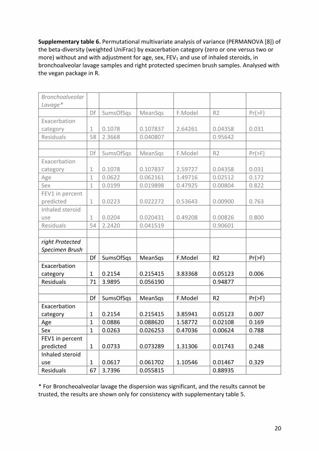
20
Supplementary table 6. Permutational multivariate analysis of variance (PERMANOVA [8]) of the beta-diversity (weighted UniFrac) by exacerbation category (zero or one versus two or more) without and with adjustment for age, sex, FEV1 and use of inhaled steroids, in bronchoalveolar lavage samples and right protected specimen brush samples. Analysed with the vegan package in R.
Bronchoalveolar Lavage*
Df SumsOfSqs MeanSqs F.Model R2 Pr(>F)
Exacerbation category 1 0.1078 0.107837 2.64261 0.04358 0.031
Residuals 58 2.3668 0.040807 0.95642
Df SumsOfSqs MeanSqs F.Model R2 Pr(>F)
Exacerbation category 1 0.1078 0.107837 2.59727 0.04358 0.031
Age 1 0.0622 0.062161 1.49716 0.02512 0.172
Sex 1 0.0199 0.019898 0.47925 0.00804 0.822
FEV1 in percent predicted 1 0.0223 0.022272 0.53643 0.00900 0.763
Inhaled steroid use 1 0.0204 0.020431 0.49208 0.00826 0.800
Residuals 54 2.2420 0.041519 0.90601
right Protected Specimen Brush
Df SumsOfSqs MeanSqs F.Model R2 Pr(>F)
Exacerbation category 1 0.2154 0.215415 3.83368 0.05123 0.006
Residuals 71 3.9895 0.056190 0.94877
Df SumsOfSqs MeanSqs F.Model R2 Pr(>F)
Exacerbation category 1 0.2154 0.215415 3.85941 0.05123 0.007
Age 1 0.0886 0.088620 1.58772 0.02108 0.169
Sex 1 0.0263 0.026253 0.47036 0.00624 0.788
FEV1 in percent predicted 1 0.0733 0.073289 1.31306 0.01743 0.248
Inhaled steroid use 1 0.0617 0.061702 1.10546 0.01467 0.329
Residuals 67 3.7396 0.055815 0.88935
* For Broncheoalveolar lavage the dispersion was significant, and the results cannot be trusted, the results are shown only for consistency with supplementary table 5.

21
Supplementary figure 7: Principle Coordinate plots of beta diversity, measured by Bray Curtis distance in second fraction of bronchoalveolar lavage (BAL2), protected specimen brush (rPSB) and oral wash (OW) samples. Each sample is coloured according to exacerbation status (zero versus one or more) during follow-up.
Supplementary figure 8: Principle Coordinate plots of beta diversity, measured by weighted UniFrac distance in second fraction of bronchoalveolar lavage (BAL2), protected specimen brush (rPSB) and oral wash (OW) samples. Each sample is coloured according to exacerbation status (zero versus one or more) during follow-up.
Axis 2 11.2%)
Axis 1 (15.8%)
Axis 1 (15.0%)
Axis 2 (13.2%)
Axis 2 (10.0%)
Axis 1 (19.3%)
Beta diversity – Bray Curtis distance
No exacerbation in follow-up Exacerbation in follow-up
BAL 2 rPSB OW
Axis 2 (22.3%)
Axis 1 (36.4%)
Axis 2 (30.0%)
Axis 1 (33.7%)
Axis 2 (14.3%)
Axis 1 (37.8%)
Beta diversity – Weighted UniFrac distance
No exacerbation in follow-up Exacerbation in follow-up
BAL 2 rPSB OW

22
Supplementary figure 9: Beta diversity, measured by weighted UniFrac distance. Box plots of distances between groups with and without later exacerbations for each sample type; oral wash (OW), right protected specimen brushes (rPSB) and second fraction of bronchoalveolar lavage (BAL2). Differences between all groups were tested with the ADONIS permutation-based test. There were no statistically significant differences.
23
References 1. Drengenes C, Wiker HG, Kalananthan T, Nordeide E, Eagan TML, Nielsen R. Laboratory
contamination in airway microbiome studies. BMC Microbiol 2019; 19, 187.
2. Bolger AM, Lohse M, Usadel B. Trimmomatic: a flexible trimmer for Illumina sequence
data. Bioinformatics 2014; 30, 2114-2120. 3. Davis NM, Proctor DM, Holmes SP, Relman DA, Callahan BJ. Simple statistical
identification and removal of contaminant sequences in marker-gene and metagenomics data.
Microbiome 2018; 6, 226.
4. Mandal, S 2015 Analysis of composition of microbiomes: a novel method for studying
microbial composition. Microb Ecol Health Dis 2015; 26, 27663.
5. Morton JT, Sanders J, Quinn RA et al. Balance Trees Reveal Microbial Niche
Differentiation. mSystems 2017; 2,
6. Fernandes, AD 2014 Unifying the analysis of high-throughput sequencing datasets:
characterizing RNA-seq, 16S rRNA gene sequencing and selective growth experiments by
compositional data analysis. 1805 21455 0
7. Chen J, King E, Deek R et al. An omnibus test for differential distribution analysis of
microbiome sequencing data. Bioinformatics 2018; 34, 643-651.
8. Anderson MJ. A new method for non‐parametric multivariate analysis of variance. Austral
ecology 2001; 26, 32-46.

Graphic design: Comm
unication Division, UiB / Print: Skipnes Komm
unikasjon AS
uib.no
ISBN: 9788230863664 (print)9788230847183 (PDF)
Copyright © 2022 FDOKUMEN