Asthma-COPD-National-Guidelines-by-SOMA.pdf - Rangpur ...
84
• • . " " - - • • • • • National Asthma Centre, National Institute of Diseases of the Chest & Hospital Mohakhali, Dhaka·1212, Bangladesh www.asthmabd.org • • NATIONAL GUIDELINES STHMA RONCHIOLITIS 3rd Edition 2005 Asthma Association Ban g ladesh OPD •
-
Upload
khangminh22 -
Category
Documents
-
view
0 -
download
0
Transcript of Asthma-COPD-National-Guidelines-by-SOMA.pdf - Rangpur ...

•
•
. ""
-
-•
• •
•
•
National Asthma Centre, National Institute of Diseases of the Chest & Hospital Mohakhali, Dhaka·1212, Bangladesh www.asthmabd.org
•
•
NATIONAL GUIDELINES
STHMA RONCHIOLITIS
3rd Edition 2005
Asthma Association Bangladesh
.... OPD
•

Published by:
Asthma Association, Bangladesh National Asthma Center NIDCH Campus, Mohakhali Dhaka-1212, Bangladesh
Address for correspondence:
National Asthma Center National Institute of Diseases of the Chest and Hospital Mohakhali, Dhaka-1212, Bangladesh Tel: +88-02-9887050 E-mail: [email protected] • Web: www.asthmabd.org
First Edition : November 1999 Second Edition : April 2001 Third Edition : May 2005
Contents of this book, whole or in part can be reproduced for research, academic or educational purposes. Acknowledgement to the Asthma Association, Bangladesh will be highly appreciated. No part of this book can be reproduced for commercial purposes as per copyright rules.
Graphic Design & Production:
Graphic Arts Jr. 59 /3-2 Purana Paltan Dhaka-lOOO Tel- 9561801
Price: Tk. 100.00 only
•
PREFACE An Appeal for Dissemination of Knowledge
Bismillahir Rahmanir Rahim.
Assalamu Alaikum.
It is a pleasure for me as we got the opportunity from Almighty Allah to publish the 3rd edition of our National Guidelines with an intention to disseminate proper knowledge through out the country. The 1st edition of "National Asthma Guidelines" was published in 1999, which was revised and the 2nd edition was published in 2001. By this time new information has came out from different research papers in home and abroad. Many physicians of the country took interest and send comments. After having long discussion with various groups we are now providing this updated version of the guidelines.
This time we included management updates of bronchiolitis and COPD in our guidelines. It is essential for all phYSicians dealing with asthma to know the diagnosis and management of bronchiolitis and COPD, because they are, to some extent, symptomatically looking alike asthma.
In Bangladesh more than 100 million people are suffering from cough and shortness of breath. Still our people are getting unplanned treatment and taking unscientific, indigenous and sometimes harmful products to get relief. Our aim is to disseminate knowledge to all groups of doctors, nurses, health care providers, medical students as well as affected peoples of the country to mitigate these sufferings.
We request all of you to follow these updated guidelines to put into practice a uniform, practical-oriented and scientific treatment regimen of asthma, bronchiolitis and COPD for the patients of Bangladesh.
Please disseminate the knowledge by implementation of guidelines and include it as teaching materials for undergraduate and postgraduate medical students as well as nursing students.
Prof. Md. Rashidul Hassan General Secretary Asthma Association On behalf of Board of Editors

PREFACE TO THE SECOND EDITION The first "National Asthma Prevalence Study" (NAPS) conducted throughout Bangladesh in 1999 has shown that about 7 million people suffer from asthma in our country. Proper scientific management practiced uniformly is imperative for amelioration of the sufferings of our fellow countrymen.
The Asthma Association published the first edition of the National Asthma Guidelines for Medical Practitioners in 1999 on a provisional basis. It has been updated and modified on the basis of detailed discussions held at the Fourth National Workshop on Asthma. By the Grace of Almighty Allah, we are publishing the 2nd edition of these guidelines for distribution within the medical community.
Even the best policies or guidelines formulated by top most experts can be miserable failures, if they are not implemented properly. We hope that through our concerted efforts, our guidelines shall see the light of success.
We earnestly request you to leave no stone unturned for the thorough implementation of these guidelines. Implementation of these guidelines can properly control asthma in majority of the patients and help them lead normal healthy lives. It can be our main pathway to achieve our cherished goal of effortless easy breathing.
Dr. Md. Rashidul Hassan General Secretary, Asthma Association On behalf of Board of Editors
PREFACE TO THE FIRST EDITION Most experts throughout the world believe that with appropriate management asthma is an evidently treatable condition. Yet recent studies of practice standards in our country have indicated that many physicians do not treat their patients optimally, prescribing too much "reliever" (bronchodilator) medicine and too little "preventer" (anti-inflammatory) medicine.
. On the basis of this background, Asthma Association has been trying to develop a 'National Asthma Guidelines for Medical Practitioners' for the last 3 years. By the grace of Almighty Allah, we are pubiishing the first edition of the guidelines. We hope these guidelines shall encourage physicians to manage asthma patients in an appropriate way. Insha-Allah we intend to publish the 2nd edition next year.
We shall be highly pleased if you kindly send your valuable comments and corrections to us regarding this 'National Asthma Guidelines for Medical Practitioners' within February 2000. Constructive criticism will be highly appreCiated. Valuable contributions will be duly acknowledged.
We intend to organize a workshop for further corrections and necessary modifications before publishing the 2nd edition.
Dr. Md. Rashidul Hassan
General Secretary, Asthma Association On behalf of Editorial Board
EXECUTIVE COMMITTEE 2004-06 Asthma Association, Bangladesh
President Prof. Md. Mostafizur Rahman
Vice-President Dr. Md. Ali Hossain Dr. AKM Kamal Uddin
General Secretary Prof. Md. Rashidul Hassan
Treasurer Dr. Mahmud Masum Attar
Joint Secretary
Organising Secretary
Office Secretary
Press & Publication Secretary
Scientific Secretary
Social Welfare Secretary
Members
Dr. Md. Rafiqul Islam Dr. AFM Kamaluddin
Dr. Md. Mizanur Rahman
Dr. Md. Mohiuddin Ahmad
Dr. Kazi Saifuddin Bennoor
Dr. Asif Mujtaba Mahmud
Dr. Rahmatul Bari
Prof. AKM Shamsul Huq Prof. ARM Luthful Kabir Dr. Mirza Mohammad Hiron Dr. Md. Atiqur Rahman Dr. Shafiqul Ahsan Dr. Mohammad Enamul Haque Dr. Md. Solaman Siddique Bhuiyan Dr. Md. Zillur Rahman Dr. AKM Mustafa Hussain Dr. Biswas Akhtar Hossain Dr. Vikarunnessa Begum Dr. Nawab Tahsin Uddin Dr. Bashir Ahmed Dr. GM Monsur Habib Dr. Md. Zahidul Islam Dr. Md. Naimul Hoque Dr. Md. Zahirul Islam Shakil Dr. Golam Sarwar Liaquat Hossain

BOARD OF EDITORS
Prof. Md. Rashidul Hassan FCPS (Medicine), MD (Chest) Professor, National Asthma Center National Institute of Diseases of the Chest & Hospital Mohakhali, Dhaka-1212, Bangladesh
Dr. Md. Ali Hossain FCPS (Medicine), MD (Chest) Associate Professor, Respiratory Medicine National Institute of Diseases of the Chest & Hospital Mohakhali, Dhaka-1212, Bangladesh
Dr. Asif Mujtaba Mahmud DICD, Ph.D. (Respiratory Medicine) Associate Professor, Respiratory Medicine National Institute of Diseases of the Chest & Hospital Mohakhali, Dhaka-1212, Bangladesh
Prof. ARM Luthful Kabir FCPS (Paediatrics) Professor, Paediatrics Institute of Child and Mother Health Matuail, Dhaka-1362, Bangladesh
Prof. Md. Ruhul Amin FCPS (Paediatrics) Professor, Paediatrics Bangladesh Institute of Child Health (BICH) Dhaka Shishu Hospital Sher-e-Bangla Nagar, Dhaka-1207, Bangladesh
Prof. Md. Mostafizur Rahman FCPS (Medicine) Professor & Director . National Institute of Diseases of the Chest & Hospital Mohakhali, Dhaka-1212, Bangladesh
Dr. Kazi Saifuddin Bennoor DICD Registrar (Medicine) National Institute of Diseases of the Chest & Hospital Mohakhali, Dhaka-1212, Bangladesh
I
•
CONTRIBUTING AUTHORS
Dr. Md. Mohiuddin Ahmad FCPS (Medicine), MD (Chest) Assistant Professor, Respiratory Medicine National Institute of Diseases of the Chest & Hospital, Mohakhali, Dhaka
Dr. G.M. Monsur Habib President, Asthma Library Sher-e-Bangla Road, Khulna & Member, Advisory Committee International Primary Care Respiratory Group (IPCRG)
Dr. Mirza Mohammad Hiron FCPS (Medicine), MD (Chest) Associate Professor, Medicine National Institute of Diseases of the Chest & Hospital, Mohakhali, Dhaka .
Dr. Shafiqui Ahsan MS (Cardiothoracic Surgery) Associate Professor (Ihoracic Surgery) National Institute of Diseases of the Chest & Hospital, Mohakhali, Dhaka
Dr. A.EM. Kamaluddin DICD Registrar National Institute of Cancer Research & Hospital, Mohakhali, Dhaka
Dr. Muhammad Khurshidul Islam DICD Associate Professor (Rtd.) National Institute of Diseases of the Chest & Hospital, Mohakhali, Dhaka
Dr. Mohammed Enamul Hoque DICD Superintendent Chest Clinic & Iraining Center Chankharpool, Dhaka
Dr. M.A. Jalil Chowdhury Associate Professor, Medicine Bangabandhu Sheikh Mujib Medical University, Shahbag, Dhaka
Dr. Md. Solaman Siddique Bhuiyan DICD, MD (Chest) Associate Professor, National Asthma Center, NIDCH, Mohakhali, Dhaka
Dr. Md. Rafiqui Islam DICD Assistant Professor (R. P.) National Institute of Diseases of the Chest & Hospital, Mohakhali, Dhaka
Dr. Md. Atiqur Rahman DICD, MD (Chest) Associate Professor, Respiratory Medicine National Institute of Diseases of the Chest & Hospital, Mohakhali, Dhaka
Dr. Biswas Akhter Hossain DICD Assistant Professor, Medicine Faridpur Medical College Faridpur
Dr. ShakiI Ahmed FCPS, MD (Paediatrics) Registrar, Paediatrics Dhaka Medical College, Dhaka
Dr. S.M. Abdullah Al Mamun MCPS (Medicine), MD (Chest) Registrar (Medicine) National Institute of Diseases of the Chest & Hospital, Mohakhali, Dhaka
Dr. Syed Azizul Haque Associate Professor, Cardiology National Institute of Cardiovascular Diseases, Sher-e-Bangla Nagar, Dhaka

CONSULTATIVE PANEL
Dr M. Fazlur Rahman M Phil, PhD Associate Professor, Epidemiology Institute of Child and Mother Health, Matuail, Dhaka
Dr. A.K.M. Mustafa Hussain DTCD Assistant Professor, Respiratory Medicine National Institute of Diseases of the Chest & Hospital, Mohakhali, Dhaka
Dr. Md. Mizanur Rahman DTCD Assistant Professor National Institute of Diseases of the Chest & Hospital Mohakhali, Dhaka
Dr. Mohammad Abdus Shakur Khan MD (Chest) Assistant Professor National Asthma Center National Institute of Diseases of the Chest & Hospital Mohakhali, Dhaka
-
Dr. Golam Sarwar Liaquat Hossain MBBS Medical Officer National Institute of Diseases of the Chest & Hospital Mohakhali, Dhaka
Dr. Md. Zahirul Islam Shakil DTCD Assistant Professor National Institute of Diseases of the Chest & Hospital Mohakhali, Dhaka
Dr. Mahmud Masum Attar DTCD Assistant Professor National Asthma Center National Institute of Diseases of the Chest & Hospital Mohakhali, Dhaka
Dr. Md. Ziaul Karim DTCD Assistant Professor National Asthma Center National Institute of Diseases of the Chest & Hospital Mohakhali, Dhaka
Dr. Md. Zahidul Islam DTCD Assistant Professor National Institute of Diseases of the Chest & Hospital Mohakhali, Dhaka
Dr. Md. Naimul Haque MBBS Medical Officer National Institute of Diseases of the Chest & Hospital, Mohakhali, Dhaka
,
,
ADVISORY BOARD
Prof. A.Q.M. Nurul Haq Professor of Medicine & Former Director National Institute of Diseases of the Chest & Hospital, Mohakhali, Dhaka
Prof. A.K.M. Moslehuddin Professor of Medicine & Former Director National Institute of Diseases of the Chest & Hospital, Mohakhali, Dhaka
Prof. A. K. M. Shariful Islam Professor of Thoracic Surgery & Former Director National Institute of Diseases of the Chest & Hospital, Mohakhali, Dhaka
Prof. M. Nabi Alam Khan Professor Emeritus National Institute of Diseases of the Chest & Hospital, Mohakhali, Dhaka
Prof. Md. Abid Hossain Molla Professor of Paediatrics Dhaka Medical College Dhaka
Prof. A.K.M. Shamsul Huq Professor of Medicine & Former Director National Institute of Diseases of the Chest & Hospital, Mohakhali, Dhaka
Prof. Md. Sofiullah Professor of Medicine & Former Director National Institute of Diseases of the Chest & Hospital, Mohakhali, Dhaka
Prof. Falahuzzaman Khan Professor of Community Medicine & Former Director National Institute of Diseases of the Chest & Hospital, Mohakhali, Dhaka
Prof. G M Akbar Chowdhury Professor of Thoracic Surgery National Institute of Diseases of the Chest & Hospital, Mohakhali, Dhaka
Prof. Sayeda Afroza Porfessor of Paediatrics & Joint Director, Institute of Child & Mother Health, Matuail, Dhaka
•

DISCUSSANT PANEL
Facilitators:
Prof. Chowdhury Ali Kawser Dr. Khaleda Begum Dr. Md. Abdur Rouf Dr. Md. Abu Hasnat Dr. Zakir Hossain Sarkar Dr. Rowshne J ahan Dr. Shah Mohammad Saifur Rahman Dr. Md. Nawab Tahsin Uddin Dr. Biswas Shaheen Hasan Dr. Md. Zillur Rahman Dr. AKM Kamaluddin Dr. Saria Tasneem Dr. Rahmatul Bari Dr. Md. Abdul Qayyum Dr. Md. Khairul Hassan Jessy Dr. Md. Asadur Rahman Dr. Selina Khanam Dr. Al Amin Mridha Dr. Md. Sofiuddin Dr. Mohammad Delwar Hossain
Rapporteurs:
Dr. Md. Azizul Haq Dr. Khan Md. Sayeduzzaman Dr. Md. Rafiqul Islam Dr. SAHM Mesbahul Islam Dr. Shameem Ahmed Dr. Md. Delwar Hossain Dr. Adnan Yusuf Chowdhury Dr. Shahedur Rahman Khan Dr. Md. Mahbub Alam Siddiqui Dr. Jalal Mohsin Uddin Dr. Amirul Morshed Khosru Dr. Khandker Rokonuddin Dr. Kazi Mahbub-e-Khoda
Members:
Prof. Soofia Khatun Prof. Sameena Chowdhury Prof. M.O. Faruq Dr. Md. Meer Mahbubul Alam Dr. Anas Darwish Dr. Ahmed Mohammed Ali Dr. Md. Mokaddes Hossain Dr. Md. Ashraful Islam Dr. Md. Emdadul Huq Dr. Muksud Ahmed Dr. Md. Quamruzzaman Dr. Md. Mokim Ali Biswas Dr. Rabindra Chandra Mitra Dr. Syed Md. Kamrul Hossain Dr. Imamuddin Ahmed Dr. Bashir Ahmed Dr. Md. Hasanul Hasib Dr. Md. Mahbub Anwar Dr. Mohammed Jahangir Alam Dr. ASM Mesbah Uddin Dr. Nirmeen Rifat Khan Dr. Md. Shahadat Hossain Dr. Rowshan Ara Islam Dr. Md. Nazibur Rahman Dr. Afzalunnessa Binte Lutfor Dr. Sk. Shahinur Hossain Dr. Atiqur Rahman Dr. Md. Nowfel Islam Dr. Farooque Ahmed Dr. Manzurul Chowdhury Dr. Shamim Ahmed Dr. Md. Zahidul Islam Dr. Parimal Kanti Debnath Dr. Faruque Ahmed Khan Dr. Md. Mizanur Rahman Dr. Ahmed Al Montasir
,
Dr. Ershad Mahmud Dr. AFM Saidur Rahman Dr.Md. Shahadat Hossain Dr. Adnan Hasan Masud Dr. Md. Abu Shahin Dr. Mohammad Shafikul Islam Dr. MH J akaria Dr. Sharif Ahmed Dr. Kh. Maqsudul Haque Dr. Shahana Huque Dr. Amar Biswas Dr. Mahadeb Chandra MandaI Dr. Mohammad Monirul Islam Dr. Nahid-E-Subha Dr. AKM Saifur Rashid Dr. Md. Azizur Rahman Dr. AI-Belal Dr. Md. Abu Ishaque Dr. Abdullah Al Mamun Dr. Touhidul Karim Majumder Dr. Sk. Mahbub Alam Dr. Rajat Shuvra Paul Dr. Md. Jahirul Hoq Dr. Lima A. Sayami Dr. Sharif Uddin Khan Dr. Md. Belal Hossain Dr. Amiruzzaman Dr. Mohammad Mohsin Dr. Mostafa Kamal Dr. Gul-A-Rana Dr. Md. Sirajul Islam Dr. Abu) Kalam Azad Dr. Mahbubul Hossain Dr. Jobaida Akhter Dr. Md. Ferdous Wahid Dr. Md. Kamal Uddin Dr. Mohammad Aminul Islam Dr. Shah Golam Nabi Dr. ATM Khalilur Rahman Dr. Md. Shafiqur Rahman Dr. Sonia Nasreen Ahmad Dr. Muhammad Humayoun Kabir Dr. Md. Rawshan Ali Basunia Dr. Md. Abul Kalam
Dr. Krishna Kanta Sen Dr. Nasrin Afrose Dr. Shamim Ahmed Dr. Md. Masudur Rahman Khan Dr. SM Quamrul Dr. Md. Radwanur Rahman Dr. Muhammad Muniruzzaman Chowdhury Dr. Dewan Mahmud Hasan Dr. KM Wahidul Hoque Dr. Md. Nurul Islam Dr. Md. Jahangir Rashid Dr. Abdus Salam Dr. Md. Main Uddin Dr. Mohammad Asadur Rahman Dr. Mazharul Islam Dr. Kazi Shah Md. Abdullah Dr. Nauruj Jahan Dr. Mansur Elahi Dr. Md. Abu Sayem Dr. Rubina Akter Dr. Md. Kamrul Alam Dr. Sohail Ahmed Dr. Md. Abul Quashem Dr. Khalifa Mahmood Tariq Dr. Haroon Rashid Dr. Syed Imtiaz Ahsan Dr. Manobendra Biswas Dr. Muhammad Jalal Uddin Dr. Md. Rezaul Hasan Dr. Shaheenul Islam Dr. AKM Akramul Haque Dr. Mohammad Aminul Islam Dr. Sunil Kumar Biswas Dr. Nazneen Kabir Dr. Dipankar Nag Dr. Jonaed Hakim Dr. Sajida Nahid Dr. AFM Azizur Rahman Siddique Dr. IHat Zaman Dr. Taskina Ali Dr. Md. Golam Abbas Dr. Md. Wahidul Islam Dr. Md. Shafiqul Islam Dr. Gazi Mohammad Imranul Haque

Dr. Chandra Nath Roy Dr. Quazi Md. Anisujjaman Dr. Amina Begum Dr. Zebun Nessa Dr. Most. Mithyla Ferdous Dr. ABM Borhan Uddin Dr. Shihab Uddin Dr. Md. Iqbal Hossain Talukder Dr. Wahiduzzaman Akhanda Dr. Md. Siddiqur Rahman Dr. Borhanuddin Ahmed Dr. Tushar Kanti Barman Dr. Rakhal Chandra Debnath Dr. Mujibur Rahman Dr. Md. Farhad Alam Dr. Md. Abdul Khaleque Dr. Md. Rifat Zia Hossain Dr. Sayed Moshfiqur Rahman Dr. Altaf Hossain Dr. Baidya Nath Saha Dr. Md. Hasanur Rashid Dr. A.R.M. Rafiqul Islam Dr. Shamsul Arafeen Khan Dr. Nishiranjan Talukder Dr. AFM Risatul Islam Dr. Hasan Imam Dr. Raihana Ahad Dr. Refath Ara Mahfuz Dr. Golam Muktadir Dr. A.R. Hawlader Dr. ATM Sulaiman Kabir Dr. Sk. Royhan Ibn Ismail Dr. Tridip Kanti Barman Dr. Md. Jamal Uddin Dr. Khorshed Minhazul Alam Dr. Selina Bll.nU Dr. H.M. Nazmul Ahsan Dr. Md. Hafizur Rahman Dr. Mahbubur Rahman Dr. Md. Mahbubul Alam Sarker Dr. KM Anwarul Huque Dr. Sabina Hossain Dr. Pavel Shahrior Mostafa Dr. Muhammad Shakhawat
Dr. Sk. Md. Abu Zafar Dr. Nasren Jebin Dr. Md. Ashadur Rahman Dr. Md. Mahbubul Hoque Dr. Mohammad Monir Hossain Dr. S.M. Shahnawaz Bin Tabib Dr. Mahfuza Shirin Dr. Shafi Uddin Ahmed Dr. Sk. Yunus Ali Dr. Md. Delwar Hossain Dr. M. Habibur Rahman Dr. ASM Areef Ahsan Dr. Iftakhar Alam Dr. Md. Mizanur Rahman Dr. M.A. Hasanat Dr. Mohammad Salman Dr. Farid Uddin Ahamed Dr. Taslim Uddin Dr. Gouranga Kumar Saha Dr. M. Shahabuddin Dr. Md. Abdur Rahim Miah Dr. Mohamamed Nasir Uddin Dr. Kazi Nazrul Islam Dr. Mir Nesaruddin Ahmed Dr. Md. Shafiul Azam Dr. Md. Towhiduz Zaman Dr. Sohely Rahman Dr. Prafulla Chandra Nath Talukder Dr. Md. Shafiqur Rahman Patwary Dr. Md. Muzibur Rahman Khan Dr. Mahibur Rahim Dr. Md. Faraque Pathan Dr. AKM Musa Dr. Mohammad Mohibur Rahman Dr. Dipankar Chandra Nag Dr. Md. Raziur Rahman Dr. Md. Majibar Rahman Dr. M.A. Khaleque Dr. Syed Atiqul Haq Dr. Md. Farid Uddin Dr. M. Shahinur Rahman Dr. Md. Jahurul Haque Dr. Md. Ferdous Rahman Dr. Md. Sultan Ahmed
ACKNOWLEDGEMENTS -
Iftekharul Islam Honorary Member, Asthma Association. Managing Director, SANOFI-AVENTIS Dhaka, Bangladesh
Md. Azizul Huq Honorary Member, Asthma Association. Managing Director GlaxoSmithKline Bangladesh Ltd
Sayed A B Tahmeed Honorary Member, Asthma Association. Marketing Manager SANOFI-AVENTIS Dhaka, Bangladesh
Anwar Hossain Khan Associate Member, Asthma Association. Ex -Librarian National Institute of Diseases of the Chest & Hospital, Mohakhali, Dhaka
Ekramul Haq Associate Member, Asthma Association.
•
Librarian-in-charge National Institute of Diseases of the Chest & Hospitals, Mohakhali, Dhaka
Abdul Muktadir Honorary Member, Asthma Association. Managing Director Incepta Pharmaceuticals Limited Dhaka, Bangladesh
Nazmul Ahsan Honorary Member, Asthma Association. C.E .O, Beximco Pharmaceuticals Limited, Dhaka, Bangladesh
Jahangir Hyder Honorary Member, Asthma Association. Marketing Manager OPSONIN Dhaka, Bangladesh
Maleka Begum Associate Member, Asthma Association. Sr. Staff Nurse In-charge, National Asthma Center, National Institute of Diseases of the Chest & Hospitals, Mohakhali, Dhaka

�2-agonists . . . . . . . . . . . . . . . . . . . . . . . . . . . . . . . . . . . . . . . . . . . . . . . . . . . . . . . . . . . . . . . . . . . . . . . . . . . . . . . . . . . . . . . . . . . . . . . . . . . . . . . . . . . . . . . . . . 47 Xanthine Derivatives . . . . . . . . . . . . . . . . . . . . . . . . . . . . . . . . . . . . . . . . . . . . . . . . . . . . . . . . . . . . . . . . . . . . . . . . . . . . . . . . . . . . . . . . . . . . . . . . . 48
Introduction . . . . . . . . . . . . . . . . . . . . . . . . . . . . . . . . . . . . . . . . . . . . . . . . . . . . . . . . . . . . . . . . . . . . . . . . . . . . . . . . . . . . . . . . . . . . . . . . . . . . . . . . . . . . . . . 19 Anticholinergics . . . . . . . . . . . . . . . . . . . . . . . . . . . . . . . . . . . . . . . . . . . . . . . . . . . . . . . . . . . . . . . . . . . . . . . . . . . . . . . . . . . . . . . . . . . . . . . . . . . . . . . . . 49 Cromones . . . . . . . . . . . . . . . . . . . . . . . . . . . . . . . . . . . . . . . . . . . . : . . . . . . . . . . . . . . . . . . . . . . . . . . . . . . . . . ; . . . . . . . . . . . . . . . . . . . . . . . . . . . . . . . . . . . . . 49
Part-A: ASTHMA Corticosteroids . . . . . . . . . . . . . . . . . . . . . . . . . . . . . . . . . . . . . . . . . . . . . . . . . . . . . . . . . . . . . . . . . . . . . . . . . . . . . . . . . . . . . . . . . . . . . . . . . . . . . . . . . . . 50 Is there any adverse effect of high dose inhaled
Section 1- Basic facts about asthma corticosteroid on children? . . . . . . . . . . . . . . . . . . . . . . . . . . . . . . . . . . . . . . . . . . . . . . . . . . . . . . . . . . . . . . . . . . . . . . . . . . . . . . . . . . . 52
DEFINITION . . . . . . . . . . . . . . . . . . . . . . . . . . . . . . . . . . . . . . . . . . . . . . . . . . . . . . . . . . . . . . . . . . . . . . . . . . . . . . . . . . . . . . . . . . . . . . . . . . . . . . . . . . . . . . 25 Leukotrienes antagonists . . . . . . . . . . . . . . . . . . . . . . . . . . . . . . . . . . . . . . . . . . . . . . . . . . . . . . . . . . . . . . . . . . . . . . . . . . . . . . . . . . . . . . . . . 53
Why do we define asthma? . . . . . . . . . . . . . . . . . . . . . . . . . . . . . . . . . . . . . . . . . . . . . . . . . . . . . . . . . . . . . . . . . . . . . . . . . . . . . . . . . . . . . 25 Newer drugs . . . . . . . . . . . . . . . . . . . . . . . . . . . . . . . . . . . . . . . . . . . . . . . . . . . . . . . . . . . . . . . . . . . . . . . . . . . . . . . . . . . . . . . . . . . . . . . . . . . . . . . . . . . . . . 54
Epidemiological Definitions . . . . . . . . . . . . . . . . . . . . . . . . . . . . . . . . . . . . . . . . . . . . . . . . . . . . . . . . . . . . . . . . . . . . . . . . . . . . . . . . . . . . 27 Disease modifying agents . . . . . . . . . . . . . . . . . . . . . . . . . . . . . . . . . . . . . . . . . . . . . . . . . . . . . . . . . . . . . . . . . . . . . . . . . . . . . . . . . . . . . . . 55 What is the role of antihistamines in management of asthma? . . . . . . . . . . . . . . . . . . . . . . . . . . 55 What is the role of ketotifen in management of asthma? . . . . . . . . . . . . . . . . . . . . . . . . . . . . . . . . . . . . 56 Should we use antibiotics is asthma? . . . . . . . . . . . . . . . . . . . . . . . . . . . . . . . . . . . . . . . . . . . . . . . . . . . . . . . . . . . . . . . . . . . . . .56
Airflow limitation . . . . . . . . . . . . . . . . . . . . . . . . . . . . . . . . . . . . . . . . . . . . . . . . . . . . . . . . . . . . . . . . . . . . . . . . . . . . . . . . . . . . . . . . . . . . . . . . . . . . . . 27 ETIOLOGY . . . . . . . . . . . . . . . . . . . . . . . . . . . . . . . . . . . . . . . . . . . . . . . . . . . . . . . . . . . . . . . . . . . . . . . . . . . . . . . . . . . . . . . . . . . . . . . . . . . . . . . . . . . . . . . . . . 28 What causes asthma episodes? . . . . . . . . . . . . . . . . . . . . . . . . . . . . . . . . . . . . . . . . . . . . . . . . . . . . . . . . . . . . . . . . . . . . . . . . . . . . . . . . 28 Can sedatives be used in asthma? . . . . . . . . . . . . . . . . . . . . . . . . . . . . . . . . . . . . . . . . . . . . . . . . . . . . . . . . . . . . . . . . . . . . . . . . . . .56 What is a trigger? . . . . . . . . . . . . . . . . . . . . . . . . . . . . . . . . . . . . . . . . . . . . . . . . . . . . . . . . . . . . . . . . . . . . . . . . . . . . . . . . . . . . . . . . . . . . . . . . . . . . . . . 28 List of asthma medicines . . . . . . . . . . . . . . . . . . . . . . . . . . . . . . . . . . . . . . . . . . . . . . . . . . . . . . . . . . . . . . . . . . . . . . . . . . . . . . . . . . . . . . . . . . 57 What are the triggers of asthma? . . . . . . . . . . . . . . . . . . . . . . . . . . . . . . . . . . . . . . . . . . . . . . . . . . . . . . . . . . . . . . . . . . . . . . . . . . . . . . 28 Doses of asthma medicines . . . . . . . . . . . . . . . . . . . . . . . . . . . . . . . . . . . . . . . . . . . . . . . . . . . . . . . . . . . . . . . . . . . . . . . . . . . . . . . . . . . . . . 59 CLASSIFICATION . . . . . . . . . . . . . . . . . . . . . . . . . . . . . . . . . . . . . . . . . . . . . . . . . . . . . . . . . . . . . . . . . . . . . . . . . . . . . . . . . . . . . . . . . . . . . . . . . . . . . 30 Why do we classify asthma? . . . . . . . . . . . . . . . . . . . . . . . . . . . . . . . . . . . . . . . . . . . . . . . . . . . . . . . . . . . . . . . . . . . . . . . . . . . . . . . . . . . . . 30 Section 2 - Management of asthma
How asthma is classified? . . . . . . . . . . . . . . . . . . . . . . . . . . . . . . . . . . . . . . . . . . . . . . . . . . . . . . . . . . . . . . . . . . . . . . . . . . . . . . . . . . . . . . . . . 30 What is the goal of asthma management plan? . . . . . . . . . . . . . . . . . . . . . . . . . . . . . . . . . . . . . . . . . . . . . . . . . . . . 64 Refractory asthma . . . . . . . . . . . . . . . . . . . . . . . . . . . . . . . . . . . . . . . . . . . . . . . . . . . . . . . . . . . . . . . . . . . . . . . . . . . . . . . . . . . . . . . . . . . . . . . . . . . . . . 33 What are the components of an effective management plan? . . . . . . . . . . . . . . . . . . . . . . . . . : . . 64 DIAGNOSIS . . . . . . . . . . . . . . . . . . . . . . . . . . . . . . . . . . . . . . . . . . . . . . . . . . . . . . . . . . . . . . . . . . . . . . . . . . . . . . . . . . . . . . . . . . . . . . . . . . . . . . . . . . . . . . . . 35 Is there a cure for asthma? . . . . . . . . . . . . . . . . . . . . . . . . . . . . . . . . . . . . . . . . . . . . . . . . . . . . . . . . . . . . . . . . . . . . . . . . . . . . . . . . . . . . . . . 64 What are the diagnostic criteria of asthma? . . . . . . . . . . . . . . . . . . . . . . . . . . . . . . . . . . . . . . . . . . . . . . . . . . . . . . . . . . . . 35 What is meant by control? . . . . . . . . . . . . . . . . . . . . . . . . . . . . . . . . . . . . . . . . . . . . . . . . . . . . . . . . . . . . . . . . . . . . . . . . . . . . . . . . . . . . . . . . 64 What are the differential diagnoses of asthma? . . . . . . . . . . . . . . . . . . . . . . . . . . . . . . . . . . . . . . . . . . . . . . . . . . . . . 36 Rule of 2 ...................................................................................................................... 65 . Differential diagnosis of childhood asthma . . . . . . . . . . . . . . . . . . . . . . . . . . . . . . . . . . . . . . . . . . . . . . . . . . . . . . . . . . . 36 Criteria of "well-controlled" and "totally-controlled" asthma . . . . . . . . . . . . . . . . . . . . . . . . . . . . . . 65 Helpful features for the diagnosis of childhood asthma . . . . . . . . . . . . . . . . . . . . . . . . . . . . . . . . . . . . . . 38 What do we mean by remission? . . . . . . . . . . . . . . . . . . . . . . . . . . . . . . . . . . . . . . . . . . . . . . . . . . . . . . . . . . . . . . . . . . . . . . . . . . . . . 66 Algorithm for the diagnosis of bronchiolitis, pneumonia and
asthma in children . . . . . . . . . . . . . . . . . . . . . . . . . . . . . . . . . . . . . . . . . . . . . . . . . . . . . . . . . . . . . . . . . . . . . . . . . . . . . . . . . . . . . . . . . . . . . . . . . 39 How can asthma episodes be prevented? . . . . . . . . . . . . . . . . . . . . . . . . . . . . . . . . . . . . . . . . . . . . . . . . . . . . . . . . . . . . . . 66 Modalities of asthma management . . . . . . . . . . . . . . . . . . . . . . . . . . . . . . . . . . . . . . . . . . . . . . . . . . . . . . . . . . . . . . . . . . . . . . . . 66
INVESTIGATIONS . . . . . . . . . . . . . . . . . . . . . . . . . . . . . . . . . . . . . . . . . . . . . . . . . . . . . . . . . . . . . . . . . . . . . . . . . . . . . . . . . . . . . . . . . . . . . . . . . . . . . 40 What are the types of home management plan? . . . . . . . . . . . . . . . . . . . . . . . . . . . . . . . . . . . . . . . . . . . . . . . . . . . 67 HOME MANAGEMENT ............................................................................ ........... 67 Why we investigate asthma patients? . . . . . . . . . . . . . . . . . . . . . . . . . . . . . . . . . . . . . . . . . . . . . . . . . . . . . . . . . . . . . . . . . . . . .40
What are the investigations for asthma? . . . . . . . . . . . . . . . . . . . . . . . . . . . . . . . . . . . . . . . . . . . . . . . . . . . . . . . . . . . . . . . . . 40 What other concomitant illnesses of an asthma patient
should be investigated? . . . . . . . . . . . . . . . . . . . . . . . . . . . . . . . . . . . . . . . . . . . . . . . . . . . . . . . . . . . . . . . . . . . . . . . . . . . . . . . . . . . . . . . .41 Spirometry . . . . . . . . . . . . . . . . . . . . . . . . . . . . . . . . . . . . . . . . . . . . . . . . . . . . . . . . . . . . . . . . . . . . . . . . . . . . . . . . . . . . . . . . . . . . . . . . . . . . . . . . . . . . . . . . . . . 42
Why management at home? . . . . . . . . . . . . . . . . . . . . . . . . . . . . . . . . . . . . . . . . . . . . . . . . . . . . . . . . . . . . . . . . . . . . . . . . . . . . . . . . . . . . 67 What is step care management? . . . . . . . . . . . . . . . . . . . . . . . . . . . . . . . . . . . : . . . . . . . . . . . . . . . . . . . . . . . . . . . . . . . . . . . . . . . . . . 68 Basic principles of step care management . . . . . . . . . . . . . . . . . . . . . . . . . . . . . . . . . . . . . . . . . . . . . . . . . . . . . . . . . . . . . 68 Step care management for children > 5 years to adults . . . . . . . . . . . . . . . . . . . . . . . . . . . . . . . . . . . . . . . 70 Step care management for::: 5 years . . . . . . . . . . . . . . . . . . . . . . . . . . . . . . . . . . . . . . . . . . . . . . . . . . . . . . . . . . . . . . . . . . . . . . . 71 Step care management: economic schedule . . . . . . . . . . . . . . . . . . . . . . . . . . . . . . . . . . . . . . . . . . . . . . . . . . . . . . . . . . . 72 Which medication should be preferred for a patient able to buy
only one inhaler - a reliever or a preventer? . . . . . . . . . . . . . . . . . . . . . . . . . . . . . . . . . . . . . . . . . . . . . . . . . . . . . . 73 Which inhaler should not be used alone? . . . . . . . . . . . . . . . . . . . . . . . . . . . . . . . . . . . . . . . . . . . . . . . . . . . . . . . . . . . . . . 73 Is there any benefit of combination inhalers? . . . . . . . . . . . . . . . . . . . . . . . . . . . . . . . . . . . . . . . . . . . . . . . . . . . . . . . . 73 Which step is appropriate for a specific patient? . . . . . . . . . . . . . . . . . . . . . . . . . . . . . . . . . . . . . . . . . . . . . . . . . . 73
Spirometric tracings . . . . . . . . . . . . . . . . . . . . . . . . . . . . . . . . . . . . . . . . . . . . . . . . . . . . . . . . . . . . . . . . . . . . . . . . . . . . . . . . . . . . . . . . . . . . . . . . . . . . 44 Tests of spirometry . . . . . . . . . . . . . . . . . . . . . . . . . . . . . . . . . . . . . . . . . . . . . . , . . . . . . . . . . . . . . . . . . . . . . . . . . . . . . . . . . . . . . . . . . . . . . . . . . . . . . 45 MEDICINES OF ASTHMA .. . . . . . . . . . . . . . . . . . . . . . . . . . . . . . . . . . . . . . . . . . . . . . . . . . . . . . . . . . . . . . . . . . . . . . . . . . . . . . . . . . . . . . 46 What are the medicines used to treat asthma? . . . . . . . . . . . . . . . . . . . . . . . . . . . . . . . . . . . . . . . . . . . . . . . . . . . . . . 46 Are asthma medicines safe? . . . . . . . . . . . . . . . . . . . . . . . . . . . . . . . . . . . . . . . . . . . . . . . . . . . . . . . . . . . . . . . . . . . . . . . . . . . . . . . . . . . . . 46 What should be done if side effects occur? . . . . . . . . . . . . . . . . . . . . . . . . . . . . . . . . . . . . . . . . . . . . . . . . . . . . . . . . . . . 46

Scoring system for step care management ......................................................... ... 73 When anticholenergic medicine is to be added in
home management plan? .......................................... ................................. , . . . . . . . 75 Wh . " k "7 75 at IS pac year . . . . . . . . . . . . . . . . . . . . . . . . . . . . . . . . . . . . . . . . . . . . . . . . . . . . . . . . . . . . , .................................... .
What is the importance of "pack-year" in asthma management? ....................... 75 When to follow-up the patient? ................................. .............................................. 75 When to step down? . . .............. ............ ........ ............................ ................... ............... 76 How to step down? ................................. , . . . . . . . . . . . . . . . . . . . . . . . . . . . . . . . . . . . . . . . . . . . . . . . . . . . . . . . . . . . . . . . . . 76 When to step up? .............................................................. ....... .. . ........................ ....... 76 How to step up? ............ .................... ....................... .............. ............... . ......... . .......... 76 Pitfalls of management ................................................ ...... ..... . . .. . . . ........ . . . . ......... ...... 77 Rescue steroid therapy ............................................... .. . . . . . ... . ......... .. . . . . .. . . .. . . ............ . 78 When a patient should contact his/her doctor? ......... . . . . . . . . . . . . . .. . ...... . . . . . . . . .. . . . ........ 79 When a general practitioner should refer a patient to pulmonologist? ... .......... 79 EMERGENCY MANAGEMENT ... .................. .. . . . . . . . . . . .. . . ..... .. . . . ... . . ........... ............ 81 What is emergency management of asthma? ... .... .. . . . . . . . . .. ..... ... ........................ ..... 81 What do we mean by acute exacerbation of asthma? ........................................... 81 What are the protocols of emergency management? ........................................ .... 81 First aid for asthma (Rule of 5) . . . . . . . . . . . . . . . . . . . . . . . . . . . . . . . . . . . . . . . . . . . . . . . . . . . . . . . . . . . . . . . . . . . . . . . . . . . . . . . . . 82 What are the components of management of acute exacerbation? .................... 82 What is meant by initial and periodic observation? .............. ................... ............ 83 Assessment of severity of acute asthma in adults ...... . ....... . . . . . . ....... ................ .... 83 Assessment of severity of acute asthma in children ................... ............. ....... ...... 84 How �2-agonists are used in emergency management? ........................ . . ............ 84 What is the role of xanthines derivatives? ........... ............... . ................ .. .. . . ............ 84 Is there any role of magnesium sulfate? ................................................................. 85 What is the role of leukutriene antagonists? ........................... .............................. 85 What is the role of anticholinergic drugs? ............................................................. 85 Why and how oxygen inhalation is given? ........ .................................................... 85 How steroid is used? .............................................. ............ ....................................... 86 Is there any role of antibiotics? ................................................................................ 86 Can sedatives be prescribed during acute attack? ................................................ 86 Therapies not recommended during acute attack .......................................... ...... 87 How to assess and follow-up the patient? ............................................................. 87 When to hospitalize a patient? ..... ........................................................................... 87 Criteria for admission in ICU ............ ............................. . . . ...................................... 88 What are the indications of artificial ventilation? .................... .................. .... ....... 88 Management of asthma attacks in hospital or ICU .................................. . ... ........ 89 Can anti-allergy vaccines (immunotherapy) cure asthma? ..................... ... ......... 91
MANAGEMENT OF CONCOMITANT DISEASES ............................................ 92 Allergic rhinitis ................................................................................... : . . . . . . . . . . . . . . . . . . . . . . 92 Atopic dermatitis (Eczema) ..................................................................................... 96 Allergic conjunctivitis ............................................................................................... 97 ASTHMA AND CO-MORBIDITIES ....................................................................... 98 ASTHMA IN SPECIAL SITUATIONS . . . . . . . . . . . . . . . . . . . . . . . . . . . . . . . . . . . . . . . . . . . . . . . . . . . . . . . . . . . . . . . . . . . 100 Pregnancy and asthma ............................................................................................. 1 00 Surgery and asthma ................................................................................................. 102
Section 3- Asthma education
What is patient education in asthma and why is it essential? .......................... .103 PREVENTION OF ASTHMA ......... ........... ............................................................. 108 How we can prevent asthma? ................................................................................ 108 What are the types of asthma prevention? .......................................................... .108 What is primary prevention? ................................................................................. 108 How to identify asthma prone persons? .............................................................. 108 Primary prevention program ................................................................................. 109 What is secondary prevention? ................. .... ......................................................... 110 Patient's concerns about asthma ............... ............................................................. 111 ASTHMA MANAGEMENT APPLIANCES ......................................................... 112 Metered dose inhalers ............................................................................................. 112 How to use MDIs? ................................................................................................... 112 Checking how much medicine is left in the canister .......................................... 114 Breath activated inhlers ............................................................... ..... .............. . ........ 114 Dry powder inhalers . . . . . . . . . . . . . . . . . . . . . . . . . . . . . . . . . . . . . . . . . . . . . . . . . . . . . . . . . . . . . . . . . . . . . . . , . . . . . . . . . . . . . . . . . . . . . . . . 114 Spacers and chambers ................................................................ ... ................ ..... . . . . . 11 6 How to use a apacer? ...................................................................... ............. . .... . . .... . 11 6 Nebulizers ................................................................................................................ 117 How to use a nebulizer? .......................................................................................... 118 Delivery devices for asthma medications in children ........................................ .119 Flow meters ............................................................................................................. 120 ·Peak flow meters ...................................................................................................... 120 Predicated values of PEF ........................................................................................ . 121 I
. . ncentl ve spIrometer ................................................................................................ 122 PIF meter . . . . . .............................................................................................................. 123 Warning signs of asthma episodes ....................................................................... . 124 GUIDED SELF MANAGEMENT PLAN FOR ASTHMA .................................. 125 Personal best peak flow result .............................................................................. .125 Peak flow zone system .......................... ................................................................. 125 Peak flow chart ......................... .............................................................................. 127 Self management chart ........................................................................................... 128

ASTHMA TRIGGER CONTROL PLAN . . . . . . . . . . . . . . . . . . . . . . . . . . . . . . . . . . . . . . . . . . . . . . . . . . . . . . . . . . . . . . . 130 Asthma and weather . . . . . . . . . . . . . . . . . . . . . . . . . . . . . . . . . . . . . . . . . . . . . . . . . . . . . . . . . . . . . . . . . . . . . . . . . . . . . . . . . . . . . . . . . . . . . . . . 133
Part-B: BRONCHIOLITIS
Bronchiolitis : Background . . . . . . . . . . . . . . . . . . . . . . . . . . . . . . . . . . . . . . . . . . . . . . . . . . . . . . . . . . . . . . . . . . . . . . . . . . . . . . . . . . . . . . 137 What is bronchiolitis? . . . . . . . . . . . . . . . . . . . . . . . . . . . . . . . . . . . . . . . . . . . . . . . . . . . . . . . . . . . . . . . . . . . . . . . . . . . . . . . . . . . . . . . . . . . . . . 137 What are the risk factors of bronchiolitis? . . . . . . . . . . . . . . . . . . . . . . . . . . . . . . . . . . . . . . . . . . . . . . . . . . . . . . . . . . . . 138 How bronchiolitis is classified? . . . . . . . . . . . . . . . . . . . . . . . . . . . . . . . . . . . . . . . . . . . . . . . . . . . . . . . . . . . . . . . . . . . . . . . . . . . . . . 139 What are the typica I radiological features? . . . . . . . . . . . . . . . . . . . . . . . . . . . . . . . . . . . . . . . . . . . . . . . . . . . . . . . . . . 139 How to differentiate bronchiolitis from pneumonia and asthma? . . . . . . . . . . . . . . . . . . .140 Management of bronchiolitis . . . . . . . . . . . . . . . . . . . . . . . . . . . . . . . . . . . . . . . . . . . . . . . . . . . . . . . . . . . . . . . . . . . . . . . . . . . . . . . . . 141 Guidlines for antibiotic use in childhood pneumonia . . . . . . . . . . . . . . . . . . . . . . . . . . . . . . . . . . . . . . . . . 142
Part-C: COPD
corD : Background . . . . . . . . . . . . . . . . . . . . . . . . , . . . . . . . . . . . . . . . . . . . . . . . . . . . , . . . . . . . . . . . . . . . . . . , . . . . , . . , , . . . . . . . . . . . . . . . 147 How to define COPD? . . . . . . . . . . . . . . . . . . . . . . . . . . . . . . . . . . . . . . . . . . . . . . . . . . , . . , . . . . . . . , . . , . . . . . . . . . . . . . . . . . . . . . . . . . . . . 147 Mechanism of underlying airflow limitation . . . . . . . . . . . . . . . . . . . . . . . . . . . . . . . . . . . . . . . . . . . . . . . . . . . . . . 148 What is the natural his try of COPD? . . . . . . . . . . . . . . . . . . . . . . . . . . . . . . . . . . . . . . . . . . . . . . . . . . . . . . . . . . . . . . . . . . . . 148 How a diagnosis of COPD is made? . . . . . . . . . . . . . . . . . . . . . . . . . . . . . . . . . . . . . . . . . . . . . . . . . . . . . . . . . . . . . . . . . . . . . 148 What are the risk factors for COPD? . . . . . . . . . . . . . . . . . . . . . . . . . . . . . . . . . . . . . . . . . . . . . . . . . . . . . . . . . . . . . . . . . . . . 150 What is the importance of differenciating COPD from asthma? . . . . . . . . . . . . . . . . . . . . . . 150 What are the diffence between COPD and asthma . . . . . . . . . . . . . . . . . . . . . . . . . . . . . . . . . . . . . . . . . . . . 150 How do we classify COPD? . . . . . . . . . . . . . . . . . . . . . . . . . . . . . . . . . . . . . . . . . . . . . . . . . . . . . . . . . . . . . . . . . . . . . . . . . . . . . . . . . . . 151 Respiratory failure . . . . . . . . . . . . . . . . . . . . . . . . . . . . . . . . . . . . . . . . . . . . . . . . . . . . , . . , . . . . . . . . . . . . . . . . . . . . . . . , . . , . . . . . . . . . . . . . . . . 152 What are the goals of COPD management? . . . . . . . . . . . . . . . . . . . . . . . . . . . . . . . . . . . . . . . . . . . . . . . . . . . . . . . . 152 What are the stage wise management of COPD? . . . . . . . . . . . . . . . . . . . . . . . . . . . . . . . . . . . . . . . . . . . . . . . 153 When do we employ oxygen therapy in COPD patients? . . . . . . . . . . . . . . . . . . . . . . . . . . . . . . . . . 154 Goal of long time oxygen therapy . . . . . . . . . . . . . . . . . . . . . . . . . . . . . . . . . . . . . . . . . . . . . . . . . . . . . . . . . . . . . . . . . . . . . . . . 154 Indication of long-term domiciliary (home) oxygen therapy . . . . . . . . . . . . . . . . . . . . . . . . . . . 154 Lung volume reduction surgery . . . . . . . . . . . . . . . . . . . . . . . . . . . . . . . . . . . . . . . . . . . . . . . . . . . . . . . . . . . . . . . . . . . . . . . . . . . . 154 Indications of steroid in COPD? . . . . . . . . . . . . . . . . . . . . . . . . . . . . . . . . . . . . . . . . . . . . . . . . . . . . . . . . . . . . . . . . . . . . . . . . . . . 155 What is oral steroid trial? . . . . . . . . . . . . . . . . . . . . . . . . . . . . . . . . . . . . . . . . . . . . . . . . . . . . . . . . . . . . . . . . . . . . . . . . . . . . . . . . . . . . . . . . 155 Auxiliary approaches in COPD management . . . . . . . . . . . . . . . . . . . . . . . . . . . . . . . . . . . . . . . . . . . . . . . . . . . . . 155 What are the types of acute exacerbation of COPD? . . . . . . . . . . . . . . . . . . . . . . . . . . . . . . . . . . . . . . . . . .156 Management of acute exacerbation of COPD . . . . . . . . . . . . . . . . . . . . . . . . . . . . . . . . . . . . . . . . . . . . . . . . . . . . . 156 Smoking cessation plan . . . . . . . . . . . . . . . . . . . . . . . . . . . . . . . . . . . . . . . . . . . . . . . . . . . . . . . . . . . . . . . . . . . . . . . . . . . . . . . . . . . . . . . . . . 157
INDEX . . . . . . . . . . . . . . . . . . . . . . . . . . . . . . . . . . . . . . . . . . . . . . . . . . . . . . . . . . . . . . . . . . . . . . . . . . . . . . . . . . . . . . . . . . . . . . . . . . . . . . . . . . . . . . . . . . . . . . . 159
BIBLIOGRAPHY . . . . . . . . . . . . . . . . . . . . . . . . . . . . . . . . . . . . . . . . . . . . . . . . . . . . . . . . . . . . . . . . . . . . . . . . . . . . . . . . . . . . . . . . . . . . . . . . . . . . . . 165
•
Asthma is an important chronic disorder of the airways with significant morbidity and mortality. Around 300 million people in the world currently have asthma. It is estimated that there may be an additional 100 million people with asthma by 2025.
According to First National . Asthma Prevalence Study (NAPS) 1999, in Bangladesh about 7 million people (5.2% of the population) are suffering from current asthma (at least three episodes of asthma attack in last 12 months). More than 90% of them do not take modern treatment. Unfortunately, majority of these patients are in 1-15 years of age group, that is, 7.4% of the total pediatric population of our country is suffering from asthma. The following points have been noted from the said study:
• Asthma is more prevalent in children than in adults •
• Asthma and all other allergic conditions are more prevalent in male children than in females
• Other atopic diseases (allergic rhinitis, allergic conjunctivitis and atopic dermatitis) are more common in older children than younger ones
• Asthma is more frequent in coastal and rural areas than in urban areas
The disease causes physical, emotional and financial sufferings for patients leading to a deleterious effect on the overall socio-economic structure of the country. Asthma accounts for about 1 in every 250 deaths worldwide, although modern management, which obviously includes patient education, can prevent 80% of such death. The economic cost of asthma is considerable both in terms of direct · medical costs (such as hospital admissions . and cost of pharmaceuticals) and indirect medical costs (such as loss of work-time and premature death).
Due to advances in the field of medicine, great progress has been achieved in the treatment of asthma. Latest scientific concepts about asthma pathogenesis and management have revolutionized its treatment. With the combination of pre venter, reliever and protector drugs and patient education we can offer an almost normal life to an asthma patient.
It is very much interesting that 11% of US athletes participating in Los Angeles Olympic games in 1984 were identified as having exercise induced asthma; 41 of those athletes won medals. In the 1998 Winter Olympics in Nagano, Japan, out of 196 US athletes who participated, 44 (22.4%) had diagnosed asthma. Of
� National Guidelines: A·B·dnCC 1 9 �

them, 11 .4% (5 athletes) won medals. Among the athletes without asthma medal-winning rate was slightly higher (17.8%).
It is a point of immense regret that when asthmatics of the developed world are taking part in world-class sports and even winning, our patients are suffering enormously and even dying of untreated asthma.
There are many false beliefs among the people of our country regarding asthma and its various management aspects. Being part and parcel of the community, many physicians also have such misconceptions. A study conducted among the health care providers of Bangladesh, from qualified consultants down to quacks, regarding perception and practice of asthma management revealed a disappointing picture. The study found that Chest x-ray was the only investigation advised to support the diagnosis of asthma. Spirometry and pulse oximetry were almost non-existent. For acute asthma management, use of nebulizer was limited to the consultants and physicians working at medical colleges. Use of rescue course of oral corticosteroids was bare minimum. Antibiotics use was found in large number of cases. There was rampant use of oral salbutamol, injectable aminophylline and ketotifen in the management of asthma. Use of inhalers by the patients was found to be low and limited only to salbutamol and beclomethasone. The technique of inhalation was very poor. Asthma education was merely confined to advising 'avoidance of trigger factors', which was often injudicious and incomplete.
It is obvious that clinical course of asthma differ from one country to another due to variation in the environmental trigger factors and allergens. There are various guidelines published in different countries to meet their patient's demand. Keeping in mind the need of the patients in our country we took this initiative to develop guidelines for asthma management. The aim of this book is to simply explain the basic facts and modern management concepts of asthma to all medical professionals, so that they can serve the community more scientifically and with greater confidence and satisfaction.
•
A fundamental premise of this guide is "patient education" for adults and children with asthma and parents of asthmatic children. We emphasize on the development of asthma management skills, and stress the fact that asthma can be controlled. Patient education must include:
• Providing basic information about asthma • Developing a partnership between the physicians in one side and the
patient or parents and family on the other side • Involving the patient and family in decision making about the management
20 CC National Guidelines: A-B-C
of asthma, including the development of a workable treatment plan and discussing problems in taking medications as prescribed as well as for environmental control measures
• Demonstrating asthma management appliances to the patient, such as how to use inhalers, nebulizers, and peak flow meters .
• Examining the patient's skill practically and correcting it if necessary • Giving special attention to vulnerable groups, such as pregnant women and
elderly people
There had been outbreaks of bronchiolitis in Bangladeshi children in the year of 2001-2002 and again in 2003-2004. It has been proved to be mainly due to respiratory syncytial virus (RSV). Though large numbers of infants in this country are the victims of viral bronchiolitis, they are often misdiagnosed as pneumonia. Any young child presenting with fast breathing and chest indrawing is erroniously diagnosed as pneumonia and indiscriminately treated with so-called "high-powered" costly antibiotics (e.g. ceftriaxone). It is important to consider the diagnosis of bronchiolitis in a child with wheeze and runny nose. We also need to practice rationale use of antibiotics in children with respiratory distress. Frequent administration of antibiotics in childhood may lead to development of asthma in later life. Recently conducted "Asthma Risk Factor Study" of Asthma Association and some other published reports suggest that, in a genetically prone infant, exposure to bronchiolitis strongly correlates with development of asthma in future. With this background a brief guideline for the management of bronchiolitis has been incorporated in this book.
Chronic Obstructive Pulmonary Disease (COPD) is a major cause of death and disability throughout the world. Cigarette smoking is the major risk factor responsible for development COPD. While there is not yet a cure for COPD, its progress can be slowed and its effects may be minimized. With proper medications, appropriate supplementation, consistent physical activity and the right attitude, most patients can regain some lung function and enjoy a happier and more productive life .
It is of great concern that often COPD is misdiagnosed as bronchial asthma and vice versa. It is necessary to differentiate between COPD and asthma, because the two diseases differ in their etiology and pathogenesis and they respond differently to treatments. A concise guideline has been provided for diagnosing and treating COPD in a more confident way.
We believe that these guidelines will be helpful for all health professionals including doctors, nurses, medical students (under-graduate and post-
National Guidelines: A-B-C IUCC 2 1 ��

graduate), pharmacists, paramedics, and even for the patients as well.
It is our appeal to everybody who is going through the book to read and follow the guidelines entirely. We shall fail to achieve our desired objectives if piecemeal implementation is practiced. We believe, with appropriate management, we can alleviate the sufferings of millions of asthma patients and make "effortless easy breathing" possible for them. Inshallah we hope to achieve our goal: �fI� ��I m, I5IlllICl'!� �{II:>j I
C=� INational Guidelines: A-S-C
,
,
\
•
•
•
•
•
,
•
,
•
•

/
J
•
•
SECTION-1: • Basic Facts about Asthma
Why do we define asthma?
We define asthma to identify the disease correctly and to differentiate it from other diseases. To fulfill this goal, definition of asthma has been changing over last 40 years. The clinician, physiologist, immunologist, pathologist or epidemiologist - all view asthma from different perspectives.
In 1997, Expert Panel-2 of National Asthma Education and Prevention Program, USA formulated a working definition of asthma. In 2002, the Expert Panel-3 discussed extensively and adopted the same definition, which is as follows:
Asthma is a chronic inflammatory disorder of the airways: • Here many cells and cellular elements play a role: in particular, mast cells,
eosinophils, T lymphocytes, macrophages, neutrophils and epithelial cells. \
• In susceptible individuals, this inflammation causes recurrent episodes of wheezing, breathlessness, chest tightness and coughing, particularly at night or in the early morning.
. • These episodes are usually associated with widespread but variable airflow obstruction that is often reversible either spontaneously or with treatment.
• The inflammation also causes an associated increase in the existing bronchial hyper-responsiveness to a variety of stimuli .
• Moreover recent evidence indicates that sub-basement membrane fibrosis may occur in some patients with asthma and that these changes contribute to persistent abnormalities in lung function .
This definition focuses on five components:
1 . Nature of disease 2. Cardinal features 3 . Reversible obstruction in pulmonary function testing 4. Hyper responsiveness to multiple stimuli 5. Cause of persistent asthma
National Guidelines: A-B-C IU=C 25 �
,
This definition is to some extent a complete one. The critical role of mflammation in asthma is so important that it is described in the first component of the definition. We can summarize this definition in a simple form:
Asth�a is a chronic inflammatory disorder causing hyper-responsiveness of aIrways to certain stimuli resulting in recurrent variable airflow limitation, at leas� partly reversible, presenting · as wheezing, breathlessness, chest ttghtness and coughing.
This flow chart represents definition of asthma:
Environmental factors (Multiple Stimuli)
Genetic factor -. Collection of cells & cellular elements !-- Bronchiolitis
P R I
M I
N G
INFLAMMATION Unknown factors
Hyper-responsiveness of airway
... 1((------- Trigger factor(s) .
Airflow limitation: reversible & variable
Cardinal features of asthma
Intermittent Asthma
I( Sub basement membrane fibrosis
Persistent asthma
[Pulmonary function is almost always abnormal (obstructive) even when patient has no symptoms]
26 Cel l i National Guidelines: A-B-(
•
Epidemiological Definitions:
For performing epidemiological surveys on asthma some questionnaire-based definitions are formulated in terms of symptoms alone. They include:
Current asthma: Three or more attacks of wheeze and / or dyspnoea and / or respiratory distress in last 12 months.
.
Ever wheeze: Wheezing or whistling in the chest at any time in the past.
Recent wheeze: Single attack of wheeze in last 12 months.
Doctor diagnosed asthma: An individual being diagnosed by a doctor as suffering
from asthma.
Perceived asthma: Patient's or parent's belief of having asthma.
Airflow limitation:
It is defined as prolonged forced expiratory time, longer than 4 seconds.
"Airflow limitation" reflects the heterogeneity of the mechanisms involved in
the physiological abnormalities of asthma. The term replaces other phrases
such as "airway obstruction" and "airway narrowing" that imply specific
mechanisms of airflow limitation.
National Guidelines: A-B-( C 27

What causes asthma episodes?
The exact etiology or causes of asthma is still unknown. However, recently conducted "Asthma Risk Factor Study 2003-04" of Asthma Association, Bangla.desh and some other published reports suggest that, in a genetically prone mfant, exposure to bronchiolitis strongly correlates with development of asthma in future. The airways of the asthmatics are found to be inflamed and hyperresponsive. Some triggers induce an asthma attack if the inflamed airways are exposed to them. Therefore, the management plan for asthma is directed towards control of inflammation of the airway as well as avoidance of triggers to prevent attacks.
What is a trigger?
Airways of asthmatics are highly sensitive to certain things, which do not bother people without asthma. These things are called "triggers" . When an asthmatic comes in contact with them, an asthma episode starts. The airways become swollen, produce too much mucus, and are tightened up.
What are the triggers of asthma? r�' - •
Common triggers of asthma can be classified as follows:
A. . Allergens (individual specific, causes IgE mediated inflammation) •
(i) Outdoor allergens ,
• Poll�ns - from flowers, grass & trees • -Molds - of some fungi (e.g. harvest molds)
•
(ii) Indoor Allergens
• House dust mites • Dander (or flakes) - from the skin, hair, feathers or excreta of
warm-blooded pets (dogs, cats, birds, rodents, etc.) • Molds - harbored in vacuum cleaners,
air-conditioners, humidifiers etc. • Insects - cockroach
28 C=� I I National Guidelines: A-B-(
..I
(iii) Food Allergens Rarely cause an asthma attack. Though some foodstuffs may cause allergic manifestations in some people, it is not wise to ban allergyproducing foods in general for an asthmatic. Advise to avoid those specific foods, which evoke an asthma/ allergy attack within few minutes or hours after intake. Commonly allergy-producing foods are: • Beef, prawn, hilsha and some ot�er fishes, seafood, duck egg,
cow's milk, some vegetables, nuts, etc. • Food additives, e.g. metabisulphite, tartrazine.
B. Irritants (more generalized, usually causes non-IgE mediated inflammation)
(i) Tobacco smoke - both active and passive smoking (ii) Wood smoke, smoke from gas and other cookers (iii) Strong odors, perfumes and sprays, cosmetics, paints, cooking
(especially with spices) (iv) Air-pollutants - smoke and toxic gases from automobiles and factories.
C. Upper respiratory tract infection - viral infections, common cold
D. Exercise - strenuous physical activities .
E.
F.
G.
Certain drugs - e.g. �-blockers (even eye drops), aspirin, NSAIDs etc.
Changes in season, weather and temperature - Asthmatics experience more exacerbations during spes:ific seasons (more in winter) and eluring the period of �as0!l changbIt is also provoked du�ing cold and/ o� �ot, humid d� during first and full mg..QD and duriI).g thun�e!" st��s. These triggers are person specific and their underlying mechanism is poorly understood. It is noted that, asthma attack is likely if temperature lowers for 3°C or more than the previous day.
Stress - i . Emotion - e.g. laughing, crying, sobbing, anxiety, mental depression
ii. Surgery iii. Pregnancy iv. Fear of an impending attack
National Guidelines: A-B·( IU=C 29

Why do we classify asthma?
We classify asthma for the purpose of precise and efficient management. Aim of our management is not merely control of symptoms, but control of inflammation, since more inflammation in the airways is associated with more manifestation of disease, which demands more drugs to be prescribed. Classification to determine the effective management plan.
is classified?
2002, the Expert Panel-3 of "National Asthma Education & Prevention Program, USA" adopted the classification of asthma proposed by the Expert Panel-2 of 1997. According to this, asthma is classified into four groups based on frequency of symptoms, severity of attack and pulmonary function tests (PFT) abnormalities.
\ 1. Intermittent asthma - Two or less than two nocturnal symptoms (i.e. patient suffering from cough, wheeze, or shortness of breath at night or early morning), in a month. Between the episodes, patient is symptom free and PFT is normal. Here sub-basement membrane fibrosis has not yet developed.
2. Persistent asthma - Frequent attack at least more than two occasions in a month. In between the attack patient may or may nofbe 'symptom free and PFT is abnormal except in mild persistent variety.
a) Mild Persistent Asthma: Usually patients have nocturnal attack of dyspnoea more than 2 times per month and baseline (i.e. during symptom free state) PEFR or FEV 1 is usually <80% to 65% of predicted value. Occasionally PFT may be normal in between attacks.
.
b ) Moderate Persistent Asthma: Usually patients have almost daily attack of dyspnoea and baseline PEFR or FEV 1 is <65% to 50% of predicted value. .
c) Severe Persistent Asthma: Usually pa tients have dyspnea to some extent continuously for 6 months or more and baseline PEFR or FEV 1 is less than 50% of predicted value.
3. Acute exacerbation - Loss of control of any class or variant of asthma, which may cause mild to life threatening attack
30 cel l i National Guidelines: A-B-C
4
a) Mild: Patient is dyspnoeic but can complete sentences.
b) Moderate: Patients is more dyspnoeic and cannot complete a sentence in one breath.
c) Severe (severe acute asthma: status asthmaticus): Patient is severely dyspnoeic, talks in words and may be restless, even unconscious.
Special Variants: There are 5 special variants of asthma . .
a) Seasonal asthma: Some patients experience asthma symptoms only in relation to certain pollens and molds appearing in the environment during specific season. ------------------------------------------------, ,
Seasonal asthma should be treated for long term according to the stepwise approach. Anti-inflammatory therapy (e.g. inhaled corticosteroids) should be initiated daily prior to the anticipated onset of symptoms and continued through the season.
b) ExerCise induced asthma (EIA): Almost all asthma patients experience bronchospasm on exertiOl), particularly during attacks. But exercise may be the'· only precipitant of asthma symptoms for some individuals. This special variant of asthma is termed as exercise induced asthma or exercise induced bronchospasm (EIB). It is a bronchospastic event caused by loss of heat, water, or both from the lung during exercise because of hyperventilation of external air that is cooler and dryer than that of the respiratory tree. Exercise induc�d asthma usually occurs during or few minutes after vi ivity, �eaches its peak 5 to 10 mInutes a er stopping the activit)j_ and "l-/ usually resolves in another 20 to 30 minutes.
- .
A history of cough, shortness of breath, chest pain or tightness, wheezing, or endurance problems during exercise suggest exerciseinduced asthma. An exercise challenge test of lung functions can be used to establish the diagnosis .
o preyent EIA, normal dose of inhaled cromones at least 15 minutes earlier or reliever inhaler (short acting �2-agonist) im.mediately before starting exercise should be taken. This will give 2-3 symptom free hours. These inhalers should be kept within reach during exercising. If any attack occurs, 2-4 puffs should be taken instantly. If the attack is severe, it should be repeated 5-10 minutes later. If the attack does not go away, emergency medical help should be sought. -
National Guidelines: A-B-CIUeC 31 �

I
c) Drug induced asthma: Some drugs, e.g. aspirin may cause severe bronchospasm to appear in some persons (usually 1 in 30 cases) . These drugs act by blocking cycloxygenase pathway of arachidonic acid metabolism, thereby enhancing lipoxygenase pathway and producing leukotrienes to aggravate asthma symptoms. �-blocker drugs, such as oral antihypertensives (e.g. propanolol) or even eye drops (e.g. timolol) may also cause bronchospasm.
Avoidance of triggering drugs is mandatory in these cases. Analgesic of choice is paracetamol. Tramadol is also safe to use. Other NSAIDs can also be used, however they may induce an attack in 1 �2% users. Usually patients themselves can identify the offending drug. However, if it is not known whether the patient is sensitive or not to a�rin or any other NSAID, the drug shou�d be tested b.tioral challe�(i.e. 1 / 4th of oral dose, e.g. 50 mg of 2QO mg Tab. Ibuprofen) along with montelukast in a controlled environment (i.e. in non-attack condition) before prescribing. If any adverse reaction occurs, that drug cannot be used.
L-__________________________ ____________________ �
d) Cough variant asthma: This variety presents with chronic cough and sputum eosinophilia, but without the abnormalities of airWa.y function seen in asthma. Eosinophilic bronchitis is an alternative name of this variety. Cough variant asthma is seen especially in young children. Cough is the principal symptom. As cough frequently occurs at night, examinations during day may not reveal any abnormality.;.. Monitoring of morning and afternoon PEF variability and/ or positive therapeutic trials with anti-inflammatory medication may be helpful in diagnosis.
Once the diagnosis is established, treatment should be according to the stepwise approach for long-term. Cromones, specially nedocromil sodium is effective aga!nst cough variant asth.ma,
. because cromones block cough receptors. For proper managemeI).t, the following points must be considered:
-
• Treat concomitant allergic rhinitis, if present. [see page 92] • Treat concomitant gastro esophageal reflux disease
(GERD), if present, with proton pump inhibitor (e.g. omeprazole) and / or gastric prokinetic agent (e.g. domperidon).
• Avoid environmental factor • Avoid antibiotics, if not indicated otherwise
32 Cel l i National Guidelines: A-B-C
•
e) Occupational asthma: Occupational asthma may be defined as asthma induced at work by exposure to occupation-related agents, which are mainly inhaled at the workplace. The most chgracteristic feature in the m�dical history is symptoms of asthma that worsens on
. workdays and improves on rest days or hoHo.ays. This type of asthma is mainly encountered in the following occupations:
• Chemical workers • Pharmaceutical workers • Farmers • Grain handlers • Cigarette manufacturers • Fabric, dye, cosmetics workers
• Press & printing workers • Laboratory workers • Poultry breeders • Textile workers • Wood workers • Bakery workers
All patients with suspected occupational asthma should have spirometry and assessment of response to bronchodilator. The most useful investigation is frequent serial peak expiratory flow monitoring. The keystone of effective management is cessation of f�rther occupational exposure. Appropriate work-place measures like masks, barriers must be arranged. If not
,.
controlled, patients are managed according to the· step car.e asthma management plan.
T •
REFRACTORY ASTHMA
Definition: A subgroup of patients with asthma have more troublesome disease reflected by (1) high medication requirements to maintain good disease control or (2) persistent symptoms, asthma exacerbations, or airflow obstruction despite high medication use. This subgroup of asthmatic patients is termed as "Refractory Asthma" . It encompasses the asthma subgroups previously described as "fatal asthma", "steroid-dependent and/ or resistant asthma", "difficult to control asthma", "poorly controlled asthma", "brittle asthma", "unstable asthma" or "irreversible asthma".
Presentation: Clinically, patients with refractory asthma may present with a variety of separate and/ or overlapping conditions. These may include:
(1) Widely varying peak flows (Type-I Brittle asthma): > 40% diurnal variations of PEFR for > 50% of the time over a period of at least 5 months, despite considerable medical therapy including a dose of inhaled steroid
� National Guidelines: A ec 33 �

of at least 1500 mcg of Beclomethasone or equivalent. (2) Severe, but chronic airflow limitation (3) Rapidly progressive loss of lung function (Type-II Brittle asthma):
characterized by sudden acute attacks occurring in less than 3 hours without an obvious trigger on a background of apparent normal airway function or well-controlled asthma.
(4) Mucus production ranging from absent to copious (5) Varying responses to corticosteroids.
Diagnosis: A patient getting step-IVA, IVB or V treatment with at least one of the following criteria may be categorized as suffering from refractory asthma: 1 . Asthma symptoms requiring short-acting �Tagonist use on a daily or near
daily basis 2. Persistent airway obstruction (FEV 1 <80% of predicted value; diurnal PEF
variability >20%; morning PEF is <80% of personal best result) 3. One or more urgent care visits for asthma per year 4. Three or more courses of oral rescue steroid per year 5. Prompt deterioration with < 25% reduction in oral or inhaled corticosteroid dose 6. Near fatal asthma event in the past
This definition is applicable only to patients in whom - (1) other differential diagnoses have been excluded, (2) exacerbating factors have been optimally controlled and (3) poor adherence does not appear to be a confounding issue.
Management: While continuing step-IVA, IVB or V treatment the following points should be considered in managing refractory asthma:
1 . 2.
3. 4. 5.
Pitfalls in management - (see page 77) Intensive Patient Education - environmental control, drug adherence
- self-management plan (see page 125) Home nebulization - continuous nebulization (see page 84) or as per need Vaccination - influenza, measles and pneumococcal vaccine Addition of ipratropium, leukotriene antagonists and disease modifying agents (see page 55) may be helpful in some patients.
� 34 Cel l i National Guidelines: A-B-C �
,
What are the diagnostic criteria of asthma?
The diagnostic criteria of asthma are:
A. Clinical criteria:
o Cardinal features of asthma • Paroxysmal respiratory distress • Recurrent cough • Wheeze • Chest tightness
o Recurrent attack due to multiple stimuli
In case of children « 5 years) chronic cough (cough persisting > 3 weeks), night cough, night awaking cough and cough induced vomiting are important clinical criteria.
B. Laboratory criteria:
o Features of eosinophilic inflammation: Sputum eosinophilia
o PFT: obstructive defects, at least partially reversible by drug
In case of children under five years of age, sputum may not be available for examination and pulmonary function test may not be possible or of acceptable standard (results widely varies from one blow to another in this age group) . So, for childhood asthma « 5 years of age) the following three criteria are included for diagnosis instead of sputum examination and PFT. Therapeutic trial finally may provide conclusive diagnosis:
o Family history of atopic conditions (i.e. family allergy score is 4 or more, see page 108)
o Presence of other concomitant atopic illnesses: • Atopic dermatitis (Eczema) • Allergic rhinitis • Allergic conjunctivitis
o Exclusion of other differential diagnoses
National Guidelines: A-B-C lUec 35 �

Wha are the differential diagnoses of asthma?
ADULT: There are some major diseases that should be excluded from asthma. These conditions may also present concomitantly with asthma.
•
1. . . n. . . . lll. •
IV. V.
•
Vl. . . vn. . . . Vlll. •
IX. X.
•
Xl. . . xn.
COPD (Chronic Obstructive Pulmonary Disease) Left ventricular failure (previously termed as cardiac asthma) Pulmonary eosinophilia Mechanical obstruction by tumor etc. Pulmonary tuberculosis Interstitial lung diseases Bron�iectasis
. ,
Castro esophageal reflux disease (also termed as gastric asthma) Post nasal drip syndrome ARDS (acute respiratory distress syndrome) Hyperventilation syndrome
•
Functional respiratory distress •
CHILD: The following childhood diseases should be differentiated from v asthma:
•
1. . . n. . . . lll.
•
IV. V.
•
Vl. . . vn. . . .
Vlll. •
IX. X.
•
Xl. . . xn.
Viral bronchiolitis Castro esophageal reflux disease (gastric asthma) Pulmonary tuberculosis Laryngotracheomalacia Recurrent pneumonia Congenital heart disease (e.g. VSD with heart failure) Bronchiectasis Foreign body aspiration Happy wheezers Post nasal drip syndrome Pulmonary eosinophilia Cystic fibrosis
DIFFERENTIAL DIAGNOSES OF CHILDHOOD ASTHMA Viral Bronchiolitis: Commonest infection, peak age 2-6 months, caused mostly by RSV virus, good health, preceding coryza, low grade fever, feeding difficulty, dyspnoea, tachypnoea, chest recession, cyanosis, wheeze, crackles, palpable liver and spleen as the hyperinflated chest pushes the diaphragm downwards, Chest X-Ray shows hyperlucent and hyperinflated lung fields,
�36 C=UNational Guidelines: A-B-(
wheeze and hypoxia may last as long as three to four days. [see Part-B of this book for details].
Gastro-esophageal reflux disease (GERD): Should be considered in children with inadequately explained chronic cough, may result either from the presence of gastric contents in the hypopharynx or due to the irritation of lower esophageal receptors. Patients present with effortless vomiting after meals, recurrent cough, recurrent pneumonia and anemia. Barium meal study, 24-hour esophageal pH study and isotope milk scan may help in diagnosis.
Pulmonary tuberculosis: H / 0 contact with TB patients, chronic illness, cough, failure to thrive, chest x-ray showing patchy opacities suggestive of Koch's infection, hilar adenopathy, raised ESR, sometimes positive Mantoux test.
Laryngotracheomalacia: Wheezing, cough, stridor, dyspnoea, tachypnoea and cyanosis. Stridor is worst in supine position, in flexed neck, during crying and with respiratory tract infection. Improvement usually noted after 6-12 months with maturity of supporting cartilages .
Recurrent pneumonia: Fever, tachypnoea, ill health, crepitations on lung fields, chest x-ray shows wooly opacities in both lung fields, repeated attacks, may be associated with immunodeficiency or congenital lung problem.
Congenital heart disease (e.g. VSD): Evidence of commonly congenital or rarely acquired heart disease, tachypnoea, tachycardia, chest indrawing, hepatomegaly, peripheral edema (periorbital puffiness, pitting of the dorsal surface of hands and feet), engorged neck vein in older children.
Bronchiectasis: Chronic productive fetid cough, inspiratory crackles over the affected area, clubbed fingers and growth failure. Chest x-ray shows multiple ring or rail line like densities. It may be normal in many cases. High resolution CT scan of chest confirms diagnosis.
Foreign body aspiration: Foreign body (FB) aspiration is an important cause of wheeze in children of 6 months to 4 years. There is sudden history of cough, choking and respiratory distress while playing with small objects. Chest x-ray shows obstructive emphysema or atelectasis on the site of affected lung field.
Happy wheezers: Persistent wheeze, thriving well, well oxygenated, but not responding to bronchodilators. Reassurance is the key point of management. Usually outgrows by 1-2 years of age.
Postnasal drip syndrome: Drainage of nasal secretions into oropharynx, .
National Guidelines: A-B-( I I �=C 37

•
nasopharynx and possibly larynx can give rise to chronic cough. Prolonged use of antihistamine (ideally Ketotifen) and cromone nasal drops gives improvement Decongestants may be used in acute stage.
Pulmonary eosinophilia: This term is applied to a group of disorders of different etiology producing a chest radiograph abnormality (migratory consolidation like shadow) associated with an increase in the number of eosinophils in the peripheral blood. Causes may be helminths (ascaris), filarial agents or toxocara. Other causes are aspergillus fumigatus, cryptogenic eosinophilic pneumonia, Churg-Strauss syndrome, hypereosinophilic syndrome etc. Tropical pulmonary eosinophilia (pulmonary eosinophilia caused by filaria) is common in our country. Paroxysmal non-productive cough, dyspnea, fever, rales, rhonchi and diffuse miliary like lesions on chest xray may be present Eosinophilia (>2000 / cumm blood), increased IgE level and high titres of antimicrofilarial antibodies in the absence of blood-borne helminths may be documented.
Cystic fibrosis: Consanguineous parents, recurrent sinopulmonary infection with or without pancreatic insufficiency (steatorrhoea), failure to thrive and raised levels of sodium and chloride in the sweat as evidenced by sweat test
Helpful features for the diagnosis of childhood asthma
•
•
•
•
•
•
•
•
•
•
Respiratory distress or wheeze following playing or physical activities Night or early morning chronic coughs awaking the child Chronic unproductive cough without apparent cause Respiratory distress, wheeze or cough when exposed to dust Associated atopic problems of allergic rhinitis, allergic conjunctivitis and atopic dermatitis Positive family history of asthma or other atopic conditions Breath sound vesicular with prolonged expiration, rhonchi on auscultation X-ray chest: Normal or hyperinflated chest, tubular heart with low flat diaphragm Elevated serum IgE level Spirometry: Airflow obstruction, reversible by bronchodilators
38 eel l lNational Guidelines: A-S-C
\
,
Algorithm for the diagnosis of bronchiolitis, pneumonia and asthma in children
Respiratory Distress
consider
Age 1-24 months Any age After 1 year -r
Associated Preceding Coryza Night cough features coryza unlikely
Fever Low grade High Low grade or absent
Wheeze More Less More
Crepitations Less More No .
Nutritional Usually good Normal or Normal, good or status poor poor
Occurrence . Usually once Mostly once Recurrent
Chest x-ray Hyperlucent and Consolidation or Normal or . Hyperinflated Hyperinflated Patchy opacities
Diagnosis � BRONCHIOLITIS PNEUMONIA ASTHMA
National Guidelines: A-S-C Inee 39
-I

Why we investigate asthma patients?
• For classification and assessment of severity • For diagnosis of concomitant illness • For exclusion of other causes of cough, wheeze, dyspnoea or chest tightness
What are the investigations for asthma?
We should do four basic investigations of all patients.
1 . Blood for TC, DC, ESR, Hb% and Total circulating Eosinophil (TCE)
2. Sputum for AFB and Eosinophil
3. Chest X-ray PIA view (Al P view in small children)
4. Pulmonary Function Tests (P.ET)
• •
• •
• •
• •
To exclude trop�£al pulmonary eosinophilia (in differential count, e�sinophil is >20% _and total . circulating eosinophil count,is >2000 I ttL of blood). To exclude pul.!11onary tuberculosis and for the diagnostic evidence
' of
pulmon�ry eosinophilia I a,sthma. To exclude pulmonary tuberc�losis, consolidation: neumothorax , pu ,monary oedema" tumour, foreign body in airway etc. S p i ro m e t r i c a n a l y s i s t o differentiate obstructive from restrictive disorders and to determine the severity of asthma and COPD.
The following two additional tests can be performed if necessary:
5. Skin Prick Tests : May be helpful in identifying causative factors of generalized allergy and atopy. The presence of allergy is not essential for diagnosis of asthma. But its absence in a person with symptoms suggestive of asthma warrants further evaluation for alternative diagnoses.
� 40 Cel l i National Guidelines: A-B-C
•
-.----------------��
6. Serum IgE estimation : Total IgE level to categorize candidates for primary prevention and allergen specific
. ' ,
IgE. to identify specific allergens. ,
After 40 years of age or in suspected cases we should also advise:
7. Blood glucose to exclude Diabetes mellitus. 8. ECG/Echocardiography to exclude cardiac diseases.
NOTES:
CFT / lFAT for filaria is suggestive but not confirmatory for the diagnosis of tropical pulmonary eosinophilia.
.
What other concomitant illnesses of an asthma patient should be
investigated?
The following problems, which may be present in association with asthma, should be investigated properly.
1 ) Atopic dermatitis (Eczema) 2) Allergic rhinitis with or without sinusitis 3) Allergic conjunctivitis 4) Chronic bronchitis or COPD 5) Corpulmonale 6) Diabetes mellitus 7) Hypertension 8) Ischaemic Heart Disease (IHD) 9) Gastro esophageal reflux disease (GERD) 10) Recurrent tonsillo-adenoiditis
Guidelines: A-B-C IUCC 41 ~

SPIROMETRY
Spirometry is a method of assessing lung function by measuring the volume of air that the patient is able to exhale from the lungs after a maximal inspiration. It is a reliable method of differentiating obstructive airway disorders (e.g. COPD, Asthma) from restrictive diseases (e.g. ILD). Spirometry can also be used to determine the severity of Asthma and COPD. This is important because the severity of Asthma and COPD cannot be predicted simply from the clinical signs and symptoms.
Spirometry gives 5 important measures on clinical perspective:
FE VI (Forced expiratory volume in 1st second): The volume of air that the patient is able to exhale in the first-second of forced expiration after full inspiration.
FVC (Forced vital capacity): The total volume of air that the patient can forcibly exhale in one breath after full inspiration.
FEV I/FVC: The ratio of FEV 1 to FVC expressed as a percentage.
PEF (Peak expiratory flow): It is the highest flow one can achieve during forceful expiration. It is used as a short-term monitoring tool at doctor's chamber and emergency room during exacerbations. Long term monitoring of asthma can be done by seeing diurnal variability of PEF at patient's home by maintaining peak flow chart. This is essential for constructing self management plan.
FEF25_75 (Forced expiratory flow in 25 to 75 percentile): It is the graphical measurement of average expiratory flow in between 25% to 75% of the expiration during FVC maneuver. This measurement denotes airflow condition in smaller airways of <2 mm of diameter, which are devoid of cartilages. It is especially important in smokers (with COPD and emphysema) and in children who cannot produce satisfactory FEV l '
In spirometric tracings, 3 values of the above parameters are shown:
- Predicted values: These are the expected normal values of a person in regard to sex, age, weight and height.
- Measured values: These are the actual values achieved by a person through various inspiratory and expiratory maneuvers.
42 CC National Guidelines: A-B-C
•
,
•
_ Percentage of predicted value: These are measured values expressed as a percentage of predicted values. That is : measured value X 100
predicted value
These values are used to differentiate and to classify asthma, COPD and restrictive diseases.
Measurement of PEF on a regular basis at home with a portable peak flow meter is especially useful for patients over 5 years of �ge with �o�:rate persistent to severe persistent asthma. Daily calculat.l�n of dlUrnal v.anabl�lty of PEF provides a reasonable index of asthma St�bIhty and seventy. DlUrnal variability in peak flow is expressed by the followmg formula:
Diurnal variability = (Highest PEF - Lowest PEF) x 100 Highest PEF
It should be noted that, PEF physiologically falls at late night or early �orning. But this fall is normally <20% of personal best result. Fall of PEF >20% �n �,arly mor�ing is known as "mQrning dipping of PEF". It is charactenshC: of uncontrolled asthma .
•
Slowly progressive respiratory symptoms in a middle aged and elderly smoker are likely to indicate COPD. However, such patients may also have �sthma. Patients whose symptoms started before the age of �O years are more hkely �o be asthmatic, particularly if they are non-smokers WIth sympt�m.s that vary m severity. Serial peak flow monitoring, lookin� �or diurnal vanahon of greater than 20%, may help to differentiate these condihons.
Spirometry indicates presence of airway abnormality, if recordings show:
- FEV 1 <80% of predicted value - FVC <80% of predicted value - FEV1 /FVC ratio <75%
Obstructive disorder shows: - FEV 1 reduced « 80% of predicted value) - FVC normal or reduced - FEV 1 / FVC ratio reduced « 75%)
Restrictive disorder shows: _ FEV 1 normal or reduced « 80% of predicted value but in proportion to FVC) _ FVC reduced « 80% of predicted value) - FEVdFVC ratio normal (>75%)
National Guidelines: A-B-C 43 �

SPIROMETRY TRACINGS
Normal Obstructive features
Name .............. " .. . . . . ..... .. . .... .. . . .. . . . .. . Name ..................... , . . . . . ..... .. .... . . . .. ...
Date Date Pt. No. # 3661 PI. No. # 3654 PRE bronc No. 39 Sex M Age 25 Height (cm) 168 Weight (cm) 80 .
Eth. corr. (%) 100
PRE bronc No. 1 2
Sex M Age 25
Height (cm) 1 78
Weight (cm) 85
Eth. corr. (%) 100 BEST TRIAL REPORT
1\ F ( l Is)
25% 8-6-1 � 50" .. c.: 4 -
BEST TRIAL RE PORT
0 -1 F ( l Is)""
8 '-\f -6-• %
• 75% 2- 4-1 /-=: 0 t (s)
1
2 3
--t 5% 2-I T (2)-1 t
'" -.- 1 2-
1 2 3 4 5 6 V (L)
Meas Pred %
-t 1- 3-i 4-
5-Best-FVC L 4. 1 5 4.69 88 6-1 Best-FVC, L 3.73 4.01 93
1 2 3 4 5 6 V (L)
FVC L 4. 1 5 4.69 88 FEV, L 3.70 4.01 92 PEF lis 8.44 9.39 90
FEV,/FVC% 89.2 85.5 104
Meas Pred % B est-FVC L 4.56 5.26 87
Best-FVC, l 3.08 4.44 69
FEF 25-75 lis 4.81 4.88 99 F VC l 4.56 5.26 87
F EV, l 3.08 4.44 69 P EF lis 8.55 10.00 86 F EV,/FVC% 67.5 84.4 80
F EF 25-75 lis 1 .92 5.08 38
44 CCUI National Guidelines:
Restrictive features .
Name . . . .. ... . ... . . . . . . . .... . .. , . . . . . . , .. . . ...... ..
Date Pt. No. # 3654
PRE bronc No. 1 6
Sex F Age 32
Height (cm) 155
Weight (cm) 61
Eth. corr. (%) 100
BEST TRIAL REPORT
F ( l Is) 3-
2-
\: 1
°t (s) t-r-t -1 -
2-
3-
4-
1 2 3 V (1 )
Meas Pred 2
Best-FVC l 1 .90 3.14 61
Best-FVC, L 1 .69 2.72 62
FVC l 1 .90 3.14 61
FEV, l 1 .69 2.72 62 PEF lis 3.48 6.46 54
FEV,/FVC% 88.9 86.6 1 03
F.EF 25-75 lis 2.09 3.77 55
We usually do spirometry for diagnostic and monitoring purposes. For these the following tests are performed:
Baseline spirometry It means spirometric assessment when patient is asymptomatic or in his best condition. It is done without reversibility test to classify asthma into intermittent and persistent (mild, moderate or severe) varieties.
lReyersibility test? Bronchodilator reversibility test can be used to differentiate between asthma and COPD. After bronchodilatation, both > 12% aQd >200 ml increase in FEV]
. -
over pre-bronchodilator levels indicate� positive reversibility te�t, suggesting djagnosis of asthma. Negative result goes in faY.or of COPD or severe ersistent asthma. Some COPD patients with very low pre- ronc odilator FEV] may show: posifiVe reversibility. However their FEV1 never reaches up to the normal value (i.e. >80% of predicted value). Most of the COPD patients show smaller improvement of FEV 1 and marked improvement of FVC after bronchodilatation due to decreased residual volume. They are likely to be benefited symptomatically from long acting bronchodilator therapy (salmeterol, bambuterol, SR theophylline etc.).
Bronchoprovocation test Fall of FEV] >20% after inhalation of methacholine or hypertonic saline is used for diagnosis of hyper-responsiveness of airways in susceptible patients with normal spirometry. Susceptible patients are: (i) Patient with cough-variant asthma, (ii) Mild intermittent asthma, (iii) Chronic bronchitis with hyperresponsive airways.
Exercise challenge test Fall of FEV] or PEFR >15% from baseline value after vigorous exercise (i.e. running or climbing stairs for 6 minutes) indicates "exercise induced asthma". The fall starts at 5 to 10 minutes after stoppage of exercise and peaks at 20 to 30 minutes and then resolves automatically. It can be reversed quickly by using bronchodilator inhalers.
Pre surgical assessment A preoperative spirometry may be done to see the adequacy of lung functions. It should .be ·done routinely in known cases of asthma and COPD. In cardiac and thoracic surgery, it is of particular importance. For example, for lobectomy, preoperative baseline or post-bronchodilator FEV] must be >1.5 liters and for pneumonectomy it must be >2 liters. For cardiac surgery, it should be > 1 liter.
• National Guidelines: A-B-CIUCC 45
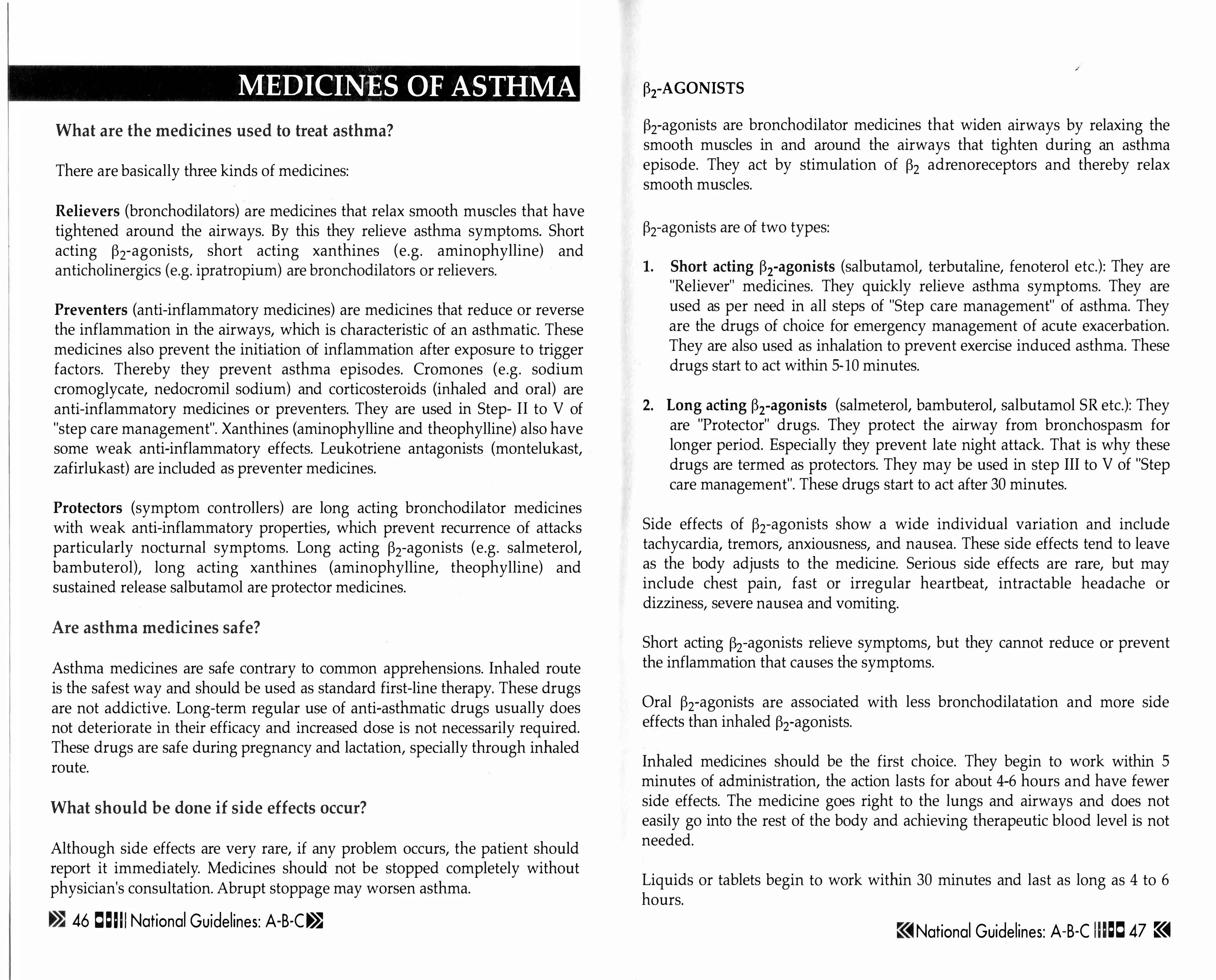
MEDICINES OF ASTHMA
What are the medicines used to treat asthma?
There are basically three kinds of medicines:
Relievers (bronchodilators) are medicines that relax smooth muscles that have tightened around the airways. By this they relieve asthma symptoms. Short acting �Tagonists, short acting xanthines (e.g. aminophylline) and anticholinergics (e.g. ipratropium) are bronchodilators or relievers. .
Preventers (anti-inflammatory medicines) are medicines that reduce or reverse the inflammation in the airways, which is characteristic of an asthmatic. These medicines also prevent the initiation of inflammation after exposure to trigger factors. Thereby they prevent asthma episodes. Cromones (e.g. sodium cromoglycate, nedocromil sodium) and corticosteroids (inhaled and oral) are anti-inflammatory medicines or preventers. They are used in Step- II to V of "step care management". Xanthines (aminophylline and theophylline) also have some weak anti-inflammatory effects. Leukotriene antagonists (montelukast, . zafirlukast) are included as preventer medicines.
Protectors (symptom controllers) are long acting bronchodilator medicines with weak anti-inflammatory properties, which prevent recurrence of attacks particularly nocturnal symptoms. Long acting �2-agonists (e.g. salmeterol, bambuterol), long acting xanthines (aminophylline, theophylline) and sustained release salbutamol are protector medicines.
Are asthma medicines safe?
Asthma medicines are safe contrary to common apprehensions. Inhaled route is the safest way and should be used as standard first-line therapy. These drugs are not addictive. Long-term regular use of anti-asthmatic drugs usually does not deteriorate in their efficacy and increased dose is not necessarily required. These drugs are safe during pregnancy and lactation, specially through inhaled route.
What should be done if side effects occur?
Although side effects are very rare, if any problem occurs, the patient should report it immediately. Medicines should not be stopped completely without physician's consultation. Abrupt stoppage may worsen asthma.
46 cel l i National Guidelines: A-B�C�
�2-AGONISTS
�2-agonists are bronchodilator medicines that widen airways by relaxing the smooth muscles in and around the airways that tighten during an asthma episode. They act by stimulation of �2 adrenoreceptors and thereby relax smooth muscles.
�2-agonists are of two types:
1. Short acting �2-agonists (salbutamol, terbutaline, fenoterol etc.): They are "Reliever" medicines. They quickly relieve asthma symptoms. They are used as per need in all steps of "Step care management" of asthma. They are the drugs of choice for emergency management of acute exacerbation. They are also used as inhalation to prevent exercise induced asthma. These drugs start to act within 5-10 minutes.
2. Long acting �2-agonists (salmeterol, bambuterol, salbutamol SR etc.): They are "Protector" drugs. They protect the airway from bronchospasm for longer period. Especially they prevent late night attack. That is why these drugs are termed as protectors. They may be used in step III to V of "Step care management". These drugs start to act after 30 minutes.
Side effects of �2-agonists show a wide individual variation and include tachycardia, tremors, anxiousness, and nausea. These side effects tend to leave as the body adjusts to the medicine. Serious side effects are rare, but may include chest pain, fast or irregular heartbeat, intractable headache or dizziness, severe nausea and vomiting.
Short acting �2-agonists relieve symptoms, but they cannot reduce or prevent the inflammation that causes the symptoms.
Oral �2-agonists are associated with less bronchodilatation and more side effects than inhaled �2-agonists.
Inhaled medicines should be the first choice. They begin to work within 5 minutes of administration, the action lasts for about 4-6 hours and have fewer side effects. The medicine goes right to the lungs and airways and does not easily go into the rest of the body and achieving therapeutic blood level is not needed.
Liquids or tablets begin to work within 30 minutes and last as long as 4 to 6 hours.
National Guidelines: A-B-C ec 47 �

Children as young as 5 years, can use the metered dose inhaler even without aided devices. A spacer device can be attached to the inhaler to make it easier to use and can enable even younger children to use a metered dose inhaler. Dry powder inhalers are also available, which may be convenient for use in certain group of children and elderly.
Using a nebulizer to take the medicine works almost the same way as using an inhaler. A nebulizer is easier to use than an inhaler. It is good for a child under age 5, for a patient who faces trouble using an inhaler, or for a patient with severe asthma episodes.
Injections are sometimes used in a doctor's chamber or an emergency room for severe episodes. They work very fast but last only 20 minutes.
XANTHINE DERIVATIVES
Xanthine derivatives are bronchodilator medicines that open airways by relaxing the muscles in and around the airways that tighten during an asthma episode and facilitate diaphragmatic movement during respiration. They also have some antiinflammatory properties. The anti-inflammatory effects occur at lower concentrations than concentrations required for bronchodilatation.
They are of two types- short acting and long acting. Short-acting preparations are "reliever" drugs whereas long-acting preparations are "protector" drugs.
Side effects of these drugs include anorexia, nausea, vomiting, stomach cramps, diarrhea, headache, muscle cramps, irregular heartbeat, restlessness, sleep disturbance. These side effects usually disappear with reduction of dose. Mild side effects often go away after few days. .
In young children, altered mood and behavior are sufficiently common as to limit theophylline's acceptability in this age group. Long-term high doses should be avoided. It may aggravate underlying GERD via relaxation of the lower esophageal sphincter.
Theophylline/ aminophylline may be taken every 8 or 12 hourly. This makes them easy medicines to use.
These drugs do not have an instant effect. It takes some time for theophylline or aminophylline to build up in the blood stream, where it must stay at a constant level to have a lasting effect. So, appropriate time of ingestion and amount of drug should be strictly maintained.
� 48 eeUi National Guidelines: A-B-C
Sustained released theophylline/ aminophylline are time-released medicines. So, tablets or capsules should not be chewed, because too much of medicine may be released all at once causing toxic effects.
The importance of theophylline in the treatment of asthma has declined over the last decade. It is a drug with a narrow therapeutic index, that is, the difference between therapeutic and toxic concentration is small. Many patients axperience minor adverse effects within the therapeutic range.
ANTICHOLINERGICS
Anticholinergic drugs (ipratropium, triotropium, oxitropium) acts as antibronchoconstrictors by blocking muscarinic receptors, which cause tightening of smooth muscles in and around airways. It reduces the bronchial tone.
Side effects of these drugs include unpleasant taste, dryness of mouth, precipitation of glaucoma in elderly and occasional paradoxical bronchoconstriction.
Onset of action of anticholinergic drugs is slow with maximum effect after approximately 30-60 minutes. Therefore in acute asthma it must be used in combination with �2-agonists.
Anticholinergic drugs are more effective in smokers. In smokers, the small airways « 2 mm diameter) are blocked due to hypersecretion from hyperplastic and hypertrophied mucous glands. Anticholinergic drugs reduce hypersecretion from those glands. That is why it is the drug of first choice in COPD patients.
Anticholinergic drugs are also very effective in children below 2 years of age. Adrenergic receptors are not fully developed at this tender age. But cholenergic receptors are well developed. So, anticholinergic drugs give better result than �2"agonists in this age group. .
Eye protection is advised for patients when anticholinergic solution is used through nebulizer.
CROMONES
Cromones are non-steroidal anti-inflammatory medicines. They prevent airways from swelling when they come in contact with an asthma trigger. They
National Guidelines: A-B-C Inee 49 �

act by inhibiting release of mediators from mast cells.
Cromones are effective in prevention of exercise induced asthma or exacerbation due to contact with a trigger. It should be taken at least 15 minutes prior to exercise or contact. The effects of the medicine last for 3 or 4 hours.
Nedocromil sodium is highly effective against cough variant asthma, because it blocks the cough receptors. Some times it gives better result than corticosteroid in cough variant asthma.
Cromones cannot be used to stop an asthma episode once it has started. They can only be used to prevent an episode from starting. Effect of cromones are variable and do not work for every patient. Recent studies suggest that, cromones are not that effective in asthma management as previously thought. Sodium cromoglycate is less effective than corticosteroid inhalers, but can be given even in infancy. Nedocromil is of benefit in 2-12 years of age. It may take up to 6 weeks for onset of action.
Cromones are "preventer" drugs. Only inhaled forms of cromones are used in asthma. If both inhaled �Tagonist and cromones are prescribed, �Tagonist should be taken first, particularly before exercise.
CORTICOSTEROIDS
Corticosteroids are anti-inflammatory medicines that prevent and reduce swelling inside the airways and decrease the amount of mucus in the lungs. It should be introduced as early as possible to prevent the detorioration of lung functions. Corticosteroids also reduce the sensitivity to triggers.
Corticosteroids are available as inhaler, oral, injectable and nebulizer solution preparations.
Inhaled corticosteroid is taken with a metered dose inhaler or dry powder inhaler. When taken at the proper dose, they are safe medicines that work well for patients with moderate or severe asthma. They reduce the sensitivity of the airways to triggers and prevent inflammation or swelling in the airways.
Oral corticosteroid (liquid and tablet) is used in serious asthma episodes to reduce inflammation of the airways and to prevent the episodes from getting worse. For people with acute exacerbation of asthma, oral corticosteroids are sometimes used for 3 to 14 days and then stopped. People with severe
•
� 50 C=1 I 1 National Guidelines: A-B-C
persistent asthma may take oral corticosteroid daily or on alternate days on long-term basis.
Injectable corticosteroid is used during serious episodes to obtain a confirmed onset of action. Remember, oral steroid is as effective as injection.
Corticosteroid nebulizer solution (e.g. budesonide) is used for those patients who fail to use MDI or DP!.
Inhaled corticosteroids may cause fungal infection in the mouth, especially in the pharynx and induce coughing. It may cause hoarseness of voice. There are two ways to avoid these problems - using a spacer device and rinsing and gargling of mouth after taking steroid inhalers.
Using oral corticosteroids as rescue therapy has minimum and reversible side effects. Short term usage may cause different side effects such as increased appetite, fluid retention, weight gain, moon-face, changes in mood and hypertension. These will reverse when medicine is discontinued.
Oral corticosteroids used for a long term may have side effects such as hypertension, thinning of the bones (osteoporosis), cataracts, muscl.e we�kness, diabetes mellitus, opportunistic infections and slower growth m chIldren. Because of these side effects, long-term oral corticosteroids should only be used in severe persistent asthma in step V management for adults. In children lon�term oral corticosteroids are not advocated in step care management and It should only be employed by an experienced pulmonologist.
Corticosteroids are not same as the anabolic steroids used by some athletes.
When corticosteroids are used to treat serious asthma episodes, they take about 2-6 hours to start working and are most effective within 6 to 12 hours. Time required for onset of action does not vary between oral and injectable route. Inhaled corticosteroid should be employed for at least 2 successive triggering seasons in seasonal asthma and for 1-2 years in perennial asthma on a regular basis; i.e. at least 6-12 months after full remission. Dosage of triamcinolone, beclomethasone and budesonide are almost equal. Fluticasone is two times more potent than these drugs in weight for weight measurement.
I Guidelines: A-B-C =C 51

Is there any adverse effect of high dose inhaled corticosteroid on children?
Chronic use of inhaled corticosteroids has been shown to lead to a slight dose-dependent adrenal suppression. The impact of inhaled high dose corticosteroids on growth rate is a theoretical concern in children. On the other hand, poorly treated asthmatic children have a delay in onset of puberty. Neverthless, there appears to be a consensus that inhaled corticosteroids are relatively safe. The following points regarding the risk/benefit ratio of inhaled corticosteroids should be borne in mind:
• The potential risks of inhaled corticosteroids are well balanced by their benefits.
• Growth rates are highly variable in children. Short-term evaluations may not be predictive of attaining final adult height.
• Poorly controlled asthma may delay growth in children. • In general, children with untreated or poorly treated asthma tend to
have delayed onset of puberty. • The potential for adverse effects on linear growth from inhaled
corticosteroids appears to be dose-dependent. In treating children with mild to moderate persistent asthma, medium-dose inhaled corticosteroid therapy may be associated with a possible, but not predictable, adverse effect on linear growth. High doses of inhaled corticosteroids have greater potential for growth suppression. Efforts should be made to limit doses of corticosteroids to minimum possible maintenance dose.
• Use of high-doses of inhaled corticosteroids in children with severe persistent asthma has Significantly less potential for having an adverse
. effect on linear growth than oral systemic corticosteroids.
Recommendations As per currently available knowledge, we confidently recommend inhaled corticosteroids in patients with persistent asthma. Oral theophylline or inhaled nedocromil sodium may be used in younger children in combination with inhaled corticosteroids as a steroid-sparing strategy.
� 52CC�1 1 National Guidelines: A-B-C)�
,
I
LEUKOTRIENE ANTAGONISTS
Leukotrienes appear to be the most important inflammatory me�iator in asthma. They can cause bronchoconstriction, mucus hypersecretIon an? increased airway vascular permeability resulting in airway wa�l edema. T�e�r action in human airway obstruction rests on the stimulatIon of specIfIc receptors termed as cysteinyl leukotriene type-! (CysLT-!) receptors.
Their potential importance in the pathogenesis of asthma has le� to development of several classes of drugs colle�tively kn�wn as leuko�nene inhibitors. They specifically inhibit the produ�tlOn or �ctIon of leu.kotnenes, either by inhibiting the enzymes needed for blOsynth.esis of leukotnenes (e.g. zileuton) or by blocking the CysLT-! receptors (e.g. zafIrlukast, montelukast).
Indications of leukotrienes inhibitors
• In step care management of asthma in Step II, it is an alternative of inhaled corticosteroids
• Prevention of aspirin induced asthma . • Prevention of exercise induced bronchoconstriction • Treatment of cough variant asthma • As a supplementary therapy in any step (from S�ep III to S�ep V),
especially when inhaled corticosteroids or long acting �Tagorusts are not responding well or not well tolerated.
. . Side effects of this group of drugs are still under evaluation. Zileuton elevates hepatic transaminases. Zafirlukast and montelukast, tho�gh reported as mildly hepatotoxic, have a remarkable safety . profIle. Development of eosinophilic vasculitis (Churg-Strauss syndrome) IS rarely reported.
Differences between montelukast and zafirlukast:
Montelukast Zafirlukast
Efficacy in exercise induced bronchospasm (EIB) Yes Yes
Efficacy in Allergen induced Asthma Yes Yes .
Efficacy in chronic asthma Yes Yes
Dose frequency . Once daily Twice daily
Relation with food No Yes (to be taken 1 hour before or 2 hour after meal)
r Drug interaction Not significant Present .
Suitable age :2: 1 years > 6 years
'onal Guidelines: A-B-( IUCC 53

NEWER DRUGS
Magnesium sulfate (MgS04) Magnesium sulfate is believed . smooth muscle contracti aecrease histamine release from mast inhibit acetylcholine release. Variable improvement in patients with severe airflow limitation who are umesponsive to standard treatment with �Tagonist, anticholinergic, and corticosteroid medications has been noticed.
In children the optimum dose is 40 mg/kg given as an intravenous bolus with a maximum dose of 2 g. Adults get maximum benefits from 2 gm of
sulfate administered intravenously as a supplement to standard
normal saline.
. sulfate can be used as a vehicle for nebulization in place of
Minor side effects include transient flushing, lightheadedness, lethargy, nausea, or burning sensation at the IV site.
Frusemide Inhalation of frusemide appears to induce bronchodilatation and improve exercise-induced dyspnoea, especially in COPD patients. It is associated with a significant improvement in lung function (FEVl and FVC). Frusemide can be used as an alternative in patients who suffers from tachycardia or other adverse effects of �Tagonists.
Omalizumab Omalizumab (Xoliar) is a monoclonal anti-IgE antibody preparation. It is effective in asthma and allergic rhinitis. Omalizumab aids to reduce the dose of corticosteroids for long-term treatment and may help to stop it. It is safe and well tolerated even in children. The recommended �ubcutaneous injectable dose is 150-300 rug depending on IgB level, given at 2-4 weeks interval. Upper respiratory tract infection, headache and urticaria are the infrequently reported adverse events.
Ciclesonide
Ciclesonide (Alves co) is a novel, inhaled corticosteroid for the treatment of asthma. Ciclesonide is a pro-drug, converted within the pulmonary system to form the active metabolite desisobutyryl-ciclesonide (des-CIC), which provides potent anti-inflammatory activity. Thus it avoids the undue systemic effects of steroid, such as suppression of the hypothalamic-pituitary (HPA) axis, osteoporosis, reduced bone growth in the young, opportunistic infections, behavioral alterations, disorders of lipid metabolism, oral candidiasis and glaucoma.
� 54 C=UI National Guidelines: A-B-C�
Ciclesonide has anti-inflammatory efficacy equivalent to fluticasone but with a significantly improved safety profile compared to �uticasone. It can be given once daily and it is effective in the treatment of mild-to-�od�rate asthma. It improves asthma symptoms, minimizes use of rescue medIcation and reduces number of asthma exacerbations.
DISEASE MODIFYING AGENTS
Methotrexate In low doses methotrexate appears to inhibit the attraction of polymorphonuclear cells by leukotrienes. Use of methotre:<ate has a significant corticosteroid-sparing effect, decrease in daily bronchodIlator use and some improvements in pulmonary functions. The dose .is 5�25 mg weekly (15 mg/ week usually). Side effects may include anoreXIa: �Iarrhea, naus�a an
.d vomiting, leucopoenia, hepatic fibrosis, acute pneumomtIs, pulmonary fIbroSIS and opportunistic pulmonary infections.
Cyclosporine A . . . It is a potent non-selective anti-inflammatory agent that acts pnmanly by
inhibiting transcription factors for cytokines deriv�d from T-lyn:phocytes. In
chronic severe asthma, it reduces daily corticosterOIds dose and Improves the
symptoms as well as PEF. However, it is not known if there is .a sustained
clinical benefit after stopping cyclosporine treatment. Indeed, thIS . drug �as
many potential side effects that may be more serious than those assocIated WIth
prednisolone.
Gold Salts Gold has been used to treat complicated rheumatoid arthritis for many years. It
also has some benefit in treating refractory asthma. Oral gold, auranofin lessens
the need of corticosteroid, reduces symptoms and exacerbations and improves
FEVl. Side effects are frequent and include urticaria, stomatitis, leucopoenia,
thrombocytopenia and proteinuria.
NOTES Disease modifying agents should be administered in specialized centers only.
What is the role of antihistamines in management of asthma?
Antihistamines usually have no helpful effect on asthma itself but ma� be used to treat associated nasal and other allergy symptoms. It can be used In people with controlled asthma but should be avoided during exacerbations.
National Guidelines: A-B-C I I � =C 55 K�

What is the role of Ketotifen in management of asthma?
Ketotifen is a potent anti-histamine. Its weak anti-inflammatory action has been demonstrated in some studies. When an asthma patient suffers from concomitant allergic rhinitis, ketotifen is the antihistamine of choice to treat running nose. Thus mouth breathing is prevented and most of the allergens are filtered in the nose. Otherwise allergens, particularly house dust mites enter into the airways through mouth and frequent and variable exacerbation of asthma episodes may occur. As ketotifen causes drowsiness it is inconvenient to use at daytime particularly in adults. Usually we recommend single bedtime dose for concomitant allergic rhinitis, conjunctivitis or dermatitis. Prolonged use of ketotifen does not have any significant side effect.
Should we use antibiotics in asthma? .
Antibiotics are rarely indicated in the treatment of asthma exacerbations. Mucus hypersecretion and a productive cough are frequent manifestations of asthma which are usually not due to infection. Discolored (yellowish or grayish) sputum may be due to allergic (eosinophilic) inflammation and should not be interpreted as an indication of infection in the absence of other symptoms and signs. Antibiotic should be reserved for overt infections.
Indications of antibiotic in asthmatics:
• Fever with purulent sputum • Suspected bacterial sinusitis • Patients with overlapping COPD • Presence of concomitant pneumonia • Frequent exacerbation of asthma (may be associated with
mycoplasma or chlamydial infections. Drug of choice is microlides)
Can sedatives be used in asthma?
Sedatives are contraindicated during an acute attack. Sleeplessness and agitation during an attack is usually due to bronchospasm and hypoxaemia. These conditions are better treated by �z-agonists and oxygen inhalation. Most sedatives including benzodiazepines and zopiclone may blunt respiratory drive precipitating respiratory failure.
Sedatives may be used with caution in controlled asthma. Bromazepam and midazolam are comparatively safe to use.
� 56 C=U I National Guidelines: A-B-( �
•
Is there any role of anti-tussives in asthma management?
The conventional anti-tussives or cough mixtures have no role in the management of asthma. Cough suppressants should not be used as they may dry up the airways and provoke more bronchoconstriction. In case of intractable dry cough, expectorants may bring some relief. But before that, other causes of dry cough should be excluded (e.g. post nasal drip, gastrooesophageal regurgitation, smoking, use of ACE inhibitors, etc. ) . Role of mucolytic agents in asthma and COPD is controversial.
LIST OF ASTHMA MEDICINES Relievers
a)
1.
2.
b)
c)
•
Adrenoreceptor agomsts
Short-acting �2-agonists (Highly selective)
a and � agorusts (Less selective)
Xanthine derivatives (Short-acting)
Anticholinergics
•
Generic name
Salbutamol (Albuterol)
Levoalbuterol (Levosalbutamol) Terbutalin Fenoterol Pirbuterol Reproterol Rimiterol Bitalterole Tulobuterol
Adrenaline Ephedrine
Aminophylline
Theophylline
Enprophylline
Ipratropium Oxitropium Triotropium
Commercial name
Salbutal, Ventolin, Brodil, Salbut, Respolin, Suitolin, Azmasol, Etol, Asthalin, Broad, Salbu, Actolin, Ventisal, Salmolin, Pulmolin, Asmatol Asmolex, Butamol, Ventol, Bronkolax, D-butamol
Tervent, Bricanyl
Breton, Bremax
Adrenaline, Adrin Ephedrine, Ephelin, Fedrin
Cardophylin, Filin, Restophylin, Aminophylline Thenglate, Asmain, Jasophylin, Theonate, Theoglate, Anlate
Ipramid, Iprex, Atrovent, Ipravent
National Guidelines: A-B-( I I �=C 57

Preventers
a) Corticosteroids
b) Cromones
c) Leukotriene antagonists
Protectors
Triamcinolone Beclomethasone
Budesonide
Fluticasone Mometasone Ciclesonide Prednisolone
Dexamethasone
Betamethasone Hydrocortisone
Sodium Cromoglycate Nedocromil Sodium
5-Lipoxygenase inhibitor Zafirlukast
Pranlukast Montelukast
a) Long-acting �2-agonist Salmeterol
b) Xanthine derivatives (Long-acting)
Formoterol Salbutamol SR Bambuterol
•
Theophylline SR
Aminophylline SR
58 C =II I National Guidelines: A-B-C
Azmacort Becotide, Becloforte, Beclod, Beclomin, Decomit, Ascon Budeson, Zycort, Pulmicort, Aeronid Flaso, Fluticon, Flixotide
Deltasone, Prednisolone , Precordil, Cortan Oradexon, Decasone Dexan, Steron Betnelan Rapicort, Solucortef, Cotsone, Hydrocortisone
Intal, Nacromin Tilade
Zileuton Zafir, Accolate, Zafnil, Freesy, Zukast, Zalukast, Zaft
Monas, Montair, Mokast, Singulair, Aeron, Provair, Reversair, Monocast, Odmon, Montene
Bexitrol, Salmate, Serevent, Salmeter, Axinat, Salmerol Efo, Foradile, Oxis Ventolin SR, Sultolin SR Aerodyl, Dilator, Buterol
Theovent SR, Unicontin, Con tin, Euphyllin Retard, Neul in SR, Spophylin Retard, Larnox LA , Theo TR, Asmanyl SR, Arofil, Quibron SR, Thenglate TR Aminophyllinum Retard, Aminomal R
Combinations Fluticasone + Salmeterol Seretide, Bexitrol-F, Tieamet, Salflu, Axinat-F
Salbutamol + Ipratropium Sulprex, Iprasol, Combivent, Combimist, Ventipra, Sipra
DOSES OF ASTHMA MEDICINES
Short acting �2 agonists
Salbutamol
Tablets 2mg, 4mg (plain); 8 mg (SR) Syrup 2 mg/5 ml
Metered Dose Inhaler (MDI) 100 meg/inhalation
Children: 0.15 mg/ kg/ dose 8 hourly (maximum single oral doses of 4 mg) Adult: Plain- 2-4 mg 8 hourly; SR- 4-8 mg 12 hourly
1-2 inhalations 3-6 hourly or as required For acute symptoms, 5 inhalations at a time; if necessary repeat after 5 minutes, total up to 5 times (Rule of 5; see page-82).
Dry Powder Inhaler (DPI) 1-2 inhalations 3-6 hourly or as required 200 meg/ inhalation (capsules or blisters)
Nebulizer solutions 5mg/ml
Single dose nebulizer (Nebules) 2.5 mg/nebule
Terbutaline
Tablets 5mg Syrup 1 .5 mg/5 ml
units
Children >18 months: 0.5 ml diluted with normal saline to make it at least 2 ml, 3-6 hourly Adults: 0.5 to 1 ml diluted with normal saline as above, 3-6 hourly
Children: 4-12 years: 1 nebule 3-6 hourly Adults: 1-2 nebules 3-6 hourly
Children <7 years: 75 meg / kg / dose 6-8 hourly Children >7 years: 1-2.5 mg/ dose 6-8 hourly Adult: 2.5-5 mg/ dose 6-8 hourly
� National Guidelines: A-B-C IU=C 59 �

MDI 2S0 meg/ inhalation
DPI SOO meg/ inhalation
Nebulizer solutions 10mg / ml
Nebules S mg/ nebule
Xanthine derivatives
Theophylline
Tablets 2S0, 300, 400 mg (SR) Syrup 120 or ISO /Sml
Aminophylline
Tablets 100 mg (plain); 350, 600 mg (SR)
Injection 12Smg /Sml or 2S0 / lOml
Anticholinergics
Ipratropium bromide
MDI 20 meg/ inhalation
Nebulizer solution 2S0 meg/ ml
60 ==1 1 1 National Guidelines: A-B-(
1-2 inhalations 3-6 hourly or as required
1 inhalation 3-6 hourly or as required
Children >3 years: 0.2-0.5 ml 6-12 hourly Adult: O.S-l ml 3-6 houly
Children >8 years: 1 nebule 6-12 hourly Adults: 1-2 nebules 3-6 hourly
Children 2-6 years: 62.5 mg 8-12 hourly Children 7-12 years: 62.5-125 mg 8-12 hourly Adults: 125-300 mg 8-12 hourly
Children >2 years: 12 mg/kg/dose 12 hourly Adults: Plain- 100mg 8 hourly SR- 175-300 mg 12 hourly
Infuse Smg / kg stat, then O.5mg/kg/hour till control
Children <6 years: 1 inhalation 8 hourly Children >6 years: 1-2 inhalation 8-12 hourly Adult: 2-4 inhalations 6-8 hourly
Children: 2S0 meg 3-4 times daily Adults: SOO meg 3-4 times daily
Long-acting �2 agonists
Salmeterol
MDI 2S meg/ inhalation DPI SO meg/inhalation
Formoterol
DPI 12 meg/ capsules
MDI 6 meg & 12 meg / inhalation
Bambuterol
Tablets 10 & 20 mg Syrup S mg / S ml
Inhaled corticosteroids
Triamcinolone
MDI 1 00 meg / inhalation
Beclomethasone D... . I �\t\ " • ., IbLl C-w · •
MOl 50 meg, 100 meg, 250 meg/inhalation DPI 50 meg, 100 meg, 250 meg/ capsule
Children over 4 years to adults: SO meg 12 hourly
Children >5 years: 12 meg 12 hourly. Total daily dose should not exceed 24 meg Adults: 12-24 meg 12 hourly. Total daily dose should not exceed 48 meg
Children > 12 years: 6-12 meg 12 hourly. The daily dose should not exceed 24 meg Adults: 6-12 meg 12 hourly. Adults with more severe airways obstruction may require 24 meg 12 hourly. The total daily dose should not exceed 48 meg
•
Children of > 2 - 5 years: 2.5 mg at bedtime Children of > 5 - 12 years: 5 mg at bedtime > 12 years and adults: Initially 10 mg, increased up to 20 mg at bedtime
Children up to S years: 200-800 meg / day Adult: 400-2000 meg/ day
<5 years: 100-800 meg / day >S years to Adults: 200-2000 meg/ day Adjust dose according to disease severity as indicated in step care management
National Guidelines: A-B-( IU== 61

•
Budesonide
MDI 50 mcg, mcg 1 inhalation DPI 100, 200 inhalation
100 mcg, 200 Dose is same as beclomethasone. Adjust dose according to disease
& 400 mcg 1 severity as indicated in step care management.
Fluticasone
MDI 50 mcg, 125 mcg, 250 mcg 1 inhalation DPI 50, 100, 250 & 500 mcgl inhalation
Cromones
Sodium cromoglycate
MDI 5 mg 1 inhalation
DPI 20 mgl capsule
Nebulizer solution 20 mg 12 ml
Nedocromil sodium
MDI 2 mg/ inhalation
Oral corticosteroids
Prednisolone
Tablets 5mg, 20mg
<5 years: 50-400 mcg 1 day >5 years to Adults: 100-1000 mcg 1 day Adjust dose according to disease severity as indicated in step care management
1-2 inhalations 4 times daily according to severity
1-2 inhalations 3-4 times daily
1 nebule 3-4 times daily
Commence with 2 puffs 4 times daily for one month. Once good symptom control and lung function improvement is achieved the dose can be reduced to 2 inhalations twice daily
An initial large (bolus) dose should be used. Then taper off if continued for more than one week.
62 CCUI National Guidelines: A-B-C
f,..J.pYM'\... t)
\ 00 +
� I Rescue steroid therapy may not require tapering.
.' II, "
! I , ,
Leukotriene antagonists
Zafirlukast
Tablets 20mg
Montelukast
Adults: 40-60 mgl day Children <5 years: 0.5-1 mg 1 kg 1 day (for rescue therapy only) Children >5 years: 20-40 mg l day
Not recommended below 7 years of age >7 years to Adults: 20 mg twice daily, one hour before or two hour after meal
Tablets 4 & 5 mg (chewable); 10 mg < 5 years: 4 mg at bedtime
Combination Preparations
Salmeterol+ Fluticasone
5 - 12 years: 5 mg at bedtime Adult: 10 mg at bedtime
DPI Adjust dose according to disease Salmeterol 50 meg + Flutieasone 100 meg severity as indicated in step care Salmeterol 50 meg + Flutieasone 250 meg management Salmeterol 50 meg + Flutieasone 500 meg
MDI Salmeterol 25 meg + Flutieasone 50 meg almeterol 25 meg + Flutieasone 125 meg
! Salmeterol 25 me + Flutieasone 250 meg 1. (lUI I � s t. 2.5"0 Salbutamol+ Ipratropium
MDI Salbutamol 100 mcg+ Ipratropium 20 mcg
Adjust dose according to disease severity as indicated in step care management
Adjust dose according to disease severity as indicated in step care management
-"" 1' , � National Guidelines: A-B-C IUCC 63 � ... I \ t:) '\

SECTION-2:
Management 0 Asthma
What is the goal of asthma management?
Effortless easy breathing is our goal. �,,\I� -0\11 �, 15I1�1CI1\1 �llJ>l I As asthma is not a "curable" disease we should achieve at least "total or well control" of the disease in all cases.
What are the components of an effective management plan?
Education, Caution and Medication fi'f'lI'1 - :>j�4�1 - fffiie,'lT are the three fundamental components of an effective management plan for asthma. Of these three components, only "Me<;lication" is discussed in this chapter. "Caution" and "Education" is elaborated in section-3.
Is there a cure for asthma?
The word "cure" is difficult to apply in case of asthma. However, asthma can be "controlled". We should expect nothing less. If a person uses anti-inflammatory preventive drugs for a long time, say for 2-5 years, then 60-80% cases of childhood asthma and 20-30% cases of adult asthma may go into complete "remission". This complete remission may be induced spontaneously in some cases, sometimes the credit going to the "faith healers" or quacks.
What is meant by control?
Control of asthma means, patient-
• is almost asymptomatic • can perform near normal daily activities • requires reliever bronchodilator (Salbutamol inhalation) <1 time / day • is free of nocturnal symptoms; if occurs, less than two times per month • has PEFR reading >80% of personal best result • has <10% diurnal variability in Peak Flow Chart, if available • has no history of emergency visit to doctors or hospitals • has no or minimal side effects of medication
64 CCI I I National Guidelines: A-B-C
Rule of 2 To assess "control", "Rule of 2" may be considered as a practical tool. If-
• asthma episodes are > 2 /week • or nocturnal attacks are :? 2 / month • or number of canister of reliever (salbutamol) inhaler used is :? 2/year
it means patient's asthma is not controlled.
Based on the above-mentioned points, asthma control is categorized as "well controlled" and "totally controlled". Totally and well-controlled asthma are defined by achievement of all of the specified criteria for a specific week. For this an asthma patient is assessed for consecutive 8 weeks or more. Totally controlled asthma is achieved if the patient during the 8 consecutive assessment weeks recorded 7 totally controlled · weeks and had no exacerbations, emergency room visits or medication-related adverse events. Well-controlled asthma is similarly assessed over the 8 weeks, with somewhat lesser achievements. The following table depicts the principles of such categories.
Criteria of "totally-controlled" and "well-controlled" asthma:
Criteria
Daytime symptoms
Rescue �2-agonist use
Morning PEF
Night-time awakening
Exacerbations
Emergency visits
Treatment-related adverse events
Each week 2 or more of the followings should be achieved
2 days
Use on 2 consecutive days and 4 occasions/ wk
80% of predicted value every day
----------------
Totally Controlled
Each week all of the followings must be achieved .
None
None
80% of predicted value every day
None
None
None
None enforcing change in current asthma treatment
•
National Guidel ines: A-B-C Incc 65

What do we mean by remission?
It is a state in which a patient remains asymptomatic for at least one year or more. If remission persists throughout life, then we can say that patient is "cured" . "Cure" is possible but still it is difficult to predict who will go into that complete remission or "cure" and who will not.
How can asthma episodes be prevented?
Effective asthma management plan can help patients to: - prevent most attacks - stay free from troublesome night and day symptoms - to keep them physically active
Physician should give emphasis on the following 6 points:
1 . Educate patients to develop a partnership in asthma care 2 . Assess and monitor asthma severity 3 . Avoid exposure to risk factors 4. Establish individual medication plans for long-term management in
children and adults 5. Establish individual self management plans to control asthma more
effectively 6. Provide regular follow-up
Modalities of Asthma Management
Management of Asthma can be described under two broad headings:
A. Home Management
B . Emergency Management
Why management at home?
Asthma is a chronic disease and it should be managed at home, except severe acute asthma. If home management plan is applied intelligently and skillfully, most asthmatics can lead a symptom-free near-normal life. They may avoid hospitalization thereby decresing the financial expenditure significantly.
What are the types of home management plan?
Horne management plan is of two types.
1 . With Guided Self-Management Plan. 2. Without Self-Management Plan.
Home Management with Guided Self-Management Plan: .
In "Self-Management Plan" system, patient education is more time consuming and laborious but chances of mortality and morbidity is reduced considerably.
Here patient is advised to maintain a peak flow chart. With the help of this chart, patient's "Personal best peak flow result" is determined (please see page 125). On the basis of "Personal best peak flow result" daily peak flow readings are recorded in a chart with three colour zones - Green, Yellow and Red. Patient will try to be in the green zone. For that the patient can modify the medication up to a certain limit as guided by the phYSician without consultation in order to be in green zone.
[For interpretation of "peak flow zone system", please see page 125]
[For structured form of "guided self management chart", please see page 128]
Home Management without Self-Management Plan:
Along with patient education and appropriate precautionary measures, "step , care management" is employed here. The prescription is quite inflexible in this
system. Whatever the condition of the patients, they will not increase their , drugs except short acting �Tagonists (Salbutamol) inhaler. Patients can take
Salbutamol inhaler as per need up to 4-6 times daily.
66 ceul National Guidelines: A-B-C � National Guidelines: A-B-C Inec 67
I

What is step care management?
Step Care Management is like a staircase. We start treatment at the appropriate step. Then we shall step up along the stairs if asthma is not controlled or becomes more severe and shall step down when patient's asthma is fully controlled for 3 months or more.
We have divided the asthma management plan into five steps for children >5 years to adults and into four steps for children ,,;:5 years of age. At first, we should understand basic principles of these steps. Then we can construct any step by combining available drugs.
BASIC PRINCIPLES OF STEP CARE MANAGEMENT For >5 years to adults
In �his age group the steps are formulated on the basis of anti-inflammatory actIon and protective action of various drugs. From the definition of asthma it is clear that control of inflammation as well as control of bronchoconstriction fr.Ol:' hy�erresponsiveness of various stimuli is our goal . On that basis we may dIVIde aIrway of asthma patients arbitrarily into 5 types and treatment of asthma into 5 steps :
1 . Minimal inflammation and minimal hyperresponsive airway _. Step-I treatment
2. Low level of inflammation and low level of hyperresponsive airway _. Step-II treatment
3 . High level of inflammation and moderate level of hyperresponsive airway _. Step-III treatment 4. High level of inflammation and high to severe level of hyperresponsive airway _. Step-IV treatment 5. Very high level of inflammation and very severe level of hyperresponsive airway _. Step-V treatment
Step.
- I: In�amma�on is so minimal that no preventer or anti-inflammatory mediCatI�n IS reqUIred. Patient will only take reliever drug (short acting bronchodIlator) as per need. Step-I is kept as a part of Step-II to Step-V onwards.
Step - II: For control of inflammation, low level anti-inflammatory medication (prevente
,:) is requir�d. We can get desired low-level anti-inflammatory action
by usmg Low dose mhaled cortrcosteroids (LOlCS)" or "Sustained release (SR) theophylline" or "Leukotriene antagonists" or "Full dose cromones".
Step - III: To control airway inflammation, high-level anti-inflammatory
� 68 CCUI National Guidelines: A-B-C �
I
medication (preventer) is required. This usually means, "High dose inhaled corticosteroids (HOlCS)" . But as high level of inflammation is usually associated with moderate hyperresponsive airway, we may get equivalent or some times better results by giving LDICS along with any one or more of the following options: (1 ) Long-acting �Tagonist (LAB A; e.g. salmeterol, bambuterol), (2) Sustained release theophylline (protectors), (3) Full dose of cromones, particularly nedocromil sodium (it is more effective when cough is the predominant symptom of asthma episode) . "Leukotriene antagonists" can be added in this step as a supplementary medicine.
Step - IV: There are two divisions of this step, viz. IVA and IVB. When high dose anti-inflammatory drugs (HDICS) are unable or insufficient to control asthma then at first we employ Step IVA, which means addition of either LABA or SR theophylline with HDICS. If control is not yet achieved, Step IVB is employed, which means both LAB A (e.g. salmeterol) and SR �heo�hylline are added with HDICS. "Leukotriene antagonists" can be added 111 thIS step as a supplementary medicine.
Step - V: It is the highest step. Oral corticosteroid, added as single m?rning dose, with all medicines of step-IVB comprises step-v. We employ thIS step when step -IVB appears to be inadequate to control asthma.
If asthma is not controlled even after giving step-V management, round the clock nebulized bronchodilators can be used and the patient must be referred to a Pulmonologist (chest disease specialist). A second thought should be given whether the diagnosis is correct or not. The total management plan including environmental (trigger) control should be reviewed meticulously.
For children <5 years of age
In this age group, due to potential syestemic side effects . of inhaled corticosteroids, particularly on bone growth and adrenal suppressIOn, the dose of inhaled steroid is the main determinant of step formation. Step I means no steroid, step II means low dose, step III means medium dose and step IV means high dose inhaled steroid usage. Use of long-term daily oral stero�d is not recommended in children <5 years of age. However, rescue oral sterOId can be given if needed.
Economic schedule
According to NAPS 1999, out of 7 million asthmatics, around 1 million peop�e cannot afford standard treatment on financial ground in our country. For thIS reason, Asthma Association developed an economic schedule for them. This schedule is formulated for patients who cannot afford inhalers and other costly medicines. It is not an alternative for standard step care schedule.
� National Guidelines: A-B-C Incc 69 �

~ D Ill:.
Z Q -_ . o :::J Q -
:::J CD V> • •
» , 0:> , n
Z Q -_ .
o :::J Q -
C'l c _ . 0. CD -
-- . :::J CD vo • •
» , 0:> , n Ill:. D '-l
•
~
b Step Care Management for >5 years to adults
STEP
� V\ \ 14 i -'OJ
:( \5\ . » l Ob :&. 2. S-o \ \I)
( 2. f>(A1f C> � Recommended Treatment �. � � � 2. �:tS
Step V Oral steroid (+) (+) All medications of step-IV Step-I
Step IV IVB HDICS (+) (+) SUPPLEMENTARY (+) LAB A SR Theophylline Leukotriene antagonists Step-I
IVA HDICS (+) & / or Anticholenergics, e.g. ipratropium (optional) LABA OR SR Theophylline
Step III HDICS OR OR OR SUPPLEMENTARY (+) Leukotriene Step-I LDrCS (+) LDrCS (+) LDrCS (+) antagonists LABA SR theophylline Full dose cromones (optional)
Step II LDICS OR OR OR (+) Leukotriene Full dose cromones Sustained release(SR) theophylline Step-I antagonists
Step r Short acting �2-agonist inhaler (Salbutamol) 200 mcg (2 puffs) as and when required. � ' That is, when patient feels mild cough, wheeze, chest tightness, 2 puffs at a time, up to 4-6 times per day. Additional 2 inhalations prior to exercise, sports or exposure to triggers are advised.
Note: 1 . LDrCS - Low dose inhaled corticosteroid; beclomethasone or equivalent, for 5-12 years = 200-400 mcg/ day; for >12 years to adults = {QO-8QO mcgNay 2. HDICS . High dose inhaled corticosteroid; beaometnasone or equivalent, for 5-12 years = up to 800 meg/ day; for >12 years to adults = up to 2000 mcg/ day 3. Full dose Cromones = Sodium chromoglycate 10 mg 4 times daily ; Nedocromil sodium 4 mg 4 times daily.
. .
4. LABA = Long acting �Tagonists (salmeterol inhaler, bambuterol etc.) 5. Oral Steroid = Prednisolone 5-20 mg, must be single morning dose
<" ,
Step Care Management for <5 years
STEP Recommended Treatment Alternative Options
Step IV HOICS OR OR OR (+) (High dose inhaled HOICS HOICS HOICS Step·l corticosteroid) (+) LAB A (+) SR Theophylline ( + ) Leukotriene
antagonists
Step III MOICS OR OR OR (+ ) (Medium dose inhaled MOICS MOICS MOICS Step-I corticosteroid) (+) LABA (+) SR Theophylline ( +) Leukotriene
antagonists
Step II LOICS OR OR OR (+) (Low dose inhaled Leukotriene Full dose Cromones SR Theophylline Step-I
corticosteroid) antagonists
Step I Short acting �2-agonist inhaler (Salbutamol) 100-200 mcg as and when required. That is, when patient feels mild cough, wheeze, chest tightness, 1-2 puffs, up to 4-6 times per day. Additional 1-2 inhalations prior to exercise, sports or exposure to triggers are advised.
Note: 1 . LOlCS- Dose of beclomethasone or equivalent = 100-2S0 mcg/ day 2. MOlCS- Do::;e of beclomethasone or equivalent = fSO-SOO meg/day 3. HOlCS- Dose of beclomethasone or equivalent = SOO-800 mcg/ day
. . . . . , . . . s reliever drug. Combination of salbutamol and ipratropium provides better result
Ing term aally oral sterOia snoula not be practleea In ::; J years. 1t reqUlrea the cruIQ must be reterrea to a pulmonologist or a respi
pediatrician
(
-
...... .
,i)
\�h /f� '

u . ....
s o s:: o u
� • • , . s:: QJ S QJ 00 fU s:: fU ~ QJ "'" fU
U p... QJ I •
C/')
...... ...... ...... ,
E" Cfl ..... o [J) <l.J � . -U .-
"d <l.J Ei
...... , 0...
� <l.J + ...... � Cfl
...... , 0...
� <l.J + ...... � Cfl
. -� -
..2
� 72 C�I I I National Guidelines: A-B-C
• en <l.I � . -u
- --0
<l.I E
Which medication should be preferred for a patient able to buy only one inhaler - a reliever or a preventer?
A pre venter corticosteroid inhaler is the drug of choice in such case, because, continuous anti-inflammatory action of this medicine may lead to remission of asthma.
Which Inhaler should not be used alone?
Salmeterol should not be used alone. It has to be used along with inhaled corticsteroid. Salmeterol does not have antiinflammatory preventer properties. Solitary use of salmeterol may increase asthma morbidity and mortality.
Is there any benefit of combination inhalers?
In step - III, we need high level of anti-inflammatory action to control asthma. It can be achieved either by inhalation of high dose of corticosteroid or by combining a protector (e.g. long acting �2-agonist) with low dose inhaler corticosteroid. Studies indicated that the combination therapies are the better options. It gives long-term protection without encountering the possible side effects of high dose corticosteroids. Moreover, these two drugs (fluticasone and salmeterol) delivered via a single device is more convenient as one single puff delivers two drugs at a time ensuring increased patient compliance, achievement of more rapid total control and no chance of skipping one drug or changing the dose of either drugs.
Which step is appropriate for a specific patient?
It is important to learn and practice step care management. At the same time it is essential to learn which step is appropriate for a particular patient. We are using a score system, developed at the "National Asthma Center", Mohakhali, Dhaka for determination of appropriate step for a patient.
We have to consider five important criteria for each patient. First four are direct questions to the patients and the last one is assessment of PEFR by the physician. There is a score for every criterion. The appropriate step of management can be determined accordingly after calculating the total score.
� National Guidelines: A-B-c ln�C 73 �

/
SCORING SYSTEM FOR STEP CARE MANAGEMENT
Criteria 1 . Do you have dyspnoea everyday?
i5/19J"llil fif; C"fH4 �1>14£ �?
2. Do you have nocturnal attack of dyspnoea more than two times per month? i5/1"i"llil fif; 1I1C>1 � 'milil � illC�il C'1'1I '11I>14£ �?
3. Have you suffered from dyspnoeic attacks which were severe enough to necessitate ste�oid tablets or injections, nebulizer therapy, ammophylline injection or hospital admission? i5/19J"llil �<R 'l!1>l4£ � i3<R fif; i5f 1I1C4I 1I1C4I <!1i3 � � � �ilC{l15 � <j"j ��4"f.l �� C'i'l'1I�'Stlil �� I ��4"1"l '1J<i�lil <i>il1 "ftCS"f �� �9JI�IC'1 'Of¥ �'Sm "ftCS"f?
4. Do you have persistent dyspnoea for last six months or more OR are you taking steroid tablets (betnelan / prednisolone/ deltason etc.) for one year or more? i5/19J"llil fif; �S"fi3 � m <j"j i3Iil'S � >I 11 {I � >I 4' "fl � "11 � 'I! 1>1 4£ �IC4? �� i5/I9jf.I fif; �S"fi3 � <l�'E1 <l1 i3Iil'S � >I>i!i � �ilC!l15 � <j"j ��4"1'1 <iJ<i�lil 4Cil I5II>1C'(oi?
5. Is patient's baseline (during asymptomatic stage) PEF <60% of predicted value? 15I1<!1'1� 15I<i� � �1'01f<i4 i51<i�m cm<1rn PEF fif; i511'.J:1IIH4 lJ'C'1il �o% �� clEf � �IC4 7 •
(This question is not applicable for children
Score Yes=1
Yes=1
Yes=1
Yes=3
Yes=1
.
No=O
No=O
No=O
No=O
No=O
under 8 years) 'T' t I S 10 a core = 7 - 0
,�HILDREN :S 5 years • " " •• •
>5 years to ADULTS Score Recommended Step Score Recommended Step 0 Step - I 0 Step - I 1 Step - II 1 Step - II 2 Step - III 2 Step - III 3 - 6 Step - IV 3 Step - IVA
4 Step - IYB 5 - 7 Step - V
74 Cel l /National Guidelines: A-B-C �
When anticholinergic medicine is to be added in home management plan?
If patient gives history of smoking for more than 10 "pack years", then we may add anticholinergic medicine (ipratropium, triotropium) in all steps from Stepn to Step-V. Smoker asthmatics usually need anticholinergic medicine in StepIV and V for their management. These patients usually suffer from COPD simultaneously. (Please see Part-C: COPD).
Under two years of age anticholinergics are used via inhalers or nebulizers as reliever drug (see page 49). Combination of short acting �Tagonist (salbutamol) and anticholinergic (ipratropium) is prefered in this age group.
What is "pack year"?
. "Pack year" is a calculation system of tobacco consumption by a person. Smoking of 20 sticks of cigarette per day for one year constitutes "one pack year." That is, smoking of 20 x 365 = 7300 sticks of cigarette is called "one pack year." For example, 10 sticks per day for 2 years is "one pack year." Again 40 sticks per day for 6 months is also "one pack year."
What is the importance of "Pack-Year" in asthma management?
Estimation of pack-years gives clues in differentiating between asthma, chronic bronchitis and COPD, which is as follows:
• History of smoking >20 pack-years with cardinal symptoms . COPD (if
not proved otherwise) • History of smoking 10 - 20 pack-years . Chronic bronchitis or Asthma
overlapping COPD • History of smoking <10 pack-years . Effect is uncertain
Remember, this categorization is only a diagnostic aid, not a definitive diagnostic criterion. (Please see Part-C: COPD).
When to follow-up the patient?
We advise a patient to come for follow-up at monthly interval till control is achieved. After achievement of control, patient should come every three to four months for review of treatment.
National Guidelines: A-B-C lneC 75

When to step down?
Once control is achieved and sustained for 3 months a reduction of drug therapy - i.e. step down is appropriate and helpful to determine the minimum therapy for maintaining control.
Reduction of therapy should be slow and gradual. Patient should be advised to come for follow-up even when completely asymptomatic.
How to step down?
If patient's asthma is under control, then at every 3 months interval, reduce the dose of inhaled corticosteroid by 25% to 50% from total dose up to minimum low dose. Patient may relapse if inhaled corticosteroids are suddenly discontinued. If patient faces relapse of symptoms at any stage of withdrawal, maintain minimum dose for indefinite period, even life long. After withdrawal of steroid, gradually stop protector drugs (salmeterol / theophylline SR) at 3 months interval.
When to step up?
If patient's asthma is not controlled even after 2 months' ntensive medications, at first check for any "Pitfalls of Management" on t e part of the patient or the physician. Correct any such loopholes, if present.
If control is not achieved after that, then increase in medications i.e step up is indicated.
How to step up?
Give medicines of the immediate higher step. Just add the new drug and / or increase the dose of the existing drug. No graduation of dosage is required as in step down procedure. Always try to give the maximum recommended dosage in each step to achieve better and rapid control. For example, in an adult patient, Step IV indicates use of 800-2000 mcg of inhaled beclomethasone or equivalent. But you should start with at least 1600-2000 mcg and then decrease gradually according to the step down procedure, if control is achieved.
76 Cel l i National Guidelines: A-B-C �
I ' PITFALLS OF MANAGEMENT
Pitfalls
Incorrect diagnosis (COPD, LVF, other differential diagnoses)
Inappropriate management plan
Inadequate education
Improper inhalation technique
Avoidance of spacer and nebulizer '
Non-compliance of treatment
Reluctance in using rescue therapy
Environmental hazards
Remedies
Proper history taking, thorough physical examination and relevant investigations
Evaluate scoring system for proper step care, judicious step up / down.
Establish patient education program. Don't give excessive message at a time, educate at every visit. Use posters, leaflets, handouts etc.
Demonstrate practically, observe patient performance repeatedly and give a handout describing the procedure.
Use of spacer gives optimum result from every puff, use nebulizer whenever necessary to control acute attacks.
Drugs: anti-inflammatory medicine works slowly, wait for at least 4-6 weeks for desired result before changing the drug.
Dose: don't reduce or enhance the dose injudiciously.
Encourage and ensure use of increased dose of reliever medicine and oral prednisolone if needed.
Chalk out an effective trigger control plan.
National Guidelines: A-B-C ec 77

RESCUE STEROID THERAPY
What is rescue steroid therapy?
During step care management, patient may suddenly lose asthma control at any step, for example due to viral respiratory tract infection. At that time we usually prescribe oral rescue steroid (Prednisolone) 30-60mg / day for adult and 1-2 mg / kg body weight / day for children in single morning dose or two divided doses for 3-14 days.
Remember, a rescue course of steroid in asthma is like "sugar intake for hypoglycemia in diabetes patient".
"Rescue" course of steroid tablets may be needed to control exacerbation of asthma at any step. Indications for this course are listed below. No stepping up is required prior to it. Patient should follow the existing step after ending the rescue course. Indications of rescue steroid therapy are:
orning symptoms persist till midday Sleep is disturbed by asthma
• Appearance of diminishing response to inhaled bronchodilators • Nebulized or injected bronchodilators are needed for control of symptoms
on emergency basis • Symptoms and peak expiratory flow rate (PEFR) get progressively worse • PEFR falls below 60% of patient's best
Method
If patient is adult giye 30-60 mg Qf oral prednisolone imrp.edi,ately. Continue this dose each morning u�til two days after control is reestablished. The drug may then be stopped or tkdose may be tapered.
In children, a dose of 1-2 mg/kg body weight should be used for one to five days. No tapering of this dose is needed.
Note •
• Without proper education, asthma management is ineffective. • Step care management should be given to every patient. • Self-Management Plan should be given to patient with moderate to
severe persistent asthma. • Rescue steroid and self-management plan can reduce morbidity or
mortality of asthma patient.
� 78 C=� I I National Guidelines: A-B-C
When a patient should contact his/her doctor?
A patient with home management plan should immediately contact his/her doctor if any of the following conditions occur :
• Cough increases severely • Wheeze is loud or absent
-
• Breathlessness occurs at rest • Pulse is > 120/ min (> 160/ min in children) • PEFR is < 50% of predicted value or personal best result • Response to bronchodilator treatment is not prompt and sustained for at
least three hours • No improvement within 2 to 6 hours after oral rescue steroid therapy • If peak flow result is at the red zone in a patient maintaining peak flow
chart.
When a general practitioner should refer a patient to a Pulmonologist?
The majority of asthmatics can be managed optimally by a general practitioner. If asthma is not controlled even after giving step-V management, round the clock nebulized bronchodilators can be used and the patient must be referred to a Pulmonologist (chest disease specialist). A second thought should be given whether the diagnosis is correct or not and management plan including environmental control should be reviewed meticulously. The indications for referral to a pulmonologist are:
A. DIFFICULTY WITH DIAGNOSIS
o persistent cough o patients receiving multiple courses of antibiotics (>3 in 3 months) for acute
respiratory tract problems o possibility of COPD o asthma for the first time after the age of 60 years o profuse productive cough (measuring about a cup per day) o suspected vocal cord dysfunction (suspected by prominent inspiratory
whistling sound)
B. OCCUPATIONAL ASTHMA
National Guidelines: A-B-C I I �=C 79

C. MANAGEMENT PROBLEMS
o refractory (brittle) asthma o recurrent exacerbations - >2 per month o exacerbation following recent discharge after admission for severe asthma o oral corticosteroid dependence o employment of oral steroid in children of <5 years o persistent symptoms despite intensive treatment o pregnancy o co-existing significant medical illnesses like thyroid disease, collagen
vascular disease, cardiac failure o frequent school or work absenteeism o significant corticosteroid side effects o consideration for disease modifying (immunosuppressive) treatment o immunotherapy or desensitization o consideration for disability grant, health insurance or medical board
formation
80 emil National Guidelines: A-B-C
•
EMERGENCY MANAGEMENT
What is emergency management of asthma?
Emergency management is the management plan to control acute exacerbation of asthma. Severe acute asthma should always be dealt on emergency basis. Acute exacerbation of asthma may appear in any class or variant of asthma.
What do we mean by acute exacerbation of asthma?
Asthma exacerbations are episodes of progressively worsening shortness of breath, cough, wheezing, chest tightness, or some combination of these symptoms. It is defined as loss of control of any class or variant of asthma, which may cause mild to life threatening attack.
NOTE: Acute exacerbation should be differentiated from other diseases mimicking asthma exacerbation. (See "Differential diagnoses of asthma" on page 36).
RISK FACTORS FOR ACUTE EXACERBATION
• Non compliance to preventive drugs • Infection, commonly viral URII • Use of more than two canisters, per month, of inhaled short acting
�Tagonist. • Current use of systemic corticosteroids or recent withdrawal from
systemic steroids. • Concomitant use of drugs like �-blocker, NSAID • H/O exposure to allergens. • Emotional instability.
What are the protocols of emergency management?
Emergency management consists of the following protocols:
1. Management at Home: If patient develop acute exacerbation at home, they are requested to take bronchodilator with spacer up to 25 puffs within 1 hour and should go to nearby hospital or consult with physiCian as soon as possible. Patients are advised to follow the protocol of "first aid for asthma", which is also known as "rule of 5".
National Guidelines: A-B-C =C 8 1

2. Management at physicians chamber: Physician will assess the severity and will give treatment as per inpatient guideline and / or send to the hospital.
3. Management at Emergency department: Principles of management, if facilities are available at emergency department, patient should be treated, otherwise send to the hospital or ICU as per admission guideline.
4. Management at Hospital and leu: See page 89
FIRST AID FOR ASTHMA
"RULE OF 5" 1 . Ensure the patient is sitting comfortably upright, be calm and reassuring 2. Give 5 puffs of reliever inhaler (e.g. salbutamol)
If spacer is available: 1 . shake inhaler and insert mouthpiece into spacer 2. place spacer mouthpiece in patient's mouth 3. give 1 puff 4. ask the person to breathe in and out normally for about 5 breaths 5. repeat in quick succession until 5 puffs have been given If spacer is not available: 1 . shake inhaler and place mouthpiece in patient's mouth 2. give 1 puff as the patient inhales slowly and steadily 3. ask the patient to hold breath for 5 seconds 4. then ask the patient to take 5 normal breaths •
5. repeat until 5 puffs have been given 3. Wait for 5 minutes. 4. If there is no improvement, give another 5 puffs 5. Repeat the process for 5 times
If little or no improvement, transfer the patient to hospital Keep giving puffs every 5 minutes till hospital care begins
What are the components of management of acute exacerbation?
There are four important components of management of asthma exacerbation, these are:
1 . Initial and periodic observations 2 . �Tagonist inhalation 3. 02 inhalation 4. Systemic corticosteroid
82 C� National Guidelines: A-B-( �
,
•
All these should be followed in emergency management of asthma at home, at physician's chamber, at emergency department or at hospital - wherever the patient is.
What do we mean by Initial and Periodic observation?
Initial observation will be helpful to classify exacerbation in to mild, moderate or severe type. Periodic observation will help to see the nature of response to treatment and whether patient needs hospital admission or can be managed at home.
Initial and periodic observation has 4 components: i) symptoms, ii) signs, iii) pulmonary function and iv) arterial oxygen saturation. Table I shows mild, moderate and severe exacerbations that can be assessed from initial and periodic observation.
ASSESMENT OF SEVERITY OF ACUTE ASTHMA IN ADULTS •
Symptoms Mild Moderate Severe +
Breathlessness during walking talking resting Talks in sentences phrases words Consciousness alert agitated confused / unconscious
Signs
Respiratory rate <25/ min > 25/min >30/ min Accessory muscle use no yes prominent Wheeze + ++ +++ / silent
•
Pulse <110/min 110-120/ min >120/ min Pulsus Paradoxus absent absent present Cyanosis absent absent may be present PEFR or FEV1 >70% <70% ->50% <50% Sa02 (Oxymetry) >95% 91% - 95% < 90%
.
� National Guidelines: A-B-( ln�C 83 �

ASSESMENT OF SEVERITY OF ACUTE ASTHMA IN CHILDREN .
Symptoms Mild Moderate Severe
Physical exhaustion no no yes Talks in sentences phrases words
•
Consciousness • • altered conSCIOus conscIOUS
. Signs .
,
Wheeze ,
variable loud often quiet Pulse < 100 100-160 >160 Cyanosis absent , absent likely to present PEFR or FEV1 >60% 40% - 60% < 40% $a02 (Oxymetry) >94% 94% - 90% < 90%
How B2-agonists are used in emergency management?
�ragonist inhalation is an important basic component of management of asthma exacerbation. It can be given by nebulizer or from metered dose inhaler. Through nebulizer �z-agonist is given as 2.5 - 5 mg salbutamol mixed with 2 ml normal saline. It is given as stat dose and at an interval of 20 minute. Three such doses can be given initially. Then it can be given 1-4 hour interval as per need. Sometimes �z-agonists are given by continuous nebulization as 0.5 mg/ kg/hour (maximum 15mg/hour).
If nebulizer is not available: �ragonists can be given through metered dose inhaler, preferably via spacer. If no improvement is observed, transfer to hospital should be considered. For this, 'rule of 5' can be followed. (see page 82)
What is the role of Xanthine derivatives in emergency management?
Aminophylline / theophylline is NOT recommended therapy in the emergency department because it has very narrow therapeutic index. It is effective when blood level of the drug is >12 mcg/ml. Its toxic effects are manifested when blood level reaches 25 mcg / ml.
However, in severely ill patients or in patients who are responding poorly to inhaled �z-agonist therapy, aminophylline / theophylline may be tried with caution as a slow intravenous injection over at least 20 minutes (5 mg/ kg x body weight) followed by continuous infusion (O.5mg/ kg /hour). But it is safe to practice if facilities for blood drug-level measurement are available.
84 C=UINational Guidelines: A-B-C �
•
Is there any role of magnesium sulfate?
Magnesium sulfate is supposed to inhibit smooth muscle contraction, decrease histamine release from mast cells, and inhibit acetylcholine release. For children the optimum dose is 40 mg / kg given as an intravenous bolus with a maximum dose of 2 gm. In adults, a single dose of 1 .2-2gm IV infusions over 20 minutes has been shown to be safe and effective in acute severe asthma. It can be continued 12 hourly for 1-2 days without monitoring blood level. For using longer period, blood magnesium level must be strictly monitored .
It may be given in patients with acute severe asthma who show a poor initial response to inhaled bronchodilator therapy. Instead of normal saline, magnesium sulfate solutions can also be used as a vehicle of dispensing nebulized bronchodilators.
What is the role of leukotriene antagonists in emergency management?
There is no evidence of benefit of the use of oral leukotriene antagonists in management of acute asthma. Recent studies show some promising result of using I /V montelukast.
What is the role of anti-cholinergic drugs in emergency management?
In addition to �z-agonist inhalation, anticholinergic drugs such as ipratropium bromide may be added in nebulizer to get relief from asthma exacerbation. Not all asthma exacerbations get benefit from ipratropium bromide. Ipratropium bromide is found to be helpful in following situation:
1 . Age of the patient less than 2 years 2. H / O smoking more than 10 pack years 3. Acute severe attack of asthma with poor response to nebulized salbutamol
(after 2 doses) 4. Refractory asthma
Why and how oxygen inhalation is given?
All patients with acute severe asthma are hypoxemic and require oxygen. This should be given via a facemask or double nasal cannula (nasal prongs) in a concentration of 2-5 L / min to maintain adequate arterial oxygen saturation.
The risk of significant carbon-dioxide retention due to ox en inhalation is unusual in bronchial asthma. High flow oxygen, i . . _35% to 40% ould be given rather than lower 24% to 28% . Goal of O2 administration is to maintain
National Guidelines: A-B-C IU=C 85

•
arterial O2 saturation > 90% in adult and >94% in children. To measure this, ideally a pulse-oxymeter should be used.
Please note that, giving of 1 L/ min of oxygen via double nasal cannula or simp�e fa.ce mask, means. patient is getting about 24% oxygen. Then increasing of 1 liter mcreases O2 delivery by about 4%. (i.e. 2L / min = 28%, 3L / min = 32%, 4L/min = 36% and 5L / min = 40%). With normally used cannula and mask, more than 40% of O2 can not be administered. Delivery of more than 40% O2 can be achieved through venti-mask.
How steroid is used in emergency management?
Systemic steroids are recommended in the treatment of patients with acute asthma who do not respond rapidly and substantially to bronchodilator �herapy. Intravenous hydrocortisone or methyl prednisolone may be used, but m most cases extremely large doses are unnecessary. A dose of hydrocortisone (or methyl prednisolone) that produces such a blood level that exceeds the level produced by stress condition has been suggested. This desired level is achieved by giving hydrocortisone 4-5 mg / kg / dose followed by the same dose 6�hourly (an empirical regimen of 200 mg followed by 200 mg 4-6 hourly is sImpler and more frequently used). Methyl prednisolone in a dose of 50-100 mg 12 hourly has also been recommended. Intravenous corticosteroids may be replaced by oral prednisolone in doses of 30-60 mg in most patients within 24-48 hours.
When patient becomes able to inhale, inhaled corticosteroid should be started concomitanty to prevent relapse after reduction or cessation of systemic steroid.
Is there any role of antibiotics in emergency management?
Antibiotics are rarely indicated in the treatment of asthma exacerbations. For indications of antibiotic use in asthma management, see page 56.
Can sedatives be prescribed during acute attack?
"No". Sedatives are contraindicated during an acute attack, because most sedatives suppress respiratory drive. Sleeplessness and agitation during an acute attack may be due to bronchospasm and hypoxaemia. These conditions shou�d be treated with �2_agonists and oxygen simultaneously to prevent �2-agorust related transient deterioration of hypoxemia.
•
� 86 eCI I INational Guidelines: A-S-C
I
Therapies not recommended during acute attack
• Sedatives (For details, please see page 56) • Anti-tussive drugs (For det.ails, please see page 57) • Chest physiotherapy (may increase patient discomfort) • Hydration with large volumes of fluid for adults and older children
(may be necessary for younger children and infants) • Antibiotics (For details, please see page 56) • Antihistamines (For details, please see page 55)
How to assess and follow-up the patient?
We should carefully assess the response of the patient getting emergency management. Response to the treatment may be of following types.
Good response criteria : Improvement almost complete No distress Physical examination - normal PEF > 70% of predicted or personal best
In case of good response, patient may go home with rescue steroid and step care management.
Incomplete response criteria : Improvement partial Mild to moderate distress Rhonchi present PEFR >50% - <70%
In case of incomplete response, patient should be admitted to the hospital and management is to be continued.
Poor response criteria : No improvement Severe symptom persists Extensive rhonchi / silent chest PEF < 50%
In case of poor response patient is to be admitted in leU for further management. If necessary, intubation and artificial ventilation is to be employed.
When to hospitalize a patient?
If a physician encounters following features, the patient should immediately be transferred to hospital and emergency management to be started:
National Guidelines: A-S-C I I iCe 87 �
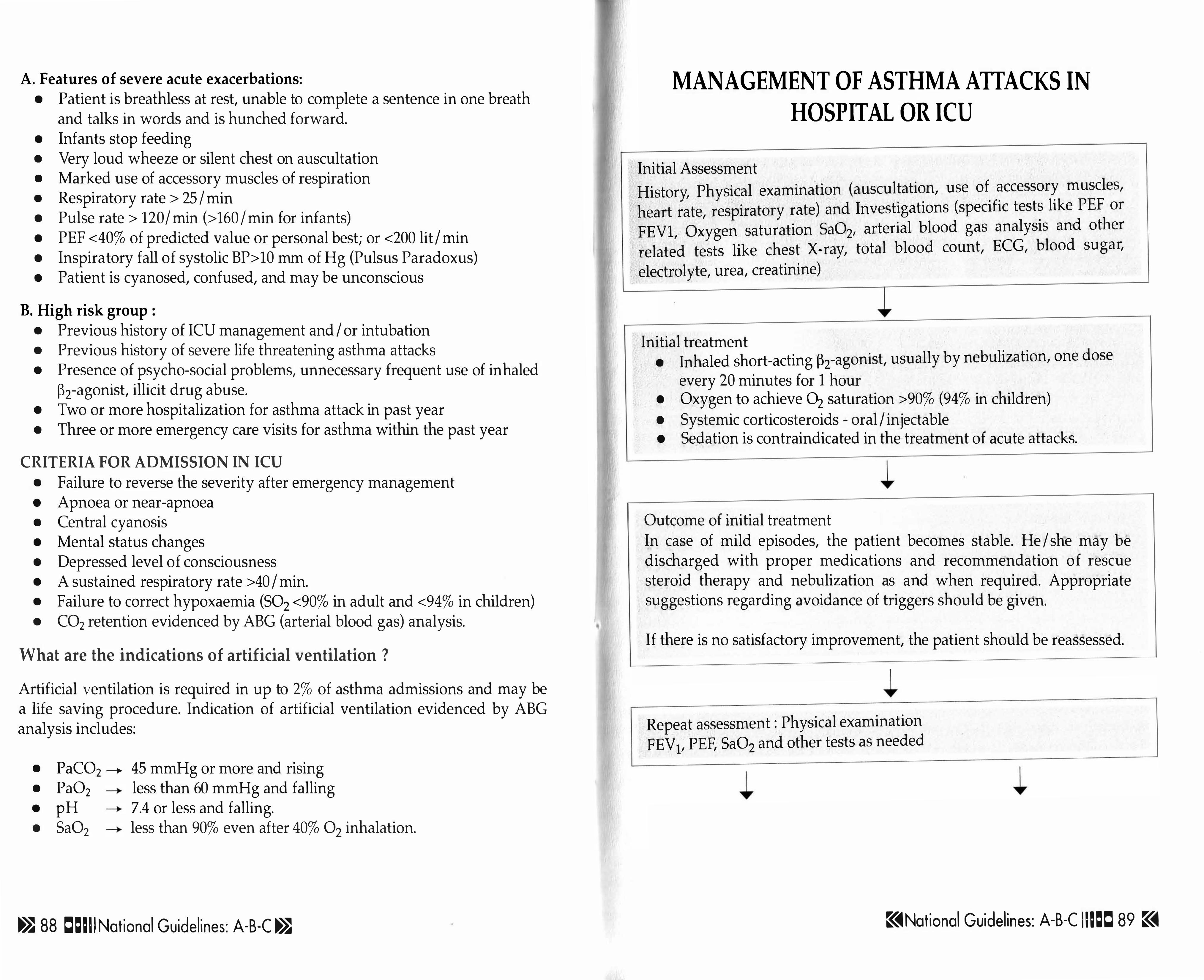
A. Features of severe acute exacerbations: • Patient is breathless at rest, unable to complete a sentence in one breath
and talks in words and is hunched forward. • Infants stop feeding • Very loud wheeze or silent chest on auscultation • Marked use of accessory muscles of respiration • Respiratory rate > 25 1 min • Pulse rate > 120/ min (>160 /min for infants) • PEF <40% of predicted value or personal best; or <200 lit I min • Inspiratory fall of systolic BP> 10 mm of Hg (Pulsus Paradoxus) • Patient is cyanosed, confused, and may be unconscious
B. High risk group : • Previous history of ICU management and I or intubation • Previous history of severe life threatening asthma attacks • Presence of psycho-social problems, unnecessary frequent use of inhaled
�2-agonist, illicit drug abuse. • Two or more hospitalization for asthma attack in past year • Three or more emergency care visits for asthma within the past year
CRITERIA FOR ADMISSION IN ICU • Failure to reverse the severity after emergency management • Apnoea or near-apnoea • Central cyanosis • Mental status changes • Depressed level of consciousness • A sustained respiratory rate >40 I min. • Failure to correct hypoxaemia (S02 <90% in adult and <94% in children) • CO2 retention evidenced by ABG (arterial blood gas) analysis.
What are the indications of artificial ventilation ?
Ar�ificial .ventilation is required in up to 2% of asthma admissions and may be a hfe savmg procedure. Indication of artificial ventilation evidenced by ABG analysis includes:
• PaC02 ---+. 45 mmHg or more and rising • Pa02 • less than 60 mmHg and falling • pH • 7.4 or less and falling. • Sa02 • less than 90% even after 40% O2 inhalation.
� 88 Clel l l National Guidelines: A-B-C � •
•
MANAGEMENT OF ASTHMA ATTACKS IN HOSPITAL OR ICU
Initial Assessment History, Physical examination (auscultation, use of accessory muscles,
heart rate, respiratory rate) and Investigations (specific tests like PEF or
FEVl, Oxygen saturation Sa02' arterial blood gas analysis and other
related tests like chest X-ray, total blood count, ECG, blood sugar,
electrolyte, urea, creatinine)
Initial treatment • Inhaled short-acting �2-agonist, usually by nebulization, one dose
every 20 minutes for 1 hour • Oxygen to achieve O2 saturation >90% (94% in children) • Systemic corticosteroids - oral I injectable • Sedation is contraindicated in the treatment of acute attacks.
Outcome of initial treatment In case of mild episodes, the patient becomes stable. He I she may be discharged with proper medications and recommendation of rescue steroid therapy and nebulization as and when required. Appropriate suggestions regarding avoidance of triggers should be given.
If there is no satisfactory improvement, the patient should be reassessed.
Repeat assessment : Physical examination FEV I, PEF, Sa02 and other tests as needed
•
�National Guidelines: A-B-C IUeCl 89 �

Moderate Episode On assessment • PEF 50-80% predicted / personal best • Physical exam: moderate symptoms,
accessory muscle use
Treatment • Inhaled �2-agonist every 60 mins • Consider corticosteroids • Continue treatment 1-3 hours,
provided there is improvement
.- ...
Severe Episode On assessment • PEF <50% of predicted or personal best • Physical exam: severe symptoms at rest,
chest retraction • High-risk patient • No improvement after initial treatrnen
Treatment • Inhaled �2-agonist hourly or
• continuous + inhaled anticholinergic Oxygen - 40% (5 Lit/min)
• Systemic corticosteroid • Consider subcutaneous, intramuscular,
or intravenous �2-agonist
� ...
Good Response Incomplete response Poor response within 1 hour within 1 hour
On assessment On assessment • On assessment
• Response sustained 60 • High-risk patient • High-risk patient minutes after last treatment • Physical exam: mild to • Physical exam: symptoms
• Physical exam: normal moderate symptoms severe, drowsiness, confusion. · PEF >70% • PEF >50% but <70% · PEF < 50% • No distress • 02 saturation not improving • PC02 >45 mm Hg . • 02 saturation >90% . P02 < 60 mm Hg
(95% in children) • 02 saturation <90%
Discharge Home Admit to hospital Admit to ICU • Continue treatment with • Systemic corticosteroid • Inhaled �2-agonist + •
inhaled �2-agonist. • Oxygen anticholinergic • Consider, in most cases, • Consider intravenous • Inhaled corticosteroid
.
corticosteroid tablets • aminophylline • Consider subcutaneous, or syrup. • Monitor PEF, 02 intramuscular, or intravenous
• Patient education: Take saturation, pulse �2-agonists medicine correctly. Review • Oxygen action plan. Intense medical • Consider intravenous follow up. aminophylline
• Follow appropiate step care • Possible intubation and management. • mechanical ventilation
90 C=I I INational Guidelines: A-B-C � ,
I
•
Can anti-allergy vaccines (immunotherapy) cure asthma?
Allergen avoidance is a cornerstone of adequate asthma management, but this is not always practical. Use of anti-inflammatory preventer drugs in regular and long term basis effectively mitigate hyperresponsiveness of airways to the triggers. Besides them, allergen-specific immunotherapy has been shown to improve the symptoms of allergic diseases to some extent.
The traditional method of immunotherapy means giving graded subcutaneous
injections over a planned long period of time. This type of immunotherapy
may improve bronchial hyper-responsiveness, minimize asthma symptoms
and reduce the use of asthma medications. However, these improvements are
usually temporary and it does not improve lung function consistently.
Traditional immunotherapy is costly inconvenient, sometimes painful and occasionally may cause severe life threatening anaphylactic reaction after injection. For these reasons, there has been a great deal of interest in deli:rering immunotherapy via the sublingual, oral, and nasal routes. Of them, sublmgual immunotherapy (SLIT) is a safer option.
Anti-allergy vaccines available in our country are exported from abroad
which are developed against foreign allergens. They may not be effective
against the allergens native to our environment. Moreover, vaccination is
not cost effective in our context. For these reasons, till now, we do not
advocate anti-allergy vaccines, particularly injectable forms, for bronchial
asthma. •
� National Guidelines: A-B-C =C 91

MANAGEMENT OF CONCOMITANT DISEASES Three allergic diseases are pathophysiologically related with bronchial asthma:
1 . Allergic Rhinitis 2. Atopic Dermatitis (Eczema) 3 . Allergic Conjunctivitis
These diseases frequently co-exist in same individual. It is found that, 38-58% of allergic rhinitis patients have asthma, 90% of children with asthma have allergic rhinitis and 50% of atopic dermatitis patient have asthma. Sometimes control of one condition enhances manifestations of another one. This phenomenon is known as "Allergic conversion reaction".
ALLERGIC RHINITIS
Allergic rhinitis is a common and troublesome condition often encountered by the physicians. It is more prevalent among the children than adults and among the boys than the girls. A study on school going children of Bangladesh revealed high prevalence of allergic rhinitis (20-25%) in comparison to other countries of the world.
Definitions
Rhinitis: It is an inflammation of the mucous membrane of the nose characterized by symptoms of nasal irritation, sneezing, rhinorrhoea and nasal blockage with at least two or more of these symptoms lasting for more than an hour a day on most days.
Allergic rhinitis: It is an IgE mediated inflammation of the mucous membrane of the nose occurring due to exposure to an inhaled allergen like pollen, dust, mould, fungi and animal dander.
Symptoms of allergic rhinitis • Sneezing • Runny nose • Nasal blockage • Nasal itching • Often associated symptoms of conjunctivitis
However, all symptoms may not exist together in any individual. Also, the dominant symptom may differ from one another. Again there is a wide individual variation in the tolerability of nasal symptoms.
92 CleUi National Guidelines: A-B-C
,
Classification of allergic rhinitis (ARIA-WHO initiative Classification)
Allergic rhinitis was previously subdivided based on the time of exposure and the triggering allergen into - seasonal, perennial and occupational. However, many patients with allergic rhinitis suffer from allergies to several allergens. For instance, patients with seasonal allergic rhinitis to one pollen may have allergy to other pollens or even house dust mites / pet dander. Also, patients with perennial allergic rhinitis to house dust mite or pets may not be symptomatic throughout the year. Based on these observations the ARIA-WHO initiative has introduced a new classification for allergic rhinitis. This new classification is as follows:
Intermittent <4 days per week or <4 weeks
Mild All of -• Normal sleep • No impairment of daily activities,
sports, leisure • Normal work and school
performance • No troublesome symptoms
Diagnosis of allergic rhinitis
Diagnosis of allergic rhinitis is based on
Persistent >4 days per week and >4 weeks
Moderate to severe One or more of -• Abnormal sleep • Impairment of daily activities,
sports, leisure • Abnormal work & school
performance • Troublesome symptoms
• A typical history of allergy symptoms • Internal examination of the nose by anterior rhinoscopy • Allergy tests .
Immediate hypersensitivity skin test - skin prick test Measurement of allergen specific IgE in the serum
• Nasal provocation test (NPT) - optional • Radiology (X-ray PNS and nasopharynx) - essential in children, optional
in adults
Patient education
The most important element in the treatment is information to the patient and if the patient is a child, information to the parents. Successful treatment depends on a good patient understanding of the nature of disease, that it is a
� National Guidelines: A-B-C I l lee 93

life long aliment but that the symptoms can be well controlled by proper treatment. Details of the therapy, the importance of continuing the treatment as advised and in case of topical sprays or drops, details on how to administer the drug should also be mentioned to the patient. The patient's cooperation plays an important role in optimizing therapeutic outcomes. Patient education booklets or pamphlets are also important modes that provide additional information.
A stepwise strategy for the treatment of allergic rhinitis is indicated; 1 . Allergen avoidance 2. Pharmacotherapy 3. Immunotherapy 4. Surgery in indicated cases
1. Allergen Avoidance Allergen avoidance should be an integral part and the first step in the management of allergic rhinitis. It is similar to trigger control plan in asthma. (see page 130).
2. Pharmacotherapy Pharmacotherapy comprises a wide variety of medications like H-l antihistamines, corticosteroids, decongestants, cromones, anticholinergics and leukotriene antagonists. Since medications do not have a long-term effect when treatment is stopped, in persistent disease, maintenance therapy is essential.
Medications can be administered by oral and topical routes (intranasal). The major advantage of administering drugs intranasally is that high concentrations can be delivered directly into the nose without causing systemic side effects and even the onset of action is fast.
Antihistamines • Histamine is the major mediator in allergic rhinitis • Antihistamines act by blocking the H-l receptors • They are effective in controlling sneezing, rhinorrhea and nasal itching
but are not so effective in controlling nasal blockage • First generation antihistamines like chlorpheniramine, diphenhydramine,
promethazine and triprolidine all have unfavorable risk-benefit ratios due to sedation as well as anticholinergic effects.
• 2nd generation antihistamines are: iX more potent iX have faster onset of action iX longer duration of action iX minimal sedative effects, and iX additional anti-allergic effects
94 C=UI National Guidelines: A-B-C
Therefore 2nd generation antihistamines are preferred. A variety of secondgeneration antihistamines, like cetirizine, levocetirizine, fexofenadine, lor at adine, desloratadine have anti-inflammatory properties and are shown to be effective in allergic rhinitis.
Corticosteroids • Corticosteroids are highly potent anti-inflammatory drugs and can
suppress many stages of the inflammatory process. • Of clinical importance in rhinitis, corticosteroids reduce inflammatory cell
infiltration (decrease mast cells and eosinophils), diminish hyperreactivity and vascular permeability and suppress the release of several inflammatory mediators (cytokines, chemokines).
• Corticosteroids may be delivered topically or taken orally. However in allergic rhinitis, topical but not oral corticosteroids are indicated.
• Topical corticosteroids are the first line of treatment in moderate-severe persistent rhini tis.
• The marked efficacy of topical corticosteroids is indisputable as it controls all the symptoms of rhinitis and has been shown to be superior to antihistamines. The effect of topical corticosteroids on nasal blockage lies in their anti-inflammatory properties.
• Beclomethasone dipropionate was the first topical corticosteroid introduced for allergic rhinitis. Subsequently, several new topical corticosteroids viz. budesonide, flunisolide, fluticasone propionate, mometasone furoate, flucortinbutyl and triamcinolone acetonide were developed which are recommended due to their relative lack of side effects.
.
• Short courses of oral corticosteroids are only indicated in severe, intractable cases.
Decongestants • Decongestants (vasoconstrictors) act on the adrenergic receptors and
provoke vasoconstriction. They may be administered topically or systemically.
• Topical decongestants such oxymetazoline, xylometazoline and naphazoline are highly effective in the short-term treatment of nasal obstruction in both allergic and non-allergic rhinitis.
• Oral decongestants like ephedrine, phenylephrine, phenylpropanolamine and pseudoephedrine can be used both short term and long term but are less effective than topical.
• Topical decongestants should not be used for more than 7-1 0 days, as it will cause rebound congestion (rhinitis medicamentosa).
• Oral decongestants are contraindicated in children, elderly patients over 60 years, pregnant women, patients with glaucoma, hyperthyroidism and
National Guidelines: A-B-C =C 95

prostate enlargement. The significant side effects of oral decongestants, which include irritability, dizziness, headache, tremor, insomnia, tachycardia, hypertension and visual hallucinations, limit the use of these drugs.
Anticholinergics • Anticholinergics like ipratropium bromide can reduce rhinorrhea in
perennial allergic rhinitis as well as non-allergic rhinitis. • Ipratropium bromide nasal spray should be considered only when
rhinorrhea is the primary symptom or when rhinorrhea is not responding to other therapy.
Cromones • Cromones like sodium cromoglycate and nedocromil sodium act by
stabilizing the mast cell and preventing the release of histamine and other mediators.
• Sodium cromoglycate and nedocromil sodium are safe and almost devoid of side effects.
• Unsatisfactory patient compliance due to multiple administration (4-6 times / day).
Leukotriene Antagonists • Cysteinyl leukotrienes are important mediators in the nasal allergic
reaction like histamine. • Drugs like zafirlukast and montelukast, which are leukotriene receptors
antagonists are effective in nasal congestion. • Since these are effective in both rhinitis and asthma, they are a potential
therapy for both these diseases especially in the context of patient compliance.
3. Immunotherapy Allergen specific immunotherapy can be employed in patients inadequately responding to pharmacotherapy, experiencing undesirable side effects and in situations when allergen avoidance is impractical or non-rewarding
4. Surgery Surgery is indicated in selective cases of severe nasal blockage, which do not improve with pharmacotherapy or immunotherapy. For this, the patient must be referred to the ENT specialist.
ATOPIC DERMATITIS (ECZEMA)
Atopic dermatitis (Eczema) is characterized by dryness of skin, intense itching and thickening or lichenification with excoriation, persists at least 6 months or
96 C=� I INational Guidelines: A-B-C
more with wax and wane characteristics. It mostly involves face in infants, extensor aspects of limbs in toddler and limb flexures in older child and in adult.
According to the previously mentioned study, 6.5-8.7% of Bangladeshi schoolgoing children suffer from eczema. However, this figure is less than the prevalence rate of other countries.
Eczema is the first menifestation of atopy in many patients who later develop allergic rhinitis (80%) and asthma (50%), a pattern that has been referred to epidemiologically as the "atopic march".
Management of atopic dermatitis comprises: i) Avoidance of specific allergens, ii) Oral antihistamines (ketotifen/ loratadine), iii) Low potency topical steroid (e.g. hydrocortisone), iv) Non-steroidal skin ointments (e.g. tacrolimus, pimecrolimus).
Note: Recurrent intense itching and rash after taking a particular food e.g. beef, aubergine (�.,), duck egg, shrimp etc is known as urticaria. It is actually a separate condition, not included in atopic dermatitis. It may be managed by avoidance of offending foods or by oral Sodium Cromoglycate, 1 /2 hour before ingestion of allergic food.
ALLERGIC CONJUNCTIVITIS
It is characterized by sudden lacrimation with itchy, red eyes, after exposure to pollen or allergen, usually associated with rhinitis.
Since conjunctivitis commonly presents with rhinitis, most of the time treatment of rhinitis is adequate to manage this condition as well. Antihistamine gives prompt relief. If conjunctivitis recurs frequently (e.g. on daily / weekly basis), instillation of topical cromones drops is helpful. It should be continued for at least 6 months after remission. Topical steroids should be avoided as prolonged use of such medication may lead to cataract or glaucoma.
� National Guidelines: A-B-C IU=C 97 �

.
ASTHMA AND CO-MORBIDITIES
Bronchial asthma may be present simultaneously with other ailments in the same patient. Some such important co-morbidities are discussed below.
Asthma with cardiovascular problems
Think of cardiac / associated cardiac disorders in the following situations -• Elderly patients presenting with dyspnoea • Patients having more crepitations than wheezes • Patients with cardiac murmur • Patients not improved with classical anti-asthma treatment • Patients with unexplained breathlessness
In these cases patients should be evaluated with ECG, Chest X-Ray and colour doppler echo cardiography.
Asthma with hypertension
• Virtually all antihypertensives in low dose can be used in asthma except propanolol
• Drug of choice is calcium channel blockers with thiazide diuretics -singly or in combination.
• Angiotensin receptor blockers (e.g. losartan, valsartan) are preferred to ACE Inhibitors (because the latter may induce dry cough).
• Non selective �-blockers must be avoided, selective �-blockers can be used.
Asthma with Ischaemic Heart Diseases (Stable & Unstable Angina)
• Aspirin should be tried first. If not tolerated clopidogrel should be used. • Anti-anginal nitrates and calcium channel bIocker� (diltiazem &
verapamil) are the drug of choice. • Cardioselective �-blockers (e.g. metaprolol) may be used. • Asthma control should be optimum to avoid hypoxemia.
Asthma with heart failure
• Diuretic is the drug of choice. • ACE Inhibitors should be continued if tolerated. • Carvedllol may be used in low aoses -• Digoxin can be used.
98 == 11 1 National Guidelines: A-B-(
•
Asthma with Arrhythmia
• Calcium channel blockers (diltiazem / verapamil) is used in supraventricular arrhythmias (e.g. atrial fibrillation)
• Digoxin is the drug of choice to control ventricular rate. • Amiodarone can be used. • Try to avoid aminophylline / theophylline to treat asthma.
Asthma with rheumatologic disorders
•
• Potent NSAIDs are well tolerated in most of the asthmatics. (for management of analgesic induced asthma, see page 32).
• Paracetamol and tramadol are the preferred agents, as they usually produce no adverse effect.
• If needed steroid can be given. • Other modalities of pain management like thermotherapy or SW therapy
may be employed. • Disease modifying drugs can be used safely in asthmatics.
Asthma with diabetes mellitus
• Steroid can be used if indicated. Regular blood sugar monitoring is necessary. • In acute severe asthma, blood sugar should be controlled by insulin. • Metformin should be avoided in poorly controlled asthma and is
contraindicated in case of acute severe asthma. •
• Dose of oral hypoglycemic agents (sulphonylurea and pioglitazone) should be adjusted when concomitant aminophylline is used (aminophylline may induce hypoglycemia).
Guidelines: A-B-( 99

ASTHMA IN SPECIAL SITUATIONS
PREGNANCY AND ASTHMA
Asthma during pregnancy follows the rule of one-third, that is, one-third asthmatics become worse, one-third remains same and one-third improve. The exact mechanism behind this is not known. It is common to experience some breathlessness near the end of the pregnancy, this is related to the size of the fetus and the pressure it puts on the diaphragm.
It is dangerous to have untreated asthma during pregnancy, because attacks of asthma may reduce the amount of oxygen available to the baby. Triggers should be controlled meticulously during pregnancy. They can influence the probability of giving birth to a wheezy baby. Active and passive smoking should also be avoided at this time. It increases the chances of wheezing in the newborn. Caesarean section delivery is not an absolute indication in an asthmatic mother. The rate of caesareans among women with asthma is no higher than in those without it.
All asthma medicines have been shown to be absolutely safe for both the mother and the baby. Inhaled route is always preferred. Asthma medications may enter breast milk, but the concentration is extremely small and do not have any adverse effect on the baby.
In pregnant asthmatics there is increased risk of: • preeclampsia • perinatal mortality . • preterm birth • low birth weight
Advice for pregnant asthmatics: • Monthly monitoring • Reduce triggers e.g. allergens and smoke • Educate patient on importance of asthma control • Postponing step down therapy until pregnancy is completed.
�1 00 ==� I INational Guidelines: A-S-C
Treatment guidelines for asthma during pregnancy:
Type of asthma Recommended treatment Comments
Intermittent Short acting inhaled �2- Terbutaline • also IS asthma agonist preferably salbutamol effective but there is no
. better efficacy as thought before.
•
Mild persistent Low dose inhaled Nedocromil has no better asthma corticosteroids (LDICS) role as advocated before.
. preferably Budesonide. May be useful in cough • Beclomethasone • also variant asthma. IS
effective. . •
SR theophylline can be given.
Moderate persistent LDICS + long acting inhaled Oral �2"agonist •
IS no asthma �2"agonist (e.g. salmeterol) more advocated as
OR medium dose ICS before
Severe persistent High dose ICS. Try . to avoid oral asthma Theophylline
.
5R can be corticosteroid added. Oral corticosteroid (e.g. prednisolone up to 45 mg)
•
can be given. I ,. Even more can be given, but
with caution
NOTE: Although it is evident that prednisolone is safe even during 1st trimester, it's use should be limited as rescue therapy for 7-14 days.
Leukotrienes antagonists have not been extensively evaluated in pregnancy. No confirmed evidence of benefits or side effects are found. So, it is better not to use these drugs during pregnancy.
During labour, induction is usually done with Prostaglandin E2 (PgE2) and oxytocin. It is better to avoid Inj. Ergometrin.
� National Guidelines: A-S-C 1 1 1== 1 01

•
SURGERY AND ASTHMA
Surgery is considered as a trauma and there is emotional stress for surgery. Both can trigger asthma.
Recommendations:
Preoperative
• Elective Surgery - control asthma with optimum treatment, assess by spirometry (see page 45)
• Emergen,!, Surgery - If needed, nebulize with bronchodilator, give IV hydrocortisone and employ O2 (humidified).
Peroperative
• Avoid volatile anesthesia. • If possible, use spinal anesthesia instead of general anaesthesia. • Use of frusemide may be beneficial. • Use pulse oxymeter to monitor oxygen saturation.
Postoperative
• For analgesia, do not use narcotic analgesics (e.g. morphine), but some opoid derivatives (e.g. tramadol) can be used, use NSAIDs cautiously.
• Postoperative respiratory physiotherapy may be beneficial.
•
02 C�UINational Guidelines: A-B-C
SECTION-3: •
Asthma Education
What is patient education in asthma and why is it essential?
Patient education regarding asthma is so important that if they are educated properly, then 73% of hospital admission from acute attack of asthma can be reduced and 80% of death from asthma can be avoided. These tasks may be performed in two steps.
. First Step: Development of rapport
Counseling with the patient and/ or parents or attendants about asthma, and thereby assessing their knowledge about asthma, it's medications; use of devices, trigger factors and other relevant points must be done. It should be done in a plain simplified language, avoiding medical terminology as far as possible. A compassionate approach is essential. These will lead to build up of patient's confidence and will increase compliance and concordance to the management plan.
Suggestions: Minimum investigations should be performed. Short course steroid therapy with tolerable doses may be started before investigation reports are available in patients with uncontrolled asthma. Diagnosis should be disclosed to the patient at subsequent visit.
Second Step: Patient Education Checklist
The following 7 points need to be addressed when educating a patient about asthma. Information and messages should be delivered and demonstrated slowly and step-by-step, not all at a single sitting, by a physician or health professional on a person-to-person basis. Printed health education material should not be solely depended upon.
1. Basic facts about asthma a) Concept of asthma as an inflammatory disease:
• Do not tell the patient that his / her airways are diseased or damaged . Rather describe that it is a condition where airways become red and swollen .
• . Emphasize that asthma is a condition that does not just go away (can not be totally cured). But the troublesome symptoms can be controlled.
• It is a chronic process that exacerbates in response to different trigger
� National Guidelines: A-B-c IU�C 1 03�

factors. • The aim of patient education is to learn how asthma can be controlled. If
it is learned properly, patient can lead an active and near-normal life. b) Concept of airway narrowing:
This happens due to a combination of -• Smooth muscle spasm • Airway swelling due to:
i:I Oedema: fluid and proteins deposited across the airway wall i:I. Mucus hypersecretion i:I Muscle and mucous gland enlargement
(Try to show pictures of normal and inflamed airways. If not available draw it infront of the patient) .
Normal airways Inflamed airways
2. Asthma medicines and appliances a) Concept of different types of asthma medications:
• Reliever medication (bronchodilators) • Preventer medication (anti-inflammatory agents) • Protector medication (long-acting �Tagonists, SR Theophylline) • Combination medications (pre venter plus protector)
The patients need to be provided with a brief knowledge about these medications, how they work, what are their doses, common side effects and how to cope with these. (For example, relievers relief distress quickly but pever treats the underlying cause, just as paracetamol relieves fever not the cause).
b) Explanation, demonstration and skill of use of delivery devices and appliances: • Types of devices and appliances • Their mode of action . • Their role in treatment • Alternative delivery devices • Need for correct inhaler technique .
1 04C=� 1 I National Guidelines: A-B-C
Inhalation technique particularly for MDI should be demonstrated and taught to the patient practically. Try to provide an illustrated handout. If necessary, how to use Spacer, Nebulizer, Peak Flow Meter etc. should also be shown practically to and learned by the patient.
3. Concordance a) Need for long-term adherence to preventive therapy:
• Emphasize that initiating treatment with asthma medications does not imply that treatment will be life-long in all patients
• Emphasize that asthma treatment is rarely short term • Discourage the notion that treatment can be discontinued as soon as the
symptoms resolve • Highlight the need for preventive therapy to be used every day whether
the patient feels well or not
b) Importance of an Asthma Management Plan Provide a structured management plan incorporating:
• Medications • Trigger control • Emergency measures • Life style factors
c) Regular peak flow monitoring • Instruct correct technique and maintenance of peak flow meter • Explain the relation between peak flow and the management plan
d) Rescue Actions: . • When a patient has cough, wheeze, dyspnoea or chest tightness, even in
mild attack, he / she should inhale reliever drug e.g. Salbutamol Inhaler, without hesitation.
• In an emergency situation, when reliever drug is not working properly, patient should start rescue steroid orally before consulting with physician.
4. Avoidance of risk factors a) Recognition of asthma triggers and precautionary measures:
The following advices must be given to the patients: i) Quit smoking and try to avoid passive smoking.
ii) D o not keep carpet in your bedroom and try to avoid carpet in your working places and in drawing room.
iii) Do not allow pets, e.g. cat, dog, bird etc. in your house. iv) Do not use insecticides or aerosols and never operate vacuum cleaners by
yourself.
National Guidelines: A-B-C IU=C 1 05

b) Prevention of exercise-induced asthma: • Optimize control of patient's asthma • Use pre-exercise medications • Encourage warm-up exercise
c) Recognition and management of occupational asthma
5. Prognosis and goal of management
a) Understanding the natural history of childhood asthma: •
Asthma is a heterogeneous disease, with different predominant expressions at different ages. Natural history of asthma is influenced by a number of factors such as genetics, atopy, air pollution, environmental tobacco smoke, gastroesophageal reflux and infection. Distinct phenotypes are transient early wheezing, late-onset wheezing and persistent wheezing.
There is a group of children who experience at least one LRTI (lower respiratory tract infection) with wheezing during the first three years of life but have no wheezing at six years of age (transient early wheezing). There is another group of children who do not wheeze before the age of three years but wheeze by the age of six years (late onset wheezing). A minority group of children wheeze before three years of age and also continue to wheeze even after six years of age (persistent wheezing). Children with persistent wheeze are more likely to have parents with a history of asthma and to have elevated IgE levels and diminished lung function at six years. The presence of atopy, positive allergic skin prick test or elevated IgE antibody levels increases the probability of asthma to over 95% .
Childhood asthma typically begins in infancy with a respiratory syncytial virus (RSV) in 25-30% cases. Recurrent wheeze remit in a large number of children who develop symptoms during the first year of life and diminished lung function seems to be the main risk factor for these transient wheezy episodes. Studies showed that in a large proportion of asthmatic children (80%) whose asthma was triggered mainly by respiratory infections, asthma symptoms appear to remit by adolescent period. Persistent asthma develops only in a few children (5%) .
•
b) Treatment goal:
Patient should have a clear idea about the treatment goal, i.e. "effortless easy breathing", which may be achieved by either complete remission or by total
� 1 06Cmll National Guidelines: A-B-()�
control of asthma.
6. Alleviation of misconceptions
There are some common but baseless concerns and fears regarding asthma and its treatment, which sometimes create problems in optimum management of the disease. Physician should carefully deal with those points. (see page 111).
7. Institutional.approaches
a) Formation of "Asthma Clubs": .
Majority of asthma patients can be and should be managed at home. To make the home management more effective the physicians may group the patients in the form of a club named "asthma club". The club members may meet once in a month to describe their experiences and status of asthma in presence of the physician who will educate, train and demonstrate to them in the light of patient's complaints and queries. The group of patients i.e. members of the asthma club may also benefit through exchange of views in such meeting.
b) School-based management
Parents should inform the teachers and school authority about the student's asthma management plan, especially when the student is going on a school camp. •
National Guidelines: A-B-C In�c 1 07

PREVENTION OF ASTHMA
How we can prevent asthma?
Development of asthma has two distinct bases: Hereditary and Environmental. For the prevention of the development of asthma we should manipulate these two factors. Regarding hereditary factors, we have nothing yet to do practically. Genetic engineering is a future probability. But we can manipulate the environmental factors. Efforts should be concentrated on early prevention of asthma.
What are the types of asthma prevention?
Prevention of asthma is of two types: 1 ) Primary prevention 2) Secondary prevention
What is primary prevention?
Primary prevention is non-therapeutic interventions even before any form of hyper- �esponsiveness in an individual are seen. The four key areas in primary prevention are:
1) Timing of hyper-responsiveness 2) The level at which allergen concentrations must be reduced to prevent the
development of hyper-responsiveness 3) The necessary duration of allergen avoidance 4) Adjunct factors involved in triggering the disease
For these, following two things are to be done: - Identification of the asthma prone persons - Well-defined prevention program for the asthma prone persons
How to identify asthma prone person?
Period before and immediately after birth is very important for the future dev,elopment of allergic diseases, such as asthma. Identification of the high-risk newborns can be done by a family allergy scoring system (FAS) . "FAS" is based on the number of immediate (first degree) family members (mother, father, brothers and sisters only) who suffer or who have suffered from one or other
. allergic condition.
1 08 C=W National Guidelines: A-B-C •
Scoring system:
Two points are scored for each immediate family member who has definite, medically confirmed allergic disease. (positive history along with clinical evidence).
One point is scored for each family member who has possible or suspected allergic disease but which has not been medically confirmed. (positive history without clinical evidence).
One point is scored for persons exposed to environmental or occupational risk factors (as they are prone to develop asthma).
No point is scored for the members without any allergic disease.
Interpretation of the score:
Score: 0 - 1 : No prevention is necessary.
•
Score: 2 - 3 : Serum IgE estimation, raised value warrants preventive program. Score: 4 or more : Strongly advocate preventive program.
PRIMARY PREVENTION PROGRAM •
Program for primary prevention of asthma varies from person to person. It is difficult to chalk out a universal program. However on the basis of recent knowledge asthma prone persons, that is, persons who have more chance of developing asthma, may be given the following advice in the form of DOs and DON'Ts:
DOs • Babies should be exclusively breast-fed until the age of six month. • Weaning should be delayed till six month of age, particularly for allergy
producing solids. • Encourage low salt diet. • Encourage more fish and less meat in diet. • Promote outdoor sports in summer and indoor sports in winter. Swimming is
best exercise for asthmatics in all seasons except winter. • Establish proper ventilation at home. • Try to change the job if development of occupational asthma is suspected. • Reduce weight if over-weight
National Guidelines: A-B-C IU=C 1 09 �

DON'Ts
• Lactating mother should not eat or drink any food or beverage allergic to her. • Prevent high-risk babies being exposed to potential allergic foods (e.g.
cow's milk) and inhaled allergens during first two years of life. • Avoid carpeting, stuffed furnishing, household pets, stuffed toys (e.g.
teddy bears) and furry dresses. • Try to avoid broad-spectrum antibiotics for bronchiolitis and viral R.T.I.
Early exposure to bacterial infection may switch off allergic response (Hygiene hypothesis).
• Try to avoid outdoor air pollution - pollen, dusts, smoke etc. • Quit active smoking and avoid indoor passive smoking. • Avoid spending 3 hours or more at a stretch in a day in front o f the
television or computer.
Hygiene Hypothesis
The observation of an inverse relation between number of children in the family and atopy formed the basis of 'hygiene hypothesis' of asthma. The immunological explanation of this concept is the distinction of Thl and Th2 lymphocyte population and the recognition that 'natural immunity' to bacterial and viral infections induce a Thl pattern of immunity, potentially suppressing the Th2 type of immune response, which is involved in IgE mediated allergy as well as asthma. The high rates of respiratory infections, tuberculosis, measles and helminth infestations in Bangladeshi children might thus contribute to lower rates of allergy and asthma in comparison to the high rates of asthma in the developed contraries. Indiscriminate use of antibiotics also results in switching immune response towards Th2 lymphocytes resulting in asthma and allergies in childhood, as Thl immune response is suppressed. Hygiene hypothesis suggests that western life-style is characterized by higher level of cleanliness, lower rates of infections and small family size leading to high risk of developing asthma.
What is secondary prevention?
Secondary prevention consists of therapeutic interventions that are especially employed for susceptible children. If a child with positive family history of bronchial asthma (i.e. asthma among immediate relations, e.g. mother, father, brothers and sisters) suffers from bronchiolitis with subsequent recurrent wheeze and / or cough, he / she should be given preventive treatment of bronchial asthma with anti-inflammatory medicines for about 6 months to 1 year after last episode of wheezing and / or coughing.
�1 1 0 Cel l i National Guidelines: A-B-C
.
PATIENT'S CONCERNS ABOUT ASTHMA There are some common concerns about asthma prevailing in the society. In majority of cases, these are misconceptions or baseless fears. For optimum control of asthma, these points should be dealt with proper care. Otherwise the management plan may go in vain. It is the physician's responsibility to eradicate such worries, if present, from the patient's mind. Some common concerns and suggested clinician's responses are listed below.
Concern or Fear
Asthma cannot be cured.
Asthma can be fataL
Inhaler is the last resort of treatment.
Asthma medicines are dangerous.
People with asthma cannot exercise
Asthma medicines are addictive.
Asthma is caused by psychological problems.
Asthma is contagious
Asthma is a hereditary disease
In pregnancy, asthma drugs are not safe
All wheezy children are life-long asthmatics
Suggested Response
Yes, but it can be controlled. If asthma is controlled, asthmatics can participate in all activities
Death from asthma is very rare, if properly managed.
Contrary to popular belief it is the first line of drug for asthma.
Asthma medicines are safe if taken as prescribed.
Exercise may be beneficial in asthma. Take medicine before starting exercise to prevent symptoms during exercise.
Asthma medicines do not cause addiction, even if used in a high dose for a long time.
Though psychological stresses may trigger or worsen asthma, it is basically a physical problem.
Absolutely not
Asthma may run in families, but it is not compulsory that an asthmatic will give birth to another asthmatic.
All asthma drugs are safe in pregnancy.
It is not true. Majority of them get rid of it. Some may develop asthma in later life.
� National Guidelines: A-B-C :UeC 1 1 1

ASTHMA MANAGEMENT APPLIANCES What are the appliances used in asthma management?
Asthma drugs are targeted to deliver at the airways. A clear understanding of the delivery of asthma drug at the site is very important in the management of asthma. A number of devices are developed for the optimum delivery of drug. New ones are coming as well. To date the available devices may be divided into six families:
1 . Metered Dose Inhalers (MDIs) 2. Breath-Actuated Inhalers (Autohalers) 3. Dry powder inhalers (DPIs) 4. Spacers and chambers 5. Nebulizers 6. Flow Meters
METERED DOSE INHALERS (MDIs)
Most commonly used device in asthma is the MDIs. It is small and easy to carry. Wide ranges of drugs are available in this device. Using a metered dose inhaler (MDI) is a good way to take asthma medicines. There are fewer side effects because almost all medicine goes right to the lungs and only a very litt le amount can go to the other parts of the body through systemic circulation. It takes few minutes for the medicine to exert an effect compared to oral asthma medicines, which may take 1 to 3 hours.
The guidelines that follow will help your patient to use the inhaler the right way. Demonstrate the procedure yourself. Ask your patient to do the following in front of you. Remember, improper use of MDI is a major cause of nonresponse to treatment.
How to use MDI (advice for patient)
Step-I . Remove the dust cap; look inside for any dust or foreign body and hold the inhaler upright.
Step-2. Shake the inhaler (at least 5 shakes) . Step-3. Tilt your head back slightly and breathe out. Step-4. Place the mouthpiece of the inhaler in between lips. Step-5. Press down the canister to release the medicine and at the same time
start to breathe in slowly up to full inspiration. Step-6. Hold the breath for 10 seconds. Step-7. Repeat puffs as prescribed. Wait 1 minute between puffs.
�1 1 2 C= National Guidelines: A-B-C
STEPS OF MDI USE
---�
Shake the inhaler (at least 5 shakes) Tilt head back slightly & breathe out
---�
•
Press the canister and breathe in slowly Hold the breath for 10 seconds
MDIs can be used by all asthma patients of more than 5 years of age. However, it needs coordination for proper use. Initially, as many as 9 out of 10 patients who use MDIs may have trouble in coordinating device actuation with inhalation. As a result insufficient deposition of drug in the lung and unwanted deposition in the oropharynx and systemic effect of the drug destruct the novel properties of the MDIs.
Recommendation Prescribe inhalers only after patients have received proper and practical training regarding the use of device and have demonstrated satisfactory technique.
A spacer or holding chamber attached to the inhaler can make taking the medicine easier by diminishing the need for coordination between actuation and inhalation. This allows use of MDIs in even children younger than 5 years.
� National Guidelines: A-B-C IU=C 1 1 3�

Cleaning
The inhaler must be cleaned often to prevent buildup that will clog it and reduce its efficiency. Once a week remove the canister and wash the plastic actuator with mild dishwashing soap and warm water. Rinse and dry it well before next use.
Checking how much medicine is left in the canister
• By shaking the canister we can guess the amount of medicine inside. • A puff-count chart is the best way to check the medicine. Some of the
pharmaceutical companies are providing such papers attached with the MDIs.
• An easy way to check the amount of medicine left in a metered dose inhaler is to place the canister in a container of water and observe the position it takes in the water. The part of the canister above the water level is empty.
BREATH-ACTIVATED INHALERS
Breath activated Inhalers (autohalers) are not available in our country to date. Unlike MDIs, it needs no coordination during use. It is suitable even for babies of 3 years of age only.
DRY POWDER INHALERS (DPIs)
Dry Powder Inhalers are now available in our country. There are varieties of different designs of DPIs with their specific characteristics. Optimum Peak Inspiratory Flow (PIF) should be generated to activate the DPIs. The required PIF for different DPI is different ranging from 30 L/min to 120 L /min.
Commonly used DPIs are accuhaler, rotahaler, cozyhaler, aerolizer, cyclohaler, turbohaler etc. In accuhalers, powder is within an in-built disk of blisters and needs low PIF (30-60 L / min) to activate. In rotahaler, cozyhaler and aerolizer dry powder capsules are used. In cozyhaler, capsules are broken into two halves leaving the powder to inhale by moderate PIF (60-90 L/ min). In the rotahaler and aerolizer capsules are pierced by pin leaving the powder in the capsule and to inhale through the hole by high PIF (90-120 L / min). If capsule is used in DPIs, PIF should be measured or at least we should ensure that a rattling sound is produced from the device when inhaled.
1 4 C=mNational Guidelines: A-B-C �
I
DPIs are small, portable and disposable or reloadable. They are suitable for 5 years or over. For the technique of use, manufacturer's instruction should be followed. Closing of lips tightly around the mouthpiece and inhaling very fast is necessary. Common mistake in its use is to blow in the device. It should be monitored and corrected carefully.
The DPI devices must be cleaned often to prevent buildup that will clog it and reduce its efficiency. Once a week wash the plastic device with mild dishwashing soap and warm water. Rinse and dry it well before next use.
. MDI ACCUHALER
ROTAHALER COZYHALER
� National Guidelines: A-B-C IU=C 1 1 5�

SPACERS AND CHAMBERS
A spacer or holding chamber is a device that attaches to a metered dose inhaler. It holds the medicine in its chamber long enough for the patient to inhale it in slow deep breaths. Spacer makes it easy to use the medicines in proper way. It helps prevent coughing while using an inhaler. It will also help protect the patient from getting a fungal infection in mouth (thrush) when taking inhaled corticosteroids. Unless patient uses the inhaler in the correct way, much of the medicine may end up on the tongue, on the back of the throat, or in the air. Use of a spacer or holding chamber may solve this problem.
Two types of spacers or holding chambers are available in our country -1 . Large volumatic spacers - it has two halves. 2. Mini aerochambers
AEROCHAMBER
..... I
AERO CHAMBER WITH MASK
How to use a Spacer (advice for patient)
VOLUMATIC SPACER
1 . Join the two halves of the volumatic spacer (not required for Aerochambers) .
2 . Shake the spacer and make sure that valve is moving making a "ticking" sound
1 6 C=U INational Guidelines: A-B-C �
3. Shake the MDI (at least 5 shakes) and place it in the spacer at the opposite end of the mouthpiece.
4. Hold the spacer horizontal, breathe out as fully as possible and place the lip around the mouthpiece.
5. Press the canister and inhale slowly and deeply, hold the breath for 10 seconds and then breathe out. Take another breath, slowly and deeply as before without pressing the canister and hold the breath for 10 seconds
•
and then breathe out (1 puff 2 sucks). 6. For another puff repeat step 3-5 after one minute. 7. If patient, especially a child or very old person, is unable to hold breath for 10
seconds, he / she will inhale and exhale 6 times after each puff (1 puff 6 sucks).
•
Cleaning
Separate the chamber in parts, rinse the parts in water with mild detergent and reassemble after drying. Do not wipe inside. Clean it once in a month to keep it free of electrostatic effect. Change the spacer at least every 6 months.
NEBULIZERS
A nebulizer unit is a device in which drug is dispensed through a jet like airflow produced by a compressor or ultrasonic machine. It delivers high dose of drugs as fine mist (wet aerosol). It is very useful in treating acute asthma attack, uncontrolled severe persistent asthma, COPD patient of stage III or IV and children below 5 years of age. A nebulizer helps make sure that patients get the required amount of medicine within a short period. Both reliever and preventer medicines can be delivered through nebulizer. It is suitable for all age groups.
A nebulizer consists of a cup, a mouthpiece attached to a T-shaped part or a mask, and thin plastic tubing to connect to the air compressor machine. The following types of patients use it mostly:
• Young children under age 5 years. • Patients who have problems using metered dose inhalers even with spacers. • Patients with severe asthma and COPD (i.e. in emergency room). • Immunocompromised patient with pneumonia for antibiotic and antifungal
therapy • Patient with uncontrolled haemoptysis to deliver haemostatic agents • Patient with intractable cough to deliver local anaesthetics.
� National Guidelines: A-B-C InCC 1 1

How to Use a Nebulizer (Advice for the patient)
1 . Directions for using the compressed air machine may vary (check the machine's directions), but generally the tubing has to be put into the outlet of the machine at first.
2. Measure the correct amount of normal saline solution using a clean dropper and put it into the cup. If medicine is premixed (as in nebules), ignore this step.
3. Draw up the correct amount of medicine using a clean dropper or syringe and put it into the cup with the saline solution. If you know the number of drops, you can count them as a check.
4. Attach the mouthpiec€ to the I-shaped part and then fasten this unit to the cup OR fasten the mask to the cup. For a child over the age of 4, try to use a mouthpiece unit because it will deliver more medicine than a mask.
5. Put the mouthpiece in mouth. Seal lips tightly around it OR place the mask over the face.
6. Turn on the air compressor machine. 7. Take in slow, deep breaths through the mouth. 8. Continue until the medicine is gone from the cup and no more mist is produced
(approximately for 10 minutes) . 9. Store the medicine as directed after each use.
Cleaning:
Regular cleaning of the nebulizer is important because an unclean nebulizer may cause an infection. A good cleaning routine keeps the nebulizer from clogging up and helps it last longer. Moreover, it may need regular servicing from authentic service center, at least once a year.
1 8 CC National Guidelines: A-B-C
Nebulizer should be cleaned once every day:
1 . Remove the mask or the mouthpiece and T-shaped part from the cup. Remove the tubing and set it aside. The tubing should not be washed or rinsed.
2. Wash the mask or the mouthpiece and T-shaped part - as well as the dropper or syringe - with a mild dishwashing soap and warm water.
3. Rinse under a stream of water for 30 seconds. Use distilled (or sterile) water, if possible.
4. Shake off excess water. Air dry on a clean cloth or paper towel. 5. Put the mask or the mouthpiece and T-shaped part, cup, and tubing back
together and connect the device to the compressed air machine. Run the machine for 10 to 20 seconds to flash out and dry the inside of the nebulizer.
6. Disconnect the tubing from the compressed air machine. Store the nebulizer in a zip lock plastic bag.
7. Place a cover over the compressed air machine.
. - -----
Delivery devices for asthma medications in children -
Mode of Administration <2 Years 2-4 Years 5-7 Years
Nebulizer Yes Yes Yes •
MDI + Spacer with mask Yes Yes --
MDI + Spacer without mask .
Yes -- --
MDI (alone) . -- -- --
>8 Years
Yes
--
Yes
Yes
� National Guidelines: A-B-C :ncc 1 1 9�

FLOW METERS
Flow meters are devices to measure the strength of the airways, that is the force of inspiration and expiration. They are of following types: • Peak flow meter • Incentive spirometer • PIF (peak inspiratory flow) meter
Peak Flow Meter
Peak flow meter measures how well air moves out from a patient's lungs. During an asthma episode, the airways of the lungs begin to narrow. The peak flow meter can be used to find out if there is any onset of narrowing in the airways, hours - even days - along with or before the patient has any symptoms of asthma. By doubling the medicine (particularly preventer medicine) early (before symptoms), a patient may be able to stop the episode quickly and avoid a serious episode of asthma. So its role in preventing severe asthma attack is very important.
The peak flow meter can also be used:
• to see whether the management plan is working well or not • to decide when to add or stop medicine • to decide when patient should seek emergency care • to identify triggers - that is, what causes patient's asthma symptoms to increase
All patients of more than 5 years of age, who have moderate to severe asthma, should be advised to use a peak flow meter. Some children as young as 4 years of age can also use it.
How to Use a Peak Flow Meter (Advice for the patient)
1 . Place the indicator at the base of the numbered scale 2. Stand up / or sit in upright posture 3. Take a deep breath 4. Place the meter in your mouth and close your lips around the mouthpiece.
Do not put your tongue inside the hole. Do not put your finger over the indicator
5 . Blow out as hard and fast as you can 6. Write down the number you get 7. Repeat steps 1 through 6 two more times 8. Write down the highest of the three numbers achieved
� 1 20C=W National Guidelines: A-B-(
// ,
17'7 n-r � ,.,:r,-,., ,.,.;: rrr / " rrr rTF" " / > \ // "'--
.. \ � , \. ..l.. , \
•
,
PREDICTED VALUES (P.V) OF PEF (L/min) Male Female
Age Height Age Height 60" 65" 70" 75" 80" 55" 60" 65" 70" 75"
20 554 602 649 693 740 20 390 423 460 496 529 25 543 590 636 679 725 25 385 418 454 490 523 30 532 577 622 664 710 30 380 413 448 483 516 35 521 565 609 651 695 35 375 408 442 476 509 40 509 552 596 636 680 40 370 402 436 470 502 45 498 540 583 622 665 45 365 397 430 464 495 50 486 527 569 607 649 50 360 391 424 457 488 55 475 515 556 593 634 55 355 386 418 451 482 60 463 502 542 578 618 60 350 380 412 445 475 65 452 490 529 564 603 65 345 375 406 439 468 70 440 477 515 550 587 70 340 369 400 432 461
Children (Male & Female)
Height P.V Height P.V Height P.V 43" 147 51" 254 59" 360 44" 160 52" 267 60" 373 45" 173 53" 280 61" 387 46" 187 54" 293 62" 400 47" 200 55" 307 63" 413 48" 214 56" 320 64" 427 49" 227 57" 334 65" 440 50" 240 58" 347 66" 454
National Guidelines: A-B-( I I I=C 1 2 1
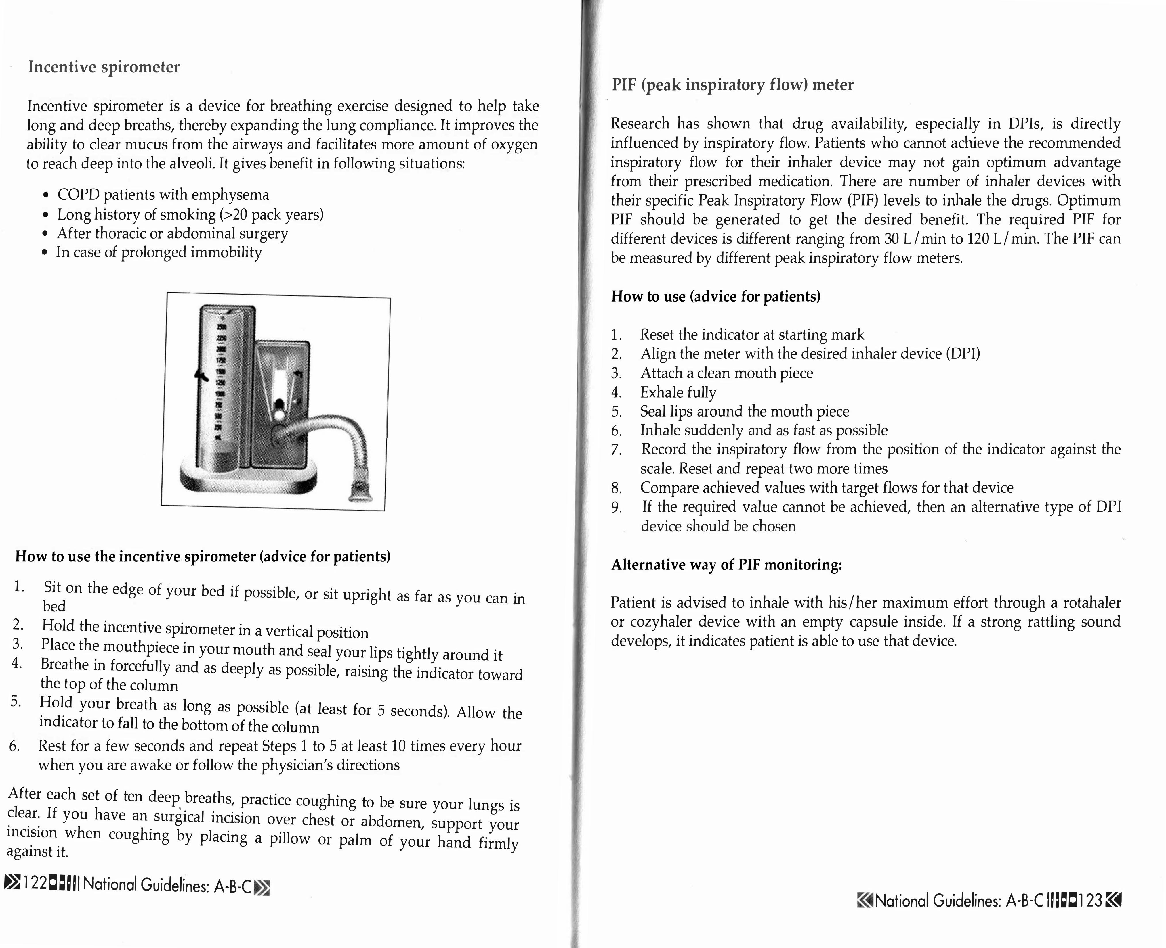
Incentive spirometer
Incentive spirometer is a device for breathing exercise designed to help take long and deep breaths, thereby expanding the lung compliance. It improves the ability to clear mucus from the airways and facilitates more amount of oxygen to reach deep into the alveoli. It gives benefit in following situations:
• COPD patients with emphysema • Long history of smoking (>20 pack years) • After thoracic or abdominal surgery • In case of prolonged immobility
e -
• -.. -
• -
.. ,-
--
III -
• -
III .-
How to use the incentive spirometer (advice for patients)
1 . Sit on the edge of your bed if possible, or sit upright as far as you can in bed 2 . Hold the incentive spirometer in a vertical position 3. Place the mouthpiece in your mouth and seal your lips tightly around it 4. Breathe in forcefully and as deeply as possible, raising the indicator toward the top of the column 5. Hold your breath as long as possible (at least for 5 seconds). Allow the indicator to fall to the bottom of the column 6. Rest for a few seconds and repeat Steps 1 to 5 at least 10 times every hour
when you are awake or follow the physician's directions
After each set of ten deep breaths, practice coughing to be sure your lungs is • clear. If you have an surgical incision over chest or abdomen, support your incision when coughing by placing a pillow or palm of your hand firmly against it.
� 1 22CCI I I National Guidelines: A-B-C
PIF (peak inspiratory flow) meter
Research has shown that drug availability, especiall� in DPIs, is directly influenced by inspiratory flow. Patients who cannot aC�leve t�e recommended inspiratory flow for their inhaler device may not gam ?ptimum a�vanta.� from their prescribed medication. There are number of inhaler deVIces. WI their specific Peak Inspiratory Flow (PIF) levels to i�ale the dru�s. OptImum PIF should be generated to get the desired be�eht. The re�Ulred PIF for different devices is different ranging from 30 L / mm to 120 L / mm. The PIF can be measured by different peak inspiratory flow meters.
How to use (advice for patients)
1 . 2. 3 . 4 . 5. 6. 7 .
8 . 9.
Reset the indicator at starting mark Align the meter with the desired inhaler device (DP!) Attach a clean mouth piece Exhale fully Seal lips around the mouth piece Inhale suddenly and as fast as possible . . ' Record the inspiratory flow from the position of the mdicator agamst the scale. Reset and repeat two more times . Compare achieved values with target flows for that deVIce . If the required value cannot be achieved, then an alternatIve type of DPI device should be chosen
•
Alternative way of PIF monitoring:
Patient is advised to inhale with his / her maximum effort through � rotahaler or cozyhaler device with an empty capsule insid� . If a strong ratthng sound develops, it indicates patient is able to use that deVIce.
National Guidelines: A-B-C IUCC1 23 �

, WARNING SIGNS OF ASTHMA EPISODES Asthma episodes rarely occur without warning. Most people with asthma have warning signs (physical changes) that occur hours before symptoms appear. Warning signs are not the same for everyone. Same patients may have different signs at different times. By knowing their warning signs and acting on them, patients may be able to avoid a serious episode of asthma.
Warning features of an impending asthma episode are: i:! Drop in peak flow reading i:! Chronic or excessive cough, especially at night i:! Breathing faster than normal . i:! Itchy, watery or glassy eyes i:! Stroking or sore-throat i:! Sneezing i:! Headache i:! Fever i:! Dark circles under eyes (due to sleep disturbance)
Warning features particularly for children are: i:! Difficulty in sucking of breast i:! Chest in-drawing i:! Restlessness
•
� 1 24C=UI National Guidelines: A-B-C
GUIDED SELF MANAGEMENT PLAN FOR ASTHMA
To develop a "guided self management plan" for asthma, patient have to learn to construct a "peak flow chart" on the basis of daily morning and evening own "maximum peak flow result" . Depending on "peak flow chart" asthma management plan is divided into 3 zones known as "peak flow zone system".
Personal Best Peak Flow Result
Personal best peak flow result is the highest peak flow measurement of a patient achieved over a 2-week period when his/her asthma is "well or totally controlled" .
Each patient's asthma is different and best peak flow varies from patient to patient and in an individual in two different times. Patient's personal best peak flow value may be higher or lower than the predicted value (i.e. average normal value for similar height, weight, and sex). It is important for a patient to find his /her own personal best peak flow result, because his / her "self management plan" needs to be constructed depending on his /her own personal best peak flow value.
To find out patients' personal best peak flow result, take peak flow readings:
.
• every day for 2 weeks • mornings and evenings (when they wake up and about 10-12 hours later) • before and after taking inhaled �Tagonist (if they take this medicine)
These reading should be written down on Peak Flow Chart.
The Peak Flow Zone System
Once patient's personal best peak flow reading is known, physician should give them a treatment plan based on that reading and will advise them to record their daily peak flow readings at home. This treatment plan based on the peak flow chart is divided into three zones that are set up like a traffic light system. 100% to 80% of personal best peak flow reading is coloured as green, 80% to 50%of personal best peak flow reading is coloured as yellow and below 50% of personal best peak flow reading is coloured as red.
Patient will record his/her personal best peak flow result and 3 peak flow zones will be demarcated in the prescribed Peak flow diary or chart. Then
� National Guidelines: A-B-C IU=C1 25 �


•
GUIDED SELF MANAGEMENT CHART
Name of the Patient:
Prepared by: Dr.
Advice for patients:
There are three steps to control asthma:
1 . Maintain "peak flow chart" and follow peak flow zone based medicine plan
• When you are in Green Zone (safe zone), follow your plan every day to prevent asthma symptoms to maintain normal or near-normal life.
• When you are in Yellow Zone (zone of alert), recognize your warning signs of an asthma episode and follow the plan to stop further deterioration of asthma symptoms and try to prevent asthma episode to become serious.
• When you are in Red Zone (zone of emergency), follow the plan to take care of a serious episode. This is an emergency plan!
2. As far as possible stay away from things that triggers your asthma symptoms. For this follow "Asthma Trigger Control Plan" to reduce the number of things in your home, workplace or classroom that bother your asthma.
3. Consult your doctor as per schedule. Talk about this plan with him/her. Your doctor may make changes on the plan is required.
This is the stage where you snould be everyday .
•
.
•
Peak flow •
(Over 80% of • , personal best result)
,
•
No symptoms. You can •
do your usual activities and can sleep well.
•
Green Zone (Safe zone)
• Take these medicines:
Name of medicine How much to take When to take it
• Follow your "Asthma Trigger Control Plan" to avoid things that bring on your asthma.
• Take before exercise or (Name of medicine)
strenuous physical activity .
28 CCI I INational Guidelines: A-S-C �
•
This is the stage where you should take action to get your asthma under control.
Peak flow (50 - 80% of
•
personal best result)
Mild to moderate symptoms. You may be coughing, wheezing, feeling short of breath or experiencing tightness over chest. These symptoms can keep you away from your usual activities and can disturb your sleep.
This is an emergency! Get help. Your asthma symptoms are serious.
Peak flow ---
(Below 50% of Personal best) You may be coughing,
. very short of breath, and / or the skin between your ribs and your neck may be pulled in tight. You may have trouble walking or talking. You may not be wheezing because air cannot move out of your
• aIrways.
Yellow Zone (Zone of alert)
• Take these medicines:
Name of medicine How much to take When to take i
• Follow your "Asthma Trigger Control Plan" to avoid things that bring on your asthma.
• Take before exercise or (Name of medicine)
strenuous physical activity.
Red Zone (Zone of emergency)
• First, take these medicines:
Name of medicine How much to take When to take it
• Next, call the doctor to ask about what you should do next. Tell him/her this is an emergency.
But, visit the doctor �IGHT AWAY or go to the hospital if any of these things are happening: - Lips or fingernails are blue. _ You/your child is struggling to breathe. _ You / your child do not feeling better 20 to 30 minutes after taking the extra medicine and your peak flow is still under ----------
(50% of personal best) _ Six hours after you take the extra medicine, if you still need inhaled �2-agonist medicine every 1 to 3 hours and your peak flow is under ------
(70% of personal best)
National Guidelines: A-S-C lUCC 1

ASTHMA TRIGGER CONTROL PLAN
Airways of an asthmatic are hyperresponsive. They may react to specific or non-specific stimuli that can cause asthma episodes. These stimuli are known as "triggers". The airways may become swollen, tighten up, and produce excess mucus in the presence of one or more of the triggers. These triggers may make asthma symptoms worse or keep the patient from getting better. It is important to find out what a patient's asthma triggers are. They should learn ways to avoid them.
Advice for Patients:
• Ask your doctor to help you find out what your triggers are. • Ask your doctor for help in deciding control of which trigger will help the
most to reduce your asthma symptoms. • Number each trigger item in order of importance. Carry out actions against
the most important one first. Once you have completed these actions, move on to actions that are of lesser importance.
• Discuss the results of these efforts with your doctor. •
REMEMBER: Making these changes will help to prevent the onset of asthma episodes. An asthma trigger control plan is an important part of controlling asthma.
•
COMMON TRIGGERS AND PLAN OF ACTION
Pollens and Molds (Outdoor) • Stay indoors during the midday and afternoon when the pollen count is high. • Use air conditioning, if possible. • Keep windows closed during sessions when pollen and mold are highest. • Avoid sources of molds (wet leaves, garden debris). • Avoid mowing the lawn, or wear a mask and eye protection • Make your garden with tropical hedge-plants (9jII!>I<iI�lil) that have no or little
pollen.
House Dust Mites These are actions you should take to gain control of dust mites - but not all are essential:
• Reduce indoor humidity to less than 50%. Use a dehumidifier if needed. • Remove carpets from your bedroom and consider a carpet-free home. • Wash bedclothes weekly in hot water (over 55°C)
30 CC National Guidelines: A-B-C �
• Avoid stuffed toys, furniture and furry dresses. Stuffed toys can be placed in the deep freezer over night to kill dust mites.
. . . • Use chemical agents to kill mites or to change mIte antIgens m the house. • Avoid using a vacuum cleaner by yourself and leave the room while it is
being vacuumed. • Use wet mop rather than vacuum.
Animal Dander (or flakes) These are from the skin, hair, or feathers of all warm-blooded pets including dogs, cats, birds, and rodents). There is no such thing as an allergen-�re� pet. The cleanliness or length of a pet's hair does not matter. The allergen IS m the saliva, urine, and dander.
• Remove the animal from the house or school classroom. • If you cannot avoid a pet, keep the pet out of your bedroom at al� times. • Wash the pet weekly. However, it does not give complete protectIon. • Avoid visits to friends or relatives with pets. • Take asthma medicine (�Tagonist or cromones) before visiting homes or
sites where animals are present. • Choose a pet without fur or feathers (such as a fish). • Avoid products made with feathers, for example, pillows, jackets and comforters .
Tobacco Smoke • Do not smoke. • Do not allow smoking in the home . • Ask household members smoke outside. • Do not allow any smoking in your bedroom. Encourage family members
to quit smoking. Their doctor can help them quit. • Use an indoor air-cleaning device (for smoke, mold, and dander).
Wood Smoke & Fumes • Avoid using a wood burning heat stove for cooking or heating your
home. The smoke increases lower respiratory symptoms. • Avoid using kerosene heaters. • Keep away from any source of smoke or fume.
Strong Odors and Sprays .
• Do not stay in your home when it is being painted. Allow enough tIme for the paint to dry. .
• Avoid perfume and perfumed cosmetics such as talcum powder and hair spray.
• Do not use room deodorizers.
National Guidelines: A-B-C I II CC1 3 1

• Use non-perfumed household cleaning products whenever possible. • Reduce strong cooking odors (especially frying) by using a fan and
opening windows.
Air Pollution • Avoid air pollution by staying indoors on days when the pollution level
is high. • Use a mask over nostrils when outside.
Colds and Infections • Keep away from people with colds or the flu. • Get rest, eat a balanced died, and exercise regularly. • Do not take over-the-counter cold remedies, such as antihistamines and cough syrup.
Indoor Molds • Keep bathrooms, kitchens, and basements well ventilated. • Clean bathrooms, kitchens, and basements regularly. • Do not use humidifiers. • Use dehumidifiers for damp basement areas, with humidity level set for
less than 50%. Empty and clean unit regularly.
Insect & Cockroach Allergen • Use insect sprays; but have someone else spray when you are outside of the home. • Air out the home for a few hours after spraying. • Seal all possible cracks of the floors and walls. • Use traps.
Exercise • Work out a medicine plan with your doctor that allows you to exercise without
symptoms. • Take inhaled �Tagonist or cromones before exercising. • Warm up before doing exercise and cool down afterwards.
Weather • Wear a scarf over your mouth and nose in cold weather. • Pull a turtleneck over your nose on windy or cold days. • Dress warmly in the winter or on windy days.
32 C=U INational Guidelines: A-B-C �
WEATHER AND ASTHMA
A close correlation between the exacerbation of asthma and weather has long been known. As evident by NAPS 1999, some patients complain that their asthma attacks occur during the first or full moon. Others show the attack during the change of season. There is evidence that asthma worsen
.s �uri�g thunderstorms. Meteorological explanation is not always clear, but It IS saId that the fluctution of temperature by 3°C or more in a day may trigger asthma. In other words, it is the sudden change of temperature rather than the degree of temperature itself to determine the possibility of an attack. In daily life it is difficult to avoid the exposure of air-temperature change, but what we can do are:
• Take the preventive drugs properly • Take special care in the daily life • Regulate body temperature by changing clothes accordingly • In winter: indoor warmth should be maintained. • In summer: avoid excessive cooling of the body in AI C room.
•
•
National Guidelines: A-B-cIU=C 1 3:(�

I \ ,
,
• •
•

, "
I \
Background
There had been outbreaks of bronchiolitis in Bangladesh in the recent years . . The bronchiolitis proved to be due to respiratory syncytial virus (RSV). Till the recognition of the fact that a large number of young children in this country are the victims of bronchiolitis, any young child presenting with fast breathing and chest indrawing used to be labeled as pneumonia. These children are indiscriminately treated with costly antibiotics (e.g. ceftriaxone) . There is fair chance of recurrent wheeze following an attack of RSV bronchiolitis and so it is important to consider the diagnosis of bronchiolitis to counsel the parents beforehand.
We need also to practice rational use of antibiotics in children with respiratory distress. Frequent administration of antibiotics in childhood may lead to development of asthma in later life (as suggested by "hygiene hypothesis" of asthma). Recently conducted "Asthma Risk Factor Study" of Asthma Association and some other published reports suggest that, in a genetically prone infant, exposure to bronchiolitis strongly correlates with development of asthma in future.
What is bronchiolitis? •
Bronchiolitis is an inflammatory disease of the smallest airways (bronchioles) and is the leading cause of respiratory distress of small children. It is a clinical diagnosis, characterized by cough and respiratory distress associated with wheeze, preceded by runny nose with or without fever in young children below 2 years of age particularly between 2-6 months of age. Apparently; the disease appears to be pneumonia but actually it is a different entity. Viruses are the causes of bronchiolitis. Respiratory Syncytial Virus (RSV) is the most important cause of bronchiolitis.
Case definition of bronchiolitis
• Child below 2 years • Respiratory distress associated with wheeze • Preceded by runny nose
National Guidelines: A-B-( 1 1 1== 1 37�

Assessment
A. History • Age • Duration of illness • Runny nose • Cough • Inconsolable cry • Restlessness • Breathing difficulty • Feeding difficulty
B. Physical examination • Nasal flaring • Fast breathing • Full chest • Chest in-drawing • Vesicular breath sound • Crepitation A child with bronchiolitis
• Rhonchi • Cyanosis
What are the risk factors of bronchiolitis?
Age : It is mostly a disease of infancy. Most of the children (83%) are below 6 months of age with the median age being 3 months.
Sex : Male children are more vulnerable (male female ratio 1 .9 :1)
Seasonality : Bronchiolitis affects young children particularly in winter and rainy seasons. It sometimes occurs in epidemics. There were epidemics of bronchiolitis in Bangladeshi children in the year of 2001-2002 and again in 2003-2004.
Prematurity : Babies who are born preterm are at risk to develop bronchiolitis.
Lower socioeconomic condition: Rates of hospitalization with bronchiolitis is more in lower socioeconomic status.
Non-breast feeding : Breast-feeding seems to protect against bronchiolitis.
Crowded environment : Infants who reside in crowded environment and have older siblings may be at risk of bronchiolitis.
�1 38 C�1 I 1 National Guidelines: A-B-C
Passive smoking : Exposure to passive smoking, particularly maternal smoking, has been shown to be a risk factor for bronchiolitis.
Wood-burning stoves : Children living in homes with wood burning stoves are at higher risk of bronchiolitis.
How bronchiolitis is classified?
Based on severity of clinical features, bronchiolitis is classified into mild, moderate and severe.
Features Mild Moderate Severe
Feeding Normal With difficulty Unable to drink or take feed
Respiratory Mild Moderate Severe distress (Mild chest indrawing) {chest indrawing and (chest indrawing,
nasal flaring) nasal flaring, grunting and cyanosis)
Oxygenation No clinical Mild to moderate Severe hypoxemia hypoxemia hypoxemia
Hypoxemia: restlessness, inconsolable crying and 502 -< 95%
Investigations (not mandatory) • WBC total count: normal • Chest x-ray: hypetranslucency and hyperinflation
What are the typical radiological features in bronchiolitis?
Evidence of air trapping in both lungs like hypertranslucency, increased interstitial markings and hyperinflation are important radiological features.
Hyperlucent and hyperinflated lung fields in bronchiolitis
National Guidelines: A-B-C IU�C1 39

How to differentiate bronchiolitis from pneumonia and asthma?
When viruses (mainly RSV) affect bronchioles the disease is called bronchiolitis. Whereas, pneumonia is resulted when bacteria, virus or other organisms affect alveoli. Though there are apparent similarities in the symptomatology of pneumonia and bronchiolitis, there are distinct differences in the etiology, investigative reports, treatment, outcome and prognosis of these two illnesses. So, it is important to differentiate bronchiolitis from bacterial pneumonia. On the other hand asthma is not an infectious disease, but in children it may present with some features similar to bronchiolitis.
Features Bronchiolitis Pneumonia Asthma Age 0-2 years, peak Any age Usually after
< 6 mo 1 year Runny nose Present Usually absent May be present Wheeze Present Usually absent Present Temperature Low grade Moderate to high Absent Crepitations ++ +++ Absent Rhonchi +++ + +++ Total WBC count Normal Neutrophilic Normal with
leukocytosis eosinophilia CXR Hypertranslucency Consolidation or Hyperinflation
and hyperinflation patchy opacities Response to Doubtful No response good bronchodilators of
Chance of Prognosis Complete recovery Recurrent attack subsequent recurrent wheeze
CXR showing features of consolidation in right lung (pneumonia)
1 40�:m l l National Guidelines: A-B-(�
. ,
MANAGEMENT OF BRONCHIOLITIS A. Home management
Home management is advised for mild bronchiolitis It includes only supportive care:
• Head up position • Normal feeding (breast and other feeding) • Cleaning of nose with normal saline drops • Bathing with lukewarm water • Paracetamol suspension for fever • Administration of salbutamol, theophylline, ketotifen and
antihistamines are not helpful Return to doctor / hospital if child:
--becomes toxic --develops high fever --has feeding difficulty
B. Hospital management
1. Supportive measures- same as home care -Airway clearance with OP suction in case of profuse secretions -Nutrition through NG tube feeding or IV 10% dextrose in 0.225-0.45% saline
2. Specific measures . --humidified oxygen is the mainstay of therapy. 40% oxygen through cannula/
nasal prongs / face musk until clinical improvement occurs. Indications for oxygen therapy are any of the following:
• Central cyanosis • Not able to drink • Restlessness • Severe chest indrawing • Grunting • Apnea • RR > 70/ min
- Bronchodilators Nebulized salbutamol (0.15 mg/ kg/ dose) 4-6 hourly for 2-3 days Oral salbutamol and theophylline has no conclusive evidence of benefit Ipratropium bromide- not helpful
- Steroids- parenteral dexamethasone may be tried only in severe cases (benefit is doubtful)
- Antibiotics- usually has no role, if there is suspicion of pneumonia with Streptococcus pneumoniae, which is common in this age group, at best oral
National Guidelines: A-B-( IUCC1 41
•

antibiotic according to "guidelines for antibiotic use in childhood pneumonia".
Clues to suspicion of pneumonia: - toxic appearance - total WBC: neutrophilic leukocytosis, >15000/ cmm - lobar consolidation or patchy opacities
When to discharge (wheeze / mild chest indrawing is not a contraindication) • No requirement of oxygen therapy • Return of social smile • Can feed adequately
Counseling to parents about bronchiolitis • It is not pneumonia • Mostly self-limiting disease • Home care is enough in most of the cases • Cough may persist for 2 or more weeks • Fair chance of subsequent recurrent wheeze
Prevention • Hand washing: before and after handling the affected child by
health care provider • Breast feeding • Avoidance of passive smoking
• • •
•
Guidelines for antibiotic use in childhood pneumonia •
• In tropical countries the common causes of bacterial pneumonia in children (below 5 years) are Streptococcus pneumonae. • Gram negative organisms, especially Escherichia coli and Klebsiella pneumonae along with Chlamydia trachomtis are important causes of pneumonia in young children below 6 months of age. • The atypical organisms are more likely to cause CAP in older children.
Mycoplasma pneumonae is common from the age of 5 years onwards an? Chlamydia pneumonae is common from-the age of 10 years. ChlamydIa trachoma tIS
is involved in pneumonia at younger ages-3 weeks to 3 months. • Mixed viral-bacterial infections have been especially common in young
children under 2 years of age, reflecting the high frequency of RSV infections
and their tendency to induce bacterial co-infections. • The high resistance of Streptococcus pneumonae to co-trimoxazole needs
reconsideration of the recommendation of WHO for co-trimoxazole
administration in CAP in children of Bangladesh. • Initial antibiotics must be effective against Streptococcus pneumoniae.
� 1 42 caul National Guidelines: A-S-C �
Age Antibiotics options Remarks
0-6 months 1 . Penicillin For non-severe 2 . Amoxicillin • pneumoma 3. Macrolides 4. Cefpodoxime
5. Ampicillin + Gentamycin For hospitalized 6. Beta lactum + aminoglycosides children with
• pneumoma .
7-24 months 1 . Penicillin For non-severe • 2. Amoxicillin pneumoma
3. Cefpodoxime
1 . Ampicillin + Cloxacillin/ For hospitalized Flucloxacillin children with
• 2. Beta lactum inhibitor + macrolides pneumoma
• Macrolides (indicated for Chlamydia and Mycoplasma pneumonia): Erythromycin, azithromycin and clarithromycin
• Beta lactum: cefuroxime, cetriaxone, cefotaxime, co-amoxiclav, cefpodoxime • Staphylococcal pneumoniae should be treated with cloxacillin, flucloxacillin, or
a beta-Iactamase resistant drugs or vancomycin (in MRSA) .
Dosage of the antibiotics
Benzylpenicillin 100 mg/kg/ day in 4 divided doses (slow IV) Amoxicillin 40-50 mg/ kg to 80-100 mg/ kg 8 hrly, 7-10 days (oral or IV) Ampicillin 50-100 mg / kg / day (oral or IV) Cefpodoxime 8 mg/ kg/ day in 2 divided doses (oral) Cefuroxime 50-100 mg/kg/ day oral or IV Gentamycin 4 mg / kg / day for 7-10 days (IV) Amikacin 15 mg/kg/ day in 2 divided doses (IV) Co-amoxiclav Amoxicillin 25 mg/ kg per dose every 8 hours (oral or IV) Azithromycin 10 mg/ kg/ day once daily for 5 days (oral) Clarithromycin 15 mg/ kg/ day in two divided doses for 10 days (oral or IV) Vancomycin 15 mg /kg initially, then 10 mg/kg every 6-8 hours
• Children should be switched to oral therapy as soon as possible. This reduces cost of therapy, allows early discharge from hospital and reduces the risk of nosocomial infections. • The guideline is useful in children up to 15 years of age
[Source: Bangladesh Paeditric Pulmonology Forum]
National Guidelines: A-S-C I IIaC1 43

•

•
-
Background
COPD is an important cause of morbidity and mortality all over the world. Being the sixth leading cause of death worldwide estimated in 1990, COPD is predicted to become the third one in 2020.
COPD is a complex disease, influenced by genetic, behavioral, and environmental factors like cigarette-smoking, occupational dusts, air pollution and childhood lower respiratory tract infections. Furthermore, diet and low socioeconomic status are correlated to the disease.
The disease is often under-diagnosed and treated only at advanced stages, whilst it is a substantial health problem even among young adults. The most important factor for developing COPD is tobacco smoke. Both intrauterine and environmental exposure to parental tobacco smoking was related to more respiratory symptoms and poorer lung function in adulthood. The prevalence of smoking in children age 13-15 years is as high as 40% in some countries. Starting to smoke in childhood is associated with an increased risk of obstructive airways disease because of the extra pack years smoked. In women, childhood smoking is itself an independent risk factor for the development of COPD.
It is of great concern that often COPD is misdiagnosed as bronchial asthma and vice versa. It is necessary to differentiate between COPD and asthma, because the two diseases differ in their etiology and pathogenesis and they respond differently to treatments.
There is not yet a cure for COPD. But its progress can be slowed and its effects may be minimized. With proper medications, appropriate supplementation, consistent physical activity and the right attitude, most patients can regain some lung function and extend their "disability ajdjusted life years" (DALY). They can enjoy a happier and more productive life.
How do we define COPD?
Conventionally COPD is defined as progressive and non-reversible slowing of airflow during expiration. According to "Global Initiative for Obstructive Lung Diseases (GOLD)", the working definition of COPD ,is a disease state characterized by airflow limitation that is not fully reve.r;sible. The airflow limitatIon. is usualiy both progressive andassociated with an abnormal
=
inflammatory response of the lungs to noxious particles or gases. Many previous definitions of COPD have emphasized the terms
National Guidelines: A =C 1

.
"emphysema" and "chronic bronchitis" which are no longer included in the definition of COPD.
Mechanisms underlying airflow limitation in COPD
INFLAMMATION
Small airway disease Parenchyma destruction
AIRFLOW LIMITATION
The chronic airflow limitation characteristic �f COPD is caused by a mixture of -- .
smaJ.1 airway disease (obstructive pronchiolitis) and parenchymal destruction (emphysema). The relative contribution of which vary from
"person to person.
Chronic inflammation causes remo�eling and narrowing of the small airways. Destruction of the lung parenchyma, also by inflammatory processes, leads to the loss of alveolar attachments to the small airways and decreases lung elastic recoil; in turn, these changes diminish the ability of the airways to remain open during expiration. Airflow limitation is measured by spirometry, as this is the most widely available and reproducible test of lung function.
What is the natural history of COPD?
COPD has a variable natural history and not all individuals follow the same course. However, COPD is generally a progressive disease, especially if a patient's exposure to noxious agents continues. If exposure is stopped, the disease may still progress due to the decline in non-reversible lung function that normally occurs with aging. Nevertheless, stopping exposure to noxious agents, even after significant airflow limitation is present, can result in some improvement in lung function and will certainly slow or even halt the progression of the disease. .
How a diagnosis of COPD is made?
History , Smoking: calculate pack years (usually > 20 pack years), age of initiation, quantity smoked per day, whether or not still smoker (if not, date of cessation), passive smoking.
• Environmental / occupational: may disclose important risk factors.
• Cough (chronic, productive): frequency and duration, whether or not productive (especially when awakening), presence or absence of blood.
48 CC National Guidelines: A-B-C �
• Dyspnoea
• Wheezing
• Acute chest illnesses: frequencies, productive cough, fever.
Physical Examinations
• Examination of chest Airflow obstruction evidenced by:
- Wheeze during auscultation slow or forced breathing. - Prolongation of forced expiratory time.
• Severe emphysema indicated by: - Over-distention of lungs in stable state, low diaphragmatic position. - Decreased intensity of breath and heart sounds.
• Severe disease suggested by: - Pursed-lip breathing. - Used of accessory respiratory muscles. - Indrawing of lower intercostal spaces.
• Other signs: unusual positions to relieve dyspnoea at rest, digital clubbing (suggest possibility of lung cancer or bronchiectasis), mild dependent edema (may be seen in absence of right heart failure.
Laboratory investigations
• Chest radiography: diagnostic only of severe emphysema but essential to exclude other lung diseases.
• Spirometry (pre- and post-bronchodilator): essential to confirm presence and reversibility of airflow obstruction and to quantify maximum level of ventilatory function. f t v I - � , r ve � f'J / --J.,
t" t,.. v I F 'V � - � =f.- bG (,
• Lung volumes: measurement of values other han forced vital capacity not necessary except in special instances (e.g., presence of giant bullae). Total lung capacity and residual values are important to understand and estimate air trapping.
• Carbon monoxide diffusing capacity: not necessary except in special instances (e.g. dyspnoea out of proportion to severity of airflow limitation)
• Arterial blood gases: not needed in stage I & II airflow obstruction - (FEV 1
>50% predicted value), but essential in stage ,II and stage IV airflow obstruction (FEV 1 <50% predicted); in very severe airflow obstruction, it is a major monitoring tool.
� National Guidelines: A-B-ClUCC 1 49�

What are the risk factors for COPD?
The division of risk factors summarized below into "Host factors" and "Exposures" reflects the current understanding of COPD as resulting from an interaction between the two groups of factors.
Host factors: • Genes (e.g. alpha-l antitrypsin deficiency) • Airway hyperresponsiveness • Lung development and growth • Gender (more in male) & Ethnicity (more in Caucasians)
Exposures: • Tobacco smoke • Occupational dusts, fumes, smokes and chemicals • Indoor & outdoor air pollution • Infections • Socioeconomic status
What is the importance of differentiating COPD from Asthma?
A diagnosis of COPD is usually made on exclusion of asthma. It is necessary to differentiate between COPD and Asthma, because -
• The two diseases differ in their etiology. • They are different with respect to the inflammatory cells, mediators
and inflammatory consequences. • Different sites are affected in the lungs. • differently to treatments. •
the differences between COPD and Asthma?
Age of onset
Smoking history
Allergies
Family history Cardinal features
COPD Mid-life usually >40 years
Principal risk factor usually >20 pack years
Uncommon
Not common Cough, expectoration and dyspnoea
1 50 C=I I INational Guidelines: A-B-C
Asthma Any age
Not casual, may exacerbate symptoms
Often in form of rhinitis & eczema, positive" skin •
pric�osieephilia Common Cough, wheeze, dyspnoea & chest tightness
,
I
Sputum production Symptom free period
Dyspnoea
Spirometry
Often copious Absent
Progressive, persistent (with exacerbations)
Airway obstruction never normalize, irreversible in 50% and
"
have reversible component in remaining half
Diurnal variation of PEFR Less
Sputum microscopy Increased neutrophil count Increased helper T (CD4) lymphocyte count
Infrequent May be present in between attacks Intermittent and variable, vary from day to day and
,peak jn the night and earJy , mommg
Airflow obstruction is totall)(!e�ersible)and often normalize except in severe persistent cases
More
Increased eosinophil count Increased cytotoxic T (CD8) lymphocyte count
Bronchodilator response May not be satisfactory Usually satisfactory
Airway hyperresponsiveness Absent in 50% cases
Destructing of lung Common parenchyma
How do we classify COPD?
Present
Uncommon
COPD is classified into 5 stages according to the degree of severity. This classification is based on�re and post-bronchodilator spirometric values.
Stage Characteristics
0: At Risk
I: Mild COPD
II: Moderate COPD
• normal Spirometry • presence of chronic symptoms (cough,
sputum production)
• FEV} / FVC <70% • FEV} >80% of predicted value • with or without chronic symptoms (cough,
sputum production)
• FEV} / FVC <70% • FEV} >50% but <80% of predicted value • with or without chronic symptoms (cough,
sputum production)
National Guidelines: A-B-C I I � =C 1 5 1

III: Severe COPD • FEV1 / FVC <70% • FEV 1 >30% but <50% of predicted value • with or without chronic symptoms (cough,
sputum production)
IV: Very severe COPD • FEV1 / FVC <70% • FEV 1 <30% of predicted value or FEV 1 <50
% of predicted value plus chronic respiratory failure or cor polmonale)
Respiratory failure •
Arterial partial pressure of oxygen (Pa02) less than 8.0 kPa (60 mm Hg) with or without ilfterial partial pressure of CO2 (PaC02) greater thal1 ,6.7 kPa (50 rum Hg) while breathing air at sea level.
, . " ,'
What are the goals of CO PO management?
Management of COPD is largely symptom driven and aims to improve the patient's quality of life. An effective COPD management plan includes three components same as management of asthma: Education, Caution, Medication.
While disease prevention is the ultimate goal, once COPD bas been diagnosed, effective management should be aimed at:
• Assessment and monitoring of disease • Relief of symptoms -• Improvement in lung function and prevention of decline in lung function
• • Reduction of risk factors • Optimum management of stable COPD • Decrease in exacerbations and hospitalizations • Prompt and efficient management of exacerbations • Improvement in quality of life • Increase in life expectancy • Accomplishment of all these in cost-effective manner
� 1 52CCUI National Guidelines: A-B-C�
,
What are the stage wise management of COPD? •
Stage 0: at risk I: mild II: moderate III: severe IV: very severe Avoidance of risk factors + influenza vaccination
Add short-acting bronchodilator (beta2 -agonist) when needed If frequency of use of beta2-agonist >1 time per day, add anticholinergic inhaler on regular basis (e.g. 3 times per day) (combination preparations, e.g. salbutamol + ipratropium, are preferred)
Add regular treatment with one or more long-acting bronchodilators (SR theophylline in full dose of 400-900 mg is preferred) + Rehabilitation therapy Trial of oral corticosteroid may be employed to see relief of symptoms and improvement of lung function. If there is improvement, add inhaled corticosteroid in moderate to high dose
Add high dose inhaled corticosteroids along with salmeterol if repeated exacerbations (combination preparations are preferred)
Add long-term oxygen if chronic respiratory failure . Consider surgery (LVRS)
Note: .• At almost all stages of treatment, bronchodilators are required when needed
(in very early stages or regularly as the disease progresses) • The prognosis is also directly related to the post-bronchodilator FEVI and
inversely related to the patient's age. The post-bronchodilator values are used for staging of COPD and they correlate better with survival than the prebronchodilator value .
• LVRS = Lung volume reduction surgery.
National Guidelines: A-B-C CC 1 53�

When do we employ oxygen therapy in COPD patients ?
1 . In Stage IV (very severe) COPD: long term continuous (>15 hours / day) . 2 . During exercise or exertion 3 . During acute exacerbation to relief dyspnea 4. During air travel
Goal of Long Term Oxygen Therapy (LTOT)
The primary goal of domiciliary oxygen therapy is to increase the baseline Pa02 to at least 8.0 kPa (60 mm Hg) at sea level during rest, and / or produce a Sa02 at leas� 90%, which will preserve vital organ function by ensuring adequate delIvery of oxygen. The long-term administration of oxygen (> 15 hours per day) to patients with chronic respiratory failure has been shown to increase survival.
Indications of long-term domiciliary (home) oxygen therapy:
In Stage IV (very severe) COPD patients who have:
• P02 at or below 7.3 kPa (55 mm Hg) or Sa02 at or below 88%, with or without hypercapnia
• P02 between 7.3 kPa (55 mm Hg) to 8.0 kPa (60 mm Hg), or Sa02 of 89%, with e,:,idence . of p.ulmonary hypertension, peripheral edema suggesting congestive cardIac faIlure, or polycythemia (hematocrit > 55%)
The Pa02 values stated here should be based on Pa02 values after waking.
,
11 ' I ' , •
, Lung volume reduction surgery (LVRS) ' . •
LVRS is a surgical procedure in which parts of the lung are resected to reduce hyperinflation, making respiratory muscles more effective pressure generators by improving the.ir m�chanical efficiency. In addition, LVRS inc�eases the elastic re�oil pressure of the lung and thus improves
I expIratory flow rates. . . . •
It is now replaced by placement of one-way valve in · the airway of emphysematous area, which facilitates to expel the trapped air, but prevents further entry of air at that area. ' "
1 54C�U1 National Guidelines:
•
I
Indications of steroid in COPD
• Stage III and IV disease • In stage II, if oral steroid trial shows responsiveness • Severe exacerbation of COPD • Frequent episodes of exacerbations .
What is oral steroid trial for C�PD?
Although several guidelines on diagnosis and management of COPD have suggested that a trial of oral steroid predicts responsiveness to inhaled corticosteroids in COPD patients, this is now being reassessed. A better role for an oral steroid trial may be to determine whether a patient suffers from asthma or from COPD, depending on how he or she responds spirometrically or even clinically to aggressive anti-inflammatory treatment.
Obstructive airways disease algorithm for symptomatic patients and/or those with significant airflow obstruction
SMOKING CESSATION + MAXIMAL BRONCHODILATATION
poorly reversible COPD
Oral steroid trial .. t .
mixed response Severe asthma or COPD
reversible component very high Asthma
,
Oral steroid trial = Prednisolone 0.5-1 mg/kg-body weight per day for 3-6 weeks.
Reversibility = change in baseline FEV 1 following oral steroid trial.
Auxiliary approaches in COPD management
1 . The patient must q�it smoJ;<.ing. For this a physician rna em loy a smoking cessation plan and use anti-smoking medications (e.g. Bupropion .
2. Patient should learn use of domiciliary oxygen, if indicated. 3. Patient 'has to check the morning sputum everyday. If there is any yellowish
or greenish colour change or foul smell or feeling of fever, one course of antibiotic should be taken as per physician's suggestion.
4. Patient should take a single dose of pneumococcal vaccine and yearly dose of influenza vaccine as a preventive measure against exacerbation.
National Guidelines: A-B-C �C1 55
,

What are the types of acute exacerbations of COPD?
TYPE - I : Mild exacerbation
One of three cardinal symptoms One or more of the following:
1 . Worsening dyspnoea Cf) upper respiratory tract infection in �
.....J p.., past 5 days
2 . Increase in sputum purulence Fever without other apparent cause 3 . Increase in sputum volume& Increased wheezing, Increased cough
Increase in respiratory or heart rate by 20% above baseline
• Administer 35-40% oxygen and repeat arterial blood gas measurement after 30 minutes or observe Sa02 with pulse oxymetry.
• Employ bronchodilator therapy: - Increase doses or frequency. - Combine �Tagonists and anticholinergics. - Use spacers or nebulizers. - Consider adding intravenous aminophylline, if needed.
• Add oral or intravenous corticosteroids.
• Add antibiotics:
•
- oral or occasionally intravenous, review detailed past history of antibiotic taking. It is better to use antibiotics of different groups in different times. - try to send sputum for c/ s before starting emperical antibiotic therapy.
• Consider noninvasive mechanical ventilation.
• At all times: - Monitor fluid balance and nutrition. - Consider subcutaneous heparin. - Identify and treat associated conditions (e.g. heart failure, arrhythmias) - Closely monitor condition of the patient.
1 56 C�I I I National Guidelines: A-B-C
I
"
•
I
SMOKING CESSATION PLAN
The 5 As for brief smoking cessation interventions
Ask about tobacco use. Identify and document tobacco use status for every patient at every visit.
Advise to quit. In a clear, strong, and personalized manner, urge every tobacco user to quit.
Assess willingness Find out whether the tobacco user is willing to make to make a quit attempt. a quit attempt at this time.
Assist in quit attempt. • Set a quit date (ideally within 2 weeks). • Anticipate and plan for challenges to planned quit attempt,
particularly within first few weeks. These include nicotine withdrawal symptoms.
• Remove tobacco products from your environment. • Not even one puff after the quit date. • Help the patient develop social support. • Recommend the use of approved pharmacotherapy.
Arrange follow-up. • If abstinent, congratulate. • If using tobacco, review circumstances and elicit
recommitment to abstinence. Remind patient that a relapse can be a learning experience.
• Assess pharmaceotherapy efficacy and adjust as necessary
The 5 Rs for enhancing motivation to quit tobacco
Relevance Ask the patient to indicate why quitting is personally relevant, being as specific as possible .
Risks Ask the patient to identify potential negative consequences of tobacco use. The clinician should emphasize that smoking low nicotine cigarettes or using other forms of tobacco will not eliminate these risks.
Rewards Ask the patient to identify potential benefits of stopping tobacco use, suggesting and highlighting those that seem most relevant to the patient.
Roadblocks Ask the patient to identify barriers or impediments to quitting and note elements of treatment that could address barriers.
Reepetition The motivational intervention should be repeated every time an unmotivated patient visits the clinic setting. Tobacco users who have failed in previous quit attempts should be told that most people make repeated quit attempts before they are successful.
�National Guidelines: A-B-C In�C1 57

Nicotine replacement products "
Nicotine gum 2 mg: < 25 cigarettes daily Chew slowly until taste emerges. then place Nicorette 4 mg: > 25 cigarettes daily between cheek and gum for buccal absorption.
Repeat intermittently until taste gone (30 minutes).
Nicotine patch 21 mg/24 hr Use 21-mg patch for 4-8 weeks. 14-mg patch for 2-4 Habitrol 14 mg/ 24 hr, 7 mg/24 hr weeks, 7-mg patch for 2-4 weeks (less dependent Nicodenn CQ smokers begin with 14-mg patch for 6 weeks, then
7-mg patch for 2-4 weeks).
Nicotine inhaler 10 mg/ cartridge (4 mg Nicotine impregnated plugs produce nicotine vapor Habitrol delivered) when warm air is inhaled through a hollow Nicotrol inhaler cigarette-like tube.
Use at least 6 and up to 16 cartridges/ day for up to 12 weeks, reduce gradually over the next 12 weeks. Max. 6 months treatment.
Nicotine nasal spray Nicotine 0.5 mg spray A dose consist of 1 spray into each nostril with Nicotrol NS head tilted back. Initial use is 1-2 doses/hour.
Max. 40 doses / day, use for 3-6 months.
Bupropion
Bupropion is a which helps to uit 'ng in an effective way. The dose is as •
•
'V 5 mg (1 tablet) at morningJor 3 days, followed by J tablet twice daily for §-12 weeks Plltient will stop smoking after 10 days from starting of the regimen. This period is required to achieve steady-state blood level of bupropion.
•
1 58 C�I I I National Guidelines: A-B-C
I I
I \ I I
A Accuhalers -115 •
Acute exacerbation of asthma - 30, 81 Acute exacerbation of COPD - 156 Admission in ICU - 88 Adverse effects of steroids - 52 Aerochambers - 116 Air pollution - 132 Airflow limitation - 27 Airflow limitation in COPD - 148 Allergens - 28 Allergic conjunctivitis - 97 Allergic conversion reaction - 92 Allergic dermatitis - 96 Allergic rhinitis -92 Aminophylline - 60 Aminophylline in emergency management - 84 Analgesic oral challenge - 32 Angina and asthma - 98 Animal dander - 131 Antiallergy vaccines - 91 Antibiotics in asthma - 56 Antibiotics in childhood pneumonia - 142 Antibiotics in emergency management - 86 Anticholenergics in emergency management - 85 Anticholinergics - 49, 60, 75 Antihistamines in asthma - 55 Antileukotrienes - 53, 63 Antismoking drugs - 158 Antismoking plan - 157 Antitussives in asthma - 57 Appliances in asthma - 112 ARIA-WHO classification of rhinitis - 93 Arrhythmia and asthma - 99 Artificial ventilation - 88 Aspirin and asthma - 32 Assessment of asthma severity in adults - 83 Assessment of asthma severity in children - 84
Assessment of bronchiolitis - 138 Asthma club - 107 Asthma education - 103 Asthma prevalence in Bangladesh - 19 Asthma prone person - 108 Asthma vs COPD - 150 Atopic dermatitis - 96 Atopic march - 97 Auxiliary approaches in COPD - 155 Avoidance of risk factors - 105
B �2-agonist - 47, 59 �2-agonist in emergency management - 84 Bambuterol - 61 Baseline spirometry - 45 Basic principles of step care management - 68 Beclomethasone - 61 Beta-2 agonist in emergency management - 84 Beta-2 agonists - 47, 59 Breath activated inhalers - 114 Brittle asthma - 33 Bronchiectasis - 37 Bronchiolitis - 137 Bronchiolitis and pneumonia -140 Bronchoprovocation tests - 45 Budesonide - 62 Bupropion - 158
C Cardiac asthma - 36 Cardinal features of asthma - 35 Cardiovascular problems in asthma - 98 Causes of asthma - 28 CFT for filariasis - 41 Chambers - 116
National Guidelines: A-B-C In�C1 59

Childhood asthma - 36 Ciclesonide - 54 Classification of asthma - 30 Classification of bronchiolitis - 139 Classification of COPD - 151 Classification of rhinitis - 93 Clinical criteria of asthma - 35 Cockroach allergen - 132 Combination inhalers - 59, 63, 73 Combination preparations - 59, 63, 73 Comorbidities in asthma - 98 Compliance in asthma management - 105 Components of asthma management - 64 Components of emergency management - 82 Concerns about asthma - 111 Concomitant diseases in asthma - 92 Concordance in asthma management - 105 Congenital heart diseases and asthma - 37 Conjunctivitis - 97 Contacting doctor in asthma - 79 Continuous nebulisation - 84 Control of asthma - 64 Controllers - 46 COPD - 147 COPD management - 153 COPD vs asthma - 150 Corticosteroid in COPD - 155 Corticosteroid in emergency management - 86 Corticosteroid induced adverse effects - 52 Corticosteroids - 50, 61, 62 Cough syrups in asthma - 57 Cough variant asthma - 32 Cozyhalers - 115 Cromones - 49, 62 Cromones in cough variant asthma - 32 Cryptogenic eosinophilic pneumonia - 38 Cure of asthma - 64, 66 Current asthma -27 Cyclosporine A - 55 Cystic fibrosis - 38
60 CCl l lNational Guidelines: A-B-C
D Dander - 131 Definition of asthma - 25 Definition of bronchiolitis - 137 Definition of COPD - 147 Dermatitis - 96 Diabetes Mellitus and asthma - 99 Diagnosis of asthma - 35 Diagnosis of COPD - 148 Diagnosis of rhinitis - 93 Differential diagnoses of asthma - 36 Difficult to control asthma - 33 Disease modifying agents - 55 Diurnal variability - 43 Doctor diagnosed asthma - 27 Domicilviary oxygen therapy - 154 Dos & don'ts in asthma - 109 Doses of asthma medicines - 59 DPIs - 114 Drug induced asthma - 32 Dry powder inhalers - 114 Dust mites -130
E Economic schedule - 69, 72 Eczema - 96 EIA - 31 EIB - 31 •
Emergency management of asthma - 81 Emotion and asthma - 29 Eosinophil in sputum - 40 Eosinophilic bronchitis - 32 Epidemiological definitions of asthma -27 Etiology of asthma - 28 Ever wheeze - 27 Exercise and asthma - 132 Exercise challenge tests - 45 Exercise induced asthma - 31 Expert panel report 2 - 30 Expert panel report 3 - 30
/ • I
" I ,
F Family allergy scoring system - 108 Fatal asthma - 33 FEF25-75 - 42 FEV1 - 42 FEV1 / FVC - 42 Filaria and asthma - 38 Filaria investigations - 41 First aid for asthma - 82 Flow meters - 120 Fluticasone - 62 Fluticasone with salmeterol - 63 Follow up in asthma management - 75 Follow up in emergency management - 87 Food allergens - 29 Foreign body aspiration - 37 Formoterol - 61 Frusemide - 54 Fumes - 131 FVC - 42
G Gastric asthma - 36 Gastro esophageal reflux disease - 37 GERD - 37 Goals of asthma management - 64, 106 Goals of COPD management - 152 Gold salts - 55 Green zone - 126, 128 Guided self management chart - 128 Guided self management plan - 67, 125 Guideline for antibiotic use in pneumonia - 142
H Happy wheezers - 37 Heart failure and asthma - 98 Home management of asthma - 67 Home oxygen therapy - 154 Hospitalisation in asthma - 87
. .
House dust mites - 130 Hygiene hypothesis - 110 Hyper eosinophilic syndrome - 38 Hypertension and asthma - 98
I ICU admission - 88 ICU management - 89 Identification of asthma prone person - 108 IFAT for filaria - 41 IHD and asthma - 98 Immunotherapy - 91 Incentive spirometer - 122 Indications of contacting doctor - 79 Indications of hospitalisation - 87 Indoor allergens -28 Indoor moulds - 132 Inflamed airway - 104 Inflammation in asthma -26 Inflammation in COPD - 148 Inhaled corticosteroids - 61 Initial and periodic observation - 83 Insects and asthma - 132 Intermittent asthma - 30 Investigation of COPD - 149 Investigations of asthma - 40 Investigations of bronchiolitis - 139 Ipratropium bromide - 60 Ipratropium with salbutamol - 63 Irreversible asthma - 33 Irritants - 29 Ischeamic heart disease and asthma - 98
K Ketotifen - 56
L Laboratory criteria of asthma - 35
.' Guidelines: A-B-C I I ICC 1 61 �

Laryngotracheomalacia - 37 Leukotriene antagonists - 53, 63, 85 Leukotriene antagonists in emergency management - 85 Long Acting �2-agonists - 47, 61 Long term oxygen therapy - 154 LTOT - 154 Lung volume reduction surgery - 154 LVRS - 154
M Magnesium sulfate - 54 Magnesium sulfate in emergency management - 85 Management appliances in asthma - 112 Management in ICU - 89 Management of acute exacerbation of COPD - 156 Management of asthma - 64 Management of bronchiolitis - 141 Management of concomitant diseases - 92 Management of COPD - 153 Management of rhinitis - 94 MDIs - 112 Measured values - 42 Mechanism of airflow limitation in COPO - 148 Medicines of asthma - 46, 57 Metered dose inhalers - 112 Methotrexate - 55 MgS04 - 54 MgS04 in emergency management - 85 Misconception study - 20 Misconceptions in asthma - 107,111 Mites -130 Modalities of asthma management - 66 Monoclonal antibody - 54 Montelukast - 53, 63 Morning dipping - 43 Moulds - 130
N NAPS - 19 National asthma prevalence study - 19
� 1 62CC I l I National Guidelines: A-B-C
Natural history of asthma - 106 Natural history of COPD - 148 Nebullsation in emergency management - 84 Nebulisers - 117 Nedocromil sodium - 62 Nicotine replacement products - 158 Nocturnal symptoms - 30 Normal airway - 104
o Obstructive disorder - 43 Occupational asthma -33 Odors -131 Olympic - 19 Omalizumab - 54 Oral challenge of drug - 32 Oral corticosteroids - 62 Oral steroid trial - 155 Outdoor allergens -28 Oxygen in emergency management - 85 Oxygen therapy in COPD - 154
p Pack year - 75 Patient education - 103 Patient's concerns about asthma - 111 Peak expiratory flow - 42 Peak flow chart - 127 Peak flow meter - 120 Peak flow zone system - 125 Peak inspiratory flow meter - 123 PEF - 42 Perceived asthma - 27 Percentage of predicted value - 43 Persistent asthma - 30 Personal based peak flow result - 125 Physical examination of COPD - 149 PIF meter - 123 Pitfalls of asthma management - 77 Pneumonia and bronchiolitis - 140
•
I
I ,
Pollens - 130 Poorly controlled asthma - 33 Postnasal drip syndrome -37 Pre surgical spirometry - 45 Predicted values - 42 Predicted values of PEF - 121 Prednisolone - 62 Pregnancy and asthma - 100 Prevalence of asthma study - 19 Preventers - 46 Prevention of asthma - 108 Prevention of asthma episodes - 66 Prevention of bronchiolitis - 141 Primary prevention of asthma - 108 Primary prevention program - 109 Prognosis of asthma management - 106 Protectors - 46 Protocols of emergency management - 81 Pulmonary eosinophilia - 38 Pulmonary tuberculosis - 37
Q Quit tobacco - 157
R Radiological features of bronchiolitis - 139 Recent wheeze - 27 Recurrent pneumonia - 37 Red zone - 126, 129 Referral to pulmonologist - 79 Refractory asthma -33 Relievers - 46 Remission of asthma - 66 Rescue steroid therapy - 78 Respiratory failure - 152 Restrictive disorder - 43 Reversibility tests -45 Rheumatologic disorders in asthma - 99 Rhinitis - 92 Risk factor study - 28
Risk factors of bronchiolitis - 138 Rotahalers - 115 Rule of 2 - 65 Rule of 5 - 82
S Safe zone - 128 Salbutamol - 59 Salbutamol with ipratropium - 63 Salmeterol - 61 Salmeterol with fluticasone - 63 School based management - 107 Scoring system - 74 Seasonal asthma - 31 Secondary prevention of asthma - 110 Sedatives in asthma - 56 Sedatives in emergency management - 86 Self management chart - 128 Self management plan - 67, 125 Serum IgE estimation - 41 Severe acute asthma - 31 Short acting �2-agonists - 47, 59 Skin prick test - 40 SLIT - 91 Smoke -131 Smoking - 75 Smoking and pack year - 75 Smoking cessation plan - 157 Sodium cromoglycate - 62 Spacers - 116 Special situations in asthma - 100 Special variants of asthma - 31 Spirometric tracings - 44 Spirometry - 42 Sprays - 131 Sputum for eosinophilia - 40 Status asthmaticus - 31 Step care management - 68 Step care management for <5 years - 71 Step care management for >5 years to adults - 70 Step down - 76
National Guidelines: A-B-C CC 1 63

Step up - 76 Steroid adverse effects - 52 Steroid dependent asthma - 33 Steroid in COPD -155 Steroid in emergency management - 86 Steroid resistant asthma - 33 Steroids - 61, 62 Stress and asthma - 29 Sublingual immunotherapy - 91 Sulbutamol in emergency management - 84 Surgery and asthma - 102 Symptom controllers - 46
T Temperature and asthma -29 Terbutalin - 59 Theophylline - 60 Therapies not recommended in acute attack - 87 Tobacco quitting plan - 157 Tobacco smoke -131 Totally controlled asthma - 65 Treatment of rhinitis - 94 Triamcinolone - 61 Trigger control plan - 130 Triggers - 28, 130 Tropical pulmonary eosinophilia - 38 Type-I brittle asthma - 33 Type-II brittle asthma - 34
•
U Unstable asthma - 33
v Volumatic spacers - 116
W Warning signs of asthma - 124
1 64C=1 I 1 National Guidelines: A-B-C �
Weather and asthma - 29, 132, 133 Well-controlled asthma - 65 Wood smoke -131
X Xanthin derivative - 48, 60 X-ray of bronchiolitis - 139
y Yellow zone - 126, 129
Z Zafirlukast - 53, 63 Zone of alert - 129 Zone of emergency - 129 Zone of safety - 128 Zone system - 125
1 . British Guidelines on the Management of Asthma. Scottish Intercollegiate Guidelines Network (SIGN) & The British Thoracic Society. UK. 2003.
2. Guidelines for Diagnosis & Management of Asthma. Expert Panel - 3. National Asthma Education & Prevention Program. Department of Health & Human Services. USA. 2002.
3. Asthma Management Handbook: Revised and Updated. National Asthma Campaign. Australia. 2002.
4. Pocket Guide for Asthma Management and Prevention. Global Initiative for Asthma (GINA). 2004 .
•
5. Global Burden of Asthma. Global Initative for Asthma (GINA). 2004.
6. Guidelines for Diagnosis & Management of Asthma. Expert Panel - 2. National Asthma Education & Prevention Program. Department of Health & Human Services. USA. 1997.
7. Global Strategy for the Diagnosis, Management and Prevention of COPD: Update. Global Initiative for Chronic Obstructive Lung Disease (GOLD). 2003.
8. ARIA-WHO Initiative: Allergic Rhinitis and Its Impact on Asthma, The New Evidence Based Guidelines on Allergic Rhinitis. World Health Organization. 2004.
.
9. Guidelines for the Diagnosis and Management of Bronchial Asthma. Japanese Society of Allergology. 2000.
10. Canadian Asthma Consensus Report. Canadian Medical Association. 1999.
11. Nordic Consensus Report on Asthma. Aarhus University Hospital, Denmark. 2000.
12. Teach Your Patients About Asthma: A Clinician's Guide. National Asthma Education Program. USA. 1997.
13. Asthma: Current Perspectives. G. Mac Cochrane, William F. Jackson, P. John
National Guidelines: A-B-C =C1 65

Rees, John 0 Warner. ASTRA. Sweden. 1996. .
14. The Chesty Child. Peter Van Asperen, Craig Mellis. HWood Medical Library, Sydney, Australia. 1994 .
•
15. Spirometry in Practice. David Bellamy. British Thoracic Society COPD Consortium, London, UK. 2000 .
•
16. Allergic Rhinitis: Cause Care Control. Ruby Pawankar. AstraZeneca, India, 2004.
17. Davidson's Principles and Practice of Medicine. 19th Edition. Churchill Livingstone. 2002.
18 . British National Formulary. Number 49 (March 2005). British Medical Association. Royal Pharmaceutical Society of Great Britain. UK. 2005.
19. First National Asthma Prevalence Study (NAPS) in Bangladesh - 1999. Asthma Association and The Chest & Heart Association of Bangladesh. 2001.
20. Hassan MR et al. Self-reported Asthma Symptoms in Children and Adults of Bangladesh: Findings of The National Asthma Prevalence Study. Int J Epidemiol 2002; 31: 483-488.
21. Kabir ARM et al. Asthma, Atopic Eczema and Allergic Rhinitis in School Children. Mymensingh Med J 2005; 14(1): 41-45.
22. Kabir ARM et al. Proposed Guidelines for Management of Bronchiolitis. Bangladesh J of Child Health 2004; 28(3): 95-99 .
•
23. Ting S. MSAGR for better adherence to national / global asthma guidelines. Ann. of Allergy, / Asthma/ ImmunoI 2002; 88: 326-330.
24. Bateman ED et al. Can Guideline Defined Asthma Control Be Achieved? The Gaining Optimum Asthma Control (GOAL) Study. Am J Respir Crit Care Med. 2004; 170: 836-844.
25. Understanding Allergy Prevention. M Haus. ALLSA. Allergy Society of South Africa. 2004.
1 66 Cel l i National Guidelines: A-B-( �