Lysophosphatidic acid signaling in airway epithelium: Role in airway inflammation and remodeling
11
Review Lysophosphatidic acid signaling in airway epithelium: Role in airway inflammation and remodeling Yutong Zhao ⁎, Viswanathan Natarajan Department of Medicine, University of Chicago, Chicago, IL, United States abstract article info Article history: Received 10 September 2008 Received in revised form 27 September 2008 Accepted 21 October 2008 Available online 26 October 2008 Keywords: Lysophosphatidic acid Airway epithelial cells Cytokine release Airway epithelium barrier function G-protein-coupled receptors RTKs Signal transduction Inflammation Lysophosphatidic acid (LPA), a potent bioactive phospholipid, induces diverse cellular responses, including cell proliferation, migration, and cytokine release. LPA can be generated intracellularly and extracellularly through multiple synthetic pathways by action of various enzymes, such as phospholipase A 1/2 (PLA 1/2 ), phospholipase D (PLD), acylglycerol kinase (AGK), and lysophospholipase D (lysoPLD). Metabolism of LPA is regulated by a family of lipid phosphate phosphatases (LPPs). Significant amounts of LPA have been detected in various biological fluids, including serum, saliva, and bronchoalveolar lavage fluid (BALF). The most significant effects of LPA appear to be through activation of the G-protein-coupled receptors (GPCRs), termed LPA 1–6 . LPA regulates gene expression through activation of several transcriptional factors, such as nuclear factor-κB (NF-κB), AP-1, and C/EBPβ. In addition to GPCRs, cross-talk between LPA receptors and receptor tyrosine kinases (RTKs) partly regulates LPA-induced intracellular signaling and cellular responses. Airway epithelial cells participate in innate immunity through the release of cytokines, chemokines, lipid mediators, other inflammatory mediators and an increase in barrier function in response to a variety of inhaled stimuli. Expression of LPA receptors has been demonstrated in airway epithelial cells. This review summarizes our recent observations of the role of LPA/LPA-Rs in regulation of airway epithelium, especially in relation to the secretion of pro- and anti-inflammatory mediators and regulation of airway barrier function. © 2008 Elsevier Inc. All rights reserved. Contents 1. Introduction . . . . . . . . . . . . . . . . . . . . . . . . . . . . . . . . . . . . . . . . . . . . . . . . . . . . . . . . . . . . . . 368 2. Biosynthesis and catabolism of LPA . . . . . . . . . . . . . . . . . . . . . . . . . . . . . . . . . . . . . . . . . . . . . . . . . . . 368 2.1. Intracellular LPA generation . . . . . . . . . . . . . . . . . . . . . . . . . . . . . . . . . . . . . . . . . . . . . . . . . . . 368 2.2. Extracellular LPA generation . . . . . . . . . . . . . . . . . . . . . . . . . . . . . . . . . . . . . . . . . . . . . . . . . . . 369 2.3. LPA degradation . . . . . . . . . . . . . . . . . . . . . . . . . . . . . . . . . . . . . . . . . . . . . . . . . . . . . . . . . 369 2.4. LPA levels in BALF . . . . . . . . . . . . . . . . . . . . . . . . . . . . . . . . . . . . . . . . . . . . . . . . . . . . . . . . 369 3. LPA receptors . . . . . . . . . . . . . . . . . . . . . . . . . . . . . . . . . . . . . . . . . . . . . . . . . . . . . . . . . . . . . 369 3.1. LPA receptors and G proteins . . . . . . . . . . . . . . . . . . . . . . . . . . . . . . . . . . . . . . . . . . . . . . . . . . . 369 3.2. LPA receptors in airway epithelium . . . . . . . . . . . . . . . . . . . . . . . . . . . . . . . . . . . . . . . . . . . . . . . . 370 3.3. LPA receptors null mice in pulmonary/airway diseases . . . . . . . . . . . . . . . . . . . . . . . . . . . . . . . . . . . . . . . 370 3.4. LPA signaling via LPA-Rs in the airway epithelial cells . . . . . . . . . . . . . . . . . . . . . . . . . . . . . . . . . . . . . . . 370 4. LPA regulates expression of pro- and anti-inflammatory genes . . . . . . . . . . . . . . . . . . . . . . . . . . . . . . . . . . . . . . 370 4.1. LPA modulates expression and release of cytokines and lipid mediators . . . . . . . . . . . . . . . . . . . . . . . . . . . . . . . 370 4.1.1. IL-8 expression and secretion. . . . . . . . . . . . . . . . . . . . . . . . . . . . . . . . . . . . . . . . . . . . . . . 370 4.1.2. IL-13R alpha2 expression and release . . . . . . . . . . . . . . . . . . . . . . . . . . . . . . . . . . . . . . . . . . . 371 4.1.3. COX-2 expression and PGE2 release . . . . . . . . . . . . . . . . . . . . . . . . . . . . . . . . . . . . . . . . . . . 371 4.2. LPA regulates transcriptional factors . . . . . . . . . . . . . . . . . . . . . . . . . . . . . . . . . . . . . . . . . . . . . . . 371 4.2.1. Nuclear factor-κB (NF-κB) . . . . . . . . . . . . . . . . . . . . . . . . . . . . . . . . . . . . . . . . . . . . . . . . 371 4.2.2. C/EBPβ . . . . . . . . . . . . . . . . . . . . . . . . . . . . . . . . . . . . . . . . . . . . . . . . . . . . . . . . . 371 4.2.3. JNK and p38 MAPK in LPA-induced NF-κB and AP-1 activation. . . . . . . . . . . . . . . . . . . . . . . . . . . . . . . 371 Cellular Signalling 21 (2009) 367–377 ⁎ Corresponding author. Department of Medicine, The University of Chicago, Center for Integrative Science Building, Room # W403M, 929 East 57th Street, Chicago, IL 60637, United States. Tel.: +1 773 834 2385; fax: +1 773 834 2687. E-mail address: [email protected] (Y. Zhao). 0898-6568/$ – see front matter © 2008 Elsevier Inc. All rights reserved. doi:10.1016/j.cellsig.2008.10.010 Contents lists available at ScienceDirect Cellular Signalling journal homepage: www.elsevier.com/locate/cellsig
-
Upload
independent -
Category
Documents
-
view
1 -
download
0
Transcript of Lysophosphatidic acid signaling in airway epithelium: Role in airway inflammation and remodeling

Cellular Signalling 21 (2009) 367–377
Contents lists available at ScienceDirect
Cellular Signalling
j ourna l homepage: www.e lsev ie r.com/ locate /ce l l s ig
Review
Lysophosphatidic acid signaling in airway epithelium: Role in airwayinflammation and remodeling
Yutong Zhao ⁎, Viswanathan NatarajanDepartment of Medicine, University of Chicago, Chicago, IL, United States
⁎ Corresponding author. Department of Medicine, ThUnited States. Tel.: +1 773 834 2385; fax: +1 773 834 26
E-mail address: [email protected] (
0898-6568/$ – see front matter © 2008 Elsevier Inc. Aldoi:10.1016/j.cellsig.2008.10.010
a b s t r a c t
a r t i c l e i n f oArticle history:
Lysophosphatidic acid (LPA Received 10 September 2008Received in revised form 27 September 2008Accepted 21 October 2008Available online 26 October 2008Keywords:Lysophosphatidic acidAirway epithelial cellsCytokine releaseAirway epithelium barrier functionG-protein-coupled receptorsRTKsSignal transductionInflammation
), a potent bioactive phospholipid, induces diverse cellular responses, includingcell proliferation, migration, and cytokine release. LPA can be generated intracellularly and extracellularlythrough multiple synthetic pathways by action of various enzymes, such as phospholipase A1/2 (PLA1/2),phospholipase D (PLD), acylglycerol kinase (AGK), and lysophospholipase D (lysoPLD). Metabolism of LPA isregulated by a family of lipid phosphate phosphatases (LPPs). Significant amounts of LPA have been detectedin various biological fluids, including serum, saliva, and bronchoalveolar lavage fluid (BALF). The mostsignificant effects of LPA appear to be through activation of the G-protein-coupled receptors (GPCRs), termedLPA1–6. LPA regulates gene expression through activation of several transcriptional factors, such as nuclearfactor-κB (NF-κB), AP-1, and C/EBPβ. In addition to GPCRs, cross-talk between LPA receptors and receptortyrosine kinases (RTKs) partly regulates LPA-induced intracellular signaling and cellular responses. Airwayepithelial cells participate in innate immunity through the release of cytokines, chemokines, lipid mediators,other inflammatory mediators and an increase in barrier function in response to a variety of inhaled stimuli.Expression of LPA receptors has been demonstrated in airway epithelial cells. This review summarizes ourrecent observations of the role of LPA/LPA-Rs in regulation of airway epithelium, especially in relation to thesecretion of pro- and anti-inflammatory mediators and regulation of airway barrier function.
© 2008 Elsevier Inc. All rights reserved.
Contents
1. Introduction . . . . . . . . . . . . . . . . . . . . . . . . . . . . . . . . . . . . . . . . . . . . . . . . . . . . . . . . . . . . . . 3682. Biosynthesis and catabolism of LPA . . . . . . . . . . . . . . . . . . . . . . . . . . . . . . . . . . . . . . . . . . . . . . . . . . . 368
2.1. Intracellular LPA generation . . . . . . . . . . . . . . . . . . . . . . . . . . . . . . . . . . . . . . . . . . . . . . . . . . . 3682.2. Extracellular LPA generation . . . . . . . . . . . . . . . . . . . . . . . . . . . . . . . . . . . . . . . . . . . . . . . . . . . 3692.3. LPA degradation. . . . . . . . . . . . . . . . . . . . . . . . . . . . . . . . . . . . . . . . . . . . . . . . . . . . . . . . . 3692.4. LPA levels in BALF . . . . . . . . . . . . . . . . . . . . . . . . . . . . . . . . . . . . . . . . . . . . . . . . . . . . . . . . 369
3. LPA receptors . . . . . . . . . . . . . . . . . . . . . . . . . . . . . . . . . . . . . . . . . . . . . . . . . . . . . . . . . . . . . 3693.1. LPA receptors and G proteins . . . . . . . . . . . . . . . . . . . . . . . . . . . . . . . . . . . . . . . . . . . . . . . . . . . 3693.2. LPA receptors in airway epithelium . . . . . . . . . . . . . . . . . . . . . . . . . . . . . . . . . . . . . . . . . . . . . . . . 3703.3. LPA receptors null mice in pulmonary/airway diseases . . . . . . . . . . . . . . . . . . . . . . . . . . . . . . . . . . . . . . . 3703.4. LPA signaling via LPA-Rs in the airway epithelial cells . . . . . . . . . . . . . . . . . . . . . . . . . . . . . . . . . . . . . . . 370
4. LPA regulates expression of pro- and anti-inflammatory genes . . . . . . . . . . . . . . . . . . . . . . . . . . . . . . . . . . . . . . 3704.1. LPA modulates expression and release of cytokines and lipid mediators . . . . . . . . . . . . . . . . . . . . . . . . . . . . . . . 370
4.1.1. IL-8 expression and secretion. . . . . . . . . . . . . . . . . . . . . . . . . . . . . . . . . . . . . . . . . . . . . . . 3704.1.2. IL-13R alpha2 expression and release . . . . . . . . . . . . . . . . . . . . . . . . . . . . . . . . . . . . . . . . . . . 3714.1.3. COX-2 expression and PGE2 release . . . . . . . . . . . . . . . . . . . . . . . . . . . . . . . . . . . . . . . . . . . 371
4.2. LPA regulates transcriptional factors . . . . . . . . . . . . . . . . . . . . . . . . . . . . . . . . . . . . . . . . . . . . . . . 3714.2.1. Nuclear factor-κB (NF-κB) . . . . . . . . . . . . . . . . . . . . . . . . . . . . . . . . . . . . . . . . . . . . . . . . 3714.2.2. C/EBPβ . . . . . . . . . . . . . . . . . . . . . . . . . . . . . . . . . . . . . . . . . . . . . . . . . . . . . . . . . 3714.2.3. JNK and p38 MAPK in LPA-induced NF-κB and AP-1 activation. . . . . . . . . . . . . . . . . . . . . . . . . . . . . . . 371
e University of Chicago, Center for Integrative Science Building, Room # W403M, 929 East 57th Street, Chicago, IL 60637,87.Y. Zhao).
l rights reserved.

368 Y. Zhao, V. Natarajan / Cellular Signalling 21 (2009) 367–377
4.3. LPA activates PKC isoforms . . . . . . . . . . . . . . . . . . . . . . . . . . . . . . . . . . . . . . . . . . . . . . . . . . . . 3724.4. Role of LPPs in regulation of LPA signaling. . . . . . . . . . . . . . . . . . . . . . . . . . . . . . . . . . . . . . . . . . . . . 372
5. Cross-talk between LPA-Rs and receptor tyrosine kinases (RTKs) . . . . . . . . . . . . . . . . . . . . . . . . . . . . . . . . . . . . . . 3735.1. LPA transactivates PDGF-Rβ . . . . . . . . . . . . . . . . . . . . . . . . . . . . . . . . . . . . . . . . . . . . . . . . . . . . 3735.2. LPA transactivates EGF-R . . . . . . . . . . . . . . . . . . . . . . . . . . . . . . . . . . . . . . . . . . . . . . . . . . . . . 3735.3. LPA transinactivates c-Met . . . . . . . . . . . . . . . . . . . . . . . . . . . . . . . . . . . . . . . . . . . . . . . . . . . . 373
6. LPA regulates epithelial barrier via adherens junction proteins. . . . . . . . . . . . . . . . . . . . . . . . . . . . . . . . . . . . . . . 3747. Conclusions and future directions . . . . . . . . . . . . . . . . . . . . . . . . . . . . . . . . . . . . . . . . . . . . . . . . . . . . 374Acknowledgements . . . . . . . . . . . . . . . . . . . . . . . . . . . . . . . . . . . . . . . . . . . . . . . . . . . . . . . . . . . . . 374References . . . . . . . . . . . . . . . . . . . . . . . . . . . . . . . . . . . . . . . . . . . . . . . . . . . . . . . . . . . . . . . . . . 374
1. Introduction
Phospholipids are major constituents of all biological membraneswhile lysophospholipids are present in relatively smaller proportion intissues, biologicalfluids and cells. Theprefix “Lyso”has beenwidely usedby researchers in basic, translational andmedical science to indicate lossor absence of a long chain fatty acid from the glycerol backbone of theglycerophospholipid. However, the prefix “Lyso” in the original contextimplied the ability of a phospholipid to induce lysis of cells, based onthe observation that snake venom caused lysis of erythrocytes due togeneration of a lipid from erythrocyte membranes, which was sub-sequently identified as lysophosphatidylcholine (LPC). Among thevarious lysoglycerophospholipids, lysophosphatidic acid (LPA) (1- or 2-radyl-sn-glycerol 3-phosphte) has the simple structure with either along-chain saturated/monounsaturated fatty acid- or alkyl- or alk-1-enyl- moiety attached to sn-1 carbon or a polyunsaturated fatty acidgroup linked to sn-2, and a phosphate group at sn-3 position of theglycerol backbone. Similar to LPC, LPA is water soluble, present in nM toµM concentrations in plasma bound to either albumin or gelsolin, andplasma levels of LPA increase following activation of platelets andcirculatingmonocytes/polymorphonuclear leukocytes [1–7]. In additionto its role as an intermediate in de novo biosynthesis of phospholipidsin mammalian tissues/cells, LPA functions as a serum-derived growthfactor, and also exhibits multiple pleiotropic effects as an inter- andintra-cellular lipid mediator of cellular functions such as proliferation[8–14], migration [11,12,15–18], and survival [19–21]. Many of thesecellular effects of LPA are mediated via specific G protein-coupled LPAreceptors [22–30], which are present on the cell surface, intra-cellularorganelles and the nucleus. Additionally, the peroxosome proliferator-activated receptor-γ (PPARγ) has been identified as an intracellularreceptor for LPA [31,32]. LPA-Rs are coupled to multiple intracellularsignaling pathways via heterotrimeric Gi, Gq, G12/13, and Gs regulatingcell proliferation, migration and survival [22–29,33–38]. While morethan 60 reviews have dealt with the emerging role of LPA in proli-feration, motility, and various diseases, there has been no mini- orcomprehensive review that addresses the role of LPA in airwayepithelium. Toews, M.L. et al. reviewed the effect of LPA on contraction,proliferation, and gene expression in airway smooth muscle cells in2002 [39]. The present review focuses on LPA and its role in airwayepithelial signaling, inflammatory responses, and remodeling with anemphasis on its pro- and anti-inflammatory effects in the airway.
2. Biosynthesis and catabolism of LPA
LPA is a natural constituent of all tissues, plasma [1–5,7], saliva[40], bronchoalveolar lavage fluid (BALF) [41–43], follicular fluid [44],malignant effusions [45], and mildly oxidized LDL [46]. Plasma levelsof LPA are low (b100 nM). However, serum concentrations of LPA aremuch higher (N1000 nM) and partly derived from activated platelets[1,2,7]. Furthermore, the fatty acid composition of LPA derived fromplasma is different compared to serum LPA, which has more poly-unsaturated fatty acids [1,2,7]. Plasma levels of LPA are normally low
and regulated by production, degradation, and uptake by tissues andcirculating cells. Mechanisms that regulate low LPA levels in plasmaunder normal conditions as well as enhanced LPA production duringinjury/pathophysiology states are not well understood, although plas-ma contains the necessary enzymes and substrates for LPA production.LPA in biological fluids could arise from at least two sources. First,LPA can be synthesized in the cells and then released, or LPA can besynthesized outside of cells. De novo synthesis of LPA is regulated bytwo key enzymes, glycerophosphate acyl transferase [47,48] and acyl-glycerol kinase (AGK) [49,50], which are predominantly localized inmicrosomes and mitochondria, respectively. Glycerophosphate acyltransferase catalyzes the transfer of long-chain fatty acid from fattyacyl CoA to glycerol-3-phosphate to biosynthesize LPA, while acylgly-cerol kinase phosphorylates monoacylglycerol to form LPA.
2.1. Intracellular LPA generation
At least two pathways have been identified for intracellular LPAgeneration. In the first pathway, phosphatidic acid (PA) generated byphosphorylation of diacylglycerol (DAG) catalyzed by DAG kinase oragonist-stimulated phospholipase D (PLD) signal transduction is con-verted to LPA, a processmediated by phospholipase (PL) A1 or PLA2 typeenzymes [7,51–54].While the specificity of PLA1 or PLA2 in using PA as asubstrate in vivo is unclear, two membrane-bound PA-specific mPLA1 αand mPLA2 β, also called LIPH and LIPI belonging to the lipase genefamily [54,55] has been demonstrated, which may have specific role(s)in generating polyunsaturated 2-acyl-LPA. Interestingly, both mPLA1 αand mPLA2 β are located in lipid rafts indicating the possibility of LPAgeneration in the raft microdomains [54,55]. However, the physiologicalimplication of this distribution in lipid microdomain remains to beestablished. Although the role of the PLD/PA pathway in generation ofLPA in airway cells has not been demonstrated, incubation of SK-OV-3cells (ovarian cell line) with 1-butanol, which diverts PA formed by PLDto phosphatidylbutanol, caused a consistent reduction of 50% of con-stitutively produced LPA and ~60% of inducible LPA production by LPAtreatment [51]. Further, Luquain et al. showed that overexpression ofPLD2 (but not PLD1) resulted in LPAproductionbyovarian cancer cells inresponse to agonists stimulation [51]. The second pathway of intracel-lular generation of LPA is mediated by AGK, an enzyme that phos-phorylates monoacylglycerol (MAG) to LPA [47–50] wherein MAG isderived either by the action of lipid phosphate phosphatases (LPPs) onLPA or lipase(s) on DAG [56–58]. In human bronchial epithelial cells(HBEpCs), overexpressed lentiviral V5-tagged AGK co-localized withMitoTracker Red [60], which was similar to mitochondrial localizationof AGK in NIH 3T3 fibroblasts, HEK 293, and PC-3 cells [49,50]. Fur-thermore, cell lysates of over-expressing V5-tagged AGK phosphory-lated MAG and DAG, but not sphingosine or ceramide [60]; however,AGK expressed in bacteria phosphorylated MAG and DAG as well assphingoid bases [49,59]. AGK expression was up-regulated in prostatecancers compared to normal prostate tissues, and expression of AGKin PC-3 prostate cancer cells increased formation and secretion ofLPA [50]. However, in HBEpCs, over-expression of AGK increased

Fig. 1. Biosynthesis and catabolism of LPA. Activation of PLD generates PA, which isconverted to LPA by the action of PLA1/PLA2. MAG is converted to LPA by AGK. PC inlipoproteins serves as a substrate of sPLA2 or LCAT, which converts PC to LPC. LPC servesas a substrate of lysoPLD for LPA generation. Levels of LPA are also regulated by LPPs andLPAAT.
369Y. Zhao, V. Natarajan / Cellular Signalling 21 (2009) 367–377
intracellular production of LPA, but the intracellularly generated LPAwasnot secreted [60], suggesting differences between the two cell typesin the mechanism of action of intracellularly generated LPA signaltransduction (Fig. 1).
2.2. Extracellular LPA generation
In this pathway, secretory PLA2 (sPLA2), phosphatidylserine (PS)-specific PLA1, and lysoPLD [also named as ectonucleotide pyropho-sphatase/phosphodiesterase 2 (ENPP2) or autotaxin (ATX)] contributeto extracellular production of LPA. In activated platelets and other celltypes, LPC, lysophsophatidylethanolamine (LPE), and lysophosphati-dylserine (LPS) are generated by sPLA2 and PS-specific PLA1 in plasma[61,62]. LPC is also formed from PC in lipoproteins by lecithin: cho-lesterol acyltransferase (LCAT) and PLA1 type enzymes [63,64]. Lyso-phospholipids, thus generated, are acted upon by lysoPLD or autotaxinto LPA. LysoPLD activity, originally described in rat plasma [65], con-verts exogenously added LPC to LPA. Subsequent purification andcharacterization of lysoPLD from plasma showed that this wasidentical to autotaxin (ATX) [6]. ATX, in addition to generation of LPAfrom lysophospholipids, also produces cyclic phosphatidic acid (cPA),which contains a dioxaphospholane ring at sn-2 and sn-3 positions ofthe glycerol backbone [66–69]. cPA is also present in human serum[69–71] and its biological activities are distinct from that of LPA,exhibiting anti-proliferative and anti-metastatic properties in vivo andin vitro [66–68,72]. In addition to hydrolysis of LPA, lysoPLD/ATX alsopossesses ecto-nucleotide pyrophosphatase/phosphodiesterase activ-ity [73,74]. Overexpression of lysoPLD mRNA in non-small cell lungcancer has been reported [75], however, the role of lysoPLD/autotaxinin airways and airway diseases is unknown. ATX deficiency is em-bryonically lethal and ATX (−/−) mice die at embryonic day 9.5 withprofound vascular defects [76,77]. Furthermore, sPLA2 activity wasincreased in BALFafter inhaled antigen challenge in asthmatics [78,79],and late-phase allergic reactions were characterized by increasedphospholipids and lysophospholipids in BALF [80,81].
2.3. LPA degradation
Three major pathways have been described for LPA degradation inmammalian systems. The first pathway involves dephosphorylation ofLPA to MAG by phosphatases that belong to phosphatidate phospha-tases type 2 (PAP-2), also known as lipid phosphate phosphohydrolase
(LPP). There are three major isoforms of LPPs, LPP-1–3, that have beencloned and characterized in mammals [82–85]. While all the isoformsdephosphorylated, in vitro, a variety of lipid phosphates including LPA,PA, S1P, and ceramide-1-phosphate, LPA is a preferred substrate forLPP-1 compared to LPP-2 and LPP-3 [85]. Expression of LPP-1–3 wasdemonstrated in human bronchial epithelial cells (HBEpCs) by realtime RT-PCR and Western blotting with LPP-specific antibodies [58].Exogenous addition of [3H]oleoyl LPA was hydrolyzed to [3H]MAG,while over-expression of LPP-1 Wt enhanced LPA hydrolysis by ~2–3fold compared to vector infected control cells [58]. As LPP-1 is an ecto-enzyme, one of its roles is to modulate LPA levels in milieu, whichregulates LPA signaling and cell functions. The second pathway of LPAdegradation involves conversion of LPA to PA catalyzed by a LPAacyltransferase (LPAAT) [86–90], which has not been well character-ized in the airway. The third pathway responsible for LPA degradationinvolves hydrolysis of the long-chain fatty acyl group from LPA by theaction of lysophospholipases. At least two distinct lysophospholipaseswith specificity towards LPC and LPA have been characterized [91–94].The participation of lysophospholipases in LPA clearance from BALFand alveoli is unclear.
2.4. LPA levels in BALF
LPA is present in human BAL fluids as determined by tandemmass spectrometry (LC–MS/MS), and significantly increased followingsegmental allergen challenge [41]. Interestingly, analysis of the LPA bytandem MS/MS revealed that 20:4, 22:4, and 22:6 LPA molecularspecies were increased following segmental allergen challenge [41].These increased accumulations of polyunsaturated LPA species,generated from complex phospholipids by activated phospholipases,are consistent with catabolic LPA production during lower airwayinflammation. However, a role for lysoPLD/autotaxin in enhancedLPA production in the BALF after allergen challenge has not beenestablished. In murine models of asthma, Schistosoma mansoni egg/soluble antigen or ovalbumin challenge increased LPA levels in BALF by~2–3 fold compared to control mice [43]. Recent studies by Tager et al.showed that LPA levels were increased in BALF in a murine model ofbleomycin-induced pulmonary fibrosis, and in BALF from idiopathicpulmonary fibrosis (IPF) patients, compared to normal controls [42].Furthermore, inhibition of LPA1 attenuated fibroblast chemotaxis in-duced by IPF BALF samples [42]. These studies fromsegmental allergenchallenged patients [41] and IPF patients [42] show that elevated LPAlevels in BALF may be a biomarker of airway inflammatory diseases.Further investigations on the mechanism(s) and cell type(s) involvedin LPA production in normal and pathological conditions are necessaryto understand the physiological role of LPA in airway inflammation.
3. LPA receptors
3.1. LPA receptors and G proteins
Discovery of LPA specific receptors in the plasma membrane ofmammalian cells has led researchers to investigate LPA-Rs in LPA-induced intracellular signaling, and biological/physiological/patholo-gical roles. Chun et al., for the first time, showed that LPAwas a ligandfor ventricular zone-1 (vzg-1) receptor, which is a member of theendothelial differentiation gene (EDG) family [23]. To date, six cell-surface LPA receptors, LPA1–6, have been cloned and described inmammals [22–29,38,95]. Based on sequence homology, three of thesereceptors belong to the EDG subfamily of the G-protein-coupled re-ceptor (GPCR) superfamily. LPA1/EDG2, LPA2/EDG4, and LPA3/EDG7share ~50% sequence homology [33–35,95], while LPA4/GPR23/P2Y9,and LPA5/GPR92, and LPA6/GPR87 are structurally distinct from theEDG family and share b40% homology with conventional LPA1–3 [26–29]. The biological effects of LPA are mediated by ligation to spe-cific LPA-Rs that are coupled to heterotrimeric G-protein families, the
370 Y. Zhao, V. Natarajan / Cellular Signalling 21 (2009) 367–377
Gs, Gi, Gq, and G12/13. LPA1 and LPA2 are known to interact with Gi, Gq,and G12/13; LPA3 interacts with Gi and Gq but not G12/13 [33,36]; LPA4
appears to couple with all the G proteins [26,37], and LPA5/GPR92 islikely coupled to Gs, G12/13, and Gq [27]. LPA signaling via LPA-Rsincludes: 1) the activation of phospholipase C (PLC) and calciummobilization mediated by Gq [96–99]; 2) Gi-mediated inhibition ofadenylate cyclase [100,101], activation of the Ras-MAPK cascade [9,15],and activation of PI3K-PKB/Akt signaling [10,102], which promotecellmotility and suppresses apoptosis; 3) G12/13-mediated activation ofRho/Rac GTPases signaling, which regulates cytoskeleton rearrange-ment [11,12,103]; and 4) G13-mediated membrane depolarization ofchloride channels [5,104]. LPA4 and LPA5 activate adenylate cyclase,resulting in the accumulation of cAMP and intracellular [Ca2+] [26,27].LPA also activates Src [105–107], PYK2 [105,108], PKC [109–112,115–120] and PLD [115–120], transactivates growth factor receptors (EGF-Rand PDGF-Rβ) [115,120–129], induces COX-2 expression and PGE2secretion [115,127,130–133], and regulates transcriptional factors suchas C/EBPβ [115,132], NF-κB [58,114,115,132,134,135] and AP-1[115,132,135–137].
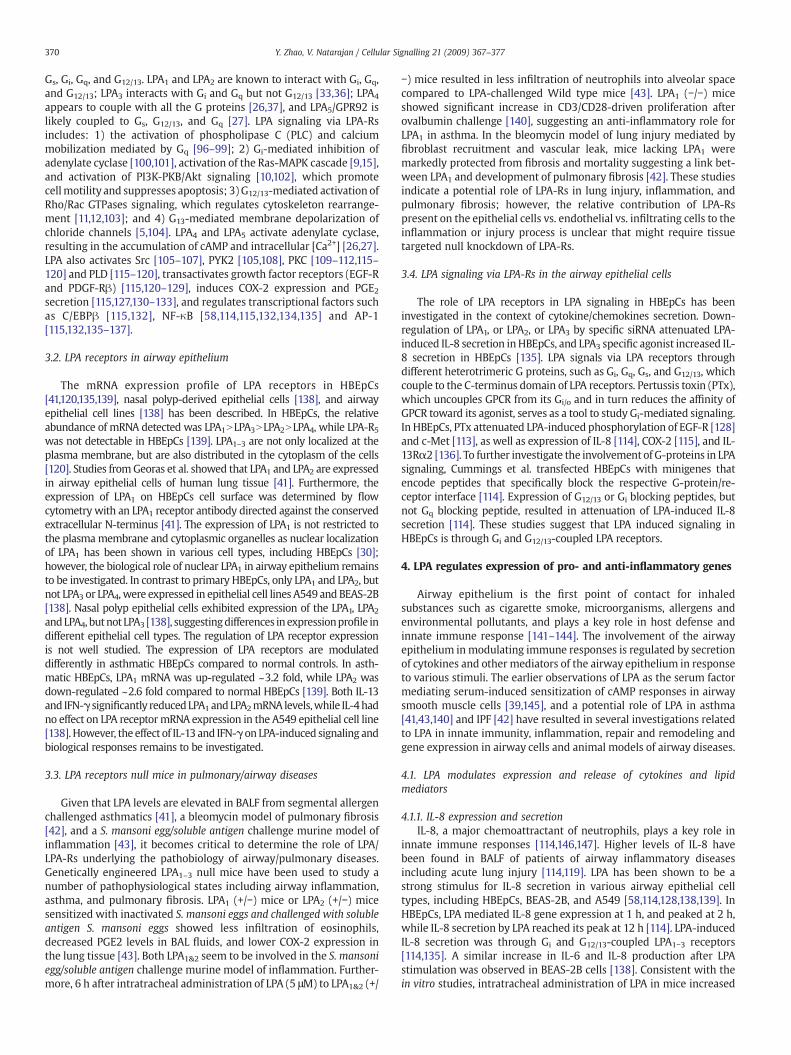
3.2. LPA receptors in airway epithelium
The mRNA expression profile of LPA receptors in HBEpCs[41,120,135,139], nasal polyp-derived epithelial cells [138], and airwayepithelial cell lines [138] has been described. In HBEpCs, the relativeabundance of mRNA detected was LPA1NLPA3NLPA2NLPA4, while LPA-R5was not detectable in HBEpCs [139]. LPA1–3 are not only localized at theplasma membrane, but are also distributed in the cytoplasm of the cells[120]. Studies fromGeoras et al. showed that LPA1 and LPA2 are expressedin airway epithelial cells of human lung tissue [41]. Furthermore, theexpression of LPA1 on HBEpCs cell surface was determined by flowcytometry with an LPA1 receptor antibody directed against the conservedextracellular N-terminus [41]. The expression of LPA1 is not restricted tothe plasmamembrane and cytoplasmic organelles as nuclear localizationof LPA1 has been shown in various cell types, including HBEpCs [30];however, the biological role of nuclear LPA1 in airway epithelium remainsto be investigated. In contrast to primary HBEpCs, only LPA1 and LPA2, butnot LPA3 or LPA4, were expressed in epithelial cell lines A549 and BEAS-2B[138]. Nasal polyp epithelial cells exhibited expression of the LPA1, LPA2
andLPA4, butnot LPA3 [138], suggestingdifferences inexpressionprofile indifferent epithelial cell types. The regulation of LPA receptor expressionis not well studied. The expression of LPA receptors are modulateddifferently in asthmatic HBEpCs compared to normal controls. In asth-matic HBEpCs, LPA1 mRNA was up-regulated ~3.2 fold, while LPA2 wasdown-regulated ~2.6 fold compared to normal HBEpCs [139]. Both IL-13and IFN-γ significantly reducedLPA1 andLPA2mRNA levels,while IL-4hadno effect on LPA receptormRNA expression in the A549 epithelial cell line[138].However, the effect of IL-13 and IFN-γ on LPA-induced signaling andbiological responses remains to be investigated.
3.3. LPA receptors null mice in pulmonary/airway diseases
Given that LPA levels are elevated in BALF from segmental allergenchallenged asthmatics [41], a bleomycin model of pulmonary fibrosis[42], and a S. mansoni egg/soluble antigen challenge murine model ofinflammation [43], it becomes critical to determine the role of LPA/LPA-Rs underlying the pathobiology of airway/pulmonary diseases.Genetically engineered LPA1–3 null mice have been used to study anumber of pathophysiological states including airway inflammation,asthma, and pulmonary fibrosis. LPA1 (+/−) mice or LPA2 (+/−) micesensitized with inactivated S. mansoni eggs and challenged with solubleantigen S. mansoni eggs showed less infiltration of eosinophils,decreased PGE2 levels in BAL fluids, and lower COX-2 expression inthe lung tissue [43]. Both LPA1&2 seem to be involved in the S. mansoniegg/soluble antigen challenge murine model of inflammation. Further-more, 6 h after intratracheal administration of LPA (5 μM) to LPA1&2 (+/
−) mice resulted in less infiltration of neutrophils into alveolar spacecompared to LPA-challenged Wild type mice [43]. LPA1 (−/−) miceshowed significant increase in CD3/CD28-driven proliferation afterovalbumin challenge [140], suggesting an anti-inflammatory role forLPA1 in asthma. In the bleomycin model of lung injury mediated byfibroblast recruitment and vascular leak, mice lacking LPA1 weremarkedly protected from fibrosis and mortality suggesting a link bet-ween LPA1 and development of pulmonary fibrosis [42]. These studiesindicate a potential role of LPA-Rs in lung injury, inflammation, andpulmonary fibrosis; however, the relative contribution of LPA-Rspresent on the epithelial cells vs. endothelial vs. infiltrating cells to theinflammation or injury process is unclear that might require tissuetargeted null knockdown of LPA-Rs.
3.4. LPA signaling via LPA-Rs in the airway epithelial cells
The role of LPA receptors in LPA signaling in HBEpCs has beeninvestigated in the context of cytokine/chemokines secretion. Down-regulation of LPA1, or LPA2, or LPA3 by specific siRNA attenuated LPA-induced IL-8 secretion inHBEpCs, and LPA3 specific agonist increased IL-8 secretion in HBEpCs [135]. LPA signals via LPA receptors throughdifferent heterotrimeric G proteins, such as Gi, Gq, Gs, and G12/13, whichcouple to the C-terminus domain of LPA receptors. Pertussis toxin (PTx),which uncouples GPCR from its Gi/o and in turn reduces the affinity ofGPCR toward its agonist, serves as a tool to study Gi-mediated signaling.In HBEpCs, PTx attenuated LPA-induced phosphorylation of EGF-R [128]and c-Met [113], as well as expression of IL-8 [114], COX-2 [115], and IL-13Rα2 [136]. To further investigate the involvement of G-proteins in LPAsignaling, Cummings et al. transfected HBEpCs with minigenes thatencode peptides that specifically block the respective G-protein/re-ceptor interface [114]. Expression of G12/13 or Gi blocking peptides, butnot Gq blocking peptide, resulted in attenuation of LPA-induced IL-8secretion [114]. These studies suggest that LPA induced signaling inHBEpCs is through Gi and G12/13-coupled LPA receptors.
4. LPA regulates expression of pro- and anti-inflammatory genes
Airway epithelium is the first point of contact for inhaledsubstances such as cigarette smoke, microorganisms, allergens andenvironmental pollutants, and plays a key role in host defense andinnate immune response [141–144]. The involvement of the airwayepithelium in modulating immune responses is regulated by secretionof cytokines and other mediators of the airway epithelium in responseto various stimuli. The earlier observations of LPA as the serum factormediating serum-induced sensitization of cAMP responses in airwaysmooth muscle cells [39,145], and a potential role of LPA in asthma[41,43,140] and IPF [42] have resulted in several investigations relatedto LPA in innate immunity, inflammation, repair and remodeling andgene expression in airway cells and animal models of airway diseases.
4.1. LPA modulates expression and release of cytokines and lipidmediators
4.1.1. IL-8 expression and secretionIL-8, a major chemoattractant of neutrophils, plays a key role in
innate immune responses [114,146,147]. Higher levels of IL-8 havebeen found in BALF of patients of airway inflammatory diseasesincluding acute lung injury [114,119]. LPA has been shown to be astrong stimulus for IL-8 secretion in various airway epithelial celltypes, including HBEpCs, BEAS-2B, and A549 [58,114,128,138,139]. InHBEpCs, LPA mediated IL-8 gene expression at 1 h, and peaked at 2 h,while IL-8 secretion by LPA reached its peak at 12 h [114]. LPA-inducedIL-8 secretion was through Gi and G12/13-coupled LPA1−3 receptors[114,135]. A similar increase in IL-6 and IL-8 production after LPAstimulation was observed in BEAS-2B cells [138]. Consistent with thein vitro studies, intratracheal administration of LPA in mice increased

371Y. Zhao, V. Natarajan / Cellular Signalling 21 (2009) 367–377
MIP-2 (the murine homolog of IL-8) levels at 3 h and neutrophilsinfiltration at 6 h. At later periods (N12 h), the levels of MIP-2 andneutrophils returned to near basal levels [114]. Hashimoto et al. alsoshowed that inhalation induced histamine release and increasednumbers of eosinophils and neutrophils in BAL of guinea pig [148,149].These results suggest that LPA regulates airway inflammation viastimulating cytokine and inflammatory mediator release and neu-trophils or eosinophils infiltration in airway.
4.1.2. IL-13R alpha2 expression and releaseThe levels of interleukin-13 (IL-13), a Th2-type cytokine, are
increased in BALF of asthma patients [150,151] and ovalbuminchallenged mice [152]. IL-13 induces phosphorylation of signaltransducer and activator of transcription 6 (STAT6), which activatesthe transcription ofmany pro-inflammatory genes in epithelial cells andplays a critical role in the pathogenesis of bronchial asthma [153–155].IL-13Rα1 binds to IL-13 and mediates IL-13-induced activation of Januskinases (JAKs) and STAT6 [156,157]. The human IL-13 decoy receptor,termed IL-13 Receptor α2 (IL-13Rα2), binds to IL-13 with much higheraffinity (Kd=0.25–1.2 nM) compared to IL-13Rα1 (Kd=2–10 nM) [158–160], and overexpression of IL-13Rα2 selectively inhibited IL-13-induced response in airway epithelium and murine lung [161–163].Elevated IL-13Rα2 mRNA expression has been detected in Schistosomaegg-induced liverfibrosis [164] and bleomycin-inducedfibrosis [165]. Inaddition, interferon-γ (IFN-γ) or IL-13 treatment induced IL-13Rα2mRNAexpression thatwas localized to the cell surface inU937 [166] andbronchial epithelial cells [167]. While LPA induced IL-13 expressionunder conditions of semimaximal activation in T cells, in HBEpCs [168],LPA treatment induced IL-13Rα2 mRNA and protein expression andsecretion through Gi-coupled LPA receptors, without altering IL-13Rα1expression [136]. Further, elevated IL-13Rα2 expression by LPA inHBEpCs attenuated IL-13-mediated phosphorylation of STAT6 andeotaxin and SOCS-1 gene expression [136]. As LPA levels are elevatedinBALF fromsegmental allergen challenged asthmatics [41] and IPF [42],and LPA modulates expression of IL-13Rα2 and IL-13-mediated STAT6signaling [136], it is postulated that increased LPA levels may have aprotective role in airway inflammation and remodeling of airways.Future studies related to LPA and its effect on production of Th2cytokines, and airway remodeling and repair would increase ourunderstanding of how LPA and its analogs ameliorate airway inflamma-tion in asthma and fibrosis.
4.1.3. COX-2 expression and PGE2 releaseProstaglandin E2 (PGE2) is an autocrine lipidmediator derived from
arachidonic acid (AA) metabolism by cyclooxygenase-1 or 2 (COX-1 or2) [169]. PGE2 plays crucial roles in various biological events such asneuronal function [170], vascular hypertension [171], tumorigenesis[172–174], and inflammation [175]. Upregulation of COX-2 expressionand PGE2 release plays a protective role in the innate immunityresponse and tissue repair process in airway inflammation [176,177].Inhaled PGE2 attenuated allergen-induced airway responses, hyper-responsiveness, and inflammation [178,179]; however, the regulationof COX-2 expression and the physiological effect of PGE2 on airwayepithelium has not beenwell examined. LPA challenge increased COX-2 expression and PGE2 production in HBEpCs [115]. Analyses of totalRNA by Real-time RT-PCR showed that both COX-1 and COX-2 wereexpressed in HBEpCs and that the expression of COX-1 is ~2 foldhigher than COX-2 [115]. Exposure of HBEpCs to LPA (1 μM) inducedCOX-2 gene and protein expression and PGE2, while such exposurehad no effect on COX-1 gene and protein expression up to 24 h [115].Further, PTx attenuated LPA-induced COX-2 mRNA and proteinexpression, suggesting the involvement of Gi-coupled LPA-Rs. COX-2siRNA effectively down-regulated COX-2 protein expression andattenuated LPA-induced PGE2 release by ~50% compared to scrambledsiRNA cells exposed to LPA [115]. These results demonstrated that LPAinduced PGE2 secretion was dependent on expression of COX-2 in
HBEpCs. As PGE2 exhibits anti-inflammatory properties in theairway [176–179], enhanced COX-2 expression and PGE2 releaseinduced by LPA may have a protective role in airway inflammationand remodeling.
4.2. LPA regulates transcriptional factors
4.2.1. Nuclear factor-κB (NF-κB)NF-κB and AP-1 are transcriptional factors expressed in almost all
mammalian cell types and play a key role in regulating the immuneresponse to infection. NF-κB is a heterodimer composed of two Relfamily members (RelA and B). In quiescent cells, NF-κB is present incytosol in an inactive form bound to an inhibitory protein, I-κB, andafter stimulation, I-κB is phosphorylated, and degraded by the 26Sproteasome. NF-κB is released from NF-κB/I-κB complex, translocatesfrom cytosol to nucleus, and activates gene transcription [180]. Theactivation of NF-κB by LPA in HBEpCs has been detected by severalprocedures including phosphorylation of I-κB, NF-κB nuclear translo-cation, NF-κB luciferase reporter assay, and electrophoretic motility-shift assay [58,114,115,135]. Signaling pathways regulating LPA-induced NF-κB activation in HBEpCs have been partially defined. LPAinduced NF-κB activation via p38 MAPK [139] and protein kinase C δ(PKCδ) [114]. The promoter region of human IL-8 and COX-2 havepotential NF-κB binding elements [181,182], however, the human IL-13Rα2 promoter region lacks NF-κB binding elements [183]. Inhibi-tion of NF-κB activation attenuated LPA-induced IL-8 [114] and COX-2[115] expression, suggesting that NF-κB plays a critical role in LPA-induced gene expression in HBEpCs. However, LPA-induced IL-13Rα2expressionwas not dependent on the NF-κB pathway in HBEpCs [136].
4.2.2. C/EBPβThe C/EBP family consists of six proteins that belong to the basic
zipper transcriptional factors [184]. The C/EBPβ is important in theregulation of genes involved in immune and inflammatory responsesand has been shown to bind to transcriptional regulatory regions ofseveral acute-phase and cytokine genes. The human COX-2 promo-ter region has been sequenced and contains a putative transcriptio-nal regulatory element of C/EBPβ [182]. The participation of C/EBPβ inCOX-2 expression to various stimuli in different cell types has beendescribed [185,186]. In HBEpCs, LPA induced phosphorylation of C/EBPβand downregulation of C/EBPβ expression by siRNA attenuated LPA-induced COX-2 expression [115], suggesting the involvement of C/EBPβin LPA-induced gene expression in HBEpCs. Further evidence showedthat LPA-induced activation of PLD2, PKCζ, and EGF-R contributed toC/EBP activation in HBEpCs [115]. The role of other C/EBP familymembers in LPA-mediated cytokine and lipid mediators release is stillnot clear.
4.2.3. JNK and p38 MAPK in LPA-induced NF-κB and AP-1 activationc-Jun N-terminal kinase (JNK) and p38MAPK belong toMAP kinase
family, which are important components of signal transductionpathways induced by growth factors, cytokines, and GPCR ligands.p38 MAPK and JNK1 and JNK2 isoforms are widely distributed inmammalian cells [187]. LPA treatment of HBEpCs induced phosphor-ylation of JNK1/2 [115,135,136] and c-Jun [135], induced c-Jun nuclearlocalization [135], and induced AP-1 activation [135]. The mechanism(s) of regulation of JNK activation by LPA is still not clear. Interestingly,overexpression of catalytic dominant negative PLD1 and 2 isoformsattenuated LPA-induced phosphorylation of JNK in HBEpCs [136],suggesting that JNK activation may be regulated by PLD-generatedphosphatidic acid (PA). Inhibition of JNK activation by JNK inhibitor(JNKiII) or downregulation of c-Jun by c-Jun siRNA attenuated LPA-induced activation of AP-1 [135], as well as IL-8 [135], COX-2 [115], andIL-13Rα2 [136] expression.
LPA-induced activation of NF-κB has been shown to be dependenton activation of p38 MAPK. An inhibitor of p38MAPK or p38 MAPK

Fig. 2. LPA signaling in airway epithelial cells. Ligation of LPA to LPA receptors increases intracellular calcium, activates p38 MAPK, PKCs and PLD, resulting in the activation oftranscriptional factors and induction of cytokine(s) expression and release; thus regulating innate and adaptive immune responses. LPA induces E-cadherin and c-Met redistributionto cell periphery and regulates airway epithelium integrity.
372 Y. Zhao, V. Natarajan / Cellular Signalling 21 (2009) 367–377
siRNA blocked phosphorylation of I-κB and NF-κB translocation to thenucleus without affecting JNK/c-Jun phosphorylation and AP-1activa-tion [135]. The role of LPA-induced activation of p38 MAPK in LPA-mediated IL-8 and COX-2 expression was investigated in HBEpCs.Inhibition of p38 MAPK activation attenuated LPA-induced IL-8 [135]through decreasing the NF-κB pathway, not the JNK/AP-1 pathway.Recent studies suggest that p38 MAPK also regulates COX-2 mRNAstability [188]. In HBEpCs, LPA induced COX-2 mRNA expression aswell as COX-2 mRNA stability [189], while downregulation of p38MAPK activation attenuated LPA-induced COX-2 mRNA stability andexpression [189]. p38 MAPK regulates LPA-induced COX-2 expressionthrough both transcriptional and post-transcriptional regulation.Further study will determine the role of p38 MAPK isoforms in theregulation of COX-2 mRNA levels in HBEpCs.
4.3. LPA activates PKC isoforms
PKC is a superfamily of kinases that phosphorylates protein sub-strates on serine and threonine residues and transduces the cellularsignals. Eleven PKC isoforms have been identified in mammals anddivided into three subgroups: classical PKCs (cPKCα, βI, βII, and γ),novel PKCs (nPKCδ, ε, η, μ and θ), and atypical PKCs (aPKCζ and λ)based on sequence homology. The cPKCs are calcium-dependent, andactivated by diacylglycerol (DAG) and DAG-mimicking phorbol esters.The nPKCs are calcium-independent, activated by DAG and DAG-mimicking phorbol esters. The aPKCs are structurally divergent fromother PKC family members and activated by acidic phospholipids suchas phosphatidylserine (PS), polyphosphoinositides (PI) and phospha-tidic acid (PA), but not by DAG or phorbol esters [190]. Expressions ofPKCα, δ, ι/λ, and ζ in HBEpCs were confirmed by Western blottingwith specific antibodies to these isoforms [114]. LPA induced phos-phorylation and plasma membrane translocation of PKCδ [114] and ζ[189]. The involvements of PKC isoforms in IL-8, IL-13Rα2, and COX-2expression have been studied by using PKC isoform specificpharmaceutical inhibitors or overexpression of dominant negativePKC isoforms (dn-PKCs) in HBEpCs. PKCδ contributes to LPA-inducedIL-8 [114], IL-13Rα2 [136], and COX-2 [189] expression. PKCζ regu-lates LPA-induced COX-2 expression [115] while PKCλ regulates LPA-
induced IL-8 expression [114] in HBEpCs. It has been shown that PKCinhibitors attenuated activation of transcriptional factors in variouscell types [191,192]. Inhibition of PKCδ attenuated LPA-induced NF-κBactivation [114] and inhibition of PKCζ modulated LPA-induced C/EBPβ activation in HBEpCs [115]. Regulation of PKCζ by PLD has beenshown in human pulmonary arterial endothelial cells (HPAECs) [193]while the relationship between PKCζ and PLD will need to be furtherdetermined in human airway epithelial cells (Fig. 2).
4.4. Role of LPPs in regulation of LPA signaling
Recent studies have revealed that LPA degradation occurs throughthe LPPs family, which consists of three family members (LPP-1, LPP-2,LPP-3), attenuated LPA-induced intracellular signaling [82–85]. LPPscontain a novel conserved phosphatase sequence motif, K(X)6RP-(X12–
54)-PSGH-(X31–54)-SR(X)5H(X)3D, that lies on the outside of the cell, oron the luminal surface of the endoplamic reticulum or Golgi [57,85]. Inthe rat, the activity of LPPs in the lung is the highest among any tissueand mRNA expression for LPP-1 and LPP-3, but not LPP-2, was detec-ted in rat lung [194]. LPPs activity is enriched in lipid-rich signalingplatforms isolated from type II cell-mouse lung epithelial cell lines(MLE12 andMLE15) [195,196], and usingWestern blotting and RT-PCR,expressions of LPP-1, -2, and -3 were detected in HBEpCs [58]. InHBEpCs, exogenously added LPA was hydrolyzed by a LPP-like ecto-enzyme activity, whichwas inhibited by tyrosine kinase inhibitors, butnot by serine–threonine inhibitors. Overexpression of LPP-1 decreasedextracellular LPA levels and attenuated LPA-induced intracellular Ca2+,ERK1/2 phosphorylation, activation of NF-κB pathway, secretion of IL-8, and expression of COX-2 [58,189]. Downregulation of LPP-1 by LPP-1siRNA enhanced LPA-induced COX-2 expression in HBEpCs [189].These results suggest that LPP-1 regulated LPA-induced gene expres-sion via degradation of extracellular LPA. Interestingly, overexpressionof LPP-1 also attenuated the IL-8 secretion mediated by a non-hydrolyzed LPA analogue, suggesting an intracellular role of LPP-1 inregulation of LPA-induced signaling [58]. As LPA plays a critical role ininnate immune response in airway diseases, and LPP-1 attenuates LPAfunction by its ecto-enzyme activity, targeting LPP-1 may regulateairway inflammation.

Fig. 4. Cross-talk between LPA receptors and RTKs. LPA via its receptors induces tyrosinephosphorylation of EGF-R and PDGF-Rβ (termed as transactivation), and regulates cellproliferation and gene expression. Cross-talk between LPA receptors and c-Met inducesserine phosphorylation of c-Met (termed as transinactivation), which enhances airwayepithelial barrier function.
373Y. Zhao, V. Natarajan / Cellular Signalling 21 (2009) 367–377
5. Cross-talk between LPA-Rs and receptor tyrosine kinases (RTKs)
Several lines of investigation in a variety of cell types have shownthat many of the growth promoting effects of GPCR agonists (LPA,sphingosine-1-phosphate (S1P), angiotensin, PGE2, and others) aremediated through activation of receptor tyrosine kinases (RTKs), aphenomenon termed as “Transactivation” [197–199]. The transactiva-tionofRTKsbyGPCR ligandshas beendemonstrated invarious cell typesand this cross-talk between GPCRs and RTKs provides additional non-canonical signaling pathways, which regulate cellular function. Inaddition, disruption of GPCR function via PTx or antagonist reducesthe growth factor stimulated activation of ERK [200]. Both the GPCR(S1P1) and the βγ subunit of the activated G-protein form a functionalsignaling complex with the RTK, which may reprogram the mitogenicsignal of the RTK to amigratory response [200]. Thus, potential signalingvia “Transactivation” between GPCRs and RTKs will allow spatialregulation of effectors dictating nature of the biological responses.
5.1. LPA transactivates PDGF-Rβ
Among the various RTKs, GPCR mediated transactivation of platelet-derived growth factor receptor (PDGF-R) has been well described[198,200–202]. The PDGF has been recognized as an important growthfactor regulating cell proliferation and the development of many diseasesincluding cancer [203–205]. PDGF-BB challenge stimulated phosphoryla-tion of Erk1/2, suggesting that PDGF plays a proliferative role in HBEpCs[120]. In addition to PDGF-BB, LPA also induced tyrosine phosphorylationof PDGF-Rβvia a transactivationmechanism involvingPLD2, but not PLD1,signaling [120]. Furthermore, transactivation of PDGF-Rβby LPA regulatedLPA-dependent phosphorylation of Erk1/2 [120] in HBEpCs. These resultssuggest that cross-talk between LPA-Rs and PDGF-Rβ may play a role inairway remodeling and repair. While the LPA1-PDGF-Rβ signalingcomplex could not be identified in HBEpCs [212], interaction betweenG-protein-coupled S1P1 and PDGF-Rβ has been identified and extensivelystudied inHEK293andairwaysmoothmuscle cells [201]. The formationofthis signaling complex between S1P1 and PDGF-Rβ permits the usage ofβγ-subunits of activated Gi by PDGF-R to enhance PDGF signaling [206].The functional implications of the “Transactivation” between RTKs andGPCRs in health and disease warrant more investigation.
5.2. LPA transactivates EGF-R
EGF-R, similar to other growth factor receptors, is activated by EGF,heparin binding-EGF (HB-EGF) and tumor growth factor-α (TGF-α) [207].Since the first report of transactivation of EGF-R by LPA infibroblast [208],recent studies have established EGF-R transactivation by LPA in airway
Fig. 3. Signaling pathways of LPA-induced transactivation of EGF-R. Ligation of LPA to itsG-protein-coupled receptors induces phosphorylation of EGF-R through PKCδ, Lynkinase, MMP, and pro-HB-EGF pathway. PLD2 and PKCζ are involved in LPA-inducedtransactivation of EGF-R; however, the relationship between PLD2 and PKCδ or betweenPKCζ and Lyn kinase is unclear. Erk1/2 and C/EBPβ are downstream targets of EGF-Rtransactivation, which regulate cell proliferation and gene expression.
epithelial cells [123,124,128,209,210]. Two pathways of LPA-mediatedtransactivation of EGF-R have been described: the first involves activationof matrix metalloproteinases (MMPs), cleavage of pro-HB-EGF, and sub-sequent ligation andactivationof EGF-R [125,128,209,211], and the secondpathway is HB-EGF shedding-independent involving the intracellularactivation of EGF-R by Src kinases [209,210]. In HBEpCs, LPA treatmentinduced tyrosine phosphorylation of EGF-R at different tyrosine residues,whichwereGi-dependent [128,212]. Furthermore, theLPA-inducedEGF-Rtransactivation was regulated by PKCζ [115] and δ and Src kinases [128].Among Src kinase family, using specific siRNA to down-regulate Srckinases, a role for Lyn kinase, but not Src or Yes kinase, in regulating LPA-induced EGF-R transactivation was established [128]. Similar to the me-chanismof PDGF-Rβ transactivation by LPA, PLD2, but not PLD1, regulatedthe LPA-induced phosphorylation of EGF-R in HBEpCs [115]. Interestingly,LPA-mediated activation of Lyn was dependent on PKCδ signal transduc-tion, with subsequent activation of MMP-2/9, and proHB-EGF shedding[128]. LPA induced PKCζ activation through PLD2 [115]. It is not clear ifPKCδ is up-stream or down-stream of PLD2 in HBEpCs. Data on thephysiological significance of EGF-R transactivation by LPA is limited.Inhibition of EGF-R tyrosine kinase with AG1478 attenuated LPA-inducedphosphorylation of ERK 1/2 in PC12 [213], ovarian cancer cells [214],prostate cancer cells [50], and HBEpCs [212], suggesting a potential role incell proliferation. Furthermore, He et al. demonstrated that though all NF-κB, AP-1, and C/EBPβ are involved in LPA-induced COX-2 expression andPGE2 release, however, only C/EBPβ is downstream signal molecule toEGF-R transactivation in HBEpCs [115] (Fig. 3). At present, it is unclearwhich of the LPA-Rs are involved in LPA-induced EGF-R transactivation.Another consequence of LPA-induced transactivation of EGF-R is a rapidand sustained decrease in EGF-R binding in human primary airwayepithelial cells andBEAS-2Bcell line [123,124]. The rapiddecrease inEGF-Rbinding via EGF-R transactivation mediated by LPA was dependent onMEK/ERK and PKC, whereas the sustained decrease was primarilyregulated by the PKC signaling pathway in BEAS-2B cell line [123]. Furtherstudies are necessary to delineate physiological significance(s) of EGF-Rtransactivation in airway inflammation, repair and remodeling.
5.3. LPA transinactivates c-Met
c-Met, a proto-oncogene product, is a RTK, the receptor for hepatocytegrowth factor (HGF), and predominantly expressed in various types ofepithelium [215–216]. Increased expression and activation of c-Met is

374 Y. Zhao, V. Natarajan / Cellular Signalling 21 (2009) 367–377
associated with tumor cell growth, scattering, invasion and metastasis[216–217]. Specific mutations of the tyrosine residues in the juxtamem-brane and semaphorin domains of c-Met resulted in the regulation of thecytoskeleton through the focal adhesion protein paxillin [218]. LPA or S1Pmediation enhanced tyrosine phosphorylation of c-Met via a transactiva-tion pathway in human colon cancer cell lines and human gastric cancercells [127,219]; however, in HBEpCs, unlike LPA-mediated tyrosinephosphorylationof PDGF-RβandEGF-R [115,120,128], LPA-induced serine,but not tyrosine, phosphorylation, which was dependent on activation ofPKCδ by LPA [113]. Further, serine phosphorylation of c-Met by LPAattenuated HGF-mediated activation of AKT and cell motility in HBEpCs,suggesting a regulatory role for c-Met serine phosphorylation in epithelialcell motility [113]. These results suggest that in the same type of cells, thesame GPCR ligand treatment might induce “yin and yang” responses ondifferent RTKs (Fig. 4). Recent study has demonstrated that the HGF-dependent tyrosine phosphorylation of c-Met was largely suppressed byPKCδ with a reciprocal relationship to Ser-985 phosphorylation in A549cell line [220]. In HBEpCs, LPA induced c-Met relocalization from thecytoplasm to cell–cell contacts and this was dependent on activation ofPKCδ and ζ and interactionwith E-cadherin, suggesting a role of c-Met incell adhesion and motility [113]. The role of c-Met in regulation of airwayepithelium barrier function and cytokine release will be investigated.
6. LPA regulates epithelial barrier via adherens junction proteins
The epithelial cell–cell junctional complex is composed of tightjunctions, adherens junctions and desmosomes [221,222]. Theseadherens junctions play a pivotal role in regulating the activity ofthe entire junctional complex since the formation of adherensjunctions subsequently leads to the formation of other cell–celljunctions, including tight junction [221–223]. The major adhesionmolecules in the adherens junctions are the cadherins [221–223]. E-cadherin is a member of the cadherin family that mediates calcium-dependent cell–cell adhesion [221–225]. The N-terminal ectodomainof E-cadherin contains homophilic-binding domain, and the cyto-plasmic domain binds to catenins, which interact with actin [221–225]. The regulated expression and the plasmamembrane localizationof E-cadherin are critical for the maintenance of epithelial cell–celljunctions crucial to the functional integrity of the epithelial barrier[221–223]. The decrease of adhesive properties of E-cadherin has beenshown to be related to the loss of differentiation and the subsequentacquisition of a higher motility and invasiveness of epithelial cells[226–228]. Dislocation or shedding of E-cadherin in the airway epi-thelium induces epithelial shedding and increases airway perme-ability in airway diseases [229–231]. However, the regulation andmechanism(s) of E-cadherin localization within the epithelium is notfully known, especially during pathological situations of inflamma-tion and airway remodeling. LPA treatment tightened the airwayepithelial cell barrier [41,232]; however, the mechanism(s) regulatingLPA-induced epithelial barrier function is yet to be fully defined.In HBEpCs, exposure to LPA enhanced accumulation of E-cadherinand c-Met complex at cell–cell contacts through activation of PKCδ,and E-cadherin relocalization to cell–cell contacts was critical for LPA-induced enhanced airway epithelial barrier function [232] (Fig. 2). Therole of LPA in regulation of airway barrier function in airway inflam-matory disease, such as acute lung injury, needs further investigation.
7. Conclusions and future directions
In this review,we summarized themechanisms of LPA in regulatinggene expression and barrier function in human airway epithelial cells.The study of the biological role of LPA in airway inflammatory diseasesis an area yet to be well defined. The recent studies reveal that LPAregulates biological responses in airway epithelial, smooth muscle,fibroblast, and lymphocyte cells. Our focus is on the role of LPA inregulation of airway epithelial cytokine production, lipid mediator
release and airway epithelial barrier function since airway epitheliumis the first site that responds to the increase of LPA levels in BALF ofasthmatic and IPF patents. LPA induces gene expression through LPA-Rs mediated intracellular signaling, such as changes of [Ca2+]i,activation of PKC isoforms, PLD, and transcriptional factors (NF-κB,AP-1, and C/EBPβ). Cross-talk between LPA-Rs and RTKs (EGF-R, PDGF-Rβ, and c-Met) regulates LPA-induced gene expression, cell prolifera-tion, and airway epithelial barrier function. LPA upregulates Th1 typecytokine (such as IL-8) and PGE2 release and attenuates the effects ofTh2 type cytokine (IL-13) by releasing of an IL-13 decoy receptor, IL-13Rα2, in airway epithelial cells. This indicates that LPA in the airwaymay play a protective role in inflammatory diseases. Furthermore, LPAincreases airway epithelial integrity through PKC isoforms-mediatedE-cadherin and c-Met protein complex assembly at cell–cell contacts,suggesting that LPA protects airway by regulation of cytokine releasesand airway epithelial integrity (Fig. 2). While a majority of LPAreceptors are coupled to heterotrimeric G protein, recently G-protein-independent signaling through GPCRs has been demonstrated [233].Whether LPA induces part of intracellular signaling in the absence of G-proteins in airway epithelial cells will be further investigated. Fur-thermore, intravenous injection with LPA attenuated bacterial endo-toxin-induced plasma TNFα production andmyeloperioxidase activityin lung, suggesting an anti-inflammatory role of LPA in amurinemodelof acute lung injury [234]. To further understand the role of LPA fromboth plasma and BALF in airway inflammatory diseases, the lysoPLDtransgenic mice and lung epithelial cells specific lysoPLD transgenicmice will serve as useful models. As there are six receptors for LPA,development of specific antagonists for each of the receptor subtypewill allow therapeutic targeting of LPA-Rs that regulate inflammationand remodeling in airway diseases.
Acknowledgements
This work was supported in part by grants from the NationalInstitutes of Health HL79396 (to V.N.) and American Cancer Associa-tion Institutional Grant IRG-58-044-47 (to Y.Z.).
References
[1] G. Tigyi, R. Miledi, J. Biol. Chem. 267 (30) (1992) 21360.[2] T. Eichholtz, K. Jalink, I. Fahrenfort, W.H. Moolenaar, Biochem. J. 291 (Pt 3) (1993)
677.[3] E.J. Goetzl, H. Lee, T. Azuma, T.P. Stossel, C.W. Turck, J.S. Karliner, J. Biol. Chem. 275
(19) (2000) 14573.[4] S. Hosogaya, Y. Yatomi, K. Nakamura, R. Ohkawa, S. Okubo, H. Yokota, M. Ohta, H.
Yamazaki, T. Koike, Y. Ozaki, Ann. Clin. Biochem. 45 (Pt 4) (2008) 364.[5] F.R. Postma, K. Jalink, T. Hengeveld, A.G. Bot, J. Alblas, H.R. de Jonge, W.H.
Moolenaar, Embo J. 15 (1) (1996) 63.[6] A. Tokumura, Y. Kanaya, M. Kitahara, M. Miyake, Y. Yoshioka, K. Fukuzawa, J. Lipid.
Res. 43 (2) (2002) 307.[7] J. Aoki, A. Taira, Y. Takanezawa, Y. Kishi, K. Hama, T. Kishimoto, K. Mizuno, K. Saku,
R. Taguchi, H. Arai, J. Biol. Chem. 277 (50) (2002) 48737.[8] E.J. van Corven, A. Groenink, K. Jalink, T. Eichholtz, W.H. Moolenaar, Cell 59 (1)
(1989) 45.[9] L.T. Budnik, B. Brunswig-Spickenheier, A.K. Mukhopadhyay, Mol. Endocrinol. 17
(8) (2003) 1593.[10] A. Yart, H. Chap, P. Raynal, Biochim. Biophys. Acta 1582 (1–3) (2002) 107–111.[11] U. Schmitz, K. Thommes, I. Beier, H. Vetter, Biochem. Biophys. Res. Commun. 291
(3) (2002) 687.[12] F.N. van Leeuwen, B.N. Giepmans, L.A. van Meeteren, W.H. Moolenaar, Biochem.
Soc. Trans. 31 (Pt 6) (2003) 1209.[13] W.H. Moolenaar, L.A. van Meeteren, B.N. Giepmans, Bioessays 26 (8) (2004) 870.[14] G.B. Mills, W.H. Moolenaar, Nat. Rev. Cancer. 3 (8) (2003) 582.[15] M. Stahle, C. Veit, U. Bachfischer, K. Schierling, B. Skripczynski, A. Hall, P.
Gierschik, K. Giehl, J. Cell Sci. 116 (Pt 18) (2003) 3835.[16] T.J. Manning Jr., J.C. Parker, H. Sontheimer, Cell Motil. Cytoskelet. 45 (3) (2000) 185.[17] C. Avraamides, M.E. Bromberg, J.P. Gaughan, S.M. Thomas, A.Y. Tsygankov, T.S.
Panetti, Am. J. Physiol. Heart. Circ. Physiol. 293 (1) (2007) H193.[18] F. Hao, M. Tan, X. Xu, J. Han, D.D. Miller, G. Tigyi, M.Z. Cui, Biochim. Biophys. Acta
1771 (7) (2007) 883.[19] W. Deng, L. Balazs, D.A. Wang, L. Van Middlesworth, G. Tigyi, L.R. Johnson,
Gastroenterology 123 (1) (2002) 206.[20] X. Ye, I. Ishii, M.A. Kingsbury, J. Chun, Biochim. Biophys. Acta 1585 (2–3) (2002)
108.

375Y. Zhao, V. Natarajan / Cellular Signalling 21 (2009) 367–377
[21] R. Rusovici, A. Ghaleb, H. Shim, V.W. Yang, C.C. Yun, Biochim. Biophys. Acta 1770(8) (2007) 1194.
[22] N. Fukushima, Y. Kimura, J. Chun, Proc. Natl. Acad. Sci. U. S. A. 95 (11) (1998) 6151.[23] J.H. Hecht, J.A. Weiner, S.R. Post, J. Chun, J. Cell Biol. 135 (4) (1996) 1071.[24] S. An, M.A. Dickens, T. Bleu, O.G. Hallmark, E.J. Goetzl, Biochem. Biophys. Res.
Commun. 231 (3) (1997) 619.[25] J. Chun, J.J. Contos, D. Munroe, Cell Biochem. Biophys. 30 (2) (1999) 213.[26] K. Noguchi, S. Ishii, T. Shimizu, J. Biol. Chem. 278 (28) (2003) 25600.[27] C.W. Lee, R. Rivera, S. Gardell, A.E. Dubin, J. Chun, J. Biol. Chem. 281 (33) (2006)
23589.[28] K. Tabata, K. Baba, A. Shiraishi, M. Ito, N. Fujita, Biochem. Biophys. Res. Commun.
363 (3) (2007) 861.[29] D.S. Im, C.E. Heise, M.A. Harding, S.R. George, B.F. O'Dowd, D. Theodorescu, K.R.
Lynch, Mol. Pharmacol. 57 (4) (2000) 753.[30] C.M. Waters, B. Saatian, N.A. Moughal, Y. Zhao, G. Tigyi, V. Natarajan, S. Pyne, N.J.
Pyne, Biochem. J. 398 (1) (2006) 55.[31] T.M. McIntyre, A.V. Pontsler, A.R. Silva, A. St Hilaire, Y. Xu, J.C. Hinshaw, G.A.
Zimmerman, K. Hama, J. Aoki, H. Arai, G.D. Prestwich, Proc. Natl. Acad. Sci. U. S. A.100 (1) (2003) 131.
[32] C. Zhang, D.L. Baker, S. Yasuda, N. Makarova, L. Balazs, L.R. Johnson, G.K. Marathe,T.M. McIntyre, Y. Xu, G.D. Prestwich, H.S. Byun, R. Bittman, G. Tigyi, J. Exp. Med.199 (6) (2004) 763.
[33] J.J. Contos, I. Ishii, J. Chun, Mol. Pharmacol. 58 (6) (2000) 1188.[34] N. Fukushima, J. Chun, Prostaglandins Other Lipid Mediat. 64 (1–4) (2001) 21.[35] M.H. Graler, E.J. Goetzl, Biochim. Biophys. Acta 1582 (1–3) (2002) 168.[36] Y. Takuwa, N. Takuwa, N. Sugimoto, J. Biochem. 131 (6) (2002) 767.[37] K. Yanagida, S. Ishii, F. Hamano, K. Noguchi, T. Shimizu, J. Biol. Chem. 282 (8)
(2007) 5814.[38] S. An, T. Bleu, O.G. Hallmark, E.J. Goetzl, J. Biol. Chem. 273 (14) (1998) 7906.[39] M.L. Toews, T.L. Ediger, D.J. Romberger, S.I. Rennard, Biochim. Biophys. Acta 1582
(1–3) (2002) 240.[40] T. Sugiura, S. Nakane, S. Kishimoto, K. Waku, Y. Yoshioka, A. Tokumura, J. Lipid.
Res. 43 (12) (2002) 2049.[41] S.N. Georas, E. Berdyshev, W. Hubbard, I.A. Gorshkova, P.V. Usatyuk, B. Saatian, A.
C. Myers, M.A. Williams, H.Q. Xiao, M. Liu, V. Natarajan, Clin. Exp. Allergy 37 (3)(2007) 311.
[42] A.M. Tager, P. LaCamera, B.S. Shea, G.S. Campanella, M. Selman, Z. Zhao, V.Polosukhin, J. Wain, B.A. Karimi-Shah, N.D. Kim, W.K. Hart, A. Pardo, T.S.Blackwell, Y. Xu, J. Chun, A.D. Luster, Nat. Med. 14 (1) (2008) 45.
[43] Y. Zhao, J. Tong, I.A. Gorshkova, D. He, R. Stern, E.V. Berdyshev, S. Pendyala, A.I.Sperling, J. Chun, V. Natarajan, Am. J. Respir. Crit. Care Med. 175 (2007) A926.
[44] A. Tokumura, M. Miyake, Y. Nishioka, S. Yamano, T. Aono, K. Fukuzawa, Biol.Reprod. 61 (1) (1999) 195.
[45] A.M. Westermann, E. Havik, F.R. Postma, J.H. Beijnen, O. Dalesio, W.H. Moolenaar,S. Rodenhuis, Ann. Oncol. 9 (4) (1998) 437.
[46] W. Siess, K.J. Zangl, M. Essler, M. Bauer, R. Brandl, C. Corrinth, R. Bittman, G. Tigyi,M. Aepfelbacher, Proc. Natl. Acad. Sci. U. S. A. 96 (12) (1999) 6931.
[47] A. Vancura, D. Haldar, J. Biol. Chem. 269 (44) (1994) 27209.[48] L.E. Hammond, P.A. Gallagher, S. Wang, S. Hiller, K.D. Kluckman, E.L. Posey-
Marcos, N. Maeda, R.A. Coleman, Mol. Cell Biol. 22 (23) (2002) 8204.[49] S. Spiegel, S. Milstien, Biochem. Soc. Trans. 33 (Pt 6) (2005) 1362.[50] M. Bektas, S.G. Payne, H. Liu, S. Goparaju, S. Milstien, S. Spiegel, J. Cell Biol. 169 (5)
(2005) 801.[51] C. Luquain, A. Singh, L. Wang, V. Natarajan, A.J. Morris, J. Lipid. Res. 44 (10) (2003)
1963.[52] Y. Xie, K.E. Meier, Cell Signal. 16 (9) (2004) 975.[53] T. Sano, D. Baker, T. Virag, A. Wada, Y. Yatomi, T. Kobayashi, Y. Igarashi, G. Tigyi,
J. Biol. Chem. 277 (24) (2002) 21197.[54] J. Aoki, Semin. Cell Dev. Biol. 15 (5) (2004) 477.[55] T. Hiramatsu, H. Sonoda, Y. Takanezawa, R. Morikawa, M. Ishida, K. Kasahara, Y.
Sanai, R. Taguchi, J. Aoki, H. Arai, J. Biol. Chem. 278 (49) (2003) 49438.[56] C. Pilquil, I. Singh, Q.X. Zhang, Z.C. Ling, K. Buri, L.M. Stromberg, J. Dewald, D.N.
Brindley, Prostaglandins Other Lipid Mediat. 64 (1–4) (2001) 83.[57] D.N. Brindley, D. English, C. Pilquil, K. Buri, Z.C. Ling, Biochim. Biophys. Acta 1582
(1–3) (2002) 33.[58] Y. Zhao, P.V. Usatyuk, R. Cummings, B. Saatian, D. He, T. Watkins, A. Morris, E.W.
Spannhake, D.N. Brindley, V. Natarajan, Biochem. J. 385 (Pt 2) (2005) 493.[59] D.W. Waggoner, L.B. Johnson, P.C. Mann, V. Morris, J. Guastella, S.M. Bajjalieh,
J. Biol. Chem. 279 (37) (2004) 38228.[60] S. Kalari, Y. Zhao, E. Berdyshev, P.V. Usatyuk, D. He, V. Natarajan, J. Invest.
Medicine 55 (2) (2007) S356.[61] T. Sato, J. Aoki, Y. Nagai, N. Dohmae, K. Takio, T. Doi, H. Arai, K. Inoue, J. Biol. Chem.
272 (4) (1997) 2192.[62] K. Horigome, M. Hayakawa, K. Inoue, S. Nojima, J. Biochem. 101 (3) (1987) 625.[63] G. Sekas, G.M. Patton, E.C. Lincoln, S.J. Robins, J. Lab. Clin. Med. 105 (2) (1985) 190.[64] Y. Taniyama, S. Shibata, S. Kita, K. Horikoshi, H. Fuse, H. Shirafuji, Y. Sumino, M.
Fujino, Biochem. Biophys. Res. Commun. 257 (1) (1999) 50.[65] A. Tokumura, Biochim. Biophys. Acta 1582 (1–3) (2002) 18.[66] Y. Fujiwara, A. Sebok, S. Meakin, T. Kobayashi, K. Murakami-Murofushi, G. Tigyi,
J. Neurochem. 87 (5) (2003) 1272.[67] Y. Fujiwara, Biochim. Biophys. Acta 1781 (9) (2008) 519.[68] E. Gendaszewska-Darmach, Acta. Biochim. Pol. 55 (2) (2008) 227.[69] S. Tsuda, S. Okudaira, K. Moriya-Ito, C. Shimamoto, M. Tanaka, J. Aoki, H. Arai, K.
Murakami-Murofushi, T. Kobayashi, J. Biol. Chem. 281 (36) (2006) 26081.[70] T. Kobayashi, R. Tanaka-Ishii, R. Taguchi, H. Ikezawa, K. Murakami-Murofushi, Life
Sci. 65 (21) (1999) 2185.
[71] L. Shan, S. Li, K. Jaffe, L. Davis, J. Chromatogr. B. Analyt. Technol. Biomed. Life Sci.862 (1–2) (2008) 161.
[72] K. Murakami-Murofushi, M. Mukai, S. Kobayashi, T. Kobayashi, G. Tigyi, H.Murofushi, Ann. N. Y. Acad. Sci. 905 (2000) 319.
[73] M.L. Stracke, H.C. Krutzsch, E.J. Unsworth, A. Arestad, V. Cioce, E. Schiffmann, L.A.Liotta, J. Biol. Chem. 267 (4) (1992) 2524.
[74] T. Clair, H.Y. Lee, L.A. Liotta, M.L. Stracke, J. Biol. Chem. 272 (2) (1997) 996.[75] Y. Yang, L. Mou, N. Liu, M.S. Tsao, Am. J. Respir. Cell Mol. Biol. 21 (2) (1999) 216.[76] M. Tanaka, S. Okudaira, Y. Kishi, R. Ohkawa, S. Iseki, M. Ota, S. Noji, Y. Yatomi, J.
Aoki, H. Arai, J. Biol. Chem. 281 (35) (2006) 25822.[77] G. Ferry, A. Giganti, F. Coge, F. Bertaux, K. Thiam, J.A. Boutin, FEBS Lett. 581 (18)
(2007) 3572.[78] A.C. Sane, T. Mendenhall, D.A. Bass, J. Leukoc. Biol. 60 (6) (1996) 704.[79] D.L. Bowton, M.C. Seeds, M.B. Fasano, B. Goldsmith, D.A. Bass, Am. J. Respir. Crit.
Care Med. 155 (2) (1997) 421.[80] F.H. Chilton, F.J. Averill, W.C. Hubbard, A.N. Fonteh, M. Triggiani, M.C. Liu, J. Exp.
Med. 183 (5) (1996) 2235.[81] M. Triggiani, A. Oriente, M.C. Seeds, D.A. Bass, G. Marone, F.H. Chilton, J. Exp. Med.
182 (5) (1995) 1181.[82] M. Kai, I. Wada, S. Imai, F. Sakane, H. Kanoh, J. Biol. Chem. 272 (39) (1997) 24572.[83] D.W. Leung, C.K. Tompkins, T. White, DNA Cell Biol. 17 (4) (1998) 377.[84] R. Roberts, V.A. Sciorra, A.J. Morris, J. Biol. Chem. 273 (34) (1998) 22059.[85] D.N. Brindley, J. Cell Biochem. 92 (5) (2004) 900.[86] J. West, C.K. Tompkins, N. Balantac, E. Nudelman, B. Meengs, T.White, S. Bursten, J.
Coleman, A. Kumar, J.W. Singer, D.W. Leung, DNA Cell Biol. 16 (6) (1997) 691.[87] C. Eberhardt, P.W. Gray, L.W. Tjoelker, Adv. Exp. Med. Biol. 469 (1999) 351.[88] D.W. Leung, Front. Biosci. 6 (2001) D944.[89] D. Li, L. Yu, H. Wu, Y. Shan, J. Guo, Y. Dang, Y. Wei, S. Zhao, J. Hum. Genet. 48 (8)
(2003) 438.[90] W. Tang, J. Yuan, X. Chen, X. Gu, K. Luo, J. Li, B.Wan, Y.Wang, L. Yu, J. Biochem.Mol.
Biol. 39 (5) (2006) 626.[91] F.J. Thompson, M.A. Clark, Biochem. J. 300 (Pt 2) (1994) 457.[92] A. Wang, E.A. Dennis, Biochim. Biophys. Acta 1439 (1) (1999) 1.[93] A. Wang, H.C. Yang, P. Friedman, C.A. Johnson, E.A. Dennis, Biochim. Biophys. Acta
1437 (2) (1999) 157.[94] R.R. Baker, H.Y. Chang, Biochim. Biophys. Acta 1438 (2) (1999) 253.[95] J. Chun, Crit. Rev. Neurobiol. 13 (2) (1999) 151.[96] H. Ohata, H. Aizawa, K. Momose, Life Sci. 60 (15) (1997) 1287.[97] F. Okajima, H. Tomura, K. Sho, T. Kimura, K. Sato, D.S. Im, M. Akbar, Y. Kondo,
Endocrinology 138 (1) (1997) 220.[98] W.L. Zhou, M. Sugioka, M. Yamashita, J. Neurobiol. 41 (4) (1999) 495.[99] Y.J. Xu, P.S. Tappia, R.K. Goyal, N.S. Dhalla, J. Cell Mol. Med. 12 (3) (2008) 942.[100] F. Pietruck, S. Busch, S. Virchow, N. Brockmeyer, W. Siffert, Naunyn. Schmiede-
bergs. Arch. Pharmacol. 355 (1) (1997) 1.[101] W.B. Thoreson, J.S. Ryan, C. Shi, M.E. Kelly, E.J. Bryson, M.L. Toews, T.L. Ediger, D.M.
Chacko, Invest. Ophthalmol. Vis. Sci. 43 (7) (2002) 2450.[102] C. Peres, A. Yart, B. Perret, J.P. Salles, P. Raynal, FEBS Lett. 534 (1–3) (2003) 164.[103] A. Hahn, H. Barth, M. Kress, P.R. Mertens, M. Goppelt-Struebe, Biochem. J. 362 (Pt
1) (2002) 33.[104] J. Wang, L.D. Carbone, M.A. Watsky, Invest. Ophthalmol. Vis. Sci. 43 (10) (2002)
3202.[105] I. Dikic, G. Tokiwa, S. Lev, S.A. Courtneidge, J. Schlessinger, Nature 383 (6600)
(1996) 547.[106] Q. He, M.C. LaPointe, Hypertension 37 (2 Part 2) (2001) 478.[107] H. Takeda, T.Matozaki, Y. Fujioka, T. Takada, T. Noguchi, T. Yamao,M. Tsuda, F. Ochi, K.
Fukunaga, S. Narumiya, T. Yamamoto, M. Kasuga, Oncogene 16 (23) (1998) 3019.[108] S.S. Wu, T. Chiu, E. Rozengurt, Am. J. Physiol. Cell Physiol. 282 (6) (2002) C1432.[109] B.V. Bassa, D.D. Roh, N.D. Vaziri, M.A. Kirschenbaum, V.S. Kamanna, Am. J. Physiol.
277 (3 Pt 2) (1999) F328.[110] W.W. Lin, S.H. Chang, S.M. Wang, Br. J. Pharmacol. 128 (6) (1999) 1189.[111] J.H. Rhim, I.S. Jang, E.J. Yeo, K.Y. Song, S.C. Park, Aging Cell. 5 (6) (2006) 451.[112] S. Seewald, U. Schmitz, C. Seul, Y. Ko, A. Sachinidis, H. Vetter, Am. J. Hypertens. 12
(5) (1999) 532.[113] Y. Zhao, D. He, R. Stern, P.V. Usatyuk, E.W. Spannhake, R. Salgia, V. Natarajan, Cell
Signal. 19 (11) (2007) 2329.[114] R. Cummings, Y. Zhao, D. Jacoby, E.W. Spannhake, M. Ohba, J.G. Garcia, T. Watkins,
D. He, B. Saatian, V. Natarajan, J. Biol. Chem. 279 (39) (2004) 41085.[115] D. He, V. Natarajan, R. Stern, I.A. Gorshkova, J. Solway, E.W. Spannhake, Y. Zhao,
Biochem. J. 412 (1) (2008) 153.[116] J.H. Hong, S.O. Oh, M. Lee, Y.R. Kim, D.U. Kim, G.M. Hur, J.H. Lee, K. Lim, B.D.
Hwang, S.K. Park, Biochem. Biophys. Res. Commun. 281 (5) (2001) 1337.[117] C. Qi, J.H. Park, T.C. Gibbs, D.W. Shirley, C.D. Bradshaw, K.M. Ella, K.E. Meier, J. Cell
Physiol. 174 (2) (1998) 261.[118] I. Shin, S.M. Kweon, Z.W. Lee, S.I. Kim, C.O. Joe, J.H. Kim, Y.M. Park, K.S. Ha, Mol.
Cells 9 (3) (1999) 292.[119] J.S. Tou, J.S. Gill, Cell Signal. 17 (1) (2005) 77.[120] L. Wang, R. Cummings, Y. Zhao, A. Kazlauskas, J.K. Sham, A. Morris, S. Georas, D.N.
Brindley, V. Natarajan, J. Biol. Chem. 278 (41) (2003) 39931.[121] A. Gohla, R. Harhammer, G. Schultz, J. Biol. Chem. 273 (8) (1998) 4653.[122] A. Gschwind, N. Prenzel, A. Ullrich, Cancer Res. 62 (21) (2002) 6329.[123] K.M. Kassel, P.R. Dodmane, N.A. Schulte, M.L. Toews, J. Pharmacol. Exp. Ther. 325
(3) (2008) 809.[124] K.M. Kassel, N.A. Schulte, S.M. Parker, A.D. Lanik, M.L. Toews, J. Pharmacol. Exp.
Ther. 323 (1) (2007) 109.[125] K. Mori, J. Kitayama, D. Shida, H. Yamashita, T. Watanabe, H. Nagawa, J. Surg. Res.
132 (1) (2006) 56.

376 Y. Zhao, V. Natarajan / Cellular Signalling 21 (2009) 367–377
[126] D. Shida, X. Fang, T. Kordula, K. Takabe, S. Lepine, S.E. Alvarez, S. Milstien, S.Spiegel, Cancer Res. 68 (16) (2008) 6569.
[127] D. Shida, J. Kitayama, H. Yamaguchi, H. Yamashita, K. Mori, T. Watanabe, H.Nagawa, World J. Gastroenterol. 11 (36) (2005) 5638.
[128] Y. Zhao, D. He, B. Saatian, T. Watkins, E.W. Spannhake, N.J. Pyne, V. Natarajan, J.Biol. Chem. 281 (28) (2006) 19501.
[129] M. Goppelt-Struebe, S. Fickel, C.O. Reiser, Biochem. J. 345 (Pt 2) (2000) 217.[130] S. Kang, R. Luo, Y. Smicun, D.A. Fishman, Y. Meng, FEBS Lett. 580 (2) (2006) 443.[131] C.I. Lin, C.N. Chen, M.T. Huang, S.J. Lee, C.H. Lin, C.C. Chang, H. Lee, Cell Signal. 20
(10) (2008) 1804.[132] R.A. Oyesanya, Z.P. Lee, J. Wu, J. Chen, Y. Song, A. Mukherjee, P. Dent, T. Kordula, H.
Zhou, X. Fang, Faseb J. 22 (8) (2008) 2639.[133] J. Symowicz, B.P. Adley, M.M. Woo, N. Auersperg, L.G. Hudson, M.S. Stack, Cancer
Res. 65 (6) (2005) 2234.[134] M. Shahrestanifar, X. Fan, D.R. Manning, J. Biol. Chem. 274 (6) (1999) 3828.[135] B. Saatian, Y. Zhao, D. He, S.N. Georas, T. Watkins, E.W. Spannhake, V. Natarajan,
Biochem. J. 393 (Pt 3) (2006) 657.[136] Y. Zhao, D. He, J. Zhao, L. Wang, A.R. Leff, E.W. Spannhake, S. Georas, V. Natarajan,
J. Biol. Chem. 282 (14) (2007) 10172.[137] X. Fang, S. Yu, R.C. Bast, S. Liu, H.J. Xu, S.X. Hu, R. LaPushin, F.X. Claret, B.B.
Aggarwal, Y. Lu, G.B. Mills, J. Biol. Chem. 279 (10) (2004) 9653.[138] E. Barekzi, J. Roman, K. Hise, S. Georas, J.W. Steinke, Prostaglandins Leukot. Essent.
Fatty Acids 74 (6) (2006) 357.[139] B. Saatian, Y. zhao, T. watkins, D. He, E.Wm. Spannhake, V. Natarajan, Proc. Am.
Thorac. Soc. 2 (2005) A756.[140] R. Chen, J.M. Roman, K. Belmote, J. Guo, S.N. Georas, Proc. Am. Thorac. Soc. 2
(2005) A366.[141] L.D. Martin, L.G. Rochelle, B.M. Fischer, T.M. Krunkosky, K.B. Adler, Eur. Respir. J. 10
(9) (1997) 2139.[142] D. Raeburn, S.E. Webber, Eur. Respir. J. 7 (12) (1994) 2226.[143] S.D. Message, S.L. Johnston, J. Leukoc. Biol. 75 (1) (2004) 5.[144] J.A. Elias, Am. J. Respir. Crit. Care Med. 161 (3 Pt 2) (2000) S168.[145] M.L. Toews, E.E. Ustinova, H.D. Schultz, J. Appl. Physiol. 83 (4) (1997) 1216.[146] A. Aggarwal, C.S. Baker, T.W. Evans, P.L. Haslam, Eur. Respir. J. 15 (5) (2000) 895.[147] R.B. Goodman, R.M. Strieter, D.P. Martin, K.P. Steinberg, J.A. Milberg, R.J. Maunder,
S.L. Kunkel, A. Walz, L.D. Hudson, T.R. Martin, Am. J. Respir. Crit. Care Med. 154(3 Pt 1) (1996) 602.
[148] T. Hashimoto, M. Yamashita, H. Ohata, K. Momose, J. Pharmacol. Sci. 91 (1) (2003)8.
[149] T. Hashimoto, Y. Nakano, M. Yamashita, Y.I. Fang, H. Ohata, K. Momose, Jpn.J. Pharmacol. 88 (3) (2002) 256.
[150] S.K. Huang, H.Q. Xiao, J. Kleine-Tebbe, G. Paciotti, D.G. Marsh, L.M. Lichtenstein, M.C. Liu, J. Immunol. 155 (5) (1995) 2688.
[151] K.J. Bodey, A.E. Semper, A.E. Redington, J. Madden, L.M. Teran, S.T. Holgate, A.J.Frew, Allergy 54 (10) (1999) 1083.
[152] W.R. Henderson Jr., L.O. Tang, S.J. Chu, S.M. Tsao, G.K. Chiang, F. Jones, M. Jonas, C.Pae, H. Wang, E.Y. Chi, Am. J. Respir. Crit. Care Med. 165 (1) (2002) 108.
[153] M. Wills-Karp, J. Luyimbazi, X. Xu, B. Schofield, T.Y. Neben, C.L. Karp, D.D.Donaldson, Science 282 (5397) (1998) 2258.
[154] Z. Zhu, R.J. Homer, Z. Wang, Q. Chen, G.P. Geba, J. Wang, Y. Zhang, J.A. Elias, J. Clin.Invest. 103 (6) (1999) 779.
[155] C. Taube, C. Duez, Z.H. Cui, K. Takeda, Y.H. Rha, J.W. Park, A. Balhorn, D.D.Donaldson, A. Dakhama, E.W. Gelfand, J. Immunol. 169 (11) (2002) 6482.
[156] S.M. Zurawski, P. Chomarat, O. Djossou, C. Bidaud, A.N. McKenzie, P. Miossec, J.Banchereau, G. Zurawski, J. Biol. Chem. 270 (23) (1995) 13869.
[157] K. Kotowicz, R.E. Callard, K. Friedrich, D.J. Matthews, N. Klein, Int. Immunol. 8 (12)(1996) 1915.
[158] J.G. Zhang, D.J. Hilton, T.A. Willson, C. McFarlane, B.A. Roberts, R.L. Moritz, R.J.Simpson, W.S. Alexander, D. Metcalf, N.A. Nicola, J. Biol. Chem. 272 (14) (1997)9474.
[159] D.D. Donaldson, M.J. Whitters, L.J. Fitz, T.Y. Neben, H. Finnerty, S.L. Henderson, R.M. O'Hara Jr., D.R. Beier, K.J. Turner, C.R. Wood, M. Collins, J. Immunol. 161 (5)(1998) 2317.
[160] K. Kawakami, J. Taguchi, T. Murata, R.K. Puri, Blood 97 (9) (2001) 2673.[161] S.O. Rahaman, P. Sharma, P.C. Harbor, M.J. Aman, M.A. Vogelbaum, S.J. Haque,
Cancer Res. 62 (4) (2002) 1103.[162] T. Tanabe, K. Fujimoto, M. Yasuo, K. Tsushima, K. Yoshida, H. Ise, M. Yamaya, Clin.
Exp. Allergy 38 (1) (2008) 122.[163] T. Zheng, W. Liu, S.Y. Oh, Z. Zhu, B. Hu, R.J. Homer, L. Cohn, M.J. Grusby, J.A. Elias,
J. Immunol. 180 (1) (2008) 522.[164] M.G. Chiaramonte, M. Mentink-Kane, B.A. Jacobson, A.W. Cheever, M.J. Whitters,
M.E. Goad, A. Wong, M. Collins, D.D. Donaldson, M.J. Grusby, T.A. Wynn, J. Exp.Med. 197 (6) (2003) 687.
[165] S. Fichtner-Feigl, W. Strober, K. Kawakami, R.K. Puri, A. Kitani, Nat. Med. 12 (1)(2006) 99.
[166] M.O. Daines, G.K. Hershey, J. Biol. Chem. 277 (12) (2002) 10387.[167] N.M. Heller, S. Matsukura, S.N. Georas, M.R. Boothby, P.B. Rothman, C. Stellato, R.P.
Schleimer, Am. J. Respir. Cell Mol. Biol. 31 (5) (2004) 573.[168] J. Rubenfeld, J. Guo, N. Sookrung, R. Chen, W. Chaicumpa, V. Casolaro, Y. Zhao, V.
Natarajan, S. Georas, Am. J. Physiol. Lung. Cell Mol. Physiol. 290 (1) (2006) L66.[169] M. Ghosh, A. Stewart, D.E. Tucker, J.V. Bonventre, R.C. Murphy, C.C. Leslie, Am.
J. Respir. Cell Mol. Biol. 30 (1) (2004) 91.[170] K. Uchida, K. Kumihashi, S. Kurosawa, T. Kobayashi, K. Itoi, T. Machida, Zoolog. Sci.
19 (11) (2002) 1211.[171] J. Blanco-Rivero, V. Cachofeiro, V. Lahera, R. Aras-Lopez, I. Marquez-Rodas, M.
Salaices, F.E. Xavier, M. Ferrer, G. Balfagon, Hypertension 46 (1) (2005) 107.
[172] W. Dempke, C. Rie, A. Grothey, H.J. Schmoll, J. Cancer. Res. Clin. Oncol. 127 (7)(2001) 411.
[173] S. Gately, W.W. Li, Semin. Oncol. 31 (2 Suppl 7) (2004) 2.[174] K. Krysan, K.L. Reckamp, S. Sharma, S.M. Dubinett, Anticancer Agents Med. Chem.
6 (3) (2006) 209.[175] W. Inoue, K. Matsumura, K. Yamagata, T. Takemiya, T. Shiraki, S. Kobayashi,
Neurosci. Res. 44 (1) (2002) 51.[176] T. Oguma, K. Asano, T. Shiomi, K. Fukunaga, Y. Suzuki, M. Nakamura, H. Matsubara,
H.K. Sheldon, K.J. Haley, C.M. Lilly, J.M. Drazen, K. Yamaguchi, Am. J. Respir. Crit.Care Med. 165 (3) (2002) 382.
[177] C. Vancheri, C. Mastruzzo, M.A. Sortino, N. Crimi, Trends Immunol. 25 (1) (2004)40.
[178] G.M. Gauvreau, R.M. Watson, P.M. O'Byrne, Am. J. Respir. Crit. Care Med. 159 (1)(1999) 31.
[179] E.H. Walters, C. Bevan, R.W. Parrish, B.H. Davies, A.P. Smith, Thorax 37 (6) (1982)438.
[180] S. Ghosh, M.J. May, E.B. Kopp, Annu. Rev. Immunol. 16 (1998) 225.[181] J.G. Mastronarde, M.M. Monick, N. Mukaida, K. Matsushima, G.W. Hunninghake,
J. Infect. Dis. 177 (5) (1998) 1275.[182] K.K. Wu, J.Y. Liou, K. Cieslik, Arterioscler. Thromb. Vasc. Biol. 25 (4) (2005) 679.[183] A.H. Wu, W.C. Low, Neuro. Oncol. 5 (3) (2003) 179.[184] A. Wedel, H.W. Ziegler-Heitbrock, Immunobiology 193 (2–4) (1995) 171.[185] M. Caivano, B. Gorgoni, P. Cohen, V. Poli, J. Biol. Chem. 276 (52) (2001) 48693.[186] B. Gorgoni, M. Caivano, C. Arizmendi, V. Poli, J. Biol. Chem. 276 (44) (2001) 40769.[187] C.A. Hazzalin, L.C. Mahadevan, Nat. Rev. Mol. Cell Biol. 3 (1) (2002) 30.[188] M. Lasa, K.R. Mahtani, A. Finch, G. Brewer, J. Saklatvala, A.R. Clark, Mol. Cell Biol.
20 (12) (2000) 4265.[189] D He, Y Zhao, R Stern, IA Gorshkova, S Kalari, E.Wm. Spannhake, V Natarajan, Am.
J. Respir. Crit. Care Med. 175 (2007) A279.[190] H. Mellor, P.J. Parker, Biochem. J. 332 (Pt 2) (1998) 281.[191] H.K. Manji, R.H. Lenox, Synapse 16 (1) (1994) 11.[192] M.W. Wooten, J. Neurosci. Res. 58 (5) (1999) 607.[193] I. Gorshkova, D. He, E. Berdyshev, P. Usatuyk, M. Burns, S. Kalari, Y. Zhao, S.
Pendyala, J.G. Garcia, N.J. Pyne, D.N. Brindley, V. Natarajan, J. Biol. Chem. 283 (17)(2008) 11794.
[194] M. Nanjundan, F. Possmayer, Am. J. Physiol. Lung. Cell Mol. Physiol. 281 (6) (2001)L1484.
[195] M. Nanjundan, F. Possmayer, Biochem. J. 358 (Pt 3) (2001) 637.[196] M. Nanjundan, F. Possmayer, Am. J. Physiol. Lung. Cell Mol. Physiol. 284 (1) (2003)
L1.[197] K. Natarajan, B.C. Berk, Methods Mol. Biol. 332 (2006) 51.[198] N.J. Pyne, C.M. Waters, J.S. Long, N.A. Moughal, G. Tigyi, S. Pyne, Adv. Enzyme
Regul. 47 (2007) 271.[199] C. Liebmann, F.D. Bohmer, Curr. Med. Chem. 7 (9) (2000) 911.[200] N.J. Pyne, S. Pyne, Biochim. Biophys. Acta 1781 (9) (2008) 467.[201] C. Waters, B. Sambi, K.C. Kong, D. Thompson, S.M. Pitson, S. Pyne, N.J. Pyne, J. Biol.
Chem. 278 (8) (2003) 6282.[202] C. Wang, L.L. Wu, J. Liu, Z.G. Zhang, D. Fan, L. Li, Chin. Med. J. (Engl) 121 (3) (2008)
236.[203] J.C. Bonner, Cytokine Growth Factor Rev. 15 (4) (2004) 255.[204] J.L. Ingram, J.C. Bonner, Curr. Mol. Med. 6 (4) (2006) 409.[205] S. Szabo, Z. Sandor, Baillieres Clin. Gastroenterol. 10 (1) (1996) 97.[206] M.J. Kluk, C. Colmont, M.T. Wu, T. Hla, FEBS Lett. 533 (1–3) (2003) 25.[207] A.C. Dreux, D.J. Lamb, H. Modjtahedi, G.A. Ferns, Atherosclerosis 186 (1) (2006)
38.[208] M. Laffargue, P. Raynal, A. Yart, C. Peres, R.Wetzker, S. Roche, B. Payrastre, H. Chap,
J. Biol. Chem. 274 (46) (1999) 32835.[209] H. Daub, C. Wallasch, A. Lankenau, A. Herrlich, A. Ullrich, Embo J. 16 (23) (1997)
7032.[210] B.H. Shah, A.J. Baukal, F.B. Shah, K.J. Catt, Mol. Endocrinol. 19 (10) (2005) 2535.[211] T. Umata, M. Hirata, T. Takahashi, F. Ryu, S. Shida, Y. Takahashi, M. Tsuneoka, Y.
Miura, M. Masuda, Y. Horiguchi, E. Mekada, J. Biol. Chem. 276 (32) (2001) 30475.[212] Y. Zhao, D. He, B. Saatian, T. Watkins, E.Wm. Spannhake, V. Natarajan, Proc. Am.
Thorac. Soc. 2 (2005) A25.[213] S.N. Kim, J.G. Park, E.B. Lee, S.S. Kim, Y.S. Yoo, J. Cell Biochem. 76 (3) (2000) 386.[214] S. Miyamoto, M. Hirata, A. Yamazaki, T. Kageyama, H. Hasuwa, H. Mizushima, Y.
Tanaka, H. Yagi, K. Sonoda, M. Kai, H. Kanoh, H. Nakano, E. Mekada, Cancer Res. 64(16) (2004) 5720.
[215] J.S. Rubin, D.P. Bottaro, S.A. Aaronson, Biochim. Biophys. Acta 1155 (3) (1993) 357.[216] E.M. Rosen, S.K. Nigam, I.D. Goldberg, J. Cell Biol. 127 (6 Pt 2) (1994) 1783.[217] N.F. Jafri, P.C. Ma, G. Maulik, R. Salgia, J. Environ. Pathol. Toxicol. Oncol. 22 (3)
(2003) 147.[218] P.C. Ma, T. Kijima, G. Maulik, E.A. Fox, M. Sattler, J.D. Griffin, B.E. Johnson, R. Salgia,
Cancer Res. 63 (19) (2003) 6272.[219] D. Shida, J. Kitayama, H. Yamaguchi, H. Yamashita, K. Mori, T. Watanabe, Y. Yatomi,
H. Nagawa, FEBS Lett. 577 (3) (2004) 333.[220] A. Hashigasako, M. Machide, T. Nakamura, K. Matsumoto, T. Nakamura, J. Biol.
Chem. 279 (25) (2004) 26445.[221] K.T. Bush, T. Tsukamoto, S.K. Nigam, Am. J. Physiol. Renal. Physiol. 278 (5) (2000)
F847.[222] X. Chen, B.M. Gumbiner, Curr. Opin. Cell Biol. 18 (5) (2006) 572.[223] J. Miyoshi, Y. Takai, Adv. Drug. Deliv. Rev. 57 (6) (2005) 815.[224] G.W. Huntley, O. Gil, O. Bozdagi, Neuroscientist 8 (3) (2002) 221.[225] M.J. Wheelock, K.R. Johnson, Curr. Opin. Cell Biol. 15 (5) (2003) 509.[226] J. Behrens, L. Vakaet, R. Friis, E. Winterhager, F. Van Roy, M.M. Mareel, W.
Birchmeier, J. Cell Biol. 120 (3) (1993) 757.

377Y. Zhao, V. Natarajan / Cellular Signalling 21 (2009) 367–377
[227] T. Otto, K. Rembrink, M. Goepel, M. Meyer-Schwickerath, H. Rubben, Urol. Res. 21(5) (1993) 359.
[228] J.Y. Yang, C.S. Zong, W. Xia, Y. Wei, M. Ali-Seyed, Z. Li, K. Broglio, D.A. Berry, M.C.Hung, Mol. Cell Biol. 26 (19) (2006) 7269.
[229] Y. Goto, Y. Uchida, A. Nomura, T. Sakamoto, Y. Ishii, Y. Morishima, K. Masuyama, K.Sekizawa, Am. J. Respir. Cell Mol. Biol. 23 (6) (2000) 712.
[230] S.M. Evans, D.I. Blyth, T. Wong, S. Sanjar, M.R. West, Am. J. Respir. Cell Mol. Biol. 27(4) (2002) 446.
[231] J. Zabner, M. Winter, K.J. Excoffon, D. Stoltz, D. Ries, S. Shasby, M. Shasby, J. Appl.Physiol. 95 (1) (2003) 394.
[232] D. He, P.V. Usatyuk, E.Wm. Spannhake, V. Natarajan, Y. Zhao, Am. J. Respir. Crit.Care Med. 175 (2008) A726.
[233] Y. Sun, D. McGarrigle, X.Y. Huang, Mol. BioSyst. 3 (2007) 849.[234] H. Fan, B. Zingarelli, V. Harris, G.E. Tempel, P.V. Halushka, J.A. Cook, Mol. Med. 14
(7–8) (2008) 422.