Prohibitin is required for transcriptional repression by the WT1–BASP1 complex
Upload
independentCategory
view
0download
0
Respiratory Medicine (2012) 106, 954e961
Available online at www.sciencedirect.com
journal homepage: www.elsevier .com/locate /rmed
Downregulation of lung mitochondrial prohibitinin COPD
Nikolaos Soulitzis a, Eirini Neofytou a, Maria Psarrou a,Aristotelis Anagnostis a, Nektarios Tavernarakis b, Nikolaos Siafakas a,Eleni G. Tzortzaki a,*
a Laboratory of Molecular and Cellular Pulmonology, Medical School, University of Crete, Heraklion, Greeceb Institute of Molecular Biology and Biotechnology, Foundation for Research and Technology, Heraklion, Crete, Greece
Received 3 January 2012; accepted 23 March 2012Available online 20 April 2012
KEYWORDSPHB1;FEV1;Oxidative stress;Reactive oxygenspecies;Smoking
* Corresponding author. DepartmentHeraklion, Crete, Greece. Tel.: þ30 2
E-mail address: [email protected]
0954-6111/$ - see front matter ª 201doi:10.1016/j.rmed.2012.03.019
Summary
Prohibitins (PHB1 and PHB2) are versatile proteins located at the innermitochondrial membrane,maintaining normal mitochondrial function and morphology. They interact with the NADH dehy-drogenase protein complex, which is essential for oxidoreductase activity within cells. However,their expression in lung epithelium, especially in smokers and patients with inflammatory lungdiseases associated with increased oxidative stress, such as COPD, is unknown. Lung tissue spec-imens from 45 male subjects were studied: 20 COPD patients [age: 65.7 � 5.8 years, smoking:84.6 � 33.6 pack-years, FEV1 (%pred.): 58.7 � 14.6, FEV1/FVC (%): 63.8 � 9.4], 15 non-COPDsmokers [age: 59.0 � 12.1 years, smoking: 52.5 � 20.8 pack-years, FEV1 (%pred.): 85.5 � 14.2,FEV1/FVC (%): 78.5 � 4.7] and 10 non-smokers. Quantitative real-time PCR experiments werecarried out for PHB1 and PHB2, using b-actin as internal control. Non-COPD smokers exhibitedlower PHB1 mRNA levels when compared to non-smokers (0.55 � 0.06 vs. 0.90 � 0.06,P Z 0.043), while PHB1 expression was even further decreased in COPD patients (0.32 � 0.02),a statistically significant finding vs. both non-COPD smokers (P Z 0.040) and non-smokers(P < 0.001). By contrast, PHB2 levels were similar among the three study groups. Western blotanalysis for the PHB1 protein verified the qPCR results (non-smokers: 1.77 � 0.13; non-COPDsmokers: 0.97 � 0.08; COPD patients: 0.59 � 0.10, P Z 0.007). Further analysis revealed thatPHB1 downregulation in COPD patients cannot be attributed solely to smoking, and that PHB1expression levels are associated with the degree of airway obstruction [FEV1 (PmRNA Z 0.004,Pprotein Z 0.014)]. The significant downregulation of PHB1 in COPD and non-COPD smokers incomparison to non-smokers possibly reflects a distortedmitochondrial function due to decreasedmitochondrial stability, especially in the mitochondria of COPD patients.ª 2012 Elsevier Ltd. All rights reserved.
of Thoracic Medicine, University Hospital of Heraklion, Medical School, University of Crete, 71110810 392433; fax: þ30 2810 542650.c.gr (E.G. Tzortzaki).
2 Elsevier Ltd. All rights reserved.

Prohibitin expression in smoking and COPD 955
Introduction
nents of the mitochondrial fusion machinery, maintainingChronic Obstructive Pulmonary Disease (COPD) is a multi-factorial cigarette smoking related disorder. Its clinicalmanifestations include chronic bronchitis, pulmonaryemphysema and small airway disease, all of which even-tually lead to the progressive destruction of lung paren-chyma and to deterioration of lung function. Severalmechanisms contribute to the pathogenesis of COPD,including influx of inflammatory cells into the lung,abnormal cellular growth, increased apoptotic events,extracellular matrix destruction and oxidative stress.Genetic background and infectious agents also play animportant role.1e3
External triggers such as tobacco smoke, noxious gasesand viruses initiate local inflammation in the lung,enhancing oxidative stress. Reactive oxygen species (ROS)cause functional and structural alterations affecting redox-sensitive cellular processes such as mitochondrialfunction.4
The formation of oxidants, which are also producedwithin cells as by-products of normal metabolism, isbalanced by antioxidants, which delay or hinder theoxidation process. A diverse group of molecules exhibitantioxidant properties; ranging from small molecules suchas glutathione and vitamins C and E, to iron-binding proteinchains, to enzymatic systems such as catalases, peroxidasesand superoxide dismutases (SOD).5 These enzymes haveisoforms with specific distribution: extracellular, cytosolicand particularly mitochondrial.6,7 Indeed, the majority ofROS compounds are formed during oxidative phosphoryla-tion at the mitochondrial electron transport chain.8
The eukaryotic mitochondrial prohibitin (PHB) familycomprises two members with w53% amino acid homology,named prohibitin-1 (PHB1), a 30 kDa protein located at17q21 and prohibitin 2 (PHB2 or REA), a 32 kDa proteinlocated at 12p13.9 Both are ubiquitously expressed ineukaryotic cells, playing a significant role in several intra-cellular processes such as transcription regulation,apoptosis, cell cycle progression and aging,10e12 as well asin many diseases like obesity, diabetes and cancer.13 Pro-hibitins have been found in several cellular compartments,such as the plasma membrane and the nucleus,14,15 but arepredominately localized in the inner mitochondrialmembrane.16 PHB1 and PHB2 form heterodimers, which arethe building blocks of large ring-shaped prohibitincomplexes, with diameter of w20e25 nm and >1 MDa
Table 1 Anthropometric characteristics and spirometric values
COPD patients (n Z 20) Non-COPD sm
Age (years) 65.7 � 5.8 59.0 � 12.1Smoking (P-Y) 84.6 � 33.6 52.5 � 20.8FEV1 (% pred.) 58.7 � 14.6 85.5 � 14.2FVC (% pred.) 73.4 � 15.3 86.1 � 12.7FEV1/FVC (%) 63.8 � 9.4 78.5 � 4.7
P-Y: pack-years of smoking (mean � SD).FEV1: forced expiratory volume in 1st second (mean � SD).FVC: forced vital capacity (mean � SD).a KruskaleWallis H test.
molecular mass.17 These complexes are essential compo-
mitochondrial stability, morphogenesis and normalfunction.18e20
Recent findings suggest that prohibitin plays an impor-tant role in combating oxidative stress,13 by interactingwith the NADH dehydrogenase subunits of the mitochon-drial respiratory complex I, acting as complex I assemblychaperons.21 This has been observed in intestinal epithelialcells,22 in endothelial cells,20 and in ex vivo rabbit lungexperiments.23 However, little is known about prohibitinexpression and function in human lung tissue.
To gain insight into the involvement of prohibitin inredox changes associated with lung pathology, we deter-mined the mRNA and protein levels of PHB1 and PHB2 in thelung epithelium, particularly under high ROS-generatingconditions such as smoking, and in patients with inflam-matory lung diseases associated with increased oxidativestress such as COPD, and we associated their expressionwith patients’ anthropometric and spirometric values.
Materials and methods
Study subjects
The study was performed on lung tissue specimens from 45male subjects who underwent open lung surgery for theexcision of malignant solitary pulmonary nodule. Subjectswere divided in three groups: a) 20 COPD current smokers,according to GOLD criteria,24 b) 15 non-COPD currentsmokers and c) 10 non-smokers.
Smokers were defined as subjects who had a history of atleast 20 pack-years of cigarette smoking. All subjectsunderwent routine pulmonary function testing. The GOLDspirometric classification of COPD severity, based on post-bronchodilator FEV1 was used for the diagnosis of COPD. AllCOPD patients that participated in this study were GOLDstage II (FEV1/FVC < 0.70, with 50% � FEV1 < 80% pre-dicted) (Table 1).
The study was approved by the Medical Research EthicsCommittee of the University Hospital of Heraklion andinformed consent was obtained from all participants.
Tissue preparation
Human lung tissue samples were collected from all subjectsfrom an uninvolved segment of the subpleural parenchyma
of COPD patients, non-COPD smokers and non-smokers.
okers (n Z 15) Non-smokers (n Z 10) P-valuea
58.5 � 12.1 0.065e 0.002105.3 � 8.5 <0.001112.0 � 14.0 0.00679.3 � 3.2 <0.001

956 N. Soulitzis et al.
at least 5 cm away from the solitary nodule. Samples wereimmediately frozen in liquid nitrogen and stored at �80 �Cuntil use.
RNA extraction and cDNA preparation
Lung tissue specimens (w100 mg) were homogenized inTRIzol� reagent (Invitrogen, Carlsbad, CA) using a powerhomogenizer and incubated at room temperature, followedby the addition of chloroform and centrifugation. Total RNAwas precipitated from the supernatant with isopropanol,washed with 75% ethanol and resuspended in 20 ml of DEPC-treated water. RNA concentration and purity were calcu-lated after measuring on a NanoDrop 1000 spectropho-tometer (NanoDrop Products, Wilmington, DE) its 260 nmabsorbance and 260/280 nm and 260/230 nm absorbanceratios, respectively. Representative samples were also runon agarose gels, in order to verify that RNA was notfragmented.
cDNA was synthesized by reverse transcription (RT) withthe RETROscript� Kit (Ambion, Austin, TX), using randomdecamers as amplification primers. In detail, 2.5 mg of totalRNA and 5 mM of random decamers were heated at 80 �C for3 min, in order to remove RNA secondary structures, andplaced on ice until the addition of cDNA synthesis mix,which contained 1� RT Buffer, 0.5 mM dNTPs Mix, 10 un¡tsRNase Inhibitor and 100 units MMLV-RT reverse transcrip-tase. The final mix (volume 20 ml) was incubated at 55 �C for1 h. The reaction was terminated by heating at 92 �C for10 min and cDNA was stored at �20 �C until use.
Quantitative real-time polymerase chain reactionassay
PHB1 and PHB2 mRNA expression was measured usinga real-time qPCR assay with the SYBR� Green I dye. Ahousekeeping gene, beta-actin (b-actin), was used as aninternal control, in order to normalize PHB1 and PHB2mRNA expression levels. The mRNA-specific primers, whichwere designed with the Lasergene� 7.0 software (DNASTAR,Madison, WI) and span at least one intron with an averagelength >800 bp, are listed in Table 2. After initial experi-ments, in order to optimize the concentration andannealing temperature of the primers, cDNA from all studysubjects (1 ml) was amplified in a PCR reaction containing2� Maxima� SYBR Green qPCR Master Mix (Fermentas, GlenBurnie, MD) (containing 2.5 mM MgCl2) and 300 nM of each
Table 2 Primer sequences used for quantitative real-timePCR.
Primerpair
Sequence (50e30) Ampliconsize (bp)
PHB1 GCG TGG TGA ACT CTG CCT TA 124TGT ACC CAC GGG ATG AGA AA
PHB2 CCG AGG GCC TTC ACT TCA 91CCT GTA GGG GAG GAG ATT TTT C
b-actin CGG CAT CGT CAC CAA CTG 70GGC ACA CGC AGC TCA TTG
primer in a final volume of 20 ml. To ensure the accuracy ofthe quantification measurements, a representative pool ofall samples was diluted in a series of six 2� dilutions andwas run on the same plate, in order to construct a standardcurve for the quantification process. After initial denatur-ation at 95 �C for 10 min, samples were subjected to 40cycles of amplification, comprised of denaturation at 95 �Cfor 20 s, annealing at 60 �C (b-actin) or 58 �C (PHB1 andPHB2) for 30 s and elongation at 72 �C for 30 s, followed bya melt curve analysis, in which the temperature wasincreased from 60 �C to 95 �C at a linear rate of 0.2 �C/s.Data collection was performed during both annealing andextension, with two measurements at each step, and at alltimes during the melt curve analysis. PCR experiments wereconducted on an Mx3000P real-time PCR thermal cyclerusing software version 4.10, Build 389 Schema 85 (Stra-tagene, La Jolla, CA). Verification of the PCR products, interms of correct size and the absence of dimers, was con-ducted using a 2% (w/v) agarose gel electrophoresis. Afteramplification, standard curves were constructed from thesamples used in the series of consecutive dilutions. Subse-quently, using these standard curves and the Ct value of thesamples, we calculated the mRNA expression of the genesstudied (Fig. 1). Samples with no amplification plots or withdissociation curves that exhibited signs of primeredimerformation or by-products were excluded. To normalize themRNA expression of each gene, its value was divided by thesame sample’s b-actin mRNA value. In each PCR reactiontwo negative controls were included, one with no cDNAtemplate and one with no reverse transcription treatment.All qPCR measurements were conducted in triplicates.
Protein extraction
Lung tissue samples (w100 mg) were dissolved in 2 ml of T-PER� Tissue Protein Extraction Reagent (Thermo FisherScientific, Waltham, MA) with added HALT� Protease andPhosphatase Inhibitor Cocktails (Thermo Scientific) andhomogenized. After centrifugation at 10,000 g for 5 min(Fig. 1) to pellet tissue debris, the supernatant wascollected and stored at �80 �C until use.
Western blot
Western blot detection of PHB1 (30 kDa) and b-actin(43 kDa), which was used as internal control, was per-formed using standard protocols. Sample preparations ofeach lung protein specimen (30 mg) were separated by12.5% SDS-polyacrylamide gel electrophoresis, using b-mercaptoethanol as reducing agent. The proteins werethen transferred electrophoretically from the gels toa 0.45 mm nitrocellulose membrane (Thermo Scientific).Membranes were incubated with 1 mg/ml of mouse anti-PHB1 monoclonal antibody MS-261-P1ABX (Thermo Scien-tific). After applying the AP124P goat anti-mouse peroxi-dase conjugated secondary antibody (Millipore, Billerica,MA), immunodetection was performed with the Super-Signal� West Pico Chemiluminescent Substrate (ThermoScientific), detected on Super RX X-ray films (Fujifilm,Japan). The mouse anti-actin antibody MAB1501 (Millipore)was used in order to normalize PHB1 protein expression.

Prohibitin expression in smoking and COPD 957
Films were scanned and protein lanes were quantified usingthe Photoshop CS2 image analysis software (Adobe Systems,San Jose, CA) (Fig. 1).
Statistical analysis
Prohibitin mRNA and protein levels were first evaluated bythe one-sample KolmogoroveSmirnov goodness-of-fit test,in order to determine whether they follow a normaldistribution pattern. Depending on the results, Pearson’s orthe non-parametric Spearman’s rank correlation was usedto examine their association with continuous variables(age, pack-years, spirometric values). Moreover, theirassociation with categorical data (smoking status) wasexamined using Student’s t-test (after examining forequality of variances with Levene’s test), or its non-parametric equivalents ManneWhitney U and Krus-kaleWallis H tests. Additionally, the Chi-square (c2) test,replaced by Fisher’s exact test when indicated by theanalysis, was used to examine PHB1 and PHB2 expressionstatus with the various clinicopathological parameters afterstratification. Finally, univariate analysis was used tocorrect crude p-values (Pc) for age and pack-years ofsmoking, producing adjusted p-values (Pa). Statisticalanalyses were 2-sided and performed with SPSS 11.5 (SPSS,Chicago, IL). Statistical significance was set at the 95% level(P < 0.05).
Results
Clinical characteristics of the study groups
The anthropometric characteristics and spirometric valuesof the three study groups (non-smokers, non-COPD smokersand COPD smokers) are shown in Table 1. As expected fromthe selection criteria, smokers with COPD had a signifi-cantly lower value of FEV1, FVC and FEV1/FVC ratio than
Figure 1 Representative examples of quantitative real-time PCR(B) Dissociation curves of PHB1 and PHB2; (C) Representative examafter electrophoresis in a 2% (w/v) agarose gel and staining withexamples of prohibitin-1 Western blot analysis in non-smokers (NS),used as internal control.
non-COPD smokers (PFEV1Z 0.001, PFVC Z 0.039,
PFEV1=FVC < 0.001, respectively). In contrast, smokingexposure was higher in COPD patients versus non-COPDsmokers (84.6 � 33.6 pack-years vs. 52.5 � 20.8 pack-years, P Z 0.012). Finally, although non-COPD smokerswere on averagew7 years younger than COPD smokers, thisage difference was not statistically significant (P Z 0.11)Table 1.
Quantitative real-time PCR analysis
Non-COPD smokers exhibited lower PHB1 mRNA levels whencompared to non-smokers (0.55 � 0.06 vs. 0.90 � 0.06,Pc Z 0.011, Pa Z 0.043), while PHB1 expression was evenfurther decreased in COPD patients (0.32 � 0.02), a findingstatistically significant vs. both non-COPD smokers(Pc Z 0.012, Pa Z 0.040) and non-smokers (Pc Z 0.009,Pa < 0.001) (Fig. 2). In contrast, PHB2 levels were similaramong the three study groups (non-smokers: 0.97 � 0.12;non-COPD smokers: 1.01 � 0.08; COPD patients:0.95 � 0.13; Pc Z 0.85, Pa Z 0.69) (Fig. 2).
Western blot analysis
Western blot analysis of PHB1 verified the above results,since PHB1 protein levels also decreased from non-smokers to non-COPD smokers to COPD patients, andwith a similar decrease rate as mRNA levels (non-smokers: 1.77 � 0.13; non-COPD smokers: 0.97 � 0.08;COPD patients: 0.59 � 0.10, Pc Z 0.026, Pa Z 0.007)(Fig. 2).
PHB1 expression was not associated with theamount of smoking exposure in COPD patients
In order to determine whether the decrease in PHB1 levelsamong COPD patients was only attributed to smoking, we
using SYBR� Green I detection dye: (A) Amplification plots andples of PCR products from the two prohibitin family membersethidium bromide. M: 100-bp DNA ladder; (D) Representativenon-COPD smokers (S) and COPD patients (COPD). b-actin was
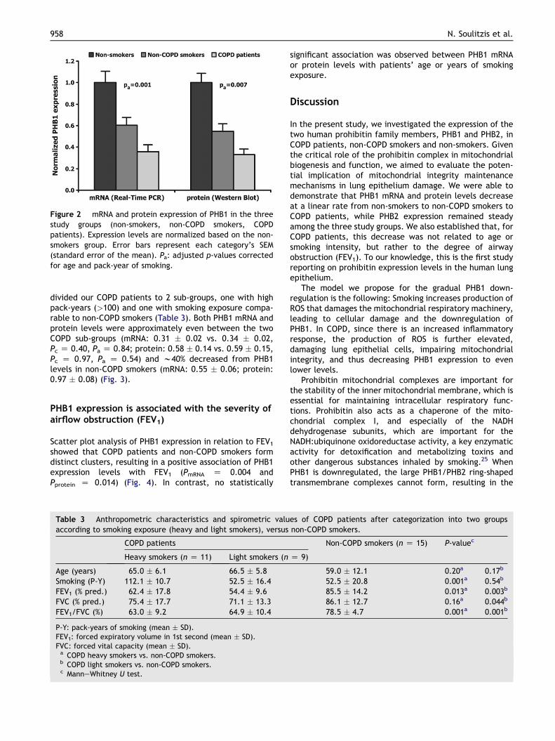
Figure 2 mRNA and protein expression of PHB1 in the threestudy groups (non-smokers, non-COPD smokers, COPDpatients). Expression levels are normalized based on the non-smokers group. Error bars represent each category’s SEM(standard error of the mean). Pa: adjusted p-values correctedfor age and pack-year of smoking.
958 N. Soulitzis et al.
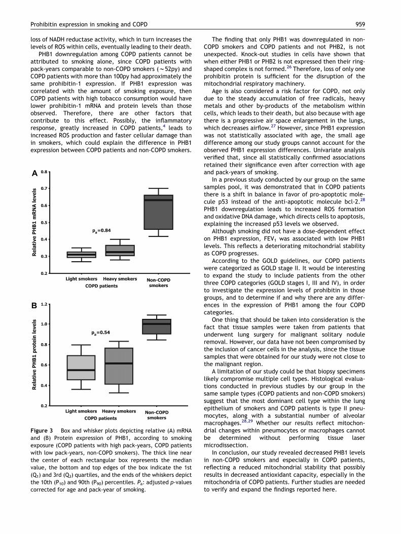
divided our COPD patients to 2 sub-groups, one with highpack-years (>100) and one with smoking exposure compa-rable to non-COPD smokers (Table 3). Both PHB1 mRNA andprotein levels were approximately even between the twoCOPD sub-groups (mRNA: 0.31 � 0.02 vs. 0.34 � 0.02,Pc Z 0.40, Pa Z 0.84; protein: 0.58 � 0.14 vs. 0.59 � 0.15,Pc Z 0.97, Pa Z 0.54) and w40% decreased from PHB1levels in non-COPD smokers (mRNA: 0.55 � 0.06; protein:0.97 � 0.08) (Fig. 3).
PHB1 expression is associated with the severity ofairflow obstruction (FEV1)
Scatter plot analysis of PHB1 expression in relation to FEV1showed that COPD patients and non-COPD smokers formdistinct clusters, resulting in a positive association of PHB1expression levels with FEV1 (PmRNA Z 0.004 andPprotein Z 0.014) (Fig. 4). In contrast, no statistically
Table 3 Anthropometric characteristics and spirometric valuaccording to smoking exposure (heavy and light smokers), versus
COPD patients
Heavy smokers (n Z 11) Light smokers (n
Age (years) 65.0 � 6.1 66.5 � 5.8Smoking (P-Y) 112.1 � 10.7 52.5 � 16.4FEV1 (% pred.) 62.4 � 17.8 54.4 � 9.6FVC (% pred.) 75.4 � 17.7 71.1 � 13.3FEV1/FVC (%) 63.0 � 9.2 64.9 � 10.4
P-Y: pack-years of smoking (mean � SD).FEV1: forced expiratory volume in 1st second (mean � SD).FVC: forced vital capacity (mean � SD).a COPD heavy smokers vs. non-COPD smokers.b COPD light smokers vs. non-COPD smokers.c ManneWhitney U test.
significant association was observed between PHB1 mRNAor protein levels with patients’ age or years of smokingexposure.
Discussion
In the present study, we investigated the expression of thetwo human prohibitin family members, PHB1 and PHB2, inCOPD patients, non-COPD smokers and non-smokers. Giventhe critical role of the prohibitin complex in mitochondrialbiogenesis and function, we aimed to evaluate the poten-tial implication of mitochondrial integrity maintenancemechanisms in lung epithelium damage. We were able todemonstrate that PHB1 mRNA and protein levels decreaseat a linear rate from non-smokers to non-COPD smokers toCOPD patients, while PHB2 expression remained steadyamong the three study groups. We also established that, forCOPD patients, this decrease was not related to age orsmoking intensity, but rather to the degree of airwayobstruction (FEV1). To our knowledge, this is the first studyreporting on prohibitin expression levels in the human lungepithelium.
The model we propose for the gradual PHB1 down-regulation is the following: Smoking increases production ofROS that damages the mitochondrial respiratory machinery,leading to cellular damage and the downregulation ofPHB1. In COPD, since there is an increased inflammatoryresponse, the production of ROS is further elevated,damaging lung epithelial cells, impairing mitochondrialintegrity, and thus decreasing PHB1 expression to evenlower levels.
Prohibitin mitochondrial complexes are important forthe stability of the inner mitochondrial membrane, which isessential for maintaining intracellular respiratory func-tions. Prohibitin also acts as a chaperone of the mito-chondrial complex I, and especially of the NADHdehydrogenase subunits, which are important for theNADH:ubiquinone oxidoreductase activity, a key enzymaticactivity for detoxification and metabolizing toxins andother dangerous substances inhaled by smoking.25 WhenPHB1 is downregulated, the large PHB1/PHB2 ring-shapedtransmembrane complexes cannot form, resulting in the
es of COPD patients after categorization into two groupsnon-COPD smokers.
Non-COPD smokers (n Z 15) P-valuec
Z 9)
59.0 � 12.1 0.20a 0.17b
52.5 � 20.8 0.001a 0.54b
85.5 � 14.2 0.013a 0.003b
86.1 � 12.7 0.16a 0.044b
78.5 � 4.7 0.001a 0.001b
Prohibitin expression in smoking and COPD 959
loss of NADH reductase activity, which in turn increases thelevels of ROS within cells, eventually leading to their death.
PHB1 downregulation among COPD patients cannot beattributed to smoking alone, since COPD patients withpack-years comparable to non-COPD smokers (w52py) andCOPD patients with more than 100py had approximately thesame prohibitin-1 expression. If PHB1 expression wascorrelated with the amount of smoking exposure, thenCOPD patients with high tobacco consumption would havelower prohibitin-1 mRNA and protein levels than thoseobserved. Therefore, there are other factors thatcontribute to this effect. Possibly, the inflammatoryresponse, greatly increased in COPD patients,4 leads toincreased ROS production and faster cellular damage thanin smokers, which could explain the difference in PHB1expression between COPD patients and non-COPD smokers.
Figure 3 Box and whisker plots depicting relative (A) mRNAand (B) Protein expression of PHB1, according to smokingexposure (COPD patients with high pack-years, COPD patientswith low pack-years, non-COPD smokers). The thick line nearthe center of each rectangular box represents the medianvalue, the bottom and top edges of the box indicate the 1st(Q1) and 3rd (Q3) quartiles, and the ends of the whiskers depictthe 10th (P10) and 90th (P90) percentiles. Pa: adjusted p-valuescorrected for age and pack-year of smoking.
The finding that only PHB1 was downregulated in non-COPD smokers and COPD patients and not PHB2, is notunexpected. Knock-out studies in cells have shown thatwhen either PHB1 or PHB2 is not expressed then their ring-shaped complex is not formed.26 Therefore, loss of only oneprohibitin protein is sufficient for the disruption of themitochondrial respiratory machinery.
Age is also considered a risk factor for COPD, not onlydue to the steady accumulation of free radicals, heavymetals and other by-products of the metabolism withincells, which leads to their death, but also because with agethere is a progressive air space enlargement in the lungs,which decreases airflow.27 However, since PHB1 expressionwas not statistically associated with age, the small agedifference among our study groups cannot account for theobserved PHB1 expression differences. Univariate analysisverified that, since all statistically confirmed associationsretained their significance even after correction with ageand pack-years of smoking.
In a previous study conducted by our group on the samesamples pool, it was demonstrated that in COPD patientsthere is a shift in balance in favor of pro-apoptotic mole-cule p53 instead of the anti-apoptotic molecule bcl-2.28
PHB1 downregulation leads to increased ROS formationand oxidative DNA damage, which directs cells to apoptosis,explaining the increased p53 levels we observed.
Although smoking did not have a dose-dependent effecton PHB1 expression, FEV1 was associated with low PHB1levels. This reflects a deteriorating mitochondrial stabilityas COPD progresses.
According to the GOLD guidelines, our COPD patientswere categorized as GOLD stage II. It would be interestingto expand the study to include patients from the otherthree COPD categories (GOLD stages I, III and IV), in orderto investigate the expression levels of prohibitin in thosegroups, and to determine if and why there are any differ-ences in the expression of PHB1 among the four COPDcategories.
One thing that should be taken into consideration is thefact that tissue samples were taken from patients thatunderwent lung surgery for malignant solitary noduleremoval. However, our data have not been compromised bythe inclusion of cancer cells in the analysis, since the tissuesamples that were obtained for our study were not close tothe malignant region.
A limitation of our study could be that biopsy specimenslikely compromise multiple cell types. Histological evalua-tions conducted in previous studies by our group in thesame sample types (COPD patients and non-COPD smokers)suggest that the most dominant cell type within the lungepithelium of smokers and COPD patients is type II pneu-mocytes, along with a substantial number of alveolarmacrophages.28,29 Whether our results reflect mitochon-drial changes within pneumocytes or macrophages cannotbe determined without performing tissue lasermicrodissection.
In conclusion, our study revealed decreased PHB1 levelsin non-COPD smokers and especially in COPD patients,reflecting a reduced mitochondrial stability that possiblyresults in decreased antioxidant capacity, especially in themitochondria of COPD patients. Further studies are neededto verify and expand the findings reported here.
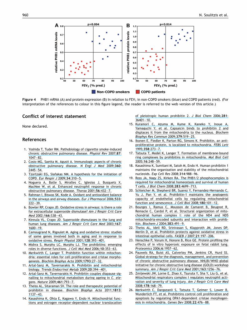
Figure 4 PHB1 mRNA (A) and protein expression (B) In relation to FEV1 in non-COPD smokers (blue) and COPD patients (red). (Forinterpretation of the references to colour in this figure legend, the reader is referred to the web version of this article.)
960 N. Soulitzis et al.
Conflict of interest statement
None declared.
References
1. Yoshida T, Tuder RM. Pathobiology of cigarette smoke-inducedchronic obstructive pulmonary disease. Physiol Rev 2007;87:1047e82.
2. Cosio MG, Saetta M, Agusti A. Immunologic aspects of chronicobstructive pulmonary disease. N Engl J Med 2009;360:2445e54.
3. Tzortzaki EG, Siafakas NM. A hypothesis for the initiation ofCOPD. Eur Respir J 2009;34:310e5.
4. Noguera A, Batle S, Miralles C, Iglesias J, Busquets X,MacNee W, et al. Enhanced neutrophil response in chronicobstructive pulmonary disease. Thorax 2001;56:432e7.
5. Rahman I, Biswas SK, Kode A. Oxidant and antioxidant balancein the airways and airway diseases. Eur J Pharmacol 2006;533:222e39.
6. Bowler RP, Crapo JD. Oxidative stress in airways: is there a rolefor extracellular superoxide dismutase? Am J Respir Crit CareMed 2002;166:S38e43.
7. Kinnula VL, Crapo JD. Superoxide dismutases in the lung andhuman lung diseases. Am J Respir Crit Care Med 2003;167:1600e19.
8. Camougrand N, Rigoulet M. Aging and oxidative stress: studiesof some genes involved both in aging and in response tooxidative stress. Respir Physiol 2001;128:393e401.
9. Mishra S, Murphy LC, Murphy LJ. The prohibitins: emergingroles in diverse functions. J Cell Mol Med 2006;10:353e63.
10. Merkwirth C, Langer T. Prohibitin function within mitochon-dria: essential roles for cell proliferation and cristae morpho-genesis. Biochim Biophys Acta 2009;1793:27e32.
11. Artal-Sanz M, Tavernarakis N. Prohibitin and mitochondrialbiology. Trends Endocrinol Metab 2009;20:394e401.
12. Artal-Sanz M, Tavernarakis N. Prohibitin couples diapause sig-nalling to mitochondrial metabolism during ageing in C. ele-gans. Nature 2009;461:793e7.
13. Theiss AL, Sitaraman SV. The role and therapeutic potential ofprohibitin in disease. Biochim Biophys Acta 2011;1813:1137e43.
14. Kasashima K, Ohta E, Kagawa Y, Endo H. Mitochondrial func-tions and estrogen receptor-dependent nuclear translocation
of pleiotropic human prohibitin 2. J Biol Chem 2006;281:36401e10.
15. Kuramori C, Azuma M, Kume K, Kaneko Y, Inoue A,Yamaguchi Y, et al. Capsaicin binds to prohibitin 2 anddisplaces it from the mitochondria to the nucleus. BiochemBiophys Res Commun 2009;379:519e25.
16. Ikonen E, Fiedler K, Parton RG, Simons K. Prohibitin, an anti-proliferative protein, is localized to mitochondria. FEBS Lett1995;358:273e7.
17. Tatsuta T, Model K, Langer T. Formation of membrane-boundring complexes by prohibitins in mitochondria. Mol Biol Cell2005;16:248e59.
18. Kasashima K, Sumitani M, Satoh M, Endo H. Human prohibitin 1maintains the organization and stability of the mitochondrialnucleoids. Exp Cell Res 2008;314:988e96.
19. Ross JA, Nagy ZS, Kirken RA. The PHB1/2 phosphocomplex isrequired for mitochondrial homeostasis and survival of humanT cells. J Biol Chem 2008;283:4699e713.
20. Schleicher M, Shepherd BR, Suarez Y, Fernandez-Hernando C,Yu J, Pan Y, et al. Prohibitin-1 maintains the angiogeniccapacity of endothelial cells by regulating mitochondrialfunction and senescence. J Cell Biol 2008;180:101e12.
21. Bourges I, Ramus C, Mousson de Camaret B, Beugnot R,Remacle C, Cardol P, et al. Structural organization of mito-chondrial human complex I: role of the ND4 and ND5mitochondria-encoded subunits and interaction with prohib-itin. Biochem J 2004;383:491e9.
22. Theiss AL, Idell RD, Srinivasan S, Klapproth JM, Jones DP,Merlin D, et al. Prohibitin protects against oxidative stress inintestinal epithelial cells. FASEB J 2007;21:197e206.
23. Henschke P, Vorum H, Honore B, Rice GE. Protein profiling theeffects of in vitro hyperoxic exposure on fetal rabbit lung.Proteomics 2006;6:1957e62.
24. Pauwels RA, Buist AS, Calverley PM, Jenkins CR, Hurd SS.Global strategy for the diagnosis, management, and preventionof chronic obstructive pulmonary disease. NHLBI/WHO globalinitiative for chronic obstructive lung disease (GOLD) workshopsummary. Am J Respir Crit Care Med 2001;163:1256e76.
25. Zmijewski JW, Lorne E, Zhao X, Tsuruta Y, Sha Y, Liu G, et al.Mitochondrial respiratory complex I regulates neutrophil acti-vation and severity of lung injury. Am J Respir Crit Care Med2008;178:168e79.
26. Merkwirth C, Dargazanli S, Tatsuta T, Geimer S, Lower B,Wunderlich FT, et al. Prohibitins control cell proliferation andapoptosis by regulating OPA1-dependent cristae morphogen-esis in mitochondria. Genes Dev 2008;22:476e88.

Prohibitin expression in smoking and COPD 961
27. Janssens JP, Pache JC, Nicod LP. Physiological changes inrespiratory function associated with ageing. Eur Respir J 1999;13:197e205.
28. Siganaki M, Koutsopoulos AV, Neofytou E, Vlachaki E,Psarrou M, Soulitzis N, et al. Deregulation of apoptosis
mediators’ p53 and bcl2 in lung tissue of COPD patients. RespirRes 2010;11:46.
29. Vlachaki EM, Koutsopoulos AV, Tzanakis N, Neofytou E,Siganaki M, Drositis I, et al. Altered surfactant protein-A expres-sion in type II pneumocytes in COPD. Chest 2010;137:37e45.
Copyright © 2022 FDOKUMEN