Circulating haemopoietic and endothelial progenitor cells are decreased in COPD
13
Circulating haemopoietic and endothelial progenitor cells are decreased in COPD P. Palange*, U. Testa # , A. Huertas*, L. Calabro ` # , R. Antonucci*, E. Petrucci # , E. Pelosi # , L. Pasquini # , A. Satta " , G. Morici +,e , M.A. Vignola 1,e,{ and M.R. Bonsignore 1,e ABSTRACT: Circulating CD34+ cells are haemopoietic progenitors that may play a role in tissue repair. No data are available on circulating progenitors in chronic obstructive pulmonary disease (COPD). Circulating CD34+ cells were studied in 18 patients with moderate-to-severe COPD (age: mean¡SD 68¡8 yrs; forced expiratory volume in one second: 48¡12% predicted) and 12 controls, at rest and after endurance exercise. Plasma concentrations of haematopoietic growth factors (FMS-like tyrosine kinase 3 (Flt3) ligand, kit ligand), markers of hypoxia (vascular endothelial growth factor (VEGF)) and stimulators of angiogenesis (VEGF, hepatocyte growth factor (HGF)) and markers of systemic inflammation (tumour necrosis factor (TNF)-a, interleukin (IL)-6, IL-8) were measured. Compared with the controls, the COPD patients showed a three-fold reduction in CD34+ cell counts (3.3¡2.5 versus 10.3¡4.2 cells?mL -1 ), and a 50% decrease in AC133+ cells. In the COPD patients, progenitor-derived haemopoietic and endothelial cell colonies were reduced by 30–50%. However, four COPD patients showed progenitor counts in the normal range associated with lower TNF-a levels. In the entire sample, CD34+ cell counts correlated with exercise capacity and severity of airflow obstruction. After endurance exercise, progenitor counts were unchanged, while plasma Flt3 ligand and VEGF only increased in the COPD patients. Plasma HGF levels were higher in the COPD patients compared with the controls and correlated inversely with the number of progenitor-derived colonies. In conclusion, circulating CD34+ cells and endothelial progenitors were decreased in chronic obstructive pulmonary disease patients and could be correlated with disease severity. KEYWORDS: CD34+ cells, chronic obstructive pulmonary disease, exercise, growth factors, hypoxia T he systemic effects of chronic obstructive pulmonary disease (COPD) have been studied with regards to inflammation, chronic oxidative stress and skeletal muscle dysfunction [1]. Despite evidence of neutrophils activation in peripheral blood of COPD patients [2], inflammatory infiltration of the airways in severe disease [3], and smoking-induced increase in granulocyte turnover, little is known about the effects of COPD at the level of bone marrow [4]. Data on the erythrocyte lineage have shown that anaemia occurs in 13% of COPD patients and is associated with increased inflammatory markers [5], and may predict poor survival [6]. Data on other blood lineages are scarce due to the need of invasive techniques to obtain bone marrow samples. This difficulty can be partly overcome by studying circulating bone marrow-derived progenitors. These cells are positive for CD34 and other markers (CD38, human leukocyte antigen (HLA)- DR) acquired during differentiation [7], and yield indirect information on haemopoiesis. Circulating CD34+ cells are also believed to be involved in tissue repair. Bone marrow-derived progenitors can engraft in several organs including the lungs [8, 9] and skeletal muscle [10]. The latter finding is of interest in COPD, given the frequent occurrence of skeletal muscle dysfunction associated with increased muscle apoptosis [11], fibrosis [12] and inflammatory activation [13]. At least part of the repair potential of CD34+ cells may depend on the AC133+ endothelial progeni- tor cell (EPC) subpopulation [14]. EPCs have been used to treat patients with myocardial [15] AFFILIATIONS *Dipartimento di Medicina Clinica, University La Sapienza, and # Istituto Superiore di Sanita’, Rome, and " Fondazione Maugeri, Tradate, Varese, and + Dept of Experimental Medicine, and 1 Institute of Medicine and Pneumology, University of Palermo, and e IBIM-CNR, Palermo, Italy. CORRESPONDENCE P. Palange Dipartimento di Medicina Clinica University La Sapienza Viale Universita ` 37 Rome 00185 Italy Fax: 39 064940421 E-mail: [email protected] Received: October 22 2004 Accepted after revision: October 15 2005 SUPPORT STATEMENT The present study was funded by the Italian National Council of Research, Agenzia 2000 (CNRC005114), and the National Institute of Health (ISS), Rome. European Respiratory Journal Print ISSN 0903-1936 Online ISSN 1399-3003 For editorial comments see page 441. EUROPEAN RESPIRATORY JOURNAL VOLUME 27 NUMBER 3 529 Eur Respir J 2006; 27: 529–541 DOI: 10.1183/09031936.06.00120604 CopyrightßERS Journals Ltd 2006 c
-
Upload
independent -
Category
Documents
-
view
0 -
download
0
Transcript of Circulating haemopoietic and endothelial progenitor cells are decreased in COPD

Circulating haemopoietic and endothelial
progenitor cells are decreased in COPDP. Palange*, U. Testa#, A. Huertas*, L. Calabro#, R. Antonucci*, E. Petrucci#,E. Pelosi#, L. Pasquini#, A. Satta", G. Morici+,e, M.A. Vignola1,e,{ andM.R. Bonsignore1,e
ABSTRACT: Circulating CD34+ cells are haemopoietic progenitors that may play a role in tissue
repair. No data are available on circulating progenitors in chronic obstructive pulmonary disease
(COPD).
Circulating CD34+ cells were studied in 18 patients with moderate-to-severe COPD (age:
mean¡SD 68¡8 yrs; forced expiratory volume in one second: 48¡12% predicted) and 12
controls, at rest and after endurance exercise. Plasma concentrations of haematopoietic growth
factors (FMS-like tyrosine kinase 3 (Flt3) ligand, kit ligand), markers of hypoxia (vascular
endothelial growth factor (VEGF)) and stimulators of angiogenesis (VEGF, hepatocyte growth
factor (HGF)) and markers of systemic inflammation (tumour necrosis factor (TNF)-a, interleukin
(IL)-6, IL-8) were measured.
Compared with the controls, the COPD patients showed a three-fold reduction in CD34+ cell
counts (3.3¡2.5 versus 10.3¡4.2 cells?mL-1), and a 50% decrease in AC133+ cells. In the COPD
patients, progenitor-derived haemopoietic and endothelial cell colonies were reduced by 30–50%.
However, four COPD patients showed progenitor counts in the normal range associated with
lower TNF-a levels. In the entire sample, CD34+ cell counts correlated with exercise capacity and
severity of airflow obstruction. After endurance exercise, progenitor counts were unchanged,
while plasma Flt3 ligand and VEGF only increased in the COPD patients. Plasma HGF levels were
higher in the COPD patients compared with the controls and correlated inversely with the number
of progenitor-derived colonies.
In conclusion, circulating CD34+ cells and endothelial progenitors were decreased in chronic
obstructive pulmonary disease patients and could be correlated with disease severity.
KEYWORDS: CD34+ cells, chronic obstructive pulmonary disease, exercise, growth factors,
hypoxia
The systemic effects of chronic obstructivepulmonary disease (COPD) have beenstudied with regards to inflammation,
chronic oxidative stress and skeletal muscledysfunction [1]. Despite evidence of neutrophilsactivation in peripheral blood of COPD patients[2], inflammatory infiltration of the airways insevere disease [3], and smoking-induced increasein granulocyte turnover, little is known about theeffects of COPD at the level of bone marrow [4].Data on the erythrocyte lineage have shown thatanaemia occurs in 13% of COPD patients and isassociated with increased inflammatory markers[5], and may predict poor survival [6]. Data onother blood lineages are scarce due to the need ofinvasive techniques to obtain bone marrowsamples.
This difficulty can be partly overcome by studyingcirculating bone marrow-derived progenitors.These cells are positive for CD34 and othermarkers (CD38, human leukocyte antigen (HLA)-DR) acquired during differentiation [7], andyield indirect information on haemopoiesis.Circulating CD34+ cells are also believed to beinvolved in tissue repair. Bone marrow-derivedprogenitors can engraft in several organsincluding the lungs [8, 9] and skeletal muscle[10]. The latter finding is of interest in COPD,given the frequent occurrence of skeletal muscledysfunction associated with increased muscleapoptosis [11], fibrosis [12] and inflammatoryactivation [13].
At least part of the repair potential of CD34+ cellsmay depend on the AC133+ endothelial progeni-tor cell (EPC) subpopulation [14]. EPCs havebeen used to treat patients with myocardial [15]
AFFILIATIONS
*Dipartimento di Medicina Clinica,
University La Sapienza, and#Istituto Superiore di Sanita’, Rome,
and"Fondazione Maugeri, Tradate,
Varese, and+Dept of Experimental Medicine, and1Institute of Medicine and
Pneumology, University of Palermo,
andeIBIM-CNR, Palermo, Italy.
CORRESPONDENCE
P. Palange
Dipartimento di Medicina Clinica
University La Sapienza
Viale Universita 37
Rome
00185 Italy
Fax: 39 064940421
E-mail: [email protected]
Received:
October 22 2004
Accepted after revision:
October 15 2005
SUPPORT STATEMENT
The present study was funded by the
Italian National Council of Research,
Agenzia 2000 (CNRC005114), and
the National Institute of Health (ISS),
Rome.
European Respiratory Journal
Print ISSN 0903-1936
Online ISSN 1399-3003For editorial comments see page 441.
EUROPEAN RESPIRATORY JOURNAL VOLUME 27 NUMBER 3 529
Eur Respir J 2006; 27: 529–541
DOI: 10.1183/09031936.06.00120604
Copyright�ERS Journals Ltd 2006
c

and vascular [16] disease. Low circulating EPC counts werefound in patients with cardiovascular risk factors [17], andbone marrow CD34+ cells showed a low proliferative capacityin patients with chronic ischemic heart disease [18]. However,exercise training increased EPCs [19]. Whether COPD affectscirculating EPCs and/or CD34+ cell proliferative capabilities isunknown.
Exercise might be a useful model to study circulating pro-genitors. Long-distance runners showed increased circulating
TABLE 1 Anthropometric characteristics and pulmonaryfunction data of chronic obstructive pulmonarydisease (COPD) patients and control subjects
COPD patients Control subjects
Subjects n 18 12
Age yrs 68¡8 63¡7
BMI kg?m-2 26.8¡4.2 25.9¡2.1
FEV1 L 1.4¡0.3* 3.3¡0.5
FEV1 % pred 48¡12* 106¡15
FVC L 2.7¡0.4* 4.1¡0.6
FVC % pred 73¡16* 102¡15
FEV1/FVC % 51¡10* 81¡9
Pa,O2 mmHg 76¡9* 93¡3
Pa,CO2 mmHg 41¡4 39¡3
pH 7.41¡0.05 7.42¡0.02
Sa,O2 % 93¡4* 98¡1
Data are presented as mean¡SD. BMI: body mass index; FEV1: forced
expiratory volume in one second; % pred: percentage predicted; FVC: forced
vital capacity; Pa,O2: arterial oxygen tension; Pa,CO2: arterial carbon dioxide
tension; Sa,O2: arterial oxygen saturation. *: significant differences (p,0.05)
between groups. 1 mmHg50.133kPa.
TABLE 2 Exercise data# in chronic obstructive pulmonarydisease (COPD) patients and control subjects
COPD patients Control subjects
Load W 92¡15* 133¡18
V9O2 peak L?min-1 1.53¡0.24* 2.22¡0.25
V9O2 peak mL?kg-1?min-1 19.8¡2.6* 28.5¡4.0
V9E peak L?min-1 48.7¡14.6* 62.5¡5.6
TV L 1.6¡0.3* 2.3¡0.3
Rf 31.4¡7.2 28.9¡6.1
tI /ttot 0.40¡0.01* 0.50¡0.01
RER 1.00¡0.1 1.02¡0.05
V9E/V9CO2 29.3¡5.4 25.4¡1.7
HR 125.8¡11.1* 138.6¡15.4
V9O2-LT L?min-1 1.07¡0.23* 1.45¡0.25
V9O2-LT/V9O2 peak 0.55¡0.13* 0.66¡0.11
Data are presented as mean¡SD. W: Watt; V9O2: oxygen uptake; V9E: minute
ventilation; TV: tidal volume; Rf: respiratory frequency, breaths per minute; tI /
ttot: inspiratory time/total respiratory time; RER: respiratory exchange ratio;
V9CO2: carbon dioxide output; HR: heart rate, beats per minute; LT: lactic
threshold. #: maximal incremental test. *: significant difference (p,0.05)
between groups.
TABLE 3 Blood cell counts in control subjects and inchronic obstructive disease (COPD) patients atbaseline (BL) and after endurance exercise (Exe)
COPD patients Control subjects
BL Exe BL Exe
RBC 106?mL-1 5.00¡0.51 5.07¡0.57 4.87¡0.35 4.93¡0.40
Hb g?dL-1 14.7¡1.3 15.1¡1.1 14.9¡1.0 15.0¡1.1
WBC 103?mL-1 6.96¡1.31 7.73¡1.64 6.14¡1.27 6.62¡1.67
Neutrophils % 64.8¡6.1* 63.0¡7.3 57.0¡6.9 58.6¡7.3
Lymphocytes % 23.2¡5.7* 25.5¡6.7 31.6¡7.4 29.7¡8.1
Monocytes % 6.5¡1.4 5.9¡0.8 6.2¡1.0 6.0¡1.1
Eosinophils % 2.8¡1.6 2.7¡1.3 2.6¡1.3 2.4¡1.3
Basophils % 0.6¡0.3 0.7¡0.3 0.7¡0.2 0.7¡0.2
Platelets
105?mL-1
234¡73 253¡74 199¡45 216¡35
Data presented as mean¡SD. RBC: red blood cells: Hb: haemoglobin; WBC:
white blood cells. *: significant difference (p,0.05) between resting values in
COPD patients and controls (unpaired t-test).
�
�
��
�
�
�
�
��
��
��
� ��
��
� �
��
�
�
��
�
��
� ��� � ���
��
����
�����
�� ��
��
��
��
FIGURE 1. a) White blood cells (WBC) and b) CD34+ cells at baseline (BL) and
after endurance exercise (Exe) in chronic obstructive pulmonary disease (COPD)
patients ($) and in control subjects (#). Note the marked uniform reduction in
CD34+ cells in COPD patients compared with the control subjects. *: p,0.001,
COPD versus controls.
CD34+ CELLS IN COPD P. PALANGE ET AL.
530 VOLUME 27 NUMBER 3 EUROPEAN RESPIRATORY JOURNAL
CD34+ cells and exercise-induced release of factors/cytokinesactive on the bone marrow (FMS-like tyrosine kinase 3 (Flt3)ligand, interleukin (IL)-6, granulocyte colony-stimulatingfactor (G-CSF), tumour necrosis factor (TNF)-a) [20, 21].Mobilisation of endothelial progenitor cells also occurred afterexercise in middle-aged untrained subjects [22], but little isknown on the relationship between circulating progenitors andexercise limitation in COPD, or on the effects of inflammatoryactivation on progenitors.
Finally, COPD may affect haemopoietic and endothelialprogenitors through alterations in arterial blood gases orinflammatory mediators. In vitro, very low arterial oxygentension preserved early haemopoietic progenitors [23], whilepromoting their differentiation [24]. No study addressed theeffects of mild hypoxaemia on circulating CD34+ cells andsubpopulations in vivo in COPD patients.
The present study was designed to answer whether or not COPDaffects circulating progenitor cell numbers and frequency.
SUBJECTS AND METHODSIn total, 18 ex-smokers with COPD and 12 age-matchednonsmokers (controls) were studied (table 1). Inclusion criteria
for patients were: 1) COPD of moderate severity (GlobalInitiative for Chronic Obstructive Lung Disease stage 2); 2)stable clinical conditions (i.e. no change in pulmonary functiontests or exacerbation in the 4 weeks preceding the study); and3) mild, resting hypoxaemia (arterial oxygen tension: Pa,O2
.7.89 kPa). No patient was receiving systemic steroids at thetime of the study. Inclusion criteria for controls were: 1) ageo50 yrs; 2) a sedentary lifestyle; 3) normal spirometry andarterial blood gases; 4) no other clinically evident disease; and5) a wish to participate in the study. Patients with cardio-vascular, cerebrovascular, neuromuscular, rheumatologicaland/or metabolic disorders, as assessed by clinical andstandard laboratory findings tests, were excluded from thestudies. Patients were also excluded if they suffered from anydisease precluding execution of exercise tests.
The protocol was approved by the Ethical Committee of theUniversity of Rome (Italy), and all subjects gave their informedconsent.
Study protocolEach subject visited the laboratory three times. At the first visit,a complete clinical assessment and pulmonary function tests
����
��
��
��
�
���
!��
����
��
��
��
�
��
���
����
��
��
��
�
��"�
��
��
��
�
����
��
��
��
�
���
!��
��#�
��
��
��
�
��$�
��
��
��
�
��%�
��
��
��
�
��&�
��
��
��
�
���
!��
��'�
��
��
��
�
��(�
��
��
��
�
����
��
��
��
�
��
���
��
���
� �� �� �� ��
��� !��� �� �� �� ��
) *�+� �� �� �� ��
����� �� �� �� ��
���,
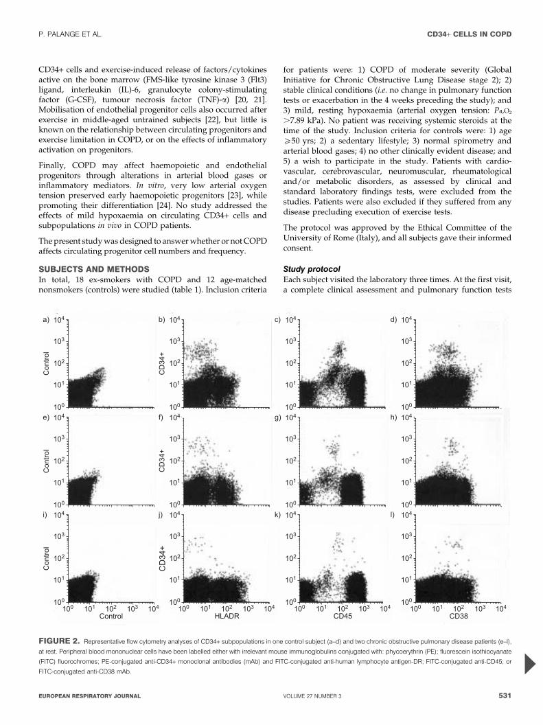
FIGURE 2. Representative flow cytometry analyses of CD34+ subpopulations in one control subject (a–d) and two chronic obstructive pulmonary disease patients (e–l),
at rest. Peripheral blood mononuclear cells have been labelled either with irrelevant mouse immunoglobulins conjugated with: phycoerythrin (PE); fluorescein isothiocyanate
(FITC) fluorochromes; PE-conjugated anti-CD34+ monoclonal antibodies (mAb) and FITC-conjugated anti-human lymphocyte antigen-DR; FITC-conjugated anti-CD45; or
FITC-conjugated anti-CD38 mAb.
P. PALANGE ET AL. CD34+ CELLS IN COPD
cEUROPEAN RESPIRATORY JOURNAL VOLUME 27 NUMBER 3 531

(Quark PFT; COSMED, Rome, Italy) were obtained. A samplewas drawn from the radial artery for arterial blood gasdetermination at rest while breathing room air. At the secondvisit each subject underwent an incremental exercise stress teston the cycle ergometer (Ergoline 800, Bitz, Germany) todetermine peak oxygen uptake (V9O2peak) and lactic threshold.Pulmonary gas exchange indexes were measured breath-by-breath by a computerised system as previously described [25].Oxygen uptake (V9O2; at standard temperature and pressure,dry (STPD)), CO2 output (V9CO2; STPD), minute ventilation(V9E; at body temperature and pressure, saturated with watervapour) and respiratory frequency were measured. Heart rate(HR) was derived from R–R intervals measured from a 12-leadECG. At the third visit, each subject performed a submaximalexercise test on a cycle ergometer at a work-rate correspondingto 70% of V9O2peak for 20 min (endurance test). Exercisesessions were planned a minimum of 48 h apart. All testswere obtained while the subjects breathed room air.
Venous blood samples (20 mL) were drawn before andimmediately after the endurance test for analysis of blood cellcounts and CD34+ cells. Total blood cell counts were
determined by standard haemocytometry. Peripheral bloodmononuclear cells (PBMCs) were obtained by standard Ficollgradient centrifugation. The cells were carefully washed withPBS, resuspended in PBS containing 2 mg?mL-1 bovine serumalbumin and labelled for 30 min at 4uC with the followingantibodies: 1) anti-CD34 conjugated with phycoerythrin (PE);2) anti-CD38; or 3) anti-HLA-DR, all labelled with fluoresceineisothiocyanate (FITC; Becton Dickinson–Pharmingen, LincolnPark, NJ, USA). In some experiments PBMCs were labelledwith anti-CD34 (FITC-conjugated) and either anti-very lateactivation antigen (VLA)-4 or anti-Flt3 (both PE-conjugated).For a negative control, the cells were labelled with isotype-matched mouse immunoglobulin labelled with either PE orFITC. After two washings in cold PBS, the cells were analysedfor fluorescence in a fluorescence-activated cell sorter flowcytometer (Becton-Dickinson). The level of positivity of entirebody was evaluated as ‘‘dim’’ or ‘‘bright’’ according to thefluorescence labelling intensity. CD34+ cells and their sub-populations were expressed as percentage of total PBMCs. Insome experiments PBMCs were labelled with PE-conjugatedanti-AC133 monoclonal antibodies (Miltenyi Biotech, BergischGladbach, Germany).
����
��
��
��
�
���
!��
����
��
��
��
�
��
���
����
��
��
��
�
��"�
��
��
��
�
����
��
��
��
�
���
!��
��#�
��
��
��
�
��$�
��
��
��
�
��%�
��
��
��
�
��&�
��
��
��
�
���
!��
��'�
��
��
��
�
��(�
��
��
��
�
����
��
��
��
�
��
���
��
���
� �� �� �� ��
��� !��� �� �� �� ��
) *�+� �� �� �� ��
����� �� �� �� ��
���,
FIGURE 3. Representative flow cytometry analyses of CD34+ subpopulations in one control subject (a–d) and two chronic obstructive pulmonary disease patients (e–l),
after endurance exercise. Peripheral blood mononuclear cells have been labelled either with irrelevant mouse immunoglobulins conjugated with: phycoerythrin (PE);
fluorescein isothiocyanate (FITC) fluorochromes; PE-conjugated anti-CD34+monoclonal antibodies (mAb) and FITC-conjugated anti-human lymphocyte antigen-DR; FITC-
conjugated anti-CD45; or FITC-conjugated anti-CD38 mAb.
CD34+ CELLS IN COPD P. PALANGE ET AL.
532 VOLUME 27 NUMBER 3 EUROPEAN RESPIRATORY JOURNAL
Flow cytometry analysis was carried out using software gatingon lymphocytes. At least 105 gated events were acquired foreach determination.
For the colony forming unit (CFU) assay, PBMCs were seededat 36105 cells?mL-1?dish-1 (Falcon 1008; Becton Dickinson) in0.9% methylcellulose and 40% foetal calf serum (Gibco, GrandIsland, NY, USA) in Iscove’s modified Dulbecco’s medium(Gibco), which was supplemented with 1.5 IU?mL-1 erythro-poietin for erythrocyte burst-forming units (E-BFU) coloniesand 10 ng?mL-1 of both granulocyte-monocyte colony-stimulating factor and G-CSF for granulocyte-monocyte CFU(GM-CFU) colonies. Colonies were counted under an invertedmicroscope after 14–16 days of culture.
For endothelial progenitor colony assay, PBMCs were resus-pended in EndoCultTM liquid medium (Stem Cell Techno-logies, Vancouver, BC, Canada) and plated (56106 cells) onfibronectin-coated 6-well dishes. After 2 days of culture at37uC, nonadherent cells were collected and 16106 cells?well-1
were plated in duplicate on fibronectin-coated 24-well dishes.After 3–5 additional days of culture, endothelial cell CFUs (EC-CFU) were counted under an inverted microscope.
Biochemical analysesThese assays were performed in 17 COPD patients and sixcontrol subjects. Aliquots of serum were prepared and storedat -80uC for determination by immunoassay (ELISA, R&DSystems, Minneapolis, MN, USA) of the following. 1) Flt3ligand and kit ligand (KL), both acting on early haemopoieticstages (detection limit: 5.0 pg?mL-1 for both) [26]. 2) Vascularendothelial growth factor (VEGF), as a marker of tissuehypoxia and angiogenesis (detection limit: 5.0 pg?mL-1) [27].3) Hepatocyte growth factor (HGF), a mediator known toactivate proliferation and migration of endothelial cells andinduce angiogenesis [28] (detection limit: 20 pg?mL-1). 4) TNF-a (detection limit 0.2 pg?mL-1), and IL-8 (detection limit:0.2 pg?mL-1) as markers of systemic inflammation. As well asIL-6 (detection limit: 0.16 pg?mL-1), which is known to bereleased during prolonged exercise [29]. The serum concentra-tion of cortisol and muscle enzymes (lactic dehydrogenase,creatine phosphokinase) as markers of muscle damage wasdetermined using standard methods.
StatisticsData are expressed as means¡SD. ANOVA and t-test orWilcoxon test were used to compare experimental conditions.Paired t-tests were used in each group to compare baseline andpost-exercise data. Unpaired t-tests were used to compare theCOPD and control groups. When data appeared to beabnormally distributed, Wilcoxon or Mann-Whitney U-testswere used for paired or unpaired comparisons, respectively.Linear regression was used to assess relationships betweenvariables. The level of significance was set at p,0.05.
RESULTSSubjects and functional dataTable 1 summarises anthropometric and pulmonary functiondata and arterial blood gas measurements. COPD patientsshowed moderate-to-severe functional deterioration. Duringmaximal exercise stress test (table 2) COPD patients attainedlower V9O2, V9E and HR at peak exercise than controls.
Endurance testMean¡SD workload was 133¡18 W in controls, and 92¡15 Win COPD patients (p,0.001 by unpaired t-test). All normalsubjects and 17 COPD patients completed the 20-min exercisetest. One patient stopped at 10 min because of intolerabledyspnoea.
Red blood cells and platelet counts were similar in the controlsubjects and COPD patients, both pre- and post-exercise(table 3). A slightly anaemic state (haemoglobin concentration,13.5 mg?dL-1) was found in one COPD patient and onecontrol subject.
White blood cell (WBC) counts were in the normal range inboth groups. However, COPD patients showed slightly higherWBC (fig. 1a) and neutrophil differential counts comparedwith controls at baseline (table 3). The latter differencebetween groups was not found in the post-exercise samples.
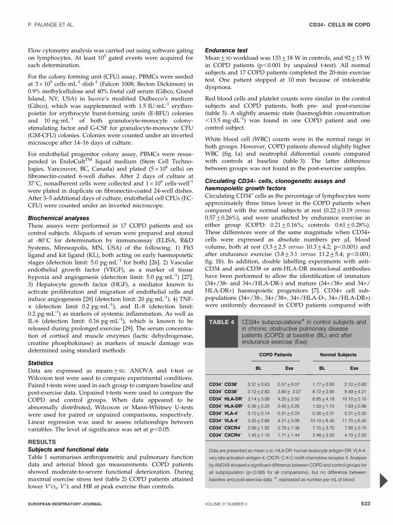
Circulating CD34+ cells, clonogenetic assays andhaemopoietic growth factorsCirculating CD34+ cells as the percentage of lymphocytes wereapproximately three times lower in the COPD patients whencompared with the normal subjects at rest (0.22¡0.19 versus0.57¡0.26%), and were unaffected by endurance exercise ineither group (COPD: 0.21¡0.16%; controls: 0.61¡0.28%).These differences were of the same magnitude when CD34+cells were expressed as absolute numbers per mL bloodvolume, both at rest (3.3¡2.5 versus 10.3¡4.2; p,0.001) andafter endurance exercise (3.8¡3.1 versus 11.2¡5.4; p,0.001;fig. 1b). In addition, double labelling experiments with anti-CD34 and anti-CD38 or anti-HLA-DR monoclonal antibodieshave been performed to allow the identification of immature(34+/38- and 34+/HLA-DR-) and mature (34+/38+ and 34+/HLA-DR+) haemopoietic progenitors [7]. CD34+ cell sub-populations (34+/38-, 34+/38+, 34+/HLA-D-, 34+/HLA-DR+)were uniformly decreased in COPD patients compared with
TABLE 4 CD34+ subpopulations# in control subjects andin chronic obstructive pulmonary diseasepatients (COPD) at baseline (BL) and afterendurance exercise (Exe)
COPD Patients Normal Subjects
BL Exe BL Exe
CD34+ CD38- 0.37¡0.63 0.57¡0.07 1.77¡0.93 2.10¡0.60
CD34+ CD38+ 3.12¡2.82 3.90¡ 3.07 8.72¡3.90 9.49¡4.21
CD34+ HLA-DR- 3.14¡3.08 4.20¡3.50 8.85¡4.18 10.10¡5.10
CD34+ HLA-DR+ 0.36¡0.20 0.40¡0.26 1.50¡1.10 1.93¡0.96
CD34+ VLA-4- 0.15¡0.14 0.31¡0.24 0.36¡0.31 0.31¡0.35
CD34+ VLA-4+ 3.35¡2.60 4.21¡3.06 10.10¡8.40 11.70¡6.40
CD34+ CXCR4- 2.06¡1.92 2.79¡1.36 7.15¡3.70 7.90¡5.10
CD34+ CXCR4+ 1.43¡1.10 1.71¡1.44 3.46¡3.55 4.10¡2.20
Data are presented as mean¡SD. HLA-DR: human leukocyte antigen-DR; VLA-4:
very late activation antigen-4; CXCR: C-X-C motif chemokine receptor 4. Analysis
by ANOVA showed a significant difference between COPD and control groups for
all subpopulation (p,0.005 for all comparisons), but no difference between
baseline and post-exercise data. #: expressed as number per mL of blood.
P. PALANGE ET AL. CD34+ CELLS IN COPD
cEUROPEAN RESPIRATORY JOURNAL VOLUME 27 NUMBER 3 533

the controls (figs 2, 3; table 4). The endurance exercise did notchange the number of circulating CD34+ cells (fig. 4; table 4).
In some COPD patients (n54, 22% of the sample) CD34+ cellcounts were in the normal range (0.56¡0.14% of lymphocytes)as opposed to the mean value of 0.12¡0.03% in the rest of theCOPD group (n514). The patients with normal CD34+ cellcounts could not be identified based on anthropometriccharacteristics or COPD severity. They only showed a trendtowards a higher V9O2peak (21.5¡3.1 versus 18.9¡2.1 mL?
kg-1?min-1; p50.07) and lower plasma TNF-a levels (rest:1.78¡0.79 versus 2.30¡1.22 pg?mL-1; post-exercise: 1.11¡0.11versus 1.96¡0.77 pg?mL-1, p,0.05).
Given the decrease in the number of circulating CD34+ cells inCOPD patients, the current authors evaluated whether thenumber of circulating haemopoietic progenitor cells, asmeasured by standard clonogenetic assays, were correspond-ingly decreased. COPD patients displayed a significantlyreduced number of CFUs compared with the controls (fig. 4aand 4c). In both groups, E-BFU and GM-CFU numbers, afterendurance exercise, were similar to those observed at rest(fig. 4a and 4c). It is of interest to note that the majority of
COPD patients displayed not only a reduced colony number,but also smaller E-BFU and GM-CFU compared with thecontrols (fig. 5). In figure 5 the representative pictures ofindividual E-BFU and GM-CFU colonies derived from theblood of a control subject (fig. 5c), a COPD patient with anormal CD34+ cell count (fig. 5b) and a COPD patient with alow CD34+ cell count (fig. 5a) are shown. The size of individualE-BFU colonies (each included in a separate box) and of GM-CFU colonies are smaller in COPD patients with low CD34+
cell counts (fig. 5a, observe the three individual E-BFUs andthe two GM-CFU colonies), compared with those observed forE-BFU and GM-CFU colonies derived from normal controls(fig. 5c) or COPD patients with normal CD34+ cell count(fig. 5b).
Given the reduced number of circulating haemopoieticprogenitors in COPD, the current authors evaluated theplasma concentration of several cytokines normally involvedin the survival and proliferation of haemopoietic progenitors.In this context, the current authors’ focused on the Flt3 ligandand KL. Flt3 ligand expression on CD34+ cell surface did notdiffer between COPD patients and controls (data not shown).Plasma Flt3 ligand concentration was similar in COPD patients
�
�
�
�-!
� ��
. ��
��
� �
/$�.
��
��0
�
�
�
���
��&�
��.
��
��
��
,
/$�.
��
"�
�
��
�1
� ��� � ���
FIGURE 4. a) Erythrocyte burst forming units E-BFU; b) plasma Flt3 ligand; c) granulocyte-monocyte colony-forming units (GM-CFU); and d) plasma kit ligand, at
baseline (BL) and after endurance exercise (Exe) in chronic obstructive pulmonary disease (COPD) patients (&) and in control subjects (h). Note the marked reduction in E-
BFU and GM-CFU in COPD patients at BL and after Exe compared with normal subjects. *: p,0.05 COPD versus controls; #: p,0.05 Exe versus BL.
CD34+ CELLS IN COPD P. PALANGE ET AL.
534 VOLUME 27 NUMBER 3 EUROPEAN RESPIRATORY JOURNAL

and controls at baseline; it increased significantly afterendurance exercise in COPD patients only (fig. 4b). PlasmaKL levels, did not differ between groups or experimentalconditions (fig. 4d).
In the entire group of subjects studied (i.e. COPD patients andcontrols) circulating CD34+ cell counts correlated directly withV9O2peak (fig 6d; r250.37, p,0.05), resting Pa,O2 (6a; r250.30,p,0.05) and with the severity of airway obstruction assessedas forced expiratory volume in one second (FEV1)/forced vitalcapacity (6c; r250.39; p,0.05). No correlation was foundbetween CD34+ cell counts and carbon dioxide arterial tension(fig. 6b). The r2 values were calculated for the entire group ofsubjects studied (i.e. COPD patients and controls combined).
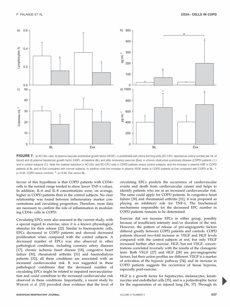
Circulating AC133+ cells, endothelial cell progenitors andangiogenetic growth factorsCirculating AC133+ cells, in COPD patients at rest, wereapproximately half the value found in control subjects andwere unaffected by endurance exercise in either group (fig. 7a).The decrease in AC133+ cells was also evident when expressedas absolute cell count per mL of blood volume both atrest (COPD: 2.5¡1.7 cells?mL-1; control: 6.1¡3.0) and after
endurance exercise (COPD: 2.7¡1.8 cells?mL-1; control: 6.5¡3.1cells?mL-1). Weak but significant direct relationships werefound in the entire group between baseline AC133+ cell countsand Pa,O2 (r250.21, p,0.05), FEV1 (r250.19, p,0.05) and thefitness level expressed as maximal load in W (r250.236,p,0.05).
A consistent decrease in the number of circulating endothelialprogenitors per mL of blood in COPD patients was also found.A specific clonogenic assay showed that their number wassignificantly decreased in COPD patients at rest (COPDpatients: 18.2¡7.2 EC-CFU ?mL-1 of blood; control subjects:30¡4 EC-CFU ?mL-1 of blood) and after exercise (COPDpatients: 20.1¡8.4 CFU-EC?mL-1 of blood; control subjects:32.2¡8.2 EC-CFU ?mL-1 of blood; fig. 7c).
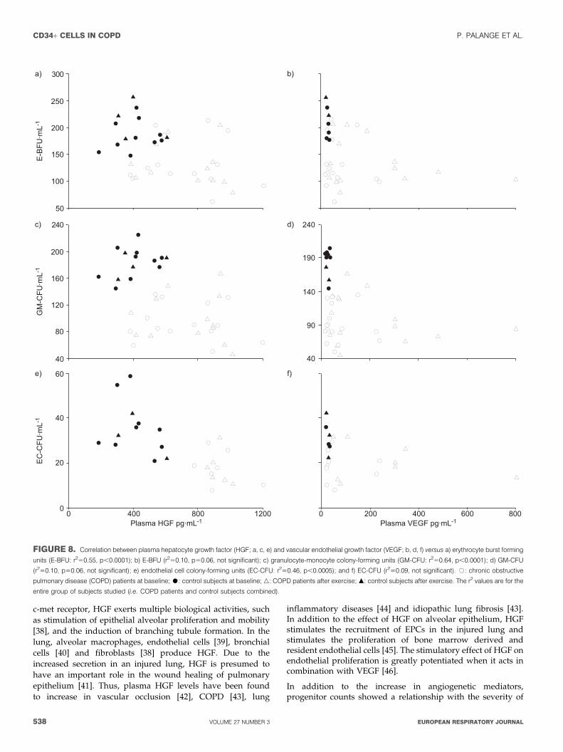
As for changes in plasma concentration of endothelial growthfactors, VEGF and HGF showed some marked and interestingdifferences (fig. 7b and d). Plasma VEGF concentration washigher in COPD patients than in control subjects at rest (COPDpatients: 69¡68 pg?mL-1; control subjects: 26¡8 pg?mL-1),and increased further after exercise (COPD patients:201¡205 pg?mL-1; control subjects: 27¡2 pg?mL-1). PlasmaHGF levels at rest were higher in COPD patients whencompared with the control subjects, but remained unchanged ineither group after exercise (fig. 7d). HGF, but not VEGF, levelscorrelated inversely with the results of clonogenetic tests (fig. 8).
Other biochemical measurementsMuscle enzymes (lactate dehydrogenase and creative kinase)and cortisol levels were similar in COPD patients and controlsubjects and remained unchanged in post-exercise samples(table 5). Plasma TNF-a concentration did not differ signifi-cantly between COPD and control groups, even though somedifferences were found between COPD patients with normaland low CD34+ cell counts as reported previously in the resultssection.
Plasma IL-6 levels were increased in COPD patients comparedwith controls (p,0.01 by ANOVA; table 5). IL-8 values did notdiffer significantly between groups but showed a largedispersion, with some COPD patients showing very highplasma concentrations at rest.
DISCUSSIONIn COPD patients with moderate-to-severe respiratory impair-ment, blood cell counts were normal while circulatinghaemopoietic and endothelial progenitor counts were greatlydecreased, when compared with control subjects. The numberof progenitor-derived colonies was correspondingly low(-30– -50%) in the COPD group. However, ,20% of COPDpatients, clinically and functionally similar to the rest of thegroup, showed normal progenitor counts associated with lessinflammatory activity as estimated by plasma TNF-a concen-trations. Endurance exercise did not affect circulating CD34+cells in either COPD patients or control subjects. The post-exercise increase in plasma VEGF suggested activation of thehypoxic pathway in COPD patients, but not in the controlsubjects. The endurance exercise in COPD patients appears tomake the initial hypoxic condition, observed at rest, moresevere and, therefore, stimulates VEGF release. In the entiresample, circulating progenitor counts correlated directly with
��
��
���23 45��23
��
FIGURE 5. Representative pictures of single colonies of erythrocyte burst
forming units (E-BFU) and granulocyte-monocyte colony-forming units (GM-CFU)
obtained in clonogenetic culture from the following. a) A chronic obstructive
pulmonary disease (COPD) patient with a low CD34+ cell count. b) A COPD patient
with a normal CD34+ cell count. c) A control subject. For E-BFU each box
represents an individual E-BFU colony, therefore, the E-BFU colonies from the
COPD patient with low CD34+ cells (a) are markedly smaller than those observed in
controls (c) or in a COPD patient with normal CD34+ cell count (b).
P. PALANGE ET AL. CD34+ CELLS IN COPD
cEUROPEAN RESPIRATORY JOURNAL VOLUME 27 NUMBER 3 535

V9O2peak and indices of COPD severity such as FEV1 and Pa,O2
tension. Overall, the current data indicates that the bonemarrow is an important, previously unrecognised, systemictarget in the pathophysiology of COPD.
To the best of the authors’ knowledge, this study is the first toreport that circulating progenitors are decreased in moderate-to-severe COPD. However, the current data only allow theindirect estimation of haemopoietic function, and detailedinvestigations on possible mechanism(s) will require invasivebone marrow sampling. Despite this limitation, the currentresults do provide some information. Normal peripheral bloodcell counts in COPD patients do not support decreased cellproduction and/or release, as a likely explanation for thecurrent authors findings, at least where the haemopoieticcompartment is concerned. WBC counts were higher in COPDpatients than control subjects and platelet counts were in thenormal range. Only one subject in each group showed slightlyreduced haemoglobin concentration, confirming the lowprevalence of anaemia in COPD [5], whereas 80% of theCOPD patients showed greatly reduced CD34+ cells comparedwith the control subjects.
Low circulating progenitor counts may be secondary to defectsin cell mobilisation mechanisms. The current authors testedthis hypothesis, but found normal expression of adhesion
molecules implicated in progenitor homing and migration(very late activation antigen-4 (VLA-4), chemokine (CC motif)receptor 5 (CCR5), C-X-C motif chemokine receptor 4(CXCR4)) on CD34+ cells, lymphocytes and monocytes ofCOPD patients (unpublished observations: Testa U., IstitutoSuperiore di Sanita’, Rome, Italy). The data in COPD patients,herefore, mimicked the low proliferation of CD34+ cells recentlyfound in patients with chronic ischemic heart disease [18],which occurred in the absence of obvious mobilisation defects.
The present authors also tested whether insufficient haemo-poietic stimulation may occur in COPD patients. Plasma Flt3ligand and KL concentrations, both being important haemo-poietic growth factors [26], were similar in COPD patients andcontrols. Furthermore, Flt3 ligand concentrations increasedafter exercise in COPD patients, similar to data obtained inathletes after intense exercise [20, 21]. No difference was foundin Flt3 ligand expression on CD34+ cells or monocytes betweenCOPD and controls (data not shown), discounting thepossibility that the increase in Flt3 ligand could be secondaryto a lack of receptors in COPD patients. Altogether, insufficientbone marrow stimulation in COPD is not supported by thecurrent results.
Inflammatory mediators could be involved in the pathogenesisof low circulating precursor counts in COPD. The evidence in
�
�
��
�1
��
,
�
�&!�
-��
&�$�
��
����
�����
�� ��
�,�1��67��..)$
�
����
�
�
�����
���
��
� �
��
��
�
�
��
��
�
��
�1���,���6�7���..)$
�
� ���
�
�
� ���
�
�
�
�
�
�
�
�
�
��
�
��
�
�
�
�
�
�8
��
�1
��
,
�
�&!�
-��
&�$�
��
����
�����
�� ��
8�8�2�9�:29�
��
��������
�
��
�
�
� �
��
�
�
�
�
��
�
�
8� 81 8� 8, 8�
"�
�8�;7�/��(� �.&���
�
�
�
�
�
�� ���
�
��
�
�
�
�
�
�
��
�
�
�
��
�
�
�8� �8� �81 �8, �8 �8� �8� �81 �8,
FIGURE 6. Correlations between CD34+ versus: a) arterial oxygen tension (Pa,O2; r250.30, p,0.05); b) carbon dioxide arterial tension (Pa,CO2); c) forced expiratory
volume in one second (FEV1)/forced vital capacity (FVC); and d) peak oxygen uptake (V9O2peak). $: chronic obstructive pulmonary disease (COPD) patients; #: control
subjects. 1 mmHg50.133 kPa.
CD34+ CELLS IN COPD P. PALANGE ET AL.
536 VOLUME 27 NUMBER 3 EUROPEAN RESPIRATORY JOURNAL
favour of this hypothesis is that COPD patients with CD34+cells in the normal range tended to show lower TNF-a values.In addition, IL-6 and IL-8 concentrations were, on average,higher in COPD patients than in the control subjects. No clearrelationship was found between inflammatory marker con-centrations and circulating progenitors. Therefore, more dataare necessary to confirm the role of inflammation in modulat-ing CD34+ cells in COPD.
Circulating EPCs were also assessed in the current study, witha special regard to exercise, since it is a known physiologicalstimulus for their release [22]. Similar to haemopoietic cells,EPCs decreased in COPD patients and showed decreasedproliferation when compared with the control subjects. Adecreased number of EPCs was also observed in otherpathological conditions, including coronary artery diseases[17], chronic ischemic heart disease [18], congestive heartfailure [30], rheumatoid arthritis [31] and haemodialysispatients [32], all these conditions are associated with anincreased cardiovascular risk. It was suggested in thesepathological conditions that the decreased number ofcirculating EPCs might be related to impaired neovascularisa-tion and could contribute to the increased cardiovascular riskobserved in these conditions. Importantly, a recent study byWERNER et al. [33] provided clear evidence that the level of
circulating EPCs predicts the occurrence of cardiovascularevents and death from cardiovascular causes and helps toidentify patients who are at an increased cardiovascular risk.The same could apply for COPD patients. In congestive heartfailure [30] and rheumatoid arthritis [31], it was proposed asplaying an inhibitory role for TNF-a. The biochemicalmechanisms responsible for the decreased EPC number inCOPD patients remains to be determined.
Exercise did not increase EPCs in either group, possiblybecause of insufficient intensity and/or duration of the test.However, the pattern of release of pro-angiogenetic factorsdiffered greatly between COPD patients and controls. COPDpatients showed two-fold increase in VEGF and HGF levelscompared with the control subjects at rest, but only VEGFincreased further after exercise. HGF, but not VEGF, concen-trations correlated inversely with the results of the clonogenictests. Both VEGF [27] and HGF [28] are pro-angiogeneticfactors, but their action profiles are different. VEGF is a markerof activation of the hypoxic pathway [34], and its increase inCOPD patients suggests the occurrence of tissue hypoxia,especially post-exercise.
HGF is a growth factor for hepatocytes, melanocytes, kerati-nocytes and endothelial cells [35], and is a pulmotrophic factorfor the regeneration of an injured lung [36, 37]. Through its
81
8�
8�
8
<.
/%��
< ��
�=
��
��
�
/$�.
��
��
�
�
�
�
�
�
0
�
�
�
���
��&�
��.
��
��
��
�
�
� ���
�
1
�
/$�.
��
"���
,
�
� ���
FIGURE 7. a) AC133+ cells, b) plasma vascular endothelial growth factor (VEGF), c) endothelial cell colony-forming units (EC-CFU, reported as colony number per mL of
blood) and d) plasma hepatocyte growth factor (HGF), at baseline (BL) and after endurance exercise (Exe), in chronic obstructive pulmonary disease (COPD) patients (&)
and in control subjects (h). Note the marked reduction in AC133+ and EC-CFU cells in COPD patients versus control subjects, and the increase in plasma HGF in COPD
patients at BL and at Exe compared with normal subjects. In addition note the increase in plasma VEGF levels in COPD patients at Exe compared with COPD at BL. *:
p,0.05, COPD versus controls; #: p,0.05, Exe versus BL.
P. PALANGE ET AL. CD34+ CELLS IN COPD
cEUROPEAN RESPIRATORY JOURNAL VOLUME 27 NUMBER 3 537
c-met receptor, HGF exerts multiple biological activities, such
as stimulation of epithelial alveolar proliferation and mobility
[38], and the induction of branching tubule formation. In the
lung, alveolar macrophages, endothelial cells [39], bronchial
cells [40] and fibroblasts [38] produce HGF. Due to the
increased secretion in an injured lung, HGF is presumed to
have an important role in the wound healing of pulmonary
epithelium [41]. Thus, plasma HGF levels have been found
to increase in vascular occlusion [42], COPD [43], lung
inflammatory diseases [44] and idiopathic lung fibrosis [43].In addition to the effect of HGF on alveolar epithelium, HGFstimulates the recruitment of EPCs in the injured lung andstimulates the proliferation of bone marrow derived andresident endothelial cells [45]. The stimulatory effect of HGF onendothelial proliferation is greatly potentiated when it acts incombination with VEGF [46].
In addition to the increase in angiogenetic mediators,progenitor counts showed a relationship with the severity of
�
��
�
�
��
�
�
���
23
�. ��
��
�
��
��
��
���
�
�
� ��
�
�
��
���
�
�
�
�
�
�
�
�
�
�
�
��
�
�
��
��
��
�
��
�
� ��
�
�
��
�
��
�
��
����
��
�
��
�
�
�
���
�
�
�
�
,
��
�1
��
45
��2
3�.
��
��
�
�
��
� �
�
�
�
�
�
��
�
�
�
�
�
� �
��
�
�
��
�
�
�
� �
�
��
�
�
�
�
�
�
�
"�
�
�
�
�
��
��
��
�
�
���
�
�
� �
�
��
��
��
���
�
� �����
��
�
�
�
��
����
�
�
1
��
��2
3�.
��
��
�
��
�
�
��,�
�
��
��
�
�
��
��
�
��
��
��
� ��
>���.��)42�/$�. ��
#�
��
�
�
�
�
��
�
��
��
�
�
�
���
�
�
��
,1��>���.��9�42�/$�. ��
FIGURE 8. Correlation between plasma hepatocyte growth factor (HGF; a, c, e) and vascular endothelial growth factor (VEGF; b, d, f) versus a) erythrocyte burst forming
units (E-BFU: r250.55, p,0.0001); b) E-BFU (r250.10, p50.06, not significant); c) granulocyte-monocyte colony-forming units (GM-CFU: r250.64, p,0.0001); d) GM-CFU
(r250.10, p50.06, not significant); e) endothelial cell colony-forming units (EC-CFU: r250.46, p,0.0005); and f) EC-CFU (r250.09, not significant). #: chronic obstructive
pulmonary disease (COPD) patients at baseline; $: control subjects at baseline; n: COPD patients after exercise; m: control subjects after exercise. The r2 values are for the
entire group of subjects studied (i.e. COPD patients and control subjects combined).
CD34+ CELLS IN COPD P. PALANGE ET AL.
538 VOLUME 27 NUMBER 3 EUROPEAN RESPIRATORY JOURNAL

hypoxaemia. The effects of low oxygen tensions on progenitorsin vivo are poorly defined, but the human bone marrow isknown to be a relatively hypoxic environment under physio-logical conditions [47]. In addition, hypoxic gradients may beimportant in regulating progenitor trafficking [48]. Exposure tohypoxia in vitro increased the maturation rate of CD34+ cells,while maintaining early precursors in a quiescent state [23, 24].The current authors did not observe preferential release of lessimmature precursors in more hypoxaemic COPD patients;rather, all CD34+ subpopulations tested were uniformlydecreased (table 4).
Depression in the number and function of circulatingprogenitors also correlated with the severity of airwayobstruction and the degree of physical fitness, suggesting thatdepletion of CD34+ cells may be proportional to COPDseverity. A large number of patients with COPD at differentstages need to be studied to confirm this hypothesis.
Finally, CD34+ cells may be low in COPD patients due to anincrease in peripheric utilisation. The current authors hypothe-sise that a high turnover of precursors may occur in COPD,possibly related to tissue repair. Skeletal muscle may be apotential target tissue for progenitors for the following reasons.1) CD34+ cells can migrate into muscle connective tissue andsatellite cell niches [49]. 2) Exercising mice showed a higherpercentage of bone marrow-derived muscle cells comparedwith sedentary mice [10], indicating precursor engraftment inresponse to muscle injury. In the current authors’ previousstudy, decreased CD34+ cell counts were found a few hoursafter a marathon race, suggesting peripheral utilisation ofprecursors [20]. Alternatively, decreased EPCs in COPDpatients may blunt the increase in muscle perfusion evokedby hypoxia [27]. Unfortunately, muscle biopsies were notobtained in the present study and association between low-progenitor counts and muscle damage remains speculative.
The major limitation of the present study is that the group ofsubjects studied was small and far from representative of theentire clinical spectrum of COPD. Nevertheless, the differencein CD34+ cells between COPD patients and age-matchedcontrols was large, indicating a likely relevant effect of thedisease on circulating precursors.
In conclusion, circulating CD34+ cells and endothelialprogenitor counts were decreased in most patients withmoderate-to-severe chronic obstructive pulmonary diseaseand correlated with hypoxaemia, severity of airway obstruc-tion, and peak oxygen consumption. The current data suggestsa possible link between systemic inflammation and a decreasein circulating progenitors, as suggested in other pathologicalconditions. The current authors conclude that bone marrowshould be considered as a previously unrecognised systemictarget of chronic obstructive pulmonary disease.
REFERENCES1 Agusti AG, Noguera A, Sauleda J, Sala E, Pons J,
Busquets X. Systemic effects of chronic obstructivepulmonary disease. Eur Respir J 2003; 21: 347–360.
2 Noguera A, Busquets X, Sauleda J, Villaverde JM,MacNee W, Agustı AG. Expression of adhesion moleculesand G proteins in circulating neutrophils in chronicobstructive pulmonary disease. Am J Respir Crit Care Med1998; 158: 1664–1668.
3 Di Stefano A, Capelli A, Lusuardi M, et al. Severity ofairflow limitation is associated with severity of airwayinflammation in smokers. Am J Respir Crit Care Med 1998;158: 1277–1285.
4 Van Eeden SF, Hogg JC. The response of human bonemarrow to chronic cigarette smoking. Eur Respir J 2000; 15:915–921.
5 John M, Hoernig S, Doehner W, Okonko DD, Witt C,Anker SD. Anemia and inflammation in COPD. Chest 2005;127: 825–829.
6 Chambellan A, Chailleux E, Similowski T, and theANTADIR Observatory Group. Prognostic value of hema-tocrit in patients with severe chronic obstructive pulmon-ary disease receiving long term oxygen therapy. Chest 2005;128: 1201–1208.
7 Terstappen LW, Huang S, Safford M, Lansdorp PM,Loken MR. Sequential generation of haematopoieticcolonies derived from single nonlineage committedCD34+CD38- progenitor cells. Blood 1991; 77: 1218–1227.
8 Kotton DN, Ma BY, Cardoso WV, et al. Bone marrow-derived cells as progenitors of lung alveolar epithelium.Development 2001; 128: 5181–5188.
9 Suratt BT, Cool CD, Serls AE, et al. Human pulmonarychimerism after haematopoietic stem cell transplantation.Am J Respir Crit Care Med 2003; 168: 318–322.
10 LaBarge MA, Blau HM. Biological progression from adultbone marrow to mononucleate muscle stem cells tomultinucleate muscle fiber in response to injury. Cell2002; 111: 589–601.
11 Agusti AG, Sauleda J, Miralles C, et al. Skeletal muscleapoptosis and weight loss in chronic obstructive pulmon-ary disease. Am J Respir Crit Care Med 2002; 166: 485–489.
TABLE 5 Biochemical analyses on plasma/serum inchronic obstructive pulmonary disease (COPD)patients and control subjects at baseline (BL)and after endurance exercise (Exe)
COPD patients Control subjects
BL Exe BL Exe
LDH U?L-1 302¡73 326¡130 278¡65 325¡46
CK U?L-1 89¡56 114¡66 113¡63 130¡52
TNF-a pg?mL-1 2.17¡1.05 1.75¡0.76 1.36¡1.12 1.51¡1.30
IL-6 pg?mL-1 3.7¡1.7 4.0¡1.9 2.5¡1.2 2.4¡1.2
IL-8 pg?mL-1 225¡526 64¡108 53¡72 44¡42
Cortisol ng?mL-1 18.0¡6.6 20.9¡6.6 22.4¡8.5 18.7¡2.3
Flt3 ligand pg?mL-1 122.4¡89.4 158.8¡100.7* 118.8¡84.1 124.6¡89.5
VEGF pg?mL-1 69¡68# 201¡205#," 26¡8 27¡5
Data are presented as mean¡SD. LDH: lactate dehydrogenase; CK: creative
kinase; TNF-a: tumour necrosis factor-a; IL: interleukin; Flt3: FMS-like tyrosine
kinase 3; VEGF: vascular endothelial growth factor. *: p,0.05 versus baseline
(paired t-test); #: p,0.05 for comparison between COPD and control groups
(Mann-Whitney test); ": p,0.03 versus baseline (Wilcoxon test).
P. PALANGE ET AL. CD34+ CELLS IN COPD
cEUROPEAN RESPIRATORY JOURNAL VOLUME 27 NUMBER 3 539
12 Gosker HR, Kubat B, Schaart G, van der Vusse GJ,Wouters EF, Schols AM. Myopathological features inskeletal muscle of patients with chronic obstructivepulmonary disease. Eur Respir J 2003; 22: 280–285.
13 Agustı A, Morla M, Sauleda J, Saus C, Busquets X. NF-kBactivation and iNOS upregulation in skeletal muscle ofpatients with COPD and low body weight. Thorax 2004; 59:483–487.
14 Gehling UM, Ergun S, Schumacher U, et al. In vitrodifferentiation of endothelial cells from AC133-positiveprogenitor cells. Blood 2000; 95: 3106–3112.
15 Assmus B, Schachinger V, Teupe C, et al. Transplantationof progenitor cells and regeneration enhancement in acutemyocardial infarction (TOPCARE-AMI). Circulation 2002;106: 3009–3017.
16 Tateishi-Yuyama E, Matsubara H, Murohara T, et al.Therapeutic angiogenesis for patients with limb ischaemiaby autologous transplantation of bone-marrow cells: a pilotstudy and a randomised controlled trial. Lancet 2002; 360:427–435.
17 Vasa M, Fichtlscherer S, Aicher A, et al. Number andmigratory activity of circulating endothelial progenitorcells inversely correlate with risk factors for coronaryartery disease. Circ Res 2001; 89: E1–E7.
18 Heeschen C, Lehmann R, Honold J, et al. Profoundlyreduced neovascularization capacity of bone marrowmononuclear cells derived from patients with chronicischemic heart disease. Circulation 2004; 109: 1615–1622.
19 Laufs U, Werner N, Link A, et al. Physical trainingincreases endothelial progenitor cells, inhibits neointimaformation, and enhances angiogenesis. Circulation 2004;109: 220–226.
20 Bonsignore MR, Morici G, Santoro A, et al. Circulatinghaematopoietic progenitor cells in runners. J Appl Physiol2002; 93: 1691–1697.
21 Morici G, Zangla D, Santoro A, et al. Supramaximalexercise mobilizes haematopoietic progenitors and reticu-locytes in athletes. Am J Physiol Regul Integr Comp Physiol2005; 289: 496–503.
22 Rehman J, Li J, Parvathaneni L, et al. Exercise acutelyincreases circulating endothelial progenitor cells andmonocyte-/macrophage-derived angiogenic cells. J AmColl Cardiol 2004; 43: 2314–2318.
23 Danet GH, Pan Y, Luongo JL, Bonnet DA, Simon MC.Expansion of human SCID-repopulating cells underhypoxic conditions. J Clin Invest 2003; 112: 126–135.
24 Hevehan DL, Papoutsakis ET, Miller WM. Physiologicallysignificant effects of pH and oxygen tension on granulo-poiesis. Exp Hematol 2000; 28: 267–275.
25 Palange P, Forte S, Onorati P, Manfredi F, Serra P,Carlone S. Ventilatory and metabolic adaptations towalking and cycling in patients with COPD. J ApplPhysiol 2000; 88: 1715–1720.
26 Lyman SD, Jacobsen SE. c-kit ligand and Flt3 ligand: stem/progenitor cell factors with overlapping yet distinctactivities. Blood 1998; 91: 1101–1134.
27 Gavin TP, Robinson CB, Yeager RC, England JA,Nifong LW, Hickner RC. Angiogenic growth factorresponse to acute systemic exercise in human skeletalmuscle. J Appl Physiol 2004; 96: 19–24.
28 Bussolino F, Di Renzo MF, Ziche M, et al. Hepatocytegrowth factor is a potent angiogenic factor which stimu-lates endothelial cell motility and growth. J Cell Biol 1992;119: 629–641.
29 Ostrowski K, Rohde T, Asp S, Schjerling P, Pedersen BK.Pro- and anti-inflammatory cytokine balance in strenuousexercise in humans. J Physiol 1999; 515: 287–291.
30 Valgimigli M, Rigolin GM, Fucili A, et al. CD34+ andendothelial progenitor cells in patients with variousdegrees of congestive heart failure. Circulation 2004; 110:1209–1212.
31 Grisar J, Aletaha D, Steiner CW, et al. Depletion ofendothelial progenitor cells in the peripheral blood ofpatients with rheumatoid arthritis. Circulation 2005; 111:204–211.
32 Eizawa T, Murakami Y, Mtsui K, et al. Circulatingendothelial progenitor cells are reduced in hemodialysispatients. Curr Med Res Opin 2003; 19: 627–633.
33 Werner N, Kosiol S, Schiegl T, et al. Circulating endothelialprogenitor cells and cardiovascular outcomes. N Engl JMed 2005; 353: 999–1007.
34 Huang LE, Bunn HF. Hypoxia-inducible factor and itsbiomedical relevance. J Biol Chem 2003; 278: 19575–19578.
35 Matsumoto K, Nakamura T. Hepatocyte growth factor:molecular structure and implications for a central role inliver regeneration. J Gastroenterol Hepatol 1991; 6: 509–519.
36 Sakamaki Y, Matsumoto K, Mizuno S, Nakamura T.Hepatocyte growth factor stimulates proliferation ofrespiratory epithelial cells during postpneumonectomycompensatory lung growth in mice. Am J Respir Cell MolBiol 2002; 26: 525–533.
37 Yanagita K, Matsumoto K, Sekiguchi K, Ishibashi H,Niho Y, Nakamura T. Hepatocyte growth factor may actas a pulmotrophic factor on regeneration after acute lunginjury. J Biol Chem 1993; 268: 21212–21217.
38 Stoker M, Gherardi E, Perryman M, Gray J. Scatter factor isa fibroblast-derived modulator of epithelial cell mobility.Nature 1987; 327: 239–242.
39 Wolf HK, Zarnegar R, Michalopoulos GK. Localization ofhepatocyte growth factor in human and rat tissues: animmunochemical study. Hepatology 1991; 14: 488–494.
40 Yanagita K, Nagaike M, Ishibashi H, Niho Y, Matsumoto K,Nakamura T. Lung may have endocrine function produ-cing hepatocyte growth factor in response to injury ofdistal organs. Biochem Biophys Res Commun 1992; 182:802–809.
41 Sato N, Takahashi H. Hepatocyte growth factor promotesgrowth and lumen formation of fetal lung epithelial cells inprimary culture. Respirology 1997; 3: 185–191.
42 Yoshitomi Y, Kojima S, Umemoto T, et al. Serumhepatocyte growth factor in patients with peripheralarterial occlusive disease. J Clin Endocrinol Metab 1999; 84:2425–2428.
43 Aharinejad S, Taghavi S, Klepetko W, Abraham D.Prediction of lung-transplant rejection by hepatocytesgrowth factor. Lancet 2004; 363: 1503–1508.
44 Huang MS, Tsai MS, Wang TH, et al. Serum hepatocytegrowth factor levels in patients with inflammatory lungdiseases. Kaohsiung J Med Sci 1999; 15: 195–201.
45 Ishizawa K, Kubo H, Yamada M, et al. Hepatocyte growthfactor induces angiogenesis in injured lungs through
CD34+ CELLS IN COPD P. PALANGE ET AL.
540 VOLUME 27 NUMBER 3 EUROPEAN RESPIRATORY JOURNAL
mobilizing endothelial progenitor cells. Biochem BiophysRes Commun 2004; 324: 276–280.
46 Gerritsen ME. HGF and VEGF: a dynamic duo. Cir Res2005; 96: 272–273.
47 Harrison JS, Rameshwar P, Chang V, Bandari P. Oxygensaturation in the bone marrow of healthy volunteers. Blood2002; 99: 394.
48 Ceradini DJ, Kulkarni AR, Callaghan MJ, et al. Progenitorcell trafficking is regulated by hypoxic gradientsthrough HIF-1 induction of SDF-1. Nat Med 2004; 10:858–864.
49 Dreyfus PA, Chretien F, Chazaud B, et al. Adult bone-marrow-derived stem cells in muscle connective tissue andsatellite niches. Am J Pathol 2004; 164: 773–779.
P. PALANGE ET AL. CD34+ CELLS IN COPD
EUROPEAN RESPIRATORY JOURNAL VOLUME 27 NUMBER 3 541