Decreased volume of the brain reward system in alcoholism
11
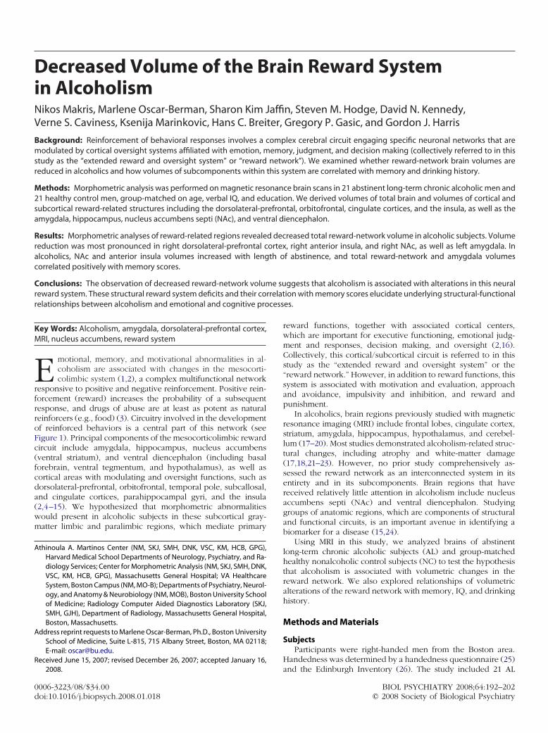
Decreased Volume of the Brain Reward System in Alcoholism Nikos Makris, Marlene Oscar-Berman, Sharon Kim Jaffin, Steven M. Hodge, David N. Kennedy, Verne S. Caviness, Ksenija Marinkovic, Hans C. Breiter, Gregory P. Gasic, and Gordon J. Harris Background: Reinforcement of behavioral responses involves a complex cerebral circuit engaging specific neuronal networks that are modulated by cortical oversight systems affiliated with emotion, memory, judgment, and decision making (collectively referred to in this study as the “extended reward and oversight system” or “reward network”). We examined whether reward-network brain volumes are reduced in alcoholics and how volumes of subcomponents within this system are correlated with memory and drinking history. Methods: Morphometric analysis was performed on magnetic resonance brain scans in 21 abstinent long-term chronic alcoholic men and 21 healthy control men, group-matched on age, verbal IQ, and education. We derived volumes of total brain and volumes of cortical and subcortical reward-related structures including the dorsolateral-prefrontal, orbitofrontal, cingulate cortices, and the insula, as well as the amygdala, hippocampus, nucleus accumbens septi (NAc), and ventral diencephalon. Results: Morphometric analyses of reward-related regions revealed decreased total reward-network volume in alcoholic subjects. Volume reduction was most pronounced in right dorsolateral-prefrontal cortex, right anterior insula, and right NAc, as well as left amygdala. In alcoholics, NAc and anterior insula volumes increased with length of abstinence, and total reward-network and amygdala volumes correlated positively with memory scores. Conclusions: The observation of decreased reward-network volume suggests that alcoholism is associated with alterations in this neural reward system. These structural reward system deficits and their correlation with memory scores elucidate underlying structural-functional relationships between alcoholism and emotional and cognitive processes. Key Words: Alcoholism, amygdala, dorsolateral-prefrontal cortex, MRI, nucleus accumbens, reward system E motional, memory, and motivational abnormalities in al- coholism are associated with changes in the mesocorti- colimbic system (1,2), a complex multifunctional network responsive to positive and negative reinforcement. Positive rein- forcement (reward) increases the probability of a subsequent response, and drugs of abuse are at least as potent as natural reinforcers (e.g., food) (3). Circuitry involved in the development of reinforced behaviors is a central part of this network (see Figure 1). Principal components of the mesocorticolimbic reward circuit include amygdala, hippocampus, nucleus accumbens (ventral striatum), and ventral diencephalon (including basal forebrain, ventral tegmentum, and hypothalamus), as well as cortical areas with modulating and oversight functions, such as dorsolateral-prefrontal, orbitofrontal, temporal pole, subcallosal, and cingulate cortices, parahippocampal gyri, and the insula (2,4 –15). We hypothesized that morphometric abnormalities would present in alcoholic subjects in these subcortical gray- matter limbic and paralimbic regions, which mediate primary reward functions, together with associated cortical centers, which are important for executive functioning, emotional judg- ment and responses, decision making, and oversight (2,16). Collectively, this cortical/subcortical circuit is referred to in this study as the “extended reward and oversight system” or the “reward network.” However, in addition to reward functions, this system is associated with motivation and evaluation, approach and avoidance, impulsivity and inhibition, and reward and punishment. In alcoholics, brain regions previously studied with magnetic resonance imaging (MRI) include frontal lobes, cingulate cortex, striatum, amygdala, hippocampus, hypothalamus, and cerebel- lum (17–20). Most studies demonstrated alcoholism-related struc- tural changes, including atrophy and white-matter damage (17,18,21–23). However, no prior study comprehensively as- sessed the reward network as an interconnected system in its entirety and in its subcomponents. Brain regions that have received relatively little attention in alcoholism include nucleus accumbens septi (NAc) and ventral diencephalon. Studying groups of anatomic regions, which are components of structural and functional circuits, is an important avenue in identifying a biomarker for a disease (15,24). Using MRI in this study, we analyzed brains of abstinent long-term chronic alcoholic subjects (AL) and group-matched healthy nonalcoholic control subjects (NC) to test the hypothesis that alcoholism is associated with volumetric changes in the reward network. We also explored relationships of volumetric alterations of the reward network with memory, IQ, and drinking history. Methods and Materials Subjects Participants were right-handed men from the Boston area. Handedness was determined by a handedness questionnaire (25) and the Edinburgh Inventory (26). The study included 21 AL Athinoula A. Martinos Center (NM, SKJ, SMH, DNK, VSC, KM, HCB, GPG), Harvard Medical School Departments of Neurology, Psychiatry, and Ra- diology Services; Center for Morphometric Analysis (NM, SKJ, SMH, DNK, VSC, KM, HCB, GPG), Massachusetts General Hospital; VA Healthcare System, Boston Campus (NM, MO-B); Departments of Psychiatry, Neurol- ogy, and Anatomy & Neurobiology (NM, MOB), Boston University School of Medicine; Radiology Computer Aided Diagnostics Laboratory (SKJ, SMH, GJH), Department of Radiology, Massachusetts General Hospital, Boston, Massachusetts. Address reprint requests to Marlene Oscar-Berman, Ph.D., Boston University School of Medicine, Suite L-815, 715 Albany Street, Boston, MA 02118; E-mail: [email protected]. Received June 15, 2007; revised December 26, 2007; accepted January 16, 2008. BIOL PSYCHIATRY 2008;64:192–202 0006-3223/08/$34.00 doi:10.1016/j.biopsych.2008.01.018 © 2008 Society of Biological Psychiatry
Transcript of Decreased volume of the brain reward system in alcoholism
DiNV
Bmsr
M2sa
Rrac
Crr
KM
ErfrroFc(fcda(wm
A
A
R
0d
ecreased Volume of the Brain Reward Systemn Alcoholismikos Makris, Marlene Oscar-Berman, Sharon Kim Jaffin, Steven M. Hodge, David N. Kennedy,erne S. Caviness, Ksenija Marinkovic, Hans C. Breiter, Gregory P. Gasic, and Gordon J. Harris
ackground: Reinforcement of behavioral responses involves a complex cerebral circuit engaging specific neuronal networks that areodulated by cortical oversight systems affiliated with emotion, memory, judgment, and decision making (collectively referred to in this
tudy as the “extended reward and oversight system” or “reward network”). We examined whether reward-network brain volumes areeduced in alcoholics and how volumes of subcomponents within this system are correlated with memory and drinking history.
ethods: Morphometric analysis was performed on magnetic resonance brain scans in 21 abstinent long-term chronic alcoholic men and1 healthy control men, group-matched on age, verbal IQ, and education. We derived volumes of total brain and volumes of cortical andubcortical reward-related structures including the dorsolateral-prefrontal, orbitofrontal, cingulate cortices, and the insula, as well as themygdala, hippocampus, nucleus accumbens septi (NAc), and ventral diencephalon.
esults: Morphometric analyses of reward-related regions revealed decreased total reward-network volume in alcoholic subjects. Volumeeduction was most pronounced in right dorsolateral-prefrontal cortex, right anterior insula, and right NAc, as well as left amygdala. Inlcoholics, NAc and anterior insula volumes increased with length of abstinence, and total reward-network and amygdala volumesorrelated positively with memory scores.
onclusions: The observation of decreased reward-network volume suggests that alcoholism is associated with alterations in this neuraleward system. These structural reward system deficits and their correlation with memory scores elucidate underlying structural-functional
elationships between alcoholism and emotional and cognitive processes.ey Words: Alcoholism, amygdala, dorsolateral-prefrontal cortex,RI, nucleus accumbens, reward system
motional, memory, and motivational abnormalities in al-coholism are associated with changes in the mesocorti-colimbic system (1,2), a complex multifunctional network
esponsive to positive and negative reinforcement. Positive rein-orcement (reward) increases the probability of a subsequentesponse, and drugs of abuse are at least as potent as naturaleinforcers (e.g., food) (3). Circuitry involved in the developmentf reinforced behaviors is a central part of this network (seeigure 1). Principal components of the mesocorticolimbic rewardircuit include amygdala, hippocampus, nucleus accumbensventral striatum), and ventral diencephalon (including basalorebrain, ventral tegmentum, and hypothalamus), as well asortical areas with modulating and oversight functions, such asorsolateral-prefrontal, orbitofrontal, temporal pole, subcallosal,nd cingulate cortices, parahippocampal gyri, and the insula2,4 –15). We hypothesized that morphometric abnormalitiesould present in alcoholic subjects in these subcortical gray-atter limbic and paralimbic regions, which mediate primary
thinoula A. Martinos Center (NM, SKJ, SMH, DNK, VSC, KM, HCB, GPG),Harvard Medical School Departments of Neurology, Psychiatry, and Ra-diology Services; Center for Morphometric Analysis (NM, SKJ, SMH, DNK,VSC, KM, HCB, GPG), Massachusetts General Hospital; VA HealthcareSystem, Boston Campus (NM, MO-B); Departments of Psychiatry, Neurol-ogy, and Anatomy & Neurobiology (NM, MOB), Boston University Schoolof Medicine; Radiology Computer Aided Diagnostics Laboratory (SKJ,SMH, GJH), Department of Radiology, Massachusetts General Hospital,Boston, Massachusetts.
ddress reprint requests to Marlene Oscar-Berman, Ph.D., Boston UniversitySchool of Medicine, Suite L-815, 715 Albany Street, Boston, MA 02118;E-mail: [email protected].
eceived June 15, 2007; revised December 26, 2007; accepted January 16,
2008.006-3223/08/$34.00oi:10.1016/j.biopsych.2008.01.018
reward functions, together with associated cortical centers,which are important for executive functioning, emotional judg-ment and responses, decision making, and oversight (2,16).Collectively, this cortical/subcortical circuit is referred to in thisstudy as the “extended reward and oversight system” or the“reward network.” However, in addition to reward functions, thissystem is associated with motivation and evaluation, approachand avoidance, impulsivity and inhibition, and reward andpunishment.
In alcoholics, brain regions previously studied with magneticresonance imaging (MRI) include frontal lobes, cingulate cortex,striatum, amygdala, hippocampus, hypothalamus, and cerebel-lum (17–20). Most studies demonstrated alcoholism-related struc-tural changes, including atrophy and white-matter damage(17,18,21–23). However, no prior study comprehensively as-sessed the reward network as an interconnected system in itsentirety and in its subcomponents. Brain regions that havereceived relatively little attention in alcoholism include nucleusaccumbens septi (NAc) and ventral diencephalon. Studyinggroups of anatomic regions, which are components of structuraland functional circuits, is an important avenue in identifying abiomarker for a disease (15,24).
Using MRI in this study, we analyzed brains of abstinentlong-term chronic alcoholic subjects (AL) and group-matchedhealthy nonalcoholic control subjects (NC) to test the hypothesisthat alcoholism is associated with volumetric changes in thereward network. We also explored relationships of volumetricalterations of the reward network with memory, IQ, and drinkinghistory.
Methods and Materials
SubjectsParticipants were right-handed men from the Boston area.
Handedness was determined by a handedness questionnaire (25)
and the Edinburgh Inventory (26). The study included 21 ALBIOL PSYCHIATRY 2008;64:192–202© 2008 Society of Biological Psychiatry
ihsw
aBcTpePNfbs
N
tapnpprfiecnwmedi
Fs(rdattt
N. Makris et al. BIOL PSYCHIATRY 2008;64:192–202 193
ndividuals, abstinent from alcohol at least 4 weeks, and 21ealthy NC subjects (Table 1). Participants were native Englishpeakers with comparable socioeconomic backgrounds. Groupsere comparable with respect to demographic variables.Participation was solicited from newspaper and Web-based
dvertisements and from Boston University Medical Center,oston Veterans Affairs (VA) Healthcare System, and VA after-are programs. Twenty participants (12 NC, 8 AL) were veterans.his study was approved by the institutional review boards of thearticipating institutions. Informed consent was obtained fromach subject before neuropsychological testing and scanning.articipants were reimbursed for time and travel expenses.eurobehavioral and psychiatric evaluations typically required
rom 7 to 9 hours over 2 or more days. Participants had frequentreaks, and sessions were discontinued and rescheduled if aubject indicated fatigue.
eurobehavioral and Psychiatric EvaluationsParticipants underwent medical history interview and vision
esting, plus a series of questionnaires (e.g., handedness, alcoholnd drug use) to ensure they met inclusion criteria. Participantserformed a computer-assisted, shortened version of the Diag-ostic Interview Schedule (DIS) (27) that provides lifetimesychiatric diagnoses according to DSM-IV (28) criteria. Partici-ants were excluded if any source (DIS scores, hospital records,eferrals, or personal interviews) indicated they had one of theollowing: history of neurological dysfunction (e.g., major headnjury with loss of consciousness greater than 15 min, stroke,pilepsy, or seizures unrelated to alcohol withdrawal); electro-onvulsive therapy; major psychiatric disorder (e.g., schizophre-ia or primary depression); symptoms of clinical depressionithin the 6 months before testing; current use of psychoactiveedication; history of abuse of drugs besides alcohol; clinical
vidence of active hepatic disease; history of serious learningisability or dyslexia; and uncorrected abnormal vision or hear-
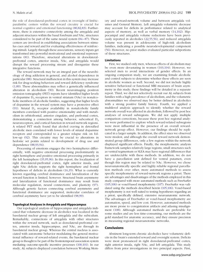
igure 1. Brain regions involved in the extended reward and oversightystem. Prefrontal and cingulate regions connect to the nucleus accumbensNAc) in the ventral striatum, the midbrain ventral tegmental area (VTA), andeciprocally with other limbic system structures (limbic brainstem, amyg-ala, and hippocampus). Limbic structures also interconnect with the NAcnd to the basal forebrain (substantia innominata, or sublenticular ex-ended amygdala [SLEA]). The VTA projects to the NAc (reciprocally), to thehalamus and hypothalamus, and to prefrontal cortex. The NAc projects tohe thalamus, which projects to prefrontal cortex.
ng problem.
Participants received a structured interview regarding theirdrinking patterns, including length of abstinence and years ofheavy drinking. A Quantity Frequency Index (QFI), which factorsthe amount, type, and frequency of alcoholic usage over the past6 months (for the nonalcoholic subjects) or over the 6 monthspreceding cessation of drinking (for the alcoholic subjects) wascalculated for each participant (29). Heavy drinking was quanti-fied as greater than 21 drinks per week (one drink: 355 ml beer,148 ml wine, or 44 ml hard liquor). The AL group had QFI of11.2 � 9.4, had heavy drinking for 18.3 � 8.5 years, and hadbeen sober for 5.9 � 10.4 years. The NC group had QFI of .4 �.5. AL participants met DSM-IV (28) criteria for alcohol abuse anddependence for a period of at least 5 years in their lives and hadabstained from alcohol for at least 4 weeks before testing.
Tests of intelligence, memory, and affect were administered,including the Wechsler Adult Intelligence Scale, 3rd edition(WAIS-III, for Verbal IQ, Performance IQ, and Full-Scale IQ) (30),Wechsler Memory Scale, 3rd edition (WMS-III, for GeneralMemory and Working Memory) (31), Hamilton Depression Scale(32), Profile of Mood States (POMS) (33), and Multiple AffectAdjective Check List (MAACL) (34). Subtests of the WAIS-III thathave been reported as sensitive to alcohol-related visuospatialdysfunction are Digit Symbol, Picture Arrangement, Block De-sign, and Object Assembly (35–37). In addition, subjects under-went the following tests sensitive to frontal brain systems: TrailMaking Test versions A and B (38); a computer scored (39)Wisconsin Card Sorting Test (WCST) (40,41); and the Con-trolled Oral Word Association Test (COWAT) (42).
MRI AcquisitionThe MRI scans were obtained at Massachusetts General
Hospital (MGH) on a Siemens 3-Tesla Trio scanner (SiemensMedical Solutions USA, Inc., Malvern, Pennsylvania). Imageacquisitions included sagittal scout, T2-weighted turbo spin echo(T2-TSE) (to rule out gross pathology), and two T1-weightedmagnetization prepared rapid gradient echo (MP-RAGE) seriesfor volumetric analysis (repetition time � 2530 msec, echo time� 3.31 msec, inversion time � 1100 msec, flip angle � 7°, fieldof view � 256 mm, slice thickness � 1.33 mm, number of slices� 128 contiguous, sagittal images of the entire brain, matrix �256 � 256, number of excitations � 2). The two MP-RAGE serieswere averaged, then the averaged series was resliced in astandard coronal three-dimensional brain coordinate system(43). Images were reformatted to standard spatial orientation, butnot rescaled in size.
MRI Morphometric AnalysisImage analyses followed semiautomated procedures devel-
oped by the Center for Morphometric Analysis at MGH (44–46).Intracranial volumes were segmented on T2-TSE images becausecerebrospinal fluid has a bright T2-TSE signal, allowing cleardemarcation within the intracranial vault (meninges/dura). Graymatter, white matter, and ventricles were segmented on T1-weighted images using a computer-assisted approach (44). Graymatter was then subdivided into cortical and subcortical compo-nents. Neocortex was subdivided further into parcellation units,involving a number of manual and computer-assisted operations(47). Cortical subcomponents of the reward network werederived: dorsolateral-prefrontal (defined as the sum of the dor-solateral superior-frontal and middle-frontal gyri, approximatingBrodmann’s cytoarchitechtonic areas 8, 9, and 46), insula, sub-callosal, orbitofrontal, and cingulate cortices, parahippocampal
gyrus, and temporal pole (see Figures 2 and 3). Gray matterwww.sobp.org/journal
sawha(vnfch(
eogtl
) (29).
194 BIOL PSYCHIATRY 2008;64:192–202 N. Makris et al.
w
ubcortical structures in the reward network included NAc (46),mygdala, hippocampus (44,48), and ventral diencephalon (49),hich according to our morphometric definition contains theypothalamus, basal forebrain, and sublenticular extendedmygdala (SLEA), as well as a large portion of ventral tegmentumwhich is included in our ventral diencephalon region by con-ention although part of midbrain). For comparisons to reward-etwork regions, we included analyses of non-reward-relatedrontal cortex (“frontal pole,” defined in each hemisphere asortex anterior to a coronal plane at the rostral end of the anteriororizontal ramus of the Sylvian fissure [47]), sensory cortexcuneal cortex), and subcortical (dorsal striatum) regions.
Segmentation and cortical parcellation were carried out by anxperienced research assistant (SKJ) with training in neuroanat-my, supervised by our neuroanatomist (NM). Blindness ofroup assignment was maintained during analysis. High interra-er and intrarater reliability of these methods has been estab-
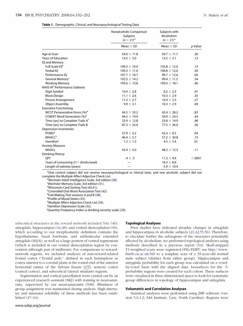
Table 1. Demographic, Clinical, and Neuropsychologic
Nona
Age at ScanYears of EducationIQ and Memory
Full Scale IQb
Verbal IQPerformance IQGeneral Memoryc
Working MemoryWAIS-IIIb Performance Subtests
Digit SymbolBlock DesignPicture ArrangementObject Assembly
Executive FunctioningWCST Perseverative Errors (%)d
COWAT Word Generation (%)e
Time (sec) to Complete Trails Af
Time (sec) to Complete Trails BDepression Inventories
POMSg
MAACLh
Hamiltoni
Anxiety MeasureMAACL
Drinking HistoryQFIj
Years of consuming 21� drinks/weekLength of sobriety (years)
aOne control subject did not receive neuropsychocomplete the Multiple Affect Adjective Check List.
bWechsler Adult Intelligence Scale, 3rd edition (30).cWechsler Memory Scale, 3rd edition (31).dWisconsin Card Sorting Test (40,41).eControlled Oral Word Association Test (42).fTrail Making Test versions A and B (38).gProfile of Mood States (33).hMultiple Affect Adjective Check List (34).iHamilton Depression Scale (32).jQuantity-Frequency Index (a drinking severity scale
ished (47–54).
ww.sobp.org/journal
Topological AnalysesPrior studies have indicated atrophic changes in amygdala
and hippocampus in alcoholic subjects (21,22,55,56). Therefore,to elucidate further the subregions of the structures potentiallyaffected by alcoholism, we performed topological analyses usingmethods described in a previous report (54). Skull-strippedT1-weighted scans were registered (FSL/FLIRT: see http://www.fmrib.ox.ac.uk/fsl) to a template scan of a 35-year-old normalmale subject (distinct from either group). Hippocampus andamygdala probability for each group was calculated on a voxel-by-voxel basis with the aligned data. Isosurfaces for the .5-probability regions were created for each cohort. These surfaceswere visualized in three-dimensional space to look for systematicgroup differences in topology of hippocampus and amygdala.
Volumetric and Correlation AnalysesStatistical analyses were performed using JMP software (ver-
ting Data
lic Comparisonbjects� 21)a
Subjects withAlcoholism
(n � 21)a
p Valuean � SD Mean � SD
0 � 11.8 50.7 � 11.7 .365 � 2.0 13.5 � 2.1 .13
2 � 10.4 103.8 � 12.0 .133 � 11.6 106.8 � 12.0 .507 � 10.7 99.7 � 12.6 .043 � 14.2 99.4 � 11.5 .346 � 13.8 109.3 � 18.1 .46
4 � 2.8 8.2 � 2.3 .011 � 2.6 10.3 � 2.9 .353 � 2.7 10.4 � 2.3 .279 � 3.1 10.3 � 2.9 .69
5 � 33.2 42.4 � 28.2 .835 � 19.9 39.0 � 24.5 .449 � 12.8 33.8 � 14.9 .983 � 32.0 77.5 � 36.0 .36
9 � 4.2 42.4 � 8.5 .044 � 5.7 57.2 � 30.8 .132 � 1.2 4.5 � 5.4 .01
4 � 5.0 48.2 � 12.3 .11
4 � .5 11.3 � 9.4 �.0001— 18.3 � 8.6— 5.9 � 10.4
al or clinical tests, and one alcoholic subject did not
al Tes
lcohoSu(n
Me
54.14.
109.109.107.103.105.
10.11.11.
9.
44.44.33.87.
37.46.
1.
43.
.
logic
sion 5.0.1.2; SAS Institute, Cary, North Carolina). Regions were
dgcvivtoansnodrwpntp
abasvauwc
N. Makris et al. BIOL PSYCHIATRY 2008;64:192–202 195
efined as raw volumes and as ratios to cerebrum size. Between-roup comparisons were made on raw volumes using analysis ofovariance (ANCOVA) controlling for age and total cerebralolume (for global brain volume measures, only age was covar-ed). We applied a multilevel data analysis approach. First, globalolumetric brain measures were assessed to determine whetherhere were global differences in brain, gray matter, white matter,r cerebrospinal fluid volumes between groups. Second, wessessed the reward network as a whole (sum of all reward-etwork regions), to determine whether the reward networkpecifically was affected in alcoholism. Third, if total reward-etwork group differences were observed, we followed theverall reward-network analysis with post hoc assessments ofifferences within the reward network by using ANCOVA ofeward-related subregions. Thus, following the single total re-ard-network analysis, in the presence of positive findings, werobed which regions were most affected within the rewardetwork, knowing that overall differences existed. Therefore,his final post hoc analysis was exploratory, and multiple com-arison corrections were not applied.
Within-group partial correlation analyses, covaried for agend total cerebrum volume, were applied to assess relationshipsetween regional reward-network volumes with memory, IQ,nd drinking history. Distribution of drinking history measuresuch as length of abstinence were highly skewed. Therefore, logalues of drinking history measures were used in correlationnalyses. Only total reward-network volume and bilateral vol-mes of those subregions showing post hoc group differencesere included in the correlation analyses to limit the number of
omparisons.
Results
SubjectsTable 1 provides group comparisons on demographic and
neuropsychological test measures. Groups did not differ signifi-cantly in age, Full-Scale or Verbal IQ, memory scores, oreducation, although AL subjects scored significantly lower onPerformance IQ. Both groups were in the clinically normal rangefor depression and anxiety scores, although the AL group’sscores were higher than the NC group’s. The only significantgroup differences on neurobehavioral comparisons were de-creased Performance IQ (especially Digit Symbol subtest scores)and higher depression scores in AL subjects.
Morphometric AnalysesMorphometric measures of global brain volume (Table 2)
showed that intracranial volume and total cerebrum volumewere both 4.5% larger in the AL than the NC group (p � .14).Although brain size and age were not significantly differentbetween groups, we covaried for these variables in subsequentregional analyses to account for modestly larger brains andyounger age in the AL group. Thus, reward-related regions wereanalyzed by covarying raw volumes for total cerebrum volumeand age. Total reward network (sum of all reward regions ofinterest volumes bilaterally) showed a significant decrease in ALsubjects (Table 2). As a second-level analysis, having identifiedoverall reward-network volume decrease in AL, we performedpost hoc analyses of reward-network subregions (Table 3).Within the reward network, individual structures that demon-strated significant (p � .05) volumetric decrease were right
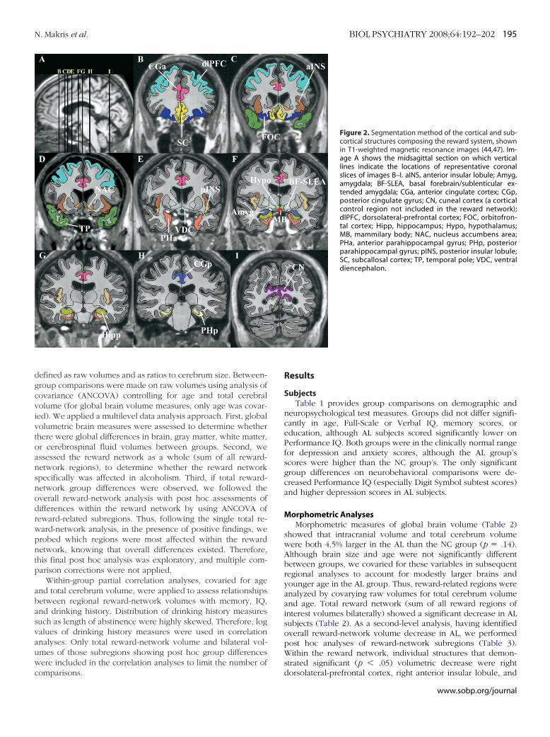
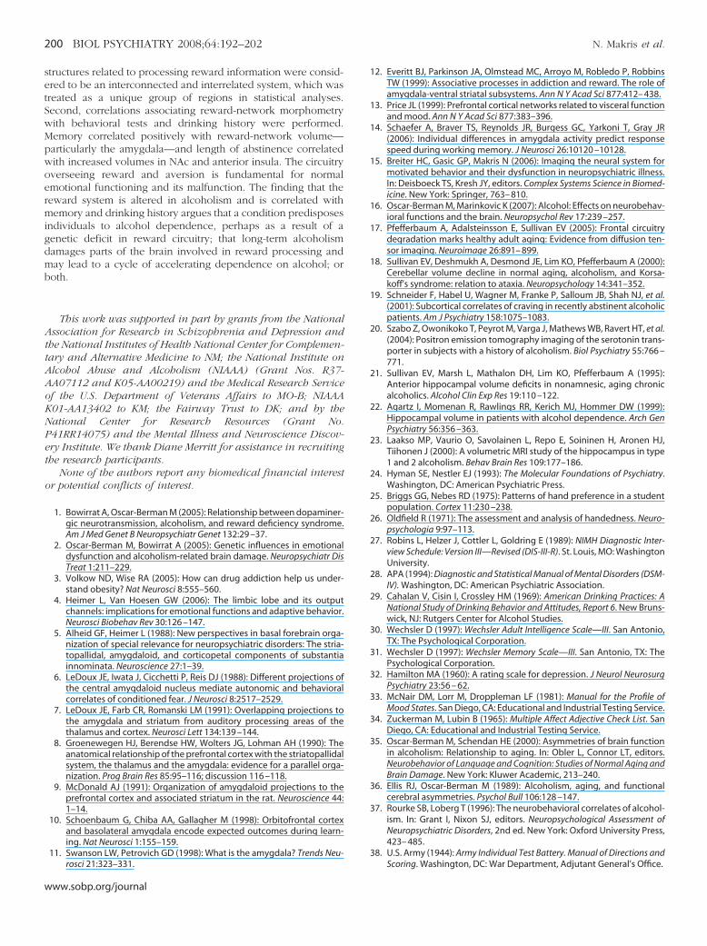
Figure 2. Segmentation method of the cortical and sub-cortical structures composing the reward system, shownin T1-weighted magnetic resonance images (44,47). Im-age A shows the midsagittal section on which verticallines indicate the locations of representative coronalslices of images B–I. aINS, anterior insular lobule; Amyg,amygdala; BF-SLEA, basal forebrain/sublenticular ex-tended amygdala; CGa, anterior cingulate cortex; CGp,posterior cingulate gyrus; CN, cuneal cortex (a corticalcontrol region not included in the reward network);dlPFC, dorsolateral-prefrontal cortex; FOC, orbitofron-tal cortex; Hipp, hippocampus; Hypo, hypothalamus;MB, mammilary body; NAC, nucleus accumbens area;PHa, anterior parahippocampal gyrus; PHp, posteriorparahippocampal gyrus; pINS, posterior insular lobule;SC, subcallosal cortex; TP, temporal pole; VDC, ventraldiencephalon.
dorsolateral-prefrontal cortex, right anterior insular lobule, and
www.sobp.org/journal
rdbd
fcr
T
glsa
CI
l
Fsv(hvztghntV
196 BIOL PSYCHIATRY 2008;64:192–202 N. Makris et al.
w
ight NAc. Left amygdala showed a trend (p � .07) towardecreased volume. However, assessed as a ratio to total cere-rum volume covaried for age, left amygdala was significantlyecreased in AL subjects (p � .05).
Nonreward reference regions included frontal pole as arontal cortex control region, cuneal cortex as a sensory-cortexontrol region, and dorsal striatum as a subcortical controlegion. These regions did not differ between groups.
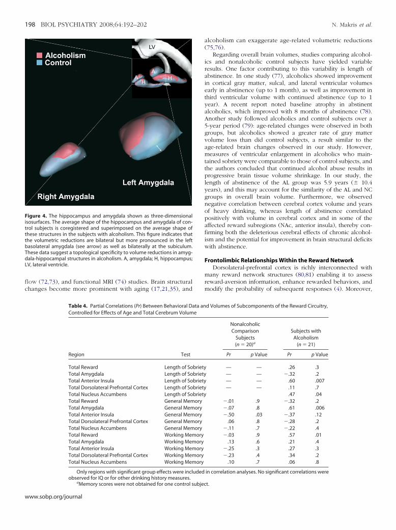
opological AnalysesTopological analyses of amygdala and hippocampus showed
roup differences in topology. Decreased volume in AL wasocalized in basolateral-central nuclear groups of amygdala andubicular region (Figure 4). This was principally noted in leftmygdala, which showed reduced volume in AL.
orrelations Associating Morphometry with Memory,Q, Age, and Drinking History
In AL subjects, age was significantly correlated with increasing
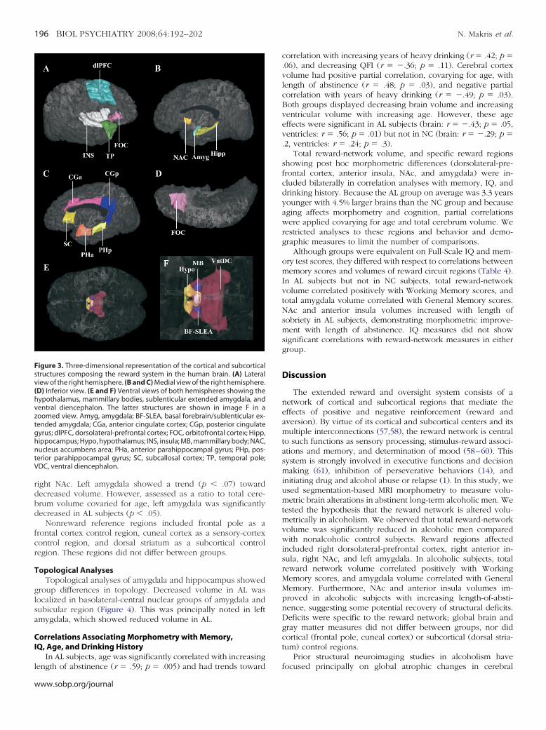
igure 3. Three-dimensional representation of the cortical and subcorticaltructures composing the reward system in the human brain. (A) Lateraliew of the right hemisphere. (B and C) Medial view of the right hemisphere.D) Inferior view. (E and F) Ventral views of both hemispheres showing theypothalamus, mammillary bodies, sublenticular extended amygdala, andentral diencephalon. The latter structures are shown in image F in aoomed view. Amyg, amygdala; BF-SLEA, basal forebrain/sublenticular ex-ended amygdala; CGa, anterior cingulate cortex; CGp, posterior cingulateyrus; dlPFC, dorsolateral-prefrontal cortex; FOC, orbitofrontal cortex; Hipp,ippocampus; Hypo, hypothalamus; INS, insula; MB, mammillary body; NAC,ucleus accumbens area; PHa, anterior parahippocampal gyrus; PHp, pos-
erior parahippocampal gyrus; SC, subcallosal cortex; TP, temporal pole;DC, ventral diencephalon.
ength of abstinence (r � .59; p � .005) and had trends toward
ww.sobp.org/journal
correlation with increasing years of heavy drinking (r � .42; p �.06), and decreasing QFI (r � �.36; p � .11). Cerebral cortexvolume had positive partial correlation, covarying for age, withlength of abstinence (r � .48; p � .03), and negative partialcorrelation with years of heavy drinking (r � �.49; p � .03).Both groups displayed decreasing brain volume and increasingventricular volume with increasing age. However, these ageeffects were significant in AL subjects (brain: r � �.43; p � .05,ventricles: r � .56; p � .01) but not in NC (brain: r � �.29; p �.2, ventricles: r � .24; p � .3).
Total reward-network volume, and specific reward regionsshowing post hoc morphometric differences (dorsolateral-pre-frontal cortex, anterior insula, NAc, and amygdala) were in-cluded bilaterally in correlation analyses with memory, IQ, anddrinking history. Because the AL group on average was 3.3 yearsyounger with 4.5% larger brains than the NC group and becauseaging affects morphometry and cognition, partial correlationswere applied covarying for age and total cerebrum volume. Werestricted analyses to these regions and behavior and demo-graphic measures to limit the number of comparisons.
Although groups were equivalent on Full-Scale IQ and mem-ory test scores, they differed with respect to correlations betweenmemory scores and volumes of reward circuit regions (Table 4).In AL subjects but not in NC subjects, total reward-networkvolume correlated positively with Working Memory scores, andtotal amygdala volume correlated with General Memory scores.NAc and anterior insula volumes increased with length ofsobriety in AL subjects, demonstrating morphometric improve-ment with length of abstinence. IQ measures did not showsignificant correlations with reward-network measures in eithergroup.
Discussion
The extended reward and oversight system consists of anetwork of cortical and subcortical regions that mediate theeffects of positive and negative reinforcement (reward andaversion). By virtue of its cortical and subcortical centers and itsmultiple interconnections (57,58), the reward network is centralto such functions as sensory processing, stimulus-reward associ-ations and memory, and determination of mood (58–60). Thissystem is strongly involved in executive functions and decisionmaking (61), inhibition of perseverative behaviors (14), andinitiating drug and alcohol abuse or relapse (1). In this study, weused segmentation-based MRI morphometry to measure volu-metric brain alterations in abstinent long-term alcoholic men. Wetested the hypothesis that the reward network is altered volu-metrically in alcoholism. We observed that total reward-networkvolume was significantly reduced in alcoholic men comparedwith nonalcoholic control subjects. Reward regions affectedincluded right dorsolateral-prefrontal cortex, right anterior in-sula, right NAc, and left amygdala. In alcoholic subjects, totalreward network volume correlated positively with WorkingMemory scores, and amygdala volume correlated with GeneralMemory. Furthermore, NAc and anterior insula volumes im-proved in alcoholic subjects with increasing length-of-absti-nence, suggesting some potential recovery of structural deficits.Deficits were specific to the reward network; global brain andgray matter measures did not differ between groups, nor didcortical (frontal pole, cuneal cortex) or subcortical (dorsal stria-tum) control regions.
Prior structural neuroimaging studies in alcoholism have
focused principally on global atrophic changes in cerebral
chSa
Tc
R
T
G
CG( ith a
Tt
R
S
C
N
Aoag
N. Makris et al. BIOL PSYCHIATRY 2008;64:192–202 197
ortex, white matter, and cerebellum, plus local effects inippocampus (62), demonstrating volume reduction (21–23,63).maller right amygdalae have been reported in relatives oflcoholics (56). Neuropathologic observations demonstrated al-
able 2. Morphometric Measures of the Total Extended Reward and Oversentimeters) and as a Ratio to Total Cerebrum Volume (%)
Nonalcoholic Comp(n � 21
egion Mean �
otal Reward Network MeasuresReward Ratio to Total Cerebrum Volume (%) 12.2 � 0Reward Raw Volume (cc) 128.9 � 1
lobal Measures (cc)Intracranial Volume 1456.9 � 1Total Brain 1210.1 � 1Total Cerebrum 1061.7 � 1Total Cerebral Cortex 524.9 � 5Total Cerebral White Matter 456.1 � 6Total Ventricular System 29.3 � 1
ANCOVA, analysis of covariance.Raw volumes (cc) of the global brain regions are presented. ComponenaFor total reward network measures, we show the t value of the Grou
erebrum Volume (p � .001) and for age (p � .4) and on Ratio to Total Cerebroup effect from the univariate ANCOVA tests on raw volumes, covaried for
decreasing volume with age), and Ventricular System (increasing volume wbp � .05.
able 3. Volumes of Regions Within the Extended Reward and Oversight So Total Cerebrum Volume (%)
egion
Left Hemisphere
Nonalcoholic ComparisonSubjects(n � 21)
Subjects withAlcoholism
(n � 21)
Mean � SD Mean � SD
ubcortical Reward RegionsNucleus Accumbens .054 � .01 .053 � .01Amygdala .15 � .03 .13 � .02Hippocampus .33 � .04 .31 � .03Ventral Diencephalon .44 � .07 .41 � .05
ortical Reward RegionsInsula, Anterior .42 � .05 .41 � .05DLPFC 1.74 � .31 1.59 � .41Insula, Posterior .21 � .04 .19 � .03Orbitofrontal Cortex .44 � .08 .45 � .12Cingulate Cortex .99 � .15 .96 � .19Subcallosal Cortex .21 � .03 .20 � .04Parahippocampal Gyrus .37 � .05 .35 � .07Temporal Pole .72 � .09 .71 � .12
onreward Control RegionsDorsal Striatum .93 � .07 .91 � .09Frontal Pole 3.65 � .78 3.55 � .53Cuneal Cortex (sensory) .50 � .09 .51 � .11
ANCOVA, analysis of covariance; DLPFC, dorsolateral prefrontal cortex.aWe show the t value of the Group effect from the univariate ANCOVA te
NCOVA (including Group, Age, and Total Cerebrum Volume in the model)rbitofrontal cortex, left parahippocampal gyrus, left cingulate cortex, anccumbens and left amygdala; Total Cerebrum Volume was a significant covyrus bilaterally.
bp � .05.cp � .07, ratio covaried for age p � .05.
dp � .08, ratio covaried for age p � .2.coholism-related neuronal loss in prefrontal association cortex,hypothalamus, and cerebellum (64,65). Frontal dysfunction inalcoholism has been demonstrated by neuropsychologicalinvestigations (66–71) and by metabolic (20), cerebral blood
ystem (Total Reward Network) Expressed as Raw Volume (in cubic
Subjects Subjects with Alcoholism(n � 21) ANCOVA Group Effecta
Mean � SD t Value
11.5 � 1.0 �2.4b
127.6 � 14.1 �2.2b
1491.1 � 121.7 .91264.3 � 110.8 1.21109.9 � 99.4 1.2
537.7 � 64.1 .4492.9 � 65.0 1.6
27.0 � 9.1 �.2
he reward network are shown in Table 3.ct from the univariate ANCOVA tests on Raw Volumes, covaried for Totalolume, covaried for age; for the global measures, we show the t value of theThere were significant Age effects (p � .05) for Total Brain, Cerebrum, Cortexge).
(Reward Regions) and Nonreward Control Regions, Expressed as a Ratio
Right Hemisphere
NCOVAoup Effecta
Nonalcoholic ComparisonSubjects(n � 21)
Subjects withAlcoholism
(n � 21)ANCOVA
Group Effecta
t Value Mean � SD Mean � SD t Value
�.5 .054 � .01 .047 � .01 �2.5b
�1.8c .14 � .03 .13 � .02 �1.1�.8 .35 � .04 .33 � .03 �1.2�.9 .43 � .06 .40 � .05 �1.1
.0 .41 � .03 .38 � .04 �2.2b
�1.5 1.76 � .30 1.57 � .31 �2.2b
�1.8d .21 � .02 .21 � .03 �.1.8 .46 � .09 .42 � .08 �1.5
�.4 1.04 � .17 1.05 � .15 .6�.7 .20 � .03 .20 � .03 �.9�.5 .35 � .06 .33 � .07 �.5�.3 .69 � .09 .69 � .09 .3
�.7 .96 � .06 .93 � .08 �1.3�.3 3.85 � .86 3.57 � .58 �.8
.7 .55 � .09 .58 � .12 .7
raw volumes, covaried for Total Cerebrum Volume and for Age. The overallificant for all regions except right amygdala, left ventral diencephalon, leftright frontal pole; Age effects were nonsignificant except for the right
in all regions except the left orbitofrontal cortex and the parahippocampal
ight S
arison)
SD
.63.6
12.724.011.23.52.86.4
ts of tp efferum VAge.
ystem
AGr
sts onis signd theariate
www.sobp.org/journal
fc
FitttbTdL
198 BIOL PSYCHIATRY 2008;64:192–202 N. Makris et al.
w
low (72,73), and functional MRI (74) studies. Brain structuralhanges become more prominent with aging (17,21,35), and
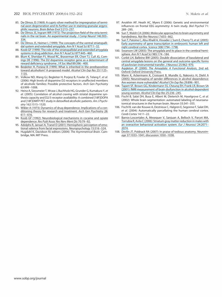
igure 4. The hippocampus and amygdala shown as three-dimensionalsosurfaces. The average shape of the hippocampus and amygdala of con-rol subjects is coregistered and superimposed on the average shape ofhese structures in the subjects with alcoholism. This figure indicates thathe volumetric reductions are bilateral but more pronounced in the leftasolateral amygdala (see arrow) as well as bilaterally at the subiculum.hese data suggest a topological specificity to volume reductions in amyg-ala-hippocampal structures in alcoholism. A, amygdala; H, hippocampus;V, lateral ventricle.
Table 4. Partial Correlations (Pr) Between Behavioral DaControlled for Effects of Age and Total Cerebrum Volum
Region Test
Total Reward Length of SoTotal Amygdala Length of SoTotal Anterior Insula Length of SoTotal Dorsolateral Prefrontal Cortex Length of SoTotal Nucleus Accumbens Length of SoTotal Reward General MemTotal Amygdala General MemTotal Anterior Insula General MemTotal Dorsolateral Prefrontal Cortex General MemTotal Nucleus Accumbens General MemTotal Reward Working MemTotal Amygdala Working MemTotal Anterior Insula Working MemTotal Dorsolateral Prefrontal Cortex Working MemTotal Nucleus Accumbens Working Mem
Only regions with significant group effects were inclobserved for IQ or for other drinking history measures.
aMemory scores were not obtained for one control subjec
ww.sobp.org/journal
alcoholism can exaggerate age-related volumetric reductions(75,76).
Regarding overall brain volumes, studies comparing alcohol-ics and nonalcoholic control subjects have yielded variableresults. One factor contributing to this variability is length ofabstinence. In one study (77), alcoholics showed improvementin cortical gray matter, sulcal, and lateral ventricular volumesearly in abstinence (up to 1 month), as well as improvement inthird ventricular volume with continued abstinence (up to 1year). A recent report noted baseline atrophy in abstinentalcoholics, which improved with 8 months of abstinence (78).Another study followed alcoholics and control subjects over a5-year period (79): age-related changes were observed in bothgroups, but alcoholics showed a greater rate of gray mattervolume loss than did control subjects, a result similar to theage-related brain changes observed in our study. However,measures of ventricular enlargement in alcoholics who main-tained sobriety were comparable to those of control subjects, andthe authors concluded that continued alcohol abuse results inprogressive brain tissue volume shrinkage. In our study, thelength of abstinence of the AL group was 5.9 years (� 10.4years), and this may account for the similarity of the AL and NCgroups in overall brain volume. Furthermore, we observednegative correlation between cerebral cortex volume and yearsof heavy drinking, whereas length of abstinence correlatedpositively with volume in cerebral cortex and in some of theaffected reward subregions (NAc, anterior insula), thereby con-firming both the deleterious cerebral effects of chronic alcohol-ism and the potential for improvement in brain structural deficitswith abstinence.
Frontolimbic Relationships Within the Reward NetworkDorsolateral-prefrontal cortex is richly interconnected with
many reward network structures (80,81) enabling it to assessreward-aversion information, enhance rewarded behaviors, andmodify the probability of subsequent responses (4). Moreover,
d Volumes of Subcomponents of the Reward Circuitry,
NonalcoholicComparison
Subjects(n � 20)a
Subjects withAlcoholism
(n � 21)
Pr p Value Pr p Value
— — .26 .3— — �.32 .2— — .60 .007— — .11 .7
.47 .04�.01 .9 �.32 .2�.07 .8 .61 .006�.50 .03 �.37 .12
.06 .8 �.28 .2�.11 .7 �.22 .4�.03 .9 .57 .01
.13 .6 .21 .4�.25 .3 .27 .3�.23 .4 .34 .2
.10 .7 .06 .8
in correlation analyses. No significant correlations were
ta ane
brietybrietybrietybrietybrietyoryoryoryoryoryoryoryoryoryory
uded
t.
tpnmacclittpdi
opr(aeoho(modnAarlhd
ertrrhkramAfc
T
cbRwtbctgms
N. Makris et al. BIOL PSYCHIATRY 2008;64:192–202 199
he role of dorsolateral-prefrontal cortex in oversight of limbic-aralimbic centers within the reward circuitry is crucial forormal cognitive and emotional functioning (80,82,83). Further-ore, there is extensive connectivity among the amygdala and
djacent structures within the basal forebrain and NAc, structuresonsidered to be part of the same neural system (84–87). Theseenters are involved in establishing associations between stimu-us cues and reward and for evaluating effectiveness of reinforc-ng stimuli. Largely through these associations, sensory inputs getransformed into powerful motivational and emotional represen-ations (81). Therefore, structural alterations in dorsolateral-refrontal cortex, anterior insula, NAc, and amygdala wouldisrupt the reward processing stream and disorganize thesentegrative functions.
The reward network may be an integral part of the neurobi-logy of drug addiction in general, and alcohol dependence inarticular (88). Structural malfunction in this system may increaseisk for drug-seeking behaviors and reward deficiency syndrome1,89). These abnormalities may reflect a genetically influencedlteration in alcoholism (90). Recent neuroimaging positronmission tomography (PET) reports have identified higher levelsf dopamine D2 receptors in ventral striatum (NAc) in nonalco-olic members of alcoholic families, suggesting that higher levelsf dopamine in the reward system may have a protective effect91). Striatal D2 receptor availability in nonalcoholic familyembers correlated with positive emotionality and with metab-lism in orbitofrontal, anterior cingulate, and prefrontal cortex,emonstrating a connection among behavior, subcortical D2
euroreceptors, and cortical function in reward-network regions.related PET study found that alcohol craving in detoxified
lcoholic men correlated with lower levels of striatal dopamineeceptors and corresponded to a greater relapse risk on fol-ow-up (92). This circuitry may be crucial to alterations inedonic set points related to development of drug use andependence (88,93,94).
Processing of emotions engages the two hemispheres differ-ntially, with negative emotional processing predominantly inight hemisphere and positive emotional processing primarily inhe left hemisphere (35,95,96). In this report, the localization ofight dorsolateral-prefrontal cortex, right anterior insula, andight NAc deficits supports the right hemisphere and frontalypotheses of deficits in alcoholism (16,35). What is currentlynown regarding cerebral dominance and lateralization of theeward function is limited, however. Structural brain asymmetrynd lateralization of functional dominance may result fromolecular regulation, neural connections, and plasticity (97).lthough genetic factors connecting cerebral asymmetry and
unctional dominance are supported, molecular correlates oferebral asymmetry have yet to be identified (98,99).
opological Analyses in Amygdala and HippocampusOur topological analysis of hippocampus and amygdala indi-
ated that volumetric decrease in alcoholics was localized in theasolateral nuclear group of left amygdala and the subiculum.emarkably, connections of amygdala with other structuresithin the reward network, such as dorsolateral-prefrontal cor-
ex, anterior insula, hippocampus, and NAc, are through itsasolateral nuclear group. Whereas the central nucleus is asso-iated with autonomic behavior modulating the general motiva-ional influence of reward-related events, the basolateral nuclearroup is thought to be part of the frontotemporal association systemediating outcome-specific incentive processes (100,101). In our
tudy, AL subjects displayed correlation between Working Mem-
ory and reward-network volume and between amygdala vol-umes and General Memory. Left amygdala volumetric decreasemay account for deficits in performance related to analyticalaspects of memory, as well as verbal memory (14,102). Hip-pocampal and amygdala volume reductions have been previ-ously reported in alcoholics (22,55,56), and reduced amygdalavolume was present in adolescents of high-risk alcoholismfamilies, indicating a possible neurodevelopmental component(56). However, no prior studies evaluated particular subportionsof these structures.
LimitationsFirst, we studied only men, whereas effects of alcoholism may
be even more devastating in women (103,104). However, weselected men to avoid interactions of gender effects. In anongoing companion study, we are examining female alcoholicand control subjects to determine whether these effects are seenin alcoholic women as well. Second, we did not include highlysensitive behavioral or functional MRI measures of brain asym-metry in this study; these findings will be detailed in a separatereport. Third, we did not selectively recruit our AL subjects fromfamilies with a high prevalence of alcoholism, and the volumetricabnormalities we observed may be better defined in alcoholicswith a strong positive family history. Fourth, we applied amultilevel analysis approach to identify whether the rewardnetwork overall was affected in alcoholism, followed by post hocanalyses of reward subregions. We did not apply multiplecomparison corrections, because these post hoc regional analy-ses were performed to explore which reward-related subregionswere most influential in the presence of an overall reward-network group effect. However, our findings should be repli-cated in a larger sample. In addition, the effect sizes we observedwere modest, and although the overall reward network demon-strated group differences, only a few regions within the networkdisplayed significant effects. Finally, the morphometric analysisframework samples relatively large regions; small structures suchas ventral tegmentum or SLEA may have regional differences thatare undetectable with these methods. In addition, we do nothave a parcellation unit defined for ventral putamen, eventhough this region may be related to NAc. However, we choseneuroanatomically specific and highly reliable regional parcella-tion methods over other, more automated methods to assessspecific morphometry of reward-network regions a priori. Thereare advantages and disadvantages of the methods employed in thisstudy compared with more automated methods such as FreeSurfer(105,106) or voxel-based morphometry (107). FreeSurfer was vali-dated using the methods described herein (105,106). Voxel-basedmorphometry is not well suited to testing hypotheses regarding ana priori, specifically defined, extensive neuroanatomic network.The advantages of FreeSurfer or voxel-based morphometry areautomation, speed, and low cost. However, automated methodsare more prone to coregistration artifacts and atlasing misalign-ment (108). Although automated methods are preferable forsome studies and are less time-consuming, our methods are thegold standard for anatomic accuracy, and they ensure precisionin measuring regional neuroanotomic networks.
Conclusions
Abstinent long-term chronic alcoholics have volumetric defi-cits in the brain’s extended reward and oversight system. Deficitswere most pronounced in right dorsolateral-prefrontal cortex,right anterior insula, right NAc, and left amygdala. This study
differs from prior investigations in two principal aspects. First,www.sobp.org/journal
setSwMpwoermigdmb
AttAAoKNPet
o
200 BIOL PSYCHIATRY 2008;64:192–202 N. Makris et al.
w
tructures related to processing reward information were consid-red to be an interconnected and interrelated system, which wasreated as a unique group of regions in statistical analyses.econd, correlations associating reward-network morphometryith behavioral tests and drinking history were performed.emory correlated positively with reward-network volume—articularly the amygdala—and length of abstinence correlatedith increased volumes in NAc and anterior insula. The circuitryverseeing reward and aversion is fundamental for normalmotional functioning and its malfunction. The finding that theeward system is altered in alcoholism and is correlated withemory and drinking history argues that a condition predisposes
ndividuals to alcohol dependence, perhaps as a result of aenetic deficit in reward circuitry; that long-term alcoholismamages parts of the brain involved in reward processing anday lead to a cycle of accelerating dependence on alcohol; oroth.
This work was supported in part by grants from the Nationalssociation for Research in Schizophrenia and Depression and
he National Institutes of Health National Center for Complemen-ary and Alternative Medicine to NM; the National Institute onlcohol Abuse and Alcoholism (NIAAA) (Grant Nos. R37-A07112 and K05-AA00219) and the Medical Research Servicef the U.S. Department of Veterans Affairs to MO-B; NIAAA01-AA13402 to KM; the Fairway Trust to DK; and by theational Center for Research Resources (Grant No.41RR14075) and the Mental Illness and Neuroscience Discov-ry Institute. We thank Diane Merritt for assistance in recruitinghe research participants.
None of the authors report any biomedical financial interestr potential conflicts of interest.
1. Bowirrat A, Oscar-Berman M (2005): Relationship between dopaminer-gic neurotransmission, alcoholism, and reward deficiency syndrome.Am J Med Genet B Neuropsychiatr Genet 132:29 –37.
2. Oscar-Berman M, Bowirrat A (2005): Genetic influences in emotionaldysfunction and alcoholism-related brain damage. Neuropsychiatr DisTreat 1:211–229.
3. Volkow ND, Wise RA (2005): How can drug addiction help us under-stand obesity? Nat Neurosci 8:555–560.
4. Heimer L, Van Hoesen GW (2006): The limbic lobe and its outputchannels: implications for emotional functions and adaptive behavior.Neurosci Biobehav Rev 30:126 –147.
5. Alheid GF, Heimer L (1988): New perspectives in basal forebrain orga-nization of special relevance for neuropsychiatric disorders: The stria-topallidal, amygdaloid, and corticopetal components of substantiainnominata. Neuroscience 27:1–39.
6. LeDoux JE, Iwata J, Cicchetti P, Reis DJ (1988): Different projections ofthe central amygdaloid nucleus mediate autonomic and behavioralcorrelates of conditioned fear. J Neurosci 8:2517–2529.
7. LeDoux JE, Farb CR, Romanski LM (1991): Overlapping projections tothe amygdala and striatum from auditory processing areas of thethalamus and cortex. Neurosci Lett 134:139 –144.
8. Groenewegen HJ, Berendse HW, Wolters JG, Lohman AH (1990): Theanatomical relationship of the prefrontal cortex with the striatopallidalsystem, the thalamus and the amygdala: evidence for a parallel orga-nization. Prog Brain Res 85:95–116; discussion 116 –118.
9. McDonald AJ (1991): Organization of amygdaloid projections to theprefrontal cortex and associated striatum in the rat. Neuroscience 44:1–14.
10. Schoenbaum G, Chiba AA, Gallagher M (1998): Orbitofrontal cortexand basolateral amygdala encode expected outcomes during learn-ing. Nat Neurosci 1:155–159.
11. Swanson LW, Petrovich GD (1998): What is the amygdala? Trends Neu-
rosci 21:323–331.ww.sobp.org/journal
12. Everitt BJ, Parkinson JA, Olmstead MC, Arroyo M, Robledo P, RobbinsTW (1999): Associative processes in addiction and reward. The role ofamygdala-ventral striatal subsystems. Ann N Y Acad Sci 877:412– 438.
13. Price JL (1999): Prefrontal cortical networks related to visceral functionand mood. Ann N Y Acad Sci 877:383–396.
14. Schaefer A, Braver TS, Reynolds JR, Burgess GC, Yarkoni T, Gray JR(2006): Individual differences in amygdala activity predict responsespeed during working memory. J Neurosci 26:10120 –10128.
15. Breiter HC, Gasic GP, Makris N (2006): Imaging the neural system formotivated behavior and their dysfunction in neuropsychiatric illness.In: Deisboeck TS, Kresh JY, editors. Complex Systems Science in Biomed-icine. New York: Springer, 763– 810.
16. Oscar-Berman M, Marinkovic K (2007): Alcohol: Effects on neurobehav-ioral functions and the brain. Neuropsychol Rev 17:239 –257.
17. Pfefferbaum A, Adalsteinsson E, Sullivan EV (2005): Frontal circuitrydegradation marks healthy adult aging: Evidence from diffusion ten-sor imaging. Neuroimage 26:891– 899.
18. Sullivan EV, Deshmukh A, Desmond JE, Lim KO, Pfefferbaum A (2000):Cerebellar volume decline in normal aging, alcoholism, and Korsa-koff’s syndrome: relation to ataxia. Neuropsychology 14:341–352.
19. Schneider F, Habel U, Wagner M, Franke P, Salloum JB, Shah NJ, et al.(2001): Subcortical correlates of craving in recently abstinent alcoholicpatients. Am J Psychiatry 158:1075–1083.
20. Szabo Z, Owonikoko T, Peyrot M, Varga J, Mathews WB, Ravert HT, et al.(2004): Positron emission tomography imaging of the serotonin trans-porter in subjects with a history of alcoholism. Biol Psychiatry 55:766 –771.
21. Sullivan EV, Marsh L, Mathalon DH, Lim KO, Pfefferbaum A (1995):Anterior hippocampal volume deficits in nonamnesic, aging chronicalcoholics. Alcohol Clin Exp Res 19:110 –122.
22. Agartz I, Momenan R, Rawlings RR, Kerich MJ, Hommer DW (1999):Hippocampal volume in patients with alcohol dependence. Arch GenPsychiatry 56:356 –363.
23. Laakso MP, Vaurio O, Savolainen L, Repo E, Soininen H, Aronen HJ,Tiihonen J (2000): A volumetric MRI study of the hippocampus in type1 and 2 alcoholism. Behav Brain Res 109:177–186.
24. Hyman SE, Nestler EJ (1993): The Molecular Foundations of Psychiatry.Washington, DC: American Psychiatric Press.
25. Briggs GG, Nebes RD (1975): Patterns of hand preference in a studentpopulation. Cortex 11:230 –238.
26. Oldfield R (1971): The assessment and analysis of handedness. Neuro-psychologia 9:97–113.
27. Robins L, Helzer J, Cottler L, Goldring E (1989): NIMH Diagnostic Inter-view Schedule: Version III—Revised (DIS-III-R). St. Louis, MO: WashingtonUniversity.
28. APA (1994): Diagnostic and Statistical Manual of Mental Disorders (DSM-IV). Washington, DC: American Psychiatric Association.
29. Cahalan V, Cisin I, Crossley HM (1969): American Drinking Practices: ANational Study of Drinking Behavior and Attitudes, Report 6. New Bruns-wick, NJ: Rutgers Center for Alcohol Studies.
30. Wechsler D (1997): Wechsler Adult Intelligence Scale—III. San Antonio,TX: The Psychological Corporation.
31. Wechsler D (1997): Wechsler Memory Scale—III. San Antonio, TX: ThePsychological Corporation.
32. Hamilton MA (1960): A rating scale for depression. J Neurol NeurosurgPsychiatry 23:56 – 62.
33. McNair DM, Lorr M, Droppleman LF (1981): Manual for the Profile ofMood States. San Diego, CA: Educational and Industrial Testing Service.
34. Zuckerman M, Lubin B (1965): Multiple Affect Adjective Check List. SanDiego, CA: Educational and Industrial Testing Service.
35. Oscar-Berman M, Schendan HE (2000): Asymmetries of brain functionin alcoholism: Relationship to aging. In: Obler L, Connor LT, editors.Neurobehavior of Language and Cognition: Studies of Normal Aging andBrain Damage. New York: Kluwer Academic, 213–240.
36. Ellis RJ, Oscar-Berman M (1989): Alcoholism, aging, and functionalcerebral asymmetries. Psychol Bull 106:128 –147.
37. Rourke SB, Loberg T (1996): The neurobehavioral correlates of alcohol-ism. In: Grant I, Nixon SJ, editors. Neuropsychological Assessment ofNeuropsychiatric Disorders, 2nd ed. New York: Oxford University Press,423– 485.
38. U.S. Army (1944): Army Individual Test Battery. Manual of Directions and
Scoring. Washington, DC: War Department, Adjutant General’s Office.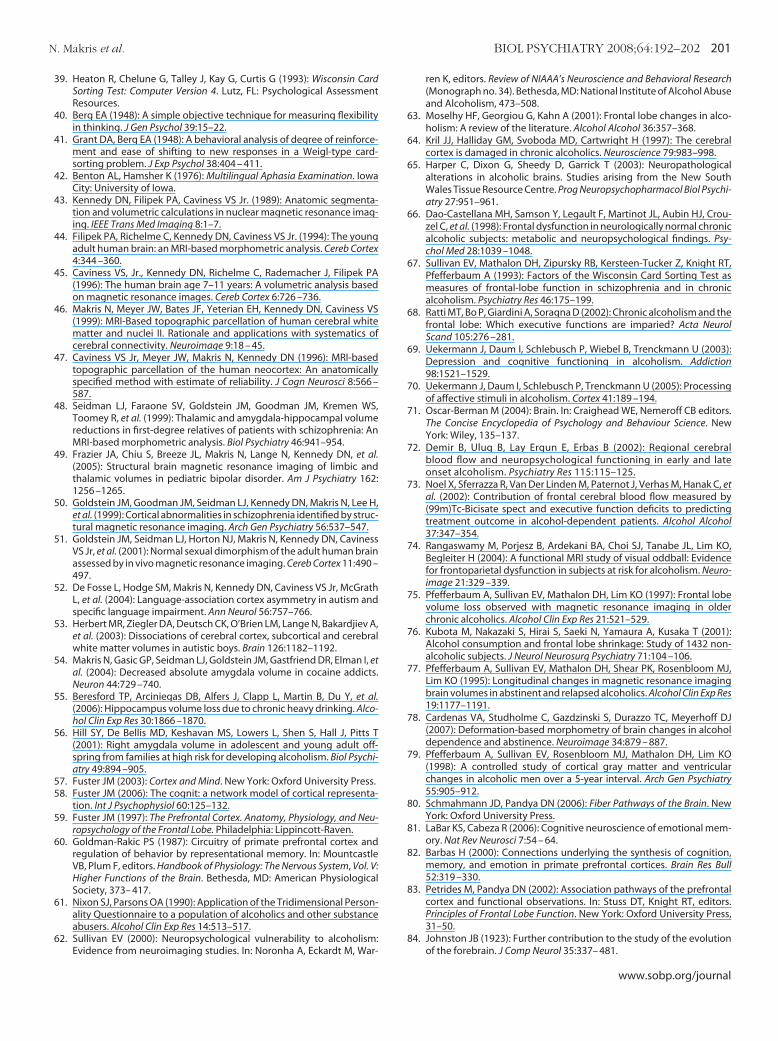
N. Makris et al. BIOL PSYCHIATRY 2008;64:192–202 201
39. Heaton R, Chelune G, Talley J, Kay G, Curtis G (1993): Wisconsin CardSorting Test: Computer Version 4. Lutz, FL: Psychological AssessmentResources.
40. Berg EA (1948): A simple objective technique for measuring flexibilityin thinking. J Gen Psychol 39:15–22.
41. Grant DA, Berg EA (1948): A behavioral analysis of degree of reinforce-ment and ease of shifting to new responses in a Weigl-type card-sorting problem. J Exp Psychol 38:404 – 411.
42. Benton AL, Hamsher K (1976): Multilingual Aphasia Examination. IowaCity: University of Iowa.
43. Kennedy DN, Filipek PA, Caviness VS Jr. (1989): Anatomic segmenta-tion and volumetric calculations in nuclear magnetic resonance imag-ing. IEEE Trans Med Imaging 8:1–7.
44. Filipek PA, Richelme C, Kennedy DN, Caviness VS Jr. (1994): The youngadult human brain: an MRI-based morphometric analysis. Cereb Cortex4:344 –360.
45. Caviness VS, Jr., Kennedy DN, Richelme C, Rademacher J, Filipek PA(1996): The human brain age 7–11 years: A volumetric analysis basedon magnetic resonance images. Cereb Cortex 6:726 –736.
46. Makris N, Meyer JW, Bates JF, Yeterian EH, Kennedy DN, Caviness VS(1999): MRI-Based topographic parcellation of human cerebral whitematter and nuclei II. Rationale and applications with systematics ofcerebral connectivity. Neuroimage 9:18 – 45.
47. Caviness VS Jr, Meyer JW, Makris N, Kennedy DN (1996): MRI-basedtopographic parcellation of the human neocortex: An anatomicallyspecified method with estimate of reliability. J Cogn Neurosci 8:566 –587.
48. Seidman LJ, Faraone SV, Goldstein JM, Goodman JM, Kremen WS,Toomey R, et al. (1999): Thalamic and amygdala-hippocampal volumereductions in first-degree relatives of patients with schizophrenia: AnMRI-based morphometric analysis. Biol Psychiatry 46:941–954.
49. Frazier JA, Chiu S, Breeze JL, Makris N, Lange N, Kennedy DN, et al.(2005): Structural brain magnetic resonance imaging of limbic andthalamic volumes in pediatric bipolar disorder. Am J Psychiatry 162:1256 –1265.
50. Goldstein JM, Goodman JM, Seidman LJ, Kennedy DN, Makris N, Lee H,et al. (1999): Cortical abnormalities in schizophrenia identified by struc-tural magnetic resonance imaging. Arch Gen Psychiatry 56:537–547.
51. Goldstein JM, Seidman LJ, Horton NJ, Makris N, Kennedy DN, CavinessVS Jr, et al. (2001): Normal sexual dimorphism of the adult human brainassessed by in vivo magnetic resonance imaging. Cereb Cortex 11:490 –497.
52. De Fosse L, Hodge SM, Makris N, Kennedy DN, Caviness VS Jr, McGrathL, et al. (2004): Language-association cortex asymmetry in autism andspecific language impairment. Ann Neurol 56:757–766.
53. Herbert MR, Ziegler DA, Deutsch CK, O’Brien LM, Lange N, Bakardjiev A,et al. (2003): Dissociations of cerebral cortex, subcortical and cerebralwhite matter volumes in autistic boys. Brain 126:1182–1192.
54. Makris N, Gasic GP, Seidman LJ, Goldstein JM, Gastfriend DR, Elman I, etal. (2004): Decreased absolute amygdala volume in cocaine addicts.Neuron 44:729 –740.
55. Beresford TP, Arciniegas DB, Alfers J, Clapp L, Martin B, Du Y, et al.(2006): Hippocampus volume loss due to chronic heavy drinking. Alco-hol Clin Exp Res 30:1866 –1870.
56. Hill SY, De Bellis MD, Keshavan MS, Lowers L, Shen S, Hall J, Pitts T(2001): Right amygdala volume in adolescent and young adult off-spring from families at high risk for developing alcoholism. Biol Psychi-atry 49:894 –905.
57. Fuster JM (2003): Cortex and Mind. New York: Oxford University Press.58. Fuster JM (2006): The cognit: a network model of cortical representa-
tion. Int J Psychophysiol 60:125–132.59. Fuster JM (1997): The Prefrontal Cortex. Anatomy, Physiology, and Neu-
ropsychology of the Frontal Lobe. Philadelphia: Lippincott-Raven.60. Goldman-Rakic PS (1987): Circuitry of primate prefrontal cortex and
regulation of behavior by representational memory. In: MountcastleVB, Plum F, editors. Handbook of Physiology: The Nervous System, Vol. V:Higher Functions of the Brain. Bethesda, MD: American PhysiologicalSociety, 373– 417.
61. Nixon SJ, Parsons OA (1990): Application of the Tridimensional Person-ality Questionnaire to a population of alcoholics and other substanceabusers. Alcohol Clin Exp Res 14:513–517.
62. Sullivan EV (2000): Neuropsychological vulnerability to alcoholism:
Evidence from neuroimaging studies. In: Noronha A, Eckardt M, War-ren K, editors. Review of NIAAA’s Neuroscience and Behavioral Research(Monograph no. 34). Bethesda, MD: National Institute of Alcohol Abuseand Alcoholism, 473–508.
63. Moselhy HF, Georgiou G, Kahn A (2001): Frontal lobe changes in alco-holism: A review of the literature. Alcohol Alcohol 36:357–368.
64. Kril JJ, Halliday GM, Svoboda MD, Cartwright H (1997): The cerebralcortex is damaged in chronic alcoholics. Neuroscience 79:983–998.
65. Harper C, Dixon G, Sheedy D, Garrick T (2003): Neuropathologicalalterations in alcoholic brains. Studies arising from the New SouthWales Tissue Resource Centre. Prog Neuropsychopharmacol Biol Psychi-atry 27:951–961.
66. Dao-Castellana MH, Samson Y, Legault F, Martinot JL, Aubin HJ, Crou-zel C, et al. (1998): Frontal dysfunction in neurologically normal chronicalcoholic subjects: metabolic and neuropsychological findings. Psy-chol Med 28:1039 –1048.
67. Sullivan EV, Mathalon DH, Zipursky RB, Kersteen-Tucker Z, Knight RT,Pfefferbaum A (1993): Factors of the Wisconsin Card Sorting Test asmeasures of frontal-lobe function in schizophrenia and in chronicalcoholism. Psychiatry Res 46:175–199.
68. Ratti MT, Bo P, Giardini A, Soragna D (2002): Chronic alcoholism and thefrontal lobe: Which executive functions are imparied? Acta NeurolScand 105:276 –281.
69. Uekermann J, Daum I, Schlebusch P, Wiebel B, Trenckmann U (2003):Depression and cognitive functioning in alcoholism. Addiction98:1521–1529.
70. Uekermann J, Daum I, Schlebusch P, Trenckmann U (2005): Processingof affective stimuli in alcoholism. Cortex 41:189 –194.
71. Oscar-Berman M (2004): Brain. In: Craighead WE, Nemeroff CB editors.The Concise Encyclopedia of Psychology and Behaviour Science. NewYork: Wiley, 135–137.
72. Demir B, Ulug B, Lay Ergun E, Erbas B (2002): Regional cerebralblood flow and neuropsychological functioning in early and lateonset alcoholism. Psychiatry Res 115:115–125.
73. Noel X, Sferrazza R, Van Der Linden M, Paternot J, Verhas M, Hanak C, etal. (2002): Contribution of frontal cerebral blood flow measured by(99m)Tc-Bicisate spect and executive function deficits to predictingtreatment outcome in alcohol-dependent patients. Alcohol Alcohol37:347–354.
74. Rangaswamy M, Porjesz B, Ardekani BA, Choi SJ, Tanabe JL, Lim KO,Begleiter H (2004): A functional MRI study of visual oddball: Evidencefor frontoparietal dysfunction in subjects at risk for alcoholism. Neuro-image 21:329 –339.
75. Pfefferbaum A, Sullivan EV, Mathalon DH, Lim KO (1997): Frontal lobevolume loss observed with magnetic resonance imaging in olderchronic alcoholics. Alcohol Clin Exp Res 21:521–529.
76. Kubota M, Nakazaki S, Hirai S, Saeki N, Yamaura A, Kusaka T (2001):Alcohol consumption and frontal lobe shrinkage: Study of 1432 non-alcoholic subjects. J Neurol Neurosurg Psychiatry 71:104 –106.
77. Pfefferbaum A, Sullivan EV, Mathalon DH, Shear PK, Rosenbloom MJ,Lim KO (1995): Longitudinal changes in magnetic resonance imagingbrain volumes in abstinent and relapsed alcoholics. Alcohol Clin Exp Res19:1177–1191.
78. Cardenas VA, Studholme C, Gazdzinski S, Durazzo TC, Meyerhoff DJ(2007): Deformation-based morphometry of brain changes in alcoholdependence and abstinence. Neuroimage 34:879 – 887.
79. Pfefferbaum A, Sullivan EV, Rosenbloom MJ, Mathalon DH, Lim KO(1998): A controlled study of cortical gray matter and ventricularchanges in alcoholic men over a 5-year interval. Arch Gen Psychiatry55:905–912.
80. Schmahmann JD, Pandya DN (2006): Fiber Pathways of the Brain. NewYork: Oxford University Press.
81. LaBar KS, Cabeza R (2006): Cognitive neuroscience of emotional mem-ory. Nat Rev Neurosci 7:54 – 64.
82. Barbas H (2000): Connections underlying the synthesis of cognition,memory, and emotion in primate prefrontal cortices. Brain Res Bull52:319 –330.
83. Petrides M, Pandya DN (2002): Association pathways of the prefrontalcortex and functional observations. In: Stuss DT, Knight RT, editors.Principles of Frontal Lobe Function. New York: Oxford University Press,31–50.
84. Johnston JB (1923): Further contribution to the study of the evolution
of the forebrain. J Comp Neurol 35:337– 481.www.sobp.org/journal
202 BIOL PSYCHIATRY 2008;64:192–202 N. Makris et al.
w
85. De Olmos JS (1969): A cupric-silver method for impregnation of termi-nal axon degeneration and its further use in staining granular argyro-philic neurons. Brain Behav Evol 2:213–237.
86. De Olmos JS, Ingram WR (1972): The projection field of the stria termi-nalis in the rat brain. An experimental study. J Comp Neurol 146:303–334.
87. De Olmos JS, Heimer L (1999): The concepts of the ventral striatopalli-dal system and extended amygdala. Ann N Y Acad Sci 877:1–32.
88. Koob GF (1999): The role of the striatopallidal and extended amygdalasystems in drug addiction. Ann N Y Acad Sci 877:445– 460.
89. Blum K, Sheridan PJ, Wood RC, Braverman ER, Chen TJ, Cull JG, Com-ings DE (1996): The D2 dopamine receptor gene as a determinant ofreward deficiency syndrome. J R Soc Med 89:396 – 400.
90. Begleiter H, Porjesz B (1999): What is inherited in the predispositiontoward alcoholism? A proposed model. Alcohol Clin Exp Res 23:1125–1135.
91. Volkow ND, Wang GJ, Begleiter H, Porjesz B, Fowler JS, Telang F, et al.(2006): High levels of dopamine D2 receptors in unaffected membersof alcoholic families: Possible protective factors. Arch Gen Psychiatry63:999 –1008.
92. Heinz A, Siessmeier T, Wrase J, Buchholz HG, Grunder G, Kumakura Y, etal. (2005): Correlation of alcohol craving with striatal dopamine syn-thesis capacity and D2/3 receptor availability: A combined [18F]DOPAand [18F]DMFP PET study in detoxified alcoholic patients. Am J Psychi-atry 162:1515–1520.
93. Wikler A (1973): Dynamics of drug dependence. Implications of a con-ditioning theory for research and treatment. Arch Gen Psychiatry 28:611– 616.
94. Koob GF (1992): Neurobiological mechanisms in cocaine and opiatedependence. Res Publ Assoc Res Nerv Ment Dis 70:79 –92.
95. Adolphs R, Jansari A, Tranel D (2001): Hemispheric perception of emo-tional valence from facial expressions. Neuropsychology 15:516 –524.
96. Hugdahl K, Davidson RJ editors (2004): The Asymmetrical Brain. Cam-bridge, MA: MIT Press.
ww.sobp.org/journal
97. Anokhin AP, Heath AC, Myers E (2006): Genetic and environmentalinfluences on frontal EEG asymmetry: A twin study. Biol Psychol 71:289 –295.
98. Sun T, Walsh CA (2006): Molecular approaches to brain asymmetry andhandedness. Nat Rev Neurosci 7:655– 662.
99. Sun T, Patoine C, Abu-Khalil A, Visvader J, Sum E, Cherry TJ, et al. (2005):Early asymmetry of gene transcription in embryonic human left andright cerebral cortex. Science 308:1794 –1798.
100. Swanson LW (2003): The amygdala and its place in the cerebral hemi-sphere. Ann N Y Acad Sci 985:174 –184.
101. Corbit LH, Balleine BW (2005): Double dissociation of basolateral andcentral amygdala lesions on the general and outcome-specific formsof pavlovian-instrumental transfer. J Neurosci 25:962–970.
102. Aggleton JP (2000): The Amygdala: A Functional Analysis, 2nd ed.Oxford: Oxford University Press.
103. Mann K, Ackermann K, Croissant B, Mundle G, Nakovics H, Diehl A(2005): Neuroimaging of gender differences in alcohol dependence:Are women more vulnerable? Alcohol Clin Exp Res 29:896 –901.
104. Tapert SF, Brown GG, Kindermann SS, Cheung EH, Frank LR, Brown SA(2001): fMRI measurement of brain dysfunction in alcohol-dependentyoung women. Alcohol Clin Exp Res 25:236 –245.
105. Fischl B, Salat DH, Busa E, Albert M, Dieterich M, Haselgrove C, et al.(2002): Whole brain segmentation: automated labeling of neuroana-tomical structures in the human brain. Neuron 33:341–355.
106. Fischl B, van der Kouwe A, Destrieux C, Halgren E, Segonne F, Salat DH,et al. (2004): Automatically parcellating the human cerebral cortex.Cereb Cortex 14:11–22.
107. Barros-Loscertales A, Meseguer V, Sanjuan A, Belloch V, Parcet MA,Torrubia R, Avila C (2006): Striatum gray matter reduction in males withan overactive behavioral activation system. Eur J Neurosci 24:2071–2074.
108. Devlin JT, Poldrack RA (2007): In praise of tedious anatomy. Neuroim-
age 37:1033–1041; discussion 1050 –1038.