Frontal White Matter and Cingulum Diffusion Tensor Imaging Deficits in Alcoholism
13
Frontal White Matter and Cingulum Diffusion Tensor Imaging Deficits in Alcoholism Gordon J. Harris, Sharon Kim Jaffin, Steven M. Hodge, David Kennedy, Verne S. Caviness, Ksenija Marinkovic, George M. Papadimitriou, Nikos Makris, and Marlene Oscar-Berman Background: Alcoholism-related deficits in cognition and emotion point toward frontal and limbic dysfunction, particularly in the right hemisphere. Prefrontal and anterior cingulate cortices are involved in cognitive and emotional functions and play critical roles in the oversight of the limbic reward system. In the present study, we examined the integrity of white matter tracts that are critical to frontal and limbic connectivity. Methods: Diffusion tensor magnetic resonance imaging (DT-MRI) was used to assess func- tional anisotropy (FA), a measure of white matter integrity, in 15 abstinent long-term chronic alcoholic and 15 demographically equivalent control men. Voxel-based and region-based analyses of group FA differences were applied to these scans. Results: Alcoholic subjects had diminished frontal lobe FA in the right superior longitudinal fascicles II and III, orbitofrontal cortex white matter, and cingulum bundle, but not in corre- sponding left hemisphere regions. These right frontal and cingulum white matter regional FA measures provided 97% correct group discrimination. Working Memory scores positively corre- lated with superior longitudinal fascicle III FA measures in control subjects only. Conclusions: The findings demonstrate white matter microstructure deficits in abstinent alco- holic men in several right hemisphere tracts connecting prefrontal and limbic systems. These white matter deficits may contribute to underlying dysfunction in memory, emotion, and reward response in alcoholism. Key Words: Alcoholism, Diffusion Tensor Magnetic Resonance Imaging, White Matter, Reward System, Right Hemisphere. L ONG-TERM CHRONIC ALCOHOLISM adversely impacts brain systems involved in cognition and emo- tion and alters sensitivity to the effectiveness of acquired rein- forcers (rewards) such as alcohol and other addictive substances (Blum et al., 2000; Bowirrat and Oscar-Berman, 2005). Alcoholism has been associated with a breakdown of the ‘‘reward cascade,’’ leading to Reward Deficiency Syn- drome (Blum et al., 2000; Bowirrat and Oscar-Berman, 2005). The reward cascade refers to brain neurotransmitter activity that contributes to a state of well-being, and Reward Defi- ciency Syndrome refers to an insensitivity in the effectiveness of rewards (Blum et al., 2000; Comings and Blum, 2000), including a diminished ability to avoid negative affect created by repeated cycles of substance abuse and dependence (Baker et al., 2004). Brain circuitry involved in alcoholism-related insensitivity to rewards includes the mesolimbic pathway linking the ven- tral tegmentum with the nucleus accumbens and the pallidum; these regions are especially important for modulating the effectiveness of reinforcement, both positive (reward) and negative (punishment) (Blum et al., 2000). The limbic aspects of this circuitry are modulated by inputs from the cortex, par- ticularly from orbitofrontal, dorsolateral prefrontal, and cin- gulate regions. We have termed these combined cortical– subcortical circuits the Extended Reward and Oversight Sys- tem (EROS). Furthermore, because there is overlap among the brain regions involved in memory, emotion, and sensitiv- ity to reinforcement, these functions could be adversely impacted by damage to the relevant cortical and subcortical gray matter regions and ⁄ or by degradation of the white mat- ter tracts that interconnect them. In a concurrent structural magnetic resonance imaging (MRI) study, we identified sig- nificant volume reductions in the cortical and subcortical components of EROS in abstinent long-term alcoholics (Makris et al., 2008). Several research groups have reported alcoholism-related damage in the frontal lobes, corpus callo- sum, cerebellum, and limbic structures (Mukamal, 2004; From the Department of Neurology (SKJ, SMH, DK, VSC, KM, GMP, NM), Athinoula A. Martinos Center, Harvard Medical School, Psychiatry and Radiology Services, Center for Morphometric Analysis, Massachusetts General Hospital, Boston, Massachusetts; Departments of Psychiatry, Neurology, and Anatomy & Neurobiology (NM, MO-B), VA Healthcare System, Boston Campus, and Boston University School of Medicine, Boston, Massachusetts; and Radiology Computer Aided Diagnostics Laboratory, Department of Radiology (GJH, SKJ, SMH), Massachusetts General Hospital, Boston, Massa- chusetts. Received for publication June 6, 2007; accepted February 19, 2008. Reprint requests: Gordon J. Harris, PhD, Massachusetts General Hospital, 25 New Chardon Street, Suite 400C, Boston, MA 02114; Fax: 617-724-6130; E-mail: [email protected] Copyright Ó 2008 by the Research Society on Alcoholism. DOI: 10.1111/j.1530-0277.2008.00661.x Alcoholism: Clinical and Experimental Research Vol. 32, No. 6 June 2008 Alcohol Clin Exp Res, Vol 32, No 6, 2008: pp 1001–1013 1001
Transcript of Frontal White Matter and Cingulum Diffusion Tensor Imaging Deficits in Alcoholism

Frontal White Matter and Cingulum Diffusion Tensor
Imaging Deficits in Alcoholism
Gordon J. Harris, Sharon Kim Jaffin, Steven M. Hodge, David Kennedy,Verne S. Caviness, Ksenija Marinkovic, George M. Papadimitriou, Nikos Makris, and
Marlene Oscar-Berman
Background: Alcoholism-related deficits in cognition and emotion point toward frontal andlimbic dysfunction, particularly in the right hemisphere. Prefrontal and anterior cingulate corticesare involved in cognitive and emotional functions and play critical roles in the oversight of thelimbic reward system. In the present study, we examined the integrity of white matter tracts thatare critical to frontal and limbic connectivity.
Methods: Diffusion tensor magnetic resonance imaging (DT-MRI) was used to assess func-tional anisotropy (FA), a measure of white matter integrity, in 15 abstinent long-term chronicalcoholic and 15 demographically equivalent control men. Voxel-based and region-based analysesof group FA differences were applied to these scans.
Results: Alcoholic subjects had diminished frontal lobe FA in the right superior longitudinalfascicles II and III, orbitofrontal cortex white matter, and cingulum bundle, but not in corre-sponding left hemisphere regions. These right frontal and cingulum white matter regional FAmeasures provided 97% correct group discrimination. Working Memory scores positively corre-lated with superior longitudinal fascicle III FA measures in control subjects only.
Conclusions: The findings demonstrate white matter microstructure deficits in abstinent alco-holic men in several right hemisphere tracts connecting prefrontal and limbic systems. These whitematter deficits may contribute to underlying dysfunction in memory, emotion, and rewardresponse in alcoholism.
Key Words: Alcoholism, Diffusion Tensor Magnetic Resonance Imaging, White Matter,Reward System, Right Hemisphere.
L ONG-TERM CHRONIC ALCOHOLISM adverselyimpacts brain systems involved in cognition and emo-
tion and alters sensitivity to the effectiveness of acquired rein-forcers (rewards) such as alcohol and other addictivesubstances (Blum et al., 2000; Bowirrat and Oscar-Berman,2005). Alcoholism has been associated with a breakdown ofthe ‘‘reward cascade,’’ leading to Reward Deficiency Syn-drome (Blum et al., 2000; Bowirrat and Oscar-Berman, 2005).The reward cascade refers to brain neurotransmitter activitythat contributes to a state of well-being, and Reward Defi-ciency Syndrome refers to an insensitivity in the effectiveness
of rewards (Blum et al., 2000; Comings and Blum, 2000),including a diminished ability to avoid negative affect createdby repeated cycles of substance abuse and dependence (Bakeret al., 2004).Brain circuitry involved in alcoholism-related insensitivity
to rewards includes the mesolimbic pathway linking the ven-tral tegmentum with the nucleus accumbens and the pallidum;these regions are especially important for modulating theeffectiveness of reinforcement, both positive (reward) andnegative (punishment) (Blum et al., 2000). The limbic aspectsof this circuitry are modulated by inputs from the cortex, par-ticularly from orbitofrontal, dorsolateral prefrontal, and cin-gulate regions. We have termed these combined cortical–subcortical circuits the Extended Reward and Oversight Sys-tem (EROS). Furthermore, because there is overlap amongthe brain regions involved in memory, emotion, and sensitiv-ity to reinforcement, these functions could be adverselyimpacted by damage to the relevant cortical and subcorticalgray matter regions and ⁄or by degradation of the white mat-ter tracts that interconnect them. In a concurrent structuralmagnetic resonance imaging (MRI) study, we identified sig-nificant volume reductions in the cortical and subcorticalcomponents of EROS in abstinent long-term alcoholics(Makris et al., 2008). Several research groups have reportedalcoholism-related damage in the frontal lobes, corpus callo-sum, cerebellum, and limbic structures (Mukamal, 2004;
From the Department of Neurology (SKJ, SMH, DK, VSC, KM,GMP, NM), Athinoula A. Martinos Center, Harvard MedicalSchool, Psychiatry and Radiology Services, Center for MorphometricAnalysis, Massachusetts General Hospital, Boston, Massachusetts;Departments of Psychiatry, Neurology, and Anatomy & Neurobiology(NM, MO-B), VA Healthcare System, Boston Campus, and BostonUniversity School of Medicine, Boston, Massachusetts; and RadiologyComputer Aided Diagnostics Laboratory, Department of Radiology(GJH, SKJ, SMH), Massachusetts General Hospital, Boston, Massa-chusetts.
Received for publication June 6, 2007; accepted February 19, 2008.Reprint requests: Gordon J. Harris, PhD, Massachusetts General
Hospital, 25 New Chardon Street, Suite 400C, Boston, MA 02114;Fax: 617-724-6130; E-mail: [email protected]
Copyright � 2008 by the Research Society on Alcoholism.
DOI: 10.1111/j.1530-0277.2008.00661.x
Alcoholism: Clinical and Experimental Research Vol. 32, No. 6June 2008
Alcohol Clin Exp Res, Vol 32, No 6, 2008: pp 1001–1013 1001
Oscar-Berman and Bowirrat, 2005; Oscar-Berman and Mar-inkovic, 2003; Sullivan et al., 2000). The goal of the presentstudy was to assess the integrity of white matter tracts thatconnect to cortical regions involved in modulating reinforcedbehaviors (the ‘‘reward response’’) in abstinent alcoholic sub-jects and demographically equivalent nonalcoholic controls,with special attention being paid to possible hemispheric dif-ferences. Consideration of structural differences between the 2cerebral hemispheres is important because, although bothcerebral hemispheres contain EROS circuitry, their specificfunctions may be lateralized to reflect differential hemisphericsensitivities to stimulus materials (e.g., linguistic vs. visuospa-tial) and task demands (e.g., attention, perception, motorresponse, etc.). For example, alcoholics commonly display apattern of deficits that includes visuospatial, attentional, andemotional abnormalities characteristic of patients with righthemisphere damage, suggesting that the right hemispheremay be more vulnerable to the effects of alcoholism than theleft hemisphere (Ellis and Oscar-Berman, 1989; Oscar-Ber-man and Marinkovic, 2007).Alcoholism is particularly damaging to cerebral white mat-
ter, as has been revealed by postmortem neuropathology (Har-per et al., 1987, 2003; Paula-Barbosa and Tavares, 1985;Pentney, 1991; Putzke et al., 1998; Tarnowska-Dziduszkoet al., 1995) and by in vivo structural MRI studies (Estruchet al., 1997; Pfefferbaum et al., 1992, 1995, 1996, 1997; Shearet al., 1994; Sullivan et al., 1996, 2000). Consistent with thesefindings, postmortem RNA analyses of superior frontal lobesamples found that genes related to myelin structure weredown-regulated in alcoholics (Lewohl et al., 2000). AnotherMRI methodology, diffusion tensor MRI (DT-MRI),recently has been used for analyzing brain white matter integ-rity in alcoholics (Pfefferbaum and Sullivan, 2002; Sullivanand Pfefferbaum, 2003). By utilizing the principles of waterdiffusion and Brownian motion to determine the directionand magnitude of molecular water freedom and binding in tis-sue, DT-MRI is uniquely sensitive to the directional orienta-tion and coherence of myelin within white mattermicrostructure (Basser et al., 1994; Pierpaoli and Basser,1996). DT-MRI quantifies these properties on a voxel-by-vo-xel basis into fractional anisotropy (FA), which is a com-monly derived scalar metric of DT-MRI data. FAmeasurements are especially well suited for white matter anal-yses because the diffusion of water in white matter axonalfibers is constrained by the structure of the tissue itself, i.e.,the myelin sheath. This nonuniform constraint of water diffu-sion due to spatial orientation of tissue (for example, whitematter tracts) is called anisotropy, whereas free diffusion isreferred to as isotropy. Due to the anisotropic characteristicof white matter, images depicting white matter fiber coher-ence, orientation, and tractography can be obtained (Pierpa-oli, 2002). Damage to white matter, or demyelination, alongneuronal axons results in more isotropic water movement andis manifested as relatively low FA values.When used to study white matter structure in alcoholics,
DT-MRI has revealed microstructural damage in cerebral
areas that appeared intact based upon macrostructural analy-ses of structural MRI (Pfefferbaum and Sullivan, 2002; Sulli-van and Pfefferbaum, 2003) or neuropathology (Tang et al.,2004). Prior DT-MRI studies have shown white matter tractdamage in alcoholic subjects in the genu and splenium of thecorpus callosum and the centrum semiovale (Pfefferbaum andSullivan, 2002, 2005; Pfefferbaum et al., 2000, 2007), as wellas widespread FA deficits in both hemispheres (Pfefferbaumet al., 2006).The current DT-MRI study focused on changes in frontal
and cingulum white matter tracts connecting cortical and lim-bic regions related to emotion, reward, and memory circuits,because of their relevance to alcoholism-related impairments.We hypothesized that alcoholic subjects would display deficitsin white matter coherence, reflected by decreased FA valueson DT-MRI, in white matter tracts connecting with fronto-limbic reward circuitry regions (such as the cingulate gyrus,orbitofrontal cortex, and dorsolateral prefrontal cortex). Spe-cifically, we investigated white matter tracts projecting fromdorsolateral prefrontal cortex [superior longitudinal fascicle(SLF) II and III], cingulate cortex (cingulum bundle), and or-bitofrontal cortex (orbitofrontal cortex white matter; OF-Cwm), and we examined possible coherence differencesbetween the 2 cerebral hemispheres.
MATERIALS AND METHODS
Subjects
The study included 15 alcoholic men (33- to 76-year old) who hadbeen abstinent for at least 4 weeks before testing and scanning (absti-nence mean ± SD: 5.7 ± 10.0 years; median: 0.25 years; range: 0.1to 28 years; 11 ⁄15 subjects were sober 15 months or less), and 15healthy nonalcoholic control subjects (men 46- to 77-year old). All ofthe participants were right-handed as determined by a handednessquestionnaire (Briggs and Nebes, 1975) and were solicited from theNeurology, Psychology, Psychiatry, Medical, and Outpatient Ser-vices of Boston University Medical Center, the Department of Veter-ans Affairs (VA) Healthcare System Boston Campus, VA after-careprograms, and advertisements (flyers, local newspapers, and web-sites). The groups performed comparably on neuropsychologicalscreening tests (described below). As shown in Table 1, the 2 groupswere similar with respect to IQ, memory scores, and education anddid not differ on depression or anxiety measures. However, there wasa trend toward the control subject group being older than the alco-holic subjects (p = 0.06). The participants were native Englishspeakers, with comparable socioeconomic backgrounds, and the eth-nic distribution of the 2 groups was identical (13 White and 2 Black).Three of the alcoholics, but none of the control subjects, had a his-tory of tobacco dependence. This study was approved by the humansubjects investigational review boards of the participating institu-tions, and informed consent for participation in the research studywas obtained from each subject prior to neuropsychological testingand also prior to scanning. Participants received monetary compen-sation for time and travel expenses.
Clinical and Diagnostic Procedures
A medical history interview and a vision test were administeredto the participants, as well as a series of questionnaires (e.g.,handedness, alcohol, and drug use) in order to ensure that they metthe inclusion criteria for the study. Participants also were given acomputer-assisted, shortened version of the Diagnostic Interview
1002 HARRIS ET AL.
Schedule (DIS) (Robins et al., 1989) that provides lifetime psychiatricdiagnoses according to Diagnostic and Statistical Manual of MentalDisorders IV (DSM-IV) criteria (APA, 1994). Participants wereexcluded if any source (i.e., DIS scores, hospital records, referrals, orpersonal interviews) indicated that they had one of the following: ahistory of neurological dysfunction (e.g., major head injury with lossof consciousness greater than 15 minutes, stroke, epilepsy, or seizuresunrelated to alcohol withdrawal); electroconvulsive therapy; majorpsychiatric disorder (e.g., schizophrenia or primary depression);symptoms of depression within the 6 months prior to testing; currentuse of psychoactive medication; history of abuse of drugs besidesalcohol; clinical evidence of active hepatic disease; history of seriouslearning disability or dyslexia; and uncorrected abnormal vision orhearing problem. Subjects also were excluded if their MRI scansdemonstrated any gross neuroanatomic abnormalities.All participants were given a structured interview (Cahalan et al.,
1969; MacVane et al., 1982) in which they were asked about theirdrinking patterns. Information was obtained about length of absti-
nence and the number of years of heavy drinking (quantified asgreater than 21 drinks per week). A Quantity-Frequency Index(QFI), which takes into consideration the amount, type, and fre-quency of use of alcoholic beverages either over the last 6 months(for the nonalcoholics), or over the 6 months preceding cessation ofdrinking (for the alcoholics), was calculated for each participant(Cahalan et al., 1969). The alcoholic group, on average, had a QFI of12.5 ± 10.1, and had 21 or more drinks per week for16.0 ± 8.0 years. Controls had an average QFI of 0.4 ± 0.5. Alco-holic subjects met DSM-IV criteria (APA, 1994) for alcohol abuse ordependence for a period of at least 5 years in their lives, and hadabstained from alcohol use for at least 4 weeks prior to testing.
Neuropsychological Measures
Neuropsychological evaluations, which typically required from 7to 9 hours of testing over a minimum of 2 days, were performedprior to DT-MRI scan sessions. Selected neuropsychological infor-mation is reported in Table 1. During the neuropsychological assess-ment sessions, participants were given frequent breaks, and a sessionwas discontinued and rescheduled if a subject indicated fatigue. Testsof intelligence, memory, and affect were administered. They consistedof the Wechsler Adult Intelligence Scale, Third Edition (WAIS-III)for Verbal IQ, Performance IQ, and Full Scale IQ (Wechsler, 1997a),the Wechsler Memory Scale, Third Edition for General Memory andWorking Memory (Wechsler, 1997b), the Hamilton Depression Scale(Hamilton, 1960), the Profile of Mood States (POMS) (McNairet al., 1981), and the Multiple Affect Adjective Check List Revised(Zuckerman and Lubin, 1965). Subtests of the WAIS that have beenreported to be sensitive to alcohol-related visuospatial dysfunctionare Digit Symbol, Picture Arrangement, Block Design, and ObjectAssembly (Ellis and Oscar-Berman, 1989; Oscar-Berman and Schen-dan, 2000; Rourke and Loberg, 1996). In addition to tests of IQ,memory, and affect, the subjects were administered the followingtests sensitive to integrity of frontal brain systems: Trail Making Testversions A and B (US-Army, 1944); a computerized version (Heatonet al., 1993) of the Wisconsin Card Sorting Test (WCST) (Berg, 1948;Grant and Berg, 1948); and the Controlled Oral Word AssociationTest (the ‘‘FAS’’ test) (Spreen and Strauss, 1998).
Imaging Parameters
3D T1-weighted anatomical magnetization-prepared rapid gradi-ent echo (MP-RAGE) and DT-MRI scans were acquired on a 3.0-Tesla Siemens Trio scanner (Siemens Medical Solutions USA, Inc.,Malvern, PA). The MP-RAGE series were obtained with the follow-ing parameters: echo time (TE) = 3.3 ms, repetition time(TR) = 2,530 ms, invertion time (TI) = 1,100 ms, flip angle = 7�,slice thickness = 1.33 mm, 128 contiguous sagittal slices, acquisitionmatrix = 256 · 256, in-plane resolution = 1 · 1 mm2 (i.e., squarefield of view (FOV) = 256 mm), 2 averages and pixel band-width = 200 Hz ⁄pixel. The DT-MRI data were constructed basedon a 7-shot acquisition (1 T2-weighted ‘‘lowb’’ anatomical referencewith b-value = 0 s ⁄mm2, and 6 directional images). The followingparameters were used: TR = 200 ms, TE = 9 ms, averages = 10,number of axial slices = 60 to cover the entire brain,FOV = 256 mm (square), data matrix = 128 · 128, in-plane reso-lution = 2 · 2 mm2, slice thickness = 2 mm, skip = 0 mm, band-width = 1,860 Hz ⁄pixel, b-value = 700 s ⁄mm2, and imaging timeof approximately 8 minutes.
DT-MRI Processing Stream
The basic steps in our DT-MRI processing were as follows: (1)motion and Eddy current distortion correction; (2) corrected volumewas used to generate FA maps (Pierpaoli and Basser, 1996) foreach subject; (3) spatial transformation of each subject’s lowb to an
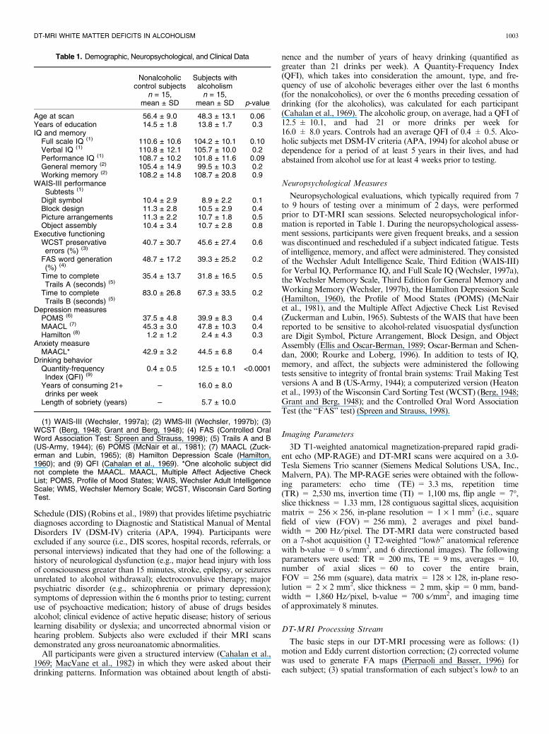
Table 1. Demographic, Neuropsychological, and Clinical Data
Nonalcoholiccontrol subjects
n = 15,mean ± SD
Subjects withalcoholism
n = 15,mean ± SD p-value
Age at scan 56.4 ± 9.0 48.3 ± 13.1 0.06Years of education 14.5 ± 1.8 13.8 ± 1.7 0.3IQ and memory
Full scale IQ (1) 110.6 ± 10.6 104.2 ± 10.1 0.10Verbal IQ (1) 110.8 ± 12.1 105.7 ± 10.0 0.2Performance IQ (1) 108.7 ± 10.2 101.8 ± 11.6 0.09General memory (2) 105.4 ± 14.9 99.5 ± 10.3 0.2Working memory (2) 108.2 ± 14.8 108.7 ± 20.8 0.9
WAIS-III performanceSubtests (1)
Digit symbol 10.4 ± 2.9 8.9 ± 2.2 0.1Block design 11.3 ± 2.8 10.5 ± 2.9 0.4Picture arrangements 11.3 ± 2.2 10.7 ± 1.8 0.5Object assembly 10.4 ± 3.4 10.7 ± 2.8 0.8
Executive functioningWCST preservativeerrors (%) (3)
40.7 ± 30.7 45.6 ± 27.4 0.6
FAS word generation(%) (4)
48.7 ± 17.2 39.3 ± 25.2 0.2
Time to completeTrails A (seconds) (5)
35.4 ± 13.7 31.8 ± 16.5 0.5
Time to completeTrails B (seconds) (5)
83.0 ± 26.8 67.3 ± 33.5 0.2
Depression measuresPOMS (6) 37.5 ± 4.8 39.9 ± 8.3 0.4MAACL (7) 45.3 ± 3.0 47.8 ± 10.3 0.4Hamilton (8) 1.2 ± 1.2 2.4 ± 4.3 0.3
Anxiety measureMAACL* 42.9 ± 3.2 44.5 ± 6.8 0.4
Drinking behaviorQuantity-frequencyIndex (QFI) (9)
0.4 ± 0.5 12.5 ± 10.1 <0.0001
Years of consuming 21+drinks per week
– 16.0 ± 8.0
Length of sobriety (years) – 5.7 ± 10.0
(1) WAIS-III (Wechsler, 1997a); (2) WMS-III (Wechsler, 1997b); (3)WCST (Berg, 1948; Grant and Berg, 1948); (4) FAS (Controlled OralWord Association Test: Spreen and Strauss, 1998); (5) Trails A and B(US-Army, 1944); (6) POMS (McNair et al., 1981); (7) MAACL (Zuck-erman and Lubin, 1965); (8) Hamilton Depression Scale (Hamilton,1960); and (9) QFI (Cahalan et al., 1969). *One alcoholic subject didnot complete the MAACL. MAACL, Multiple Affect Adjective CheckList; POMS, Profile of Mood States; WAIS, Wechsler Adult IntelligenceScale; WMS, Wechsler Memory Scale; WCST, Wisconsin Card SortingTest.
DT-MRI WHITE MATTER DEFICITS IN ALCOHOLISM 1003
average template created by co-registering (initial target was a T2-weighted 152-subject average template) and averaging all lowb vol-umes; (4) generation of voxel-wise group-difference statistical mapsbased on the common-space FA data; (5) assessment of group differ-ences in anatomic white matter regions according to a priori hypothe-ses, guided by statistical thresholding; (6) FA values calculated onthe spatially normalized data for each subject for the hypothesis-dri-ven regions of interest (ROIs), identified in the above step (5). Addi-tional detail is provided below.
DT-MRI Methods
For the DT-MRI data analysis, we used a combination of the fol-lowing 2 processing streams: Martinos Center Free Diffusion Tools(Salat et al., 2005a,b; Tuch et al., 2005), which include a set of pro-cessing scripts for reconstruction and analysis of DT-MRI data,developed at the Athinoula A. Martinos Center for BiomedicalImaging at Massachusetts General Hospital (Boston, MA); and FSL(http://www.fmrib.ox.ac.uk/fsl) (Jenkinson and Smith, 2001; Jenkin-son et al., 2002; Smith, 2002; Smith et al., 2001).The DT-MRI data were acquired as a 7-shot acquisition, consist-
ing of 6 directional image volumes plus a lowb structural image setwith no diffusion weighting. All of the above DT-MRI images werecollected within the same sequence and with identical parameters. A12-degree affine mutual information cost function transformation(procedure available with FLIRT, FSL-FMRIB’s Linear Image Reg-istration Tool, University of Oxford, England) was applied to allthese volumes to help reduce Eddy current distortions and motionalong different repetitions as follows: Each slice of the acquisitionconsisted of a lowb (T2) image and 6 directional images, which wererepeated a total of 10 times (70 images per slice). The averaging stepwas to take the first lowb image (#1 of 70 images) and co-register allremaining images (lowb and directional images alike) to the first lowbimage. Then, the DT was calculated for each voxel in the volume,using a least-squares fit to the diffusion signal (Basser et al., 1994).The FA was calculated from the DT (Pierpaoli and Basser, 1996).
Spatial Normalization
Each subject’s lowb volume was registered to the SPM (StatisticalParametric Mapping; http://www.fil.ion.ucl.ac.uk/spm) T2 152-sub-ject average template (ICBM, NIH P-20 project, Principal Investiga-tor, John Mazziotta). This template has a 2 mm isotropic resolutionand was smoothed with an 8 mm full width at half maximum(FWHM) Gaussian filter (sequence details: dual echo spin echo,TE = 120 ms, TR = 3,300 ms, flip angle = 90�). Then, a subject-based T2 anatomical template was created by averaging the SPM-registered lowb volumes for the entire cohort of alcoholics and nonal-coholic controls. The FSL Brain Extraction Tool was used to skull-strip the input lowb volumes to facilitate more uniform and preciseregistrations. Each registration was performed using a 12-degreeaffine mutual information cost function transformation (procedureavailable with FLIRT).Each subject’s native lowb image was subsequently registered to
the subject-based average template using FLIRT as above. The FAmaps were then spatially transformed to the common coordinatespace by using the transformations identified for their respective lowbvolume.
Group Maps
Group analyses were performed to examine the regional distribu-tion of diagnostic-group related differences in FA, by performing avoxel-based two-tailed t-test between the 2 diagnostic groups. In thiscase, voxels that differed at p < 0.01 using a two-tailed t-test (corre-sponding to t > 2.76) were identified in an image that showed 2types of colorized regions: those where the alcoholic group had a sig-
nificantly higher FA value than the nonalcoholic control group(blue), and where the alcoholic group had a significantly lower FAvalue than that of the control group (red). This image of colorizedregions was then superimposed on the T2-weighted structural ana-tomical template volume (lowb) that was created as a group averagefor the entire cohort to get an anatomical reference for the regions ofsignificant difference.
Regions of Interest Procedures
We first executed a voxel-based analysis by creating regions of FAdifferences and taking clusters above a significance threshold(p < 0.05) and size (greater than 5 contiguous voxels). Afterwards,these clusters were identified by one rater (NM) based on anatomicaltopography and the mapping of these fiber pathways from known lit-erature (Makris et al., 1999, 2002b, 2005; Mufson and Pandya, 1984;Petrides and Pandya, 1988; Talairach and Tournoux, 1988; Yakovlevand Locke, 1961). Regions of FA difference located in predominantlygray matter were noted for discussion, but excluded from regionalanalyses as we were primarily interested in white matter tracts.There were 4 principal clusters of anatomical interest correspond-
ing to white matter tracts connecting to reward circuit corticalregions: The OFCwm, located in the ventral anterior sector of thefrontal lobes (Caviness et al., 1996); the cingulum bundle, whichincluded white matter voxels contained within the cingulate gyrusabove the corpus callosum (Makris et al., 2002a); SLF II, in theregion above the insula, the extreme capsule, the claustrum, the exter-nal capsule, the lenticular nucleus, and the internal capsule (Makriset al., 2005); and SLF III, within the parietal and frontal Sylvianopercula (Makris et al., 2005). We then created ROIs, defined by theextent of these clusters, and we calculated the average FA value inthese clusters for each subject. These values were then comparedbetween the 2 groups (alcoholic vs. control) with analyses of covari-ance (ANCOVA) to evaluate the group differences in FA values co-varied for age. Furthermore, each of these ROIs was mirrored aboutthe inter-hemispheric fissure to the contralateral hemisphere, and FAvalues in these contralateral hemisphere ROIs were determined andcompared.As an exploratory analysis, we also noted and discuss below the
locations of other significant regions of FA group differences inregions beyond those connecting with specific reward-related regionsof the EROS network.
Data Analysis: ROI and Correlation Analyses
All statistical analyses were performed using JMP statistical analy-sis software (version 5.0.1.2; SAS Institute Inc., Cary, NC). Between-group ROI comparisons were made on regional FA measures usingANCOVA controlling for age. To assess laterality effects in the 4index regions, we applied ANOVA including effects of group (alco-holic vs. control) and hemisphere (right vs. left), with subjects as arepeated factor, to determine whether there were group by hemi-sphere interaction effects. Correlation analyses then were applied tocorrelate regional FA measures in frontal ROIs with memory, IQ,age, and drinking history. Correlation coefficients were comparedbetween groups using Fisher’s Z transformation. Regions that hadsignificant within group correlations in conjunction with significantbetween group differences in correlation coefficients are reportedbelow.In order to determine how well the index regions could discrimi-
nate group membership, we performed linear discriminant functionanalysis (DFA) with and without partialling out the effect of age.Age was covaried by running the DFA on the residual FA valuesfrom the correlation of age with each ROI. In the absence of a sec-ond sample to test the discriminant function, we then created thediscriminant function using n-1 subjects and used it to classify the1-out subject. This leave-one-out cross validation of the discriminant
1004 HARRIS ET AL.
analysis enables a test of the specificity and accuracy of the DFAwithin a sample using each subject as a test case of the discriminantfunction.
RESULTS
FA Maps and a Priori Hypothesized ROIs
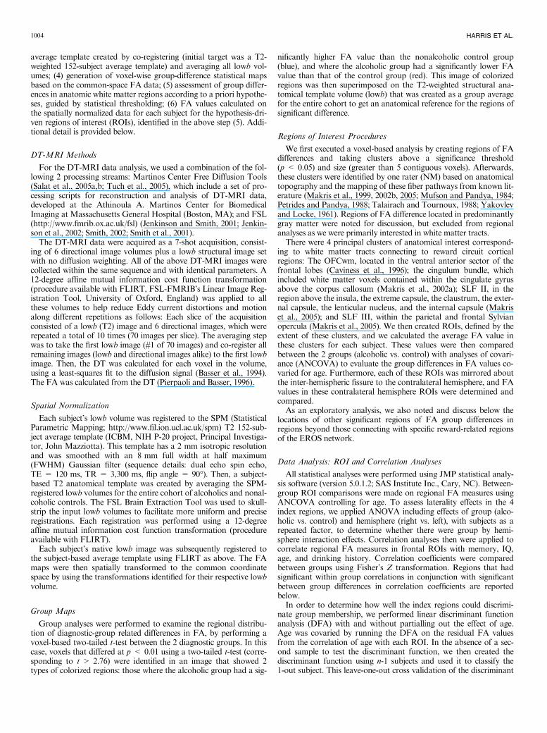
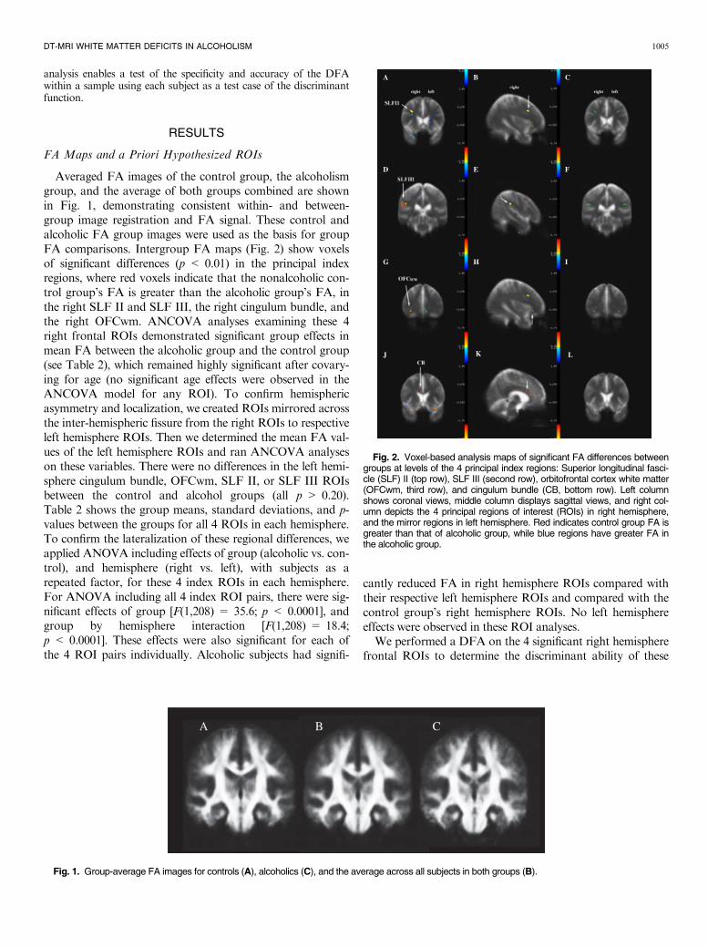
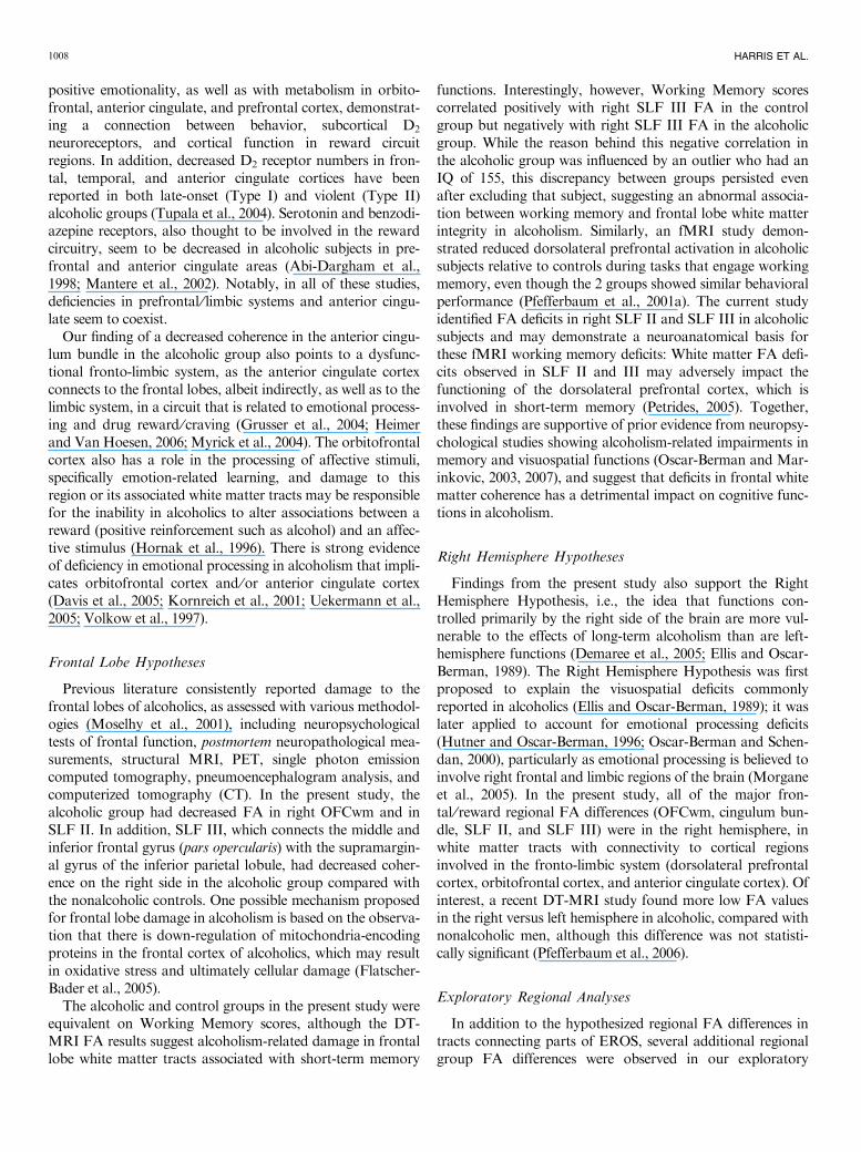
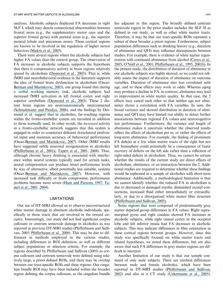
Averaged FA images of the control group, the alcoholismgroup, and the average of both groups combined are shownin Fig. 1, demonstrating consistent within- and between-group image registration and FA signal. These control andalcoholic FA group images were used as the basis for groupFA comparisons. Intergroup FA maps (Fig. 2) show voxelsof significant differences (p < 0.01) in the principal indexregions, where red voxels indicate that the nonalcoholic con-trol group’s FA is greater than the alcoholic group’s FA, inthe right SLF II and SLF III, the right cingulum bundle, andthe right OFCwm. ANCOVA analyses examining these 4right frontal ROIs demonstrated significant group effects inmean FA between the alcoholic group and the control group(see Table 2), which remained highly significant after covary-ing for age (no significant age effects were observed in theANCOVA model for any ROI). To confirm hemisphericasymmetry and localization, we created ROIs mirrored acrossthe inter-hemispheric fissure from the right ROIs to respectiveleft hemisphere ROIs. Then we determined the mean FA val-ues of the left hemisphere ROIs and ran ANCOVA analyseson these variables. There were no differences in the left hemi-sphere cingulum bundle, OFCwm, SLF II, or SLF III ROIsbetween the control and alcohol groups (all p > 0.20).Table 2 shows the group means, standard deviations, and p-values between the groups for all 4 ROIs in each hemisphere.To confirm the lateralization of these regional differences, weapplied ANOVA including effects of group (alcoholic vs. con-trol), and hemisphere (right vs. left), with subjects as arepeated factor, for these 4 index ROIs in each hemisphere.For ANOVA including all 4 index ROI pairs, there were sig-nificant effects of group [F(1,208) = 35.6; p < 0.0001], andgroup by hemisphere interaction [F(1,208) = 18.4;p < 0.0001]. These effects were also significant for each ofthe 4 ROI pairs individually. Alcoholic subjects had signifi-
cantly reduced FA in right hemisphere ROIs compared withtheir respective left hemisphere ROIs and compared with thecontrol group’s right hemisphere ROIs. No left hemisphereeffects were observed in these ROI analyses.We performed a DFA on the 4 significant right hemisphere
frontal ROIs to determine the discriminant ability of these
A B C
Fig. 1. Group-average FA images for controls (A), alcoholics (C), and the average across all subjects in both groups (B).
A B C
D E F
G H I
J K L
Fig. 2. Voxel-based analysis maps of significant FA differences betweengroups at levels of the 4 principal index regions: Superior longitudinal fasci-cle (SLF) II (top row), SLF III (second row), orbitofrontal cortex white matter(OFCwm, third row), and cingulum bundle (CB, bottom row). Left columnshows coronal views, middle column displays sagittal views, and right col-umn depicts the 4 principal regions of interest (ROIs) in right hemisphere,and the mirror regions in left hemisphere. Red indicates control group FA isgreater than that of alcoholic group, while blue regions have greater FA inthe alcoholic group.
DT-MRI WHITE MATTER DEFICITS IN ALCOHOLISM 1005
DT-MRI FA regional measures for categorizing the groupmembership of each subject. The DFA was highly effectivefor determining group membership; all controls and all but 1alcoholic subject were correctly classified based on the 4 fron-tal right-hemisphere ROIs [29 ⁄30, 97% correct classification,F(4, 25) = 18.8, p < 0.0001]. After covarying for age, 93%were correctly classified [28 ⁄30, 1 alcoholic and 1 control sub-ject misclassified, F(4, 25) = 10.7, p < 0.0001]. The leave-one-out cross validation of the DFA showed similar results:29 of 30 correct classifications [F(4, 24) = 18.3, p < 0.0001],and 90% correct classifications with age as covariate [27 ⁄30, 1alcoholic and 2 control subjects misclassified, F(4,24) = 10.4, p < 0.0001].In order to examine whether methodological artifacts might
have contributed to hemispheric lateralization of frontalregional DT-MRI tract differences, we examined non-thres-holded group-difference maps, and also examined whetherapplying a smaller cluster size threshold (3 contiguous voxelsrather than 5), would demonstrate left hemisphere differencesin these index regions that might have missed the statistical orcluster size thresholds, or been missed by the contralaterallymirrored ROI placement. However, we did not find evidenceof systematic hemispheric artifacts that might have contrib-uted to the lateralized results above. While reducing the clus-ter size threshold resulted in more regions being noted overall,the lateralization of DT-MRI deficits in alcoholics remainedheavily weighted toward the right hemisphere; there were 12right hemisphere regions versus 4 left hemisphere regions withclusters of 3 voxels or more, and none was located in left fron-tal lobe white matter tracts.
Behavioral Correlations With Regional FA Measures
Age at scan, drinking history, IQ, and memory scores wereexamined for correlation with mean FA of the index ROIsthat were created in the study (right and left hemisphere cin-
gulum bundle, SLF II, SLF III, and OFCwm). Regions thathad significant structure ⁄behavior correlations within a sub-ject group, combined with significant correlation differencesbetween groups, are reported here. No group differences wereobserved in correlations between regional FA values and age,drinking history, or IQ scores. However, age was correlatedwith duration of abstinence [age vs. (log) years sober:r = 0.76, p = 0.001; nonparametric rank correlationrho = 0.61, p = 0.02], which may have inhibited our abilityto detect age or abstinence-related changes. In the nonalco-holic control group (but not in the alcoholic group), WorkingMemory was significantly correlated with right and with leftSLF III FA (right: r = 0.69, p = 0.005; left: r = 0.72,p = 0.002). Correlation coefficients for right and left SLF IIIvs. Working Memory scores were significantly differentbetween groups (right: Z = 3.5, p = 0.0004 and left:Z = 3.2, p < 0.01). Right SLF III was negatively correlatedwith Working Memory in the alcoholic group (r = )0.53,p = 0.04), but this negative correlation was influenced by anoutlier subject with IQ of 155. Without including this subject,the alcoholic group correlation for right SLF III andWorkingMemory remained negative, but was no longer significant(r = )0.39, p = 0.17), and was comparable to the left SLFIII correlation with Working Memory in alcoholics(r = )0.38, p = 0.16). However, even without including theoutlier subject, the between-group Fisher Z comparison con-tinued to demonstrate strongly significant difference betweengroups in correlation between right SLF III and WorkingMemory (Z = 3.01, p = 0.003).
Additional Exploratory Regional Group FA Differences
In addition to the hypothesis-driven analyses of white mat-ter tracts with direct connections to reward-related corticalregions involved in EROS, there were several white matterregions depicting group differences in other parts of the brain.
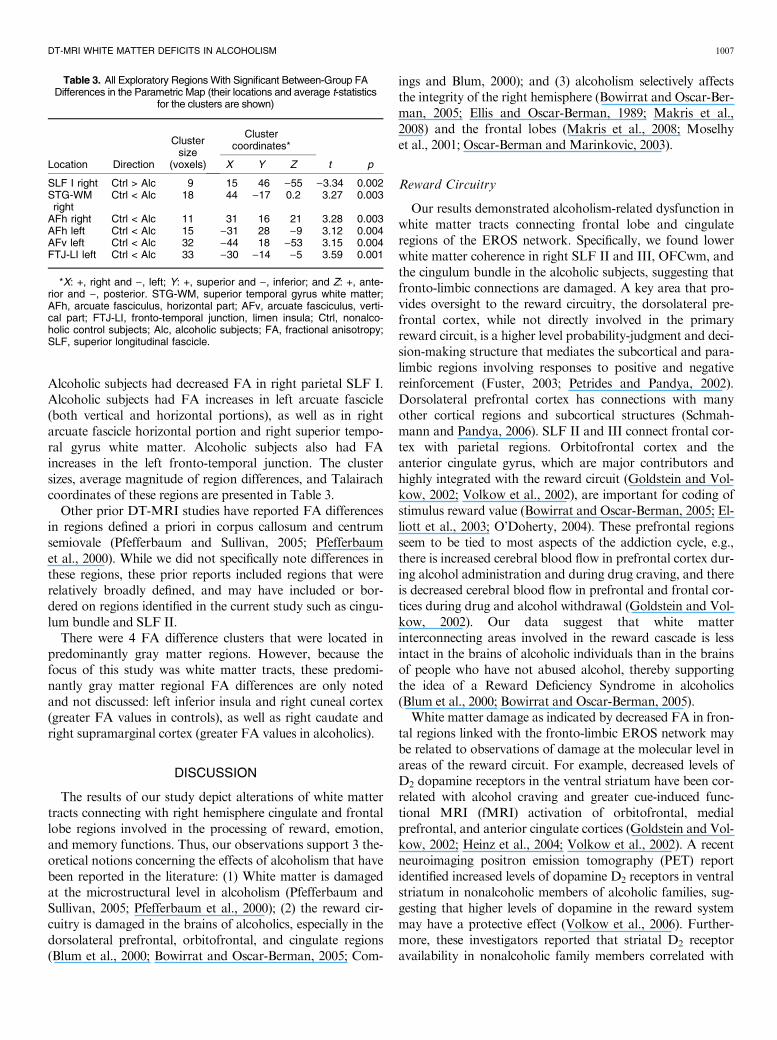
Table 2. Mean Differences of Average DTI FA Values in Selected ROI
RegionCluster size
(voxels)
Cluster coordinates* Nonalcoholiccontrol subjects
n = 15, mean ± SD
Subjects withalcoholism
n = 15, mean ± SD
Group effect
X Y Z F(1,29) p
Right HemisphereSLF II 27 33 26 19 0.43 ± 0.05 0.25 ± 0.13 23.1 <0.0001SLF III 33 48 28 )29 0.27 ± 0.07 0.15 ± 0.06 21.0 <0.0001CB 15 5 26 11 0.46 ± 0.09 0.29 ± 0.10 19.9 <0.0001OFCwm 6 34 )9 31 0.36 ± 0.12 0.23 ± 0.11 7.1 0.013
Left HemisphereSLF II 27 )33 26 19 0.38 ± 0.11 0.33 ± 0.14 2.0 0.2SLF III 33 )48 28 )29 0.28 ± 0.06 0.30 ± 0.11 0.2 0.7CB 15 )5 26 11 0.37 ± 0.13 0.33 ± 0.12 0.7 0.4OFCwm 6 )34 )9 31 0.32 ± 0.13 0.30 ± 0.17 0.1 0.7
F-values are for the ANCOVA group effect after covarying for Age. No significant age effects were observed in the ANCOVA model for anyROI. *Talairach coordinates: X = right + and left ); Y = superior + and inferior ); and Z = anterior + and posterior ). Overall ANCOVA results(includes group and age effects in model): right SLF II F(2,27) = 12.6, p = 0.0001; right SLF III F(2,27) = 12.1, p = 0.0002; right CBF(2,27) = 10.8, p = 0.0004; right OFCwm F(2,27) = 6.8, p = 0.004; left SLF II F(2,27) = 1.3, p = 0.3; left SLF III F(2,27) = 0.1, p = 0.9; left CBF(2,27) = 0.4, p = 0.7; left OFCwm F(2,27) = 0.2, p = 0.8. SLF, superior longitudinal fascicle; CB, cingulum bundle; OFCwm, orbitofrontal cortexwhite matter; FA, fractional anisotropy; ROI, regions of interest.
1006 HARRIS ET AL.
Alcoholic subjects had decreased FA in right parietal SLF I.Alcoholic subjects had FA increases in left arcuate fascicle(both vertical and horizontal portions), as well as in rightarcuate fascicle horizontal portion and right superior tempo-ral gyrus white matter. Alcoholic subjects also had FAincreases in the left fronto-temporal junction. The clustersizes, average magnitude of region differences, and Talairachcoordinates of these regions are presented in Table 3.Other prior DT-MRI studies have reported FA differences
in regions defined a priori in corpus callosum and centrumsemiovale (Pfefferbaum and Sullivan, 2005; Pfefferbaumet al., 2000). While we did not specifically note differences inthese regions, these prior reports included regions that wererelatively broadly defined, and may have included or bor-dered on regions identified in the current study such as cingu-lum bundle and SLF II.There were 4 FA difference clusters that were located in
predominantly gray matter regions. However, because thefocus of this study was white matter tracts, these predomi-nantly gray matter regional FA differences are only notedand not discussed: left inferior insula and right cuneal cortex(greater FA values in controls), as well as right caudate andright supramarginal cortex (greater FA values in alcoholics).
DISCUSSION
The results of our study depict alterations of white mattertracts connecting with right hemisphere cingulate and frontallobe regions involved in the processing of reward, emotion,and memory functions. Thus, our observations support 3 the-oretical notions concerning the effects of alcoholism that havebeen reported in the literature: (1) White matter is damagedat the microstructural level in alcoholism (Pfefferbaum andSullivan, 2005; Pfefferbaum et al., 2000); (2) the reward cir-cuitry is damaged in the brains of alcoholics, especially in thedorsolateral prefrontal, orbitofrontal, and cingulate regions(Blum et al., 2000; Bowirrat and Oscar-Berman, 2005; Com-
ings and Blum, 2000); and (3) alcoholism selectively affectsthe integrity of the right hemisphere (Bowirrat and Oscar-Ber-man, 2005; Ellis and Oscar-Berman, 1989; Makris et al.,2008) and the frontal lobes (Makris et al., 2008; Moselhyet al., 2001; Oscar-Berman and Marinkovic, 2003).
Reward Circuitry
Our results demonstrated alcoholism-related dysfunction inwhite matter tracts connecting frontal lobe and cingulateregions of the EROS network. Specifically, we found lowerwhite matter coherence in right SLF II and III, OFCwm, andthe cingulum bundle in the alcoholic subjects, suggesting thatfronto-limbic connections are damaged. A key area that pro-vides oversight to the reward circuitry, the dorsolateral pre-frontal cortex, while not directly involved in the primaryreward circuit, is a higher level probability-judgment and deci-sion-making structure that mediates the subcortical and para-limbic regions involving responses to positive and negativereinforcement (Fuster, 2003; Petrides and Pandya, 2002).Dorsolateral prefrontal cortex has connections with manyother cortical regions and subcortical structures (Schmah-mann and Pandya, 2006). SLF II and III connect frontal cor-tex with parietal regions. Orbitofrontal cortex and theanterior cingulate gyrus, which are major contributors andhighly integrated with the reward circuit (Goldstein and Vol-kow, 2002; Volkow et al., 2002), are important for coding ofstimulus reward value (Bowirrat and Oscar-Berman, 2005; El-liott et al., 2003; O’Doherty, 2004). These prefrontal regionsseem to be tied to most aspects of the addiction cycle, e.g.,there is increased cerebral blood flow in prefrontal cortex dur-ing alcohol administration and during drug craving, and thereis decreased cerebral blood flow in prefrontal and frontal cor-tices during drug and alcohol withdrawal (Goldstein and Vol-kow, 2002). Our data suggest that white matterinterconnecting areas involved in the reward cascade is lessintact in the brains of alcoholic individuals than in the brainsof people who have not abused alcohol, thereby supportingthe idea of a Reward Deficiency Syndrome in alcoholics(Blum et al., 2000; Bowirrat and Oscar-Berman, 2005).White matter damage as indicated by decreased FA in fron-
tal regions linked with the fronto-limbic EROS network maybe related to observations of damage at the molecular level inareas of the reward circuit. For example, decreased levels ofD2 dopamine receptors in the ventral striatum have been cor-related with alcohol craving and greater cue-induced func-tional MRI (fMRI) activation of orbitofrontal, medialprefrontal, and anterior cingulate cortices (Goldstein and Vol-kow, 2002; Heinz et al., 2004; Volkow et al., 2002). A recentneuroimaging positron emission tomography (PET) reportidentified increased levels of dopamine D2 receptors in ventralstriatum in nonalcoholic members of alcoholic families, sug-gesting that higher levels of dopamine in the reward systemmay have a protective effect (Volkow et al., 2006). Further-more, these investigators reported that striatal D2 receptoravailability in nonalcoholic family members correlated with
Table 3. All Exploratory Regions With Significant Between-Group FADifferences in the Parametric Map (their locations and average t-statistics
for the clusters are shown)
Location Direction
Clustersize
(voxels)
Clustercoordinates*
t pX Y Z
SLF I right Ctrl > Alc 9 15 46 )55 )3.34 0.002STG-WMright
Ctrl < Alc 18 44 )17 0.2 3.27 0.003
AFh right Ctrl < Alc 11 31 16 21 3.28 0.003AFh left Ctrl < Alc 15 )31 28 )9 3.12 0.004AFv left Ctrl < Alc 32 )44 18 )53 3.15 0.004FTJ-LI left Ctrl < Alc 33 )30 )14 )5 3.59 0.001
*X: +, right and ), left; Y: +, superior and ), inferior; and Z: +, ante-rior and ), posterior. STG-WM, superior temporal gyrus white matter;AFh, arcuate fasciculus, horizontal part; AFv, arcuate fasciculus, verti-cal part; FTJ-LI, fronto-temporal junction, limen insula; Ctrl, nonalco-holic control subjects; Alc, alcoholic subjects; FA, fractional anisotropy;SLF, superior longitudinal fascicle.
DT-MRI WHITE MATTER DEFICITS IN ALCOHOLISM 1007
positive emotionality, as well as with metabolism in orbito-frontal, anterior cingulate, and prefrontal cortex, demonstrat-ing a connection between behavior, subcortical D2
neuroreceptors, and cortical function in reward circuitregions. In addition, decreased D2 receptor numbers in fron-tal, temporal, and anterior cingulate cortices have beenreported in both late-onset (Type I) and violent (Type II)alcoholic groups (Tupala et al., 2004). Serotonin and benzodi-azepine receptors, also thought to be involved in the rewardcircuitry, seem to be decreased in alcoholic subjects in pre-frontal and anterior cingulate areas (Abi-Dargham et al.,1998; Mantere et al., 2002). Notably, in all of these studies,deficiencies in prefrontal ⁄ limbic systems and anterior cingu-late seem to coexist.Our finding of a decreased coherence in the anterior cingu-
lum bundle in the alcoholic group also points to a dysfunc-tional fronto-limbic system, as the anterior cingulate cortexconnects to the frontal lobes, albeit indirectly, as well as to thelimbic system, in a circuit that is related to emotional process-ing and drug reward ⁄craving (Grusser et al., 2004; Heimerand Van Hoesen, 2006; Myrick et al., 2004). The orbitofrontalcortex also has a role in the processing of affective stimuli,specifically emotion-related learning, and damage to thisregion or its associated white matter tracts may be responsiblefor the inability in alcoholics to alter associations between areward (positive reinforcement such as alcohol) and an affec-tive stimulus (Hornak et al., 1996). There is strong evidenceof deficiency in emotional processing in alcoholism that impli-cates orbitofrontal cortex and ⁄or anterior cingulate cortex(Davis et al., 2005; Kornreich et al., 2001; Uekermann et al.,2005; Volkow et al., 1997).
Frontal Lobe Hypotheses
Previous literature consistently reported damage to thefrontal lobes of alcoholics, as assessed with various methodol-ogies (Moselhy et al., 2001), including neuropsychologicaltests of frontal function, postmortem neuropathological mea-surements, structural MRI, PET, single photon emissioncomputed tomography, pneumoencephalogram analysis, andcomputerized tomography (CT). In the present study, thealcoholic group had decreased FA in right OFCwm and inSLF II. In addition, SLF III, which connects the middle andinferior frontal gyrus (pars opercularis) with the supramargin-al gyrus of the inferior parietal lobule, had decreased coher-ence on the right side in the alcoholic group compared withthe nonalcoholic controls. One possible mechanism proposedfor frontal lobe damage in alcoholism is based on the observa-tion that there is down-regulation of mitochondria-encodingproteins in the frontal cortex of alcoholics, which may resultin oxidative stress and ultimately cellular damage (Flatscher-Bader et al., 2005).The alcoholic and control groups in the present study were
equivalent on Working Memory scores, although the DT-MRI FA results suggest alcoholism-related damage in frontallobe white matter tracts associated with short-term memory
functions. Interestingly, however, Working Memory scorescorrelated positively with right SLF III FA in the controlgroup but negatively with right SLF III FA in the alcoholicgroup. While the reason behind this negative correlation inthe alcoholic group was influenced by an outlier who had anIQ of 155, this discrepancy between groups persisted evenafter excluding that subject, suggesting an abnormal associa-tion between working memory and frontal lobe white matterintegrity in alcoholism. Similarly, an fMRI study demon-strated reduced dorsolateral prefrontal activation in alcoholicsubjects relative to controls during tasks that engage workingmemory, even though the 2 groups showed similar behavioralperformance (Pfefferbaum et al., 2001a). The current studyidentified FA deficits in right SLF II and SLF III in alcoholicsubjects and may demonstrate a neuroanatomical basis forthese fMRI working memory deficits: White matter FA defi-cits observed in SLF II and III may adversely impact thefunctioning of the dorsolateral prefrontal cortex, which isinvolved in short-term memory (Petrides, 2005). Together,these findings are supportive of prior evidence from neuropsy-chological studies showing alcoholism-related impairments inmemory and visuospatial functions (Oscar-Berman and Mar-inkovic, 2003, 2007), and suggest that deficits in frontal whitematter coherence has a detrimental impact on cognitive func-tions in alcoholism.
Right Hemisphere Hypotheses
Findings from the present study also support the RightHemisphere Hypothesis, i.e., the idea that functions con-trolled primarily by the right side of the brain are more vul-nerable to the effects of long-term alcoholism than are left-hemisphere functions (Demaree et al., 2005; Ellis and Oscar-Berman, 1989). The Right Hemisphere Hypothesis was firstproposed to explain the visuospatial deficits commonlyreported in alcoholics (Ellis and Oscar-Berman, 1989); it waslater applied to account for emotional processing deficits(Hutner and Oscar-Berman, 1996; Oscar-Berman and Schen-dan, 2000), particularly as emotional processing is believed toinvolve right frontal and limbic regions of the brain (Morganeet al., 2005). In the present study, all of the major fron-tal ⁄ reward regional FA differences (OFCwm, cingulum bun-dle, SLF II, and SLF III) were in the right hemisphere, inwhite matter tracts with connectivity to cortical regionsinvolved in the fronto-limbic system (dorsolateral prefrontalcortex, orbitofrontal cortex, and anterior cingulate cortex). Ofinterest, a recent DT-MRI study found more low FA valuesin the right versus left hemisphere in alcoholic, compared withnonalcoholic men, although this difference was not statisti-cally significant (Pfefferbaum et al., 2006).
Exploratory Regional Analyses
In addition to the hypothesized regional FA differences intracts connecting parts of EROS, several additional regionalgroup FA differences were observed in our exploratory
1008 HARRIS ET AL.
analyses. Alcoholic subjects displayed FA decreases in rightSLF I, which may denote connectional abnormalities betweenfrontal areas (e.g., the supplementary motor area and thesuperior frontal gyrus) with parietal areas (e.g., the superiorparietal lobule and precuneus areas). These cortical regionsare known to be involved in the regulation of higher motorbehaviors (Makris et al., 2005).There were several regions where the alcoholic subjects had
higher FA values than the control group. The observation ofFA increases in alcoholic subjects supports the hypothesisthat there is compensatory activity in brain regions relativelyspared by alcoholism (Desmond et al., 2003). That is, whilefMRI and neurobehavioral evidence in the literature supportsthe idea of frontal brain dysfunction in alcoholism (Oscar-Berman and Marinkovic, 2003), one group found that duringa verbal working memory task, alcoholic subjects hadincreased fMRI activation in left frontal cortex and rightsuperior cerebellum (Desmond et al., 2003). These 2 dis-tant brain regions are neuroanatomically interconnected(Schmahmann and Pandya, 2006), and the findings by Des-mond et al. suggest that in alcoholics, far-reaching regionswithin the fronto-cerebellar system are recruited in additionto those normally used. In other words, the increased activityin a fronto-cerebellar network suggests that this system isengaged in order to counteract deficient dorsolateral prefron-tal input and maintain accuracy of behavioral performance(Oscar-Berman and Marinkovic, 2007). Other fMRI resultshave suggested subtle neuronal reorganization in alcoholics(Pfefferbaum et al., 2001a; Tapert et al., 2001, 2004). Thus,although chronic heavy drinking is associated with interfer-ence within neural systems typically used for certain tasks,neural compensation can produce intact performance levelsby way of alternative, albeit potentially less suitable, systems(Oscar-Berman and Marinkovic, 2007). However, withincreased task difficulty or brain compromise, performanceproblems become more severe (Ham and Parsons, 1997; Ta-pert et al., 2001, 2004).
LIMITATIONS
Our use of DT-MRI allowed us to observe microstructuralwhite matter damage in abstinent alcoholic individuals, spe-cifically in those tracts that are involved in the reward cir-cuitry. Interestingly, our study did not find significant corpuscallosum or centrum semiovale damage in alcoholics as wasreported in previous DT-MRI studies (Pfefferbaum and Sulli-van, 2005; Pfefferbaum et al., 2000). This may be due to dif-ferences in methods employed in the various studies,including differences in ROI definition, as well as differentsubject populations or selection criteria. For example, theregions described by Pfefferbaum and Sullivan (2005) as cor-pus callosum and centrum semiovale were defined using rela-tively large, a priori defined ROIs, and there may be overlapbetween our tract-specific ROIs and these regions. Our cingu-lum bundle ROI may have been included within the broaderregion defining the corpus callosum, as the cingulum bundle
lies adjacent to this region. The broadly defined centrumsemiovale region in the prior studies includes the SLF II asdefined in our study, as well as other white matter tracts.Therefore, it may be that our tract-specific ROIs represent asubset of these broader a priori regions. Furthermore, subjectpopulation differences such as drinking history (e.g., durationof abstinence and QFI) may influence discrepancies betweenstudies. For example, there is evidence of white matter regen-eration with continued abstinence from alcohol (Crews et al.,2005; O’Neill et al., 2001; Pfefferbaum et al., 1995, 2001b). Inthe present study, the distribution of duration of abstinence inour alcoholic subjects was highly skewed, so we could not reli-ably assess the impact of duration of abstinence on outcomevariables. Duration of abstinence also was correlated withage, and so these effects may work at odds: Whereas agingmay produce a decline in FA, in contrast, abstinence may leadto improvement in white matter quality. In that case, the 2effects may cancel each other so that neither age nor absti-nence shows a correlation with FA variables. In sum, thebroad variance and skewed distribution in duration of absti-nence and QFI may have limited our ability to detect furtherassociations between regional FA values and neurocognitivetest performance. Furthermore, the variance and length ofabstinence makes it uncertain whether the observed resultsreflect the effects of alcoholism per se, or rather the effects oflong-term abstinence. For example, the finding of prominentFA deficits in a few white matter tracts of the right but notleft hemisphere could potentially be a consequence of fasterrecovery of deficits on the left side rather than a reflection ofright-sided deficits in alcoholism. Thus, we cannot be certainwhether the results of the current study are direct effects ofalcoholism, abstinence, or an interaction between the 2. Addi-tional studies are required to determine whether these findingswould be replicated in a sample of alcoholics with short-termabstinence. Additionally, a methodological limitation is thatwe cannot identify whether the reduced FA in alcoholism isdue to decreased or damaged myelin, diminished axonal con-nections, increased fluid either intracellularly or extracellu-larly, or due to a disorganized white matter fiber structure(Pfefferbaum and Sullivan, 2005).Some regions that were composed of predominantly gray
matter depicted group differences in FA values: Right supra-marginal gyrus and right caudate showed FA increases inalcoholic subjects, while right cuneal cortex in the occipitallobe and left inferior insula had FA decreases in alcoholicsubjects. This may indicate differences in fiber orientation inthese cortical regions between groups. However, since thisstudy was specifically focused on white matter tracts andrelated hypotheses, we noted these differences, but are alsoaware that such FA differences in gray matter regions are dif-ficult to interpret.Another limitation of our study is that our sample con-
sisted of only male subjects. There are cerebral differencesbetween male and female alcoholics, which have beenreported in DT-MRI studies (Pfefferbaum and Sullivan,2002) and also in a CT study (Uekermann et al., 2005).
DT-MRI WHITE MATTER DEFICITS IN ALCOHOLISM 1009
Females are also reported to be more vulnerable to the effectsof alcohol, but the evidence is inconclusive (Hommer, 2003;Prendergast, 2004). Therefore, additional alcoholism studiesare needed with gender-specific controls. Another limitationof our study is the small sample size of 15 individuals in eachgroup.It may seem to be another limitation that our control group
was somewhat older than the alcoholic group (see Table 1).However, prior DT-MRI studies have shown white mattertract degeneration with normal aging (Madden et al., 2004;Pfefferbaum and Sullivan, 2003; Pfefferbaum et al., 2005),and so we may have even underestimated the extent of FAdeficits in the alcoholic group. In other words, if Age effectspredominate over Group effects, the trend toward older con-trol subjects would be expected to generate decreased FA inthe control group compared with the alcoholic group in ourstudy. However, despite the control group being slightly olderthan the alcoholic group, the alcoholic group displayeddecreased FA in right frontal lobe white matter tracts, andthese differences remained highly significant after covaryingfor age. It is possible, however, that the trend toward oldercontrol subjects may have contributed to the higher FA inalcoholics observed in several regions.Finally, our ROI analyses may have bias due to the fact
that we selected ROIs as the regions defined by significantFA clusters, and not as cytoarchitectonic or functional brainregions. Nevertheless, our ROIs seem to have high predictivevalue of whether an individual subject is classified as an alco-holic or a nonalcoholic control. A DFA was run to determinehow well the right frontal FA ROIs classified group member-ship (i.e., alcoholic or control), and we noted 97% correctgroup classification (93% covarying for age). Thus, it ishighly unlikely that artifacts or bias in methods could explainsuch a dramatic group discrimination effect. Another poten-tial source of region bias was that we created the left hemi-sphere ROIs as mirrored regions from the right hemisphereregions, to estimate the location of the corresponding contra-lateral tracts. Because mirrored regions may not exactlymatch the location of the opposite hemisphere’s tracts, the lefthemisphere ROIs may be less accurate measures of the tractROIs. However, the exploratory analyses reported all signifi-cant group difference clusters, and even after examining smal-ler cluster size thresholds and non-thresholded groupdifference maps to look for possible left-sided effects thatmay have been overlooked, we did not observe left hemi-sphere regional differences proximal to the index ROIs tosuggest regional findings were simply missed by the ROIplacement or thresholds, or resulted from methodologicalartifacts.
CONCLUSIONS
This DT-MRI study is the first to report damage in rightfronto-limbic and cortico-cortical white matter connectivity inalcoholism, evidenced by lower FA values in right SLF II,SLF III, OFCwm, and cingulum bundle in the alcoholic
group. The white matter abnormalities observed in our studysupport previous literature on alcoholism, specifically thosestudies showing damage to brain regions that comprise a sys-tem that is essential for normal emotional functioning and formodulating the effectiveness of positive and negative rein-forcement in human behavior: the EROS network. The whitematter abnormalities in reward-related white matter tractsthat we observed were localized to the right hemisphere, afinding that is consistent with observations of visuospatialand emotional abnormalities in alcoholism. Lastly, our resultsalso support previous studies that reported alcoholism-relateddeficits in working memory.
ACKNOWLEDGMENTS
This work was supported in part by Grants from: theNational Association for Research in Schizophrenia andDepression (NARSAD) and the National Institutes of HealthNational Center for Complementary and Alternative Medi-cine NCAM to Dr. Nikos Makris; National Institute onAlcohol Abuse and Alcoholism (NIAAA) Grants R01-AA07112 and K05-AA00219, and the Medical Research Ser-vice of the U.S Department of Veterans Affairs to Dr. Mar-lene Oscar-Berman; NIAAA K01-AA13402 to Dr. KsenijaMarinkovic; the Fairway Trust to Dr. David Kennedy, andby the National Center for Research Resources(P41RR14075) and the Mental Illness and Neuroscience Dis-covery (MIND) Institute. We thank Diane Merritt for assis-tance in recruiting the research participants.
SUPPLEMENTARY MATERIAL
The following supplementary material is available for thisarticle:Fig. S1. Nonthresholded voxel-based analysis maps of FA
differences between groups at levels of the four principalindex regions (noted by white arrows): superior longitudinalfascicle (SLF) II (top row), SLF III (second row), orbitofron-tal cortex white matter (OFCwm, third row), and cingulumbundle (CB, bottom row). Left column shows coronal views,middle column displays sagittal views through these righthemisphere index regions, and right column depicts sagittalviews through the corresponding left hemisphere plane. Redindicates control group FA is greater than that of alcoholicgroup, while blue regions have greater FA in the alcoholicgroup. Nonthresholded maps do not demonstrate evidence ofleft frontal DT-MRI deficits comparable to the right-sideddifferences.This material is available as part of the online article
from:http://www.blackwell-synergy.com/doi/abs/10.1111/j.1530-0277.2008.00661.x (This link will take you to the articleabstract).Please note: Blackwell Publishing is not responsible for the
content or functionality of any supplementary materials sup-plied by the authors. Any queries (other thanmissing material)should be directed to the corresponding author for the article.
1010 HARRIS ET AL.
REFERENCES
Abi-Dargham A, Krystal JH, Anjilvel S, Scanley BE, Zoghbi S, Baldwin RM,
Rajeevan N, Ellis S, Petrakis IL, Seibyl JP, Charney DS, Laruelle M, Innis
RB (1998) Alterations of benzodiazepine receptors in type II alcoholic sub-
jects measured with SPECT and [123I]iomazenil. Am J Psychiatry
155:1550–1555.
APA (1994) Diagnostic and Statistical Manual of Mental Disorders (DSM-
IV). American Psychiatric Association, Washington, DC.
Baker TB, Piper ME, McCarthy DE, Majeskie MR, Fiore MC (2004) Addic-
tion motivation reformulated: an affective processing model of negative
reinforcement. Psychol Rev 111:33–51.
Basser PJ, Mattiello J, LeBihan D (1994) Estimation of the effective self-diffu-
sion tensor from the NMR spin echo. J Magn Reson B 103:247–254.
Berg EA (1948) A simple objective technique for measuring flexibility in think-
ing. J Gen Psychol 39:15–22.
Blum K, Braverman ER, Holder JM, Lubar JF, Monastra VJ, Miller D,
Lubar JO, Chen TJ, Comings DE (2000) Reward deficiency syndrome: a
biogenetic model for the diagnosis and treatment of impulsive, addictive,
and compulsive behaviors. J Psychoactive Drugs 32(Suppl. i–iv):1–112.
Bowirrat A, Oscar-BermanM (2005) Relationship between dopaminergic neu-
rotransmission, alcoholism, and Reward Deficiency Syndrome. Am J Med
Genet B Neuropsychiatr Genet 132:29–37.
Briggs GG, Nebes RD (1975) Patterns of hand preference in a student popula-
tion. Cortex 11:230–238.
Cahalan V, Cisin I, Crossley HM (1969) American Drinking Practices: A
National Study of Drinking Behavior and Attitudes, Report 6. Rutgers
Center for Alcohol Studies, New Brunswick, NJ.
Caviness VS Jr, Meyer JW, Makris N, Kennedy DN (1996) MRI-based topo-
graphic parcellation of the human neocortex: an anatomically specified
method with estimate of reliability. J Cogn Neurosci 8:566–587.
Comings DE, Blum K (2000) Reward deficiency syndrome: genetic aspects of
behavioral disorders. Prog Brain Res 126:325–341.
Crews FT, Buckley T, Dodd PR, Ende G, Foley N, Harper C, He J, Innes D,
Lohel W, Pfefferbaum A, Zou J, Sullivan EV (2005) Alcoholic neurobiol-
ogy: changes in dependence and recovery. Alcohol Clin Exp Res 29:1504–
1513.
Davis KD, Taylor KS, Hutchison WD, Dostrovsky JO, McAndrews MP,
Richter EO, Lozano AM (2005) Human anterior cingulate cortex neurons
encode cognitive and emotional demands. J Neurosci 25:8402–8406.
Demaree HA, Everhart DE, Youngstrom EA, Harrison DW (2005) Brain lat-
eralization of emotional processing: historical roots and a future incorporat-
ing ‘‘dominance’’. Behav Cogn Neurosci Rev 4:3–20.
Desmond JE, Chen SH, DeRosa E, Pryor MR, Pfefferbaum A, Sullivan EV
(2003) Increased frontocerebellar activation in alcoholics during verbal
working memory: an fMRI study. Neuroimage 19:1510–1520.
Elliott R, Newman JL, Longe OA, Deakin JF (2003) Differential response
patterns in the striatum and orbitofrontal cortex to financial reward in
humans: a parametric functional magnetic resonance imaging study. J Neu-
rosci 23:303–307.
Ellis RJ, Oscar-Berman M (1989) Alcoholism, aging, and functional cerebral
asymmetries. Psychol Bull 106:128–147.
Estruch R, Nicolas JM, Salamero M, Aragon C, Sacanella E, Fernandez-Sola
J, Urbano-Marquez A (1997) Atrophy of the corpus callosum in chronic
alcoholism. J Neurol Sci 146:145–151.
Flatscher-Bader T, van der Brug M, Hwang JW, Gochee PA, Matsumoto I,
Niwa S, Wilce PA (2005) Alcohol-responsive genes in the frontal cortex and
nucleus accumbens of human alcoholics. J Neurochem 93:359–370.
Fuster JM (2003) Cortex andMind. Oxford University Press, New York.
Goldstein RZ, Volkow ND (2002) Drug addiction and its underlying neurobi-
ological basis: neuroimaging evidence for the involvement of the frontal cor-
tex. Am J Psychiatry 159:1642–1652.
Grant DA, Berg EA (1948) A behavioral analysis of degree of reinforcement
and ease of shifting to new responses in a Weigl-type card-sorting problem.
J Exp Psychol 38:404–411.
Grusser SM, Wrase J, Klein S, Hermann D, Smolka MN, Ruf M, Weber-
Fahr W, Flor H, Mann K, Braus DF, Heinz A (2004) Cue-induced
activation of the striatum and medial prefrontal cortex is associated with
subsequent relapse in abstinent alcoholics. Psychopharmacology 175:296–
302.
Ham HP, Parsons OA (1997) Organization of psychological functions in alco-
holics and nonalcoholics: a test of the compensatory hypothesis. J Stud
Alcohol 58:67–74.
Hamilton MA (1960) A rating scale for depression. J Neurol Neurosurg Psy-
chiatry 23:56–62.
Harper C, Dixon G, Sheedy D, Garrick T (2003) Neuropathological altera-
tions in alcoholic brains. Studies arising from the New South Wales Tissue
Resource Centre. Prog Neuropsychopharmacol Biol Psychiatry 27:951–961.
Harper C, Kril J, Daly J (1987) Are we drinking our neurones away? Br Med
J (Clin Res Ed) 294:534–536.
Heaton R, Chelune G, Talley J, Kay G, Curtis G (1993) Wisconsin Card Sort-
ing Test: Computer Version 4. Psychological Assessment Resources Inc.,
Lutz, FL.
Heimer L, Van Hoesen GW (2006) The limbic lobe and its output channels:
implications for emotional functions and adaptive behavior. Neurosci Bio-
behav Rev 30:126–147.
Heinz A, Siessmeier T, Wrase J, Hermann D, Klein S, Grusser SM, Flor H,
Braus DF, Buchholz HG, Grunder G, Schreckenberger M, Smolka MN,
Rosch F, Mann K, Bartenstein P (2004) Correlation between dopamine
D(2) receptors in the ventral striatum and central processing of alcohol cues
and craving. Am J Psychiatry 161:1783–1789.
Hommer DW (2003) Male and female sensitivity to alcohol-induced brain
damage. Alcohol Res Health 27:181–185.
Hornak J, Rolls ET, Wade D (1996) Face and voice expression identification
in patients with emotional and behavioural changes following ventral fron-
tal lobe damage. Neuropsychologia 34:247–261.
Hutner N, Oscar-Berman M (1996) Visual laterality patterns for the percep-
tion of emotional words in alcoholic and aging individuals. J Stud Alcohol
57:144–154.
Jenkinson M, Bannister P, Brady M, Smith S (2002) Improved optimization
for the robust and accurate linear registration and motion correction of
brain images. Neuroimage 17:825–841.
Jenkinson M, Smith S (2001) A global optimisation method for robust affine
registration of brain images. Med Image Anal 5:143–156.
Kornreich C, Blairy S, Philippot P, Hess U, Noel X, Streel E, Le Bon O, Dan
B, Pelc I, Verbanck P (2001) Deficits in recognition of emotional facial
expression are still present in alcoholics after mid- to long-term abstinence.
J Stud Alcohol 62:533–542.
Lewohl JM, Wang L, Miles MF, Zhang L, Dodd PR, Harris RA (2000) Gene
expression in human alcoholism: microarray analysis of frontal cortex.
Alcohol Clin Exp Res 24:1873–1882.
MacVane J, Butters N, Montgomery K, Farber J (1982) Cognitive functioning
in men social drinkers; a replication study. J Stud Alcohol 43:81–95.
Madden DJ, Whiting WL, Huettel SA, White LE, MacFall JR, Provenzale
JM (2004) Diffusion tensor imaging of adult age differences in cerebral
white matter: relation to response time. Neuroimage 21:1174–1181.
Makris N, Kennedy DN, McInerney S, Sorensen AG, Wang R, Caviness VS
Jr, Pandya DN (2005) Segmentation of subcomponents within the superior
longitudinal fascicle in humans: a quantitative, in vivo, DT-MRI study.
Cereb Cortex 15:854–869.
Makris N, Meyer JW, Bates JF, Yeterian EH, Kennedy DN, Caviness VS
(1999) MRI-Based topographic parcellation of human cerebral white matter
and nuclei II. Rationale and applications with systematics of cerebral con-
nectivity. Neuroimage 9:18–45.
Makris N, Oscar-Berman M, Kim SE, Hodge SM, Kennedy DN, Caviness
VS, Marinkovic K, Breiter HC, Gasic GP, Harris GJ (2008) Decreased vol-
ume of the brain reward system in alcoholism. Biol Psychiatry (In press).
Makris N, Pandya DN, Normandin JJ (2002a) Quantitative DT-MRI investi-
gations of the human cingulum bundle. Cent Nerv Sys Spectrums 7:522–
528.
Makris N, Papadimitriou GM, Worth AJ, Jenkins BG, Garrido L,
Sorensen AG, Wedeen V, Tuch DS, Wu O, Cudkowicz ME, Caviness
VS Jr, Rosen B, Kennedy DN (2002b) Diffusion tensor imaging, in
DT-MRI WHITE MATTER DEFICITS IN ALCOHOLISM 1011

Neuropsychopharmacology: The Fifth Generation of Progress (Davis KL,
Charney D, Coyle J, Nemeroff C eds), pp 357–371. Lippincott, Williams,
andWilkins, New York.
Mantere T, Tupala E, Hall H, Sarkioja T, Rasanen P, Bergstrom K, Callaway
J, Tiihonen J (2002) Serotonin transporter distribution and density in the
cerebral cortex of alcoholic and nonalcoholic comparison subjects: a whole-
hemisphere autoradiography study. Am J Psychiatry 159:599–606.
McNair DM, Lorr M, Droppleman LF (1981) Manual for the Profile of
Mood States. Educational and Industrial Testing Service, San Diego, CA.
Morgane PJ, Galler JR, Mokler DJ (2005) A review of systems and networks
of the limbic forebrain ⁄ limbic midbrain. Prog Neurobiol 75:143–160.
Moselhy HF, Georgiou G, Kahn A (2001) Frontal lobe changes in alcoholism:
a review of the literature. Alcohol Alcohol 36:357–368.
Mufson EJ, Pandya DN (1984) Some observations on the course and compo-
sition of the cingulum bundle in the rhesus monkey. J Comp Neurol
225:31–43.
Mukamal KJ (2004) Alcohol consumption and abnormalities of brain struc-
ture and vasculature. Am J Geriatr Cardiol 13:22–28.
Myrick H, Anton RF, Li X, Henderson S, Drobes D, Voronin K, George MS
(2004) Differential brain activity in alcoholics and social drinkers to
alcohol cues: relationship to craving. Neuropsychopharmacology 29:393–
402.
O’Doherty JP (2004) Reward representations and reward-related learning in
the human brain: insights from neuroimaging. Curr Opin Neurobiol
14:769–776.
O’Neill J, Cardenas VA, Meyerhoff DJ (2001) Effects of abstinence on the
brain: quantitative magnetic resonance imaging and magnetic resonance
spectroscopic imaging in chronic alcohol abuse. Alcohol Clin Exp Res
25:1673–1682.
Oscar-Berman M, Bowirrat A (2005) Genetic influences in emotional dysfunc-
tion and alcoholism-related brain damage. Neuropsychiatr Dis Treat 1:211–
229.
Oscar-Berman M, Marinkovic K (2003) Alcoholism and the brain: an over-
view. Alcohol Res Health 27:125–133.
Oscar-Berman M, Marinkovic K (2007) Alcohol: effects on neurobehavioral
functions and the brain. Neuropsychol Rev 17:239–257.
Oscar-Berman M, Schendan HE (2000) Asymmetries of brain function in
alcoholism: relationship to aging, in Neurobehavior of Language and
Cognition: Studies of Normal Aging and Brain Damage (Obler L, Connor
LT eds), pp 213–240. Kluwer Academic Publishers, New York.
Paula-Barbosa MM, Tavares MA (1985) Long term alcohol consumption
induces microtubular changes in the adult rat cerebellar cortex. Brain Res
339:195–199.
Pentney RJ (1991) Remodeling of neuronal dendritic networks with aging and
alcohol. Alcohol Alcohol Suppl 1:393–397.
Petrides M (2005) Lateral prefrontal cortex: architectonic and functional orga-
nization. Philos Trans R Soc Lond B Biol Sci 360:781–795.
Petrides M, Pandya DN (1988) Association fiber pathways to the frontal cor-
tex from the superior temporal region in the rhesus monkey. J CompNeurol
273:52–66.
Petrides M, Pandya DN (2002) Association pathways of the prefrontal
cortex and functional observations, in Principles of Frontal Lobe
Function (Stuss DT, Knight RT eds), pp 31–50. Oxford University Press,
New York.
Pfefferbaum A, Adalsteinsson E, Sullivan EV (2005) Frontal circuitry degra-
dation marks healthy adult aging: evidence from diffusion tensor imaging.
Neuroimage 26:891–899.
Pfefferbaum A, Adalsteinsson E, Sullivan EV (2006) Supratentorial profile of
white matter microstructural integrity in recovering alcoholic men and
women. Biol Psychiatry 59:364–372.
Pfefferbaum A, Desmond JE, Galloway C, Menon V, Glover GH, Sullivan
EV (2001a) Reorganization of frontal systems used by alcoholics for spatial
working memory: an fMRI study. Neuroimage 14:7–20.
Pfefferbaum A, Lim KO, Desmond JE, Sullivan EV (1996) Thinning of the
corpus callosum in older alcoholic men: a magnetic resonance imaging
study. Alcohol Clin Exp Res 20:752–757.
Pfefferbaum A, Lim KO, Zipursky RB, Mathalon DH, Rosenbloom MJ,
Lane B, Ha CN, Sullivan EV (1992) Brain gray and white matter volume
loss accelerates with aging in chronic alcoholics: a quantitative MRI study.
Alcohol Clin Exp Res 16:1078–1089.
Pfefferbaum A, Rosenbloom MJ, Adalsteinsson E, Sullivan EV (2007) Diffu-
sion tensor imaging with quantitative fibre tracking in HIV infection and
alcoholism comorbidity: synergistic white matter damage. Brain 130(Pt. 1):
48–64.
Pfefferbaum A, Rosenbloom M, Deshmukh A, Sullivan E (2001b) Sex differ-
ences in the effects of alcohol on brain structure. Am J Psychiatry 158:188–
197.
Pfefferbaum A, Sullivan EV (2002) Microstructural but not macrostructural
disruption of white matter in women with chronic alcoholism. Neuroimage
15:708–718.
Pfefferbaum A, Sullivan EV (2003) Increased brain white matter diffusivity in
normal adult aging: relationship to anisotropy and partial voluming. Magn
ResonMed 49:953–961.
Pfefferbaum A, Sullivan EV (2005) Disruption of brain white matter micro-
structure by excessive intracellular and extracellular fluid in alcoholism: evi-
dence from diffusion tensor imaging. Neuropsychopharmacology 30:423–
432.
Pfefferbaum A, Sullivan EV, Hedehus M, Adalsteinsson E, Lim KO, Moseley
M (2000) In vivo detection and functional correlates of white matter micro-
structural disruption in chronic alcoholism. Alcohol Clin Exp Res 24:1214–
1221.
Pfefferbaum A, Sullivan EV, Mathalon DH, Lim KO (1997) Frontal lobe vol-
ume loss observed with magnetic resonance imaging in older chronic alco-
holics. Alcohol Clin Exp Res 21:521–529.
Pfefferbaum A, Sullivan EV, Mathalon DH, Shear PK, RosenbloomMJ, Lim
KO (1995) Longitudinal changes in magnetic resonance imaging brain vol-
umes in abstinent and relapsed alcoholics. Alcohol Clin Exp Res 19:1177–
1191.
Pierpaoli C (2002) Inferring structural and architectural features of brain tissue
from DT-MRI measurements. CNS Spectrums 7:510–515.
Pierpaoli C, Basser PJ (1996) Toward a quantitative assessment of diffusion
anisotropy. Magn Reson Med 36:893–906.
Prendergast MA (2004) Do women possess a unique susceptibility to the neu-
rotoxic effects of alcohol? J AmMedWomens Assoc 59:225–227.
Putzke J, De Beun R, Schreiber R, De Vry J, Tolle TR, Zieglgansberger W,
Spanagel R (1998) Long-term alcohol self-administration and alcohol
withdrawal differentially modulate microtubule-associated protein 2
(MAP2) gene expression in the rat brain. Brain Res Mol Brain Res 62:196–
205.
Robins L, Helzer J, Cottler L, Goldring E (1989) NIMHDiagnostic Interview
Schedule: Version III Revised (DIS-III-R). Washington University Louis, St
Louis, MO.
Rourke SB, Loberg T (1996) The neurobehavioral correlates of alcoholism, in
Neuropsychological Assessment of Neuropsychiatric Disorders, 2nd ed.
(Grant I, Nixon SJ eds), pp 423–485. Oxford University Press, New York.
Salat DH, Tuch DS, Greve DN, Van der Kouwe AJ, Hevelone ND, Zaleta
AK, Rosen BR, Fischl B, Corkin S, Rosas HD, Dale AM (2005a) Age-
related alterations in white matter microstructure measured by diffusion ten-
sor imaging. Neurobiol Aging 26:1215–1227.
Salat DH, Tuch DS, Hevelone ND, Fischl B, Corkin S, Rosas HD, Dale AM
(2005b) Age-related changes in prefrontal white matter measured by diffu-
sion tensor imaging. Ann NY Acad Sci 1064:37–49.
Schmahmann JD, Pandya DN (2006) Fiber Pathways of the Brain. Oxford
University Press, New York.
Shear PK, Jernigan TL, Butters N (1994) Volumetric magnetic resonance
imaging quantification of longitudinal brain changes in abstinent alcoholics.
Alcohol Clin Exp Res 18:172–176.
Smith SM (2002) Fast robust automated brain extraction. Hum Brain Mapp
17:143–155.
Smith SM, De Stefano N, Jenkinson M, Matthews PM (2001) Normalized
accurate measurement of longitudinal brain change. J Comput Assist To-
mogr 25:466–475.
1012 HARRIS ET AL.
Spreen O, Strauss E (1998) A Compendium of Neuropsychological Tests.
Oxford University Press, New York.
Sullivan EV, Deshmukh A, Desmond JE, Lim KO, Pfefferbaum A (2000) Cer-
ebellar volume decline in normal aging, alcoholism, and Korsakoff’s syn-
drome: relation to ataxia. Neuropsychology 14:341–352.
Sullivan EV, Marsh L, Mathalon DH, Lim KO, Pfefferbaum A (1996) Rela-
tionship between alcohol withdrawal seizures and temporal lobe white mat-
ter volume deficits. Alcohol Clin Exp Res 20:348–354.
Sullivan EV, Pfefferbaum A (2003) Diffusion tensor imaging in normal aging
and neuropsychiatric disorders. Eur J Radiol 45:244–255.
Talairach J, Tournoux P (1988) Co-Planar Stereotaxic Atlas of the Human
Brain. ThiemeMedical Publishers Inc., New York.
Tang Y, Pakkenberg B, Nyengaard JR (2004) Myelinated nerve fibres in the
subcortical white matter of cerebral hemispheres are preserved in alcoholic
subjects. Brain Res 1029:162–167.
Tapert SF, Brown GG, Kindermann SS, Cheung EH, Frank LR, Brown SA
(2001) fMRI measurement of brain dysfunction in alcohol-dependent young
women. Alcohol Clin Exp Res 25:236–245.
Tapert SF, Schweinsburg AD, Barlett VC, Brown SA, Frank LR, Brown GG,
Meloy MJ (2004) Blood oxygen level dependent response and spatial work-
ing memory in adolescents with alcohol use disorders. Alcohol Clin Exp
Res 28:1577–1586.
Tarnowska-Dziduszko E, Bertrand E, Szpak GM (1995) Morphological
changes in the corpus callosum in chronic alcoholism. Folia Neuropathol
33:25–29.
Tuch DS, Salat DH, Wisco JJ, Zaleta AK, Hevelone ND, Rosas HD (2005)
Choice reaction time performance correlates with diffusion anisotropy in
white matter pathways supporting visuospatial attention. Proc Natl Acad
Sci USA 102:12212–12217.
Tupala E, Hall H, Halonen P, Tiihonen J (2004) Cortical dopamine D2 recep-
tors in type 1 and 2 alcoholics measured with human whole hemisphere
autoradiography. Synapse 54:129–137.
Uekermann J, Daum I, Schlebusch P, Trenckmann U (2005) Processing of
affective stimuli in alcoholism. Cortex 41:189–194.
US-Army (1944) Army Individual Test Battery. Manual of Directions and
Scoring. War Department, Adjutant General’s Office, Washington, DC.
Volkow ND, Fowler JS, Wang GJ, Goldstein RZ (2002) Role of dopamine,
the frontal cortex and memory circuits in drug addiction: insight from imag-
ing studies. Neurobiol Learn Mem 78:610–624.
Volkow ND, Wang GJ, Begleiter H, Porjesz B, Fowler JS, Telang F, Wong C,
Ma Y, Logan J, Goldstein R, Alexoff D, Thanos PK (2006) High levels of
dopamine D2 receptors in unaffected members of alcoholic families: possi-
ble protective factors. Arch Gen Psychiatry 63:999–1008.
Volkow ND, Wang GJ, Overall JE, Hitzemann R, Fowler JS, Pappas N, Fre-
cska E, Piscani K (1997) Regional brain metabolic response to lorazepam in
alcoholics during early and late alcohol detoxification. Alcohol Clin Exp
Res 21:1278–1284.
Wechsler D (1997a) Wechsler Adult Intelligence Scale-III. The Psychological
Corporation, San Antonio, TX.
Wechsler D (1997b) Wechsler Memory Scale-III. The Psychological Corpora-
tion, San Antonio, TX.
Yakovlev PI, Locke S (1961) Limbic nuclei of thalamus and connections of
limbic cortex. III. Corticocortical connections of the anterior cingulate
gyrus, the cingulum, and the subcallosal bundle in monkey. Arch Neurol
5:364–400.
Zuckerman M, Lubin B (1965) Multiple Affect Adjective Check List. Educa-
tional and Industrial Testing Service, San Diego, CA.
DT-MRI WHITE MATTER DEFICITS IN ALCOHOLISM 1013