Corrigendum: The deubiquitinase USP9X suppresses pancreatic ductal adenocarcinoma
Upload
khangminh22Category
view
2download
0
�����������������
Citation: Mohamed, D.I.; Alaa El-Din
Aly El-Waseef, D.; Nabih, E.S.;
El-Kharashi, O.A.; Abd El-Kareem,
H.F.; Abo Nahas, H.H.; Abdel-Wahab,
B.A.; Helmy, Y.A.; Alshawwa, S.Z.;
Saied, E.M. Acetylsalicylic Acid
Suppresses Alcoholism-Induced
Cognitive Impairment Associated
with Atorvastatin Intake by Targeting
Cerebral miRNA155 and NLRP3: In
Vivo, and In Silico Study.
Pharmaceutics 2022, 14, 529.
https://doi.org/10.3390/
pharmaceutics14030529
Academic Editor: Giulio Preta
Received: 7 February 2022
Accepted: 25 February 2022
Published: 27 February 2022
Publisher’s Note: MDPI stays neutral
with regard to jurisdictional claims in
published maps and institutional affil-
iations.
Copyright: © 2022 by the authors.
Licensee MDPI, Basel, Switzerland.
This article is an open access article
distributed under the terms and
conditions of the Creative Commons
Attribution (CC BY) license (https://
creativecommons.org/licenses/by/
4.0/).
pharmaceutics
Article
Acetylsalicylic Acid Suppresses Alcoholism-Induced CognitiveImpairment Associated with Atorvastatin Intake by TargetingCerebral miRNA155 and NLRP3: In Vivo, and In Silico StudyDoaa I. Mohamed 1,*, Dalia Alaa El-Din Aly El-Waseef 2 , Enas S. Nabih 3, Omnyah A. El-Kharashi 1 ,Hanaa F. Abd El-Kareem 4, Hebatallah H. Abo Nahas 5, Basel A. Abdel-Wahab 6,7 , Yosra A. Helmy 8,9 ,Samar Zuhair Alshawwa 10 and Essa M. Saied 11,12,*
1 Department of Clinical Pharmacology and Therapeutics, Faculty of Medicine, Ain Shams University,Cairo 11566, Egypt; [email protected]
2 Department of Histology and Cell Biology, Faculty of Medicine, Ain Shams University, Cairo 11566, Egypt;[email protected]
3 Department of Medical Biochemistry and Molecular Biology, Faculty of Medicine, Ain Shams University,Cairo 11566, Egypt; [email protected]
4 Zoology Department, Faculty of Science, Ain Shams University, Abbasseya, Cairo 11566, Egypt;[email protected]
5 Zoology Department, Faculty of Science, Suez Canal University, Ismailia 41522, Egypt;[email protected]
6 Department of Medical Pharmacology, College of Medicine, Assiut University, Assiut 71111, Egypt;[email protected]
7 Department of Pharmacology, College of Pharmacy, Najran University, Najran 1988, Saudi Arabia8 Department of Veterinary Science, College of Agriculture, Food, and Environment, University of Kentucky,
Lexington, KY 40503, USA; [email protected] Department of Animal Hygiene, Zoonoses and Animal Ethology, Faculty of Veterinary Medicine,
Suez Canal University, Ismailia 41522, Egypt10 Department of Pharmaceutical Sciences, College of Pharmacy, Princess Nourah bint Abdulrahman University,
P.O. Box 84428, Riyadh 11671, Saudi Arabia; [email protected] Chemistry Department, Faculty of Science, Suez Canal University, Ismailia 41522, Egypt12 Institute for Chemistry, Humboldt Universität zu Berlin, Brook-Taylor-Str. 2, 12489 Berlin, Germany* Correspondence: [email protected] (D.I.M.); [email protected] (E.M.S.)
Abstract: Alcoholism is one of the most common diseases that can lead to the development of severalchronic diseases including steatosis, and cognitive dysfunction. Statins are lipid-lowering drugs thatare commonly prescribed for patients with fatty liver diseases; however, the exact effect of statinson cognitive function is still not fully understood. In the present study, we have investigated themolecular and microscopic basis of cognitive impairment induced by alcohol and/or Atorvastatin(ATOR) administration to male Wistar albino rats and explored the possible protective effect ofacetylsalicylic acid (ASA). The biochemical analysis indicated that either alcohol or ATOR or togetherin combination produced a significant increase in the nucleotide-binding domain–like receptor3 (NLRP3), interleukin-1β (IL-1β) miRNA155 expression levels in the frontal cortex of the braintissue. The histological and morphometric analysis showed signs of degeneration in the neuronsand the glial cells with aggregations of inflammatory cells and a decrease in the mean thickness ofthe frontal cortex. Immunohistochemical analysis showed a significant increase in the caspase-8immunoreaction in the neurons and glial cells of the frontal cortex. Interestingly, administrationof ASA reversed the deleterious effect of the alcohol and ATOR intake and improved the cognitivefunction as indicated by biochemical and histological analysis. ASA significantly decreased theexpression levels of miRNA155, NLRP3, and IL1B, and produced a significant decrease in caspase-8immunoreaction in the neurons and glial cells of the frontal cortex with a reduction in the process ofneuroinflammation and neuronal damage. To further investigate these findings, we have performedan extensive molecular docking study to investigate the binding affinity of ASA to the bindingpockets of the NLRP3 protein. Our results indicated that ASA has high binding scores toward theactive sites of the NLRP3 NACHT domain with the ability to bind to the NLRP3 pockets by a set of
Pharmaceutics 2022, 14, 529. https://doi.org/10.3390/pharmaceutics14030529 https://www.mdpi.com/journal/pharmaceutics

Pharmaceutics 2022, 14, 529 2 of 26
hydrophilic and hydrophobic interactions. Taken together, the present study highlights the protectivepharmacological effect of ASA to attenuate the deleterious effect of alcohol intake and long termATOR therapy on the cognitive function via targeting miRNA155 and NLRP3 proteins.
Keywords: alcoholism; statins; atorvastatin; acetylsalicylic acid; histopathology; miRNA155; NLRP3inflammasomes; molecular docking
1. Introduction
With over 2.5 million deaths annually worldwide, alcohol abuse causes a myriadof serious health consequences leading to physical and mental damage [1]. Alcohol isabsorbed in the gastrointestinal tract, and transferred to the liver and lungs where it is me-tabolized leading to direct toxicity, an accumulation of fatty acid, ethyl esters and oxidativestress [2]. Furthermore, alcohol can impact blood pressure giving rise to cardiovasculardisease, coronary heart disease, stroke, peripheral arterial disease, and cardiomyopathy [3].Chronic alcohol abuse might affect the lung parenchyma increasing the incidence of acuterespiratory disease [4]. Alcoholic fatty liver (steatosis), alcoholic hepatitis, and fibrosisare associated with the progression of liver cirrhosis, with the highest risk in alcoholism.Chronic alcoholism is also implicated with impaired cognitive function and structuralabnormalities in different brain areas [5]. Long-term alcohol intake results in neuroinflam-mation and neurodegeneration in humans as well as animal models [6]. Several studiesreported that the prefrontal cortex, which plays a vital role in cognitive functions, is morevulnerable to alcohol-related brain damage [7], while steatosis occurs in almost all indi-viduals who excessively consume alcohol. Statins are lipid-lowering drugs that act ascompetitive inhibitors for 3-hydroxy-3-methylglutaryl coenzyme A reductase (HMG-CoA),a key enzyme in the synthesis of cholesterol. Statins are the first drug line for treating lipiddisorders and are commonly prescribed for patients with alcoholic and non-alcoholic fattyliver diseases to help in the reduction of atherosclerotic cardiovascular events by reducinglow-density lipoprotein cholesterol [8]. They are also recommended for liver cirrhosis toreduce disease complications and the mortality rate [9]. Although statins are prevalentdrugs, they have several reported side effects such as, muscle complaints, neurological andneurocognitive effects, renal toxicity, and hepatotoxicity [10]. Among the known statins,Atorvastatin (ATOR), a synthetic lipophilic statin, can passively diffuse across the cellmembrane which decreases its hepatoselectivity [10].
Yang et al. recently reported that chronic alcohol intake upregulates the expressionof proinflammatory interleukin (IL)-1β cytokine (IL-1β) and nucleotide-binding domain–like receptor3 (NLRP3) in different brain areas [11]. NLRP3 is a subcellular multiproteincomplex that is expressed in the central nervous system (CNS) and it is responsible forneuroinflammation and associated brain disease. NLRP3 is an inflammasome-formingNLR that has been involved in the pathogenesis of many inflammatory diseases [12]. TheNLRP3 inflammasome is a critical component of the innate immune system that mediatescaspase-1 activation and the secretion of proinflammatory cytokines: IL-1β and IL-18 inresponse to cellular damage [13]. Activation of NLRP3 inflammasomes by caspase-8, anapoptotic caspase, plays a crucial role in regulating the inflammasome activation andpro-inflammatory cytokine levels [13]. Further, stimulation of NLRP3 causes the inductionof the apoptosis-associated speck-like protein containing a C-terminal caspase recruitmentdomain (ASC), which activates the cleavage of pro-caspase-1 into its cleaved form, caspase-1, which in consequence plays a role in the maturation of IL-1β into its active form [14].Recently, it was shown that caspase-8 modulates the NLRP3-dependent caspase-1 signalingcascades that initiate IL-1β production and pyroptotic cell death [15].
Cognitive dysfunction has been considered as a potential severe consequence of alco-hol abuse. Alcohol exposure induces the dysregulation of microRNAs (miRNAs) of thebrain tissues. In human post-mortem brain samples, robust changes in miRNA expression

Pharmaceutics 2022, 14, 529 3 of 26
have been reported in the prefrontal cortex of subjects with a history of chronic alcoholabuse [16,17]. Among the different brain miRNAs, miRNA-155 is the most prominentmiRNA that is significantly upregulated in the brain of chronic alcoholism with possibleimplications for inflammatory conditions and cognitive dysfunction [18,19]. miRNA155 isa proinflammatory intermediary of the CNS that is stimulated within macrophages andmicroglia [20]. In an interesting study, Lippai et al. showed that alcohol abuse stimulatesneuroinflammation in the cerebellum by mediating miR-155 in a TLR4-dependent man-ner [21]. Further, it was shown that miRNA155 enhances the expression of NLRP3-IL-1βwhich has been involved in the pathogenesis of alcohol-induced neuroinflammation [21].Interestingly, several studies have demonstrated that ATOR can also affect the NLRP3inflammasomes and IL-1β expression [22–24]. Indeed, the effect of ATOR (statins) oncognitive function has not been fully investigated. It is well known that statins inducethe suppression of cholesterol synthesis which could cause cognitive impairment by af-fecting cholesterol levels locally in the CNS [25]. Although the association of miRNA155and NLRP3 to cognitive function in alcoholism has been recently explored, the correla-tion between the miRNA155–NLRP3 pathway and alcohol-induced cognitive dysfunctionassociated with ATOR treatment is still not fully understood.
Acetylsalicylic acid (ASA, aspirin) is a non-steroidal anti-inflammatory drug that isusually used as an analgesic, anti-pyretic, and anti-inflammatory drug. With numerousbenefits over other non-steroidal drugs, ASA decreases the risk of CVD by decreasingblood clot formation and inhibiting platelets’ activation through its antithrombotic proper-ties [26]. Preclinical models have suggested that ASA may decrease neuroinflammationand oxidative stress in the CNS. The pleiotropic mechanisms of action of ASA could aid inthe prevention of cognitive dementia [27]. Recent studies showed that a low dose of ASAalleviates the hepatotoxic effects of alcohol by modulating the NLRP3 inflammasome path-way and IL-1β [28,29]. Further studies have showed that ASA improves the endothelialdysfunction by targeting NLRP3 inflammasome through the ROS/TXNIP pathway [26].These results suggest that ASA could be applied for the treatment of cognitive dysfunctionby targeting the NLRP3-IL-1β pathway.
Encouraged by the above-mentioned facts, the presented study aimed at exploringthe role of the miRNA155–NLRP3 inflammasomes pathway on the cognitive dysfunctioninduced by chronic alcohol ingestion and/or ATOR treatment. Further, we investigatedthe possible role of ASA in targeting this pathway via biochemical, histopathological,immunohistochemical, and in silico molecular modelling studies.
2. Materials and Methods2.1. Animals and Grouping
Animal experiments have been approved by the Institutional Animal Ethics Commit-tee for Ain Shams University, Faculty of Medicine (No. FWA 00021368, 5/2021). ThirtyWistar male rats (weight 150–200 g) have been acquired from the National Research Insti-tute (Cairo, Egypt). The animals were housed with lighting control (12 h light/dark cycle)at 22 ◦C. An adaptation period of 1 week was allowed before admission of the experimentalprocedure. The rats have been randomly and equally divided into 5 groups (n = 6):
Group I; The control group.Group II; Chronic alcohol ingestion (10 g/kg/day) [30].Group III; Atorvastatin (10 mg/kg/day, p.o.) treated [31].Group IV; Chronic alcohol ingestion + atorvastatin.Group V; Chronic alcohol ingestion + atorvastatin + ASA (10 mg/kg/day, p.o.) [32].
2.2. Chemical Reagents and Drugs
Absolute ethanol (100%), atorvastatin and ASA have been acquired from the SigmaChemical Company, Cairo, Egypt [30,33,34].

Pharmaceutics 2022, 14, 529 4 of 26
2.3. Induction of Alcoholic Brain Injury
After acclimation for 6-7 days, the animals were treated with alcohol (10 g/kg; oralgavage) as a water solution (with solutions maximally containing 56% alcohol) for 10 weeksand treated with atorvastatin (10 mg/kg) alone and/or in combination with a dose of ASA(10 mg/kg) for the same duration. All rats had frequent standard rat chow available duringthe 6-week period [28].
2.4. Cognitive Function Assessment; Morris Water Maze (MWM)
The Morris water maze (diameter 1.8 m) was used for testing, along with a "Atlantisplatform" (diameter 10 cm). The platform was placed in the middle of the pool's northeastquadrant, the water was made dim with powdered milk, and the chamber was lighted byfour 30-W spotlights pointing at the ceiling. The water was kept at a constant temperatureof 23 ◦C. Numerous constants, or prominent visual signals, were present in the testing room(posters, objects, and equipment). A video camera was installed just above the pool on theceiling to monitor each rat's swim path. Each day for five days, rats underwent four trialsin the Morris water-maze, without any signals indicating the position of the platform. Thesubmerged platform remained constant in one quadrant of the maze throughout testing,and the latency to locate it (as well as the distance travelled by the rats) were recorded.Each trial in this and subsequent studies began with the precise placement of an individualrat into the water, facing the pools outside edge, at one of four possible beginning places(e.g., north, south, east, west). Each experiment began at a random site, with the provisothat all start locations were used on any given day. A trial was ended, and the latencywas measured when the rat reached and remained on the platform for ten seconds. Theexperiment was cancelled if the rat did not reach the platform within 120 seconds, and therat was placed on the platform for 10 seconds. Following that, rats were transported to adry holding cage for 60 seconds until the following session. Rats were moved to their homecages following training. On the sixth day, rats received an additional 60-second probesession in which the pool lacked a platform. As before, rats were placed in the pool and thelatency to reach the target quadrant as well as the duration spent within the quadrant ofthe platform were recorded [35].
2.5. Biochemical Analysis
Stored brain samples (from frontal lobes) at −80 ◦C were used in the determination ofthe miRNA155, NLRP3 and IL-1β expression level.
Real-Time PCR
Total RNA was extracted utilizing a RNeasy Mini Kit. The RNA was reversed tran-scribed utilizing a QuantiTect Reverse Transcription Kit and real-time PCR was performedfor the miRNA155 NLRP3 and IL-1β using a QuantiTect SYBR Green PCR Kit. The kitswere acquired from Qiagen, Hilden, Germany. β-actin has been utilized as the housekeepinggene. The miRNA155 primers were Forward: 5′-AGGGAAATCGTGCGTGAC-3′, Reverse:5′-CGCTCATTGCCGATAGTG-3′ (GenBank NM_031144.3). For the NLRP3, the primers wereForward: 5′−CCAGGGCTCTGTTCATTG-3′, Reverse: 5-CCTTGGCTTTCACTTCG-3′ (Gen-Bank NM_001191642.1) and for the IL1β, the primers were Forward: 5′-CACCTTCTTTTCCTTCATCTTTG-3′, Reverse: 5′-GTCGTTGCTTGTCTCTCCTTGTA-3′ (GenBank NM_031512.2).The PCR reaction mixture was 20 µL and contained 500 ng RT product and 0.5 µM of eachprimer. The cycling technique included initial heating for 15 min at 95 ◦C followed by40 cycles. Each cycle consisted of a denaturation stage for 15 s at 94 ◦C, annealing step for30 s at 55 ◦C, and extension step for 30 s at 70 ◦C. The relative expression of target geneswas estimated using the 2−∆∆Ct equation.
2.6. Histology Study
The frontal lobe of the brain was extracted immediately at the end of the experimentand divided into two halves for the following analysis:

Pharmaceutics 2022, 14, 529 5 of 26
i. Light microscopic (LM) analysis
Half of the frontal lobe was divided into small pieces and immersed in 10% of neutralbuffered formalin followed by dehydration to get paraffin blocks for the LM analysis.The paraffin blocks have been divided into sections with 5 micron-thick for a H&E stain.Immunohistochemical staining has been performed employing an avidin biotin-peroxidasemethod for the detection of cleaved caspase-8 (acquired from Cell Signaling Technology,Danvers, MA, USA). The reaction was performed with a DAB solution (acquired fromDAKO, Glostrup, Denmark) for 10 min. Subsequently, the counterstain was performedutilizing Mayer’s hematoxylin. The sections for negative control were obtained followingthe same protocol, with the exception of the use of the primary antibody [36].
ii. Semi-thin sections analysis
Examining semi-thin sections—stained with toluidine blue—was a very important stepbefore preparing ultrathin sections to be examined by a transmission electron microscope(TEM). The semi-thin sections helped in examining a large field of the specimen with agood resolution using a light microscope. This was important to locate and trim the specificarea in the specimen that was required to be later examined by the TEM. This is especiallyrelevant in brain samples where it is very important to determine the brain region thatwill be studied. In addition, the high resolution of the brain semi-thin sections helped indifferentiating between the neurons and glial cells by their morphology and the specificcriteria of their nuclei. This was based on an algorithm that can be used to systematicallydistinguish the cellular types in the cerebral cortex, using semi-thin and ultrathin brainsections [37].
iii. Transmission electron microscopic (TEM) analysis
The half of the frontal lobe material was chopped into small (1 mm3) pieces andpreserved in 2.5% glutaraldehyde for TEM analysis. Subsequently, we cut semi-thinsections (1 µm) and stained them with toluidine blue stain. Ultra-sections as thin as 80 nmwere stained with lead citrate and uranyl acetate before being analyzed by TEM (Joel,Tokyo, Japan) at the EM unit, Ain Shams University, Faculty of Science [38].
2.7. Morphometric Study
The image analyzer computer system, Leica Qwin 500, UK at the Faculty of Medicine,Ain Shams University, Department of Histology and Cell Biology, was used to evaluatethe cerebral cortex thickness (frontal cortex) (×20 power lens) and the area % of Caspase-8positive reaction in the cells of the cerebral cortex (×40 power lens). Each parameter wasassessed in 5 fields of two serial sections in each group for all animals.
2.8. In Silico Molecular Modelling Study
The binding affinity of ASA was explored toward the active site of the NLRP3 proteinby in silico molecular docking using Molecular Operating Environment software (MOE®,2015.10). In the protein data bank (PDB), there was only two crystal structures available forthe NLRP3 domain in complex with a substrate and/or 2-furanylsulfonyl-urea derivativeinhibitor (PDB code: 7alv and 6npy) [39,40]. To deeply investigate the potency of ASA asan inhibitor for the NLRP3 protein, we have explored their binding toward the differentavailable 3D crystal structures of the NLRP3 protein. The 3D structures of the NLRP3 (PDBcode: 7alv and 6npy) were retrieved from PDB (http://www.rcsb.org/pdb, 6 February2022). The structures of the ASA were obtained in 2D using the ChemDraw program. The3D structures of the ASA were obtained as MDB files employing the Discovery Studiosoftware. The extra chains and water molecules were deleted. The 3D protonated proteinstructure was obtained employing the default protocol in the MOE program. Energyminimization, expression of partial charges, and geometry optimizations were obtained byapplying the default Conf Search module and MMFF94x force field in the MOE program.The docking protocol was applied using the London dG scoring function and TriangleMatcher placement method as reported previously [41–46]. The docking parameters and

Pharmaceutics 2022, 14, 529 6 of 26
protocol were then validated by redocking the co-crystallized ligand into the active site ofthe protein. The obtained binding poses were then compared to that of the reported crystalstructure to affirm the similarity in binding mode. The validated protocol was then usedto dock the ASA to the NLRP3 binding site to explore their binding affinity. The acquiredresults were collected and assessed to obtain the poses with the highest protein-ligandbinding scores.
2.9. Statistical Analysis
Sample size was defined utilizing the GraphPad StatMate software program, 16 January1998 Version 1. Statistical analysis was employed utilizing the GraphPad Prism softwareprogram, version 5.0 (2007) (Inc., San Diego, CA, USA) and Excel (2007). Statistical differ-ence among the groups has been defined employing one-way (ANOVA) analysis, followedby a post hoc Tukey test to compare between more than two groups.
3. Results3.1. Effect of Alcohol and/or ATOR Treatment on Cognitive Function and Assessment of ASAAdministration
We first examined the effect of alcohol administration and ATOR intake on cognitivefunction by using the Morris water maze test. As shown in Table 1, the administrationof alcohol and/or ATOR induced a significant increase in latency time to reach the targetquadrant in 1–6 days, while a significant decrease was observed in the percentage of timespent in the target quadrant during the sixth day. These results could be attributed to thedirect toxic effect of alcohol on the brain and through the release of various mediators suchas an inflammatory, endogenous antioxidant, caspases and brain-derived neurotrophicfactor BDNF, which mainly affects the prefrontal area. The ATOR treatment can alsopotentiate the neuroinflammatory process via the activation of NLRP3 and its subsequentmediator IL1B-inducing cognitive impairment. Interestingly, the ASA treatment decreasedthe latency time to reach the target quadrant in the six days and significantly decreased thepercentage time spent in the target quadrant on the sixth day (p < 0.001), when comparedto the alcohol and/or ATOR groups. These results indicate that ASA with its nonsteroidalanti-inflammatory effect could repress the production of pro-inflammatory moleculesand accordingly decrease the neuro-inflammation and oxidative stress in the CNS. Thispleiotropic mode of action of the ASA could aid in the prevention of cognitive decline inrats treated with alcohol and/or ATOR.
Table 1. Assessment of cognitive function in the different treated groups by the Morris water maze(MWM) test (mean ± SD).
Groups Latency to ReachTarget Quadrant (s) Day 1 Day 2 Day 3 Day 4 Day 5 Day 6 % Time Spent in
Target Quadrant
Control group 6.68 ± 19.9 27.88 ± 6.6 22.18 ± 8.1 15.3 ± 7.5 8.37 ± 2.5 7.7 ± 4.9 47.33 ± 7.5
Alcohol group 10.9 ± 9.9 a 67.63 ± 5.4 a 92.9 ± 7.1 a 55.52 ± 6.3 a 45.38 ± 8.2 a 21.08 ± 7.2 a 19.25 ± 5.9 a
ATOR group 8.06 ± 17.2 a 50.77 ± 10.2 a 80.9 ± 9.8 a 32.6 ± 5.8 a 35.33 ± 13.3 a 19.17 ± 6.4 a 21.6 ± 10.3 a
Alcohol + ATOR group 13.71 ± 7.3 a 85.19 ± 8.8 a 71.46 ± 7.7 a 48.23 ± 13.2 a 55.5 ± 11.9 a 33.17 ± 9.2 a 12.81 ± 14.9 a
Alcohol + ATOR + ASA group 7.73 ± 16.6 abcd 33.1 ± 15.1 abcd 32.23 ± 7.1 abcd 21.17± 11.4 abcd 21.9 ± 11.0 abcd 15.08 ± 5.2 abcd 36.6 ± 18.2 bcd
Data are mean ± SD of 6 rats per group. p < 0.05 is significant; a P versus control group, b P versus Alcohol group,c P versus ATOR group, d p versus Alcohol + ATOR + ASA group.
3.2. Biochemical Analysis3.2.1. Effect of Alcohol and/or ATOR Treatment on miRNA155 Expression and Influence ofASA Administration
Dysregulation of brain miRNAs has been recently correlated to alcohol exposure.The upregulation of miRNA155 expression in brain tissue was found to induce neuroin-flammation via amplification of IL-1β, IL-6 and TNF-α with an implication on cognitivedysfunction; however, the correlation between miRNA155 and alcohol-induced cognitiveimpairment associated with an ATOR treatment is still elusive. To gain insights into this

Pharmaceutics 2022, 14, 529 7 of 26
correlation and the molecular changes related to the brain neuroinflammation process,miRNA155 expression in the prefrontal cortex was evaluated. As shown in Figure 1, alco-hol and/or ATOR administration significantly increased (p < 0.0001) the brain miRNA155expression as compared to the control group. These findings indicate that alcohol and/orATOR treatments have a direct effect on upregulating miRNA155 expression which playsan important role in neuroinflammation in activated microglia and macrophages. Thehigh expression of miRNA155 induces the release of inflammatory mediators, includingnitric oxide, and pro-inflammatory cytokines (IL-1β, TNFα), and increases microglial cell-mediated neurotoxicity. Furthermore, miR-155 has been shown to be involved in chemokinesignaling, both in the periphery and within the CNS.
Pharmaceutics 2022, 14, x FOR PEER REVIEW 7 of 26
Alcohol + ATOR group 13.71 ± 7.3 a 85.19 ± 8.8 a 71.46 ± 7.7 a 48.23 ± 13.2 a 55.5 ± 11.9 a 33.17 ± 9.2 a
12.81 ± 14.9 a
Alcohol + ATOR + ASA group
7.73 ± 16.6 abcd
33.1 ± 15.1 abcd
32.23 ± 7.1 abcd
21.17 ± 11.4 abcd
21.9 ± 11.0 abcd
15.08 ± 5.2 abcd
36.6 ± 18.2 bcd
Data are mean ± SD of 6 rats per group. p < 0.05 is significant; a P versus control group, b P versus Alcohol group, c P versus ATOR group, d p versus Alcohol + ATOR + ASA group.
3.2. Biochemical Analysis 3.2.1. Effect of Alcohol and/or ATOR Treatment on miRNA155 Expression and Influence of ASA Administration
Dysregulation of brain miRNAs has been recently correlated to alcohol exposure. The upregulation of miRNA155 expression in brain tissue was found to induce neuroinflam-mation via amplification of IL-1β, IL-6 and TNF-α with an implication on cognitive dys-function; however, the correlation between miRNA155 and alcohol-induced cognitive im-pairment associated with an ATOR treatment is still elusive. To gain insights into this correlation and the molecular changes related to the brain neuroinflammation process, miRNA155 expression in the prefrontal cortex was evaluated. As shown in Figure 1, alco-hol and/or ATOR administration significantly increased (p < 0.0001) the brain miRNA155 expression as compared to the control group. These findings indicate that alcohol and/or ATOR treatments have a direct effect on upregulating miRNA155 expression which plays an important role in neuroinflammation in activated microglia and macrophages. The high expression of miRNA155 induces the release of inflammatory mediators, including nitric oxide, and pro-inflammatory cytokines (IL-1β, TNFα), and increases microglial cell-mediated neurotoxicity. Furthermore, miR-155 has been shown to be involved in chemo-kine signaling, both in the periphery and within the CNS.
On the other hand, the ASA-treated group showed a significant decrease (p < 0.0001) in brain miRNA155 expression, indicating the ability of ASA to reduce the possible neu-roinflammation induced by miRNA155 overexpression that occurred due to the alcohol and/or ATOR administration. These results could be attributed to the ASA’s anti-inflam-matory properties and its ability to downregulate TNF-α-and NF-κB-dependent miR-155 transcriptional biogenesis.
Figure 1. Expression of brain miRNA155 in the different treated groups (mean ± SD). Data are mean ± SD of 6 rats per group. p < 0.05 is significant; a p versus control group, b p versus Alcohol group, c p versus ATOR group, d p versus Alcohol + ATOR + ASA group.
3.2.2. Effect of ASA on Brain NLRP3 and IL-1β Expression after Alcohol and/or ATOR Treatment
Neuroinflammation is a known factor in the pathogenesis of neurodegenerative dis-eases and impairs the cognition and memory function. To examine the process of neuroin-flammation, the expression of NLRP3 and IL-1β were examined. As depicted in Figure 2,
Figure 1. Expression of brain miRNA155 in the different treated groups (mean ± SD). Data are mean± SD of 6 rats per group. p < 0.05 is significant; a p versus control group, b p versus Alcohol group, c
p versus ATOR group, d p versus Alcohol + ATOR + ASA group.
On the other hand, the ASA-treated group showed a significant decrease (p < 0.0001)in brain miRNA155 expression, indicating the ability of ASA to reduce the possible neuroin-flammation induced by miRNA155 overexpression that occurred due to the alcohol and/orATOR administration. These results could be attributed to the ASA’s anti-inflammatoryproperties and its ability to downregulate TNF-α-and NF-κB-dependent miR-155 transcrip-tional biogenesis.
3.2.2. Effect of ASA on Brain NLRP3 and IL-1β Expression after Alcohol and/or ATORTreatment
Neuroinflammation is a known factor in the pathogenesis of neurodegenerative dis-eases and impairs the cognition and memory function. To examine the process of neuroin-flammation, the expression of NLRP3 and IL-1β were examined. As depicted in Figure 2,the administration of alcohol and/or ATOR significantly increased (p < 0.0001) NLRP3and IL1 β expression. These results reveal the possible role of NLRP3 inflammasome inpromoting neuroinflammation and aggravating cognitive impairment upon alcohol and/orATOR intake. NLRP3 inflammasome has been known as a key contributor to promote thesecretion of pro-inflammatory IL-1β and the formation of caspase-1, which leads to aggra-vating the inflammatory reaction, neutrophil infiltration, neurotoxicity and a worseningneurological function.
The ASA treatment caused a significant decrease (p < 0.0001) in the NLRP3 and IL-1β brain expression and was able to ameliorate the process of neuroinflammation andconsequently the cognitive impairment induced by the administration of alcohol and/orATOR. This could be explained by the ability of the ASA to downregulate pro-IL-1β and pro-IL-18 transcription and to stimulate the brain autophagy leading to elimination of impairedbrain mitochondria and reactive oxygen species, which finally leads to the ameliorationof neuroinflammation and cognitive decline. Overall, ASA could induce high epigenetic

Pharmaceutics 2022, 14, 529 8 of 26
regulation of the inflammasome pathway by targeting miRNA 155 and downregulatingNLRP3 and IL1 β expression in the brain tissue (Figure 2).
Pharmaceutics 2022, 14, x FOR PEER REVIEW 8 of 26
the administration of alcohol and/or ATOR significantly increased (p < 0.0001) NLRP3 and IL1 β expression. These results reveal the possible role of NLRP3 inflammasome in pro-moting neuroinflammation and aggravating cognitive impairment upon alcohol and/or ATOR intake. NLRP3 inflammasome has been known as a key contributor to promote the secretion of pro-inflammatory IL-1β and the formation of caspase-1, which leads to aggra-vating the inflammatory reaction, neutrophil infiltration, neurotoxicity and a worsening neurological function.
The ASA treatment caused a significant decrease (p < 0.0001) in the NLRP3 and IL-1β brain expression and was able to ameliorate the process of neuroinflammation and conse-quently the cognitive impairment induced by the administration of alcohol and/or ATOR. This could be explained by the ability of the ASA to downregulate pro-IL-1β and pro-IL-18 transcription and to stimulate the brain autophagy leading to elimination of impaired brain mitochondria and reactive oxygen species, which finally leads to the amelioration of neuroinflammation and cognitive decline. Overall, ASA could induce high epigenetic regulation of the inflammasome pathway by targeting miRNA 155 and downregulating NLRP3 and IL1 β expression in the brain tissue (Figure 2).
Figure 2. Effect of alcohol and/or ATOR administration on the expression of brain NLRP3, and IL-1β and assessment of ASA treatment (mean ± SD). Data are mean ± SD of 6 rats per group. p < 0.05 is significant; a p versus control group, b p versus Alcohol group, c p versus ATOR group, d p versus Alcohol + ATOR + ASA group.
3.3. Histological Results 3.3.1. Light Microscopic Analysis
To affirm our biochemical results, the effect of chronic alcohol and/or ATOR intake on brain tissue and the possible protective effect of ASA was implemented using light microscopic examination of H&E stained brain sections. Light microscopic examination of the control group (Group I) showed that the frontal cortex was formed of six layers with no sharp demarcations; molecular, outer granular, outer pyramidal, inner granular, inner pyramidal and multiform (Figure 3A). These layers contained nerve cells (mainly pyram-idal cells and granule cells) and glial cells. Nerve cells (pyramidal (P) and granule (G)) in the different layers of the cerebral cortex appeared as large cells with large, rounded nuclei and prominent nucleoli. The nerve cells were separated by homogenous neuropil, con-taining small, deeply stained nuclei—most probably glial cells—(Figure 3B). The alcohol-
Figure 2. Effect of alcohol and/or ATOR administration on the expression of brain NLRP3, and IL-1βand assessment of ASA treatment (mean ± SD). Data are mean ± SD of 6 rats per group. p < 0.05 issignificant; a p versus control group, b p versus Alcohol group, c p versus ATOR group, d p versusAlcohol + ATOR + ASA group.
3.3. Histological Results3.3.1. Light Microscopic Analysis
To affirm our biochemical results, the effect of chronic alcohol and/or ATOR intakeon brain tissue and the possible protective effect of ASA was implemented using lightmicroscopic examination of H&E stained brain sections. Light microscopic examination ofthe control group (Group I) showed that the frontal cortex was formed of six layers with nosharp demarcations; molecular, outer granular, outer pyramidal, inner granular, inner pyra-midal and multiform (Figure 3A). These layers contained nerve cells (mainly pyramidalcells and granule cells) and glial cells. Nerve cells (pyramidal (P) and granule (G)) in thedifferent layers of the cerebral cortex appeared as large cells with large, rounded nuclei andprominent nucleoli. The nerve cells were separated by homogenous neuropil, containingsmall, deeply stained nuclei—most probably glial cells—(Figure 3B). The alcohol-treatedgroup (Group II) showed distortion of the layers of the cerebral cortex. Occasionally, areasof aggregated mononuclear inflammatory cells (granuloma) were seen (Figure 3C). Most ofthe pyramidal cells and granule cells in the different layers were shrunken, condensed, anddeeply stained and surrounded by a hallow. Small, deeply stained nuclei—most probablyglial cells—were also surrounded by hallows. The neuropil was pale and vacuolated,and the blood vessels were dilated (Figure 3D). In the ATOR-treated group (Group III),some pyramidal cells and granule cells were shrunken, condensed, deeply stained and sur-rounded by hallows; however, some nerve cells had large, rounded nuclei and prominentnucleoli. Small, deeply stained nuclei, most probably glial cells, were also surrounded byhallows. The neuropil was pale and vacuolated. Some dilated blood vessels were seen(Figure 3E). The alcohol-group treated with ATOR (Group IV) showed distorted layers ofthe cerebral cortex. Many pyramidal cells and granule cells were shrunken, condensed,deeply stained and surrounded by hallows. Few nerve cells had large, rounded nuclei andprominent nucleoli. Small, deeply stained nuclei—most probably glial cells—were also

Pharmaceutics 2022, 14, 529 9 of 26
surrounded by hallows. The neuropil was pale and markedly vacuolated. Dilated bloodvessels were seen (Figure 3F). Finally, the alcohol-group treated with both the ATOR andASA (Group V) showed more regular arrangement of the layers of the cerebral cortex. Mostof the pyramidal and granule cells had large, rounded nuclei. Some nuclei had prominentnucleoli and others were vacuolated. Some small, deeply stained nuclei, most probablyglial cells, were surrounded by hallows. The neuropil appeared homogenous and fewdilated blood vessels were detected (Figure 3G).
Moreover, we have investigated the thickness of the frontal cortex. A morphometricstudy showed a significant decrease in the mean thickness of the frontal cortex in thealcohol-group (Group II) as compared to the control group (Group I), ATOR-treated group(Group III) and alcohol-group treated with ATOR + ASA (Group V). While it showednon-significant change as compared to the alcohol-group treated with ATOR (Group IV).On the other hand, Group V showed a significant increase as compared to Group II, GroupIII and Group IV. Moreover, there was a non-significant change when compared to GroupV to Group I, as well as for Group II, Group III and Group IV when compared to eachother (Table 2). These results indicate that there was damage that occurred in the prefrontalcortex due to chronic alcohol intake associated with the ATOR administration. While thetreatment with the ASA improved these damages.
Pharmaceutics 2022, 14, x FOR PEER REVIEW 9 of 26
treated group (Group II) showed distortion of the layers of the cerebral cortex. Occasion-ally, areas of aggregated mononuclear inflammatory cells (granuloma) were seen (Figure 3C). Most of the pyramidal cells and granule cells in the different layers were shrunken, condensed, and deeply stained and surrounded by a hallow. Small, deeply stained nu-clei—most probably glial cells—were also surrounded by hallows. The neuropil was pale and vacuolated, and the blood vessels were dilated (Figure 3D). In the ATOR-treated group (Group III), some pyramidal cells and granule cells were shrunken, condensed, deeply stained and surrounded by hallows; however, some nerve cells had large, rounded nuclei and prominent nucleoli. Small, deeply stained nuclei, most probably glial cells, were also surrounded by hallows. The neuropil was pale and vacuolated. Some dilated blood vessels were seen (Figure 3E). The alcohol-group treated with ATOR (Group IV) showed distorted layers of the cerebral cortex. Many pyramidal cells and granule cells were shrunken, condensed, deeply stained and surrounded by hallows. Few nerve cells had large, rounded nuclei and prominent nucleoli. Small, deeply stained nuclei—most probably glial cells—were also surrounded by hallows. The neuropil was pale and mark-edly vacuolated. Dilated blood vessels were seen (Figure 3F). Finally, the alcohol-group treated with both the ATOR and ASA (Group V) showed more regular arrangement of the layers of the cerebral cortex. Most of the pyramidal and granule cells had large, rounded nuclei. Some nuclei had prominent nucleoli and others were vacuolated. Some small, deeply stained nuclei, most probably glial cells, were surrounded by hallows. The neuropil appeared homogenous and few dilated blood vessels were detected (Figure 3G).
(A) (B)
(C) (D)
Figure 3. Cont.

Pharmaceutics 2022, 14, 529 10 of 26Pharmaceutics 2022, 14, x FOR PEER REVIEW 10 of 26
(E) (F)
(G)
Figure 3. Photomicrograph of brain tissue (H& E stain) from (A,B) Group I, showing the six layers of the cerebral cortex in the control group (H&E × 100). The nerve cells (pyramidal (P) and granule (G)) in the different layers of the cerebral cortex with large, rounded nuclei and prominent nucleoli. Nerve cells are separated by homogenous neuropil, containing small, deeply stained nuclei—most probably glial cells—(↑) (H&E × 400). (C,D) Group II showed distorted layers of the cerebral cortex in the alcoholic group. An area of aggregated mononuclear inflammatory cells (granuloma) can be seen (▲) (H&E × 100). Most of the pyramidal cells (P) and granule cells (G) in the different layers seen are shrunken, condensed, deeply stained and surrounded by a hallow. Small, deeply stained nuclei—most probably glial cells—are also surrounded by hallows (↑). Notice the pale, vacuolated neuropil (*) and the dilated blood vessel (V) (H&E × 400). (E) Group III, some pyramidal cells (P) and granule cells (G) are seen shrunken, condensed, deeply stained and surrounded by hallows. Notice some nerve cells have large, rounded nuclei and prominent nucleoli (▲). Small, deeply stained nuclei—most probably glial cells—are also surrounded by hallows (↑). The neuropil is pale and vacuolated (*). A dilated blood vessel (V) is seen (H&E × 400). (F) Group IV showed distorted layers of the cerebral cortex in group IV. Many pyramidal cells (P) and granule cells (G) are seen shrunken, condensed, deeply stained and surrounded by hallows. Notice few nerve cells have large, rounded nuclei and prominent nucleoli (▲). Small, deeply stained nuclei—most probably glial cells—are also surrounded by hallows (↑). Dilated blood vessels (V) are seen (H&E × 400). (G) Group V showed a more regular arrangement of layers of the cerebral cortex in group V. Most of the py-ramidal(P) and granule (G) cells have large, rounded nuclei. Some nuclei have prominent nucleoli and others are vacuolated (∆). Some small, deeply stained nuclei—most probably glial cells—are surrounded by hallows (↑) (H&E × 400).
Moreover, we have investigated the thickness of the frontal cortex. A morphometric study showed a significant decrease in the mean thickness of the frontal cortex in the al-cohol-group (Group II) as compared to the control group (Group I), ATOR-treated group (Group III) and alcohol-group treated with ATOR + ASA (Group V). While it showed non-significant change as compared to the alcohol-group treated with ATOR (Group IV). On the other hand, Group V showed a significant increase as compared to Group II, Group
Figure 3. Photomicrograph of brain tissue (H& E stain) from (A,B) Group I, showing the six layers ofthe cerebral cortex in the control group (H&E × 100). The nerve cells (pyramidal (P) and granule(G)) in the different layers of the cerebral cortex with large, rounded nuclei and prominent nucleoli.Nerve cells are separated by homogenous neuropil, containing small, deeply stained nuclei—mostprobably glial cells—(↑) (H&E × 400). (C,D) Group II showed distorted layers of the cerebral cortexin the alcoholic group. An area of aggregated mononuclear inflammatory cells (granuloma) can beseen (N) (H&E × 100). Most of the pyramidal cells (P) and granule cells (G) in the different layersseen are shrunken, condensed, deeply stained and surrounded by a hallow. Small, deeply stainednuclei—most probably glial cells—are also surrounded by hallows (↑). Notice the pale, vacuolatedneuropil (*) and the dilated blood vessel (V) (H&E × 400). (E) Group III, some pyramidal cells (P)and granule cells (G) are seen shrunken, condensed, deeply stained and surrounded by hallows.Notice some nerve cells have large, rounded nuclei and prominent nucleoli (N). Small, deeply stainednuclei—most probably glial cells—are also surrounded by hallows (↑). The neuropil is pale andvacuolated (*). A dilated blood vessel (V) is seen (H&E × 400). (F) Group IV showed distorted layersof the cerebral cortex in group IV. Many pyramidal cells (P) and granule cells (G) are seen shrunken,condensed, deeply stained and surrounded by hallows. Notice few nerve cells have large, roundednuclei and prominent nucleoli (N). Small, deeply stained nuclei—most probably glial cells—are alsosurrounded by hallows (↑). Dilated blood vessels (V) are seen (H&E × 400). (G) Group V showed amore regular arrangement of layers of the cerebral cortex in group V. Most of the pyramidal(P) andgranule (G) cells have large, rounded nuclei. Some nuclei have prominent nucleoli and others arevacuolated (∆). Some small, deeply stained nuclei—most probably glial cells—are surrounded byhallows (↑) (H&E × 400).

Pharmaceutics 2022, 14, 529 11 of 26
Table 2. Effect of alcohol and/or ATOR on mean thickness of frontal cortex and assessment of ASAtreatment (mean ± SD).
Group Mean Thickness of Frontal Cortex
Group I 1832.15 ± 198.5
Group II 1455.60 ± 78.3 a
Group III 1644.70 ± 185.2 b
Group IV 1554.16 ± 69.8 c
Group V 1800.70 ± 167.3 d
a Significant decrease compared to groups I, III and V, non-significant change compared to IV. b Significantchange compared to groups, I, II and V, non-significant change compared to IV. c Significant change compared togroups I and V, non-significant change compared to II, III. d Significant increase compared to groups II, III and IV,non-significant change compared to I.
3.3.2. Caspase-8 Immunostained Analysis
Caspase-8 is considered as a pro-apoptotic initiator which mediates the productionof IL-1β and hence neuroinflammation. In order to investigate the role of caspase-8 inthe process of neuroinflammation under treatment with alcohol and/or ATOR, and thepossible protective role of ASA, caspase-8 immunostained analysis on the brain tissue wasperformed. A negative caspase-8 immune-reaction was detected in the nerve cells andglial cells in the frontal cortex of the control group (Figure 4A). The alcohol-treated group(Group II) showed a positive immune-reaction to caspase-8 (seen as a dark brown reaction)in the cytoplasm of many nerve cells and glial cells (Figure 4B). The ATOR-treated group(Group III) and the alcoholic group treated with ATOR (Group IV) showed a significantdecrease in the percentage area of caspase-8 expression in the nerve cells and glial cellscompared to the alcoholic group (Group II). While they showed a significant increase ascompared to the control group (Group I) and the alcoholic group treated with ATOR andASA (Group V), they did, however, show a non-significant change when compared to eachother (Figure 4C,D, respectively). In contrast, the alcoholic group treated with the ATORand ASA (Group V) showed a minimal or negative (in some fields) immune-reaction tocaspase-8 in both the nerve cells and glial cells. Indeed, Group V statistically showed anon-significant change compared to the control group, but it showed a significant decreasewhen compared to all other groups (Figure 4E) (Table 3). These results indicate that thetreatment with alcohol and/or ATOR significantly increased the expression of caspase-8,and that the ASA treatment was able to diminish caspase-8 expression in the nerve cellsand glial cells of the frontal cortex.
Table 3. Effect of alcohol and/or ATOR treatment on caspase-8 immune reaction and the influence ofASA administration (mean ± SD).
Group Area % of Caspase-8 Positive Immune Reaction
Group I 0
Group II 4.41 ± 0.5 a
Group III 1.75 ± 0.3 b
Group IV 2.73 ± 0.4 c
Group V 0.42 ± 0.1 d
a Significant increase compared to all groups. b Significant change compared to groups I, II and V and non-significant change compared to IV. c Significant change compared to groups I, II and V and non-significant changecompared to III. d Significant decrease compared to groups II, III and IV and non-significant change compared to I.

Pharmaceutics 2022, 14, 529 12 of 26
Pharmaceutics 2022, 14, x FOR PEER REVIEW 12 of 26
and non-significant change compared to III. d Significant decrease compared to groups II, III and IV and non-significant change compared to I.
(A)
(B) (C)
(D) (E)
Figure 4. Photomicrograph of brain tissue from (A) Group I, showing a negative caspase-8 immune-reaction in nerve cells and glial cells in the frontal cortex of the control group. (B) Group II showed positive immune-reaction to caspase-8 in the cytoplasm of some nerve cells (↑) and glial cells (▲). (C) Group III showed a positive immune-reaction to caspase-8 in the cytoplasm of some nerve cells (↑) and glial cells (▲). (D) Group IV, showing a positive immune-reaction to caspase-8 (↑) in the cytoplasm of some nerve cells and glial cells. (E) Group V showed a negative caspase-8 immune-reactions in nerve cells and glial cells in the frontal cortex of group V. (Avidin Biotin Peroxidase for Caspase-8 × 400).
3.3.3. Semi-Thin Sections Analysis Next, we performed semi-thin sections analysis on the brain tissue in order to explore
the effect of chronic alcohol treatment and/or ATOR intake and the possible protective effect of ASA on the cells which are involved in the process of cognitive impairment and neuroinflammation. Examination of the semi-thin sections of the frontal cortex for the
Figure 4. Photomicrograph of brain tissue from (A) Group I, showing a negative caspase-8 immune-reaction in nerve cells and glial cells in the frontal cortex of the control group. (B) Group II showedpositive immune-reaction to caspase-8 in the cytoplasm of some nerve cells (↑) and glial cells (N).(C) Group III showed a positive immune-reaction to caspase-8 in the cytoplasm of some nerve cells(↑) and glial cells (N). (D) Group IV, showing a positive immune-reaction to caspase-8 (↑) in thecytoplasm of some nerve cells and glial cells. (E) Group V showed a negative caspase-8 immune-reactions in nerve cells and glial cells in the frontal cortex of group V. (Avidin Biotin Peroxidase forCaspase-8 × 400).
3.3.3. Semi-Thin Sections Analysis
Next, we performed semi-thin sections analysis on the brain tissue in order to explorethe effect of chronic alcohol treatment and/or ATOR intake and the possible protectiveeffect of ASA on the cells which are involved in the process of cognitive impairmentand neuroinflammation. Examination of the semi-thin sections of the frontal cortex forthe control group (Group I) showed pyramidal cells with large rounded vesicular nuclei

Pharmaceutics 2022, 14, 529 13 of 26
and prominent nucleoli. Nissl granules were seen in their cytoplasm. Granule cellswere seen with small rounded euchromatic nuclei surrounded by a rim of cytoplasm.Different types of glial cells were seen with their characteristic features: the astrocytes hadlarge, pale nuclei with a dense rim of peripheral heterochromatin; the oligodendrocyteshad rounded or oval, darkly stained nuclei; while the microglia had small, irregular,dense nuclei. The neuropil appeared homogenous (Figure 5A,B). In the alcohol treatedgroup (Group II), most of the pyramidal cells were shrunken, condensed, and deeplystained. Few pyramidal cells had large, rounded nuclei but with absent Nissl granules.Others had large, rounded, dark nuclei. Astrocytes were seen with irregular nuclei andsurrounded by vacuoles. Oligodendrocytes and microglia were seen. The neuropil wasseen vacuolated (Figure 5C). In the ATOR treated group (Group III), some pyramidalcells were seen shrunken, condensed and deeply stained. Some had large darkly stainednuclei while others had large rounded vesicular nuclei but with absent Nissl granules.Some granule cells had small rounded euchromatic nuclei, while others had darkly stainednuclei. Astrocytes, oligodendrocyte and microglia were seen. The neuropil was vacuolated(Figure 5D). The alcoholic group treated with ATOR (Group IV) showed some shrunken,condensed and deeply stained pyramidal cells. Some had large darkly stained nuclei. Someothers were seen with pale, irregular nuclei, while others had large, rounded nuclei butwith absent Nissl granules. Astrocytes, oligodendrocyte and microglia were also seen.Large vacuoles were seen in the neuropil (Figure 5E). In the alcoholic group treated with theATOR and ASA (Group V), most of the pyramidal cells showed large, rounded nuclei andprominent nucleoli. Nissl granules were seen in their cytoplasm. Interestingly, pyramidalcells with dark irregular nuclei were seen. Granule cells had small rounded euchromaticnuclei. Astrocytes, oligodendrocyte and microglia were present. Small vacuoles were seenin the neuropil (Figure 5F). These results indicate that treatment with alcohol and/or ATORcaused extensive damages to the microglial cells and astrocyte cells, while administrationof the ASA could have attenuated these damages and returned the cells to almost thenormal state.
Pharmaceutics 2022, 14, x FOR PEER REVIEW 13 of 26
control group (Group I) showed pyramidal cells with large rounded vesicular nuclei and prominent nucleoli. Nissl granules were seen in their cytoplasm. Granule cells were seen with small rounded euchromatic nuclei surrounded by a rim of cytoplasm. Different types of glial cells were seen with their characteristic features: the astrocytes had large, pale nuclei with a dense rim of peripheral heterochromatin; the oligodendrocytes had rounded or oval, darkly stained nuclei; while the microglia had small, irregular, dense nuclei. The neuropil appeared homogenous (Figure 5A,B). In the alcohol treated group (Group II), most of the pyramidal cells were shrunken, condensed, and deeply stained. Few pyrami-dal cells had large, rounded nuclei but with absent Nissl granules. Others had large, rounded, dark nuclei. Astrocytes were seen with irregular nuclei and surrounded by vac-uoles. Oligodendrocytes and microglia were seen. The neuropil was seen vacuolated (Fig-ure 5C). In the ATOR treated group (Group III), some pyramidal cells were seen shrunken, condensed and deeply stained. Some had large darkly stained nuclei while others had large rounded vesicular nuclei but with absent Nissl granules. Some granule cells had small rounded euchromatic nuclei, while others had darkly stained nuclei. Astrocytes, oligodendrocyte and microglia were seen. The neuropil was vacuolated (Figure 5D). The alcoholic group treated with ATOR (Group IV) showed some shrunken, condensed and deeply stained pyramidal cells. Some had large darkly stained nuclei. Some others were seen with pale, irregular nuclei, while others had large, rounded nuclei but with absent Nissl granules. Astrocytes, oligodendrocyte and microglia were also seen. Large vacuoles were seen in the neuropil (Figure 5E). In the alcoholic group treated with the ATOR and ASA (Group V), most of the pyramidal cells showed large, rounded nuclei and prominent nucleoli. Nissl granules were seen in their cytoplasm. Interestingly, pyramidal cells with dark irregular nuclei were seen. Granule cells had small rounded euchromatic nuclei. As-trocytes, oligodendrocyte and microglia were present. Small vacuoles were seen in the neuropil (Figure 5F). These results indicate that treatment with alcohol and/or ATOR caused extensive damages to the microglial cells and astrocyte cells, while administration of the ASA could have attenuated these damages and returned the cells to almost the nor-mal state.
(A) (B)
Figure 5. Cont.

Pharmaceutics 2022, 14, 529 14 of 26Pharmaceutics 2022, 14, x FOR PEER REVIEW 14 of 26
(C) (D)
(E) (F)
Figure 5. Photomicrograph of brain tissue semi-thin sections (toluidine blue stained) for (A) Group I, showing pyramidal cells (P) with large, rounded nuclei and prominent nucleoli. Granule cells (G) have small rounded euchromatic nuclei. Notice the presence of astrocytes (A), oligodendrocyte (O) and microglia (M). The neuropil is homogenous (*). (B) Group I, showing pyramidal cells (P) with large, rounded nuclei and prominent nucleoli. Nissl granules (↑) are seen in their cytoplasm. Notice the presence of astrocytes (A) and microglia (M). (C) Group II, showing most of the pyramidal cells (P) are shrunken, condensed and deeply stained. Few pyramidal cells are seen with large, rounded nuclei but with absent Nissl granules (↑). Others have large rounded dark nuclei (▲). Notice the presence of astrocytes (A) with irregular nuclei and surrounded by vacuoles, oligodendrocyte (O) and microglia (M). The neuropil is vacuolated (*). (D) Group III, demonstrating some pyramidal cells are shrunken, condensed and deeply stained (P). Some are seen with dark nuclei (▲) while others have large, rounded nuclei but with absent Nissl granules (↑). Some granule cells (G) have small rounded euchromatic nuclei while others have darkly stained nuclei (∆). Notice the presence of astrocytes (A), oligodendrocyte (O) and microglia (M). The neuropil is vacuolated (*). € Group IV, showing some pyramidal cells are shrunken, condensed and deeply stained (P). Some are seen with dark nuclei (▲) Some others are seen with pale, irregular nuclei (∆) while others have large, rounded nuclei but with absent Nissl granules (↑). Notice the presence of astrocytes (A), oligoden-drocyte (O) and microglia (M). Notice the large vacuoles in the neuropil (*). (F) Group V, showing most of the pyramidal cells have large, rounded nuclei and prominent nucleoli (P). Notice the pres-ence of Nissl granules (↑). A pyramidal cell with dark irregular nucleus is present (▲). Granule cells (G) have small rounded euchromatic nuclei. Notice the presence of astrocytes (A), oligodendrocyte (O) and microglia (M). Small vacuoles are seen in the neuropil (*). (toluidine blue × 1000).
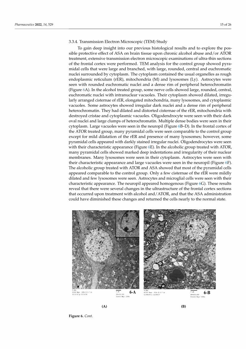
3.3.4. Transmission Electron Microscopic (TEM) Study To gain deep insight into our previous histological results and to explore the possible
protective effect of ASA on brain tissue upon chronic alcohol abuse and/or ATOR treat-ment, extensive transmission electron microscopic examinations of ultra-thin sections of the frontal cortex were performed. TEM analysis for the control group showed pyramidal
Figure 5. Photomicrograph of brain tissue semi-thin sections (toluidine blue stained) for (A) Group I,showing pyramidal cells (P) with large, rounded nuclei and prominent nucleoli. Granule cells (G)have small rounded euchromatic nuclei. Notice the presence of astrocytes (A), oligodendrocyte (O)and microglia (M). The neuropil is homogenous (*). (B) Group I, showing pyramidal cells (P) withlarge, rounded nuclei and prominent nucleoli. Nissl granules (↑) are seen in their cytoplasm. Noticethe presence of astrocytes (A) and microglia (M). (C) Group II, showing most of the pyramidal cells(P) are shrunken, condensed and deeply stained. Few pyramidal cells are seen with large, roundednuclei but with absent Nissl granules (↑). Others have large rounded dark nuclei (N). Notice thepresence of astrocytes (A) with irregular nuclei and surrounded by vacuoles, oligodendrocyte (O) andmicroglia (M). The neuropil is vacuolated (*). (D) Group III, demonstrating some pyramidal cells areshrunken, condensed and deeply stained (P). Some are seen with dark nuclei (N) while others havelarge, rounded nuclei but with absent Nissl granules (↑). Some granule cells (G) have small roundedeuchromatic nuclei while others have darkly stained nuclei (∆). Notice the presence of astrocytes (A),oligodendrocyte (O) and microglia (M). The neuropil is vacuolated (*). (E) Group IV, showing somepyramidal cells are shrunken, condensed and deeply stained (P). Some are seen with dark nuclei (N)Some others are seen with pale, irregular nuclei (∆) while others have large, rounded nuclei but withabsent Nissl granules (↑). Notice the presence of astrocytes (A), oligodendrocyte (O) and microglia(M). Notice the large vacuoles in the neuropil (*). (F) Group V, showing most of the pyramidal cellshave large, rounded nuclei and prominent nucleoli (P). Notice the presence of Nissl granules (↑).A pyramidal cell with dark irregular nucleus is present (N). Granule cells (G) have small roundedeuchromatic nuclei. Notice the presence of astrocytes (A), oligodendrocyte (O) and microglia (M).Small vacuoles are seen in the neuropil (*). (toluidine blue × 1000).
Pharmaceutics 2022, 14, 529 15 of 26
3.3.4. Transmission Electron Microscopic (TEM) Study
To gain deep insight into our previous histological results and to explore the pos-sible protective effect of ASA on brain tissue upon chronic alcohol abuse and/or ATORtreatment, extensive transmission electron microscopic examinations of ultra-thin sectionsof the frontal cortex were performed. TEM analysis for the control group showed pyra-midal cells that were large and branched, with large, rounded, central and euchromaticnuclei surrounded by cytoplasm. The cytoplasm contained the usual organelles as roughendoplasmic reticulum (rER), mitochondria (M) and lysosomes (Ly). Astrocytes wereseen with rounded euchromatic nuclei and a dense rim of peripheral heterochromatin(Figure 6A). In the alcohol treated group, some nerve cells showed large, rounded, central,euchromatic nuclei with intranuclear vacuoles. Their cytoplasm showed dilated, irregu-larly arranged cisternae of rER, elongated mitochondria, many lysosomes, and cytoplasmicvacuoles. Some astrocytes showed irregular dark nuclei and a dense rim of peripheralheterochromatin. They had dilated and distorted cisternae of the rER, mitochondria withdestroyed cristae and cytoplasmic vacuoles. Oligodendrocyte were seen with their darkoval nuclei and large clumps of heterochromatin. Multiple dense bodies were seen in theircytoplasm. Large vacuoles were seen in the neuropil (Figure 6B–D). In the frontal cortex ofthe ATOR treated group, many pyramidal cells were seen comparable to the control groupexcept for mild dilatation of the rER and presence of many lysosomes; however, somepyramidal cells appeared with darkly stained irregular nuclei. Oligodendrocytes were seenwith their characteristic appearance (Figure 6E). In the alcoholic group treated with ATOR,many pyramidal cells showed marked deep indentations and irregularity of their nuclearmembranes. Many lysosomes were seen in their cytoplasm. Astrocytes were seen withtheir characteristic appearance and large vacuoles were seen in the neuropil (Figure 6F).The alcoholic group treated with ATOR and ASA showed that most of the pyramidal cellsappeared comparable to the control group. Only a few cisternae of the rER were mildlydilated and few lysosomes were seen. Astrocytes and microglial cells were seen with theircharacteristic appearance. The neuropil appeared homogenous (Figure 6G). These resultsreveal that there were several changes in the ultrastructure of the frontal cortex sectionsthat occurred upon treatment with alcohol and/ATOR, and that the ASA administrationcould have diminished these changes and returned the cells nearly to the normal state.
Pharmaceutics 2022, 14, x FOR PEER REVIEW 15 of 26
cells that were large and branched, with large, rounded, central and euchromatic nuclei surrounded by cytoplasm. The cytoplasm contained the usual organelles as rough endo-plasmic reticulum (rER), mitochondria (M) and lysosomes (Ly). Astrocytes were seen with rounded euchromatic nuclei and a dense rim of peripheral heterochromatin (Figure 6A). In the alcohol treated group, some nerve cells showed large, rounded, central, euchro-matic nuclei with intranuclear vacuoles. Their cytoplasm showed dilated, irregularly ar-ranged cisternae of rER, elongated mitochondria, many lysosomes, and cytoplasmic vac-uoles. Some astrocytes showed irregular dark nuclei and a dense rim of peripheral heter-ochromatin. They had dilated and distorted cisternae of the rER, mitochondria with de-stroyed cristae and cytoplasmic vacuoles. Oligodendrocyte were seen with their dark oval nuclei and large clumps of heterochromatin. Multiple dense bodies were seen in their cy-toplasm. Large vacuoles were seen in the neuropil (Figure 6B–D). In the frontal cortex of the ATOR treated group, many pyramidal cells were seen comparable to the control group except for mild dilatation of the rER and presence of many lysosomes; however, some pyramidal cells appeared with darkly stained irregular nuclei. Oligodendrocytes were seen with their characteristic appearance (Figure 6E). In the alcoholic group treated with ATOR, many pyramidal cells showed marked deep indentations and irregularity of their nuclear membranes. Many lysosomes were seen in their cytoplasm. Astrocytes were seen with their characteristic appearance and large vacuoles were seen in the neuropil (Figure 6F). The alcoholic group treated with ATOR and ASA showed that most of the pyramidal cells appeared comparable to the control group. Only a few cisternae of the rER were mildly dilated and few lysosomes were seen. Astrocytes and microglial cells were seen with their characteristic appearance. The neuropil appeared homogenous (Figure 6G). These results reveal that there were several changes in the ultrastructure of the frontal cortex sections that occurred upon treatment with alcohol and/ATOR, and that the ASA administration could have diminished these changes and returned the cells nearly to the normal state.
(A) (B)
Figure 6. Cont.

Pharmaceutics 2022, 14, 529 16 of 26Pharmaceutics 2022, 14, x FOR PEER REVIEW 16 of 26
(C) (D)
(E) (F)
(G)
Figure 6. Electron microscopic examination (TEM) of frontal cortex in brain tissue for (A) Group I, showing a pyramidal cell in cerebral cortex of Group I. A large, rounded, central and euchromatic nucleus (Nu) is seen surrounded by cytoplasm. Notice the rough endoplasmic reticulum (rER) and
Figure 6. Electron microscopic examination (TEM) of frontal cortex in brain tissue for (A) GroupI, showing a pyramidal cell in cerebral cortex of Group I. A large, rounded, central and euchromatic

Pharmaceutics 2022, 14, 529 17 of 26
nucleus (Nu) is seen surrounded by cytoplasm. Notice the rough endoplasmic reticulum (rER) andmitochondria (m). Inset showing an astrocyte (A) Inset (TEM × 2000). (B) Group I, demonstratinga nerve cell with dilated, irregularly arranged cisternae of rough endoplasmic reticulum (rER).Notice the presence of lysosomes (Ly), vacuoles (↑) and a Golgi apparatus (GA) in the nerve cell.Notice the presence of an astrocyte (A) with irregular dark nucleus. Large vacuoles (*) are seenin the neuropil (TEM × 1500). (C) Group II, showing a nerve cell in the frontal cortex of Group IIwith large, rounded, central and euchromatic nucleus (Nu). Notice the intranuclear vacuoles (↑).Lysosomes (Ly), elongated mitochondria (m) and cytoplasmic vacuoles are seen. Notice a microglialcell is present beside the nerve cell. Many large vacuoles are seen in the neuropil (*) (TEM × 1500).(D) Group II, showing two glial cells in the frontal cortex of Group II. An astrocyte (A) is seen withan irregular dark nucleus and a dense rim of peripheral heterochromatin. Notice the dilated anddistorted cisternae of rough endoplasmic reticulum (rER), mitochondria (m) with destroyed cristaeand cytoplasmic vacuoles (↑). An oligodendrocyte (O) is seen with its dark oval nucleus and largeclumps of heterochromatin. Multiple dense bodies are seen in its cytoplasm (N). Large vacuoles areseen in the neuropil (*) (TEM × 2500). (E) Group III, showing pyramidal cells in the frontal cortex ofGroup III with large, rounded, central and euchromatic nuclei (Nu). Notice mild dilatation of therough endoplasmic reticulum (rER). Lysosomes are seen (Ly). An oligodendrocyte (O) is presentbeside a nerve cell. Inset: showing a pyramidal cell with darkly stained irregular nucleus (TEM ×1200). (F) Group IV, showing a pyramidal cell in frontal cortex with marked deep indentation of thenuclear membrane (Nu1). Many lysosomes are seen (Ly). An astrocyte (A) is present beside the nervecells. Inset: showing a pyramidal cell with indented and irregular nucleus (Nu2). Large vacuoles areseen in the neuropil (*) (TEM × 1200). (G) Group V, showing pyramidal cells in the frontal cortexof Group V with large, rounded, central and euchromatic nuclei (Nu). Notice mild dilatation of afew rough endoplasmic reticulum cisternae (rER). Few lysosomes are seen (Ly). An astrocyte (A)is present beside a nerve cell. The neuropil is homogenous (*). Inset: showing a microglial cell (M)(TEM × 1200).
3.4. In Silico Molecular Docking Study
In silico molecular modelling represents a modern computational analysis that canbe applied in investigating the possible mode of action for a drug [47]. To further explorethe possibility of ASA to target the NLRP3 protein, we performed an extensive in silicomolecular docking study to investigate the binding affinity of ASA into the active sites ofthe binding pockets of the NLRP3 protein. In the protein data bank, there were only twoPDB codes available for NLRP3 proteins (PDB codes, n6py and 7alv) which represent the 3Dstructure of theNLRP3 NACHT domain co-crystallized with a potent inhibitor (NP3-146)and/or ADP [39,40]. To gain insights into the different possible modes of binding for ASA,we performed molecular docking studies for both 3D crystal structures available (PDBcodes, n6py and 7alv), and the obtained results were isolated to be evaluated (Table 4).The obtained poses were evaluated and selected based upon the capability of the dockedligand to possess the main binding interactions to the active site of the binding protein.After adjusting different parameters, we examined our docking protocol by redocking theco-crystallized ligand, either ADP or NP3-146, to the active sites of the NLRP3 NACHTdomain to ensure that the re-docked ligand could form the main interactions reported inthe database. Next, the validated docking protocol was used to investigate and to assessthe binding affinity of ASA to the ADP-binding site and the inhibitory-binding site of theNLRP3 NACHT domain. The obtained results from the docking study, docking score andobserved binding interactions, are presented in Table 4. Based on the recently reportedcrystal structure, the NLRP3 NACHT domain had two binding sites: an ADP-binding siteand inhibitory-binding site [39,40]. Our results revealed that the ASA had high bindingscores to both ADP- and inhibitor-binding pockets with the ability to bind to the activesites of the NLRP3 NACHT domain by both hydrophilic and hydrophobic interactions in asimilar mode compared to the original co-crystallized ligands (Figure 7, Table 4). Theseresults indicate that the suppression effect of ASA on cognitive dysfunction could be alsoattributed to its ability to target the NLRP3 protein.

Pharmaceutics 2022, 14, 529 18 of 26
Table 4. Scores (kcal/mol) and interactions of the molecular modelling process of ASA and ATORdrugs in the NLRP3 binding sites.
PDBDocking Score
(kcal/mol)Interactive Residues
Hydrophilic Interactions Hydrophobic Interactions
7alv −12.17 Ala228, Arg351, Tyr632 Val353, Ile574, Phe575,Pro352, Ala227
6npy −11.68 Arg165, Thr167, Tyr166 Leu162, Ile228, Trp414,Leu169, Pro410, Ile232
Pharmaceutics 2022, 14, x FOR PEER REVIEW 18 of 26
PDB Docking
Score (kcal/mol)
Interactive Residues
Hydrophilic interactions Hydrophobic interactions
7alv −12.17 Ala228, Arg351, Tyr632 Val353, Ile574, Phe575, Pro352, Ala227
6npy −11.68 Arg165, Thr167, Tyr166 Leu162, Ile228, Trp414, Leu169, Pro410, Ile232
(A) (B)
(C) (D)
Figure 7. The 2D and 3D molecular modelling interactions of ASA (green and grey in 3D interac-tions) with NLRP3 protein: 7alv (A, B), and 6npy (C, D). Dotted green arrows represent the hydrogen bonds; (C atoms are colored green, S yellow and O red).
4. Discussion Cognitive dysfunction is a common and potentially severe consequence of long-term
alcohol abuse [48]. Continuous consumption of alcohol in high levels can lead to long-term functional changes such as impairments in visuo-spatial functioning [49]. Functional impairments in alcohol abuse can be explained by the changes in grey matter structure, resulting from the neurotoxicity of alcohol in chronic high consumption patterns [50]. Sev-eral studies have showed that in alcohol abuse patients the grey matter volume reduced as compared to healthy controls. Yang et al. reported several alterations in the grey matter in left and right precentral gyri, as well as in subcortical regions like the left thalamus and right hippocampus [51].
Alcohol abuse induces brain neurodegeneration resulting in elevation of proinflam-matory cytokines and chemokines expression and, in particular, microglia can be
Figure 7. The 2D and 3D molecular modelling interactions of ASA (green and grey in 3D interactions)with NLRP3 protein: 7alv (A,B), and 6npy (C,D). Dotted green arrows represent the hydrogen bonds;(C atoms are colored green, S yellow and O red).
4. Discussion
Cognitive dysfunction is a common and potentially severe consequence of long-termalcohol abuse [48]. Continuous consumption of alcohol in high levels can lead to long-term functional changes such as impairments in visuo-spatial functioning [49]. Functionalimpairments in alcohol abuse can be explained by the changes in grey matter structure,resulting from the neurotoxicity of alcohol in chronic high consumption patterns [50].Several studies have showed that in alcohol abuse patients the grey matter volume reducedas compared to healthy controls. Yang et al. reported several alterations in the grey matter

Pharmaceutics 2022, 14, 529 19 of 26
in left and right precentral gyri, as well as in subcortical regions like the left thalamus andright hippocampus [51].
Alcohol abuse induces brain neurodegeneration resulting in elevation of proinflam-matory cytokines and chemokines expression and, in particular, microglia can be activatedthrough their receptors (such as Toll-like receptor 4) [52]. Robust changes in miRNA ex-pression have been reported in the prefrontal cortex of subjects with a history of chronicalcohol abuse [17], which may provide the opportunity to evaluate ongoing changes inthe CNS upon the initiation of neurodegeneration [53]. Among the different miRNA,miRNA155 is highly expressed in numerous tissues, including the brain, suggesting its pro-inflammatory action in CNS. miRNA155 induces neuroinflammation through a reductionin the endogenous anti-inflammatory response resulting in increased inflammation [54].Neuroinflammation has been identified as a causative factor of multiple neurodegener-ative diseases. The NLRP3 inflammasome—a subcellular multiprotein complex that isabundantly expressed in the CNS—can be activated by a wide range of exogenous andendogenous stimuli [55]. Activation of the NLRP3 inflammasomes pathway is responsiblefor neuroinflammation and is associated with several brain diseases [56]. Toll-like receptors(TLRs) in the CNS trigger the activation of pro-IL-1β and pro-IL-18 that are converted intotheir active forms by the NLRP3 inflammasome [57].
Alcoholic fatty liver (steatosis), alcoholic hepatitis, and fibrosis are associated withthe progression of liver cirrhosis, with the highest risk in alcoholism [5]. Statins are lipid-lowering drugs that are utilized to treat lipid disorders and commonly prescribed forpatients with fatty liver diseases to reduce low-density lipoprotein cholesterol [8]. Theexact effect of statins on cognitive function has been not fully addressed; however, it wassuggested that statins may cause a short-term cognitive impairment [58,59]. Cholesterolis a crucial lipid for brain functions, and it is used in the formation of the nervous sys-tem as well as the manufacture of steroid hormones that are involved in brain signaling.Accordingly, decreasing the serum cholesterol level, upon statins treatment, may affectcognition function [25]. Interestingly, it was suggested that switching from lipophilic statins(e.g., ATOR) to hydrophilic statins could resolve the cognitive impairment with vascularbenefits [60].
Based on the abovementioned facts, in the present study we aimed at investigatingthe molecular and cellular effects of ATOR on cognitive dysfunction induced by alcoholintake. Accordingly, rats were treated with alcohol and/or ATOR and a set of biochemical,histopathological, and immunohistochemical analyses were performed. A Morris watermaze test showed that the cognitive function was significantly impaired, as indicated bythe increase in latency time to reach the target quadrant and the decrease in the % timespent in the target quadrant. Our results were in accordance with the previous findingswhich reported a significant impairment in the Morris water maze test for rats treated withalcohol, together with an impairment in the memory as demonstrated by the markedlyincreased latency time [61,62]. Further studies have showed that alcohol intake causesvarious neurological diseases via the toxic effect of various key mediators on the brain,such as inflammation, caspase-3, and the brain-derived neurotrophic factor BDNF [63].Additionally, Stragier et al. reported that ethanol treatment in C57BL/6J mice causesprofound impairment in the learning and memory place [64]. Toward the effect of statinson cognitive function, in accordance with our results, King et al. reported that statin therapysignificantly and temporally impairs cognitive function and recommended that cliniciansshould be aware of cognitive impairment as a potential adverse effect associated with statintherapy [65].
As the alcohol induced neuroinflammation in the brain, the IL-1β level increasedthrough the activation of the NLRP3 inflammasome [66]. Our results demonstrated apivotal role for NLRP3 inflammasome activation that mediates the IL-1β level in thealcohol-related neuroinflammation. In agreement with Orio et al., we found that treatmentwith alcohol or in combination with ATOR produced a significant increase in caspase-8 andthe IL1β and NLRP3 inflammasomes in the brain tissue [67]. Indeed, Orio et al. showed

Pharmaceutics 2022, 14, 529 20 of 26
that alcohol abuse causes oxidative damage to the mitochondria and cellular proteinswhich leads to the development of neuroinflammation and neurological disorders. Further,the inflammasome components (NLRP1, NLRP3, ASC) and proinflammatory cytokines(TNF-α) levels were increased in alcohol-treated brains, together with an enhancement ofcaspase-1 activity and the IL-1β protein [21]. Our results revealed that the ATOR increasedthe expression of IL-1β, caspase-8 and the NLRP3 inflammasome pathway. Statins possessanti-inflammatory effects which lead to enhancing the activity of NLRP3 inflammasomeand IL-1β [68,69]. According to Peng et al. and Shu et al., ATOR has an antioxidant capacityvia increasing the level of GSSG/GSH, and is considered as an anti-inflammatory agentand proinflammatory compound [70,71].
In the present study, the treatment with alcohol alone or in combination with ATORproduced a significant increase in the expression of miRNA155 levels in the brain tissue.Although previous studies have confirmed the relation between cognitive dysfunctionand miRAN155 expression, the link between miRNA155 expression and alcohol-inducedcognitive dysfunction was unique in our study. In our study, we hypothesized that the linkbetween alcohol-induced cognitive dysfunction and miRNA155 could be via the NLRP3inflammasome pathway. This hypothesis was also supported by the recent findings byLiu et al. which showed that miRNA155 expression increases in the hippocampus of ADrats, together with IL-1β, IL-6 and TNF-α [18]. Our findings were in agreement with Yinet al.’s study which showed that miRNA155 induced the expression of NLRP3 inflamma-some which could induce the secretion of IL-1β and IL-18 leading to the development ofinflammation and atherosclerosis [72]. Silencing of miRNA155 was shown to significantlydecrease the inflammation and NLRP3/caspase-1 signaling [73].
As mentioned above, the brain tissue was affected by alcohol and/or ATOR ad-ministration leading to neuroinflammation. Our results were further confirmed by lightmicroscopic examination of the brain tissue. In the control group, the frontal cortex showeda normal appearance with the six layers of the cortex formed of neurons and glial cells,as previously described [74]. On the other hand, the groups treated with alcohol, ATORor a combination of both showed signs of degeneration in the neurons as well as the glialcells, in the form of shrunken cells surrounded by hallows, a loss of Nissl’s granules (seenin the semi-thin sections) and deeply stained, pyknotic nuclei. Interestingly, the alcoholicgroup showed aggregations of inflammatory cells—most probably microglia—forming agranuloma-like structure. These lesions were previously shown to be associated with thedegeneration of neurons that was caused due to the consumption of the alcohol and/orATOR [75]. Moreover, the present morphometric study showed a significant decrease in themean thickness of the frontal cortex in the alcoholic group as compared to the control group.These results were in accordance with an Olawale et al. study which showed that cerebralcortex damage, due to acute alcohol intake, is associated with oxidative stress [76]. Fortieret al. reported that the thickness of the brain cortex significantly decreased in specific areasin alcoholic compared with non-alcoholic members, suggesting that alcohol may affect thelarge pyramidal cells in the frontal cortex [77]. Moreover, Saito et al. reported changes inthe count of glial cells after chronic ethanol exposure [52]. Together, these findings indicatethat alcohol consumption causes a decline in grey matter size in certain brain regions andhas effects on the behavioral depiction of these brain areas that may lead to cognitiveimpairment [50].
Caspase-8 plays a central role in apoptosis, as well as regulating inflammation bymodulating IL-1β expression. In the present study, examination of caspase-8 stainedbrain sections of the control group showed a negative reaction. While the alcoholic groupand ATOR-treated alcoholic group showed a positive reaction to caspase-8, in both thenerve cells and the glial cells. Previous co-immunoprecipitation studies demonstratedthat caspase-8 presents in the NLRP3 inflammasome complex, where it is believed to beinvolved in the cleavage of pro-caspase-1 [78]. Further, different inflammatory stimulican cause activation of NF-kB via the association of caspase-8 [79,80]. These results couldexplain the observed increased caspase-8 in our investigations.

Pharmaceutics 2022, 14, 529 21 of 26
Our findings were further confirmed by a TEM study which revealed neuronal andglial damage in the alcoholic group. Damaged neurons showed dilated, irregularly ar-ranged cisternae of the rER, elongated mitochondria, many lysosomes and cytoplasmicvacuoles. Similar changes were previously described [81]. Some astrocytes showed ir-regular dark nuclei and a dense rim of peripheral heterochromatin. They had dilatedand distorted cisternae of the rER, mitochondria with destroyed cristae and cytoplasmicvacuoles. A similar pattern of rER was previously observed in hepatocytes due to chronicalcohol consumption, and was attributed to endoplasmic reticulum stress [28]. In thepresent study, treatment with alcohol and/or ATOR caused nearly similar effects on theneurons structure and glial cells in the frontal lobe of the brain tissue.
ASA, as a nonsteroidal anti-inflammatory, is frequently used as a potent drug inseveral diseases. The anti-inflammatory activity of ASA is derived from its ability to inhibitcyclooxygenase which suppresses the production of pro-inflammatory molecules, such asprostaglandins [82]. Several studies have demonstrated the capability of ASA to attenuatethe hepatotoxicity and endothelial dysfunction by regulating the NLRP3 inflammasomeand IL-1β levels [26,28,29]. These pleiotropic modes of action suggest that ASA could beutilized in alcoholism for the treatment of cognitive dysfunction. Toward this aim, wehave investigated the efficacy of ASA to treat the alcohol-induced cognitive impairmentassociated with ATOR intake (Figure 8). In our investigations, we reported that treatmentwith ASA significantly reduced the expression of caspase-8, NLRP3, and IL-1β levelsin brain tissue. These results could be attributed to the anti-inflammatory properties ofASA which could change the innate immune response and autophagy via the eliminationof impaired mitochondria and oxidative stress [83,84]. Further, it was shown that ASAadministration decreased the expression of miRNA155 in brain tissue. Taken together, ourresults indicated that ASA treatment has suppression effects on several molecular factorsof cognitive dysfunction. These findings were further confirmed by the histopathologicalstudies which revealed a regular arrangement of the cerebral cortex layers with noticeableimprovement in the structure of both neurons and glial cells in brain tissue.
Pharmaceutics 2022, 14, x FOR PEER REVIEW 21 of 26
irregular dark nuclei and a dense rim of peripheral heterochromatin. They had dilated and distorted cisternae of the rER, mitochondria with destroyed cristae and cytoplasmic vacuoles. A similar pattern of rER was previously observed in hepatocytes due to chronic alcohol consumption, and was attributed to endoplasmic reticulum stress [28]. In the pre-sent study, treatment with alcohol and/or ATOR caused nearly similar effects on the neu-rons structure and glial cells in the frontal lobe of the brain tissue.
ASA, as a nonsteroidal anti-inflammatory, is frequently used as a potent drug in sev-eral diseases. The anti-inflammatory activity of ASA is derived from its ability to inhibit cyclooxygenase which suppresses the production of pro-inflammatory molecules, such as prostaglandins [82]. Several studies have demonstrated the capability of ASA to attenuate the hepatotoxicity and endothelial dysfunction by regulating the NLRP3 inflammasome and IL-1β levels [26,28,29]. These pleiotropic modes of action suggest that ASA could be utilized in alcoholism for the treatment of cognitive dysfunction. Toward this aim, we have investigated the efficacy of ASA to treat the alcohol-induced cognitive impairment associated with ATOR intake (Figure 8). In our investigations, we reported that treatment with ASA significantly reduced the expression of caspase-8, NLRP3, and IL-1β levels in brain tissue. These results could be attributed to the anti-inflammatory properties of ASA which could change the innate immune response and autophagy via the elimination of impaired mitochondria and oxidative stress [83,84]. Further, it was shown that ASA ad-ministration decreased the expression of miRNA155 in brain tissue. Taken together, our results indicated that ASA treatment has suppression effects on several molecular factors of cognitive dysfunction. These findings were further confirmed by the histopathological studies which revealed a regular arrangement of the cerebral cortex layers with noticeable improvement in the structure of both neurons and glial cells in brain tissue.
Figure 8. Molecular and cellular effects of ASA on cognitive dysfunction induced by alcohol and/or ATOR intake.
Finally, an extensive molecular modelling study was performed to investigate the binding affinity of ASA to the binding pockets of the NLRP3 NACHT domain. Recent examination of the 3D structures of the NLRP3 NACHT domain revealed that the protein has two binding sites: an ADP-binding pocket and a small inhibitor-stimulate-binding pocket [39,40]. The ADP-binding pocket revealed a set of key amino acid residues (Thr231, Ile232, Gly239, Thr167, Tyr166, His520, Lys 230) that interacted with one molecule of ADP to form a stable network of hydrophilic interactions. While the recently discovered inhib-itor-binding pocket was shown to possess a slightly basic active site (Arg351, Arg578, Pro352, Ala228) which mainly binds to the sulfonylurea part of the co-crystallized inhibi-tor (NP3-146). As shown in Table 4, the ASA demonstrated high negatively-binding scores
Figure 8. Molecular and cellular effects of ASA on cognitive dysfunction induced by alcohol and/orATOR intake.
Finally, an extensive molecular modelling study was performed to investigate thebinding affinity of ASA to the binding pockets of the NLRP3 NACHT domain. Recentexamination of the 3D structures of the NLRP3 NACHT domain revealed that the proteinhas two binding sites: an ADP-binding pocket and a small inhibitor-stimulate-binding

Pharmaceutics 2022, 14, 529 22 of 26
pocket [39,40]. The ADP-binding pocket revealed a set of key amino acid residues (Thr231,Ile232, Gly239, Thr167, Tyr166, His520, Lys 230) that interacted with one molecule ofADP to form a stable network of hydrophilic interactions. While the recently discoveredinhibitor-binding pocket was shown to possess a slightly basic active site (Arg351, Arg578,Pro352, Ala228) which mainly binds to the sulfonylurea part of the co-crystallized inhibitor(NP3-146). As shown in Table 4, the ASA demonstrated high negatively-binding scorestoward the binding pockets of the NLRP3 NACHT domain, indicating that the binding ofASA to the active sites of NLRP3 is thermodynamically favorable. Toward the ADP-bindingpocket, the ASA showed the ability to bind to the active site through interaction of thecarboxylic moiety to two of the main active amino acid residues (Ala228, Tyr632), andan additional basic amino acid residue (Arg351). This interaction was also supported byhydrophobic interactions with a set of greasy amino acid residues. Similarly, the ASAshowed the possibility to bind to the active site of the inhibitor-binding pocket via ahydrophilic interaction with three of the main amino acid residues (Ala228, Arg351, Tyr632)together with a set of hydrophobic interactions with greasy amino acid residues. Takentogether, our molecular docking study findings indicate that the suppression effect of ASAon cognitive dysfunction could be attributed to its ability to target the active sites of theNLRP3 NACHT domain. Further investigation should be performed in future to deeplyinvestigate the inhibitory activity of ASA toward the NLRP3 protein which would highlightASA as a possible drug for cognitive dysfunction.
5. Conclusions
In the present study, we showed that alcohol intake either alone or in combinationwith ATOR caused changes in the biochemical, immunohistochemical and histologicalparameters of brain tissue. Our study revealed that administration of alcohol either aloneor in combination with ATOR caused an upregulation of NLRP3/IL1B inflammasome andits epigenetic regulator, miRNA155, suggesting a key role in neuroinflammation and neu-rodegeneration. The histological and morphometric analysis indicated neuronal and glialcell damage as well as aggregations of microglial cells in brain tissue. On the other hand,ASA administration caused a significant downregulation of the miRNA155/NLRP3/IL1Binflammasome pathway. The protective effect of ASA was confirmed by histopathological,morphological and immunohistochemistry studies which revealed a decrease in neu-ronal damage and diminished microglial cell infiltration in brain tissue with a decreasein caspase-8 immunoreaction in neurons and glial cells. Finally, our molecular modellingstudy indicated the ability of ASA to bind to the binding pockets of the NLRP3 NACHTdomain with high binding scores. Collectively, our findings demonstrated that ASA ad-ministration has beneficial and protective effects on cognitive function. Accordingly, wehypothesize that the administration of ASA to patients with alcoholism and/or long-termATOR treatment could effectively attenuate the progression of cognitive impairment.
Author Contributions: Conceptualization, D.I.M., D.A.E.-D.A.E.-W., E.S.N. and O.A.E.-K.; method-ology, D.I.M., D.A.E.-D.A.E.-W., E.S.N., O.A.E.-K., H.F.A.E.-K. and E.M.S.; software, H.F.A.E.-K.,H.H.A.N., B.A.A.-W., Y.A.H. and E.M.S.; validation, D.I.M., D.A.E.-D.A.E.-W., E.S.N., O.A.E.-K.,H.F.A.E.-K., B.A.A.-W. and Y.A.H.; formal analysis, D.I.M., D.A.E.-D.A.E.-W., E.S.N., H.F.A.E.-K.,O.A.E.-K., H.H.A.N., B.A.A.-W. and Y.A.H.; investigation, D.I.M., H.F.A.E.-K., H.H.A.N., S.Z.A.,B.A.A.-W., Y.A.H. and E.M.S.; resources, S.Z.A. and B.A.A.-W.; data curation, D.I.M., D.A.E.-D.A.E.-W., E.S.N., H.F.A.E.-K., O.A.E.-K., H.H.A.N. and E.M.S.; writing—original draft preparation, D.I.M.,D.A.E.-D.A.E.-W., E.S.N., H.F.A.E.-K., O.A.E.-K., H.H.A.N. and E.M.S.; writing—review and editing,D.I.M., D.A.E.-D.A.E.-W., E.S.N., H.F.A.E.-K., O.A.E.-K., H.H.A.N., S.Z.A., B.A.A.-W., Y.A.H. andE.M.S.; visualization, H.F.A.E.-K., H.H.A.N., S.Z.A., B.A.A.-W., Y.A.H. and E.M.S.; supervision, D.I.M.,D.A.E.-D.A.E.-W., E.S.N., O.A.E.-K. and Y.A.H.; project administration, D.I.M., D.A.E.-D.A.E.-W.,E.S.N., O.A.E.-K. and H.F.A.E.-K.; funding acquisition, S.Z.A. All authors have read and agreed tothe published version of the manuscript.

Pharmaceutics 2022, 14, 529 23 of 26
Funding: This research was funded by Princess Nourah bint Abdulrahman University ResearchersSupporting Project number (PNURSP2022R165), Princess Nourah bint Abdulrahman University,Riyadh, Saudi Arabia.
Institutional Review Board Statement: The study was conducted according to the guidelines of theDeclaration of Helsinki and approved by the Ethics Committee of the Faculty of Medicine, Ain ShamsUniversity (FMASU REC). (No. FWA 00021368, 5/2021).
Informed Consent Statement: Not applicable.
Data Availability Statement: Not applicable.
Acknowledgments: The authors extend their appreciation to Princess Nourah bint AbdulrahmanUniversity Researchers Supporting Project number (PNURSP2022R165), Princess Nourah bint Abdul-rahman University, Riyadh, Saudi Arabia.
Conflicts of Interest: The authors declare no conflict of interest.
References1. Ishii, T.; Hashimoto, E.; Ukai, W.; Tateno, M.; Yoshinaga, T.; Ono, T.; Watanabe, K.; Saito, S.; Saito, T. Epigenetic regulation in
alcohol-related brain damage. Nihon Arukoru Yakubutsu Igakkai Zasshi 2008, 43, 705–713.2. Bajaj, J.S. Alcohol, Liver Disease and the Gut Microbiota. Nat. Rev. Gastroenterol. Hepatol. 2019, 16, 235–246. [CrossRef] [PubMed]3. Piano, M.R. Alcohol’s Effects on the Cardiovascular System. Alcohol. Res. 2017, 38, 219–241. [PubMed]4. Moss, M.; Burnham, E.L. Chronic Alcohol Abuse, Acute Respiratory Distress Syndrome, and Multiple Organ Dysfunction. Crit.
Care Med. 2003, 31, S207–S212. [CrossRef]5. Osna, N.A.; Donohue, T.M.; Kharbanda, K.K. Alcoholic Liver Disease: Pathogenesis and Current Management. Alcohol. Res. 2017,
38, 147–161. [PubMed]6. Crews, F.T. Alcohol-Related Neurodegeneration and Recovery. Alcohol. Res. Health 2008, 31, 377–388.7. Friedman, N.P.; Robbins, T.W. The Role of Prefrontal Cortex in Cognitive Control and Executive Function. Neuropsychopharmacology
2022, 47, 72–89. [CrossRef] [PubMed]8. Vargas, J.I.; Arrese, M.; Shah, V.H.; Arab, J.P. Use of Statins in Patients with Chronic Liver Disease and Cirrhosis: Current Views
and Prospects. Curr. Gastroenterol. Rep. 2017, 19, 43. [CrossRef]9. Gu, Y.; Yang, X.; Liang, H.; Li, D. Comprehensive Evaluation of Effects and Safety of Statin on the Progression of Liver Cirrhosis:
A Systematic Review and Meta-Analysis. BMC Gastroenterol. 2019, 19, 231. [CrossRef]10. Ward, N.C.; Watts, G.F.; Eckel, R.H. Statin Toxicity. Circ. Res. 2019, 124, 328–350. [CrossRef]11. Yang, Q.; Liu, R.; Yu, Q.; Bi, Y.; Liu, G. Metabolic Regulation of Inflammasomes in Inflammation. Immunology 2019, 157, 95–109.
[CrossRef] [PubMed]12. Guo, H.; Callaway, J.B.; Ting, J.P.-Y. Inflammasomes: Mechanism of Action, Role in Disease, and Therapeutics. Nat. Med. 2015, 21,
677–687. [CrossRef] [PubMed]13. Kelley, N.; Jeltema, D.; Duan, Y.; He, Y. The NLRP3 Inflammasome: An Overview of Mechanisms of Activation and Regulation.
Int. J. Mol. Sci. 2019, 20, 3328. [CrossRef] [PubMed]14. Marchesan, J.T.; Girnary, M.S.; Moss, K.; Monaghan, E.T.; Egnatz, G.J.; Jiao, Y.; Zhang, S.; Beck, J.; Swanson, K.V. Role of
Inflammasomes in the Pathogenesis of Periodontal Disease and Therapeutics. Periodontology 2020 2020, 82, 93–114. [CrossRef][PubMed]
15. Sharma, M.; de Alba, E. Structure, Activation and Regulation of NLRP3 and AIM2 Inflammasomes. Int. J. Mol. Sci. 2021, 22, 872.[CrossRef] [PubMed]
16. Lim, Y.; Beane-Ebel, J.E.; Tanaka, Y.; Ning, B.; Husted, C.R.; Henderson, D.C.; Xiang, Y.; Park, I.-H.; Farrer, L.A.; Zhang, H.Exploration of Alcohol Use Disorder-Associated Brain MiRNA–MRNA Regulatory Networks. Transl. Psychiatry 2021, 11, 1–10.[CrossRef]
17. Tapocik, J.D.; Solomon, M.; Flanigan, M.; Meinhardt, M.; Barbier, E.; Schank, J.; Schwandt, M.; Sommer, W.H.; Heilig, M.Coordinated Dysregulation of MRNAs and MicroRNAs in the Rat Medial Prefrontal Cortex Following a History of AlcoholDependence. Pharm. J. 2013, 13, 286–296. [CrossRef]
18. Liu, D.; Zhao, D.; Zhao, Y.; Wang, Y.; Zhao, Y.; Wen, C. Inhibition of MicroRNA-155 Alleviates Cognitive Impairment inAlzheimer’s Disease and Involvement of Neuroinflammation. Curr. Alzheimer Res. 2019, 16, 473–482. [CrossRef]
19. Obora, K.; Onodera, Y.; Takehara, T.; Frampton, J.; Hasei, J.; Ozaki, T.; Teramura, T.; Fukuda, K. Inflammation-Induced MiRNA-155 Inhibits Self-Renewal of Neural Stem Cells via Suppression of CCAAT/Enhancer Binding Protein β (C/EBPβ) Expression.Sci. Rep. 2017, 7, 43604. [CrossRef]
20. Slota, J.A.; Booth, S.A. MicroRNAs in Neuroinflammation: Implications in Disease Pathogenesis, Biomarker Discovery andTherapeutic Applications. Noncoding RNA 2019, 5, 35. [CrossRef]

Pharmaceutics 2022, 14, 529 24 of 26
21. Lippai, D.; Bala, S.; Petrasek, J.; Csak, T.; Levin, I.; Kurt-Jones, E.A.; Szabo, G. Alcohol-Induced IL-1β in the Brain Is Mediatedby NLRP3/ASC Inflammasome Activation That Amplifies Neuroinflammation. J. Leukoc. Biol. 2013, 94, 171–182. [CrossRef][PubMed]
22. Fitzsimons, S.; Oggero, S.; Bruen, R.; McCarthy, C.; Strowitzki, M.J.; Mahon, N.G.; Ryan, N.; Brennan, E.P.; Barry, M.; Perretti, M.;et al. MicroRNA-155 Is Decreased During Atherosclerosis Regression and Is Increased in Urinary Extracellular Vesicles DuringAtherosclerosis Progression. Front. Immunol. 2020, 11, 576516. [CrossRef] [PubMed]
23. Parsamanesh, N.; Moossavi, M.; Bahrami, A.; Fereidouni, M.; Barreto, G.; Sahebkar, A. NLRP3 Inflammasome as a TreatmentTarget in Atherosclerosis: A Focus on Statin Therapy. Int. Immunopharmacol. 2019, 73, 146–155. [CrossRef]
24. Tabaei, S.; Tabaee, S.S. Implications for MicroRNA Involvement in the Prognosis and Treatment of Atherosclerosis. Mol. CellBiochem. 2021, 476, 1327–1336. [CrossRef] [PubMed]
25. Schultz, B.G.; Patten, D.K.; Berlau, D.J. The Role of Statins in Both Cognitive Impairment and Protection against Dementia: A Taleof Two Mechanisms. Transl. Neurodegener 2018, 7, 5. [CrossRef] [PubMed]
26. Zhou, X.; Wu, Y.; Ye, L.; Wang, Y.; Zhang, K.; Wang, L.; Huang, Y.; Wang, L.; Xian, S.; Zhang, Y.; et al. Aspirin AlleviatesEndothelial Gap Junction Dysfunction through Inhibition of NLRP3 Inflammasome Activation in LPS-Induced Vascular Injury.Acta Pharm. Sin. B 2019, 9, 711–723. [CrossRef] [PubMed]
27. Veronese, N.; Stubbs, B.; Maggi, S.; Thompson, T.; Schofield, P.; Muller, C.; Tseng, P.-T.; Lin, P.-Y.; Carvalho, A.F.; Solmi, M.Low-Dose Aspirin Use and Cognitive Function in Older Age: A Systematic Review and Meta-Analysis. J. Am. Geriatr. Soc. 2017,65, 1763–1768. [CrossRef] [PubMed]
28. El-Kharashi, O.A.; El-Din Aly El-Waseef, D.A.; Nabih, E.S.; Mohamed, D.I. Targeting NLRP3 Inflammasome via AcetylsalicylicAcid: Role in Suppressing Hepatic Dysfunction and Insulin Resistance Induced by Atorvastatin in Naïve versus Alcoholic Liverin Rats. Biomed. Pharmacother. 2018, 107, 665–674. [CrossRef] [PubMed]
29. Williams, C.D.; Antoine, D.J.; Shaw, P.J.; Benson, C.; Farhood, A.; Williams, D.P.; Kanneganti, T.-D.; Park, B.K.; Jaeschke, H. Roleof the Nalp3 Inflammasome in Acetaminophen-Induced Sterile Inflammation and Liver Injury. Toxicol. Appl. Pharmacol. 2011, 252,289–297. [CrossRef]
30. Tiwari, V.; Chopra, K. Resveratrol Abrogates Alcohol-Induced Cognitive Deficits by Attenuating Oxidative-Nitrosative Stress andInflammatory Cascade in the Adult Rat Brain. Neurochem. Int. 2013, 62, 861–869. [CrossRef]
31. Solmaz, V.; Atasoy, Ö.; Erbas, O. Atorvastatin Has Therapeutic Potential for the Fatty Liver-Induced Memory Dysfunction in Rats,Likely via Its Antioxidant and Anti-Inflammatory Properties. Neurol. Res. 2020, 42, 497–503. [CrossRef] [PubMed]
32. Fu, C.; Melethil, S.; Mason, W.D. The Pharmacokinetics of Aspirin in Rats and the Effect of Buffer. J. Pharmacokinet. Biopharm.1991, 19, 157–173. [CrossRef]
33. Mischinger, H.J.; Walsh, T.R.; Liu, T.; Rao, P.N.; Rubin, R.; Nakamura, K.; Todo, S.; Starzl, T.E. An Improved Technique for IsolatedPerfusion of Rat Livers and an Evaluation of Perfusates. J. Surg. Res. 1992, 53, 158–165. [CrossRef]
34. Matthews, D.R.; Hosker, J.P.; Rudenski, A.S.; Naylor, B.A.; Treacher, D.F.; Turner, R.C. Homeostasis Model Assessment: InsulinResistance and β-Cell Function from Fasting Plasma Glucose and Insulin Concentrations in Man. Diabetologia 1985, 28, 412–419.[CrossRef] [PubMed]
35. Tiwari, V.; Chopra, K. Protective Effect of Curcumin against Chronic Alcohol-Induced Cognitive Deficits and Neuroinflammationin the Adult Rat Brain. Neuroscience 2013, 244, 147–158. [CrossRef] [PubMed]
36. Khedr, S.A.; Elmelgy, A.A.; El-Kharashi, O.A.; Abd-Alkhalek, H.A.; Louka, M.L.; Sallam, H.A.; Aboul-Fotouh, S. MetforminPotentiates Cognitive and Antidepressant Effects of Fluoxetine in Rats Exposed to Chronic Restraint Stress and High Fat Diet:Potential Involvement of Hippocampal c-Jun Repression. Naunyn-Schmiedebergs. Arch. Pharmacol. 2018, 391, 407–422. [CrossRef]
37. García-Cabezas, M.Á.; John, Y.J.; Barbas, H.; Zikopoulos, B. Distinction of Neurons, Glia and Endothelial Cells in the CerebralCortex: An Algorithm Based on Cytological Features. Front. Neuroanat. 2016, 10, 107. [CrossRef]
38. Seligson, D.; Marino, J.; Dodson, E. Determination of Sulfobromophthalein in Serum. Clin. Chem. 1957, 3, 638–645. [CrossRef]39. Dekker, C.; Mattes, H.; Wright, M.; Boettcher, A.; Hinniger, A.; Hughes, N.; Kapps-Fouthier, S.; Eder, J.; Erbel, P.; Stiefl, N.; et al.
Crystal Structure of NLRP3 NACHT Domain With an Inhibitor Defines Mechanism of Inflammasome Inhibition. J. Mol. Biol.2021, 433, 167309. [CrossRef]
40. Sharif, H.; Wang, L.; Wang, W.L.; Magupalli, V.G.; Andreeva, L.; Qiao, Q.; Hauenstein, A.V.; Wu, Z.; Núñez, G.; Mao, Y.; et al.Structural Mechanism for NEK7-Licensed Activation of NLRP3 Inflammasome. Nature 2019, 570, 338–343. [CrossRef]
41. El Azab, I.H.; Saied, E.M.; Osman, A.A.; Mehana, A.E.; Saad, H.A.; Elkanzi, N.A. Novel N-Bridged Pyrazole-1-Carbothioamideswith Potential Antiproliferative Activity: Design, Synthesis, in Vitro and in Silico Studies. Future Med. Chem. 2021, 13, 1743–1766.[CrossRef]
42. Gaber, A.; Alsanie, W.F.; Kumar, D.N.; Refat, M.S.; Saied, E.M. Novel Papaverine Metal Complexes with Potential AnticancerActivities. Molecules 2020, 25, 5447. [CrossRef] [PubMed]
43. Gaber, A.; Refat, M.S.; Belal, A.A.M.; El-Deen, I.M.; Hassan, N.; Zakaria, R.; Alhomrani, M.; Alamri, A.S.; Alsanie, W.F.; Saied,E.M. New Mononuclear and Binuclear Cu(II), Co(II), Ni(II), and Zn(II) Thiosemicarbazone Complexes with Potential BiologicalActivity: Antimicrobial and Molecular Docking Study. Molecules 2021, 26, 2288. [CrossRef] [PubMed]
44. Healey, R.D.; Saied, E.M.; Cong, X.; Karsai, G.; Gabellier, L.; Saint-Paul, J.; Del Nero, E.; Jeannot, S.; Drapeau, M.; Fontanel, S.;et al. Discovery and Mechanism of Action of Small Molecule Inhibitors of Ceramidases**. Angew. Chem. 2022, 134, e202109967.[CrossRef]

Pharmaceutics 2022, 14, 529 25 of 26
45. Mohamed, D.I.; Abou-Bakr, D.A.; Ezzat, S.F.; El-Kareem, H.F.A.; Nahas, H.H.A.; Saad, H.A.; Mehana, A.E.; Saied, E.M. VitaminD3 Prevents the Deleterious Effects of Testicular Torsion on Testis by Targeting MiRNA-145 and ADAM17: In Silico and In VivoStudy. Pharmaceuticals 2021, 14, 1222. [CrossRef]
46. Saied, E.M.; El-Maradny, Y.A.; Osman, A.A.; Darwish, A.M.G.; Abo Nahas, H.H.; Niedbała, G.; Piekutowska, M.; Abdel-Rahman, M.A.; Balbool, B.A.; Abdel-Azeem, A.M. A Comprehensive Review about the Molecular Structure of Severe AcuteRespiratory Syndrome Coronavirus 2 (SARS-CoV-2): Insights into Natural Products against COVID-19. Pharmaceutics 2021, 13,1759. [CrossRef]
47. Sliwoski, G.; Kothiwale, S.; Meiler, J.; Lowe, E.W. Computational Methods in Drug Discovery. Pharmacol. Rev. 2014, 66, 334–395.[CrossRef]
48. Sachdeva, A.; Chandra, M.; Choudhary, M.; Dayal, P.; Anand, K.S. Alcohol-Related Dementia and Neurocognitive Impairment: AReview Study. Int. J. High Risk Behav. Addict. 2016, 5, e27976. [CrossRef]
49. Stavro, K.; Pelletier, J.; Potvin, S. Widespread and Sustained Cognitive Deficits in Alcoholism: A Meta-Analysis. Addict. Biol. 2013,18, 203–213. [CrossRef]
50. Spindler, C.; Trautmann, S.; Alexander, N.; Bröning, S.; Bartscher, S.; Stuppe, M.; Muehlhan, M. Meta-Analysis of Grey MatterChanges and Their Behavioral Characterization in Patients with Alcohol Use Disorder. Sci. Rep. 2021, 11, 5238. [CrossRef]
51. Yang, X.; Tian, F.; Zhang, H.; Zeng, J.; Chen, T.; Wang, S.; Jia, Z.; Gong, Q. Cortical and Subcortical Gray Matter Shrinkage inAlcohol-Use Disorders: A Voxel-Based Meta-Analysis. Neurosci. Biobehav. Rev. 2016, 66, 92–103. [CrossRef] [PubMed]
52. Saito, M.; Chakraborty, G.; Hui, M.; Masiello, K.; Saito, M. Ethanol-Induced Neurodegeneration and Glial Activation in theDeveloping Brain. Brain Sci. 2016, 6, 31. [CrossRef] [PubMed]
53. Osterndorff-Kahanek, E.A.; Tiwari, G.R.; Lopez, M.F.; Becker, H.C.; Harris, R.A.; Mayfield, R.D. Long-Term Ethanol Exposure:Temporal Pattern of MicroRNA Expression and Associated MRNA Gene Networks in Mouse Brain. PLoS ONE 2018, 13, e0190841.[CrossRef] [PubMed]
54. Zingale, V.D.; Gugliandolo, A.; Mazzon, E. MiR-155: An Important Regulator of Neuroinflammation. Int. J. Mol. Sci. 2021, 23, 90.[CrossRef] [PubMed]
55. Song, L.; Pei, L.; Yao, S.; Wu, Y.; Shang, Y. NLRP3 Inflammasome in Neurological Diseases, from Functions to Therapies. Front.Cell Neurosci. 2017, 11, 63. [CrossRef]
56. Singhal, G.; Jaehne, E.J.; Corrigan, F.; Toben, C.; Baune, B.T. Inflammasomes in Neuroinflammation and Changes in Brain Function:A Focused Review. Front. Neurosci. 2014, 8, 315. [CrossRef]
57. Hou, B.; Zhang, Y.; Liang, P.; He, Y.; Peng, B.; Liu, W.; Han, S.; Yin, J.; He, X. Inhibition of the NLRP3-Inflammasome PreventsCognitive Deficits in Experimental Autoimmune Encephalomyelitis Mice via the Alteration of Astrocyte Phenotype. Cell DeathDis. 2020, 11, 1–16. [CrossRef]
58. Trompet, S.; van Vliet, P.; de Craen, A.J.M.; Jolles, J.; Buckley, B.M.; Murphy, M.B.; Ford, I.; Macfarlane, P.W.; Sattar, N.; Packard,C.J.; et al. Pravastatin and Cognitive Function in the Elderly. Results of the PROSPER Study. J. Neurol. 2010, 257, 85–90. [CrossRef]
59. Alsehli, A.M.; Olivo, G.; Clemensson, L.E.; Williams, M.J.; Schiöth, H.B. The Cognitive Effects of Statins Are Modified by Age. Sci.Rep. 2020, 10, 6187. [CrossRef]
60. Rojas-Fernandez, C.H.; Cameron, J.-C.F. Is Statin-Associated Cognitive Impairment Clinically Relevant? A Narrative Review andClinical Recommendations. Ann. Pharmacother. 2012, 46, 549–557. [CrossRef]
61. Tiwari, V.; Kuhad, A.; Chopra, K. Epigallocatechin-3-Gallate Ameliorates Alcohol-Induced Cognitive Dysfunctions and ApoptoticNeurodegeneration in the Developing Rat Brain. Int. J. Neuropsychopharmacol. 2010, 13, 1053–1066. [CrossRef] [PubMed]
62. Tipyasang, R.; Kunwittaya, S.; Mukda, S.; Kotchabhakdi, N.J.; Kotchabhakdi, N. Enriched Environment Attenuates Changes inWater-Maze Performance and BDNF Level Caused by Prenatal Alcohol Exposure. EXCLI J. 2014, 13, 536–547. [PubMed]
63. Bathina, S.; Das, U.N. Brain-Derived Neurotrophic Factor and Its Clinical Implications. Arch. Med. Sci. 2015, 11, 1164–1178.[CrossRef]
64. Stragier, E.; Martin, V.; Davenas, E.; Poilbout, C.; Mongeau, R.; Corradetti, R.; Lanfumey, L. Brain Plasticity and CognitiveFunctions after Ethanol Consumption in C57BL/6J Mice. Transl. Psychiatry 2015, 5, e696. [CrossRef] [PubMed]
65. King, D.S.; Wilburn, A.J.; Wofford, M.R.; Harrell, T.K.; Lindley, B.J.; Jones, D.W. Cognitive Impairment Associated withAtorvastatin and Simvastatin. Pharmacotherapy 2003, 23, 1663–1667. [CrossRef]
66. Priyanka, S.H.; Thushara, A.J.; Rauf, A.A.; Indira, M. Alcohol Induced NLRP3 Inflammasome Activation in the Brain of Rats IsAttenuated by ATRA Supplementation. Brain Behav. Immun.-Health 2020, 2, 100024. [CrossRef]
67. Orio, L.; Alen, F.; Pavón, F.J.; Serrano, A.; García-Bueno, B. Oleoylethanolamide, Neuroinflammation, and Alcohol Abuse. Front.Mol. Neurosci. 2019, 11, 490. [CrossRef]
68. Henriksbo, B.D.; Tamrakar, A.K.; Phulka, J.S.; Barra, N.G.; Schertzer, J.D. Statins Activate the NLRP3 Inflammasome and ImpairInsulin Signaling via P38 and MTOR. Am. J. Physiol.-Endocrinol. Metab. 2020, 319, E110–E116. [CrossRef]
69. Koushki, K.; Shahbaz, S.K.; Mashayekhi, K.; Sadeghi, M.; Zayeri, Z.D.; Taba, M.Y.; Banach, M.; Al-Rasadi, K.; Johnston, T.P.;Sahebkar, A. Anti-Inflammatory Action of Statins in Cardiovascular Disease: The Role of Inflammasome and Toll-Like ReceptorPathways. Clin. Rev. Allergy. Immunol. 2021, 60, 175–199. [CrossRef]
70. Peng, S.; Xu, L.-W.; Che, X.-Y.; Xiao, Q.-Q.; Pu, J.; Shao, Q.; He, B. Atorvastatin Inhibits Inflammatory Response, Attenuates LipidDeposition, and Improves the Stability of Vulnerable Atherosclerotic Plaques by Modulating Autophagy. Front. Pharmacol. 2018,9, 438. [CrossRef]

Pharmaceutics 2022, 14, 529 26 of 26
71. Shu, N.; Hu, M.; Ling, Z.; Liu, P.; Wang, F.; Xu, P.; Zhong, Z.; Sun, B.; Zhang, M.; Li, F.; et al. The Enhanced AtorvastatinHepatotoxicity in Diabetic Rats Was Partly Attributed to the Upregulated Hepatic Cyp3a and SLCO1B1. Sci. Rep. 2016, 6, 33072.[CrossRef] [PubMed]
72. Yin, R.; Zhu, X.; Wang, J.; Yang, S.; Ma, A.; Xiao, Q.; Song, J.; Pan, X. MicroRNA-155 Promotes the Ox-LDL-Induced Activation ofNLRP3 Inflammasomes via the ERK1/2 Pathway in THP-1 Macrophages and Aggravates Atherosclerosis in ApoE −/−Mice.Ann. Palliat. Med. 2019, 8, 67689. [CrossRef] [PubMed]
73. Luo, Q.; Zeng, J.; Li, W.; Lin, L.; Zhou, X.; Tian, X.; Liu, W.; Zhang, L.; Zhang, X. Silencing of MiR-155 Suppresses InflammatoryResponses in Psoriasis through Inflammasome NLRP3 Regulation. Int. J. Mol. Med. 2018, 42, 1086–1095. [CrossRef]
74. Afifi, O.K.; Embaby, A.S. Histological Study on the Protective Role of Ascorbic Acid on Cadmium Induced Cerebral CorticalNeurotoxicity in Adult Male Albino Rats. J. Microsc. Ultrastruct. 2016, 4, 36–45. [CrossRef] [PubMed]
75. Garman, R.H. Histology of the Central Nervous System. Toxicol. Pathol. 2011, 39, 22–35. [CrossRef]76. Olawale, E.O.; Shunom, A.L.; Abayomi, A. Cerebral Cortex Damage Induced by Acute Oral Alcohol Intake Is Associated with
Oxidative Stress in Wistar Rats (Rattus Norvegicus). Anat. J. Afr. 2018, 7, 1113–1120. [CrossRef]77. Fortier, C.B.; Leritz, E.C.; Salat, D.H.; Venne, J.R.; Maksimovskiy, A.L.; Williams, V.; Milberg, W.P.; McGlinchey, R.E. Reduced
Cortical Thickness in Abstinent Alcoholics and Association with Alcoholic Behavior. Alcohol. Clin. Exp. Res. 2011, 35, 2193–2201.[CrossRef]
78. Orning, P.; Lien, E. Multiple Roles of Caspase-8 in Cell Death, Inflammation, and Innate Immunity. J. Leukoc. Biol. 2021, 109,121–141. [CrossRef]
79. Burguillos, M.A.; Deierborg, T.; Kavanagh, E.; Persson, A.; Hajji, N.; Garcia-Quintanilla, A.; Cano, J.; Brundin, P.; Englund, E.;Venero, J.L.; et al. Caspase Signalling Controls Microglia Activation and Neurotoxicity. Nature 2011, 472, 319–324. [CrossRef]
80. Gurung, P.; Kanneganti, T.-D. Novel Roles for Caspase-8 in IL-1β and Inflammasome Regulation. Am. J. Pathol. 2015, 185, 17–25.[CrossRef]
81. Jin, M.; Song, P.; Li, N.; Li, X.; Chen, J. A Plastic Stabilizer Dibutyltin Dilaurate Induces Subchronic Neurotoxicity in Rats. Neural.Regen. Res. 2012, 7, 2213–2220. [CrossRef]
82. Chandra, S.; Jana, M.; Pahan, K. Aspirin Induces Lysosomal Biogenesis and Attenuates Amyloid Plaque Pathology in a MouseModel of Alzheimer’s Disease via PPARα. J. Neurosci. 2018, 38, 6682–6699. [CrossRef] [PubMed]
83. Jorda, A.; Aldasoro, M.; Aldasoro, C.; Guerra-Ojeda, S.; Iradi, A.; Vila, J.M.; Campos-Campos, J.; Valles, S.L. Action of Low Dosesof Aspirin in Inflammation and Oxidative Stress Induced by Aβ 1-42 on Astrocytes in Primary Culture. Int. J. Med. Sci. 2020, 17,834–843. [CrossRef] [PubMed]
84. Raza, H.; John, A. Implications of Altered Glutathione Metabolism in Aspirin-Induced Oxidative Stress and MitochondrialDysfunction in HepG2 Cells. PLoS ONE 2012, 7, e36325. [CrossRef] [PubMed]
Copyright © 2022 FDOKUMEN