Diffusion Tensor Imaging of the Cingulum Bundle in Children After Traumatic Brain Injury
19
Diffusion Tensor Imaging of the Cingulum Bundle in Children After Traumatic Brain Injury Elisabeth A. Wilde, Physical Medicine and Rehabilitation Alliance of Baylor College of Medicine and the University of Texas-Houston Medical School, Houston, Texas, Departments of Radiology and Neurology, Baylor College of Medicine, Houston, Texas, and Michael E. DeBakey VA Medical Center, Houston, Texas Marco A. Ramos, E.B. Singleton Department of Diagnostic Imaging, Texas Children’s Hospital, Houston, Texas Ragini Yallampalli, Physical Medicine and Rehabilitation Alliance of Baylor College of Medicine and the University of Texas-Houston Medical School, Houston, Texas Erin D. Bigler, Department of Psychology and Neuroscience Center, Brigham Young University, Provo, Utah and Department of Psychiatry and the Utah Brain Institute, University of Utah, Salt Lake City, Utah Stephen R. McCauley, Physical Medicine and Rehabilitation Alliance of Baylor College of Medicine and the University of Texas-Houston Medical School, Houston, Texas, and Department of Pediatrics, Section of Hematology-Oncology, Baylor College of Medicine, Houston, Texas Zili Chu, Department of Radiology, Baylor College of Medicine, Houston, Texas and E.B. Singleton Department of Diagnostic Imaging, Texas Children’s Hospital, Houston, Texas Trevor C. Wu, Department of Psychology, Brigham Young University, Provo, Utah Gerri Hanten, Physical Medicine and Rehabilitation Alliance of Baylor College of Medicine and the University of Texas-Houston Medical School, Houston, Texas Randall S. Scheibel, Physical Medicine and Rehabilitation Alliance of Baylor College of Medicine and the University of Texas-Houston Medical School, Houston, Texas and Michael E. DeBakey VA Medical Center, Houston, Texas Xiaoqi Li, Physical Medicine and Rehabilitation Alliance of Baylor College of Medicine and the University of Texas-Houston Medical School, Houston, Texas Ana C. Vásquez, Copyright © 2010 Taylor & Francis Group, LLC Correspondence should be addressed to Elisabeth A. Wilde, Ph.D., Cognitive Neuroscience Laboratory, Baylor College of Medicine, 1709 Dryden Rd., Ste. 1200, Houston, TX 77030. [email protected]. NIH Public Access Author Manuscript Dev Neuropsychol. Author manuscript; available in PMC 2011 December 2. Published in final edited form as: Dev Neuropsychol. 2010 May ; 35(3): 333–351. doi:10.1080/87565641003696940. NIH-PA Author Manuscript NIH-PA Author Manuscript NIH-PA Author Manuscript
Transcript of Diffusion Tensor Imaging of the Cingulum Bundle in Children After Traumatic Brain Injury
Diffusion Tensor Imaging of the Cingulum Bundle in ChildrenAfter Traumatic Brain Injury
Elisabeth A. Wilde,Physical Medicine and Rehabilitation Alliance of Baylor College of Medicine and the University ofTexas-Houston Medical School, Houston, Texas, Departments of Radiology and Neurology,Baylor College of Medicine, Houston, Texas, and Michael E. DeBakey VA Medical Center,Houston, Texas
Marco A. Ramos,E.B. Singleton Department of Diagnostic Imaging, Texas Children’s Hospital, Houston, Texas
Ragini Yallampalli,Physical Medicine and Rehabilitation Alliance of Baylor College of Medicine and the University ofTexas-Houston Medical School, Houston, Texas
Erin D. Bigler,Department of Psychology and Neuroscience Center, Brigham Young University, Provo, Utahand Department of Psychiatry and the Utah Brain Institute, University of Utah, Salt Lake City,Utah
Stephen R. McCauley,Physical Medicine and Rehabilitation Alliance of Baylor College of Medicine and the University ofTexas-Houston Medical School, Houston, Texas, and Department of Pediatrics, Section ofHematology-Oncology, Baylor College of Medicine, Houston, Texas
Zili Chu,Department of Radiology, Baylor College of Medicine, Houston, Texas and E.B. SingletonDepartment of Diagnostic Imaging, Texas Children’s Hospital, Houston, Texas
Trevor C. Wu,Department of Psychology, Brigham Young University, Provo, Utah
Gerri Hanten,Physical Medicine and Rehabilitation Alliance of Baylor College of Medicine and the University ofTexas-Houston Medical School, Houston, Texas
Randall S. Scheibel,Physical Medicine and Rehabilitation Alliance of Baylor College of Medicine and the University ofTexas-Houston Medical School, Houston, Texas and Michael E. DeBakey VA Medical Center,Houston, Texas
Xiaoqi Li,Physical Medicine and Rehabilitation Alliance of Baylor College of Medicine and the University ofTexas-Houston Medical School, Houston, Texas
Ana C. Vásquez,
Copyright © 2010 Taylor & Francis Group, LLCCorrespondence should be addressed to Elisabeth A. Wilde, Ph.D., Cognitive Neuroscience Laboratory, Baylor College of Medicine,1709 Dryden Rd., Ste. 1200, Houston, TX 77030. [email protected].
NIH Public AccessAuthor ManuscriptDev Neuropsychol. Author manuscript; available in PMC 2011 December 2.
Published in final edited form as:Dev Neuropsychol. 2010 May ; 35(3): 333–351. doi:10.1080/87565641003696940.
NIH
-PA Author Manuscript
NIH
-PA Author Manuscript
NIH
-PA Author Manuscript
Physical Medicine and Rehabilitation Alliance of Baylor College of Medicine and the University ofTexas-Houston Medical School, Houston, Texas
Jill V. Hunter, andDepartment of Radiology, Baylor College of Medicine, Houston, Texas E.B. Singleton Departmentof Diagnostic Imaging, Texas Children’s Hospital, Houston, Texas
Harvey S. LevinPhysical Medicine and Rehabilitation Alliance of Baylor College of Medicine and the University ofTexas-Houston Medical School, Houston, Texas and Departments of Neurology, Neurosurgery,and Pediatrics, Baylor College of Medicine, Houston, Texas, and Michael E. DeBakey VA MedicalCenter, Houston, Texas
AbstractStructural damage to the prefrontal-cingulate network has been implicated in cognitive andneurobehavioral deficits associated with traumatic brain injury (TBI). Forty-six children who hadsustained moderate-to-severe TBI and 43 children with extracranial injury were imaged usingdiffusion tensor imaging (DTI). Decreased fractional anisotropy (FA) and increased apparentdiffusion coefficient (ADC) values were found in the cingulum bundles bilaterally in the TBIgroup. Cingulum ADC was related to frontal lesion volume, injury severity, and injurymechanism. Finally, cingulum DTI parameters were related to cognitive control measures. DTIdetects TBI-related injury to the cingulum, which may facilitate advances in assessment andtreatment.
Traumatic brain injury (TBI) is the leading cause of injury-related morbidity and mortalityamong children and young adults (J. F. Kraus, Rock, & Hemyari, 1990; Thurman,Coronado, & Selassie, 2007). Every year in the United States alone, an estimated half amillion children under the age of 14 sustain a TBI (Langlois et al., 2003). Deficits frequentlyassociated with TBI include cognitive impairment, affecting memory and executivefunctioning. Behavioral disturbance and psychosocial maladjustment are also common andpersistent sequelae of moderate to severe TBI (Mateer & Sira, 2006). Furthermore, injury tothe developing brain may alter subsequent maturation and impact neurobehavioral andcognitive development (Catroppa & Anderson, 2005; Suskauer & Huisman, 2009).
TBI, particularly at the moderate-to-severe level, results in both focal and diffuse injury inthe human brain (Gennarelli, 1993; Povlishock & Katz, 2005). Although conventionalmagnetic resonance imaging (MRI) techniques readily detect focal lesions, the correlation ofneuropathological changes to functional outcomes in TBI is hampered by the insensitivity ofconventional MRI to diffuse axonal injury (DAI) (Azouvi, 2000; Goetz et al., 2004). Moreadvanced MRI techniques, including diffusion tensor imaging (DTI), may lead to moreaccurate assessment of the extent and degree of pathological changes in brain white matter(WM) including DAI and its relationship to outcome. DTI allows for a determination ofWM fiber integrity in specific areas of the brain through common metrics such as fractionalanisotropy (FA) or apparent diffusion coefficient (ADC; also referred to as mean diffusivity)(Alexander, Lee, Lazar,&Field, 2007; Huisman et al., 2004). FA is dependent ondirectionality of WM fibers as well as anisotropic diffusion, or the tendency of watermolecules to move faster in parallel to nerve fibers rather than perpendicular to them. A highdegree of anisotropic diffusion correlates with homogeneity in fiber orientation, increasedfiber density or axonal diameter, and the ratio of intracellular/extracellular space. ADCrepresents the average diffusivity of free water movement within tissue, which also enablesinferences regarding the microstructure in tissue; generally higher average diffusivity isindicative of decreased fiber density, axonal diameter, decreased myelination and/orincreased extracellular space. The validity of DTI metrics in the study of DAI has already
Wilde et al. Page 2
Dev Neuropsychol. Author manuscript; available in PMC 2011 December 2.
NIH
-PA Author Manuscript
NIH
-PA Author Manuscript
NIH
-PA Author Manuscript
been established through significant correlation of FA and ADC (or mean diffusivity) valuesas well as other DTI-related metrics with injury indicators such as the Glasgow Coma Scale(GCS) and outcome measures in adult patients with head trauma (Benson et al., 2007;Huisman et al., 2004; M. F. Kraus et al., 2007; Kumar et al., 2009; Perlbarg et al., 2009;Sidaros et al., 2008). Additionally, this has been established in limited studies involvingchildren with TBI using FA (Wozniak et al., 2007; Yuan et al., 2007) as well as both FA,ADC, and other DTI metrics (Ewing-Cobbs et al., 2008; Levin, Wilde et al., 2008; Wilde etal., 2006; Wozniak et al., 2007; Yuan et al., 2007). Other investigators have also shown thatDTI can detect WM injury independent of T1- and T2-weighted image findings in TBIpatients (Goetz et al., 2004; Lipton et al., 2008). In addition, FA reductions, ADC increases,and/or changes in other DTI-derived metrics following TBI are demonstrated in WMpathways including, but not necessarily limited to, the corpus callosum, posterior limb of theinternal capsule, and the external capsule (Arfanakis et al., 2002; Ewing-Cobbs et al., 2008;Huisman et al., 2004; Inglese et al., 2005; Wilde et al., 2006).
The few DTI studies that have addressed pediatric TBI are limited, and are generally basedon relatively small numbers of subjects. Wilde et al. (2006) reported significantly reducedFA in the genu, body, and splenium of the corpus callosum in the TBI group, with higherFA related to increased cognitive processing speed and faster interference resolution on aninhibition task. Yuan et al. (2007) reported significantly reduced FA values in the TBI groupin genu of corpus callosum, posterior limb of internal capsule, superior longitudinalfasciculus, superior fronto-occipital fasciculus, and centrum semiovale, and a correlation ofGCS scores with FA in several of these white matter areas. Wozniak et al. (2007) alsoreported lower FA in inferior frontal, superior frontal, and supracallosal white matter in aTBI group, where FA in the frontal and supracallosal regions was correlated with executivefunctioning, and supracallosal FA was correlated with motor speed and behavioral. Ewing-Cobbs et al. (2008) reported that TBI was associated with significant reduction in FA andincreased diffusivity in the posterior third and genu of the corpus callosum. IQ, workingmemory, motor, and academic skills correlated significantly with radial diffusion and/or FAfrom the isthmus and splenium of the corpus callosum in the TBI group. Knowledge of theeffects of pediatric TBI on WM integrity and its relation to cognitive and functionaloutcome should be expanded with DTI concurrent with follow-up assessments.
The anterior cortex of the cingulate gyrus, a part of the limbic system, has extensive WMconnectivity with the prefrontal cortex and has been associated with several aspects ofcognition, emotion, and motor processing, while the posterior portion is linked primarilywith memory and pain (Nielson & Bryant, 2005; Paus, Petrides, Evans, & Meyer, 1993).Structural damage to the prefrontal-cingulate network has been offered as an explanation forthe cognitive and neurobehavioral deficits associated with TBI (Azouvi, 2000).Morphometric MRI studies demonstrate significant atrophy of the cingulate following TBI(Yount et al., 2002), and TBI patients suffering cognitive deficits show functional damage ofthe cingulate in fMRI and PET studies (Fontaine, Azouvi, Remy, Bussel,& Samson, 1999;Y. H. Kim et al., 2009; Scheibel et al., 2009; Soeda et al., 2005).
Although the cingulate is likely involved in several of the neurobehavioral and cognitivesequelae commonly associated with TBI, the anterior cingulate cortex has been presumed anecessary neural substrate for cognitive control, or the ability to guide informationprocessing and behavior in the service of a goal, which has been considered central to manyhigher level cognitive functions such as attention, working memory, and executivefunctioning (Miller & Cohen, 2001). Functional imaging studies have supported the role ofthe anterior cingulate cortex in detecting cognitive conflict and signaling the need forincreased allocation of attentional resources to resolve conflict and to prevent futureperformance decrements (M. Botvinick, Nystrom, Fissell, Carter,&Cohen, 1999; M. M.
Wilde et al. Page 3
Dev Neuropsychol. Author manuscript; available in PMC 2011 December 2.
NIH
-PA Author Manuscript
NIH
-PA Author Manuscript
NIH
-PA Author Manuscript
Botvinick, Cohen,&Carter, 2004; Carter et al., 1998; Carter & van Veen, 2007; Kerns et al.,2004; A. W. MacDonald, 3rd, Cohen, Stenger, & Carter, 2000). However, studiesspecifically examining the relation between integrity of the cingulum bundle (fiber bundlepassing longitudinally in the white matter of the cingulate gyrus) as measured by DTI andmeasures cognitive control in patients with TBI have not been performed despite thefrequency of TBI-induced deficits in cognitive domains implicating cognitive control.
Using DTI-derived metrics, our aim was to determine whether TBI-related injury affectedthe structural integrity of the cingulum bundle, and to examine the extent to which thisstructural injury related to cognitive performance on tasks of cognitive control. Wehypothesized first that TBI induces damage to WM fiber connectivity and integrity in thecingulate region, resulting in decreased FA and increased ADC of the right and leftcingulum bundle in TBI patients as compared to a demographically similar group oforthopedically injured (OI) children. We also hypothesized that TBI injury severity (i.e.,initial Glasgow Coma Scale score) would relate to DTI-derived parameters in the region,further implicating injury as the cause of the structural changes in this region. Third, sinceno focal injury (i.e., lesions visible on conventional imaging sequences) was evident in theTBI group, we hypothesized that focal injury (i.e., lesion volume) in other brain regionssuch as the frontal, temporal and parietal lobes that connect via the cingulum bundle wouldrelate to DTI-derived metrics in the cingulum itself, given that axonal disconnection anddownstream axonal degeneration are mechanisms thought to be involved in TBI. Finally, wehypothesized that DTI-derived changes in the cingulum bundle reflecting decreasedstructural integrity would affect aspects of cognition thought to be mediated by the cingulatesuch as cognitive control (i.e., measured by the Flanker and Sternberg tasks).
METHODSParticipants
Forty-six children (32 M, 14 F) aged 7.1 to 17.2 years (mean = 13.5 ± 2.9) who hadsustained closed head trauma (primarily due to motor vehicle–related or pedestrian–motorvehicle injuries) comprised the TBI group and as part of the study design, neuroimaging,outcome, and cognitive assessments were planned for three months post-injury. Post-resuscitation GCS scores recorded in the emergency center ranged from 3–15 with a meanof 7.7 ± 4.3. Consistent with convention, severe TBI was defined by the lowestpostresuscitation GCS (Teasdale & Jennett, 1974) score of 3–8 indicative of coma, whereasa moderate TBI was defined by a lowest postresuscitation GCS score of 9–12, which reflectsimpaired consciousness but not coma (Teasdale & Jennett, 1974) or a GCS score of 13–15associated with an acute brain lesion seen on the CT scan within 24 hours after injury—often termed “complicated mild” TBI. The rationale for including patients with brain lesionsdespite mild impairment of consciousness is based on a recent finding (Levin, Hanten et al.,2008) that children in this category exhibit significant long-term cognitive deficit at 12months post-injury. Based on these criteria, the TBI group was comprised of 26 childrenwith severe TBI, 8 children with moderate TBI, and 9 children with complicated mild TBI.Eligibility criteria for TBI patients included a score less than 4 on an Abbreviated InjuryScale (AIS) (Committee on Injury Scaling, 1990) for areas of the body other than the headand absence of post-resuscitation hypoxia or hypotension exceeding 30 minutes in duration.
The comparison group comprised 43 children (31 M, 12 F) aged 7.6 to 16.6 years (mean12.1 ± 2.4) who had sustained orthopedic injury (OI) and were admitted to the hospital for atleast an overnight hospitalization. In this study, orthopedic injury was defined by anytraumatic bone fracture or other extracranial injury requiring at least an overnighthospitalization provided that the AIS score was 1–3, indicating relatively mild injury. Themodal injury for participants in this study was a fracture to an upper or lower extremity. The
Wilde et al. Page 4
Dev Neuropsychol. Author manuscript; available in PMC 2011 December 2.
NIH
-PA Author Manuscript
NIH
-PA Author Manuscript
NIH
-PA Author Manuscript
OI comparison group controls for risk factors (Bijur & Haslum, 1995; Stancin et al., 2001;Stancin et al., 1998) that predispose to injury, including preexisting behavioral problems,subtle learning disabilities, and family variables. The absence of significant previous headtrauma in the OI group was confirmed through a detailed developmental questionnaireadministered to the parent or legal guardian, and the absence of concurrent head injury wasconfirmed through medical records and/or physician report of relevant history and physicalexamination findings and, when available, clinical imaging results (i.e., negative CT).
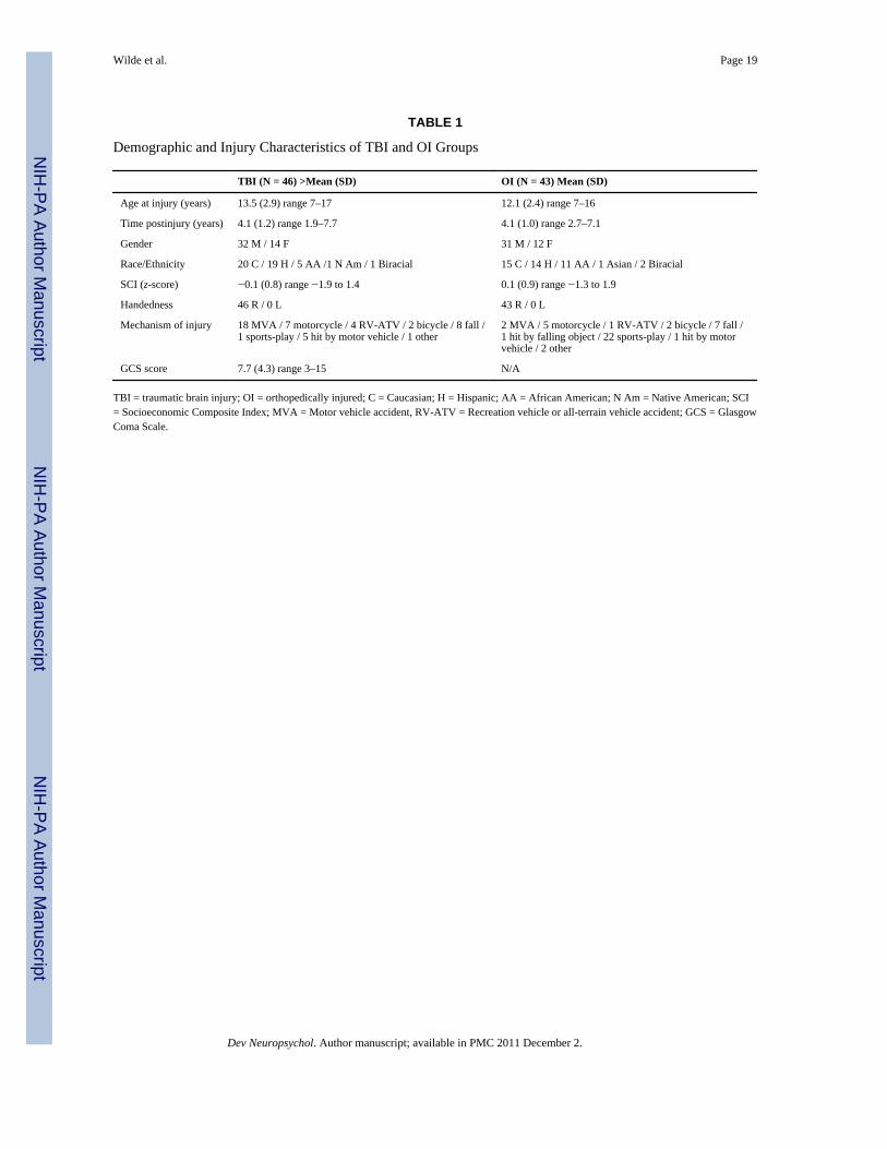
For both groups, all participants were recruited as consecutive admissions to the traumacenters of the participating hospitals, generally in the emergency department. All childrenincluded in the study were right-handed, English-speaking, and had no pre-existing headinjury involving loss of consciousness or post-concussive symptoms, neurologic disorderassociated with cerebral dysfunction and/or cognitive deficit (e.g., cerebral palsy, mentalretardation, epilepsy), diagnosed learning disability, psychiatric disorder, or history of childabuse. Additionally, minimum birth weight of 2500 grams (5 lbs, 8 oz) and a minimum of37 weeks of gestation were required for inclusion, and this was verified by parent report on adetailed developmental questionnaire. Of the eligible patients that were approached (in bothgroups), an estimated 30–50% agreed to participate at each site. Patients and parents ofpatients who declined to participate most frequently stated time constraints and schedulingdifficulties as reasons for not wishing to participate. There was no apparent systematic biasin injury severity or age for subjects who elected to participate versus those who did not.Demographic and injury-related characteristics for each group, including age at injury,ethnicity, gender, handedness, socioeconomic index, time post-injury, injury severity asmeasured by GCS scores appear in Table 1.
MRI AcquisitionAll subjects underwent MRI without sedation on Philips 1.5 T Intera scanners (Philips,Cleveland, OH) at Texas Children’s Hospital (Houston), the Rogers MRI Center, Universityof Texas South-western Medical Center (Dallas), Jackson Memorial Hospital (Miami), orMiami Children’s Hospital (Miami) using identical protocols.
Lesion analysis—A coronal T2-w fluid attenuated inversion recovery (FLAIR) sequencewas used (1100 msec TR, 140 msec TE, 5.0 mm slices). For this sequence, a 220 mm FOVwas used with a reconstructed voxel size of 0.86 × 0.86 × 5.0 mm.
DTI fiber tracking analysis—Transverse multislice spin echo, single shot, echo planarimaging (EPI) sequences were used (10150.5 msec TR, 90 msec TE, 2.7 mm slices, 0 mmgap). A 256 FOV (RFOV = 100%) was used with a measured voxel size of 2.67 × 2.69 ×2.70 mm. Diffusion was measured along 15 directions (number of b-value = 2, low b-value= 0 and high b-value = 860 sec/mm2). To improve signal to noise ratio, each high b-valueimage was acquired twice and averaged, but the low b image was only acquired once. Theacquisition time was approximately 5:45 minutes, and 55 slices were acquired.
Lesion analysis—Areas of signal abnormality were identified and traced by a board-certified neuroradiologist using FLAIR imaging as previously described (Wilde et al., 2005).No lesions were identified in the cingulate gyri or the underlying WM of the cingulumbundle of either TBI or the OI children.
DTI analysis—The Philips PRIDE-registration tool (Netsch, 2001) was used to removeshear and eddy current distortion and head motion prior to calculating FA maps with Philipsfiber tracking 4.1v3 Beta 2 software. Regions of interest (ROIs) were created on the rightand left parasagittal images using the protocol described later. After ROIs were created,
Wilde et al. Page 5
Dev Neuropsychol. Author manuscript; available in PMC 2011 December 2.
NIH
-PA Author Manuscript
NIH
-PA Author Manuscript
NIH
-PA Author Manuscript
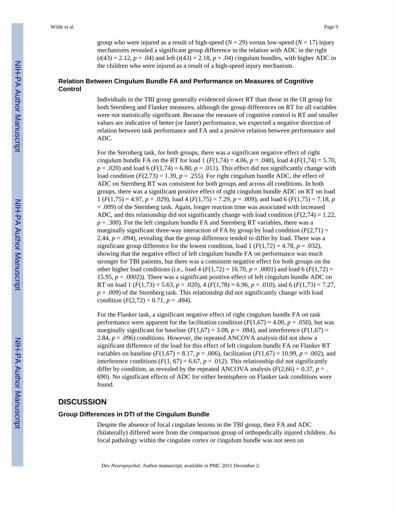
automated Philips three-dimensional fiber tracking tool (Hoogenraad, 2002) was utilized todetermine fiber tracks passing through the cingulum bundle; mean FA of the fiber systemsidentified was used as the quantitative measure for all DTI variables. For example, Figure 1illustrates the fiber system that emerged from the left cingulum bundle in both anorthopedically injured child and an age- and gender-equivalent child with TBI overlaid on aT1-weighted image. The algorithm for fiber tracking is based on the fiber assignment bycontinuous tracking (FACT) method (Mori, Crain, Chacko, & van Zijl, 1999). Trackingterminated if the FA in the voxels decreased below 0.2 or if the angle between adjacentvoxels along the track was larger than 7 degrees.
Cingulum Region of InterestROIs in DTI fiber tracking regions originating from the cingulum bundle were created onFA color maps in the left and right parasagittal planes. Using the automatic 3D ROIalgorithm function, two to three seed points were placed linearly along the cingulum bundlein the right parasagittal plane. The multi-ROI fiber tracking function was used to selectfibers that were common to the three ROIs, and the software provided the statistics includingthe mean FA and ADC for the selected fibers. This procedure was repeated for the left side.
Interoperator ReliabilityTwo experienced raters independently measured FA twice for both the right and leftcingulum bundles in a subset of 11 TBI patients and 11 OI subjects. Intra-rater and inter-rater reliabilities were calculated using Shrout-Fleiss reliability statistics to obtain intraclasscorrelation coefficients (ICCs), which were all above 0.90 (range 0.94 to 0.99; mean = 0.975± 0.02.)
Socioeconomic Composite IndexThe SCI provides a measure of a family’s socioeconomic status and has been shown tomoderate the effects of severe TBI on long-term outcome (Yeates et al., 1997). The index isderived by computing z-scores based on the combined distributions of the OI and TBIgroups for three variables including: (1) an 8-point scale rating family income, (2) a 7-pointscale of parent/guardian education, and (3) a rating of occupational prestige using the TotalSocioeconomic Index (TSI) (Hauser &Warren, 1999). The z-scores for these variables weresummed and standardized (mean = 0, SD = 1) based on the aggregate sample of participants(OI and TBI groups) to form the SCI score.
Cognitive Testing ProceduresWe indexed two processes fundamental to cognitive control: working memory andinhibition. To avoid effects of TBI on language, we selected tasks that are relativelylanguage-free, are stable, and that rely on reaction time. Our measure of inhibition, theEriksen Flanker Task, has been used with fMRI to study cognitive control in children(Bunge, Dudukovic, Thomason, Vaidya, & Gabrieli, 2002). To measure working memory,we used the Sternberg Item Recognition Task, which has been used with fMRI to examinethe relationship between working memory and inhibition under demands of cognitivecontrol (Bunge, Ochsner, Desmond, Glover, & Gabrieli, 2001). The tasks are furtherdetailed next.
Sternberg Item Recognition Task—Processing speed (reaction time) on a workingmemory task with varying memory loads was investigated with a modification of theSternberg Item Recognition Task (Bunge et al., 2001; Sternberg, 1966) for 89 subjects (datawas missing for 9 participants on this task, including 3 from the OI group and 6 from theTBI group). Missing data was due to technical problems with the recording mechanism of
Wilde et al. Page 6
Dev Neuropsychol. Author manuscript; available in PMC 2011 December 2.
NIH
-PA Author Manuscript
NIH
-PA Author Manuscript
NIH
-PA Author Manuscript
this computerized task, the child’s refusal to complete the task, or time constraints of theparent or guardian that precluded administration. There did not appear to be any source ofsystematic bias for the subjects with missing data such as greater injury severity or cognitiveimpairment, and missing data was distributed across the three sites. For each of 128 trials,children viewed a memory set of 1, 4, or 6 uppercase alphabetic letters and, after a 3-seconddelay, used “yes” (responding with right hand) and “no” (responding with left hand) keypresses to indicate whether a single probe letter had been in the memory set. There wereequal numbers of target and non-target trials. Memory load condition (1, 4, or 6), targetpresence, and position of the target in the memory set were varied randomly by trial.Subjects were instructed to respond as quickly as possible without making errors. Variablesused in this study were mean reaction time (RT) on memory loads 1, 4, and 6.
Flanker Task of Cognitive Processing Speed and Interference—To measurecognitive processing speed and visual interference, we used the Flanker Task (Eriksen &Eriksen, 1974), which was administered to 73 subjects (data was missing for 16 subjects,including 5 OI subjects and 9 TBI subjects). As with the Sternberg task, missing data wasdue to technical problems with this computerized task, the child’s refusal to complete thetask, or time constraints of the parent or guardian that precluded administration. There didnot appear to be any source of systematic bias for the subjects with missing data such asgreater injury severity or cognitive impairment, and data was missing from all three sites. Inthis task, a horizontal central arrow pointing to the left or to the right appeared on each trial.The child was asked to quickly press the button on the right or the left consistent with thedirection that the arrow was pointing. Task conditions included in this study were baseline,facilitation, and interference. Under the baseline condition, the arrow was flanked byhorizontal dashes that provided no cue to the child as to which button to press. Theinterference condition consisted of flanker arrows that pointed in the direction opposite tothe central arrow. In the facilitation condition, the flanker arrows pointed in the samedirection as the central arrow. There were 112 trials, including 28 trials of each taskcondition which were randomly interspersed. RT in each condition was the performancemeasure.
Design and Statistical AnalysisDemographic and injury severity data were tested using chi-square analysis for gender, t-testfor age at injury, time post-injury, and Socioeconomic Composite Index (SCI) score, andFisher’s exact test for mechanism of injury and race/ethnicity. A general linear model(GLM) analysis approach was used to examine group differences in mean FA between boththe right and left cingulum bundles, with age at testing included as a covariate. Whereappropriate, age was controlled in each subsequent model. Critical assumptions of GLManalysis were examined and no violations were noted. To determine whether there was adifference between right and left hemispheric FA in the two groups, a paired t-test wasemployed within each group, using the difference between right and left sides as adependent variable (right FA – left FA). Correlation analyses were used to examine therelation of lesion volumes in the right and left (1) frontal, (2) temporal, and (3) parietal lobesto both FA and ADC in the ipsilateral cingulum bundle (e.g., right frontal lobe with rightcingulum bundle), yielding a total of twelve comparisons. Furthermore, the TBI group wassubdivided into subgroups of children with and without lesions in the area of interest (e.g.,frontal, temporal, parietal and occipital), and t-tests were used to compare mean FA andADC for these subgroups. Spearman correlation analysis was used to examine the relationbetween mean FA and ADC of right and left cingulum bundles and GCS score.Additionally, the TBI group was subdivided into two subgroups based upon havingsustained injury as a result of either a high-velocity mechanism of injury (e.g., motor vehiclecrash) or a low-velocity injury (e.g., fall), and t-tests were used to determine whether
Wilde et al. Page 7
Dev Neuropsychol. Author manuscript; available in PMC 2011 December 2.
NIH
-PA Author Manuscript
NIH
-PA Author Manuscript
NIH
-PA Author Manuscript
mechanism of injury was related to right and left cingulum bundle FA and ADC. Finally, weused repeated ANCOVA models to examine the effect of right and left cingulum bundle FAand ADC and RT on the each of the working memory load conditions (loads 1, 4, and 6) ofthe Sternberg Item Recognition Task and the baseline, facilitation, and interferenceconditions on the Flanker Task.
RESULTSDemographics
No significant differences were noted in gender composition, SCI score, post-injury interval,or race/ethnicity between the two groups. The OI group was younger than the TBI group(t(86) = −2.14, p = .03); therefore, age at testing/scanning was controlled in subsequentstatistical models. As expected, the TBI group was more frequently injured as a result ofhigh-speed mechanisms of injury, such as motor vehicle crashes (χ2(1) = 17.31, p < .0001).
Group Differences in DTI of the Cingulum BundleGLM analyses revealed a significant group difference in mean FA for fibers systememanating from the right (F(1, 83) = 5.02, p = .03) and left (F(1, 83) = 11.09, p = .001)cingulate bundles, with higher FA in the OI as compared to the TBI group for bothhemispheres. Additionally, analyses revealed significant group differences in the mean ADCfor the right (F(1, 83) = 4.58, p = .04) and left (F(1, 83) = 7.70, p = .007) cingulum bundles,with higher ADC in the TBI group.
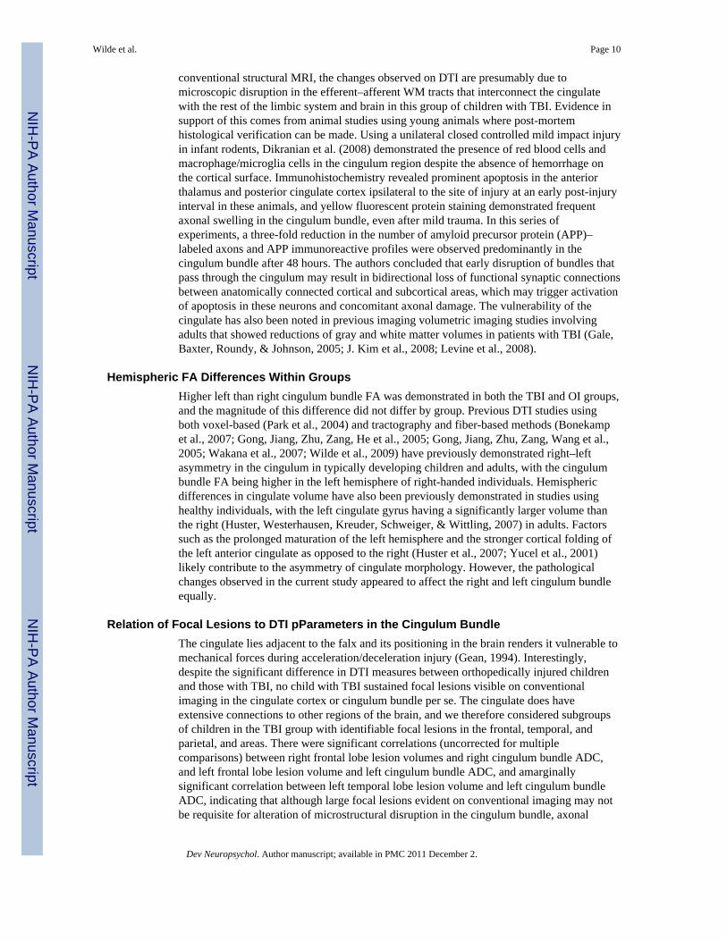
Hemispheric FA and ADC Differences Within GroupsAn analysis of the difference between right and left cingulum bundle mean FA revealed thatthe left cingulum bundle FA was significantly higher than the right cingulum bundle meanFA in both the OI (t(42) = −8.57, p < .0001) and TBI groups (t(45) = −4.76, p < .0001) (seeFigures 2a–b). Groups did not significantly differ on the difference score between right andleft cingulum bundle mean FA. Neither group demonstrated significant hemisphericdifferences for ADC.
Relation of Focal Lesions to DTI Parameters in the Cingulum BundleGLM analyses demonstrated no differences in cingulum bundle FA or ADC between TBIpatients with and without lesions in brain regions including the right and left (1) frontal, (2)temporal, and (3) parietal lobes. However, for children with TBI who had lesions in aparticular brain region, there were significant correlations between lesion volume in the rightfrontal lobes and right cingulum bundle ADC (number (N) of children in the TBI group withbrain lesions in the lobe = 36; r = −0.34, p = .04), left frontal lobe and left cingulum bundleADC (N = 32; r = −0.34, p = .05), and a marginally significant correlation between lefttemporal lobe and left cingulum bundle ADC (N = 25; r = −0.37, p = .07). There were nosignificant correlations between the right temporal lobe lesion volume (N = 24) and rightcingulum bundle ADC or between the right or left parietal lobe lesion volume (N = 17) andthe ipsilateral cingulum bundle ADC. There were no significant correlations between lesionvolume in any lobe and ipsilateral cingulum bundle FA.
Relation Between GCS Scores, Mechanism of Injury, and Cingulum Bundle FA and ADCSpearman correlation analysis revealed no significant relation with initial GCS score foreither right or left cingulum bundle mean FA in the TBI group. However, there was asignificant correlation with GCS for the right (r = −0.37, p = .02) and left (r = −0.35, p = .02) cingulum bundle ADC, with lower GCS associated with higher ADC in the TBI group.T-tests examining the difference between subgroups of children and adolescents in the TBI
Wilde et al. Page 8
Dev Neuropsychol. Author manuscript; available in PMC 2011 December 2.
NIH
-PA Author Manuscript
NIH
-PA Author Manuscript
NIH
-PA Author Manuscript
group who were injured as a result of high-speed (N = 29) versus low-speed (N = 17) injurymechanisms revealed a significant group difference in the relation with ADC in the right(t(43) = 2.12, p = .04) and left (t(43) = 2.18, p = .04) cingulum bundles, with higher ADC inthe children who were injured as a result of a high-speed injury mechanism.
Relation Between Cingulum Bundle FA and Performance on Measures of CognitiveControl
Individuals in the TBI group generally evidenced slower RT than those in the OI group forboth Sternberg and Flanker measures, although the group differences on RT for all variableswere not statistically significant. Because the measure of cognitive control is RT and smallervalues are indicative of better (or faster) performance, we expected a negative direction ofrelation between task performance and FA and a positive relation between performance andADC.
For the Sternberg task, for both groups, there was a significant negative effect of rightcingulum bundle FA on the RT for load 1 (F(1,74) = 4.06, p = .048), load 4 (F(1,74) = 5.70,p = .020) and load 6 (F(1,74) = 6.80, p = .011). This effect did not significantly change withload condition (F(2,73) = 1.39, p = .255). For right cingulum bundle ADC, the effect ofADC on Sternberg RT was consistent for both groups and across all conditions. In bothgroups, there was a significant positive effect of right cingulum bundle ADC on RT on load1 (F(1,75) = 4.97, p = .029), load 4 (F(1,75) = 7.29, p = .009), and load 6 (F(1,75) = 7.18, p= .009) of the Sternberg task. Again, longer reaction time was associated with increasedADC, and this relationship did not significantly change with load condition (F(2,74) = 1.22,p = .300). For the left cingulum bundle FA and Sternberg RT variables, there was amarginally significant three-way interaction of FA by group by load condition (F(2,71) =2.44, p = .094), revealing that the group difference tended to differ by load. There was asignificant group difference for the lowest condition, load 1 (F(1,72) = 4.78, p = .032),showing that the negative effect of left cingulum bundle FA on performance was muchstronger for TBI patients, but there was a consistent negative effect for both groups on theother higher load conditions (i.e., load 4 (F(1,72) = 16.70, p = .0001) and load 6 (F(1,72) =15.95, p = .0002)). There was a significant positive effect of left cingulum bundle ADC onRT on load 1 (F(1,73) = 5.63, p = .020), 4 (F(1,78) = 6.96, p = .010), and 6 (F(1,73) = 7.27,p = .009) of the Sternberg task. This relationship did not significantly change with loadcondition (F(2,72) = 0.71, p = .494).
For the Flanker task, a significant negative effect of right cingulum bundle FA on taskperformance were apparent for the facilitation condition (F(1,67) = 4.00, p = .050), but wasmarginally significant for baseline (F(1,67) = 3.08, p = .084), and interference (F(1,67) =2.84, p = .096) conditions. However, the repeated ANCOVA analysis did not show asignificant difference of the load for this effect of left cingulum bundle FA on Flanker RTvariables on baseline (F(1,67) = 8.17, p = .006), facilitation (F(1,67) = 10.99, p = .002), andinterference conditions (F(1, 67) = 6.67, p = .012). This relationship did not significantlydiffer by condition, as revealed by the repeated ANCOVA analysis (F(2,66) = 0.37, p = .690). No significant effects of ADC for either hemisphere on Flanker task conditions werefound.
DISCUSSIONGroup Differences in DTI of the Cingulum Bundle
Despite the absence of focal cingulate lesions in the TBI group, their FA and ADC(bilaterally) differed were from the comparison group of orthopedically injured children. Asfocal pathology within the cingulate cortex or cingulum bundle was not seen on
Wilde et al. Page 9
Dev Neuropsychol. Author manuscript; available in PMC 2011 December 2.
NIH
-PA Author Manuscript
NIH
-PA Author Manuscript
NIH
-PA Author Manuscript
conventional structural MRI, the changes observed on DTI are presumably due tomicroscopic disruption in the efferent–afferent WM tracts that interconnect the cingulatewith the rest of the limbic system and brain in this group of children with TBI. Evidence insupport of this comes from animal studies using young animals where post-mortemhistological verification can be made. Using a unilateral closed controlled mild impact injuryin infant rodents, Dikranian et al. (2008) demonstrated the presence of red blood cells andmacrophage/microglia cells in the cingulum region despite the absence of hemorrhage onthe cortical surface. Immunohistochemistry revealed prominent apoptosis in the anteriorthalamus and posterior cingulate cortex ipsilateral to the site of injury at an early post-injuryinterval in these animals, and yellow fluorescent protein staining demonstrated frequentaxonal swelling in the cingulum bundle, even after mild trauma. In this series ofexperiments, a three-fold reduction in the number of amyloid precursor protein (APP)–labeled axons and APP immunoreactive profiles were observed predominantly in thecingulum bundle after 48 hours. The authors concluded that early disruption of bundles thatpass through the cingulum may result in bidirectional loss of functional synaptic connectionsbetween anatomically connected cortical and subcortical areas, which may trigger activationof apoptosis in these neurons and concomitant axonal damage. The vulnerability of thecingulate has also been noted in previous imaging volumetric imaging studies involvingadults that showed reductions of gray and white matter volumes in patients with TBI (Gale,Baxter, Roundy, & Johnson, 2005; J. Kim et al., 2008; Levine et al., 2008).
Hemispheric FA Differences Within GroupsHigher left than right cingulum bundle FA was demonstrated in both the TBI and OI groups,and the magnitude of this difference did not differ by group. Previous DTI studies usingboth voxel-based (Park et al., 2004) and tractography and fiber-based methods (Bonekampet al., 2007; Gong, Jiang, Zhu, Zang, He et al., 2005; Gong, Jiang, Zhu, Zang, Wang et al.,2005; Wakana et al., 2007; Wilde et al., 2009) have previously demonstrated right–leftasymmetry in the cingulum in typically developing children and adults, with the cingulumbundle FA being higher in the left hemisphere of right-handed individuals. Hemisphericdifferences in cingulate volume have also been previously demonstrated in studies usinghealthy individuals, with the left cingulate gyrus having a significantly larger volume thanthe right (Huster, Westerhausen, Kreuder, Schweiger, & Wittling, 2007) in adults. Factorssuch as the prolonged maturation of the left hemisphere and the stronger cortical folding ofthe left anterior cingulate as opposed to the right (Huster et al., 2007; Yucel et al., 2001)likely contribute to the asymmetry of cingulate morphology. However, the pathologicalchanges observed in the current study appeared to affect the right and left cingulum bundleequally.
Relation of Focal Lesions to DTI pParameters in the Cingulum BundleThe cingulate lies adjacent to the falx and its positioning in the brain renders it vulnerable tomechanical forces during acceleration/deceleration injury (Gean, 1994). Interestingly,despite the significant difference in DTI measures between orthopedically injured childrenand those with TBI, no child with TBI sustained focal lesions visible on conventionalimaging in the cingulate cortex or cingulum bundle per se. The cingulate does haveextensive connections to other regions of the brain, and we therefore considered subgroupsof children in the TBI group with identifiable focal lesions in the frontal, temporal, andparietal, and areas. There were significant correlations (uncorrected for multiplecomparisons) between right frontal lobe lesion volumes and right cingulum bundle ADC,and left frontal lobe lesion volume and left cingulum bundle ADC, and amarginallysignificant correlation between left temporal lobe lesion volume and left cingulum bundleADC, indicating that although large focal lesions evident on conventional imaging may notbe requisite for alteration of microstructural disruption in the cingulum bundle, axonal
Wilde et al. Page 10
Dev Neuropsychol. Author manuscript; available in PMC 2011 December 2.
NIH
-PA Author Manuscript
NIH
-PA Author Manuscript
NIH
-PA Author Manuscript
degeneration or demyelination from areas of focal damage in the frontal and temporal areasmay still contribute as potential sources of injury-related dysfunction (Povlishock & Katz,2005; Rugg-Gunn, Symms, Barker, Greenwood, & Duncan, 2001). As mentionedpreviously, the most common TBI pathologies are those disrupting the long-coursing WMtracts of the brain (Ashwal, Holshouser, & Tong, 2006; Greenberg, Mikulis, Ng, DeSouza,& Green, 2008; Smith, Meaney, & Shull, 2003). However, the correlations between focallesion volume and cingulum bundle ADC are modest, and would not be significant whencorrected for multiple comparisons (e.g., Bonferroni correction), suggesting that, while focalinjury-induced axonal degeneration or demyelination may contribute to changes in thecingulum bundle detected via DTI, they are likely not the only mechanisms of TBI-relatedinjury in this structure. The diffuse TBI-related processes described earlier including axonalinjury or disruption within the cingulum bundle itself, immunoreactivity, and apoptosis mayalso play a significant role in damage to this structure.
Relation Between GCS Scores and Cingulum Bundle FAIn this study, there was a significant correlation with GCS for the right and left cingulumbundles, with lower GCS associated with higher ADC in the TBI group. This is consistentwith previous imaging studies that have demonstrated the relation of injury severity tovolume loss in the cingulate (Levine et al., 2008; C. L. MacDonald et al., 2008). Notsurprisingly, children and adolescents in the TBI group in our study who were injured as aresult of high-speed injury mechanisms demonstrated higher ADC in the right and leftcingulum bundles than children who were injured as a result of a low-speed mechanism.
Relation Between Cingulum Bundle FA and Performance on Measures of CognitiveControl
Reaction time on measures of cognitive control was related to white matter integrity withinthe cingulum bundle, including a significant relation between left and right cingulum bundleFA and ADC and RT during the Sternberg task and left cingulum bundle FA and Flankertasks in children with TBI. This finding is consistent with reports that the cingulate region isinvolved in various aspects of cognitive control, and in this case, the speed of cognitiveprocessing and response selection. Interestingly, the effect of cingulate DTI metrics wasgenerally stronger on the Sternberg task, a measure of working memory, than on the Flankertask, which contains a condition to measure interference. Additionally, while not significant,there was a tendency for the effect of left cingulum bundle FA on task performance to begreater in the higher load conditions on the Sternberg, but there was no change in therelationship by condition in the Flanker task. This is consistent with reports from a priorfMRI study that found that although there was an overlapping pattern of activation formemory load and interference and that these tasks may rely on some of the same structures,there was a dissociation between the two components, such that memory load wasassociated with activation in the anterior cingulate, but interference was not (Bunge et al.,2001).
The anterior cingulate gyrus has been said to mediate performance monitoring, responseselection, and target identification (Mesulam, Nobre, Kim, Parrish, & Gitelman, 2001),while the posterior region of the cingulate gyrus may be involved in the allocation of spatialattention (Small et al., 2003) and in the modulation of effort during attention demandingtasks (Y. H. Kim et al., 1999). Functional neuroimaging studies have found changes incingulate metabolism following TBI that relate to these cognitive functions in adults (CohenKadosh, Cohen Kadosh, Henik, & Linden, 2008; Scheibel et al., 2009; Soeda et al., 2005).An fMRI study of adults with moderate to severe TBI found that greater injury severity, asassessed by the GCS, was associated with increased anterior cingulate activation duringstimulus–response incompatibility (Scheibel et al., 2009). However, in that study, between
Wilde et al. Page 11
Dev Neuropsychol. Author manuscript; available in PMC 2011 December 2.
NIH
-PA Author Manuscript
NIH
-PA Author Manuscript
NIH
-PA Author Manuscript
group comparisons also identified the posterior cingulate as an area with increasedactivation in patients with severe TBI, relative to OI subjects and individuals with moderateTBI. Soeda et al. (2005) suggested that axonomy of neurons connecting the posteriorcingulate gyrus with other brain regions is a frequent mechanism of injury for this structure.The resulting deafferentiation may produce degeneration within the cingulate gyrus (Yountet al., 2002) and, in relation to findings from the current study and (Scheibel et al., 2009),DAI may contribute to decreased neural efficiency within distributed networks that mediateperformance during cognitive control.
Limitations and Future DirectionsOur study represents the first to specifically examine changes in the cingulum bundle ofchildren with TBI using quantitative DTI tractography and the relation of metrics such asFA and ADC to injury severity and mechanism, and measures of cognitive control.Strengths of the study include its prospective design with imaging performed at a generallyuniform post-injury interval, a reasonable size cohort of children with TBI as well as acomparison group of children with orthopedic injury. Reliability of DTI variablemeasurement is also a strength of this report, with excellent intra- and inter-operatoragreement.
Limitations include inter-individual variability in neuroanatomy of the cingulum bundle andthe lack of detailed investigation of gender differences given the preponderance of males inour population and the cross-sectional design. Future directions include examining theimpact of changes in the cingulate and their relation to additional standardized andfunctional cognitive outcome measures and emotional functioning measures. Longer-termchanges in the cingulate that may be influenced by injury (in TBI) and developmentalfactors in both TBI and comparison groups will also be explored in future longitudinalstudies.
CONCLUSIONDTI has increasingly demonstrated its utility in detecting injury that may not be visible onconventional MRI sequences. This study documents the sensitivity of DTI in revealinginjury to structures such as the cingulum bundle where, despite the absence of focal lesions,significant changes in white matter integrity are evident and potentially clinically useful.The cingulate is an essential member of neural networks underlying several importantcognitive abilities known to be deleteriously impacted in TBI, and an increasedunderstanding of the disruption of pathways involving the cingulum bundle other relatedstructures using DTI may enable advances in the assessment and treatment of these injuries.
AcknowledgmentsThe authors gratefully acknowledge the contribution of Deleene Menefee, Ph.D., Summer Lane, Lori Cook, SandraB. Chapman, Ph.D., and Gillian Hotz, Ph.D. in data collection, Matthew B. Mors in data analysis, and Stacey K.Martin in manuscript preparation. The authors thank the participants and their families for their willingness to bepart of this research.
This work was supported by the National Institute Neurological Disorders and Stroke grant R01-NS21889(“Neurobehavioral outcome of head injury in children,” Levin, PI). The authors also acknowledge the generouscontribution of Mission Connect of the TIRR Foundation.
REFERENCESAlexander AL, Lee JE, Lazar M, Field AS. Diffusion tensor imaging of the brain. Neurotherapeutics.
2007; 4(3):316–329. [PubMed: 17599699]
Wilde et al. Page 12
Dev Neuropsychol. Author manuscript; available in PMC 2011 December 2.
NIH
-PA Author Manuscript
NIH
-PA Author Manuscript
NIH
-PA Author Manuscript
Arfanakis K, Haughton VM, Carew JD, Rogers BP, Dempsey RJ, Meyerand ME. Diffusion tensor MRimaging in diffuse axonal injury. American Journal of Neuroradiology. 2002; 23(5):794–802.[PubMed: 12006280]
Ashwal S, Holshouser BA, Tong KA. Use of advanced neuroimaging techniques in the evaluation ofpediatric traumatic brain injury. Developmental Neuroscience. 2006; 28(4–5):309–326. [PubMed:16943654]
Azouvi P. Neuroimaging correlates of cognitive and functional outcome after traumatic brain injury.Current Opinion of Neurology. 2000; 13(6):665–669.
Benson RR, Meda SA, Vasudevan S, Kou Z, Govindarajan KA, Hanks RA, et al. Global white matteranalysis of diffusion tensor images is predictive of injury severity in traumatic brain injury. Journalof Neurotrauma. 2007; 24(3):446–459. [PubMed: 17402851]
Bijur, P.; Haslum, M. Cognitive, behavioral, and motoric sequelae of mild head injury in a nationalbirth cohort. In: Broman, SH.; Michel, ME., editors. Traumatic head injury in children. New York:Oxford University Press; 1995. p. 147-164.
Bonekamp D, Nagae LM, Degaonkar M, Matson M, Abdalla WM, Barker PB, et al. Diffusion tensorimaging in children and adolescents: Reproducibility, hemispheric, and age-related differences.Neuroimage. 2007; 34(2):733–742. [PubMed: 17092743]
Botvinick M, Nystrom LE, Fissell K, Carter CS, Cohen JD. Conflict monitoring versus selection-for-action in anterior cingulate cortex. Nature. 1999; 402(6758):179–181. [PubMed: 10647008]
Botvinick MM, Cohen JD, Carter CS. Conflict monitoring and anterior cingulate cortex: An update.Trends in Cognitive Sciences. 2004; 8(12):539–546. [PubMed: 15556023]
Bunge SA, Dudukovic NM, Thomason ME, Vaidya CJ, Gabrieli JD. Immature frontal lobecontributions to cognitive control in children: evidence from fMRI. Neuron. 2002; 33(2):301–311.[PubMed: 11804576]
Bunge SA, Ochsner KN, Desmond JE, Glover GH, Gabrieli JD. Prefrontal regions involved in keepinginformation in and out of mind. Brain. 2001; 124(Pt 10):2074–2086. [PubMed: 11571223]
Carter CS, Braver TS, Barch DM, Botvinick MM, Noll D, Cohen JD. Anterior cingulate cortex, errordetection, and the online monitoring of performance. Science. 1998; 280(5364):747–749.[PubMed: 9563953]
Carter CS, van Veen V. Anterior cingulate cortex and conflict detection: An update of theory and data.Cognitive, Affective, & Behavioral Neuroscience. 2007; 7(4):367–379.
Catroppa C, Anderson V. A prospective study of the recovery of attention from acute to 2 yearsfollowing pediatric traumatic brain injury. Journal of the International NeuropsychologicalSociety. 2005; 11(1):84–98. [PubMed: 15686611]
Cohen Kadosh R, Cohen Kadosh K, Henik A, Linden DE. Processing conflicting information:Facilitation, interference, and functional connectivity. Neuropsychologia. 2008; 46(12):2872–2879. [PubMed: 18632120]
Committee on Injury Scaling. Abbreviated Injury Scale. Des Plaines, IL: Association for theAdvancement of Automotive Medicine; 1990.
Dikranian K, Cohen R, Mac Donald C, Pan Y, Brakefield D, Bayly P, et al. Mild traumatic braininjury to the infant mouse causes robust white matter axonal degeneration which precedesapoptotic death of cortical and thalamic neurons. Experimental Neurology. 2008; 211(2):551–560.[PubMed: 18440507]
Eriksen BA, Eriksen CW. Effects of noise letters upon the identification of a target letter in a non-search task. Perception and Psychophysics. 1974; 16:143–149.
Ewing-Cobbs L, Prasad MR, Swank P, Kramer L, Cox CS Jr, Fletcher JM, et al. Arrested developmentand disrupted callosal microstructure following pediatric traumatic brain injury: Relation toneurobehavioral outcomes. Neuroimage. 2008; 42(4):1305–1315. [PubMed: 18655838]
Fontaine A, Azouvi P, Remy P, Bussel B, Samson Y. Functional anatomy of neuropsychologicaldeficits after severe traumatic brain injury. Neurology. 1999; 53(9):1963–1968. [PubMed:10599766]
Gale SD, Baxter L, Roundy N, Johnson SC. Traumatic brain injury and grey matter concentration: Apreliminary voxel based morphometry study. Journal of Neurology, Neurosurgery and Psychiatry.2005; 76(7):984–988.
Wilde et al. Page 13
Dev Neuropsychol. Author manuscript; available in PMC 2011 December 2.
NIH
-PA Author Manuscript
NIH
-PA Author Manuscript
NIH
-PA Author Manuscript

Gean, A. Imaging of the head. New York: Raven Press; 1994.Gennarelli TA. Mechanisms of brain injury. The Journal of Emergency Medicine. 1993; 11 Suppl 1:5–
11. [PubMed: 8445204]Goetz P, Blamire A, Rajagopalan B, Cadoux-Hudson T, Young D, Styles P. Increase in apparent
diffusion coefficient in normal appearing white matter following human traumatic brain injurycorrelates with injury severity. Journal of Neurotrauma. 2004; 21(6):645–654. [PubMed:15253793]
Gong G, Jiang T, Zhu C, Zang Y, He Y, Xie S, et al. Side and handedness effects on the cingulumfrom diffusion tensor imaging. NeuroReport. 2005; 16(15):1701–1705. [PubMed: 16189481]
Gong G, Jiang T, Zhu C, Zang Y, Wang F, Xie S, et al. Asymmetry analysis of cingulum based onscale-invariant parameterization by diffusion tensor imaging. Human Brain Mapping. 2005; 24(2):92–98. [PubMed: 15455461]
Greenberg G, Mikulis DJ, Ng K, DeSouza D, Green RE. Use of diffusion tensor imaging to examinesubacute white matter injury progression in moderate to severe traumatic brain injury. Archives ofPhysical Medicine and Rehabilitation. 2008; 89(12 Suppl):S45–S50. [PubMed: 19081441]
Hauser, RM.; Warren, JR. Raftery, A. Sociological methodology. Vol. vol 27. Blackwell Publishing;1999. Socioeconomic indexes for occupations: A review, update, and critique; p. 177-298.
Hoogenraad, F. Multi-center evaluation of in-vivo fibertracking. Philips Medical Systems; 2002.Huisman TA, Schwamm LH, Schaefer PW, Koroshetz WJ, Shetty-Alva N, Ozsunar Y, et al. Diffusion
tensor imaging as potential biomarker of white matter injury in diffuse axonal injury. AmericanJournal of Neuroradiology. 2004; 25(3):370–376. [PubMed: 15037457]
Huster RJ, Westerhausen R, Kreuder F, Schweiger E, Wittling W. Morphologic asymmetry of thehuman anterior cingulate cortex. Neuroimage. 2007; 34(3):888–895. [PubMed: 17161625]
Inglese M, Makani S, Johnson G, Cohen BA, Silver JA, Gonen O, et al. Diffuse axonal injury in mildtraumatic brain injury: A diffusion tensor imaging study. Journal of Neurosurgery. 2005; 103(2):298–303. [PubMed: 16175860]
Kerns JG, Cohen JD, MacDonald AW 3rd, Cho RY, Stenger VA, Carter CS. Anterior cingulateconflict monitoring and adjustments in control. Science. 2004; 303:1023–1026. [PubMed:14963333]
Kim J, Avants B, Patel S, Whyte J, Coslett BH, Pluta J, et al. Structural consequences of diffusetraumatic brain injury: A large deformation tensor-based morphometry study. Neuroimage. 2008;39(3):1014–1026. [PubMed: 17999940]
Kim YH, Gitelman DR, Nobre AC, Parrish TB, LaBar KS, Mesulam MM. The large-scale neuralnetwork for spatial attention displays multifunctional overlap but differential asymmetry.Neuroimage. 1999; 9(3):269–277. [PubMed: 10075897]
Kim YH, Yoo WK, Ko MH, Park CH, Kim ST, Na DL. Plasticity of the attentional network after braininjury and cognitive rehabilitation. Neurorehabilitation and Neural Repair. 2009; 23(5):468–477.[PubMed: 19118131]
Kraus JF, Rock A, Hemyari P. Brain injuries among infants, children, adolescents, and young adults.American Journal of Diseases in Children. 1990; 144(6):684–691.
Kraus MF, Susmaras T, Caughlin BP, Walker CJ, Sweeney JA, Little DM. White matter integrity andcognition in chronic traumatic brain injury: A diffusion tensor imaging study. Brain. 2007; 130(Pt10):2508–2519. [PubMed: 17872928]
Kumar R, Gupta RK, Husain M, Chaudhry C, Srivastava A, Saksena S, et al. Comparative evaluationof corpus callosum DTI metrics in acute mild and moderate traumatic brain injury: Its correlationwith neuropsychometric tests. Brain Injury. 2009; 23(7):675–685. [PubMed: 19557571]
Langlois JA, Kegler SR, Butler JA, Gotsch KE, Johnson RL, Reichard AA, et al. Traumatic braininjury-related hospital discharges. Results from a 14-state surveillance system, 1997. Morbidityand Mortality Weekly Report Surveillance Summaries. 2003; 52(4):1–20. [PubMed: 12836629]
Levin HS, Hanten G, Roberson G, Li X, Ewing-Cobbs L, Dennis M, et al. Prediction of cognitivesequelae based on abnormal computed tomography findings in children following mild traumaticbrain injury. Journal of Neurosurgery: Pediatrics. 2008; 1:461–470. [PubMed: 18518697]
Wilde et al. Page 14
Dev Neuropsychol. Author manuscript; available in PMC 2011 December 2.
NIH
-PA Author Manuscript
NIH
-PA Author Manuscript
NIH
-PA Author Manuscript
Levin HS, Wilde EA, Chu Z, Yallampalli R, Hanten GR, Li X, et al. Diffusion tensor imaging inrelation to cognitive and functional outcome of traumatic brain injury in children. Journal of HeadTrauma Rehabilitation. 2008; 23(4):197–208. [PubMed: 18650764]
Levine B, Kovacevic N, Nica EI, Cheung G, Gao F, Schwartz ML, et al. The Toronto traumatic braininjury study: Injury severity and quantified MRI. Neurology. 2008; 70(10):771–778. [PubMed:18316688]
Lipton ML, Gellella E, Lo C, Gold T, Ardekani BA, Shifteh K, et al. Multifocal white matterultrastructural abnormalities in mild traumatic brain injury with cognitive disability: A voxel-wiseanalysis of diffusion tensor imaging. Journal of Neurotrauma. 2008; 25(11):1335–1342. [PubMed:19061376]
MacDonald AW 3rd, Cohen JD, Stenger VA, Carter CS. Dissociating the role of the dorsolateralprefrontal and anterior cingulate cortex in cognitive control. Science. 2000; 288(5472):1835–1838.[PubMed: 10846167]
MacDonald CL, Schwarze N, Vaishnavi SN, Epstein AA, Snyder AZ, Raichle ME, et al. Verbalmemory deficit following traumatic brain injury: Assessment using advanced MRI methods.Neurology. 2008; 71(15):1199–1201. [PubMed: 18838668]
Mateer CA, Sira CS. Cognitive and emotional consequences of TBI: Intervention strategies forvocational rehabilitation. NeuroRehabilitation. 2006; 21(4):315–326. [PubMed: 17361048]
Mesulam MM, Nobre AC, Kim YH, Parrish TB, Gitelman DR. Heterogeneity of cingulatecontributions to spatial attention. Neuroimage. 2001; 13(6 Pt 1):1065–1072. [PubMed: 11352612]
Miller EK, Cohen JD. An integrative theory of prefrontal cortex function. Annual Review ofNeuroscience. 2001; 24:167–202.
Mori S, Crain BJ, Chacko VP, van Zijl PC. Three-dimensional tracking of axonal projections in thebrain by magnetic resonance imaging. Annals of Neurology. 1999; 45(2):265–269. [PubMed:9989633]
Netsch, T. Towards real-time multi-modality 3-d medical image registration. Paper presented at theInternational Conference on Computer Vision; Vancouver. 2001.
Nielson KA, Bryant T. The effects of non-contingent extrinsic and intrinsic rewards on memoryconsolidation. Neurobiology of Learning and Memory. 2005; 84(1):42–48. [PubMed: 15936682]
Park HJ, Westin CF, Kubicki M, Maier SE, Niznikiewicz M, Baer A, et al. White matter hemisphereasymmetries in healthy subjects and in schizophrenia: A diffusion tensor MRI study. Neuroimage.2004; 23(1):213–223. [PubMed: 15325368]
Paus T, Petrides M, Evans AC, Meyer E. Role of the human anterior cingulate cortex in the control ofoculomotor, manual, and speech responses: A positron emission tomography study. Journal ofNeurophysiology. 1993; 70(2):453–469. [PubMed: 8410148]
Perlbarg V, Puybasset L, Tollard E, Lehericy S, Benali H, Galanaud D. Relation between brain lesionlocation and clinical outcome in patients with severe traumatic brain injury: A diffusion tensorimaging study using voxel-based approaches. Human Brain Mapping. 2009
Povlishock JT, Katz DI. Update of neuropathology and neurological recovery after traumatic braininjury. Journal of Head Trauma Rehabilitation. 2005; 20(1):76–94. [PubMed: 15668572]
Rugg-Gunn FJ, Symms MR, Barker GJ, Greenwood R, Duncan JS. Diffusion imaging showsabnormalities after blunt head trauma when conventional magnetic resonance imaging is normal.Journal of Neurology, Neurosurgery & Psychiatry. 2001; 70(4):530–533.
Scheibel RS, Newsome MR, Troyanskaya M, Steinberg JL, Goldstein FC, Mao H, et al. Effects ofseverity of traumatic brain injury and brain reserve on cognitive-control related brain activation.Journal of Neurotrauma. 2009; 26(9):1447–1461. [PubMed: 19645622]
Sidaros A, Engberg AW, Sidaros K, Liptrot MG, Herning M, Petersen P, et al. Diffusion tensorimaging during recovery from severe traumatic brain injury and relation to clinical outcome: Alongitudinal study. Brain. 2008; 131(Pt 2):559–572. [PubMed: 18083753]
Small DM, Gitelman DR, Gregory MD, Nobre AC, Parrish TB, Mesulam MM. The posterior cingulateand medial prefrontal cortex mediate the anticipatory allocation of spatial attention. Neuroimage.2003; 18(3):633–641. [PubMed: 12667840]
Smith DH, Meaney DF, Shull WH. Diffuse axonal injury in head trauma. Journal of Head TraumaRehabilitation. 2003; 18(4):307–316. [PubMed: 16222127]
Wilde et al. Page 15
Dev Neuropsychol. Author manuscript; available in PMC 2011 December 2.
NIH
-PA Author Manuscript
NIH
-PA Author Manuscript
NIH
-PA Author Manuscript
Soeda A, Nakashima T, Okumura A, Kuwata K, Shinoda J, Iwama T. Cognitive impairment aftertraumatic brain injury: A functional magnetic resonance imaging study using the Stroop task.Neuroradiology. 2005; 47(7):501–506. [PubMed: 15973537]
Stancin T, Kaugars AS, Thompson GH, Taylor HG, Yeates KO, Wade SL, et al. Child and familyfunctioning 6 and 12 months after a serious pediatric fracture. Journal of Trauma. 2001; 51(1):69–76. [PubMed: 11468470]
Stancin T, Taylor HG, Thompson GH, Wade S, Drotar D, Yeates KO. Acute psychosocial impact ofpediatric orthopedic trauma with and without accompanying brain injuries. Journal of Trauma.1998; 45(6):1031–1038. [PubMed: 9867044]
Sternberg S. High-speed scanning in human memory. Science. 1966; 153(736):652–654. [PubMed:5939936]
Suskauer SJ, Huisman TA. Neuroimaging in pediatric traumatic brain injury: Current and futurepredictors of functional outcome. Developmental Disabilities Research Reviews. 2009; 15(2):117–123. [PubMed: 19489082]
Teasdale G, Jennett B. Assessment of coma and impaired consciousness. A practical scale. Lancet.1974; 2(7872):81–84. [PubMed: 4136544]
Thurman, DJ.; Coronado, V.; Selassie, A. The epidemiology of TBI: Implications for public health. In:Zasler, ND.; Katz, RD.; Zafonte, RD., editors. Brain injury medicine: Principles and practice. NewYork: Demos Medical Publishing; 2007.
Wakana S, Caprihan A, Panzenboeck MM, Fallon JH, Perry M, Gollub RL, et al. Reproducibility ofquantitative tractography methods applied to cerebral white matter. Neuroimage. 2007; 36(3):630–644. [PubMed: 17481925]
Wilde EA, Chu Z, Bigler ED, Hunter JV, Fearing MA, Hanten G, et al. Diffusion tensor imaging in thecorpus callosum in children after moderate to severe traumatic brain injury. Journal ofNeurotrauma. 2006; 23(10):1412–1426. [PubMed: 17020479]
Wilde EA, Hunter JV, Newsome MR, Scheibel RS, Bigler ED, Johnson JL, et al. Frontal and temporalmorphometric findings on MRI in children after moderate to severe traumatic brain injury. Journalof Neurotrauma. 2005; 22(3):333–344. [PubMed: 15785229]
Wilde EA, McCauley SR, Chu Z, Hunter JV, Bigler ED, Yallampalli R, et al. Diffusion tensor imagingof hemispheric asymmetries in the developing brain. Journal of Clinical and ExperimentalNeuropsychology. 2009; 31(2):205–218. [PubMed: 19052951]
Wozniak JR, Krach L, Ward E, Mueller BA, Muetzel R, Schnoebelen S, et al. Neurocognitive andneuroimaging correlates of pediatric traumatic brain injury: A diffusion tensor imaging (DTI)study. Archives of Clinical Neuropsychology. 2007; 22(5):555–568. [PubMed: 17446039]
Yeates KO, Taylor HG, Drotar D, Wade SL, Klein S, Stancin T, et al. Preinjury family environment asdeterminant of recovery from traumatic brain injuries in school-age children. Journal of theInternational Neuropsychological Society. 1997; 3(6):617–630. [PubMed: 9448375]
Yount R, Raschke KA, Biru M, Tate DF, Miller MJ, Abildskov T, et al. Traumatic brain injury andatrophy of the cingulate gyrus. The Journal of Neuropsychiatry and Clinical Neurosciences. 2002;14(4):416–423. [PubMed: 12426409]
Yuan W, Holland SK, Schmithorst VJ, Walz NC, Cecil KM, Jones BV, et al. Diffusion tensor MRimaging reveals persistent white matter alteration after traumatic brain injury experienced duringearly childhood. American Journal of Neuroradiology. 2007; 28(10):1919–1925. [PubMed:17905895]
Yucel M, Stuart GW, Maruff P, Velakoulis D, Crowe SF, Savage G, et al. Hemispheric and gender-related differences in the gross morphology of the anterior cingulate/paracingulate cortex innormal volunteers: An MRI morphometric study. Cerebral Cortex. 2001; 11(1):17–25. [PubMed:11113032]
Wilde et al. Page 16
Dev Neuropsychol. Author manuscript; available in PMC 2011 December 2.
NIH
-PA Author Manuscript
NIH
-PA Author Manuscript
NIH
-PA Author Manuscript
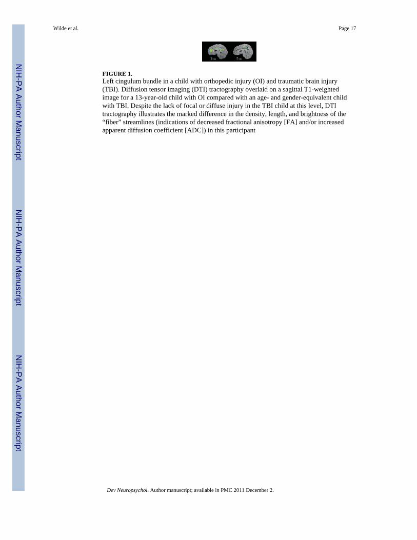
FIGURE 1.Left cingulum bundle in a child with orthopedic injury (OI) and traumatic brain injury(TBI). Diffusion tensor imaging (DTI) tractography overlaid on a sagittal T1-weightedimage for a 13-year-old child with OI compared with an age- and gender-equivalent childwith TBI. Despite the lack of focal or diffuse injury in the TBI child at this level, DTItractography illustrates the marked difference in the density, length, and brightness of the“fiber” streamlines (indications of decreased fractional anisotropy [FA] and/or increasedapparent diffusion coefficient [ADC]) in this participant
Wilde et al. Page 17
Dev Neuropsychol. Author manuscript; available in PMC 2011 December 2.
NIH
-PA Author Manuscript
NIH
-PA Author Manuscript
NIH
-PA Author Manuscript
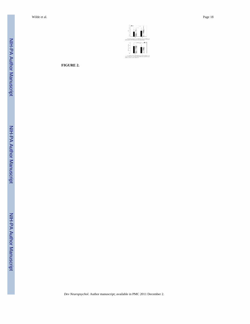
FIGURE 2.
Wilde et al. Page 18
Dev Neuropsychol. Author manuscript; available in PMC 2011 December 2.
NIH
-PA Author Manuscript
NIH
-PA Author Manuscript
NIH
-PA Author Manuscript
NIH
-PA Author Manuscript
NIH
-PA Author Manuscript
NIH
-PA Author Manuscript
Wilde et al. Page 19
TABLE 1
Demographic and Injury Characteristics of TBI and OI Groups
TBI (N = 46) >Mean (SD) OI (N = 43) Mean (SD)
Age at injury (years) 13.5 (2.9) range 7–17 12.1 (2.4) range 7–16
Time postinjury (years) 4.1 (1.2) range 1.9–7.7 4.1 (1.0) range 2.7–7.1
Gender 32 M / 14 F 31 M / 12 F
Race/Ethnicity 20 C / 19 H / 5 AA /1 N Am / 1 Biracial 15 C / 14 H / 11 AA / 1 Asian / 2 Biracial
SCI (z-score) −0.1 (0.8) range −1.9 to 1.4 0.1 (0.9) range −1.3 to 1.9
Handedness 46 R / 0 L 43 R / 0 L
Mechanism of injury 18 MVA / 7 motorcycle / 4 RV-ATV / 2 bicycle / 8 fall /1 sports-play / 5 hit by motor vehicle / 1 other
2 MVA / 5 motorcycle / 1 RV-ATV / 2 bicycle / 7 fall /1 hit by falling object / 22 sports-play / 1 hit by motorvehicle / 2 other
GCS score 7.7 (4.3) range 3–15 N/A
TBI = traumatic brain injury; OI = orthopedically injured; C = Caucasian; H = Hispanic; AA = African American; N Am = Native American; SCI= Socioeconomic Composite Index; MVA = Motor vehicle accident, RV-ATV = Recreation vehicle or all-terrain vehicle accident; GCS = GlasgowComa Scale.
Dev Neuropsychol. Author manuscript; available in PMC 2011 December 2.