Histoplasma capsulatum proteome response to decreased iron availability
Upload
independentCategory
view
0download
0
Decreased metastatic phenotype in cells resistant toAminolevulinic acid-Photodynamic therapy
Adriana Casasa, Gabriela Di Venosaa, Silvia Vanzullib, Christian Perottia, LeandroMamomea, Lorena Rodrigueza, Marina Simianc, Angeles Juarranzd, Osvaldo Pontiggiac,Tayyaba Hasane, and Alcira Batllea
a Centro de Investigaciones sobre Porfirinas y Porfirias (CIPYP). CONICET and Hospital de Clínicas Joséde San Martín, University of Buenos Aires. Córdoba 2351 1er subsuelo; CP=1120AAF, Buenos Aires,Argentina
b Instituto de Estudios Oncológicos, Academia Nacional de Medicina, Las Heras 3092, C1425ASU BuenosAires, Argentina
c Instituto Angel H Roffo. Av. San Martín 5481, Ciudad de Buenos Aires, Argentina
d Departamento de Biología, Facultad de Ciencias, Universidad Autónoma de Madrid. Cantoblanco E-28049Madrid, Spain
e Wellman Laboratories of Photomedicine WEL-224, Department of Dermatology, Massachusetts GeneralHospital, Harvard Medical School, 55 Fruit Street, Boston, MA 02114, USA
AbstractPhotodynamic therapy (PDT) is a novel cancer treatment utilising a photosensitiser, visible light andoxygen. PDT often leaves a significant number of surviving tumour cells. In a previous work, weisolated and studied two PDT resistant clones derived from the mammary adenocarcinoma LM3 line(Int. J. Oncol. 29 (2006) 397–405). The isolated Clon 4 and Clon 8 exhibited a more fibroblastic,dendritic pattern and were larger than the parentals. In the present work we studied the metastaticpotential of the two clones in comparison with LM3.
We found that 100 % of LM3 invaded Matrigel, whereas only 19 ± 6 % and 24 ± 7 % of Clon 4 andClon 8 cells invaded. In addition, 100% of LM3 cells migrated towards a chemotactic stimuluswhereas 38 ± 8 % and 73 ± 10 % of Clones 4 and 8 respectively were able to migrate. In vivo, 100%of the LM3 injected mice developed spontaneous lung metastasis, whereas none of the Clon 8 did,and only one of the mice injected with Clon 4 did. No differences were found in the proteolyticenzyme profiles among the cells. Anchorage-dependent adhesion was also impaired in vivo in theresistant clones, evidenced by the lower tumour take, latency time and growth rates, although bothclones showed in vitro higher binding to collagen I without overexpression of β1 integrin.
This is the first work where the metastatic potential of cells surviving to PDT has been studied. PDTstrongly affects the invasive phenotype of these cells, probably related to a higher binding to collagen.These findings may be crucial for the outcome of ALA-PDT of metastatic tumours, although furtherstudies are needed to extrapolate the results to the clinic employing another photosensitisers and celltypes.
Corresponding author Dr. Adriana Casas, Viamonte 1881 10 A, (1056) Ciudad de Buenos Aires, Argentina, FAX: 54 11 4811 7447, E-mail: [email protected]'s Disclaimer: This is a PDF file of an unedited manuscript that has been accepted for publication. As a service to our customerswe are providing this early version of the manuscript. The manuscript will undergo copyediting, typesetting, and review of the resultingproof before it is published in its final citable form. Please note that during the production process errors may be discovered which couldaffect the content, and all legal disclaimers that apply to the journal pertain.
NIH Public AccessAuthor ManuscriptCancer Lett. Author manuscript; available in PMC 2009 November 28.
Published in final edited form as:Cancer Lett. 2008 November 28; 271(2): 342–351. doi:10.1016/j.canlet.2008.06.023.
NIH
-PA Author Manuscript
NIH
-PA Author Manuscript
NIH
-PA Author Manuscript

Keywordsphotodynamic therapy; aminolevulinic acid; metastasis; invasion; adhesion
INTRODUCTIONPhotodynamic therapy (PDT) is a novel cancer treatment modality utilising a photosensitiser,visible light and oxygen [1,2]. The photosensitiser absorbs light and, in the presence of oxygen,transfers the energy, producing cytotoxic oxygen species [3].
5-aminolevulinic acid (ALA) is an alternative compound for the treatment of malignanttumours [4,5]. ALA is a naturally occurring delta amino acid that is ultimately converted intoProtoporphyrin IX (PpIX), the immediate precursor of heme and an endogenousphotosensitiser.
Currently, topical photodynamic therapy (PDT) has received approval for the treatment ofdermato-oncologic conditions like actinic keratoses, Bowen’s disease, in-situ squamous cellcarcinoma and basal cell carcinoma in many countries all over the world [6]. ALA is alsofrequently applied topically or systemically in PDT of superficial tumours such as cancer ofthe oral cavity, localised cutaneous lymphomas, and metastatic skin secondaries from breastor pinna [7] as well as non-tumour applications, especially psoriasis, viral-induced diseases,or acne vulgaris [8]. Non-superficial cancers such as duodenal, oesophageal, colorectal andcervical intraepithelial neoplasias, among others, have also been treated with ALA-PDT [9,10,11]. ALA has also been employed in the photodiagnosis and treatment of lung cancer,malignant glioma, bladder cancer and peritoneal carcinomatosis [12,13,14,15].
The accumulation of ALA is more pronounced in malignant cells as compared to their normalcounterparts in vitro and in vivo, and the reason for this phenomenon is attributed to an increaseof the rate-limiting enzyme porphobilinogen deaminase [16,17], and a decrease of the acitivityof ferrochelatase in tumour cells [18].
PDT, similarly to the rest of the anticancer therapies, often leaves a significant number ofsurviving tumour cells which have been exposed to reactive oxygen species arising from lightexcitation of a photosensitiser, but not enough to be destroyed. The characteristics of the cellsresistant to PDT allow us to study the changes induced in the long term by the treatment. Thesechanges can be exploited to selectively treat the surviving cells either with modified PDTprotocols or with other therapies [19].
A reduction of metastasis has been reported in vivo after PDT compared to surgery [20,21,22]. Rousset et al [23] also reported a decrease of metastasis induced by colon adenocarcinomacells treated with Photofrin-PDT respect to the control. On the contrary, Momma et al. [24],employing an orthopic prostate tumour model, showed that PDT induced an increase in thenumber of metastasis due to the induction of the vascular endothelial growth factor [25], whichis reversed by simultaneous administration of antioangiogenic therapies [26].
Since the plasma membrane is the target for various photosensitisers [27], it is not surprisingto find that PDT induces changes in cell adhesion, invasion and metastasis. In addition, tumourcells treated with PDT release prostanoids [28] that have been shown to influence the in vivodissemination, and in vitro migration of carcinoma cells.
The metastasis process involves the detachment and infiltration of the cells from the originalprimary tumour. In the first step, the cells released from the primary tumour have to penetrateto the blood or lymphatic vessels. Circulating cells can then migrate through the walls of vessels
Casas et al. Page 2
Cancer Lett. Author manuscript; available in PMC 2009 November 28.
NIH
-PA Author Manuscript
NIH
-PA Author Manuscript
NIH
-PA Author Manuscript

to surrounding tissues (extravasation) where they settle, adhere, proliferate, and induceangiogenesis, creating metastasis. Circulating cells can then migrate through the walls ofvessels to surrounding tissues (extravasation) where they settle, adhere, proliferate, and induceangiogenesis, creating metastasis. The processes of intra- and extravasation are regulated bythe activation of proteolytic enzymes capable of degrading the extracellular matrix (ECM)[29].
In a previous work, we isolated and studied two ALA-PDT resistant clones from the murinemammary adenocarcinoma LM3 cell line [19]. The isolated clones exhibited a morefibroblastic, dendritic pattern, and a higher cell spreading while the parental line presentedpolyhedric shape, grew in clusters and was smaller. These different features led us to think thatthe resistant lines could present different adhesive, invasive or metastatic phenotypes.
In the present work we studied the metastatic potential of the clonal lines derived from theLM3 adenocarcinoma resistant to ALA-PDT. We examined in vitro the adhesion, migrationand invasion processes, together with the proteolytic enzymes profile and in vivo, we assayedthe ability of the resistant clones to induce spontaneous metastasis.
MATERIALS AND METHODSChemicals
Bovine serum albumin (BSA) was obtained from Sigma, St. Louis, MO, USA. Anti mousemonoclonal antibody against β1-integrin was from Transduction Laboratories, UK and β-actinfrom GE Healthcare UK limited, Buckinghamshire, UK. The secondary antibody wasperoxidase-conjugated goat anti-mouse IgG (Jackson ImmunoResearch Laboratories, Inc,West Grove, PA, USA). Fibronectin, laminin and Collagen I, were obtained from CollaborativeBiomedical Products (Becton Dickinson, Bedford, MA, USA).
Cell lines and cell culturesLM3 cell line [30] derived from the murine mammary adenocarcinoma M3 was cultured inminimum essential Eagle’s medium supplemented with 2 mM L-glutamine, 80 μg/mlgentamycin and 5% fetal bovine serum, and incubated at 37°C in an atmosphere containing5% CO2. Clon 4 and Clon 8 cells resistant to ALA-PDT were obtained after multiple ALA-PDT treatments of LM3 parental cells as previously described [19].
AnimalsRandomized inbred male BALB/c mice 12 weeks old, weighing 20–25 g were used. They wereprovided with food (Purina 3, Molinos Río de la Plata) and water ad libitum. Animals receivedhuman care and were treated in accordance with guidelines established by the Animal Careand Use Committee of the Argentine Association of Specialists in Laboratory Animals(AADEALC), in full accord with the UK Guidelines for the Welfare of animals in ExperimentalNeoplasia [31].
Evaluation of tumour growthLM3 and the resistant clones were compared in their ability to growth as subcutaneous (s.c).primary tumours and to spontaneously metastasize to the lung. An amount of 105 to 106 cells/0.2 ml were injected s.c. into the left flank of BALB/c mice (n=7 per point). Tumour volumewas measured every week to establish the growth rate. Latency period was defined as the timewhen 50% of mice of each group developed palpable tumours. Tumour take was defined asthe percentage of mice that developed palpable tumours at latency time.
Casas et al. Page 3
Cancer Lett. Author manuscript; available in PMC 2009 November 28.
NIH
-PA Author Manuscript
NIH
-PA Author Manuscript
NIH
-PA Author Manuscript

Evaluation of spontaneous lung metastasisA cell suspension of 106 cells/0.2 ml was injected s.c in the mice. When the tumours reacheddifferent fixed diameters, animals were sacrificed and the subpleural visible metastatic noduleswere counted under a dissecting microscope. In addition, lungs were fixed in formaldehydeand embedded in paraffin according to standard histological procedures. Serial sections (4 um)from the whole lungs were cut and stained with H&E for counting metastasis in the parenchima.
Invasion and chemotaxis assaysThe invasiveness of LM3 and resistant clones was tested in vitro in a 24-well insert systemusing an 8 μm PET membrane coated with Matrigel Basement Membrane Matrix (BD BioCoatMatrigel invasion chambers, BD Biosciences, Bedford, MA, USA) following a describedmethod [32]. Inserts were rehydrated with medium without serum according to manufacturer’sinstructions. Exponentially growing mammary tumour cells (7 ×10⁴ cells/filter) suspended inmedium without serum, were added to the upper chamber and assembled with the lowerchamber, which had been prefilled with filtrated lung conditioned complete medium aschemoattractant [33]. After 20-hour incubation at 37°C, the cells on the upper surface of thefilter were removed with a cotton swab. Time and cell density conditions were establishedbefore. The filters were fixed with ice-cold methanol and stained with 0.1 % crystal violet. Thedegree of invasion was measured by counting the number of cells in five randomly selectedareas of the lower surface of the filter at × 400 magnification. Tumour cell chemotaxis wasmeasured following the same procedure employing the control inserts without the Matrigelcoating.
Cell adhesion to ECM proteins: for adhesion assays, 96-well high binding plates were coatedat 4°C overnight with ECM proteins: 5μg ml−1 fibronectin, 40 μg ml−1 laminin, and 20 μgml−1 collagen I dissolved in PBS pH 8.8. At least 1 h before the plates were used, the ECMliquid was discarded, and the wells were blocked with a solution of 0.5% bovine serum albumin(BSA). Cells (5 × 10⁵ ml−1) were trypsinised and plated in medium with BSA on the coatedwells. Other wells were coated with BSA as a control for non-specific binding. After 30 minincubation at 37°C, the cells were washed three times with PBS. The remaining adherent cellswere fixed in methanol: acetone (1:1) for 15 min. and stained with 0.5% crystal violet. Adherentcells were quantified by absorbance at 560 nm after solubilisation in 2% sodium dodecylsulfate. Adhesion values were determined by quadruplicate measurements.
Plating efficiencyCells were detached using trypsin/EDTA and resuspended in complete medium. Dilutedsuspensions were plated on 100 mm dishes and incubated for 10 days. Cells were fixed withice-cold methanol and stained with 0.1% aqueous crystal violet. Colonies of more than 50 cellswere counted.
Detection of metalloprotease (MMP) activityCollagenolytic activity secreted by LM3 and ALA-PDT resistant cells was determined inconditioned media by gel electrophoresis SDS-polyacrylamide copolymerized with 0.1%gelatin. Protein content in the cells was determined with the Lowry et al. method [34] and theconditioned media were diluted before seeding. To detect specific activity, after running, thegels were washed in 2% Triton X-100 and incubated for 72 h in 0.25 M Tris-HCl/1 M NaCl/25 mM CaCl2 buffer (pH 7.4). To detect non-specific activity, 40 mM EDTA was added to thebuffer solution. Gels were fixed and stained with Coomassie Blue. Gelatinolytic bands wereidentified by negative staining and quantified by a scan image densitometer coupled to an imageanalyzer. MMP activities were expressed as arbitrary units.
Casas et al. Page 4
Cancer Lett. Author manuscript; available in PMC 2009 November 28.
NIH
-PA Author Manuscript
NIH
-PA Author Manuscript
NIH
-PA Author Manuscript

Immunoblots for β1-integrinSemiconfluent monolayers were washed and then lysed with RIPA buffer [150 mM NaCl, 1%Triton X-100, 0.05% deoxycholate, 0.1% SDS, 1% Nonidet-40, 50 mM Tris (pH 8)] containingPhosphatase cocktail 2 and Protease inhibitor cocktail (Sigma). The samples were adjusted tothe same protein concentration (Bichinconinic acid protein assay kit, Pierce, Rockford, IL,USA), denatured by boiling in Laemmli sample buffer with 5% β-mercaptoethanol andseparated by SDS-PAGE, performed as described by Laemmli. Gels were electroblotted to aPDVF Immobilon-P membrane (Millipore, Bedford, MA, USA), blocked with 5% skim milkin Tris buffered saline (Tris HCl 10 mM pH 7.6, NaCl 0.9%, 0.05% Tween 20) overnight at4°C. Afterwards, the membranes were incubated 1 h at room temperature with a specificantibodiy anti β1-integrin diluted 1:1000 in blocking buffer. Secondary antibody was useddiluted 1:10,000 and incubated 1 h at room temperature. Detection was performed using ECLPlus Westernblotting detection system (GE Healthcare UK limited, Buckinghamshire, UK).Bands were quantified by scanning on a digital GS-700 densitometer and employing MolecularAnalyst software.
Statistical analysisKruskal-Wallis test was employed to evaluate differences in the latency periods. The unpairedt-test was used to establish the significance of differences between groups in the rest of theexperiments. Differences were considered statistically significant when P < 0.05.
RESULTSInvasion in vitro
Invasion was assayed in vitro (Figure 1) employing a 24-well insert system coated withMatrigel, using lung conditioned medium as chemoattractant. We observed that 100 % of LM3were able to invade Matrigel, whereas only 19 ± 6 % and 24 ± 7% of Clon 4 and Clon 8 cellsinvaded.
In addition, we tested chemotaxis or directional migration using control inserts, and we sawthat 100% of LM3 cells migrated through the porous membrane, whereas 38 ± 8 % and 73 ±10 % of Clones 4 and 8 respectively were able to migrate.
In vitro and in vivo behaviourColony formation assay in vitro was significantly impaired (25–30%) in both Clon 4 and Clon8 compared to LM3 (Table 1).
Tumour take was decreased in the resistant clones compared with the LM3 line (Table 1), mostmarkedly in Clon 8. When 105 LM3 cells were injected s.c. into mice, 30% of mice developedtumours, whereas no tumours were developed in the resistant clones. Increasing the amountinjected to 5×10⁵ cells, tumour take was 100% for LM3 cells, 60% for Clon 4 and 30% forClon 8. Further increasing the amount injected to 106 cells, 100% of mice injected with LM3and Clon 4 developed tumours, and only 60% of mice injected with Clon 8 did.
Growth rate was also significantly decreased in Clon 4 (p=0.047) compared with LM3, andClon 8 growth delay was even more marked, 3.5-fold lower than the control. Latency time wassimilar for LM3 and Clon 4 whereas it was markedly longer for Clon 8 cells (p< 0.001).
Induction of spontaneous metastasisSpontaneous lung metastasis induced by LM3 and resistant clones are shown in Figure 2.Whereas subcutaneous implanted LM3 cells mestastasised to lung in a tumour-size dependant
Casas et al. Page 5
Cancer Lett. Author manuscript; available in PMC 2009 November 28.
NIH
-PA Author Manuscript
NIH
-PA Author Manuscript
NIH
-PA Author Manuscript

way, Clones 8 and 4 almost did not induce nearly any metastasis at all. Only one smallmetastasis was found in one Clon 4 lung in the 7–19 mm tumour diameter range, whereas Clon8 cells did not induce any metastasis at all.
Cell adhesionWe did not find any significant differences in the adhesion of the three cell lines to the ECMproteins fibronectin and laminin, whereas Clon 4 adhered 1.3-fold to Collagen I (p= 0.027)and Clon 8 adhered 2-fold (0.001) as compared to LM3 (Figure 3). BSA was employed as anon-ECM control.
Metalloprotease activitiesBoth LM3 and resistant clones exhibited strong MMP-9 and weak MMP-2 signals. However,the comparison of the arbitrary units obtained after quantification of both bands, revealed thatthere were no significant differences between LM3 and the resistant clones (Figure 4)
Westernblot assaysWesternblot assays did not show significant differences in the expression of β1-integrin in theresistant clones as compared to LM3 cells (Figure 5).
DISCUSSIONIn a previous work we had shown that the ALA-PDT resistant Clones 4 and 8 exhibited a morefibroblastic, dendritic pattern, and a higher spreading in plastic while the parental cell linepresented polyhedric shape, grew in clusters and was smaller [19]. These different features ledus to think that the resistant lines could present different adhesive, invasive or metastaticphenotypes.
In the present work we showed that the ALA-PDT resistant clones have a severely impairedmetastatic potential. The metastasis process involves the detachment and infiltration of thecells from the original primary tumour, intravascular invasion, transport in the blood,extravasation, adhesion and ultimately, proliferation at the metastatic site [29]. We studied thedifferences in motility, invasion and adhesion of these low metastatic cell lines, and it is clearthat some of these steps are altered.
Plating efficiency dropped significantly in Clon 4 and Clon 8 cells, and in vivo, tumour takeand growth rate were also decreased in both clones, and latency time was longer in Clon 8,showing that the general anchorage-dependent adhesion is somehow affected in these cells.
Proteolytic activity and remodelling of the extracellular matrix are important players in tumourprogression. There is consensus to say that MMP activities correlate with higher metastaticpotential [35]. However, during tumour regression the increase in cell death has been shownto be accompanied by an increase in proteolytic activity and extracellular matrix remodelling[34].
Both LM3 and resistant clones exhibit MMP-2 and MMP-9 activities, but no significantdifferences were found between LM3 and the resistant clones, suggesting that an impairedcollagenolitic activity is not the reason for the different metastatic phenotype. It was found thatPDT induced MMP-1 and MMP-3 activities in a fibroblast model [37], whereas PDT impairedMMP-2 and MMP-8 activities in glioma cells [38] and no alterations in the MMPs pattern wereobserved in some other PDT-treated cells [39,40].
Casas et al. Page 6
Cancer Lett. Author manuscript; available in PMC 2009 November 28.
NIH
-PA Author Manuscript
NIH
-PA Author Manuscript
NIH
-PA Author Manuscript
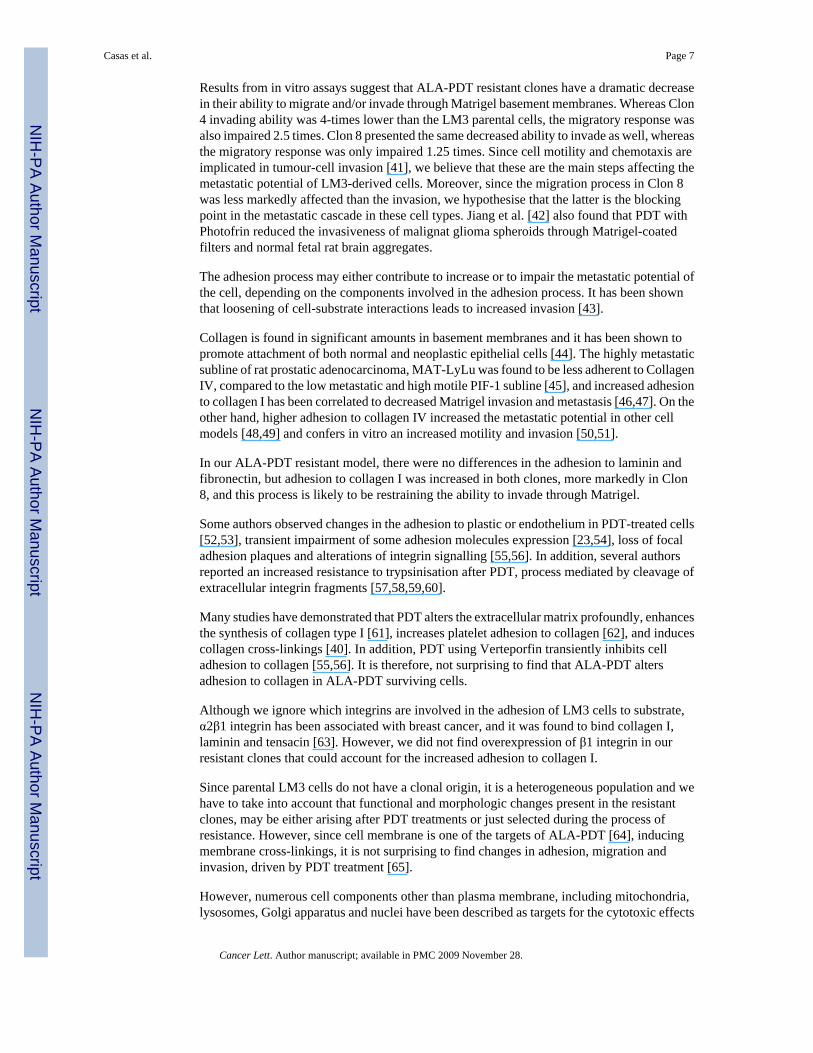
Results from in vitro assays suggest that ALA-PDT resistant clones have a dramatic decreasein their ability to migrate and/or invade through Matrigel basement membranes. Whereas Clon4 invading ability was 4-times lower than the LM3 parental cells, the migratory response wasalso impaired 2.5 times. Clon 8 presented the same decreased ability to invade as well, whereasthe migratory response was only impaired 1.25 times. Since cell motility and chemotaxis areimplicated in tumour-cell invasion [41], we believe that these are the main steps affecting themetastatic potential of LM3-derived cells. Moreover, since the migration process in Clon 8was less markedly affected than the invasion, we hypothesise that the latter is the blockingpoint in the metastatic cascade in these cell types. Jiang et al. [42] also found that PDT withPhotofrin reduced the invasiveness of malignat glioma spheroids through Matrigel-coatedfilters and normal fetal rat brain aggregates.
The adhesion process may either contribute to increase or to impair the metastatic potential ofthe cell, depending on the components involved in the adhesion process. It has been shownthat loosening of cell-substrate interactions leads to increased invasion [43].
Collagen is found in significant amounts in basement membranes and it has been shown topromote attachment of both normal and neoplastic epithelial cells [44]. The highly metastaticsubline of rat prostatic adenocarcinoma, MAT-LyLu was found to be less adherent to CollagenIV, compared to the low metastatic and high motile PIF-1 subline [45], and increased adhesionto collagen I has been correlated to decreased Matrigel invasion and metastasis [46,47]. On theother hand, higher adhesion to collagen IV increased the metastatic potential in other cellmodels [48,49] and confers in vitro an increased motility and invasion [50,51].
In our ALA-PDT resistant model, there were no differences in the adhesion to laminin andfibronectin, but adhesion to collagen I was increased in both clones, more markedly in Clon8, and this process is likely to be restraining the ability to invade through Matrigel.
Some authors observed changes in the adhesion to plastic or endothelium in PDT-treated cells[52,53], transient impairment of some adhesion molecules expression [23,54], loss of focaladhesion plaques and alterations of integrin signalling [55,56]. In addition, several authorsreported an increased resistance to trypsinisation after PDT, process mediated by cleavage ofextracellular integrin fragments [57,58,59,60].
Many studies have demonstrated that PDT alters the extracellular matrix profoundly, enhancesthe synthesis of collagen type I [61], increases platelet adhesion to collagen [62], and inducescollagen cross-linkings [40]. In addition, PDT using Verteporfin transiently inhibits celladhesion to collagen [55,56]. It is therefore, not surprising to find that ALA-PDT altersadhesion to collagen in ALA-PDT surviving cells.
Although we ignore which integrins are involved in the adhesion of LM3 cells to substrate,α2β1 integrin has been associated with breast cancer, and it was found to bind collagen I,laminin and tensacin [63]. However, we did not find overexpression of β1 integrin in ourresistant clones that could account for the increased adhesion to collagen I.
Since parental LM3 cells do not have a clonal origin, it is a heterogeneous population and wehave to take into account that functional and morphologic changes present in the resistantclones, may be either arising after PDT treatments or just selected during the process ofresistance. However, since cell membrane is one of the targets of ALA-PDT [64], inducingmembrane cross-linkings, it is not surprising to find changes in adhesion, migration andinvasion, driven by PDT treatment [65].
However, numerous cell components other than plasma membrane, including mitochondria,lysosomes, Golgi apparatus and nuclei have been described as targets for the cytotoxic effects
Casas et al. Page 7
Cancer Lett. Author manuscript; available in PMC 2009 November 28.
NIH
-PA Author Manuscript
NIH
-PA Author Manuscript
NIH
-PA Author Manuscript

of photodynamic therapy with other photosensitisers [66], and these different targets can leadto different metastatic phenotypes.
To sum up, this is the first work where the metastatic potential of cells resistant to PDT hasbeen studied. We found that PDT strongly affects the invasive phenotype of these cells,probably related to a higher binding to collagen. These findings may be crucial for the clinicaloutcome of ALA-PDT of metastatic tumours. Further studies are needed to elucidate if this isa common feature of all the cells subjected to multiple PDT treatments, if any photosensitiserin any cell type would induce the same alterations and moreover, if these alterations wouldappear after in vivo photodynamic treatment.
AcknowledgementsThis research was funded by grants from the National Cancer Center PO1 CA84203, the National Institutes for HealthRO1 AR040352, the Department of Defense FA 9550-04-1-0079, and the Argentine National Research Council(CONICET) (PIP 4108/96, 5263/05 and 105508/99-00). During the writing of this article, support to the authors wasprovided by National Institute of Health Grants PO1 CA084203 and RO1 AR040352. A.C. thanks Bunge y BornFoundation and Jorge Oster Argentina for financial support.
AbbreviationsALA
5-aminolevulinic acid
ALA-PDT ALA-based PDT
BSA bovine serum albumin
ECM extracellular matrix
MMP matrix metalloprotease
PBS phosphate-buffered saline
PDT photodynamic therapy
s.c subcutaneous
References1. Dougherty T. Photodynamic therapy (PDT) of malignant tumours. Crit Rev Oncol Hematol 1984;2:83–
116. [PubMed: 6397270]2. Gomer, C. Photodynamic Therapy. Pergamon Press; Oxford, England: 1987.3. Weishaupt K, Gomer C, Dougherty T. Identification of singlet oxygen as the cytotoxic agent in
photoactivation of a murine tumour. Cancer Res 1976;36:2326–2329. [PubMed: 1277137]4. Kennedy J, Pottier R, Pross G. Photodynamic Therapy with endogenous protoporphyrin IX: basic
principles and present clinical experience. J Photochem Photobiol B 1990;6:143–148. [PubMed:2121931]
Casas et al. Page 8
Cancer Lett. Author manuscript; available in PMC 2009 November 28.
NIH
-PA Author Manuscript
NIH
-PA Author Manuscript
NIH
-PA Author Manuscript
5. Fukuda H, Casas A, Chueke F, Paredes S, Batlle A. Photodynamic action of endogenously syntesizedporphyrins from aminolevulinic acid, using a new model for assaying the effectiveness of tumouralcell killing. Int J Biochem 1993;25:1395–1398. [PubMed: 8224354]
6. Babilas P, Landthaler M, Szeimies R. Photodynamic therapy in dermatology. Eur J Dermatol2006;16:340–8. [PubMed: 16935788]
7. Cairnduff F, Stringer MR, Hudson EJ, Ash DV, Brown SB. Superficial photodynamic therapy withtopical 5-aminolaevulinic acid for superficial primary and secondary skin cancer. Br J Cancer1994;69:605–608. [PubMed: 8123497]
8. Fritsch C, Ruzicka T. Fluorescence diagnosis and photodynamic therapy in dermatology fromexperimental state to clinic standard methods. J Environ Pathol Toxicol Oncol 2006;25:425–439.[PubMed: 16566733]
9. Kelty CJ, Brown NJ, Reed MW, Ackroyd R. The use of 5-aminolaevulinic acid as a photosensitiserin photodynamic therapy and photodiagnosis. Photochem Photobiol Sci 2002;1:158–168. [PubMed:12659511]
10. Kurwa H, Barlow R. The role of photodynamic therapy in dermatology. Clin Exp Der 1999;24:143–148.
11. Gossner L, Stolte M, Sroka R, Rick K, May A, Hahn E, Ell C. Photodynamic ablation of high-gradedysplasia and early cancer in Barrett’s esophagus by means of 5-aminolevulinic acid.Gastroenterology 1998;114:448–455. [PubMed: 9496934]
12. Hautmann H, Pichler JP, Stepp H, Baumgartner R, Gamarra F, Huber RM. In-vivo kinetics of inhaled5-aminolevulinic acid-induced protoporphyrin IX fluorescence in bronchial tissue. Respir Res2007;8:33. [PubMed: 17445266]
13. Stepp H, Beck T, Pongratz T, Meinel T, Kreth FW, Tonn JCh, Stummer W. ALA and malignantglioma: fluorescence-guided resection and photodynamic treatment. J Environ Pathol Toxicol Oncol2007;26:157–164. [PubMed: 17725542]
14. Kriegmair M, Baumgartner R, Knuechel R, Stepp H, Hofstadter F, Hofstetter A. Detection of earlybladder cancer by 5-aminolevulinic acid induced porphyrin fluorescence. J Urology 1996;155:105–110.
15. Gahlen J, Prosst RL, Pietschmann M, Haase T, Rheinwald M, Skopp G, Stern J, Herfarth C.Laparoscopic fluorescence diagnosis for intraabdominal fluorescence targeting of peritonealcarcinosis experimental studies. Ann Surg 2002;60:235–252.
16. Navone N, Polo C, Frisardi A, Andrade N, Batlle A. Heme biosynthesis in human breastadenocarcinoma. Mimetic in vitro studies and some heme enzymic activity levels. Int J Biochem1990;22:1407–1411. [PubMed: 2276414]
17. Navone N, Polo C, Frisardi A, Batlle A. Mouse mammary carcinoma PBGase andhydroxymethylbilane synthetase. Comp Biochem Physiol B 1991;98:67–71. [PubMed: 2060282]
18. Van Hillesberg R, Van der Berg J, Kort W, Terpstra O, Wilson J. Selective accumulation ofendogenously produced porphyrins in a liver metastasis model in rats. Gastroenterology1992;103:647–651. [PubMed: 1386052]
19. Casas A, Perotti C, Ortel B, Di Venosa G, Saccoliti M, Batlle A, Hasan T. Induction of murine tumourcell lines ressistant to ALA-mediated Photodynamic Therapy. Int J Oncol 2006;29:397–405.[PubMed: 16820882]
20. Gomer C, Ferrario A, Murphree A. The effect of localized porphyrin photodynamic therapy on theinduction of tumour metastasis. Br J Cancer 1987;56:27–32. [PubMed: 2956984]
20. Schreiber S, Gross S, Brandis A, Harmelin A, Rosenbach A, Belkin V, Scherz A, Salomon Y. Localphotodynamic therapy (PDT) of rat C6 glioma xenografts with Pd-bacteriopheophorbide leads todecreased metastases and increase of animal cure compared with surgery. Int J Cancer 2002;99:279–285. [PubMed: 11979445]
22. Lisnjak I, Kutsenok V, Polyschuk L, Gorobets O, Gamaleia N. Effect of photodynamic therapy ontumour angiogenesis and metastasis in mice bearing Lewis lung carcinoma. Exp Oncol 2005;27:333–335. [PubMed: 16404357]
23. Rousset N, Vonarx V, Eleouet S, Carre J, Kerninon E, Lajat Y, Patrice T. Effects of photodynamictherapy on adhesion molecules and metastasis. J Photochem Photobiol B 1999;52:65–73. [PubMed:10643074]
Casas et al. Page 9
Cancer Lett. Author manuscript; available in PMC 2009 November 28.
NIH
-PA Author Manuscript
NIH
-PA Author Manuscript
NIH
-PA Author Manuscript

24. Momma T, Hamblin M, Wu H, Hasan T. Photodynamic therapy of orthotopic prostate cancer withbenzoderivative: local control and distant metastasis. Cancer Res 1998;58:5425–5431. [PubMed:9850075]
25. Solban N, Selbo P, Sinha A, Chang S, Hasan T. Mechanistic investigation and implications ofphotodynamic therapy induction of vascular endothelial growth factor in prostate cancer. Cancer Res2006;66:5633–40. [PubMed: 16740700]
26. Kosharskyy B, Solban N, Chang S, Rizvi I, Chang Y, Hasan T. A mechanism-based combinationtherapy reduces local tumour growth and metastasis in an orthotopic model of prostate cancer. CancerRes 2006;66:10953–10958. [PubMed: 17108133]
27. Moor AC. Signaling pathways in cell death and survival after photodynamic therapy. J PhotochemPhotobiol B 2000;57:1–13. [PubMed: 11100832]
28. Cecic I, Korbelik M. Mediators of peripheral blood neutrophilia induced by photodynamic therapyof solid tumors. Cancer Lett 2002;183:43–51. [PubMed: 12049813]
29. Christofori G. New signals from the invasive front. Nature 2006;441:444–50. [PubMed: 16724056]30. Werbajh S, Urtreger A, Puricelli L, De Lustig E, Bal de Kier Joffe E, Kornblihtt A. Downregulation
of fibronectin transcription in highly metastatic adenocarcinoma cells. FEBS Lett 1992;440:277–281. [PubMed: 9872386]
31. Workman P, Balmain A, Hickman J. United Kingdom Co-ordinating Comittee on Cancer Research(UKCCCR) guidelines for the welfare of animals in experimental neoplasia (Second Edition). Br JCancer 1998;77:1–10.
32. Albini A, Iwamoto Y, Kleinman H, Martin G, Aaronson S, Kozlowski J, McEwan RN. A rapid invitro assay for quantitating the invasive potential of tumour cells. Cancer Res 1987;47:3239–3245.[PubMed: 2438036]
33. Ladeda V, Adam A, Puricelli L, Bal de Kier Joffe E. Apoptotic cell dearth in mammaryadenocarcinoma cells is prevented by soluble factors present in the target organ of metastasis. BreastCancer Res Treat 2001;69:39–51. [PubMed: 11759827]
34. Lowry O, Rosebrough N, Farr L, Randall R. Protein measurement with the folin phenol reagents. JBiol Chem 1951;193:265–275. [PubMed: 14907713]
35. Liotta L, Steeg P, Stetler-Stevenson W. Cancer metastasis and angiogenesis: an inbalance of positiveand negative regulation. Cell 1991;64:327–336. [PubMed: 1703045]
36. Simian M, Molinolo A, Lanari C. Involvement of matrix metalloproteinase activity in hormone-induced mammary tumor regression. Am J Pathol 2006;168:270–279. [PubMed: 16400029]
37. Karrer S, Bosserhof A, Weiderer P, Landthaler M, Szeimies R. Influence of 5-aminolevulinic acidand red light on collagen metabolism of human dermal fibroblasts. J Invest Dermatol 2003;120:325–331. [PubMed: 12542540]
38. Au C, Luk S, Jackson C, Ng H, Yow C, To S. Differential effects of photofrin, 5-aminolevulinic acidand calphostin C on glioma cells. J Photochem Photobiol B 2006;85:92–101. [PubMed: 16829117]
39. Overhaus M, Heckenkamp J, Kossodo S, Leszcynski D, LaMuraglia G. Photodynamic Therapygenerates a matrix barrier to invasive vascular cell migration. Circ Res 2000;86:334–340. [PubMed:10679486]
40. Waterman PR, Overhaus M, Heckenkamp J, Nigri GR, Fungaloi PF, Landis ME, Kossodo SC,LaMuraglia GM. Mechanisms of reduced human vascular cell migration after photodynamic therapy.Photochem Photobiol 2002;75:46–50. [PubMed: 11841040]
41. Eccles S. Targeting key steps in metastatic tumour progression. Curr Opin Genet Dev 2005;15:77–86. [PubMed: 15661537]
42. Jiang F, Chopp M, Katakowski M, Cho KK, Yang X, Hochbaum N, Tong L, Mikkelsen T.Photodynamic therapy with Photofrin reduces invasiveness of malignant human glioma cells. LasersMed Sci 2002;17:280–288. [PubMed: 12417983]
43. Pauli B, Schwartz D, Thonar E, Kuettner K. Tumour invasion and host extracellular matrix. CancerMetastasis Rev 1983;2:129–152. [PubMed: 6352011]
44. Terranova V, Rohrbach D, Martin G. Role of laminin in the attachment of PAM 212 (epithelial) cellsto basement membrane collagen. Cell 1980;22:719–726. [PubMed: 7460011]
Casas et al. Page 10
Cancer Lett. Author manuscript; available in PMC 2009 November 28.
NIH
-PA Author Manuscript
NIH
-PA Author Manuscript
NIH
-PA Author Manuscript

45. Mohler JL, Levy F, Sharief Y. Metastatic potential and substrate dependence of cell motility andattachment in the Dunning R-3327 rat prostatic adenocarcinoma model. Cancer Res 1991;51:6580–6585. [PubMed: 1742730]
46. Yoshimura M, Nishikawa A, Ihara Y, Taniguchi S, Taniguchi NN. Suppression of lung metastasisof B16 mouse melanoma by N-acetylglucosaminyltransferase III gene transfection. Proc Natl AcadSci USA 1995;92:8754–8758. [PubMed: 7568011]
47. Ek E, Dass C, Contreras K, Choong P. Inhibition of orthotopic osteosarcoma growth and metastasisby multitargeted antitumour activities of pigment epithelium-derived factor. Clin Exp Metastasis2007;24:93–106. [PubMed: 17458711]
48. Dumont J, Jones W, Bitonti A. Inhibition of experimental metastasis and cell adhesion of B16F1melanoma cells by inhibitors of protein kinase C. Cancer Res 1992;52:1195–1200. [PubMed:1737379]
49. Lichtner R, Kaufmann A, Kittmann A, Rohdeschulz B, Walter J, Williams L, Ullrich A, SchirrmacherV, Khazaie K. Ligand mediated activation of ectopic EGF receptor promotes matrix protein adhesionand lung colonization of rat mammary adenocarcinoma cells. Oncogene 1995;10:1823–1832.[PubMed: 7753557]
50. Azzam H, Thompson E. Collagen-induced activation of the Mr 72,000 type IV collagenase in normaland malignant human fibroblastoid cells. Cancer Res 1992;52:4540–4544. [PubMed: 1322793]
51. Savarese D, Russel J, Fatatis A, Liotta L. Type IV collagen stimulates an increase in intracellularcalcium: potential in tumour cell motility. J Biol Chem 1992;267:21928–21935. [PubMed: 1328249]
52. Foultier M, Vonarx V, Cordel S, Combre A, Patrice T. Modulation of colonic cancer cell adhesivenessby hematoporphyrin derivative photodynamic therapy. J Photochem Photobiol B: Biol 1994;23:9–17.
53. Vonarx V, Moultier M, de Brito X, Anasagasti L, Morlet L, Patrice T. Photodynamic therapy decreasescancer colonic cell adhesiveness and metastatic potential. Res Exp Med 1995;195:101–116.
54. Uzdensky A, Juzeniene A, Kolpakova E, Hjortland G, Juzenas P, Moan J. Photosensitization withprotoporphyrin IX inhibits attachment of cancer cells to a substratum. Biochem Biophys ResCommun 2004;322:452–457. [PubMed: 15325251]
55. Runnels J, Chen N, Ortel B, Kato D, Hasan T. BPD-MA-mediated photosensitization in vitro and invivo: cellular adhesion and β1 integrin expression in ovarian cancer cells. Br J Cancer 1999;80:946–953. [PubMed: 10362101]
56. Margaron P, Sorrenti L, Levi J. Photodynamic therapy inhibits cell adhesion without altering integrinexpression. Biochim Biophys Acta 1997;359:200–210. [PubMed: 9434126]
57. Denstman S, Dillehay L, Williams J. Enhanced susceptibility to HpD-sensitized phototoxicity andcorrelated resistance to trypsin detachment in SV40 transformed IMR-90 cells. Photochem Photobiol1986;43:145–147. [PubMed: 3010344]
58. Ball D, Mayhew S, Vernon D, Griffin M, Brown S. Decreased efficiency of trypsinization of cellsfollowing photodynamic therapy: evaluation of a role for tissue transglutaminase. PhotochemPhotobiol 2001;73:47–53. [PubMed: 11202365]
59. Uzdensky A, Juzeniene A, Ma L, Moan J. Photodynamic inhibition of enzymatic detachment ofhuman cancer cells from a substratum. Biochim Biophys Acta 2004;1670:1–11. [PubMed: 14729136]
60. Tsai J, Wu C, Chien H, Chen C. Reorganization of cytoskeleton induced by 5-aminolevulinic acid-mediated photodynamic therapy and its correlation with mitochondrial dysfunction. Lasers Surg Med2005;36:398–408. [PubMed: 15856508]
61. Uehara M, Inokuchi T, Tobita T, Ohba S, Asahina I. Expression of heat shock protein 47 in the fibroustissue adjacent to mouse tumour subjected to photodynamic therapy. Oral Oncol 2007;43:804–810.[PubMed: 17174144]
62. Fungaloi P, Statius van Eps R, Wu YP, Blankensteijn J, de Groot P, van Urk H, van Hillegersberg R,LaMuraglia G. Platelet adhesion to photodynamic therapy-treated extracellular matrix proteins.Photochem Photobiol 2002;75:412–417. [PubMed: 12003132]
63. Uhm J, Gladson C, Rao J. The role of integrins in the malignant phenotype of gliomas. FrontiersBiosci 1999;4:d188–199.
64. Girotti A. Photosensitized oxidation of membrane lipids: reaction pathways, cytotoxic effects, andcytoprotective mechanisms. J Photochem Photobiol B 2001;63:103–113. [PubMed: 11684457]
Casas et al. Page 11
Cancer Lett. Author manuscript; available in PMC 2009 November 28.
NIH
-PA Author Manuscript
NIH
-PA Author Manuscript
NIH
-PA Author Manuscript

65. Shen H, Spikes J, Kopecekov A, Kopecek J. Photodynamic crosslinking of proteins. I. Model studiesusing histidine- and lysine-containing N-(2-hydroxypropyl)methacrylamide copolymers. JPhotochem Photobiol B 1996;34:203–210. [PubMed: 8810538]
66. Stockert JC, Juarranz A, Villanueva A, Nonell S, Horobin R, Soltermann A, Durantini E, RivalonaV, Colombo L, Espada JM, Cañete M. Photodynamic therapy:selective uptake of photosensitizingdrugs into tumor cells. Curr Topics Pharmacol 2004;8:185–217.
Casas et al. Page 12
Cancer Lett. Author manuscript; available in PMC 2009 November 28.
NIH
-PA Author Manuscript
NIH
-PA Author Manuscript
NIH
-PA Author Manuscript

Figure 1. In vitro migration and invasion of LM3, Clon 4 and Clon 8The invasiveness of the cells was tested in vitro in Matrigel-coated inserts, whereas thechemotaxis was measured using the control uncoated inserts. Bars, means ± SD of 4independent experiments run in triplicate. The results are expressed as % of total cells platedin the inserts.
Casas et al. Page 13
Cancer Lett. Author manuscript; available in PMC 2009 November 28.
NIH
-PA Author Manuscript
NIH
-PA Author Manuscript
NIH
-PA Author Manuscript

Figure 2. Lung spontaneous metastasis induced by LM3, Clon 4 and Clon 8A cell suspension of 106 cells/0.2 ml was injected s.c. When the tumour reached different fixeddiameters, animals were sacrificed and the total number of metastasis (macroscopic +microscopic) were counted. Points, number of metastasis of single mouse. At least 7 mice perpoint were used.
Casas et al. Page 14
Cancer Lett. Author manuscript; available in PMC 2009 November 28.
NIH
-PA Author Manuscript
NIH
-PA Author Manuscript
NIH
-PA Author Manuscript

Figure 3. Cell adhesion of LM3, Clon 4 and Clon 8 cells to ECM proteinsAfter 30 min of culture on BSA, laminin, fibronectin or collagen I, cells were quantified usinga colorimetric method described in Materials and Methods. The Figure illustrates results fromone representative experiment of 3; each experimental condition is mean ± SE from 4 individualsamples.
Casas et al. Page 15
Cancer Lett. Author manuscript; available in PMC 2009 November 28.
NIH
-PA Author Manuscript
NIH
-PA Author Manuscript
NIH
-PA Author Manuscript

Figure 4. Gelatin zymography of conditioned culture media of LM3, Clon 4 and Clon 8 cellsA representative image of a gelatin zymogram is shown.
Casas et al. Page 16
Cancer Lett. Author manuscript; available in PMC 2009 November 28.
NIH
-PA Author Manuscript
NIH
-PA Author Manuscript
NIH
-PA Author Manuscript

Figure 5. Western blot analysis β1-integrinTotal extracts of subconfluent cultures were employed. Actin was employed as a loadingcontrol. The figure is representative of at least three independent experiments
Casas et al. Page 17
Cancer Lett. Author manuscript; available in PMC 2009 November 28.
NIH
-PA Author Manuscript
NIH
-PA Author Manuscript
NIH
-PA Author Manuscript

NIH
-PA Author Manuscript
NIH
-PA Author Manuscript
NIH
-PA Author Manuscript
Casas et al. Page 18
Table 1In vitro and in vivo behaviour of LM3 and resistant clones injected subcutaneously in mice
LM3 Clone 4 Clone 8
Plating efficiency (%)a 95 ± 8 75 ± 10* 70 ± 4Tumour take (%) (106/5×105/105 cells/mouse)b 100/100/30 100/60/0 60/30/0Growth rate in exponential phase (cm3/day)c 0.091 ± 0.017 0.075 ± 0.015* 0.025 ± 0.001*
Latency (days)d 20 (15–23) 22 (17–27) 35 (28–45) **
an=3
bn=5 per point.
c,d106 cells/mouse, n =9, three independent experiments.
dmean (range).
*p< 0.05 vs. LM3, unpaired Student’s t-test.
**p<0.001 vs. LM3, Kruskal-Wallis test.
Cancer Lett. Author manuscript; available in PMC 2009 November 28.
Copyright © 2022 FDOKUMEN