Photodynamic diagnosis of bladder cancer in ex vivo urine cytology
Upload
independentCategory
view
0download
0
Cellular and Molecular Mechanisms of Photodynamic HypericinTherapy for Nasopharyngeal Carcinoma Cells□S
Xiaoli Wang, Yi Guo, Shu Yang, Caihong Wang, Xuping Fu, Jinling Wang, Yumin Mao,Junsong Zhang, and Yao LiState Key Laboratory of Genetic Engineering, Institute of Genetics, School of Life Science, Fudan University, Shanghai,People’s Republic of China (Y.G., S.Y., X.F., Y.M., Y.L.); and School of Applied Chemistry and Biological, ShenzhenPolytechnic, Shenzhen, Guangdong, People’s Republic of China (X.W., C.W., J.W., J.Z.)
Received March 31, 2010; accepted June 11, 2010
ABSTRACTHypericin-mediated photodynamic therapy (HY-PDT) has becomea potential treatment for tumors and nonmalignant disorders.Some studies reported that HY-PDT could lead to apoptosis insome carcinoma cells. However, the molecular mechanism ofHY-PDT remains unknown. In this study, we evaluated the mo-lecular mechanisms of hypericin associated with light-emittingdiode irradiation on the poorly differentiated human nasopharyn-geal carcinoma cell line CNE-2 in vitro. To comprehensively un-derstand the effects of HY-PDT on CNE-2 cells, we detected cellviability, cell cycle, apoptosis, intracellular glutathione content,and intracellular caspase (caspase-9, caspase-3, and caspase-8)
activity. Furthermore, we performed genome-wide expressionanalysis via microarrays at different time points in response toHY-PDT, and we found that differentially expressed genes werehighly enriched in the pathways related to reactive oxygen speciesgeneration, mitochondrial activity, DNA replication and repair, cellcycle/proliferation, and apoptosis. These results were consistentwith our cytology test results and demonstrated that caspase-dependent apoptosis occurred after HY-PDT. Taken together,both cellular and molecular data revealed that HY-PDT couldinhibit the growth of CNE-2 cells and induce their apoptosis.
IntroductionPhotodynamic therapy (PDT) is one of the newest advance-
ments in the management of different microbial, viral, fun-gal, and inflammatory disorders and a variety of cancers.Light-induced growth inhibition is used in this method. Itinvolves the targeting of cells or tissues that have been sen-sitized to light by administration of a photosensitizing agent.One such agent is hypericin (HY; 1,3,4,6,8,13-hexahydroxy-10,11-dimethyl-phenanthro[1,10,9,8-opqra]perylene-7,14-dione; Falk, 1999), a secondary metabolite that can be isolatedfrom the plant Hypericum performatum, commonly known asSt. Johns wort. Because of its photoactive properties and low
cytotoxicity, attention has been focused on its application inPDT (Okpanyi et al., 1990; Kersten et al., 1999; Agostinis et al.,2002; Roscetti et al., 2004; Kiesslich et al., 2006).
Apoptosis and necrosis are two kinds of PDT-induced celldeath (Fiers et al., 1999). Which pathway is induced dependson the different properties of the photosensitizer, the type ofcells, the density of population, and the experimentalmethod. Furthermore, the method itself varies by photosen-sitizing agent concentration, light dose, and incubation time(Blank et al., 2002; Alvarez et al., 2003). Which pathway thecell takes to PDT-induced death is organelle-dependent aswell. That is, plasma membrane and lysosome can lead tonecrosis, whereas mitochondrial activity can lead to pro-grammed cell death, including both caspase-dependent and-independent apoptosis (Chen et al., 2000). Caspase-depen-dent apoptosis includes two pathways, the extrinsic deathpathway (death receptor-dependent) and the intrinsic deathpathway (mitochondria-dependent). In the past decades, mi-tochondria has played an important role in initiating andexecuting apoptosis in several types of cells (Green and Kro-emer, 2004; Bras et al., 2005; Zoratti et al., 2005).
This work was supported by the Technology Fund of Shenzhen Bureau ofScience Technology and Information [Grant 06KJP038] and was also a partof Project 30860081 supported by the National Natural Science Foundation ofChina.
X.W. and Y.G. contributed equally to this article.Article, publication date, and citation information can be found at
http://jpet.aspetjournals.org.doi:10.1124/jpet.110.168856.□S The online version of this article (available at http://jpet.aspetjournals.org)
contains supplemental material.
ABBREVIATIONS: PDT, photodynamic therapy; HY, hypericin; HY-PDT, hypericin-mediated photodynamic therapy; LED, light-emitting diode;FBS, fetal bovine serum; DMSO, dimethyl sulfoxide; HP, hematoporphyrin; MTT, methylthiazolyldiphenyl-tetrazolium bromide; PBS, phosphate-buffered saline; IR, inhibition rate; GSH, glutathione; MCB, monochlorobimane; FITC, fluorescein isothiocyanate; ROS, reactive oxygen species;DE, differentially expressed; FADD, FAS-associated death domain-containing protein; HSP, heat shock protein.
0022-3565/10/3343-847–853$20.00THE JOURNAL OF PHARMACOLOGY AND EXPERIMENTAL THERAPEUTICS Vol. 334, No. 3Copyright © 2010 by The American Society for Pharmacology and Experimental Therapeutics 168856/3616668JPET 334:847–853, 2010 Printed in U.S.A.
847
at Beijing B
ook Co Inc/P
eriodicalsFudan U
niv Med C
tr/Shanghai/149411 on M
arch 6, 2011jpet.aspetjournals.org
Dow
nloaded from
DC1.htmlhttp://jpet.aspetjournals.org/content/suppl/2010/06/15/jpet.110.168856.Supplemental Material can be found at:

Previous studies showed PDT associated with hypericincould lead to apoptosis in the human nasopharyngeal carci-noma cell line CNE-2 (Ali et al., 2001; Ali and Olivo, 2002),but others found it yielded necrosis (Du et al., 2003). Ali et al.(2002) reported photosensitization of HY could enhanceCD95/CD95L expression and CD95 signaling-dependent celldeath in all tumor cell lines. This was accomplished inpoorly differentiated CNE-2 cells by measuring the changeof membrane potential in mitochondria, the release of cy-tochrome c, the activity of caspases-3 and -8, the status ofcaspase-3-specific substrate poly(ADP-ribose) polymerase,and expression of CD95/CD95L. Thong et al. (2005) usedPDT associated with hypericin-treated cells and reportedthe intracellular concentration of Ca2� in experimentalgroup to be significantly higher than in the control.
Furthermore, many other reports indicated HY-PDT couldlead to apoptosis in the human laryngeal squamous cell car-cinoma strain Hep-2 (Sun et al., 2005), hepatoblastoma, pe-diatric hepatocellular carcinoma cells (Seitz et al., 2008),GH4C1 rat pituitary tumors (Cole et al., 2008), childhoodrhabdomyosarcoma (Seitz et al., 2007), and esophageal can-cer cells (Hopfner et al., 2003). Nevertheless, these workslacked sufficient evidence regarding to the molecular mech-anism for HY-PDT. In addition, some experiments demon-strated apoptosis induced by HY-PDT could be promoted byinhibiting p38 mitogen-activated protein kinases and thatsuch cell demise could be stimulated by HY-PDT associatedwith some other drugs (Kocanova et al., 2007; Buytaert et al.,2008; Schneider-Yin et al., 2009).
Microarray analysis has been widely used in the detectionof differentially expressed genes and the pathway analysisfor potential molecular mechanisms (Watts et al., 2001;Sarkozi et al., 2008). Recently, Sanovic et al (2009) detectedan alteration of the gene expression in the human squamouscell carcinoma cell line A-431 after HY-PDT by cDNA mi-croarray technique. A total of 168 genes were found to bedifferentially up-regulated and 45 were down-regulated. Be-cause of the significant expression changes, the followingcould be concluded: 1) lipoprotein receptor-mediated endocy-tosis could play an important role in the uptake of lipophilichypericin; and 2) cytoskeleton rearrangement and formationof apoptotic bodies occurred.
Although some studies showed apoptosis induced by HY-PDT, genome-wide expression profile studies or systematicmolecular evidence could not reveal the mechanisms ofapoptosis in CNE-2 cells subjected to HY-PDT. To gaininsight into the complex molecular mechanisms of thistherapy, we conducted both cytology tests and genome-wide expression analysis of hypericin associated withlight-emitting diode (LED) irradiation of human CNE-2cells line in vitro. We found numerous features with re-spect to molecular pathways that are involved in variousaspects of cell proliferation, DNA repair, and apoptosis.
Materials and MethodsCell Culture. Human nasopharyngeal carcinoma cell line CNE-2
was purchased from Shanghai Cell Biology Institute (Chinese Acad-emy of Science, Shanghai. China). Cells were grown at 37°C, 100%humidity, and 95% air, 5% CO2, and they were fed with 10% fetalbovine serum (FBS) and RPMI medium 1640 (Invitrogen, Carlsbad,CA). RPMI medium 1640 was supplemented with 1% L-glutamine,100 �g/ml penicillin, and 100 �g/ml streptomycin.
Drug Treatment. In cell viability experiments, CNE-2 cells wereseeded in 96-well plates (104cells/100 �l media/well) and cultured asdescribed above. After 24 h, hypericin (Alexis Laboratories, SanDiego, CA), which was dissolved in DMSO, was added to increasingconcentrations (0.04, 0.08, 0.12, 0.16, and 0.20 �g/ml). Cells wereincubated for 6 h in the dark and then exposed to yellow LED forhypericin and red LED for hematoporphyrin (HP) irradiation for 90min PDT. The light energy was 5.67 J/cm2. After drug treatment,cells were incubated in the dark.
Afterward, we established the following experimental group andcontrol groups: treatment of 0.20 �g/ml HY and light irradiation asthe experimental group (group A) and treatment of 0.20 �g/ml HYwithout irradiation as the negative control group (group B). In ad-dition, we added one or two extra negative controls in differentexperiments: treatment of irradiation without HY (group C) andtreatment without HY or irradiation (group D).
Cell Viability. After drug treatment, cells were incubated in thedark for an additional 20 h. Meanwhile, the group that containedonly DMSO and the groups that contained HP in increasing concen-trations (0.5, 1.0, 1.5, 2.0, 2.5, 3.0, 3.5, and 4.0 �g/ml) were desig-nated as the blank control and positive control, respectively. Cellviability was assessed by 3-(4,5-dimethylthiazol-2-yl)-2,5-diphe-nyltetrazolium bromide (MTT) assay. After 20-h incubation, mediumwas removed, and cells were washed with PBS, pH 7.4. Then, anadditional 100 �l of medium containing MTT (0.5 mg/ml) withoutFBS was added, and cells were incubated 4 h in the dark. Afterward,the medium was decanted carefully and dissolved in formazan in 100�l of DMSO and absolute ethanol (1:1) with agitation for 15 min.Absorption was measured on a SpectraMax M2 microplate reader(Molecular Devices, Sunnyvale, CA) at 570 nm. Inhibition rate (IR)(%) � (1 � average A570 of the experimental group/average A570 ofthe control group) � 100%. IC50 was calculated using the Blissmethod.
Cell Cycle. Cell cycle test kits (CycleTest Plus DNA Reagentkit; BD Biosciences, Franklin Lakes, NJ) were used according tothe manufacturer’s instructions at 20 h after light irradiation.Cells were collected by centrifugation at 400g for 5 min and thenthe supernatant was removed. We then resuspended the pellet in250 �l of solution A, containing trypsin, and incubated it for 15min. Next, we added 200 �l of solution B, containing trypsininhibitor and RNase A, and then carefully mixed them. After15-min incubation, 200 �l of solution C, containing propidiumiodide, was added and incubated at 4°C for 15 min in the dark.Finally, each sample was filtered by 50-�m aperture nylon mem-brane. The results were detected by a FACSCalibur flow cytome-ter (BD Biosciences). After using CellQuest software to collectapproximately 20,000 cells, the cell cycle was analyzed by ModFitLT 2.0 software (Verity Software House, Topsham, ME).
Apoptosis. Cells in group A and B were incubated in the dark foran additional 18, 28, and 48 h. Then, apoptosis was determined withan apoptosis test kit (APO-BRDU kit; BD Biosciences) according tothe manufacturer’s instructions. Cells were collected and resus-pended in 0.5 ml of 1� PBS. Next, we added 5 ml of 1% paraformal-dehyde. Cells were then incubated on ice for 15 min. We then resus-pended cells in 0.5 ml of 1� PBS and centrifuged at 400g for 5 min.After this, we resuspended cells in 0.5 ml of 1� PBS. Next, 70%ethanol was added, and cells were incubated at �20°C overnight. Wethen centrifuged cells and resuspended them in 1 ml of wash buffer.After another round of centrifugation, cells were resuspended in 50�l of DNA marker solution and incubated at 37°C for 60 min. Wenext added 1 ml of RNase buffer, and each tube was incubated for 10min and centrifuged at 300g for 5 min. We next removed supernatantand resuspended cells in 0.1 ml of fluorescent-labeled anti-5-bromo-2�-deoxyuridine antibody. After 30-min incubation at 37°C, we added0.5 ml of propidium iodide (0.25%)/RNase A solution and incubatedagain at 37°C for 30 min in the dark. The results were detected byflow cytometry.
848 Wang et al.
at Beijing B
ook Co Inc/P
eriodicalsFudan U
niv Med C
tr/Shanghai/149411 on M
arch 6, 2011jpet.aspetjournals.org
Dow
nloaded from

Intracellular Glutathione Content. Cells in group A, B, C, and Dwere collected by centrifugation. Content of intracellular glutathione(GSH) was determined by test kit (ApoAlert Glutathione Detection kit;Clontech, Otsu, Japan) according to the manufacturer’s instructions. Cellswere collected and resuspended in 10 ml of fresh RPMI medium containing10% FBS. Cells were collected by centrifugation at 700g. The pellet wasresuspended in 1 ml of ice-cold 1� cell wash buffer. We then transferredresuspended pellet into a 1.5-ml microcentrifuge tube and centrifuged at700g for 5 min. We then resuspended the pellet in 100 �l of ice-cold 1� celllysis buffer and incubated for 10 min on ice and centrifuged at maximalspeed using a tabletop centrifuge for 10 min. Next, we added 2 �l of 100mM monochlorobimane (MCB) to each supernatant. We prepared a neg-ative control sample by adding 2 �l of MCB to 100 �l of 1� cell lysis buffer.All samples were incubated at 37°C for 15 min. The fluorescence wasdetected with a fluorometer (Cary Eclipse fluorescence spectrophotometer;Varian, Inc., Palo Alto, CA) at 395 and 480 nm.
Intracellular Caspases, Caspase-9, Caspase-3, and Caspase-8 Ac-tivities. Cells in group A, B, C, and D were collected by centrifugation.According to the manufacturer’s instructions, activities of the overallcaspases, caspase-9, caspase-3, and caspase-8 were determined by theappropriate caspase test kit (CaspGLOW Fluorescein Caspase Stainingkit; BioVision, Mountain View, CA), respectively. Aliquots of 300 �l fromcell culture from each group were transferred to Eppendorf tubes. We thenadded 1 �l of FITC-VAD-FMK, FITC-LEHD-FMK, FITC-DEVD-FMK,and FITC-IETD-FMK, respectively, into each tube and incubated them for0.5 h at 37°C with 5% CO2. Cells were then centrifuged at 3000 rpm for 5min, and the supernatant was removed. We then resuspended cells in 0.5ml of wash buffer and centrifuged again. This step was repeated once. Theresults were detected by flow cytometry.
Transcriptional Analysis of Time Course in Response toHY-PDT. After both treatment (0.20 �g/ml HY) and irradiation,cells were incubated in the dark for 0, 2, 6, 12, and 20 h as experi-mental groups. Cells in HY treatment without irradiation were in-cubated in the dark for 20 h as the control group. Total RNAs wereisolated from the cultured cells in both experimental and controlgroups and hybridized to Illumina Sentrix Human WG-6_V2 expres-sion BeadChip arrays (Illumina, San Diego, CA) separately accord-ing to the manufacturer’s instructions.
Microarray Data Analysis. Gene expressions measures wereavailable at GEO (http://www.ncbi.nlm.nih.gov/geo/query/acc.cgi?acc�GSE20134). The data were extracted using BeadArray scannerand analyzed by BeadStation software (both from Illumina) providedby the manufacturer. Genes, whose DiffScore was above 13 com-pared with the control, were defined as differentially expressedgenes. The Cluster program (Eisen et al., 1998) was used to clusterthe candidate genes into a hierarchical tree. Pathway analysis wasperformed with GenMAPP 2.0 software (Dahlquist et al., 2002; Doni-ger et al., 2003). Significantly altered pathways were defined whentheir Z scores were above 1.96.
Statistics. Means and standard deviations of individual groups(n � 3) were calculated. P values were assessed by performingtwo-sided Student’s t test.
ResultsCell Viability. Irradiated with LED light, the cell viability
showed significant differences between the cells added withincreasing concentrations of HY (0.04, 0.08, 0.12, 0.16, and0.20 �g/ml) and blank control (two-sided t test, P � 0.01).When the final concentration of HY reached 0.16 �g/ml, IRwas more than 90% (Fig. 1), with an IC50 value of 0.049�g/ml. In the presence of HY without irradiation, cell viabil-ity was slightly inhibited (Fig. 1). However, there was adifference between treatment with irradiation and nonirra-diation at the same concentration of HY (P � 0.01; Fig. 1). Asillustrated, the HY-PDT was dose-dependent and the IR ofthe irradiation group was much more than the nonirradia-
tion group. Furthermore, application of hematoporphyrinand irradiation with red LED light yielded inhibition ofCNE-2 (P � 0.01; Fig. 2), with an IC50 value of 0.650 �g/ml.The IC50 value of HY was 13 times less than that of HP.
Cell Cycle. Our data detected by flow cytometry showedcell proportion at the G1 phase in the experimental groupswas lower than that in the control groups. However, the cellproportions of S and G2 phase were higher in the experimen-tal group than those in controls (Table 1).
Apoptosis. Our data detected by flow cytometry showedthere was a small amount of apoptotic cells in the group ofHY without irradiation, and the proportion of apoptosis cells
Fig. 1. Inhibition rates of cell viability test of HY in treatment with orwithout irradiation by MTT assay. Cells were treated with increasingconcentrations (0.04, 0.08, 0.12, 0.16, and 0.20 �g/ml) of HY with orwithout irradiation and incubated an additional 20 h in the dark. Then,cell viability was tested by MTT assay. The mean and the standarddeviation of the inhibition rate for each HY concentration are shown as adot and vertical line, respectively. F, HY with irradiation; �, HY withoutirradiation.
Fig. 2. Inhibition rates of cell viability test of HP treatment with irradi-ation by MTT assay. Cells were treated with increasing concentrations(0.5, 1.0, 1.5, 2.0, 2.5, 3.0, 3.5, and 4.0 �g/ml) of HP and irradiation andthen incubated an additional 20 h in the dark. Then, cell viability wastested by MTT assay. In our data, cell viability was inhibited by HP-PDT.When the final concentration of HP reached 1.5 �g/ml, IR was more than90%. The mean and the standard deviation of the inhibition rate for eachHP concentration are shown as a dot and vertical line, respectively. f, HPwith irradiation.
HY-PDT for Naspharyngeal Carcinoma Cells 849
at Beijing B
ook Co Inc/P
eriodicalsFudan U
niv Med C
tr/Shanghai/149411 on M
arch 6, 2011jpet.aspetjournals.org
Dow
nloaded from
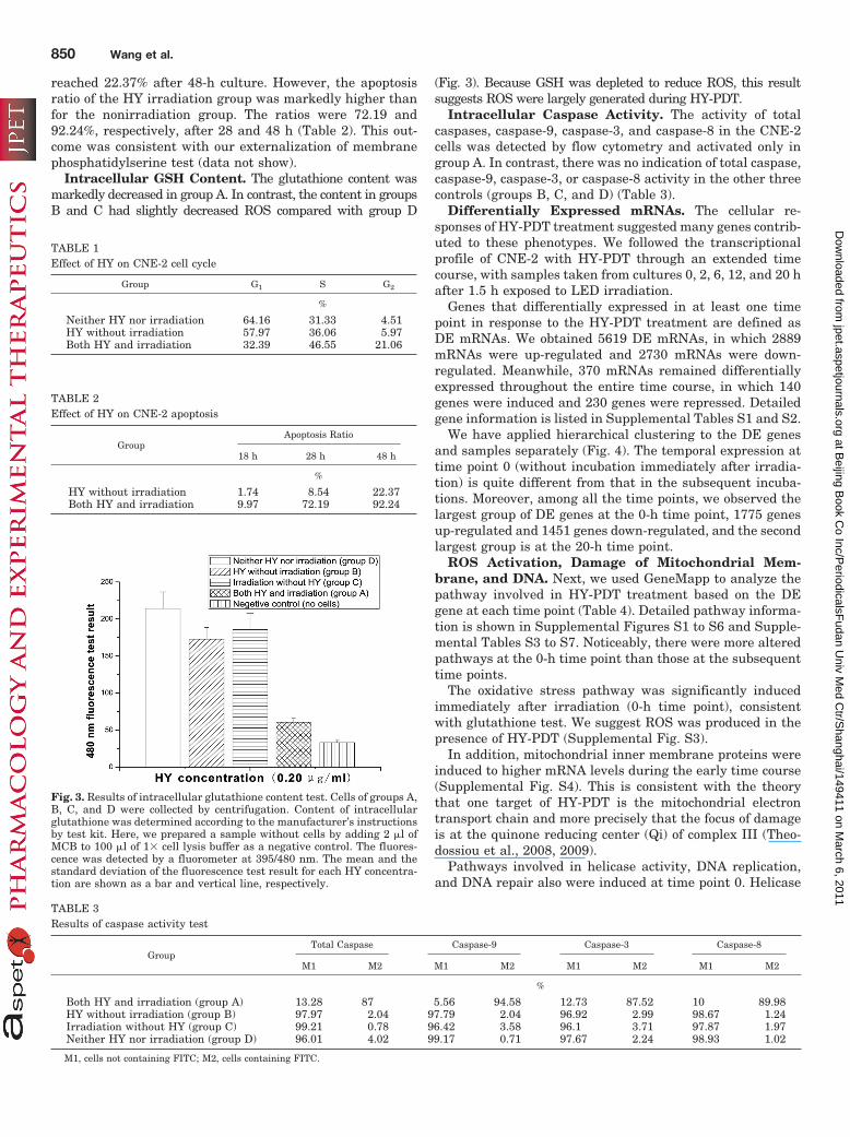
reached 22.37% after 48-h culture. However, the apoptosisratio of the HY irradiation group was markedly higher thanfor the nonirradiation group. The ratios were 72.19 and92.24%, respectively, after 28 and 48 h (Table 2). This out-come was consistent with our externalization of membranephosphatidylserine test (data not show).
Intracellular GSH Content. The glutathione content wasmarkedly decreased in group A. In contrast, the content in groupsB and C had slightly decreased ROS compared with group D
(Fig. 3). Because GSH was depleted to reduce ROS, this resultsuggests ROS were largely generated during HY-PDT.
Intracellular Caspase Activity. The activity of totalcaspases, caspase-9, caspase-3, and caspase-8 in the CNE-2cells was detected by flow cytometry and activated only ingroup A. In contrast, there was no indication of total caspase,caspase-9, caspase-3, or caspase-8 activity in the other threecontrols (groups B, C, and D) (Table 3).
Differentially Expressed mRNAs. The cellular re-sponses of HY-PDT treatment suggested many genes contrib-uted to these phenotypes. We followed the transcriptionalprofile of CNE-2 with HY-PDT through an extended timecourse, with samples taken from cultures 0, 2, 6, 12, and 20 hafter 1.5 h exposed to LED irradiation.
Genes that differentially expressed in at least one timepoint in response to the HY-PDT treatment are defined asDE mRNAs. We obtained 5619 DE mRNAs, in which 2889mRNAs were up-regulated and 2730 mRNAs were down-regulated. Meanwhile, 370 mRNAs remained differentiallyexpressed throughout the entire time course, in which 140genes were induced and 230 genes were repressed. Detailedgene information is listed in Supplemental Tables S1 and S2.
We have applied hierarchical clustering to the DE genesand samples separately (Fig. 4). The temporal expression attime point 0 (without incubation immediately after irradia-tion) is quite different from that in the subsequent incuba-tions. Moreover, among all the time points, we observed thelargest group of DE genes at the 0-h time point, 1775 genesup-regulated and 1451 genes down-regulated, and the secondlargest group is at the 20-h time point.
ROS Activation, Damage of Mitochondrial Mem-brane, and DNA. Next, we used GeneMapp to analyze thepathway involved in HY-PDT treatment based on the DEgene at each time point (Table 4). Detailed pathway informa-tion is shown in Supplemental Figures S1 to S6 and Supple-mental Tables S3 to S7. Noticeably, there were more alteredpathways at the 0-h time point than those at the subsequenttime points.
The oxidative stress pathway was significantly inducedimmediately after irradiation (0-h time point), consistentwith glutathione test. We suggest ROS was produced in thepresence of HY-PDT (Supplemental Fig. S3).
In addition, mitochondrial inner membrane proteins wereinduced to higher mRNA levels during the early time course(Supplemental Fig. S4). This is consistent with the theorythat one target of HY-PDT is the mitochondrial electrontransport chain and more precisely that the focus of damageis at the quinone reducing center (Qi) of complex III (Theo-dossiou et al., 2008, 2009).
Pathways involved in helicase activity, DNA replication,and DNA repair also were induced at time point 0. Helicase
Fig. 3. Results of intracellular glutathione content test. Cells of groups A,B, C, and D were collected by centrifugation. Content of intracellularglutathione was determined according to the manufacturer’s instructionsby test kit. Here, we prepared a sample without cells by adding 2 �l ofMCB to 100 �l of 1� cell lysis buffer as a negative control. The fluores-cence was detected by a fluorometer at 395/480 nm. The mean and thestandard deviation of the fluorescence test result for each HY concentra-tion are shown as a bar and vertical line, respectively.
TABLE 1Effect of HY on CNE-2 cell cycle
Group G1 S G2
%
Neither HY nor irradiation 64.16 31.33 4.51HY without irradiation 57.97 36.06 5.97Both HY and irradiation 32.39 46.55 21.06
TABLE 2Effect of HY on CNE-2 apoptosis
GroupApoptosis Ratio
18 h 28 h 48 h
%
HY without irradiation 1.74 8.54 22.37Both HY and irradiation 9.97 72.19 92.24
TABLE 3Results of caspase activity test
GroupTotal Caspase Caspase-9 Caspase-3 Caspase-8
M1 M2 M1 M2 M1 M2 M1 M2
%
Both HY and irradiation (group A) 13.28 87 5.56 94.58 12.73 87.52 10 89.98HY without irradiation (group B) 97.97 2.04 97.79 2.04 96.92 2.99 98.67 1.24Irradiation without HY (group C) 99.21 0.78 96.42 3.58 96.1 3.71 97.87 1.97Neither HY nor irradiation (group D) 96.01 4.02 99.17 0.71 97.67 2.24 98.93 1.02
M1, cells not containing FITC; M2, cells containing FITC.
850 Wang et al.
at Beijing B
ook Co Inc/P
eriodicalsFudan U
niv Med C
tr/Shanghai/149411 on M
arch 6, 2011jpet.aspetjournals.org
Dow
nloaded from

activity remained for a longer time to cause DNA unwindingand chromatin disassembling in the end of time course.
Cell Cycle and Cell Proliferation. DNA microarrayalso revealed some interesting expression patterns of thecell cycle pathway. They were altered in the early timepoints in response to HY-PDT, consistent with the cellcycle test (Table 1). This suggests the cells were enrichedin the S and G2 phases for repairing the damaged DNA(Supplemental Fig. S2). Meanwhile, cellular metabolismwas negatively regulated.
The pathway of negative regulation of cell proliferationwas significantly activated during the entire time course,confirming the result of MTT assay that HY-PDT can inhibitcell proliferation (Supplemental Fig. S1).
Apoptosis. Apoptosis pathways were significantly alteredin the entire time course. Change of mitochondrial mem-brane indicated that the intrinsic cell death pathway wasactivated. Furthermore, the Fas extrinsic cell death pathwaywas significantly activated during the early time course, inwhich FADD in death-inducing signal complex was up-regu-lated. Moreover, extrinsic cell death pathway modulation byHSP70 appeared markedly different at the 6-h time point,which is suggestive of extrinsic cell death pathway activa-tion. These results were confirmed by detection of capase-8activity (Table 3).
It should be noted that most of the caspases were notsignificantly induced in mRNA level during the treatment,whereas the caspase test showed the activation of caspases.Many cysteine-type endopeptidases involved in proteolysiswere induced immediately after irradiation, such as mem-bers of ubiquitin-specific processing protease family (e.g.,USP8, USP10, USP13, USP30, USP38, USP39, and USP49).,indicating cellular proteolysis was significantly promoted byHY-PDT.
Other Pathways Related with Different Cellular Be-haviors. HY-PDT also can induce an inflammatory response
in the middle of the time course. Pathways related to intra-cellular structures, such as the lysosome, Golgi apparatus,and cytoskeleton, also showed alteration compared with thecontrol, which indicated that HY-PDT might induce the in-stability of intracellular structure. Finally, G protein-medi-ated signaling pathways were altered at the 20-h time point.
DiscussionTheoretically, HY-PDT treatment could result in both ROS
overproduction and disruption of the homeostasis of the innermitochondrial membrane, and then damage the outer mem-brane of mitochondria. This damage would decrease the mem-brane potential and change the permeability of the outer mito-chondrial membrane, which is related to both apoptosis andnecrosis. Reportedly, HY-PDT could induce apoptosis in severalkinds of carcinoma cells (Ali et al., 2001; Ali and Olivo, 2002;Hopfner et al., 2003; Sun et al., 2005; Seitz et al., 2007, 2008;Cole et al., 2008). Apoptosis was detected in HY-PDT treatmentcells and was promoted by using this therapy.
Although some reports showed there was an obviouseffect on CNE-2 using PDT (Ali et al., 2001, 2002; Ali andOlivo, 2002), we have tried to explain the molecular mech-anism of HY-PDT in the inhibition of viability and induc-tion of apoptosis in vitro, by using LED light source toreplace the expensive laser.
Here, we found a dose-dependent inhibition of CNE-2 cellproliferation by HY-PDT in vitro. It is important to note thatHP has been shown to have more side effects such as reten-tion in the body as long as 4 weeks. Thus, patients need toavoid light during this period, and higher concentrations ofHP increase the photoallergic reaction. According to ourdata, HY as an ideal photosensitizer had more advantagesthan HP. The IC50 value of HY (0.049 �g/ml) was 13 timesless than that of HP (0.650 �g/ml).
Furthermore, the apoptotic ratio significantly increasedwith prolonging time in the irradiation group. At 28 h therewas 72.19% and at 48 h was 92.24% compared with thenonirradiation group. This result showed the significant in-hibition of cell proliferation with irradiation in CNE-2 cells.Furthermore, it is in agreement with several reports (Seitz etal., 2008; Mikes et al., 2009; Ferenc et al., 2010), showing HYhad the ability to inhibit biological activity with irradiation.
According to the previous studies, ROS were largely pro-duced during PDT (Buytaert et al., 2007). With this in mind,we further tested the intracellular glutathione content. Alarge amount of GSH was depleted during this therapy, so wededuced ROS was induced and GSH was used to eliminateROS. In this case, we hypothesized that the photogeneratedROS would induce release of cytochrome c and activation ofthe caspase cascade.
To understand whether or which phase cells were orwere not arrested, we characterized the cell cycle andfound the cells of S and G2 phase increased. According toprevious studies, the S phase was a sensitive period forchemotherapeutics, and drugs could inhibit DNA synthesisand the growth of carcinoma cells by arresting this phase.Thus, we suggest HY-PDT probably affects DNA synthesisand replication.
To determine which kind of apoptosis pathway was stim-ulated, we tested the activation of overall caspases. Com-pared with the control group, caspases were markedly
Fig. 4. Cluster analysis of five different time point gene expressionscompared with control. The full heat map for the unsupervised clusteringof CNE-2 lines, with the indicated time points. Each horizontal rowindicates a single time point gene expression from the indicated treat-ment group. Genes in color have a DiffScore of 13 or more. Red indicatesgenes that were up-regulated relative to control, whereas green indicatesgenes that were down-regulated. Black indicates that the gene expressionwas not changed relative to control.
HY-PDT for Naspharyngeal Carcinoma Cells 851
at Beijing B
ook Co Inc/P
eriodicalsFudan U
niv Med C
tr/Shanghai/149411 on M
arch 6, 2011jpet.aspetjournals.org
Dow
nloaded from

activated in the HY-PDT group. We found that cell deathcaused by HY-PDT therapy was caused by caspase-dependent apoptosis.
Caspase-dependent apoptosis signaling pathways havebeen found in two forms: mitochondria-dependent (intrinsicsignal) and extrinsic signal (such as tumor necrosis factorand Fas ligand)-dependent. Some reports showed mitochon-dria were not only the target of HY but also the first target ofphotodynamic therapy (Theodossiou et al., 2008), acting onmitochondria, releasing cytochrome c, and changing DNAfragments and cell morphology.
We further detected the activity of caspase-3, -8, and -9,respectively. Results showed all of them were activated. Theactivated caspase-3 and -9 indicated that intrinsic cell deathpathway was activated by HY-PDT. Unexpectedly caspase-8also was activated during HY-PDT, which suggested thatextrinsic cell death pathway also contributed to this therapy.
Analysis of the genes of HY-PDT induction should offersinsight into the response of CNE-2 cells and resultingapoptosis. Therefore, we detected the gene expression pro-filing at the transcriptional level at five different timepoints in response to HY-PDT.
By analysis of DE genes related to HY-PDT and pathwaysinvolved in biological processes, we found that DE genes were
highly enriched in the pathways involved in ROS, mitochon-dria, DNA replication and repair, cell cycle, cell proliferation,and apoptosis. Microarray analysis results were consistentwith our cytology test results and evidently demonstratedmitochondria-dependent apoptosis occurs by HY-PDT. In ad-dition, we detected the differential expression of genes in theFAS pathway and stress induction of HSP regulation, whichconfirmed the existence of extrinsic cell death (SupplementalFig. S6). The reason for this phenomenon remains unknown.According to our detected FADD up-regulation and caspase-8activation, we deduced the Fas pathway was not activateddirectly by Fas ligand but by amplification of internal signal-ing through the recruitment of more death domain-contain-ing protein (FADD) and initiator caspase-8. Caspase-8 sub-sequently led to the proteolytic activation of the main effectorcaspases-3/7 and generated truncated Bid, which could, inturn, bind to Bcl-2, inhibiting its antiapoptotic function.
Interestingly, most caspases were not significantly inducedin mRNA level during the treatment, whereas the caspasetest showed that the caspase activities were activated. Thisphenomenon suggested that caspase-3, -8, and -9 were acti-vated via promoting of the cleavage of inactive procaspases inresponse to HY-PDT induction.
Among all the time points, we observed that the DE gene at
TABLE 4Significant altered pathways during the time course
Function MAPP Pathway NameZ Score
0 h 2 h 6 h 12 h 20 h
ROS Hs_Oxidative_Stress 2.305 — — — —Mitochondrial Mitochondrial membrane 2.085 — — — —
Mitochondrial inner membrane 2.334 2.18 2.583 — —DNA replication DNA replication 3.51 — — — —
Hs_DNA_replication_Reactome 2.94 — — — —DNA damage and repair Response to DNA damage stimulus 2.794 — — — —
DNA repair 2.833 — — — —Cell cycle Hs_Cell_Cycle-G1_to_S_control_Reactome 2.973 — — — —
Cell cycle 3.054 — — — —Hs_Cell_cycle_KEGG 2.859 — — — —Regulation of cell cycle 2.164 — — — —
Negative regulation of cellularmetabolism
Negative regulation of cellular metabolism 2.153 — — — —Negative regulation of metabolism 2.479 — — — —Hs_Biotin_metabolism — — — 3.136 —
Cysteine-type endopeptidaseactivity
Cysteine-type peptidase activity 2.871 — — — —Cysteine-type endopeptidase activity 2.115 — 2.143 — —
Extrinsic apoptosis pathway Hs_Fas_Pathway_and_Stress_Induction_of_HSP_Regulation_Biocarta 2.121 — — — —Hs_HSP70_and_Apoptosis — — 2.695 — —
Apoptosis Hs_Apoptosis 2.19 — 2.017 — —Regative regulation of programmed cell death — — 2.081 — —Apoptosis 2.516 2.917 3.381 2.244 3.535Negative regulation of apoptosis — — 2.129 — 2.694Negative regulation of programmed cell death — — — — 2.644
Regulation of cell proliferation Negative regulation of cell proliferation 3.138 2.889 3.792 — 3.779Regulation of cell proliferation — — 2.552 — —
DNA unwinding and chromatindisassembly
Helicase activity 3.564 2.923 4.365 6.834Chromatin — — — 2.797 3.893Chromatin assembly — — — — 2.537Chromatin assembly or disassembly — — — — 3.132Establishment and or maintenance of chromatin architecture — — — — 2.798Nucleosome assembly — — — — 2.978
Inflammatory response I-�B kinase NF-�B cascade — 2.227 — 3.229 —Hs_Inflammatory_Response_Pathway — — 2.29 — —
Structural change Lysosome — 3.716 3.772 2.471 —Golgi apparatus 2.581 — — — —Cytoskeleton — — — — 2.159Structural constituent of cytoskeleton — 2.081 — 2.524 2.069
G protein-mediated signalingpathway
Small GTPase regulator activity — — — — 2.321GTPase activity — — — — 2.658Hs_G13_Signaling_Pathway — — — — 2.229
—, Z score �1.96, with no significant change.
852 Wang et al.
at Beijing B
ook Co Inc/P
eriodicalsFudan U
niv Med C
tr/Shanghai/149411 on M
arch 6, 2011jpet.aspetjournals.org
Dow
nloaded from

the 0-h time point was the largest, 1775 genes were up-regulated and 1451 genes were down-regulated. The reasonwas that the cells at the 0-h time point had been irradiatedwith HY-PDT for 90 min. During this period, cells dramati-cally responded to photodynamic pressure and had enoughtime to change their expression behavior to adapt to theenvironment rapidly. Supplemental Table S3 has detailedinformation on the 0-h time point pathway analysis. It isinteresting to note that differentially expressed pathways inthis time point included the majority of organelle pathwaysthat were altered in other time points, such as helicase ac-tivity, DNA replication, response to DNA damage, cell pro-liferation, cell cycle, apoptosis to stimulus, oxidative stress,mitochondrial membrane, caspase, Fas pathway, stress in-duction of HSP regulation, and Golgi apparatus. With thedeprivation of PDT and the prolonging of culture time, cellsno longer yielded to the photodynamic pressure and becamemoderate gradually.
In conclusion, from the results of these experiments, HY-PDT induces the generation of ROS, which attacks the mito-chondrial membrane and DNA in both direct and indirectways. In addition, HY-PDT activates cysteine-type endopep-tidase, which inhibits cell growth and induces apoptosis. Wepropose that the apoptosis induced by HY-PDT is subservedby both the mitochondria-dependent intrinsic pathway andthe activation of the extrinsic pathway.
Acknowledgments
We thank Dr. Brian Giunta (Department of Psychiatry and Be-havioral Medicine, University of South Florida, Tampa, FL) for crit-ical corrections.
ReferencesAgostinis P, Vantieghem A, Merlevede W, and de Witte PA (2002) Hypericin in
cancer treatment: more light on the way. Int J Biochem Cell Biol 34:221–241.Ali SM and Olivo M (2002) Bio-distribution and subcellular localization of Hypericin
and its role in PDT induced apoptosis in cancer cells. Int J Oncol 21:531–540.Ali SM, Chee SK, Yuen GY, and Olivo M (2002) Hypericin induced death receptor-
mediated apoptosis in photoactivated tumor cells. Int J Mol Med 9:601–616.Ali SM, Olivo M, Yuen GY, and Chee SK (2001) Induction of apoptosis by Hypericin
through activation of caspase-3 in human carcinoma cells. Int J Mol Med 8:521–530.
Alvarez MG, Moran F, Yslas EI, Vittar NB, Bertuzzi M, Durantini EN, and RivarolaV (2003) Pharmacokinetic and tumour-photosensitizing properties of methoxyphe-nyl porphyrin derivative. Biomed Pharmacother 57:163–168.
Blank M, Kostenich G, Lavie G, Kimel S, Keisari Y, and Orenstein A (2002) Wave-length-dependent properties of photodynamic therapy using hypericin in vitro andin an animal model. Photochem Photobiol 76:335–340.
Bras M, Queenan B, and Susin SA (2005) Programmed cell death via mitochondria:different modes of dying. Biochemistry 70:231–239.
Buytaert E, Dewaele M, and Agostinis P (2007) Molecular effectors of multiple celldeath pathways initiated by photodynamic therapy. Biochim Biophys Acta 1776:86–107.
Buytaert E, Matroule JY, Durinck S, Close P, Kocanova S, Vandenheede JR, deWitte PA, Piette J, and Agostinis P (2008) Molecular effectors and modulators ofhypericin-mediated cell death in bladder cancer cells. Oncogene 27:1916–1929.
Chen JY, Cheung NH, Fung MC, Wen JM, Leung WN, and Mak NK (2000) Subcel-lular localization of merocyanine 540 (MC540) and induction of apoptosis inmurine myeloid leukemia cells. Photochem Photobiol 72:114–120.
Cole CD, Liu JK, Sheng X, Chin SS, Schmidt MH, Weiss MH, and Couldwell WT(2008) Hypericin-mediated photodynamic therapy of pituitary tumors: preclinicalstudy in a GH4C1 rat tumor model. J Neurooncol 87:255–261.
Dahlquist KD, Salomonis N, Vranizan K, Lawlor SC, and Conklin BR (2002)GenMAPP, a new tool for viewing and analyzing microarray data on biologicalpathways. Nat Genet 31:19–20.
Doniger SW, Salomonis N, Dahlquist KD, Vranizan K, Lawlor SC, and Conklin BR(2003) MAPPFinder: using Gene Ontology and GenMAPP to create a global gene-expression profile from microarray data. Genome Biol 4:R7.
Du HY, Olivo M, Tan BK, and Bay BH (2003) Hypericin-mediated photodynamictherapy induces lipid peroxidation and necrosis in nasopharyngeal cancer. Int JOncol 23:1401–1405.
Eisen MB, Spellman PT, Brown PO, and Botstein D (1998) Cluster analysis anddisplay of genome-wide expression patterns. Proc Natl Acad Sci USA 95:14863–14868.
Falk H (1999) From the photosensitizer hypericin to the photoreceptor stentorin- thechemistry of phenanthroperylene quinones. Angew Chem Int Ed Engl 38:3116–3136.
Ferenc P, Solar P, Kleban J, Mikes J and Fedorocko P (2010) Down-regulation ofBcl-2 and Akt induced by combination of photoactivated hypericin and genistein inhuman breast cancer cells. J Photochem Photobiol B 98:25–34.
Fiers W, Beyaert R, Declercq W, and Vandenabeele P (1999) More than one way todie: apoptosis, necrosis and reactive oxygen damage. Oncogene 18:7719–7730.
Green DR and Kroemer G (2004) The pathophysiology of mitochondrial cell death.Science 305:626–629.
Hopfner M, Maaser K, Theiss A, Lenz M, Sutter AP, Kashtan H, von Lampe B,Riecken EO, Zeitz M, and Scherubl H (2003) Hypericin activated by an incoherentlight source has photodynamic effects on esophageal cancer cells. Int J ColorectalDis 18:239–247.
Kersten B, Zhang J, Brendler-Schwaab SY, Kasper P, and Muller L (1999) Theapplication of the micronucleus test in Chinese hamster V79 cells to detect drug-induced photogenotoxicity. Mutat Res 445:55–71.
Kiesslich T, Krammer B, and Plaetzer K (2006) Cellular mechanisms and prospec-tive applications of hypericin in photodynamic therapy. Curr Med Chem 13:2189–2204.
Kocanova S, Buytaert E, Matroule JY, Piette J, Golab J, de Witte P, and Agostinis P(2007) Induction of heme-oxygenase 1 requires the p38MAPK and PI3K pathwaysand suppresses apoptotic cell death following hypericin-mediated photodynamictherapy. Apoptosis 12:731–741.
Mikes J, Koval’ J, Jendzelovsky R, Sackova V, Uhrinova I, Kello M, Kulikova L, andFedorocko P (2009) The role of p53 in the efficiency of photodynamic therapy withhypericin and subsequent long-term survival of colon cancer cells. PhotochemPhotobiol Sci 8:1558–1567.
Okpanyi SN, Lidzba H, Scholl BC, and Miltenburger HG (1990) Genotoxicity of astandardized Hypericum extract. Arzneimittelforschung 40:851–855.
Roscetti G, Franzese O, Comandini A, and Bonmassar E (2004) Cytotoxic activity ofHypericum perforatum L. on K562 erythroleukemic cells: differential effects be-tween methanolic extract and hypericin. Phytother Res 18:66–72.
Sanovic R, Krammer B, Grumboeck S, and Verwanger T (2009) Time-resolved geneexpression profiling of human squamous cell carcinoma cells during the apoptosisprocess induced by photodynamic treatment with hypericin. Int J Oncol 35:921–939.
Sarkozi R, Perco P, Hochegger K, Enrich J, Wiesinger M, Pirklbauer M, Eder S,Rudnicki M, Rosenkranz AR, Mayer B, et al. (2008) Bortezomib-induced survivalsignals and genes in human proximal tubular cells. J Pharmacol Exp Ther 327:645–656.
Schneider-Yin X, Kurmanaviciene A, Roth M, Roos M, Fedier A, Minder EI, and WaltH (2009) Hypericin and 5-aminolevulinic acid-induced protoporphyrin IX induceenhanced phototoxicity in human endometrial cancer cells with non-coherentwhite light. Photodiagnosis Photodyn Ther 6:12–18.
Seitz G, Krause R, Fuchs J, Heitmann H, Armeanu S, Ruck P, and Warmann SW(2008) In vitro photodynamic therapy in pediatric epithelial liver tumors promotedby hypericin. Oncol Rep 20:1277–1282.
Seitz G, Warmann SW, Armeanu S, Heitmann H, Ruck P, Hoffman RM, Fuchs J, andWessels JT (2007) In vitro photodynamic therapy of childhood rhabdomyosarcoma.Int J Oncol 30:615–620.
Sun YX, Meng Y, Dong Z, and Yang ZQ (2005) Effects of hypericin associated withlight irradiation on human laryngeal squamous cell carcinoma strain Hep-2.Zhonghua Er Bi Yan Hou Tou Jing Wai Ke Za Zhi 40:128–132.
Theodossiou TA, Hothersall JS, De Witte PA, Pantos A, and Agostinis P (2009) Themultifaceted photocytotoxic profile of hypericin. Mol Pharm 6:1775–1789.
Theodossiou TA, Papakyriakou A, and Hothersall JS (2008) Molecular modeling andexperimental evidence for hypericin as a substrate for mitochondrial complex III;mitochondrial photodamage as demonstrated using specific inhibitors. Free RadicBiol Med 45:1581–1590.
Thong PSP, Watt F, Ren MQ, Soo KC, and Olivo M (2005) Investigating the role ofcalcium and biological trace elements in hypericin photodynamic therapy inducedtumor cell death using nuclear microscopy. Nucl Instrum Methods Phys Res B231:315–320.
Watts GS, Futscher BW, Isett R, Gleason-Guzman M, Kunkel MW, and Salmon SE(2001) cDNA microarray analysis of multidrug resistance: doxorubicin selectionproduces multiple defects in apoptosis signaling pathways. J Pharmacol Exp Ther299:434–441.
Zoratti M, Szabo I, and De Marchi U (2005) Mitochondrial permeability transitions:how many doors to the house? Biochim Biophys Acta 1706:40–52.
Address correspondence to: Prof. Yao Li, State Key Laboratory of GeneticEngineering, Institute of Genetics, School of Life Science, Fudan University,Shanghai 200433, People’s Republic of China. E-mail: [email protected]
HY-PDT for Naspharyngeal Carcinoma Cells 853
at Beijing B
ook Co Inc/P
eriodicalsFudan U
niv Med C
tr/Shanghai/149411 on M
arch 6, 2011jpet.aspetjournals.org
Dow
nloaded from
Copyright © 2022 FDOKUMEN