Peptide-conjugated nanoparticles for targeted photodynamic ...
47
HAL Id: hal-03350919 https://hal.archives-ouvertes.fr/hal-03350919 Submitted on 21 Sep 2021 HAL is a multi-disciplinary open access archive for the deposit and dissemination of sci- entific research documents, whether they are pub- lished or not. The documents may come from teaching and research institutions in France or abroad, or from public or private research centers. L’archive ouverte pluridisciplinaire HAL, est destinée au dépôt et à la diffusion de documents scientifiques de niveau recherche, publiés ou non, émanant des établissements d’enseignement et de recherche français ou étrangers, des laboratoires publics ou privés. Peptide-conjugated nanoparticles for targeted photodynamic therapy Batoul Dhaini, Bibigul Kenzhebayeva, Amina Ben-Mihoub, Mickaël Gries, Samir Acherar, Francis Baros, Noémie Thomas, Joël Daouk, Hervé Schohn, Tayssir Hamieh, et al. To cite this version: Batoul Dhaini, Bibigul Kenzhebayeva, Amina Ben-Mihoub, Mickaël Gries, Samir Acherar, et al.. Peptide-conjugated nanoparticles for targeted photodynamic therapy. Nanophotonics, Wal- ter de Gruyter, 2021, Nanophotonics and immunotherapy for cancer, 10 (12), pp.3089-3134. 10.1515/nanoph-2021-0275. hal-03350919
-
Upload
khangminh22 -
Category
Documents
-
view
2 -
download
0
Transcript of Peptide-conjugated nanoparticles for targeted photodynamic ...
HAL Id: hal-03350919https://hal.archives-ouvertes.fr/hal-03350919
Submitted on 21 Sep 2021
HAL is a multi-disciplinary open accessarchive for the deposit and dissemination of sci-entific research documents, whether they are pub-lished or not. The documents may come fromteaching and research institutions in France orabroad, or from public or private research centers.
L’archive ouverte pluridisciplinaire HAL, estdestinée au dépôt et à la diffusion de documentsscientifiques de niveau recherche, publiés ou non,émanant des établissements d’enseignement et derecherche français ou étrangers, des laboratoirespublics ou privés.
Peptide-conjugated nanoparticles for targetedphotodynamic therapy
Batoul Dhaini, Bibigul Kenzhebayeva, Amina Ben-Mihoub, Mickaël Gries,Samir Acherar, Francis Baros, Noémie Thomas, Joël Daouk, Hervé Schohn,
Tayssir Hamieh, et al.
To cite this version:Batoul Dhaini, Bibigul Kenzhebayeva, Amina Ben-Mihoub, Mickaël Gries, Samir Acherar, etal.. Peptide-conjugated nanoparticles for targeted photodynamic therapy. Nanophotonics, Wal-ter de Gruyter, 2021, Nanophotonics and immunotherapy for cancer, 10 (12), pp.3089-3134.�10.1515/nanoph-2021-0275�. �hal-03350919�
Review
Batoul Dhaini, Bibigul Kenzhebayeva, Amina Ben-Mihoub, Mickaël Gries, Samir Acherar,Francis Baros, Noémie Thomas, Joël Daouk, Hervé Schohn, Tayssir Hamieh andCéline Frochot*
Peptide-conjugated nanoparticles for targetedphotodynamic therapy
https://doi.org/10.1515/nanoph-2021-0275Received June 1, 2021; accepted July 16, 2021;published online September 17, 2021
Abstract: Cancer is the second leading cause of deathworldwide after cardiovascular disease. Depending on thetype and the locationof the tumor, several cancer treatmentsare implemented.Among these, the threemost conventionaltherapies are surgery, radiotherapy and chemotherapy.However, there are other therapeutic approaches such asphotodynamic therapy (PDT). PDT relies on the combinedaction of light, a photoactivable molecule called photosen-sitizer (PS) and molecular oxygen. Most of the PSs used forclinical applications are not cancer-cell specific. One of the
solutions to overcome this problem is the use of nano-particles (NPs) to induce a passive targeting. It is alsopossible to graft a vector onto the NPs to specifically targetmembrane receptors overexpressed in the tumor cells orneovessels surrounding the tumor. In this review, we focuson the NPs loaded with PSs and coupled to peptides fortargeted PDT. We described nanosystems that targetedNeuropilin-1 (NRP-1), αvβ3 integrins, nucleolin membranereceptor, epidermal growth factor (EGF) receptor, protein-glutamine-gamma-glutamyltransferase (TGM2), p32, trans-ferrin, PD-1, and mitochondrial membrane. The use of a cellabsorbing-peptide is also described.
Keywords: cancer; nanoparticle; peptide; photodynamictherapy; photosensitizer; targeting.
1 Introduction
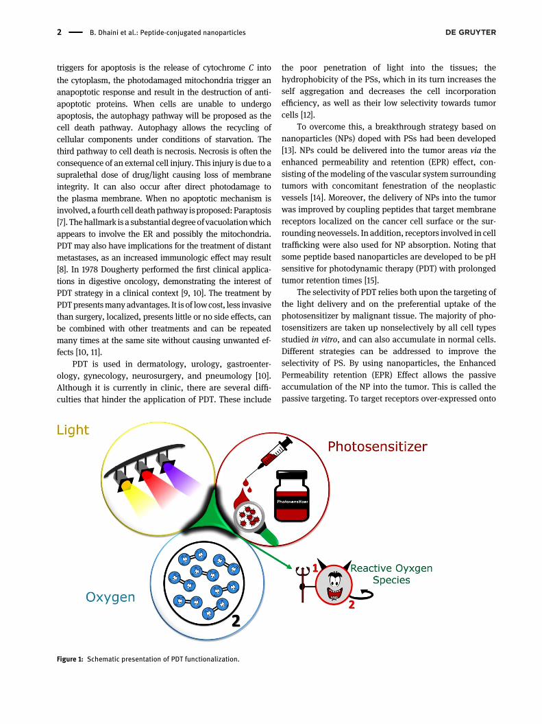
Cancer is the second leading cause of death in theworld aftercardiovascular disease [1]. Cancer treatment is mainly basedon surgery, radiotherapy, and/or chemotherapy, where theused therapeutic strategy depends on the cancer type andlocalization [2, 3]. The chemoradiotherapy treatment, whichcombines both radiotherapy and chemotherapy, exertsadverse effects on thenormal cells surrounding the tumors. Itis alsowell-known that the anticancerdrugs could lead to theappearance of cell resistance mechanisms, resulting in theprogression of tumors and rendering drug-treatment inef-fective. An alternative strategy is the photodynamic therapy(PDT) [4]. PDT has been discovered more than 100 years agoby Raab and Tappeiner [5]. The principle of PDT consists ofthe transfer of light photons to a molecule, a photosensitizer(PS), which, in the presence of molecular oxygen, producesmainly singlet oxygen (1O2) and other reactive oxygen spe-cies (ROS), through photoreaction of type II and I, respec-tively (Figure 1). The production of ROS/singlet oxygen at theloci of the mitochondrial, lysosomal, or endoplasmic retic-ulum (ER) can directly initiate cell death by apoptosis [6].Apoptosis is an irreversible pathway to cell death. One of the
*Corresponding author: Céline Frochot, Reactions and ChemicalEngineering Laboratory, Université de Lorraine, LRGP-CNRS, F-54000,Nancy, France, E-mail: [email protected]. https://orcid.org/0000-0002-7659-3864Batoul Dhaini, Reactions and Chemical Engineering Laboratory,Université de Lorraine, LRGP-CNRS, F-54000, Nancy, France; andLaboratory of Materials, Catalysis, Environment and AnalyticalMethods Laboratory (MCEMA), Faculty of Sciences, LebaneseUniversity, Hadath, LebanonBibigul Kenzhebayeva, Laboratory of Macromolecular PhysicalChemistry, Université de Lorraine, LCPM-CNRS, F-54000, Nancy,France; and The Department of Chemical and BiochemicalEngineering, University of Satbayev, Almaty, Kasakhstan. https://orcid.org/0000-0002-0531-4805Amina Ben-Mihoub and Samir Acherar, Laboratory of MacromolecularPhysical Chemistry, Université de Lorraine, LCPM-CNRS, F-54000,Nancy, FranceMickaël Gries, Noémie Thomas, Joël Daouk and Hervé Schohn,Department of Biology, Signals and Systems in Cancer andNeuroscience, Université de Lorraine, CRAN-CNRS, F-54000 Nancy,FranceFrancis Baros, Reactions and Chemical Engineering Laboratory,Université de Lorraine, LRGP-CNRS, F-54000, Nancy, FranceTayssir Hamieh, Laboratory of Materials, Catalysis, Environment andAnalytical Methods Laboratory (MCEMA), Faculty of Sciences,Lebanese University, Hadath, Lebanon; and Faculty of Science andEngineering, Maastricht University, P.O. Box 616, 6200 MDMaastricht, The Netherlands
Nanophotonics 2021; aop
Open Access. © 2021 Batoul Dhaini et al., published by De Gruyter. This work is licensed under the Creative Commons Attribution 4.0International License.
triggers for apoptosis is the release of cytochrome C into
the cytoplasm, the photodamaged mitochondria trigger ananapoptotic response and result in the destruction of anti-apoptotic proteins. When cells are unable to undergoapoptosis, the autophagy pathway will be proposed as thecell death pathway. Autophagy allows the recycling ofcellular components under conditions of starvation. Thethird pathway to cell death is necrosis. Necrosis is often theconsequence of an external cell injury. This injury is due to asupralethal dose of drug/light causing loss of membraneintegrity. It can also occur after direct photodamage tothe plasma membrane. When no apoptotic mechanism isinvolved, a fourth cell deathpathway isproposed:Paraptosis[7]. Thehallmark is a substantial degreeofvacuolationwhichappears to involve the ER and possibly the mitochondria.PDT may also have implications for the treatment of distantmetastases, as an increased immunologic effect may result[8]. In 1978 Dougherty performed the first clinical applica-tions in digestive oncology, demonstrating the interest ofPDT strategy in a clinical context [9, 10]. The treatment byPDTpresentsmany advantages. It is of lowcost, less invasivethan surgery, localized, presents little or no side effects, canbe combined with other treatments and can be repeatedmany times at the same site without causing unwanted ef-fects [10, 11].
PDT is used in dermatology, urology, gastroenter-ology, gynecology, neurosurgery, and pneumology [10].Although it is currently in clinic, there are several diffi-culties that hinder the application of PDT. These include
the poor penetration of light into the tissues; thehydrophobicity of the PSs, which in its turn increases theself aggregation and decreases the cell incorporationefficiency, as well as their low selectivity towards tumorcells [12].
To overcome this, a breakthrough strategy based onnanoparticles (NPs) doped with PSs had been developed[13]. NPs could be delivered into the tumor areas via theenhanced permeability and retention (EPR) effect, con-sisting of the modeling of the vascular system surroundingtumors with concomitant fenestration of the neoplasticvessels [14]. Moreover, the delivery of NPs into the tumorwas improved by coupling peptides that target membranereceptors localized on the cancer cell surface or the sur-rounding neovessels. In addition, receptors involved in celltrafficking were also used for NP absorption. Noting thatsome peptide based nanoparticles are developed to be pHsensitive for photodynamic therapy (PDT) with prolongedtumor retention times [15].
The selectivity of PDT relies both upon the targeting ofthe light delivery and on the preferential uptake of thephotosensitizer by malignant tissue. The majority of pho-tosensitizers are taken up nonselectively by all cell typesstudied in vitro, and can also accumulate in normal cells.Different strategies can be addressed to improve theselectivity of PS. By using nanoparticles, the EnhancedPermeability retention (EPR) Effect allows the passiveaccumulation of the NP into the tumor. This is called thepassive targeting. To target receptors over-expressed onto
Figure 1: Schematic presentation of PDT functionalization.
2 B. Dhaini et al.: Peptide-conjugated nanoparticles

membranes of tumor cells or neovessels, it is possible toattach a vector to the photosensitizer or the nanoparticle. Itis active targeting. Different vectors are described in theliterature such as folic acid that targets folic acid receptorover-expressed on many tumoral cell membranes [16], butcan suffer from low stability [17]. Stability of folic acidunder several parameters [18], antitumor monoclonal an-tibodies [19] that present drawbacks such as their large sizeand nonspecific uptake of the antibody molecules by theliver and the reticulo-endothelial system [18]. Moreover,anti-tumor monoclonal antibodies exhibit low tissuepenetration and poor cellular uptake when used in vivo[20]. Protein [21] can also be coupled with success to PS aswell as aptamer [22]. Small peptides represent excellenttargeting agents for receptors over-expressed in humancancers. We already describe in a review [23] all the ad-vantages of using peptides such as their small size, theypresent good tissue permeability, rapid access to the tumorsite, they can cross a disturbed blood–brain barrier (BBB),and they present low antigenicity. They are easy to syn-thesize in liquid or solid phases, easy to modify (pseudo-peptides), easy to link to a spacer via amide bond forexample, they can present high affinity for receptors andrapid clearance from the body. Two drawbacks can becited: They are potentially degraded by endo- and/or exo-peptidases and they do not cross a normal BBB.
Different receptors overexpressed in tumor or endothe-lial cells that hadbeen chosen for cell targeting aredescribedin this review:Neuropilin-1 (NRP-1),αvβ3 integrins, nucleolinmembrane receptor, epidermal growth factor (EGF), protein-glutamine-gamma-glutamyltransferase (TGM2), p32, as wellas transferrin and mitochondrial membrane. The use of aspecific cell absorbing-peptide is also described.
2 Nanoparticles loaded withphotosensitizer and coupled topeptide
2.1 Peptides targeting neuropilin-1 receptor
2.1.1 NPs@PS@ATWLPPR
The neuropilin-1 (NRP-1) is a transmembrane glycoproteinand a coreceptor of the vascular endothelial growth factorreceptor (VEGFR). It is involved in the axon guidance,angiogenesis, and immune responses [24]. NRP-1 is over-expressed in many types of cancers such as colon carci-noma, prostate, pancreatic carcinoma, lung carcinoma,melanoma, astrocytoma, and neuroblastoma [25].
NRP-1 has been described as a potential target againstglioblastoma [26]. A high NRP-1 expression in a glioblas-toma sample is correlated with increased malignancy. Incontrast, NRP-1 under-expression is associated to lowercancer stem cell migration and proliferation in vitro, and toreduce tumor growth, in vivo. The enhanced NRP-1expression has been observed in endothelial cells and iscorrelated to the development of tumor neovascularization[27, 28]. Vascular targeted photodynamic therapy (VTP)was applied using ATWLPPR heptapeptide as a part ofspecific NRP-1 recognizing sequence. Linked by a spacerarm (6-aminohexanoic acid, Ahx) to chlorin PS, the peptideaccumulated in the tumor tissue and potentiated thephotodynamic activity. However, the biodistributionstudies conducted on mice demonstrated a rapid uptake ofthe peptide by the liver and the spleen. Consequently, 2 hafter intravenous injection (IV), 85% of the total amount ofthe compound was degraded in the liver [29]. Since thepeptide arm, conjugated to the PS, was responsible for itsselectivity, the degradation of this peptide fragment wasrelated to the decrease in the PS accumulation in the tumortissue [30].
In order to reduce the hepato-splenic clearance andpeptide degradation, functionalized silica-based NPs, graf-ted with ATWLPPR-peptide by hydrophilic polymer, weredesigned for vessel targeting. The NPwas also designed as amagnetic resonance imaging (MRI) contrast agent, since it iscomposed of a silica shell coupled to polyethylene glycol(PEG), doped with gadolinium oxide (Gd2O3). The PS was achlorin derivative, 5-(4-carboxyphenyl)-10, 15, 20 triphenyl-chlorin (TPC), substitutedwith a succinimidyl ester. In brief,aminopropyltriethoxysilane (APTES) reacted with PEG orpropylenediaminetetra-acetic acid (PDTA) containingGd2O3
and was cycle-hydrolysis to obtain the NPs (referred to asNP-TPC) [31–33] (Figure 2(A)). The size of the NPs was esti-mated between 3.3 and 3.8 nm [31, 33]. The coupling of thepeptide (referred to as NP-TPC-ATWLPPR) raised the NPs’size to 4.6 ± 3.8 nm. NP-TPC-ATWLPPR was tested for theiraffinity to NRP-1 using biotinylated VEGF in competitivebinding assays. The IC50 values were estimated to be 27 and56.6 µM for NP-TPC-ATWLPPR and NP-TPC, respectively.The photocytotoxic effect was tested on human breastMDA-MB-231 cells that overexpress a high level of NRP-1.The calculated LD50 (Light dose) for P-TPC-ATWLPPR wasestimated at 2.8–5.0 J cm−2 when cellswere treatedwith 1 µMof NP-TPC-ATWLPPR (power 0.7 W, irradiance 4.54 J cm−2).The LD50 value was close to that obtained by a similartreatment with 1 µM NP-TPC [31, 32]. In vivo, the maximalMRI enhancement was found at 2–7 min, after the IV injec-tion ofNP-TPC-ATWLPPR innudemice. The biodistribution,analyzed 75 min after the IV injection, suggested both renal
B. Dhaini et al.: Peptide-conjugated nanoparticles 3

and hepatic clearance for NP-TPC and NP-TPC-ATWLPPR.However, the renal elimination was higher for NP-TPC. Theexposure of the tumor U87 grafted cells to NP-TPC-ATWLPPR, revealed that the NPs targeted the peripheralvessels surrounding the tumors in accordance with the highexpression of NRP-1 observed in endothelial cells [31, 32](Figure 2(B)).
2.1.2 NPs@PS@KDKPPR
Based on the sequence homology of the natural ligand ofNRP-1, VEGF-A165, a screening of several peptides wasperformed. Among the selected sequences, KDKPPRpeptide showed a higher affinity to NRP-1 than ATWLPPR;the inhibitor dissociation constant (Ki) was estimated to be9.0 × 10−8 mol L−1 for the former as compared to1.4 × 10−4 mol L−1 for the latter [34]. Themeasurement of theaffinity to NRP-1 for the K(P1)DKPPR conjugate (i.e.monocarboxylic tetraphenyl porphyrin, P1COOH, linked tothe first ε-NH2 lysine of KDKPPR) was 6 versus 171 μM forthe P1-ATWLPPR complexes [35, 36].
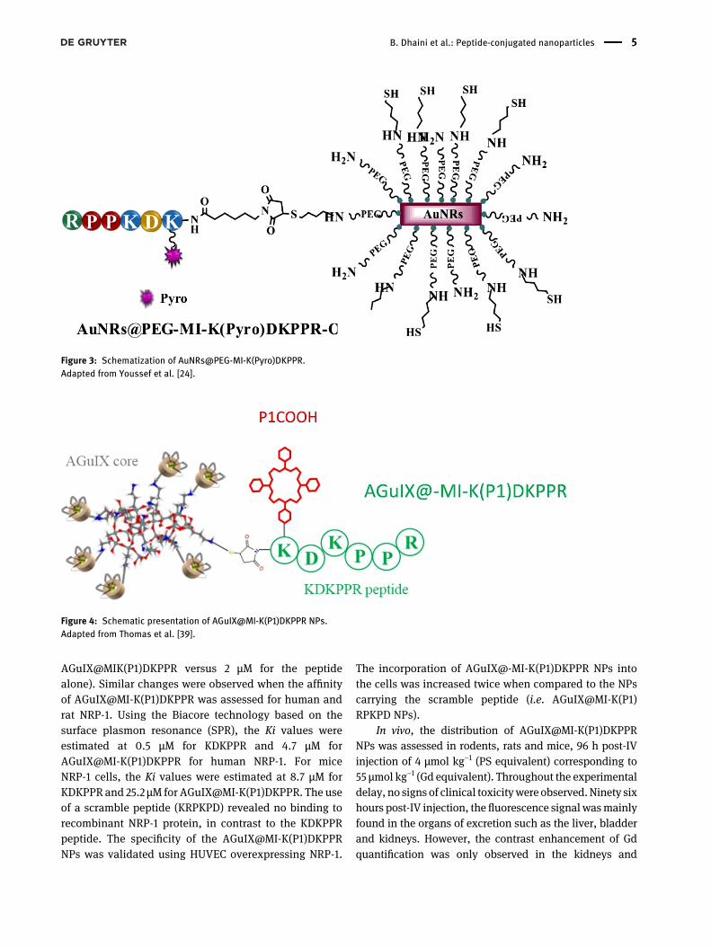
TheK(Pyro)DKPPRconjugate, usingpyropheophorbide-a (Pyro) as PS, was coupled to PEGylated gold nanorods(AuNRs@PEG) through a thiol-maleimide (MI) click reactionto attain a combined hyperthermia and PDT effect (Figure 3)[37]. TheAuNRswere functionalizedwith PEG to prevent anycytotoxicity. The AuNRs@PEG-MI-K(Pyro)DKPPR, illus-trated in Figure 3, possessed a length of about 44 nm and awidthof about 8nm. Thephotophysical properties of the freePyro were preserved in AuNRs@PEG-MI-K(Pyro)DKPPR,thus showing an unmodified visible absorption profile, agood fluorescence intensity (ϕF = 0.30 in EtOH versus 0.38for Pyro) and a preserved 1O2 production (ϕ0 = 0.40 in EtOHversus 0.51 for Pyro).
The affinity of the AuNRs@PEG-MI-K(Pyro)DKPPRand the K(Pyro)DKPPR free peptide to recombinant NRP-1
was evaluated by competitive binding assay giving IC50values of 1.5 and 2.0 µM, respectively. The efficacy ofAuNRs@PEG-MI-K(Pyro)DKPPR was tested in vitro onhuman glioblastoma U87 MG cells. No cytotoxicity wasobtained with concentrations up to 30 µM (concentrationrelative to PS), in contrast to the cells treated with the PSalone. A good photodynamic efficiency was found whencells were treated with 30 µM of AuNRs@PEG-MI-K(Pyro)DKPPR for 24 h and then exposed to light irradiation at652 nm (Fluence 10 J cm−2, fluence rate 4.54 mW cm−2) witha 67% decrease in cell viability. This result supported theeffect of both photodynamic and photothermal (PTT)therapies due to the presence of Gold.
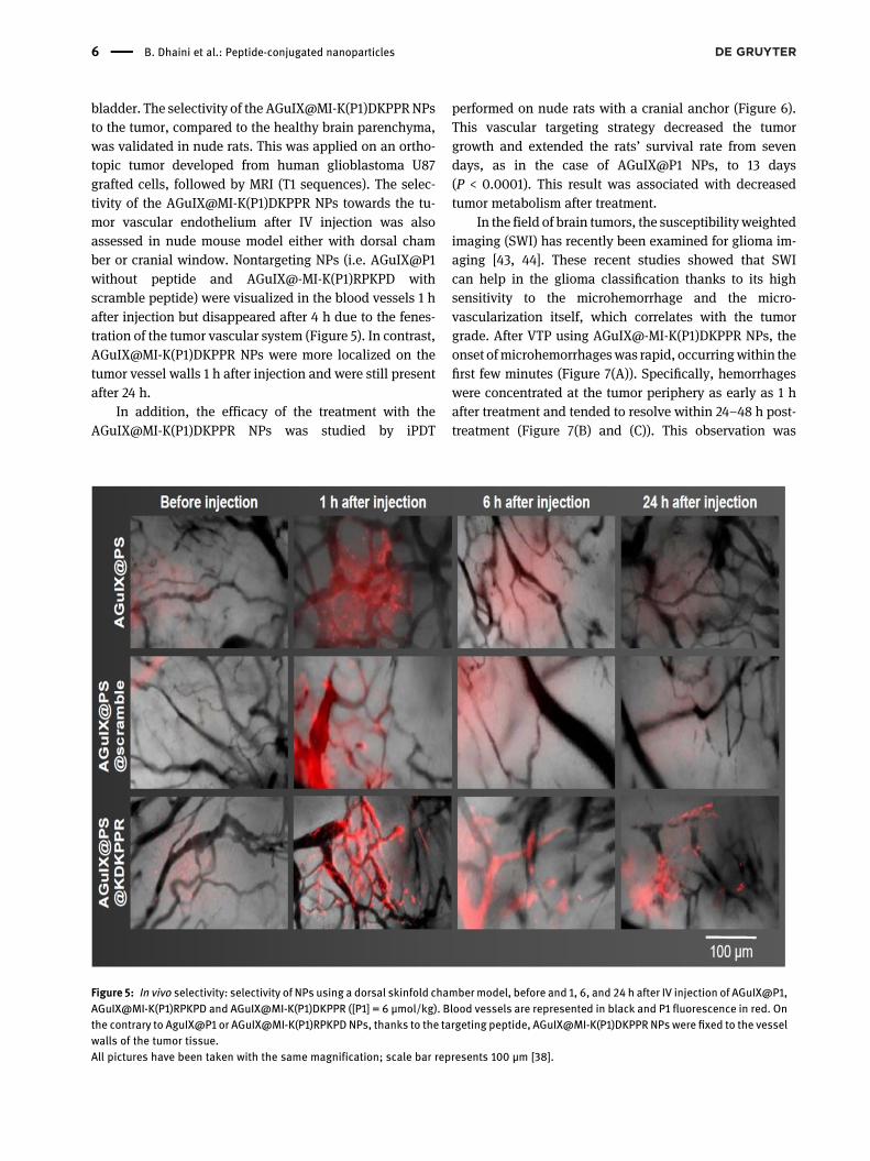
The same methodology was also used to graft K(P1)DKPPR conjugate on multifunctional NP platform, namelyAGuIX@ [38–40]. The designed NPs were first proposed asnontoxic resonance magnetic agents for their imagingproperties [41]. The so-called AGuIX@MI-K(P1)DKPPR NPs,illustrated in Figure 4, were tested for vascular-targetedinterstitial photodynamic therapy (iPDT), using humanumbilical vein endothelial cells (HUVEC) as an in vitromodel and human U87 grafted tumors in rodents [38, 39].
AGuIX@MI-K(P1)DKPPR NPs had a hydrodynamicdiameter of approximately 10 nm, making them particu-larly suitable for rapid renal elimination [42]. The fluorescence (ϕF = 0.7 for P1COOH and ϕF = 0.1 forAGuIX@MI-K(P1)DKPPR in D2O) and singlet oxygen(ϕO = 0.24 for P1COOH and ϕ0 = 0.28 for AGuIX@MI-K(P1)DKPPR in D2O) quantum yields of P1COOH PS were notaltered after the peptide addition, demonstrating that thePS could be photoactivated to produce a photocytotoxiceffect in vitro and in vivo.
HUVEC cell exposure to AGuIX@MI-K(P1)DKPPR NPsshowed no dark cytotoxicity at PS concentrations upto 10 μM. However, the affinity to NRP-1 was alteredas the peptide was coupled to the NPs (19 µM for
Figure 2: (A) Schematic presentation of NP-TPC-ATWLPPR, (B)maximalMRI signal intensity after injecting 84.2 μmol of Gd for a bodyweight of250 g for cerebral biodistribution and brain tumor tissue selectivity of NP-TPC-ATWLPPR, and (C) overview clinical picture of the device appliedon the mice [32] with permission from Elsevier and Copyright Clearance Center.
4 B. Dhaini et al.: Peptide-conjugated nanoparticles
AGuIX@MIK(P1)DKPPR versus 2 µM for the peptidealone). Similar changes were observed when the affinityof AGuIX@MI-K(P1)DKPPR was assessed for human andrat NRP-1. Using the Biacore technology based on thesurface plasmon resonance (SPR), the Ki values wereestimated at 0.5 μM for KDKPPR and 4.7 μM forAGuIX@MI-K(P1)DKPPR for human NRP-1. For miceNRP-1 cells, the Ki values were estimated at 8.7 μM forKDKPPR and 25.2 μM for AGuIX@MI-K(P1)DKPPR. The useof a scramble peptide (KRPKPD) revealed no binding torecombinant NRP-1 protein, in contrast to the KDKPPRpeptide. The specificity of the AGuIX@MI-K(P1)DKPPRNPs was validated using HUVEC overexpressing NRP-1.
The incorporation of AGuIX@-MI-K(P1)DKPPR NPs intothe cells was increased twice when compared to the NPscarrying the scramble peptide (i.e. AGuIX@MI-K(P1)RPKPD NPs).
In vivo, the distribution of AGuIX@MI-K(P1)DKPPRNPs was assessed in rodents, rats and mice, 96 h post-IVinjection of 4 µmol kg−1 (PS equivalent) corresponding to55 µmol kg−1 (Gd equivalent). Throughout the experimentaldelay, no signs of clinical toxicitywere observed. Ninety sixhours post-IV injection, the fluorescence signal wasmainlyfound in the organs of excretion such as the liver, bladderand kidneys. However, the contrast enhancement of Gdquantification was only observed in the kidneys and
Figure 3: Schematization of AuNRs@PEG-MI-K(Pyro)DKPPR.Adapted from Youssef et al. [24].
Figure 4: Schematic presentation of AGuIX@MI-K(P1)DKPPR NPs.Adapted from Thomas et al. [39].
B. Dhaini et al.: Peptide-conjugated nanoparticles 5
bladder. The selectivity of the AGuIX@MI-K(P1)DKPPRNPsto the tumor, compared to the healthy brain parenchyma,was validated in nude rats. This was applied on an ortho-topic tumor developed from human glioblastoma U87grafted cells, followed by MRI (T1 sequences). The selec-tivity of the AGuIX@MI-K(P1)DKPPR NPs towards the tu-mor vascular endothelium after IV injection was alsoassessed in nude mouse model either with dorsal chamber or cranial window. Nontargeting NPs (i.e. AGuIX@P1without peptide and AGuIX@-MI-K(P1)RPKPD withscramble peptide) were visualized in the blood vessels 1 hafter injection but disappeared after 4 h due to the fenes-tration of the tumor vascular system (Figure 5). In contrast,AGuIX@MI-K(P1)DKPPR NPs were more localized on thetumor vessel walls 1 h after injection and were still presentafter 24 h.
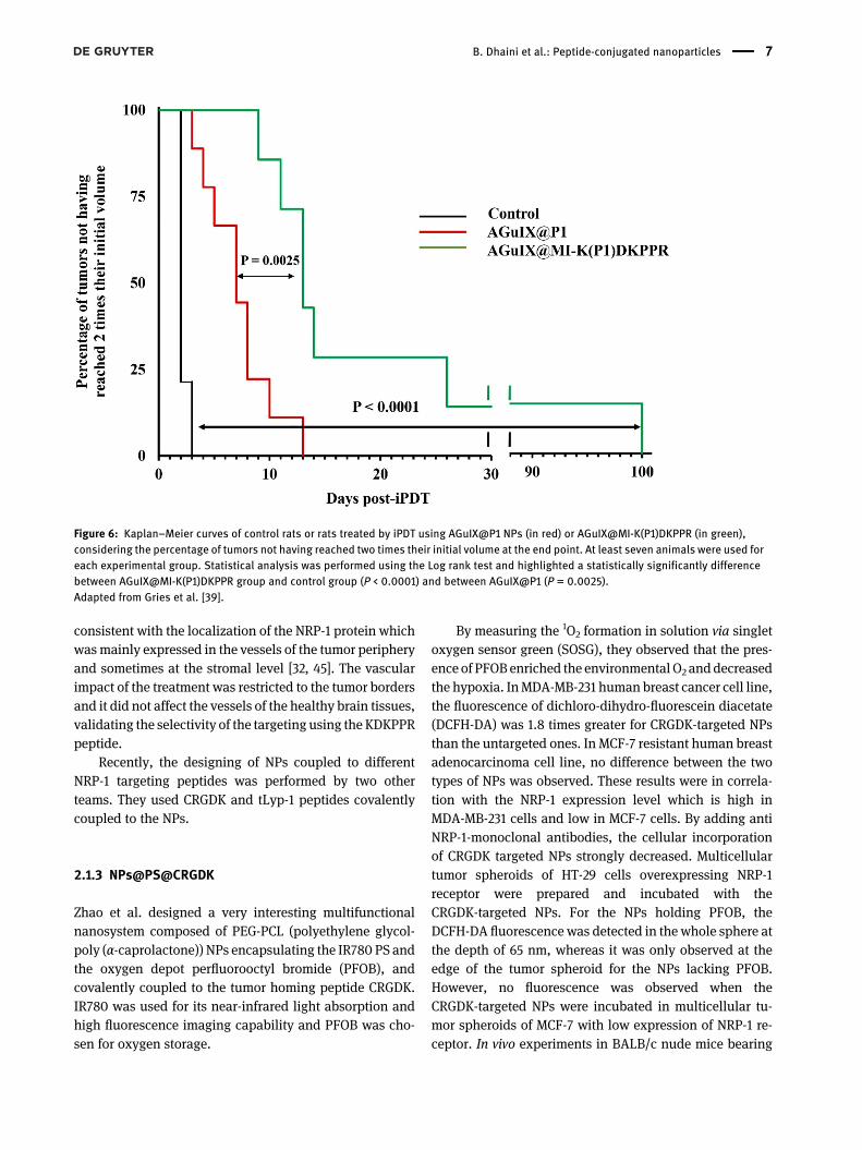
In addition, the efficacy of the treatment with theAGuIX@MI-K(P1)DKPPR NPs was studied by iPDT
performed on nude rats with a cranial anchor (Figure 6).This vascular targeting strategy decreased the tumorgrowth and extended the rats’ survival rate from sevendays, as in the case of AGuIX@P1 NPs, to 13 days(P < 0.0001). This result was associated with decreasedtumor metabolism after treatment.
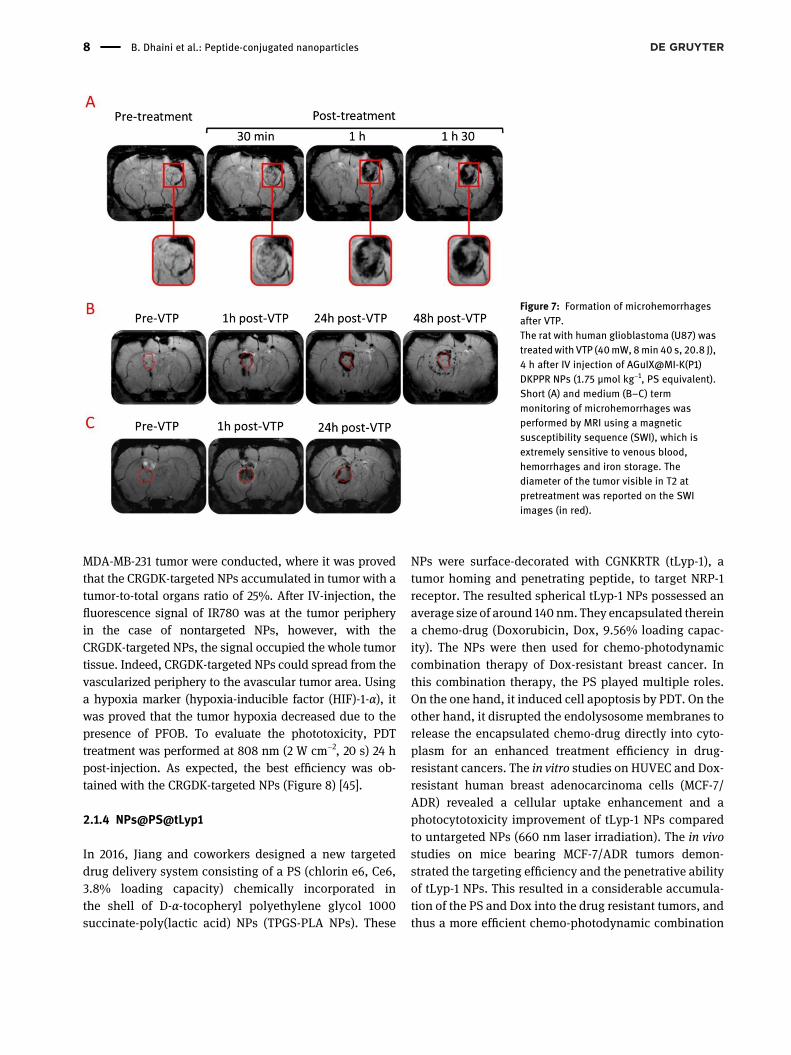
In the field of brain tumors, the susceptibility weightedimaging (SWI) has recently been examined for glioma im-aging [43, 44]. These recent studies showed that SWIcan help in the glioma classification thanks to its highsensitivity to the microhemorrhage and the micro-vascularization itself, which correlates with the tumorgrade. After VTP using AGuIX@-MI-K(P1)DKPPR NPs, theonset ofmicrohemorrhageswas rapid, occurringwithin thefirst few minutes (Figure 7(A)). Specifically, hemorrhageswere concentrated at the tumor periphery as early as 1 hafter treatment and tended to resolve within 24–48 h post-treatment (Figure 7(B) and (C)). This observation was
Figure 5: In vivo selectivity: selectivity of NPs using a dorsal skinfold chambermodel, before and 1, 6, and 24 h after IV injection of AGuIX@P1,AGuIX@MI-K(P1)RPKPD and AGuIX@MI-K(P1)DKPPR ([P1] = 6 μmol/kg). Blood vessels are represented in black and P1 fluorescence in red. Onthe contrary to AguIX@P1 or AGuIX@MI-K(P1)RPKPD NPs, thanks to the targeting peptide, AGuIX@MI-K(P1)DKPPR NPs were fixed to the vesselwalls of the tumor tissue.All pictures have been taken with the same magnification; scale bar represents 100 μm [38].
6 B. Dhaini et al.: Peptide-conjugated nanoparticles
consistent with the localization of the NRP-1 protein whichwasmainly expressed in the vessels of the tumor peripheryand sometimes at the stromal level [32, 45]. The vascularimpact of the treatment was restricted to the tumor bordersand it did not affect the vessels of the healthy brain tissues,validating the selectivity of the targeting using the KDKPPRpeptide.
Recently, the designing of NPs coupled to differentNRP-1 targeting peptides was performed by two otherteams. They used CRGDK and tLyp-1 peptides covalentlycoupled to the NPs.
2.1.3 NPs@PS@CRGDK
Zhao et al. designed a very interesting multifunctionalnanosystem composed of PEG-PCL (polyethylene glycol-poly (α-caprolactone)) NPs encapsulating the IR780 PS andthe oxygen depot perfluorooctyl bromide (PFOB), andcovalently coupled to the tumor homing peptide CRGDK.IR780 was used for its near-infrared light absorption andhigh fluorescence imaging capability and PFOB was cho-sen for oxygen storage.
By measuring the 1O2 formation in solution via singletoxygen sensor green (SOSG), they observed that the pres-ence of PFOB enriched the environmental O2 anddecreasedthe hypoxia. InMDA-MB-231 human breast cancer cell line,the fluorescence of dichloro-dihydro-fluorescein diacetate(DCFH-DA) was 1.8 times greater for CRGDK-targeted NPsthan the untargeted ones. In MCF-7 resistant human breastadenocarcinoma cell line, no difference between the twotypes of NPs was observed. These results were in correla-tion with the NRP-1 expression level which is high inMDA-MB-231 cells and low in MCF-7 cells. By adding antiNRP-1-monoclonal antibodies, the cellular incorporationof CRGDK targeted NPs strongly decreased. Multicellulartumor spheroids of HT-29 cells overexpressing NRP-1receptor were prepared and incubated with theCRGDK-targeted NPs. For the NPs holding PFOB, theDCFH-DA fluorescence was detected in the whole sphere atthe depth of 65 nm, whereas it was only observed at theedge of the tumor spheroid for the NPs lacking PFOB.However, no fluorescence was observed when theCRGDK-targeted NPs were incubated in multicellular tu-mor spheroids of MCF-7 with low expression of NRP-1 re-ceptor. In vivo experiments in BALB/c nude mice bearing
Figure 6: Kaplan–Meier curves of control rats or rats treated by iPDT using AGuIX@P1 NPs (in red) or AGuIX@MI-K(P1)DKPPR (in green),considering the percentage of tumors not having reached two times their initial volume at the end point. At least seven animals were used foreach experimental group. Statistical analysis was performed using the Log rank test and highlighted a statistically significantly differencebetween AGuIX@MI-K(P1)DKPPR group and control group (P < 0.0001) and between AGuIX@P1 (P = 0.0025).Adapted from Gries et al. [39].
B. Dhaini et al.: Peptide-conjugated nanoparticles 7
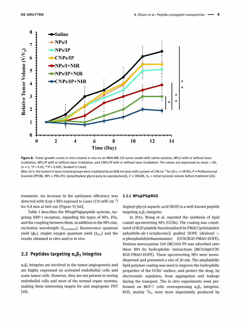
MDA-MB-231 tumor were conducted, where it was provedthat the CRGDK-targeted NPs accumulated in tumor with atumor-to-total organs ratio of 25%. After IV-injection, thefluorescence signal of IR780 was at the tumor peripheryin the case of nontargeted NPs, however, with theCRGDK-targeted NPs, the signal occupied the whole tumortissue. Indeed, CRGDK-targeted NPs could spread from thevascularized periphery to the avascular tumor area. Usinga hypoxia marker (hypoxia-inducible factor (HIF)-1-α), itwas proved that the tumor hypoxia decreased due to thepresence of PFOB. To evaluate the phototoxicity, PDTtreatment was performed at 808 nm (2 W cm−2, 20 s) 24 hpost-injection. As expected, the best efficiency was ob-tained with the CRGDK-targeted NPs (Figure 8) [45].
2.1.4 NPs@PS@tLyp1
In 2016, Jiang and coworkers designed a new targeteddrug delivery system consisting of a PS (chlorin e6, Ce6,3.8% loading capacity) chemically incorporated inthe shell of D-α-tocopheryl polyethylene glycol 1000succinate-poly(lactic acid) NPs (TPGS-PLA NPs). These
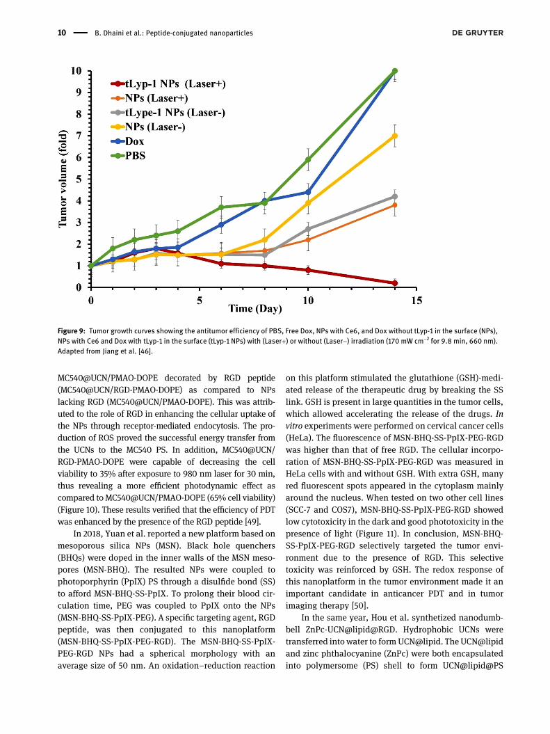
NPs were surface-decorated with CGNKRTR (tLyp-1), atumor homing and penetrating peptide, to target NRP-1receptor. The resulted spherical tLyp-1 NPs possessed anaverage size of around 140 nm. They encapsulated thereina chemo-drug (Doxorubicin, Dox, 9.56% loading capac-ity). The NPs were then used for chemo-photodynamiccombination therapy of Dox-resistant breast cancer. Inthis combination therapy, the PS played multiple roles.On the one hand, it induced cell apoptosis by PDT. On theother hand, it disrupted the endolysosome membranes torelease the encapsulated chemo-drug directly into cyto-plasm for an enhanced treatment efficiency in drug-resistant cancers. The in vitro studies on HUVEC and Dox-resistant human breast adenocarcinoma cells (MCF-7/ADR) revealed a cellular uptake enhancement and aphotocytotoxicity improvement of tLyp-1 NPs comparedto untargeted NPs (660 nm laser irradiation). The in vivostudies on mice bearing MCF-7/ADR tumors demon-strated the targeting efficiency and the penetrative abilityof tLyp-1 NPs. This resulted in a considerable accumula-tion of the PS and Dox into the drug resistant tumors, andthus a more efficient chemo-photodynamic combination
Figure 7: Formation of microhemorrhagesafter VTP.The rat with human glioblastoma (U87) wastreatedwith VTP (40mW, 8min 40 s, 20.8 J),4 h after IV injection of AGuIX@MI-K(P1)DKPPR NPs (1.75 μmol kg−1, PS equivalent).Short (A) and medium (B–C) termmonitoring of microhemorrhages wasperformed by MRI using a magneticsusceptibility sequence (SWI), which isextremely sensitive to venous blood,hemorrhages and iron storage. Thediameter of the tumor visible in T2 atpretreatment was reported on the SWIimages (in red).
8 B. Dhaini et al.: Peptide-conjugated nanoparticles
treatment. An increase in the antitumor efficiency wasdetected with tLyp-1 NPs exposed to Laser (170 mW cm−2)for 9.8 min at 660 nm (Figure 9) [46].
Table 1 describes the NPs@PS@peptide systems, tar-geting NRP-1 receptors, regarding the types of NPs, PSs,and the coupling between them, in addition to theNPs size,excitation wavelength (λexcitation), fluorescence quantumyield (ϕF), singlet oxygen quantum yield (ΔO.S) and theresults obtained in vitro and/or in vivo.
2.2 Peptides targeting αvβ3 integrins
αvβ3 integrins are involved in the tumor angiogenesis andare highly expressed on activated endothelial cells andsome tumor cells. However, they are not present in restingendothelial cells and most of the normal organ systems,making them interesting targets for anti angiogenic PDT[48].
2.2.1 NPs@PS@RGD
Arginyl-glycyl-aspartic acid (RGD) is a well-known peptidetargeting αvβ3 integrins.
In 2014, Wang et al. reported the synthesis of lipidcoated upconverting NPs (UCNs). The coating was consti-tuted of RGD peptide functionalized by PMAO (poly(maleicanhydride-alt-1-octadecene)) grafted DOPE (dioleoyl L-α-phosphatidylethanolamine) (UCN/RGD-PMAO-DOPE).Dextran merocyanine 540 (MC540) PS was adsorbed ontothese NPs by hydrophobic interactions (MC540@UCN/RGD-PMAO-DOPE). These upconverting NPs were mono-dispersed and presented a size of 20 nm. The amphiphiliclipid polymer coating was used to improve the hydrophilicproperties of the UCNs’ surface, and protect the drug, byelectrostatic repulsion, from aggregation and leakageduring the transport. The in vitro experiments were per-formed on MCF-7 cells overexpressing αvβ3 integrins.ROS, mainly 1O2, were more importantly produced by
Figure 8: Tumor growth curves in mice treated in vivo on an MDA-MB-231 tumor model with saline solution, NPs/I with or without laserirradiation, NPs/IP with or without laser irradiation, and CNPs/IP with or without laser irradiation. The values are expressed as mean ± SD,(n = 5, *P < 0.01, **P < 0.005, Student’s t-test).After 24 h, the tumors in laser treated groupswere irradiated by an 808 nm laser with a power of 2W cm−2 for 20 s. I= IR780, P= Perfluorooctylbromide (PFOB), NPs = PEG-PCL (polyethylene glycol-poly (α-caprolactone)), C = CRGDK, V0 = Initial tumoral volume before treatment [45].
B. Dhaini et al.: Peptide-conjugated nanoparticles 9
MC540@UCN/PMAO-DOPE decorated by RGD peptide(MC540@UCN/RGD-PMAO-DOPE) as compared to NPslacking RGD (MC540@UCN/PMAO-DOPE). This was attrib-uted to the role of RGD in enhancing the cellular uptake ofthe NPs through receptor-mediated endocytosis. The pro-duction of ROS proved the successful energy transfer fromthe UCNs to the MC540 PS. In addition, MC540@UCN/RGD-PMAO-DOPE were capable of decreasing the cellviability to 35% after exposure to 980 nm laser for 30 min,thus revealing a more efficient photodynamic effect ascompared to MC540@UCN/PMAO-DOPE (65% cell viability)(Figure 10). These results verified that the efficiency of PDTwas enhanced by the presence of the RGD peptide [49].
In 2018, Yuan et al. reported a new platform based onmesoporous silica NPs (MSN). Black hole quenchers(BHQs) were doped in the inner walls of the MSN meso-pores (MSN-BHQ). The resulted NPs were coupled tophotoporphyrin (PpIX) PS through a disulfide bond (SS)to afford MSN-BHQ-SS-PpIX. To prolong their blood cir-culation time, PEG was coupled to PpIX onto the NPs(MSN-BHQ-SS-PpIX-PEG). A specific targeting agent, RGDpeptide, was then conjugated to this nanoplatform(MSN-BHQ-SS-PpIX-PEG-RGD). The MSN-BHQ-SS-PpIX-PEG-RGD NPs had a spherical morphology with anaverage size of 50 nm. An oxidation–reduction reaction
on this platform stimulated the glutathione (GSH)-medi-ated release of the therapeutic drug by breaking the SSlink. GSH is present in large quantities in the tumor cells,which allowed accelerating the release of the drugs. Invitro experiments were performed on cervical cancer cells(HeLa). The fluorescence of MSN-BHQ-SS-PpIX-PEG-RGDwas higher than that of free RGD. The cellular incorpo-ration of MSN-BHQ-SS-PpIX-PEG-RGD was measured inHeLa cells with and without GSH. With extra GSH, manyred fluorescent spots appeared in the cytoplasm mainlyaround the nucleus. When tested on two other cell lines(SCC-7 and COS7), MSN-BHQ-SS-PpIX-PEG-RGD showedlow cytotoxicity in the dark and good phototoxicity in thepresence of light (Figure 11). In conclusion, MSN-BHQ-SS-PpIX-PEG-RGD selectively targeted the tumor envi-ronment due to the presence of RGD. This selectivetoxicity was reinforced by GSH. The redox response ofthis nanoplatform in the tumor environment made it animportant candidate in anticancer PDT and in tumorimaging therapy [50].
In the same year, Hou et al. synthetized nanodumb-bell ZnPc-UCN@lipid@RGD. Hydrophobic UCNs weretransferred into water to form UCN@lipid. The UCN@lipidand zinc phthalocyanine (ZnPc) were both encapsulatedinto polymersome (PS) shell to form UCN@lipid@PS
Figure 9: Tumor growth curves showing the antitumor efficiency of PBS, Free Dox, NPs with Ce6, and Dox without tLyp-1 in the surface (NPs),NPs with Ce6 and Dox with tLyp-1 in the surface (tLyp-1 NPs) with (Laser+) or without (Laser−) irradiation (170 mW cm−2 for 9.8 min, 660 nm).Adapted from Jiang et al. [46].
10 B. Dhaini et al.: Peptide-conjugated nanoparticles
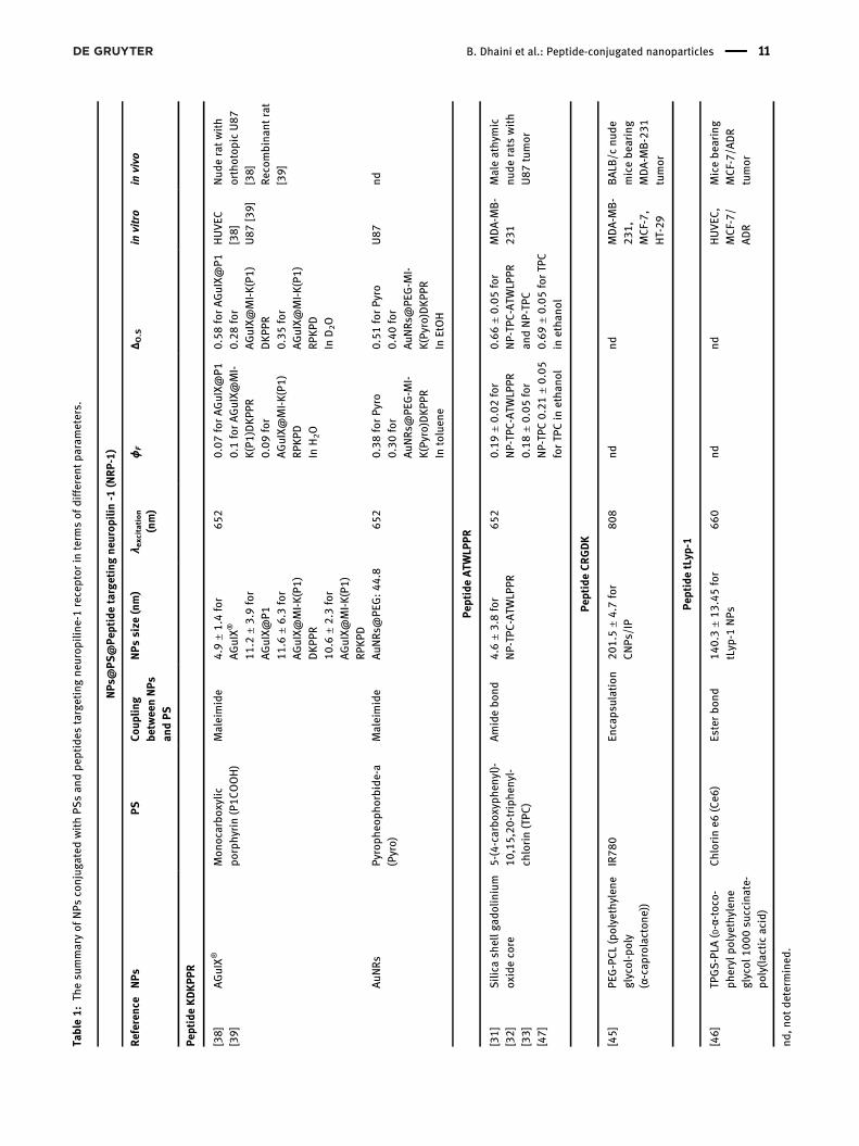
Table:Th
esu
mmaryof
NPs
conjug
ated
withPS
san
dpe
ptides
targetingne
urop
iline
-receptor
interm
sof
differen
tpa
rameters.
NPs
@PS
@Pe
ptidetargetingne
urop
ilin-
(NRP-)
Referen
ceNPs
PSCo
uplin
gbe
twee
nNPs
andPS
NPs
size
(nm)
λ excitation
(nm)
ϕF
Δ O.S
invitro
invivo
PeptideKDKPP
R
[]
[]
AGuIX®
Mon
ocarbo
xylic
porphy
rin(PCOOH)
Maleimide
.
±.
for
AGuIX®
.
±.
for
AGuIX@
P.
±.
for
AGuIX@
MI-K(P)
DKPP
R.
±.
for
AGuIX@
MI-K(P)
RPKPD
.forAGuIX@
P.
forAGuIX@
MI-
K(P)DKPP
R.for
AGuIX@
MI-K(P
)RP
KPD
InHO
.forAGuIX@
P.for
AGuIX@
MI-K(P)
DKPP
R.for
AGuIX@
MI-K(P)
RPKPD
InDO
HUVEC
[]
U[]
Nud
eratwith
orthotop
icU
[]
Recombina
ntrat
[]
AuN
RsPy
roph
eoph
orbide
-a(Pyro)
Maleimide
AuN
Rs@PE
G:
.
.forPy
ro.for
AuN
Rs@PE
G-M
I-K(Pyro)DKPP
RIn
toluen
e
.forPy
ro.for
AuN
Rs@PE
G-M
I-K(Pyro)DKPP
RIn
EtOH
U
nd
PeptideATW
LPPR
[]
[]
[]
[]
Silica
shellg
adolinium
oxidecore
-(-carbo
xyph
enyl)-
,,-triph
enyl-
chlorin(TPC
)
Amidebo
nd.
±.
for
NP-TP
C-ATW
LPPR
.±.for
NP-TP
C-ATW
LPPR
.±.for
NP-TP
C.±.
forTP
Cin
etha
nol
.±.for
NP-TP
C-ATW
LPPR
andNP-TP
C.±.forTP
Cin
etha
nol
MDA-M
B-
Maleathy
mic
nude
rats
with
Utumor
PeptideCR
GDK
[]
PEG-PCL(polyethylen
eglycol-poly
(α-cap
rolacton
e))
IR
Encaps
ulation
.
±.
for
CNPs
/IP
ndnd
MDA-M
B-
,
MCF-,
HT-
BALB
/cnu
demicebe
aring
MDA-M
B-
tumor
PeptidetLyp
-
[]
TPGS-PLA
( D-α-toco-
pherylpo
lyethy
lene
glycol
su
ccinate-
poly(lacticacid)
Chlorin
e(Ce
)Esterbo
nd.
±.for
tLyp
-NPs
ndnd
HUVEC
,MCF-/
ADR
Micebe
aring
MCF-/A
DR
tumor
nd,n
otde
term
ined
.
B. Dhaini et al.: Peptide-conjugated nanoparticles 11

nanodumbbell. The exterior of the polymersomepossessed many carboxyl functional groups that werecoupled to RGD peptide. The UCN’s core converted theNIR rays into visible ones, thus overcoming the problemof the limited light penetration into tissues. The ZnPc-UCN@lipid@PS had a high drug loading efficiency of18.03%. The transmission electron microscopy (TEM)showed that the ZnPc-UCN@lipid@PS were of sphericalform with a size of 150 nm. Whereas, the dynamic lightscattering (DLS) gave an average diameter of 195 nm.After coupling the UCNs to ZnPc, the fluorescence intensityof the UCNs decreased, indicating an energy transfer be-tween the UCNs and ZnPc. Following the excitation at980 nm, ZnPc-UCN@lipid@PS and ZnPc-UCN@lipid@PS–RGD showed the greatest production of 1O2. These tests werecarried out using 9,10-anthracenediyl-bis(methylene)dima-lonic acid (ABDA) probe. The study of the in vitro cytotox-icity in HeLa cells exposed to different concentrations ofNPs, showed a greater biocompatibility in the case ofUCN@lipid@PS-RGD (97% of cell viability for 500 μg mL−1
of NPs) as compared to UCN@lipid@PS (90%) and UCN@-lipid (65%). This result was credited to the presence of RGDthat induced the specific incorporation of the NPs in thecells, and hence decreased the unwanted cytotoxicity(Figure 12(A)). After incubation with Hela cells andexcitation with a NIR source (980 nm, 1.5 W cm−2),ZnPc-UCN@lipid@PS-RGD showed the lowest tumorcell viability as compared to the untreated controlcells and those exposed to the other ZnPc loaded-PSNPs (ZnPc-UCN@lipid@PS, UCN@lipid@PS nano-dumbbells) (Figure 12(B) and (C)) [51]. In the sameconditions, ZnPc loaded UCN@lipid@PS with andwithour RGD showed the best and higher production ofsinglet Oxygen in comparaison with other couplesdetailed in Figure 12(C).
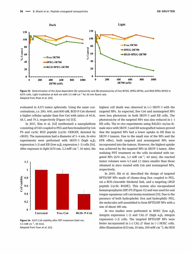
In 2015, Zhao et al. successfully synthesized a novelmulti arm polymeric nanosystem for PDT (RGD-8PEG-IR700). The RGD and the IR700 units were coupled to a PEGarm (8 polyethylene glycol). The hydrodynamic diameterof the nanosystem was 6.6 nm. The in vitro results onspheroid tumor model A375 and SKOV3 cells that expressαvβ3 integrins, showed a stronger fluorescence of theRGD-8PEG-IR700 NPs as compared to 8PEG-IR700. Incontrast to IR700 and 8PEG-IR700, RGD-8PEG-IR700induced a significant phototoxicity on A375 cells, underexcitation at 504 nm with an IC50 value of 57.8 nM. Nocytotoxic effect was observed in the dark even with aconcentration of 1 μM (IR700 equivalent) (Figure 13) [52].
In 2015, Yuan et al. used the seventh generation poly(amidoamine) (PAMAM-G7, P) dendrimer of 8 nm. ThePAMAM was coupled with Ce6 PS in addition to PEGor RGD, to obtain the PEG-P-Ce6 NPs and RGD-P-Ce6,respectively. The ΦΔ was 2.5 times higher for RGD-P-Ce6than free Ce6 inwater. In vitro experiments were performedin A375 cells (nonpigmented melanoma cell line express-ing αvβ3 integrins (+)) and NIH3T3 cells (mouse fibroblastthat do not express αvβ3 integrins (−)). Cellular uptake in(+) A375 cells was 4.7-fold superior for targeted RGD-P-Ce6than nontargeted PEG-P-Ce6 NPs, whereas the incorpora-tion into (−) NIH3T3 cells was the same for both NPs. Thephotocytotoxicity was evaluated with various concentra-tions in both types of cells by illumination at 660 nm(3.5 mW cm−2, 30 min). No cell killing was witnessed whenA375 cells were treated with Ce6 due to its poor incorpo-ration. RGD-P-Ce6 presented an enhanced phototoxicity inA375 cells compared to nontargeted NPs. As expected, inNIH3T3 cells, no difference in the phototoxicity betweenRGD-targeted NPs and PEG-P-Ce6 NPs was detected.The penetration of RGD-targeted NPs and free Ce6 was
Figure 10: PDT treatment efficiency on MCF-7 cells. Untreated cellsas control group and cells treated with UCN/PMAO-DOPE NPs,MC540 loaded UCN/PMAO-DOPE NPs or MC540 loaded UCN/RGD-PMAO-DOPE NPs after NIR laser irradiation (980 nm, 30 min).Adapted from Wang et al. [49].
12 B. Dhaini et al.: Peptide-conjugated nanoparticles

Figure 11: In vitro cell viability of (a) COS7,(b) SCC-7, and (c) HeLa cells incubated withdifferent concentrations of MSN-BHQ-SS-PpIX-PEG-RGD without light and under30min. Data is shown asmean ± SD (n = 4).Adapted from Yuan et al. [38].
Figure 12: (A) Cytotoxicity on Hela cells of UCN@lipid, UCN@lipid@PS, and UCN@lipid@PS-RGD. (B) Viability of Hela cells treated with 1 PBS(Control group), 2 UCN@lipid@PS nanodumbbells plus NIR laser, 3 ZnPc loaded-PS NPs plus NIR laser, 4 (ZnPc + UCN@lipid)@PS NPs, 5(ZnPc + UCN@lipid)@PS NPs plus NIR laser, 6 (ZnPc + UCN@lipid)@PS-RGD NPs plus NIR laser (980 nm, 1.5 W cm−2). The concentration is500 μg mL−1. (C) Comparison of 1O2 production between control groups and experiment groups.Adapted from Hou et al. [39].
B. Dhaini et al.: Peptide-conjugated nanoparticles 13
evaluated in A375 tumor spheroids. Using the same con-centrations, i.e. 200, 400, and 800 nM, RGD-P-Ce6 showeda higher cellular uptake than free Ce6 with ratios of 40.8,58.7, and 79.3, respectively (Figure 14) [53].
In 2017, Kim et al. [42] synthesized a nanoplatformconsisting of C60 coupled to PEG and functionalized by Ce6PS and cyclic RGD peptide (cyclic CKRGDf, denoted bycRGD). The nanosystem had a diameter of 3–4 nm. In vitroexperiments were performed with SKOV-3 (high αvβ3expression (+)) and KB (low αvβ3 expression (−)) cells [54].After exposure to light (670 nm, 5.2 mW cm−2, 10 min), the
highest cell death was observed in (+) SKOV-3 with thetargeted NPs. As expected, free Ce6 and nontargeted NPswere less phototoxic in both SKOV-3 and KB cells. Thephototoxicity of the targeted NPs was also reduced in (−)KB cells. The in vivo experiments using BALB/c nu/nu fe-male mice with SKOV-3 and KB xenografted tumors provedthat the targeted NPs had a lower uptake in KB than inSKOV-3 tumors. Due to the small size of the NPs and theEPR effect, both targeted and nontargeted NPs wereincorporated into the tumors. However, the highest uptakewas achieved by the targeted NPs in SKOV-3 tumor. Afterrealizing PDT treatment on the cells incubated with tar-geted NPs (670 nm, 5.2 mW cm−2, 40 min), the reachedtumor volumes were 4.5 and 2.1 times smaller than thoseobtained in mice treated with Ce6 and nontargeted NPs,respectively.
In 2019, Shi et al. described the design of targetedRPTD/HP NPs made of chemo-drug Dox coupled to PEG,via a ROS-cleavable thioketal link, and a targeting cRGDpeptide (cyclic RGDfC). This system also encapsulatedhematoporphyrin (HP) PS (Figure 15) and was used for oraltongue squamous cell carcinoma treatment [55]. Due to thepresence of both hydrophobic Dox and hydrophilic PEG,the molecules self-assembled to form RPTD/HP NPs with asize of about 180 nm.
In vivo studies were performed in HOEC (low αvβ3integrin expression (−)) and CAL-27 (high αvβ3 integrinexpression (+)) cells. The targeted RPTD/HP NPs werebetter incorporated in (+) CAL-27 than in (−) HOEC cells.After illumination (633 nm, 10 min, 100mW cm−2), the ROS
Figure 13: Determination of the dose-dependent (A) cytotoxicity and (B) phototoxicity of free IR700, 8PEG-IR700, and RGD-8PEG-IR700 inA375 cells. Light irradiation at 660 nm with 3.5 mW cm−2 for 30 min fluent rate.Adapted from Zhao et al. [40].
Figure 14: A375 Cell viability after PDT treatment (660 nm,3.5 mW cm−2, 30 min).Adapted from Yuan et al. [41].
14 B. Dhaini et al.: Peptide-conjugated nanoparticles

were formed, which triggered the cleavage of the thioketalbond. Consequently, the Dox was released and entered thenuclei of CAL-27 cells. Synergistic effects of PDT andchemotherapy was observed with both the targeted RPTD/HP and the nontargeted PTD/HP NPs with an IC50 value of0.89 and 0.68 µM, respectively. Targeted RPTD/HP NPsdisplayed higher cytotoxicity and phototoxicity than non-targeted PTD/HP NPs, since they delivered more amountsof Dox and HP into the cells. The in vivo studies wereimplemented with free Dox, free HP, targeted RPTD/HPNPs, and nontargeted PTD/HP NPs in CAL-27 tumorbearing BALB/c nude mice. All the treatments inhibitedthe tumor growth to a certain extent when compared tothe control. However, the best results were obtained withthe targeted RPTD/HP NPs (Figure 16). In addition tothat, these NPs displayed a strong effect on tumor angio-genesis [55].
2.2.2 NPs@PS@iRGD
Internalizing-RGD (iRGD, sequence: CRGDKGPDC) is adisulfide-based cyclic RGD peptide that targets integrinαvβ3 receptors. The process of tumor-targeting by the iRGDpeptide takes place in several steps: First, iRGD is proteo-lytically cleaved by binding to the surface of cells
expressing αv integrins (αvβ3 and αvβ5). This cleavagegenerates the CRGDK fragment, which then binds to NRP-1and penetrates deeper into the tumor parenchyma. Theaffinity of iRGD for αv integrins, compared to conventionalRGD, is in the nanomolar range. Besides, the affinity of theCRGDK fragment is stronger for NRP-1 than for αv integrins.This is due to the C-terminal exposure of a conditional C-end Rule (CendR) motif (R/KXXR/K). The receptor of thismotif was proved to be NRP-1. On this basis, the CendRmotif is able to bind to NRP-1, thus activating an endocy-totic/exocytotic transport pathway that leads to a deeperpenetration into the tumor [56].
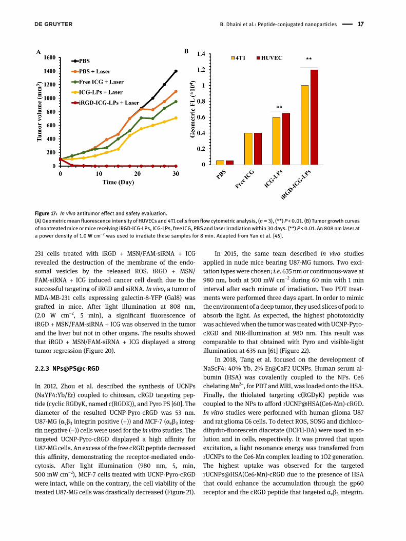
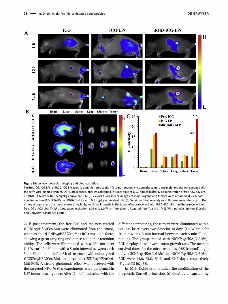
In 2015, Yan et al. synthesized new NPs, namediRGD-ICG-LPs, by the thin-layer rehydration process [57].These NPs were liposome-based (LP) in which the indoc-yanine green (ICG) PS was encapsulated (ICG-LPs) with anefficiency of 93.32 ± 1.25%. ICG-LPs were then grafted withiRGD peptide. The mean dynamic diameter of iRGD-ICG-LPs was 115.91 nm. The in vitro assays were performed us-ing three different cell lines, HUVECs (high expression ofαvβ3), 4T1 (high expression of αvβ3 and NRP-1), and MCF-7(low expression of αvβ3). After the incubation of these cellswith ICG-LPs and iRGD-ICG-LPs, iRGD-ICG-LPs showed1.86-fold and 1.69-fold higher fluorescence intensity inHUVEC and 4T1 cells, respectively, as compared to ICG-LPs
Figure 15: Synthesis of RPTD/HP NPs. Adapted from Shi et al. [55].
B. Dhaini et al.: Peptide-conjugated nanoparticles 15
(Figure 17(A)). Due to their lower expression of αvβ3,MCF-7 cells exhibited an inferior fluorescence as comparedto HUVEC and 4T1 cells. The cytotoxicity studies showedthat iRGD-ICG-LPs were biocompatible. Under laser illu-mination, iRGD-ICG-LPs and ICG-LPs exhibited a strongercytotoxic effect than ICGalone (P < 0.01) at the samedose ofICG. The amount of 1O2 produced without light, but inpresence of iRGD-ICG-LPs alone, was 1.91 times smallerthan with iRGD-ICG-LPs excited by laser at 480 nm. Thecoupling of iRGD with ICG-LPs induced a very importantPDT-PTT effect, thus demonstrating the specific targetingof the tumors. Using the luminescence of ICG, the in vivoresults showed a stronger accumulation of the NPs in thetumors than that in the liver, spleen and other organs(Figure 18(A)–(C)). After light illumination of the cellsexposed to iRGD-ICG-LPs, the growth of the tumor wassuppressed (P < 0.01) (Figure 17(B)). For these NPs, theamount of the generated ROS was 3.82 times greater thanthat produced in the tumors treated with PBS alone(control).
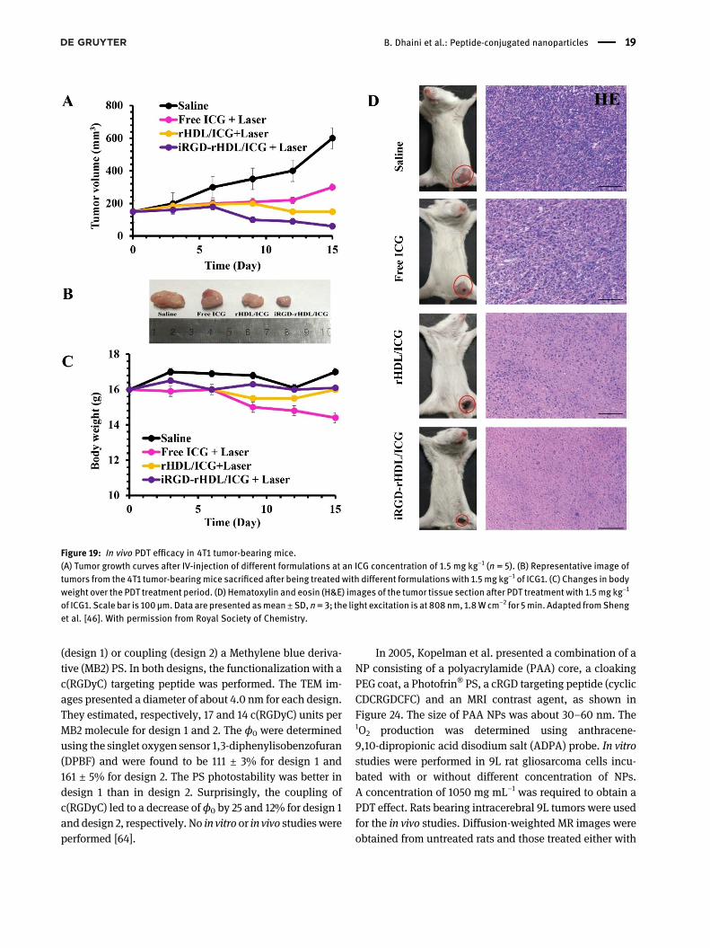
In 2019, Sheng et al. synthesized a novel nanoscaledrug [58]. The basis of this NP was the high-density lipo-proteins (HDL) in which ICG was encapsulated to giverHDL/ICG. The encapsulation of ICG in NPs presentedseveral advantages such as the stability in the plasma andthe lack of precipitation and aggregation. The iRGDpeptidewas coupled onto this system to afford iRGD-rHDL/ICG.These NPs possessed a hydrodynamic diameter of 90 nm.The in vitro assays in 4T1 cells overexpressing αvβ3
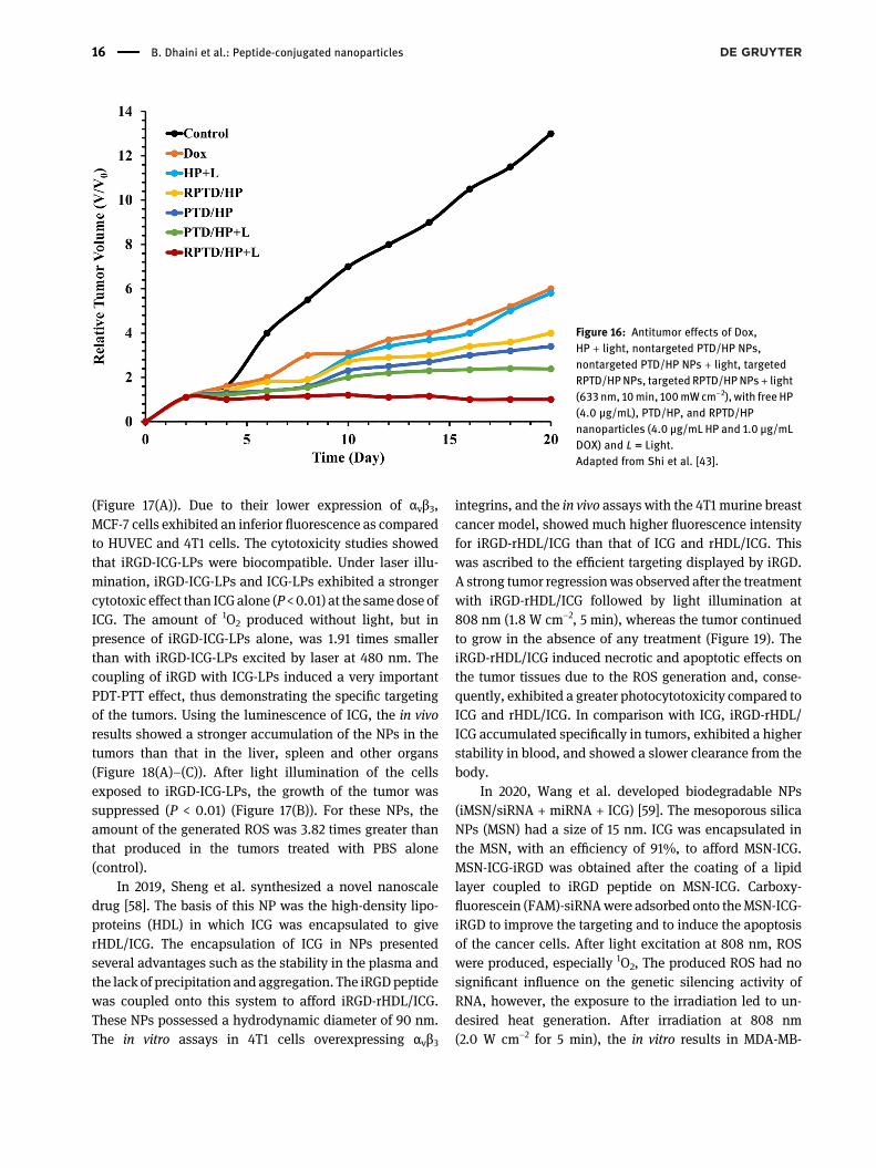
integrins, and the in vivo assays with the 4T1 murine breastcancer model, showed much higher fluorescence intensityfor iRGD-rHDL/ICG than that of ICG and rHDL/ICG. Thiswas ascribed to the efficient targeting displayed by iRGD.A strong tumor regressionwas observed after the treatmentwith iRGD-rHDL/ICG followed by light illumination at808 nm (1.8 W cm−2, 5 min), whereas the tumor continuedto grow in the absence of any treatment (Figure 19). TheiRGD-rHDL/ICG induced necrotic and apoptotic effects onthe tumor tissues due to the ROS generation and, conse-quently, exhibited a greater photocytotoxicity compared toICG and rHDL/ICG. In comparison with ICG, iRGD-rHDL/ICG accumulated specifically in tumors, exhibited a higherstability in blood, and showed a slower clearance from thebody.
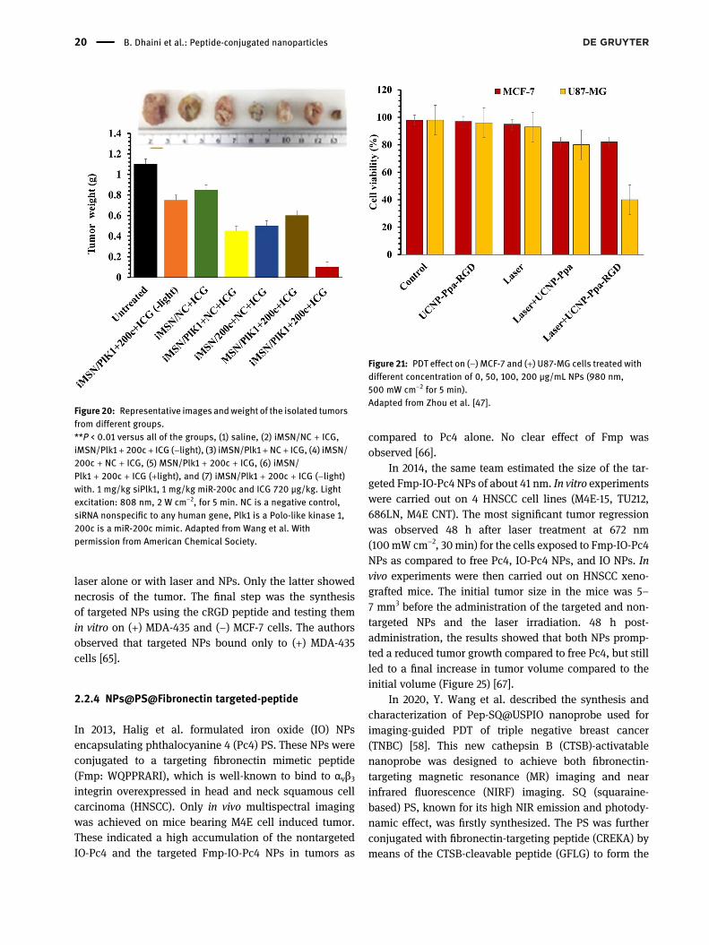
In 2020, Wang et al. developed biodegradable NPs(iMSN/siRNA + miRNA + ICG) [59]. The mesoporous silicaNPs (MSN) had a size of 15 nm. ICG was encapsulated inthe MSN, with an efficiency of 91%, to afford MSN-ICG.MSN-ICG-iRGD was obtained after the coating of a lipidlayer coupled to iRGD peptide on MSN-ICG. Carboxy-fluorescein (FAM)-siRNAwere adsorbed onto theMSN-ICG-iRGD to improve the targeting and to induce the apoptosisof the cancer cells. After light excitation at 808 nm, ROSwere produced, especially 1O2, The produced ROS had nosignificant influence on the genetic silencing activity ofRNA, however, the exposure to the irradiation led to un-desired heat generation. After irradiation at 808 nm(2.0 W cm−2 for 5 min), the in vitro results in MDA-MB-
Figure 16: Antitumor effects of Dox,HP + light, nontargeted PTD/HP NPs,nontargeted PTD/HP NPs + light, targetedRPTD/HPNPs, targeted RPTD/HPNPs+ light(633 nm, 10min, 100mWcm−2), with free HP(4.0 μg/mL), PTD/HP, and RPTD/HPnanoparticles (4.0 μg/mL HP and 1.0 μg/mLDOX) and L = Light.Adapted from Shi et al. [43].
16 B. Dhaini et al.: Peptide-conjugated nanoparticles
231 cells treated with iRGD + MSN/FAM-siRNA + ICGrevealed the destruction of the membrane of the endo-somal vesicles by the released ROS. iRGD + MSN/FAM-siRNA + ICG induced cancer cell death due to thesuccessful targeting of iRGD and siRNA. In vivo, a tumor ofMDA-MB-231 cells expressing galectin-8-YFP (Gal8) wasgrafted in mice. After light illumination at 808 nm,(2.0 W cm−2, 5 min), a significant fluorescence ofiRGD + MSN/FAM-siRNA + ICG was observed in the tumorand the liver but not in other organs. The results showedthat iRGD + MSN/FAM-siRNA + ICG displayed a strongtumor regression (Figure 20).
2.2.3 NPs@PS@c-RGD
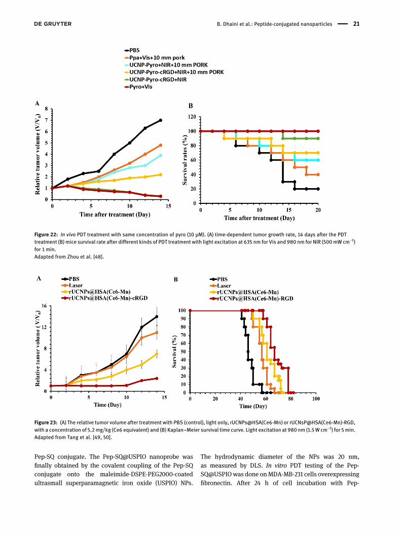
In 2012, Zhou et al. described the synthesis of UCNPs(NaYF4:Yb/Er) coupled to chitosan, cRGD targeting pep-tide (cyclic RGDyK, named c(RGDK)), and Pyro PS [60]. Thediameter of the resulted UCNP-Pyro-cRGD was 53 nm.U87-MG (αvβ3 integrin positive (+)) and MCF-7 (αvβ3 integ-rin negative (−)) cells were used for the in vitro studies. Thetargeted UCNP-Pyro-cRGD displayed a high affinity forU87-MG cells. An excess of the free cRGDpeptide decreasedthis affinity, demonstrating the receptor-mediated endo-cytosis. After light illumination (980 nm, 5, min,500 mW cm−2), MCF-7 cells treated with UCNP-Pyro-cRGDwere intact, while on the contrary, the cell viability of thetreated U87-MG cells was drastically decreased (Figure 21).
In 2015, the same team described in vivo studiesapplied in nude mice bearing U87-MG tumors. Two exci-tation typeswere chosen; i.e. 635 nmor continuous-wave at980 nm, both at 500 mW cm−2 during 60 min with 1 mininterval after each minute of irradiation. Two PDT treat-ments were performed three days apart. In order to mimicthe environment of a deep tumor, they used slices of pork toabsorb the light. As expected, the highest phototoxicitywas achievedwhen the tumorwas treatedwithUCNP-Pyro-cRGD and NIR-illumination at 980 nm. This result wascomparable to that obtained with Pyro and visible-lightillumination at 635 nm [61] (Figure 22).
In 2018, Tang et al. focused on the development ofNaScF4: 40% Yb, 2% Er@CaF2 UCNPs. Human serum al-bumin (HSA) was covalently coupled to the NPs. Ce6chelatingMn2+, for PDT andMRI, was loaded onto the HSA.Finally, the thiolated targeting c(RGDyK) peptide wascoupled to the NPs to afford rUCNP@HSA(Ce6-Mn)-cRGD.In vitro studies were performed with human glioma U87and rat glioma C6 cells. To detect ROS, SOSG and dichloro-dihydro-fluorescein diacetate (DCFH-DA) were used in so-lution and in cells, respectively. It was proved that uponexcitation, a light resonance energy was transferred fromrUCNPs to the Ce6-Mn complex leading to 1O2 generation.The highest uptake was observed for the targetedrUCNPs@HSA(Ce6-Mn)-cRGD due to the presence of HSAthat could enhance the accumulation through the gp60receptor and the cRGD peptide that targeted αvβ3 integrin.
Figure 17: In vivo antitumor effect and safety evaluation.(A) Geometricmean fluorescence intensity of HUVECs and 4T1 cells from flow cytometric analysis, (n= 3), (**) P < 0.01. (B) Tumor growth curvesof nontreatedmice ormice receiving iRGD-ICG-LPs, ICG-LPs, free ICG, PBS and laser irradiationwithin 30 days. (**) P < 0.01. An 808 nm laser ata power density of 1.0 W cm−2 was used to irradiate these samples for 8 min. Adapted from Yan et al. [45].
B. Dhaini et al.: Peptide-conjugated nanoparticles 17
24 h post treatment, the free Ce6 and the non-argetedrUCNPs@HSA(Ce6-Mn) were eliminated from the tumor,whereas the rUCNPs@HSA(Ce6-Mn)-RGD was still there,showing a great targeting and hence a superior retentionability. The cells were illuminated with a 980 nm laser(1.5 W cm−2 for 30 min with a 5-min interval between each5 min illumination) after 6 h of treatment with nontargetedrUCNPs@HSA(Ce6-Mn) or targeted rUCNPs@HSA(Ce6-Mn)-RGD. A strong phototoxic effect was observed withthe targeted NPs. In vivo experiments were performed inU87 tumor-bearing mice. After 12 h of incubation with the
different compounds, the tumors were illuminated with a980 nm laser every two days for 14 days (1.5 W cm−2 for30 min with a 5-min interval between each 5 min illumi-nation). The group treated with rUCNPs@HSA(Ce6-Mn)-RGD displayed the lowest tumor growth rate. The mediansurvival times for the mice treated by PBS (control), lightonly, rUCNPs@HSA(Ce6-Mn) or rUCNsP@HSA(Ce6-Mn)-RGD were 45.0, 51.0, 54.5, and 59.2 days, respectively(Figure 23) [62, 63].
In 2019, Kohle et al. studied the modification of thediagnostic Cornell prime dots (C′ dots) by encapsulating
Figure 18: In vivo molecular imaging and biodistribution.The free ICG, ICG-LPs, or iRGD-ICG-LPswere IV administrated to the4T1 tumor-bearingmice and the tumors andmajor organswere imagedwiththe ex/in vivo imaging system. (A) Fluorescence signalwas obtained in tumor sites at 1, 12, and 24h after IV administration of free ICG, ICG-LPs,or iRGD– ICG-LPs with 0.5 mg/kg equivalent ICG. (B) Ex vivo fluorescence images of major organs and tumors were obtained at 24 h postinjection of free ICG, ICG-LPs, or iRGD-ICG-LPs with 0.5 mg/kg equivalent ICG. (C) Semiquantitative analysis of fluorescence intensity for thedifferent organs and the tumor showedmuch higher signal intensity in the tumor of mice receivedwith iRGD–ICG-LPs than those receivedwithfree ICG or ICG-LPs. (**) P < 0.01. Laser excitation: 808 nm, 1.0 W cm−2 for 10 min. Adapted from Yan et al. [45]. With permission from Elsevierand Copyright Clearance Center.
18 B. Dhaini et al.: Peptide-conjugated nanoparticles
(design 1) or coupling (design 2) a Methylene blue deriva-tive (MB2) PS. In both designs, the functionalization with ac(RGDyC) targeting peptide was performed. The TEM im-ages presented a diameter of about 4.0 nm for each design.They estimated, respectively, 17 and 14 c(RGDyC) units perMB2 molecule for design 1 and 2. The ϕ0 were determinedusing the singlet oxygen sensor 1,3-diphenylisobenzofuran(DPBF) and were found to be 111 ± 3% for design 1 and161 ± 5% for design 2. The PS photostability was better indesign 1 than in design 2. Surprisingly, the coupling ofc(RGDyC) led to a decrease ofϕ0 by 25 and 12% for design 1and design 2, respectively. No in vitro or in vivo studieswereperformed [64].
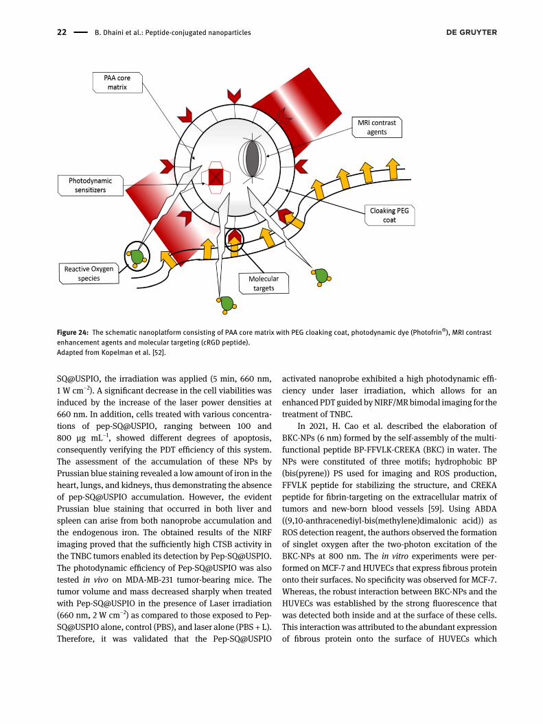
In 2005, Kopelman et al. presented a combination of aNP consisting of a polyacrylamide (PAA) core, a cloakingPEG coat, a Photofrin® PS, a cRGD targeting peptide (cyclicCDCRGDCFC) and an MRI contrast agent, as shown inFigure 24. The size of PAA NPs was about 30–60 nm. The1O2 production was determined using anthracene-9,10-dipropionic acid disodium salt (ADPA) probe. In vitrostudies were performed in 9L rat gliosarcoma cells incu-bated with or without different concentration of NPs.A concentration of 1050 mg mL−1 was required to obtain aPDT effect. Rats bearing intracerebral 9L tumors were usedfor the in vivo studies. Diffusion-weighted MR images wereobtained from untreated rats and those treated either with
Figure 19: In vivo PDT efficacy in 4T1 tumor-bearing mice.(A) Tumor growth curves after IV-injection of different formulations at an ICG concentration of 1.5 mg kg−1 (n = 5). (B) Representative image oftumors from the 4T1 tumor-bearingmice sacrificed after being treated with different formulations with 1.5 mg kg−1 of ICG1. (C) Changes in bodyweight over the PDT treatment period. (D) Hematoxylin and eosin (H&E) images of the tumor tissue section after PDT treatment with 1.5mg kg−1
of ICG1. Scale bar is 100μm.Data are presented asmean±SD,n=3; the light excitation is at 808nm, 1.8Wcm−2 for 5min. Adapted fromShenget al. [46]. With permission from Royal Society of Chemistry.
B. Dhaini et al.: Peptide-conjugated nanoparticles 19
laser alone or with laser and NPs. Only the latter showednecrosis of the tumor. The final step was the synthesisof targeted NPs using the cRGD peptide and testing themin vitro on (+) MDA-435 and (−) MCF-7 cells. The authorsobserved that targeted NPs bound only to (+) MDA-435cells [65].
2.2.4 NPs@PS@Fibronectin targeted-peptide
In 2013, Halig et al. formulated iron oxide (IO) NPsencapsulating phthalocyanine 4 (Pc4) PS. These NPs wereconjugated to a targeting fibronectin mimetic peptide(Fmp: WQPPRARI), which is well-known to bind to αvβ3integrin overexpressed in head and neck squamous cellcarcinoma (HNSCC). Only in vivo multispectral imagingwas achieved on mice bearing M4E cell induced tumor.These indicated a high accumulation of the nontargetedIO-Pc4 and the targeted Fmp-IO-Pc4 NPs in tumors as
compared to Pc4 alone. No clear effect of Fmp wasobserved [66].
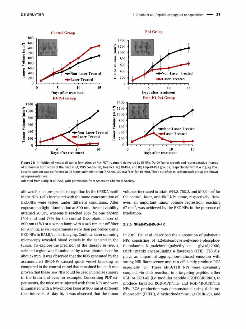
In 2014, the same team estimated the size of the tar-geted Fmp-IO-Pc4 NPs of about 41 nm. In vitro experimentswere carried out on 4 HNSCC cell lines (M4E-15, TU212,686LN, M4E CNT). The most significant tumor regressionwas observed 48 h after laser treatment at 672 nm(100mW cm−2, 30min) for the cells exposed to Fmp-IO-Pc4NPs as compared to free Pc4, IO-Pc4 NPs, and IO NPs. Invivo experiments were then carried out on HNSCC xeno-grafted mice. The initial tumor size in the mice was 5–7 mm3 before the administration of the targeted and non-targeted NPs and the laser irradiation. 48 h post-administration, the results showed that both NPs promp-ted a reduced tumor growth compared to free Pc4, but stillled to a final increase in tumor volume compared to theinitial volume (Figure 25) [67].
In 2020, Y. Wang et al. described the synthesis andcharacterization of Pep-SQ@USPIO nanoprobe used forimaging-guided PDT of triple negative breast cancer(TNBC) [58]. This new cathepsin B (CTSB)-activatablenanoprobe was designed to achieve both fibronectin-targeting magnetic resonance (MR) imaging and nearinfrared fluorescence (NIRF) imaging. SQ (squaraine-based) PS, known for its high NIR emission and photody-namic effect, was firstly synthesized. The PS was furtherconjugated with fibronectin-targeting peptide (CREKA) bymeans of the CTSB-cleavable peptide (GFLG) to form the
Figure 20: Representative images andweight of the isolated tumorsfrom different groups.**P < 0.01 versus all of the groups, (1) saline, (2) iMSN/NC + ICG,iMSN/Plk1 + 200c + ICG (−light), (3) iMSN/Plk1 +NC + ICG, (4) iMSN/200c + NC + ICG, (5) MSN/Plk1 + 200c + ICG, (6) iMSN/Plk1 + 200c + ICG (+light), and (7) iMSN/Plk1 + 200c + ICG (−light)with. 1 mg/kg siPlk1, 1 mg/kg miR-200c and ICG 720 μg/kg. Lightexcitation: 808 nm, 2 W cm−2, for 5 min. NC is a negative control,siRNA nonspecific to any human gene, Plk1 is a Polo-like kinase 1,200c is a miR-200c mimic. Adapted from Wang et al. Withpermission from American Chemical Society.
Figure 21: PDT effect on (−) MCF-7 and (+) U87-MG cells treated withdifferent concentration of 0, 50, 100, 200 μg/mL NPs (980 nm,500 mW cm−2 for 5 min).Adapted from Zhou et al. [47].
20 B. Dhaini et al.: Peptide-conjugated nanoparticles
Pep-SQ conjugate. The Pep-SQ@USPIO nanoprobe wasfinally obtained by the covalent coupling of the Pep-SQconjugate onto the maleimide-DSPE-PEG2000-coatedultrasmall superparamagnetic iron oxide (USPIO) NPs.
The hydrodynamic diameter of the NPs was 20 nm,as measured by DLS. In vitro PDT testing of the Pep-SQ@USPIO was done on MDA-MB-231 cells overexpressingfibronectin. After 24 h of cell incubation with Pep-
Figure 22: In vivo PDT treatment with same concentration of pyro (10 µM). (A) time-dependent tumor growth rate, 14 days after the PDTtreatment (B) mice survival rate after different kinds of PDT treatment with light excitation at 635 nm for Vis and 980 nm for NIR (500mW cm−2)for 1 min.Adapted from Zhou et al. [48].
Figure 23: (A) The relative tumor volume after treatment with PBS (control), light only, rUCNPs@HSA(Ce6-Mn) or rUCNsP@HSA(Ce6-Mn)-RGD,with a concentration of 5.2 mg/kg (Ce6 equivalent) and (B) Kaplan–Meier survival time curve. Light excitation at 980 nm (1.5W cm−2) for 5 min.Adapted from Tang et al. [49, 50].
B. Dhaini et al.: Peptide-conjugated nanoparticles 21
SQ@USPIO, the irradiation was applied (5 min, 660 nm,1 W cm−2). A significant decrease in the cell viabilities wasinduced by the increase of the laser power densities at660 nm. In addition, cells treated with various concentra-tions of pep-SQ@USPIO, ranging between 100 and800 µg mL−1, showed different degrees of apoptosis,consequently verifying the PDT efficiency of this system.The assessment of the accumulation of these NPs byPrussian blue staining revealed a low amount of iron in theheart, lungs, and kidneys, thus demonstrating the absenceof pep-SQ@USPIO accumulation. However, the evidentPrussian blue staining that occurred in both liver andspleen can arise from both nanoprobe accumulation andthe endogenous iron. The obtained results of the NIRFimaging proved that the sufficiently high CTSB activity inthe TNBC tumors enabled its detection by [email protected] photodynamic efficiency of Pep-SQ@USPIO was alsotested in vivo on MDA-MB-231 tumor-bearing mice. Thetumor volume and mass decreased sharply when treatedwith Pep-SQ@USPIO in the presence of Laser irradiation(660 nm, 2 W cm−2) as compared to those exposed to Pep-SQ@USPIO alone, control (PBS), and laser alone (PBS + L).Therefore, it was validated that the Pep-SQ@USPIO
activated nanoprobe exhibited a high photodynamic effi-ciency under laser irradiation, which allows for anenhancedPDTguided byNIRF/MRbimodal imaging for thetreatment of TNBC.
In 2021, H. Cao et al. described the elaboration ofBKC-NPs (6 nm) formed by the self-assembly of the multi-functional peptide BP-FFVLK-CREKA (BKC) in water. TheNPs were constituted of three motifs; hydrophobic BP(bis(pyrene)) PS used for imaging and ROS production,FFVLK peptide for stabilizing the structure, and CREKApeptide for fibrin-targeting on the extracellular matrix oftumors and new-born blood vessels [59]. Using ABDA((9,10-anthracenediyl-bis(methylene)dimalonic acid)) asROS detection reagent, the authors observed the formationof singlet oxygen after the two-photon excitation of theBKC-NPs at 800 nm. The in vitro experiments were per-formed on MCF-7 and HUVECs that express fibrous proteinonto their surfaces. No specificity was observed for MCF-7.Whereas, the robust interaction between BKC-NPs and theHUVECs was established by the strong fluorescence thatwas detected both inside and at the surface of these cells.This interaction was attributed to the abundant expressionof fibrous protein onto the surface of HUVECs which
Figure 24: The schematic nanoplatform consisting of PAA core matrix with PEG cloaking coat, photodynamic dye (Photofrin®), MRI contrastenhancement agents and molecular targeting (cRGD peptide).Adapted from Kopelman et al. [52].
22 B. Dhaini et al.: Peptide-conjugated nanoparticles
allowed for a more specific recognition by the CREKAmotifin the NPs. Cells incubated with the same concentration ofBKC-NPs were tested under different conditions. Afterexposure to light illumination at 800 nm, the cell viabilityattained 20.8%, whereas it reached 16% for one photon(405 nm) and 73% for the control (two-photon laser of800 nm (1 W) or a xenon lamp with a 405 nm cut-off filterfor 20 min). In vivo experiments were then performed usingBKC-NPs in BALB/c mice imaging. Confocal laser scanningmicroscopy revealed blood vessels in the ear and in thetumor. To explore the precision of the therapy in vivo, aselected region was illuminated by a two-photon laser forabout 2 min. It was observed that the ROS generated by theaccumulated BKC-NPs caused quick vessel breaking ascompared to the control vessel that remained intact. It wasproven that these newNPs could be used in precise surgeryin the brain and eyes for example. Concerning PDT ex-periments, the mice were injected with these NPs and wereilluminated with a two-photon laser at 800 nm at differenttime intervals. At day 16, it was observed that the tumor
volumes increased to attain 695.8, 780.2, and 610.3mm3 forthe control, laser, and BKC-NPs alone, respectively. How-ever, an important tumor volume regression, reaching67 mm3, was achieved by the BKC-NPs in the presence ofirradiation.
2.2.5 NPs@PS@RGD-4R
In 2019, Dai et al. described the elaboration of polymericNPs consisting of 1,2-distearoyl-sn-glycero-3-phosphoe-thanolamine-N-[maleimide(polyethylene glycol)-2000](MPD) matrix encapsulating a fluorogen (TTB). TTB dis-plays an important aggregation-induced emission withstrong NIR fluorescence and can efficiently produce ROSespecially 1O2. These MPD/TTB NPs were covalentlycoupled, via click reaction, to a targeting peptide, eitherRGD or RGD-4R (i.e. modular peptide RGDFGGRRRRC), toproduce targeted RGD-MPD/TTB and RGD-4R-MPD/TTBNPs. ROS production was demonstrated using dichloro-fluorescein (DCFH), dihydrorhodamine 123 (DHR123), and
Figure 25: Inhibition of xenograft tumor formation by Pc4 PDT treatment delivered by IO NPs. (A–D) Tumor growth and representative imagesof tumors on both sides of the mice in (A) PBS control, (B) free Pc4, (C) IO-Pc4, and (D) Fmp-IO-Pc4 groups, respectively with 0.4 mg/kg Pc4.Laser treatment was performed at 48 h post administration (672 nm, 100mW/cm2 for 30min). Three out of sixmice from each group are shownas representatives.Adapted from Halig et al. [54]. With permission from American Chemical Society.
B. Dhaini et al.: Peptide-conjugated nanoparticles 23
EPR spectroscopy to identify O2•− and ABPA to detect 1O2.
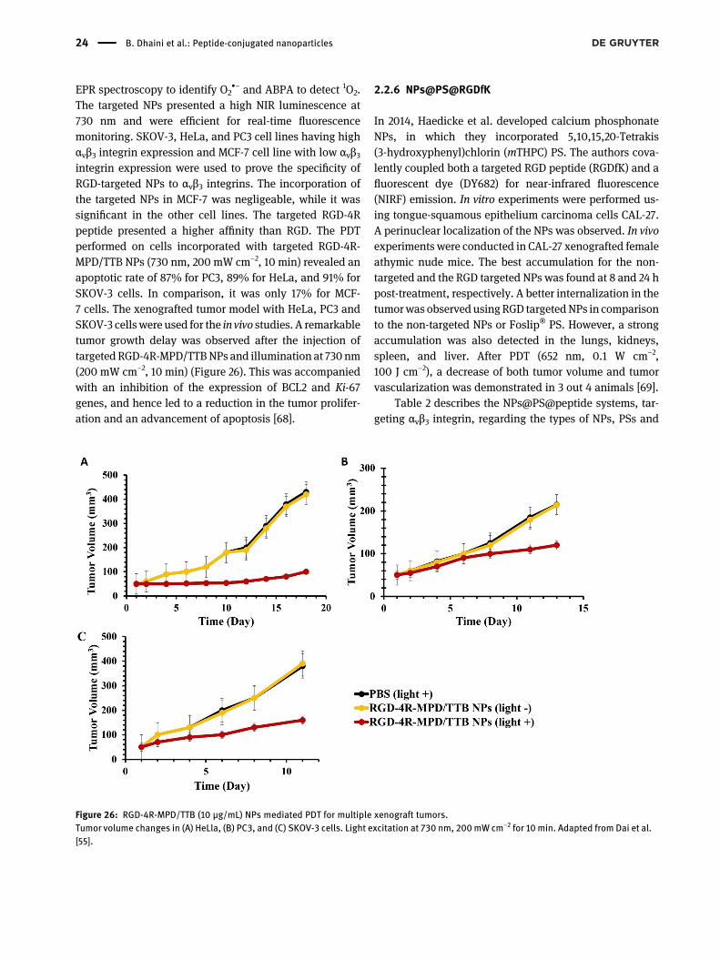
The targeted NPs presented a high NIR luminescence at730 nm and were efficient for real-time fluorescencemonitoring. SKOV-3, HeLa, and PC3 cell lines having highαvβ3 integrin expression and MCF-7 cell line with low αvβ3integrin expression were used to prove the specificity ofRGD-targeted NPs to αvβ3 integrins. The incorporation ofthe targeted NPs in MCF-7 was negligeable, while it wassignificant in the other cell lines. The targeted RGD-4Rpeptide presented a higher affinity than RGD. The PDTperformed on cells incorporated with targeted RGD-4R-MPD/TTB NPs (730 nm, 200 mW cm−2, 10 min) revealed anapoptotic rate of 87% for PC3, 89% for HeLa, and 91% forSKOV-3 cells. In comparison, it was only 17% for MCF-7 cells. The xenografted tumor model with HeLa, PC3 andSKOV-3 cells were used for the in vivo studies. A remarkabletumor growth delay was observed after the injection oftargeted RGD-4R-MPD/TTBNPs and illumination at 730 nm(200 mW cm−2, 10 min) (Figure 26). This was accompaniedwith an inhibition of the expression of BCL2 and Ki-67genes, and hence led to a reduction in the tumor prolifer-ation and an advancement of apoptosis [68].
2.2.6 NPs@PS@RGDfK
In 2014, Haedicke et al. developed calcium phosphonateNPs, in which they incorporated 5,10,15,20-Tetrakis(3-hydroxyphenyl)chlorin (mTHPC) PS. The authors cova-lently coupled both a targeted RGD peptide (RGDfK) and afluorescent dye (DY682) for near-infrared fluorescence(NIRF) emission. In vitro experiments were performed us-ing tongue-squamous epithelium carcinoma cells CAL-27.A perinuclear localization of the NPs was observed. In vivoexperiments were conducted in CAL-27 xenografted femaleathymic nude mice. The best accumulation for the non-targeted and the RGD targeted NPs was found at 8 and 24 hpost-treatment, respectively. A better internalization in thetumorwas observed usingRGD targetedNPs in comparisonto the non-targeted NPs or Foslip® PS. However, a strongaccumulation was also detected in the lungs, kidneys,spleen, and liver. After PDT (652 nm, 0.1 W cm−2,100 J cm−2), a decrease of both tumor volume and tumorvascularization was demonstrated in 3 out 4 animals [69].
Table 2 describes the NPs@PS@peptide systems, tar-geting αvβ3 integrin, regarding the types of NPs, PSs and
Figure 26: RGD-4R-MPD/TTB (10 μg/mL) NPs mediated PDT for multiple xenograft tumors.Tumor volume changes in (A) HeLla, (B) PC3, and (C) SKOV-3 cells. Light excitation at 730 nm, 200mW cm−2 for 10 min. Adapted from Dai et al.[55].
24 B. Dhaini et al.: Peptide-conjugated nanoparticles
the coupling between them, in addition to the NPs size,excitation wavelength (λexcitation), fluorescence quantumyield (ϕF), singlet oxygen quantum yield (ΔO.S) and theresults obtained in vitro and/or in vivo.
2.3 Peptide for nucleolin membranereceptors
Nucleolin is a nucleolar protein that has several roles in theintracellular pathways and is involved in tumorigenesis[72]. It is expressed in the nucleus of resting cells. In tumorcells, nucleolin cycles between the cell nucleus and theplasma membrane. Its overexpression was identified indifferent kind of cancers. Therefore, nucleolin is consid-ered a target for anticancer therapies [73].
F3 peptide has a sequence of KDEPQRRSARLSAK-PAPPKPEPKPKKAPAKK. It is well-known to target tumorneovessels as well as some tumor cells [74, 75]. F3 peptidecan bind to the nucleolin membrane receptors, which al-lows its internalization into the cells and its further local-ization into the nucleolin.
Kopelman’s team reported the elaboration of an F3-targeted nanosystem, consisting of a polyacrylamide coreinwhich a photodynamic agent, Photofrin® PS, and anMRIagent, iron oxide, were embedded. The F3 targeting pep-tide was also attached to the NPs in addition to thecoupling to Alexa Fluor 594 for fluorescent imaging. Sixmolecules of Photofrin® were encapsulated into each NPand average of 30 F3 peptides were coupled. No cytotox-icity was observed in MDA-435 cells after 4 h of incubationwith the F3-targeted NPs. However, a significant photo-toxic effect was achieved after light illumination (630 nm,1.5 mW, 5 min) as 90 % of the cells were destroyed. Usingfluorescence microscopy, the authors detected a cellularuptake and a nuclear localization of F3-targeted NPs. Invivo studies in rats bearing 9L gliomas were performed inthe presence of nontargeted and F3-targeted NPs forcomparative reasons. The F3-targeted NPs had about three-fold prolonged tumor transit time. Their presence led alsoto an improved contrast-to-noise ratio of about two-fold at1 h. A median survival time after treatment and illumina-tion was found to be 7.0 days for the control untreatedgroup, 8.5 days for the group treated only with laser and13.0 days for the one treated with Photofrin® alone.Conversely, the median survival time was up to 33 days forthe group treated with F3-targeted NPs (Figure 27) [76].
The same team [77] reported polyacrylamide NPsconjugated with MB PS and F3 targeting peptide. First, MB
was coupled to the 3-aminopropyl)methacrylamide hy-drochloride (APMA) monomer before the formation of NPsin a reverse microemulsion. The amino functions of APMAwere then used to couple the F3 peptide. Four cell lineswere cultivated, human melanoma MDA-MB-435, rat gli-oma F98, human breast adenocarcinoma MCF-7 and ratglioma 9L. The nucleolin expression was described previ-ously by the same team [78]. The F3-targeted NPs presentedthe lowest affinity for MCF-7 cells. Yet, a better affinity by2.5, 4, and 5.4 times was achieved for F98, MDA-MB-435and 9L cells, respectively. No incorporation of the non-targeted MB-conjugated NPs was observed. Comparableresults in terms of the photodynamic efficiency were ob-tained on all the four cell lines when the targeted NPs weretested at 1.5 mg mL−1 and illuminated at 647 nm (20 J cm−2,1 min). Further experiments performed on F98 cell linesproved that the cell death increased with illumination timeand NPs concentration.
Feng et al. described the synthesis of targeted F3-PTX-(Pyro-PLA)2PEG NPs (denoted by F3-targeted NPs). Theseconsist of pyro-conjugated amphiphilic (Pyro-PLA)2PEGNPs covalently attached to the F3 targeting peptide.Paclitaxel (PTX, Taxol) was then encapsulated to demon-strate a combination of PDT and chemotherapy [79]. (Pyro-PLA)2PEGNPs presented a good stability in vitro. In HUVECand human colorectal cancer cells (HCT-15), a betteraccumulation of the F3-targeted NPs was observed with afactor of 1.21 and 1.18, respectively, when compared withthat of (Pyro-PLA)2PEG. These NPswere localizedmostly inthe endolysosomal compartment. The production of ROSwas demonstrated using 2′,7′-dichlorofluorescein (DCF)fluorescence. The IC50 values in HUVEC were 122.1 ng mL−1
for Taxol, 84.09 ng mL−1 for non-targeted NPs (i.e. PTX-(Pyro-PLA)2PEG NPs) in the absence of laser, 41.21 ng mL−1
for non-targeted NPs in the presence of laser and17.0 ng mL−1 for F3-targeted NPs accompanied by laserirradiation. The IC50 values in HCT-15 cells were426.7 ng mL−1 for nontargeted NPs in the absence of laser,86.32 ng mL−1 for non-targeted NPs in the presence of laserand 32.86 ng mL−1 for F3-targeted NPs accompanied bylaser irradiation. These outcomes validated the importanceof adding a targeting peptide to boost the PDT efficiency.The in vivo experiments performed in male BALB/c nudemice bearing colorectal tumor (HCT-15 injected subcuta-neously) confirmed the in vitro results. After injection, thenontargeted NPs were slightly accumulated in the tumorand around the blood vessels, whereas the F3-targeted NPswere effectively accumulated into the tumor due tothe efficient targeting of both the tumor cells and the
B. Dhaini et al.: Peptide-conjugated nanoparticles 25
Table:Th
esu
mmaryof
NPs
conjug
ated
withPS
san
dpe
ptides
targetingα vβ
integrin
interm
sof
differen
tpa
rameters.
NPs
@PS
@Pe
ptidetargetingα vβ
Referen
ceNPs
PSCo
uplin
gbe
twee
nNPs
andPS
NPs
size
(nm)
λ excitation(nm)
ϕF
Δ O.S
invitro
invivo
PeptideRGD
[]
Polymeric(PEG
)IR
Maleimide
.
forRG
D-PE
G-IR
ndnd
A
Mou
sefibrob
last
NIH/
T[]
Mesop
orou
ssilica
(MSN)
Photop
orph
yrin
IX(PpIX)
Amidebo
ndforMSN-BHQ-
SS-PpIX-PE
G-RGD
ndnd
ndCOS
SCC-
HeLa
nd
[]
Upc
onversion(UCN)
Zinc
(II)ph
thalocyanine
(ZnP
c)En
caps
ulation
forZn
Pc-
UCN@lip
id@PS
ndnd
Hela
nd
[]
Upc
onversion(UCN)
Merocyanine
(M
C)
Ads
orption
forMCload
edUCN/R
GD-PMAO-DOPE
ndnd
MCF-
nd
PeptideiRGD
[]
High-de
nsitylip
o-proteins
(HDL)
Indo
cyan
inegree
n(ICG)
Encaps
ulation
.
foriRGD-rHDL/ICG
ndnd
T
FemaleBALB
/cmice
[]
Mesop
orou
ssilica
(MSN)
Indo
cyan
inegree
n(ICG)
Encaps
ulation
-forAmine-
func
tion
alized
MSNs
ndnd
MDA-MB-
.Luc
BF.Luc.
Immun
ocompro-
mised
female
NCGmice
[ ]
Lipo
some(LP)
Indo
cyan
inegree
n(ICG)
Encaps
ulation
.foriRGD-ICG
-LPs
ndnd
T
T
Tumor-
bearingmice
PeptidecR
GD
[]
Globu
larPE
GChlorin
e(Ce
)Maleimide
–
forcR
GD-gPE
G-Ce
ndnd
SKOV-
KB
Seven
-wee
k-old
Femalenu
demice
(BALB
/cnu
/nu
mice,
Instituteof
Med
ical
Scien
ce,
Tokyo)
[]
Poly(amidoam
ine)
dend
rimer
(PAMAM)
Chlorin
e(Ce
)Amidebo
ndforPE
G-P-Ce
and
RGD-P-Ce
nd.-fold
high
erthan
that
offree
Ce
ANIHT
nd
[]
Selfa
ssem
bly
Hem
atop
orph
yrin
(HP)
forRP
TD/H
P
ndnd
ndnd
26 B. Dhaini et al.: Peptide-conjugated nanoparticles
Table:(con
tinu
ed)
NPs
@PS
@Pe
ptidetargetingα vβ
Referen
ceNPs
PSCo
uplin
gbe
twee
nNPs
andPS
NPs
size
(nm)
λ excitation(nm)
ϕF
Δ O.S
invitro
invivo
Thioke
tal
linka
ge
Peptidec(RGDyK
)
[]
Upc
onversion(UCN)
Pyroph
eoph
orbide
-a(Pyro)
Amidebo
ndforOCMC-UCNP
ndnd
U-M
GMCF-
nd
[]
Silica
Methy
lene
blue
(MB)
Encaps
ulation
(Design),Mal-
eimide(Design
)
Below
forde
sign
edNPs
an
d
nd±%
forDesign
±%
forDesign
ndnd
[]
Upc
onversion(UCN)
Pyroph
eoph
orbide
-a(Pyro)
Amidebo
ndforUCNP-Py
ro-RGD
ndnd
U-M
GNud
emice
bearingU-M
Gtumor
[]
Upc
onversion(UCN)
Chlorin
eMn
+(Ce
-Mn)
Encaps
ultation
.
forUCNPs
@HSA
ndnd
U
U-Bea
ring
mice
PeptidecR
GD
[]
Polyacrylamide
(PAA)
Photofrin®
Encaps
ultation
–
forPA
A
ndnd
Lrat
Rats
bearing
intracereb
ral
Ltumors
PeptideFm
pfibron
ectin
[]
Iron
oxide
Phthalocyanine
(Pc
)En
caps
ultation
nd–
ndnd
ndMicebe
aringME
tumor
indu
ced
[]
Iron
oxide
Phthalocyanine
(Pc
)En
caps
ultation
forFm
p-IO-Pc
ndnd
ME,
ME-,
LN,T
U
Micebe
aringME
tumor
indu
ced
PeptideRGDor
RGD-R
[]
Polymericmatrix
TTBa
Encaps
ulation
.
forRG
D-R-MPD
/TTB
–
.in
solid
state
ndMCF,P
C,
Hela,
SKOV-
BALB
/cNud
emice(m
alefor
PCxeno
grafttu-
mor
andfemale
forbo
thHelaan
dSKOV-xeno
-graftedtumor
mod
els)
B. Dhaini et al.: Peptide-conjugated nanoparticles 27
Table:(con
tinu
ed)
NPs
@PS
@Pe
ptidetargetingα vβ
Referen
ceNPs
PSCo
uplin
gbe
twee
nNPs
andPS
NPs
size
(nm)
λ excitation(nm)
ϕF
Δ O.S
invitro
invivo
PeptideRGDfK
[]
Calcium
phos
phate
,,,-Tetrakis(-hyd
roxyph
enyl)
chlorin(m
THPC
)En
caps
ulation
forNP-DY
-
mTH
PC-RGD
ndnd
CAL
-
Femaleathy
mic
nude
mice
PeptideCR
EKA
[]
Selfa
ssem
bleof
bis(py
rene
)-FFVLK
bis(py
rene
)Amidebo
nd
nm
(one
photon
)and
nm
(two-
photon
,day
and:
min
(.
Wcm
−),da
y:
min
with
W
cm−
laser)
nd.%
MCF
-an
dHUVE
CBALB
/Cmice
[]
Ultrasm
all
Sup
erpa
ramag
netic
iron
oxide
SQ(Squ
araine
)am
idebo
nd
ndnd
MDA-MB-
MDA-MB-
tumor-bea
ring
mice
aTTB:C
ontainsan
electron
-accep
ting
benz
o[,-b:,-b′]d
ithiop
hene
,,,-tetraox
-ide
core
andelectron
-don
ating,′-(,-diphe
nylethen
e-,-diyl)bis(N,N-diphe
nylanilin
e)grou
psforim
age-gu
ided
targeted
PDT,
*nd,
notde
term
ined
.
28 B. Dhaini et al.: Peptide-conjugated nanoparticles
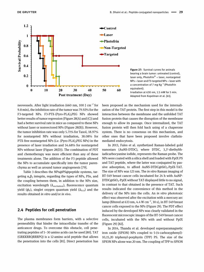
neovessels. After light irradiation (660 nm, 100 J cm−2 for9.8min), the inhibition rate of the tumorwas 79.92% for theF3-targeted NPs. F3-PTX-(Pyro-PLA)2PEG NPs showedbetter results of tumor regression (Figure 28(A) and (C)) andhad a better survival rate in mice as compared to these NPswithout laser or nonvectored NPs (Figure 28(B)). However,the tumor inhibition rate was only 5.71% for Taxol, 18.97%for nontargeted NPs without irradiation, 38.08% forPTX-free nontargeted NPs (i.e. (Pyro-PLA)2PEG NPs) in thepresence of laser irradiation and 54.68% for nontargetedNPs without laser (Figure 28(D)). The combination of PDTand chemotherapy was more efficient than any of thesetreatments alone. The addition of the F3 peptide allowedthe NPs to accumulate specifically into the tumor paren-chyma as well as around tumor angiogenesis [79].
Table 3 describes the NPs@PS@peptide systems, tar-geting αvβ3 integrin, regarding the types of NPs, PSs, andthe coupling between them, in addition to the NPs size,excitation wavelength (λexcitation), fluorescence quantumyield (ϕF), singlet oxygen quantum yield (ΔO.S) and theresults obtained in vitro and/or in vivo.
2.4 Peptides for cell penetration
The plasma membranes form barriers, with a selectivepermeability that hinder the intracellular transfer of theanticancer drugs. To overcome this obstacle, cell pene-trating peptides of 5–30 amino acids can be used [80]. TAT(GRKKRRQRRRPQ) is a 12-amino acid peptide that allowsthe penetration into the cells [81]. Direct penetration has
been proposed as the mechanism used for the internali-zation of the TAT protein. The first step in this model is theinteraction between the membrane and the unfolded TATfusion protein that causes the disruption of the membraneenough to allow its passage. Once internalized, the TATfusion protein will then fold back using of a chaperonesystem. There is no consensus on this mechanism, andother ones that have been proposed involve clathrin-mediated endocytosis.
In 2013, Fales et al. synthetized Raman-labeled goldnanostars (AuNS-DTDC), where DTDC, 3,3′-diethylth-iadicarbocyanine iodide, represents the Raman probe. TheNPswere coatedwith a silica shell and loadedwith PpIX PSand TAT peptide, where the latter was conjugated by pas-sive adsorption, to afford [email protected] size of NPs was 123 nm. The in vitro Raman imaging ofBT-549 breast cancer cells incubated for 24 h with AuNP-DTDC@SiO2-PpIXwithout TAT displayed little to no signal,in contrast to that obtained in the presence of TAT. Suchresults indicated the convenience of this method in thedelivery of the NPs into the cells. An in vitro phototoxiceffect was observed after the excitation with a mercury arclamp (filtered at 633 nm, 4.4W cm−2, 30 s), in BT-549 breastcancer cells exposed to the NPs (Figure 29). The PDT effectinduced by the developed NPs was clearly validated in thefluorescentmicroscopic images of the BT-549 breast cancercells, incubated with the NPs with and without PpIX(Figure 29) [82].
In 2014, Thandu et al. developed superparamagneticiron oxide (SPION) NPs coupled to 5-(4-carboxyphenyl)-10,15,20 triphenyl-porphyrin (TPP) PS. The size of theSPION NPs alone was 20 nm. The coupling of TPP to SPION
Figure 27: Survival curves for animalsbearing a brain tumor: untreated (control),laser only, Photofrin® + laser, nontargetedNPs+ laser and F3-targetedNPs+ laser witha concentration of 7 mg Kg−1 (Photofrinequivalent).Irradiation at 630 nm, 1.5 mW for 5 min.Adapted from Kopelman et al. [61].
B. Dhaini et al.: Peptide-conjugated nanoparticles 29
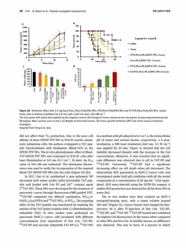
did not affect their 1O2 production. Due to the poor cellaffinity of these SPION-TPP NPs in B78-H1 murine amela-notic melanoma cells, the authors conjugated a TAT pep-tide functionalized with rhodamine (Rhod-TAT) to theSPION-TPP NPs. The in vitro photodynamic effect of Rhod-TAT-SPION-TPP NPs was evaluated in B78-H1 cells afterlaser illumination at 543 nm (14 J cm−2, 30 min). An IC50value of 500 nM was estimated. The rhodamine fluores-cence was used to verify the incorporation of the targetedRhod-TAT-SPION-TPP NPs into the cells (Figure 30) [83].
In 2017, Gao et al. synthetized a new polymeric NPdecorated with tumor acidity (pHe)-activatable TAT pep-tide and loaded with Ce6 PS and Gd3+ contrast agent(DATAT-NP). These NPs were developed for the treatment ofpancreatic cancer through fluorescence/MRI-guided PDT.DATAT-NP comprised two diblock copolymers, PCL45-b-PAEP35-Cya/DTPA and DATAT-PEG77-b-PCL25. The targetingeffect of the TAT peptide was inactivated by masking theamines of the TAT lysine residues with 2,3-dimethylmaleicanhydride (DA). In vitro studies were performed onpancreatic BxPC-3 cancer cells incubated with differentconcentrations (Ce6 equivalent) of free Ce6, TAT-NP,DATAT-NP and succinic anhydride TAT-NP (i.e. SATAT-NP)
in amediumwith pHadjusted at 6.5 or 7.4, the extracellularpH of tumor and normal tissues, respectively. 4 h post-incubation, a NIR laser irradiation (660 nm, 0.5 W cm−2)was applied for 20 min. Figure 31 showed that the cellviability decreased linearly with the increase in the Ce6concentration. Moreover, it was revealed that no signifi-cant difference was observed due to pH in TAT-NP andSATAT-NP. Conversely, DATAT-NP had a significantincreasing effect on cell death when pH decreased. Theintracellular ROS generation in BxPC-3 cancer cells wasinvestigated under both pH conditions with all the testedcompounds at a concentration of 20 mg mL−1 (Ce6 equiv-alent). ROS were detected using the DCFH-DA reagent. Asimilar ROSproductionwas detected for all the threeNPs inwater [84].
The in vivo studies were then realized on BxPC-3xenograft-bearing mice, with a tumor volume around100 mm3 (Figure 32). Cancer tissues were imaged by fluo-rescence 48 h after IV-injection of free Ce6, TAT-NP,DATAT-NP, and SATAT-NP. DATAT-NP treatedmice exhibitedthe highest Ce6 fluorescence in the tumor when comparedto other NPs and free Ce6. In healthy tissues, no differencewas observed. This was in favor of a process in which
Figure 28: Antitumor effect with 2.5 mg/kg of Pyro, (Pyro-PLA)2PEG NPs, PTX-(Pyro-PLA)2PEG NPs and F3-PTX-(Pyro-PLA)2PEG NPs, respec-tively, with or without irradiation for 9.8 min with a 660 nm laser, 170 mW cm−2.The mice given with saline were applied as the negative control. (A) Changes in tumor volume of mice during the 14 days experimental period.(B) Kaplan–Meir survival curve of mice. (C) Weight of dissected tumors. (D) Tumor-growth inhibition (IRT) rate of the various treatmentstrategies.Adapted from Feng et al. [64].
30 B. Dhaini et al.: Peptide-conjugated nanoparticles
DATAT-NP had a prolonged blood circulation lifetime. Inaddition, the acidic environment in the tumor reactivatedthemasked TAT peptide, leading to an important reductionin the tumor volume by more than 65% after 16 days oftherapy [84].
In 2019, Wan et al. combined PDT, PTT, and chemo-therapy in order to treat breast cancer cells. The fluorescentinfrared dye, IR780, was first conjugated with the nucleartargeting TAT peptide. Then, TAT-IR780 and chemothera-peutic Dox self assembled using hydrophobic interactionsto afford TID NPs (TAT-IR780-Dox). These NPs possessed asize of around 100 nm. For comparative reasons, the au-thors also tested TAT-IR780 in the absence of Dox (TIR). Invitro studies were performed on 4T1 cancer cells. 2- and 6-hpost incubation, it was revealed that the fluorescencesignificantly increased when the NPs (TID or TIR) wereadministrated instead of IR780 alone. This proved thegreater internalization of both NPs in the nuclear region(Figure 33) [85]. 24 h after irradiation (785 nm, of1.0 W cm−2, 5 min), the viability of the cells treated withDox, IR780, TIR, and TID reached 60, 35, 20% and >10%,respectively. This synergetic effect was also validated invivo when 4T1 tumor-bearing mice were treated thensacrificed 6 h later. The 100 mm3 tumor was nearly ablated6 days posttreatment with TID NP and IR irradiation,however, it was increased again, very slight, after 18 days(Figure 34) [85].
Table 4 describes the NPs@PS@peptide systems, tar-geting αvβ3 integrin, regarding the types of NPs, PSs, andthe coupling between them, in addition to the NPs size,excitation wavelength (λexcitation), fluorescence quantumyield (ϕF), singlet oxygen quantum yield (ΔO.S), and theresults obtained in vitro and/or in vivo.
2.5 Peptide targeting EGFR
Epidermal growth factor receptor (EGFR) has a role in thetumor progression in several cancers. EGFR also promotestumor proliferation, angiogenesis and metastasis. Theoverexpression of EGFR in many types of cancer cellsmakes it a good candidate for targeted PDT [86]. The HER2gene, one of the members of the EGFR family is expressedin two copies in healthy cells [87]. The modification of thisgene and its amplification causes tumor proliferation.
2.5.1 NPs@PS@HER2 3-340
Narsireddy et al. synthesized 35–40 nm sized multimodalNPs constituting of an Fe3O4 core and a chitosan shell. Thechitosan-covered Fe3O4 NPs were then deposited withTa
ble:Th
esu
mmaryof
NPs
conjug
ated
withPS
san
dF
targetingpe
ptides
interm
sof
differen
tpa
rameters.
NPs
@PS
@F-Pep
tide
Referen
ceNPs
PSCo
uplin
gbe
twee
nNPs
andPS
NPs
size
(nm)
λ excitation
(nm)
ϕF
Δ O.S
invitro
invivo
F-pep
tide
targeting
[]
Polyacrylamide(PAA)
Methy
lene
blue
(MB)
Amidebo
nd.
±.
for
MB-con
juga
tedPA
ANPs
ndnd
MDA-MB-,
F,M
CF-L
nd
[]
Hyd
roxyl-po
ly(lacticacid) -poly-
(ethylen
eglycol) -poly(lacticracid)
-hyd
roxyl(HO-PLA
-PEG
-PLA
-OH)
Pyroph
eoph
orbide
-a(Pyro)
Esterbo
nd.±.forPT
X-(Pyro-PL
A) PE
GNPs
ndnd
HUVE
CHCT-
MaleBALB
/cnu
demice
[]
-aminop
rolylm
etha
crylam
ide()e
ncap
su-
lating
Iron
oxide
Photofrin®
Encaps
ulation
forF-targe
tedNPs
ndnd
MDA-MB-
RatLgliomain
maleFische
r
nd,n
otde
term
ined
.
B. Dhaini et al.: Peptide-conjugated nanoparticles 31
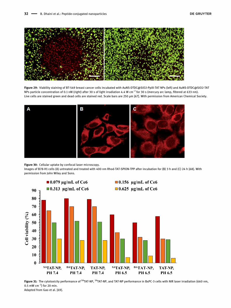
Figure 29: Viability staining of BT-549 breast cancer cells incubated with AuNS-DTDC@SiO2-PpIX-TAT NPs (left) and AuNS-DTDC@SiO2-TATNPs particle concentration of 0.1 nM (right) after 30 s of light irradiation 4.4 W cm−2 for 30 s (mercury arc lamp, filtered at 633 nm).Live cells are stained green and dead cells are stained red. Scale bars are 250 μm [67]. With permission from American Chemical Society.
Figure 30: Cellular uptake by confocal laser microscopy.Images of B78-H1 cells (A) untreated and treated with 400 nm Rhod-TAT-SPION-TPP after incubation for (B) 3 h and (C) 24 h [68]. Withpermission from John Wiley and Sons.
Figure 31: The cytotoxicity performance of DATAT-NP, SATAT-NP, and TAT-NP performance in BxPC-3 cells with NIR laser irradiation (660 nm,0.5 mW cm−2) for 20 min.Adapted from Gao et al. [69].
32 B. Dhaini et al.: Peptide-conjugated nanoparticles
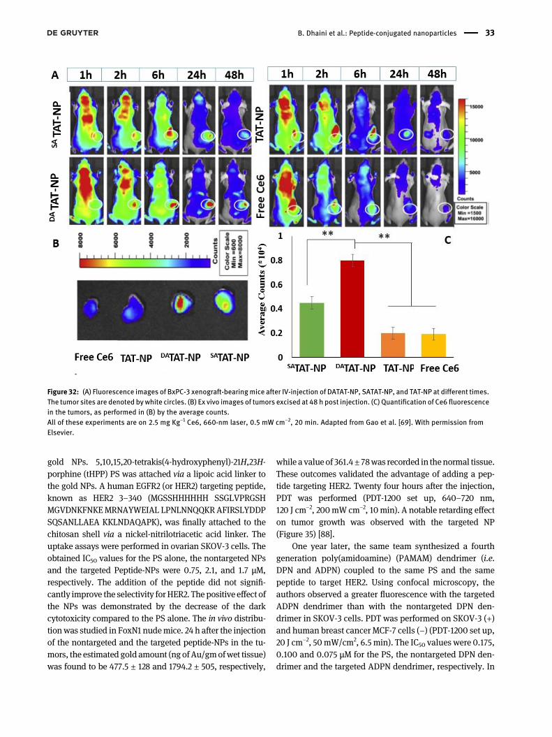
gold NPs. 5,10,15,20-tetrakis(4-hydroxyphenyl)-21H,23H-porphine (tHPP) PS was attached via a lipoic acid linker tothe gold NPs. A human EGFR2 (or HER2) targeting peptide,known as HER2 3–340 (MGSSHHHHHH SSGLVPRGSHMGVDNKFNKEMRNAYWEIAL LPNLNNQQKRAFIRSLYDDPSQSANLLAEA KKLNDAQAPK), was finally attached to thechitosan shell via a nickel-nitrilotriacetic acid linker. Theuptake assays were performed in ovarian SKOV-3 cells. Theobtained IC50 values for the PS alone, the nontargeted NPsand the targeted Peptide-NPs were 0.75, 2.1, and 1.7 μM,respectively. The addition of the peptide did not signifi-cantly improve the selectivity for HER2. The positive effect ofthe NPs was demonstrated by the decrease of the darkcytotoxicity compared to the PS alone. The in vivo distribu-tionwas studied in FoxN1 nudemice. 24 h after the injectionof the nontargeted and the targeted peptide-NPs in the tu-mors, the estimated gold amount (ng ofAu/gmofwet tissue)was found to be 477.5 ± 128 and 1794.2 ± 505, respectively,
while a valueof 361.4± 78was recorded in thenormal tissue.These outcomes validated the advantage of adding a pep-tide targeting HER2. Twenty four hours after the injection,PDT was performed (PDT-1200 set up, 640–720 nm,120 J cm−2, 200mW cm−2, 10min). A notable retarding effecton tumor growth was observed with the targeted NP(Figure 35) [88].
One year later, the same team synthesized a fourthgeneration poly(amidoamine) (PAMAM) dendrimer (i.e.DPN and ADPN) coupled to the same PS and the samepeptide to target HER2. Using confocal microscopy, theauthors observed a greater fluorescence with the targetedADPN dendrimer than with the nontargeted DPN den-drimer in SKOV-3 cells. PDT was performed on SKOV-3 (+)and human breast cancer MCF-7 cells (−) (PDT-1200 set up,20 J cm−2, 50mW/cm2, 6.5 min). The IC50 values were 0.175,0.100 and 0.075 μM for the PS, the nontargeted DPN den-drimer and the targeted ADPN dendrimer, respectively. In
Figure 32: (A) Fluorescence images of BxPC-3 xenograft-bearingmice after IV-injection of DATAT-NP, SATAT-NP, and TAT-NP at different times.The tumor sites are denoted bywhite circles. (B) Ex vivo images of tumors excised at 48 h post injection. (C) Quantification of Ce6 fluorescencein the tumors, as performed in (B) by the average counts.All of these experiments are on 2.5 mg Kg−1 Ce6, 660-nm laser, 0.5 mW cm−2, 20 min. Adapted from Gao et al. [69]. With permission fromElsevier.
B. Dhaini et al.: Peptide-conjugated nanoparticles 33
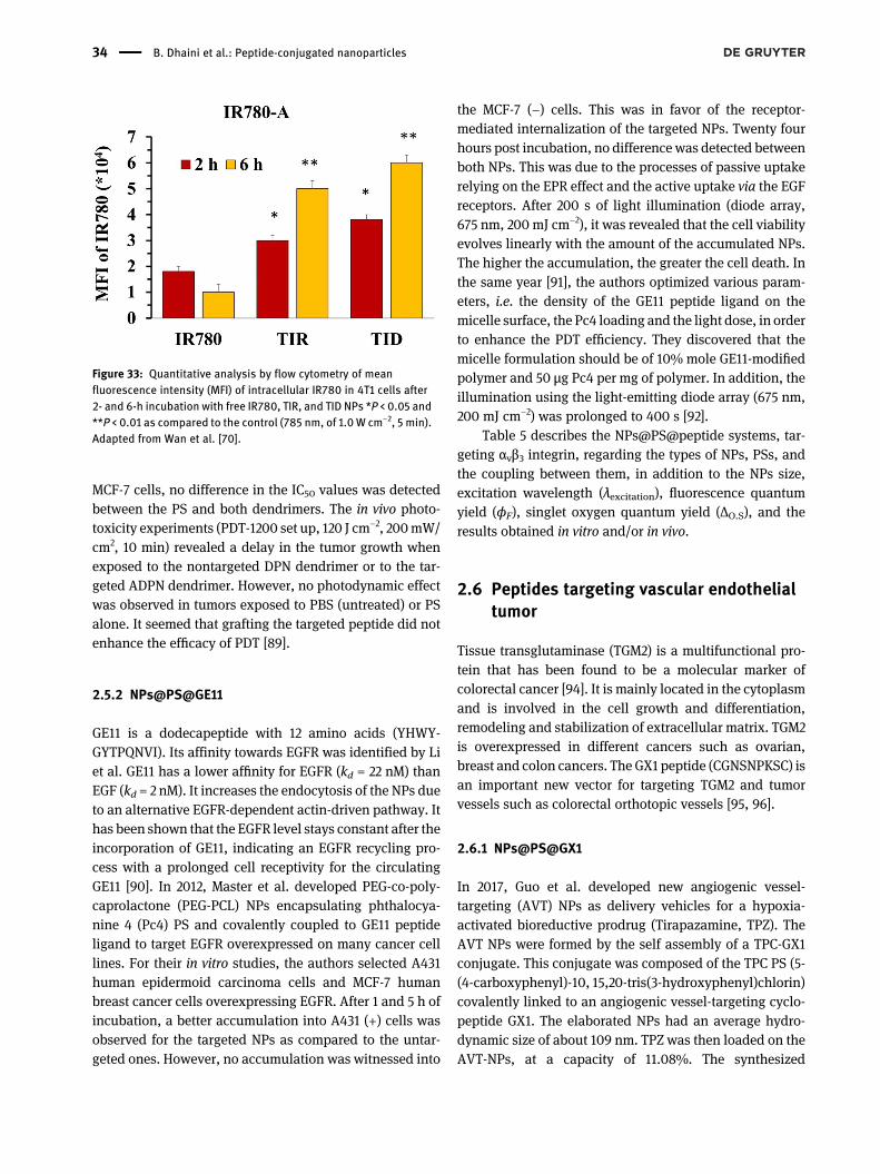
MCF-7 cells, no difference in the IC50 values was detectedbetween the PS and both dendrimers. The in vivo photo-toxicity experiments (PDT-1200 set up, 120 J cm−2, 200mW/cm2, 10 min) revealed a delay in the tumor growth whenexposed to the nontargeted DPN dendrimer or to the tar-geted ADPN dendrimer. However, no photodynamic effectwas observed in tumors exposed to PBS (untreated) or PSalone. It seemed that grafting the targeted peptide did notenhance the efficacy of PDT [89].
2.5.2 NPs@PS@GE11
GE11 is a dodecapeptide with 12 amino acids (YHWY-GYTPQNVI). Its affinity towards EGFR was identified by Liet al. GE11 has a lower affinity for EGFR (kd = 22 nM) thanEGF (kd = 2 nM). It increases the endocytosis of the NPs dueto an alternative EGFR-dependent actin-driven pathway. Ithas been shown that the EGFR level stays constant after theincorporation of GE11, indicating an EGFR recycling pro-cess with a prolonged cell receptivity for the circulatingGE11 [90]. In 2012, Master et al. developed PEG-co-poly-caprolactone (PEG-PCL) NPs encapsulating phthalocya-nine 4 (Pc4) PS and covalently coupled to GE11 peptideligand to target EGFR overexpressed on many cancer celllines. For their in vitro studies, the authors selected A431human epidermoid carcinoma cells and MCF-7 humanbreast cancer cells overexpressing EGFR. After 1 and 5 h ofincubation, a better accumulation into A431 (+) cells wasobserved for the targeted NPs as compared to the untar-geted ones. However, no accumulation was witnessed into
the MCF-7 (−) cells. This was in favor of the receptor-mediated internalization of the targeted NPs. Twenty fourhours post incubation, no difference was detected betweenboth NPs. This was due to the processes of passive uptakerelying on the EPR effect and the active uptake via the EGFreceptors. After 200 s of light illumination (diode array,675 nm, 200 mJ cm−2), it was revealed that the cell viabilityevolves linearly with the amount of the accumulated NPs.The higher the accumulation, the greater the cell death. Inthe same year [91], the authors optimized various param-eters, i.e. the density of the GE11 peptide ligand on themicelle surface, the Pc4 loading and the light dose, in orderto enhance the PDT efficiency. They discovered that themicelle formulation should be of 10% mole GE11-modifiedpolymer and 50 μg Pc4 per mg of polymer. In addition, theillumination using the light-emitting diode array (675 nm,200 mJ cm−2) was prolonged to 400 s [92].
Table 5 describes the NPs@PS@peptide systems, tar-geting αvβ3 integrin, regarding the types of NPs, PSs, andthe coupling between them, in addition to the NPs size,excitation wavelength (λexcitation), fluorescence quantumyield (ϕF), singlet oxygen quantum yield (ΔO.S), and theresults obtained in vitro and/or in vivo.
2.6 Peptides targeting vascular endothelialtumor
Tissue transglutaminase (TGM2) is a multifunctional pro-tein that has been found to be a molecular marker ofcolorectal cancer [94]. It is mainly located in the cytoplasmand is involved in the cell growth and differentiation,remodeling and stabilization of extracellular matrix. TGM2is overexpressed in different cancers such as ovarian,breast and colon cancers. The GX1 peptide (CGNSNPKSC) isan important new vector for targeting TGM2 and tumorvessels such as colorectal orthotopic vessels [95, 96].
2.6.1 NPs@PS@GX1
In 2017, Guo et al. developed new angiogenic vessel-targeting (AVT) NPs as delivery vehicles for a hypoxia-activated bioreductive prodrug (Tirapazamine, TPZ). TheAVT NPs were formed by the self assembly of a TPC-GX1conjugate. This conjugate was composed of the TPC PS (5-(4-carboxyphenyl)-10, 15,20-tris(3-hydroxyphenyl)chlorin)covalently linked to an angiogenic vessel-targeting cyclo-peptide GX1. The elaborated NPs had an average hydro-dynamic size of about 109 nm. TPZ was then loaded on theAVT-NPs, at a capacity of 11.08%. The synthesized
Figure 33: Quantitative analysis by flow cytometry of meanfluorescence intensity (MFI) of intracellular IR780 in 4T1 cells after2- and 6-h incubation with free IR780, TIR, and TID NPs *P < 0.05 and**P < 0.01 as compared to the control (785 nm, of 1.0 W cm−2, 5 min).Adapted from Wan et al. [70].
34 B. Dhaini et al.: Peptide-conjugated nanoparticles
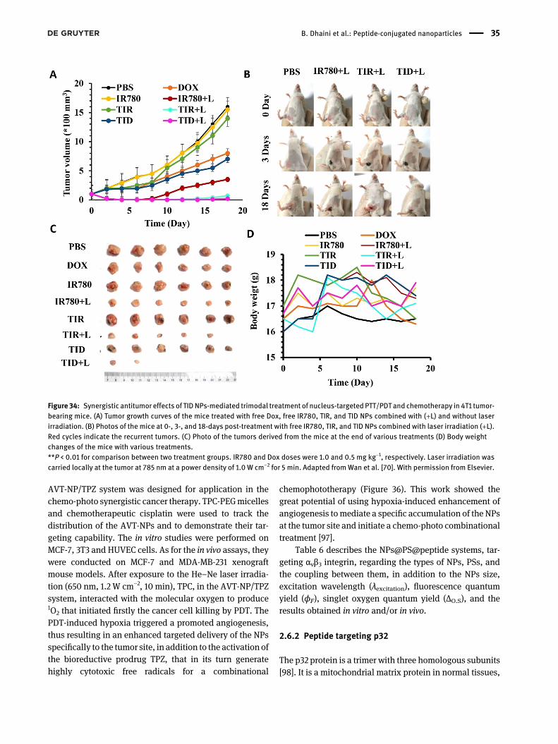
AVT-NP/TPZ system was designed for application in thechemo-photo synergistic cancer therapy. TPC-PEGmicellesand chemotherapeutic cisplatin were used to track thedistribution of the AVT-NPs and to demonstrate their tar-geting capability. The in vitro studies were performed onMCF-7, 3T3 and HUVEC cells. As for the in vivo assays, theywere conducted on MCF-7 and MDA-MB-231 xenograftmouse models. After exposure to the He–Ne laser irradia-tion (650 nm, 1.2 W cm−2, 10 min), TPC, in the AVT-NP/TPZsystem, interacted with the molecular oxygen to produce1O2 that initiated firstly the cancer cell killing by PDT. ThePDT-induced hypoxia triggered a promoted angiogenesis,thus resulting in an enhanced targeted delivery of the NPsspecifically to the tumor site, in addition to the activation ofthe bioreductive prodrug TPZ, that in its turn generatehighly cytotoxic free radicals for a combinational
chemophototherapy (Figure 36). This work showed thegreat potential of using hypoxia-induced enhancement ofangiogenesis tomediate a specific accumulation of the NPsat the tumor site and initiate a chemo-photo combinationaltreatment [97].
Table 6 describes the NPs@PS@peptide systems, tar-geting αvβ3 integrin, regarding the types of NPs, PSs, andthe coupling between them, in addition to the NPs size,excitation wavelength (λexcitation), fluorescence quantumyield (ϕF), singlet oxygen quantum yield (ΔO.S), and theresults obtained in vitro and/or in vivo.
2.6.2 Peptide targeting p32
The p32 protein is a trimer with three homologous subunits[98]. It is a mitochondrial matrix protein in normal tissues,
Figure 34: Synergistic antitumor effects of TIDNPs-mediated trimodal treatment of nucleus-targeted PTT/PDT and chemotherapy in 4T1 tumor-bearing mice. (A) Tumor growth curves of the mice treated with free Dox, free IR780, TIR, and TID NPs combined with (+L) and without laserirradiation. (B) Photos of the mice at 0-, 3-, and 18-days post-treatment with free IR780, TIR, and TID NPs combined with laser irradiation (+L).Red cycles indicate the recurrent tumors. (C) Photo of the tumors derived from the mice at the end of various treatments (D) Body weightchanges of the mice with various treatments.**P < 0.01 for comparison between two treatment groups. IR780 and Dox doses were 1.0 and 0.5 mg kg−1, respectively. Laser irradiation wascarried locally at the tumor at 785 nm at a power density of 1.0 W cm−2 for 5 min. Adapted fromWan et al. [70]. With permission from Elsevier.
B. Dhaini et al.: Peptide-conjugated nanoparticles 35
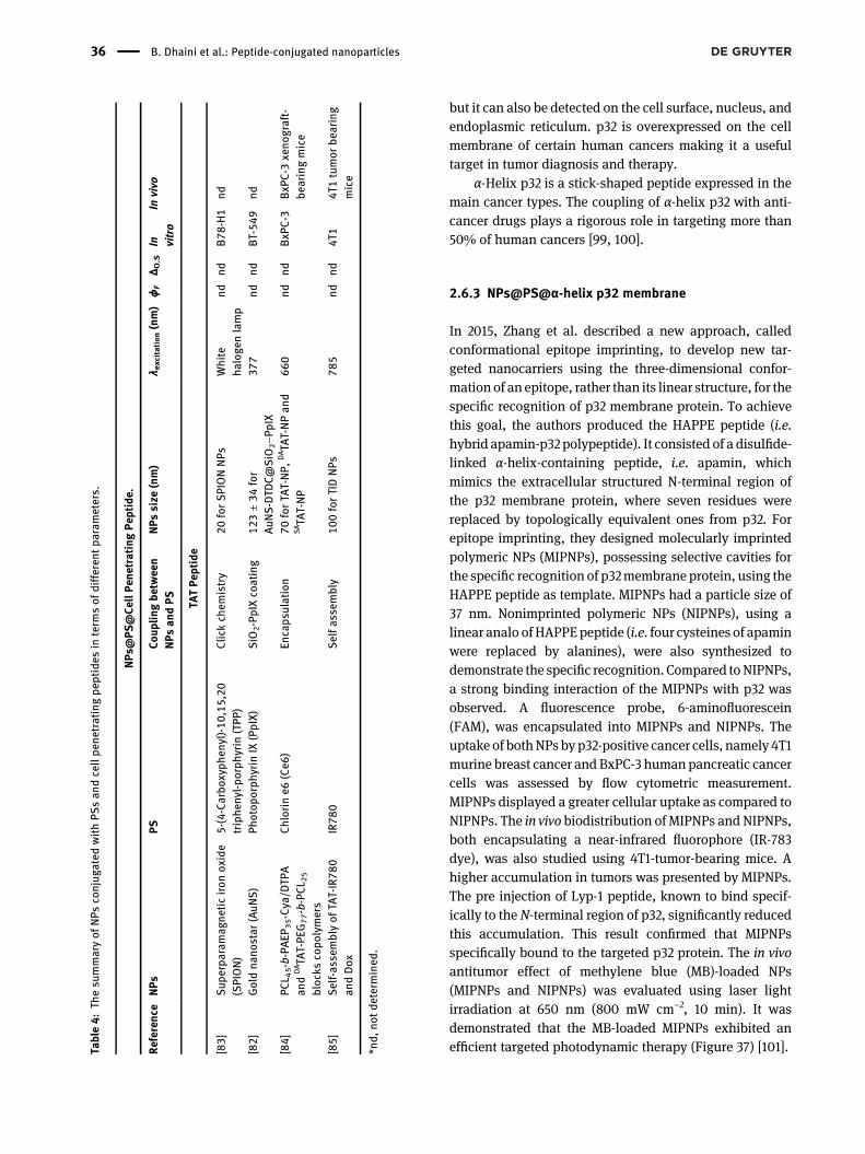
but it can also be detected on the cell surface, nucleus, andendoplasmic reticulum. p32 is overexpressed on the cellmembrane of certain human cancers making it a usefultarget in tumor diagnosis and therapy.
α-Helix p32 is a stick-shaped peptide expressed in themain cancer types. The coupling of α-helix p32 with anti-cancer drugs plays a rigorous role in targeting more than50% of human cancers [99, 100].
2.6.3 NPs@PS@α-helix p32 membrane
In 2015, Zhang et al. described a new approach, calledconformational epitope imprinting, to develop new tar-geted nanocarriers using the three-dimensional confor-mation of an epitope, rather than its linear structure, for thespecific recognition of p32 membrane protein. To achievethis goal, the authors produced the HAPPE peptide (i.e.hybrid apamin-p32 polypeptide). It consisted of a disulfide-linked α-helix-containing peptide, i.e. apamin, whichmimics the extracellular structured N-terminal region ofthe p32 membrane protein, where seven residues werereplaced by topologically equivalent ones from p32. Forepitope imprinting, they designed molecularly imprintedpolymeric NPs (MIPNPs), possessing selective cavities forthe specific recognition of p32membrane protein, using theHAPPE peptide as template. MIPNPs had a particle size of37 nm. Nonimprinted polymeric NPs (NIPNPs), using alinear analo ofHAPPEpeptide (i.e. four cysteines of apaminwere replaced by alanines), were also synthesized todemonstrate the specific recognition. Compared toNIPNPs,a strong binding interaction of the MIPNPs with p32 wasobserved. A fluorescence probe, 6-aminofluorescein(FAM), was encapsulated into MIPNPs and NIPNPs. Theuptake of bothNPs by p32-positive cancer cells, namely 4T1murine breast cancer andBxPC-3 human pancreatic cancercells was assessed by flow cytometric measurement.MIPNPs displayed a greater cellular uptake as compared toNIPNPs. The in vivo biodistribution of MIPNPs and NIPNPs,both encapsulating a near-infrared fluorophore (IR-783dye), was also studied using 4T1-tumor-bearing mice. Ahigher accumulation in tumors was presented by MIPNPs.The pre injection of Lyp-1 peptide, known to bind specif-ically to the N-terminal region of p32, significantly reducedthis accumulation. This result confirmed that MIPNPsspecifically bound to the targeted p32 protein. The in vivoantitumor effect of methylene blue (MB)-loaded NPs(MIPNPs and NIPNPs) was evaluated using laser lightirradiation at 650 nm (800 mW cm−2, 10 min). It wasdemonstrated that the MB-loaded MIPNPs exhibited anefficient targeted photodynamic therapy (Figure 37) [101].Ta
ble:Th
esu
mmaryof
NPs
conjug
ated
withPS
san
dcellpe
netratingpe
ptides
interm
sof
differen
tpa
rameters.
NPs
@PS
@Ce
llPe
netratingPe
ptide.
Referen
ceNPs
PSCo
uplin
gbe
twee
nNPs
andPS
NPs
size
(nm)
λ excitation(nm)
ϕF
Δ O.S
In vitro
Invivo
TATPe
ptide
[]
Sup
erpa
ramag
neticiron
oxide
(SPION)
-(-Carbo
xyph
enyl)-,,
triphe
nyl-po
rphy
rin(TPP
)Clickch
emistry
forSPIONNPs
White
haloge
nlamp
ndnd
B-H
nd
[]
Goldna
nostar
(AuN
S)
Photop
orph
yrin
IX(PpIX)
SiO
-PpIXcoating
±for
AuN
S-DTD
C@SiO
−P
pIX
ndnd
BT-
nd
[]
PCL
-b-PAE
P -Cya/D
TPA
and
DA TAT-PE
G-b-PCL
blocks
copo
lymers
Chlorin
e(Ce
)En
caps
ulation
forTA
T-NP,
DATA
T-NPan
dSA TAT-NP
ndnd
BxP
C-
BxP
C-
xeno
graft-
bearingmice
[]
Self-assemblyof
TAT-IR
andDox
IR
Selfa
ssem
bly
forTIDNPs
ndnd
T
T
tumor
bearing
mice
*nd,
notde
term
ined
.
36 B. Dhaini et al.: Peptide-conjugated nanoparticles

Figure 35: Tumor growth curve in nude mice.On day 0, PS formulations were injected viatail vein; and on day 1, PDT was conducted.(A) PBS, (B) PS (2 µM), (C1) ADPN (2 µM ofPS), and (C2) ADPN (2 µM of PS). Laserirradiation with 640–720 nm (200 mW cm−2
for 10 min).Adapted from Narsireddy et al. [74]. Withpermission from Elsevier.
Table : The summary of NPs conjugated with PSs and human EGFR targeting peptides in terms of different parameters.
NPs@PS@Peptide targeting human EGFR
Reference NPs PS Coupling be-tween NPsand PS
NPs size(nm)
λexcitation(nm)
ϕF ΔO.S In vitro In vivo
HER – Peptide
[] Dendrimer (G) ,,,-Tetrakis(-hydroxyphenyl)-H,H-porphine(tHPP)
Amide bond for DPNand ≈– forADPNaggregates
nd nd SKOV-MCF-
FoxNNudemice
[] FeO-Chitosan-Au
,,,-Tetrakis(-hydroxyphenyl)-H,H-porphine(tHPP)
Ester bond – forFeO NPsand forMGPSa
nd nd SKOV- FoxNNudemice
GE Peptide
[, ] Polyethyleneglycol-co-polycaprolactone(PEG-PCL)
Phthalocyanine
(Pc)Encapsulated < for
Pc inPEG-PCLmicelles
nd nd AMCF-c
nd
a MGPS: Magnetic/gold particles with PS, *nd, not determined.
Figure 36: MCF-7 tumor (A) andMDA-MB-231 (B) growth curves after intravenous injection of different drug formulations (0.1 mg/kg; Ps/bodyweight).Error bars indicate standard deviations (n = 5).*P < 0.05, **P < 0.01, ***P < 0.001 (two-tailed Student’s-test). He–Ne laser irradiation (650 nm,1.2 W/cm2 for 10 min). Adapted from Guo et al. [82].
B. Dhaini et al.: Peptide-conjugated nanoparticles 37
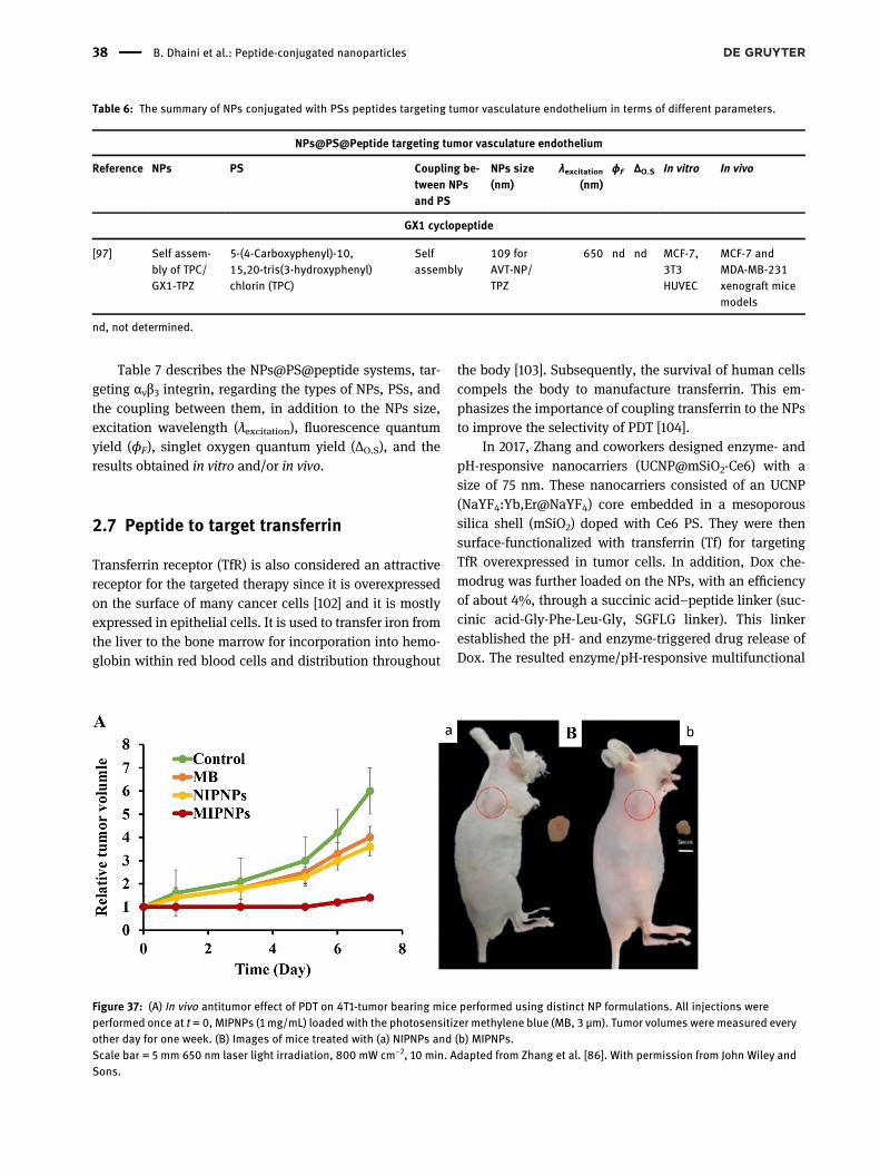
Table 7 describes the NPs@PS@peptide systems, tar-geting αvβ3 integrin, regarding the types of NPs, PSs, andthe coupling between them, in addition to the NPs size,excitation wavelength (λexcitation), fluorescence quantumyield (ϕF), singlet oxygen quantum yield (ΔO.S), and theresults obtained in vitro and/or in vivo.
2.7 Peptide to target transferrin
Transferrin receptor (TfR) is also considered an attractivereceptor for the targeted therapy since it is overexpressedon the surface of many cancer cells [102] and it is mostlyexpressed in epithelial cells. It is used to transfer iron fromthe liver to the bone marrow for incorporation into hemo-globin within red blood cells and distribution throughout
the body [103]. Subsequently, the survival of human cellscompels the body to manufacture transferrin. This em-phasizes the importance of coupling transferrin to the NPsto improve the selectivity of PDT [104].
In 2017, Zhang and coworkers designed enzyme- andpH-responsive nanocarriers (UCNP@mSiO2-Ce6) with asize of 75 nm. These nanocarriers consisted of an UCNP(NaYF4:Yb,Er@NaYF4) core embedded in a mesoporoussilica shell (mSiO2) doped with Ce6 PS. They were thensurface-functionalized with transferrin (Tf) for targetingTfR overexpressed in tumor cells. In addition, Dox che-modrug was further loaded on the NPs, with an efficiencyof about 4%, through a succinic acid–peptide linker (suc-cinic acid-Gly-Phe-Leu-Gly, SGFLG linker). This linkerestablished the pH- and enzyme-triggered drug release ofDox. The resulted enzyme/pH-responsive multifunctional
Table : The summary of NPs conjugated with PSs peptides targeting tumor vasculature endothelium in terms of different parameters.
NPs@PS@Peptide targeting tumor vasculature endothelium
Reference NPs PS Coupling be-tween NPsand PS
NPs size(nm)
λexcitation(nm)
ϕF ΔO.S In vitro In vivo
GX cyclopeptide
[] Self assem-bly of TPC/GX-TPZ
-(-Carboxyphenyl)-,,-tris(-hydroxyphenyl)chlorin (TPC)
Selfassembly
forAVT-NP/TPZ
nd nd MCF-,THUVEC
MCF- andMDA-MB-xenograft micemodels
nd, not determined.
Figure 37: (A) In vivo antitumor effect of PDT on 4T1-tumor bearing mice performed using distinct NP formulations. All injections wereperformed once at t = 0, MIPNPs (1 mg/mL) loaded with the photosensitizer methylene blue (MB, 3 µm). Tumor volumes were measured everyother day for one week. (B) Images of mice treated with (a) NIPNPs and (b) MIPNPs.Scale bar = 5 mm 650 nm laser light irradiation, 800 mW cm−2, 10 min. Adapted from Zhang et al. [86]. With permission from John Wiley andSons.
38 B. Dhaini et al.: Peptide-conjugated nanoparticles

nanocomposite (UCNP@mSiO2/Ce6-Dox-SGFLG-Tf) wasdeveloped for application in synergistic PDT and chemo-therapy. After the endocytic uptake of the nanocompositeinto the tumor cells by pinocytosis or phagocytosis, varioussteps took place. First of all, the pH- and enzyme-triggeredDox release occurred due to the high concentration ofcathepsin B and the low pH condition, thus illustrating achemotherapeutic effect. Secondly, a 5 min NIR irradiationof UCNPs (980 nm, 0.5 W cm−2) led to a visible-lightemission, which enabled the efficient excitation of Ce6 andthe further generation of ROS, illustrating a photodynamiceffect. The enzyme- and pH-dependent Dox release fromUCNP@mSiO2/Ce6-Dox-SGFLG-Tf was estimated in PBSusing different parameters. With the pH fixed at 6.8, bettervalues were obtained at high cathepsin concentration, forexample 60.17% of Dox were released at 2 × 10⁻⁷ mol/L. Bytesting various pH conditions, from 1.2 to 6.8, better valueswere obtained at low pH, for instance 78.36% of Dox wasreleased at pH 1.2. The in vitro cell viability assays werecarried out on cancerousHeLa cells and L02 normal humanliver cells. These tests demonstrated a synergistic PDT andchemotherapeutic effect under NIR irradiation. This effectwas more drastic using UCNP@mSiO2/Ce6-Dox-SGFLG-Tfnanocomposite and NIR irradiation (980 nm, 0.5 W cm−2,15 min) leading to a HeLa cell viability of 33% (Figure 38)[105].
Table 8 describes the NPs@PS@peptide systems, tar-geting αvβ3 integrin, regarding the types of NPs, PSs, andthe coupling between them, in addition to the NPs size,excitation wavelength (λexcitation), fluorescence quantumyield (ϕF), singlet oxygen quantum yield (ΔO.S), and theresults obtained in vitro and/or in vivo.
2.8 Peptide to target PD-1
Programed death-1 (PD-1) is a cell surface moleculethat regulates the adaptive immune response [106]. The
engagement of PD-1 by its PD-L1 or PD-L2 ligands trans-duces a signal that inhibits T cell proliferation, cytokineproduction, and cytolytic function. T lymphocytes arewhite blood cells that are specialized in certain immunereactions. It helps fight infections and destroy abnormalcells, including cancer cells.
In 2020, B. Liu et al. described the development ofmultifunctional PEG-coated gold nanoprisms (GNPs)incorporating Ce6 PS and coupled to PD-L1 targeting pep-tide [96]. These NPs were designed for enhanced imagingand PTT/PDT applications. Using NIR and photoacousticimaging, the authors proved that the NPs presented a highaffinity for PD-L1 receptors both in vivo and ex vivo in themain organs and tissues. They also showed a synergeticPDT and PTT effect leading to the inhibition of the tumorgrowth in vivo.
Table 9 describes the NPs@PS@peptide systems, tar-geting PD-1, regarding the types of NPs, PSs, and thecoupling between them, in addition to the NPs size, exci-tation wavelength (λexcitation), fluorescence quantum yield(ϕF), singlet oxygen quantum yield (ΔO.S), and the resultsobtained in vitro and/or in vivo.
2.9 Bioactive peptide targetingmany cancertypes
Bioactive peptides are short protein fragments of 2–20amino acids [108]. These peptides have revealed the abilityto overcome cell membranes and cause cell death [109].Since then, bioactive peptides have been used to targettumor cells in general [108]. Some researchers cited thatcoupling a bioactive targeting peptide can target plasmamembrane and mitochondria.
In 2019, Cheng et al. described the synthesis of achimeric peptide that consisted of a hydrophobic PpIX PSconjugated, via a lysine linker, to a bioactive peptide(rFxrFxrFxr; r and Fx stand for D-arginine and L-
Table : The summary of NPs conjugated with PSs and peptides targeting tumor suppressor protein p in terms of different parameters.
NPs@PS@Peptide targeting tumor suppressor protein p
Reference NPs PS Coupling betweenNPs and PS
NPs size(nm)
λexcitation(nm)
ϕF ΔO.S In vitro In vivo
α-helix containing peptide, apamin
[] Molecularly imprintedpolymeric (MIPNPs)
Methylene blue(MB)
Encapsulated forMIPNPs
nd nd TBxPC-
T-Tumor-bearing mice
nd, not determined.
B. Dhaini et al.: Peptide-conjugated nanoparticles 39
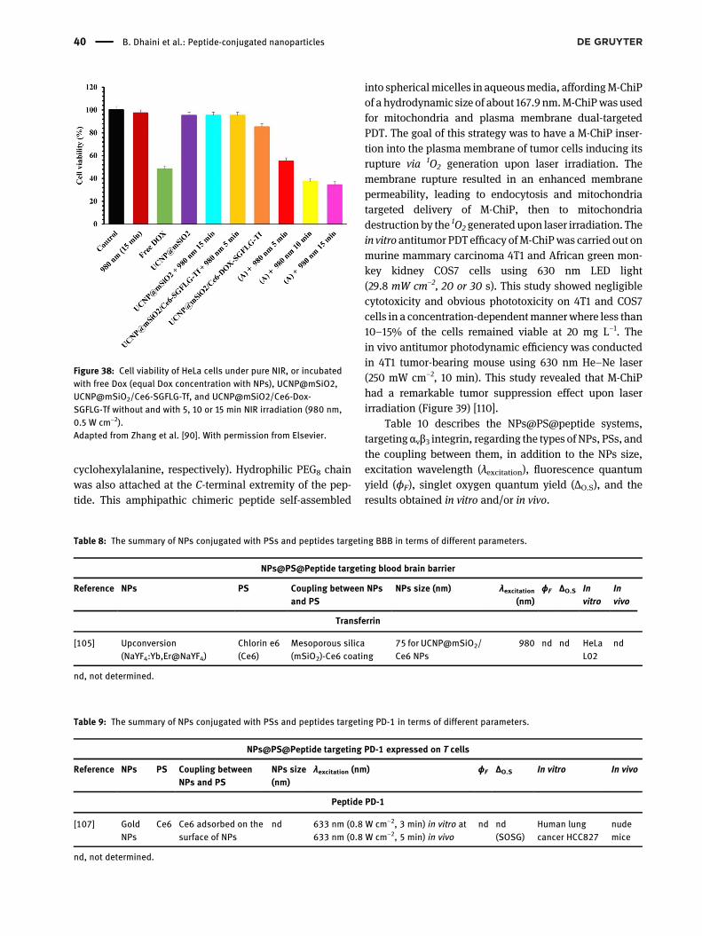
cyclohexylalanine, respectively). Hydrophilic PEG8 chainwas also attached at the C-terminal extremity of the pep-tide. This amphipathic chimeric peptide self-assembled
into sphericalmicelles in aqueousmedia, affordingM-ChiPof a hydrodynamic size of about 167.9 nm.M-ChiPwas usedfor mitochondria and plasma membrane dual-targetedPDT. The goal of this strategy was to have a M-ChiP inser-tion into the plasma membrane of tumor cells inducing itsrupture via 1O2 generation upon laser irradiation. Themembrane rupture resulted in an enhanced membranepermeability, leading to endocytosis and mitochondriatargeted delivery of M-ChiP, then to mitochondriadestruction by the 1O2 generated upon laser irradiation. Thein vitro antitumor PDT efficacy ofM-ChiPwas carried out onmurine mammary carcinoma 4T1 and African green mon-key kidney COS7 cells using 630 nm LED light(29.8 mW cm−2, 20 or 30 s). This study showed negligiblecytotoxicity and obvious phototoxicity on 4T1 and COS7cells in a concentration-dependentmannerwhere less than10–15% of the cells remained viable at 20 mg L−1. Thein vivo antitumor photodynamic efficiency was conductedin 4T1 tumor-bearing mouse using 630 nm He–Ne laser(250 mW cm−2, 10 min). This study revealed that M-ChiPhad a remarkable tumor suppression effect upon laserirradiation (Figure 39) [110].
Table 10 describes the NPs@PS@peptide systems,targeting αvβ3 integrin, regarding the types of NPs, PSs, andthe coupling between them, in addition to the NPs size,excitation wavelength (λexcitation), fluorescence quantumyield (ϕF), singlet oxygen quantum yield (ΔO.S), and theresults obtained in vitro and/or in vivo.
Table : The summary of NPs conjugated with PSs and peptides targeting BBB in terms of different parameters.
NPs@PS@Peptide targeting blood brain barrier
Reference NPs PS Coupling between NPsand PS
NPs size (nm) λexcitation(nm)
ϕF ΔO.S Invitro
Invivo
Transferrin
[] Upconversion(NaYF:Yb,Er@NaYF)
Chlorin e(Ce)
Mesoporous silica(mSiO)-Ce coating
for UCNP@mSiO/Ce NPs
nd nd HeLaL
nd
nd, not determined.
Figure 38: Cell viability of HeLa cells under pure NIR, or incubatedwith free Dox (equal Dox concentration with NPs), UCNP@mSiO2,UCNP@mSiO2/Ce6-SGFLG-Tf, and UCNP@mSiO2/Ce6-Dox-SGFLG-Tf without and with 5, 10 or 15 min NIR irradiation (980 nm,0.5 W cm−2).Adapted from Zhang et al. [90]. With permission from Elsevier.
Table : The summary of NPs conjugated with PSs and peptides targeting PD- in terms of different parameters.
NPs@PS@Peptide targeting PD- expressed on T cells
Reference NPs PS Coupling betweenNPs and PS
NPs size(nm)
λexcitation (nm) ϕF ΔO.S In vitro In vivo
Peptide PD-
[] GoldNPs
Ce Ce adsorbed on thesurface of NPs
nd nm (. W cm−, min) in vitro at nm (. W cm−, min) in vivo
nd nd(SOSG)
Human lungcancer HCC
nudemice
nd, not determined.
40 B. Dhaini et al.: Peptide-conjugated nanoparticles
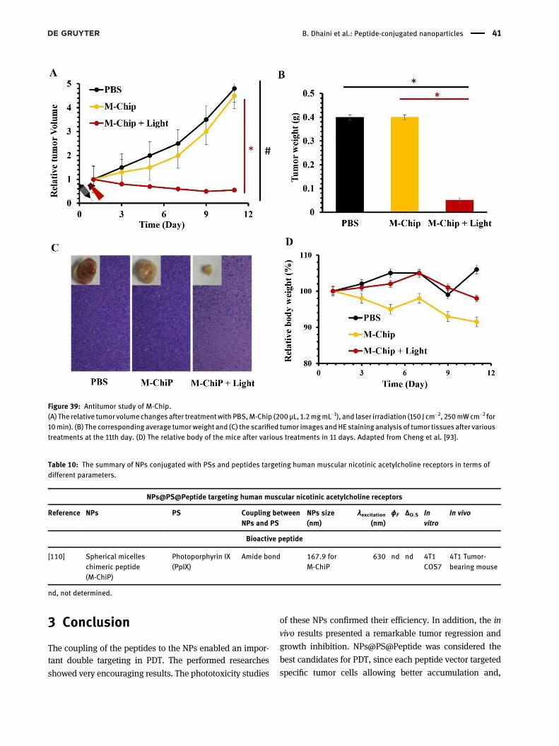
3 Conclusion
The coupling of the peptides to the NPs enabled an impor-tant double targeting in PDT. The performed researches
showed very encouraging results. The phototoxicity studies
of these NPs confirmed their efficiency. In addition, the in
vivo results presented a remarkable tumor regression and
growth inhibition. NPs@PS@Peptide was considered the
best candidates for PDT, since each peptide vector targeted
specific tumor cells allowing better accumulation and,
Figure 39: Antitumor study of M-Chip.(A) The relative tumor volume changesafter treatmentwith PBS,M-Chip (200µL, 1.2mgmL−1), and laser irradiation (150 J cm−2, 250mWcm−2 for10min). (B) The corresponding average tumorweight and (C) the scarified tumor images andHE staining analysis of tumor tissues after varioustreatments at the 11th day. (D) The relative body of the mice after various treatments in 11 days. Adapted from Cheng et al. [93].
Table : The summary of NPs conjugated with PSs and peptides targeting human muscular nicotinic acetylcholine receptors in terms ofdifferent parameters.
NPs@PS@Peptide targeting human muscular nicotinic acetylcholine receptors
Reference NPs PS Coupling betweenNPs and PS
NPs size(nm)
λexcitation(nm)
ϕF ΔO.S Invitro
In vivo
Bioactive peptide
[] Spherical micelleschimeric peptide(M-ChiP)
Photoporphyrin IX(PpIX)
Amide bond . forM-ChiP
nd nd TCOS
T Tumor-bearing mouse
nd, not determined.
B. Dhaini et al.: Peptide-conjugated nanoparticles 41
consequently, a superior photodynamic effect. Among thesebioactive peptides, research results validated the effective-ness of the cell-penetrating peptides, such as TAT, thatfacilitated the penetration of the NPs into the cells. The invitro and in vivo tests confirmed that the peptides DKPPR,ATWLPPR, CRGDK, and tLyp-1 targeted NRP-1 receptor,which has been evaluated in the whole sections of 65 pri-mary breast carcinomas, 95 primary colorectal adenocarci-nomas, 90 primary lung carcinomas, 59 additional humanmetastases, and 16 xenografts. NRP-1 immunoreactivity hadbeen observed in normal tissue vessels adjacent to thecancer cells and in 98–100% of carcinomas. The expressionof NRP-1 in tumor cells was also observed in 36% of primarylung carcinomas and 6% of primary mammary carcinomas,but not in the colorectal adenocarcinoma. NRP-1 was alsoevaluated in mouse embryos, where their expression waslimited to the nervous system, endocardium, and vascularsmooth muscle and in endothelium on subsets of vessels.Other peptides such as RGD, iRGD, cRGD, c(RGDyK),RGD-4R,RGDfK, andfibronectin (Fmp), validated, by in vitroand in vivo tests, their ability to target αvβ3 integrins that arestrongly expressed in normal mammary epithelium and inother primary invasive breast carcinomas. This integrinheterodimer was also abundant in all the breast cancer andthe metastatic bone cancer cells. The in situ hybridizationrevealed high levels of mRNA at steady state correspondingto the protein expression sites. In addition toNRP-1 and αvβ3integrins, transferrin was also a receptor of interest in thetargeted PDT. Transferrin (TfR) is a receptor found on theBBB. Regarding the high selective permeability imposed byBBB, the photodynamic therapy with NPs@PS@transferrinallowed the penetration of transferrin protein, thus over-coming the difficulty of treating central nervous system(CNS) diseases by standard drugs such as chemotherapy.Coupling transferrin to the nanoparticles (NPs@PS@trans-ferrin) enabled a superior and selective accumulation. Thisaccumulation was confirmed by the fluorescence of the PS.On this basis, the Transferrin peptide targeted successfullythe brain and blood cancers. Moreover, the efficiency oftargeting by α-helix p32 peptide was also assessed. Thispeptide targeted all the tumors overexpressing p32 protein.In addition, the efficacy of F3 Peptide to target tissue factorF3 was confirmed. This factor is responsible for activatingblood clotting and is expressed in most solid tumors. Otherresearch efforts had also verified that GX1 peptide targetedthe human gastric cancer (GC) vasculature by generallybinding to the endothelial cells. The overexpression of EGFRcontributes to the malignant transformation of humanepithelial cells indicating gene mutations. It is mostlyexpressed in breast cancer. Various studies validated thatthe peptides GX1, HER2 3–340, GE11 are important vectors
for targeting EGFR due to the overexpression of tissuetransglutaminase (TGM2). TGM2 is amultifunctional proteinlocated in the cytoplasm which has been shown to be amolecular marker of colorectal cancer.
Author contributions: All the authors have acceptedresponsibility for the entire content of this submittedmanuscript and approved submission.Research funding: None declared.Conflict of interest statement: The authors declare noconflicts of interest regarding this article.
References
[1] A. l. Doerflinger, Design and Synthesis of Polymerized Micellesfor the Diagnosis and Therapy of Tumours, Paris, UniversitéParis-Saclay, 2017.
[2] M. S. Rajagopalan, D. E. Heron, R. E. Wegner, et al., “Pathologicresponsewith neoadjuvant chemotherapy and stereotactic bodyradiotherapy for borderline resectable and locally-advancedpancreatic cancer,” Radiat. Oncol., vol. 8, p. 254, 2013.
[3] M. A. Bellavance,M. Blanchette, andD. Fortin, “Recent advancesin blood-brain barrier disruption as a CNS delivery strategy,”AAPS J., vol. 10, pp. 166–177, 2008.
[4] A. F. dos Santos, D. R. Q. de Almeida, L. F. Terra, M. S. Baptista,and L. Labriola, “Photodynamic therapy in cancer treatment - anupdate review,” J. Canc. Metastasis Treat., vol. 5, p. 25, 2019.
[5] R.-M. Szeimies, J. Dräger, C. Abels, andM. Landthaler, “Chapter 1history of photodynamic therapy in dermatology,” inComprehensive Series in Photosciences, vol. 2,P. Calzavara-Pinton, R.-M. Szeimies, and B. Ortel, Eds.,Amsterdam, Elsevier, 2001, pp. 3–15.
[6] C. Frochot, M. Barberi-Heyob, M. Blanchard-Desce, et al,“La thérapie photodynamique : état de l’art et perspectives,”L'Actualité Chimique, Société chimique de France, La chimie fêtela lumière, 2015, pp. 46–50.
[7] P. D. Silva, M. A. Saad, H. C. Thomsen, S. Bano, S. Ashraf, andT. Hasan, “Photodynamic therapy, priming and optical imaging:potential co-conspirators in treatment design and optimization—a Thomas Dougherty Award for Excellence in PDT paper,”J. Porphyr. Phthalocyanines, vol. 24, pp. 1320–1360, 2020.
[8] D. Kessel and N. L. Oleinick, “Cell death pathways associatedwith photodynamic therapy: An update,”Photochem. Photobiol.,vol. 94, pp. 213–218, 2018.
[9] T. J. Dougherty, J. E. Kaufman, A. Goldfarb, K. R. Weishaupt,D. Boyle, and A. Mittleman, “Photoradiation therapy for thetreatment of malignant tumors,” Canc. Res., vol. 38,pp. 2628–2635, 1978.
[10] R. R. Allison and K. Moghissi, “Photodynamic therapy (PDT): PDTmechanisms,” Clin. Endosc., vol. 46, pp. 24–29, 2013.
[11] S. B. Brown, E. A. Brown, and I. Walker, “The present and futurerole of photodynamic therapy in cancer treatment,” LancetOncol, vol. 5, pp. 497–508, 2004.
[12] M. Lan, S. Zhao, W. Liu, C.-S. Lee, W. Zhang, and P. Wang,“Photosensitizers for photodynamic therapy,” Adv. Healthc.Mater., vol. 8, 2019, Art no. 1900132.
42 B. Dhaini et al.: Peptide-conjugated nanoparticles
[13] X. Dai, T. Du, and K. Han, “Engineering nanoparticles foroptimized photodynamic therapy,” ACS Biomater. Sci. Eng.,vol. 5, pp. 6342–6354, 2019.
[14] L. Larue, A. Ben Mihoub, Z. Youssef, et al., “Using X-rays inphotodynamic therapy: An overview,” Photochem. Photobiol.Sci., vol. 17, pp. 1612–1650, 2018.
[15] B. Sun, R. Chang, S. Cao, et al., “Acid-Activatable transmorphicpeptide-based nanomaterials for photodynamic therapy,”Angew. Chem. Int. Ed., vol. 59, pp. 20582–20588, 2020.
[16] H. Azaïs, C. Schmitt, M. Tardivel, et al., “Assessment of thespecificity of a new folate-targeted photosensitizer forperitoneal metastasis of epithelial ovarian cancer to enableintraperitoneal photodynamic therapy. A preclinical study,”Photodiagnosis Photodyn. Ther., vol. 13, pp. 130–138, 2016.
[17] A. M. Gazzali, M. Lobry, L. Colombeau, et al., “Stability of folicacid under several parameters,” Eur. J. Pharmaceut. Sci., vol. 93,pp. 419–430, 2016.
[18] W. M. Sharman, J. E. van Lier, and C. M. Allen, “Targetedphotodynamic therapy via receptor mediated delivery systems,”Adv. Drug Deliv. Rev., vol. 56, pp. 53–76, 2004.
[19] D. Zahavi and L. Weiner, “Monoclonal antibodies in cancertherapy,” Antibodies, vol. 9, p. 34, 2020.
[20] O. H. Aina, T. C. Sroka, M.-L. Chen, and K. S. Lam, “Therapeuticcancer targeting peptides,” Biopolymers, vol. 66, pp. 184–199,2002.
[21] S. Li, Y. Jin, Y. Su, et al., “Anti-HER2 affibody-conjugatedphotosensitizer for tumor targeting photodynamic therapy,”Mol. Pharm., vol. 17, pp. 1546–1557, 2020.
[22] J. Yan, T. Gao, Z. Lu, J. Yin, Y. Zhang, and R. Pei, “Aptamer-targeted photodynamic platforms for tumor therapy,” ACS Appl.Mater. Interfaces, vol. 13, pp. 27749–27773, 2021.
[23] R. Schneider, L. Tirand, C. Frochot, et al., “Recent improvements inthe use of synthetic peptides for a selective photodynamictherapy,”Anticanc. AgentsMed. Chem., vol. 6, pp. 469–488, 2006.
[24] R. Mehdi, A. Mary, and A. M. Shaker, “Peptide-conjugatednanoparticles as targeted anti-angiogenesis therapeutic anddiagnostic in cancer,”Curr.Med. Chem., vol. 26, pp. 5664–5683,2019.
[25] G. Graziani and P. M. Lacal, “Neuropilin-1 as therapeutic targetfor malignant melanoma,” Front. Oncol., vol. 5, p. 125, 2015.
[26] S. Sun, Y. Lei, Q. Li, et al., “Neuropilin-1 is a glial cell line-derivedneurotrophic factor receptor in glioblastoma,” Oncotarget,vol. 8, pp. 74019–74035, 2017.
[27] P. Hamerlik, J. D. Lathia, R. Rasmussen, et al., “AutocrineVEGF-VEGFR2-Neuropilin-1 signaling promotes glioma stem-likecell viability and tumor growth,” J. Exp. Med., vol. 209,pp. 507–520, 2012.
[28] A. Starzec, P. Ladam, R. Vassy, et al., “Structure-functionanalysis of the antiangiogenic ATWLPPR peptide inhibitingVEGF(165) binding to neuropilin-1 and molecular dynamicssimulations of the ATWLPPR/neuropilin-1 complex,” Peptides,vol. 28, pp. 2397–2402, 2007.
[29] E. Kamarulzaman, H. Benachour, M. Barberi-Heyob, et al.,“Vascular-Targeted Photodynamic Therapy (VTP),” in Advancesin Cancer Therapy, Hala Gali-Muhtasib, Ed., InTech, 2011,pp. 99–122, ISBN: 978-953-307-703-1.
[30] C. L. Thomas, E. M. Bayer, C. Ritzenthaler, L. Fernandez-Calvino,and A. J. Maule, “Specific targeting of a plasmodesmal proteinaffecting cell-to-cell communication,” PLoS Biol., vol. 6, p. e7,2008.
[31] H. Benachour, A. Sève, T. Bastogne, et al., “Multifunctionalpeptide-conjugated hybrid silica nanoparticles forphotodynamic therapy and MRI,” Theranostics, vol. 2,pp. 889–904, 2012.
[32] D. Bechet, F. Auger, P. Couleaud, et al., “Multifunctionalultrasmall nanoplatforms for vascular-targeted interstitialphotodynamic therapy of brain tumors guided by real-timeMRI,”Nanomedicine, vol. 11, pp. 657–670, 2015.
[33] P. Couleaud, D. Bechet, R. Vanderesse, et al., “Functionalizedsilica-based nanoparticles for photodynamic therapy,”Nanomedicine, vol. 6, pp. 995–1009, 2011.
[34] E. E. Kamarulzaman, R. Vanderesse, A. M. Gazzali, et al.,“Molecular modelling, synthesis and biological evaluation ofpeptide inhibitors as anti-angiogenic agent targetingNeuropilin-1 for anticancer application,” J. Biomol. Struct. Dyn.,vol. 35, pp. 26–45, 2017.
[35] E. Kamarulzaman, R. Vanderesse, A. Gazzali, et al., “Molecularmodelling, synthesis and biological evaluation of peptideinhibitors as anti-angiogenic agent targeting Neuropilin-1 foranticancer application,” J. Biomol. Struct. Dynam., vol. 35,pp. 1–49, 2016.
[36] L. Tirand, C. Frochot, R. Vanderesse, et al., “A peptide competingwith VEGF165 binding on neuropilin-1 mediates targeting of achlorin-type photosensitizer and potentiates its photodynamicactivity in human endothelial cells,” J. Contr. Release, vol. 111,pp. 153–164, 2006.
[37] Z. Youssef, N. Yesmurzayeva, L. Larue, et al., “New targeted goldnanorods for the treatment of glioblastoma by photodynamictherapy,” J. Clin. Med., vol. 8, p. 2205, 2019.
[38] E. Thomas, L. Colombeau, M. Gries, et al., “Ultrasmall AGuIXtheranostic nanoparticles for vascular-targeted interstitialphotodynamic therapy of glioblastoma,” Int. J. Nanomed.,vol. 12, pp. 7075–7088, 2017.
[39] M. Gries, N. Thomas, J. Daouk, et al., “Multiscale selectivity andin vivo biodistribution of NRP-1-targeted theranostic AGuIXnanoparticles for PDT of glioblastoma,” Int. J. Nanomed., vol. 15,pp. 8739–8758, 2020.
[40] L. Sancey, S. Kotb, C. Truillet, et al., “Long-term in vivo clearanceof gadolinium-based AGuIX nanoparticles and theirbiocompatibility after systemic injection,” ACS Nano, vol. 9,pp. 2477–2488, 2015.
[41] O. Leduc, E. Fumière, S. Banse, et al., “Identification anddescription of the axillary web syndrome (AWS) by clinical signs,MRI and US imaging,” Lymphology, vol. 47, pp. 164–176, 2014.
[42] L. Sancey, F. Lux, S. Kotb, et al., “The use of theranosticgadolinium-based nanoprobes to improve radiotherapyefficacy,” Br. J. Radiol., vol. 87, p. 20140134, 2014.
[43] C. C. Hsu, T. W. Watkins, G. N. Kwan, and E. M. Haacke,“Susceptibility-weighted imaging of glioma: update on currentimaging status and future directions,” J. Neuroimaging, vol. 26,pp. 383–390, 2016.
[44] D. Schwarz, M. Bendszus, and M. O. Breckwoldt, “Clinical value ofsusceptibility weighted imaging of brain metastases,” Front.Neurol., vol. 11, 2020, https://doi.org/10.3389/fneur.2020.00055.
[45] C. Zhao, Y. Tong, X. Li, et al., “Photosensitive nanoparticlescombining vascular-independent intratumor distribution andon-demand oxygen-depot delivery for enhanced cancerphotodynamic therapy,” Small, vol. 14, 2018, Art no. e1703045.
[46] D. Jiang, X. Gao, T. Kang, et al., “Actively targeting d-α-tocopherylpolyethylene glycol 1000 succinate-poly(lactic acid)
B. Dhaini et al.: Peptide-conjugated nanoparticles 43
nanoparticles as vesicles for chemo-photodynamic combinationtherapy of doxorubicin-resistant breast cancer,” Nanoscale,vol. 8, pp. 3100–3118, 2016.
[47] H. Benachour, T. Bastogne, M. Toussaint, et al., “Real-timemonitoring of photocytotoxicity in nanoparticles-basedphotodynamic therapy: a model-based approach,” PloS One,vol. 7, p. e48617, 2012.
[48] C. Frochot, B. Di Stasio, R. Vanderesse, et al., “Interest ofRGD-containing linear or cyclic peptide targetedtetraphenylchlorin as novel photosensitizers for selectivephotodynamic activity,” Bioorg. Chem., vol. 35, pp. 205–220,2007.
[49] H. Wang, C. Dong, P. Zhao, S. Wang, Z. Liu, and J. Chang, “Lipidcoated upconverting nanoparticles as NIR remote controlledtransducer for simultaneous photodynamic therapy and cellimaging,” Int. J. Pharm., vol. 466, pp. 307–313, 2014.
[50] F. Yuan, J.-L. Li, H. Cheng, X. Zeng, and X.-Z. Zhang, “A redox-responsive mesoporous silica based nanoplatform for in vitrotumor-specific fluorescence imaging and enhancedphotodynamic therapy,” Biomater. Sci., vol. 6, pp. 96–100,2018.
[51] B. Hou, B. Zheng, W. Yang, C. Dong, H. Wang, and J. Chang,“Construction of near infrared light triggered nanodumbbell forcancer photodynamic therapy,” J. Colloid Interface Sci., vol. 494,pp. 363–372, 2017.
[52] Y. Zhao, F. Li, C. Mao, and X.Ming, “Multiarmnanoconjugates forcancer cell-targeted delivery of photosensitizers,” Mol. Pharm.,vol. 15, pp. 2559–2569, 2018.
[53] A. Yuan, B. Yang, J. Wu, Y. Hu, and X. Ming, “Dendriticnanoconjugates of photosensitizer for targeted photodynamictherapy,” Acta Biomater, vol. 21, pp. 63–73, 2015.
[54] S. K. Kim, J.M. Lee, K. T. Oh, and E. S. Lee, “Extremely small-sizedglobular poly(ethylene glycol)-cyclic RGD conjugates targetingintegrin α(v)β(3) in tumor cells,” Int. J. Pharm., vol. 528, pp. 1–7,2017.
[55] S. Shi, L. Zhang, M. Zhu, et al., “Reactive oxygen species-responsive nanoparticles based on PEGlated prodrug fortargeted treatment of oral tongue squamous cell carcinoma bycombining photodynamic therapy and chemotherapy,” ACSAppl. Mater. Interfaces, vol. 10, pp. 29260–29272, 2018.
[56] K. N. Sugahara, T. Teesalu, P. P. Karmali, et al., “Tissue-penetrating delivery of compounds and nanoparticles intotumors,” Canc. Cell, vol. 16, pp. 510–520, 2009.
[57] F. Yan, H. Wu, H. Liu, et al., “Molecular imaging-guidedphotothermal/photodynamic therapy against tumor byiRGD-modified indocyanine green nanoparticles,” J. Contr.Release, vol. 224, pp. 217–228, 2016.
[58] Y. Sheng, Z. Wang, G. M. Ngandeu Neubi, et al.,“Lipoprotein-inspired penetrating nanoparticles for deeptumor-targeted shuttling of indocyanine green and enhancedphoto-theranostics,” Biomater. Sci., vol. 7, pp. 3425–3437,2019.
[59] Y. Wang, Y. Xie, K. V. Kilchrist, J. Li, C. L. Duvall, and D. Oupický,“Endosomolytic and tumor-penetrating mesoporous silicananoparticles for siRNA/miRNA combination cancertherapy,” ACS Appl. Mater. Interfaces, vol. 12, pp. 4308–4322,2020.
[60] A. Zhou, Y. Wei, B. Wu, Q. Chen, and D. Xing, “PyropheophorbideA and c(RGDyK) comodified chitosan-wrapped upconversionnanoparticle for targeted near-infrared photodynamic therapy,”Mol. Pharm., vol. 9, pp. 1580–1589, 2012.
[61] A. Zhou, Y. Wei, Q. Chen, and D. Xing, “Vivo near-infraredphotodynamic therapy based on targeted upconversionnanoparticles,” J. Biomed. Nanotechnol., vol. 11, pp. 2003–2010,2015.
[62] Z. Yi, X. Li, Z. Xue, et al., “Tumor detection: remarkable NIRenhancement of multifunctional nanoprobes for in vivo trimodalbioimaging and upconversion optical/T2-weighted MRI-guidedsmall tumor diagnosis,” Adv. Funct. Mater., vol. 25, p. 7102, 2015.
[63] X. L. Tang, J. Wu, B. L. Lin, et al., “Near-infrared light-activatedred-emitting upconverting nanoplatform for T(1)-weightedmagnetic resonance imaging and photodynamic therapy,” ActaBiomater, vol. 74, pp. 360–373, 2018.
[64] F. F. E. Kohle, S. Li, M. Z. Turker, and U. B. Wiesner, “UltrasmallPEGylated and targeted core–shell silica nanoparticles carryingmethylene blue photosensitizer,”ACSBiomater. Sci. Eng., vol. 6,pp. 256–264, 2020.
[65] R. Kopelman, Y.-E. Lee Koo, M. Philbert, et al., “Multifunctionalnanoparticle platforms for in vivo MRI enhancement andphotodynamic therapy of a rat brain cancer,” J. Magn. MagnMater., vol. 293, pp. 404–410, 2005.
[66] L. V. Halig, D. Wang, A. Y. Wang, Z. G. Chen, and B. Fei,“Biodistribution study of nanoparticle encapsulatedphotodynamic therapy drugs using multispectral imaging,”Proc. SPIE-Int. Soc. Opt. Eng., vol. 8672, 2013, https://doi.org/10.1117/12.2006492.
[67] D. Wang, B. Fei, L. V. Halig, et al., “Targeted iron-oxidenanoparticle for photodynamic therapy and imaging of head andneck cancer,” ACS Nano, vol. 8, pp. 6620–6632, 2014.
[68] J. Dai, Y. Li, Z. Long, et al., “Efficient near-infraredphotosensitizer with aggregation-induced emission for imaging-guided photodynamic therapy in multiple xenograft tumormodels,” ACS Nano, vol. 14, pp. 854–866, 2020.
[69] K. Haedicke, D. Kozlova, S. Gräfe, U. Teichgräber, M. Epple, andI. Hilger, “Multifunctional calcium phosphate nanoparticles forcombining near-infrared fluorescence imaging andphotodynamic therapy,” Acta Biomater., vol. 14, pp. 197–207,2015.
[70] H. Cao, Y. Qi, X. Gao, et al., “Two-photon excited peptidenanodrugs for precise photodynamic therapy,” Chem. Commun.,vol. 57, pp. 2245–2248, 2021.
[71] Y. Wang, L. Jiang, Y. Zhang, et al., “Fibronectin-targeting andcathepsin B-activatable theranostic nanoprobe for MR/fluorescence imaging and enhanced photodynamic therapy fortriple negative breast cancer,” ACS Appl. Mater. Interfaces,vol. 12, pp. 33564–33574, 2020.
[72] L. S. Carvalho, N. Gonçalves, N. A. Fonseca, and J. N. Moreira,“Cancer stem cells and nucleolin as drivers of carcinogenesis,”Pharmaceuticals, vol. 14, p. 60, 2021.
[73] N. A. Fonseca, A. S. Rodrigues, P. Rodrigues-Santos, et al.,“Nucleolin overexpression in breast cancer cell sub-populationswith different stem-like phenotype enables targetedintracellular delivery of synergistic drug combination,”Biomaterials, vol. 69, pp. 76–88, 2015.
44 B. Dhaini et al.: Peptide-conjugated nanoparticles
[74] K. Porkka, P. Laakkonen, J. A. Hoffman, M. Bernasconi, andE. Ruoslahti, “A fragment of the HMGN2 protein homes to thenuclei of tumor cells and tumor endothelial cells in vivo,” Proc.Natl. Acad. Sci. U. S. A., vol. 99, pp. 7444–7449, 2002.
[75] M. E. Akerman, W. C. Chan, P. Laakkonen, S. N. Bhatia, andE. Ruoslahti, “Nanocrystal targeting in vivo,” Proc. Natl. Acad.Sci. U. S. A., vol. 99, pp. 12617–12621, 2002.
[76] G. R. Reddy, M. S. Bhojani, P. McConville, et al., “Vasculartargeted nanoparticles for imaging and treatment of braintumors,” Clin. Canc. Res., vol. 12, pp. 6677–6686, 2006.
[77] H. J. Hah, G. Kim, Y. E. Lee, et al., “Methylene blue-conjugatedhydrogel nanoparticles and tumor-cell targeted photodynamictherapy,” Macromol. Biosci., vol. 11, pp. 90–99, 2011.
[78] D. A. Orringer, Y.-E. L. Koo, T. Chen, et al., “In vitrocharacterization of a targeted, dye-loaded nanodevice forintraoperative tumor delineation,” Neurosurgery, vol. 64,pp. 965–972, 2009.
[79] X. Feng, D. Jiang, T. Kang, et al., “Tumor-homing and penetratingpeptide-functionalized photosensitizer-conjugated PEG-PLAnanoparticles for chemo-photodynamic combination therapy ofdrug-resistant cancer,” ACS Appl. Mater. Interfaces, vol. 8,pp. 17817–17832, 2016.
[80] A. Borrelli, A. L. Tornesello, M. L. Tornesello, andF. M. Buonaguro, “Cell penetrating peptides as molecularcarriers for anti-cancer agents,” Molecules, vol. 23, p. 295,2018.
[81] A. Subrizi, E. Tuominen, A. Bunker, T. Róg, M. Antopolsky, andA. Urtti, “Tat(48-60) peptide amino acid sequence is not uniquein its cell penetrating properties and cell-surfaceglycosaminoglycans inhibit its cellular uptake,” J. Contr.Release, vol. 158, pp. 277–285, 2012.
[82] A. M. Fales, H. Yuan, and T. Vo-Dinh, “Cell-penetrating peptideenhanced intracellular Raman imaging and photodynamictherapy,” Mol. Pharm., vol. 10, pp. 2291–2298, 2013.
[83] M. Thandu, V. Rapozzi, L. Xodo, F. Albericio, C. Comuzzi, andS. Cavalli, ““Clicking” porphyrins to magnetic nanoparticles forphotodynamic therapy,” ChemPlusChem, vol. 79, pp. 90–98,2014.
[84] M. Gao, F. Fan, D. Li, et al., “Tumor acidity-activatable TATtargeted nanomedicine for enlarged fluorescence/magneticresonance imaging-guided photodynamic therapy,”Biomaterials, vol. 133, pp. 165–175, 2017.
[85] G. Wan, Y. Cheng, J. Song, et al., “Nucleus-targeting near-infrared nanoparticles based on TAT peptide-conjugated IR780for photo-chemotherapy of breast cancer,” Chem. Eng. J.,vol. 380, p. 122458, 2020.
[86] S. Gomez, A. Tsung, and Z. Hu, “Current targets andbioconjugation strategies in photodynamic diagnosis andtherapy of cancer,” Molecules, vol. 25, p. 4964, 2020.
[87] T.-Y. Sun, Q. Wang, J. Zhang, T. Wu, and F. Zhang, “Trastuzumab-peptide interactions: mechanism and application in structure-based ligand design,” Int. J. Mol. Sci., vol. 14, pp. 16836–16850,2013.
[88] A. Narsireddy, K. Vijayashree, J. Irudayaraj, S. V. Manorama, andN. M. Rao, “Targeted in vivo photodynamic therapy withepidermal growth factor receptor-specific peptide linkednanoparticles,” Int. J. Pharm., vol. 471, pp. 421–429, 2014.
[89] A. Narsireddy, K. Vijayashree, M. G. Adimoolam,S. V. Manorama, and N. M. Rao, “Photosensitizer andpeptide-conjugated PAMAM dendrimer for targeted in vivophotodynamic therapy,” Int. J. Nanomed., vol. 10,pp. 6865–6878, 2015.
[90] I. Genta, E. Chiesa, B. Colzani, T.Modena, B. Conti, andR. Dorati, “GE11 peptide as an active targeting agent in antitumortherapy: A minireview,” Pharmaceutics, vol. 10, 2017, https://doi.org/10.3390/pharmaceutics10010002.
[91] R. S. Agnes, A. M. Broome, J. Wang, A. Verma, K. Lavik, andJ. P. Basilion, “An optical probe for noninvasive molecularimaging of orthotopic brain tumors overexpressing epidermalgrowth factor receptor,” Mol. Canc. Therapeut., vol. 11,pp. 2202–2211, 2012.
[92] A. M. Master, M. Livingston, N. L. Oleinick, and A. Sen Gupta,“Optimization of a nanomedicine-based silicon phthalocyanine4 photodynamic therapy (Pc 4-PDT) strategy for targetedtreatment of EGFR-overexpressing cancers,” Mol. Pharm.,vol. 9, pp. 2331–2338, 2012.
[93] A. M. Master, Y. Qi, N. L. Oleinick, and A. S. Gupta,“EGFR-mediated intracellular delivery of Pc 4 nanoformulationfor targeted photodynamic therapy of cancer: in vitro studies,”Nanomedicine, vol. 8, pp. 655–664, 2012.
[94] N. Miyoshi, H. Ishii, K. Mimori, et al., “TGM2 is a novel markerfor prognosis and therapeutic target in colorectal cancer,” Ann.Surg Oncol., vol. 17, pp. 967–972, 2010.
[95] Z. J. Li, W. K. Wu, S. S. Ng, et al., “A novel peptide specificallytargeting the vasculature of orthotopic colorectal cancer forimaging detection and drug delivery,” J. Contr. Release,vol. 148, pp. 292–302, 2010.
[96] A. de Oliveira É, B. L. Faintuch, D. Seo, et al., “Radiolabeled GX1peptide for tumor angiogenesis imaging,” Appl. Biochem.Biotechnol., vol. 185, pp. 863–874, 2018.
[97] D. Guo, S. Xu, N. Wang, et al., “Prodrug-embedded angiogenicvessel-targeting nanoparticle: A positive feedback amplifier inhypoxia-induced chemo-photo therapy,” Biomaterials,vol. 144, pp. 188–198, 2017.
[98] V. Fogal, L. Zhang, S. Krajewski, and E. Ruoslahti,“Mitochondrial/cell-surface protein p32/gC1qR as a moleculartarget in tumor cells and tumor stroma,” Canc. Res., vol. 68,pp. 7210–7218, 2008.
[99] L. Chen, H. Yin, B. Farooqi, S. Sebti, A. D. Hamilton, and J. Chen,“p53alpha-Helixmimetics antagonize p53/MDM2 interaction andactivate p53,”Mol. Canc. Therapeut., vol. 4, pp. 1019–1025, 2005.
[100] J. Banerjee, E. Radvar, andH. S. Azevedo, “10 - self-assemblingpeptides and their application in tissue engineering andregenerative medicine,” in Peptides and Proteins asBiomaterials for Tissue Regeneration and Repair,M. A. Barbosa and M. C. L. Martins, Eds., Cambridge,Woodhead Publishing, 2018, pp. 245–281.
[101] Y. Zhang, C. Deng, S. Liu, et al., “Active targeting of tumorsthrough conformational epitope imprinting,” Angew. Chem. Int.Ed., vol. 54, pp. 5157–5160, 2015.
[102] T. R. Daniels, E. Bernabeu, J. A. Rodríguez, et al., “Thetransferrin receptor and the targeted delivery of therapeuticagents against cancer,” Biochim. Biophys. Acta, vol. 1820,pp. 291–317, 2012.
B. Dhaini et al.: Peptide-conjugated nanoparticles 45
[103] M. Ruivard, M. Boursiac, G. Mareynat, x et al., “Diagnostic de lacarence en fer: évaluation du rapport « récepteur soluble de latransferrine/ferritine »,” Rev. Med. Interne, vol. 21,pp. 837–843, 2000.
[104] S. Li, H. Zhao, X. Mao, et al., “Transferrin receptor targetedcellular delivery of doxorubicin via a reduction-responsivepeptide-drug conjugate,” Pharmaceut. Res., vol. 36, p. 168,2019.
[105] T. Zhang, S. Huang, H. Lin, et al., “Enzyme and pH-responsivenanovehicles for intracellular drug release and photodynamictherapy,” New J. Chem., vol. 41, pp. 2468–2478, 2017.
[106] N. Kamimura, A. M. Wolf, and Y. Iwai, “Development of cancerimmunotherapy targeting the PD-1 pathway,” J. Nippon Med.Sch., vol. 86, pp. 10–14, 2019.
[107] B. Liu,G.Qiao, Y. Han, et al., “Targeted theranostics of lung cancer:PD-L1-guided delivery of gold nanoprisms withchlorin e6 for enhanced imaging andphotothermal/photodynamictherapy,” Acta Biomater, vol. 117, pp. 361–373, 2020.
[108] L. Wang, C. Dong, X. Li, W. Han, and X. Su, “Anticancer potentialof bioactive peptides from animal sources (Review),” Oncol.Rep., vol. 38, pp. 637–651, 2017.
[109] S. Li, L. Liu, G. He, and J. Wu, “Molecular targets andmechanismsof bioactive peptides against metabolic syndromes,” Food Funct.,vol. 9, pp. 42–52, 2018.
[110] H. Cheng, R. R. Zheng, G. L. Fan, et al., “Mitochondria andplasma membrane dual-targeted chimeric peptide for single-agent synergistic photodynamic therapy,” Biomaterials,vol. 188, pp. 1–11, 2019.
46 B. Dhaini et al.: Peptide-conjugated nanoparticles