Peptide-conjugated nanoparticles for targeted photodynamic ...

Dynamic Article LinksC<Journal ofMaterials Chemistry
Cite this: J. Mater. Chem., 2011, 21, 2909
www.rsc.org/materials PAPER
Publ
ishe
d on
10
Janu
ary
2011
. Dow
nloa
ded
by H
arva
rd U
nive
rsity
on
29/0
4/20
14 0
3:58
:11.
View Article Online / Journal Homepage / Table of Contents for this issue
Functionalisation of recombinant spider silk with conjugated polyelectrolytes
Christian M€uller,a Ronnie Jansson,b Anders Elfwing,a Glareh Askarieh,c Roger Karlsson,d Mahiar Hamedi,a
Anna Rising,b Jan Johansson,b Olle Ingan€asa and My Hedhammarb
Received 29th September 2010, Accepted 3rd December 2010
DOI: 10.1039/c0jm03270k
Conjugated polyelectrolytes are demonstrated to permit facile staining of recombinant spider silk
fibres. We find that the polyelectrolyte concentration and pH of the staining solution as well as the
incubation temperature strongly influence the efficiency of this self-assembly process, which appears to
be principally mediated through favourable electrostatic interactions. Thus, depending on the choice of
staining conditions as well as the polyelectrolyte, electrically conductive or photoluminescent
recombinant silk fibres could be produced. In addition, staining of natural Bombyx mori silk is
established, which emphasises the versatility of the here advanced approach to functionalise silk-based
materials.
1. Introduction
Recombinant protein expression currently represents the most
promising approach to realise biomaterials that mimic the
attractive properties of natural spider silk,1–6 such as its
outstanding mechanical properties7–10 and biocompatibility,11–13
as large-scale harvesting is complicated by the cannibalistic
behaviour and low production levels of suitable spider species.
Recently, considerable efforts have been devoted to further
enhance the functionality of natural spider silk, as well as silk
from silkworms, by e.g. surface-coating with inorganic nano-
particles,14–16 carbon nanotubes17 or in situ polymerisation of
conjugated moieties,14,18 thereby inducing properties such as
fluorescence,15 vapour sensing ability,16 magnetism14 or electrical
conductivity.16–18 In a similar fashion, it may be desirable to
equip the industrially more relevant recombinant silk fibres with
additional qualities in order to facilitate advanced biomedical as
well as bioelectronic applications. Thus, here we demonstrate
that bulk-staining with conjugated polyelectrolytes readily
renders this promising biomaterial electrically conducting,
respectively, photoluminescent. Although these compounds are
uniquely suited to functionalise polypeptide structures,19–22 so
far, they have not been explored for their efficacy to decorate
spider silk.
In particular, we elected to work with silk fibres prepared from
the miniature recombinant spider silk protein 4RepCT
aBiomolecular and Organic Electronics, Department of Physics, Chemistry& Biology, Link€oping University, 58183 Link€oping, Sweden. E-mail:[email protected] of Anatomy, Physiology and Biochemistry, SLU, BiomedicalCentre, 751 23 Uppsala, Sweden. E-mail: [email protected] of Molecular Biology, Uppsala BioCenter, SLU, BiomedicalCentre, 751 24 Uppsala, SwedendDepartment of Physics, Chemistry & Biology, Link€oping University,58183 Link€oping, Sweden
This journal is ª The Royal Society of Chemistry 2011
(Fig. 1a),5,23 which constitutes the C-terminal part of the dragline
silk protein Major ampullate Spidroin 1 (MaSp1) of the African
nursery web spider Euprosthenops australis. 4RepCT consists of
four polyalanine and five glycine-rich segments followed by
a non-repetitive C-terminal domain that was previously found to
be crucial for in vitro fibre formation.23,24 Such recombinant silk
fibres from 4RepCT currently constitute one of the most
successful attempts to imitate their natural counterpart in terms
of mechanical strength5,25 as well as biocompatibility.6,26,27
These fibres we combine with the recently developed poly-
electrolyte poly(4-(2,3-dihydrothieno[3,4-b]-[1,4]dioxin-2-yl-
methoxy)-1-butanesulfonic acid) (PEDOT-S, Fig. 1b), which is
water-soluble, self-doped, features a high electrical conductivity
in the solid state28 and readily binds to, e.g., b-sheet rich amyloid
fibrils.22 In addition, the related conjugated polymer poly(3,4-
ethylenedioxythiophene) (PEDOT) has been reported to be
biologically benign29,30 and thus PEDOT-S is likely to display
a similar level of biocompatibility.
In particular we find that under appropriate conditions,
PEDOT-S readily self-assembles onto 4RepCT fibres, which as
a result display an appreciable level of electrical conductivity.
Furthermore, in order to demonstrate the general applicability of
the here introduced method to functionalise silk-based materials,
we explore the self-assembly process of the photoluminescent
polyelectrolyte poly(thiophene acetic acid) (PTAA) onto
4RepCT fibres. Using the same protocols, we could also
successfully stain natural silk fibres from the silkworm Bombyx
mori.
2. Results and discussion
Self-assembly of PEDOT-S onto 4RepCT fibres
In order to establish the affinity of the polyelectrolyte to bind to
4RepCT recombinant silk, in a first set of experiments we
J. Mater. Chem., 2011, 21, 2909–2915 | 2909
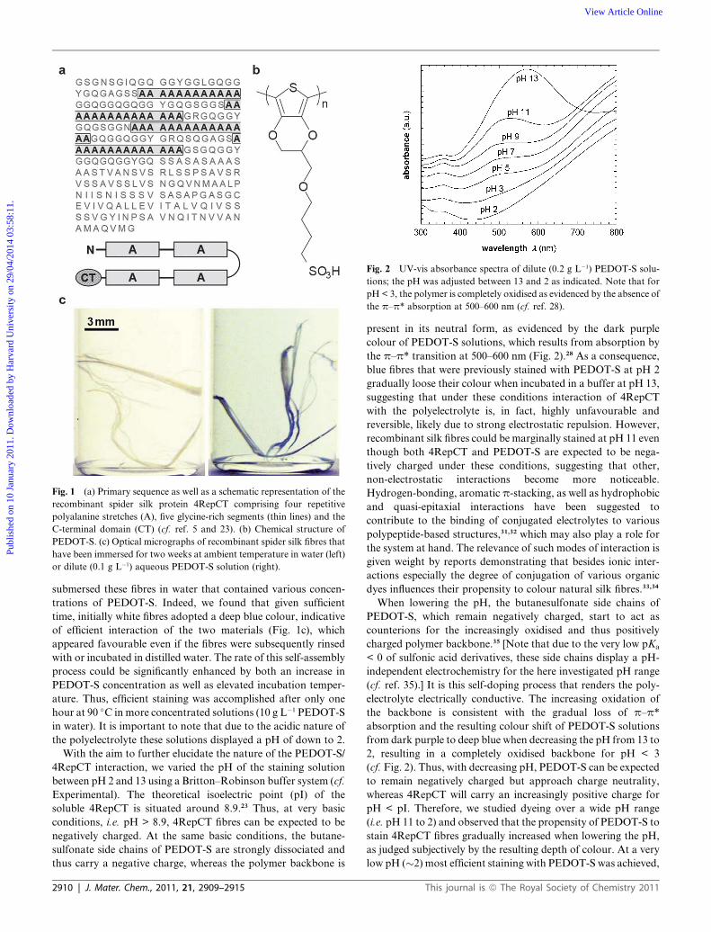
Fig. 2 UV-vis absorbance spectra of dilute (0.2 g L�1) PEDOT-S solu-
tions; the pH was adjusted between 13 and 2 as indicated. Note that for
pH < 3, the polymer is completely oxidised as evidenced by the absence of
the p–p* absorption at 500–600 nm (cf. ref. 28).
Fig. 1 (a) Primary sequence as well as a schematic representation of the
recombinant spider silk protein 4RepCT comprising four repetitive
polyalanine stretches (A), five glycine-rich segments (thin lines) and the
C-terminal domain (CT) (cf. ref. 5 and 23). (b) Chemical structure of
PEDOT-S. (c) Optical micrographs of recombinant spider silk fibres that
have been immersed for two weeks at ambient temperature in water (left)
or dilute (0.1 g L�1) aqueous PEDOT-S solution (right).
Publ
ishe
d on
10
Janu
ary
2011
. Dow
nloa
ded
by H
arva
rd U
nive
rsity
on
29/0
4/20
14 0
3:58
:11.
View Article Online
submersed these fibres in water that contained various concen-
trations of PEDOT-S. Indeed, we found that given sufficient
time, initially white fibres adopted a deep blue colour, indicative
of efficient interaction of the two materials (Fig. 1c), which
appeared favourable even if the fibres were subsequently rinsed
with or incubated in distilled water. The rate of this self-assembly
process could be significantly enhanced by both an increase in
PEDOT-S concentration as well as elevated incubation temper-
ature. Thus, efficient staining was accomplished after only one
hour at 90 �C in more concentrated solutions (10 g L�1 PEDOT-S
in water). It is important to note that due to the acidic nature of
the polyelectrolyte these solutions displayed a pH of down to 2.
With the aim to further elucidate the nature of the PEDOT-S/
4RepCT interaction, we varied the pH of the staining solution
between pH 2 and 13 using a Britton–Robinson buffer system (cf.
Experimental). The theoretical isoelectric point (pI) of the
soluble 4RepCT is situated around 8.9.23 Thus, at very basic
conditions, i.e. pH > 8.9, 4RepCT fibres can be expected to be
negatively charged. At the same basic conditions, the butane-
sulfonate side chains of PEDOT-S are strongly dissociated and
thus carry a negative charge, whereas the polymer backbone is
2910 | J. Mater. Chem., 2011, 21, 2909–2915
present in its neutral form, as evidenced by the dark purple
colour of PEDOT-S solutions, which results from absorption by
the p–p* transition at 500–600 nm (Fig. 2).28 As a consequence,
blue fibres that were previously stained with PEDOT-S at pH 2
gradually loose their colour when incubated in a buffer at pH 13,
suggesting that under these conditions interaction of 4RepCT
with the polyelectrolyte is, in fact, highly unfavourable and
reversible, likely due to strong electrostatic repulsion. However,
recombinant silk fibres could be marginally stained at pH 11 even
though both 4RepCT and PEDOT-S are expected to be nega-
tively charged under these conditions, suggesting that other,
non-electrostatic interactions become more noticeable.
Hydrogen-bonding, aromatic p-stacking, as well as hydrophobic
and quasi-epitaxial interactions have been suggested to
contribute to the binding of conjugated electrolytes to various
polypeptide-based structures,31,32 which may also play a role for
the system at hand. The relevance of such modes of interaction is
given weight by reports demonstrating that besides ionic inter-
actions especially the degree of conjugation of various organic
dyes influences their propensity to colour natural silk fibres.33,34
When lowering the pH, the butanesulfonate side chains of
PEDOT-S, which remain negatively charged, start to act as
counterions for the increasingly oxidised and thus positively
charged polymer backbone.35 [Note that due to the very low pKa
< 0 of sulfonic acid derivatives, these side chains display a pH-
independent electrochemistry for the here investigated pH range
(cf. ref. 35).] It is this self-doping process that renders the poly-
electrolyte electrically conductive. The increasing oxidation of
the backbone is consistent with the gradual loss of p–p*
absorption and the resulting colour shift of PEDOT-S solutions
from dark purple to deep blue when decreasing the pH from 13 to
2, resulting in a completely oxidised backbone for pH < 3
(cf. Fig. 2). Thus, with decreasing pH, PEDOT-S can be expected
to remain negatively charged but approach charge neutrality,
whereas 4RepCT will carry an increasingly positive charge for
pH < pI. Therefore, we studied dyeing over a wide pH range
(i.e. pH 11 to 2) and observed that the propensity of PEDOT-S to
stain 4RepCT fibres gradually increased when lowering the pH,
as judged subjectively by the resulting depth of colour. At a very
low pH (�2) most efficient staining with PEDOT-S was achieved,
This journal is ª The Royal Society of Chemistry 2011

Table 1 Mechanical properties and electrical conductivity of 4RepCT/PEDOT-S fibres and for comparison pristine 4RepCT fibres as well as spin-coated PEDOT-S thin films: Young’s modulus E, fractures stress sfracture and electrical conductivity c (errors were estimated through comparison of nsimilar samples). [Note that the here reported fracture stress probably does not correspond to the material’s ultimate tensile strength as fibres often failedat the grip point; the fragility of fibres complicated handling and is likely to have weakened samples prior to mechanical testing, e.g. during fixation.]Samples were prepared at the indicated pH, temperature T (a.t. ¼ ambient temperature) and staining time t; ‘—’ denotes not measured
Preparation Tensile testing Conductivity
Sample pH T/�C t n E/GPa sfracture/MPa n ca/S cm�1
4RepCT 7 a.t. — 5 6.5 � 1.4 89 � 9 2 <10�7
4RepCT/PEDOT-S 2 a.t. 10–14 d 6 5.6 � 2.1 61 � 29 3 (7 � 2) � 10�5
4RepCT/PEDOT-S 2 90 1–2 h 2b 5.9, 6.9 61, 85 6 (8 � 2) � 10�4
4RepCT/PEDOT-S 3–11 a.t. 14 d — — — 1c <10�7
PEDOT-S 2 a.t. — — — — 4 18 � 4
a c < 10�7 S cm�1 was not accessible because of experimental limitations. b The majority of fibres fractured prematurely during handling. c One sampleeach for pH 3, 5, 7, 9, and 11.
Fig. 3 (a) Transmission wide-angle X-ray diffraction patterns of a pris-
tine 4RepCT fibre (i) and a fibre stained with PEDOT-S for two hours at
pH z 2 and 90 �C (ii). (b) Corresponding radially integrated X-ray
diffractograms. (c) Optical micrograph of a cross-section of a cut
PEDOT-S stained fibre. (d) A dry PEDOT-S stained fibre fixated with
silver paste (top) elongates and curls upon wetting (bottom).
Publ
ishe
d on
10
Janu
ary
2011
. Dow
nloa
ded
by H
arva
rd U
nive
rsity
on
29/0
4/20
14 0
3:58
:11.
View Article Online
aided by now favourable electrostatic interactions (cf. Table 1).
[Note that quantitative means such as UV-vis or fluorescence
spectroscopy were not practical for this set of samples because of
the poor optical response of the doped PEDOT-S.]
Microstructure and mechanical properties of PEDOT-S stained
4RepCT fibres
The dried 4RepCT fibres are composed of a multitude of tightly
packed filaments with a diameter of approximately 30–200
nm.5,36 Similar to other silk structures, on a molecular level they
consist of b-sheet crystallites that are embedded in a more
disordered, i.e. amorphous matrix.1,7–9 Necessary structural
cross-links are provided by such b-sheet crystallites as well as
hydrogen-bonding in amorphous domains. This is in agreement
with our observation that 4RepCT fibres have to be dried under
tension in order to maximise their mechanical strength, a proce-
dure which is known to promote formation of hydrogen-bonds
between partially aligned amorphous peptide sections (cf. ref. 8).
The structure and distribution of hydrophobic b-sheet crys-
tallites was found to be little affected by the staining process, as
evidenced by X-ray diffractograms of pristine as well as PEDOT-
S stained 4RepCT fibres displayed in Fig. 3a. This is in agreement
with our recent observation that the microstructure of pristine
fibres is not affected by autoclaving at 121 �C (cf. ref. 27).
Diffractions are present as full circles, indicating that crystalline
domains are mostly unoriented within monofilaments.
Furthermore, various diffractions are consistent with the
b-poly(L-alanine) but also poly(L-alanylglycine) structure,37
suggesting that it is in particular the polyalanine segments of
4RepCT that form b-sheet crystallites (Fig. 3b).
Most significantly, we found that our 4RepCT fibres were
stained with PEDOT-S throughout the bulk as evidenced by
optical microscopy on fibre cross-sections (Fig. 3c). Certainly,
the polyelectrolyte can efficiently enter the free volume between
filaments but, in addition, may also penetrate water-soaked
amorphous regions. Water uptake by amorphous regions is
corroborated by the swelling of fibres when soaked in water,
which resulted in a reversible extension of the fibre length
(Fig. 3d; also cf. ref. 9). Wet fibres were found to be reasonably
flexible whereas dried fibres appeared to be more stiff and fragile
(cf. ref. 5). The stiffness and brittleness of dried fibres is also
reflected by the high Young’s modulus E and linear
This journal is ª The Royal Society of Chemistry 2011
stress–elongation relationship with no obvious yielding point as
revealed by tensile deformation experiments (Fig. 4). Unfortu-
nately, we found that dried PEDOT-S stained fibres were even
more fragile than pristine fibres during handling. This observa-
tion is in agreement with the apparent decrease in fracture stress
sfracture upon staining with PEDOT-S: pristine 4RepCT fibres
fractured at sfracture z 89 � 9 MPa, whereas for instance fibres
stained with PEDOT-S at pH 2 and ambient temperature failed
at sfracture z 61 � 29 MPa (Fig. 4 and Table 1). [Note that
a Student’s t-test between these two values yielded a probability
value p ¼ 0.063, indicating a limited statistical relevance.] This
increase in brittleness of the dried PEDOT-S stained fibres is
consistent with the picture that the polyelectrolyte had in part
intercalated between amorphous 4RepCT segments and thus
partially inhibited hydrogen bond formation. Moreover, it
J. Mater. Chem., 2011, 21, 2909–2915 | 2911

Fig. 5 Representative ohmic current–voltage, I–V, characteristics of
recombinant spider silk fibres stained with PEDOT-S for one hour at pH
2 and 90 �C (ii) or for 10 days at pH 2 and ambient temperature (iii). The
current was normalised with respect to the length, L, of the conducting
fibre segment as well as the approximate cross-sectional area, pR2, where
R is the mean radius. Note that pristine 4RepCT fibres (i) displayed no
measurable conductivity.
Fig. 4 Stress–elongation curves of a representative pristine 4RepCT
fibre (i), a fibre stained with PEDOT-S for two hours at pH 2 and 90 �C
(ii) or for 14 days at pH 2 and ambient temperature (iii).
Publ
ishe
d on
10
Janu
ary
2011
. Dow
nloa
ded
by H
arva
rd U
nive
rsity
on
29/0
4/20
14 0
3:58
:11.
View Article Online
should be noted that the dried stained fibres regained their flex-
ibility if wetted again, comparable to the behaviour of pristine
fibres. We rule out that the used staining conditions affect the
mechanical properties of 4RepCT fibres because of their
remarkable stability during autoclaving at 121 �C as discussed
above.27 Here, it is interesting to note that dyeing of natural silk
fibres is often found to result in a slight deterioration of their
mechanical properties,38,39 especially if reactive dyes are
employed.40 Although the hydrodynamic radius of PEDOT-S
particles in water is at the order of at least 2 nm,28 we suggest that
PEDOT-S can to some extent diffuse into amorphous regions of
4RepCT if given enough time, which will gradually provide
access to the charged surfaces of the protein. In fact, the long
time required to achieve good staining of 4RepCT fibres is
consistent with this picture. As a result, besides coating the
surface of filaments, PEDOT-S may be found within positively
charged amorphous regions or adhere to the surface of electri-
cally neutral and hydrophobic polyalanine b-sheet crystallites, as
has been suggested for PEDOT-S/amyloid fibril complexes from
insulin.22
Electrical conductivity of PEDOT-S stained 4RepCT fibres
As we set out to produce electrically conducting recombinant
silk, in a further set of experiments we examined PEDOT-S
stained 4RepCT fibres for their electrical properties. Gratify-
ingly, we found that fibres stained at a sufficiently low pH, i.e. pH
2, displayed an electrical conductivity c z (7 � 2) � 10�5 S cm�1
if treated at ambient temperature, whereas staining at 90 �C
resulted in s z (8 � 2) � 10�4 S cm�1 (Fig. 5 and Table 1). This
unfavourably compares with the conductivity of PEDOT-S thin
films, which we measured to be 18 � 4 S cm�1. However, some of
this discrepancy can be accounted for by considering that the
conductivity of fibres was estimated with respect to their cross-
sectional area and thus disregards the presence of a majority
fraction of the insulating 4RepCT material (cf. Experimental). In
addition, it is important to note that the polyelectrolyte used in
this study was of rather low molecular weight,28 the increase of
which can be expected to strongly enhance percolation of charge-
transport pathways along the fibre and thus electrical conduc-
tivity, especially if only a small amount of the conjugated
2912 | J. Mater. Chem., 2011, 21, 2909–2915
material is bound. In stark contrast, staining at pH > 2 and
ambient temperature appeared to result in incorporation of
a significantly lower amount of PEDOT-S, even when extending
the staining process to several weeks, as suggested by the absence
of measurable electrical conductivity (Table 1). Acidic post-
treatment of fibres that had been stained at pH > 2 did not result
in more conductive fibres, confirming that the lack of sufficient
PEDOT-S incorporation and not inadequate doping of the
polymer likely is the explanation for this observation (cf. Fig. 2).
Use of other polyelectrolytes/silk structures
In order to demonstrate the general applicability of the here
discussed approach to functionalise recombinant spider silk, we
also stained fibres with poly(thiophene acetic acid) (PTAA).21,31
In this way photoluminescent fibres could readily be produced as
illustrated by the fluorescence micrograph and spectra presented
in Fig. 6. Similar to staining with PEDOT-S discussed above,
most efficient decoration of 4RepCT fibres with PTAA was
achieved after staining for two hours at 90 �C. Conversely, at
ambient temperature this process required considerably more
time; for instance one week at pH 7 was needed to reach a similar
level of fluorescence. Again, the pH of staining solutions was
found to be crucial. PTAA only dissolved in appreciable quan-
tities at pH $ 6, which can be rationalised with the pKa z 4.8 of
acetic acid. Thus, we performed staining experiments in the range
of pH 7–11. As evidenced by the fluorescence spectra of PTAA
stained 4RepCT fibres in Fig. 6b, at pH 7–9 PTAA displayed
a strong affinity for 4RepCT. In most significant contrast,
attempts to decorate fibres with PTAA at pH 10 resulted in a�30
times lower fluorescence as compared to fibres stained at pH 7.
Ultimately, at pH 11 fluorescence from PTAA was found to be
virtually absent. Evidently, the pI z 8.9 of 4RepCT strongly
influenced binding of PTAA. For pH < 8.9, a strong attraction
between 4RepCT, which carries a net positive charge, and the
negatively charged side chains of PTAA can be expected, high-
lighting the dominance of electrostatic interactions between the
conjugated polyelectrolyte and protein. However, a weak
This journal is ª The Royal Society of Chemistry 2011

Fig. 6 (a) Fluorescence micrograph of a recombinant silk fibre stained
with PTAA for two hours at 90 �C and pH 9. (b) Fluorescence spectra of
fibres stained for two hours at 90 �C and the indicated pH (solid lines) as
well as of a pristine 4RepCT fibre (dashed line). The inset shows the
chemical structure of PTAA.
Publ
ishe
d on
10
Janu
ary
2011
. Dow
nloa
ded
by H
arva
rd U
nive
rsity
on
29/0
4/20
14 0
3:58
:11.
View Article Online
interaction still appeared to be present in more alkaline solutions,
i.e. at pH 10, suggesting that other modes of binding may also
contribute to the attachment of PTAA to 4RepCT fibres, which
can to some extent overcome the electrostatic repulsion between
the two now negatively charged species.
The here proposed method to functionalise 4RepCT fibres
could also be extended to other silk structures, such as solution-
cast 4RepCT thin films as well as natural silk fibres from the
silkworm B. mori. Gratifyingly, we found that also PEDOT-S
stained B. mori fibres displayed an electrical conductivity of at
least 10�3 S cm�1 when stained at elevated temperature and low
pH (90 �C and pH 2).
3. Conclusions
The combination of recombinant spider silk and conjugated
polyelectrolytes such as PEDOT-S and PTAA permitted us to
equip this highly versatile biomaterial with novel bulk-electrical
and optical properties. The generality of this approach was
illustrated by demonstrating its applicability also to natural silk
fibres from the silkworm B. mori. Clearly, the here proposed
method based on a self-assembly process may prove highly
useful in future biomedical and bioelectronic applications, such
as the growth of stem or nerve cells on three-dimensional
conducting templates,41–44 as well as the realisation of
biocompatible actuators,45 electronic sensors and textile-based
electrodes.46
This journal is ª The Royal Society of Chemistry 2011
4. Experimental
Materials
Poly(4-(2,3-dihydrothieno[3,4-b]-[1,4]dioxin-2-yl-methoxy)-1-
butanesulfonic acid, sodium salt) (PEDOT-S) and poly-
(thiophene acetic acid) (PTAA) were synthesised according to
the previously reported procedures.28,47 Aqueous solution of
the sodium salt PEDOT-S polymer was stirred with Dowex
Marathon C cation exchange resin to obtain the sulfonic acid
form. Then, PEDOT-S was loaded on a PD-10 size exclusion
column (Sephadex� G-25 M) in 3 mL batches and eluated
with 3 mL of water. Finally, PEDOT-S was dialysed against
de-ionized water for two days using a 3500 g mol�1 cut-off
membrane (Spectra/Por) and freeze-dried prior to use. The
yield was �45% with respect to the monomer unit.
Preparation of the recombinant miniature spider silk protein
4RepCT (23.4 kDa) as well as fabrication of fibres therewith have
been reported elsewhere.5,23 Degummed silk fibres from the
silkworm B. mori were used as received.
Sample preparation
For photographs of fibres, structures were stained with PEDOT-
S by suspension in water (0.1 g L�1 PEDOT-S) for two weeks at
ambient temperature.
For electrical conductivity and tensile deformation measure-
ments on solid fibres, structures were first dried under tension at
ambient temperature. Staining with PEDOT-S was achieved by
submerging these fibres in water (10 g L�1 PEDOT-S) for up to
15 days at ambient temperature or 1–2 hours at 90 �C, subse-
quent to which the fibres were washed with distilled water and
again dried under tension. The pH of PEDOT-S and PTAA
solutions was adjusted using a Britton–Robinson buffer system
(0.04 M H3BO4, 0.04 M H3PO4 and 0.04 M CH3COOH titrated
with 0.2 M NaOH), which permitted buffering in the range of pH
2 to 12. pH 13 was reached with 0.2 M NaOH.
Staining with PTAA was achieved by submersion of fibres in
water (0.1 g L�1 PTAA) for one week at ambient temperature or
90 �C for two hours, subsequent to which the fibres were washed
with distilled water.
pH determination
The pH of PEDOT-S and PTAA solutions was determined with
a Biotrode pH-meter (Mettler Toledo). The theoretical net
charge of soluble 4RepCT was calculated using www.expasy.ch/
tools, yielding an isoelectric point pI z 8.9 (cf. ref. 23; 4RepCT
carries a net positive charge for pH < pI and a net negative
charge for pH > pI).
Optical microscopy
Optical microscopy was carried out with a Nicon SMZ 1000
stereomicroscope equipped with a Sony Exwave HAD camera.
Ultraviolet-visible light (UV-vis) absorbance spectroscopy
UV-vis absorbance spectra of PEDOT-S solutions (0.2 g L�1;
quartz cuvette with 1 mm path length) were recorded using
a Perkin Elmer Lambda 950 UV-vis spectrophotometer.
J. Mater. Chem., 2011, 21, 2909–2915 | 2913

Publ
ishe
d on
10
Janu
ary
2011
. Dow
nloa
ded
by H
arva
rd U
nive
rsity
on
29/0
4/20
14 0
3:58
:11.
View Article Online
X-Ray diffraction
Transmission wide-angle X-ray diffraction was carried out with
an in-house rotating anode instrument using CuKa-radiation
(l ¼ 1.5418 A).
Differential Mechanical Analysis (DMA)
Mechanical testing of fibers with a radius R z 20–50 mm was
performed at ambient temperature with a TA Instruments DMA
Q800 V7.4 analyser using a ramp force of 0.2 N min�1. The gauge
length of 0.8 cm was ensured by gluing fibres with cyanoacrylate
(LoctiteVR Super AttakVR) to a double paper frame that was
subsequently mounted in the tensile cell and released before
measurements. Fibres that displayed macroscopic defects or
appeared to suffer during mounting were excluded. A circular
cross-section was assumed to calculate the stress.
Electrical conductivity measurements
PEDOT-S stained fibres were contacted with silver paste (Agar
Scientific) and two-point electrical conductivity measurements
were performed with a Keithley 4200 parameter analyser on fibre
segments with a typical length L z 0.5–1 cm and radius R z20–70 mm. 4-Point probe measurements (1 mm probe spacing) on
particularly thick fibres were used to confirm the electrical
conductivities estimated from two-point measurements as well as
to determine the conductivity of 60–80 nm thick spin-coated
PEDOT-S thin films.
The electrical conductivity c was estimated as
c ¼ I
V� L
pR2
where I is the measured current and V the applied voltage; L and
R are the individual dimensions of fibres.
Fluorescence microscopy
Fluorescence microscopy was carried out with an A200 Zeiss
Axiovert inverted epifluorescence microscope equipped with
a CCD camera (Axiocam HRc 3-chip), using a 470/40 nm filter
(LP515). Fluorescence spectra were recorded with a SpectraCube
SD-300 VDS system. The fluorescence spectra of PTAA stained
4RepCT fibres were corrected for the fibre diameter.
Acknowledgements
We would like to acknowledge funding from the Swedish
Foundation for Strategic Research (SSF) through the pro-
gramme Organic hybrid Printed Electronics and Nanoelectronics
(OPEN), Vinnova, Formas and The Swedish Research Council.
References
1 J. O. O’Brien, S. R. Fahnestock, Y. Termonia and K. C. H. Gardner,Adv. Mater., 1998, 10, 1185–1195.
2 A. Lazaris, S. Arcidiacono, Y. Huang, J.-F. Zhou, F. Duguay,N. Chretien, E. A. Welsh, J. W. Soares and C. N. Karatzas,Science, 2002, 295, 472–476.
3 S. Arcidiacono, C. M. Mello, M. Butler, E. Welsh, J. W. Soares,A. Allen, D. Ziegler, T. Laue and S. Chase, Macromolecules, 2002,35, 1262–1266.
2914 | J. Mater. Chem., 2011, 21, 2909–2915
4 J. H. Exler, D. H€ummerich and T. Scheibel, Angew. Chem., Int. Ed.,2007, 46, 3559–3562.
5 M. Stark, S. Grip, A. Rising, M. Hedhammar, W. Engstr€om,G. Hj€alm and J. Johansson, Biomacromolecules, 2007, 8, 1695–1701.
6 A. Rising, M. Widhe, J. Johansson and M. Hedhammar, Cell. Mol.Life Sci., 2010, DOI: 10.1007/s00018-010-0462-z.
7 M. Denny, J. Exp. Biol., 1976, 65, 483–506.8 Y. Termonia, Macromolecules, 1994, 27, 7378–7381.9 J. M. Gosline, P. A. Guerette, C. S. Ortlepp and K. N. Savage, J. Exp.
Biol., 1999, 202, 3295–3303.10 C. Riekel, B. Madsen, D. Knight and F. Vollrath, Biomacromolecules,
2000, 1, 622–626.11 F. Vollrath, P. Barth, A. Basedow, W. Engstr€om and H. List, In Vivo,
2002, 16, 229–234.12 C. Allmeling, A. Jokuszies, K. Reimers, S. Kall, C. Y. Choi,
G. Brandes, C. Kaspar, T. Scheper, M. Guggenheim andP. M. Vogt, Cell Proliferation, 2008, 41, 408–420.
13 Y. Yang, X. Chen, F. Ding, P. Zhang, J. Liu and X. Gu, Biomaterials,2007, 28, 1643–1652.
14 E. L. Mayes, F. Vollrath and S. Mann, Adv. Mater., 1998, 10, 801–805.
15 M. Chu and Y. Sun, Smart Mater. Struct., 2007, 16, 2453–2456.16 A. Singh, S. Hede and M. Sastry, Small, 2007, 3, 466–473.17 H. S. Kim, M. Kang, H.-J. Jin, I.-J. Chin, H. J. Choi and J. Joo, Mol.
Cryst. Liq. Cryst., 2007, 464, 15–21.18 Y. Xia and Y. Lu, Compos. Sci. Technol., 2008, 68, 1471–1479.19 K. P. R. Nilsson, J. Rydberg, L. Baltzer and O. Ingan€as, Proc. Natl.
Acad. Sci. U. S. A., 2004, 101, 11197–11202.20 A. Herland, P. Bj€ork, K. P. R. Nilsson, J. D. M. Olsson, P. Asberg,
P. Konradsson, P. Hammarstr€om and O. Ingan€as, Adv. Mater.,2005, 17, 1466–1471.
21 A. Herland, P. Bj€ork, P. R. Hania, I. G. Scheblykin and O. Ingan€as,Small, 2007, 3, 318–326.
22 M. Hamedi, A. Herland, R. H. Karlsson and O. Ingan€as, Nano Lett.,2008, 8, 1736–1740.
23 M. Hedhammar, A. Rising, S. Grip, A. S. Martinez, K. Nordling,C. Casals, M. Stark and J. Johansson, Biochemistry, 2008, 47,3407–3417.
24 G. Askarieh, M. Hedhammar, K. Nordling, A. Saenz, C. Casals,A. Rising, J. Johansson and S. D. Knight, Nature, 2010, 465, 236–238.
25 S. Grip, J. Johansson and M. Hedhammar, Protein Sci., 2009, 18,1012–1022.
26 C. Fredriksson, M. Hedhammar, R. Feinstein, K. Nordling,G. Kratz, J. Johansson, F. Huss and A. Rising, Materials, 2009, 2,1908–1922.
27 M. Hedhammar, H. Bramfeldt, T. Baris, M. Widhe, G. Askarieh,K. Nordling, S. von Aulock and J. Johansson, Biomacromolecules,2010, 11, 953–959.
28 R. H. Karlsson, A. Herland, M. Hamedi, J. A. Wigenius, A. Aslund,X. Liu, M. Fahlman, O. Ingan€as and P. Konradsson, Chem. Mater.,2009, 21, 1815–1821.
29 R. M. Miriani, M. R. Abidian and D. R. Kipke, Proc. IEEE Eng.Med. Biol. Soc., 2008, 1841–1844.
30 M. Asplund, E. Thaning, J. Lundberg, A. C. Sandberg-Nordqvist,B. Kostyszyn, O. Ingan€as and H. von Holst, Biomed. Mater., 2009,4, 045009.
31 K. P. R. Nilsson, P. Hammarstr€om, F. Ahlgren, A. Herland,E. A. Schnell, M. Lindgren, G. T. Westermark and O. Ingan€as,ChemBioChem, 2006, 7, 1096–1104.
32 A. Herland, D. Thomsson, O. Mirzov, I. G. Scheblykin andO. Ingan€as, J. Mater. Chem., 2008, 18, 126–132.
33 M. R. De Giorgi and A. Cerniani, Dyes Pigm., 1991, 15, 47–55.34 G. Seu, Dyes Pigm., 1993, 23, 267–273.35 A. O. Patil, Y. Ikenoue, N. Basescu, N. Colaneri, J. Chen, F. Wudl
and A. J. Heeger, Synth. Met., 1987, 20, 151–159.36 M. Widhe, H. Bysell, S. Nystedt, I. Schenning, M. Malmsten,
J. Johansson, A. Rising and M. Hedhammar, Biomaterials, 2010,31, 9575–9585.
37 C. Riekel, C. Br€anden, C. Craig, C. Ferrero, F. Heidelbach andM. M€uller, Int. J. Biol. Macromol., 1999, 24, 179–186.
38 H. Somashekarappaa, V. Annaduraib, Sangappa, G. Subramanyacand R. Somashekar, Mater. Lett., 2002, 53, 415–420.
39 C. Wang, K. Fang and W. Ji, Fibres Polym., 2007, 8, 225–229.40 N. Y. Dang, W. Ma, S. F. Zhang, B. T. Tang and J. Z. Yang, Text.
Res. J., 2010, 80, 374–382.
This journal is ª The Royal Society of Chemistry 2011

Publ
ishe
d on
10
Janu
ary
2011
. Dow
nloa
ded
by H
arva
rd U
nive
rsity
on
29/0
4/20
14 0
3:58
:11.
View Article Online
41 M. Li, Y. Guo, Y. Wei, A. G. MacDiarmid and P. I. Lelkes,Biomaterials, 2006, 27, 2705–2715.
42 Y. Liu, X. Liu, J. Chen, K. J. Gilmore and G. G. Wallace, Chem.Commun., 2008, 3729–3731.
43 M. H. Bolin, K. Svennersten, X. Wang, I. S. Chronakis, A. Richter-Dahlfors, E. W. H. Jager and M. Berggren, Sens. Actuators, B,2009, 142, 451–456.
This journal is ª The Royal Society of Chemistry 2011
44 K. D. McKeon, A. Lewis and J. W. Freeman, J. Appl. Polym. Sci.,2010, 115, 1566–1572.
45 E. W. H. Jager, E. Smela and O. Ingan€as, Science, 2000, 290, 1540–1545.
46 M. Asplund, PhD thesis, KTH, 2009.47 L. Ding, M. Jonforsen, L. S. Roman, M. R. Andersson and
O. Ingan€as, Synth. Met., 2000, 110, 133–140.
J. Mater. Chem., 2011, 21, 2909–2915 | 2915
Copyright © 2022 FDOKUMEN