Implicit and explicit dosimetry in photodynamic therapy: a new paradigm
18
Lasers in MedicalScience 1997, 12:182-199 Review Article Implicit and Explicit Dosimetry in Photodynamic Therapy: a New Paradigm B.C. WILSON a'b'c, M.S. PATTERSON d, L. LILGE b'c aOntario Cancer Institute, Toronto, Ontario, Canada bDepartment of Medical Biophysics, University of Toronto, Toronto, Ontario, Canada cOntario Laser and Lightwave Research Centre, Toronto, Ontario, Canada dHamilton Regional Cancer Center and Departments of Radiology and Physics, McMaster University, Hamilton, Ontario, Canada Correspondence to B.C. Wilson, Department of Medical Biophysics, Ontario Cancer Institute, 7th Floor, 610 University Avenue, Toronto, Ontario, Canada M5G 2M9 Received 19 December 1996; accepted in final form 4 February 1997 Abstract. Dosimetry for photodynamic therapy (PDT) is becoming increasingly complex as more factors are identified which may influence the effectiveness of a given treatment. The simple prescription of a PDT treatment in terms of the administered photosensitizer dose, the incident light and the drug-light time interval does not account for patient-to-patient variability in either the photosensitizer uptake, tissue optical properties or tissue oxygenation, nor for the interdependence of the photosensitizer-light-tissue factors. This interdependence is examined and the implications for developing adequate dosimetry for PDT are considered. The traditional dosimetric approach, measuring each dose factor independently, and termed here 'explicit dosimetry', may be contrasted with the recent trend to use photosensitizer photobleaching as an index of the effective delivered dose, termed here 'implicit dosimetry'. The advantages and limitations of each approach are discussed, and the need to understand the degree to which the photobleaching mechanism is linked, or 'coupled', to the photosensitizing mechanism is analysed. Finally, the influence of the tissue-response endpoints on the optimal dosimetry methods is considered. INTRODUCTION Photodynamic therapy (PDT) of malignant lesions involves administration to the patient of a photosensitizer, a time delay to allow adequate concentration of the drug in the tumour, followed by irradiation of the target tissue volume by light of a wavelength appro- priate to activate the photosensitizer ef- ficiently (1-3). The consequent photochemical damage results in tissue necrosis, by direct tumour cell kill and/or by vascular damage leading to ischaemic necrosis (4). For most photosensitizers, it is believed that the PDT effect is mediated by the production of highly active singlet oxygen, 10 2, formed by energy transfer from the excited-state photosensitizer to molecular oxygen in the tissue. In its simplest terms, a PDT treatment may be described by specifying the administered photosensitizer 'dose' (eg mg kg -1 body weight), the incident light 'dose' (eg J cm-e) and the drug-light time interval. Historically, and indeed currently, most clinical protocols still utilize only these three prescribed treat- ment parameters, despite the fact that there are many additional factors which may influ- ence the effective 'dose' actually delivered to any particular lesion, including: (1) The subject-to-subject variation in specific tumour uptake of photosensitizer (5-8); (2) The large range of (wavelength- dependent) optical absorption and scattering coefficients of different tissues, which deter- mine the light penetration and distribution in the target volume (9, 10); (3) The variability in tissue oxygenation (11), which affects the photodynamic efficiency; 0268-8921/97/030182 + 18 $12.00/0 1997 W.B. SaundersCompany Ltd
-
Upload
georgetown -
Category
Documents
-
view
1 -
download
0
Transcript of Implicit and explicit dosimetry in photodynamic therapy: a new paradigm

Lasers in Medical Science 1997, 12:182-199
Review Article Implicit and Explicit Dosimetry in Photodynamic Therapy: a New Paradigm
B.C. WILSON a'b'c, M.S. P A T T E R S O N d, L. L ILGE b'c
aOntario Cancer Institute, Toronto, Ontario, Canada bDepartment of Medical Biophysics, University of Toronto, Toronto, Ontario, Canada cOntario Laser and Lightwave Research Centre, Toronto, Ontario, Canada dHamilton Regional Cancer Center and Departments of Radiology and Physics, McMaster University, Hamilton, Ontario, Canada
Correspondence to B.C. Wilson, Department of Medical Biophysics, Ontario Cancer Institute, 7th Floor, 610 University Avenue, Toronto, Ontario, Canada M5G 2M9
Received 19 December 1996; accepted in final form 4 February 1997
Abstract . Dosimetry for photodynamic therapy (PDT) is becoming increasingly complex as more factors are identified which may influence the effectiveness of a given treatment. The simple prescription of a PDT treatment in terms of the administered photosensitizer dose, the incident light and the drug-light time interval does not account for patient-to-patient variability in either the photosensitizer uptake, tissue optical properties or tissue oxygenation, nor for the interdependence of the photosensitizer-light-tissue factors. This interdependence is examined and the implications for developing adequate dosimetry for PDT are considered.
The traditional dosimetric approach, measuring each dose factor independently, and termed here 'explicit dosimetry', may be contrasted with the recent trend to use photosensitizer photobleaching as an index of the effective delivered dose, termed here 'implicit dosimetry'. The advantages and limitations of each approach are discussed, and the need to understand the degree to which the photobleaching mechanism is linked, or 'coupled', to the photosensitizing mechanism is analysed.
Finally, the influence of the tissue-response endpoints on the optimal dosimetry methods is considered.
INTRODUCTION
Photodynamic therapy (PDT) of malignant lesions involves administration to the patient of a photosensitizer, a time delay to allow adequate concentration of the drug in the tumour, followed by irradiation of the target tissue volume by light of a wavelength appro- priate to activate the photosensitizer ef- ficiently (1-3). The consequent photochemical damage results in tissue necrosis, by direct tumour cell kill and/or by vascular damage leading to ischaemic necrosis (4). For most photosensitizers, it is believed that the PDT effect is mediated by the production of highly active singlet oxygen, 10 2, formed by energy transfer from the excited-state photosensitizer to molecular oxygen in the tissue.
In its simplest terms, a PDT treatment may be described by specifying the administered
photosensitizer 'dose' (eg mg kg -1 body weight), the incident light 'dose' (eg J cm-e) and the drug-light time interval. Historically, and indeed currently, most clinical protocols still utilize only these three prescribed treat- ment parameters, despite the fact that there are many additional factors which may influ- ence the effective 'dose' actually delivered to any particular lesion, including:
(1) The subject-to-subject variation in specific tumour uptake of photosensitizer (5-8); (2) The large range of (wavelength-
dependent) optical absorption and scattering coefficients of different tissues, which deter- mine the light penetration and distribution in the target volume (9, 10);
(3) The variability in tissue oxygenation (11), which affects the photodynamic efficiency;
0268-8921/97/030182 + 18 $12.00/0 �9 1997 W.B. Saunders Company Ltd

Implicit and Explicit PDT Dosimetry
(4) Changes in light penetrat ion during ir- radiation, due primarily to rapid PDT-induced blood flow changes (12, 13);
(5) 'Self shielding', which occurs with second- generation photosensitizers of large molar extinction coefficient, and which limits the light penetration due to the added absorp- tion of the photosensitizer itself in the tissue (14);
(6) 'Photobleaching', shown by many photo- sensitizers during light irradiation (2, 5-8, 15- 17), which may reduce the concentration of (photo-active) photosensitizer in the tissue during irradiation; and
(7) Photochemical depletion of oxygen in tis- sues under irradiation at high light fluence rates, leading to reduced photodynamic effect (18, 19).
Thus, there is a large gap between current clinical dosimetry and a complete and compre- hensive description of an actual PDT treat- ment. While clinical treatments will, no doubt, continue to be improved empirically, this gap is now a major impediment to progress, par- t icularly in understanding how to optimize and standardize treatments with the numerous new photosensitizers which are coming into clinical use (1, 3, 20).
Two main challenges to improving PDT dosimetry can be identified, namely:
(1) To develop methods, and corresponding instrumentation, to measure the various dose factors in individual patients; and
(2) To understand how, both in principle and in practice, these dose-modifying factors fit together to determine the effective delivered dose, to correlate with the tissue reponse.
This paper will examine the second issue. As new dose-modifying factors are revealed, and as the interdependency of these factors becomes more apparent, new paradigms are needed to tackle the increasing complexity of PDT dosimetry. This paper will introduce the concept of 'implicit' vs 'explicit' PDT dosim- etry, and discuss the advantages and limi- tations of each. It is not the objective here to present specific solutions to the many problems in PDT dosimetry, but rather to introduce a new way to view PDT dosimetry in its broadest sense. This requires first an exami- nation of the interdependency of the PDT treatment factors.
183
INTERDEPENDENCE OF PDT DOSIMETRY FACTORS
Figure 1 illustrates the major interdependen- cies that have been identified to date between the three fundamental treatment variables; light, photosensitizer and oxygen. As can be seen, for each of these there are various possible measures (metrics) which can be applied. For example, in the case of the photo- activating light, the parameters which may be involved are: the incident fluence and, possibly, fluence rate; the spatial distribution of the light in the target tissue volume; and the temporal characteristics of the light (pulsed vs continuous irradiation). Similarly, for the photosensitizer, in addition to the adminis- tered amount of drug (mg kg 1 body weight or m -2 body surface), the consequent concen- tration and microdistribution of photosensi- tizer in the target and adjacent normal tissues are important. For oxygen, the pre-treatment concentration and microdistribution, and repletion rate are relevant, as is the vascular status before and during treatment, since these determine the oxygen available to participate in the photodynamic action. In defining clini- cal protocols for PDT, it has been usually considered that the light and photosensitizer (and oxygen) are independent treatment vari- ables. However, in reality, each may affect and be affected by (changes in) the others, as in the following cases.
Effect of light on photosensitizer
The photosensitizer can be photobleached, either permanently and/or transiently, by the treatment light. (Note that the term 'photo- bleaching' is variously used to denote actual photochemical destruction of the photosensi- tizer or simply decreased optical absorbance and/or fluorescence, which may not be equal and which do not necessarily involve molecu- lar decomposition. In this paper, the term will usually refer to loss of the measurable fluor- escence emission of the photosensitizer and it will be assumed that this corresponds to loss of photodynamic activity.) In the case of per- manent photobleaching, the concentration of photo-active photosensitizer in the target tissue decreases during the light irradi- ation. Transient photobleaching is the result of photosensitizer ground-state depletion, and is usually only significant using short

184 B.C. Wilson, M.S. Patterson, L. Lilge
Light Photobleaching +/- 02
�9 Incident fluence (J cm -2) " ~ e l f s h m l d i n g ~ S �9 Distribution, ~ (r) �9 Fluence rate (roW cm -2) Photosensitizer
�9 ~ �9 �9 �9 - 1 �9 Pulsed vs continuous wave / \ f ~ ~ Administered (mg kg ) ~ f_~ / ~ ..~P~ �9 Tissue concentration, c (t)
" J ~ ( / �9
Other . . . . . . . . . -I~ k ] ~1- . . . . . Intrinsic aoslmetric \ / tissue parameters ~ ~ / sensitivity
Photochemical ~ ~ / " Photochemical depletion l depletion
Oxygen �9 Intrinsic
�9 Micro-distribution (oxic-anoxic-hypoxic)
�9 Diffusion rate
�9 Vascular status (tissue response)
Fig. 1. Diagram illustrating the interdepency of the different dosimetry factors (photosensitizer-, light- and oxygen-related) involved in the photodynamic therapy response of tissue in vivo. The curved arrows indicate the interdependent mechanisms: eg the light fluence affects the photosensitizer through photobleaching; light and photosensitizer together affect the tissue oxygenation through photochemical depletion. As also indicated, the intrinsic photodynamic sensitivity of the tissue (31-34) determines the effectiveness of the resultant combined dosimetric variables and there may be other dosimetric factors (eg tissue temperature) which are not included here.
(sub-microsecond), high peak power pulsed irradiation (21, 22). Since, for any given ir- radiat ion technique, there are usually substan- tial light fluence-rate gradients within the tissue due to the limited penetration of the light, the rate of photobleaching will not be uniform. An example of this is shown in Plate l(b) where, in the treatment of a gastro- intestinal lesion, the photosensitizer, as moni- tored by its fluorescence endoscopic image, was bleached only in part of the target volume. The corresponding pre- and post-irradiation fluorescence spectra are presented in Fig. 2.
A critical issue for PDT dosimetry using photobleaching as a dose metric is whether or not the photobleaching is oxygen, and/or singlet oxygen, dependent or independent, as discussed below.
absorption, thereby reducing the penetration of the light ('self-shielding' effect). Thus, the effective treatment volume decreases. This effect may be partially countered by the photo- sensitizer photobleaching, so that the light and photosensitizer have a complex interplay (which may depend also on the oxygenation) during irradiation, which is not reflected sim- ply in the initial values of each. Other factors may set an upper limit on the useful tissue concentration of photosensitizer, which may then be too low to produce a significant self- shielding effect: for example, where photo- chemical depletion of oxygen is the limiting factor in the photodynamic action, such that further increase in photosensitizer con- centration does not alter the photodynamic effectiveness�9
Effect of photosensitizer on light
As was recognized a decade ago (14), with second-generation photosensitizers having a high extinction coefficient at the treatment wavelength, the absorption due to the photo- sensitizer itself adds to the intrinsic tissue
Effect of light+photosensitizer on tissue oxygenation
If the photosensitizer concentration and light fluence rate are high enough, it has been shown (18), both theoretically and experimen- tally, that photochemical depletion of tissue

Implicit and Explicit PDT Dosimetry 185
Plate 1. (a) White-light endoscopic image and fluorescence endoscopic images of an oesophageal lesion in a patient before and after Photofrin-photodynamic therapy (PDT) treatment. The photosensitizer appears to be photobleached uniformly throughout the tumour. (b) Similar images, plus a schematic showing the approximate irradiation geometry, in a second oesophageal patient, showing a region at the tumour base with residual Photofrin fluorescence after treatment. Treatment conditions: 2 mg kg -1 Photofrin i.v., -100 J cm 1 of 630 nm light at 24 h. Note that the interpretation of such photobleaching images, for example, whether or not the zone with residual fluorescence is 'undertreated', is particularly complicated with Photofrin since this comprises multiple porphyrin components of different pharmacokinetics, photodynamic activities, fluorescence yields and photobleaching rates.

0.01
v
z
0.01 [ (a)
0.009
0.008
0.007
0.006
0.005
~ o.ooj i i i L
f (b)
0.009
0.008
0.007
"~ 0.006
0.005
0.004
0.003 Q
z 0.002
0.001
186 B.C. Wilson, M.S. Patterson, L. Lilge
450 500 550 600 650 700 750 800 450 500 550 600 650 700 750 800 Wavelength (nm) Wavelength (nm)
Fig. 2. Corresponding point fluorescence spectra for Case b of Plate l(a) before and (b) immediately after light irradiation, measured using an optical multichannel analyser with a fibre-optic probe placed on the tumour tissue surface. Note the change in the spectral shape with photobleaching. Each set of spectra has been normalized to the same area under the curve, since the endoscope-tissue distance, and hence the excitation intensity, was not constant between procedures. Thick line, normal; thin line, tumour.
oxygen can occur. For oxygen-dependent photosensitization, this results in a reduced photodynamic effect. The extent to which this occurs depends also on how well perfused the tissue is, and on the oxygen diffusion rate from capillaries. The effect may be mitigated either by reducing the light fluence rate or by an interrupted irradiation regimen of l ight-dark cycles (typically ~ 3 0 s) to allow re-diffusion of oxygen during the dark phases, in each case maintaining the same total fluence delivered.
The tissue oxygenation can also be altered during treatment if there is an acute vascular response. It is known (4) that the severity of the vascular response (and its contribution to the resultant tissue damage relative to direct tumour cell killing) depends on the tissue and on the photosensitizer parameters: delivery vehicle, route of administration and drug-light time interval.
Effect of tissue blood oxygenation and blood content on light and photosensitizer
As mentioned previously, the photobleaching of the photosensitizer may be oxygen depen- dent, so that the photobleaching rate can change if the tissue oxygenation alters due either to photochemical depletion or to chang- ing blood flow. An altered tissue blood volume can also affect the light penetration, by
increasing or decreasing the absorption due to (oxy)haemoglobin (23, 24).
Thus, there is a multiplicity of ways in which the primary dose parameters affect, and are affected by, each other and by the response of the tissue during the PDT treatment. How then can this interdependency be taken into account in PDT dosimetry? The first option is to measure each parameter directly and independently (including measurements dur- ing treatment) and to build a resultant dose metric by combining these, using some model of how the photodynamic response depends on the variables. This will be referred to here as 'explicit dosimetry', since each dose variable is measured explicitly. The second option, 'implicit dosimetry', is to use a metric (eg photobleaching) which implicitly incorporates some, or preferably all, of the dose parameters, while not necessarily measuring any of them directly.
The advantages and disadvantages of each form of dosimetry are summarized in Table 1 and will be discussed below.
EXPLICIT DOSIMETRY
The explicit aproach to PDT dosimetry, which has been most commonly used to date, is illus- trated in Figs 3(a) and 4(a) by the dose metrics employed. In its simplest form, this involves

Implicit and Explicit PDT Dosimetry
Table 1. Advantages and disadvantages of explicit vs implicit dosimetry
187
Explicit Implicit
+ �82 No strong a priori assumptions �82 Incorporates 'all' photophysical/ �82 Photobleaching not necessary photochemical/photobiological factors
�82 Technically simple: eg fluorescence measurements only
�82 Need model(s) to combine all factors �82 Technically complex: complete data set may
not be possible �82 Does not incorporate/account for
microdosimetric factors (eg local 02 depletion)
�82 Assumes common pathway(s) for photobiological and/or photosensitizer fluorescence changes
�82 May need additional information to make quantitative (eg tissue optics)
�82 Not clear how to define effective 'dose'
measuring the average photosensitizer concen- tration in or around the target tissue just prior to light treatment, and the light fluence (rate) at some points within or around the target volume. This has the merit, compared with simply using the prescribed photosensitizer and delivered light doses, that it takes into account the patient-to-patient variabili ty in photosensitizer pharmacokinetics and tissue optical properties. Substantial progress has been made in developing techniques and instruments to make such measurements either invasively or non-invasively (25-28). The main a priori assumption involved is that the product of local photosensitizer concen- tration and light fluence is a predictor of tissue response. Certainly, the present authors (31, 32) and others (15, 33, 34) have shown in a number of studies of PDT threshold dose, which is calculated from explicit measures of the local light fluence and photosensitizer concentration, that this can be accurate for specific tissue-photosensitizer combinations, using gross volume necrosis as a response endpoint. There are, however, reports, both in vivo (35), and in vitro (36), where the drug- light product was not predictive of outcome (excluding 'trivial' cases such as reciprocity failure due to photobleaching not being taken into account: see below). In particular, the effect of photochemical depletion of local oxy- gen is not accounted for by monitoring only the photosensitizer and light fluence. It is possible to add dynamic tissue oxygenation (pO2) measurements, although at present this involves invasive single micro-electrodes or complex and expensive clinical instruments, such as the Eppendorf probe (29), and measure- ments can only be made at a limited number of
positions. A promising, non-invasive optical method based on oxygen-dependent quenching of the phosphorescence of an exogenous probe molecule (eg metal porphine derivatives) has been reported recently (30), which may make oxygen mapping more practical.
An advantage of the explicit approach is that it can be used even if there is no trackable photosensitizer change, such as photobleach- ing, as is required for implicit dosimetry. Explicit dosimetry can be refined to take into account factors such as photosensitizer photo- bleaching or changing optical properties dur- ing treatment (21, 37). However, the approach can be technically complex, especially if the objective is to obtain a complete set of dose factor measurements. It also requires a model for which all the dose factors can be used as input, which presently exists only for some relatively crude biological endpoints. More fundamentally, however, a major limitation is that the explicit approach does not take into account microdosimetric factors or the 'down- stream' photophysical/photochemical pro- cesses in the photodynamic pathway. These important elements are built into the implicit dosimetry concept, at least in principle.
IMPLICIT DOSIMETRY
The concept of implicit dosimetry is to use a metric which, as far as possible, incorporates all of the response-determining treatment fac- tors and their interdependencies, thereby avoiding the need to measure these factors separately and for a model in which to combine them. In essence, as il lustrated in Figs 3(b) and 4(b), the objective is to move the dose

188 B.C. Wilson, M.S. Patterson, L. Lilge
(a)
Biological effect
Singlet A k ""'" I I //4
Triplet "~ 102
Fluorescence
1 Ground *_,_,4 �9 Photosensitizer 30~ state c,5
* Activation light
(b)
Biological effect
"~IL 102 __ _, ,,
F ,, ',
PS 302 i
Stl i I i
(c)
J
Biological effect /,
lo2.
302
'~/ 1270 nm Luminescence
V
(d)
Excited triplet Biological effect
"~ . 102
Photon 2
Ground PS 302 s t a t e c ~
Photon 1
(e)
Biological* effect
" ' ~ 102
F
PS 302
S
Fig. 3. Energy-level diagrams, indicating (*) the dose metrics used in different dosimetric approaches (102-mediated pathways). (a) Explicit metrics: light fluence (rate), photosensitizer concentration, tissue oxygenation. (b) Implicit metric: photosensitizer photobleaching (rate). (c) Direct metric: singlet oxygen concentration, eg by 1270 nm luminescence. (d) Triplet-state metrics: photosensitizer triple-state concentration, generation rate. (e) Photobiological metrics: measures of biochemical/biological changes during or immediately after irradiation.
metric further along the photophysical/ photochemical /photobiological pathway.
Implicit dosimetry depends on the use of photosensit izer photobleaching to generate the dose metric. This is most simply carried out in vivo using fluorescence measure- ments, because of the high sensitivity of this
technique even for relatively low photosensi- tizer concentra t ion or fluorescence quantum yield. It is also possible to use in vivo absorp- t ion spectroscopy (determined indirectly via, for example, diffuse reflectance spectroscopy) to monitor photodest ruct ion of the photosensi- tizer (28, 38). This is generally less sensitive

Implicit and Explicit PDT Dosimetry
Fig. 4. Schemalic illustrations of (a) explicit and (b) implicit dose measurements. Measurements are shown made either at the target tissue surface or at a point of interest, r, within the tissue volume. (p is the light fluence, C is the (tissue average) photosensitizer concentration and F* is the apparent, measured photosensitizer fluorescence signal.
than fluorescence measurements, but may give complementary information: for example, if photobleaching leads to loss of fluorescence but not to proportional loss of photodynamic activity (39). Here, for simplicity, only implicit dosimetry via fluorescence monitoring will be discussed.
The principle is to use some measure, F*, of the photosensitizer fluorescence in vivo (normalized to the excitation light irradiance or power) and to monitor the decrease in F* during irradiation. The extent to which the
189
photosensitizer is photobleached is related to the photochemical activation of the drug and thereby, it is assumed, to the photobiological effect on the tissue. However, two distinct cases must be considered, depending on whether or not the photobleaching is caused, in whole or in part, by the 102 (or, more generally, by the cytotoxic photoproduct) which is responsible for the photodynamic effect. The terms 'coupled' and 'uncoupled' will be used here to indicate this. Note, however, that the fraction of the 102 molecules which interact with the photosensitizer and result in photobleaching is probably 41 for most photosensitizers, independent of the degree of coupling, which represents the fraction of photobleaching interactions due to 102 com- pared to those due to other processes not involved in the photodynamic effect. Coupled photobleaching is indicated in Fig. 3(b) by the dashed line, representing a negative feedback from the 102 production to the photosensitizer ground state.
The degree of coupling makes a significant difference to how photobleaching measure- ments should be interpreted and applied as a PDT dose metric. Two extreme cases serve to illustrate this point. If the photodestruction is completely uncoupled, then the photobleach- ing simply results in loss of photo-active drug during the irradiation. Consequently, an appropriate metric is the area under the fluor- escence photobleaching curve and, as illus- trated in Fig. 5, the effective PDT dose is then some multiplicative function of the incident light fluence, (Po, the integrated fluorescence, ~F*(~)d~, and the tissue oxygenation, with the dose normally increasing with increase in each of these factors. (In the case of constant t reatment light irradiance or power, ~F*(r where t is the light ex- posure time. However, the more general form, integrating over fluence, will be used here.) In this case, strong photobleaching reduces the photodynamic effect (compared to the case of no photobleaching), because of loss of photo- active sensitizer. Conversely, if there is full coupling (100% feedback), the greater the singlet oxygen production (and resulting photodynamic effect), the faster the photo- bleaching. In this case a high degree of photo- bleaching indicates a strong photodynamic effect. It is likely in practice that there will be always some degree of coupling since reactions involving, for example, singlet oxygen may affect a wide range of chemical structures.

190 B.C. Wilson, M.S. Patterson, L. Lilge
Fig. 5. Implicit dosimetry metrics defined with reference to the measured fluorescence photobleaching curve F* (t or (p). For fully uncoupled photobleaching, the area under the fluorescence curve, ~F*(cp)d% is a measure of the total effective photosensitizer concentration, so that the metric is a multiplicative function (denoted by | of this quantity, the integrated light fluence (approximately proportional to %) and, if measured, the tissue oxygenation. For fully coupled photobleaching, one possible metric is the ratio of the fluorescence signal at the end of treatment to the initial value, as indicated. In cases where the photosensitizer does not photobleach, implicit dosimetry does not apply, and the dose metric is given by the product of the initial photosensitizer fluorescence, F*o (~ initial photosensitizer concentration) and the total light fluence.
It is important to note that, regardless of whether it is coupled or uncoupled, the photo- bleaching can be incorporated into a dose metric which is predictive of the tissue response. It is then the form of the dose metric with which this response correlates that distinguishes between the two cases.
Figure 5 illustrates the situation at a given light fluence rate. However, if, for example, the light fluence rate is reduced under con- ditions where there is significant photochemi- cal oxygen depletion at high fluence rate, the singlet oxygen generation rate increases, resulting in an enhanced photodynamic effect. If the photosensitizer is fully uncoupled, its measured photobleaching rate will simply decrease proportionally with the fluence rate. However, with a coupled photosensitizer, the photobleaching rate may increase, due to the higher instantaneous 10 2 concentration.
As an example of how an implicit dose metric can be derived, consider the chemical kinetics for the concentration of sensitizer ground state, [So], and biological targets, [A]. Using the notation of Georgakoudi et al (40):
d[So]_ ko~[So][iOz] (1) dt
and:
d[A] d ~ - ko,[A] [102] (2)
where [102] represents the instantaneous singlet oxygen concentration at time t, and kos and koa are the rate constants for interaction of the singlet oxygen with the sensitizer and the biological targets, respectively. It can then be shown that:
[A]( t ){[So]( t )} k~176 [A](0) - [Sol (0) (3)
Assuming that the observed fluorescence signal, F*(t), is proportional to [Sol(t), this becomes:
[A](t) _ ~ ~ ( , ) ~ kOa/kOs [A](0) I F * ( 0 ) J (4)
Thus, if the fraction of 'surviving' targets is a good predictor of eventual biological response, the ratio of the measured fluorescence, after a given treatment time or corresponding light fluence, to its initial value would be an appro- priate dose metric for coupled photobleaching. Note that, under these several simplifying assumptions, it is not necessary to measure the

Implicit and Explicit PDT Dosimetry 191
26 100 f 5 24 -(a) (b) 2 2 - / ~ 2O- ~ 8O ~ - 1 8 - o o
16 �9 ~ 60 3 0.//.. J 10- A ~ 40 - 2 8 - 6 - ~ 4 - ~ 20 1
2 - ~:][mA A 0 ---o OC 0
Dose metric Dose metric
Fig. 6. Examples of in vivo photosensitizer fluorescence measurements correlated with the photodynamic therapy (PDT) tissue response. (a) Tumour response vs the dose metric [tpo. ~F*(t)dt] for Photo#in-mediated PDT in a transplanted rodent tumour model, with the photosensitizer fluorescence (630 nm excitation, 690 nm detection) measured at the tumour base. Various combinations of injected photosensitizer dose and incident light fluence were used, as indicated (unpubl. data, courtesy of Dr D. Do#on, PDT Inc., CA, USA and Dr C. Gomer, Childrens Hospital of Los Angeles, CA, USA). A, 2.5 mg kg -1, 300 J cm-2; D, 6.0 mg kg -1, 300 J cm-2; o, 7.5 mg kg -1, 100 J cm -2. (b) Tumour and normal skin response vs the dose metric [% �9 F*(t=O)] for topical ALA-mediated PDT in human T-cell lymphoma patients, with the photosensitizer fluorescence measured at the tissue surface (unpubl. data, courtesy of Dr A. Oseroff, Roswell Park Cancer Institute, Buffalo, NY, USA). I-], % tumour reduction; o, epidermal toxic response.
O
o
light fluence, oxygen concentrat ion or absol- ute photosensitizer concentrat ion explicitly, and that a measure of the fractional loss of fluorescence signal alone may be used as a predictor of tissue response. It would still be required, however, to establish empirically what fractional loss of fluorescence corre- sponds to the desired biological endpoint for a given tissue and photosensitizer, so that it remains to be seen how applicable in clinical practice this simple form of implicit dosimetry metric will be.
The fundamental advantage of the implicit dosimetry approach is that it folds together the multiple photophysical/photochemical/ photobiological factors involved in the PDT effect. In the case of a fully coupled photosen- sitizer, a measurement of the relative change in F* may provide an adquate dose metric, as above. For partially coupled or uncoupled photobleaching, it is also necessary to measure some of the other explicit dose factors, such as the initial photosensitizer concentrat ion or the delivered light fluence. Furthermore, although monitoring F* is technically simple in prin- ciple, it is challenging to make in vivo fluor- escence measurements truly quanti tat ive in practice (24, 41, 42).
Recently, as summarized below, there have been a number of preliminary reports using in vivo photosensitizer fluorescence or photo- bleaching thereof. Although these have not been discussed in terms of implicit dosimetry and have incorporated the photobleaching
measurements into different metrics, they both illustrate the potential of the approach and raise some fundamental questions.
EXAMPLES OF IN VlVO PHOTOBLEACHING STUDIES
Surface or interstitial fluorescence monitoring of relative photosensitizer uptake and/or photobleaching during PDT has been reported (6, 7, 16, 43-47), both in pre-clinical animal models and in a limited number of patient studies. Few studies have looked specifically at the correlation between these fluorescence measurements and the resulting photodynamic response of the tissue (38, 49, 50). Figure 6(a) shows an example for a transplanted animal tumour model using Photofrin, where the tumour response was measured following PDT treatment and correlated with in situ fluorescence measurements made with a small isotropic optic-fibre probe at the tumour base during treatment. In this case, a strong correlation was found between the tumour response and the product of the incident fluence and the integrated fluorescence, ie with the dose metric D~[Q0.~F*(~)d~]. In the context of the implicit dosimetry model, these data are consistent with the Photofrin bleaching being (predominantly) uncoupled in this tissue. However, to reach a more definitive conclusion would require that the treatments be done such that a range of

192
fractional photobleaching values was achieved between the individual tumours. It was also observed in these experiments (D. Doiron, pers. comm.) that the response did not corre- late well with the total light fluence alone, confirming the value of the photobleaching measurements.
Analogous observations have been made by A. Oseroff et al in the response of both normal skin and cutaneous T-cell lymphoma lesions to ALA-mediated PDT, while measur- ing the initial fluorescence at the tissue sur- face. This is i l lustrated in Fig. 6(b), and the data show excellent correlation between the tissue response and the photobleaching dose metric in this patient group. The ALA-induced PpIX was almost fully bleached in these lesions, so that the metric used, D ~ [~0" F*(0)], is proportional to [~0'~F*(~)d~], again con- sistent with predominantly uncoupled photobleaching.
Recently, Georgakoudi et al (40) have reported measurements of oxygen concen- trat ion at the surface of tumour cell spheroids receiving PDT. The oxygen concentration showed an initial rapid decrease due to photo- chemical depletion, followed by a slow recov- ery as the rate of oxygen consumption dropped due to photosensitizer bleaching. With Photo- frin, the time dependence of the oxygen con- centrat ion was consistent with ~O2-mediated photobleaching, but not with bleaching which depended only on the delivered light fluence, suggesting in this case that Photofrin is a coupled photosensitizer, for which implicit dosimetry should work well. Similar findings were made with the porphyrin photosensitizer PpIX, synthesized from ALA (49).
In the same paper (49), it was demonstrated that earlier measurements of Photofrin photo- bleaching, such as those of Moan (48), which showed deviation from first-order kinetics, could be fitted assuming ~O2-mediated bleach- ing. In fact, Forrer et al have suggested that, in the presence of abundant oxygen, the photobleaching is governed by the following equation:
d[So]_ SA~H(~kos[So] 2 dt kd+ko,[A]
(5)
where SA is the fraction of triplet-state quench- ing collisions between ground-state oxygen and triplet-state sensitizer resulting in singlet oxygen formation, ~t is the sensitizer triplet yield, H is the local fluence rate, (~ is the
3.5
B.C. Wilson, M.S. Patterson, L. Lilge
3.0
"~ 2.5
2.0
1.5
1.0 0
t
I I I 5 10 15 20
Time (rain)
Fig. 7. In vivo F* measurements made in a Radiation Induced Fibrosarcoma (RIF) mouse tumour model. Measurements were made at 24 h post i.p. injection of 10 mg kg -1 Photofrin, during surface irradiation at 630 nm and 100 mW cm -2. The fluorescence at 690 nm was detected using 400/~m diameter optical fibre probes inserted at two different depths in the tumour (upper curves 2 mm, lower curves 5 mm). The dashed and solid lines represent the best fits to the experimental data for first-order and second-order photosensitizer fluorescence decay kinetics, respectively, plus a constant autofluorescence component.
sensitizer ground-state absorption cross- section and kd is the rate of monomolecular decay of singlet oxygen. If [A] remains approxi- mately constant during treatment, the solution of this equation is:
[So] (O)SA~)tH~kos .7-1 [So](t)=[So](O) 1+ ~ t j (6)
This would also describe the observed fluor- escence decay if F*(t) is proportional to [So](t). Forrer et al (49) showed that Equation 6 could be used to describe the measured bleaching of the sensitizer mTHPC in patients receiving PDT of oesophageal lesions. Analogously, Fig. 7 shows the fluorescence measured at 690 nm in a transplanted murine tumour sensi- tized with Photofrin and irradiated at 630 nm. Data were collected with two optical fibres implanted at different locations in the tumour. As in Forrer et al (49), the observed bleaching kinetics were consistent with Equation 6, but could not be fitted well using first-order kinetics.
Despite these encouraging results, to the authors' knowledge there have been no

Implicit and Explicit PDT Dosimetry
published studies to date either to test the implicit vs explicit dosimetry approaches systematically or, with the exception of the recent work in spheroids by Georgakoudi & Foster (51), to assess the degree of coupling/ uncoupling of clinical or preclinical photosen- sitizers. In addition, there are some apparently conflicting data. For example, for ALA- induced PpIX, the observation of second-order oxygen-dependent photobleaching noted above (49) is not in agreement with the authors' interpretation of initial human studies by Oseroff [Fig. 6(b)]. As a second example, Potter et al (pers. comm.) have studied photobleach- ing, using point fibre-optic surface fluorimetry, of Photofrin in vivo in tumour-bearing mice, both while alive and 20 min following nitrogen asphyxiation. The measured photobleaching rate increased in the latter case. However, as discussed above, Georgakoudi et al (40), using an in vitro tumour cell spheroid model, showed that there was a strong oxygen dependence in the opposite direction in the measured loss of Photofrin phototoxicity during irradiation. The in vivo results may indicate a change in the balance between oxygen-dependent and -independent photobleaching mechanisms. They could also have a component due to changing tissue albedo from the altered blood absorption, which would affect the apparent photobleaching rate at the tissue surface with- out necessarily a corresponding change in the true photobleaching rate (44). However, unless this latter measurement artefact is the full explanation, it is not clear how to reconcile the in vivo and in vitro observations. These apparent discrepancies point out the need for: (a) studies which are designed explicitly and rigorously to test the implicit dosimetry formalisms; and (b) care to be taken in the methods and reporting of pre-clinical and clinical correlations between photosensitizer fluorescence/photobleaching and tissue PDT response.
In further photobleaching studies in multi- cell spheroids, Georgakoudi & Foster (51) have also shown that, unlike porphyrin sensitizers (represented by protoporphyrin IX), a Nile Blue Selenium compound was rapidly photo- bleached via non-oxygen-mediated mechan- isms, whereas a Nile Blue Sulphur analogue had quite different oxygen consumption kinetics. Thus, the degree to which oxygen- mediated pathways are involved in the photo- bleaching and hence, by implication, the degree of photosensitizer coupling involved,
193
is strongly dependent on the photosensitizer class and specific molecular structure.
ISSUES IN THE USE OF PHOTOBLEACHING AS A DOSE METRIC
Although the use of photosensitizer photo- bleaching in implicit dosimetry is attractive in principle, there are a number of issues which must be resolved for it to become routinely useful.
(1) It is only relevant for photosensitizers which demonstrate significant photobleaching at clinical light fluences. An alternative, if this does not hold, might be to use a second 'reporter' fluorophore which undergoes photobleaching at these fluences.
(2) As mentioned above, it is necessary to know the degree of photosensitizer coupling in the specific tissue environment, which may depend on biophysical factors such as the photosensitizer binding to the tissue substrate and, hence, on the photosensitizer struc- ture, the tissue type and the time interval between photosensitizer administration and light irradiation. For example, the local oxy- gen concentration may be much higher in lipid (membrane) cellular compartments than in cytosol, which could alter the (coupled) photobleaching rate.
(3) It is not clear exactly how to calculate the effective PDT dose from the photobleaching measurements, particularly if there is partial coupling of the photobleaching. This requires further modelling of the inter-relationships between photobleaching, singlet oxygen generation and biological damage. (4) The relationship between the true photo-
bleaching rate in solu and the 'apparent' photobleaching rate (for example, measured by a fluorescence probe placed on the irradi- ated tissue surface or at depth within the tissue such as at the tumour base) depends in a complex way on the tissue optical proper- ties at the fuorescence excitation and emis- sion wavelengths, ~ex and ~'em, respectively (24, 41, 44, 49). For example, measuring the fluorescence of ALA-induced PpIX in a rat tumour model, Jacques et al (44) found the apparent photobleaching rate at the tumour surface (630 nm excitation) to be a factor of 5 greater than the true value in solution. Indeed, even placing an invasive fibre-optic fluor- escence probe at the point of interest within

194
the target tissue, as illustrated in Fig. 7, does not completely solve the problem, since the fluorescence signal is still averaged over some tissue- and wavelength-dependent volume surrounding the fibre tip. Thus, it may be necessary also to measure the tissue optical properties at Xex and ~'em in order to correct the measured photobleaching values. This issue is further complicated (a) by the fact that the absorption, fluorescence and/or photodynamic action spectrum of the photosensitizer may change, and in particular the spectral peak(s) may shift, in vivo compared to in solution (52, 53); and (b) by possible treatment-induced changes in the tissue optical properties at ~ex and/or ~ern, SO that, even with implicit dosimetry, direct monitoring of the tissue optical properties during t reatment may be required.
(5) As with any quantitative application of in vivo photosensitizer measurements, the tissue autofluorescence background must be sub- tracted in order to obtain the signal due to the photosensitizer only. The tissue autofluor- escence can be substantial compared to the photosensitizer fluorescence, as seen in Fig. 2(b). This may require making measurements at more than one fluorescence excitation or emission wavelength (15, 42), which compli- cates the method and instrumentation needed.
(6) Forrer et al (49) have also noted a signifi- cant autofluorescence signal decrease using 514nm irradiation in the oesophagus of patients undergoing PDT treatment with the photosensitizer mTHPC. The decrease was more rapid than the apparent photobleaching of the photosensitizer itself. If this was indeed due to photobleaching of the endogenous tis- sue fluorophores (as opposed, say, to increased light at tenuat ion at this short wavelength resulting from increased tissue blood content), then it would be critical to monitor the autofluorescence throughout the t reatment in order to subtract the autofluorescence con- tribution from the total fluorescence signal. Partial photobleaching of tissue autofluor- escence has also been reported by van der Veen et al (54).
(7) As illustrated also in Plate 1, the photosensitizer components may not photo- bleach uniformly and the resulting post- photobleaching spectral shape may be different from that pre-treatment. This may be due, for example, to differential photo- bleaching of separate photosensitizer compo- nents, each of which may also have different
B.C. Wilson, M.S. Patterson, L. Lilge
photodynamic effectiveness, as shown recently in vitro by Bezdatnaya et al (39).
(8) Fluorescent photoproducts may be generated (55-57) which may have different photodynamic effectiveness as well as alter- ing the overall fluorescence spectrum. With respect to both this point and the previous one, Andersson-Engels et al (47) have suggested that monitoring such changes in the shape of the photosen-sitizer fluorescence spectrum could be used for PDT dosimetry.
(9) The relationship between tissue response, for example depth of necrosis, and photo- sensitizer photobleaching is different for con- tinuous vs pulsed irradiation (21, 22). For the latter, care is then needed in making and interpreting the in vivo measurements so as to take into account the effects of transient photobleaching due to photosensitizer ground- state depletion, which is distinct from the permanent photobleaching. The transient bleaching does, however, depends on the triplet-state lifetime, since this determines the ground-state repopulation rate, and so is indirectly affected by the later stages in the photophysical pathway. Measuring the tran- sient photobleaching may, therefore, provide additional photophysical information. (10) As pointed out by Stringer et al (16), fluorescence monitoring as a PDT dose metric assumes that the ratio of the quantum yield of fluorescence and triplet-state formation remains constant during phototransformation of the photosensitizer, but this has not been specifically demonstrated to date in vivo. This would influence the use of photobleaching as a measure of cytotoxic photoproduction. (11) A choice must be made between using the PDT treatment wavelength or some other wavelength for the fluorescence excitation. Since the fluorescence quantum yield is independent of the excitation wavelength, the photobleaching kinetics should not be affected. However, the fluorescence signal strength is different in the two cases, as is the influence of optical a t tenuat ion by the tissue. Using the t reatment wavelength has the advantages that the measurements can be done without interrupting the t reatment and that the tissue 'sampling volume' is compar- able to the PDT treatment volume. However, many photosensitizers have relatively small Stokes shifts when activated at long wave- length (as normally used for treatment), which makes separation of the fluorescence emission signal difficult. In addition, the use

Implicit and Explicit PDT Dosimetry
of a short wavelength restricts the fluor- escence excitation volume, and so provides superior localization for the photobleaching measurements.
ALTERNATIVE DOSE METRICS
There are potential alternatives to the explicit and implicit forms of PDT dosimetry. Those illustrated in Fig. 3(c-e) refer to photosensiti- zation via singlet oxygen as the cytotoxic photoproduct, and only this case will be con- sidered here. The most straightforward dose metric, at least in principle, is to measure directly the putative cytotoxic agent, 102, as in Fig. 3(c). This can be done by monitoring the luminescence emission at 1270 nm as 10 2 returns to the triplet ground state. However, while this has been demonstrated in solution, it has not proved possible with current tech- nology to measure the luminescence in cells or tissues in vivo due to the very short (sub-~s) lifetime of 102 in biological environments (58, 59), which results from its high chemical reactivity. The development of near-infra-red photodetectors of high sensitivity/low noise may solve this problem in the future. An alter- native, indirect method, is to use a 'reporter molecule', eg a fluorophore (60, 61) which is altered by interaction with singlet oxygen.
Another option [Fig. 3(d)] is to measure the photosensitizer triplet-state concentrat ion and lifetime using, for example, t ransient triplet-state absorption (diffuse reflectance) spectroscopy (62). Following the initial photon absorption of a short light pulse by the photo- sensitizer ground state, a second pulse is used with a wavelength matched to the triplet-state absorption spectrum. Then, by varying, for example, the time delay between the pulses, the concentrat ion and lifetime of the photosen- sitizer triplet state in vivo can be monitored, thereby probing indirectly the conversion of molecular to singlet oxygen. To date, few studies of this two-photon/two-colour method have been published, and there are a number of limitations: at present, the pulsed laser technologies are expensive and not suitable for routine clinical use; in order to distinguish the ground-state and triplet-state absorptions, the absorption spectra must have minimal spectral overlap, so that the method cannot be used for all photosensitizers; and the sensi- tivity may be inadequate for some photo- sensitizers, since a significant fraction of
195
these molecules must be excited during each light pulse in order to populate the triplet state adequately. However, this approach does provide information further along the photo-activation pathway and, therefore, can yield dose metrics which should have strong correlation with the tissue response. [It is of interest that absorption from the triplet state could itself be expoited to access other photo- chemical pathways, including the possibility of achieving oxygen-independent photobiological mechanisms (63).]
The last scheme, shown in Fig. 3(e), is the concept of true photobiological dosimetry, ie the use of measurable change(s) in the tissue, leading to, and hence correlated with, the ultimate clinical tissue response. As in, for example, the use of radiological image moni- toring of thermal tissue destruction (64), the tissue changes should be detected during or shortly after t reatment in order to be maxi- mally useful. The technique required may be tissue dependent: for example, the authors are currently investigating the use of contrast- enhanced computerized tomographic imaging to measure the changes in blood-brain barrier permeability and blood volume during and after PDT in normal brain tissue (65, 66). Other forms of quantitative radiological imaging, such as high-resolution ultrasound or contrast- enhanced magnetic resonance imaging (67), might be used to determine the depth of the immediate PDT effect, which could subse- quently be correlated with the tissue necrosis. Similarly, radionuclide imaging may be used to monitor changes in blood perfusion (68), which can correlate with tissue response, although the applicability of this will depend on the extent to which the main PDT effect is vascu- lar vs cellular, which depends in turn on the photosensitizer and the time interval from its administration to light irradiation.
Alternatively, fluorescence 'reporter ' mol- ecules whose spectral or time-decay character- istics are environmentally sensitive, for example to oxygenation or pH (30, 69), could monitor specific tissue factors which are altered by treatment. In general, such forms of immediate tissue response assessment have been little exploited, even qualitatively.
If the observation of autofluorescence photo- bleaching by Forrer et al (49) is confirmed for different tissue, irradiation wavelengths and physiological states, then, as suggested by them, it could also serve as a photobio- logical dose metric, and would give additional
196 B.C. Wilson, M.S. Patterson, L. Lilge
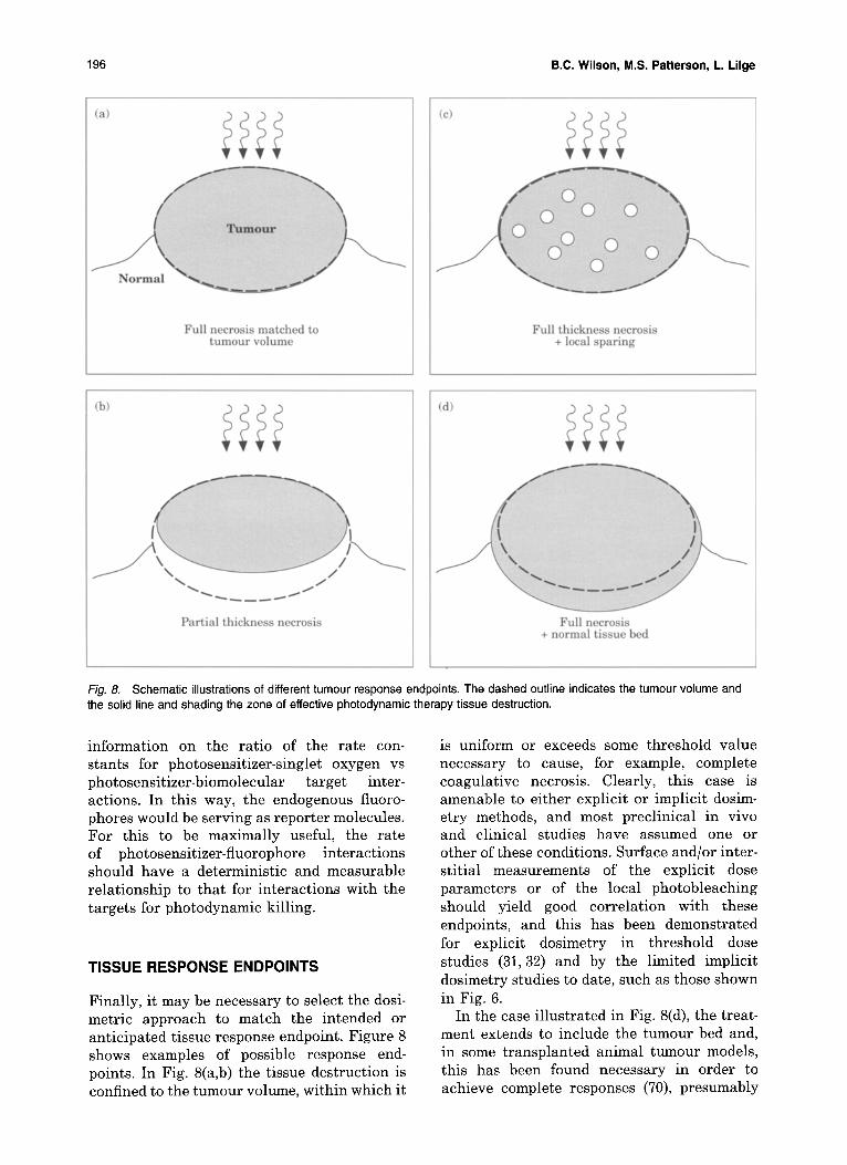
Fig. 8. Schematic illustrations of different tumour response endpoints. The dashed outline indicates the tumour volume and the solid line and shading the zone of effective photodynamic therapy tissue destruction.
information on the ratio of the rate con- stants for photosensitizer-singlet oxygen vs photosensitizer-biomolecular target inter- actions. In this way, the endogenous fiuoro- phores would be serving as reporter molecules. For this to be maximally useful, the rate of photosensitizer-fluorophore interactions should have a deterministic and measurable relationship to that for interactions with the targets for photodynamic killing.
TISSUE RESPONSE ENDPOINTS
Finally, it may be necessary to select the dosi- metric approach to match the intended or anticipated tissue response endpoint. Figure 8 shows examples of possible response end- points. In Fig. 8(a,b) the tissue destruction is confined to the tumour volume, within which it
is uniform or exceeds some threshold value necessary to cause, for example, complete coagulative necrosis. Clearly, this case is amenable to either explicit or implicit dosim- etry methods, and most preclinical in vivo and clinical studies have assumed one or other of these conditions. Surface and/or inter- stitial measurements of the explicit dose parameters or of the local photobleaching should yield good correlation with these endpoints, and this has been demonstrated for explicit dosimetry in threshold dose studies (31, 32) and by the limited implicit dosimetry studies to date, such as those shown in Fig. 6.
In the case il lustrated in Fig. 8(d), the treat- ment extends to include the tumour bed and, in some transplanted animal tumour models, this has been found necessary in order to achieve complete responses (70), presumably
Implicit and Explicit PDT Dosimetry
through shut-down of the vascular supply to the tumour mass. Again, in principle, either dosimetric approach should apply, with the added complication that the photosensi- tizer uptake, light distribution, tissue oxygen- ation and intrinsic photodynamic sensitivity will be different in the tumour and normal tissue regions, so that separate, localized measurements in each may be required.
The most difficult case, i l lustrated in Fig. 8(c), is where there is sparing or incomplete response at localized regions within the target volume. This may be due, for example, to inadequate photosensitizer uptake or local tissue hypoxia, which may be constitutive or PDT-induced. A fundamental problem here is that there is no clear approach to measuring the presence of these focal regions of under- response, and they may result in predictions based on gross tissue necrosis disagreeing with findings based on tumour regrowth.
CONCLUSIONS
Continuing progress in making PDT a quanti- tative therapy will depend on developing prac- tical but meaningful measures of PDT 'dose' which will correlate strongly and, ultimately, predictively, with the tissue response and clinical outcome. This paper has presented a framework for the various approaches to PDT dosimetry, and identified some of the import- ant issues and limitations which arise in these. It should be noted that there are few studies to date which have directly investigated critical issues such as the degree of coupling of photo- sensitizer photobleaching. Hence, those con- clusions made above which are necessarily based on specific but limited (and in some cases unpublished) data should be considered as tentative. Nevertheless, they serve well to illustrate the basic concepts.
Fundamental to this framework is the recog- nition of the interdependency of many of the dosimetric measures, of the real differences between what have been called 'explicit' and 'implicit' dosimetries in this paper, and of the need to tailor the dosimetry to the clinically relevant tissue response endpoints. Un- tangling these complex relationships will be a major challenge, both conceptually and technically, but is critical to the optimum application of new photodynamic agents and treatment techniques.
ACKNOWLEDGEMENTS
197
The authors wish to acknowledge the support of the National Cancer Institute of Canada. The images in Plate 1 were obtained using a Laser Induced Fluorescence Endoscopic (XILLIX LIFE) imaging system supplied by Xillix Technologies Corp, Richmond, BC, Canada. The measurements in Fig. 7 were performed using instrumen- tation developed with support from the National Institutes of Health, USA (grant #P01-CA43892). Photofrin was kindly supplied by QLT Phototherapeutics, Vancouver, BC, Canada. Thanks also to Dr D. Doiron and Dr C. Gomer, and Dr A. Oseroff for the information shown in Fig. 6(a,b), respectively, to Dr N. Marcon for perform- ing the clinical sudy shown in Plate 1 and Fig. 2, and to Dr T. Foster for helpful discussions.
REFERENCES
1 Dougherty TJ (ed). Photodynamic therapy. J. Clin Laser Med Surg 1996, 14:219-348
2 Fisher AMR, Murphree AL, Gomer CJ. Clinical and preclinical photodynamic therapy. Lasers Surg Med 1995, 17:2-31
3 Dougherty TJ. Photodynamic therapy. Photochem Photobiol 1993, 58:895-900
4 Fingar VH. Vascular effects of photodynamic therapy. J Clin Laser Surg Med 1996, 14:323-8
5 Braichotte DR, Savary J-F, Monnier P et al. Optimizing light dosimetry in photodynamic therapy of early stage carcinoma of the esophagus using fluorescence spectroscopy. Lasers Surg Med 1996, 19:340-6
6 Braichotte DR, Wagnieres GA, Bays R et al. Clinical pharmacokinetic studies of Photofrin by fluorescence spectroscopy in the oral cavity, the esophagus and the bronchi. Cancer 1995, 75:2768-78
7 Braichotte DR, Savary J-F, Glanzmann T et al. Clinical pharmacokinetic studies of tetra (meta-hydroxyphenyl) chlorin in squamous cell carcinoma by fluorescence sectroscopy at 2 wavelengths. Int J Cancer 1995, 63: 198-204
8 Wang I, Svanberg K, Andersson-Engels Se t al. Photo- dynamic therapy of non-melanoma skin malignancies with topical amino levulinic acid: diagnostic measure- ments. In: Cortese DA (ed) 5th International Photody- namic Association Biennial Meeting. Proc. SPIE, 1995, 2371:243-52
9 Cheong WF, Prahl SA, Welch AJ. A review of the optical properties of biological tissues. IEEE J Quant Electr 1990, 26:2166-85
10 Bays R, Wagnieres G, Robert D et al. Clinical measure- ments of tissue optical properties in the esophagus and in the oral cavity. In: Cortese DA (ed) 5th International Photodynamic Association Biennial Meeting. Proc. SPIE, 1995, 2371:388-95
11 Stone HB, Brown JM, Phillips TL et al. Oxygen in human tumors: Correlations between methods of measurement and response to therapy. Rad Res 1993, 136:422-34
12 Chert Q, Wilson BC, Shetty Se t al. Changes in in vivo optical properties and light distributions in normal canine prostate during photodynamic therapy. Radiat Res 1997, 147:86-91

198
13 Van Geel IPJ, Oppelaar H, Oussoren YG, Stewart FA. Changes in perfusion of mouse tumours after photody- namic therapy. Int J Cancer 1994, 56:224 8
14 Wilson BC, Patterson MS, Burns DM. Effect of photo- sensitizer concentration in tissue on the penetration depth of photoactivating light. Lasers Med Sci 1986, 1:235-44
15 Potter WR. PDT dosimetry and response. In: Dougherty TJ (ed) Photodynamic Therapy: Mechanisms. Proc. SPIE, 1989, 1065:88-99
16 Stringer MR, Robinson DJ, Hudson EJ et al. In vivo monitoring of photosensitizer fluorescence during photodynamic therapy. In: Cortese DA (ed) 5th Inter- national Photodynamic Association Biennial Meeting. Proc. SPIE, 1995, 2371:104-8
17 Svaasand LO, Potter WR. The implications of photo- bleaching for photodynamic therapy. In: Henderson BW, Dougherty TJ (eds) Photodynamic Therapy New York: Marcel Dekker Inc, 1992; Chap 23, pp. 369~85
18 Foster TH, Gao L. Dosimetry in photodynamic therapy: oxygen and the critical importance of capillary density. Radiat Res 1992, 130:379-83
19 Foster TH, Nichols MG. Oxygen sensitivity of PDT determined from time-dependent electrode measure- ments in spheroids. In: Dougherty TJ (ed) Optical Methods for Tumor Treatment and Detection: Mech- anisms and Techniques in Photodynamic Therapy IV. Proc. SPIE, 1995, 2392:141-51
20 Grossweiner LI. Photodynamic therapy. In: The Science of Phototherapy Boca Raton: CRC Press, 1994; Chap 8, pp. 139-55
21 Patterson MS, Wilson BC. A theoretical study of the influence of sensitizer photobleaching on depth of necrosis in photodynamic therapy. In: Dougherty TJ (ed) Optical Methods for Tumor Treatment and Detec- tion: Mechanisms and Techniques in Photodynamic Therapy III. Proc. SPIE, 1994, 2133:208-19
22 Sterenborg HJCM, van Gemert MJC. Photodynamic therapy with pulsed light sources: A theoretical analy- sis. Phys Med Biol 1996, 41:835-49
23 Wilson BC, Jeeves WP, Lowe D. In vivo and post mortem measurements of the attenuation spectra of light in tissues. Photochem Photobiol 1985, 42:15~62
24 Jacques SL. Tissue fluorescence. In: Cortese DA (ed) 5th International Photodynamic Association Biennial Meeting. Proc. SPIE, 1995, 2371:2-13
25 Star WM, Wilson BC, Patterson MS. Light delivery and optical dosimetry in photodynamic therapy of solid tumors. In: Henderson BW, Dougherty TJ (eds) Photo- dynamic Therapy New York: Marcel Dekker Inc, 1992; Chap 22, pp. 335~8
26 Pandey RK, Potter WR, Meunier I e t al. Evaluation ofnew benzoporphyrin derivatives with enhanced PDT activity. Photochem Photobiol 1995, 62:764-8
27 Patterson MS, Hayward JE, Farrell TF, Wilson BC. A general purpose instrument for PDT dosimetry. In: Cortese DA (ed) 5th International Photodynamic Association Biennial Meeting. Proc. SPIE, 1995, 2371:477-81
28 Jones LR, Grossweiner LI. Effects of Photofrin on in vivo skin reflectivity. J Photochem Photobiol 1996, B33:153-6
29 Vaupel P, Schlenger K, Knoop C et al. Oxygenation of human tumors: Evaluation of tissue oxygen distribu- tion in breast cancers by computerized 02 tension measurements. Cancer Res 1991, 51:3316-22
B.C. Wilson, M.S. Patterson, L. Lilge
30 Vinogradov SA, Lo L-W, Jenkins WT et al. Noninva- sive imaging of the distribution of oxygen in tissue in vivo using near-infrared phosphors. Biophys J 1996, 70:1609-17
31 Patterson MS, Wilson BC, Graft R. In vivo tests of the concept of photodynamic threshold dose in normal rat liver photosensitized by aluminum chlorosul- phonated phthalocyanine. Photochem Photobiol 1990, 51:343~9
32 Lilge L, Olivo MC, Schatz SW et al. The sensitivity of normal brain and intracranially implanted VX2 tumour to interstitial photodynamic therapy. Br J Cancer 1996, 73:332-43
33 Chen Q, Chopp M, Madigan Le t al. Damage threshold of normal rat brain in photodynamic therapy. Photo- chem Photobiol 1996, 64:16~7
34 Messmann H, Mlkvy P, Davies C et al. Threshold effects of PDT in the normal rat colon with ALA photosensitization. In: Cortese DA (ed) 5th Inter- national Photodynamic Association Biennial Meeting. Proc. SPIE, 1995, 2371:532-5
35 Foster TH, Murant RS, Bryant RG et al. Oxygen consumption and diffusion effects in photodynamic therapy. Radiat Res 1991, 126:29~303
36 Foster TH, Hartley DF, Nichols MG et al. Fluence rate effects in photodynamic therapy of multicell tumor spheroids. Cancer Res 1993, 53:1249-54
37 Wilson BC. Optical and photobiological dosimetry for photodynamic therapy of solid tumors. In: Dewey WC et al (eds) Radiation Research, A Twentieth Century Perspective. New York: Academic Press, 1992:674-9
38 Potter WR, Bellnier DA, Pandy R, Parsons JC, Dougherty TJ. Sensitizer pharmacokinetics by in vivo reflectance spectroscopy. Photochem Photobiol 1997 (in press)
39 Bezdetnaya L, Zeghari N, Belitchenko I et al. Spectro- scopic and biological testing of photobleaching of porphyrins in solution. Photochem Photobiol 1996, 64:382~
40 Georgakoudi I, Nichols MG, Foster TH. The mechan- ism of Photofrin photobleaching and its consequences for photodynamic therapy. Photochem Photobiol 1997, 65:13~44
41 Jacques SL, Joseph R, Gofstein G. How photobleaching affects dosimetry and fluorescence monitoring of PDT in turbid media. In: Dougherty TJ (ed) Optical Methods for Tumor Treatment and Detection: Mechanisms and Techniques in Photodynamic Therapy II. Proc. SHE, 1881:168-79
42 Sinaasappel M, Sterenborg HJCM. Quantification of hematoporphyrin derivative by fluorescence measure- ment using dual wavelength excitation and dual wavelength detection. Appl Opt 1993, 32:541-8
43 Chen J-Y, Chen W, Chai H-X, Dong R-C. Studies on pharmacokinetics of sulfonated aluminum phthalo- cyanine in a transplantable mouse tumor by in vivo fluorescence. J Photochem Photobiol 1993, B18:233-7
44 Jacques SL, Rodriguez T, Schwartz J. Kinetics of ALA- induced protoporphyrin IX accumulation in the liver, skin and tumor of a rat model. In: Dougherty TJ (ed) Optical Methods for Tumor Treatment and Detection: Mechanisms and Techniques in Photodynamic Therapy IV. Proc. SPIE, 1995, 2392:8-12
45 Katsumi T, Aizawa K, Kuroiwa Yet al. Effectiveness of photodynamic therapy with a diode laser used mono-L- aspartyl chlorin e6 for implanted fibrosarcoma in mice.

Implicit and Explicit PDT Dosimetry
In: Cortese DA (ed) 5th International Photodynamic Association Biennial Meeting. Proc. SPIE, 1995, 2371:86-9
46 Glanzmann T, Theumann J-F, Forrer M et al. Evalu- ation of mTHPC of "early" squamous cell carcinomas of the cheek pouch mucosa of Golden Syrian hamsters as a model for clinical PDT of "early" cancers in the upper aerodigestive tract, the esophagus and the tracheo-bronchial tree. In: Cortese DA (ed) 5th Inter- national Photodynamic Association Biennial Meeting. Proc. SPIE, 1995, 2371:51-8
47 Andersson-Engels S, Berg R, Svanberg K et al. Multi- colour fluorescence imaging in connection with photo- dynamic therapy of 5-amino levulinic acid (ALA) sensitised skin malignancies. Bioimaging 1995, 3:134 43
48 Moan J. Effect of bleaching of porphyrin sensitizers during photodynamic therapy. Cancer Lett 1986, 33:45-53
49 Forrer M, Glanzman T, Braichotte D et al. In vivo measurement of fluorescence bleaching of meso-tetra hydroxy phenyl chlorin (mTHPC) in the esophagus and oral cavity. In: Cubeddu R, Mordon SR, Svanberg K (eds) Optical Biopsies. Proc. SPIE, 1995, 2627:1 7
50 Rhodes LE, Tsoukas M, Anderson RR et al. A quanti- tative model for ALA pharmacokinetics and phototox- icity using iontophoretic delivery of ALA. Photochem Photobiol 1996, 63:90S, abstract
51 Georgakoudi I, Foster TH. Photobleaching of PpIX, NBSe and NBS during photodynamic therapy (PDT) of multicell tumor spheroids. Photochem Photobiol 1996, 63:90S, abstract
52 Cubeddu R, Canti G, Musolino M et al. In vivo absorp- tion spectrum of disulphonated aluminium phthalocya- nine in a murine tumour model. J Photochem Photobiol 1996, B34:229-35
53 Star WM. In vivo action spectra, absorption and fluor- escence excitation spectra of photosensitizers for photodynamic therapy. J Photochem Photobiol 1995, B28:101-2
54 van der Veen N, van Leengoed HLLM, Star WM. In vivo fluorescence kinetics and photodynamic therapy using 5-aminolaevutinic acid-induced porphyrin: increased damage after multiple irradiations. Br J Cancer 1994, 70:867-72
55 Gudgin Dickson EF, Pottier RH. On the role of protoporphyrin IX photoproducts in photodynamic therapy. J Photochem Photobiol 1995, B29:91 3
56 Rotomskis R, Bagdonas S, Streckyte G. Spectroscopic studies of photobleaching and photoproduct formation of porphyrins used in tumour therapy. J Photochem Photobiol 1996, B33:61-7
57 Moan J, Kessel D. Photoproducts formed from Photo- Din II in cells. J Photochem Photobiol 1988, B1:429-36
58 Patterson MS, Madsen SJ, Wilson BC. Experimental tests of the feasibility of singlet oxygen luminescence
199
monitoring in vivo during photodynamic therapy. J Photochem Photobiol 1990, B5:69-84
59 Moan J. On the diffusion length of singlet oxygen in cells and tissues. J Photochem Photobiol 1990, B6:343-7
60 Hudson E, Stringer M, Scholfield J et al. Measurement of the photodynamic dose in an optical phantom. In: Cortese DA (ed) 5th International Photodynamic Association Biennial Meeting. Proc. SPIE, 1995, 2371: 159~3
61 Pogue BW, Lilge L, Patterson MS et al. The absorbed photodynamic dose examined from pulsed and CW light using tissue-simulating dosimeters. Appl Opt 1997 (in press)
62 Aveline BM, Hasan T, Remond RW. The effects of aggregation, protein binding and cellular incorpor- ation on the photophysical properties of benzoporphy- rin derivative monoacid ring A (BPDMA). J Photochem Photobiol 1995, B30:161-9
63 Smith G, McGimpsey WG, Lynch MC et al. An efficient oxygen independent two-photon photosensitization mechanism. Photochem Photobiol 1994, 59:135-9
64 Sanghvi NT, Hynynen K, Lizzi FL. New developments in therapeutic ultrasound. IEEE Eng Med Biol 1996, Nov/Dec:83-92
65 Yeung WTI, Lee T-Y, Del Maestro RF et al. In vivo CT measurement of blood-brain transfer constant of iopa- midol in human brain tumors. J Neuro-Oncol 1992, 14:177-87
66 Yeung I, Lilge L, Wilson BC. Photodynamic therapy (PDT) induced alterations of blood-brain-barrier trans- fer constant of a tracer molecule in normal brain. In: Dougherty TJ (ed) Optical Methods for Tumor Treat- ment and Detection: Mechanisms and Techniques in Photodynamic Therapy VI. Proc. SPIE, 1997, 2972: 54~3
67 Dodd NJF, Moore JV, Poppitt DG et al. In vivo mag- netic resonance imaging of the effects of photodynamic therapy. Br J Cancer 1989, 60:164-7
68 van Geel IPJ, Oppelaar H, Oussoren YG et al. Mech- anisms for optimising photodynamic therapy: second- generation photosensitizers in combination with mitomycin C Br J Cancer 1995, 72:344-50
69 Mordon S, Devoisselle JM, Maunoury V. In vivo measurement and imaging of tumor tissue using a pH-sensitive fluorescent probe (5,6-carboxyfluorescein): instrumental and experimental studies. Photochem Photobiol 1994, 60:274-9
70 Fingar VH, Henderson BW. Drug and light dose depen- dence of photodynamic therapy: a study of tumor and normal tissue response. Photochem Photobiol 1987, 46:837-41
Key words: Photodynamic therapy; Dosimetry; Photo- bleaching; Tissue response