Recent Advances in Photodynamic Imaging and Therapy in ...
13
Review Recent Advances in Photodynamic Imaging and Therapy in Hepatobiliary Malignancies: Clinical and Experimental Aspects Atsushi Nanashima * , Masahide Hiyoshi, Naoya Imamura, Koichi Yano, Takeomi Hamada and Kengo Kai Citation: Nanashima, A.; Hiyoshi, M.; Imamura, N.; Yano, K.; Hamada, T.; Kai, K. Recent Advances in Photodynamic Imaging and Therapy in Hepatobiliary Malignancies: Clinical and Experimental Aspects. Curr. Oncol. 2021, 28, 4067–4079. https://doi.org/10.3390/curroncol 28050345 Received: 9 September 2021 Accepted: 28 September 2021 Published: 11 October 2021 Publisher’s Note: MDPI stays neutral with regard to jurisdictional claims in published maps and institutional affil- iations. Copyright: © 2021 by the authors. Licensee MDPI, Basel, Switzerland. This article is an open access article distributed under the terms and conditions of the Creative Commons Attribution (CC BY) license (https:// creativecommons.org/licenses/by/ 4.0/). Division of Hepato-Biliary-Pancreas Surgery, Department of Surgery, University of Miyazaki Faculty of Medicine, 5200 Kihara, Kiyotake, Miyazaki 889-1692, Japan; [email protected] (M.H.); [email protected] (N.I.); [email protected] (K.Y.); [email protected] (T.H.); [email protected] (K.K.) * Correspondence: [email protected] Abstract: The therapeutic and diagnostic modalities of light are well known, and derivative photody- namic reactions with photosensitizers (PSs), specific wavelengths of light exposure and the existence of tissue oxygen have been developed since the 20th century. Photodynamic therapy (PDT) is an effective local treatment for cancer-specific laser ablation in malignancies of some organs, including the bile duct. Although curability for extrahepatic cholangiocarcinoma is expected with surgery alone, patients with unresectable or remnant biliary cancer need other effective palliative therapies, including PDT. The effectiveness of PDT for cholangiocarcinoma has been reported experimentally or clinically, but it is not the standard option now due to problems with accompanied photosensitivity, limited access routes of irradiation, tumor hypoxia, etc. Novel derivative treatments such as pho- toimmunotherapy have not been applied in the field hepatobiliary system. Photodynamic diagnosis (PDD) has been more widely applied in the clinical diagnoses of liver malignancies or liver vascu- larization. At present, 5-aminolevulinic acid (ALA) and indocyanine green (ICG) dyes are mainly used as PSs in PDD, and ICG has been applied for detecting liver malignancies or vascularization. However, no ideal tools for combining both PDD and PDT for solid tumors, including hepatobiliary malignancies, have been clinically developed. To proceed with experimental and clinical trials, it is necessary to clarify the effective photosensitive drugs that are feasible for photochemical diagnosis and local treatment. Keywords: photodynamic reaction; photodynamic eye imaging; photodynamic therapy; hepatobil- iary malignancies 1. Introduction 1.1. Photomedicine and Photodynamic Reaction Photomedicine is a well-known therapy that utilizes various reactions with light irra- diation for diseases including oncological malignancies, and it has been developed in the fields of dermatology, surgery, radiology or optical diagnostics, ophthalmology, infectology and oncology [1–8]. In photomedicine, photodynamic diagnosis (PDD) and therapy (PDT) involve the unique biological processes of photodynamic reactions with the delivery of suitable photosensitizers (PSs), which are transported via the vasculature after administra- tion, such as vascular injection and oral intake [4,8–10]. Selective intracellular excitation and activation of PSs accumulate in target cells, including cancer cells, surrounding feeding vessels or immune cells, and are accompanied by direct tissue injury from light energy sources [11,12]. Although PSs exert cytotoxic or at least cytostatic effects due to sunlight or increased luces of light without specific irradiation and activation, after irradiation with a suitably specific wavelength of light, PS is detectable by fluorescence and reactive to oxygen species [13,14]. These are applied in diagnosis as well as in therapy [4]. Immune reactions are also induced, which may influence cellular immunity [4,15]. At increased Curr. Oncol. 2021, 28, 4067–4079. https://doi.org/10.3390/curroncol28050345 https://www.mdpi.com/journal/curroncol
-
Upload
khangminh22 -
Category
Documents
-
view
1 -
download
0
Transcript of Recent Advances in Photodynamic Imaging and Therapy in ...

Review
Recent Advances in Photodynamic Imaging and Therapy inHepatobiliary Malignancies: Clinical and Experimental Aspects
Atsushi Nanashima * , Masahide Hiyoshi, Naoya Imamura, Koichi Yano, Takeomi Hamada and Kengo Kai
�����������������
Citation: Nanashima, A.; Hiyoshi,
M.; Imamura, N.; Yano, K.; Hamada,
T.; Kai, K. Recent Advances in
Photodynamic Imaging and Therapy
in Hepatobiliary Malignancies:
Clinical and Experimental Aspects.
Curr. Oncol. 2021, 28, 4067–4079.
https://doi.org/10.3390/curroncol
28050345
Received: 9 September 2021
Accepted: 28 September 2021
Published: 11 October 2021
Publisher’s Note: MDPI stays neutral
with regard to jurisdictional claims in
published maps and institutional affil-
iations.
Copyright: © 2021 by the authors.
Licensee MDPI, Basel, Switzerland.
This article is an open access article
distributed under the terms and
conditions of the Creative Commons
Attribution (CC BY) license (https://
creativecommons.org/licenses/by/
4.0/).
Division of Hepato-Biliary-Pancreas Surgery, Department of Surgery, University of Miyazaki Faculty of Medicine,5200 Kihara, Kiyotake, Miyazaki 889-1692, Japan; [email protected] (M.H.);[email protected] (N.I.); [email protected] (K.Y.);[email protected] (T.H.); [email protected] (K.K.)* Correspondence: [email protected]
Abstract: The therapeutic and diagnostic modalities of light are well known, and derivative photody-namic reactions with photosensitizers (PSs), specific wavelengths of light exposure and the existenceof tissue oxygen have been developed since the 20th century. Photodynamic therapy (PDT) is aneffective local treatment for cancer-specific laser ablation in malignancies of some organs, includingthe bile duct. Although curability for extrahepatic cholangiocarcinoma is expected with surgeryalone, patients with unresectable or remnant biliary cancer need other effective palliative therapies,including PDT. The effectiveness of PDT for cholangiocarcinoma has been reported experimentally orclinically, but it is not the standard option now due to problems with accompanied photosensitivity,limited access routes of irradiation, tumor hypoxia, etc. Novel derivative treatments such as pho-toimmunotherapy have not been applied in the field hepatobiliary system. Photodynamic diagnosis(PDD) has been more widely applied in the clinical diagnoses of liver malignancies or liver vascu-larization. At present, 5-aminolevulinic acid (ALA) and indocyanine green (ICG) dyes are mainlyused as PSs in PDD, and ICG has been applied for detecting liver malignancies or vascularization.However, no ideal tools for combining both PDD and PDT for solid tumors, including hepatobiliarymalignancies, have been clinically developed. To proceed with experimental and clinical trials, it isnecessary to clarify the effective photosensitive drugs that are feasible for photochemical diagnosisand local treatment.
Keywords: photodynamic reaction; photodynamic eye imaging; photodynamic therapy; hepatobil-iary malignancies
1. Introduction1.1. Photomedicine and Photodynamic Reaction
Photomedicine is a well-known therapy that utilizes various reactions with light irra-diation for diseases including oncological malignancies, and it has been developed in thefields of dermatology, surgery, radiology or optical diagnostics, ophthalmology, infectologyand oncology [1–8]. In photomedicine, photodynamic diagnosis (PDD) and therapy (PDT)involve the unique biological processes of photodynamic reactions with the delivery ofsuitable photosensitizers (PSs), which are transported via the vasculature after administra-tion, such as vascular injection and oral intake [4,8–10]. Selective intracellular excitationand activation of PSs accumulate in target cells, including cancer cells, surrounding feedingvessels or immune cells, and are accompanied by direct tissue injury from light energysources [11,12]. Although PSs exert cytotoxic or at least cytostatic effects due to sunlight orincreased luces of light without specific irradiation and activation, after irradiation witha suitably specific wavelength of light, PS is detectable by fluorescence and reactive tooxygen species [13,14]. These are applied in diagnosis as well as in therapy [4]. Immunereactions are also induced, which may influence cellular immunity [4,15]. At increased
Curr. Oncol. 2021, 28, 4067–4079. https://doi.org/10.3390/curroncol28050345 https://www.mdpi.com/journal/curroncol

Curr. Oncol. 2021, 28 4068
lethal doses of PS, cells die by apoptosis, necrosis or autophagic cell death. Photody-namic therapy-induced apoptosis is conducted via the mitochondrial or receptor-mediatedpathway [4,6,7,16,17]. In the field of hepatobiliary malignancies, the theme of this specialissue in this journal, PDD and PDT appear to have advanced in the 21st century, whichis supposed to be a dawn of “photon and photomedicine”. Dolmans or dos Santos et al.summarized the basis and clinical applications in 2003 [4,8]. At that time, PDT had onlybeen tested in the clinic in oncology to treat various cancers, such as head and neck, brain,lung, pancreas, intraperitoneal cavity, breast, prostate and skin cancers, but had not beenfully applied in hepatobiliary diseases at the end of the 20th century. In PS, talaporfin alonehas been shown to accumulate well in the liver [8].
In the clinical background situation, the adequate treatment of cholangiocarcinomain case of unresectable cases with multiple severe biliary stenosis, or the residual cancertreatment at the bile duct stump or bilio-intestinal anastomosis has not been established,while for liver malignancies, the efficient locoregional treatment for multiple lesion orextrahepatic lesion is required because these can be detected by the recent fluorescentdiagnosis. By this point of view, we describe this review article.
1.2. Mechanism of Photodynamic Reaction and PS in Digestive Cancers
PDT involves two nontoxic components that combine cellular and tissue effects viaan oxygen-dependent reaction and require two components: (1) PS: a photosensitivesubstance localized in specific cells or tissues and (2) irradiation of light of a specificwavelength activating the accumulated PS. PS in the ground (stable) state transfers lightenergy to an excited state, and tissue molecular oxygen transfers reactive free radicalsor singlet oxygen [11–14]. The biological responses to PS are activated only in the partsof tissues or organs where they accumulate and upon which specific light exposure isfocused. Another photochemical reaction causes direct injury to DNA or tissues [8,18].PDT is thought to be involved in chemotherapy or brachytherapy for cancers, includingbiliary malignancies [19]. The most extensively applied PSs are porphyrin compounds.Most PDT occurs in an oxygen-dependent manner, and thus PS typically does not occur inanoxic tissue. The degree of photodamage and cytotoxicity depends on the type of PS, itsextracellular and intracellular localization, the administered light energy dose and time,the oxygen availability, etc. A combination of components, such as direct cellular killing,vascular injury and immune responses, is required for long-term tumor control.
2. PDT2.1. Effect on Tumors
Historically, hematoporphyrin accompanied by light exposure first exhibited celltoxicity in vitro in an animal cancer model and in cancer patients in the early 1990s. Aninterval between PS administration and light exposure was necessary until the maximumdifference in PS concentration between target cancer tissues and surrounding normal cellswas reached to achieve cancer toxicity and preserve normal tissues; this takes 24–72 h forporfimer sodium activated with an eximer dye laser (Photofrin; Wyeth Pharmaceuticals,Collegeville, PA, USA, and Wyeth, Tokyo, Japan) and 4–6 h for talaporfin sodium activatedby a semiconductor laser (NPe6; Laserphyrin® 100 mg for injection; Meiji Seika Pharma.,Co. Ltd., Tokyo, Japan) [20]. As PS causes long-lasting cutaneous photosensitivity, patientswho have been managed with PS must undergo shading from strong white light or sunlightfor various periods, up to weeks. Photosensitivity of skin has been a problematic issuein patients undergoing PDT. After Dougherty’s clinical success for cancer therapy in1983, PDT was attempted in esophageal squamous cell carcinoma, gastric or colorectaladenocarcinoma, cholangiocarcinoma and pancreatic adenocarcinoma [20–22]. Since themiddle of the 1990s, successful treatment of skin disorders with 5-ALA has also been usedfor photodetection, photodiagnosis (described later) or PDT in malignant lesions [23].

Curr. Oncol. 2021, 28 4069
2.2. PDT for Primary Liver Malignancies
The cytocidal effect of PDT on hepatocellular carcinoma (HCC) has been examinedin vitro and in vivo in original English articles since 1997 [24–33]. In the clinically acceptedPS worldwide, PDT using 5-ALA showed cytocidal molecular responses in an HCC cellline according to Abo’s report [24]. Recently, experimental PDT for HCC using PS underexperimental compounds has been mainly reported by Chinese investigators [27,28,31,33].However, the clinical effectiveness of PDT for HCC has not been clarified, although endo-scopic laser irradiation of PDT for biliary obstruction by HCC might be attempted withmultidisciplinary treatments [34,35]. Because the usefulness of near-infrared fluorescenceimaging using indocyanine green (ICG) or 5-ALA (PDD, described above) was recentlynoted, the possibility of PDT for accumulated fluorescein substances in HCC in experimen-tal HCC models would be expected [36–39]. Recently, nanoparticles composed of PS-basedPDT showed cytocidal effects in HCC cells in the experimental setting [38,40,41]. PDTalso causes microvascular damage in the tumor or peritumorous region, which leads tosevere tissue hypoxia [42]. In the case of HCC or other hypervascular liver malignancies,the effectiveness of the clinical application of PDT under sufficient oxygen concentra-tions is promising for clinical development in the near future. In our experiment using anovel highly water-soluble porphyrin (phosphorus tetraphenyl-porphyrin), the cytocidaleffect of PDT using the low energy light-emitted diode (LED) was highly observed invarious malignant cells but not in HepG2 human hepatoma cells, which are tolerant toporphyrin compound-based PDT [43,44]. However, by modifying glucose-conjugatedPS, the tumor viability of HepG2 cells dramatically decreased; therefore, HepG2 cellsmay also be integrated with the development of PS (not published yet), as noted in arecent report [45]. Preclinical trial of PDT using ICG for primary liver cancer seemed tobe scheduled by the animal model [37]. With respect to intrahepatic cholangiocellularcarcinoma as a mass-forming type, except for perihilar cholangiocarcinoma, no clinicalreports have been observed. In our preliminary (preclinical research) report in 2004, onecase of recurrent intrahepatic cholangiocarcinoma at the transected stump received anendoscopy applied PDT with a porfimer sodium and eximer dye laser, and the obstructivejaundice was relieved for two months [46]. However, regrowth of remnant tumors wasrapid, and effectiveness was not shown. For intrahepatic peripheral lesions, irradiationof PDT would be mainly performed via transhepatic surface using laparoscopy or openlaparotomy.
2.3. Extrahepatic Cholangiocarcinoma
In the late 1990s, basic research and preclinical trials for extrahepatic, periductalinvasive-type cholangiocarcinoma or large bile duct-origin adenocarcinoma were per-formed worldwide. In the basic study, certain reports elucidated that cholangiocellularcarcinoma was sensitive to cytocidal effects by PDT using porphyrin chemical compounds,although cholangiocarcinoma is not a relatively hypervascular malignancy [22,47–53]. Pah-ernik reported that the accumulation of porfimer sodium in the bile wall was significantlyincreased in comparison with that in surrounding peripancreatic organs, such as the gallbladder, pancreas or duodenum, in humans, and the maximum difference duct in theaccumulated tissue concentration of PS between 24 and 48 h after administration was overtwice that between normal tissue and cancer [48]. Talaporphin sodium also accumulated,and the concentration of fluid was increased in bile juice [54]. Thus, cholangiocarcinomawas considered to be a good target of PDT if light exposure was possible in the late 1990s.In the clinical setting, PDT has been attempted for biliary stricture of cholangiocarcinomavia a transhepatic or jejunal biliary approach since the 1990s [55,56]. Rumalla first reportedsuccessful results of PDT for cholangiocarcinoma using an endoscopic transpapillary ap-proach with a monorail procedure [57]. Consecutive clinical reports from Germany or otherinvestigators in Europe with respect to the usefulness of PDT to amend biliary obstruc-tion and impaired patient prognosis by cholangiocarcinoma have revealed great impactsworldwide, which were clarified by prospective or randomized multi-institutional trials,
Curr. Oncol. 2021, 28 4070
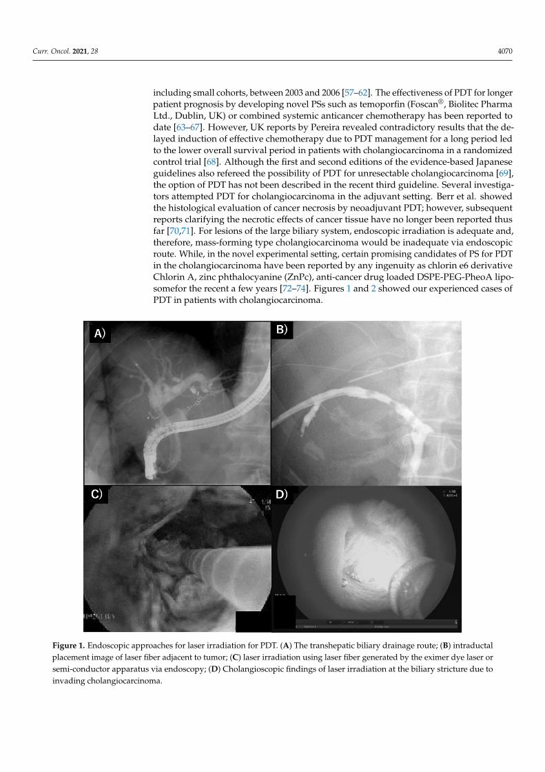
including small cohorts, between 2003 and 2006 [57–62]. The effectiveness of PDT for longerpatient prognosis by developing novel PSs such as temoporfin (Foscan®, Biolitec PharmaLtd., Dublin, UK) or combined systemic anticancer chemotherapy has been reported todate [63–67]. However, UK reports by Pereira revealed contradictory results that the de-layed induction of effective chemotherapy due to PDT management for a long period ledto the lower overall survival period in patients with cholangiocarcinoma in a randomizedcontrol trial [68]. Although the first and second editions of the evidence-based Japaneseguidelines also refereed the possibility of PDT for unresectable cholangiocarcinoma [69],the option of PDT has not been described in the recent third guideline. Several investiga-tors attempted PDT for cholangiocarcinoma in the adjuvant setting. Berr et al. showedthe histological evaluation of cancer necrosis by neoadjuvant PDT; however, subsequentreports clarifying the necrotic effects of cancer tissue have no longer been reported thusfar [70,71]. For lesions of the large biliary system, endoscopic irradiation is adequate and,therefore, mass-forming type cholangiocarcinoma would be inadequate via endoscopicroute. While, in the novel experimental setting, certain promising candidates of PS for PDTin the cholangiocarcinoma have been reported by any ingenuity as chlorin e6 derivativeChlorin A, zinc phthalocyanine (ZnPc), anti-cancer drug loaded DSPE-PEG-PheoA lipo-somefor the recent a few years [72–74]. Figures 1 and 2 showed our experienced cases ofPDT in patients with cholangiocarcinoma.
Curr. Oncol. 2021, 28, 4
have revealed great impacts worldwide, which were clarified by prospective or random-ized multi-institutional trials, including small cohorts, between 2003 and 2006 [57–62]. The effectiveness of PDT for longer patient prognosis by developing novel PSs such as temo-porfin (Foscan®, Biolitec Pharma Ltd., Dublin, UK) or combined systemic anticancer chem-otherapy has been reported to date [63–67]. However, UK reports by Pereira revealed con-tradictory results that the delayed induction of effective chemotherapy due to PDT man-agement for a long period led to the lower overall survival period in patients with chol-angiocarcinoma in a randomized control trial [68]. Although the first and second editions of the evidence-based Japanese guidelines also refereed the possibility of PDT for unre-sectable cholangiocarcinoma, [69] the option of PDT has not been described in the recent third guideline. Several investigators attempted PDT for cholangiocarcinoma in the adju-vant setting. Berr et al. showed the histological evaluation of cancer necrosis by neoadju-vant PDT; however, subsequent reports clarifying the necrotic effects of cancer tissue have no longer been reported thus far [70,71]. For lesions of the large biliary system, endoscopic irradiation is adequate and, therefore, mass-forming type cholangiocarcinoma would be inadequate via endoscopic route. While, in the novel experimental setting, certain prom-ising candidates of PS for PDT in the cholangiocarcinoma have been reported by any in-genuity as chlorin e6 derivative Chlorin A, zinc phthalocyanine (ZnPc), anti-cancer drug loaded DSPE-PEG-PheoA liposomefor the recent a few years [72–74]. Figures 1 and 2 showed our experienced cases of PDT in patients with cholangiocarcinoma.
Figure 1. Endoscopic approaches for laser irradiation for PDT. (A) The transhepatic biliary drainage route; (B) intraductal placement image of laser fiber adjacent to tumor; (C) laser irradiation using laser fiber generated by the eximer dye laser or semi-conductor apparatus via endoscopy; (D) Cholangioscopic findings of laser irradiation at the biliary stricture due to invading cholangiocarcinoma.
Figure 1. Endoscopic approaches for laser irradiation for PDT. (A) The transhepatic biliary drainage route; (B) intraductalplacement image of laser fiber adjacent to tumor; (C) laser irradiation using laser fiber generated by the eximer dye laser orsemi-conductor apparatus via endoscopy; (D) Cholangioscopic findings of laser irradiation at the biliary stricture due toinvading cholangiocarcinoma.

Curr. Oncol. 2021, 28 4071Curr. Oncol. 2021, 28, 5
Figure 2. (A) Cholangioscopy findings showed encircled cancer stenosis; (B) stenosis was released
by cancer shrinkage with a white necrotic plaque and reddish tissues after PDT; (C) mild photo-
reaction of the forearm at day 7 after PDT.
2.4. Limitation and Disadvantages of PDT in Hepatobiliary Malignancies
A large cohort of PDTs for hepatobiliary malignancies has not yet been reported. The
several reasons were considered as follows. (1) The detailed data of PS delivery to or ac-
cumulation in both normal and targeted cancer tissue were uncertain because the cholan-
giocarcinoma or bile duct samples were too small in unresectable patients. (2) The accom-
panying photosensitivity by PS often leads to severe adverse effects of dermatitis. There-
fore, complicated management, including longer periods under sunlight shielding, is re-
quired to avoid photosensitivity [75]. (3) Simultaneous combination therapy with sys-
temic anticancer drugs or other treatments has not been fully established. (4) The access
route of light (laser) exposure has not been established, particularly the limited space of
biliary tracts [76]. (5) The permeability of light exposure that leads to sufficient PDT activ-
ity is still unknown in limited space and maintaining the stability of light exposure
strength leading to sufficient PDT activity is difficult. In the case of biliary stricture, a sin-
gle light exposure might not be enough. Furthermore, the efficacy or safety of PDT under
the replacement of biliary stenting (tube or metal) is still uncertain [68]. (6) Targeted tumor
locations cannot be precisely detected because the excited light wavelength is inadequate
for PDD at present. In the case of cholangiocarcinoma, PDD has not been established thus
far. (7) Particularly in Japan, the cost of PS and the apparatus is still high; therefore, the
health economic balance has not improved. How is the cost in European countries? In the
case of liver cancer, although light exposure may be feasible for the liver surface via lapa-
roscopy or laparotomy, the permeability of light exposure would become a similar limi-
tation in the deeper part or in deeply located tumors, as well as in photodynamic eye
imaging described later.
3. PDD
3.1. PDD during PDT
Diagnostic tools using photoreaction for autofluorescence diagnosis or fluorescein-
conjugated immunostaining in malignancy are different from photodynamic methods;
therefore, the former is not described in this chapter. The photodynamic effect can be used
Figure 2. (A) Cholangioscopy findings showed encircled cancer stenosis; (B) stenosis was released by cancer shrinkage witha white necrotic plaque and reddish tissues after PDT; (C) mild photo-reaction of the forearm at day 7 after PDT.
2.4. Limitation and Disadvantages of PDT in Hepatobiliary Malignancies
A large cohort of PDTs for hepatobiliary malignancies has not yet been reported.The several reasons were considered as follows. (1) The detailed data of PS delivery toor accumulation in both normal and targeted cancer tissue were uncertain because thecholangiocarcinoma or bile duct samples were too small in unresectable patients. (2) Theaccompanying photosensitivity by PS often leads to severe adverse effects of dermatitis.Therefore, complicated management, including longer periods under sunlight shielding,is required to avoid photosensitivity [75]. (3) Simultaneous combination therapy withsystemic anticancer drugs or other treatments has not been fully established. (4) The accessroute of light (laser) exposure has not been established, particularly the limited spaceof biliary tracts [76]. (5) The permeability of light exposure that leads to sufficient PDTactivity is still unknown in limited space and maintaining the stability of light exposurestrength leading to sufficient PDT activity is difficult. In the case of biliary stricture, a singlelight exposure might not be enough. Furthermore, the efficacy or safety of PDT under thereplacement of biliary stenting (tube or metal) is still uncertain [68]. (6) Targeted tumorlocations cannot be precisely detected because the excited light wavelength is inadequatefor PDD at present. In the case of cholangiocarcinoma, PDD has not been established thusfar. (7) Particularly in Japan, the cost of PS and the apparatus is still high; therefore, thehealth economic balance has not improved. How is the cost in European countries? Inthe case of liver cancer, although light exposure may be feasible for the liver surface vialaparoscopy or laparotomy, the permeability of light exposure would become a similarlimitation in the deeper part or in deeply located tumors, as well as in photodynamic eyeimaging described later.
3. PDD3.1. PDD during PDT
Diagnostic tools using photoreaction for autofluorescence diagnosis or fluorescein-conjugated immunostaining in malignancy are different from photodynamic methods;

Curr. Oncol. 2021, 28 4072
therefore, the former is not described in this chapter. The photodynamic effect can be usedto detect tumors, and Dolmans detected the localization of PS to the vascular endotheliumof breast cancer in vivo in 2002 [77]. Hematoporphyrin, porphyrins, hematoporphyrinderivatives (HPDs) and ALA derivatives were tested for use in tumor detection [78]. Clin-ically, PDD for cancer detection has been attempted since the 1950s, and these agentsshowed contrast between squamous cell carcinoma and normal tissue between the 1960sand 1990s [4], but there were no remarkable reports in the case of cholangiocarcinomaor HCC up to the 2000s. In our series, photodetection by optical endoscopy with a com-mercial filter lens for a specific wavelength was attempted during PDT in patients withcholangiocarcinoma or bronchial cancer; however, clear contrast of the PS accumulatedarea could not be confirmed (data not indicated) [46]. In animal experiments with a sub-cutaneously transplanted bile duct cancer cell line (NOZ), PDT using glucose-conjugatedchlorin showed an increase in fluorescence intensity at the tumor; however, clear contrastwith normal mouse skin could not be detected [52].
3.2. Recent PDD Using ALA and ICG-Based NIR Fluorescence Imaging
PDD using 5-ALA and near-infrared (NIR) fluorescence imaging with ICG has beendramatically applied worldwide in the clinical setting to detect vascular or lymphaticflow or to detect hypervascular cancer in the 2010s [79–81]. First, 5-ALA is a precursorof protoporphyrin IX (PpIX), which is a photosensitive compound, PS, and accumulatesspecifically in cancer cells. By irradiation with 375–445-nm blue light exposure, the excitedintracellular PpIX emits 600–740 nm red light. 5-ALA is metabolized to PpIX in mito-chondria. In cancer cells, the biosynthesis of PpIX is promoted, but the metabolism ofPpIX is conversely suppressed compared to normal cells [79]. As a result, this markedaccumulation in cancer cells can be visualized using the appropriate detectors. PDD using5-ALA is possible for liver cancer cell lines, and in addition, the effect of PDT using 5-ALAis still controversial in vitro [24,82,83]. A clinical trial of 5-ALA PDD for patients withhepatocellular carcinoma and metastatic liver cancer during hepatectomy was attemptedby Inoue, and they concluded that residual tumors at the cut surface of the remnantliver were improved by PDD with 5-ALA [84]. For cholangiocarcinoma, 5-ALA PDD isfeasible in a cholangiocarcinoma mouse model [85]. At present, PDD with 5-ALA is anexperimentally promising state for hepatobiliary malignancies. Fluorescence imaging withICG for malignant hepatic tumors has been rapidly approved since Ishizawa’s report in2009 [86]. ICG is a pigment dye that has been widely used clinically for liver functionexaminations since the late 1950s, [79,87] in which the molecular energy state increases toemit a higher energy radiation of NIR and accumulated region excited light exposure canbe visualized by exclusive detectors [79,88]. ICG binds to plasma proteins and is removedfrom circulation exclusively by the liver to the bile juice; therefore, it might be trapped inthe hypervascular area of the hepatobiliary system as malignant tumors. The absorptionand fluorescence spectrum of ICG is in the near infrared region. ICG emits fluorescenceat a specific wavelength of light in the near-infrared spectrum (approximately 835 nm)when excited by light with a wavelength of 760 nm. ICG-guided surgery has been recentlydeveloped, and real-time cancer or blood flow visualization in the hepatobiliary region canbe achieved at present [89–91]. In addition to HCC, the hypervascular area of intrahepaticcholangiocarcinoma, hepatoblastoma, and [89] the area surrounding metastatic colorectalcancer have been well examined [92]. Although NIR light cannot be seen with the nakedeye, the distribution of subtle fluorescent materials in tissues can be precisely detectedthrough dedicated CCD camera, which has been conventionally applied. Furthermore,the detection of intraabdominal metastatic lesions of HCC can also be applied [93–95].ICG photodynamic eye imaging equivalent to PDD allows segmental visualization ofhepatic blood flow, which has been useful for anatomical liver resections [79,90,91,96]. In2021, the first worldwide consensus guideline for the fluorescence imaging using ICGin hepatobiliary Surgery has been eventually published [97]. Cyanine dye also has pho-tosensitizing characteristics [98]. Experimental reports regarding PDT with ICG and its

Curr. Oncol. 2021, 28 4073
combination with other chemical compounds, such as nanoparticles, for liver cancer haverecently increased [36,99,100]. In biliary cancers, Hishikawa et al. reported the usefulness ofNIR fluorescence imaging and photodynamic therapy with ICG lactosomes in gallbladdercancer cell lines [101]. Furthermore, photoimmune therapy, chemotherapy, and thermaltherapy have been experimentally developed in a liver cancer model. Thus, the clinicalapplication of PDT using the novel PS is promising for hepatobiliary malignancy treatmentin the future [102–104]. Figure 3 showed our experienced cases of PD imaging in HCCpatients.
Curr. Oncol. 2021, 28, 7
bination with other chemical compounds, such as nanoparticles, for liver cancer have re-
cently increased [36,99,100]. In biliary cancers, Hishikawa et al. reported the usefulness of
NIR fluorescence imaging and photodynamic therapy with ICG lactosomes in gallbladder
cancer cell lines [101]. Furthermore, photoimmune therapy, chemotherapy, and thermal
therapy have been experimentally developed in a liver cancer model. Thus, the clinical
application of PDT using the novel PS is promising for hepatobiliary malignancy treat-
ment in the future [102–104]. Figure 3 showed our experienced cases of PD imaging in
HCC patients.
Figure 3. Representative image of the ICG-photodynamic eye image during operation in a hepatocellular carcinoma pa-
tient, which showed (A) fluorescence of the main HCC and (B) portal vein tumor thrombus in the right portal vein (thick
white arrow). The thin arrow showed a taped each portal vein and trunk. (C) A case of hepatic venous tumor thrombus
(black arrow). (D) Identification of anatomical demarcation border. (E) Observation by the ICG-PDE camera at operative
field (Black arrowhead, Hamamatsu Photonics, Hamamatsu, Japan).
3.3. Limitation, Disadvantages of PDT in Hepatobiliary Malignancies
Similar to PDT, detection of tumor or vascularity in hepatobiliary lesion is limited
due to light penetration in the parenchymal organs as liver or hepatoduodenal ligament
because the deeper part less than approximately 10 mm cannot be visualized by the latest
CCD camera from the surface thus far, [89,93–95] although the intrahepatic transected
boundary seems to be detected. PDD for malignancies is depended to tumor vascularity
and, further, false positive surrounding lesion as liver cyst, cirrhotic nodule, hemangi-
oma [96].
4. Problems and Debates
Issues to solve issues for the future application were summarizes in this part. PDD
or PDT require the three components of PS accumulation, light exposure for excitation
and cellular oxygen; therefore, all components are necessary to achieve PDT cytotoxicity
in cancer cells clinically [4]. Endoscopy is necessary for the access route of light exposure.
In hepatobiliary diseases, a specific wavelength light source is optimally placed as a laser
fiber via cholangioscopy or laparoscopy. At present, a special and expensive light gener-
ator apparatus is required for each PS. Therefore, this modality is for local but not systemic
photochemical diagnosis or treatment; thus, combined systemic chemotherapy is usually
Figure 3. Representative image of the ICG-photodynamic eye image during operation in a hepatocellular carcinoma patient,which showed (A) fluorescence of the main HCC and (B) portal vein tumor thrombus in the right portal vein (thick whitearrow). The thin arrow showed a taped each portal vein and trunk. (C) A case of hepatic venous tumor thrombus (blackarrow). (D) Identification of anatomical demarcation border. (E) Observation by the ICG-PDE camera at operative field(Black arrowhead, Hamamatsu Photonics, Hamamatsu, Japan).
3.3. Limitation, Disadvantages of PDT in Hepatobiliary Malignancies
Similar to PDT, detection of tumor or vascularity in hepatobiliary lesion is limited dueto light penetration in the parenchymal organs as liver or hepatoduodenal ligament becausethe deeper part less than approximately 10 mm cannot be visualized by the latest CCDcamera from the surface thus far [89,93–95], although the intrahepatic transected boundaryseems to be detected. PDD for malignancies is depended to tumor vascularity and, further,false positive surrounding lesion as liver cyst, cirrhotic nodule, hemangi-oma [96].
4. Problems and Debates
Issues to solve issues for the future application were summarizes in this part. PDDor PDT require the three components of PS accumulation, light exposure for excitationand cellular oxygen; therefore, all components are necessary to achieve PDT cytotoxicityin cancer cells clinically [4]. Endoscopy is necessary for the access route of light expo-sure. In hepatobiliary diseases, a specific wavelength light source is optimally placed asa laser fiber via cholangioscopy or laparoscopy. At present, a special and expensive lightgenerator apparatus is required for each PS. Therefore, this modality is for local but notsystemic photochemical diagnosis or treatment; thus, combined systemic chemotherapy
Curr. Oncol. 2021, 28 4074
is usually necessary. What is the real indication of these modalities in the era of multi-disciplinary options for cancer treatment that have been developed in this malignancy?One is supposed to release the obstructive jaundice by malignant biliary strictures, andanother is an additional treatment for remnant cancer cells at the edge of surgical resec-tion, the transected plane of liver parenchyma or a stump of bile duct wall [20,46,105],because remnant cancer positivity is significantly associated with patient survival. To avoidthe photosensitivity or photoinjury of normal tissue, the development of cancer-specificaccumulation or photoreaction of PS is necessary. NIR-induced photoimmunotherapyapplying monoclonal antibodies to cancer-specific antigens is promising [106]. Inductionof systemic immune reaction by this modality is expected. However, in hypoxic tumorenvironments such as cholangiocarcinoma, it will be difficult to produce reactive oxygenspecies as singlet oxygen species by PDT in the center or deep part of the tumor [4,79].Light penetration into the central or deeper part of cancer tissue may be attenuated in bothPDT and PDD. Lasers have been applied for excitation of PS in PDT; however, light emitteddiodes (LEDs) are also expected to be used because of their economically cheaper costand the safety due to their low energy at direct exposure [107,108]. PDT can be achievedby recent reports, including our experimental results [20,43,44,46,52,105,109]. Eventually,concomitant procedures using both PDD and PDT are ideal; however, no successful reporthas been seen in the clinical setting. If the combination of two kinds of photosensitivechemical substances for tumor fluorescence detection and high cancer-cellular toxicity ineach is developed, both PDD and PDT can be achieved. Although applying the existingPS for PDT is considered, we must develop novel PS discoveries in future photodynamicmedicines, particularly for the treatment of hepatobiliary malignancies.
5. Conclusions
It has been over 100 years since the first PDT report, and PDT has been developedas an experimental clinical modality. In Eastern and Western countries, several PSs havebeen approved for clinical use for the treatment or diagnosis of malignancies, includinghepatobiliary diseases. Overall, PDT is potentially promising, depending on the limitedindications in comparison with the wide clinical development of PDD. However, thereare many problems associated with the use of PDT in a large series. In the future, it islikely that PDT will continue to be used as a stand-alone modality or in combinationwith chemotherapy, surgery, radiotherapy or other new strategies. Achieving ideallycombined PDT with PDD provides a new cancer treatment strategy, including in refractoryhepatobiliary malignant diseases, in the future.
Author Contributions: A.N. is the primary researcher and an organizer of this review issue. M.H.,N.I., K.Y., T.H. and K.K. were the main staff who attended the clinical trial of PDT and its relatedresearch works. All authors have read and agreed to the published version of the manuscript.
Funding: This research received no funding.
Acknowledgments: In the process of revision, the associate professor, Jin Matsumoto, supportedus to confirm the authenticity regarding contents or theory of photo engineering, who is a special-ist of producing the novel photosensitizer, chemistry and medical engineering at the institute ofDepartment of Applied Chemistry, Faculty of Engineering, University of Miyazaki, Japan. Writtenpermission here was obtained during process.
Conflicts of Interest: The authors declare no conflict of interest.
References1. Grzybowski, A.; Pietrzak, K. From patient to discoverer–Niels Ryberg Finsen (1860–1904)—The founder of phototherapy in
dermatology. Clin. Dermatol. 2012, 30, 451–455. [CrossRef]2. Daniell, M.D.; Hill, J.S. A history of photodynamic therapy. Aust. N. Z. J. Surg. 1991, 61, 340–348. [CrossRef] [PubMed]3. Ackroyd, R.; Kelty, C.; Brown, N.; Reed, M. The history of photodetection and photodynamic therapy. Photochem. Photobiol. 2001,
74, 656–669. [CrossRef]4. Dolmans, D.; Fukumura, D.; Jain, R. Photodynamic therapy for cancer. Nat. Rev. 2003, 3, 381–387. [CrossRef] [PubMed]

Curr. Oncol. 2021, 28 4075
5. Kharkwal, G.B.; Sharma, S.K.; Huang, Y.Y.; Dai, T.; Hamblin, M.R. Photodynamic Therapy for Infections: Clinical Applications.Lasers Surg. Med. 2011, 43, 755–767. [CrossRef]
6. Agostinis, P.; Buytaert, E.; Breyssens, H.; Hendrickx, N. Regulatory pathways in photodynamic therapy induced apoptosis.Photochem. Photobiol. Sci. 2004, 3, 721–729. [CrossRef] [PubMed]
7. Pogue, B.W.; Ortel, B.; Chen, N.; Redmond, R.W.; Hasan, T. A photobiological and photophysical-based study of phototoxicity oftwo chlorins. Cancer Res. 2001, 61, 717–724.
8. dos Santos, F.A.; Queiroz de Almeida, D.R.; Terra, L.F.; Baptista, M.S.; Labriola, L. Photodynamic therapy in cancer treatment—Anupdate review. J. Cancer Metastasis Treat. 2019, 5, 25–45. [CrossRef]
9. Kataoka, H.; Hayashi, N.; Tanaka, M.; Kubota, E.; Yano, S.; Joh, T. Tumor affinity photosensitizers for photodynamic therapy.JJSLSM 2015, 36, 159–165. [CrossRef]
10. Huang, Z. Photodynamic Diagnosis and Photodynamic Therapy Techniques. Optical Detection of Cancer. In Optical DetectionCancer; Meyers, A., Ed.; World Scientific Publishing Co., Pte., Ltd.: Singapore, 2011; pp. 79–98.
11. Gomer, C.J.; Razum, N.J. Acute skin response in albino mice following porphyrin photosensitization under oxic and anoxicconditions. Photochem. Photobiol. 1984, 40, 435–439. [CrossRef]
12. Moan, J.; Berg, K. The photodegradation of porphyrins in cells can be used to estimate the lifetime of singlet oxygen. Photochem.Photobiol. 1991, 53, 549–553. [CrossRef]
13. Epstein, J.H. Phototoxicity and photoallergy. Semin. Cutan. Med. Surg. 1999, 18, 274–284. [CrossRef]14. Nauta, J.M.; van Leengoed, H.L.; Star, W.M.; Roodenburg, J.L.; Witjes, M.J.; Vermey, A. Photodynamic therapy of oral cancer. A
review of basic mechanisms and clinical applications. Eur. J. Oral Sci. 1996, 104, 69–81. [CrossRef] [PubMed]15. Gollnick, S.O.; Liu, X.; Owczarczak, B.; Musser, D.A.; Henderson, B.W. Altered expression of interleukin 6 and interleukin 10 as a
result of photodynamic therapy in vivo. Cancer Res. 1997, 57, 3904–3909. [PubMed]16. Oleinick, N.L.; Evans, H.H. The photobiology of photodynamic therapy: Cellular targets and mechanisms. Radiat. Res. 1998, 150,
S146–S156. [CrossRef]17. Wilson, B.C.; Olivo, M.; Singh, G. Subcellular localization of Photofrin and aminolevulinic acid and photodynamic cross-resistance
in vitro in radiation-induced fibrosarcoma cells sensitive or resistant to photofrin-mediated photodynamic therapy. Photochem.Photobiol. 1997, 65, 166–176. [CrossRef]
18. Castano, A.P.; Demidova, T.N.; Hamblin, M.R. Mechanisms in photodynamic therapy: Part three—Photosensitizer pharma-cokinetics, biodistribution, tumor localization and modes of tumor destruction. Photodiagn. Photodyn. Ther. 2005, 2, 91–106.[CrossRef]
19. Tamada, K.; Sugano, K. Diagnosis and non-surgical treatment of bile duct carcinoma: Developments in the past decade. J.Gastroenterol. 2000, 35, 319–325. [CrossRef]
20. Nanashima, A.; Nagayasu, T. Current status of photodynamic therapy in digestive tract carcinoma in Japan. Int. J. Mol. Sci. 2015,16, 3434–3440. [CrossRef]
21. Dougherty, T.J. Hematoporphyrin as a photosensitizer of tumors. Photochem. Photobiol. 1983, 38, 377–379. [CrossRef]22. Ortner, M.A.; Liebetruth, J.; Schreiber, S.; Hanft, M.; Wruck, U.; Fusco, V.; Müller, J.M.; Hörtnagl, H.; Lochs, H. Photodynamic
therapy of nonresectable cholangiocarcinoma. Gastroenterology 1998, 114, 536–542. [CrossRef]23. Thunshelle, C.; Yin, R.; Chen, Q.; Hamblin, M.R. Current Advances in 5-aminolevulinic Acid Mediated Photodynamic Therapy.
Curr. Dermatol. Rep. 2016, 5, 179–190. [CrossRef]24. Abo-Zeid, M.A.M.; Abo-Elfadl, M.T.; Mostafa, S.M. Photodynamic therapy using 5-aminolevulinic acid triggered DNA damage
of adenocarcinoma breast cancer and hepatocellular carcinoma cell lines. Photodiagn. Photodyn. Ther. 2018, 21, 351–356. [CrossRef]25. Egger, N.G.; Schoenecker, J.A., Jr.; Gourley, W.K.; Motamedi, M.; Anderson, K.E.; Weinman, S.A. Photosensitization of experi-
mental hepatocellular carcinoma with protoporphyrin synthesized from administered delta-aminolevulinic acid: Studies withcultured cells and implanted tumors. J. Hepatol. 1997, 26, 913–920. [CrossRef]
26. Date, M.; Fukuchi, K.; Namiki, Y.; Okumura, A.; Morita, S.; Takahashi, H.; Ohura, K. Therapeutic effect of photodynamic therapyusing PAD-S31 and diode laser on human liver cancer cells. Liver Int. 2004, 24, 142–148. [CrossRef] [PubMed]
27. Tang, P.M.; Chan, J.Y.; Au, S.W.; Kong, S.K.; Tsui, S.K.; Waye, M.M.; Mak, T.C.; Fong, W.P.; Fung, K.P. Pheophorbide a, an activecompound isolated from Scutellaria barbata, possesses photodynamic activities by inducing apoptosis in human hepatocellularcarcinoma. Cancer Biol. Ther. 2006, 5, 1111–1116. [CrossRef] [PubMed]
28. Yow, C.M.; Wong, C.K.; Huang, Z.; Ho, R.J. Study of the efficacy and mechanism of ALA-mediated photodynamic therapy onhuman hepatocellular carcinoma cell. Liver Int. 2007, 27, 201–208. [CrossRef] [PubMed]
29. Fadel, M.; Kassab, K.; Youssef, T. Photodynamic efficacy of hypericin targeted by two delivery techniques to hepatocellularcarcinoma cells. Lasers Med. Sci. 2010, 25, 675–683. [CrossRef] [PubMed]
30. Shao, J.; Xue, J.; Dai, Y.; Liu, H.; Chen, N.; Jia, L.; Huang, J. Inhibition of human hepatocellular carcinoma HepG2 by phthalocyaninephotosensitiser PHOTOCYANINE: ROS production, apoptosis, cell cycle arrest. Eur. J. Cancer 2012, 48, 2086–2096. [CrossRef][PubMed]
31. Mirzaei, H.; Djavid, G.E.; Hadizadeh, M.; Jahanshiri-Moghadam, M.; Hajian, P. The efficacy of Radachlorin-mediated pho-todynamic therapy in human hepatocellular carcinoma cells. J. Photochem. Photobiol. B Biol. 2015, 142, 86–91. [CrossRef][PubMed]

Curr. Oncol. 2021, 28 4076
32. Liu, C.; Wu, T.; Wang, S.; Zhou, W.; Li, Y.; Chen, X.; Li, W.; Huang, Z.; Li, T.; Yang, L.; et al. Anticancer effect of LS-HB-mediatedphotodynamic therapy on hepatocellular carcinoma in vitro and in vivo. Photodiagn. Photodyn. Ther. 2020, 30, 101718. [CrossRef][PubMed]
33. Bahng, S.; Yoo, B.C.; Paik, S.W.; Koh, K.C.; Lee, K.T.; Lee, J.K.; Lee, J.H.; Choi, M.S.; Lee, K.H. Photodynamic therapy for bile ductinvasion of hepatocellular carcinoma. Photochem. Photobiol. Sci. 2013, 12, 439–445. [CrossRef] [PubMed]
34. Renzulli, M.; Ramai, D.; Singh, J.; Sinha, S.; Brandi, N.; Ierardi, A.M.; Albertini, E.; Sacco, R.; Facciorusso, A.; Golfieri, R.Locoregional Treatments in Cholangiocarcinoma and Combined Hepatocellular Cholangiocarcinoma. Cancers 2021, 13, 3336.[CrossRef] [PubMed]
35. Wang, Y.; Wang, C.; Ding, Y.; Li, J.; Li, M.; Liang, X.; Zhou, J.; Wang, W. Biomimetic HDL nanoparticle mediated tumor targeteddelivery of indocyanine green for enhanced photodynamic therapy. Colloids Surf. B Biointerfaces 2016, 148, 533–540. [CrossRef]
36. Kaneko, J.; Kokudo, T.; Inagaki, Y.; Hasegawa, K. Innovative treatment for hepatocellular carcinoma (HCC). Transl. Gastroenterol.Hepatol. 2018, 3, 78. [CrossRef] [PubMed]
37. Tsuda, T.; Kaibori, M.; Hishikawa, H.; Nakatake, R.; Okumura, T.; Ozeki, E.; Hara, I.; Morimoto, Y.; Yoshii, K.; Kon, M. Near-infrared fluorescence imaging and photodynamic therapy with indocyanine green lactosome has antineoplastic effects forhepatocellular carcinoma. PLoS ONE 2017, 12, e0183527. [CrossRef]
38. Hong, F.; Park, J.S.; Kim, S.W.; Park, S.J.; Kim, S.K. Near-infrared phototherapy for patient-derived orthotopic xenograft model ofhepatocellular carcinoma in combination with indocyanine green. J. Photochem. Photobiol. B Biol. 2020, 209, 111938. [CrossRef]
39. Abdel Fadeel, D.; Al-Toukhy, G.M.; Elsharif, A.M.; Al-Jameel, S.S.; Mohamed, H.H.; Youssef, T.E. Improved photodynamicefficacy of thiophenyl sulfonated zinc phthalocyanine loaded in lipid nano-carriers for hepatocellular carcinoma cancer cells.Photodiagn. Photodyn. Ther. 2018, 23, 25–31. [CrossRef]
40. Gao, Y.; Zheng, Q.C.; Xu, S.; Yuan, Y.; Cheng, X.; Jiang, S.; Kenry, Y.Q.; Song, Z.; Liu, B.; Li, M. Theranostic Nanodotswith Aggregation-Induced Emission Characteristic for Targeted and Image-Guided Photodynamic Therapy of HepatocellularCarcinoma. Theranostics 2019, 9, 1264–1279. [CrossRef]
41. van Straten, D.; Mashayekhi, V.; de Bruijn, H.S.; Oliveira, S.; Robinson, D.J. Oncologic photodynamic therapy: Basic principles,current clinical status and future directions. Cancers 2017, 9, 19. [CrossRef]
42. Matsumoto, J.; Suzuki, K.; Yasuda, M.; Yamaguchi, Y.; Hishikawa, Y.; Imamura, N.; Nanashima, A. Photodynamic therapy ofhuman biliary cancer cell line using combination of phosphorus porphyrins and light emitting diode. Bioorg. Med. Chem. 2017, 25,6536–6541. [CrossRef] [PubMed]
43. Mai, N.N.H.; Yamaguchi, Y.; Choijookhuu, N.; Matsumoto, J.; Nanashima, A.; Takagi, H.; Sato, K.; Tuan, L.Q.; Hishikawa, Y.Photodynamic Therapy Using a Novel Phosphorus Tetraphenylporphyrin Induces an Anticancer Effect via Bax/Bcl-xL-relatedMitochondrial Apoptosis in Biliary Cancer Cells. Acta Histochem. Cytochem. 2020, 53, 61–72. [CrossRef]
44. Tanaka, M.; Kataoka, H.; Yano, S.; Ohi, H.; Moriwaki, K.; Akashi, H.; Taguchi, T.; Hayashi, N.; Hamano, S.; Mori, Y.; et al.Antitumor effects in gastrointestinal stromal tumors using photodynamic therapy with a novel glucose-conjugated chlorin. Mol.Cancer Ther. 2014, 13, 767–775. [CrossRef]
45. Nanashima, A.; Yamaguchi, H.; Shibasaki, S.; Ide, N.; Sawai, T.; Tsuji, T.; Hidaka, S.; Sumida, Y.; Nakagoe, T.; Nagayasu, T.Adjuvant photodynamic therapy for bile duct carcinoma after surgery: A preliminary study. J. Gastroenterol. 2004, 39, 1095–1101.[CrossRef]
46. Wong Kee Song, L.M.; Wang, K.K.; Zinsmeister, A.R. Mono-l-aspartyl chlorine e6 (NPe6) and haematoporphyrin derivative(HpD) in photodynamic therapy administered to a human cholangiocarcinoma model. Cancer 1998, 82, 421–427. [CrossRef]
47. Pahernik, S.A.; Dellian, M.; Berr, F.; Tannapfel, A.; Wittekind, C.; Goetz, A.E. Distribution and pharmacokinetics of Photofrin inhuman bile duct cancer. J. Photochem. Photobiol. B Biol. 1998, 47, 58–62. [CrossRef]
48. Kiesslich, T.; Neureiter, D.; Alinger, B.; Alinger, B.; Jansky, G.L.; Berlanda, J.; Mkrtchyan, V.; Ocker, M.; Plaetzer, K.; Berr, F. Uptakeand phototoxicity of meso-tetrahydroxyphenyl chlorine are highly variable in human biliary tract cancer cell lines and correlatewith markers of differentiation and proliferation. Photochem. Photobiol. Sci. 2010, 9, 734–743. [CrossRef]
49. Nonaka, T.; Nanashima, A.; Nonaka, M.; Uehara, M.; Isomoto, H.; Asahina, I.; Nagayasu, T. Analysis of apoptotic effects inducedby photodynamic therapy in a human biliary cancer cell line. Anticancer Res. 2010, 30, 2113–2118. [PubMed]
50. Nonaka, T.; Nanashima, A.; Nonaka, M.; Uehara, M.; Isomoto, H.; Nonaka, Y.; Nagayasu, T. Advantages of laserphyrin comparedwith photofrin in photodynamic therapy for bile duct carcinoma. J. Hepatobiliary Pancreat. Sci. 2011, 18, 592–600. [CrossRef][PubMed]
51. Nonaka, Y.; Nanashima, A.; Nonaka, T.; Uehara, M.; Isomoto, H.; Abo, T.; Nagayasu, T. Synergic effect of photodynamic therapyusing talaporfin sodium with conventional anticancer chemotherapy for the treatment of bile duct carcinoma. J. Surg. Res. 2013,181, 234–241. [CrossRef]
52. Murakami, G.; Nanashima, A.; Nonaka, T.; Tominaga, T.; Wakata, K.; Sumida, Y.; Akashi, H.; Okazaki, S.; Kataoka, H.; Nagayasu,T. Photodynamic Therapy Using Novel Glucose-conjugated Chlorin Increases Apoptosis of Cholangiocellular Carcinoma inComparison with Talaporfin Sodium. Anticancer Res. 2016, 36, 4493–4501. [CrossRef]
53. Kiesslich, T.; Berlanda, J.; Plaetzer, K.; Krammer, B.; Berr, F. Comparative characterization of the efficiency and cellular pharma-cokinetics of Foscan- and Foslip-based photodynamic treatment in human biliary tract cancer cell lines. Photochem. Photobiol. Sci.2007, 6, 619–627. [CrossRef] [PubMed]
Curr. Oncol. 2021, 28 4077
54. Kasuya, K.; Shimazu, M.; Suzuki, M.; Kuroiwa, Y.; Usuda, J.; Itoi, T.; Tsuchida, A.; Aoki, T. Novel photodynamic therapy againstbiliary tract carcinoma using mono-L-aspartyl chlorine e6, basic evaluation for its feasibility and efficacy. J. Hepatobiliary PancreatSci. 2010, 17, 313–321. [CrossRef] [PubMed]
55. McCaughan, J.S.; Mertens, B.F.; Cho, C.; Barabash, R.D.; Payton, H.W. Photodynamic therapy to treat tumors of the extrahepaticbiliary ducts: A case report. Arch. Surg. 1991, 126, 111–113. [CrossRef] [PubMed]
56. Abulafi, A.M.; Allardice, J.T.; Williams, N.S.; Van Someren, N.; Swain, C.P.; Ainley, C. Photodynamic therapy for malignanttumours of the ampulla of Vater. Gut 1995, 36, 853–856. [CrossRef]
57. Rumalla, A.; Baron, T.H.; Wang, K.K.; Gores, G.J.; Stadheim, L.M.; de Groen, P.C. Endoscopic application of photodynamictherapy for cholangiocarcinoma. Gastrointest. Endosc. 2001, 53, 500–504. [CrossRef] [PubMed]
58. Berr, F. Photodynamic therapy for cholangiocarcinoma. Semin. Liver Dis. 2004, 241, 77–187. [CrossRef] [PubMed]59. Ortner, M.A. Photodynamic therapy in cholangiocarcinomas. Best Pract. Res. Clin. Gastroenterol. 2004, 18, 147–154. [CrossRef]60. Ortner, M.E.; Caca, K.; Berr, F.; Liebetruth, J.; Mansmann, U.; Huster, D.; Voderholzer, W.; Schachschal, G.; Mössner, J.; Lochs, H.
Successful photodynamic therapy for nonresectable cholangiocarcinoma: A randomized prospective study. Gastroenterology 2003,125, 1355–1363. [CrossRef]
61. Zoepf, T.; Jakobs, R.; Arnold, J.C.; Apel, D.; Riemann, J.F. Palliation of Nonresectable Bile Duct Cancer: Improved Survival afterPhotodynamic Therapy. Am. J. Gastroenterol. 2005, 100, 2426–2430. [CrossRef]
62. Witzigmann, H.; Berr, F.; Ringel, U.; Caca, K.; Uhlmann, D.; Schoppmeyer, K.; Tannapfel, A.; Wittekind, C.; Mossner, J.; Hauss, J.;et al. Surgical and palliative management and outcome in 184 patients with hilar cholangiocarcinoma: Palliative photodynamictherapy plus stenting is comparable to r1/r2 resection. Ann. Surg. 2006, 244, 230–239. [CrossRef]
63. Kniebühler, G.; Pongratz, T.; Betz, C.S.; Göke, B.; Sroka, R.; Stepp, H.; Schirra, J. Photodynamic therapy for cholangiocarcinomausing low dose mTHPC (Foscan). Photodiagnosis Photodyn. Ther. 2013, 10, 220–228. [CrossRef]
64. Ortner, M.A. Photodynamic therapy for cholangiocarcinoma: Overview and new developments. Curr. Opin. Gastroenterol. 2009,25, 472–476. [CrossRef] [PubMed]
65. Gao, F.; Bai, Y.; Ma, S.R.; Liu, F.; Li, Z.S. Systematic review: Photodynamic therapy for unresectable cholangiocarcinoma. J.Hepatobiliary Pancreat. Sci. 2010, 17, 125–131. [CrossRef] [PubMed]
66. Quyn, A.J.; Ziyaie, D.; Polignano, F.M.; Tait, I.S. Photodynamic therapy is associated with an improvement in survival in patientswith irresectable hilar cholangiocarcinoma. HPB 2009, 11, 570–577. [CrossRef]
67. Hong, M.J.; Cheon, Y.K.; Lee, E.J.; Lee, T.Y.; Shim, C. Long-term outcome of photodynamic therapy with systemic chemotherapycompared to photodynamic therapy alone in patients with advanced hilar cholangiocarcinoma. Gut Liver 2014, 8, 318–323.[CrossRef] [PubMed]
68. Pereira, S.P.; Aithal, G.P.; Ragunath, K.; Devlin, J.; Owen, F.; Meadows, H. Safety and long term efficacy of porfimer sodiumphotodynamic therapy in locally advanced biliary tract carcinoma. Photodiagn. Photodyn. Ther. 2012, 9, 287–292. [CrossRef][PubMed]
69. Miyazaki, M.; Yoshitomi, H.; Miyakawa, S.; Uesaka, K.; Unno, M.; Endo, I.; Ota, T.; Ohtsuka, M.; Kinoshita, H.; Shimada, K.; et al.Clinical practice guidelines for management of biliary tract cancers 2015, the 2nd English edition. J. Hepato-Biliary-Pancreat. Sci.2015, 22, 249–273. [CrossRef]
70. Wiedmann, M.; Caca, K.; Berr, F.; Schiefke, I.; Tannapfel, A.; Wittekind, C.; Mössner, J.; Hauss, J.; Witzigmann, H. Neoadjuvantphotodynamic therapy as a new approach to treating hilar cholangiocarcinoma: A phase II pilot study. Cancer 2003, 97, 2783–2790.[CrossRef]
71. Wagner, A.; Wiedmann, M.; Tannapfel, A.; Mayr, C.; Kiesslich, T.; Wolkersdörfer, G.W.; Berr, F.; Hauss, J.; Witzigmann, H.Neoadjuvant Down-Sizing of Hilar Cholangiocarcinoma with Photodynamic Therapy–Long-Term Outcome of a Phase II PilotStudy. Int. J. Mol. Sci. 2015, 16, 26619–26628. [CrossRef]
72. He, C.; Xia, J.; Gao, Y.; Chen, Z.; Wan, X. Chlorin A-mediated photodynamic therapy induced apoptosis in human cholangiocarci-noma cells via impaired autophagy flux. Am. J. Transl. Res. 2020, 12, 5080–5094.
73. Schmidt, J.; Kuzyniak, W.; Berkholz, J.; Steinemann, G.; Ogbodu, R.; Hoffmann, B.; Nouailles, G.; Gürek, A.G.; Nitzsche, B.;Höpfner, M. Novel zinc- and silicon-phthalocyanines as photosensitizers for photodynamic therapy of cholangiocarcinoma. Int. J.Mol. Med. 2018, 42, 534–546. [CrossRef]
74. Kim, D.H.; Im, B.N.; Hwang, H.S.; Na, K. Gemcitabine-loaded DSPE-PEG-PheoA liposome as a photomediated immunemodulator for cholangiocarcinoma treatment. Biomaterials 2018, 183, 139–150. [CrossRef]
75. Nanashima, A.; Nakashima, K.; Kawakami, H.; Ashizuka, S.; Kubota, Y. Nursing care management of photodynamic therapy indigestive tract carcinomas at a single cancer center. Photodiagn. Photodyn. Ther. 2017, 17, 221–225. [CrossRef] [PubMed]
76. Nanashima, A.; Isomoto, H.; Abo, T.; Nonaka, T.; Morisaki, T.; Arai, J.; Takagi, K.; Ohnita, K.; Shoji, H.; Urabe, S.; et al. How toaccess photodynamic therapy for bile duct carcinoma. Ann. Transl. Med. 2014, 2, 23.
77. Dolmans, D.E.; Kadambi, A.; Hill, J.S.; Flores, K.R.; Gerber, J.N.; Walker, J.P.; Borel Rinkes, I.H.; Jain, R.K.; Fukumura, D. Targetingtumor vasculature and cancer cells in orthotopic breast tumor by fractionated photosensitizer dosing photodynamic therapy.Cancer Res. 2002, 62, 4289–4294.
78. Lipson, R.L.; Baldes, E.J.; Olsen, A.M. Hematoporphyrin derivative: A new aid for endoscopic detection of malignant disease. J.Thorac. Cardiovasc. Surg. 1961, 42, 623–629. [CrossRef]

Curr. Oncol. 2021, 28 4078
79. Namikawa, T.; Fujisawa, K.; Munekage, E.; Iwabu, J.; Uemura, S.; Tsujii, S.; Maeda, H.; Kitagawa, H.; Fukuhara, H.; Inoue, K.;et al. Clinical application of photodynamic medicine technology using light-emitting fluorescence imaging based on a specializedluminous source. Med. Mol. Morphol. 2018, 51, 187–193. [CrossRef]
80. Gashev, A.A.; Nagai, T.; Bridenbaugh, E.A. Indocyanine green and lymphatic imaging: Current problems. Lymphat. Res. Biol.2010, 8, 127–130. [CrossRef]
81. Marshall, M.V.; Rasmussen, J.C.; Tan, I.C.; Aldrich, M.B.; Adams, K.E.; Wang, X.; Fife, C.E.; Maus, E.A.; Smith, L.A.; Sevick-Muraca, E.M. Near-Infrared Fluorescence Imaging in Humans with Indocyanine Green: A Review and Update. Open Surg. Oncol.J. 2010, 2, 12–25. [CrossRef] [PubMed]
82. Vonarx-Coinsman, V.; Foultier, M.T.; de Brito, L.X.; Morlet, L.; Gouyette, A.; Patrice, T. HepG2 human hepatocarcinoma cells:An experimental model for photosensitization by endogenous porphyrins. J. Photochem. Photobiol. B Biol. 1995, 30, 201–208.[CrossRef]
83. Otake, M.; Nishiwaki, M.; Kobayashi, Y.; Baba, S.; Kohno, E.; Kawasaki, T.; Fujise, Y.; Nakamura, H. Selective accumulation ofALA-induced PpIX and photodynamic effect in chemically induced hepatocellular carcinoma. Br. J. Cancer 2003, 89, 730–736.[CrossRef] [PubMed]
84. Inoue, Y.; Tanaka, R.; Komeda, K.; Hirokawa, F.; Hayashi, M.; Uchiyama, K. Fluorescence Detection of Malignant Liver Tumorsusing 5-Aminolevulinic Acid-Mediated Photodynamic Diagnosis: Principles, Technique, and Clinical Experience. World J. Surg.2014, 38, 1786–1794. [CrossRef] [PubMed]
85. Fujiwara, H.; Takahara, N.; Tateishi, K.; Tanaka, M.; Kanai, S.; Kato, H.; Nakatsuka, T.; Yamamoto, K.; Kogure, H.; Arita, J.; et al.5-Aminolevulinic acid-mediated photodynamic activity in patient-derived cholangiocarcinoma organoids. Surg. Oncol. 2020, 35,484–490. [CrossRef]
86. Ishizawa, T.; Fukushima, N.; Shibahara, J.; Masuda, K.; Tamura, S.; Aoki, T.; Hasegawa, K.; Beck, Y.; Fukayama, M.; Kokudo, N.Real-time identification of liver cancers by using indocyanine green fluorescent imaging. Cancer 2009, 115, 2491–2504. [CrossRef][PubMed]
87. Fox, I.J.; Wood, E.H. Indocyanine green: Physical and physiologic properties. Proc. Staff. Meet. Mayo Clin. 1960, 35, 732–744.88. Rangaraj, A.T.; Ghanta, R.K.; Umakanthan, R.; Soltesz, E.G.; Laurence, R.G.; Fox, J.; Cohn, L.H.; Bolman, R.M., 3rd; Frangioni, J.V.;
Chen, F.Y. Real-time visualization and quantification of retrograde cardioplegia delivery using near infrared fluorescent imaging.J. Card Surg. 2008, 23, 701–708. [CrossRef]
89. Nakaseko, Y.; Ishizawa, T.; Saiura, A. Fluorescence-guided surgery for liver tumors. J. Surg. Oncol. 2018, 118, 324–331. [CrossRef]90. Ishizawa, T.; Saiura, A.; Kokudo, N. Clinical application of indocyanine green-fluorescence imaging during hepatectomy.
Hepatobiliary Surg. Nutr. 2016, 5, 322–328. [CrossRef]91. Rossi, G.; Tarasconi, A.; Baiocchi, G.; De’ Angelis, G.L.; Gaiani, F.; Di Mario, F.; Catena, F.; Dalla Valle, R. Fluorescence guided
surgery in liver tumors: Applications and advantages. Acta Biomed. 2018, 89, 135–140.92. Yamada, Y.; Ohno, M.; Fujino, A.; Kanamori, Y.; Irie, R.; Yoshioka, T.; Miyazaki, O.; Uchida, H.; Fukuda, A.; Sakamoto, S.; et al.
Fluorescence-Guided Surgery for Hepatoblastoma with Indocyanine Green. Cancers 2019, 11, 1215. [CrossRef]93. Satou, S.; Ishizawa, T.; Masuda, K.; Kaneko, J.; Aoki, T.; Sakamoto, Y.; Hasegawa, K.; Sugawara, Y.; Kokudo, N. Indocyanine
green fluorescent imaging for detecting extrahepatic metastasis of hepatocellular carcinoma. J. Gastroenterol. 2013, 48, 1136–1143.[CrossRef]
94. Nanashima, A.; Tominaga, T.; Sumida, Y.; Tobinaga, S.; Nagayasu, T. Indocyanine green identification for tumor infiltration ormetastasis originating from hepatocellular carcinoma. Int. J. Surg. Case Rep. 2018, 46, 56–61. [CrossRef] [PubMed]
95. He, P.; Huang, T.; Fang, C.; Su, S.; Tian, J.; Xia, X.; Li, B. Identification of extrahepatic metastasis of hepatocellular carcinoma usingindocyanine green fluorescence imaging. Photodiagn. Photodyn. Ther. 2019, 25, 417–420. [CrossRef] [PubMed]
96. Abo, T.; Nanashima, A.; Tobinaga, S.; Hidaka, S.; Taura, N.; Takagi, K.; Arai, J.; Miyaaki, H.; Shibata, H.; Nagayasu, T. Usefulnessof intraoperative diagnosis of hepatic tumors located at the liver surface and hepatic segmental visualization using indocyaninegreen-photodynamic eye imaging. Eur. J. Surg. Oncol. 2015, 41, 257–264. [CrossRef] [PubMed]
97. Wang, X.; The, C.S.C.; Ishizawa, T.; Aoki, T.; Cavallucci, D.; Lee, S.Y.; Panganiban, K.M.; Perini, M.V.; Shah, S.R.; Wang, H.; et al.Consensus Guidelines for the Use of Fluorescence Imaging in Hepatobiliary Surgery. Ann. Surg. 2021, 274, 97–106. [CrossRef][PubMed]
98. Delaey, E.; van Laar, F.; De Vos, D.; Kamuhabwa, A.; Jacobs, P.; de Witte, P. A comparative study of the photosensitizingcharacteristics of some cyanine dyes. J. Photochem. Photobiol. B Biol. 2000, 55, 27–36. [CrossRef]
99. El-Daly, S.M.; Gamal-Eldeen, A.M.; Abo-Zeid, M.A.; Borai, I.H.; Wafay, H.A.; Abdel-Ghaffar, A.R. Photodynamic therapeuticactivity of indocyanine green entrapped in polymeric nanoparticles. Photodiagn. Photodyn. Ther. 2013, 10, 173–185. [CrossRef]
100. Li, S.; Yang, S.; Liu, C.; He, J.; Li, T.; Fu, C.; Meng, X.; Shao, H. Enhanced Photothermal-Photodynamic Therapy by IndocyanineGreen and Curcumin-Loaded Layered MoS2 Hollow Spheres via Inhibition of P-Glycoprotein. Int. J. Nanomed. 2021, 16, 433–442.[CrossRef]
101. Hishikawa, H.; Kaibori, M.; Tsuda, T.; Matsui, K.; Okumura, T.; Ozeki, E.; Yoshii, K. Near-infrared fluorescence imaging andphotodynamic therapy with indocyanine green lactosomes has antineoplastic effects for gallbladder cancer. Oncotarget 2019, 10,5622–5631. [CrossRef]
Curr. Oncol. 2021, 28 4079
102. Cai, H.; Dai, X.; Guo, X.; Zhang, L.; Cao, K.; Yan, F.; Ji, B.; Liu, Y. Ataxia telangiectasia mutated inhibitor-loaded coppersulfide nanoparticles for low-temperature photothermal therapy of hepatocellular carcinoma. Acta Biomater. 2021, 127, 276–286.[CrossRef] [PubMed]
103. Ji, B.; Cai, H.; Yang, Y.; Peng, F.; Song, M.; Sun, K.; Yan, F.; Liu, Y. Hybrid membrane camouflaged copper sulfide nanoparticles forphotothermal-chemotherapy of hepatocellular carcinoma. Acta Biomater. 2020, 111, 363–372. [CrossRef] [PubMed]
104. Gao, Y.H.; Li, M.Y.; Sajjad, F.; Wang, J.H.; Meharban, F.; Gadoora, M.A.; Yan, Y.J.; Nyokong, T.; Chen, Z.L. Synthesis andpharmacological evaluation of chlorin derivatives for photodynamic therapy of cholangiocarcinoma. Eur. J. Med. Chem. 2020, 189,112049. [CrossRef] [PubMed]
105. Matull, W.R.; Dhar, D.K.; Ayaru, L.; Sandanayake, N.S.; Chapman, M.H.; Dias, A.; Bridgewater, J.; Webster, G.J.; Bong, J.J.;Davidson, B.R.; et al. R0 but not R1/R2 resection is associated with better survival than palliative photodynamic therapy inbiliary tract cancer. Liver Int. 2011, 31, 99–107. [CrossRef]
106. Liang, S.; Sun, M.; Lu, Y.; Shi, S.; Yang, Y.; Lin, Y.; Feng, C.; Liu, J.; Dong, C. Cytokine-induced killer cells-assisted tumor-targetingdelivery of Her-2 monoclonal antibody-conjugated gold nanostars with NIR photosensitizer for enhanced therapy of cancer. J.Mater. Chem. B 2020, 8, 8368–8382. [CrossRef]
107. Kim, M.M.; Darafsheh, A. Light Sources and Dosimetry Techniques for Photodynamic Therapy. Photochem. Photobiol. 2020, 96,280–294. [CrossRef]
108. Liu, H.; Daly, L.; Rudd, G.; Khan, A.P.; Mallidi, S.; Liu, Y.; Cuckov, F.; Hasan, T.; Celli, J.P. Development and evaluation of alow-cost, portable, LED-based device for PDT treatment of early-stage oral cancer in resource-limited settings. Lasers Surg. Med.2019, 51, 345–351. [CrossRef]
109. Hatakeyama, T.; Murayama, Y.; Komatsu, S.; Shiozaki, A.; Kuriu, Y.; Ikoma, H.; Nakanishi, M.; Ichikawa, D.; Fujiwara, H.;Okamoto, K.; et al. Efficacy of 5-aminolevulinic acid-mediated photodynamic therapy using light-emitting diodes in humancolon cancer cells. Oncol. Rep. 2013, 29, 911–916. [CrossRef]