Recent advances in blood-related proteomics
16
REVIEW Recent advances in blood-related proteomics Lynne Thadikkaran, Michèle A. Siegenthaler, David Crettaz, Pierre-Alain Queloz, Philippe Schneider and Jean-Daniel Tissot Service régional vaudois de transfusion sanguine, Lausanne, Switzerland Blood is divided in two compartments, namely, plasma and cells. The latter contain red blood cells, leukocytes, and platelets. From a descriptive medical discipline, hematology has evolved towards a pioneering discipline where molecular biology has permitted the development of prognostic and diagnostic indicators for disease. The recent advance in MS and protein separa- tion now allows similar progress in the analysis of proteins. Proteomics offers great promise for the study of proteins in plasma/serum, indeed a number of proteomics databases for plasma/ serum have been established. This is a very complex body fluid containing lipids, carbohydrates, amino acids, vitamins, nucleic acids, hormones, and proteins. About 1500 different proteins have recently been identified, and a number of potential new markers of diseases have been char- acterized. Here, examples of the enormous promise of plasma/serum proteomic analysis for di- agnostic/prognostic markers and information on disease mechanism are given. Within the blood are also a large number of different blood cell types that potentially hold similar information. Proteomics of red blood cells, until now, has not improved our knowledge of these cells, in con- trast to the major progresses achieved while studying platelets and leukocytes. In the future, proteomics will change several aspects of hematology. Received: December 14, 2004 Revised: February 14, 2005 Accepted: March 1, 2005 Keywords: Biochemistry / Blood cells / Human plasma / Review / Serum / Two-dimensional gel electrophoresis Proteomics 2005, 5, 3019–3034 3019 Contents 1 Introduction.......................... 3019 2 Plasma/serum ........................ 3020 2.1 Overview ............................ 3020 2.2 Descriptive plasma/serum proteomics . . . 3021 2.3 Clinical applications – biomarker identification ......................... 3021 2.4 Amyloidosis and other deposition diseases ............................. 3024 2.5 Genetic diseases...................... 3025 2.6 Cryoproteins ......................... 3025 2.7 Immunoproteomics ................... 3025 2.8 Lipidomics ........................... 3025 2.9 Degradomics and peptidomics ......... 3025 3 Blood cells ........................... 3026 3.1 Red blood cells ....................... 3026 3.2 Platelets ............................. 3026 3.3 Leukocytes........................... 3027 3.4 Leukemia and lymphoma .............. 3030 4 Conclusions and perspectives .......... 3030 5 References........................... 3031 1 Introduction Despite the fact that examination of peripheral blood and bone marrow smears remains a critical step in the diagnosis of a large variety of hematological disorders, its role has recently been displaced by more sophisticated diagnostic tools like those offered by molecular biology. Nevertheless, the recognition of typical abnormal cytological findings on a blood smear limits the number of possible etiologies, con- tributing to diagnosis without multiplying laboratory tech- Correspondence: Professor Jean-Daniel Tissot, Service régional vaudois de transfusion sanguine, Rue du Bugnon 27, CH-1005 Lausanne, Switzerland E-mail: [email protected] Fax: 141-21-314-6597 Abbreviation: Igs, immunoglobulins © 2005 WILEY-VCH Verlag GmbH & Co. KGaA, Weinheim www.proteomics-journal.de DOI 10.1002/pmic.200402053
-
Upload
independent -
Category
Documents
-
view
0 -
download
0
Transcript of Recent advances in blood-related proteomics

REVIEW
Recent advances in blood-related proteomics
Lynne Thadikkaran, Michèle A. Siegenthaler, David Crettaz, Pierre-Alain Queloz,Philippe Schneider and Jean-Daniel Tissot
Service régional vaudois de transfusion sanguine, Lausanne, Switzerland
Blood is divided in two compartments, namely, plasma and cells. The latter contain red bloodcells, leukocytes, and platelets. From a descriptive medical discipline, hematology has evolvedtowards a pioneering discipline where molecular biology has permitted the development ofprognostic and diagnostic indicators for disease. The recent advance in MS and protein separa-tion now allows similar progress in the analysis of proteins. Proteomics offers great promise forthe study of proteins in plasma/serum, indeed a number of proteomics databases for plasma/serum have been established. This is a very complex body fluid containing lipids, carbohydrates,amino acids, vitamins, nucleic acids, hormones, and proteins. About 1500 different proteins haverecently been identified, and a number of potential new markers of diseases have been char-acterized. Here, examples of the enormous promise of plasma/serum proteomic analysis for di-agnostic/prognostic markers and information on disease mechanism are given. Within the bloodare also a large number of different blood cell types that potentially hold similar information.Proteomics of red blood cells, until now, has not improved our knowledge of these cells, in con-trast to the major progresses achieved while studying platelets and leukocytes. In the future,proteomics will change several aspects of hematology.
Received: December 14, 2004Revised: February 14, 2005
Accepted: March 1, 2005
Keywords:
Biochemistry / Blood cells / Human plasma / Review / Serum / Two-dimensional gelelectrophoresis
Proteomics 2005, 5, 3019–3034 3019
Contents
1 Introduction. . . . . . . . . . . . . . . . . . . . . . . . . . 30192 Plasma/serum . . . . . . . . . . . . . . . . . . . . . . . . 30202.1 Overview . . . . . . . . . . . . . . . . . . . . . . . . . . . . 30202.2 Descriptive plasma/serum proteomics . . . 30212.3 Clinical applications – biomarker
identification . . . . . . . . . . . . . . . . . . . . . . . . . 30212.4 Amyloidosis and other deposition
diseases . . . . . . . . . . . . . . . . . . . . . . . . . . . . . 30242.5 Genetic diseases. . . . . . . . . . . . . . . . . . . . . . 30252.6 Cryoproteins . . . . . . . . . . . . . . . . . . . . . . . . . 30252.7 Immunoproteomics . . . . . . . . . . . . . . . . . . . 3025
2.8 Lipidomics. . . . . . . . . . . . . . . . . . . . . . . . . . . 30252.9 Degradomics and peptidomics . . . . . . . . . 30253 Blood cells . . . . . . . . . . . . . . . . . . . . . . . . . . . 30263.1 Red blood cells . . . . . . . . . . . . . . . . . . . . . . . 30263.2 Platelets . . . . . . . . . . . . . . . . . . . . . . . . . . . . . 30263.3 Leukocytes. . . . . . . . . . . . . . . . . . . . . . . . . . . 30273.4 Leukemia and lymphoma . . . . . . . . . . . . . . 30304 Conclusions and perspectives . . . . . . . . . . 30305 References. . . . . . . . . . . . . . . . . . . . . . . . . . . 3031
1 Introduction
Despite the fact that examination of peripheral blood andbone marrow smears remains a critical step in the diagnosisof a large variety of hematological disorders, its role hasrecently been displaced by more sophisticated diagnostictools like those offered by molecular biology. Nevertheless,the recognition of typical abnormal cytological findings on ablood smear limits the number of possible etiologies, con-tributing to diagnosis without multiplying laboratory tech-
Correspondence: Professor Jean-Daniel Tissot, Service régionalvaudois de transfusion sanguine, Rue du Bugnon 27, CH-1005Lausanne, SwitzerlandE-mail: [email protected]: 141-21-314-6597
Abbreviation: Igs, immunoglobulins
© 2005 WILEY-VCH Verlag GmbH & Co. KGaA, Weinheim www.proteomics-journal.de
DOI 10.1002/pmic.200402053
3020 L. Thadikkaran et al. Proteomics 2005, 5, 3019–3034
niques. So, from a descriptive medical discipline based onmicroscopic evaluation of red blood cells, leukocytes, andplatelets, hematology has evolved towards a dynamic scienceat the crossroads of genomics and proteomics. PCR basedanalyses have radically changed the study of chromosomaltranslocation products [1]. With the development of DNAmicroarray technology, gene expression can be analyzed atthe mRNA level. The “transcriptome” is the mRNA poolfound within a cell. Most often there is poor correlation be-tween mRNA and protein expression. This is due to the factthat changes in the expression pattern at the mRNA level donot necessarily correlate with changes of the functionalexpression pattern at the protein level [2–5]. The word “pro-teome” describes the pool of proteins expressed in a cell or atissue at a given time. Its analysis provides an idea of biolog-ical processes happening at their level of occurrence, allow-ing the comparison of physiological and pathological statesof a cell line or a tissue [4, 6]. The challenge of clinical prote-omic studies is to link protein expression profiles to specificdisease phenotypes and to find out relevant biomarkers inorder to develop diagnostic tools [7]. Proteomics involvesseveral complementary technologies. One of the mostimportant tools for proteomic analysis is 2-DE, which hasbeen available since the 1970s. Current 2-DE technology aswell as cellular fractionation strategies for proteomics havebeen recently reviewed by Görg et al. [8] and Stasyk andHuber [9], respectively. Since 1990, MS techniques haveimproved, leading to rapid identification of separated pro-teins with the help of protein sequence databases [10].Another way to analyze changes at the protein level is SELDI.This method is of special interest for the identification ofnew biomarkers in the low molecular weight protein frac-tion, which may give an early marker of disordered cellularfunction, at a premalignant stage, for example. The principleof this technique is to use protein chips with an affinitymatrix (charge, hydrophobicity, or other). Various wash buf-
fers allow the differential binding of proteins to the surfaceof the chip, based on the stringency of their binding in var-ious conditions. Differences in protein patterns betweentumors and controls can then be elicited. However, SELDI/MS is a partial analysis, not giving sequence data in manycases. Finally, other methods, combining different separa-tion techniques (LC, multidimensional chromatography,tryptic digestion and multidimensional chromatography ofpeptides, off-gel electrophoresis) followed by MS, are cur-rently more and more employed [11–15]. Recently, because abroad range of mass spectrometers have been used, severalinvestigators proposed a new format for MS data, which willhopefully facilitate data management, interpretation, anddissemination of information in proteomic research [16].
The understanding of hematological diseases, includingmalignancies, is a critical issue and can be improved with thehelp of proteomics. A number of proteomic databases deal-ing with plasma/serum and/or blood cells are now availableon the internet (Table 1). The purpose of this paper is toreview the advances in hematology, obtained by proteomicresearch done over the last 5 years.
2 Plasma/serum
2.1 Overview
Blood plasma is a complex body fluid, which contains a largediversity of proteins. Their concentrations range over at least15 orders of magnitude, resulting in an enormous range thatprecludes full analysis of low abundance peptide/proteinentities using MS, unless the sample is fractionated orotherwise processed. Intact as well as partially degradedproteins or protein fragments circulate in the blood. Fur-thermore, genetic polymorphisms as well as numerouspost-translationally modified forms of proteins are present,
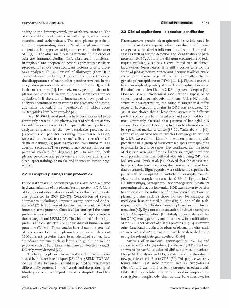
Table 1. Some internet sites devoted to blood proteomics
Subject Internet addres
Epstein-Barr virus transformed B-lymphoblastoid cells http://proteome.tmig.or.jp/2D/Hematopoietic cell lines http://www-smbh.univ-paris13.fr/lbtp/Biochemistry/Biochimie/bque.htmHuman plasma proteome project (HUPO) http://www.hupo.orgHuman platelets http://www.bioch.ox.ac.uk/glycob/ogp/Human serum proteome http://bpp.nci.nih.govIndex to 2-D PAGE databases and services http://www.expasy.org/ch2d/2d-index.htmlJurkat-T cell http://www.mpiib-berlin.mpg.de/2D-PAGE/organisms/index.htmlMyeloid development http://bioinfo.mbb.yale.edu/expression/myelopoiesis/Proteomics and protein structure http://linux.farma.unimi.it/Swiss-2DPAGE (lymphocyte) http://www.expasy.org/cgi-bin/map2/def?LYMPHOCYTE_HUMANSwiss-2DPAGE (lymphoma) http://www.expasy.org/cgi-bin/map2/def?LYMPHOMA_HUMANSwiss-2DPAGE (macrophage) http://www.expasy.org/cgi-bin/map2/def?U937_HUMANSwiss-2DPAGE (plasma) http://www.expasy.org/cgi-bin/map2/def?PLASMA_HUMANSwiss-2DPAGE (platelets) http://www.expasy.org/cgi-bin/map2/def?PLATELET_HUMANSwiss-2DPAGE (red blood cells) http://www.expasy.org/cgi-bin/map2/def?RBC_HUMANThe plasma proteome institute http://www.plasmaproteome.org/
© 2005 WILEY-VCH Verlag GmbH & Co. KGaA, Weinheim www.proteomics-journal.de
Proteomics 2005, 5, 3019–3034 Clinical Proteomics 3021
adding to the diversity complexity of plasma proteins. Theother constituents of plasma are salts, lipids, amino acids,vitamins, and carbohydrates. The core plasma protein isalbumin, representing about 50% of the plasma proteincontent and being present at high concentration (in the orderof 30 g/L). The other main plasma proteins (in the order ofg/L) are immunoglobulins (Igs), fibrinogen, transferrin,haptoglobin, and lipoproteins. Several approaches have beenproposed to remove these abundant proteins prior to prote-omic analyses [17–20]. Removal of fibrinogen (Factor I) iseasily obtained by clotting. However, this method inducedthe disappearance of many other proteins involved in thecoagulation process such as prothrombin (Factor II), whichis absent in serum [21]. Inversely, many peptides, absent inplasma but detectable in serum, can be identified after co-agulation. It is therefore of importance to have good pre-analytical conditions when mining the proteome of plasma,and more particularly its “peptidome”, in which about5000 peptides have been revealed [22].
Over 10 000 different proteins have been estimated to becommonly present in the plasma, most of which are at verylow relative abundances [23]. A major challenge of proteomicanalysis of plasma is the low abundance proteins, like(1) proteins or peptides resulting from tissue leakage,(2) proteins released from normal cells as a result of celldeath or damage, (3) proteins released from tumor cells asaberrant secretions. These proteins may represent importantpotential markers for diagnosis [24]. In addition, bothplasma proteome and peptidome are modified after stress,sleep, sport training, or meals, and in women during preg-nancy.
2.2 Descriptive plasma/serum proteomics
In the last 5 years, important progresses have been achievedin characterization of the plasma/serum proteome [24]. Mostof the relevant information is available in three leading arti-cles published in 2004 [25–27]. Combination of severalapproaches, including a literature survey, permitted Ander-son et al. [25] to build one of the most precise available lists ofhuman plasma proteins. Chan et al. [26] analyzed the serumproteome by combining multidimensional peptide separa-tion strategies and MS/MS [26]. They identified 1444 uniqueproteins and constructed a public database of human serumproteome (Table 1). These studies have shown the potentialof proteomics to explore plasma/serum, in which about1500 different proteins have been identified so far. Lowabundance proteins such as leptin and ghrelin as well aspeptides such as bradykinin, which are not detected using 2-DE only, were detected [27].
The lymph, a plasma-derived biologic fluid, was also an-alyzed by proteomic techniques [28]. Using SELDI-TOF MS,2-DE, and MS, two proteins could be pointed out which weredifferentially expressed in the lymph and the plasma (glialfibrillary astrocyte acidic protein and neutrophil cytosol fac-tor-1).
2.3 Clinical applications – biomarker identification
Plasma/serum protein electrophoresis is widely used inclinical laboratories, especially for the evaluation of proteinchanges associated with inflammation, liver, or kidney dis-eases as well as for the detection and identification of para-proteins [29, 30]. Among the different electrophoretic tech-niques available, 2-DE has a very limited role in clinicallaboratories. Nevertheless, it is still a cornerstone for thestudy of plasma/serum proteomics, because it allows analy-sis of the microheterogeneity of proteins, either due togenetic polymorphisms or PTMs [31–33]. Figure 1 shows atypical example of genetic polymorphism (haptoglobin a andb chains) easily identified in 2-DE of plasma samples [34].However, several biochemical modifications appear to besuperimposed on genetic polymorphisms. Using MS proteinstructure characterization, the cause of migrational differ-ences of haptoglobin a chains in 2-DE was elucidated [35,36]. It was shown that at least three structurally differentprotein species can be differentiated and accounted for themost commonly observed spot patterns of haptoglobin achains. As shown in Table 2, haptoglobin has been shown tobe a potential marker of cancer [37–39]. Watanabe et al. [40],after having analyzed serum samples from pregnant womenby 2-DE, were able to identify in women presenting withpreeclampsia a group of overexpressed spots correspondingto clusterin. In a large series, they confirmed that the levelsof clusterin were significantly higher in pregnant womenwith preeclampsia than without [40]. Also using 2-DE andMS analyses, Kwak et al. [41] showed that the serum pro-teome of patients with acute myeloid leukemia differed fromthat of controls. Eight peptides were differently expressed inpatients when compared to controls, for example, a-2-HS-glycoprotein, complement-associated SP-40, lipoprotein C-III. Interestingly, haptoglobin-1 was up-regulated in patientspresenting with acute leukemia. 2-DE was shown to be ableto demonstrate the influence of photochemical reactions onplasma proteins such as those induced, for example, bymethylene blue and visible light (Fig. 2), one of the tech-niques used to inactivate viruses in plasma in transfusionmedicine [42]. By contrast, inactivation of viruses using thesolvent/detergent method (tri-(N-butyl)-phosphate and Tri-ton X-100) was apparently not associated with modificationsof the 2-DE spot pattern of plasma proteins (Fig. 3). However,other functional protein alterations of plasma proteins, suchas protein S and a2-antiplasmin, have been described whileusing the solvent/detergent method [43, 44].
Analysis of monoclonal gammopathies [45, 46] andcharacterization of cryoproteins [47–49] using 2-DE has beenshown to be useful in selected difficult clinical situations.Using 2-DE analyses and MS, we also recently identified anew peptide, called hSpa or CD5L [50]. This peptide was onlyfound when IgM were present, like in cryoglobulins(Fig. 4A), and was found as being strongly associated withIgM. CD5L is a soluble protein expressed in lymphoid tis-sues (spleen, lymph node, thymus, and bone marrow), for
© 2005 WILEY-VCH Verlag GmbH & Co. KGaA, Weinheim www.proteomics-journal.de

3022 L. Thadikkaran et al. Proteomics 2005, 5, 3019–3034
Table 2. Examples of plasma/serum biomarkers
Context Methods Protein(s) identified References
Lung cancer 2-DE Haptoglobin [38]Ovarian cancer 2-DE Haptoglobin [39]Pregnancy (preeclampsia) 2-DE Clusterin [40]Acute myeloid leukemia 2-DE a-2-HS gylcoprotein, complement-associated SP-40,
apolipoprotein C-III, haptoglobin[41]
Prostate cancer SELDI Abnormal protein profile [68]Ovarian cancer SELDI Apolipoprotein A-I, transthyretin, fragment of inter-a-trypsin [69]Pregnancy (ectopic) SELDI Abnormal protein profile [70]Stroke SELDI Heart fatty acid, apolipoprotein C-I, apolipoprotein C-III [71, 72]Heart infarction 2-DE a-1-antitrypsin isoform 1 [73]Benzene exposure 2-DE T cell receptor b chain, FK506-binding protein, matrix
metalloproteinase-13[75]
Polycyclic hydrocarbon exposure 2-DE Putative capacitative calcium entry channel [76]Severe acute respiratory syndrome 2-DE Truncated a-1-antitrypsin [77]Severe acute respiratory syndrome 2-DE Peroxiredoxin II [78]Cardiac allograft rejection 2-DE aB-crystallin, tropomyosin [81]
Figure 1. Genetic polymorphism of plasma proteins evidenced by 2-DE (within the frames). Panel A shows the 2-D pattern of the plasma ofa “normal” blood donor. In panel B, several spots arrowed in panel A are absent (deficiency in haptoglobins b chain, and genetic poly-morphism of haptoglobin a1 and a2 chains). The picture shows silver-stained 2-D gels; first dimension: immobilized 4 to 7 pH gradient,second dimension: 9–16% PAGE. Twenty-five micrograms of plasma proteins were loaded onto the first dimension gel.
which little functional and structural information is available[51]. It belongs to group B of the scavenger receptor cysteine-rich superfamily, which includes the lymphocyte surfacereceptors CD5 and CD6 amongst others. CD5L is able tobind to different cells of the immune system (monocytes andlymphocytes), which suggests that it may play an importantrole in the regulation of the immune system. CD5L is a rela-tively abundant serum protein (60 mg/mL) circulating
essentially in association with other serum proteins, notablyIgM, as evidenced by the study of purified polyclonal ormonoclonal IgM fractions isolated from human plasma(Fig. 4B–D). Previous studies in mice have implicated mSpain the control of macrophage survival, the protein being anegative regulator of apoptosis [52, 53]. It was shown in micethat mSpa is a direct target for regulation by the liver X re-ceptor a [54]. Taken together, these results indicate that an
© 2005 WILEY-VCH Verlag GmbH & Co. KGaA, Weinheim www.proteomics-journal.de

Proteomics 2005, 5, 3019–3034 Clinical Proteomics 3023
Figure 2. Influence of high con-centration of methylene blue(50 mmol) and prolonged exposureto visible light (3 h) on plasma pro-teins. (A) Control plasma, (B) treatedplasma. The picture shows silver-stained 2-D gels; first dimension:immobilized 4 to 7 pH gradient, sec-ond dimension: 9–16% PAGE.Twenty-five micrograms of plasmaproteins were loaded onto the firstdimension gel. Note that the 2-Dpattern (acidic shift of most plasmaprotein spots) is completely dis-turbed by the photochemical treat-ment.
Figure 3. Influence of solvent/detergent (1% tri-(N-butyl)-phosphate and 1% Triton X-100) on plasma proteins. (A) Plasma pool fromOctapharma, Uniplas® (ABO universal plasma). (B) Plasma pool from Octapharma, Octaplas® (non-ABO universal plasma). (C) Nontreatedplasma pool of 750 samples from voluntary blood donors (control). The picture shows silver-stained 2-D gels; first dimension: immobilized4 to 7 pH gradient, second dimension: 9–16% PAGE. Twenty-five micrograms of plasma proteins were loaded onto the first dimension gel.Note the absence of apparent modification of the 2-D patterns after solvent/detergent treatment.
Figure 4. Type III cryoglobulins(polyclonal IgM mixed with poly-clonal IgG) analyzed by 2-DE (silver-stained 2-D gel; first dimension:immobilized 3 to 10 nonlinear pHgradient, second dimension: 9–16% PAGE). m, polyclonal IgM heavychains; g, polyclonal IgG heavychains; k-l, polyclonal Ig lightchains; m-s, fragments of the con-stant part of IgM heavy chains;
CD5L, h-Spa (IgM associated peptide). Twenty-five micrograms of proteins were loaded onto the first dimension gels. Association of CD5Lwith IgM is demonstrated by immunoblots (panels B and C) of purified monoclonal (M) and polyclonal (P) IgM fractions with anti-m, anti-CD5L, and J-chain antibodies. Anti-CD5L were kindly provided by Dr. F. Lozano, Servei d’Immunologia, Hospital Clinic, Barcelona, Spain.
© 2005 WILEY-VCH Verlag GmbH & Co. KGaA, Weinheim www.proteomics-journal.de

3024 L. Thadikkaran et al. Proteomics 2005, 5, 3019–3034
unexpected link exists between humeral immunity, innateimmunity, and the common nuclear receptor pathways thatmediate macrophage responses to modified lipoproteins.
Various non-2-DE based approaches have been success-fully developed to study the proteome of biological samples,and a number of new potential plasma/serum biologicalmarkers have been described [55]. Proteomics is now estab-lished in the medical community and most university hos-pitals develop such an approach to ameliorate patient care.Nevertheless, as recently pointed out by N. L. Anderson,proteomics for biomarker discovery has many importantlimitations [56].
By combining protein chip arrays with high-resolutionMS, notably SELDI-TOF MS, the analysis of the plasma/serum proteome appears to be a rapidly emerging area forboth biomarker discovery and clinical diagnostics [57–59],especially in oncology [13, 60–63]. However, the reproduci-bility of SELDI-TOF MS approaches have been questioned[64–67]. Nevertheless, interesting results have been obtainedby the evaluation of SELDI-TOF MS patterns of the serum ofpatients with total prostate specific antigen levels between2.5 and 15.0 ng/mL, presenting either with prostate canceror with benign prostate pathologies [68]. Using artificialintelligence based on pattern recognition algorithms, it waspossible to find patterns distinguishing these two categoriesof patients. Recently, three biomarkers, identified throughproteomic analysis of the serum of patients presenting withearly stage ovarian cancer, have been validated [69]. SELDI-TOF MS allowed the identification of apolipoprotein A1(down-regulated in cancer), a truncated form of transthyretin(down-regulated), and a cleavage fragment of inter-a-trypsininhibitor heavy chain H4 (up-regulated). The sensitivity of amultivariate model combining these three markers washigher than that of CA 125 alone. Using the same technique,Gerton et al. [70] showed that several serum proteins in themolecular weight range of 7500–18 000 Da were able to dis-criminate ectopic from intrauterine pregnancies. Heart fattyacid binding protein as well as apolipoprotein C-I and C-IIIhave been proposed as potential biological markers for thediagnosis of stroke [71, 72]. Interestingly, apolipoprotein C-Iand C-III appeared to be the first reported plasmatic bio-markers able to accurately distinguish between ischemic andhemorrhagic stroke within a small number of patients [72].Proteomic analysis of plasma from patients suffering fromacute myocardial infarction revealed also modifications ofthe protein pattern different from that of patients withunstable angina [73]. Proteomics has allowed the detection ofdrug-induced toxicity, and toxicoproteomics has developed[74]. The proteins in the plasma of workers exposed to ben-zene were analyzed by 2-DE by Joo et al. [75] who showed twosignificantly different protein profiles in workers exposed tothe toxic when compared to controls. Using MALDI-TOF,they identified T cell receptor b chain, FK506-binding pro-tein, and matrix metalloproteinase-13 as being up-regulatedin the benzene-exposed workers. In addition, the plasmaticT cell receptor b chain could be used for the early detection of
exposure to benzene. A study of the plasma of workersoccupationally exposed to polycyclic hydrocarbons, combin-ing 2-DE with MALDI-TOF MS, allowed Kap-Soon et al. [76]to identify several proteins as markers of such an exposure.An up-regulated protein (putative capacitative calcium entrychannel) appeared to be a representative marker of exposure.By the same technique, truncated forms of a1-antitrypsinwere discovered in serum samples of patients presentingwith severe acute respiratory syndrome [77]. Chen et al. [78]studied four patients with severe acute respiratory syndrome.After 2-DE, a total of 38 differential spots were selected forprotein identification, and most of them corresponded toacute phase proteins. The authors also identified proteinssuch as peroxiredoxin II, not detected before on plasma 2-D.Comparative proteome analysis of human plasma and LC-MS/MS analysis allowed Qian et al. [79] to identify 32 pro-teins that were significantly increased after lipopolysaccha-ride administration. Among the proteins identified, severalknown inflammatory and acute phase proteins were pointedout.
After infusion of stable isotopes of amino acids followedby collection of plasma samples, van Eijk and Deutz [80]evaluated protein synthesis according to the incorporation oflabeled isotopes in peptides identified by ESI-LC MS.
The discovery of markers detecting graft organ rejectionusing a blood assay is an important task, however, untilrecently, no tests have proved reliable enough to replaceinvasive histological investigations. Using 2-DE and MS,Borozdenkova et al. [81] showed however that aB-crystallinand tropomyosin may be novel serum markers of humancardiac allograft rejection.
2.4 Amyloidosis and other deposition diseases
Amyloidosis consists in a group of diseases characterized bythe accumulation of an amorphous, eosinophilic, hyalineextracellular deposit. The physical nature of amyloid underelectron microscopy is predominantly a fibrillar protein witha b-pleated sheet structure. Amyloid also contains P compo-nents and other glycoproteins [82, 83]. There are several dis-tinct proteins which have been found in amyloid deposits ofa variety of clinical settings. In systemic amyloidosis, pro-teins such as immunoglobulin light chain, serum amy-loid A, transthyretin, fibrinogen a chain, cystatin C, b2-microglobulin, lysozyme, gelsolin, apolipoprotein A-I, orapolipoprotein A-II may be involved [84]. MS analysis ofimmunoaffinity purified transthyretin from plasma samplesof six patients previously diagnosed with amyloidosis and of25 controls, allowed identification of the variants of trans-thyretin responsible for the disease [85]. The use of prote-omics to identify abnormal proteins and more particularly Iglight chains will certainly be very promising. Via high-reso-lution MS, it was possible to characterize in details both Igheavy and light chains [47]. Crystal-storing histiocytosis is arare event associated with monoclonal gammopathy. Theintracellular crystal formation is almost always accompanied
© 2005 WILEY-VCH Verlag GmbH & Co. KGaA, Weinheim www.proteomics-journal.de
Proteomics 2005, 5, 3019–3034 Clinical Proteomics 3025
by the expression of k light chains, as shown by Lebeau et al.[86]. A general approach, derived from the one described byNedelkov et al. [87], will certainly have a place in the study ofabnormal plasma proteins involved in deposition diseases.The authors reported a high-throughput comprehensiveanalysis for assaying proteins directly from human plasma.Proteins are selectively retrieved by using antibodies immo-bilized within affinity pipette tips and eluted onto enzymati-cally active MS targets for subsequent digestion and struc-tural characterization. The approach has been validated viaparallel high-throughput analysis of transthyretin and trans-ferrin from 96 plasma samples [87].
2.5 Genetic diseases
Microheterogeneity of serum glycoproteins has been studiedby proteomics in patients with chronic alcohol abuse andcompared with patients with carbohydrate-deficient glyco-protein syndrome [32, 88]. In alcoholism, abnormal isoformsof transferrin and a1-antitrypsin were devoid of a variablenumber of entire N-glycan moieties and were identical tothose present in carbohydrate-deficient glycoprotein syn-drome type 1. In contrast to carbohydrate-deficient glyco-protein syndrome type 1, there was no decrease in clusterinor serum amyloid P. Tammen et al. [89] used a completelydifferent approach to show that phenotyping of Val34Leu ofcoagulation factor XIII can be done by differential peptidedisplay, which is a technique that generates comprehensivepeptide maps covering a mass range of 950–15 000 Da. Thisapproach will certainly be useful for studies in which thephenotype has to be determined, namely, when DNA sam-ples are not available.
The evaluation of the risk of hemorrhage or of thrombo-sis is a difficult medicinal task, requiring sophisticated eval-uative techniques. Many different proteins are involved, andprotein-protein interactions are crucial events in this context.The complexity arising from the heterogeneity of the humanhemostatic proteome is presented in a quite provocativearticle by Mann et al. [90], who discuss the consequences ofmolecular heterogeneity as well as relations between thegenotype and phenotype in coagulation diseases.
A high-throughput SELDI-TOF MS immunoassay forcharacterizing plasmatic transthyretin variants arising fromamino acid substitutions, PTMs, and/or products of proteindegradation or proteolysis has recently been proposed bySchweigert et al. [91].
2.6 Cryoproteins
Proteomics proves useful in evaluating patients with differ-ent types of cryoproteins. Cryoproteins are defined as pro-teins precipitating at low temperature. Most frequently, theprecipitates contain Igs and are therefore called cryoglobu-lins (reviewed in [46]). Three types of cryoglobulins havebeen described so far: type I contains a single monoclonal Ig,whereas type II is a mixture of a monoclonal Ig with poly-
clonal Igs, and type III is a mixture of polyclonal Igs of dif-ferent isotypes, most frequently IgG and IgM. Type II andtype III are also called mixed cryoglobulins. But a new type ofcryoglobulins, named type II–III, containing polyclonal IgGassociated with a mixture of polyclonal and monoclonalIgMs has been recently described [46]. In addition, proteinssuch as albumin [48] and fibrinogen [49] can also cryopreci-pitate.
2.7 Immunoproteomics
Combining SDS-PAGE, 2-DE, Western blotting, and MS(immunoproteomics), Almeras et al. [92] were able to identifybrain antigens that are recognized by plasma antibodies ofpatients with multiple sclerosis, demonstrating the interestof this approach for the characterization of potentially newantigens associated with autoimmune diseases. Similarly,Pitarch et al. [93] showed, also using immunoproteomics, acombination of peptides of Candida albicans that may proveuseful for its diagnosis in patients presenting with hemato-logical malignancies and systemic candidiasis. Mining the“allergome”, the allergen repertoire, is a new avenue of pro-teomics [94]. Plasma antibodies can be identified, and theirspecificity evaluated by MS. For example, by combining af-finity-proteomics and antibody-proteomics, Przybylski et al.[95], with high-resolution Fourier transform cyclotron reso-nance MS, were able to elucidate antibodies paratopes ofboth polyclonal and monoclonal antibodies. With such tools,proteomics will open new avenues for research in immuno-hematology and basic immunology.
2.8 Lipidomics
Lipoproteins are very important for mediating lipid metabo-lism. The term lipidomics has just emerged in the literature.It can be defined as “the full characterization of lipid molec-ular species and of their biological roles with respect toexpression of proteins involved in lipid metabolism andfunction, including gene regulation” [96]. Pioneer worksdealing with 2-DE and lipoproteins, notably HDL have beenpublished several years ago [97]. The protein content of theselipoproteins has been reevaluated using proteomic tools [98],or using an approach called “virtual 2-DE” by Ogorzalek et al.[99]. Novel proteins were identified in HDL by these approa-ches. Also using 2-DE and the new MS technologies, Karls-son et al. [100] studied the protein content of LDL, and iden-tified calgranulin and lysozyme C in these lipoproteins.Their observation raises the possibility of new links betweenlipid metabolism, immunity and inflammation, which maybe of relevance in the context of atherosclerosis.
2.9 Degradomics and peptidomics
To identify substrates of matrix metalloproteinase-14, in hu-man plasma, Hwang et al. [101] proposed an attractiveapproach. They incubated plasma proteins in the presence of
© 2005 WILEY-VCH Verlag GmbH & Co. KGaA, Weinheim www.proteomics-journal.de
3026 L. Thadikkaran et al. Proteomics 2005, 5, 3019–3034
catalytic domain metalloproteinase-14, and compared 2-Dpatterns of controls with those obtained after the incubation.Fifteen different proteins were identified by MS from31 spots differing between the two groups. These proteinsincluded six known substrates for the protease and ninepotential ones. Such an approach, combined with the studyof plasma peptidome [102] will be interesting to evaluateother proteases as well as the effect on plasma proteins ofsystemic fibrinolytic treatments that are currently used totreat patients after cardiovascular events such as heartinfarction [103]. “Degradomics” and “peptidomics” will becertainly also useful to restudy the coagulation cascade, theactivation either of the complement or of the fibrinolyticpathways.
3 Blood cells
All blood cells are derived from a common progenitor, thepluripotent hematopoietic stem cell [104]. Appropriate cellcycle control, especially in the early stages of stem/pro-genitor cells, is required to maintain a normal hematopoiesis[105]. Hematopoietic stem cells must balance self-renewaland differentiation to provide sufficient primitive cells tosustain hematopoiesis, while generating mature cells withspecialized capabilities [106]. A common myeloid progenitorgives rise to both granulocytes and monocytes. Granulocytesinclude neutrophils, eosinophils, and basophils. Neutrophilsand monocytes (macrophages) are the key elements of theinnate host defense system. Eosinophils, basophils, andmast cells constitute the response to allergens and parasiticinfections. Comparative proteomic analyses of humanCD341 stem cells and mature CD151 myeloid cell isolatedfrom human cord blood have been reported by Tao et al.[107]. In this study, the authors have studied the extractedcytosolic proteins by 2-DE and MS and could show thatCD341 stem cells have a larger proteome than matureCD151 myeloid cells, and that many stem cell-associatedproteins appeared to be dramatically down-regulated as theCD341 cells undergo differentiation.
The lymphoid lineages, consisting of B, T, and naturalkiller cells, are derived from a common lymphoid pro-genitor. Human T and B cell precursors continuouslymature in thymus and bone marrow, respectively. B andT cells are key components of the adaptative immune re-sponse. Approximately 3 6 109 new erythrocytes are pro-duced per kg per day in adult. It is well established thaterythropoietin is responsible both for maintaining normalerythropoiesis and increasing red cell production in re-sponse to hypoxia. Platelets, also known as thrombocytes,are the smallest circulating blood particles. They derivefrom megakaryocytes located in bone marrow and play crit-ical roles in primary and secondary hemostasis (blood coa-gulation). Megakaryocyte development is a process in whicha wide variety of signals are working together to create ahighly specific response.
3.1 Red blood cells
Only a couple of proteomics-related papers have been pub-lished on this topic. While working on red blood cell mem-brane proteins separated by SDS-PAGE and analyzed by MS,Low et al. [108] were able to identify 44 polypeptides, of whichonly 19 were also found on 2-DE gels. The most completestudy describing the proteome of red blood cells has beenreported by Kakhniashvili et al. [109]. Erythrocyte mem-branes as well as cytoplasm were analyzed by IT MS/MS inline with LC. About 100 membrane proteins were identified,including several proteins of the cytoskeletal membranesuch as spectrin, ankyrin, and protein band 3, 4.1, 4.2, 4.9,7.2b. By contrast, a few red blood cell antigens were char-acterized (Rh blood group D, glycophorin A, aquaporin, andLutheran blood group protein). At the immunohematologi-cal point of view, it was very interesting to note that onedonor was probably an RhD variant, because Rhesus D cate-gory VI was identified by MS. The study of the cytoplasmicfraction of erythrocytes allowed the identification of manyproteins, including hemoglobin a, b, and g chains. Quanti-tative proteomics was used by Brand et al. [110] to evaluatethe dynamic changes in transcription factor complexes dur-ing erythroid differentiation. The analysis of phosphoproteinprofiling of erythropoietin receptor-dependent pathways wasevaluated by Körbel et al. [111], by combining two proteomicapproaches (2-DE/MALDI-TOF; 1-D/LC-MS/MS). Thisstrategy offered the detection of low expressed signalingmolecules, because an effective enrichment of phosphopro-teins was achieved. Proteomics was also successfullyemployed to analyze invasion of malaria parasites into hu-man red blood cells [112]. Two novel surface proteins wereidentified by Florens et al. [113] on Plasmodium falciparum-infected erythrocytes. A restricted number of red blood cellmembrane proteins such as flotillin-1, syntaxin 1C, andarginase, appear to be dysregulated in diabetic type 2patients [114].
3.2 Platelets
Platelets are cytoplasmic fragments derived from mega-karyocytes. They contain proteins as well as mRNA. Theabsence of a nucleus prevents platelets from being studiedvia classic molecular biology techniques and the analysis ofthe transcriptome is also difficult due to the little amount ofmRNA left. The platelets are therefore excellent candidatesfor proteomic approaches. The remaining mRNA makesthem able to keep on synthesizing some proteins and theypossess the apparatus to modify the proteins post-translationally.
The main function of platelets is to form thrombi onvascular lesions in order to stop bleeding and to contribute tothe healing process thereafter. They can also be part of thepathogenesis of diseases like arterial thromboembolic dis-ease or be affected by genetic defects such as Glanzmanthrombasthenia or Bernard-Soulier disease. It is therefore
© 2005 WILEY-VCH Verlag GmbH & Co. KGaA, Weinheim www.proteomics-journal.de
Proteomics 2005, 5, 3019–3034 Clinical Proteomics 3027
essential to study the proteome of activated as well as ofinactivated platelets. In addition, ongoing characterization ofthe platelet transcriptome, secretome, and proteome sug-gests that platelets have additional functions relevant to theinnate and adaptative immunity [115].
Different proteomic techniques (2-DE, multi-dimensional chromatography followed by various tech-niques of MS identification) have been applied to identifyplatelet proteins either at different stages of activation or indifferent locations inside the cell. Several types of proteinscan be characterized, namely, signal transduction proteins,cytoskeletal proteins, proteins synthesized by platelet at rest,and proteins synthesized and/or secreted during their acti-vation phase. The concentration of the different platelet pro-teins varies. Proteins such as actin outweigh others by far.Therefore, it is difficult to isolate and analyze low abundancebut biologically active proteins. The first proteomic analysisof the platelet cytosol was done using 2-DE with a broad pI(pI 3–10) followed by MALDI-TOF MS and identified186 proteins [116]. Another study, by O’Neill et al. [117],using broad (pI 3–10) as well as different narrow ranges of pI(4–7, 4–5, 6–11), detected 2300 protein spots. One hundredtwenty-three different gene products were identified, corre-sponding to proteins with very different functions, such assignal transduction, protein kinases, protein phosphatases,calcium binding proteins, GTP-binding proteins, and pro-tein complexes. Several proteins, like actin, tubulin, vimen-tin, proteins controlling the stability of the cytoskeleton (e.g.,a-actinin 1), or helping the movement of proteins along thecytoskeleton, have been identified. Proteins belonging to theprotein processing group (degradation, synthesis, enzymesfor modification of proteins) as well as proteins associatedwith the movement of vesicles were also recognized. Pep-tides involved in extracellular functions (coagulation factors,mitochondrial proteins, metabolic enzymes, proteins of thevarious metabolic pathways including apoptosis) were found-ed. Surprisingly, transcription factors, elongation factors,and centromer proteins, whose functions in platelets are allbut clear, were also identified [118]. In another study, Garciaet al. [119] separately analyzed the pI 4–5 region and pI 5–11 regions of the human platelet proteome, and identified311 gene products. The largest group of these proteinsexpressed signal transduction functions.
As already mentioned, the proteome expression of plate-lets is quite dynamic, depending on their state of activation.The initiation of platelet activation is due to adhesive ligandssuch as collagen and von Willebrand factor on glycoproteinreceptors (reviewed in [120–123]). This state of activation isthen enhanced by activators (thrombin, ADP) on G-protein-coupled membrane receptors [122]. Cascades of signalingevents are produced, resulting in cytoskeleton remodelingand activation of the integrin receptor aIIbb3, which is themain receptor involved in platelet aggregation [124]. Thisactivation goes on until exhaustion of elements or inhibitionby negative signals. Both inside-out and outside-in signalingare regulated by positive and/or negative phosphorylation
[125]. The activation state of platelets has been studied by invitro activation of platelets with the thrombin receptor acti-vating peptide [126]. In another study, Garcia et al. [127]detected 62 proteins differentially expressed in activated andinactivated platelets, either appearing, disappearing, or beingdown- or up-regulated between the two states. Forty-one pro-teins were identified, of which 14 were only present at basalstate, eight when platelets were activated with the thrombinreceptor activating peptide. Nine peptides were down-regu-lated and ten up-regulated with the thrombin receptor acti-vating peptide. These proteins mainly belonged to the cyto-skeleton or were signaling or processing proteins.
Another feature of activated platelets is the release ofa granules, dense granules, as well as lysosomes. The globalcharacterization of these released proteins is called “secre-tome” [128, 129]. The study of the secretome is important,because it contains peptides with paracrine or autocrinefunctions and peptides with prothrombotic properties. Inaddition, peptides of the secretome may be involved in cellproliferation and may have immunomodulating functions[130]. Maguire and Fitzgerald [128] studied the secretome oflow-dose thrombin-activated platelets isolated by differentialcentrifugation and then purified by ultracentrifugation. Theyfound a higher protein concentration in the supernatant ofactivated platelets. With more complex proteomic approa-ches (multidimensional chromatography followed by diges-tion with trypsin, strong cation exchange, RP chromatogra-phy, and IT MS), more than 300 proteins were isolated, 82 ofwhich repeatedly; 40% were known secreted proteins, theremaining not being known to be present in platelets orsecreted by them. Interesting recent studies provided evi-dence that the platelet transcriptome correlated quite wellwith the platelet proteome, and that proteomic analysisrevealed the expression of many previously unreported genesin platelets [131–133]. Proteomic technologies applied toplatelet research have been recently reviewed [134].
3.3 Leukocytes
The pluripotent hematopoietic stem cell is able to differ-entiate into various mature blood cells when stimulated withhematopoietic growth factors. The description of the pro-cesses underlying hematopoietic stem cell development andthe functional activity of mature cells has improved withproteomic approaches of characterization of proteins, pro-tein-protein interactions, and relative quantification of pro-teins. Hematopoietic development has been studied by Tianet al. [135]. Two myeloid cell lines, representing distinctstages of differentiation, have been analyzed by DNA micro-arrays, ICAT labeling, and mLC-MS/MS analysis. Differentialexpression of mRNA only correlated with 40% of those of theproteins, showing the importance of combining the twoapproaches in order to gain insight into the mechanismsinvolved in cellular developments. Of note, the authorsobserved anticorrelation, with significant changes at bothmRNA and protein levels but in opposite directions.
© 2005 WILEY-VCH Verlag GmbH & Co. KGaA, Weinheim www.proteomics-journal.de

3028 L. Thadikkaran et al. Proteomics 2005, 5, 3019–3034
Leukocyte activation is expressed by the production andrelease of inflammatory mediators which induce inflamma-tory cascades leading to cell damage and dysfunction. This isthe mechanism by which leukocytes have been suggested toplay a significant role in the progression of several diseases.The mediators involved in the inflammation processes aremainly proteins, making proteomics the ideal approach toinvestigate them [136]. Functional proteomics provides agood tool to elucidate the complex signal transduction net-work of inflammation in order to find out disease-associatedtargets and to improve current therapies.
A number of works on proteomic applications ofinflammatory cells including neutrophils, monocytes, mac-rophages, eosinophils, and lymphocytes have been pub-lished. Neutrophils represent the main class of white bloodcells in human peripheral blood. They play a critical role ineliminating extracellular pathogens by phagocytosis and arethe major component of the innate immune response. Lipo-polysaccharides are one of the components of the outermembrane of Gram-negative bacteria able to activate humanneutrophils after binding to a plasma membrane receptorcomplex. As a result, the nuclear factor kB (NF-kB) is acti-vated, followed by an increase in the secretion of inflamma-tory mediators [137]. Genomic and proteomic studies wereperformed on activated neutrophils. The conclusion was thatlipopolysaccharides induce an up-regulation of inflamma-tory mediators and signaling molecules, as well as theremodeling of the cytoskeleton, which may explain therelease of secretory granules and the migration of neu-trophils towards the site of infection [138]. The central path-way in the regulation of neutrophil function is the p38 mito-gen-activated protein kinase signal transduction as shown bySingh et al. [139]. Activation of neutrophil phorbol 12-myr-istate, tumor necrosis factor a or interferon g has beenshown to induce tyrosylation of a number of endogenousproteins such as lactoferrin, catalase, vimentin, filamin A,myeloperoxidase, ATP synthetase b, annexin 1, cytoker-atin 10, or glyceraldehyde 3-phosphate dehydrogenase after2-DE and MS [140].
Macrophages are not only phagocytic cells important inhost defense but also antigen-presenting cells. The mono-cytic differentiation into macrophages is an important pro-cess secondary to local inflammation and is the subject ofseveral proteomic studies. Seong et al. [141] analyzed theeffect of oxidative stress generated at sites of inflammationand injury on the proteomic profile of monocytes. As aresult, 28 identified proteins mainly involved in energy me-tabolism, translation, and mediation of protein folding, wereoverexpressed. Dupont et al. [142] elaborated 2-DE referencemaps of the human macrophage proteome and secretome inorder to elucidate the macrophage dysfunctions involved ininflammatory, immunological, and infectious diseases. Theyshowed that macrophages are involved in a wide array ofbiological functions, including cytoskeletal machinery, car-bohydrate metabolism, apoptosis, and protein metabolism.Jin et al. [143] published a study comparing the proteome of
alveolar macrophages and blood monocytes, and showed thatblood monocytes displayed higher levels of proteins involvedin transcription, metabolism, inflammation, and in controlof proteolysis. Preliminary studies of monocyte-derivedmacrophages from patients with human immunodeficiencyvirus type 1-associated dementia showed that seven uniqueprotein peaks, between 3 and 20 kDa, were selectively pres-ent in samples from patients with dementia [144]. Dotzlaw etal. [145] showed that a pattern of protein expression in pe-ripheral blood mononuclear cells was able to distinguishrheumatoid arthritis patients from healthy individuals.Using 2-DE, 18 proteins were shown to be expressed at ahigher level in patients than in controls, and 11 proteins wereexpressed at a higher level in controls than in patients. Hier-archical cluster analyses of the data segregated the samplesinto two groups, one which contained only controls and theother only the patients. Combined oligonucleotide micro-array and proteomic approaches have been used to studygenes associated with dendritic cell differentiation [146, 147].Dendritic cells are antigen-presenting cells essential for theinitiation of primary immune responses. They derive fromhuman CD141 monocytes. Protein analysis of these cells wasdone, and about 4% of the protein spots separated by 2-DEexhibited quantitative changes during differentiation andmaturation. The differentially expressed proteins were iden-tified by MS and represent proteins with calcium binding,fatty acid binding, or chaperone activities, as well as cellmotility functions.
Activation of eosinophils plays an important role duringallergic and inflammatory responses. Levi-Schaffer et al.[148] performed proteomic analyses to understand themechanisms underlying eosinophil activation after incuba-tion with mast cells, granulocyte macrophage colony stimu-lating factor, and tumor necrosis factor a. Categories of spotsup- or down-regulated in various treatment groups were set-tled leading to the conclusion that eosinophils are bio-synthetically active. The next step in the study of proteome ofeosinophils would be to identify the relevant proteins by MS.
The lymphoid lineages, consisting of B, T, and naturalkiller cells, are generated from a common lymphoid pro-genitor [149]. B lymphocytes eventually differentiate intoplasma cells and secrete antibodies, which corresponds tothe humoral immune response, whereas T lymphocytes playa determinant role in the cellular immune response. Severalstudies report on the proteome of T cells [21]. T lymphocytescan be divided into two main classes. The first one differ-entiates after activation into cytotoxic T cells, which recog-nize and destroy infected cells, whereas the second class dif-ferentiates into helper T cells able to activate other cellsinvolved in immunity, such as B lymphocytes and macro-phages. A proteome database of human helper T cells wasestablished using classical proteomics [150]. They can them-selves be separated in two subsets, Th1 and Th2 cells,according to their cytokine production profile. The proteinsdifferentially expressed in Th1 and Th2 cells are described inthe paper by Rautajoki et al. [151]. Detailed proteomic studies
© 2005 WILEY-VCH Verlag GmbH & Co. KGaA, Weinheim www.proteomics-journal.de

Proteomics 2005, 5, 3019–3034 Clinical Proteomics 3029
have been also published on lymphoblastoid cells [152], andstrategies for studying signaling pathways in lymphocytes,combining proteomics and genomics, have been proposed[153]. Signaling via immunoreceptors is orchestrated at spe-cific plasma membrane microdomains, referred to as lipidrafts. Lipid rafts are dynamic assemblies floating freely in thesurrounding membranes of living cells. The proteins parti-cipating in lipid rafts in T lymphocytes have been studiedwith proteomics [154–158], and the subject was recentlyreviewed by Wollscheid et al. [159] and by Razzaq et al. [160].MS was used by Li et al. [158] to specifically detect proteinsdepleted from rafts by cholesterol-disrupting drugs. Theauthors detected a large proportion of signaling molecules inlipid rafts and provided evidence for a connection betweencytoskeletal proteins and lipid rafts. [158]. 2-DE allowed Tu etal. [161] to provide evidence that a subset of lipid rafts isinvolved in NF-kB activation, which is an important findingin cellular signaling pathway.
Vuadens et al. [162] showed some of the differencesthat are present at the protein level between B, T helperand T cytotoxic cells. They also identified a new protein,called swiprosin 1 (Fig. 5). This protein helps differentiat-ing these three lymphocyte populations [163]. Currentstudies are being performed in order to characterize itsfunction. Interestingly, swiprosin 1 has also been detectedin monocytes (Fig. 5C), and was one of the proteinsapparently down-regulated in monocytes of patients pre-senting with rheumatoid arthritis [145]. Swiprosin 1 hasbeen recently shown to be implicated in phosphotyrosine-
based signaling events involved in the cellular stimulationof early growth factors [164]. In their study, the authorsanalyzed the proteomes of HeLa cell populations metabo-lically marked with different stable isotopic forms of argi-nine, and each population was stimulated by the epidermalgrowth factor for a various length of time. Interestingly,gelsolin which is known to be tyrosine-phosphorylated andto actively participate in actin remodeling, presented thesame dynamics as swiprosin 1, suggesting that this proteinmay be involved in actin rearrangement. Stenz and Kitab-chi [165] reported on the transcriptome as well as on theproteome expression in activated (with phytohemaggluti-nin) human CD41 and CD81 T lymphocytes. About10 500 genes were increased in activated cells, 7000decreased, and 9500 were unchanged. Using SELDI-TOF,changes in the molecular weight range of 29–130 kDa wereobserved between activated and nonactivated T cells. Prote-omic and transcriptomic studies of human primaryCD41 cells, after stimulation with interferon-a were repor-ted by Rosengren et al. [166]. The authors were able toidentify two proteins induced by the cytokine (soluble N-ethylmaleimide-sensitive factor attachment protein a andcleavage stimulation factor-64).
Proteomic studies of apoptotic cells, including hemato-logical cell lines, have been reviewed by Thiede and Rudel[167]. Apoptosis refers to predetermined cell death. A hun-dred different proteins, including caspase-3, caspase-8 havebeen identified as being involved in the regulation of thisvery complex pathway of cell death.
Figure 5. Swiprosin 1 in human CD8lymphocytes. (A) Silver-stained 2-Dgel (first dimension: immobilized 4to 7 pH gradient, second dimension:9–16% PAGE) of purified CD81 lym-phocytes showing the presence ofswiprosin 1 (swiprosin 1 appearedas two spots, arrowed in the frame).(B) Confocal microscopy of aCD81 lymphocyte showing the pres-ence of swiprosin 1 in the cytoplasmof the cell (the membrane is stained
with anti-CD45 in red, the nucleus in blue with 4’,6-diamidino-2-phenylindole, whereas the cytoplasm is in green with polyclonal anti-swi-prosin 1). (C) Confocal microscopy of CD141 leukocyte (monocyte) also showing the presence of swiprosin 1 in the cytoplasm (the mem-brane is stained with anti-CD45 in red, the nucleus in blue with 4’,6-diamidino-2-phenylindole, whereas the cytoplasm is in green withpolyclonal anti-swiprosin 1. (D) Immunoblots of CD81 lymphocytic proteins showing the presence of swiprosin 1 at various dilutions ofpolyclonal anti-swiprosin 1 antibodies.
© 2005 WILEY-VCH Verlag GmbH & Co. KGaA, Weinheim www.proteomics-journal.de
3030 L. Thadikkaran et al. Proteomics 2005, 5, 3019–3034
3.4 Leukemia and lymphoma
Leukemia and lymphoma are two important groups of dis-eases which have been the subject of several studies recentlyreviewed by Hanash et al. [168]. Currently, comprehensivehigh-throughput proteomic techniques of 2-D gels areapplied to elucidate the molecular mechanisms underlyingacute promyelocytic leukemia. This disease leads to theaccumulation of pathological hematopoietic precursors inbone marrow. In this study, Harris et al. [169] showed thatall-trans-retinoic acid treatment of acute promyelocytic leu-kemia cells resulted in resetting the regulation of the post-transcriptional suppressive pathways. Fifty-nine differen-tially expressed proteins were pointed out, including eukar-yotic initiation and elongation factors, heterogeneousnuclear ribonucleoproteins (RNPs), and small nuclearRNPs [169]. The response to p38 mitogen-activated proteinkinase inhibitor SB203580 was evaluated in transformedfollicular lymphoma cells by quantitative proteomic (ICAT-LC-MS/MS) and transcriptional (cDNA microarrays) analy-ses by Lin et al. [170]. This complementary approachdemonstrated that the treatment with p38 mitogen-activatedprotein kinase inhibitor SB203580 induced deregulation ofmultiple signaling pathways involved in either growth orsurvival of transformed follicular lymphoma cells.
Based on CE and MS, urine was analyzed from patientshaving undergone hematopoietic stem cell transplantation[171]. Sixteen differentially excreted polypeptides formed apattern of early graft-versus-host disease markers, allowingdiscrimination of patients with graft-versus-host disease frompatients without, with a specificity of 82% and a sensitivity of100%. Sequencing led to the identification of a peptidederived from leukotriene A4 hydrolase and another derivedfrom albumin. The identification of membrane proteins thatcan be used as potential targets for antibody-based therapiesis a crucial step in the development of new therapeutics.Such an approach was used by Boyd et al. [172]. Using pro-teomic analysis of the cell-surface membrane in chroniclymphocytic leukemia, the authors were able to identify twonovel proteins, BCNP1 and MIG2B.
Chronic lymphocytic leukemia can be divided into dis-tinct groups based on whether somatic hypermutation hasoccurred in the variable region of the Ig heavy-chain locusor alternatively if the cells express higher levels of theCD38 protein. Proteomics of the two types of chronic lym-phocytic leukemia have been recently studied by Cochranet al. [173], and significant differences in patterns of pro-tein expression between the cases with and withoutsomatic mutation have been identified. Specific proteinssuch as F-actin-capping protein b subunit, 14-3-3 b protein,and laminin-binding protein precursor were identified byMS, and were significantly increased in the group ofchronic lymphocytic leukemia with mutated Ig genes.Interestingly, no specific differences were found betweenCD38-positive and CD38-negative patient samples usingthe same approach.
Cui et al. [174, 175] studied a series of patients with acuteleukemia diagnosed according to the French-American-Brit-ish classification. After 2-DE and MALDI-TOF MS as well asESI-MS/MS, distinct protein profiles were identified accord-ing to the type of acute leukemia identified according tostandard hematological criteria. A group of proteins highlyexpressed in subgroups of acute myeloid leukemia wasidentified [174]. In addition, it appeared possible to recognizeanother group of proteins distinguishing acute lympho-blastic leukemia from normal white blood cells, as well asfrom acute myeloblastic leukemia cell subtypes [175].
Another group of heterogeneous diseases well studied byproteomics is lymphoma. Antonucci et al. [176] were able toidentify leukocyte differentiation and tumor markers, andimportant cell cycle regulatory molecules by analyzing B celllymphoma with the combination of 2-DE and immunode-tection. Very recently, Joubert-Caron and Caron [177] pub-lished a very detailed review on proteomics of lymphoma.The paper presents many different aspects of proteomicanalysis of hematopoietic cancer cells as well as of the effectsof various drugs on these cells. In addition, the authorsdescribe the strategies that can be used to study the proteomeof lymphoma, highlight the effect of drug treatments on theproteome of lymphoma cells, and propose a functional clas-sification.
4 Conclusions and perspectives
Proteomics is becoming important for all medical special-ties, notably hematology, and several papers have been pub-lished in leading hematology journals in order to introducethe subject to nonspecialists [4, 133, 178, 179]. Here we havehighlighted some of the progress achieved recently by stud-ying the proteome of plasma/serum as well as of blood cells,and have shown that, in the near future, proteomics certainlywill change the face of hematology. Platelet proteomicsshows a great potential for dissecting the mechanismsinvolved both in thrombotic and hemorrhagic disorders.Several biomarkers have been established for patients pre-senting with various pathologies, and a number of studieshave demonstrated that the application of leukocyte prote-omics has emerged as an invaluable contribution to the elab-oration of protein profiles of inflammatory cells of leukemiaas well as of lymphoma cells. The understanding of potentialmechanisms involved in leukocyte function is a crucial stepfor the development of specific drugs. Leukocyte proteomicsshould be highly focused on signaling pathways involvedeither in cell differentiation or in diseases. Therefore theclassification of acute leukemia by marker protein expressionwill be of great interest [180].
Proteomics is a rapidly developing science, depending onmany basic developments. New technologies allowing arapid and efficient isolation of multiple families of proteinswill certainly modify the approaches to be used in the future.Studies of membrane proteins as well as of protein-protein
© 2005 WILEY-VCH Verlag GmbH & Co. KGaA, Weinheim www.proteomics-journal.de

Proteomics 2005, 5, 3019–3034 Clinical Proteomics 3031
interactions will be made possible. The rapidly growingdevelopment of bioinformatics will also transform the hand-ling of the multitude of data accumulating in proteomicexperiments. The identification of proteins per se is not suf-ficient to understand biological function because most pro-teins are post-translationally modified. The promise ofphosphoproteomics [181], or of glycoproteomics [182], whichenables the study of important physiological PTMs of pro-teins will revolutionize our understanding of the function ofproteins.
The authors are indebted to all the collaborators who activelyparticipated over the years to research activities at the Servicerégional vaudois de transfusion sanguine, and more particularlyto Danielle Gasparini as well as Dr. Dinh-Hao Vu. They alsowant to acknowledge the financial support of the FondationCetrasa, in Lausanne, Switzerland. Finally, the help of Géral-dine Tissot, who, during her holidays, spent many hours prepar-ing a pool of plasma samples from 750 blood donors, is greatlyappreciated.
5 References
[1] Braziel, R. M., Shipp, M. A., Feldman, A. L., Espina, V., Win-ters, M., Jaffe, E. S., Petricoin, E. F. et al., Hematology (Am.Soc. Hematol. Educ. Program) 2003, 279–293.
[2] Anderson, N. L., Seilhamer, J., Electrophoresis 1997, 18,533–537.
[3] Anderson, N. L., Anderson, N. G., Electrophoresis 1998, 19,1853–1861.
[4] Cristea, I. M., Gaskell, S. J., Whetton, A. D., Blood 2004, 103,3624–3634.
[5] de Hoog, C. L., Mann, M., Annu. Rev. Genomics Hum. Genet.2004, 5, 267–293.
[6] Blackburn, E. H., Cell 2001, 106, 661–673.
[7] Marko-Varga, G., Fehniger, T., J. Proteome Res. 2004, 3, 167–178.
[8] Görg, A., Weiss, W., Dunn, M. J., Proteomics 2004, 4, 3665–3685.
[9] Stasyk, T., Huber, L. A., Proteomics 2004, 4, 3704–3716.
[10] Steen, H., Mann, M., Nat. Rev. Mol. Cell Biol. 2004, 5, 699–711.
[11] Aldred, S., Grant, M. M., Griffiths, H. R., Clin. Biochem. 2004,37, 943–952.
[12] Heller, M., Michel, P. E., Crettaz, D., Tissot, J. D. et al., Elec-trophoresis 2005, 26, 1174–1188.
[13] Petricoin, E. F., Liotta, L. A., Curr. Opin. Biotechnol. 2004, 15,24–30.
[14] Ramstrom, M., Bergquist, J., FEBS Lett. 2004, 567, 92–95.
[15] Wang, H., Hanash, S., Mass Spectrom. Rev. 2005, 24, 413–426.
[16] Pedrioli, P. G., Eng, J. K., Hubley, R., Vogelzang, M., Deutsch,E. W., Raught, B., Pratt, B. et al., Nat. Biotechnol. 2004, 22,1459–1466.
[17] Wang, Y. Y., Cheng, P., Chan, D. W., Proteomics 2003, 3, 243–248.
[18] Fountoulakis, M., Juranville, J. F., Jiang, L., Avila, D., Roder,D., Jakob, P., Berndt, P. et al., Amino Acids 2004, 27, 249–259.
[19] Fujii, K., Nakano, T., Kawamura, T., Usui, F., Bando, Y., Wang,R., Nishimura, T., J. Proteome Res. 2004, 3, 712–718.
[20] Björhall, K., Miliotis, T., Davidsson, P., Proteomics 2005, 5,307–317.
[21] Tissot, J. D., Schneider, P., in: Hondermarck, H. (Eds.), Pro-teomics: Biomedical and Pharmaceutical Applications,Kluwer Academic Publishers, Amsterdam 2004, pp. 57–99.
[22] Richter, R., Schulz-Knappe, P., Schrader, M., Standker, L.,Jurgens,, M., Tammen, H., Forssmann, W. G., J. Chroma-togr. B Biomed. Sci. Appl. 1999, 726, 25–35.
[23] Adkins, J. N., Varnum, S. M., Auberry, K. J., Moore, R. J.,Angell, N. H., Smith, R. D., Springer, D. L. et al., Mol. Cell.Proteomics 2002, 1, 947–955.
[24] Anderson, N. L., Anderson, N. G., Mol. Cell. Proteomics2002, 1, 845–867.
[25] Anderson, N. L., Polanski, M., Pieper, R., Gatlin, T., Titumalai,R. S., Conrads, T. P., Veenstra, T. D. et al., Mol. Cell. Prote-omics 2004, 3, 311–326.
[26] Chan, K. C., Conrads, T. P., Hise, D., Schaefer, C. F., Xiao, Z.,Janini, G. M., Buetow, K. H. et al., Clin. Proteomics 2004, 1,101–226.
[27] Rose, K., Bougueleret, L., Baussant, T., Bohm, G., Botti, P,Colinge, J., Cusin, I. et al., Proteomics 2004, 4, 2125–2150.
[28] Leak, L. V., Liotta, L. A., Krutzsch, H., Jones, M., Fusaroa, V.A., Ross, S. J., Zhao, Y. et al., Proteomics 2004, 4, 753–765.
[29] Tissot, J. D., Schneider, P., in: Wilson, I. D., Adlar, E. R., Cook,M., Poole, C. F. (Eds.), Encyclopedia of Separation Science,Academic Press, London 2000, pp. 1364–1371.
[30] Tissot, J. D., Layer, A., Schneider, P., Henry, H., in: Wilson, I.D., Adlar, E. R., Cook, M., Poole, C. F. (Eds.), Encyclopedia ofSeparation Science, Academic Press, London 2000,pp. 2461–2467.
[31] Henry, H., Tissot, J. D., Messerli, B., Markert, M., Muntau, A.,Skladal, D., Sperl, W. et al., J. Lab. Clin. Med. 1997, 129, 412–421.
[32] Henry, H., Froehlich, F., Perret, R., Tissot, J. D., Eilers-Mes-serli, B., Lavanchy, D., Dionisi-Vici, C. et al., Clin. Chem. 1999,45, 1408–1413.
[33] Mills, K., Mills, P. B., Clayton, P. T., Mian, N., Johnson, A. W.,Winchester, B. G., Glycobiology 2003, 13, 73–85.
[34] Hochstrasser, D. F., Tissot, J. D., in: Chrambach, A., Dunn, M.J., Radola, B. J. (Eds.), Advances in Electrophoresis, Vol. 6,VCH, Weinheim 1993, pp. 268–375.
[35] Koy, C., Mikkat, S., Raptakis, E., Sutton, C., Resch, M.,Tanaka, K., Glocker, M. O., Proteomics 2003, 3, 851–858.
[36] Mikkat, S., Koy, C., Ulbrich, M., Ringel, B., Glocker, M. O.,Proteomics 2004, 4, 3921–3932.
[37] Ye, B., Cramer, D. W., Skates, S. J., Gygi, S. P., Pratomo, V.,Fu, L., Horick, N. K. et al., Clin. Cancer Res. 2003, 9, 2904–2911.
[38] Bharti, A., Ma, P. C., Maulik, G., Singh, R., Khan, E., Skarin, A.T., Salgia, R., Anticancer Res. 2004, 24, 1031–1038.
[39] Ahmed, N., Barker, G., Oliva, K. T., Hoffmann, P., Riley, C.,Reeve, S., Smith, A. I. et al., Br. J. Cancer 2004, 91, 129–140.
© 2005 WILEY-VCH Verlag GmbH & Co. KGaA, Weinheim www.proteomics-journal.de

3032 L. Thadikkaran et al. Proteomics 2005, 5, 3019–3034
[40] Watanabe, H., Hamada, H., Yamada, N., Sohda, S., Yama-kawa-Kobayashi, K., Yoshikawa, H., Arinami, T., Proteomics2004, 4, 537–543.
[41] Kwak, J. Y., Ma, T. Z., Yoo, M. J., Choi, B. H., Kim, H. G., Kim,S. R., Yim, C. Y. et al., Exp. Hematol. 2004, 32, 836–842.
[42] Crettaz, D., Sensebe, L., Vu, D. H., Schneider, P., Depasse, F.,Bienvenut, W. V., Quadroni, M. et al., Proteomics 2004, 4,881–891.
[43] Hellstern, P., Sachse, H., Schwinn, H., Oberfrank, K., VoxSang. 1992, 63, 178–185.
[44] Mast, A. E., Stadanlick, J. E., Lockett, J. M., Dietzen, D. J.,Blood 1999, 94, 3922–3927.
[45] Vu, D. H., Schneider, P., Tissot, J. D., J. Chromatogr. B Anal.Technol. Biomed. Life Sci. 2002, 771, 355–368.
[46] Tissot, J. D., Vu, D. H., Aubert, V., Schneider, P., Vuadens, F.,Crettaz, D., Duchosal, M. A., Proteomics 2002, 2, 813–824.
[47] Damoc, E., Youhnovski, N., Crettaz, D., Tissot, J. D., Przy-bylski, M., Proteomics 2003, 3, 1425–1433.
[48] Trendelenburg, M., Lutz, H. U., Tissot, J. D., Moll, S., Hoff-mann, T., Schifferli, J. A., Scand. J. Rheumatol. 2003, 32,367–373.
[49] Siegenthaler, M. A., Vu, D. H., Ebnother, M., Ketterer, N.,Luthi, F., Schmid, P., Bargetzi, M. et al., Bone Marrow Trans-plant. 2004, 33, 765–767.
[50] Tissot, J. D., Sanchez, J. C., Vuadens, F., Scherl, A., Schifferli,J. A., Hochstrasser, D. F., Schneider, P. et al., Electrophoresis2002, 23, 1203–1206.
[51] Sarrias, M. R., Padilla, O., Monreal, Y., Carrascal, M., Abian,J., Vives, J., Yelamos, J. et al., Tissue Antigens 2004, 63, 335–344.
[52] Miyazaki, T., Hirokami, Y., Matsuhashi, N., Takatsuka, H.,Naito, M., J. Exp. Med. 1999, 189, 413–422.
[53] Kuwata, K., Watanabe, H., Jiang, S. Y., Yamamoto, T.,Tomiyama-Miyaji, C., Abo, T., Miyazaki, T. et al., Am. J.Pathol. 2003, 162, 837–847.
[54] Joseph, S. B., Bradley, M. N., Castrillo, A., Bruhn, K. W., Mak,P. A., Pei, L., Hogenesch, J. et al., Cell 2004, 119, 299–309.
[55] Veenstra, T. D., Conrads, T. P., Hood, B. L., Avellino, A. M.,Ellenbogen, R. G., Morrison, R. S., Mol. Cell. Proteomics2005, 4, 409–418.
[56] Anderson, N. L., J. Physiol 2005, 563, 23–60.
[57] Conrads, T. P., Fusaro, V. A., Ross, S., Johann, D., Rajapakse,V., Hitt, B. A., Steinberg, S. M. et al., Endocr. Relat. Cancer2004, 11, 163–178.
[58] Johann, D. J., Jr., McGuigan, M. D., Patel, A. R., Tomov, S.,Ross, S., Conrads, T. P., Veenstra, T. D. et al., Ann. N.Y. Acad.Sci. 2004, 1022, 295–305.
[59] Espina, V., Woodhouse, E. C., Wulfkuhle, J., Asmussen, H. D.et al., J. Immunol. Methods 2004, 290, 121–133.
[60] Loni, L., De Braud, F., Zinzani, P. L., Danesi, R., Leuk. Lym-phoma 2003, 44 (Suppl. 3), S115–S122.
[61] Johann, D. J., Jr., McGuigan, M. D., Tomov, S., Fusaro, V. A.,Ross, S., Conrads, T. P., Veenstra, T. D. et al., Dis. Markers2003, 19, 197–207.
[62] Gillespie, J. W., Gannot, G., Tangrea, M. A., Ahram, M., Best,C. J., Bichsel, V. E., Petricoin, E. F. et al., Toxicol. Pathol. 2004,32 (Suppl. 1), 67–71.
[63] Petricoin, E. F., Ornstein, D. K., Liotta, L. A., Urol. Oncol.2004, 22, 322–328.
[64] Grizzle, W. E., Semmes, O. J., Basler, J., Izbicka, E., Feng, Z.,Kagan, J., Adam, B. L. et al., Urol. Oncol. 2004, 22, 337–343.
[65] Jock, C. A., Paulauskis, J. D., Baker, D., Olle, E., Bleavins, M.R., Johnson, K. J., Heard, P. L., Biotechniques 2004, 37, 30–34.
[66] Liggett, W. S., Barker, P. E., Semmes, O. J., Cazares, L. H.,Dis. Markers 2004, 20, 295–307.
[67] Semmes, O. J., Feng, Z., Adam, B. L., Banez, L. L., Bigbee, W.L., Campos, D., Cazares, L. H. et al., Clin. Chem. 2005, 51,102–112.
[68] Ornstein, D. K., Rayford, W., Fusaro, V. A., Conrads, T. P.,Ross, S. J., Hitt, B. A., Wiggins, W. W. et al., J. Urol. 2004,172, 1302–1305.
[69] Zhang, Z., Bast, R. C., Jr., Yu, Y., Li, J., Rai, A. J., Rosenzweig,J. M. et al., Cancer Res. 2004, 64, 5882–5890.
[70] Gerton, G. L., Fan, X. J., Chittams, J., Sammel, M., Hummel,A., Strauss, J. F., Barnhart, K., Ann. N.Y. Acad. Sci. 2004,1022, 306–316.
[71] Zimmermann-Ivol, C. G., Burkhard, P. R., Floch-Rohr, J.,Allard, L., Hochstrasser, D. F., Sanchesz, J. C., Mol. Cell. Pro-teomics 2004, 3, 66–72.
[72] Allard, L., Lescuyer, P., Burgess, J., Leung, K. Y., Ward, M.,Walter, N., Burkhard, P. R. et al., Proteomics 2004, 4, 2242–2251.
[73] Mateos-Caceres, P. J., Garcia-Mendez, A., Lopez, F. A.,Macaya, C., Nunez, A., Gomez, J., Alonso-Org et al., J. Am.Coll. Cardiol. 2004, 44, 1578–1583.
[74] Petricoin, E. F., Rajapaske, V., Herman, E. H., Arekani, A. M.,Ross, S., Johann, D., Knapton, A. et al., Toxicol. Pathol. 2004,32 (Suppl. 1), 122–130.
[75] Joo, W. A., Sul, D., Lee, D. Y., Lee, E., Kim, C. W., Mutat. Res.2004, 558, 35–44.
[76] Kap-Soon, N., Do-Youn, L., Hak, C. J., Joo, W. A., Lee, E.,Chan-Wha, K., Proteomics 2004, 4, 3505–3513.
[77] Ren, Y., He, Q. Y., Fan, J., Jones, B., Zhou, Y., Xie, Y., Cheung,C. Y. et al., Proteomics 2004, 4, 3477–3484.
[78] Chen, J. H., Chang, Y. W., Yao, C. W., Chiueh, T. S., Huang, S.C., Chien, K. Y., Chen, A. et al., Proc. Natl. Acad. Sci. USA2004, 101, 17039–17044.
[79] Qian, W. J., Jacobs, J. M., Camp, D. G., Monroe, M. E.,Moore, R. J., Gritsenko, M. A., Calvano, S. E. et al., Prote-omics 2005, 5, 572–584.
[80] van Eijk, H. M., Deutz, N. E., J. Nutr. 2003, 133, 2084S-2089S.
[81] Borozdenkova, S., Westbrook, J. A., Patel, V., Wait, R., Bolad,I., Burke, M. M., Bell, A. D. et al., J. Proteome Res. 2004, 3,282–288.
[82] Gertz, M. A., Am. J. Clin. Pathol. 2004, 121, 787–789.
[83] Nilsson, M. R., Methods 2004, 34, 151–160.
[84] Merlini, G., Bellotti, V., N. Engl. J. Med. 2003, 349, 583–596.
[85] Bergen III, H. R., Zeldenrust, S. R., Butz, M. L., Snow, D. S.,Dyck, P. J., Dyck, P. J., Klein, C. J. et al., Clin. Chem. 2004, 50,1544–1552.
[86] Lebeau, A., Zeindl-Eberhart, E., Muller, E. C., Muller-Hocker,J., Jungblut, P. R., Emmerich, B., Lohrs, U., Blood 2002, 100,1817–1827.
[87] Nedelkov, D., Tubbs, K. A., Niederkofler, E. E., Kiernan, U. A.,Nelson, R. W., Anal. Chem. 2004, 76, 1733–1737.
© 2005 WILEY-VCH Verlag GmbH & Co. KGaA, Weinheim www.proteomics-journal.de

Proteomics 2005, 5, 3019–3034 Clinical Proteomics 3033
[88] Kranz, C., Denecke, J., Lehrman, M. A., Ray, S., Kienz, P.,Kreissel, G., Sagi, D. et al., J. Clin. Invest. 2001, 108, 1613–1619.
[89] Tammen, H., Mohring, T., Kellmann, M., Pich, A., Kreipe, H.H., Hess, R., Clin. Chem. 2004, 50, 545–551.
[90] Mann, K. G., Brummel-Ziedins, K., Undas, A., Butenas, S.,J. Thromb. Haemost. 2004, 2, 1727–1734.
[91] Schweigert, F. J., Wirth, K., Raila, J., Proteome Sci. 2004, 2,5.
[92] Almeras, L., Lefranc, D., Drobecq, H., de Seze, J., Dubuc-quoi, S., Vermersch, P., Prin, L., Proteomics 2004, 4, 2184–2194.
[93] Pitarch, A., Abian, J., Carrascal, M., Sanchez, M., Nombela,C., Gil, C., Proteomics 2004, 4, 3084–3106.
[94] Peltre, G., in: Affolter, M., Gadola, S., Haeberli, A., Heller,M., Palagi, P. M., Sanchez, J. C., Solioz, R., Stöcklin, R.(Eds.), Proc. Swiss Proteomics Society, SPS, Geneva, 2004,pp. 20–21.
[95] Przybylski, M., Amstalden, E., Marquardt, A., Tian, X.,Iacob, R., Stefanescu, R., Damoc, E., in: Affolter, M., Gadola,S., Haeberli, A., Heller, M., Palagi, P. M., Sanchez, J. C.,Solioz, R., Stöcklin, R. (Eds.), Proc. Swiss ProteomicsSociety, SPS, Geneva, 2004, pp. 139–140.
[96] Lagarde, M., Geloen, A., Record, M., Vance, D., Spencer, F.,Biochim. Biophys. Acta 2003, 1634, 61.
[97] James, R. W., Hochstrasser, D., Tissot, J. D., Funk, M.,Appel, R., Barja, F., Pellegrini, C. et al., J. Lipid Res. 1988, 29,1557–1571.
[98] Farwig, Z. N., Campbell, A. V., Macfarlane, R. D., Anal.Chem. 2003, 75, 3823–3830.
[99] Ogorzalek Loo, R. R., Yam, L., Loo, J. A., Schumaker, V. N.,Electrophoresis 2004, 25, 2384–2391.
[100] Karlsson, H., Leanderson, P., Tagesson, C., Lindahl, M.,Proteomics 2005, 5, 551–565.
[101] Hwang, I. K., Park, S. M., Kim, S. Y., Lee, S. T., Biochim.Biophys. Acta 2004, 1702, 79–87.
[102] Richter, R., Schulz-Knappe, P., Schrader, M., Standker, L.,Jurgens, M., Tammen, H., Forsmann, W. G., J. Chroma-togr. B Biomed. Sci. Appl. 1999, 726, 25–35.
[103] Welsh, R. C., Armstrong, P. W., Thromb. Haemost. 2004, 92,1194–1200.
[104] Muller-Sieburg, C. E., Cho, R. H., Thoman, M., Adkins, B.,Siegburg, H. B., Blood 2002, 100, 1302–1309.
[105] Ezoe, S., Matsumura, I., Satoh, Y., Tanaka, H., Kanakura, Y.,Cell Cycle 2004, 3, 314–318.
[106] Attar, E. C., Scadden, D. T., Leukemia 2004, 18, 1760–1768.
[107] Tao, W., Wang, M., Voss, E. D., Cocklin, R. R., Smith, J. A.,Cooper, S. H., Broxmeyer, H. E., Stem Cells 2004, 22, 1003–1014.
[108] Low, T. Y., Seow, T. K., Chung, M. C., Proteomics 2002, 2,1229–1239.
[109] Kakhniashvili, D. G., Bulla, L. A., Jr., Goodman, S. R., Mol.Cell. Proteomics 2004, 3, 501–509.
[110] Brand, M., Ranish, J. A., Kummer, N. T., Hamilton, J., Igar-ashi, K., Francastel, C., Chi, T. H. et al., Nat. Struct. Mol. Biol.2004, 11, 73–80.
[111] Körbel, S., Buchse, T., Prietzsch, H., Sasse, T., Schumann,M., Krause, E., Brock, J. et al., Proteomics 2005, 5, 91–100.
[112] Florens, L., Washburn, M. P., Raine, J. D., Anthony, R. M.,Grainger, M., Haynes, J. D., Moch, J. K. et al., Nature 2002,419, 520–526.
[113] Florens, L., Liu, X., Wang, Y., Yang, S., Schwartz, O., Peglar,M., Carucci, D. J. et al., Mol. Biochem. Parasitol. 2004, 135,1–11.
[114] Jiang, M., Jia, L., Jiang, W., Hu, X., Zhou, H., Gao, X, Lu, Z.et al., Biochem. Biophys. Res. Commun. 2003, 309, 196–200.
[115] Weyrich, A. S., Zimmerman, G. A., Trends Immunol. 2004,25, 489–495.
[116] Marcus, K., Immler, D., Sternberger, J., Meyer, H. E., Elec-trophoresis 2000, 21, 2622–2636.
[117] O’Neill, E. E., Brock, C. J., von Kriegsheim, A. F., Pearce, A.C., Dwek, R. A., Watson, S. P., Hebestreit, H. F., Proteomics2002, 2, 288–305.
[118] Delage, R., Jacques, L., Massinga-Loembe, M., Poulin, J.,Bilodeau, D., Mignault, C., Leblond, P. F. et al., Br. J. Hae-matol. 2001, 114, 666–670.
[119] Garcia, A., Prabhakar, S., Brock, C. J., Pearce, A. C., Dwek,R. A., Watson, S. P., Hebestreit, H. F. et al., Proteomics 2004,4, 656–668.
[120] Willoughby, S., Holmes, A., Loscalzo, J., Eur. J. Cardiovasc.Nurs. 2002, 1, 273–288.
[121] Szasz, R., Dale, G. L., Curr. Opin. Hematol. 2003, 10, 351–355.
[122] Dorsam, R. T., Kunapuli, S. P., J. Clin. Invest. 2004, 113, 340–345.
[123] Alberio, L., Clemetson, K. J., Curr. Hematol. Rep. 2004, 3,338–343.
[124] Giancotti, F. G., Ruoslahti, E., Science 1999, 285, 1028–1032.
[125] Santos, M. T., Moscardo, A., Valles, J., Martinez, M., Pinon,M., Aznar, J., Broekman, M. J. et al., Circulation 2000, 102,1924–1930.
[126] Fuste, B., Diaz-Ricart, M., Jensen, M. K., Ordinas, A., Esco-lar, G., White, J. G., Am. J. Pathol. 2002, 160, 2245–2252.
[127] Garcia, A., Prabhakar, S., Hughan, S., Anderson, T. W.,Brock, C. J., Pearce, A. C., Dwek, R. A. et al., Blood 2004,103, 2088–2095.
[128] Maguire, P. B., Fitzgerald, D. J., J. Thromb. Haemost. 2003,1, 1593–1601.
[129] Coppinger, J. A., Cagney, G., Toomey, S., Kislinger, T., Bel-ton, O., McRedmond, J. P., Cahill, D. J. et al., Blood 2004,103, 2096–2104.
[130] Maguire, P. B., Moran, N., Cagney, G., Fitzgerald, D. J.,Trends Cardiovasc. Med. 2004, 14, 207–220.
[131] McRedmond, J. P., Park, S. D., Reilly, D. F., Coppinger, J. A.,Maguire, P. B. et al., Mol. Cell. Proteomics 2004, 3, 133–144.
[132] Marcus, K., Meyer, H. E., Methods Mol. Biol. 2004, 273, 421–434.
[133] Perrotta, P. L., Bahou, W. F., Curr. Hematol. Rep. 2004, 3,462–469.
[134] Garcia, A., Watson, S. P., Dwek, R. A., Zitzmann, N., MassSpectrom. Rev. 2005 in press.
[135] Tian, Q., Stepaniants, S. B., Mao, M., Weng, L., Feetham, M.C., Doyle, M. J., Yi, E. C. et al., Mol. Cell. Proteomics 2004, 3,960–969.
[136] Wang, X., Zhao, H., Andersson, R., J. Proteome Res. 2004,3, 921–929.
© 2005 WILEY-VCH Verlag GmbH & Co. KGaA, Weinheim www.proteomics-journal.de

3034 L. Thadikkaran et al. Proteomics 2005, 5, 3019–3034
[137] Yang, R. B., Mark, M. R., Gray, A., Huang, A., Xie, M. H.,Zhang, M., Goddard, A. et al., Nature 1998, 395, 284–288.
[138] Fessler, M. B., Malcolm, K. C., Duncan, M. W., Worthen, G.S., J. Biol. Chem. 2002, 277, 31291–31302.
[139] Singh, S., Powell, D. W., Rane, M. J., Millard, T. H., Trent, J.O., Perce, W. M., Klein, J. B. et al., J. Biol. Chem. 2003, 278,36410–36417.
[140] Avram, D., Romijn, E. P., Pap, E. H., Heck, A. J. et al., Prote-omics 2004, 4, 2397–2407.
[141] Seong, J. K., Kim, D. K., Choi, K. H., Oh, S. H., Kim, K. S.,Lee, S. S., Um, H. D., Exp. Mol. Med. 2002, 34, 374–378.
[142] Dupont, A., Tokarski, C., Dekeyzer, O., Guihot, A. L.,Amouyel, P., Rolando, C., Pinet, F., Proteomics 2004, 4,1761–1778.
[143] Jin, M., Opalek, J. M., Marsh, C. B., Wu, H. M., Am. J.Respir. Cell Mol. Biol. 2004, 31, 322–329.
[144] Wojna, V., Carlson, K. A., Luo, X., Mayo, R., Melendez, L. M.,Kraiselburd, E., Gendelman, H. E., J. Neurovirol. 2004, 10(Suppl. 1), 74–81.
[145] Dotzlaw, H., Schulz, M., Eggert, M., Neeck, G., Biochim.Biophys. Acta 2004, 1696, 121–129.
[146] Le Naour, F., Hohenkirk, L., Grolleau, A., Misek, D. E., Les-cure, P., Geiger, J. D., Hanash, S. et al., J. Biol. Chem. 2001,276, 17920–17931.
[147] Richards, J., Le Naour, F., Hanash, S., Beretta, L., Ann. N.Y.Acad. Sci. 2002, 975, 91–100.
[148] Levi-Schaffer, F., Temkin, V., Simon, H. U., Kettman, J. R.,Frey, J. R., Lefkovits, I., Proteomics 2002, 2, 1616–1626.
[149] Akashi, K., Traver, D., Kondo, M., Weissman, I. L., Int. J.Hematol. 1999, 69, 217–226.
[150] Nyman, T. A., Rosengren, A., Syyrakki, S., Pellinen, T. P.,Rautajoki, K., Lahesmaa, R., Electrophoresis 2001, 22,4375–4382.
[151] Rautajoki, K., Nyman, T. A., Lahesmaa, R., Proteomics 2004,4, 84–92.
[152] Poirier, F., Pontet, M., Labas, V., le Caer, J. P., Sghiouar-Imam, N., Raphael, M., Caron, M. et al., Electrophoresis2001, 22, 1867–1877.
[153] Wollscheid, B., Watts, J. D., Aebersold, R., Curr. Opin.Immunol. 2004, 16, 337–344.
[154] Nebl, T., Pestonjamasp, K. N., Leszyk, J. D., Crowley, J. L.,Oh, S. W., Luna, E. J., J. Biol. Chem. 2002, 277, 43399–43409.
[155] Bini, L., Pacini, S., Liberatori, S., Valensin, S., Pellegrini, M.,Raggiaschi, R., Pallini, V. et al., Biochem. J. 2003, 369, 301–309.
[156] Foster, L. J., de Hoog, C. L., Mann, M., Proc. Natl. Acad. Sci.USA 2003, 100, 5813–5818.
[157] Fullekrug, J., Simons, K., Ann. N.Y. Acad. Sci. 2004, 1014,164–169.
[158] Li, N., Shaw, A. R., Zhang, N., Mak, A. et al., Proteomics2004, 4, 3156–3166.
[159] Wollscheid, B., von Haller, P. D., Yi, E., Donohoe, S.,Vaughn, K., Keller, A., Nesvizhskii, A. I. et al., Subcell. Bio-chem. 2004, 37, 121–152.
[160] Razzaq, T. M., Ozegbe, P., Jury, E. C., Sembi, P., Blackwell,N. M., Kabouridis, P. S., Immunology 2004, 113, 413–426.
[161] Tu, X., Huang, A., Bae, D., Slaughter, N., Whitelegge, J.,Crother, T., Bickel, P. E. et al., J. Proteome Res. 2004, 3,445–454.
[162] Vuadens, F., Crettaz, D., Telenti, A., Quadroni, M., Duchosal,M. A., Schneider, P., Tissot, J. D., in: Sanchez, J. C., Cor-thals, G. L., Hochstrasser, D. F. (Eds.), Biomedical Applica-tions of Proteomics Wiley-VCH, Weinheim 2004, pp. 245–262.
[163] Vuadens, F., Rufer, N., Kress, A., Corthésy, P., Schneider, P.,Tissot, J. D., Proteomics 2004, 4, 2216–2220.
[164] Blagoev, B., Ong, S. E., Kratchmarova, I., Mann, M., Nat.Biotechnol. 2004, 22, 1139–1145.
[165] Stentz, F. B., Kitabchi, A. E., Biochem. Biophys. Res. Com-mun. 2004, 324, 692–696.
[166] Rosengren, A. T., Nyman, T. A., Syyrakki, S., Matikainen, S.,Lahesmaa, R., Proteomics 2005, 5, 371–379.
[167] Thiede, B., Rudel, T., Mass Spectrom. Rev. 2004, 23, 333–349.
[168] Hanash, S. M., Madoz-Gurpide, J., Misek, D. E., Leukemia2002, 16, 478–485.
[169] Harris, M. N., Ozpolat, B., Abdi, F., Gu, S., Legler, A.,Mawuenyega, K. G., Tirado-Gomez, M. et al., Blood 2004,104, 1314–1323.
[170] Lin, Z., Crockett, D. K., Jenson, S. D., Lim, M. S., Elenitoba-Johnson, K. S., Mol. Cell. Proteomics 2004, 3, 820–833.
[171] Kaiser, T., Kamal, H., Rank, A., Kolb, H. J., Holler, E., Ganser,A., Hertenstein, B. et al., Blood 2004, 104, 340–349.
[172] Boyd, R. S., Adam, P. J., Patel, S., Loader, J. A., Berry, J.,Redpath, N. T., Poyser, H. R. et al., Leukemia 2003, 17, 1605–1612.
[173] Cochran, D. A., Evans, C. A., Blinco, D., Burthem, J., Ste-venson, F. K., Gaskell, S. J., Whetton, A. D., Mol. Cell. Pro-teomics 2003, 2, 1331–1341.
[174] Cui, J. W., Wang, J., He, K., Jin, B. F., Wang, H. X., Li, W.,Kang, L. H. et al., Clin. Cancer Res. 2004, 10, 6887–6896.
[175] Cui, J. W., Wang, J., He, K., Jin, B. F., Wang, H. X., Li, W.,Kang, L. H. et al., Electrophoresis 2005, 26, 268–279.
[176] Antonucci, F., Chilosi, M., Santacatterina, M., Herbert, B.,Righetti, P. G., Electrophoresis 2002, 23, 356–362.
[177] Joubert-Caron, R., Caron, M., Mass Spectrom. Rev. 2004,24, 455–468.
[178] Reddy, K. S., Perrotta, P. L., Transfusion 2004, 44, 601–604.
[179] Rees-Unwin, K. S., Morgan, G. J., Davies, F. E., Clin. Lab.Haematol. 2004, 26, 77–86.
[180] te Kronnie, K. G., Bicciato, S., Basso, G., Hematology 2004,9, 165–170.
[181] Salih, E., Mass Spectrom. Rev. 2005, in press.
[182] Wada, Y., Tajiri, M., Yoshida, S., Anal. Chem. 2004, 76, 6560–6565.
© 2005 WILEY-VCH Verlag GmbH & Co. KGaA, Weinheim www.proteomics-journal.de